/


Автор: Werner Kahle
Теги: medicine biology anatomy practical medicine thieme medical publishers atlas of human anatomy
ISBN: 3-13-533403-1
Год: 1986


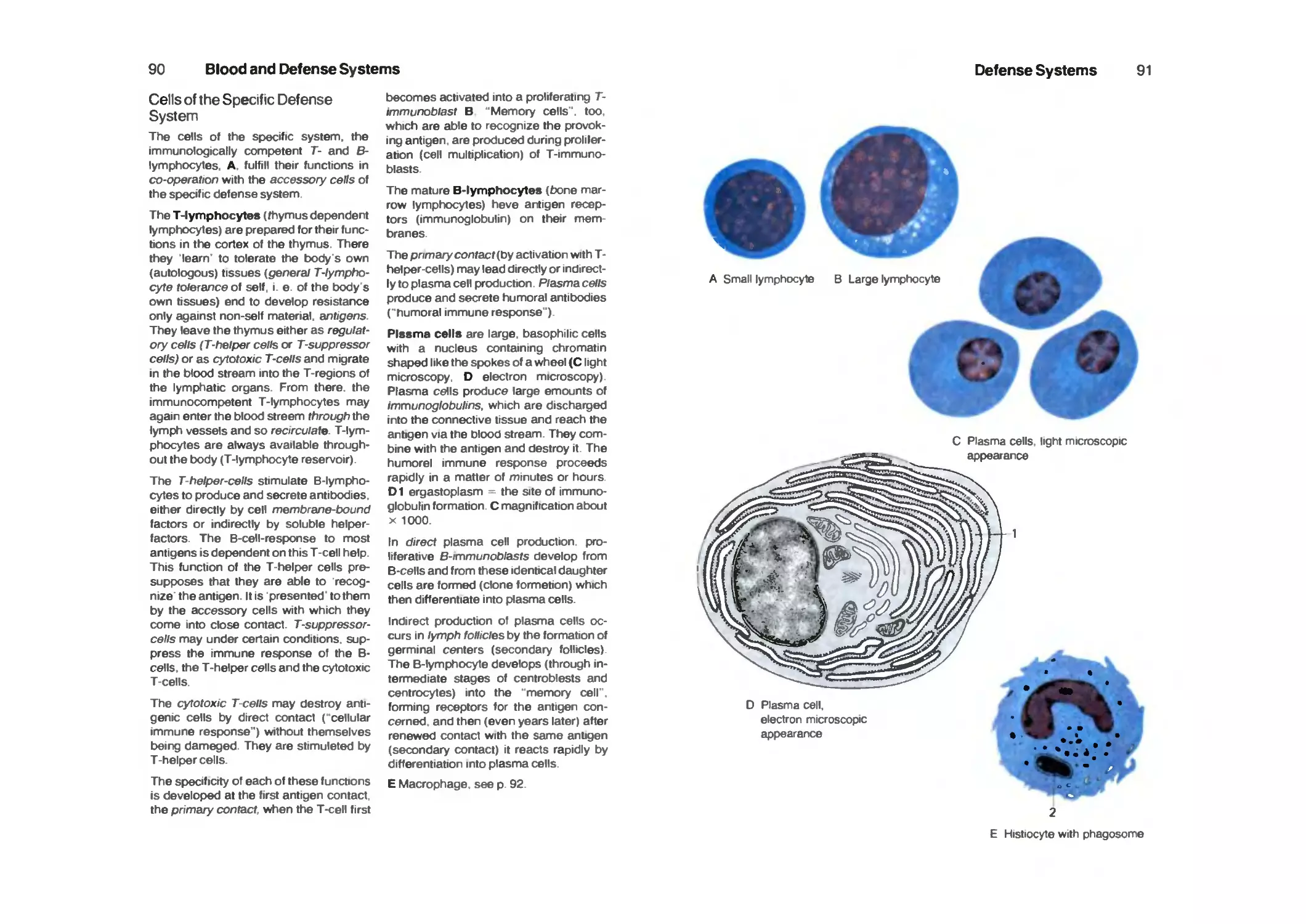
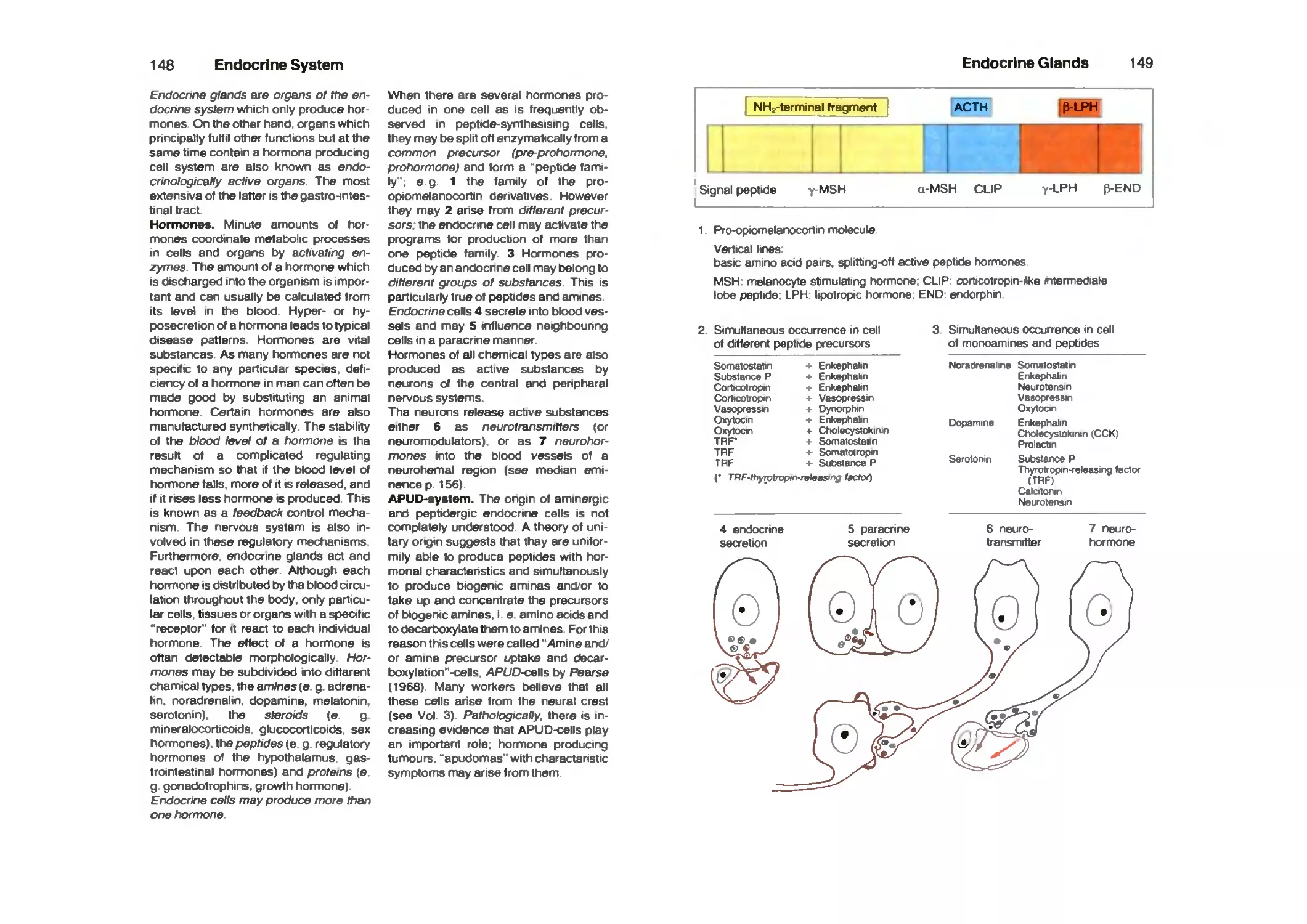
Текст
MED
Thieme Flexi
A well-balanced combination of a full-fledged anatomic
atlas and textbook, eminently useful to students and
medical practitioners alike. Skilful visual approach to
anatomy, which is a must in every physician's education,
is happily wedded to a lucid text juxtaposed page by page
to magnificent multicolor illustrations in such a manner that
the concise description of the functional aspects of
anatomy provideslet a useful guide for the perceptive
student. Aspects of physiology and biochemistry are
included to the extent they have a bearing on the material
presented.
ISBN 3-13-533403-1 (Thieme)
ISBN 0-86577-250-9 (Tl)
Color Atlas and Textbook
of Human Anatomy
in 3 Volumes
W. Kahle • H. Leonhardt • W. Platzer
Volume 2:
Internal Organs
Helmut Leonhardt
3rd revised edition
W. Kahle • H. Leonhardt • W. Platzer
Color Atlas and Textbook
of Human Anatomy
in 3 Volumes
Volume 2:
Internal Organs
by Helmut Leonhardt
Translated by H. L. and A. D. Dayan
3rd revised edition
170 color plates with 584 drawings
by Gerhard Spitzer
1986
Georg Thieme Verlag Thieme Inc.
Stuttgart • New York New York
Prof. Dr. med. Werner Kahle
Neurologisches Inslitut
(Edinger Instftut) der Univer-
sitat Frankfurt/Main, FRG
Prof. Dr. med. Helmut
Leonhardt
Direktordes Anatomischen
Instituts der Universitat Kiel,
FRG
Univ.-Prof. Dr. med. univ.
Werner Platzer
Vorstand des Anatomischen
Instituts 6er Universitat
Innsbruck, Austria
Gerhard Spiizer,
Frankfurt/Main, FRG
HediL. Dayan, M.B., and
Anthony D. Dayan, M.D.,
Beckenham, Kent, UK
1 St German edition 1976
2nd German edition 1978
3rd German edition 1979
4th German edition 1984
5th German edition 1986
1st English edition 1978
2nd English edition 1984
1 St Dutch edition 1978
2nd Dutch edition 1981
1 St French edition 1979
1st Greek edition 1985
1st Italian edition 1979
1 St Japanese edition 1979
2nd Japanese edition 1981
3rd Japanese edition 1984
1st Spanish edition 1977
Library of Congress
Cataloguing In Publication Data
Kahle, W. (Werner)
Color atlas and textbook of
human anatomy.
Translation of: Ta-
schenatlas der Anatomie.
Includes bibliographies
and indexes.
Contents: v. 1. Locomotor
system / by Werner Platzer -
V. 2. Internal organs / by
Helmut Leonhardt — v. 3.
Nervous system and sensory
organs / Werner Kahle.
1. Anatomy, Human-Atlases.
I. Leonhardt, Helmut.
II. Platzer, Werner. III. Title.
[DNLM: 1. Anatomy-atlases.
QSt7K12t|
QM25.K3413 1986
611'.022'2 86-5679
This book is an authorized translation from the 5th
German edition published and copyrighted © 1976,
1986 by Georg Thieme Verlag Stuttgart, Germany, and
may not be reproduced in part or in whole without written
permission from the publisher. Title of the German
edition: Taschenatlas der Anatomie, Band 2: Innere Or-
gane.
Some of the product names, patents and registered
designs referred to in this book are in fact registered
trademarks or proprietary names even though specific
reference to this fact is not always made in the text.
Therefore, the appearance of a name without
designation as proprietary is not to be construed as a
representation by the publishers that it is in the public domain.
This book, including all parts thereof, is legally protected
by copyright. Any use, exploitation or commercialization
outside the narrow limits set by copyright legislation,
without the publisher's consent, is illegal and liable to
prosecution. This applies in particular to photostat
reproduction, ccpying. mimeographing or duplication of any
kind, translating, preparation of microfilms, and
electronic data processing and storage.
© 1978,1986 Georg Thieme Verlag, Rudlgerstrasse 14,
D-7000 Stuttgart 30, FRG
Typesetting by Druckhaus Dorr, (Linotype System 5
[202])
Printed in West Germany by Druckhaus Dorr,
D-7140 Ludwigsburg
ISBN 3-13-533403-1 (Georg Thieme Verlag, Stuttgart)
ISBN 0-86577-250-9 (Thieme Inc.. New York)
1 2 3 4 5 6
Foreword
This pocket atlas is designed to provide a plain and clear compendium of
the essentral facts of human anatomy for the student of medicine. It also
demonstrates the basic knowledge of the subject for students of related
disciplines and for the interested layman. For all students preparation for
their examinations and practice requires repetition of visual experiences.
Text and illustrations in this book have been deliberately juxtaposed to
provide visual demonstration of the topics of anatomy.
The pocket atlas is divided according to organ systems into three volumes:
Volume 1 deals with the locomotor system, Volume 2 with the internal
organs and skin and Volume 3 with the nervous system and the organs of
the special senses. The topographic relationships of the peripheral
pathways of nerves and vessels are considered in Volume 1, in so far as they
are closely related to the locomotor system; Volume 2 systematically
describes the distribution of the vessels. The floor of the pelvis (pelvic
cavity), which has a close functional relationship with the organs of the
lesser pelvis, and the relevant topography are incorporated in Volume 2.
The developmental anatomy (embryology) of the teeth is briefly mentioned
in Volume 2 because it aids unterstanding of the eruption of the teeth. The
common embryological origins of the male and female genital organs are
also discussed because it helps to explain their structure in the adult, as
well as their not infrequent variants and malformations. Certain problems
connected with pregnancy and childbirth are mentioned in the chapter on
the female reproductive organs. But these do not cover all the knowledge
of embryology required by students. The notes on physiology and
biochemistry are deliberately brief and only serve to provide better
understanding of structural details. Reference should be made to textbooks of
physiology and biochemistry. Finally, it must be emphasized that no
pocket atlas can replace a major textbook or the opportunity to examine
macroscopic dissections and microscopic preparations.
The reference list mentions textbooks and original papers as a guide to the
more advanced literature, and it also cites clinical textbooks of relevance
to the study of anatomy.
Those who require less detailed knowledge of the structure of the human
body will find clear illustrations, too, of the anatomic bases of the more
important methods of medical examination. To help the nonmedical
reader, everyday English terms for the major organs and their parts have
been supplied as far as feasible; these terms are also listed in the index.
Frankfurt/Main, Kiel, Innsbruck
The Editors
Foreword to the 3rd English Edition of Volume 2
I owe thanks to many colleagues who have given valuable advie about
facts and technical matters. I particularly wish to thank Prof. Junzo Ochi,
Shiga University, Seta, Otsu City, who translated the book into Japanese,
for his numerous improvements. Suggestions from students have led to
better presentation of the material. I must thank Dr. h. c. G. Hauff and Dr.
D. Bremkamp, the initiators of the work and their colleagues, the
publishers, Georg Thieme Verlag, for the willingness with which they have
overcome the technical problems.
Kiel, December 1985
Helmut Leonhardt
From the Foreword to 1st Edition of Volume 2
Changes in medical education have led to a shortening of the preclinical
studies and to major changes in their emphasis. Knowledge of anatomy in
particular now tends to be gained mostly from formal lecture courses and
examinations place little reliance on practical work, such as dissection.
Under these circumstances, there is a danger that the learning of anatomy
will become just repetition of lessons without any visualisation of the facts.
However, anatomical knowledge can only live through visual
understanding.
This pocket book is brought to life mainly by its illustrations. I thank Herr G.
Spitzer who has produced them. The book originated from knowledge
gained by teaching students. It is meant principally for medical students
and the interested lay reader, whose place has been taken by Renate and
Matthias.
Homburg (Saar), October 1972
Helmut Leonhardt
VI
Contents
Foreword Ill
Preface IV
How to Use This Book 1
Viscera 2
Circulatory System 4
Heart 6
Shape of the Heart 6
Chambers of the Heart 8
Valves of the Heart 10
Cardiac Muscle 12
Action of the Heart 16
Intrinsic Impulse-Conducting System and Cardiac Nerves 18
Coronary Vessels 20
Pericardium 22
Position of the Heart I 24
Position of the Heart II
(Auscultation and Percussion) 26
Radiology of the Heart 28
Measurements of the Heart; Alterations in its Shape and Size 30
Blood Vessels 32
Tasks of the Blood Vessel Walls 32
Structure of the Walls of Blood Vessels 34
Arteries 36
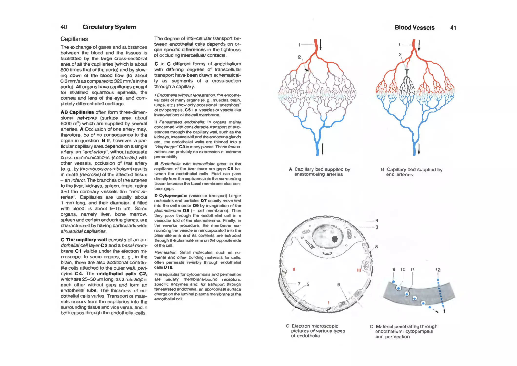
Capillaries 40
Veins 42
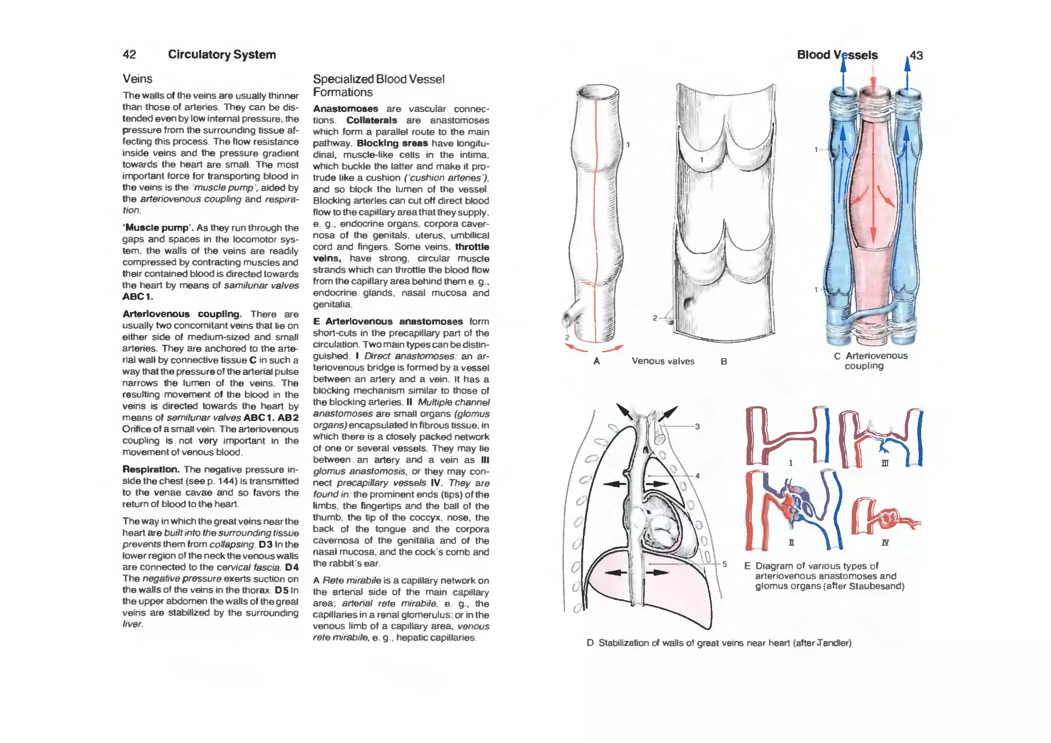
Specialized Blood Vessel Formations 42
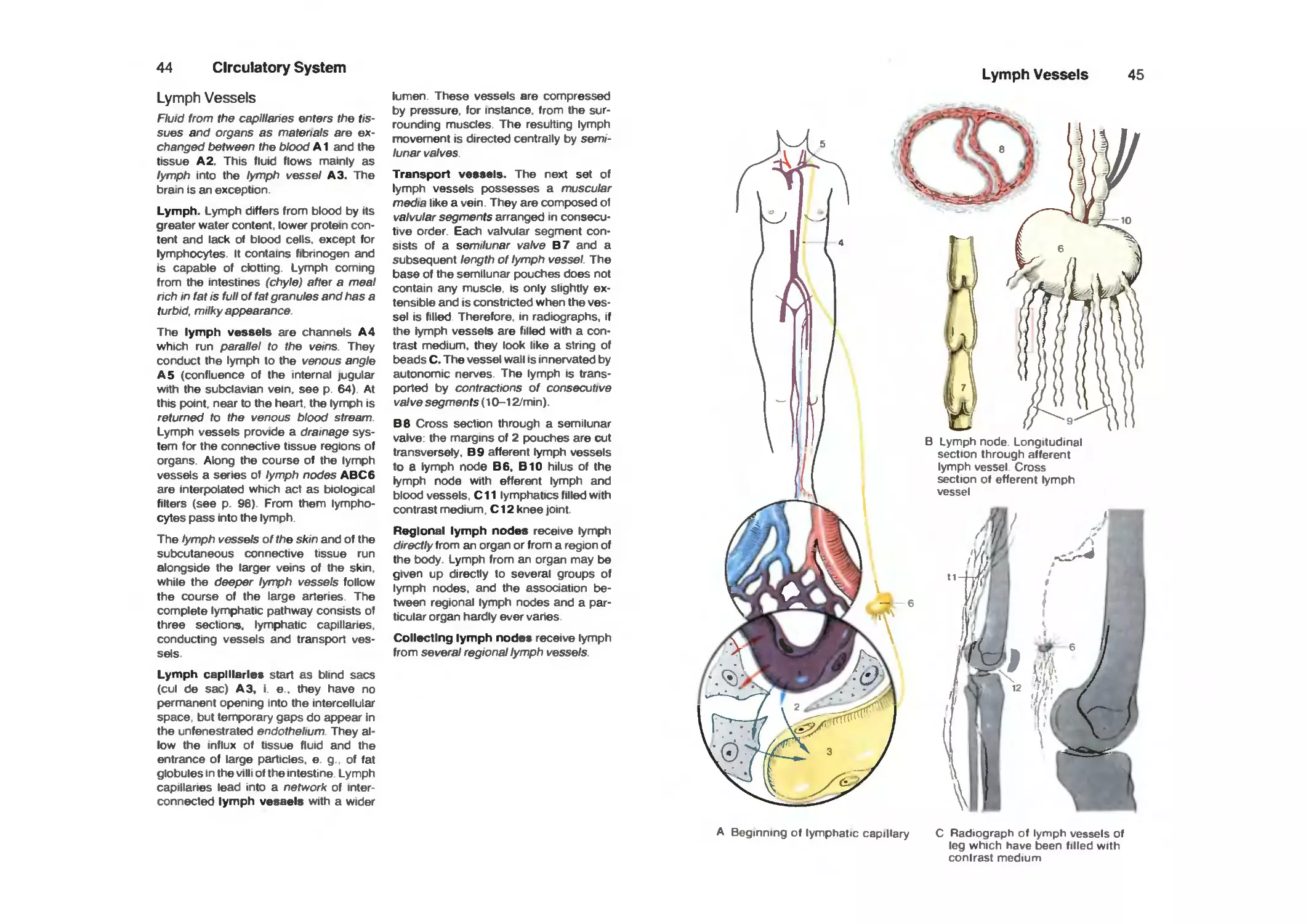
Lymph Vessels 44
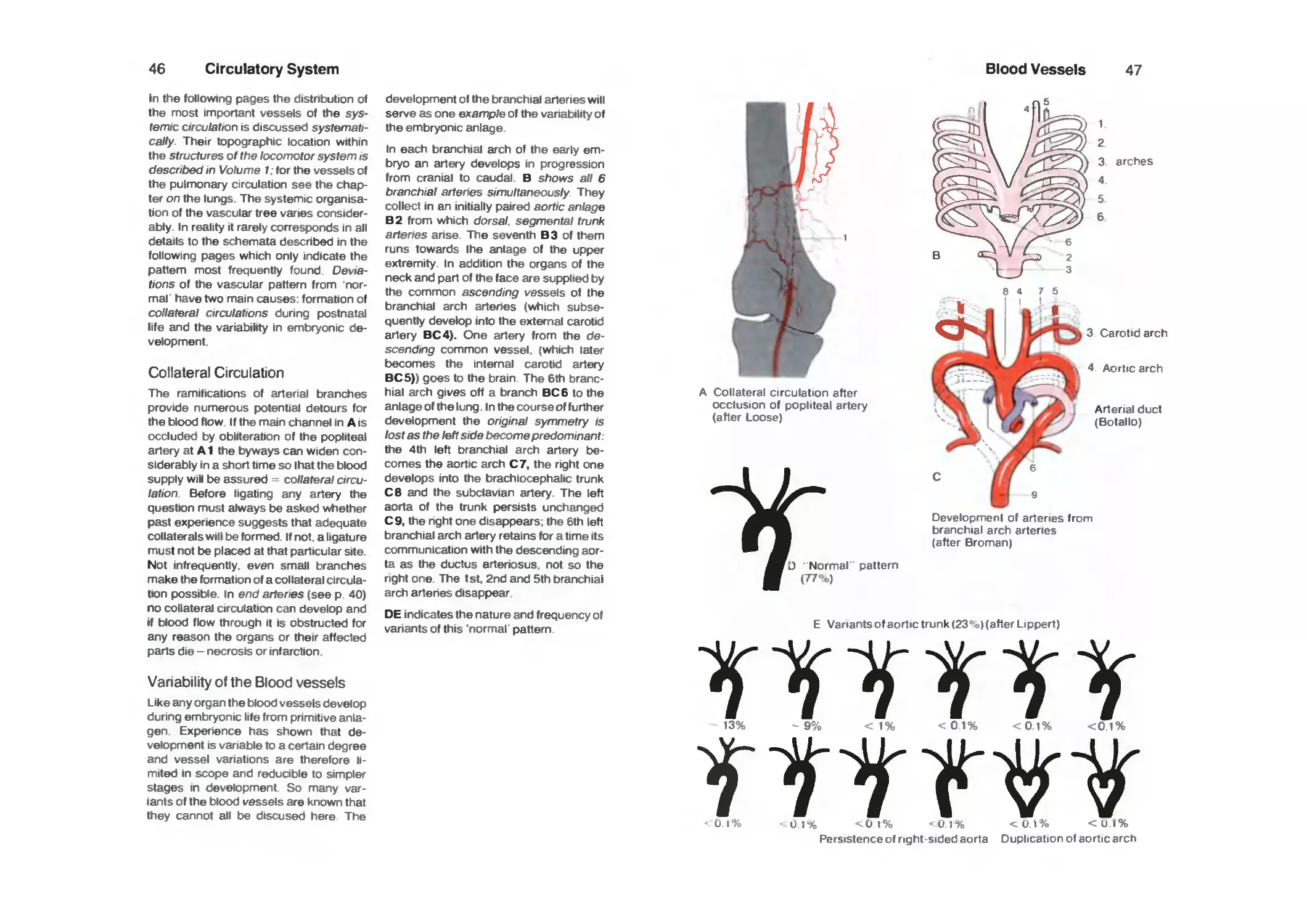
Collateral Circulation 46
Variability of the Blood Vessels 46
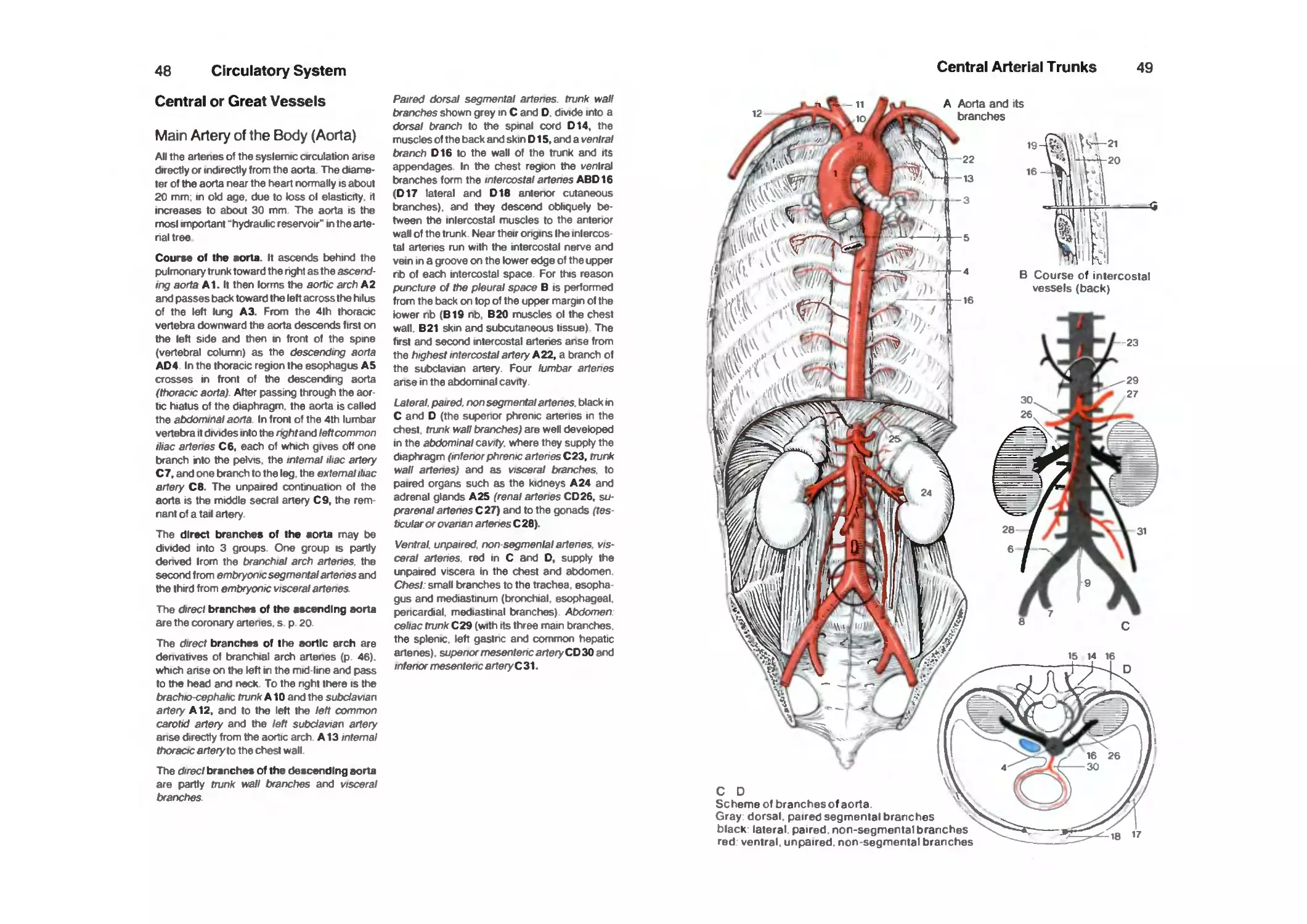
Central or Great Vessels 48
Main Artery of the Body (Aorta) 48
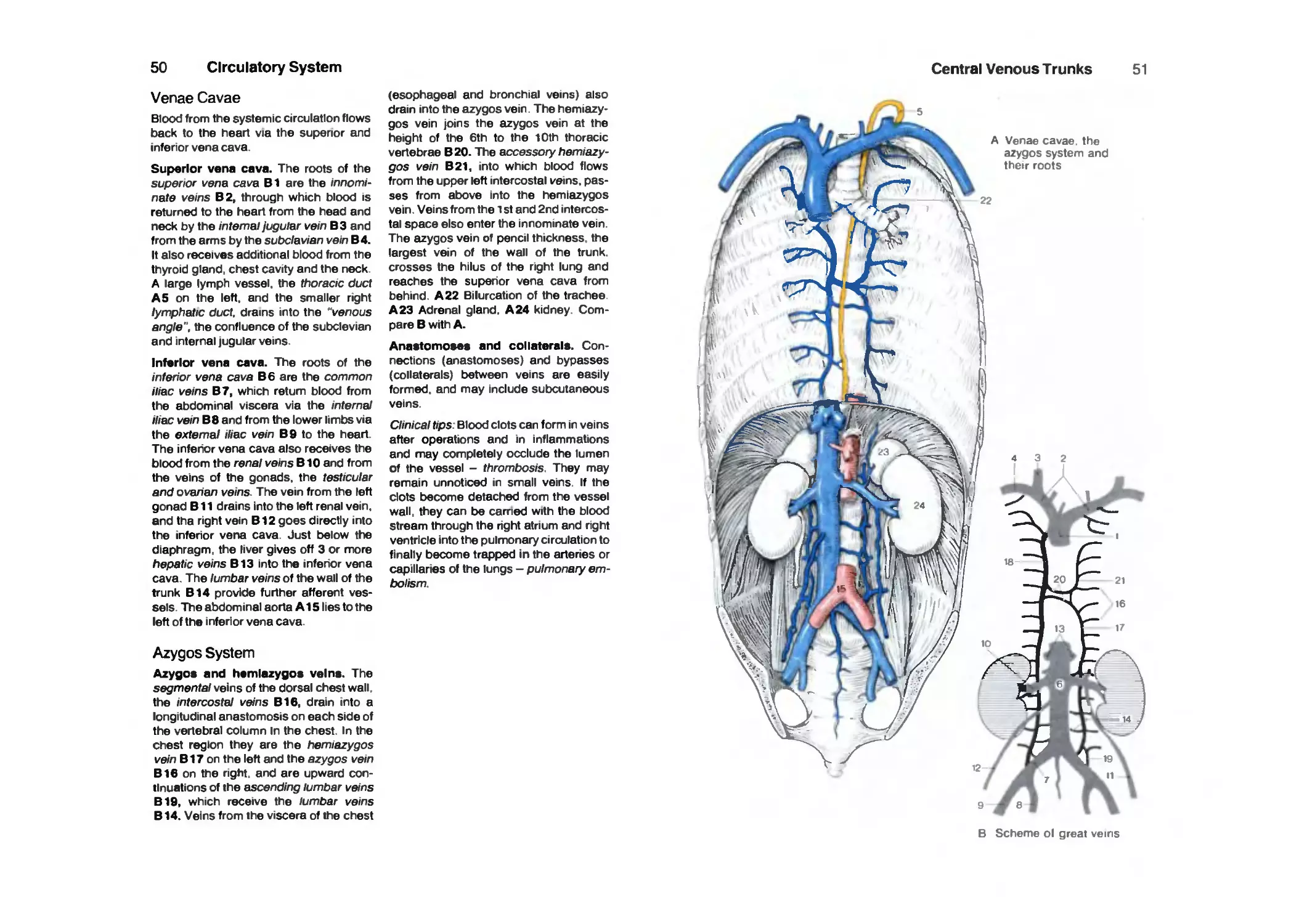
Venae Cavae 50
Azygos System 50
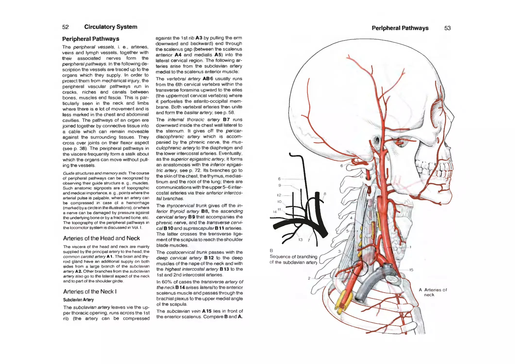
Peripheral Pathways 52
Arteries of the Head and Neck 52
Arteries of the Neck I 52
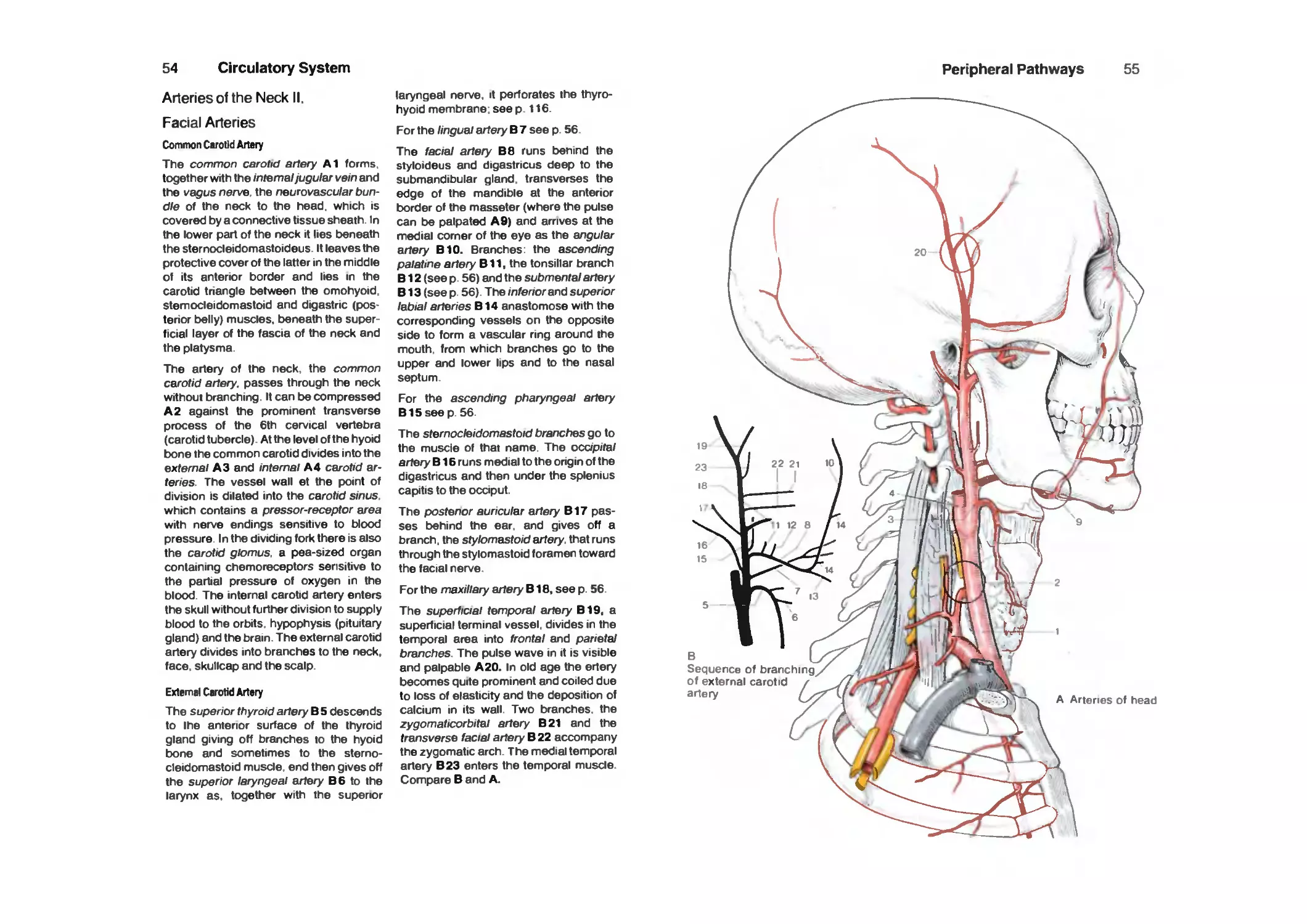
Arteries of the Neck II, Facial Arteries I 54
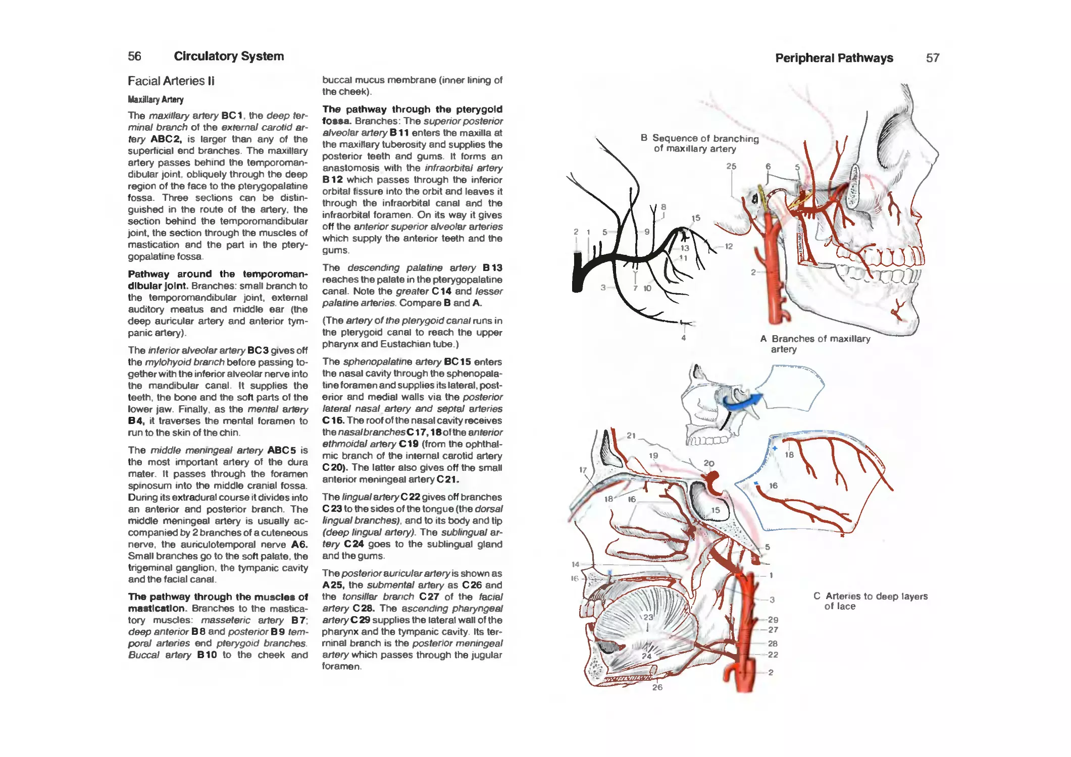
Facial Arteries II 56
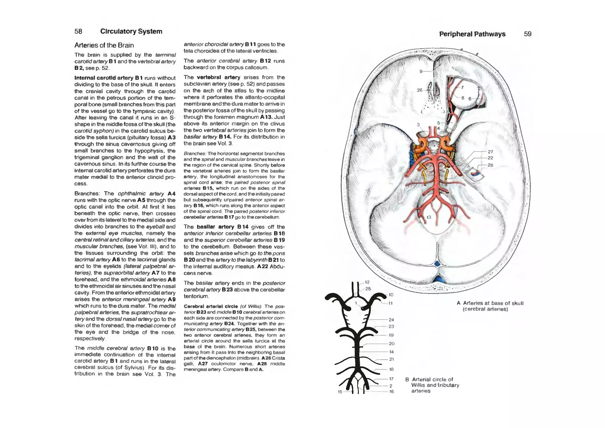
Arteries of the Brain 58
Veins of the Brain and Venous Sinuses of the Dura Mater I,
Veins of the Vertebral Column 60
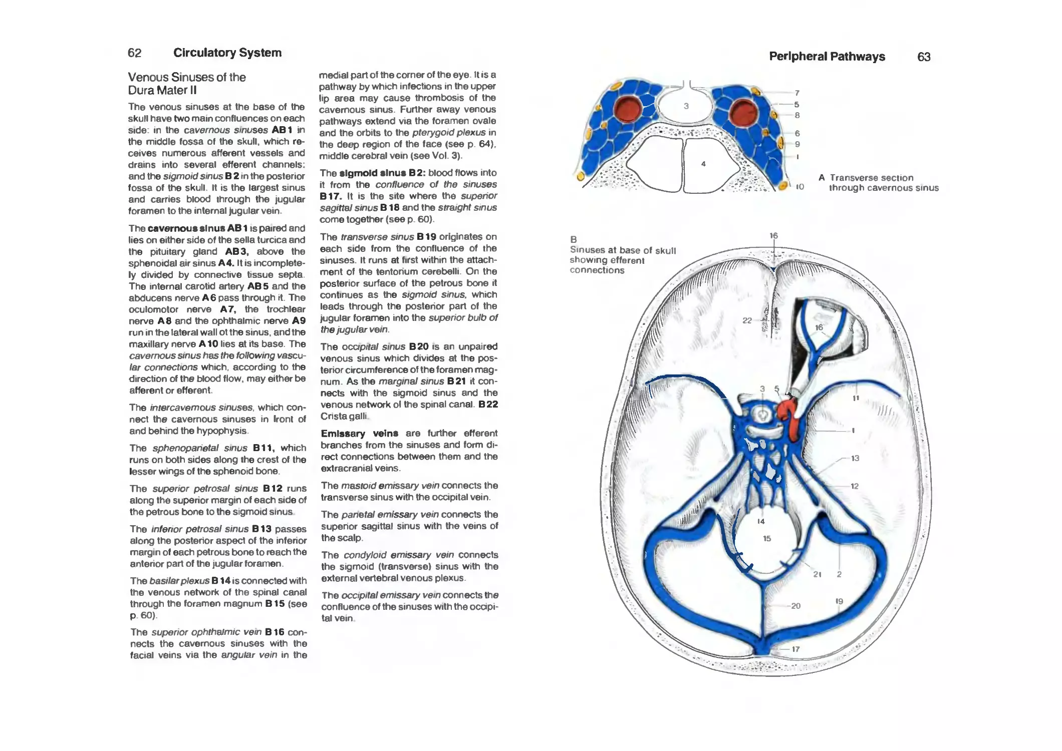
Venous Sinuses of the Dura Mater II 62
Contents
VII
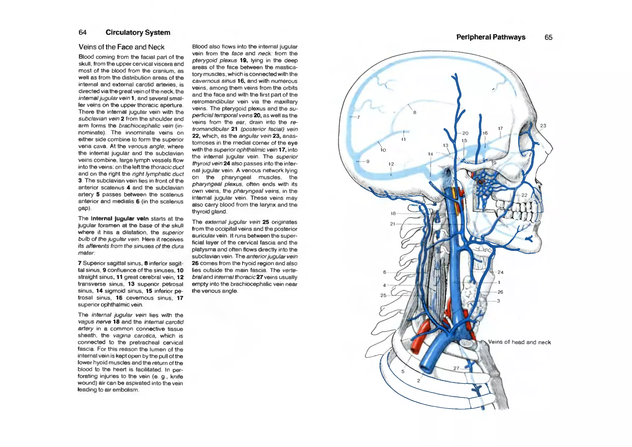
Veins of the Face and Neck 64
Arteries of the Shoulder and Upper Arm 66
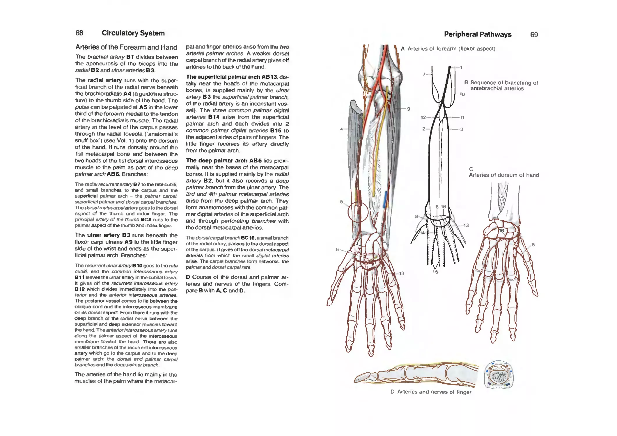
Arteries of the Forearm and Hand 68
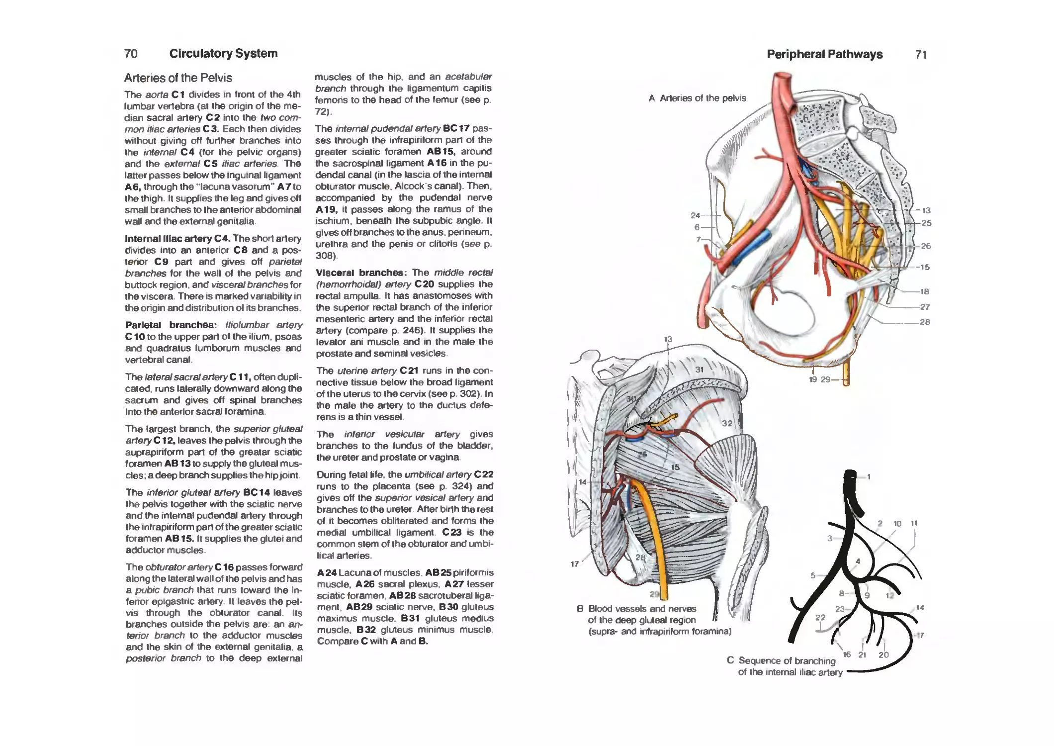
Arteries of the Pelvis 70
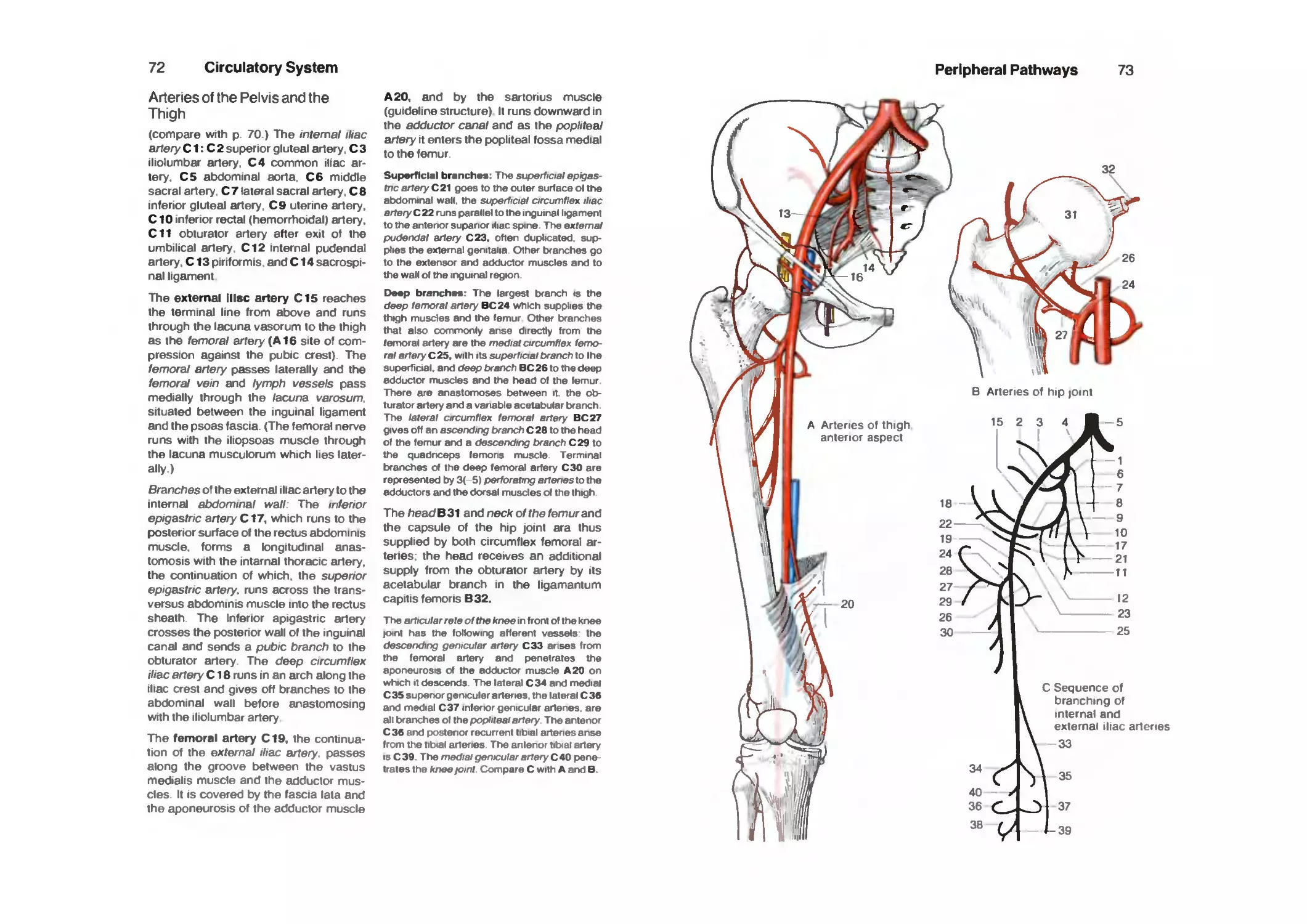
Arteries of the Pelvis and the Thigh 72
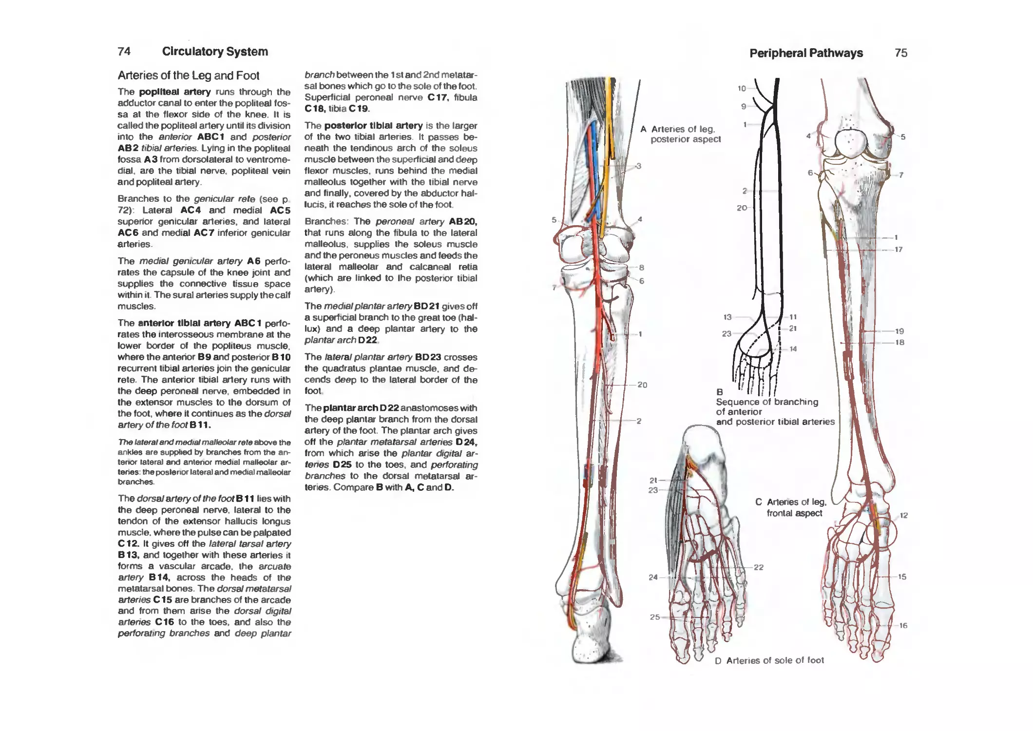
Arteries of the Leg and Foot 74
Subcutaneous Veins 76
Superficial Lymph Vessels of the Trunk and Lymph Nodes of the Arm
and Leg 78
Lymph Vessels (Lymph Nodes) of the Head and Neck and the Deep
Lymph Vessels (Lymph Nodes) of the Trunk 80
Blood and Defense Mechanisms 82
Blood 82
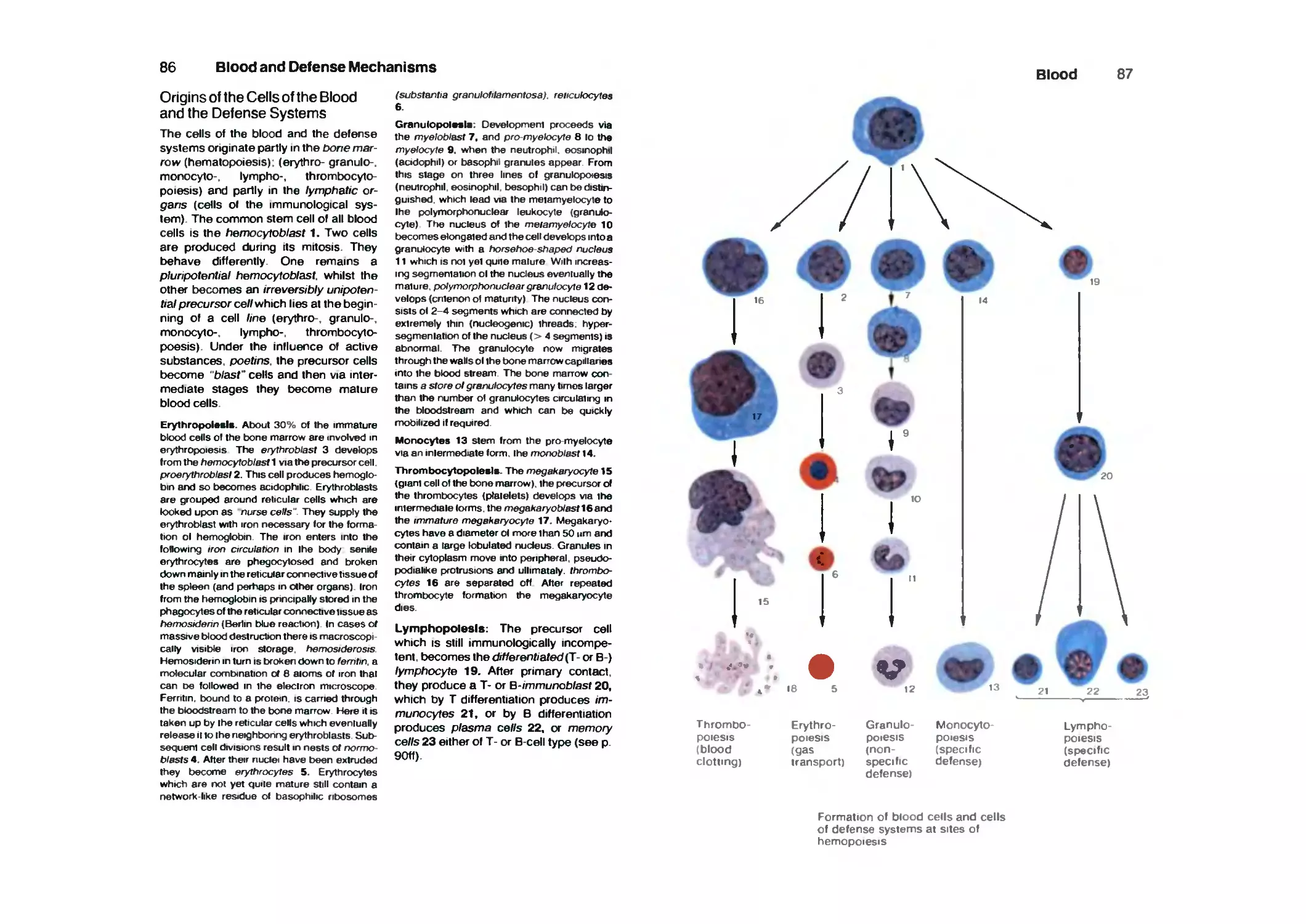
Origins of the Cells of the Blood and the Defense Systems 86
Blood and Defense Systems 88
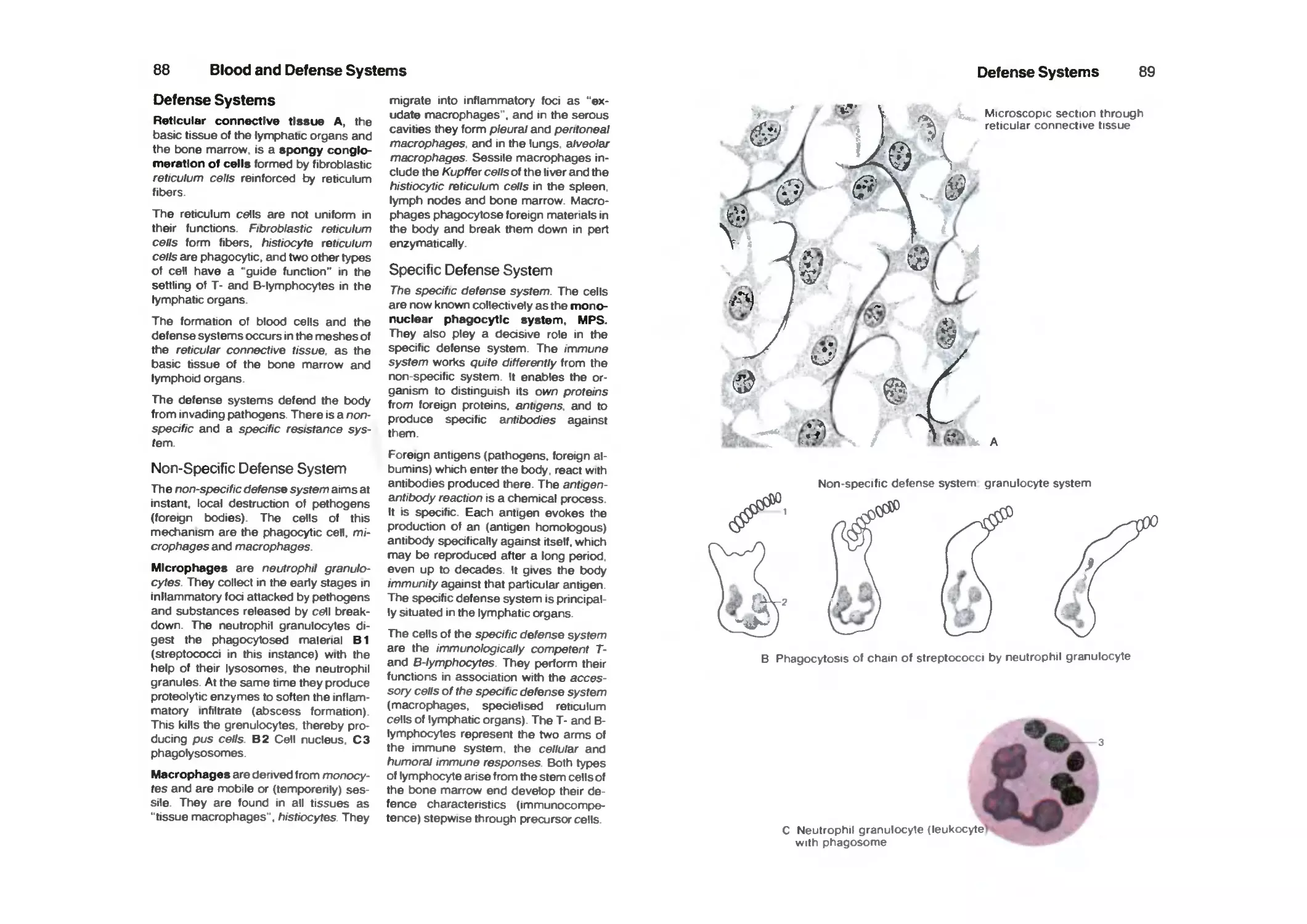
Reticular Connective Tissue 88
Defense Systems 88
Non-specific Defense System 88
Specific Defense System 88
Lymphatic Organs 92
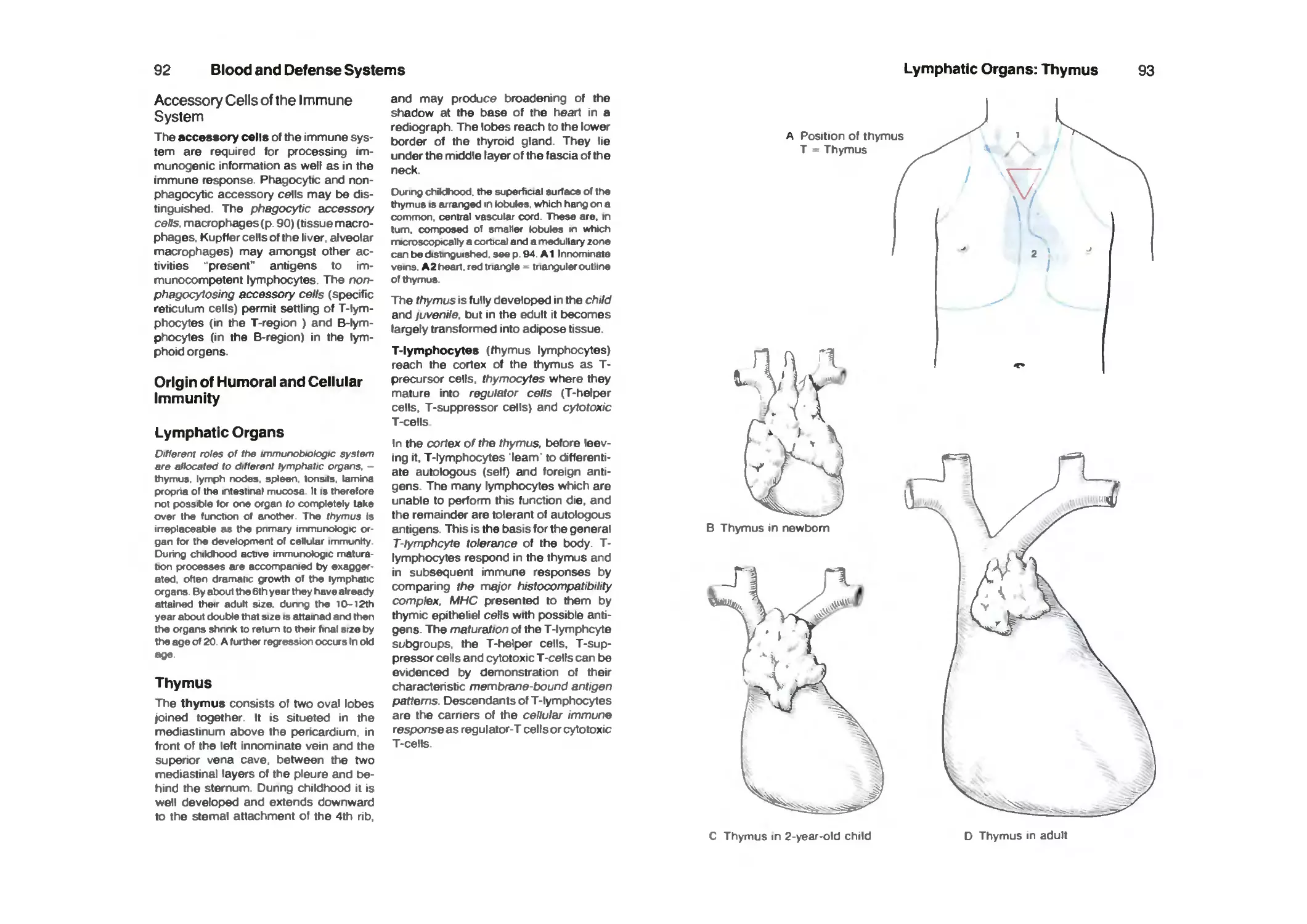
Thymus 92
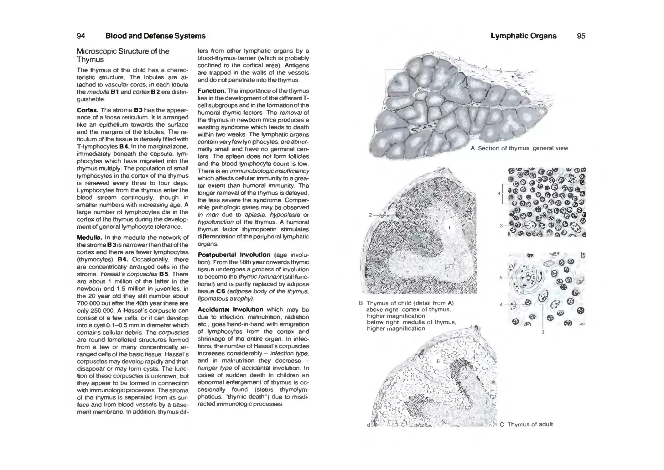
Microscopic Structure of the Thymus 94
Lymph Follicles 96
Lymph Nodes 96
Spleen 98
Fine Structure of the Spleen 100
Tonsils 102
Lymphatic Tissue of the Mucous Membranes 102
Respiratory System 104
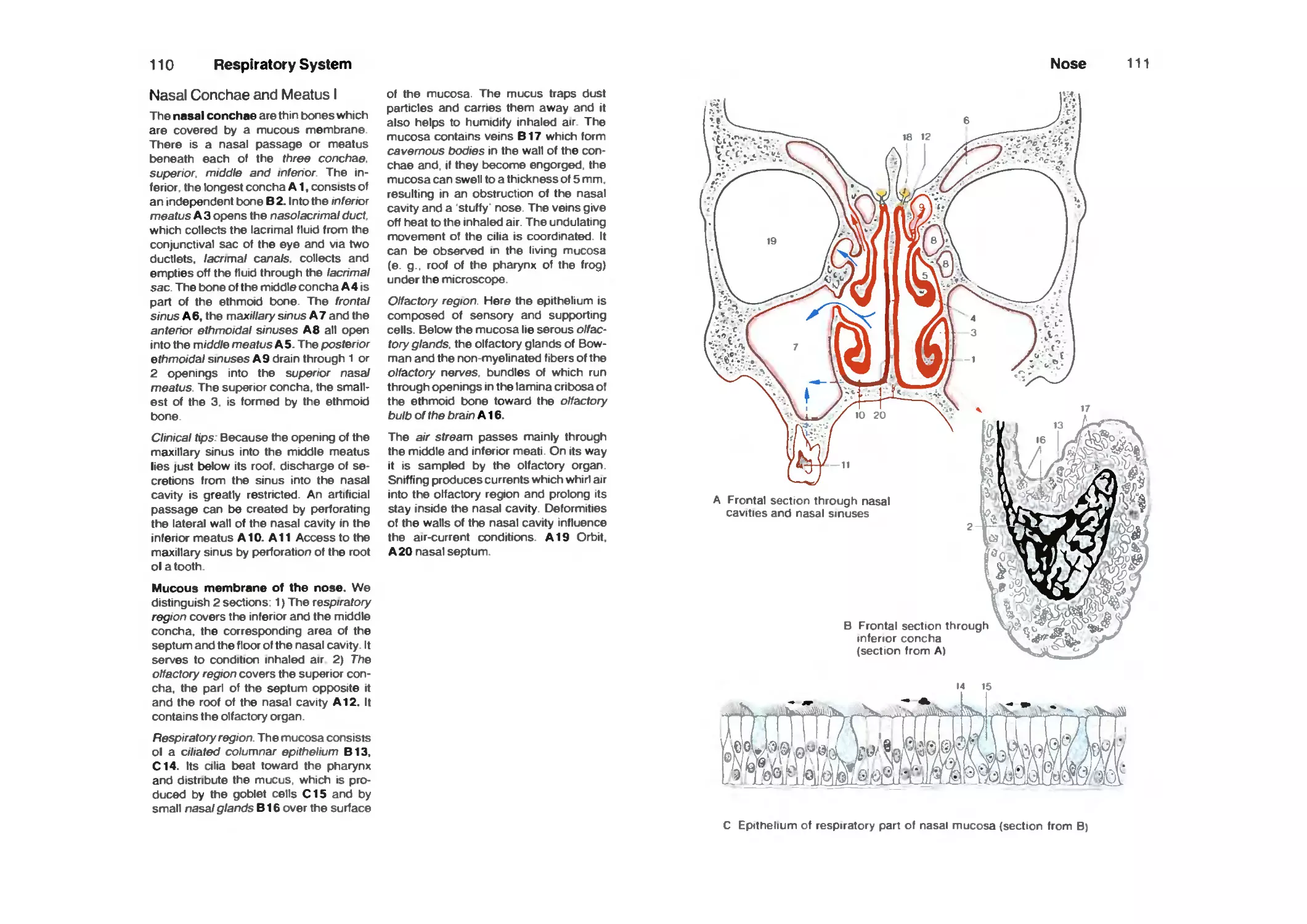
Nose . -. 106
Nasal Conchaeand Meatus I 108
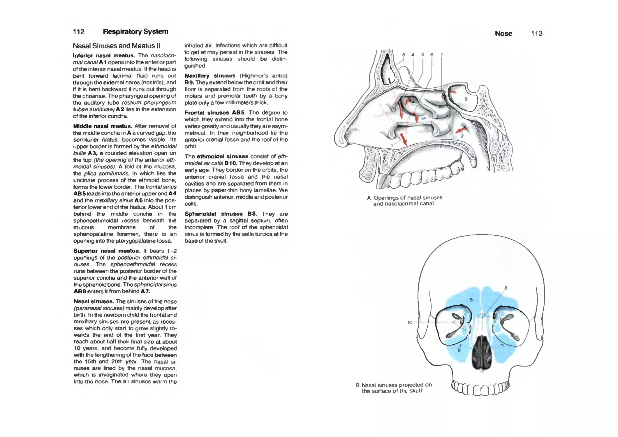
Nasal Sinuses and Meatus II 110
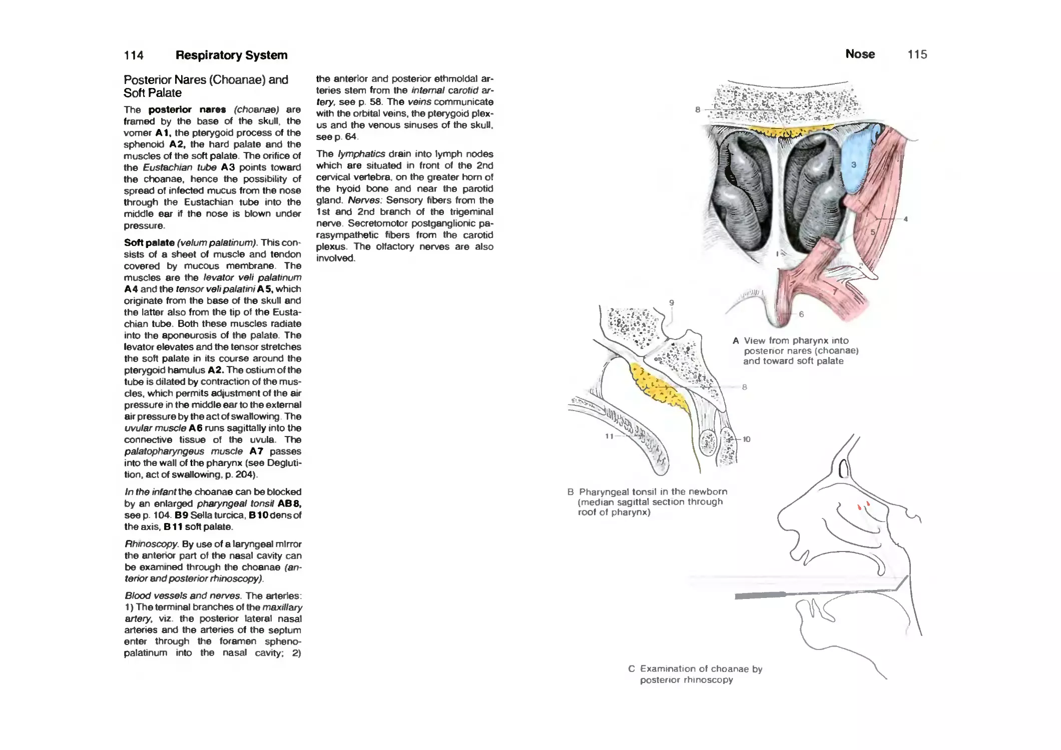
Posterior Nares (Choanae) and Soft Palate 112
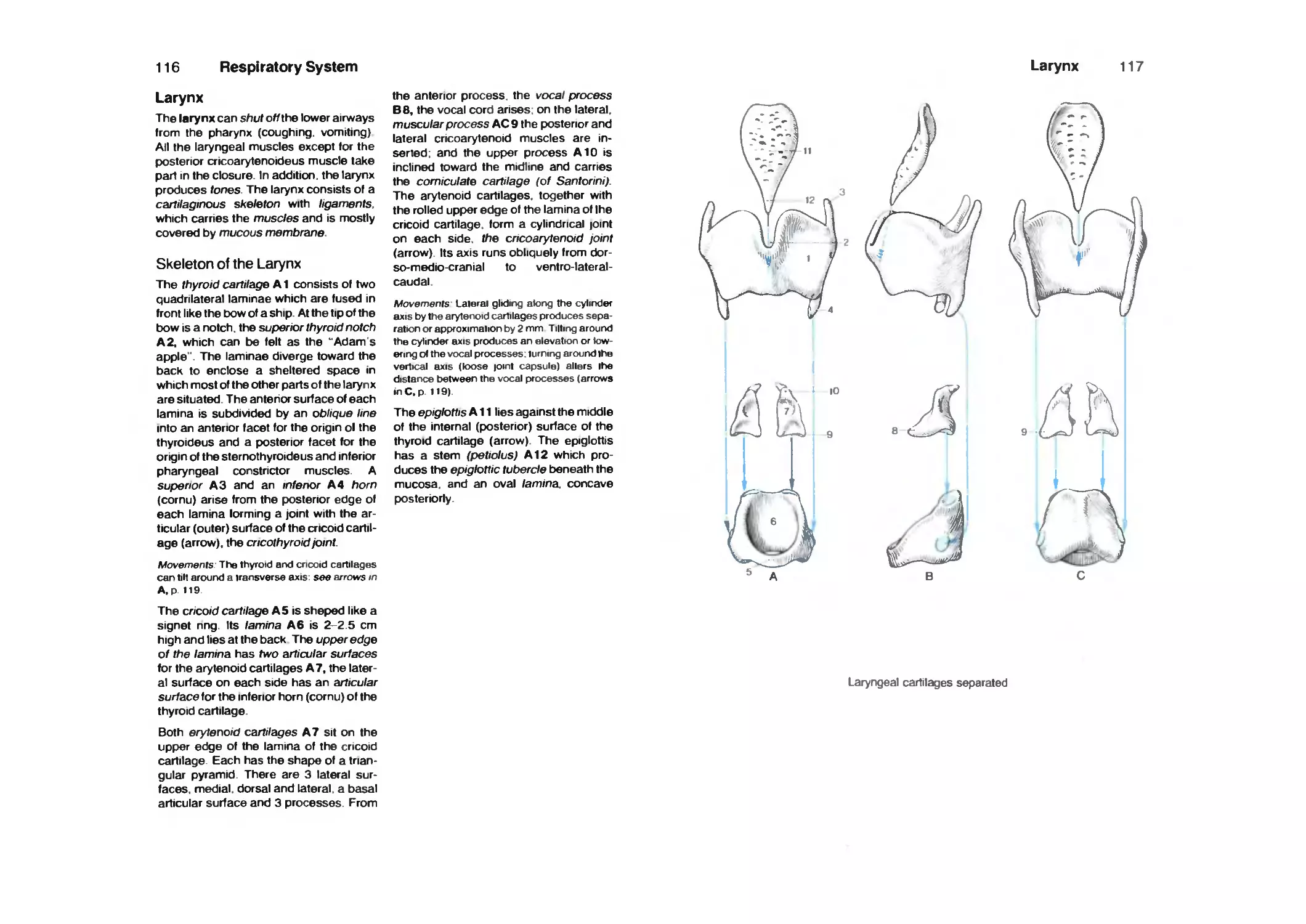
Larynx 114
Skeleton of the Larynx 114
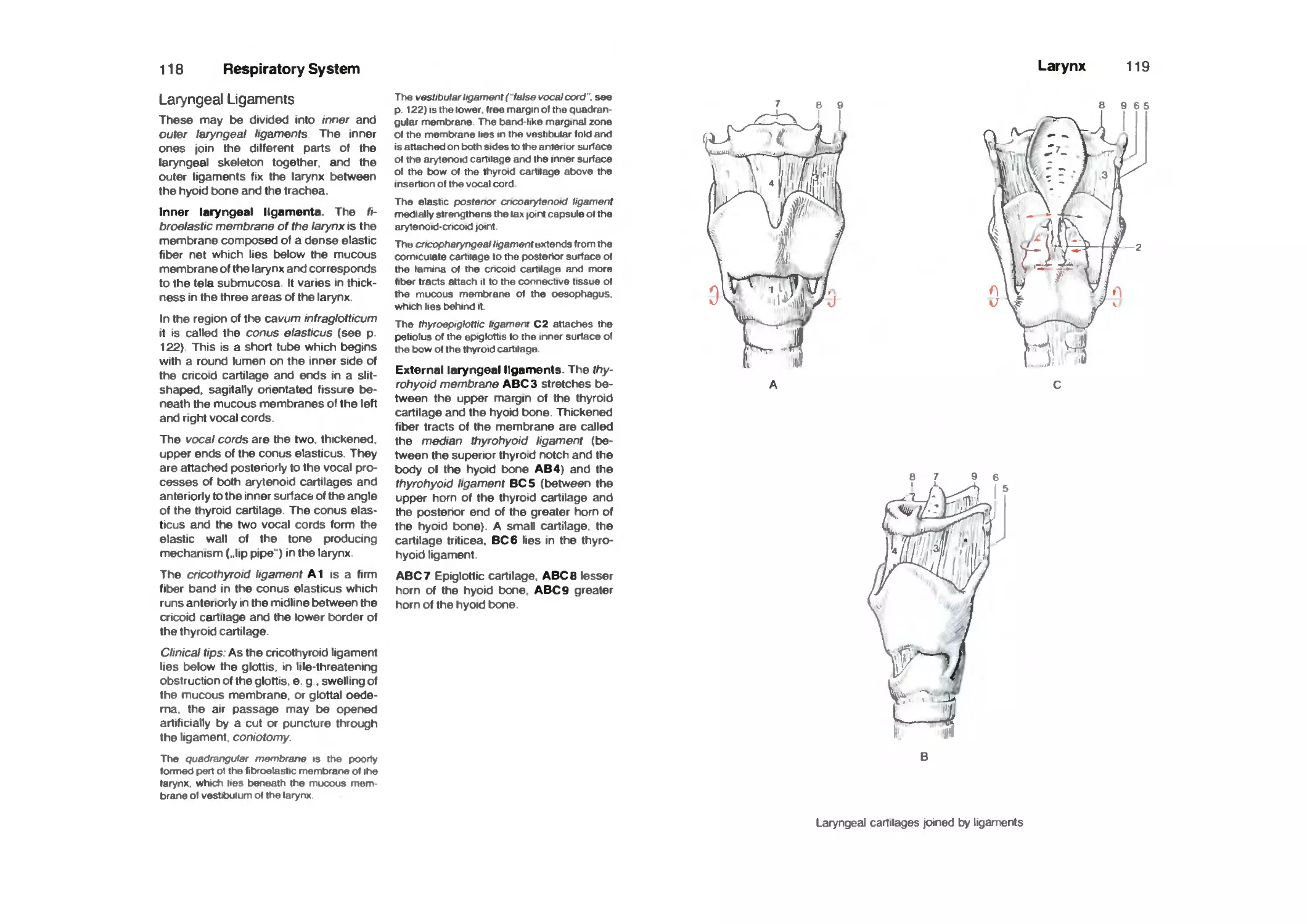
Laryngeal Ligaments 116
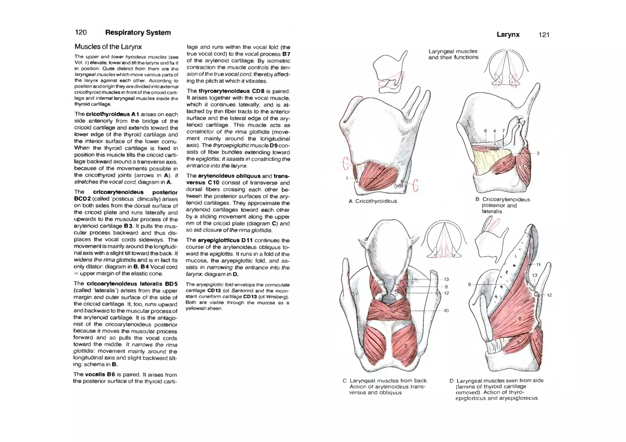
Muscles of the Larynx 118
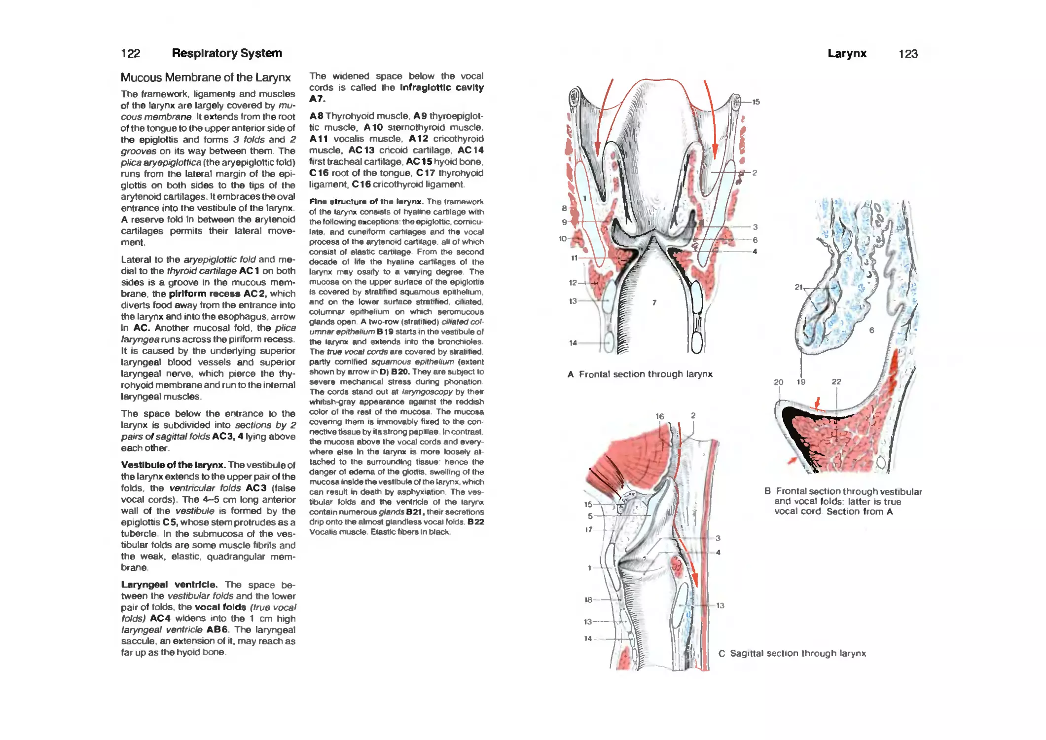
Mucous Membrane of the Larynx 120
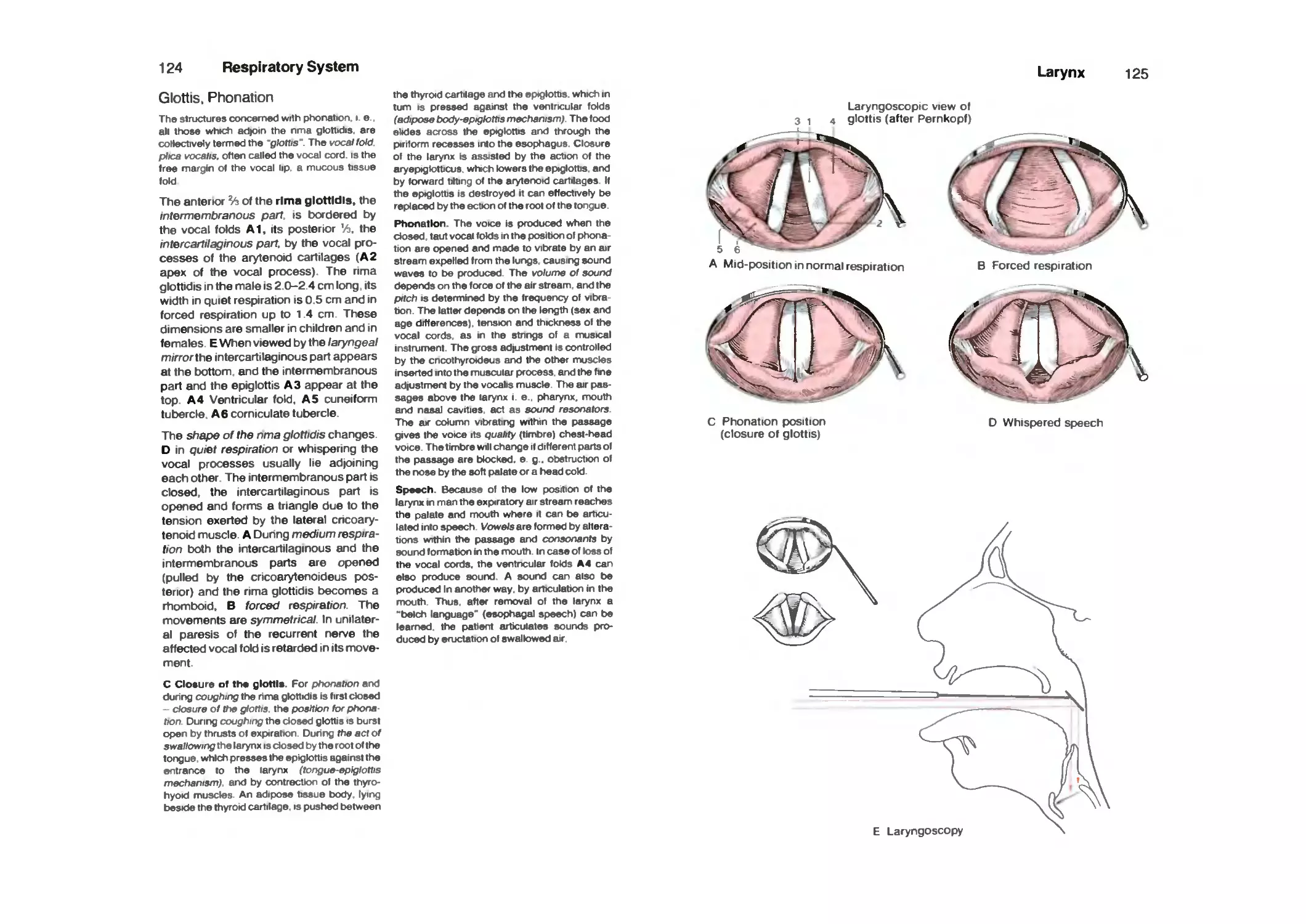
Glottis, Phonation 122
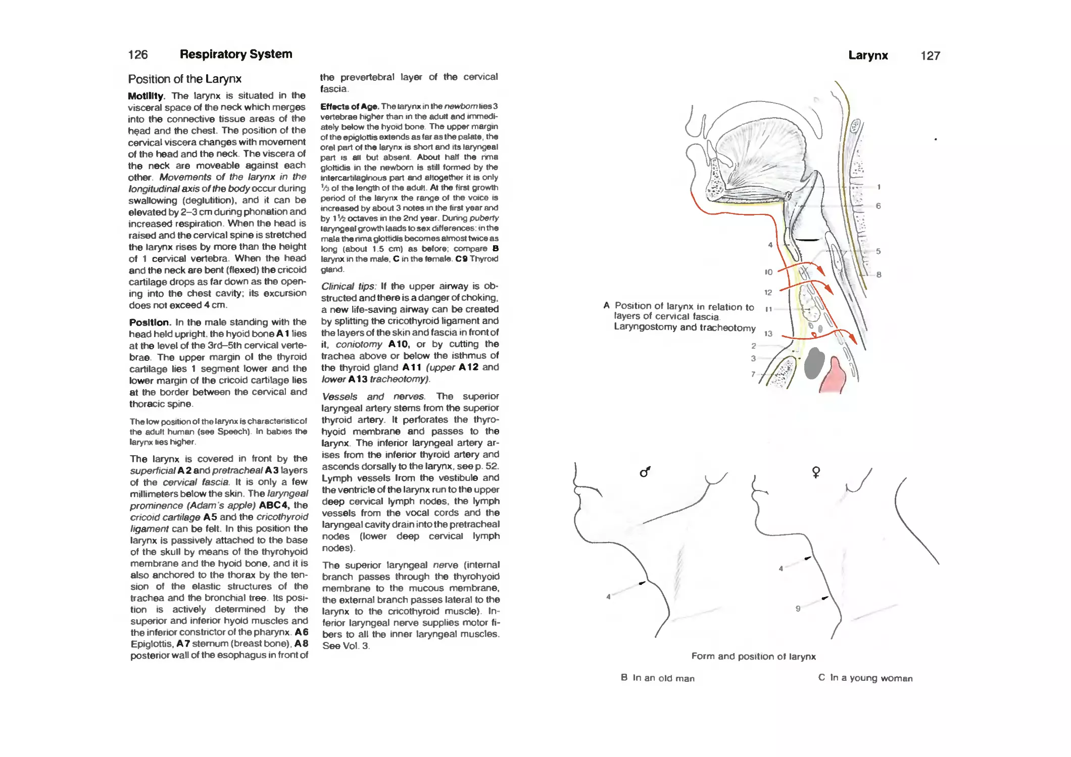
Position of the Larynx 124
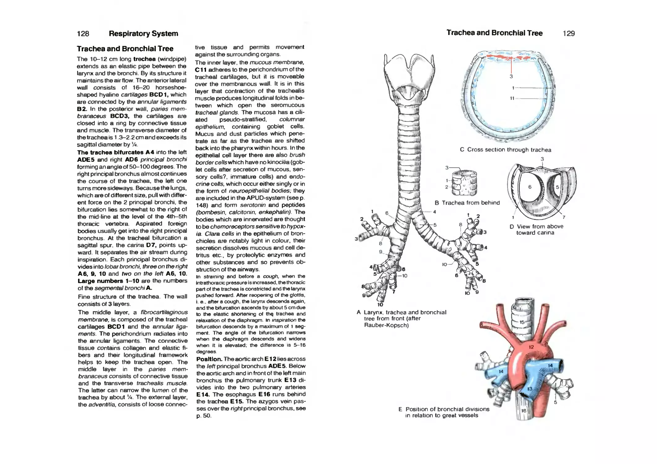
Trachea and Bronchial Tree 126
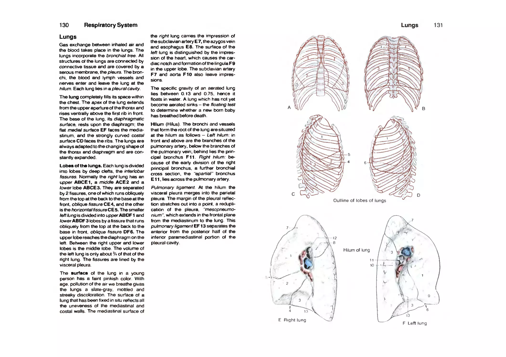
Lungs 128
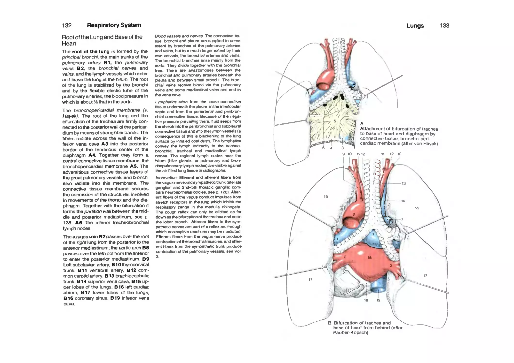
Root of the Lung and Base of the Heart 130
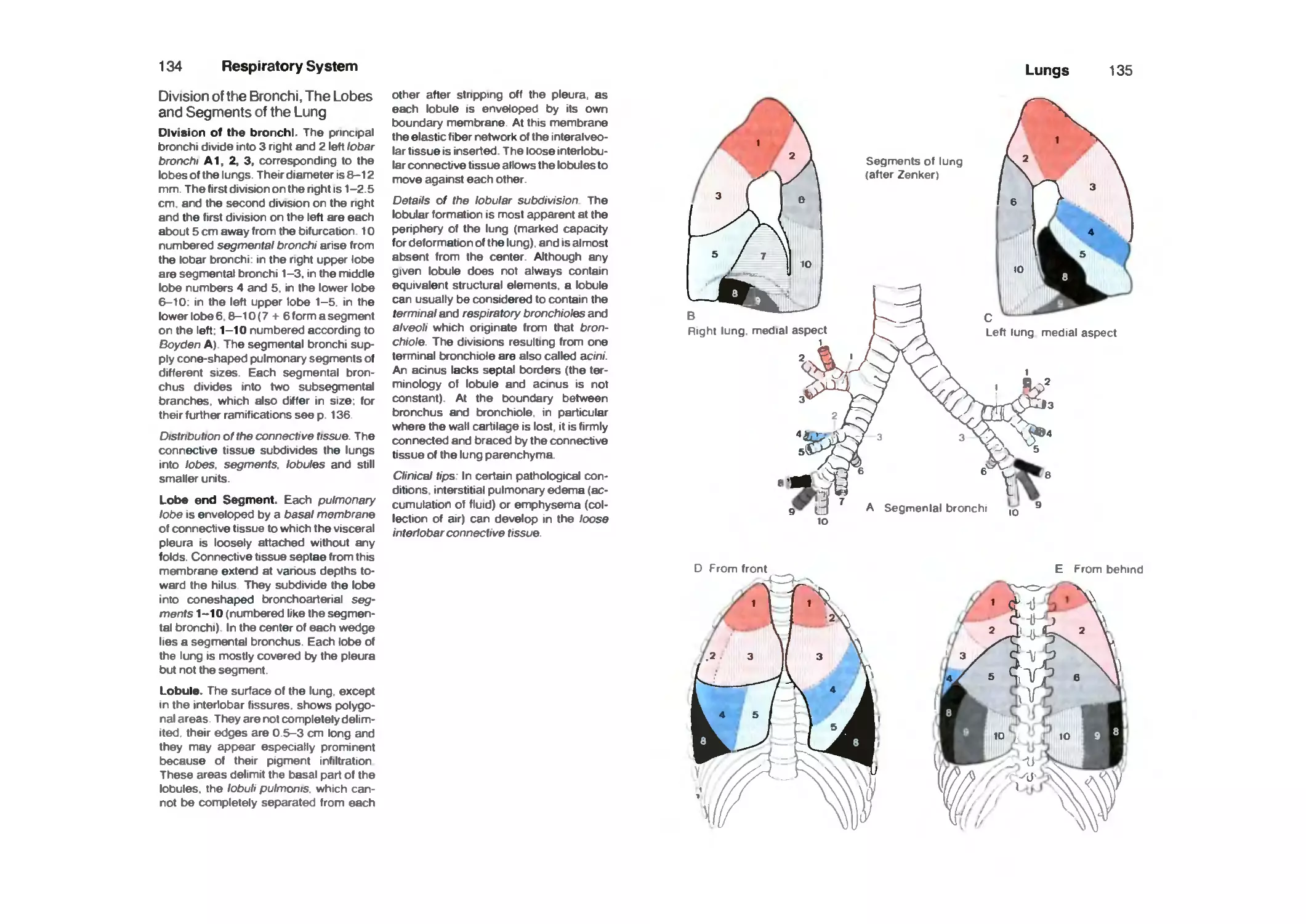
Division of the Bronchi, the Lobes and Segments of the Lung 132
VIII
Contents
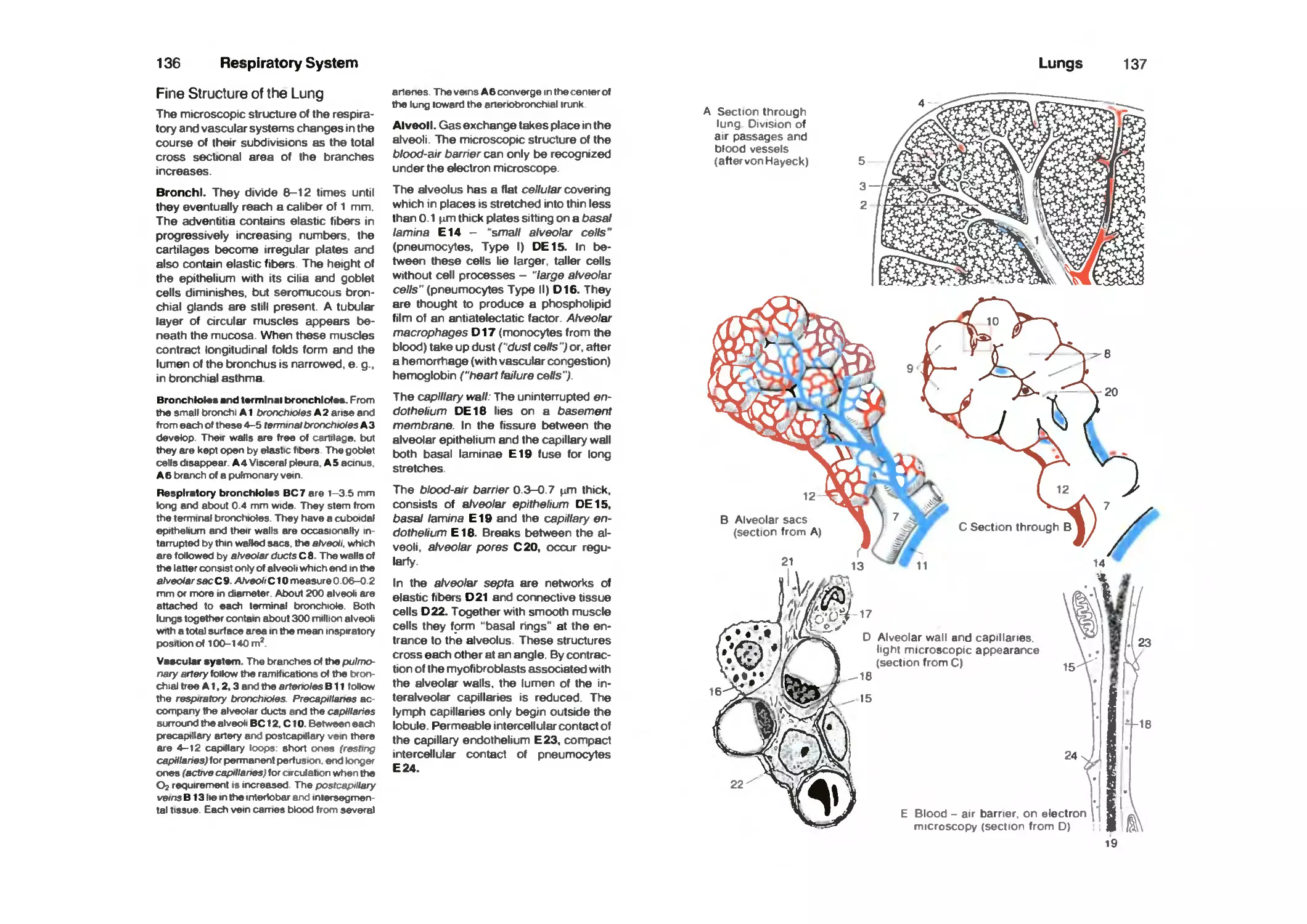
Fine Structure of the Lung 134
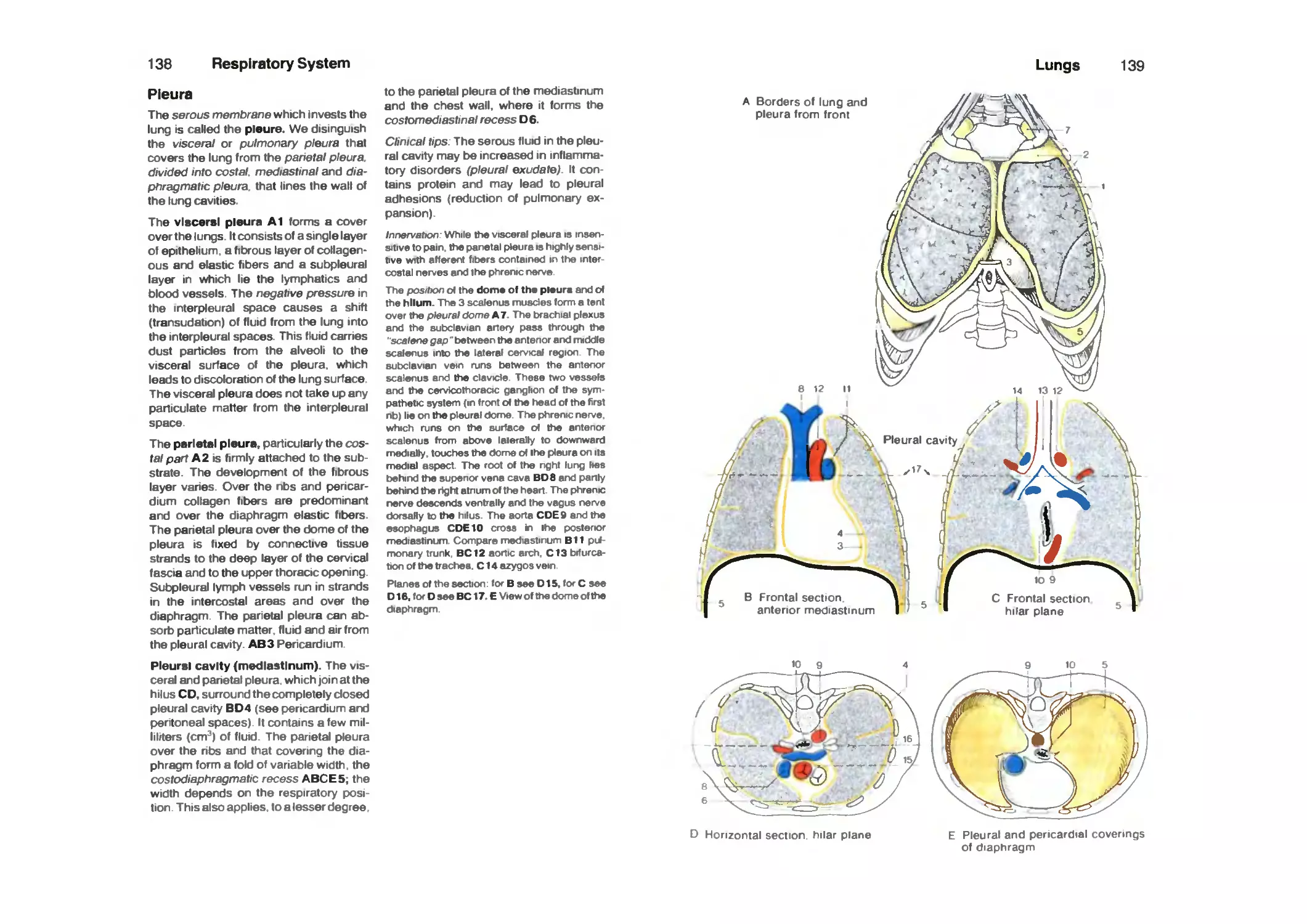
Pleura 136
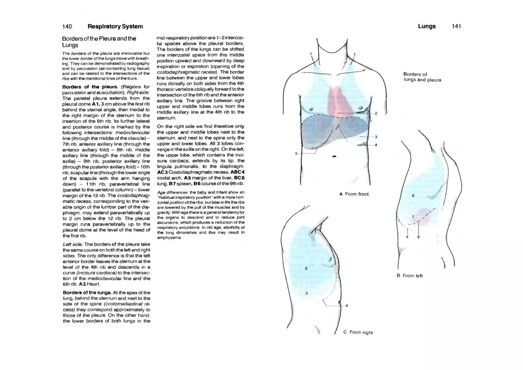
Borders of the Pleura and the Lungs 138
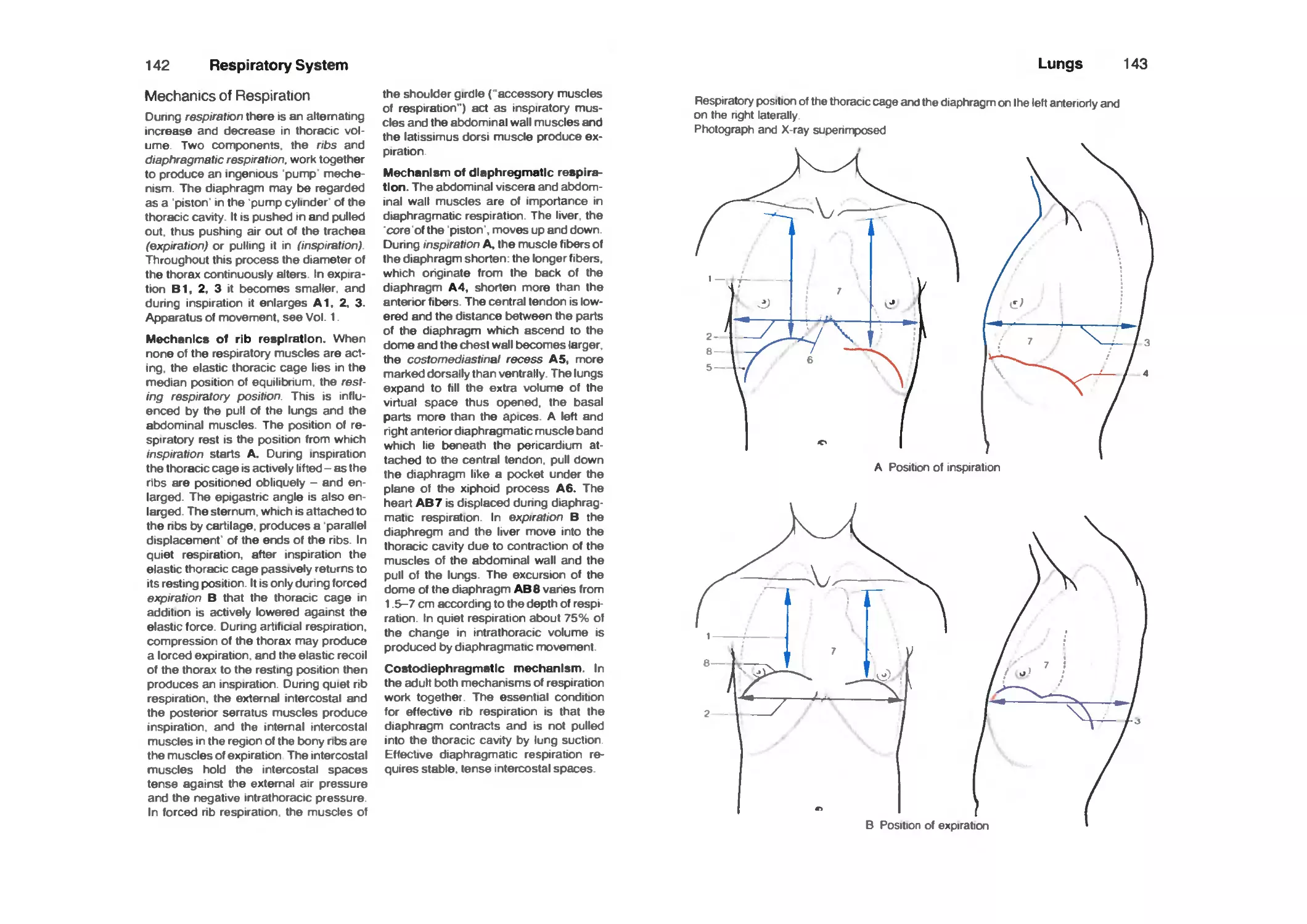
Mechanics of Respiration 140
Respiratory Dynamics 142
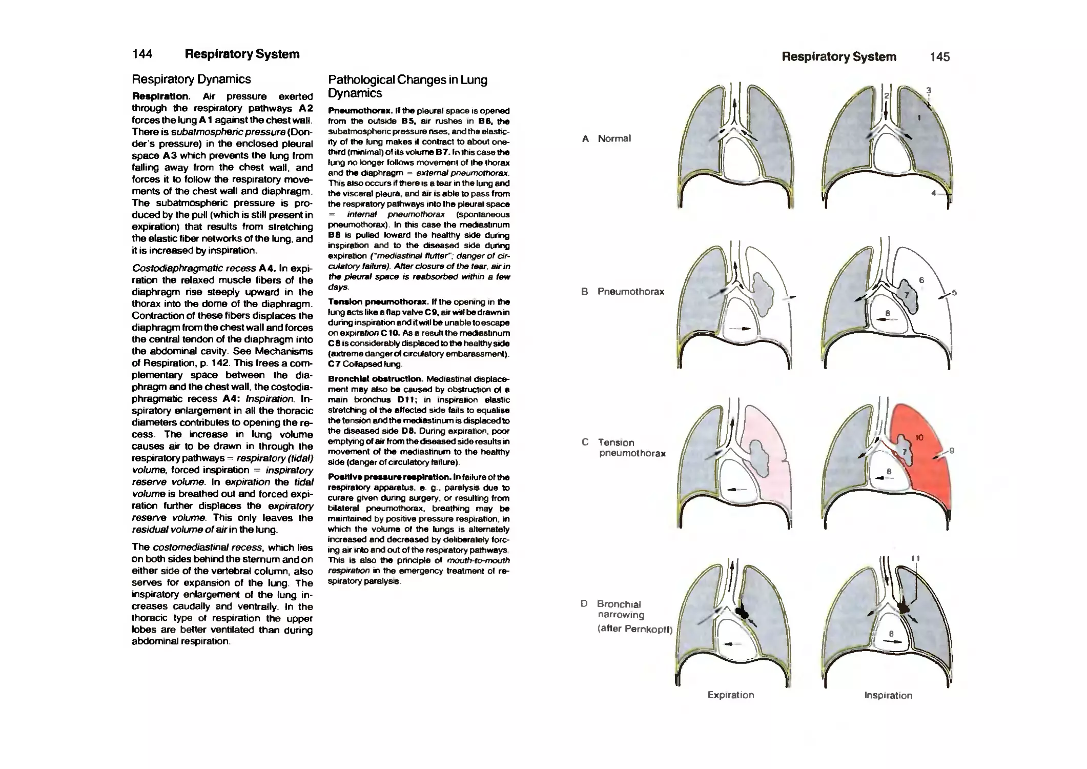
Pathological Changes in Lung Dynamics 142
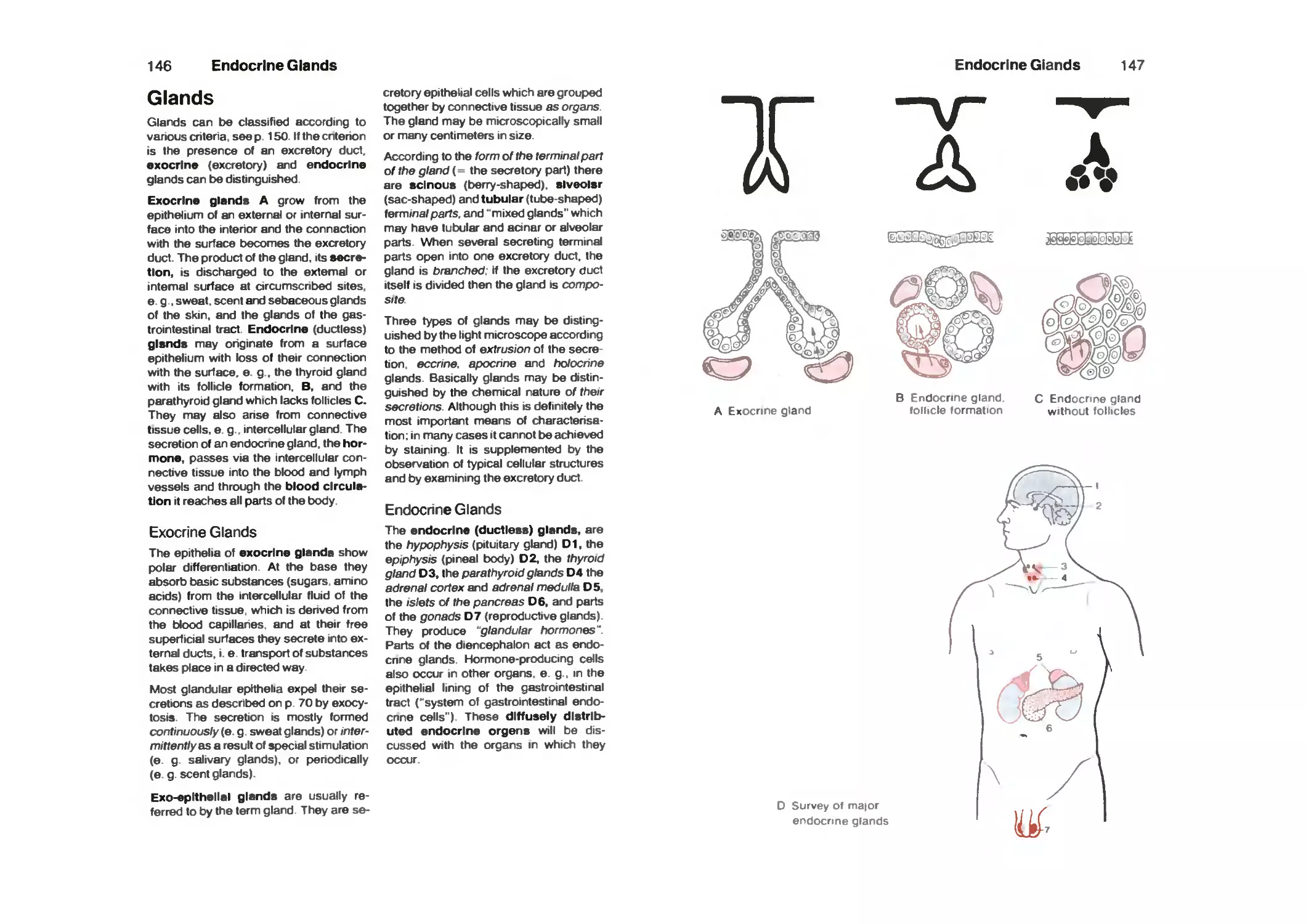
Glands 144
Endocrine (Ductless) Glands 144
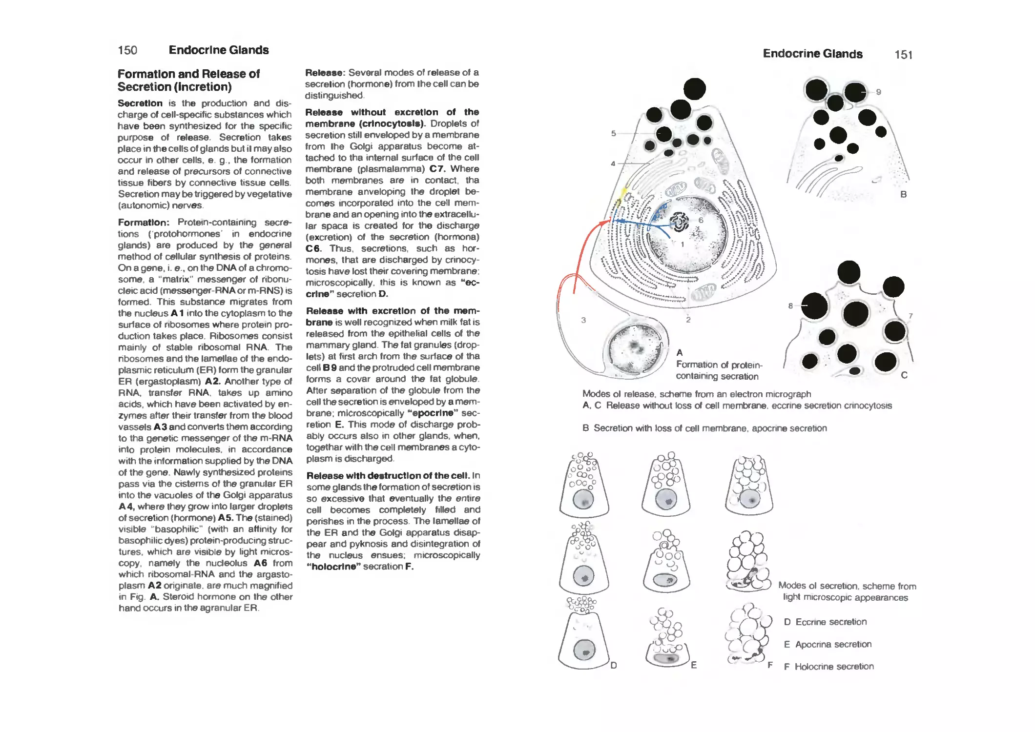
Formation and Release of Secretion (Incretion) 146
Arrangement of the Hypothalamo-Hypophyseal System 148
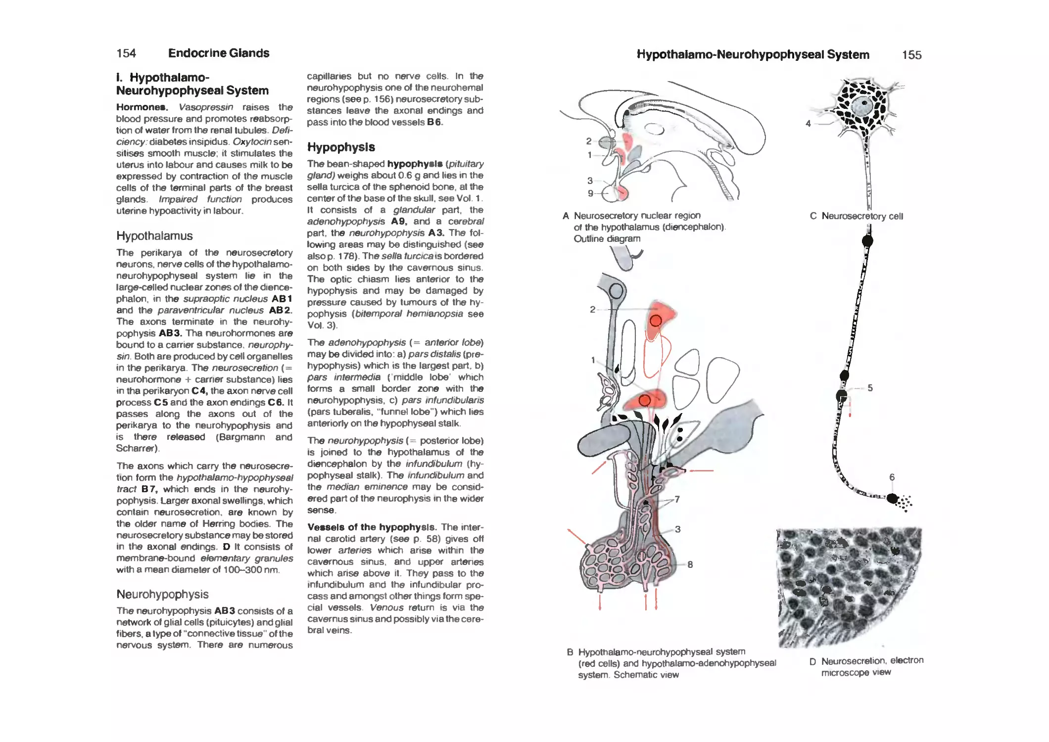
Hypothalamo-Neurophypophyseal System 150
Hypothalamus 150
Neurohypophysis 150
Hypophysis 150
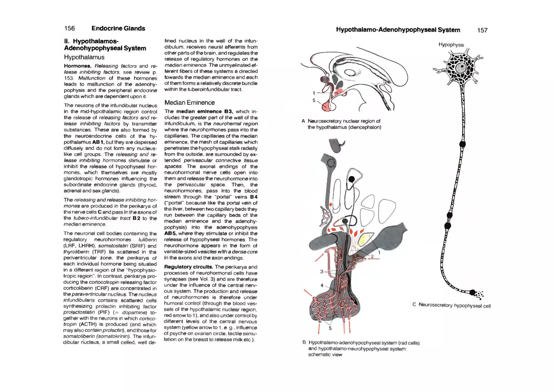
Hypothalamo-Adenohypophyseal System 152
Hypothalamus 152
Median Eminence 152
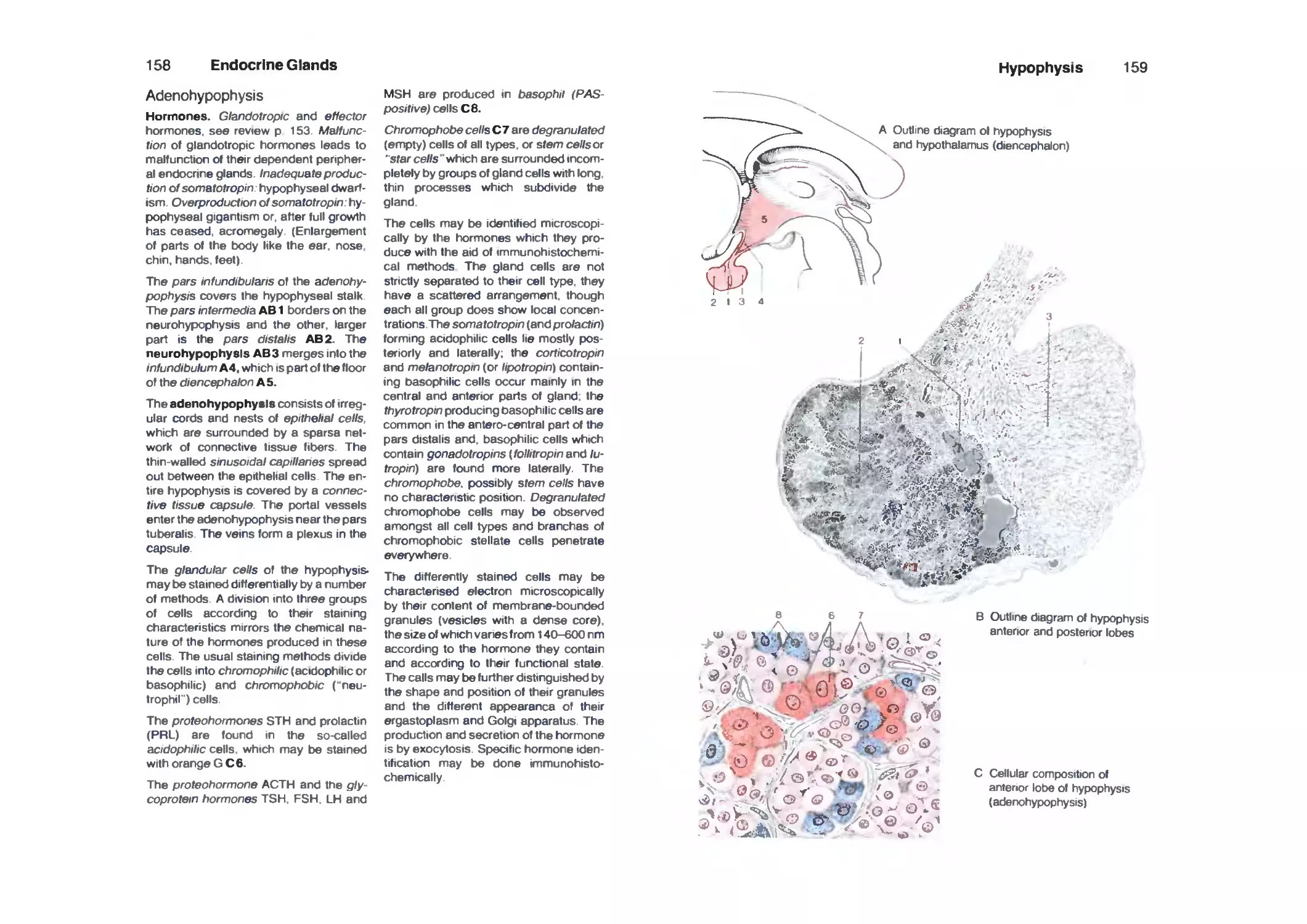
Adenohypophysis 154
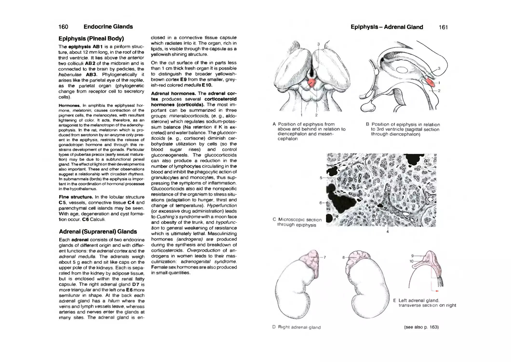
Epiphysis (Pineal Body) 156
Adrenal (Suprarenal) Glands 156
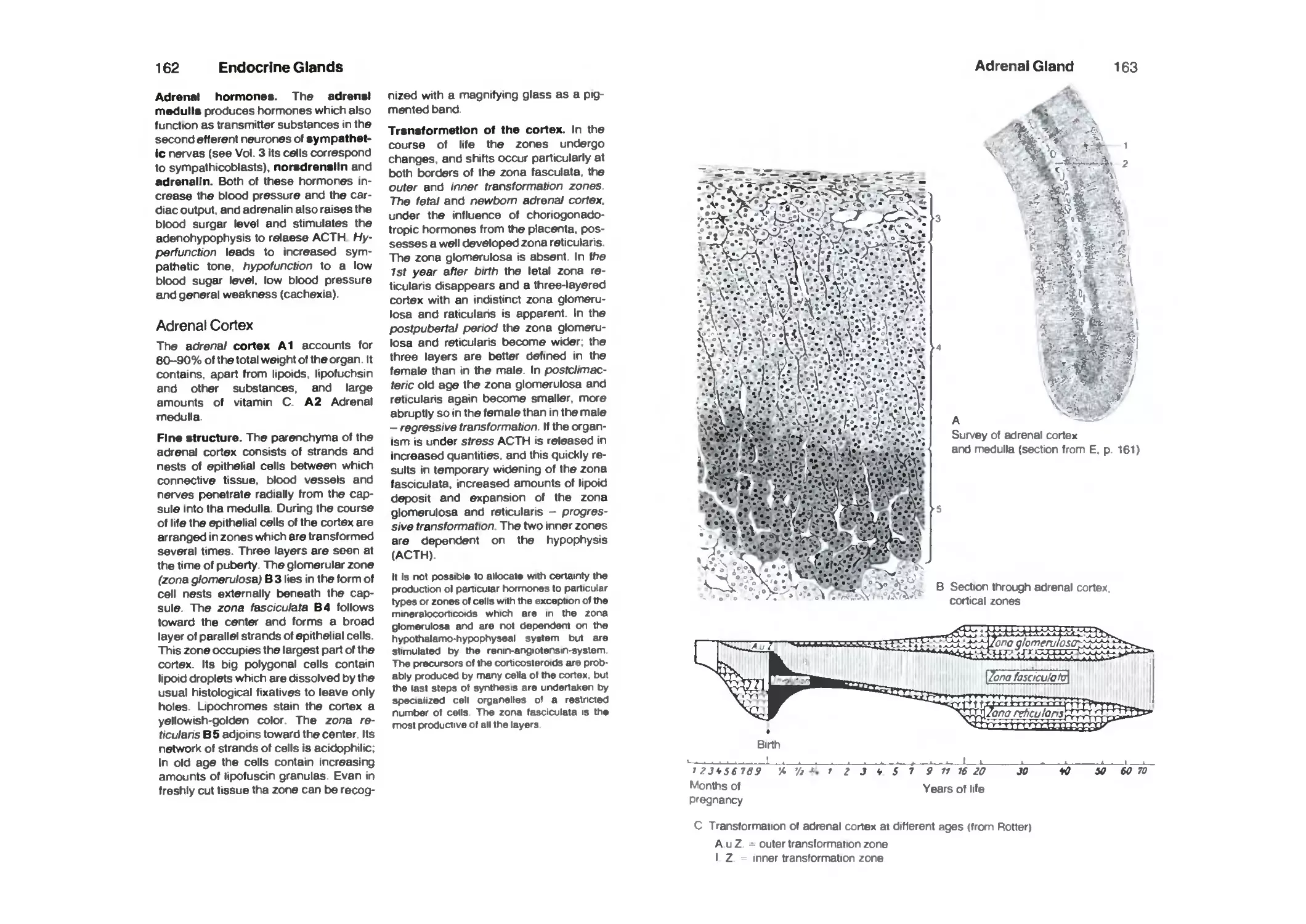
Adrenal Cortex 158
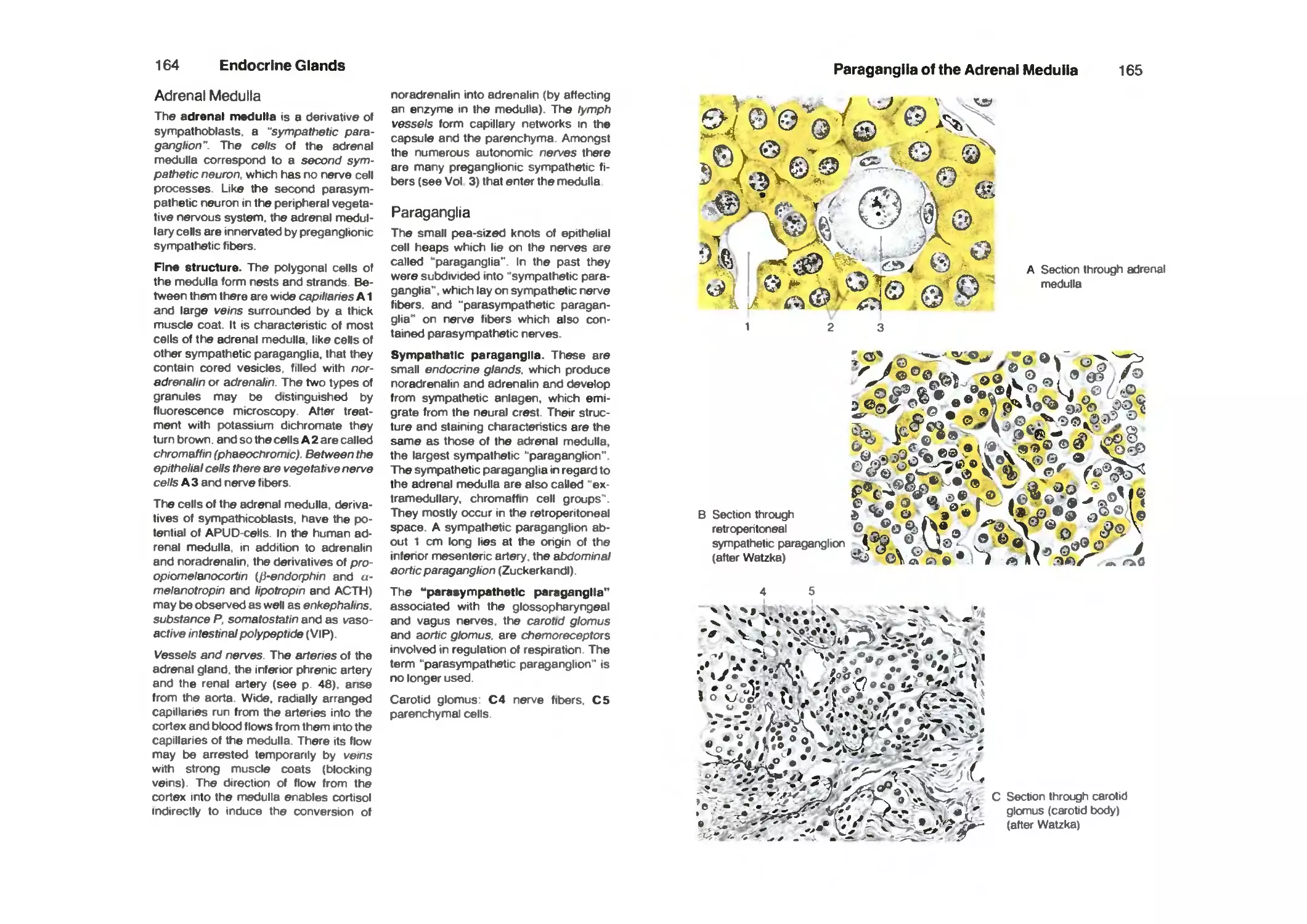
Adrenal Medulla 160
Paraganglia 160
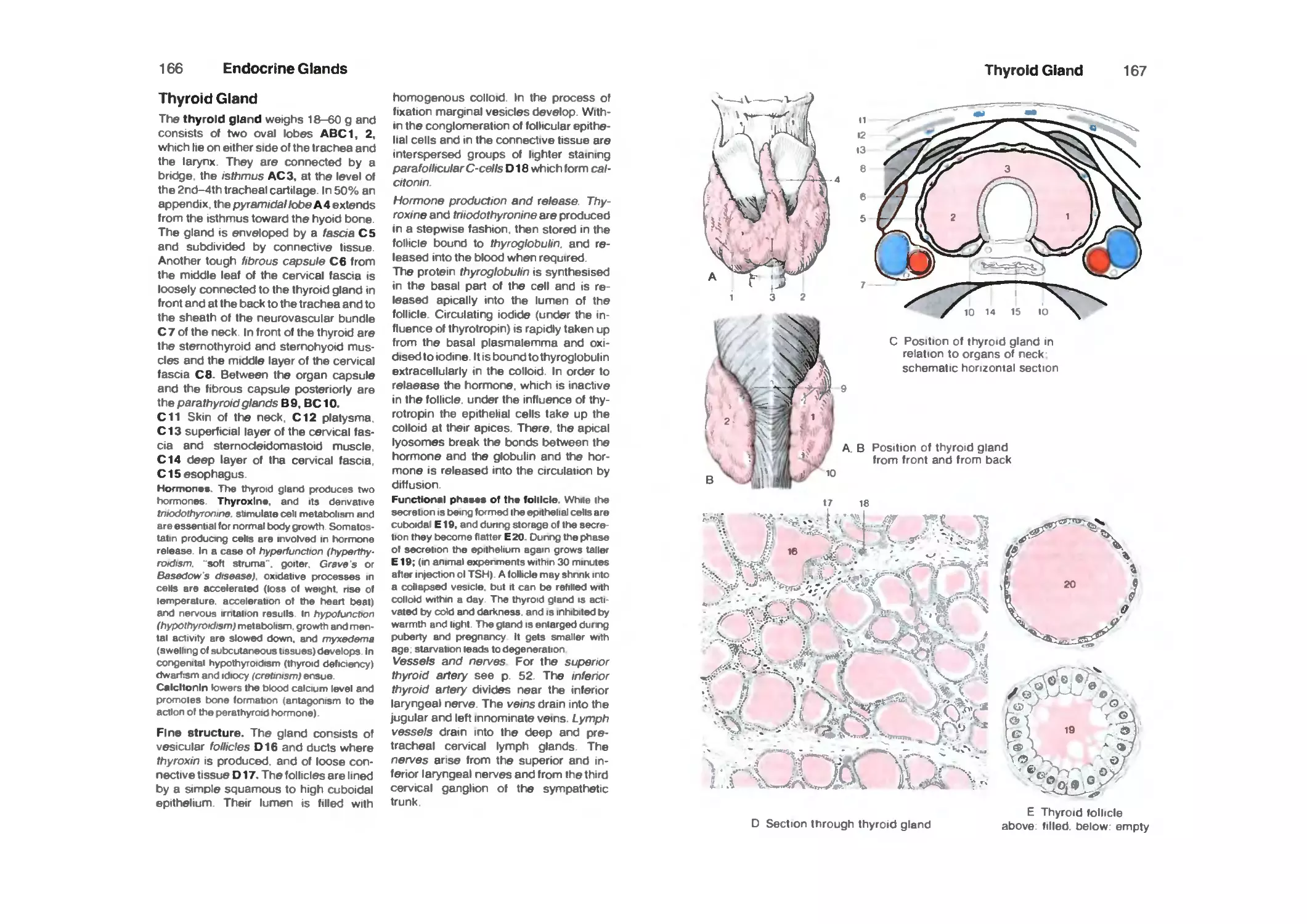
Thyroid Gland 162
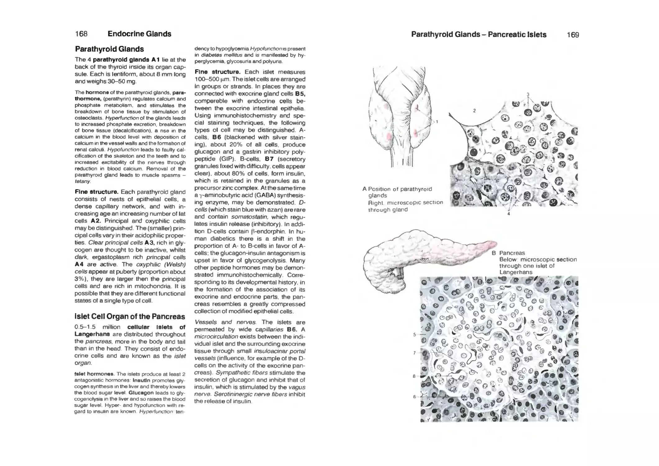
Parathyroid Glands 164
Islet Cell Organ of the Pancreas 164
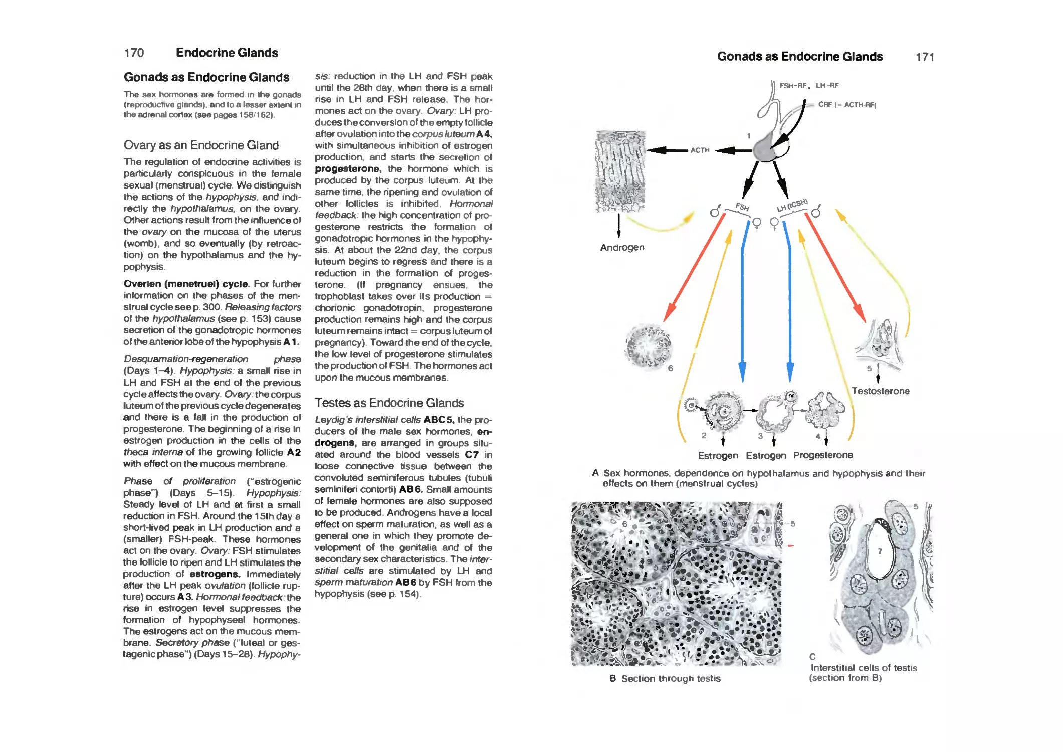
Gonads as Endocrine Glands 166
Ovary as an Endocrine Gland 166
Testis as an Endocrine Gland 166
Digestive System 168
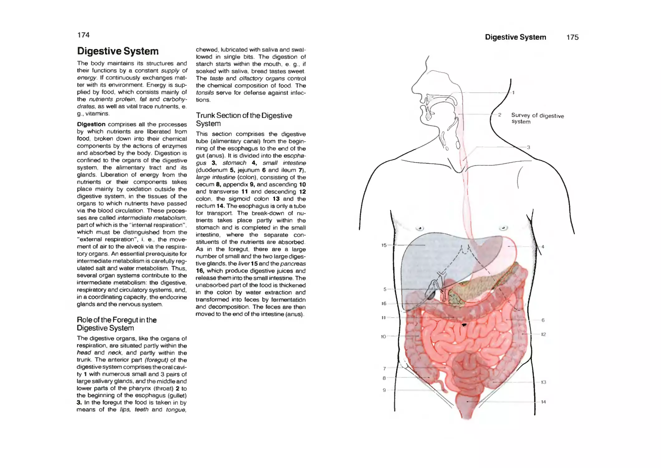
Role of the Foregut in the Digestive System 168
Trunk Section of the Digestive System 168
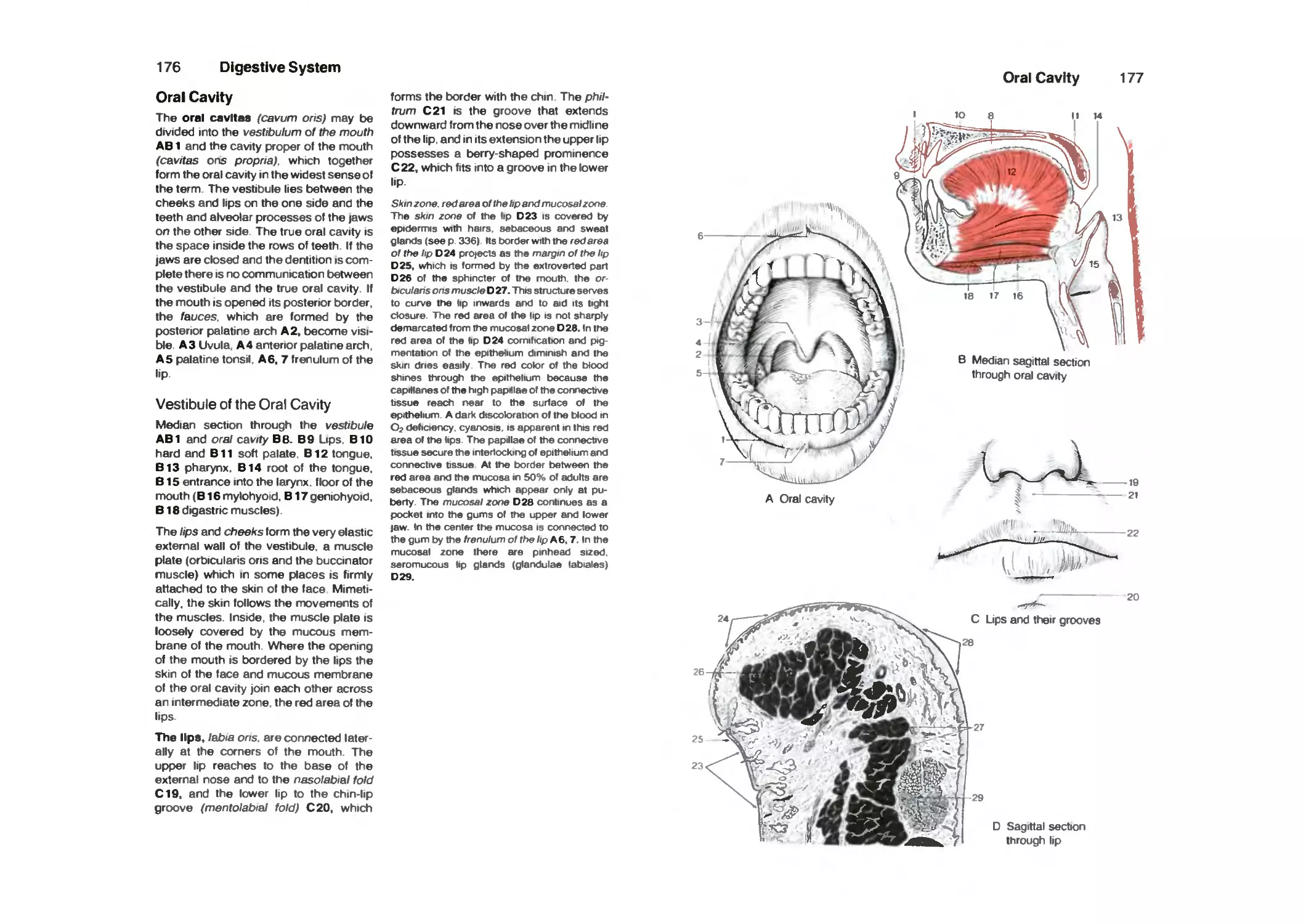
Oral Cavity 170
Vestibule of the Oral Cavity 170
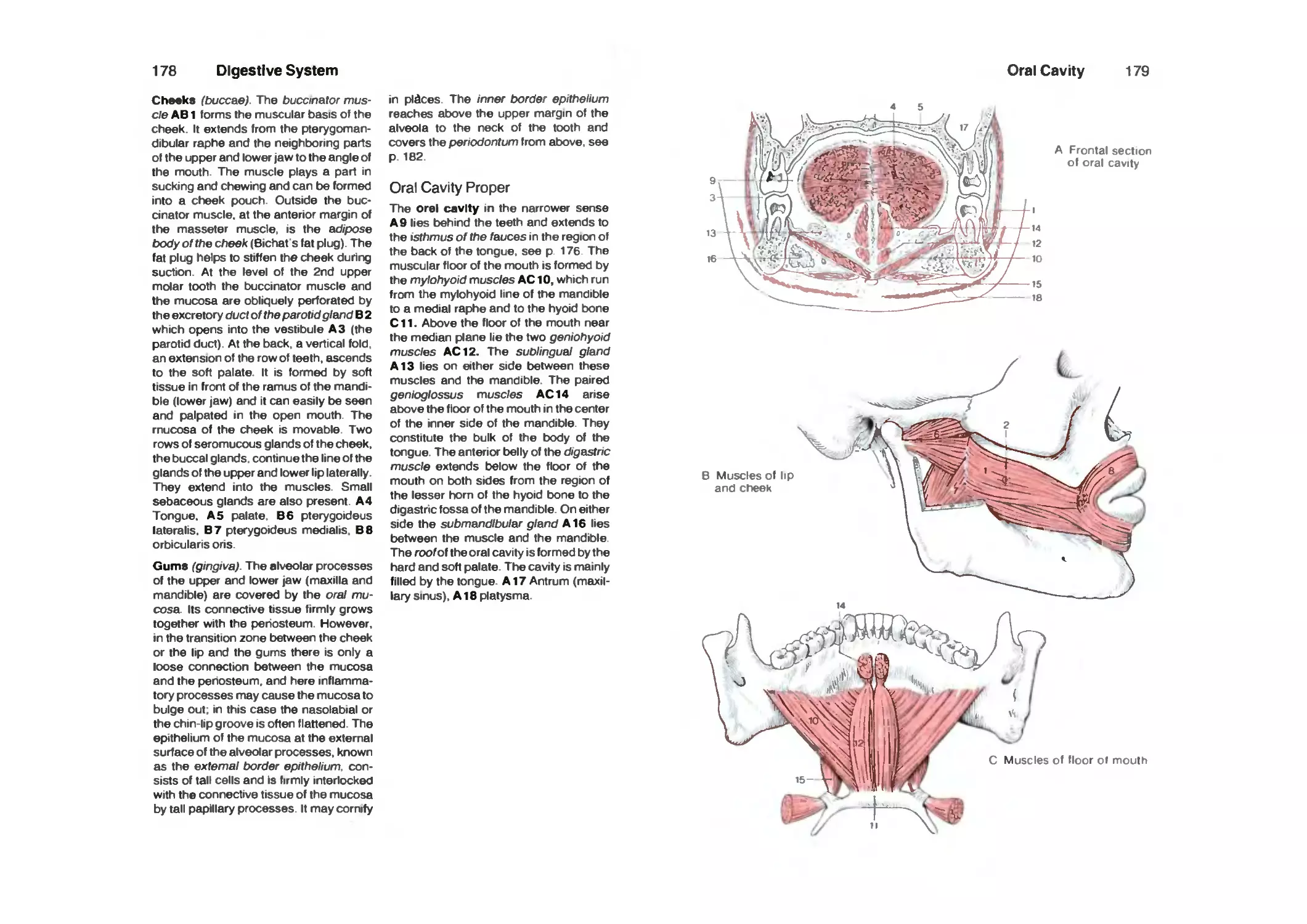
Oral Cavity Proper 172
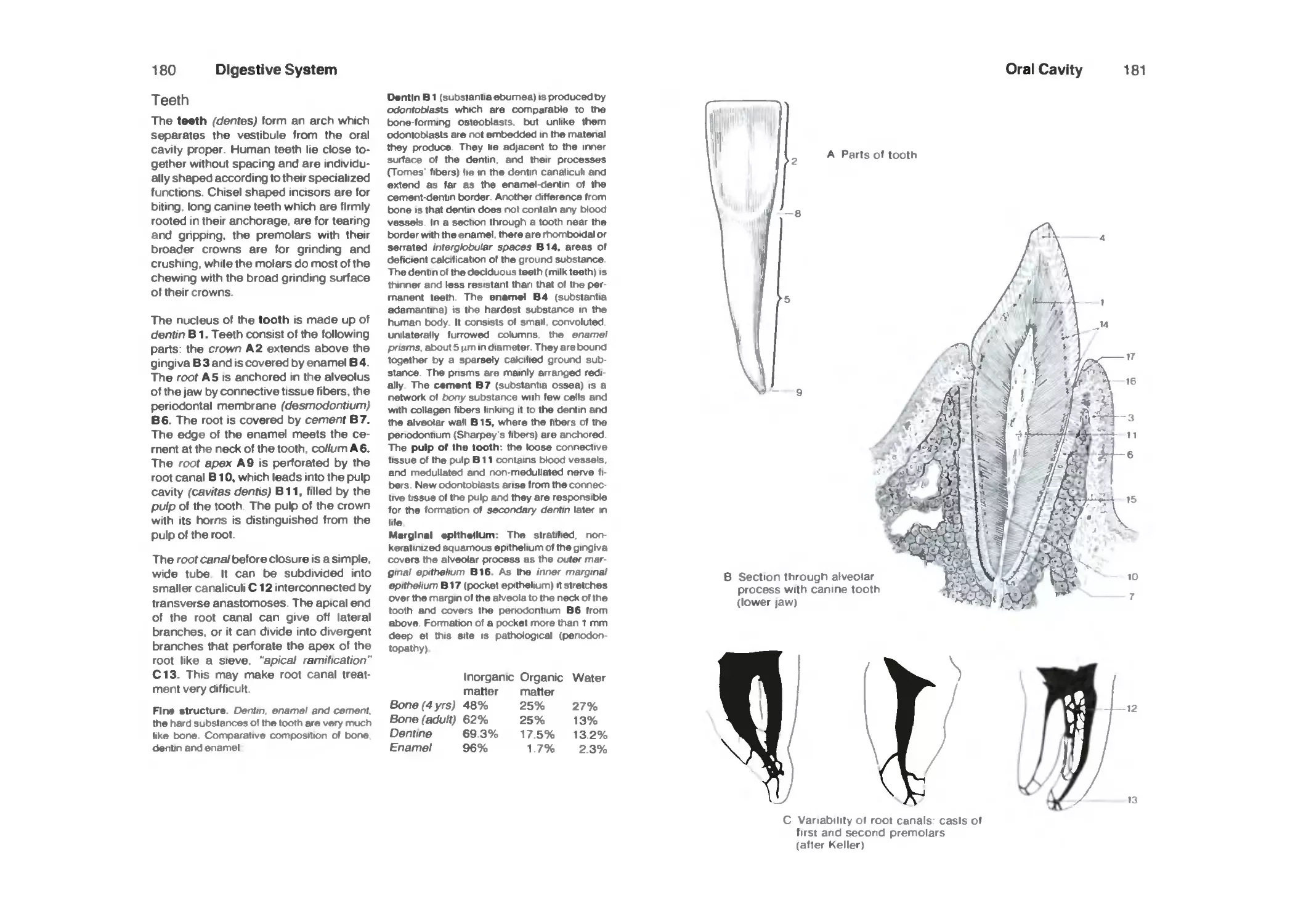
Teeth 174
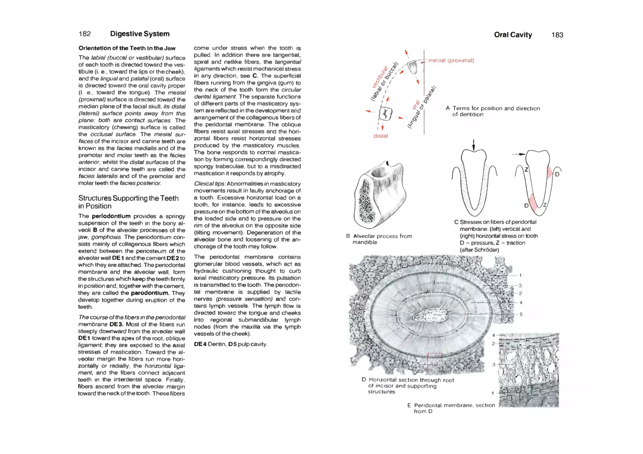
Structures Supporting the Teeth in Position 176
Permanent Teeth 178
Milk Teeth 178
Position and Movements of the Teeth Within the Dentition 180
Movement of the Dental Arches Against Each Other (Articulation) . 182
Contents
IX
Movements in the Mandibular Joint 182
Movements of the Footh Inside the Alveolus 182
Milk Teeth (Deciduous Teeth) 184
Eruption of the Teeth 184
Tongue 186
Palate 190
Salivary Glands 192
Large Salivary Glands 192
Fine Structure of the Salivary Glands 194
Pharynx (Throat) 196
Deglutition (Swallowing) 198
Esophagus 200
Esophagus and the Posterior Mediastinum 202
Stomach 204
Peritoneum 204
Muscle Layer of the Stomach 206
Gastric Mucosa 208
Small Intestine 210
Duodenum 210
Jejunum and Ileum 210
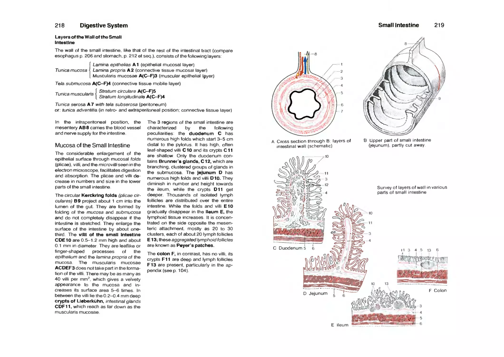
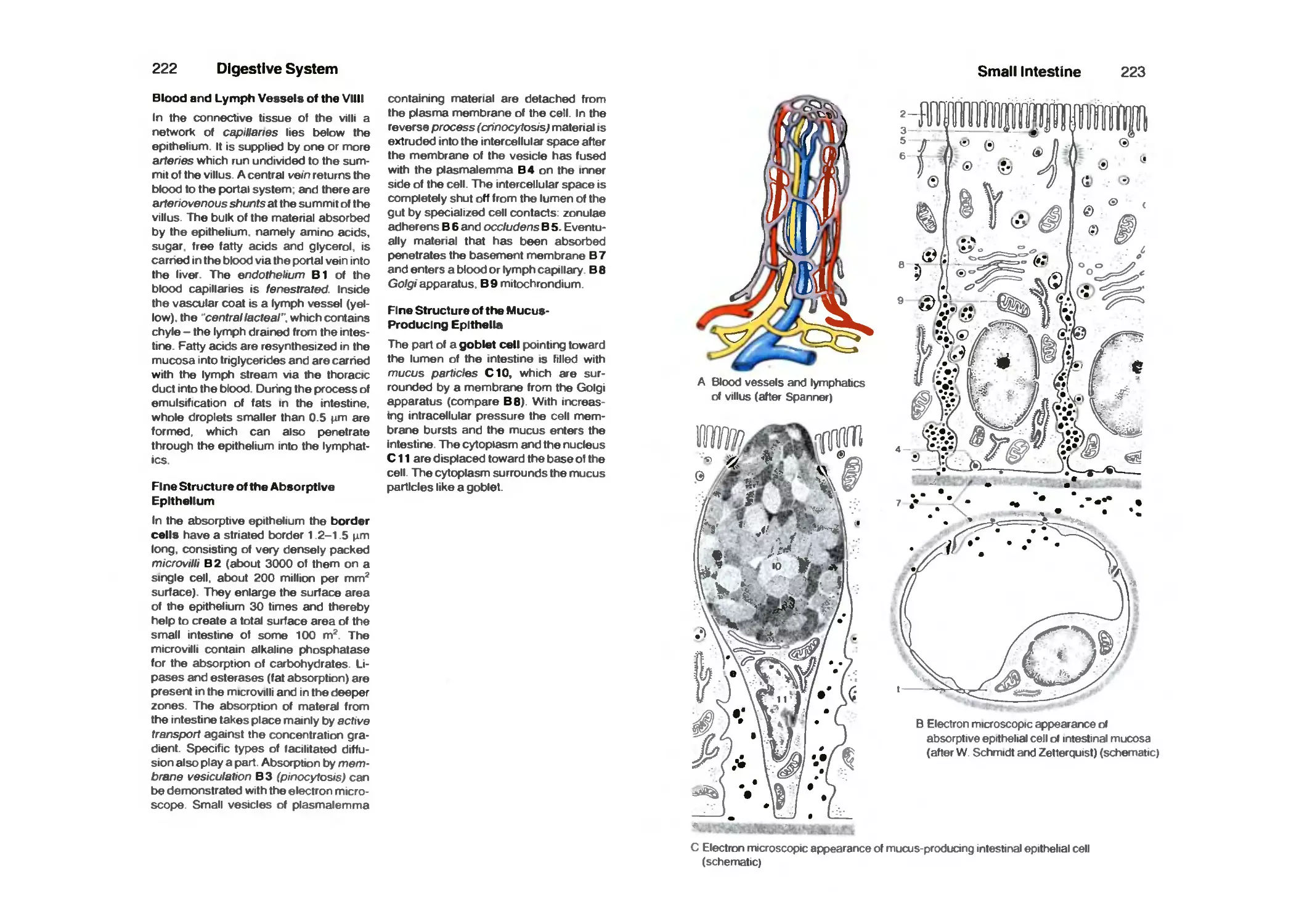
Mucosa of the Small Intestine 212
Muscle Layerof the Small Intestine 214
Large Intestine 218
Cecum and the Ileocecal Valve (Colic Valve) 220
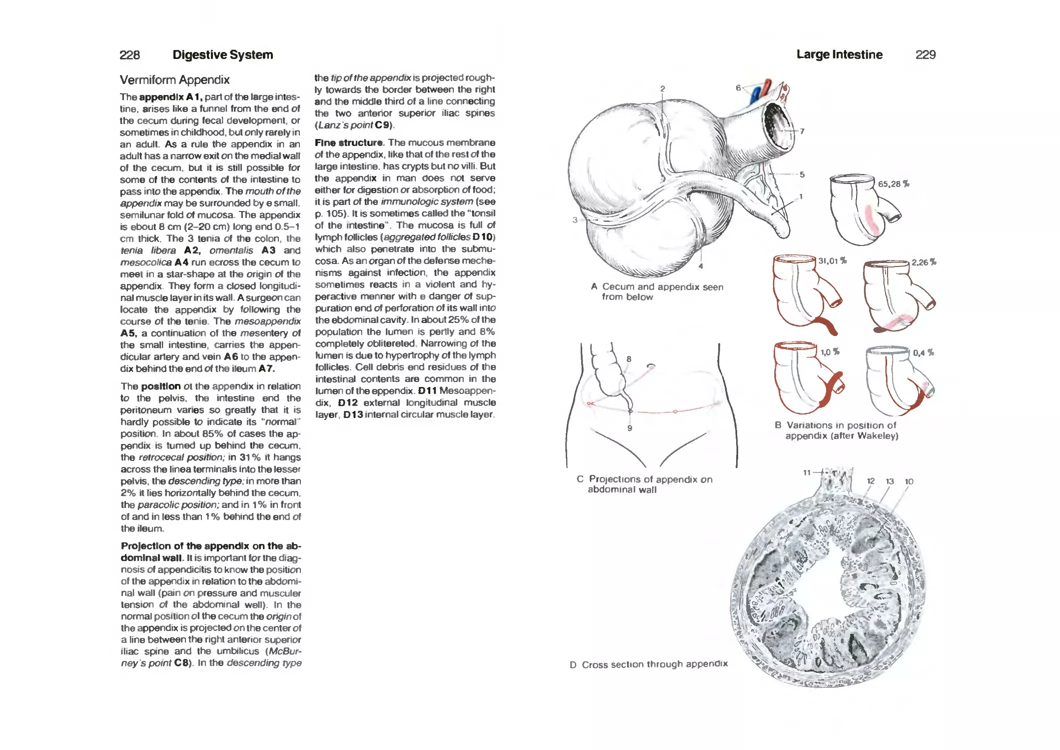
Vermiform Appendix 222
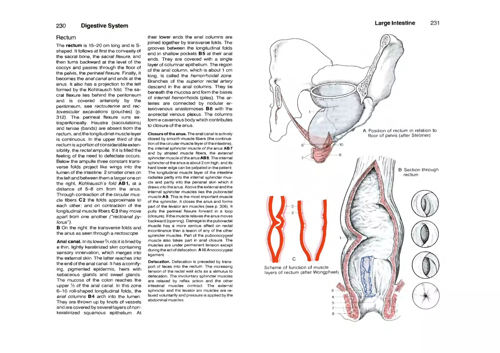
Rectum 224
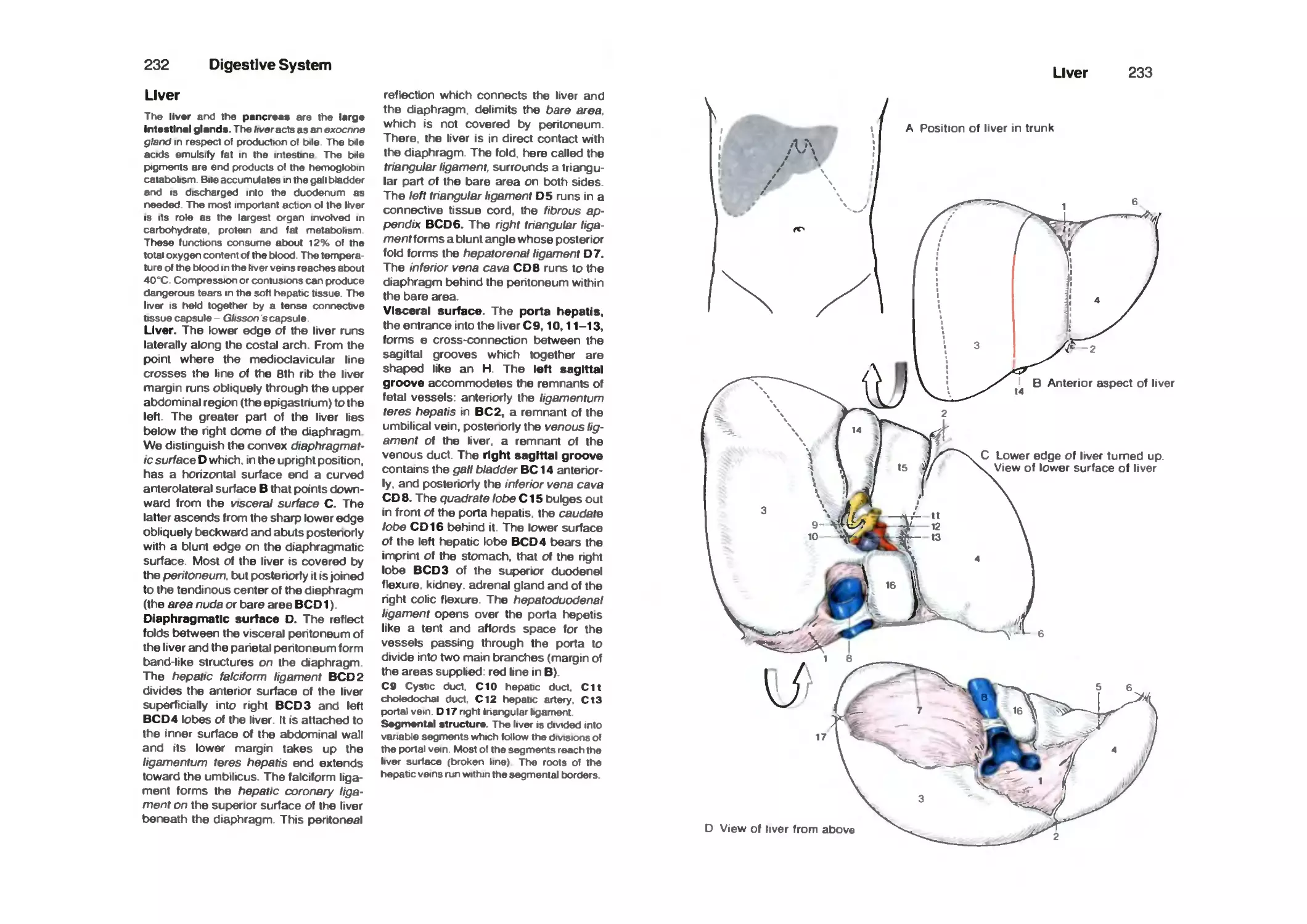
Liver 226
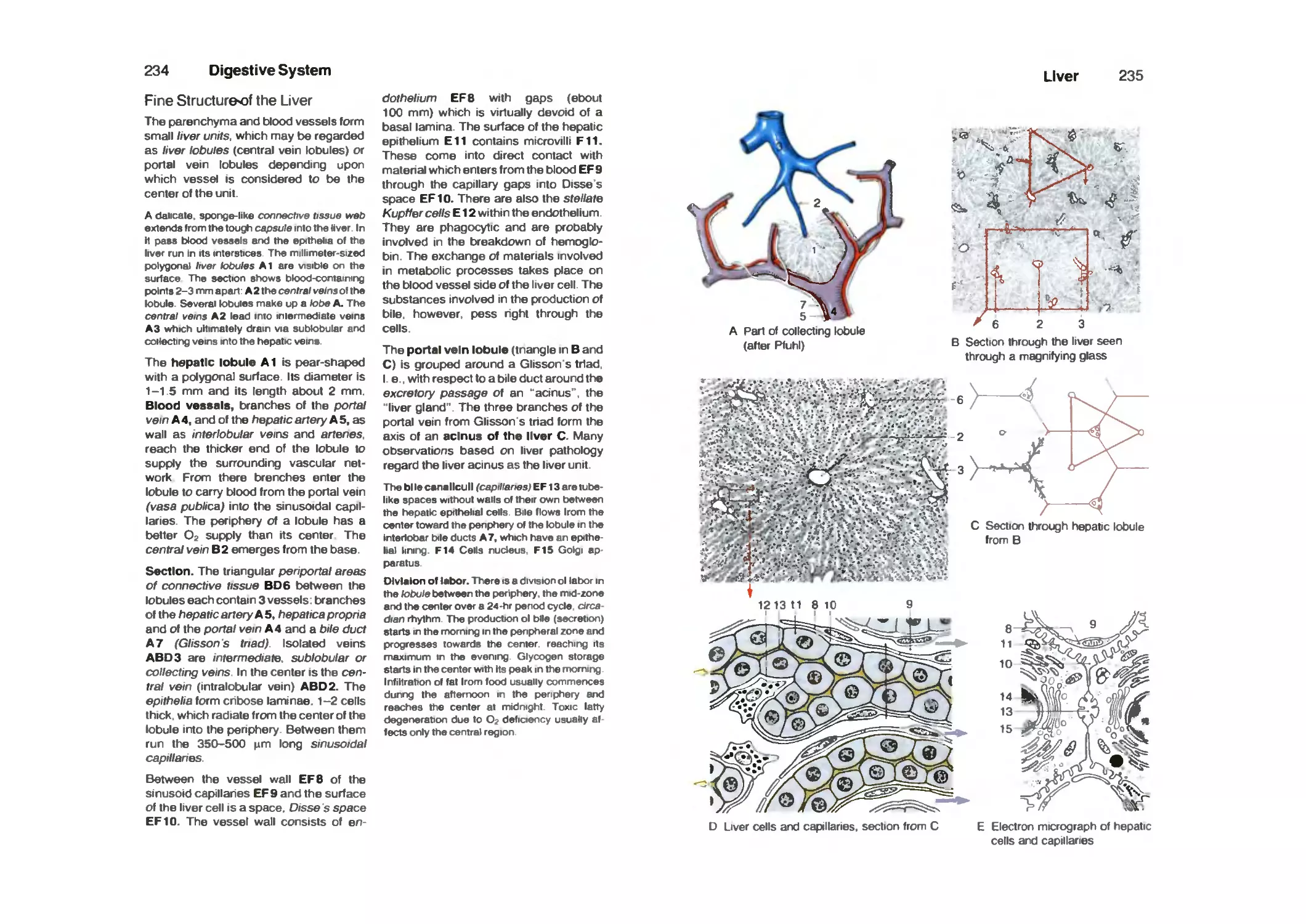
Fine Structure of the Liver 228
Bile Ducts and Gall Bladder 230
Pancreas 232
Greater and Lesser Omentum 234
Blood Vessels and Lymphatics of the Upper Abdominal Organs ... 234
Abdominal Cavity 236
Blood Vessels and Lymphatics of the Lower Abdominal Viscera.... 238
Portal Vein 242
Urogenital System 244
Urinary Organs 244
Kidneys 246
Section Through the Kidney 248
Blood Vessels of the Kidney 248
Fine Structure of the Kidney 250
X Contents
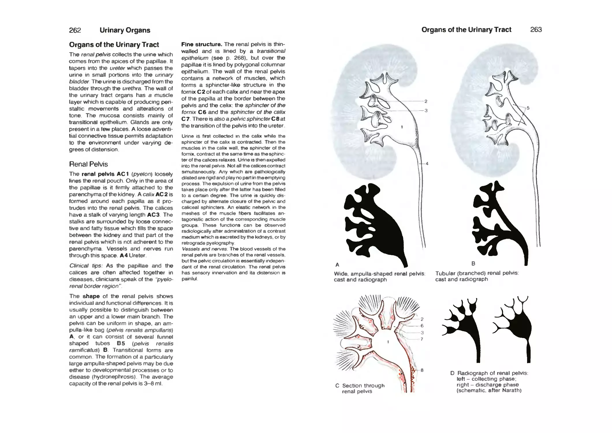
Organs of the Urinary Tract 256
Renal Pelvis 256
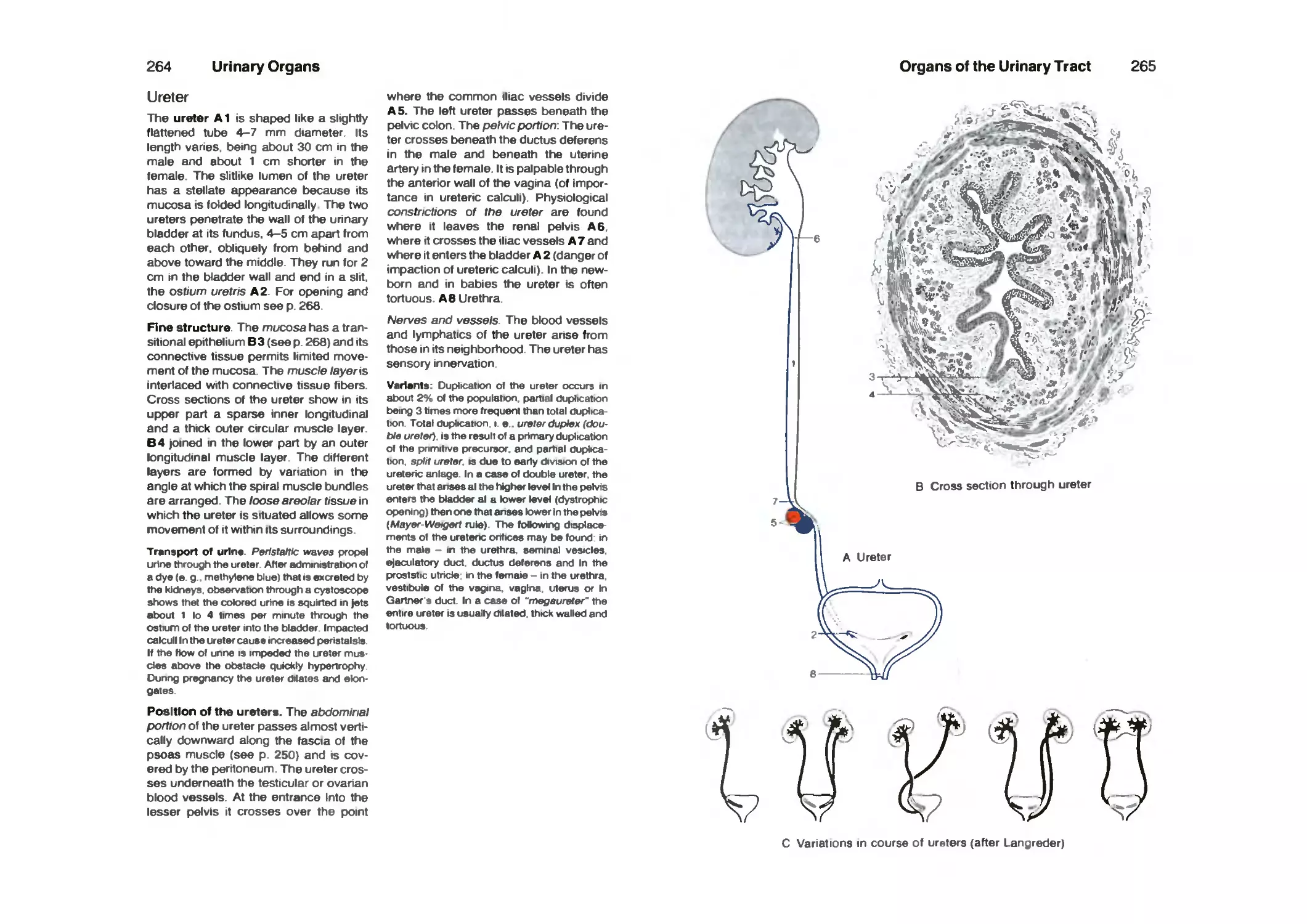
Ureter 258
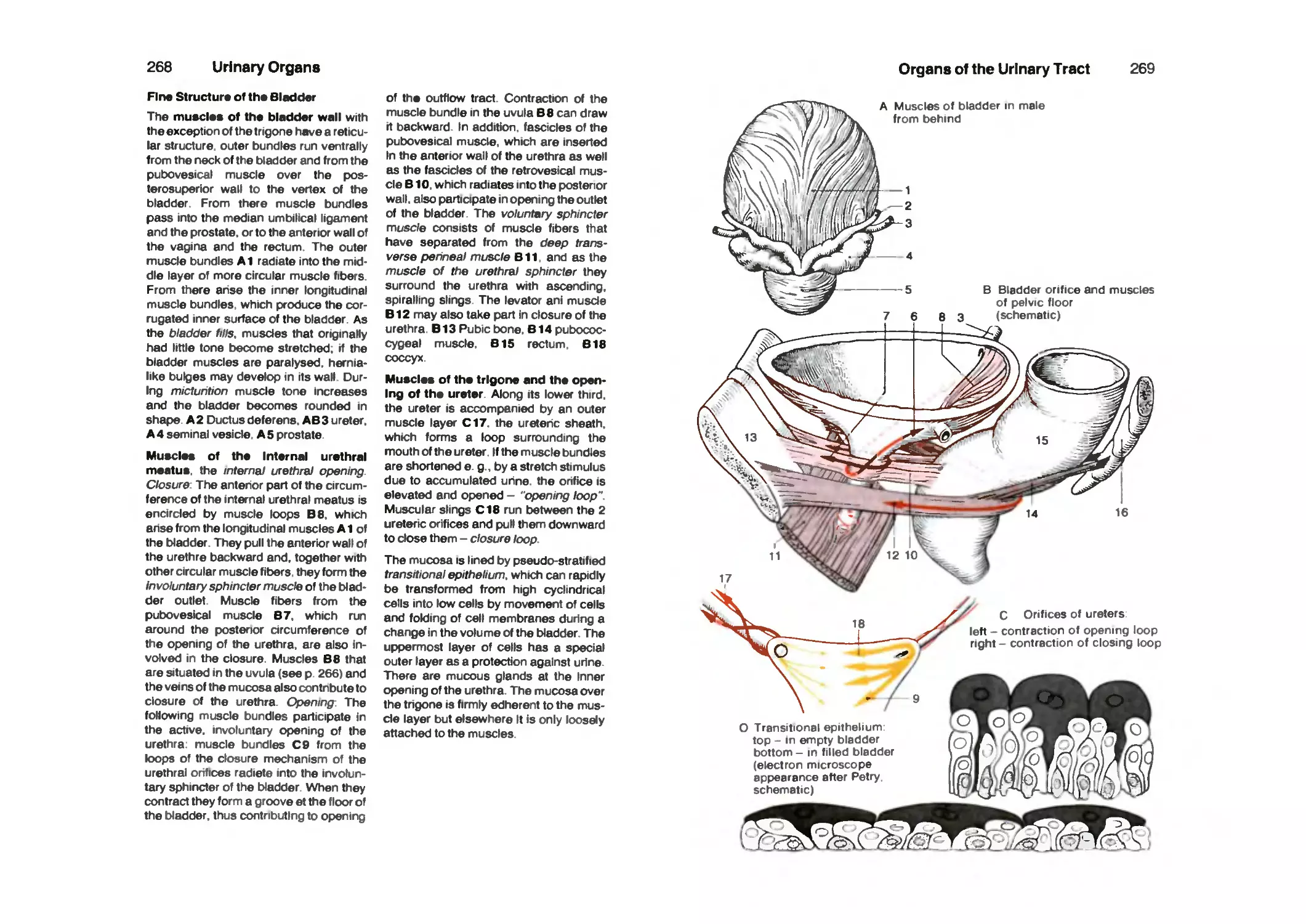
UrinaryBladder 260
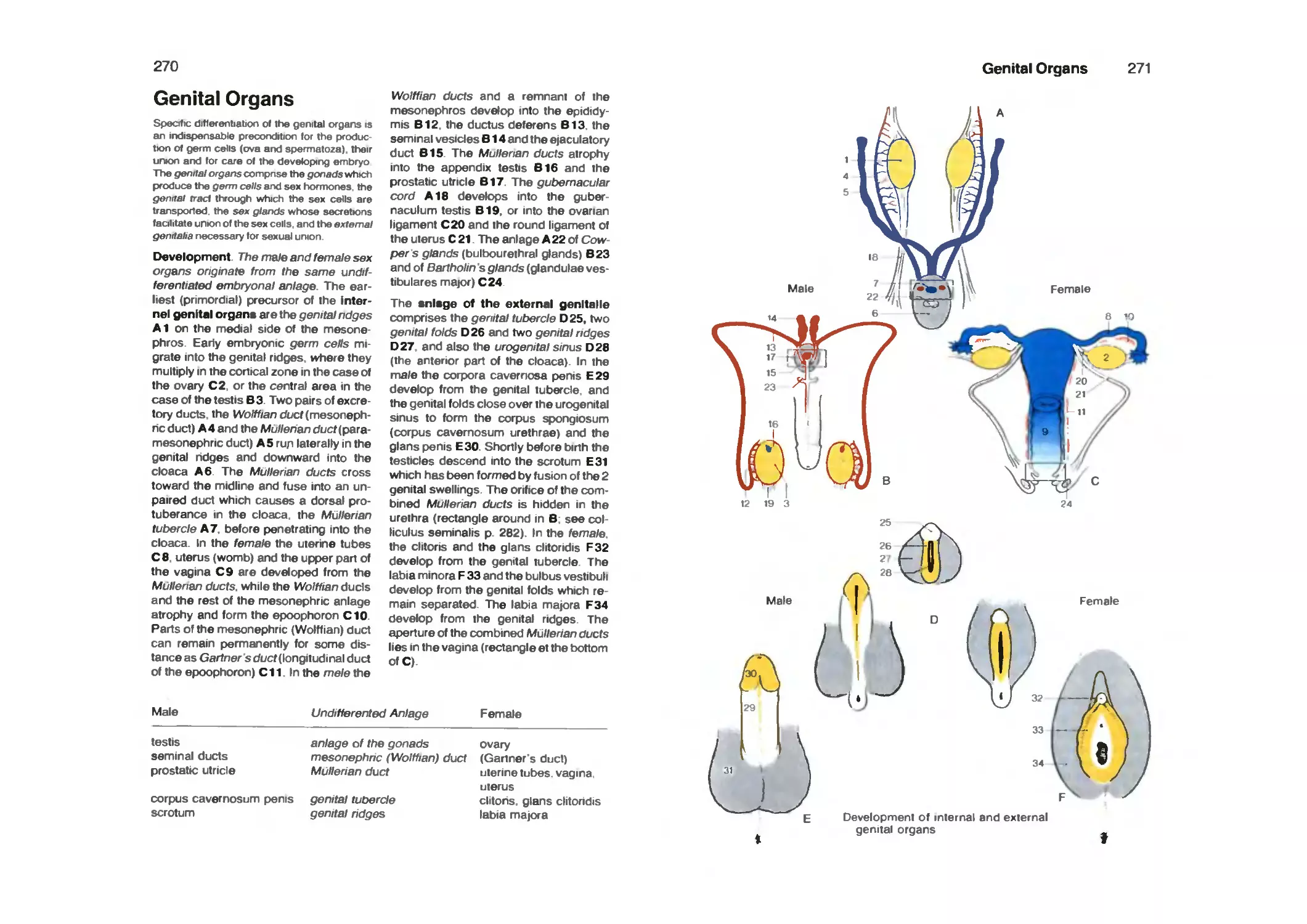
Genital Organs 264
Male Genital Organs 266
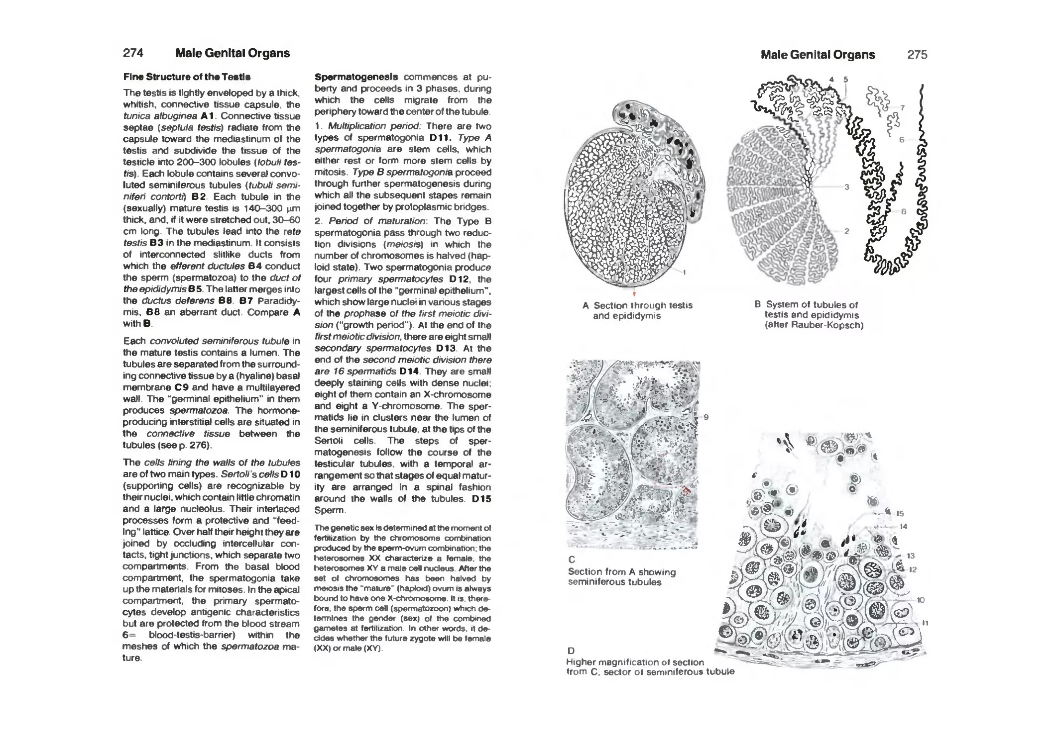
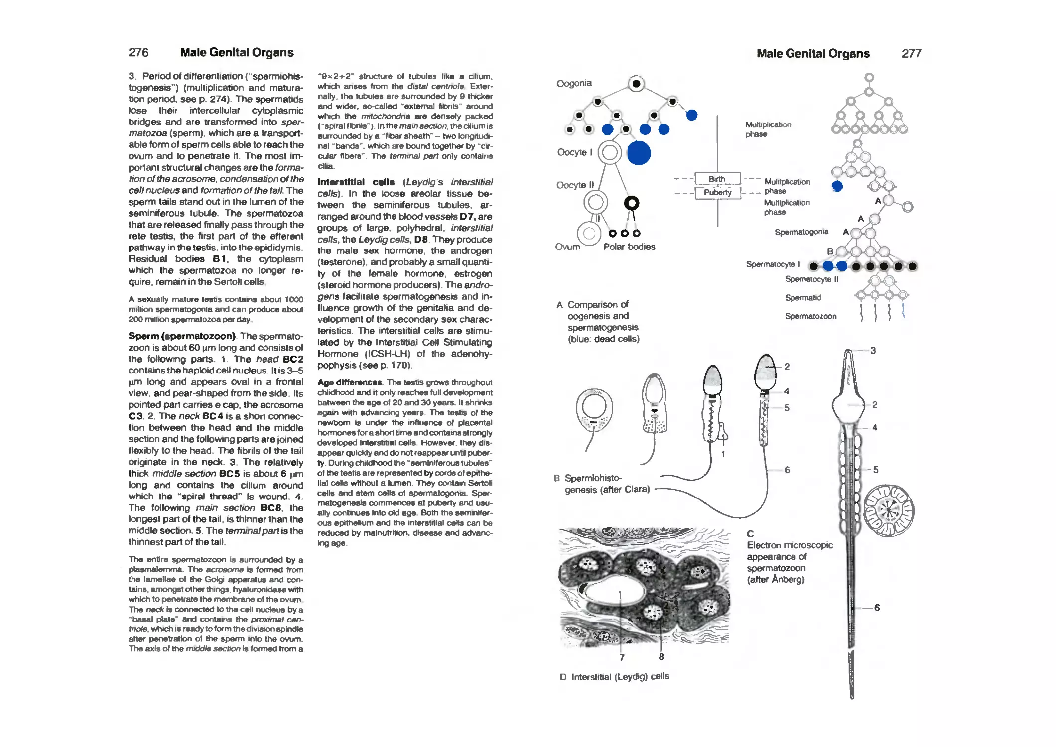
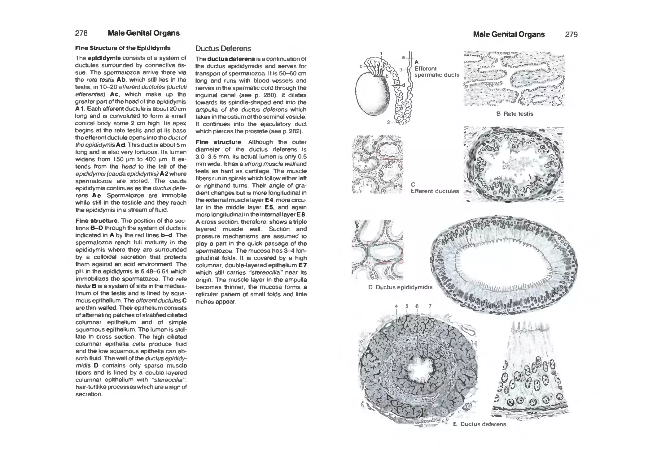
Testis and Epididymis 266
Ductus Deferens 272
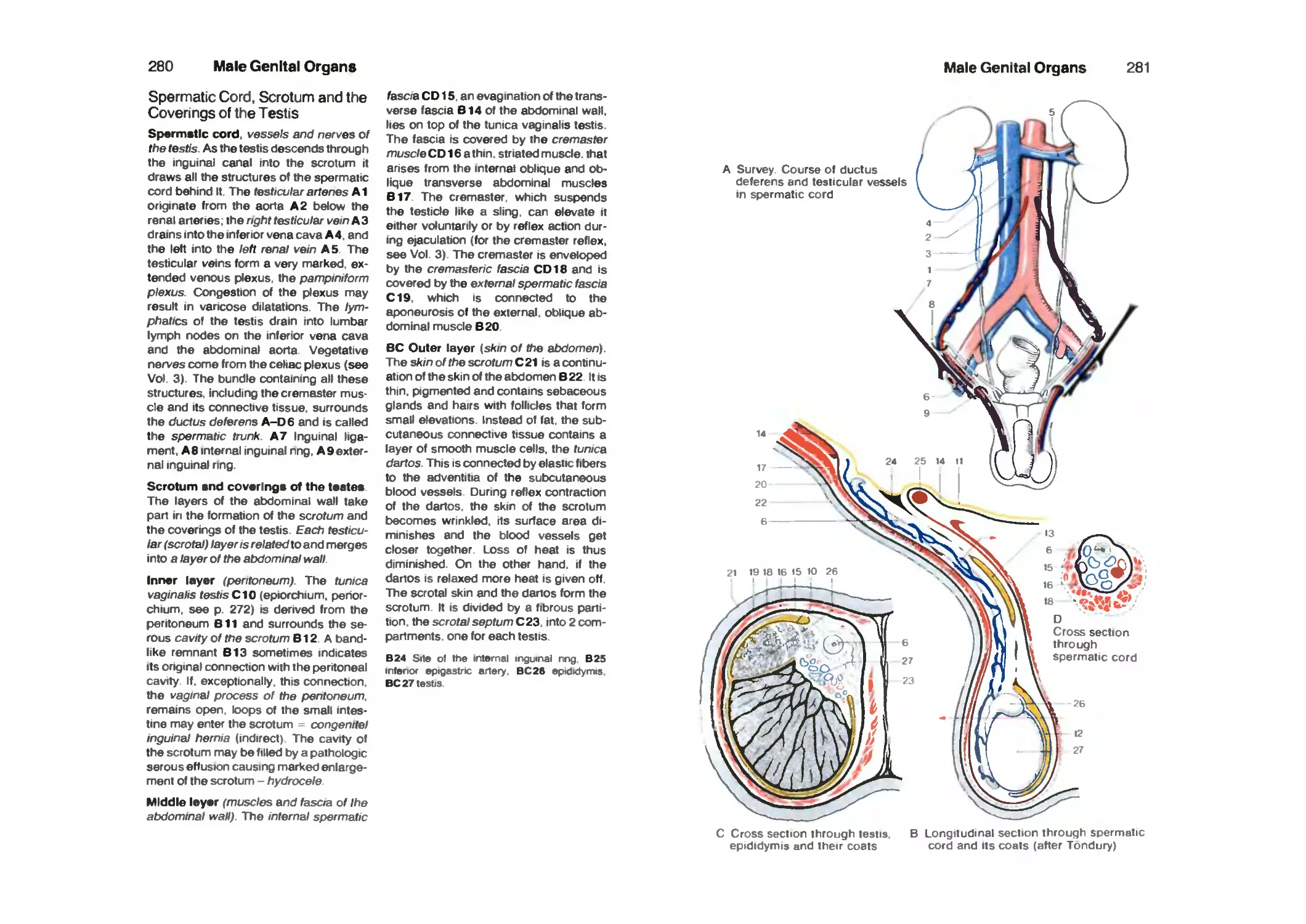
Spermatic Cord, Scrotum and the Coverings of the Testis 274
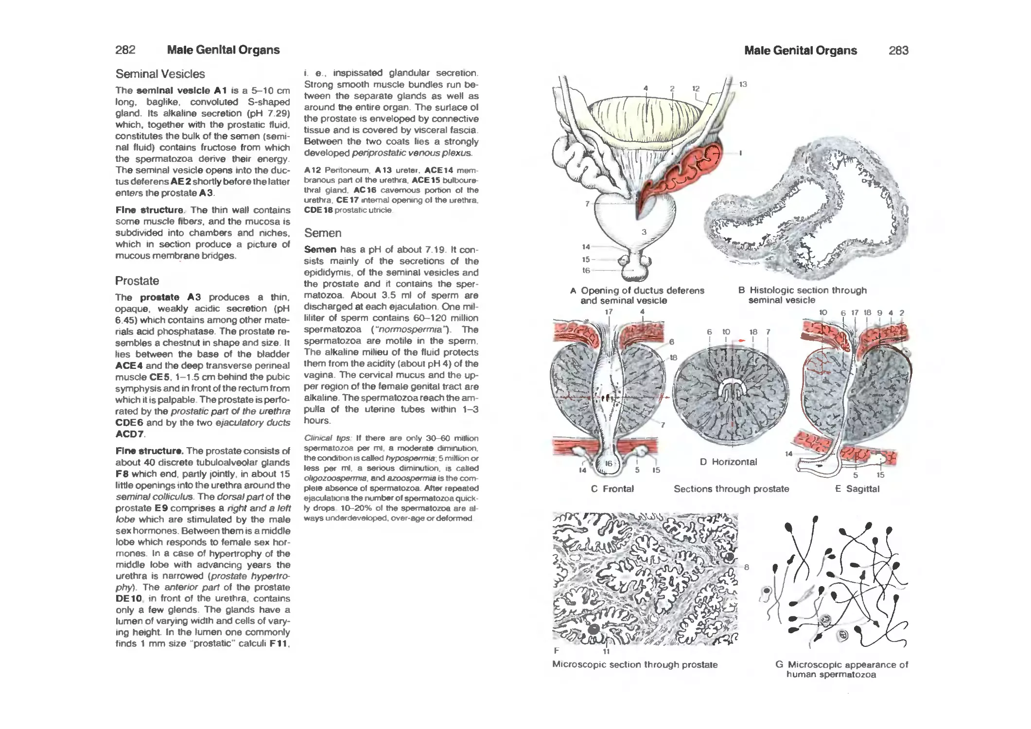
Seminal Vesicles 276
Prostate 276
Semen 276
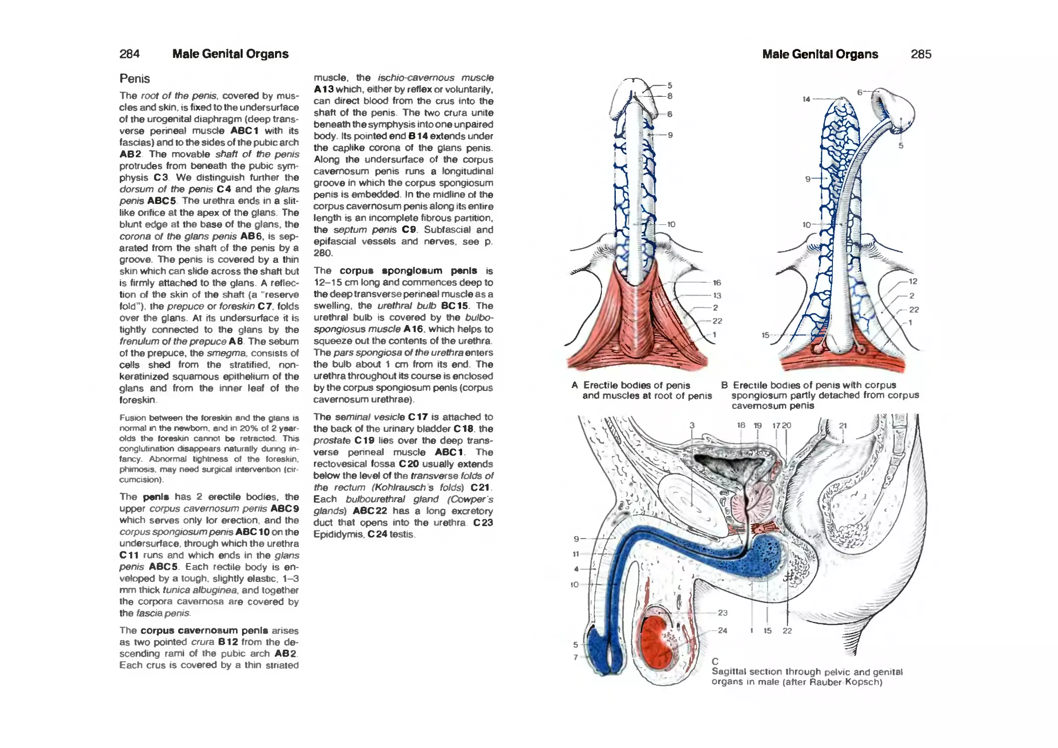
Penis 278
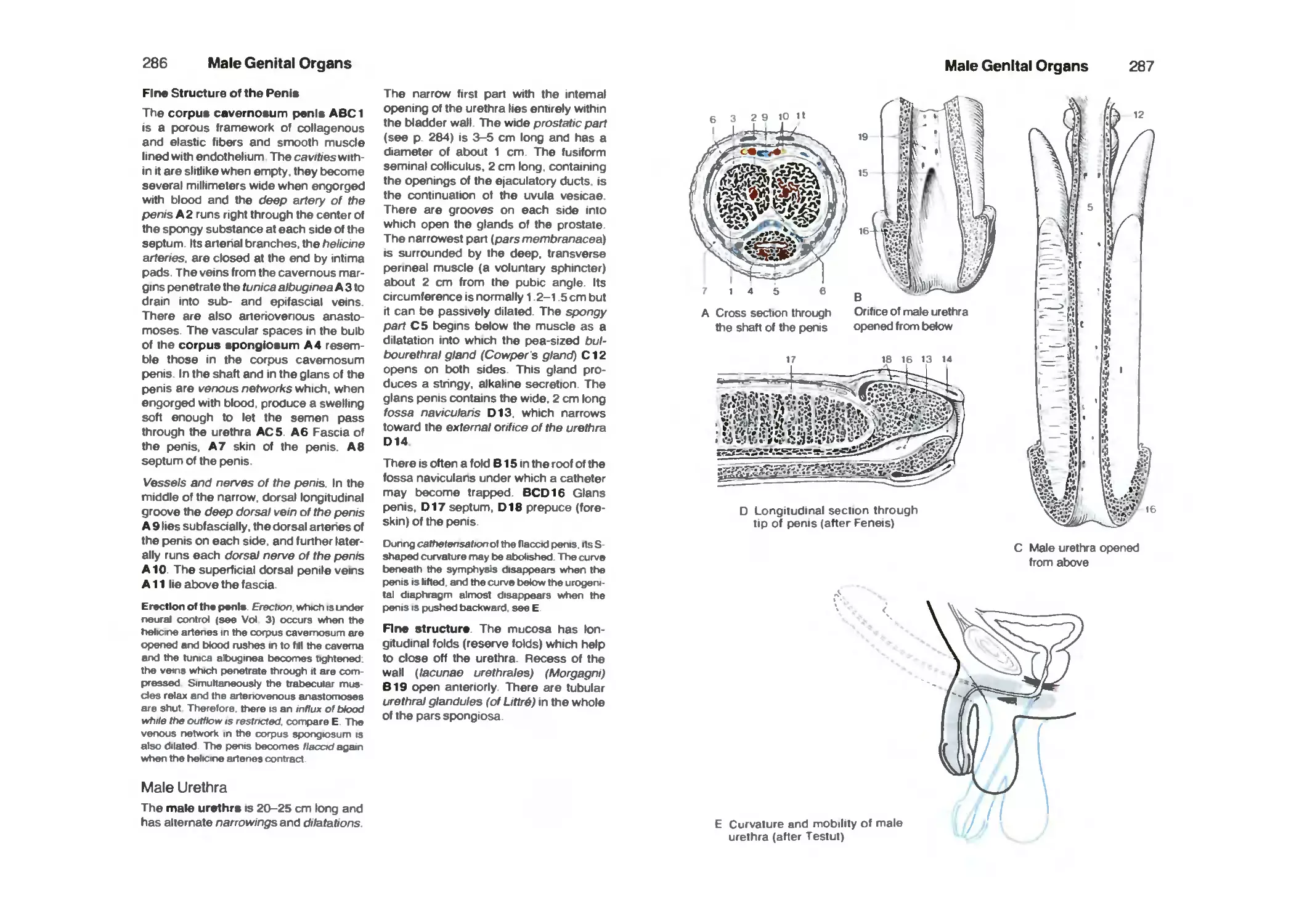
Male Urethra 280
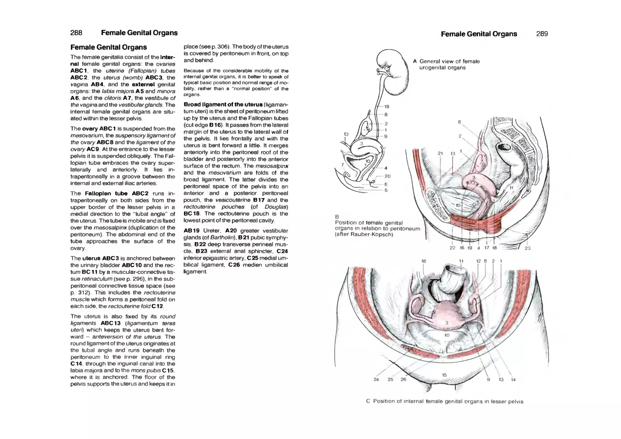
Female Genital Organs 282
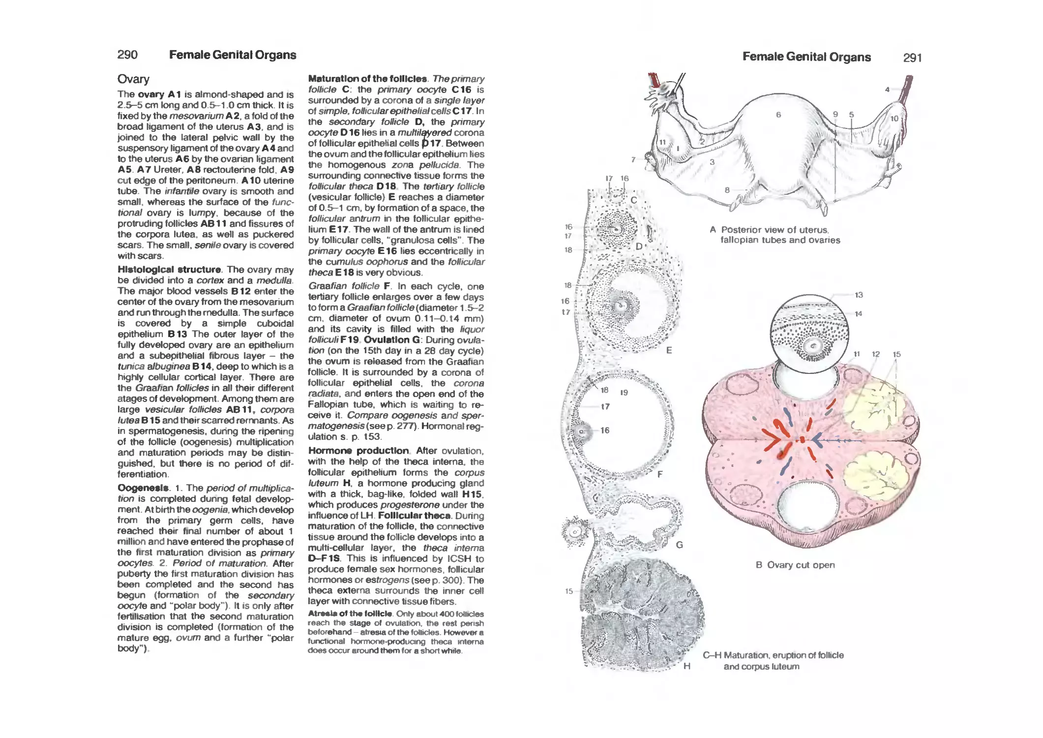
Ovary 284
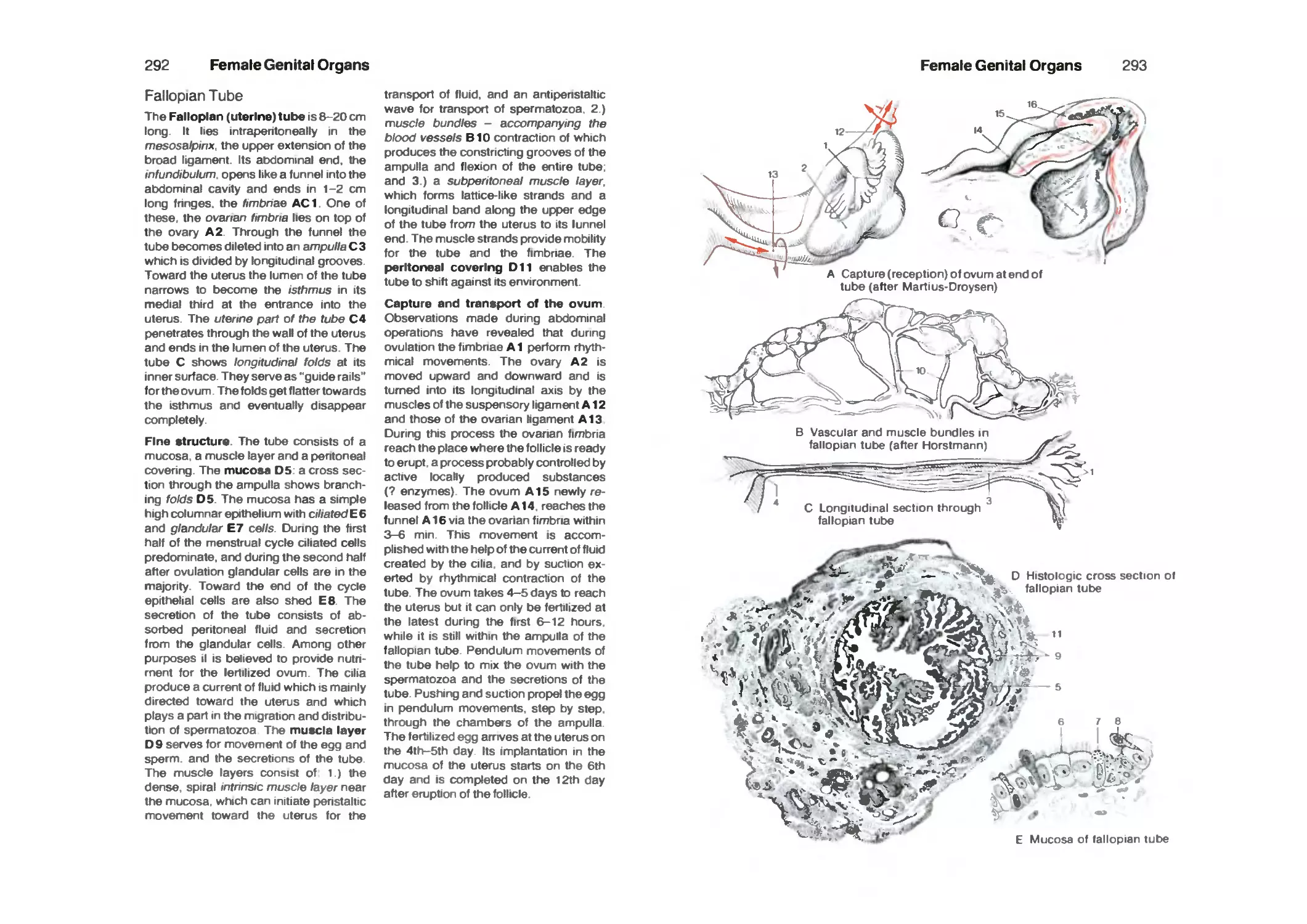
Fallopian Tube 286
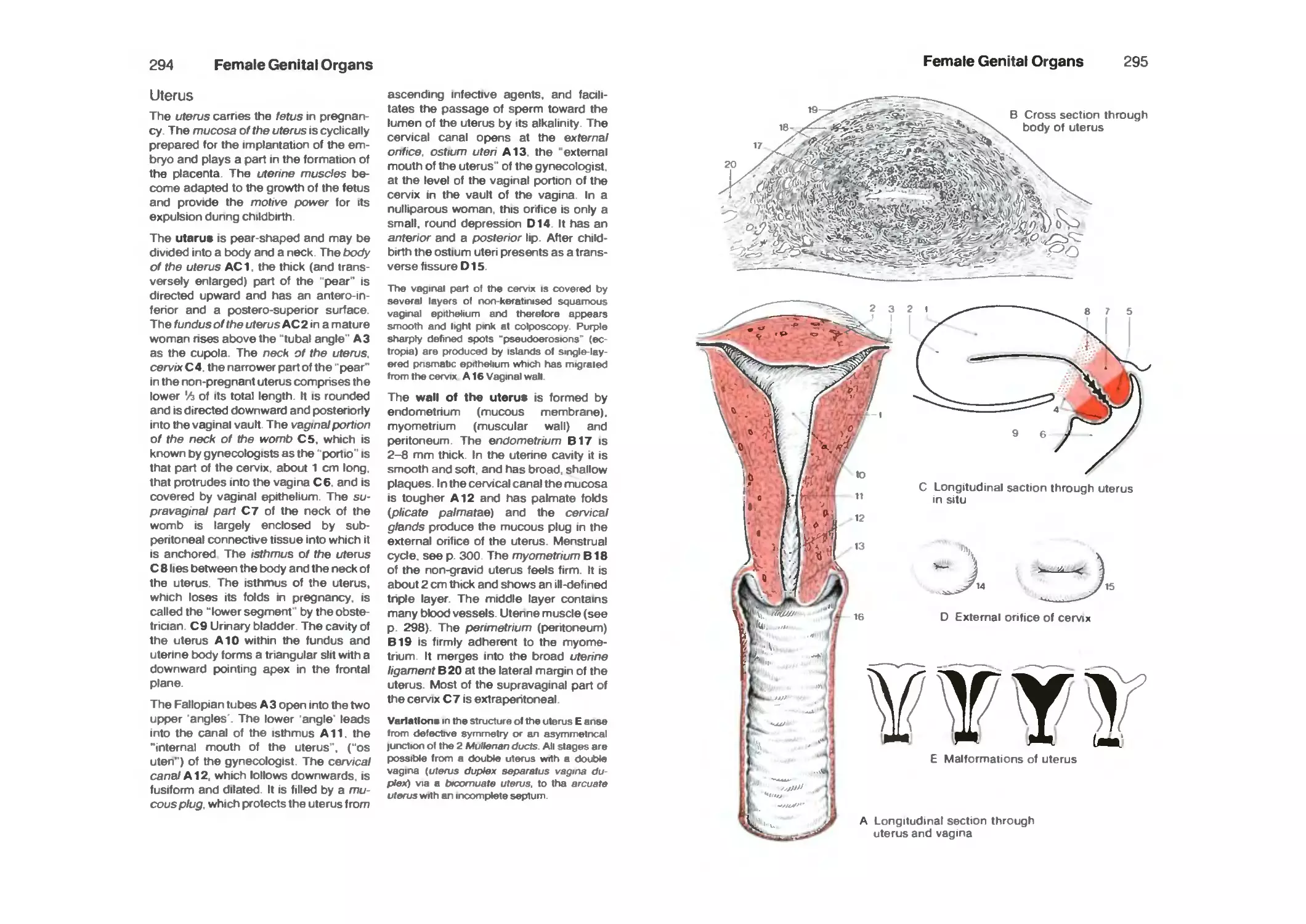
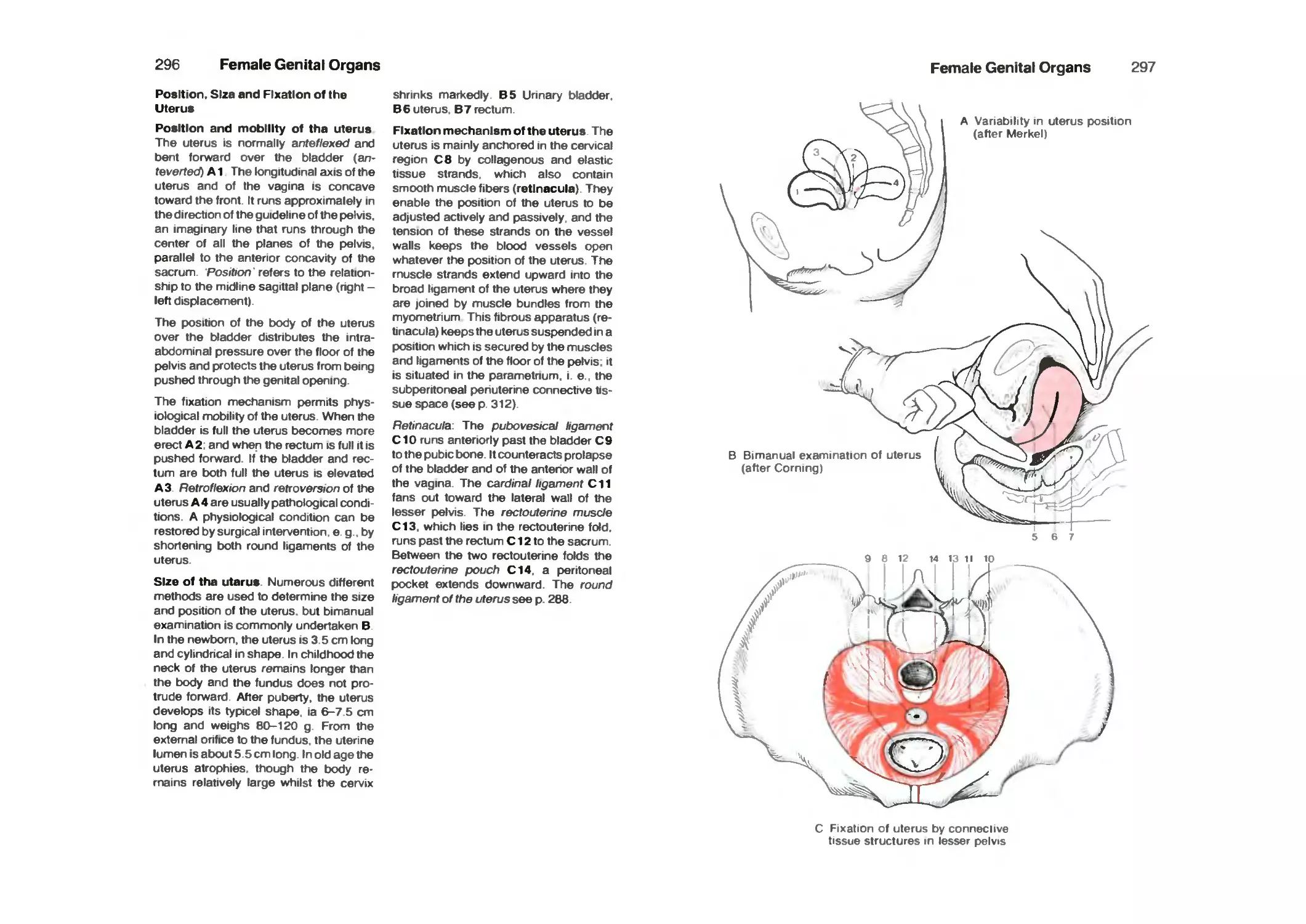
Uterus 288
Vagina 296
Blood Vessels of the Internal Genitalia in the Female 296
External Female Genitalia (the Vulva) and the Urethra 298
Pelvic Floor 300
Spaces of the Lesser Pelvis 306
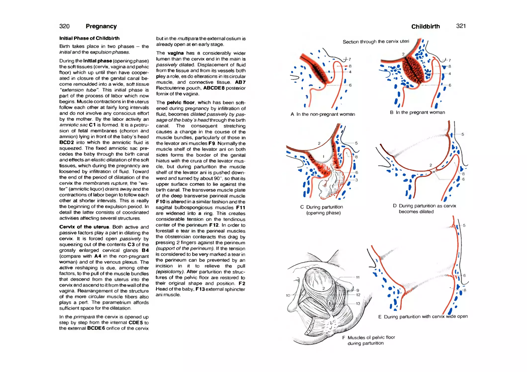
Pregnancy 310
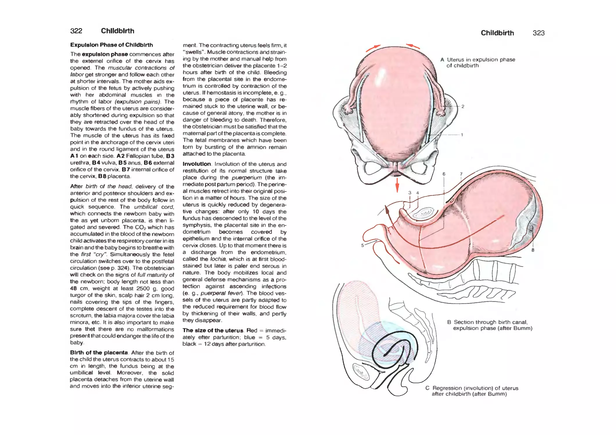
Childbirth 312
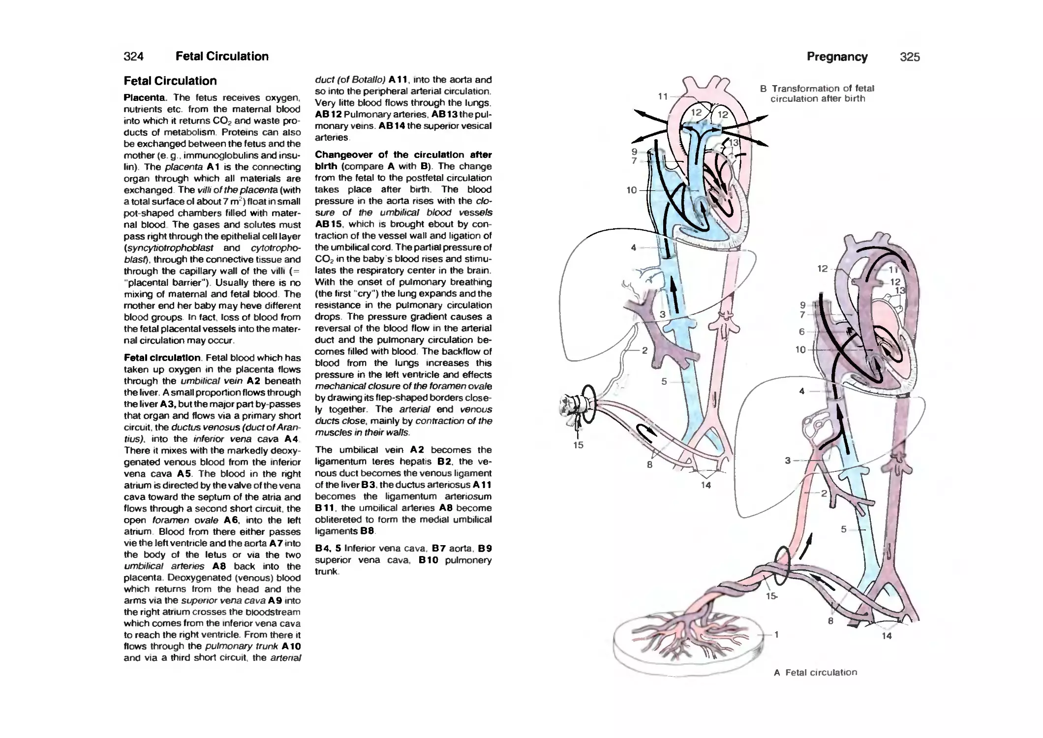
Fetal Circulation 318
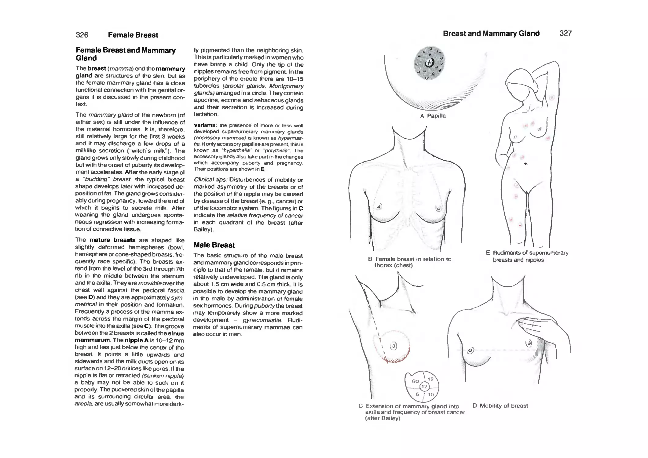
Female Breast and Mammary Gland 320
Male Breast 320
Female Breast 322
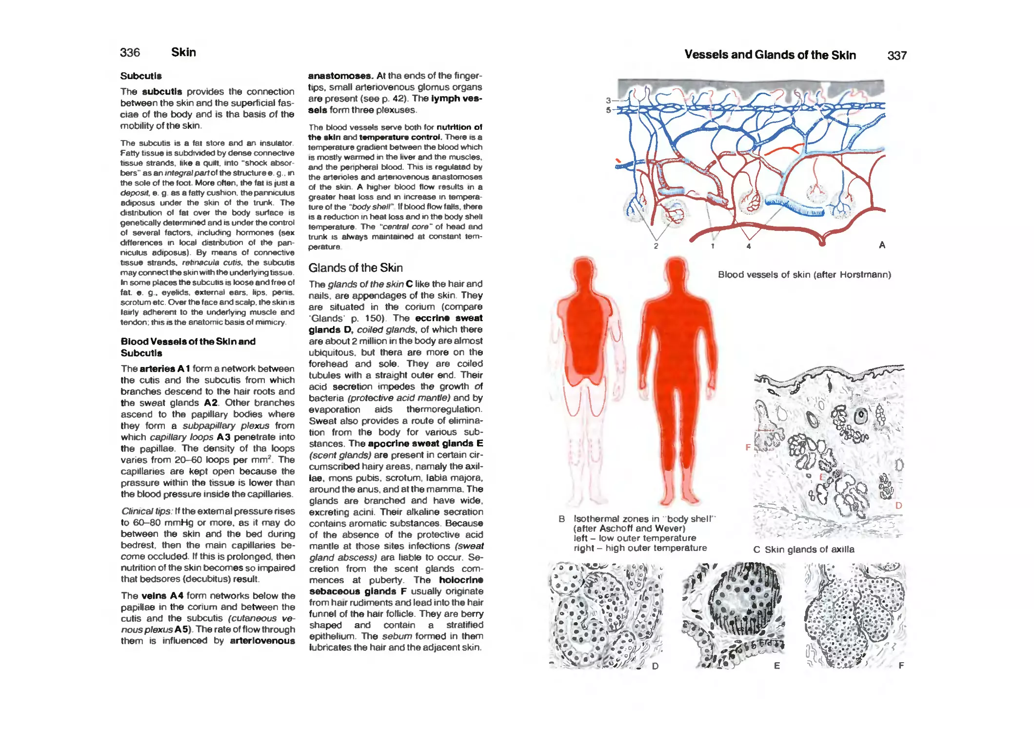
Skin 324
Layers of the Skin 326
Glands of the Skin 330
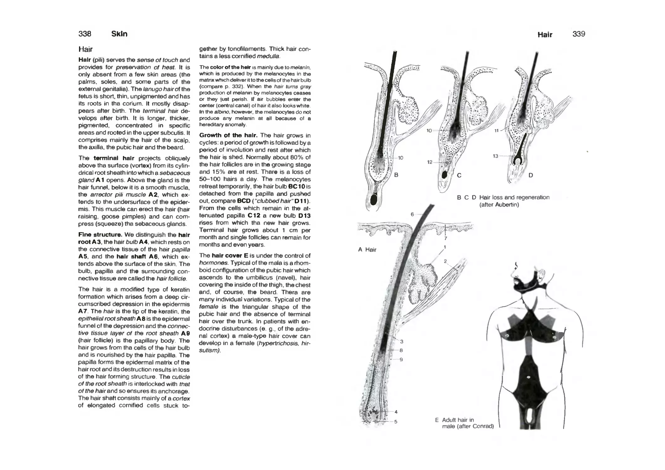
Hair 332
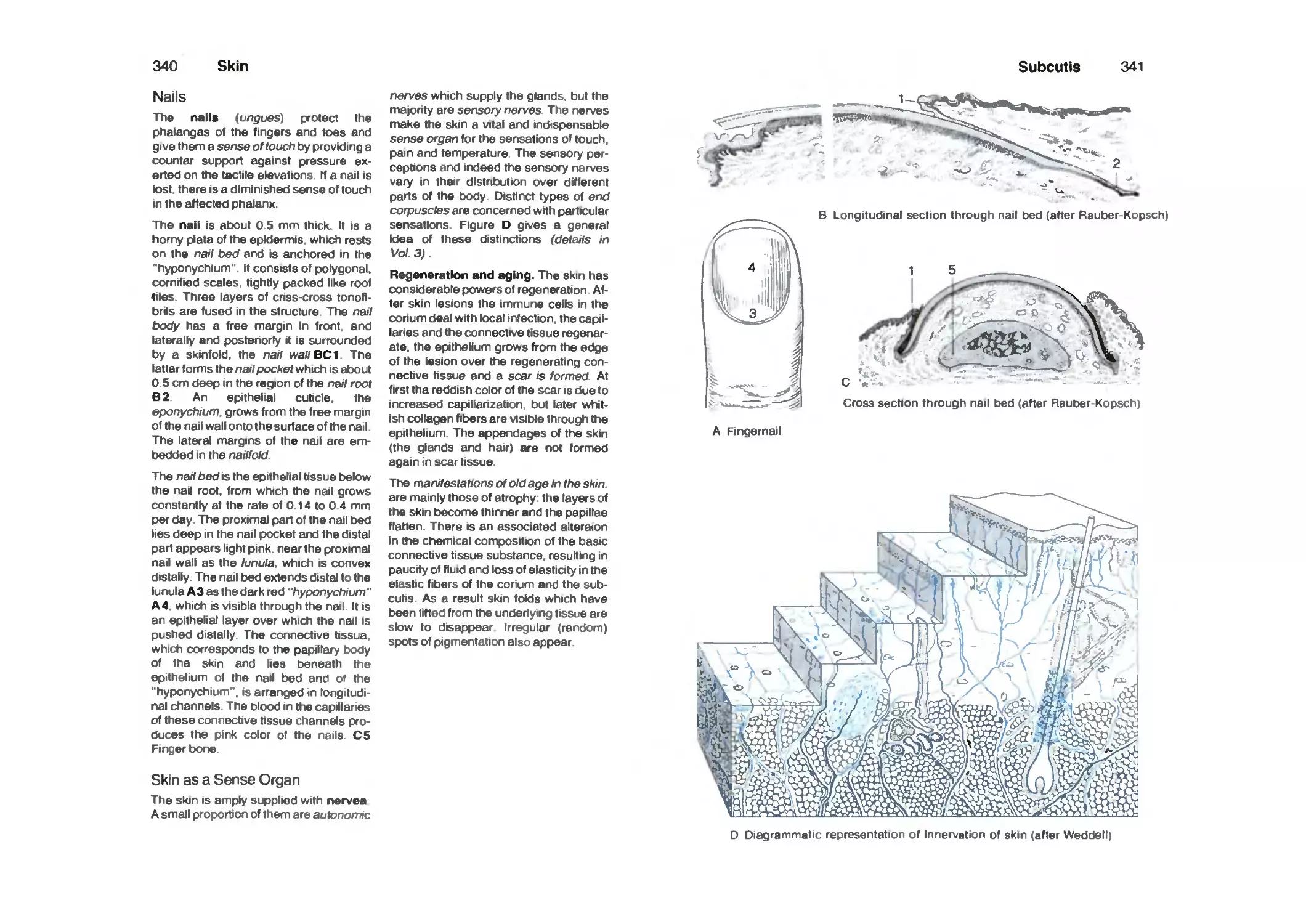
Nails 334
Skin as a Sense Organ 334
Literature 336
Index 348
Volume 1: Locomotor System by W. Platzer
Volume 3: Central Nervous System and Sensory Organs by W. Kahle
How to Use This Book
Each illustration is marked by a capital
letter and the illustrations on each page
have consecutive reference numbers.
Duplicate pictures of the same structure
or organ have the same number.
References in the text to the illustrations show
the corresponding letters and numbers.
Thus, it is easy to refer from the picture to
the corresponding text and from the text
to look at the appropriate illustration.
Students can use the book as a synopsis
and as an aid to recall facts acquired
during lectures and courses in
macroscopic and microscopic anatomy. In
order to refresh the memory, e. g. when
preparing for an examination, It is useful
for two candidates to work together; one
reads aloud a page of the text with its
references and the other looks up the
structures indicated on the page of
illustrations. Subsequently, students should
exchange roles and go through the same
part of the text again. In this way the
information is taken in by both the eye
and the ear and is repeated in a
convenient fashion. An advantage of the
concise text is that it refreshes the memory of
facts acquired in regular courses. If
particular topics are not recalled, or if any
doubts arise during revison, the two
candidates should discuss the subject in
question and look it up in one of the larger
text books (see the list of references at
the end of the book). Despite its brevity
this book contains a sufficient number of
repetitions as particularly important or
complicated matters are often discussed
again from another aspect on the same
or a subsequent page.
Viscera
The human viscera, organs on whose
activities Ihe life of the entire organism
depends, lie within the neck, chest,
abdomen and pelvis. From a functional point
of view they can be classified into
"systems": circulatory system (heart and
blood vessels), blood and def enss
system (blood, lymphatics, bone marrow)
•ndocrlns system (ductless glands and
cells which produce hormones)
respiratory system (nose, airways, larynx and
lungs), digestive system (mouth,
esophagus, gastrointestinal tract, liver
and pancreas), urogenital system and
unnary and genital or reproductive
organs.
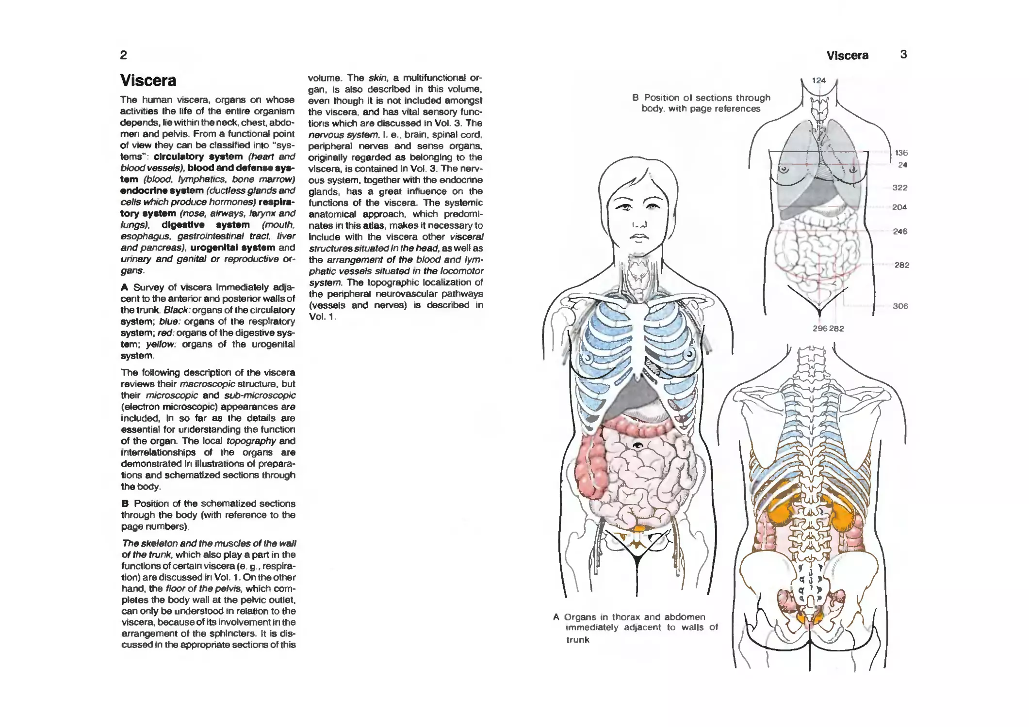
A Survey of viscera Immediately
adjacent to the anterior and posterior walls of
the trunk. Black: organs of the circulatory
system; blue: organs of the respiratory
system; red: organs of the digestive
system; yellow: organs of the urogenital
system.
The following description of the viscera
reviews their macroscopic structure, but
their microscopic and sub-microscopic
(electron microscopic) appearances are
included. In so far as the details are
essential for understanding the function
of the organ. The local topography and
interrelationships of the organs are
demonstrated In illustrations of
preparations and schematized sections through
the body.
B Position of the schematized sections
through the body (with refererKe to the
page numbers).
The skeleton and the muscles of the wall
of the trunk, which also play a part in the
functions of certain viscera (e. g.,
respiration) are discussed in Vol. 1. On the other
hand, the floor of the pelvis, which
completes the body wall at the pelvic outlet,
can only be understood in relation to the
viscera, because of its involvement in the
arrangement of the sphincters. It is
discussed in the appropriate sections of this
volume. The skin, a multifunctional
organ, is also described in this volume,
even though it is not included amongst
the viscera, and has vftal sensory
functions which are discussed in Vol. 3. The
nervous system. I. e.. brain, spinal cord,
peripheral nerves and sense organs.
originally regarded as belonging to the
viscera, is contained In Vol. 3. The
nervous system, together with the endocrine
glands, has a great inftuerKe on the
functions of the viscera. The systemic
anatomical approach, which
predominates in this atlas, makes it necessary to
Include with the viscera other visceral
structures situated in the head, as well as
the arrangement of the blood and
lymphatic vessels situated in the locomotor
system. The topographic localization of
the peripheral neurovascular pathways
(vessels and nerves) is described in
Vol.1.
Viscera
124
B Position ol sections through
body, with page references
A Organs in thorax and abdomen
immediately adjacent to walls of
trunk
Circulatory System
The heart and the vessels are the organs
of circulation. They contain blood (with
the exception of lymphatic vessels).
Circulatory systems. In man and the
higher mammals after birth it is possible
to distinguish between the greater
(systemic) circulation, that supplies blood to
all the organs, and the lesser
(pulmonary) circulation, which serves for the
exchange of gases. Together they form a
continuous circuit shaped like a figure 8
At its center is a suction and pressure
pump, the heart.
Arteries, capillaries, veins. All vessels
that carry blood away from the heart are
called artenes. and all vessels which
carry blood toward the heart are called
veins. Between the arteries and the veins
in the systemic and pulmonary
circulations lies the capillary network It is
important to remember that blood rich in
oxygen is known as "arterial"
(oxygenated) and blood deficient in oxygen is
called "venous" (deoxygenated) In the
systemic circulation arteries carry
oxygenated blood and veins cerry
deoxygenated blood. In the pulmonary
circulation venous blood flows through the
artenes into the lungs and arterial blood
passes through the veins to the heart. In
the pulmonary circulation there is only
one organ the lungs, through which all
the blood flows, and it is of uniform
composition. In the systemic arculation
there are various organs, e. g.. the
kidneys, intestines, endocrine glands, etc
The systemic blood flow can take
different routes since the systemic circulation
consists of numerous parallel subcir-
cuits The composition of the blood
flowing through them is not uniform
Heart. The heart is divided into twoparts
Each part has an antechamber, the
atnum. and a mam chamber the
ventricle The 'nght heart' (right atrium end
ventricle) propels blood through the
pulmonary orculation, and the left heart"
(left atnum and ventncle) is responsible
for flow through the sysfemrc circulation.
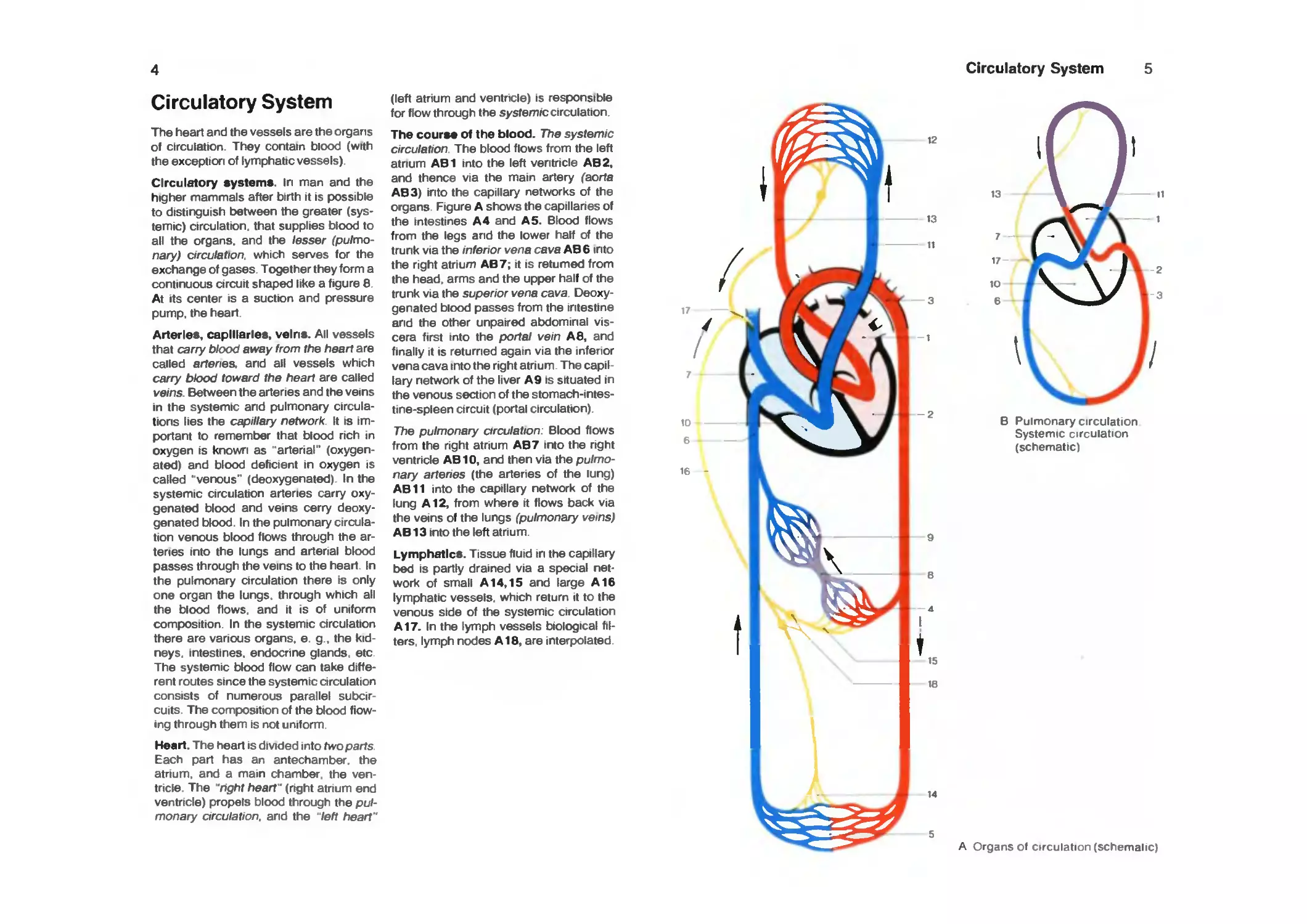
The course of the blood. The systemic
circulation. The blood flows from the left
atrium AB1 into the left ventricle AB2,
and thence via the main artery ^aorfa
AB3) into the capillary networks of the
organs. Figure A shows the capillaries of
the intestines A 4 and A 5. Blood flows
from the legs and the lower half of the
trunk via the inferior vena cava AB6 into
the right atrium AB7; it is retumed from
the head, arms and the upper half of the
trunk via the superior vena cava.
Deoxygenated blood passes from the intestine
and the other unpaired abdominal
viscera first into the portal vein A 8, and
finally it is returned again via the infenor
vena cava into the right atnum. The
capillary network of the liver A9 is situated in
the venous section of the
stomach-intestine-spleen circuit (portal circulation).
TTie pulmonary circulation: Blood flows
from the right atnum AB7 into the right
ventricle AB10, and then via the
pulmonary arteries (the artenes of the lung)
AB11 into the capillary network of the
lung A12, from where it flows back via
the veins of the lungs (pulmonary veins)
AB13 into the left atrium.
Lymphatics. Tissue fluid in the capillary
bed IS partly drained via a special
network of small A14,15 and large A16
lymphatic vessels, which return it to the
venous side of the systemic circulation
A17. In the lymph vessels biological
filters, lymph nodes A18, are interpolated
Circulatory System
B Pulmonary circulation
Systemic circulation
(schematic)
A Organs of circulation (schemalic)
Circulatory System
Heart
Shape of the Heart
The heart can be compared to a rour>ded cone
lying on Its side The apex-apex oord/s-points
f onward toward the left and downward, the base
- basis cordis - points backward toward the
nght and upward The size of the heart is
determined by the amount of work it has to
periorm. and is at least as big as the clenched
f tst of Its possessor
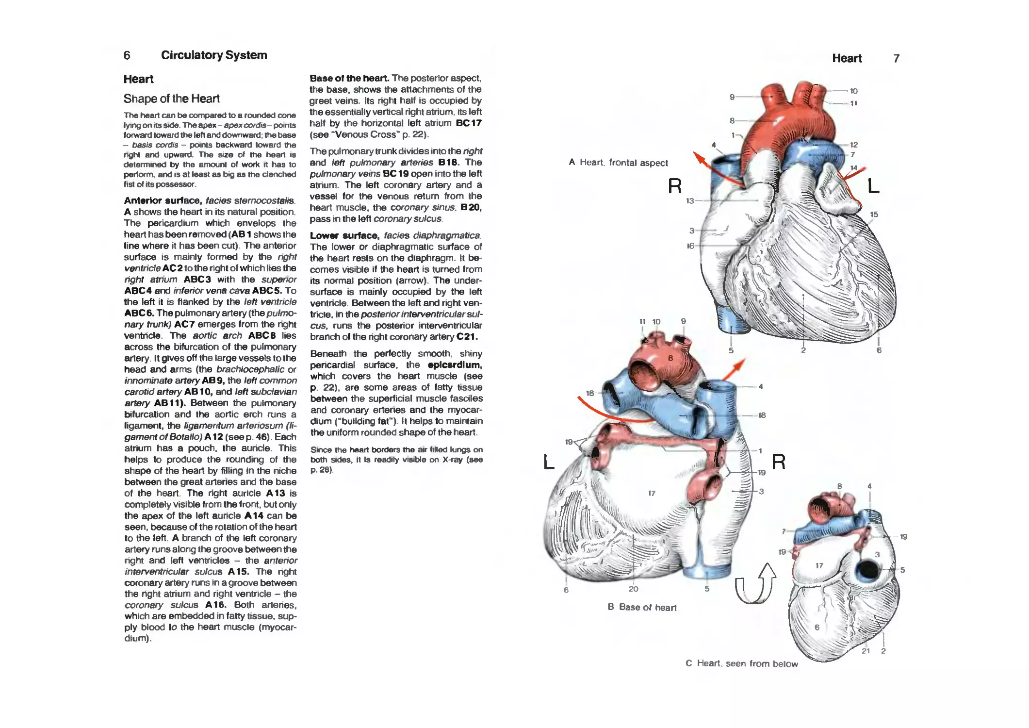
Anterior surface, fades sternocostalis.
A shows the heart in its natural position.
The pericardium which envelops the
heart has been removed (AB1 shows the
line where it has been cut). The anterior
surface is mainly formed by the right
ventricle AC 2 to the right of which lies the
right atnum ABC 3 with the superior
ABC4 and inferior vena cava ABC5. To
the left it is flanked by the left ventricle
ABC 6. The pulmonary artery (the
pulmonary trunk) AC 7 emerges from the nght
ventricle. The aortic arch ABC 8 lies
across the bifurcation of the pulmonary
artery. It gives off the large vessels to the
head and arms (the brachiocephalic or
innominate artery AB 9, the left common
carotid artery AB 10, and left subdavian
artery AB11). Between the pulmonary
bifurcation and the aortic erch runs a
ligament, the ligamentum arteriosum
(ligament of Botallo) A12 (see p. 46). Each
atrium has a pouch, the auricle. This
helps to produce the rounding of the
shape of the heart by filling In the niche
between the great arteries and the base
of the heart. The right auricle A13 is
completely visible from the front, but only
the apex of the left auricle A14 can be
seen, because of the rotation of the heart
to the left. A branch of the left coronary
artery runs along the groove between the
right and left ventricles - the anterior
interventricular sulcus A15. The right
coronary artery runs in a groove between
the right atrium and right ventricle - the
coronary sulcus A16. Both arteries,
which are embedded in fatty tissue,
supply blood io the heart muscle
(myocardium).
Base of the heart. The posterior aspect.
the base, shows the attachments of the
greet veins. Its right half is occupied by
the essentially vertical right atrium, its left
half by the horizontal left atrium BC17
(see "Venous Cross" p. 22).
The pulmonary trunk divides into the right
and left pulmonary arteries B18. The
pulmonary veins BC 19 open irrto the left
atrium. The left coronary artery and a
vessel for the venous return from the
heart muscle, the coronary sinus. B20.
pass in the left coronary sulcus.
Lower surface, fades diaphragmatica.
The lower or diaphragmatic surface of
the heart reels on the diaphragm. It
becomes visible if the heart is turned from
its normal position (arrow). The under-
surface is mainly occupied by the left
ventricle. Between the left and right
ventricle. In the posterior interventricular
sulcus, runs the posterior interventricular
branch of the right coronary artery C21.
Beneath the perfectly smooth, shiny
pericardial surface, the epicsrdlum,
which covers the heart muscle (see
p. 22). are some areas of fatty tissue
between the superficial muscle fasciles
and coronary erteries and the
myocardium ("building fat"). It helps to maintain
the uniform rounded shape of the heart.
Sirx» the heart borders the air filled lungs on
both sides, it Is readily visit>le on X-ray (see
p. 28).
Heart
A Heart, frontal aspect
20 5
8 Base of heart
1 2
C Heart seen from below
8 Circulatory System
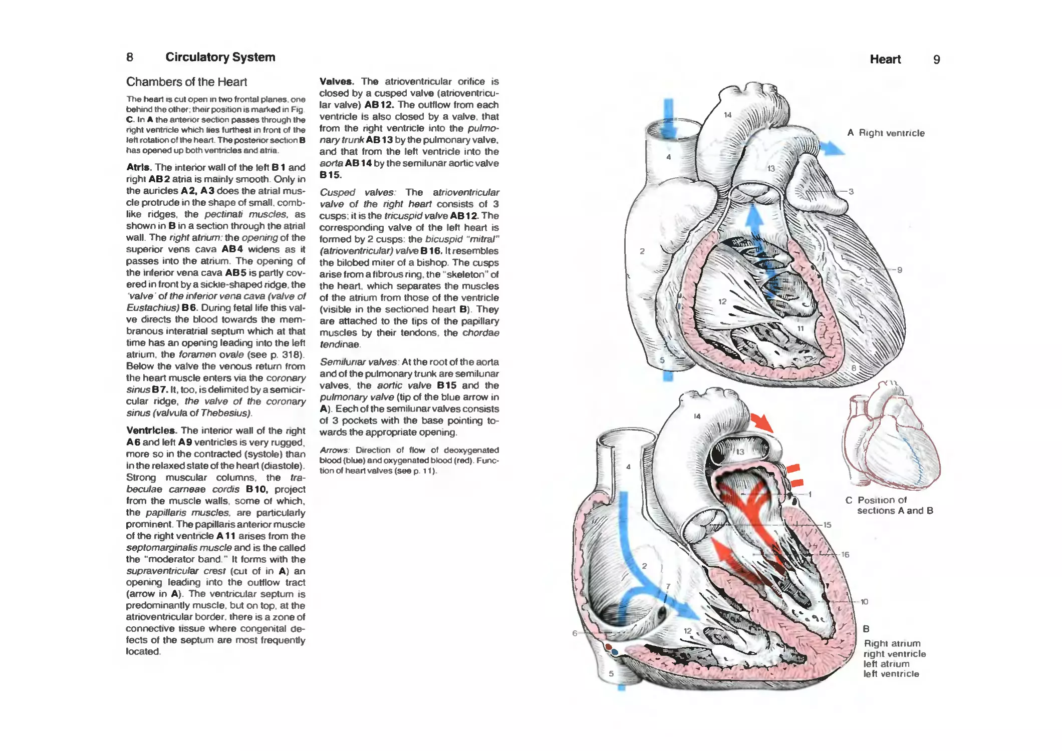
Chambers of the Heart
The head is cut open in two frontal planes one
behind the other their position is marked in Fig
C. In A the anterior sectton passes through the
nght ventncle which lies furthest in front of the
left rotation of the heart The postenor sections
has oper>ed up both ventncles arxl atria
Atrls. The interior wall of the left B1 and
right AB2 atna is mainly smooth. Only in
the auricles A2, A3 does the atrial
muscle protrude in the shape of small,
comblike ridges, the pectinati muscles, as
shown in B in a section through the atnal
wall. The right atnum. the opening of the
superior vens cava AB4 widens as it
passes into the atnum The opening of
the inferior vena cava AB5 is partly
covered in front by a sickie-shaped ridge, the
valve of the inferior vena cava (vatve of
Eustachius) B6. During fetal life this
valve directs the blood towards the
membranous interatrial septum which at that
time has an opening leading into the left
atrium, the foramen ovale (see p 316)
Below the valve the venous return from
the heart muscle enters via the coronary
sinusBl. It. too. is delimited by a
semicircular ridge, the valve of the coronary
sinus (va/vula of Thebesius)
Ventricles. The intenor wall of the nght
A 6 and left A 9 ventricles is very rugged.
more so in the contracted (systole) than
in the relaxed state of the heart (diastole)
Strong muscular columns, the tra-
beculae carneae cordis BIO, project
from the muscle walls, some of which,
the papillaris muscles, are particulariy
prominent. The papillans anterior muscle
of the right ventrx;le All anses from the
septomarginalis muscle and is the called
the "moderator band." It forms with the
supraventricular crest (cut of in A) an
opening leading into the outflow tract
(arrow in A) The ventricular septum is
predominantly muscle but on top at the
atnoventricular border, there is a zone of
connective tissue where congenital
defects of the septum are most frequently
located
Valves. The atrioventricular orifice is
closed by a cusped valve (atnoventncu-
lar valve) AB12. The outflow from each
ventncle is also closed by a valve, that
from the nght ventricle into the
pulmonary trunk AB13 by the pulmonary valve,
and that from the left ventncle into the
aorta AB 14 by the semilunar aortic valve
B15.
Cusped valves. The atrioventricular
valve of the right heart consists of 3
cusps: It IS the tricuspid valve AB 12. The
corresponding valve of the left heart is
formed by 2 cusps the bicuspid 'mitral"
(atrioventricular) valve B16. It resembles
the bilobed miter of a bishop The cusps
arise from a fibrous nng, the' skeleton' of
the heart, which separates the muscles
of the atrium from those of the ventricle
(visible in the sectioned heart B). They
are attached to the tips of the papllary
muscles by their tendons the chordae
tendinae.
Semilunar valves. At the root of the aorta
and of the pulmonary trunk are semilunar
valves, the aortic valve B15 and the
pulmonary valve (tip of the blue arrow in
A). Eech of the semilunar valves consists
of 3 pockets with the base pointing
towards the appropriate opening
Arrows- Direction of flow of deoxygenated
blood (blue) arxl oxygenated blood (red)
Function of head valves (see p 11)
Heart 9
A Right ventricle
Right atnum
right ventricle
left atnum
left ventricle
10
circulatory System
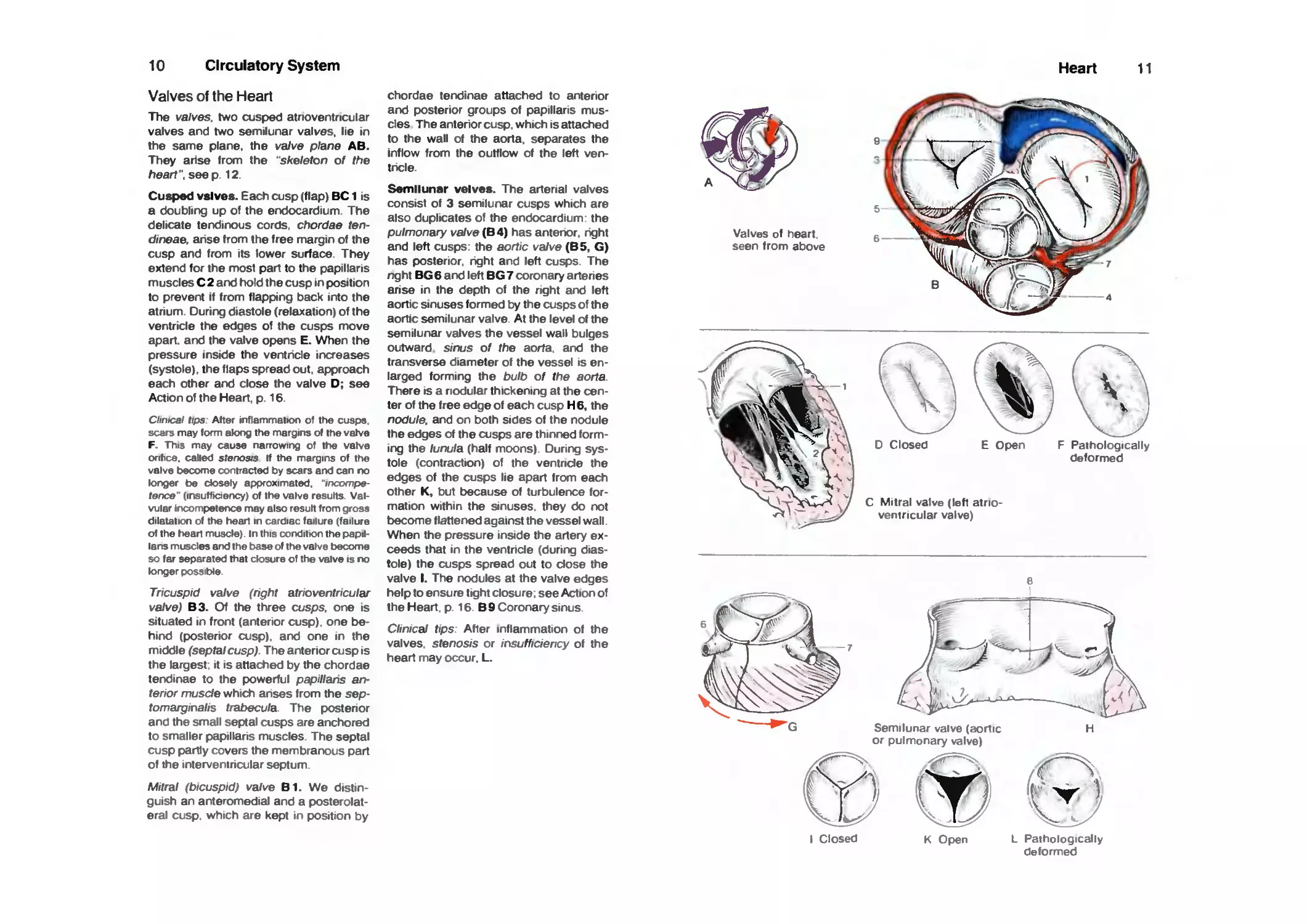
Valves of the Heart
The valves, two cusped atrioventricular
valves and two semilunar valves, lie in
the same plane, the vaive plane AB.
They arise from the "skeleton of the
heart", seep 12
Cusped vslves. Each cusp (flap) BC1 is
a doubling up of the endocardium The
delicate tendinous cords, chordae ten-
dineae, anse from the free margin of the
cusp and from its lower surface They
extend for the most part to the papillaris
muscles C 2 and hold the cusp in position
to prevent if from flapping back into the
atrium During diastole (relaxation) of the
ventricle the edges of the cusps move
apart, and the valve opens E. When the
pressure inside the ventncle increases
(systole), the flaps spread out. approach
each other and close the valve D; see
Action of the Heart, p. 16
Clinical tips After inflammation of the cusps
scars may fomi along the margins of the vatve
F. This may cause narrowing of the vatve
orifice, called stenosis If the margins of the
valve become contracted by scars and can rx)
longer be closely approximated,
incompetence (insuffictency) of the valve results
Valvular irKX)mpeterx» may also result from gross
dilatation of the head in cardiac failure (failure
of the heart muscle) In this corxlitton the papil-
lans muscles arxJ the base of the valve become
so far separated that closure of the valve is no
longer possible
Tricuspid valve (nght atrioventricular
valve) B3. Of the three cusps, one is
situated in front (anterior cusp) one
behind (posterior cusp) and one in the
middle (septalcusp) Theantenorcuspis
the largest it is attached by the chordae
tendinae to the powerful papillans
anterior muscle which anses from the sep-
tomarginalis trabecula The postenor
and the small septal cusps are anchored
to smaller papillaris muscles The septal
cusp partly covers the membranous part
of the interventncular septum
Mitral (bicuspid) valve B1. We
distinguish an anteromedial and a
posterolateral cusp which are kept in position by
chordae tendinae attached to anterior
and posterior groups of papillaris
muscles The anterior cusp, which is attached
to the wall of the aorta, separates the
inflow from the outflow of the left
ventncle.
Semilunar velves. The arterial valves
consist of 3 semilunar cusps which are
also duplicates of the endocardium, the
pulmonary vaive (B4) has antenor. nght
and left cusps the aortic vaive (B5, G)
has posterior, right and left cusps The
rTght BG 6 and left BG 7 coronary artenes
arise in the depth of the right and left
aortic sinuses formed by the cusps of the
aortic semilunar valve At the level of the
semilunar valves the vessel wall bulges
outward sinus of the aorta, and the
transverse diameter of the vessel is
enlarged forming the bulb of the aorta
There is a nodular thickening at the
center of the free edge of each cusp H6, the
nodule, and on both sides of the nodule
the edges of the cusps are thinned
forming the lunula (half moons) During
systole (contraction) of the ventncle the
edges of the cusps lie apart from each
other K, but t>ecause of turbulence
formation within the sinuses they do not
t>ecome flattened against the vessel wall
When the pressure inside the artery
exceeds that in the ventncle (during
diastole) the cusps spread out to dose the
valve I. The nodules at the valve edges
help to ensure tight closure, see Action of
the Heart p 16 B 9 Coronary sinus
Clinicai tips After inflammation of the
valves stenosis or insufficiency of the
heart may occur, L.
Heart 11
Valves of heart
seen from above
^
\
D Closed
E Open F Pathologically
deformed
C Mitral valve (left
atrioventricular valve)
-^^*H-;;v_^
Semilunar valve (aortic
or pulmonary valve)
n OfJ 0
I Closed
K Open
L Pathologically
deformed
12
Circulatory System
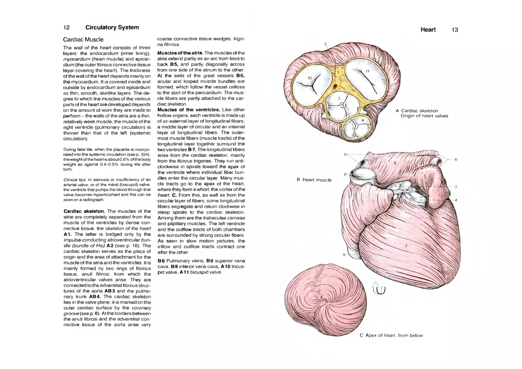
Cardiac Muscle
The wall of the heart consists of three
layers: the endocardium (inner lining).
myocarc^um (heart musde) and
epicardium (the outer fibrous connective tissue
layer covering the heart). The thickness
of the wall of the heart depends mainly on
the myocardium. It is covered inside and
outside by endocardium and epicardium
as thin, smooth, skinlike layers. The
degree to which the muscles of the vanous
parts of the heart are developed depends
on the amount of work they are made to
perform - the walls of the atria are a thin
relatively weak muscle; the muscle of the
right ventricle (pulmonary circulation) is
thinner than that of the left (systemic
circulation)
Dunng fetal lile. when the placenta is incorpo-
raied into the systemic circulatton (see p 324)
the weight of the head is about 0 6% of the body
weight as against 0 4-0 5% during life after
birth
Clinical tips in stenosis or insufficiency of an
anenal valve, or of the mitral (biscuptd) valve
the ventncle that pumps the blood through that
valve becomes hypertrophied arxl this can be
seen on a radiograph
Csrdisc skeleton. The muscles of the
atrie are completely separated from the
muscle of the ventricles by dense
connective tissue, the skeleton of the heart
A1. The latter is bndged only by the
impulse-conducting atrioventricular
bundle (bundle of His) A 2 (see p. 16). The
cardiac skeleton serves as the place of
origin and the area of attachment for the
muscle of the atria and the ventricles It is
mainly formed by two nngs of fibrous
tissue, anuli fibrosi from which the
atrioventricular valves arise. They are
connected to the adventitial fibrous
structures of the aorta AB3 and the
pulmonary trunk AB4. The cardiac skeleton
lies in the valve plane* it is marked on the
outer cardiac surface by the coronary
proove (see p. 6) At the borders between
the anuli fibrosi and the adventitial
connective tissue of the aorta arise very
coarse connective tissue wedges, fripo-
na fibrosa.
Muscles of the atria. The muscles of the
atria extend partly as an arc from front to
back B5, and partly diagonally across
from one side of the atrium to the other
At the exits of the great vessels 86,
anular and looped muscle bundles are
formed, which follow the vessel orifices
to the start of the pericardium The
muscle fibers are partly attached to the
cardiac skeleton.
Muscles of the ventricles. Like other
hollow organs, each ventncle is made up
of an external layer of longitudinal fibers,
a middle layer of arcular and an internal
layer of longitudinal fibers The
outermost muscle fibers (muscle tracts) of the
longitudinal layer together surround the
two ventricles B 7. The longitudinal fibers
arise from the cardiac skeleton, mainly
from the fibrous trigones. They run
anticlockwise in spirals toward the apex of
the ventricle where individual fiber
bundles enter the circular layer. Many
muscle tracts go to the apex of the heart,
where they form a whori. the vortex of the
heart. C. From this as well as from the
circular layer of fibers, some longitudinal
fibers segregate and return clockwise in
steep spirals to the cardiac skeleton
Among them are the trabeculae carneae
and papillary muscles. The left ventricle
and the outflow tracts of both chambers
are surrounded by strong circular fibers
As seen in slow motion pictures, the
inflow and outflow tracts contract one
after the other.
B6 Pulmonary veins. B6 superior vena
cava B9 inferior vena cava. A10
tricuspid valve. All bicuspid valve
Heart 13
A Cardiac skeleton
Origin of heart valves
77/S?>i
A^^/^i^
B Heart muscle
Apex of heart from below
14
Circulatory System
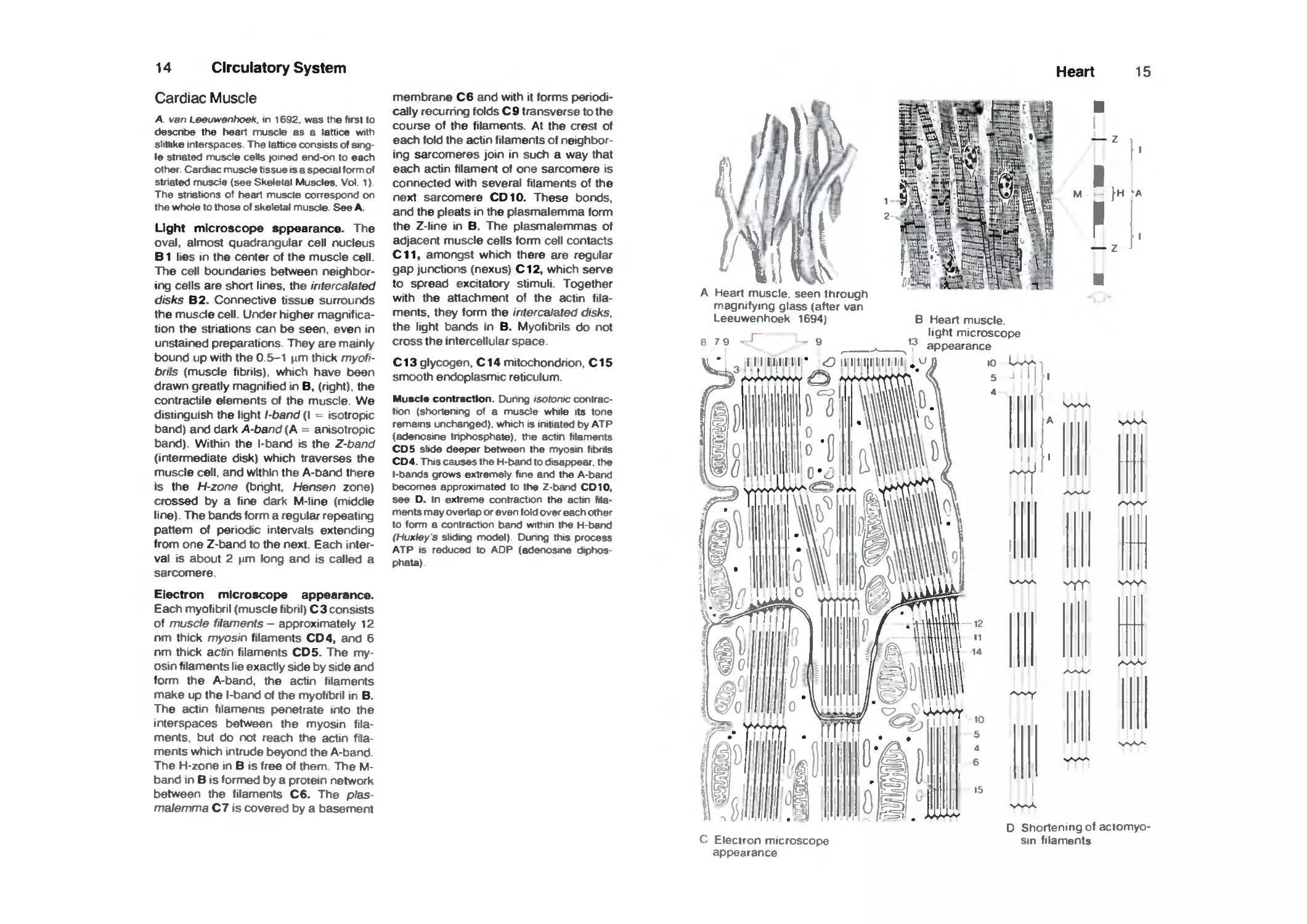
Cardiac Muscle
A. van Leeuwanhoek, in 1692. was the first to
descntw the hean muscle as a lattice with
slitiike interspaces. The lattice consists of
single stnated muscle cells joined end-on to each
other Cardiac musOe tissue is a special form of
striated muscle (see Skeletal Muscles. Vol 1)
The stnations of head muscle correspond on
the whole to those of skeletal muscle. See A.
Ught microscope sppearance. The
oval, almost quadrangular cell nucleus
B1 lies in the center of the muscle cell.
The cell boundaries between
neighboring cells are short lines, the intercalated
disks B2. Connective tissue surrounds
the musde cell. Under higher
magnification the striations can be seen, even in
unstained preparations. They are mainly
bound up with the 0.5-1 \im thick
myofibrils (muscle fibrils), which have been
drawn greatly magnified in B, (right), the
contractile elements of the muscle. We
distinguish the light l-band (I - isotropic
band) and dark A-band (A = anisotropic
band). Within the l-band is the Z-band
(intermediate disk) which traverses the
muscle cell, and within the A-band there
Is the H-zone (bright, Hensen zone)
crossed by a fine dark M-line (middle
line). The bands form a regular repeating
pattern of periodic intervals extending
from one Z-band to the next Each
interval IS about 2 ^m long and is called a
sarcomere
Electron microscope appearance.
Each myofibril (muscle fibril) C 3 consists
of musde filaments - approximately 12
nm thick myosin filaments CD 4, and 6
nm thick acfrn filaments CDS. The
myosin filaments lie exactly side by side and
form the A-band. the actin filaments
make up the l-band of the myofibril in B.
The actin filaments penetrate into the
interspaces between the myosin
filaments but do not reach the actin
filaments which intrude beyond the A-band
The H-zone in B is free of them The M-
band in B is formed by a protein network
between the filaments C6. The p/as-
malemma C7 is covered by a basement
membrane C6 and with it forms
periodically recurnng folds C9 transverse to the
course of the filaments. At the crest of
each fold the actin filaments of
neighboring sarcomeres join in such a way that
each actin filament of one sarcomere is
connected with several filaments of the
next sarcomere CD 10. These bonds,
and the pleats in the plasmalemma form
the Z-line in B. The plasmalemmas of
adjacent muscle cells form cell contacts
C11, amongst which there are regular
gap junctions (nexus) CI2, which serve
to spread excitatory stimuli. Together
with the attachment of the actin
filaments, they form the intercalated disks.
the light bands in B. Myofibrils do not
cross the intercellular space.
C13 glycogen, C14 mitochondrion. CIS
smooth endoplasmic reticulum.
Muscle contraction. Dunng isotontc conlrac-
tton (shortening of a muscle while its tone
remains unchanged), which is initiated t)y ATP
(aderwsine tnphosphate) the actin filaments
CDS slide deeper between the myosin fibrils
CD4. This causes the H-barxl to disappear, the
l-bands grows extremely fine and the A-barxl
becomes approximated to the Z-barxJ CD 10,
see D. In extreme contraction the aclin
filaments may overlap or even fold over each other
to form a contraction barxJ within the H-tjarxJ
(Huxley 3 sliding model) Dunng this process
ATP IS reduced to ADP (aderwsme diphos
phata)
Heart
15
A Heart muscle seen through
magnifying glass (after van
Leeuwenhoek 1694)
I
]h -a
79
g
t3
B Heart muscle
light microscope
appearance
iiiiiiiir <o iiiiiiiiiii .»J^ 10 W^
5
A s^-sAA
-1
vYY^ vw-
AA>v
-s,^^
C Electron microscope
appearance
D Shortening of actomyo-
sin filaments
16 Circulatory System
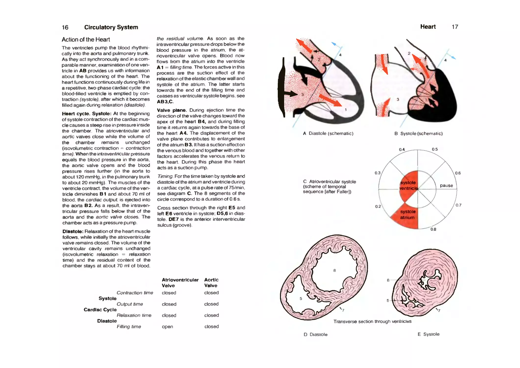
Action of the Heart
The ventricles pump the blood
rhythmically into the aorta and pulmonary trunk
As they act synchronously and in a
comparable manner examination of one ven-
tncle In AB provides us with infomiation
about the functioning of the heart The
heart furx^tions continuously dunng life in
a repetitive two-phase cardiac cycle the
blood-filled ventricle is emptied by
contraction (systole), after which it becomes
filled again during relaxation (diastole)
Heart cycle. Systole: At the beginning
of systole contraction of the cardiac
muscle causes a steep rise in pressure inside
the chamber The atnoventncular and
aortic valves close while the volume of
the chamber remains unchanged
(isovolumetnc contraction contraction
time). When the intraveritncular pressure
equals the blood pressure in the aorta
the aortic valve opens and the blood
pressure rises further (in the aorta to
about 120 mmHg in the pulmonary trunk
to about 20 mmHg) The muscles of the
ventricle contract the volume of the
ventricle diminishes B1 and about 70 ml of
blood, the cardiac output is ejected into
the aorta B2. As a result, the
intraventricular pressure falls below that of the
aorta and the aortic vafve doses The
chamber acts as a pressure pump
Diastole: Relaxation of the heart muscle
follows while initially the atrioventricular
valve remains closed The volume of the
ventricular cavity remains unchanged
(isovolumetnc relaxation relaxation
time) and the residual content of the
chamber stays at about 70 ml of blood
the residual volume As soon as the
intraventncular pressure drops below the
blood pressure in the atnum the
atnoventncular valve opens Blood now
flows from the atnum into the ventnde
A1 filling ttme The forces active in this
process are the suction effect of the
relaxation of the elastic chamber wall and
systole of the atnum The latter starts
towards the end of the filling time and
ceases as ventncular systole begins see
AB3,C.
Valve plane. Dunng ejection time the
direction of the valve changes toward the
apex of the heart B4, and dunng filling
time It returns again towards the base of
the heart A 4. The displacement of the
valve plane contnbutes to enlargement
of the atrium B 3. It has a suction effect on
the venous blood and together with other
factors accelerates the venous return to
the heart Dunng this phase the heart
acts as a suction pump
Timing For the time taken by systole and
diastole of the atnum and ventnde dunng
a cardiac cycle, at a pulse rate of 75/min
see diagram C. The 6 segments of the
circle correspond to a duration of 0 6 s
Cross section through the right E5 and
left E6 ventricle in systole D5,6 in
diastole DE7 IS the anterior interventricular
sulcus (groove)
Contraction time
Systole
Output time
Cardisc Cycle
Relaxation time
Diastole
Filling time
Atrioventricular
Velve
closed
closed
closed
ofjen
Aortic
Valve
closed
closed
closed
closed
Heart 17
A Diastole (schematic)
B Systole (schematic)
04
05
C Atnoventncular systole
(scheme of temporal
sequence [after Faller])
03
02
06
pause
07
08
Transverse section through ventricles
D Diastole E Systole
18
Circulatory System
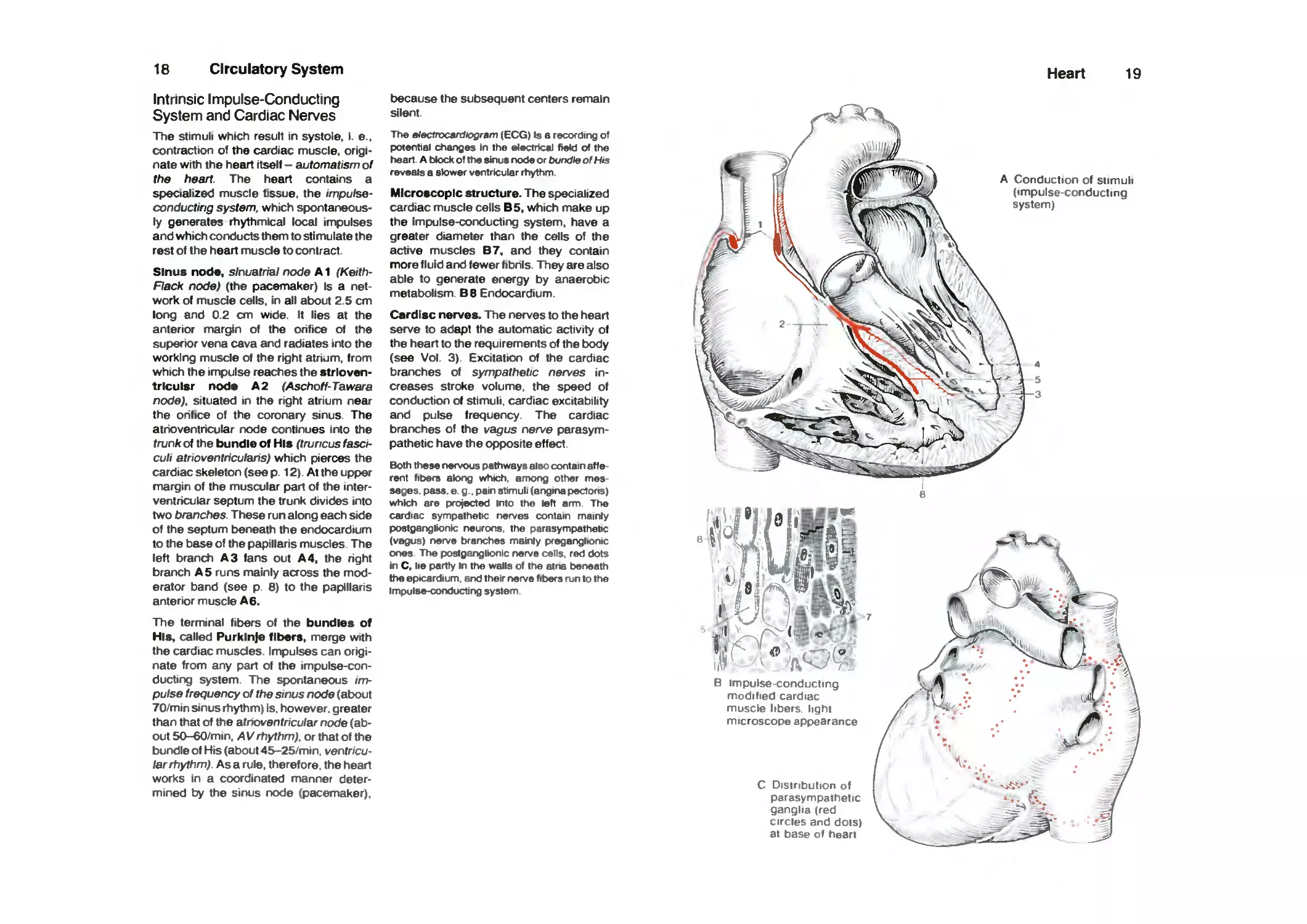
Intrinsic Impulse-Conducting
System and Cardiac Nerves
The stimuli which result in systole. I. e..
contraction of the cardiac muscle,
originate with the heart Itself - automatism of
the heart. The heart contains a
specialized muscle tissue, the impulse-
conducting system, which
spontaneously generates rhythmical local impulses
and which conducts them to stimulate the
rest of the heart muscle to contract.
Sinus node, slnuatrial node A1 (Keith-
Hack node) (the pacemaker) Is a
network of muscle cells, in all about 2.5 cm
long and 0.2 cm wide. It lies at the
anterior margin of the orifice of the
superior vena cava and radiates into the
working muscle of the right atrium, from
which the impulse reaches the strloven-
trlculsr nods A2 (Aschoff-Tawara
node), situated in the right atrium near
the orifice of the coronary sinus. The
atrioventricular node continues into the
trunk of the bundle of His (truncus
fasciculi athoventricularis) which pierces the
cardiac skeleton (see p. 12). At the upper
margin of the muscular part of the
interventricular septum the trunk divides into
two branches. These run along each side
of the septum beneath the endocardium
to the base of the papillaris muscles. The
left branch A3 fans out A4, the right
branch A 5 runs mainly across the
moderator band (see p. 6) to the papillaris
anterior muscle A6.
The terminal fibers of the bundles of
His, called Purklnje fibers, merge with
the cardiac muscles. Impulses can
originate from any part of the
impulse-conducting system The spontaneous
impulse frequency of the sinus node (about
70/min sinus rhythm) is. however, greater
than that of the atrioventricular node
(about 50-60/min. AVrhythm), or that of the
bundle of His (about 45-25/min.
ventricular rhythm). As a rule, therefore, the heart
works in a coordinated manner
determined by the sinus node (pacemaker).
t>ecause the subsequent centers remain
silent.
The Blectrocardtogram (EGG) Is a recording of
poientiai changes In the electHcal field of the
head. A t>k>ck of ttw sinus rwde or bundle of His
reveals a stower ventricular rhythm.
Microscopic structure. The specialized
cardiac muscle cells B5, which make up
the impulse-conducting system, have a
greater diameter than the cells of the
active muscles B7, and they contain
more fluid and fewer fibrils. They are also
able to generate energy by anaerobic
metabolism. B8 Endocardium.
Cardisc nerves. The nerves to the heart
serve to adapt the automatic activity of
the heart to the requirements of the body
(see Vol. 3). Excitation of the cardiac
branches of sympathetic nerves
increases stroke volume, the speed of
conduction of stimuli, cardiac excitability
and pulse frequency. The cardiac
branches of the vagus nerve
parasympathetic have the opposite effect.
Both these r>ervous pathways also contain
afferent fit>ers atong whtch, among other
messages, pass. e. g.. pain stimuli (angiru pectoris)
whtch are projected into the left arm The
cardiac sympathetic rwrves contain mainry
postgangtontc neurons, the parasympathetic
(vagus) nerve brarKhes mainry pregar^lionic
or>es- The postganglkxitc nerve cells, red dots
in C, lie partty In the walls of the atna beneath
the epicardium, arxJ their nerve fibers njn to the
Impulse-conducting system
Heart
19
A Conduction of stimuli
(impulse-conducting
system)
:-J^ 1,-^
B impulse conducting
modified cardiac
muscle libers light
microscope appearance
C Distribution of
parasympathetic
ganglia (red
circles and dots)
at base of heart
20
Circulatory System
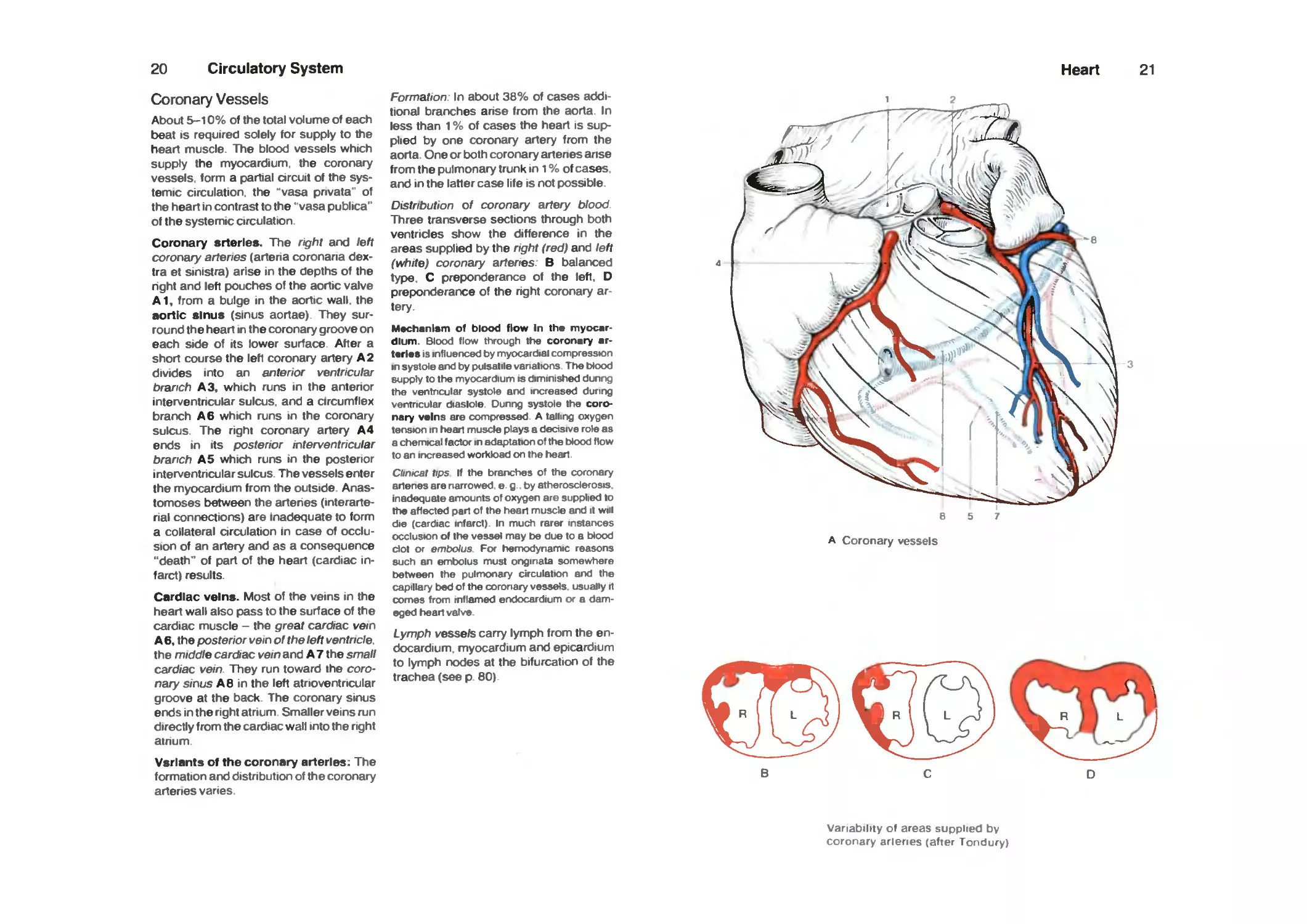
Coronary Vessels
About 5-10% of the total volume of each
beat IS required solely for supply to the
heart muscle. The blood vessels which
supply the myocardium, the coronary
vessels, form a partial arcuit of the
systemic circulation, the "vasa pnvata" of
the heart in contrast to the "vasa publica"
of the systemic circulation.
Coronary srterles. The right and left
coronary arteries (artena coronaria dex-
tra et sinistra) arise in the depths of the
right and left pouches of the aortic valve
A1, from a bulge in the aortic wall, the
aortic sinus (sinus aortae). They
surround the heart in the coronary groove on
each side of its lower surface After a
short course the left coronary artery A 2
divides into an anterior ventricular
branch A3, which runs in the antenor
interventricular sulcus, and a circumflex
branch A 6 which runs in the coronary
sulcus. The right coronary artery A 4
ends in its posterior interventncular
branch A 5 which runs in the posterior
interventncular sulcus. The vessels enter
the myocardium from the outside
Anastomoses between the artenes (interarte-
rial connections) are inadequate to form
a collateral circulation in case of
occlusion of an artery and as a consequence
"death" of part of the heart (cardiac
infarct) results.
Cardiac veins. Most of the veins in the
heart wall also pass to the surface of the
cardiac muscle - the great cardUac vein
A 6, the posterior vein of the left ventricle.
the middle cardiac vein an6 A 7 the small
cardiac vein They run toward the
coronary sinus A 6 in the left atrioventricular
groove at the back The coronary sinus
ends in the right atrium Smaller veins run
directly from the cardiac wall into the right
atnum
Vsriants of the coronary arteries: The
formation and distribution of the coronary
arteries vanes
Formation- In atx>ut 38% of cases
additional branches arise from the aorta In
less than 1 % of cases the heart is
supplied by one coronary artery from the
aorta One or both coronary artenes arise
from the pulmonary trunk in 1 % of cases
and in the latter case life is not possible.
Distribution of coronary artery blood
Three transverse sections through both
ventricles show the difference in the
areas supplied by the nght (red) and left
(white) coronary artenes. B balanced
type. C preponderance of the left. D
preponderance of the right coronary
artery.
Mechanism of blood flow in the
myocardium. Blood flow through the coronary
arteries IS influerx^d by myocardial compression
in systole arxl by pulsatile venations The blood
supply to the myocardium is diminished dunrig
the ventncular systole arxl irK:reased during
ventricular diaslole Dunng systole the
coronary veins are compressed A tailing oxygen
tension in heart muscle plays a decisive rote as
a chemical factor in adaptation of the blood flow
to an irK:reased worKk>ad on the head
Chnicat tips If the brarx:hes of the coronary
artenes are narrowed e g by atherosclerosis
inadequate amounts of oxygen are supplied to
ttie affected pan of the hean muscle and it will
die (cardiac infarct) In much rarer instances
occlusion of the vessel may t>e due to a blood
dot or embotu3 For hemodynamic reasons
such an emtx>lus must onginata somewhere
twtween the puinxxiary circulation and the
capillary bed of the coronary vessels usually rt
comes from inflamed erxlocardium or a
damaged hean valve
Lymph vessels carry lymph from the
endocardium myocardium and epicardium
to lymph nodes at the bifurcation of the
trachea (see p 60)
Heart
21
8 5 7
A Coronary vessels
Variability of areas supplied by
coronary arlenes (after Tondury)
22
Circulatory System
Serous Cavities
Viscera which undergo marked changes
In size and/or position in relation to neigh*
bonng organs, the heart, lungs, the major
part of the gastrointestinal tract etc.. lie in
serous cavities: the pericardial, pleural,
and peritoneal cavities.
A serous cavity is a capillary cavity closad on sH
sides. It IS covered by a shiny, smooth serous
layer, the 'serosa*, and contains a small amount
of serous fluid The serous membrarie covers
the organs with the visceral serosa and the
wall of the serous cavity with the parietal
serosa. The two layers merge at reflections,
e. g . at the pedical of an organ.
Clinical tips A pathological irK:rease in the
volume of serous fluid constitutes an effusion or
exudate It may eventuairy be followed by
fusion of the two layers of serosa if they form
adhesions
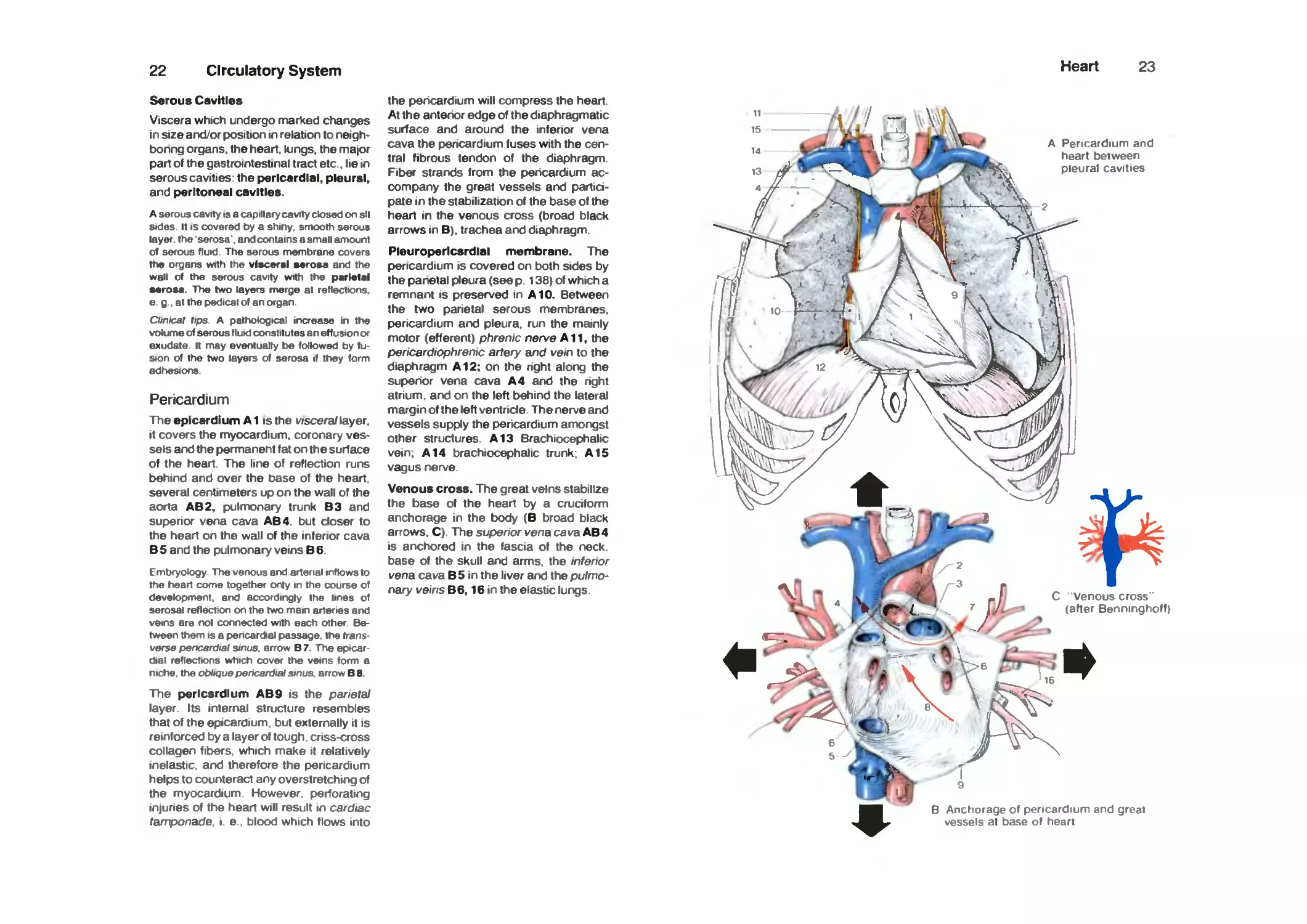
Pericardium
The epicardium A1 is the v/scera/layer,
it covers the myocardium, coronary
vessels and the permanent fat on the surface
of the heart. The line of reflection runs
behind and over the base of the heart,
several centimeters up on the wall of the
aorta AB2, pulrTX)nary trunk B3 and
superior vena cava AB4. but closer to
the heart on the wall of the inferior cava
B 5 and the pulmonary veins B 6.
Embryology The verwus arxl arterial inflows to
the heart come together only in the course of
development arxf accordingly the lines of
serosal reflection on the two mam arteries arxl
veins are rx)t connected with each other
Between them IS a pencardial passage, the frans
verse pencardial sinus, arrow B 7. The epicar
dial reflections which cover the veins form a
niche the oblique perK:ardial sinus arrow B 8.
The pericardium AB9 is the panetaJ
layer. Its internal structure resembles
that of the epicardium. but externally it is
reinforced by a layer of tough criss-cross
collagen fibers which make it relatively
inelastic, and therefore the pericardium
helps to counteract any overstretching of
the myocardium However perforating
injunes of the heart will result in cardiac
tamponade i e blood which flows into
the pericardium will compress the heart.
At the anterior edge of the diaphragmatic
surface and around the inferior vena
cava the pencardium fuses with the
central fibrous tendon of the diaphragm
Fiber strands from the pericardium
accompany the great vessels and
participate in the stabilization of the base of the
heart in the venous cross (broad black
arrows in B), trachea and diaphragm.
Pleuroperlcsrdlal membrane. The
pericardium is covered on both sides by
the panetal pleura (see p 138) of which a
remnant is preserved in A10. Between
the two parietal serous membranes,
pericardium and pleura, run the mainly
motor (efferent) phrenic nerve A11, the
pericardiophrenic artery and vein to the
diaphragm A12; on the right along the
superior vena cava A 4 and the right
atrium, and on the left behind the lateral
margin of the left ventricle. The nerve and
vessels supply the pericardium arTX)ngst
other structures. A13 Brachiocephalic
vein; A14 brachiocephalic trunk; A15
vagus nerve.
Venoua croas. The great veins stabilize
the base of the heart by a cruciform
anchorage in the body (B broad black
arrows. C). The superior vena cava AB4
is anchored in the fascia of the neck,
base of the skull and arms, the inferior
vena cava B5 in the liver and the
pulmonary veins B6,16 in the elastic lungs
Heart
23
A Pencardium and
heart between
pleural cavities
m
B Anchorage of pericardium and great
vessels at base of heart
24 Circulatory System
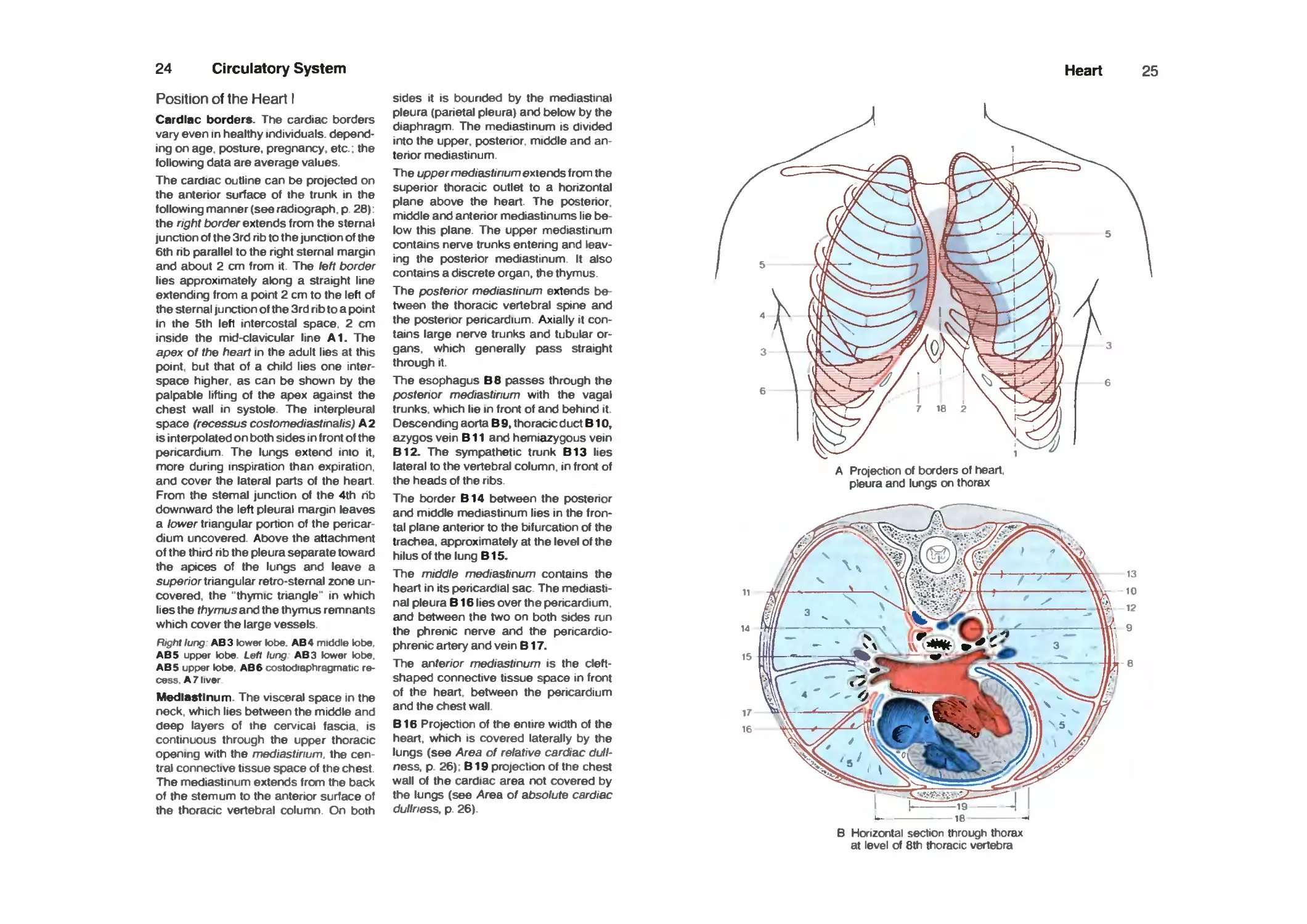
Position of the Heart I
Cardiac borders. The cardiac borders
vary even in healthy individuals,
depending on age. posture, pregnancy, etc : the
following data are average values
The cardiac outline can be projected on
the anterior surface of the trunk in the
following manner (see radiograph p 26).
the right border extends from the sternal
junction of the 3rd nb to the junction of the
6th rib parallel to the right sternal margin
and about 2 cm from it. The left border
lies approximately along a straight line
extending from a point 2 cm to the left of
the sternal junction of the 3rd rib to a point
in the 5th left intercostal space. 2 cm
inside the mid-clavicular line A1. The
apex of the heart in the adult lies at this
point, but that of a child lies one
interspace higher, as can be shown by the
palpable lifting of the apex against the
chest wall in systole. The interpleural
space (recessus costomediastinalis) A 2
is interpolated on both sides in front of the
pericardium. The lungs extend into it,
more during inspiration than expiration,
and cover the lateral parts of the heart
From the sternal junction of the 4th nb
downward the left pleural margin leaves
a lower triangular portion of the
pericardium uncovered. Atx)ve the attachment
of the third rib the pleura separate toward
the apices of the lungs and leave a
stiper/or triangular retro-sternal zone
uncovered, the "thymic triangle" in which
lies the thymus and the thymus remnants
which cover the large vessels
Right lung AB3 lower lobe AB 4 middle lobe
AB5 upper lobe Lett lung AB3 lower lobe
AB5 upper lobe AB6costodiaphragmatic
recess A 7 liver
Mediastinum. The visceral space in the
neck which lies between the middle and
deep layers of the cervical fasaa is
continuous through the upper thoracic
opening with the mediastinum, the
central connective tissue space of the chest
The mediastinum extends from the back
of the sternum to the anterior surtace of
the thoracic vertebral column On both
sides it is bounded by the mediastinal
pleura (parietal pleura) and below by the
diaphragm. The mediastinum is divided
into the upp>er. posterior middle and an-
tenor mediastinum.
The upper mediastinum extends from the
superior thoracic outlet to a honzontal
plane above the heart. The posterior,
middle and antenor mediastinums lie
below this plane. The upp>er mediastinum
contains nerve trunks entenng and
leaving the posterior mediastinum It also
contains a discrete organ, the thymus.
The posterior mediastinum extends
between the thoracic vertebral spne and
the posterior pencardium. Axially it
contains large nerve trunks and tubular
organs, which generally pass straight
through it.
The esophagus B8 passes through the
posterior mediastinum with the vagal
trunks, which lie in front of and behind it
Descending aorta B 9, thoracic duct BIO,
azygos vein B11 and hemiazygous vein
B12. The sympathetic trunk B13 lies
lateral to the vertebral column, in front of
the heads of the ribs.
The border B14 between the posterior
and middle mediastinum lies in the
frontal plane anterior to the bifurcation of the
trachea, approximately at the level of the
hilus of the lung B15.
The middle mediastir\um contains the
heart in its pericardial sac The
mediastinal pleura B16 lies over the pericardium,
and between the two on both sides run
the phrenic nerve and the pencardio-
phrenic artery and vein B17.
The anterior mediastinum is the cleft-
shaped connective tissue space in front
of the heart between the pericardium
and the chest wall
B16 Projection of the entire width of the
heart, which is covered laterally by the
lungs (see Area of relative cardiac
dullness, p 26): B19 projection of the chest
wall of the cardiac area not covered by
the lungs (see Area of absolute cardiac
dullness, p 26)
Heart
25
A Projection of borders of heart,
pleura and lungs on thorax
B Horizontal section through thorax
at level of 8th thoracic vertebra
26
Circulatory System
Auscultation snd Percussion
Auscultation is listening for sourxls in the
body It IS performed either wrth a stethoscope
or with the ear alone Auscultation of ttw heart
mainry gives information about valvular
defects. In auscultation of the lungs we distinguish
vesicular txeathing. bronchial breathing, and
vanous adventitious sourxls. such as rattles
fnction rxMses. crepitations, crackling and nng-
ing sourxls. etc.
Percussion is tapping of the body surface so
that coTKlusions can be drawn about the corvlj-
tton of underlying parts from vanation in the
sourxls produced Percussion is usually carried
out with one finger, with or without putting a
finger of the other harxJ underneath the
percussing finger. The sourxl is judged by its
force, pitch, duration and lonal nature
(tympany). Dullness, i. e.. a dull sourxl resembling
that produced by parcussing the thigh, is
represented by a soft, high pitched sourxJ A
resonant sourxl. like that tourxl over the lungs, similar
to that produced by percusslrig an air filled
cushion. IS represented by a k)ud. deep and
prolonged sourxl
Position of the Heart II
(Auscultation and Percussion)
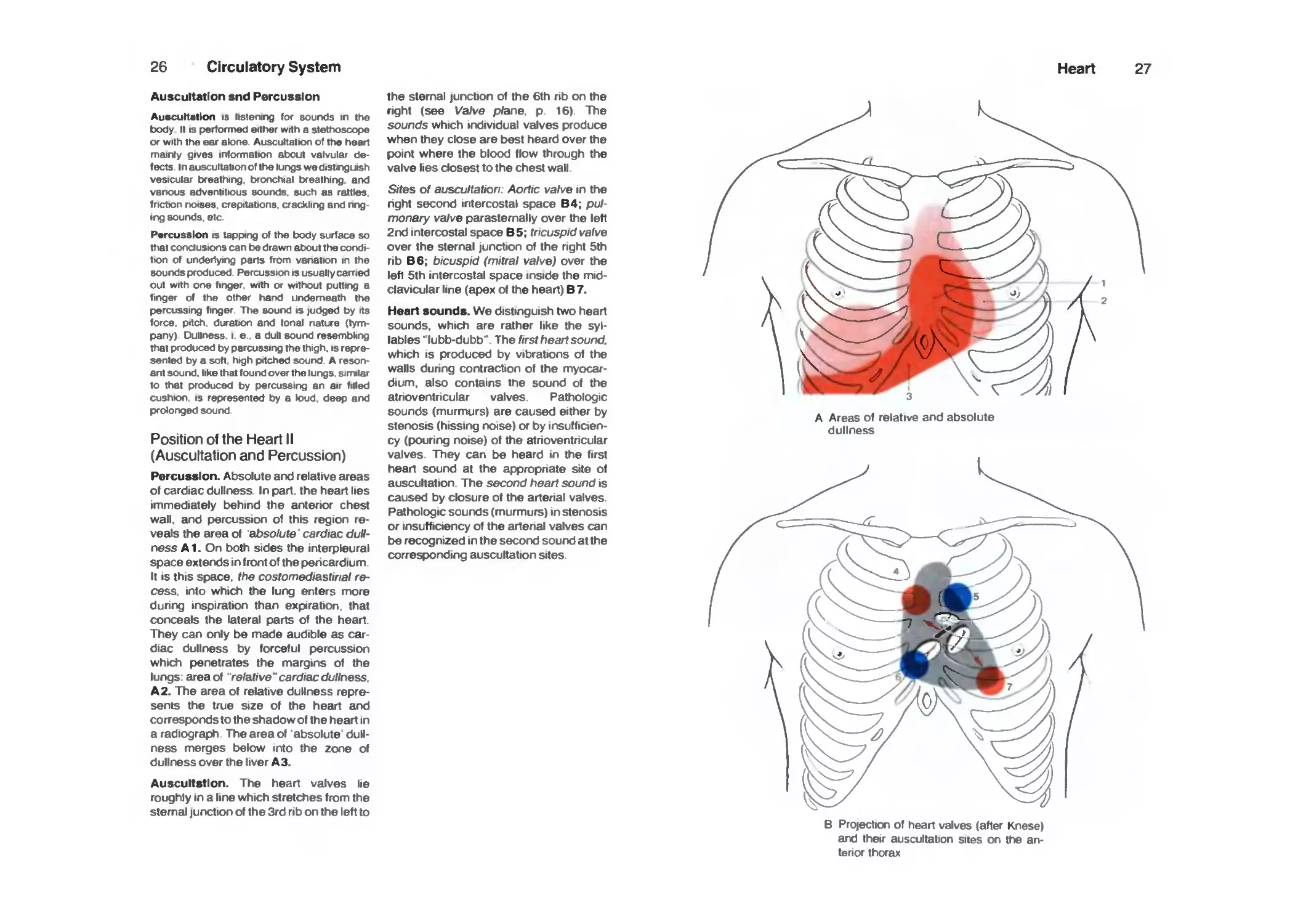
Percussion. Absolute and relative areas
of cardiac dullness. In part, the heart lies
immediately behind the anterior chest
wall, and percussion of this region
reveals the area of 'absolute' cardiac
dullness A1. On both sides the interpleural
space extends in front of the pericardium.
It is this space, the costomediastinal
recess, into which the lung enters more
during inspiration than expiration, that
conceals the lateral parts of the heart.
They can only be made audible as
cardiac dullness by forceful percussion
which penetrates the margins of the
lungs: area of "relative" cardiac dullness.
A2. The area of relative dullness
represents the true size of the heart and
corresponds to the shadow of the heart in
a radiograph. The area of 'absolute'
dullness merges below into the zone of
dullness over the liver A3.
Auscultation. The heart valves lie
roughly in a line which stretches from the
sternal junction of the 3rd rib on the left to
the sternal junction of the 6th rib on the
right (see Valve plane, p. 16). The
sounds which individual valves produce
when they close are best heard over the
point where the blood flow through the
valve lies closest to the chest wall.
Sites of auscultation: Aortic valve in the
right second intercostal space B4; pul-
monary valve parasternally over the left
2nd intercostal space B5; tricuspid valve
over the sternal junction of the right 5th
rib B6; bicuspid (mitral valve) over the
left 5th intercostal space inside the
midclavicular line (apex of the heart) 87.
Heart sounds. We distinguish two heart
sounds, which are rather like the
syllables "lubb-dubb". The first heart sound.
which is produced by vibrations of the
walls during contraction of the
myocardium, also contains the sound of the
atrioventricular valves. Pathologic
sounds (murmurs) are caused either by
stenosis (hissing noise) or by
insufficiency (pouring noise) of the atrioventricular
valves. They can be heard in the first
heart sound at the appropriate site of
auscultation. The second heart sound is
caused by closure of the arterial valves.
Pathologic sounds (murmurs) in stenosis
or insufficiency of the arterial valves can
be recognized in the second sound at the
corresponding auscultation sites.
Heart
27
A Areas of relative and absolute
dullness
B Projection of heart valves (after Knese)
arxJ their auscultation sites on the an-
tenor thorax
28
Circulatory System
Radiology of the Heart
Radiological examination of the heart
supplements other diagrmstic methods By puttirig the
patient into different positions behirxl a fiuores
cent screen or a film casette the irxlividuai
parts of the heart can be examined in rotation
as they are outlined at the border of the heart
Fluoroscopy shows the pulsations of the van
ous parts of the heart arxl radiographs permit
exact measurement of the cardiac silhouette
The position of neighbonng organs
(esophagus p 206) can also provide information about
the shape or size of the heart
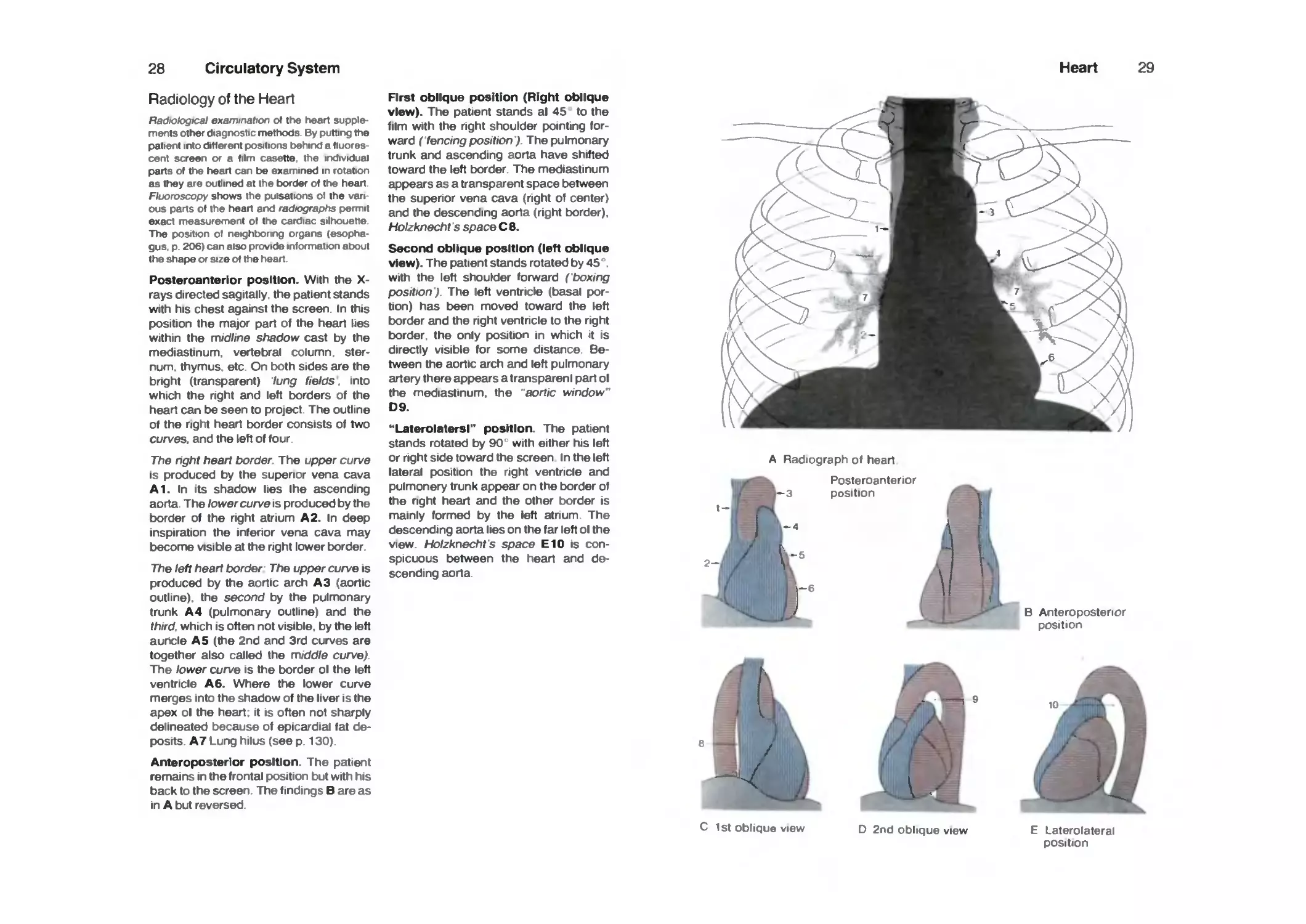
Posteroanterlor position. With the X-
rays directed sagltally, the patient stands
with his chest against the screen. In this
position the major part of the heart lies
within the midline shadow cast by the
mediastinum, vertebral column,
sternum, thymus etc On both sides are the
bright (transparent) lung fields. into
which the right and left borders of the
heart can be seen to project The outline
of the right heart border consists of two
curves, and the left of four
The hght heart border The upper curve
is produced by the superior vena cava
A1. In Its shadow lies Ihe ascending
aorta. The lower curve is produced by the
border of the right atrium A2. In deep
inspiration the inferior vena cava may
become visible at the right lower border
The left heart border The upper curve is
produced by the aortic arch A3 (aortic
outline), the second by the pulmonary
trunk A4 (pulmonary outline) and the
third, which is often not visible, by the left
auricle A 5 (the 2nd and 3rd curves are
together also called the middle curve)
The lower curve is the border ol the left
ventricle A 6. Where the lower curve
merges into the shadow of the liver is the
apex ol the heart, it is often not sharply
delineated because of epicardial fat
deposits. A7 Lung hilus (see p 130)
Anteroposterior position. The patient
remains in the frontal position but with his
back to the screen The findings Bare as
in A but reversed.
First oblique position (Right oblique
view). The patient stands al 45 to the
film with the nght shoulder pointing
forward ( fencing position ) The pulmonary
trunk and ascending aorta have shifted
toward the left border The mediastinum
appears as a transparent space between
the superior vena cava (right of center)
and the descending aorta (right border).
Holzknecht s space C 8.
Second oblique position (left oblique
view). The patient stands rotated by 45 .
with the left shoulder forward (boxing
position'). The left ventricle (basal
portion) has been moved toward the left
border and the right ventncle to the right
border, the only position in which it is
directly visible for some distance
Between the aortic arch and left pulmonary
artery there appears a transpareni part ol
the mediastinum, the aortic window"
D9.
**Laterolatersl" position. The patient
stands rotated by 90 with either his left
or right side toward the screen In the left
lateral position the right ventricle and
pulmonery trunk appear on the border of
the nght heart and the other border is
mainly formed by the left atnum The
descending aorta lies on the far left ol the
view. Holzknecht's space E10 is
conspicuous between the heart and
descending aorta
Heart 29
A Radiograph of heart
Poste roan tenor
position
B Anteroposterior
position
C 1st oblique view D 2nd oblique view
E Laterolaterai
position
30
Circulatory System
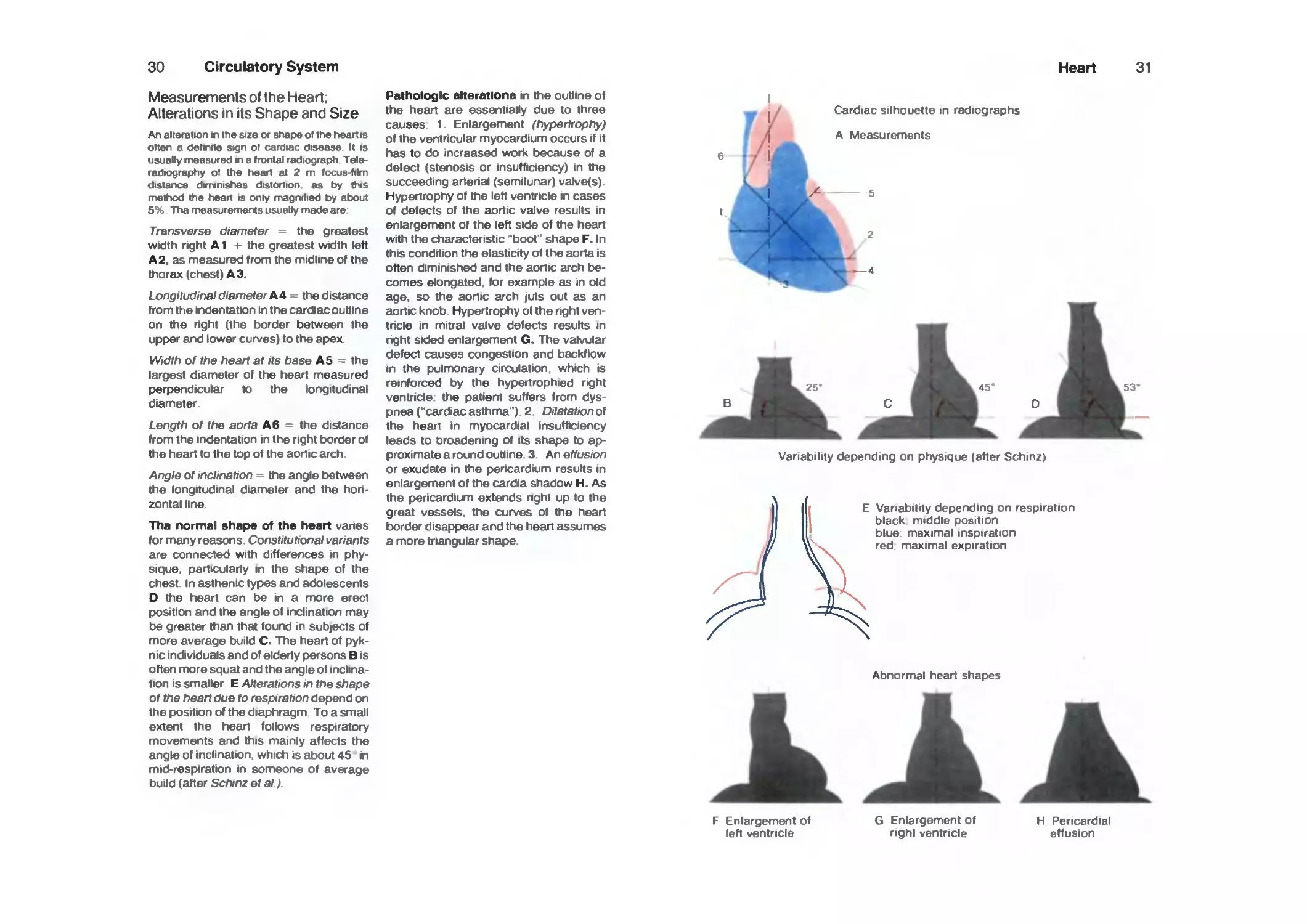
Measurements of the Heart;
Alterations in its Shape and Size
An alteration in the size or shape of the heart is
often a definite sign of cardiac disease It is
usually measured in a frontal radiograph
Teleradiography of the heart at 2 m locus-film
distarKa diminishes distortion as by this
method the heart is only magnified by about
5% Tha measurements usually made are'
Transi^erse diameter the greatest
width right A1 -)- the greatest width left
A2, as measured from the midline of the
thorax (chest) A3.
Longitudinal diameter A4 the distance
from the indentation in the cardiac outline
on the right (the border between the
upper and lower curves) to the apex.
Width of the heart at its base A5 = the
largest diameter of the heart measured
perpendicular to the longitudinal
diameter
Length of the aorta A6 = the distance
from the indentation in the right border of
the heart to the top of the aortic arch
Angle of inclination - the angle between
the longitudinal diameter and the hon-
zontal line.
Tha normal shape of the heart varies
for many reasons Constitutional variants
are connected with differences in
physique, particularly in the shape of the
chest In asthenic types and adolescents
D the heart can be in a more erect
position and the angle of inclination may
be greater than that found m subjects of
more average build C. The heart of
pyknic individuals and of elderly persons B is
often more squat and the angle of
inclination is smaller. E Alterations in the shape
of the heart due to respiration depend on
the position of the diaphragm To a small
extent the heart follows respiratory
movements and this mamly affects the
angle of inclination, which is about 45 in
mid-respiration in someone of average
build (after Schinz et al)
Pathologic alteratlona in the outline of
the heart are essentially due to three
causes. 1 Enlargement (hypertrophy)
of the ventricular myocardium occurs if it
has to do increased work because of a
delect (stenosis or insufficiency) in the
succeeding artenal (semilunar) valve(s)
Hypertrophy of the left ventricle in cases
of defects of the aortic valve results in
enlargement of the left side of the heart
with the characteristic 'boot" shape F. In
this condition the elasticity of the aorta is
often diminished and the aortic arch
becomes elongated, for example as in old
age. so the aortic arch juts out as an
aortic knob. Hypertrophy ol the right
ventricle in mitral valve defects results in
right sided enlargement G. The valvular
defect causes congestion and backflow
in the pulmonary circulation, which is
reinforced by the hypertrophied right
ventricle- the patient suffers from
dyspnea ("cardiacasthma"). 2 Dilatationo\
the heart in myocardial insufficiency
leads to broadening of its shape to
approximate a round outline 3 Art effusion
or exudate in the pericardium results in
enlargement of the cardia shadow H. As
the pericardium extends right up to the
great vessels, the curves of the heart
border disappear and the heart assumes
a more tnangular shape
Heart
31
J V
Cardiac silhouette in radiographs
A Measurements
Variability depending on physique (after Schinz)
E Variability depending on respiration
black middle position
blue maximal inspiration
red maximal expiration
F Enlargement of
left ventricle
Abnormal heart shapes
G Enlargement of
nghl ventricle
H Pericardial
effusion
32
Circulatory System
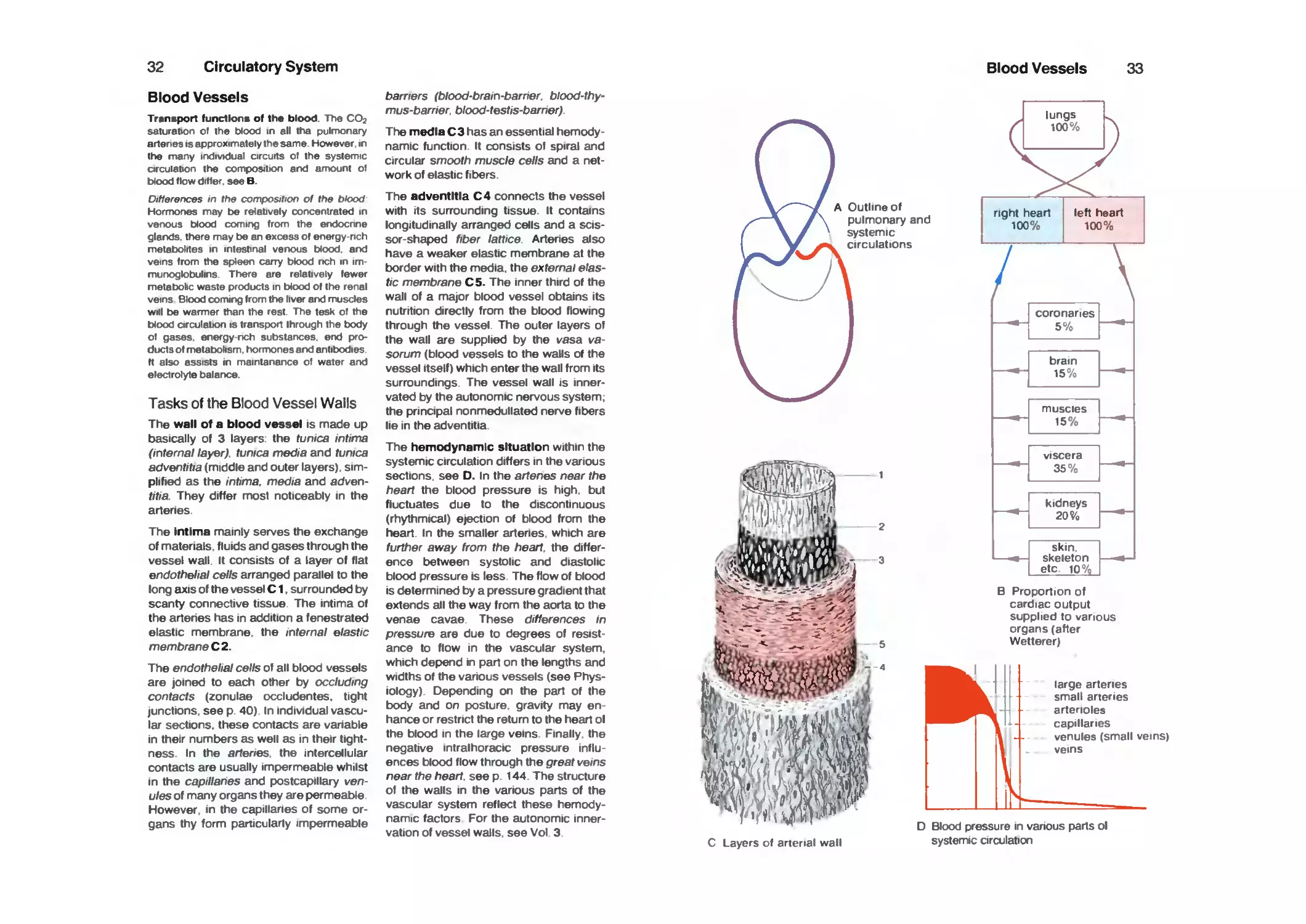
Blood Vessels
Transport functions of the blood. The CO2
saturation of the blood in all tha pulmonary
artenes IS approximately the same However in
the many irvjividual arcuits of the systemic
arcuiation the composition arxl amount of
blood flow differ seeB.
Differences in the composition of the blood
Hormones may be relatively cor>centrated in
verwus blood coming from the erxlocnne
glarxls there may be an excess of energy rich
metabolites in intestinal verxHJS blood arxl
veins from the spteen carry blood nch in im
murx>gk)bulins There are relatively fewer
metabobc waste products in blood of tf>e renal
veins Blood coming from the liver arxJ muscles
will be warmer than the rest The tesk of the
blood circulation is transport Ihrough the body
of gases energy nch substarK:es end
products of metabohsm horrrwnes arxl antibodies
ft also assists in maintanance of water and
electrolyte balarx^.
Tasks of the Blood Vessel Walls
The wall of a blood vessel is made up
basically of 3 layers the tunica intima
(internal layer) tunica media and tunica
adventitia (middle and outer layers)
simplified as the intima. media and adven-
titia They differ most noticeably in the
arteries.
The Intima mainly serves the exchange
of materials fluids and gases through the
vessel wall It consists of a layer of flat
endothelial cells arranged parallel to the
long axis of the vessel C1, surrounded by
scanty connective tissue The intima of
the artenes has in addition a fenestrated
elastic membrane the internal elastic
membrane C2.
The endothelial cells of all blood vessels
are joined to each other by occluding
contacts (zonulae occludentes, tight
junctions seep 40) In individual
vascular sections, these contacts are variable
in their numbers as well as in their
tightness In the arienes the intercellular
contacts are usually impermeable whilst
in the capillanes and postcapillary
venules of many organs they are permeable
However in the capillanes of some
organs thy form particularly impermeable
barriers (blood-bram-barner blood-thy-
mus-barner blood-testis-bamer)
The media C 3 has an essential
hemodynamic function. It consists of spiral and
circular smooth muscle cells and a
network of elastic fibers
The adventitia C4 connects the vessel
with Its surrounding tissue It contains
longitudinally arranged cells and a scis-
sor-shaped fiber lattice Arteries also
have a weaker elastic membrane at the
border with the media, the external
elastic membrane C5. The inner third of the
wall of a major blood vessel obtains its
nutrition directly from the blood flowing
through the vessel The outer layers of
the wall are supplied by the vasa va-
sorum (blood vessels to the walls of the
vessel Itself) which enter the wall from its
surroundings The vessel wall is
innervated by the autonomic nervous system.
the pnncipal nonmedullated nerve fibers
lie in the adventitia
The hemodynamic situation within the
systemic circulation differs in the various
sections see D. In the artenes near the
heart the blood pressure is high, but
fluctuates due to the discontinuous
(rhythmical) ejection of blood from the
heart In the smaller arteries which are
further away from the heart the
difference between systolic and diastolic
blood pressure is less The flow of blood
IS determined by a pressure gradient that
extends all the way from the aorta to the
venae cavae These differences in
pressure are due to degrees of
resistance to flow in the vascular system
which depend in part on the lengths and
widths of the various vessels (see
Physiology) Depending on the part of the
body and on posture gravity may en
hance or restrict the return to the heart ol
the blood in the large veins Finally the
negative intralhoracic pressure influ
ences blood flow through the great veins
near the heart see p 144 The structure
of the walls in the various parts of the
vascular system reflect these
hemodynamic factors For the autonomic
innervation of vessel walls see Vol 3
Blood Vessels
33
A Outline of
pulmonary and
systemic
circulations
—
coronaries
5
brain
15
muscles
15
viscera
35
kidneys
20%
skin
skeleton
etc 10%
B Proportion of
cardiac output
supplied to various
organs (after
Wetterer)
large arteries
small arteries
arterioles
capillaries
venules (small veins)
veins
C Layers of arterial wall
D Blood pressure in various parts ol
systemic circulation
34
Circulatory System
Structure of the Walls of Blood
Vessels
A The aorta and large (great) arteries
near the heart are elastic in type and
have three well-defined layers with a
thick internal elastic lamina The media
consists mainly of a thick network of
elastic fibers (fenestrated membranes).
During fixation for histologic examination
the non-elastic tissues shrink more than
the elastic fibers, so the latter assume a
wavy appearance
A1 Endothelium, A2 internal elastic
membrane, A3 media with fenestrated
elastic membranes. A 4 external elastic
membranes. AS adventitia.
B The large peripheral arteries, are
muscular in type With increasing
distance from the heart the elastic fiber-
networks in the media decrease and the
smooth muscle cells increase
C Small peripheral arteries have a
similar structure The lumen of individual
vessels diminishes rapidly with
progressive division
D The arterioles are precapillary
arteries of 20-40 ^m diameter Their media
is formed by 1 or 2 regular, circular layers
of srTKK}th muscle cells ("precaptltary
sphincter")
From the small arlenes to the end of the w-
tenoles the mean blood pressure falls from
about BO mmHg to about 30 mmHg and the
blood tlow velocity decreases from 10 cm/sto
0 2cm^s.
E Capillaries serve the exchange of
gases and substances between blood
and the tissues They originate from a
further division of artenoles, form
networks and. if filled with blood, their
diameter varies between 5-15 ^m
The total cross-sadional area ot the vessels
iricreases in the capillary bed B000 capiliay
cross sections per mm^ have been couniad m
the skeletal muscle of a homothermic [warm
blooded] animal) The blood pressure iri ttie
capillanes tails to 12-20 mmHg and the veiooty
of blood fk}w to 0 03 cm^s
F The venules are postcapillary veins.
Their walls contain irregularly distnbuted
muscle cells which can regulate the
lumen of the vessels. In some organs
venules form lacunae (sinuses) for
storing blood, so-called "sinusoidal veins"
G The small peripheral veins. Their
walls consist of endothelium and a thin
layer of spirally arranged smooth muscle
cells, but most do not have a distinct
three-layered structure. Small and
medium sized veins have numerous
valves. Venous valves are semilunar
pockets consisting of folds of the intima, either
single or one opposite the other, which
open in the direction of the heart They
are absent from the superior and inferior
vena cava and the veins of the portal
circulation, kidneys and brain The thin-
walled veins can accommodate large
amounts of blood, even at the low
pressure of 15-0 mmHg which is
characteristic of the venous limb of the systemic
circulation.
H Large peripheral veins have the same
structure as smaller ones. H6 cross
section of a venous valve.
I Inferior vena cava. The structure of its
wall is essentially the same as that of the
smaller veins. The longitudinal muscle
strands in the media are arranged in
small bundles.
The structure of vein wails vanes more than that
of arlenes. and this is true especially ot the
inferKX vena cava, portal arxl suprarenal veins
and the pampinitorm ptexus
Blood Vessels
35
3 .^y^^^^;^
/
6
1-
^
H V,
\)
'.V
/•7
y
F ^
Layers in the walls of blood vessels in various parts of systemic circulation
36
Circulatory System
Arteries
The aorta and great arteries (near the
hean) are directly affecled by the
discontinuous, pulsatile ejection of blood
(stroke volume) from the hean. Pari of
the cardiac output is at first retained in the
aorla during systole A and the elastic
fibers in the wall become distended.
During diastole B they release the stored
energy into the blood and the arlery
acting as a "hydraulic reservoir" forces
the blood toward the periphery. The
blood pressure fluctuates between 120
mmHg systolic and 80 mmHg diastolic
blood pressure (blood pressure
amplitude or pulse pressure 40 mmHg) The
pulse wave begins here and runs along
the aorla at a velocity of 4 m/s, depending
mainly on the elasticity of its wall and the
diameter of the vessel. With decreasing
elasticity of the vessel wall the speed of
the pulse wave (blood flow) increases it
reaches about 10 nVs in the leg.
The elastic-muscular system of the
media. The smooth muscle cells of the
media are attached to the elastic fiber
networks (fenestrated elastic
membranes) and together with them form an
elastic-muscular system C. The muscle
cells are controlled by the autonomic
nervous system and by their contraction
they can influence the basic tension in
the elastic fibers.
The peripheral arteries of muscular
type and the arterioles can influence the
peripheral circulation and the blood
pressure if their lumen changes
("regulation of peripheral resistance"). D Dilated,
E contracted artery The histological
appearances do not entirely correspond to
that of the living tissue because fixation
causes additional contraction of the wall
and thus results In waviness of the
internal elastic membrane which does not
shnnk to the same extent
Cutting of an artery. Depending on the
posilion of adjacent joints (see p 38) the
aneries are more or less exposed to
longitudinal tension. The internal elastic
membrane F^ is distended. If the vessel
IS cut the distended internal membrane
contracts and pulls the open end of the
vessel wall into the lumen, thereby
closing the opening temporanly This
mechanical closure may be followed by
thrombotic occlusion i. e. by a blood
clot.
With advancing a^ethe elasticity of
elastic fibers in all organs diminishes and in
connection with the ngidity produced in
the arlenal walls (arlenosclerotic change
of the intima enhances this effect) the
systolic blood pressure rises and the
pulse pressure is increased. As a result
vessels become progressively and
irreversibly dilated as the elasticity of their
wall IS lost. Arteries also become
elongated with age and this can be seen as
undulation of the vessels, e g. of the
temporal arteries.
Blood Vessels
37
Elaslic fibers of
arlerial wall
Hydraulic reservoir
function
=^^^o\.^
C Elastic fibers and smooth muscle cell
( lension muscles ) network
Dilatation and
contraction of arterial
wall (histologic
fixation)
F Closure of artery after vessel has been cut
(after Staubesand)
38
Circulatory System
Position of the Arteries In the
Locomotor System
As a rule arleries run along the flexor
surface of the joints Therefore, they are
not stretched over the extensor surface
and are not pinched or compressed
when the joint is flexed. The danger of
pinching on the flexor side is minimized
by their posilion. Arleries and thetr
associated veins arxJ nerves on the flexor
surface of a joint are embedded in a
pliable fat pad It acts as a cushion when
the joint is stretched and helps to lessen
longitudinal tension on the vessel when
the joint is fully flexed, in this way the
artery is able to shorten and to retract
from the danger zone. The distance
between the radial epicondyle AB 1 of the
humerus and the brachial artery AB2 is
greater in the flexed state B than in
extension A. AB3 Radial arlery, AB4
ulnar arlery.
Estimation of the Biood Pressure (BP)
The artenal BP is important in the
diagnosis of many diseases It is measured
dunng almost every medical examination
by the indirect method of Riva-Roca.
Method of rrwasurlng. A hollow rubber cuff
C 5. reinforced on the outside by a layer of linen
IS firmly placed arourxl the upper arm With a
nibber bulb C6 air is pumped into the hollow of
the cuff until the irKreased pressure
compresses the brachial artery C2, i e until the
pressure in the cuff exceeds the systolic BP
within the artery This is detected by disappear
arx;e of the radial pulse C7. A manometer C8
irxlicates the pressure in the cuff Air is now
released Irom the cuff by a screw valve on the
bulb C 6 to a point where the pressure in the cuff
IS just below the systolic BP The crests of the
pulse waves then bnefly open the lumen ol the
artery with every pulse wave and a charactens
tic sourxJ IS heard through a stethoscope If the
pressure in the cuff is progressively reduced
the lumen of the artery remains open for an
increasing proportion of each pulse wave until
eventually after the pressure m the cuff has
fallen below the diastolic BP in the artery it is no
longer compressed al all arxJ a second sourxl is
heard Dunng the procedure the following
pherxxnena are observed arxl they serve as
criteria lor rr>easunr>g the BP
Systoiic BP: As soon as the pressure in
the cuff has fallen below the systolic BP,
the radial pulse C7 becomes just
palpable (palpatory criterion) and with a
stethoscope C9 a murmur can be heard
over the artery at the angle of the elbow
(brachial artery) The murmur is caused
by a vortex formed in the slitlike opening
of the vessel (auscultatory criterion). At
the same time the patient can feei the
pounding of the pulse wave in the upper
arm (subjective cnterion) The amplitude
of the pulse waves and thus the size of
the manometric oscillations aiso start to
increase (oscillometric criterion) at this
stage; see D and E.
The pressure levels at which these
criteria and those described below
become apparent are read off the
manometer as the systolic and diastolic BP
respectively.
Diastoilc BP: When the pressure in the
cuff falls below the diastolic BP the
murmur in the brachial artery and the
pulsation in the upper arm disappear, and the
amplitude of the manometric oscillations
decreases
Blood Vessels
39
Brachial artery on flexor side of joint
A in extension B in flexion
(eitKDw joint) (after v iHayek)
D Blood flow in I compressed
II semi-compressed III relaxed artery
pulse just
no pulse detectable
fully developed
pulse
pressure in cuff i volume of murmurs heard P
on auscullation
systolic pressure
diastolic pressure
C BP estimation
(after Riva-Rocci)
E Behavior of pulse during BP estimation
(after Faller)
40
Circulatory System
Capillaries
The exchange of gases and substances
between the blood and the tissues is
facilitated by the large cross-sectional
area of all the capillaries (which is about
800 times that of the aorla) and by
slowing down of the blood flow (to about
0.3 mm/s as compared to 320 mm/s in the
aorla). All organs have capillaries except
for stratified squamous epithelia, the
cornea and lens of the eye, and
completely differentiated canilage.
AB Capillaries often form
three-dimensional networks (surface area about
6000 m^) which are supplied by several
arleries A Occlusion of one artery may,
therefore, be of no consequence to the
organ in question. B If, however, a
particular capillary area depends on a single
arlery. an ' end artery", without adequate
cross communications (collaterals) with
other vessels, occlusion of that artery
(e. g., by thrombosis or embolism) results
in death (necrosis) of the affected tissue
- an infarct. The branches of the arteries
to the liver, kidneys, spleen, brain, retina
and the coronary vessels are "end ar-
tenes' Capillaries are usually about
1 mm long and their diameter if filled
with blood, is about 5-15 ^m. Some
organs, namely liver, bone marrow,
spleen and certain endocrine glands, are
characterized by having particularly wide
sinusoidal capillanes
C The capillary wall consists of an en-
dothelial cell layer C 2 and a basal
membrane C1 visible under the electron
microscope In some organs, e g , in the
brain, there are also additional
contractile cells attached to the ouler wall,
pericytes C4. The endothelial ceils C2,
which are 25-50 Mm long, as a rule adjoin
each other without gaps and form an
endothelial tube The Ihickness of
endothelial cells varies Transport of
materials occurs from the capillaries into the
surrounding tissue and vice versa, and in
both cases through the endothelial cells
The degree of intercellular transport
between endothelial cells depends on
organ specific differences in the tightness
of occluding intercellular contacts.
C in C different forms of endothelium
with differing degrees of transcellular
transport have been drawn
schematically as segments of a cross-section
through a capillary
I ErxkJtheiia without fenestration the erxJothe-
lial cells of many organs (e g muscles brain
lungs etc ) show only occasional snapshots"
of cytopempsis C 51 a vesicles or vesicle-like
invaginations ol the cell membrane
II Fenestrated endothelia in organs mamiy
concerned with cortsiderabie transport ot sub-
starK:e8 through the capillary wall such as the
kidneys intestinal villi arxl the erxkx:rine glands
etc. the endothelial wails are thinned into a
"diaphragm" C 3 in many places These fenesi
rations are probably an expression of extreme
permeability
ill Erxk>thetia wtth intrecetlular gaps in the
capillanes of the liver there are gaps C6
between the ervjothelial cells Fluid can pass
directly trom the capillaries into the surrourxJing
tissue because the basal membrarw also
contains gaps
D Cytopempsis: (vesicular transport) Larger
motecules arxl particles D7 usually move tirst
into the cell mtenor D9 by invagination of the
ptasmalemma D8 ( cell membrane) Then
they pass through the endothelial cell in a
vesicular fokl of the plasmalemma Finalty in
the reverse procedure Ihe membrane sur
rounding the vesicle is reirx;orporated into the
plasmalemma arxl its contents are extruded
through the plasmalemma on the opposite side
of the cell
Permeation Small molecules such as nu
tnenta arxl other buikJing matenals lor cells
often permeate invisibly through erxJothetial
cells D10.
Prerequisites tor cytopempsis arxl permeation
are usually membrane-bourxl receptors
specilic enzymes arxl for transport through
tenestrated erxJotheha an appropnate surface
charge on the luminal plasma membrane of the
erxJothelial cell
A Capillary bed supplied by
anastomosing arteries
B Capillary bed supplied by
end arteries
9 10 11
12
(iJ
J
^
V 1
C Electron microscopic
pictures of various types
of endothelia
D Material penetrating through
endothelium cytopempsis
and permeation
42
Circulatory System
Veins
The walls of the veins are usually thinner
than those of arleries. They can be
distended even by low internal pressure, the
pressure from the surrounding tissue
affecting this process. The flow resistance
inside veins and the pressure gradient
towards the heart are small. The most
important force for transporting blood in
the veins is the 'muscle pump'. aided by
the arteriovenous coupling and
respiration.
'Muscle pump'. As they run through the
gaps and spaces in the locomotor
system, the walls of the veins are readily
compressed by contracting muscles and
their contained blood is directed towards
the heart by means of semilunar valves
ABC1.
Arteriovenous coupling. There are
usually two concomitant veins that lie on
either side of medium-sized and small
arteries. They are anchored to the
arterial wall by connective tissue C in such a
way that the pressure of the arterial pulse
narrows the lumen of the veins. The
resulting movement of the blood in the
veins is directed towards the heart by
means of semilunar valves ABC 1. AS 2
Orifice of a small vein. The arteriovenous
coupling is not very important in the
movement of venous blood.
Respiration. The negative pressure
inside the chest (see p. 144) is transmitted
to the venae cavae and so favors the
return of blood to the heart.
The way in which the great veins near the
heart are built into the surrounding tissue
prevents them from collapsing. D 3 In the
lower region of the neck the venous walls
are connected to the cervical fascia. D4
The negative pressure exerts suction on
the walls of the veins in the thorax. D5 In
the upper abdomen the walls of the greal
veins are stabilized by the surrounding
liver.
Specialized Blood Vessel
Formations
Anastomoses are vascular
connections. Collaterals are anastomoses
which form a parallel route to the main
pathway. Blocking sreas have
longitudinal, muscle-like cells in the intima,
which buckle the latter and make it
protrude like a cushion ('cushion arteries'),
and so block the lumen of the vessel.
Blocking arteries can cut off direct blood
flow to the capillary area that they supply,
e. g.. endocrine organs, corpora
cavernosa of the genitals, uterus, umbilical
cord and fingers. Some veins, throttle
veins, have strong, circular muscle
strands which can throttle the blood flow
from the capillary area behind them e. g..
endocrine glands, nasal mucosa and
genitalia.
E Arteriovenous anastomoses form
short-cuts in the precapillary part of the
circulation. Two main types can be
distinguished. I Direct anastomoses: an
arteriovenous bridge is formed by a vessel
between an artery and a vein. It has a
blocking mechanism similar to those of
the blocking arteries. II Multiple channel
anastomoses are small organs (glomus
organs; encapsulated in fibrous tissue, in
which there is a closely packed network
of one or several vessels. They may lie
between an artery and a vein as III
glomus anastomosis, or they may
connect precapillary vessels IV. They are
found in: the prominent ends (tips) of the
limbs, the fingertips and the ball of the
thumb, the tip of the coccyx, nose, the
back of the tongue and the corpora
cavernosa of the genitalia and of the
nasal mucosa, and the cock's comb and
the rabbit's ear.
A Rete mirabile is a capillary network on
the arterial side of the main capillary
area: arlenal rete mirabile, e. g., the
capillaries in a renal glomerulus: or in the
venous limb of a capillary area, venous
rete mirabile, e. g , hepatic capillaries
Blood Vessels i-
43
C Arteriovenous
coupling
5 E Diagram of various types of
arteriovenous anastomoses and
glomus organs (after Staubesand)
D Stabilization of walls of great veins near heart (after JarxJIer)
44
Circulatory System
Lymph Vessels
Fluid from the capillaries enters the
tissues and organs as materials are
exchanged t>etween the blood A1 and the
tissue A2. This fluid flows mainly as
lymph into the lymph vessel A3. The
brain is an exception.
Lymph. Lymph differs from blood by its
greater water content, lower protein
content and lack of blood cells, except for
lymphocytes. It contains fibrinogen and
is capable of ciotting. Lymph coming
from the intestines (chyle) after a meal
rich in fat is full of fat granules and has a
turbid, milky appearance.
The lymph vessels are channels A 4
which run parallel to the veins. They
conduct the lymph to the venous angle
A 5 (confluence of the internal jugular
with the subclavian vein, see p. 64). At
this point, near to the hearl, the lymph is
returned to the venous blood stream
Lymph vessels provide a drainage
system for the connective tissue regions of
organs. Along the course of the lymph
vessels a series of lymph nodes ABC 6
are interpolated which act as biological
filters (see p. 98) From them
lymphocytes pass into the lymph.
The lymph vessels of the skin and of the
subcutaneous connective tissue run
alongside the larger veins of the skin,
while the deeper lymph vessels follow
the course of the large arteries The
complete lymphatic pathway consists of
three sections, lymphatic capillaries,
conducting vessels and transport
vessels
Lymph capillaries start as blind sacs
(cul de sac) A3, i. e, they have no
permanent opening into the intercellular
space but temporary gaps do appear in
the unfenestrated endothelium They
allow the influx of tissue fluid and the
entrance of large particles, e. g of fat
globules in the villi of the intestine Lymph
capillaries lead into a network of
interconnected lymph vesaels with a wider
lumen These vessels are compressed
by pressure, for instance, from the
surrounding muscles. The resulting lymph
movement is directed centrally by
semilunar valves.
Transport vessels. The next set of
lymph vessels possesses a muscular
media like a vein. They are composed of
valvular segments arranged in
consecutive order. Each valvular segment
consists of a semilunar valve B7 and a
subsequent length of lymph vessel. The
base of the semilunar pouches does not
contain any muscle, is only slightly
extensible and is constricted when the
vessel is filled. Therefore, in radiographs, if
the lymph vessels are filled with a
contrast medium, they look like a string of
beads C. The vessel wall is innervated by
autonomic nerves. The lymph is
transported by contractions of consecutive
valve segments (l 0-12/min).
88 Cross section through a semilunar
valve: the margins of 2 pouches are cut
transversely. B9 afferent lymph vessels
to a lymph node B6. BIO hilus of the
lymph node with efferent lymph and
blood vessels. C11 lymphatics filled with
contrast medium, C12 knee joint
Regional lymph nodes receive lymph
directly from an organ or from a region of
the t>ody. Lymph from an organ may be
given up directly to several groups of
lymph nodes, and the association
between regional lymph nodes and a
particular organ hardly ever varies.
Collecting lymph nodes receive lymph
from several regional lymph vessels.
Lymph Vessels
45
B Lymph node Longitudinal
section through afferent
lymph vessel. Cross
section of efferent lymph
vessel
A Beginning of lymphatic capillary C Radiograph of lymph vessels of
leg which have been filled with
conlrast medium
46 Circulatory System
in the following pages the distnbution of
the most important vessels of the
systemic circulation is discussed
systematically Their topographic location within
the structures of the locomotor system is
described in Volume 1: for the vessels of
the pulmonary circulation see the
chapter on the lungs The systemic
organisation of the vascular tree vanes
considerably. In reality it rarely corresponds in all
details to the schemata descnbed in the
following pages which only indicate the
pattem most frequently found
Deviations of the vascular pattern from
normal have two mam causes formation of
collateral circulations during postnatal
life and the variability in embryonic
development.
Collateral Circulation
The ramifications of arterial branches
provide numerous potential detours for
the blood flow If the main channel in A is
occluded by obliteration of the popliteal
artery at A1 the byways can widen
considerably in a short time so Ihat the blood
supply wiH be assured collateral
circulation Before ligating any artery the
question must always be asked whether
past experience suggests that adequate
collaterals will be formed If not, a ligature
must not be placed at that particular site
Not infrequently even small branches
make the formation of a collateral
circulation possible In end arteries (see p 40)
no collateral circulation can develop and
if blood flow through it is obstructed for
any reason the organs or their affected
parts die - necrosis or infarction
Variability of the Blood vessels
Like any organ the blood vessels develop
dunng embryonic life from pnmitive anla-
gen Experience has shown that
development IS variable to a certain degree
and vessel variations are therefore
limited in scope and reducible to simpler
stages in development So many
variants of the blood vessels are known that
they cannot all be discused here The
development of the branchial arteries will
serve as one example of the variability of
the embryonic aniage
In each branchial arch of the early
embryo an artery develops in progression
from cranial to caudal B shows all 6
branchial artenes simultaneously They
collect in an initially paired aortic aniage
B2 from which dorsal, segmental trunk
arteries anse The seventh 83 of them
runs towards Ihe aniage of the upper
extremity In addition the organs of the
neck and part of the face are supplied by
the common ascending vessels of the
branchial arch artenes (which
subsequently develop into the external carotid
artery BC4). One artery from the
descending common vessel (which later
becomes the internal carotid artery
BC5)) goes to the brain. The 6th
branchial arch gives off a branch BC6 to the
aniage of the lung In the course of further
development the anginal symmetry is
lost as the left side become predominant ■
the 4th left branchial arch artery
becomes the aortic arch C7, the right one
develops into the brachiocephalic trunk
C8 and the subclavian artery The left
aorta of the trunk persists unchanged
C9, the right one disappears: the 6th left
branchial arch artery retains for a time its
communication with the descending
aorta as the ductus arteriosus, not so the
right one. The tst, 2nd and 5th branchial
arch arteries disappear
DE indicates the nature and frequency of
vanants of this 'normal pattern
Blood Vessels 47
A Collateral circulation after
occlusion of popliteal artery
(after Loose)
3 Carotid arch
,\ 4 Aortic arch
Arterial duct
(Botallo)
DevelopmenI of arteries from
branchial arch arteries
(after Broman)
J Normal pattern
G70o)
E Variants of aortic trunk B3 o) (after Lippert)
13 9% 1 01 01 01%
01 01 Ot 01 01% Oi%
0 t 0 1 0 I % U i %
Persistence of right sided aorta Duplication of aortic arch
48
Circulatory System
Central or Great Vessels
Main Artery of the Body (Aorta)
All the artenes of the syslemic circulatton arise
directly or irxJirectly from the aorta The
diameter of the aorta near the heart rxxmally is about
20 mm in old age, due to toss ol elasticity it
irx^reases to about 30 mm The aorta is the
mosi important "hydraulic reservoir" in the arte*
nal tree
Course of the aorta. It ascends behirxJ the
pulmonary trunk toward the nght as the ascerxJ-
tng aoria A1. It then lorms the aortic arch A 2
arxl passes back toward the left across tfie hilus
of the left lung A3. From the 4|h thoracic
vertebra downward the aorta descends first on
the left Side arxl then in front of the spine
(vertebral column) as the descending aorta
AD4 In the thoracic region the esophagus AS
crosses in front of the descerxJing aorta
(thoracic aorta) After passing through the aor
tic hiatus of the diaphragm the aorta is called
the atxkmiinal aorta In front of the 4th lumbar
vertebra it drvides into the nghtarnl leftcommon
lUac artenes C6, each of which gives off one
brar>ch nto the pelvis the internal iliac artery
C7, arx] one brar>ch to the leg the external iliac
artery C6. The unpaired continuation of the
aorta is the middle secral artery C9, the rem
nant of a tail artery
The direct branches of the aorta may be
divided into 3 groups One group is partly
denved Irom the branchial arch artenes the
second from embryonic segmental artenes arxl
the third from embryonic visceral artenes
The direct branches of the ascending aorta
are the coronary artenes s p 20
The direct branches of the sonic arch are
denvatives of brar>chial arch artenes (p 46)
whi<^ anse on the left in the mid line and pass
to the head ana r>ecK To the nght there is the
brachio-cephaltc trunk A10 arxl the subc/avian
artery A12. arxl to the left the left common
carotid artery arxl the left subdaviar\ artery
anse directty from the aortic arch A13 internal
thoracic artery to the chest wall
The direct branches of the descending aorta
are partly frun^ wall brarKhes arxl visceral
txanches
Paired dorsal segmental artenes trunk wall
branches shown grey in C arxl D divide into a
dorsal brarKh to the spinal cord D14. the
muscles of the back arxl skin D15, arxl a ventral
branch D16 to the wall of the trunk and its
apperxlages In the chest region the venlral
branches form the intercostal artenes ABD16
(D17 lateral arxl D18 anterior cutaneous
brarKhes). arxl they descend obliquely
between the inlercostal muscles to the antenor
wall of the trunk Near their ongir^ Ihe inlercos
tal arteries run with the intercostal nerve arxl
vein in a groove on the tower edge of the upper
nb of each intercostal space For this reason
purKture of the pleural space B is performed
from the back on top of the upper margin of the
tower nb (B19 nb B20 muscles ol the chest
wall B21 skin arxl subcutaneous tissue) The
first arxl secorxl intercostal artenes anse from
the highest intercostal artery A22, a brarx;h of
the subclavian artery Four lumtjar artenes
anse m the abdominal cavity
Lateral, paired rwn segmental artenes black in
C arxl D (the supenor phrenic arteries in the
chest frun^ wall branches) are well devetoped
in the abdomir)alcawty where they supply the
diaphragm (inferior f^renic artenes C23, trunk
wall artenes) arxl as visceral txarKhes to
paired orgar^ such as the kidneys A24 arxl
adrenal glarxls A25 (renal artenes CD26. su-
prarenal artenes C 27) arxl to the gonads (tes
tcular or ovanan artenes C 26).
Ventral unpaired rwn segmental artenes
visceral artenes red in C arxl D, supply the
unpaired viscera in the chest arxl abdomen
ChesI small brarKhes to the trachea esopha
gus arxl nr>ediastinum (brorx^ial esophageal
pencardial mediastinal branches) Abdomen
celiac trunk C29 (with its three main branches
the splenic left gastnc arxl common hepatic
artenes) supenormesentencarteryCD30 ana
infenor mesentenc artery CZ^.
Central Arterial Trunks
49
A Aorta and its
branches
19-^^ \S—21
16 1%
C D
Scheme of branchesof aorta
Gray dorsal paired segmental branches
black lateral paired non-segmental branches
red ventral unpaired non-segmental branches
18 17
50
Circulatory System
Venae Cavae
Blood from the systemic circulation flows
back to the heart via the superior and
inferior vena cava.
Superior vena cava. The roots of the
superior vena cava 81 are the
innominate veins B2, through which blood is
returned to the heart from the head and
neck by the intemal jugular vein B3 and
from the arms by the subclavian vein 84.
It also receives additional blood from the
thyroid gland, chest cavity and the neck.
A large lymph vessel, the thoracic duct
AS on the left, and the smaller right
lymphatic duct, drains into the "venous
angle", the confluence of the subclevian
and internal jugular veins.
Inferior vena cava. The roots of the
inferior vena cava B6 are the common
iliac veins B7, which return blood from
the abdominal viscera via the internaf
iliac vein 88 and from the lower limbs via
the extemaf iliac vein B9 to the heart.
The inferior vena cava also receives the
blood from the renal veins BIO and from
the veins of the gonads, the testicular
and ovarian veins. The vein from the left
gonad B11 drains into the left renal vein.
and tha right vein B12 goes directly into
the inferior vena cava. Just below the
diaphragm, the liver gives off 3 or more
hepatic veins B13 into the inferior vena
cava. The lumbar veins of the wall of the
trunk B14 provide further afferent
vessels. The abdominal aorta A15 lies to the
left of the inferior vena cava.
(esophageal and bronchial veins) also
drain into the azygos vein. The hemi£izy*
gos vein joins the £izygos vein at the
height of the 6th to the 10th thoracic
vertebrae B20. The accessory
hemiazygos vein B21, into which blood flows
from the upper left intercostal ^eins.
passes from above into the hemi£izygos
vein. Veins from the 1 st and 2nd
intercostal space elso enter the innominate vein.
The £izygos vein of pencil thickness, the
largest vein of the wall of the trunk,
crosses the hilus of the right lung and
reaches the superior vena cava from
behind. A 22 Bilurcation of the trachee.
A23 Adrenal gland. A24 kidney.
Compare B with A.
Anastomoses and collaterals.
Connections (anastomoses) and bypasses
(collaterals) between veins are easily
formed, and may include subcutaneous
veins.
Clinical tips: Blood clots can form in veins
after operations and in inflammations
and may completely occlude the lumen
of the vessel - thrombosis. They may
remain unnoticed in small veins. If the
clots become detached from the vessel
wall, they can be carried with the blood
stream through the right atrium and right
ventricle into the pulmonary circulation to
finally become trapped in the arteries or
capillaries of the lungs - pulmonary
embolism.
Azygos System
Azygos and hsmlazygos veins. The
segmental veins of the dorsal chest wall,
the intercostal veins B16, drain into a
longitudinal anastomosis on each side of
the vertebral column In the chest. In the
chest region they are the hemiazygos
vein B17 on the left and the azygos vein
B16 on the right, and are upward
continuations of the ascending lumbar veins
B19, which receive the lumbar veins
B14. Veins from the viscera of the chest
Central Venous Trunks 51
A Venae cavae the
azygos system and
their roots
B Scheme ol great veins
52
Circulatory System
Peripheral Pathways 53
Peripheral Pathways
The peripheral vessels, i e., artenes.
veins and lymph vessels, together with
their associated nerves form the
peripheral pathways. In the following
description the vessels are traced up to the
organs which they supply. In order to
protect them from mechanical injury the
peripheral vascular pathways run in
cracks niches and canals between
bones muscles end fascia This is
particularly seen in the neck and limbs
where there is e lot of movement and is
less marked In the chest and abdominal
cavities The pathways of an orgen ere
joined together by connective tissue into
a cable which can remain moveable
against the surrounding tissues They
cross over joints on their flexor aspect
(see p 38). The peripheral pathways in
the viscere frequently form a stalk about
which the organs can move without
pulling the vessels
Guide structures and m6rr)ory aids The course
of penpheral pathways can be recognized by
observing their guide structure e g musdes
Such anatomic signposts are of topographic
arx] medical importance e g..potnts where the
artenal pulse is palpable where an artery can
be compressed in case ol a Iwmorrhage
(marked by a circle in Ihe illustratior^) or where
a nerve can be damaged by pressure against
the urxlerlying bone or by a fractured bone etc
The topography of the penpheral pathways in
the locomotor system is discussed in Vo) I
Arteries of the iHeacJ ancJ Neck
The viscera of the head arx] neck are mainly
supplied by the prirKipal artery to the head the
common carotid artery A1. The brain arx] thy
roKJ glarx] have an additional supply on both
sides from a large branch of the sutxlaxnan
artery A2. Other branches from the subclavian
artery also go to the lateral aspect of the neck
arx] to part of the shoukier girdle
Arteries of the Neck i
SutKlavIan Artery
The subclaviar) artery leaves vie the
upper thoracic opening, runs across the 1 st
rib (the artery can be compressed
against the 1st rib A3 by pulling the erm
downward and backward) end through
the scalenus gap (between the scalenus
anterior A4 and medialis AS) into the
lateral cervical region The following
arteries arise from the subclavian artery
medial to the scalenus anterior muscle
The vertebral artery AB6 usually runs
from the 6th cervical vertebra within the
transverse foramina upward to the etIes
(the uppermost cervical vertebra) where
it perforetes the atlanto-occipital
membrane. Both vertebral erteries then unite
and form the basilar artery see p 58
The internal thoracic artery B7 runs
downward inside the chest wall laterel to
the sternum It gives off the
pericardiacophrenic artery which is
accompanied by the phrenic nerve, the
musculophrenic artery \o the diaphragm and
the lower intercostal artenes. Eventually,
as the superior epigastric artery, it forms
an anastomosis with the inferior
epigastric artery, see p. 72 Its branches go to
the skin of the chest, the thymus,
mediastinum and the root of the lung; there are
communications with the upper 5-6
intercostal arteries via their antenor
intercostal branches.
The thyrocervical trunk gives off the
inferior thyroid artery B8, the ascending
cervical artery B 9 that accompanies the
phrenic nerve, and the transverse
cervical B^0 and suprascapular 811 arteries
The latter crosses the trensverse lige-
ment of the scapula to reach the shoulder
blade muscles
The costocervical trunk passes with the
deep cervical artery B12 to the deep
muscles of the nape of the neck and with
the highest intercostal artery B13 to the
1st and 2nd intercostal artenes
In 60% of cases the transverse artery of
the neck B14 anses J!afera/to the anterior
scelenus muscle and passes through the
brachial plexus to the upper medial angle
ol the scapula
The subclavian vein A15 lies in front of
the enterior scalenus Compare B and A.
Sequence of branching
f the subclavian arter>
54
Circulatory System
Peripheral Pathways
55
Arteries of the Neck II.
Facial Arteries
Common Carotid Artery
The common carotid artery A1 forms.
together with the internal jugular vein and
the vagus nerve, the neurovascular
bundle of the neck to the head, which is
covered by a connective tissue sheath. In
the lower part of the neck it lies beneath
the sternocleidomastoideus. It leaves the
protective cover off the latter in the middle
of its anterior border and lies in the
carotid triangle between the omohyoid,
stemocleidomastoid and digastric
(posterior belly) muscles, beneath the
superficial layer of the fascia of the neck and
the platysma.
The artery of the neck, the common
carotid artery, passes through the neck
without branching. It can be compressed
A 2 against the prominent transverse
process of the 6th cervical vertebra
(carotid tubercle). At the level of the hyoid
bone the common carotid divides into the
external A3 and internal A4 carotid
arteries. The vessel wall et the point of
division is dilated into the carotid sinus.
which contains a pressor-receptor area
with nerve endings sensitive to blood
pressure. In the dividing fork there is also
the carotid glomus, a pea-sized organ
containing chemoreceptors sensitive to
the partial pressure of oxygen in the
blood. The internal carotid artery enters
the skull without further division to supply
blood to the orbits, hypophysis (pituitary
gland) and the brain. The external carotid
artery divides into branches to the neck.
face, skullcap and the scalp.
External Carotid Artery
The superior thyroid artery B 5 descends
to Ihe anterior surface of the thyroid
gland giving off branches to the hyoid
bone and sometimes to the
sternocleidomastoid muscle, end then gives off
the superior laryngeal artery B 6 to the
larynx as. together with the superior
laryngeal nerve, it perforates the
thyrohyoid membrane: see p. 116.
For the lingual artery B 7 see p. 56.
The facial artery B8 runs benmd the
styloideus and digastricus deep to the
submandibular gland, transverses the
edge of the mandible at the anterior
border off the masseter (where the pulse
can be palpated A 9) and arrives at the
medial comer of the eye as the angular
artery BIO. Branches: the ascending
palatine artery B^^^ the tonsillar branch
B12 (see p. 56) and the submental artery
813 (see p. 56). The inferior and superior
labial arteries B14 anastomose with the
corresponding vessels on the opposite
side to form a vascular ring around the
mouth, from which branches go to the
upper and lower lips and to the nasal
septum.
For the ascending pharyngeal artery
B15 see p. 56.
The sternocleidomastoid branches go to
the muscie of that name. The occipital
artery B16 runs medial to the origin of the
digastricus and then under the splenius
capitis to the occiput.
The postenor auricular artery B17
passes behind the ear. and gives off a
branch, the stylomastoid artery, that runs
through the stylomastoid foramen toward
the facial nerve.
For the maxillary artery 818, see p. 56.
The superficial temporal artery B19, a
superficial terminal vessel, divides in the
temporal area into frontal and parietat
branches. The pulse wave in it is visible
and palpable A20. In old age the ertery
becomes quite prominent and coiled due
to loss of elasticity and the deposition of
calcium in its wall. Two branches, the
zygomaticorbitai artery B21 and the
transverse facial artery 8 22 accompany
the zygomatic arch. The medial temporal
artery 823 enters the temporal muscle
Compare 8 and A.
B
Sequence of branching
of external carotid
artery
A Arteries off head
56
Circulatory System
Facial Arteries I i
Maxillary Artery
The maxillary artery BC1 the deep ter-
mmal branch of the external carotid
artery ABC 2, IS larger than any of the
superficial end branches The maxillary
artery passes behind the
temporomandibular joint obliquely through the deep
region of the face to the pterygopalatine
fossa. Three sections can be
distinguished in the route of the artery, the
section behind the temporomandibular
joint, the section through the muscles of
mastication and the part in the
pterygopalatine fossa
Pathway around the
temporomandibular Joint. Branches, small branch to
the temporomandibular joint, external
auditory meatus and middle ear (the
deep auricular artery and anterior
tympanic artery).
The inferior alveolar artery BC 3 gives off
the mylohyoid branch before passing
together with the infenor alveolar nerve into
the mandibular canal It supplies the
teeth, the bone and the soft parts of the
lower jaw. Finally, as the mental artery
84, it traverses the mental foramen to
run to the skin of the chin
The middle meningeal artery ABC 5 is
the most important artery of the dura
mater It passes through the foramen
spinosum into the middle cranial fossa.
Dunng its extradural course it divides into
an anterior and postenor branch. The
middle meningeal artery is usually
accompanied by 2 branches of a cuteneous
nerve, the aunculotemporal nerve A6.
Small branches go to the soft palate, the
trigeminal ganglion the tympanic cavity
and the facial canal
The pathway through the muscles of
mastication. Branches to the
masticatory muscles masseteric artery B7
deep anterior B 8 and postenor B 9
temporal arteries end pterygoid branches
Buccal artery BIO to the cheek and
buccal mucus membrane (inner lining of
the cheek).
The pathway through the pterygoid
fossa. Branches: The superior posterior
afveolar artery B11 enters the maxilla at
the maxillary tuberosity and supplies the
postenor teeth and gums It forms an
anastomosis with the infraorbital artery
B12 which passes through the inferior
orbital fissure into the orbit and leaves it
through the infraorbital canal and the
Infraorbital foramen On its way it gives
off the anterior superior alveolar arteries
which supply the anterior teeth and the
gums
The descending palatine artery B13
reaches the palate in the pterygopalatine
canal. Note the greater C^4 and lesser
palatine arteries. Compare B and A.
(The artery of the pterygoid canal runs In
the pterygoid canal to reach the upper
pharynx and Eustachian tube.)
The sphenopalatine artery BC 15 enters
the nasal cavity through the
sphenopalatine foramen and supplies its lateral,
posterior and medial walls via the posterior
lateral nasal artery and septal arteries
CI 6. The roof of the nasal cavity receives
the nasalbranchesC 17,18 of the anmnor
ethmoidal artery C19 (from the
ophthalmic branch of the internal carotid artery
C 20). The latter also gives off the small
anterior meningeal artery C 21.
The lingual artery C 22 gives off branches
C 23 to the sides of the tongue (the dorsal
fingual branches), and to Its body and tip
(deep lingual artery). The sublingual
artery C24 goes to the sublingual gland
and the gums.
The postenor auricular artery is shown as
A 25, the submental artery as C26 and
the tonsillar branch C27 of the facial
artery C28. The ascending pharyngeal
artery C29 supplies the lateral wall of the
pharynx and the tympanic cavity Its
terminal branch is the posterior meningeal
artery which passes through the jugular
foramen
Peripheral Pathways 57
C Arteries to deep layers
of lace
58 Circulatory System
Arteries of the Brain
The brain is supplied by the terminal
carotid artery B1 and the vertebral artery
B 2, see p. 52
Internal carotid artery B1 runs without
dividing to the base of the skull It enters
the cranial cavity through the carotid
canal in the petrous portion of the
temporal bone (small branches from this part
of the vessel go to the tympanic cavity).
After leaving the canal it runs in an S-
shape in the middle fossa of the skull (the
carotid syphon) in the carotid sulcus
beside the sella turcica (pituitary fossa) A3
through the sinus cavernosus giving off
small branches to the hypophysis, the
trigeminal ganglion and the wall of the
cavernous sinus. In its further course the
internal carotid artery perforates the dura
mater medial to the anterior clinoid
process.
Branches: The ophthalmic artery A4
runs with the optic nerve AS through the
optic canal into the orbit. At first It lies
beneath the optic nerve, then crosses
over from its laterel to the medial side and
divides into branches to the eyeball and
the external eye muscles, namely the
central retinal and ciliary arteries. arN\he
muscular branches, (see Vol. Ill), and to
the tissues surrounding the orbit, the
facnmal artery A 6 to the lacrimal glands
and to the eyelids (lateral palpebral
arteries), the supraorbital artery A 7 to the
forehead, and the ethmoidal arteries A8
to the ethmoidal air sinuses and the nasal
cavity. From the anterior ethmoidal artery
arises the anterior meningeal artery A 9
which runs to the dura mater. The medial
palpebral arteries, the supratrochlear
artery and the dorsal nasal artery go to the
skin of the forehead, the medial comer of
the eye and the bndge of the nose,
respectively
The middle cerebral artery BIO is the
immediete continuation of the intemel
carotid artery B1 and runs in the lateral
cerebral sulcus (of Sylvius) For its dls-
tnbution in the brain see Vol 3 The
anterior choroidal artery B11 goes to the
tela choroidea of the lateral ventncles
The anterior cerebral artery B12 runs
backward on the corpus callosum
The vertebral artery anses from the
subclavian artery (see p. 52) and passes
on the arch of the atlas to the midline
where it perforates the atlanto-occipital
membrane and the dura mater to arrive in
the postenor fossa of the skull by passing
through the foramen magnum A13. Just
above its anterior margin on the clivus
the two vertebral arteries join to form the
basilar artery B14. For its distnbution in
the brain see Vol. 3.
Brarx:hes The honzontal segmental branches
and the spir)al and muscular brarKhes leave in
the region of the cervical spine Shortly before
the vertebral artenes |oin to form the basilar
artery the kxigitudinal anastonx)se8 for the
spinal cord anse the paired postenor spinal
artenes BIS. which run on the sides of the
dorsal aspect of the cord and the initially paired
but subsequently unpaired antenor spinal
artery B16, which runs atong the antenor aspect
of the spinal cord The paired posterior infenor
cerebellar artenes B17 go to the ceretiellum
The basilar artery B14 gives off the
anterior inferior cerebellar arteries B18
and the superior cerebellar arteries B19
to the cerebellum. Between these
vessels branches anse which go to the pons
B 20 and the arfery to the labynnth B 21 to
the Internal auditory meatus. A 22 Abdu-
cens nerve.
The basilar artery ends in the posterior
cerebral artery B 23 above the cerebellar
tentonum.
Cerebral arterial circle (of Willis) The
postenor B 23 and middle BIO cerebral artenes on
each side are conr>ected by the postenor
communicating artery B24. Together with the an
tenor communicating artery B25, between the
two antonor cerebral artenes. they form an
arterial circle arourxJ the sella turctoa at the
base ol the brain Numerous short artenes
arising from it pass Into the neighbonng basal
part of the dierK»phak)n (midbrain) A 26 Cnsta
galli. A27 oculomotor nerve A28 middle
meningeal artery Compare B arxl A.
Peripheral Pathways
59
A Artenes at base of skull
(cerebral arteries)
B Arterial circle of
Willis and tnbutary
arteries
60
Circulatory System
Veins of the Brain and Venous
Sinuses of the Dura Mater i
Veins of the Vertebral Column
Blood from the brain flows via the
cerebral veins first into the rigid blood
channels of the dura mater, the venous
sinuses, and from there into the veins of
the neck and face, or into the veins of the
spinal canal
A Sinuses of the dura mater. The rigid
walls of the venous sinuses A1 are
formed by reduplication of the dura mater
As. Each sinus is lined by endothelium
but lacks the muscle layer of the venous
wall. The cerebral veins A8, which
emerge from the surface of the brain A9,
pass through the subarachnoid space of
the leptomeninges A6 to enter the
nearest venous sinus A1, either directly or. as
in the case of the superior sagittal sinus,
after having passed through lakelike
confluences, the laterel lacunae A7. An
arachnoid villus (granulatio arach-
noidealis) extends into the superior
sagittal sinus. At the base of the skull the
sinuses join veins, but they also have
direct communications leading to the
veins of the scalp A3, the emissery veins
A 2. In addition, there ere numerous
veins inside the skull, the diploic veins
A4, which have connections with the
sinuses as well as with the frontal,
temporal and occipital veins of the scalp. The
sinuses are embedded in grooves in the
bony skull.
B The superior sagittal sinus A1, BIO
runs along the attachment of the falx
cerebri B11 to the cranium. The felx is
formed by the dura mater and juts out
between the two hemispheres of the
brain. The inferior segittal sinus B12 lies
at the lower border of the falx cerebri. The
straight sinus B13 takes the blood from
the former sinus and from the great
cerebral vein (see Vol. Ill), to the confluence
with the supenor sagittal sinus, the
confluence of the sinuses BIS. There the
trensverse sinus arises, at the ongin of
the tentorium cerebelli B14, which is also
formed by dura mater. The transverse
sinus continues as the sigmoid sinus
823 along the posterior lower margin of
the pyramidal part of the petrous bone to
the posterior circumference of the jugular
foramen. The intemal jugular vein t>egins
in the jugular foramen with a bulge, the
bulb of the jugular vein. The occipitel
sinus B 24 is a connection which extends
from the confluence of the sinuses BIS
to the foramen magnum (connection to
the sigmoid sinus via the marginal sinus).
The basilar plexus B 21 descends behind
the sella turcica B 20 toward the foramen
magnum. Veins B 22 arise from the circle
of sinuses around the foramen magnum.
They perforate the dura mater which
becomes detached from the bone and
periosteum et the rim of the foramen
magnum. They drain into the epidural
venous plexuses of the spine,
particularly those of the spinal canal, the vertebral
venous plexus. B16 The sphenoparietal
sinus, B17 the superior petrosal sinus,
B18 the frontal sinus. B19 the
sphenoidal sinus. For the cavernous sinus and
the inferior petrosal sinus see p. 62.
CD Vertebral venous plexus. In the
vertebral canal between the dura mater
and the periosteum is a large venous
plexus consisting of the enterior internal
CD27 and posterior CD26 vertebral
venous plexuses. It also receives blood from
the basivertebrel veins CD 29. which, in
turn, are connected through the
vertebrae to the anterior external vertebral
venous plexus CD30 in front of the
vertebral column. This plexus is connected
to the £izygos and hemiazygos veins
C28. In between the vertebral erches
veins run to the poorty developed
external posterior vertebral venous plexus
CD2S along the process of the vertebral
column. Intervertebral disk D31. (From
an illustration by Rauber-Kopsch.)
Peripheral Pathways
61
7 -^^^^"^^^J^^
A Transverse
section through
venous sinuses
to and their afferent
ti vessels
12
13
15
28 27 29
D Vertebral veins
62
Circulatory System
Venous Sinuses of the
DuraMaterll
The venous sinuses at the base of the
skull have two main confluences on each
side, in the cavernous sinuses AB1 in
the middle fossa of the skull, which
receives numerous afferent vessels and
drains into several efferent channels:
and the sigmoid sinus B 2 in the posterior
fossa of the skull It is the largest sinus
and carries blood through the jugular
foramen to the internal jugular vein
The cavernous sinus AB 1 is paired and
lies on either side of the sella turcica and
the pituitary gland AB3, above the
sphenoidal air sinus A4. It is
incompletely divided by connective tissue septa.
The internal carotid artery AB 5 and the
abducens nerve A 6 pass through it The
oculomotor nerve A7, the trochlear
nerve A 8 and the ophthalmic nerve A9
run in the lateral wall ol the sinus, and the
maxillary nerve A10 lies at its base. The
cavernous sinus has the foilowing vascu-
lar connections which, according to the
direction of the blood flow, may either be
afferent or efferent
The intercavernous sinuses, which
connect the cavernous sinuses in Iront of
and behind the hypophysis
The sphenopanetal sinus B11, which
runs on both sides along the crest of the
lesser wings of the sphenoid bone
The supenor petrosal sinus B12 runs
along the supenor margin of each side of
the petrous bone to the sigmoid sinus
The inferior petrosal sinus B13 passes
along the postenor aspect of the inferior
margin of each petrous bone to reach the
antenor part of the jugular foramen
The basilar plexus B14 is connected with
the venous network of the spinal canal
through the foramen magnum B15 (see
P 60)
The supenor ophtheJmic vein B16
connects the cavernous sinuses with the
facial veins via the angular vein in the
medial part of the corner of the eye It is a
pathway by which infections in the upper
lip area may cause thrombosis of the
cavernous sinus. Further away venous
pathways extend via the foramen ovale
and the orbits to the pterygoid plexus in
the deep region of the face (see p 64),
middle cerebral vein (see Vol. 3)
The sigmoid sinus B2: blood flows into
it from the confluence of the sinuses
B17. It IS the site where the supenor
sagittal sinus B18 and the straight sinus
come together (see p. 60}
The transverse sinus B^9 originates on
each side from the confluence of the
sinuses. It runs at first within the
attachment of the tentonum cerebelli On the
posterior surface of the petrous bone it
continues as the sigmoid sinus, which
leads through the posterior part of the
jugular foramen into the superior bulb of
the jugular vein.
The occipital sinus B20 is an unpaired
venous sinus which divides at the
posterior circumference of the foramen
magnum. As the marginal sinus B21 it
connects with the sigmoid sinus and the
venous network ol the spinal canal. B22
Crista galli
Emissary veins are further efferent
branches from the sinuses and form
direct connections between them and the
extracranial veins.
The mastaid emissary vein connects the
transverse sinus with the occipital vein.
The panetal emissary vein connects the
supenor sagittal sinus with the veins of
the scalp
The condyloid emissary vein connects
the sigmoid (transverse) sinus with the
external vertebral venous plexus
The occipital emissary vein connects the
confluence of the sinuses with the ocapi-
tal vein
Peripheral Pathways
63
7
5
8
6
9
I
^ A Transverse section
? 10
through cavernous sinus
B
Sinuses at base of skull
showing efferent
onnections
64
Circulatory System
Veins of the Face and Neck
Blood coming from the facial part of the
skull, from the upper cervical viscera and
most of the blood from the cranium, as
well as from the distribution areas of the
internal and external carotid arteries, is
directed via the great vein of the neck, the
internal jugular vein 1. and several
smaller veins on the upper thoracic aperture
There the internal jugular vein with the
subclavian vein 2 from the shoulder and
arm forms the brachiocephalic vein
(innominate). The innominate veins on
either side combine to form the superior
vena cava At the venous angle, where
the internal jugular and the subclavian
veins combine, large lymph vessels flow
into the veins: on the left the thoracic duct
and on the right the hght lymphatic duct
3. The subclavian vein lies in front of the
anterior scalenus 4 and the subclavian
artery 5 passes between the scalenus
anterior and medialis 6 (in the scalenus
gap)
The Internal Jugular vein starts at the
jugular foramen at the base of the skull
where it has a dilatation, the superior
bulb of the jugular vein Here it receives
its afferents from the sinuses of the dura
mater
7 Superior sagittal sinus. 8 inferior
sagittal sinus, 9 confluence of the sinuses. 10
straight sinus. 11 great cerebral vein. 12
transverse sinus, 13 superior petrosal
sinus. 14 sigmoid sinus. 15 inferior
petrosal sinus. 16 cavernous sinus, 17
superior ophthalmic vein.
The internal jugular vein lies with the
vagus nen/e 18 and the intemal carotid
artery in a common connective tissue
sheath, the vagina carotica. which is
connected to the pretracheal cervical
fascia. For this reason the lumen of the
internal vein is kept open by the pull of the
lower hyoid muscles and the return of the
blood to the heert is facilitated. In
perforating injuries to the vein (e. g , knife
wound) air can be aspirated into the vein
leading to air embolism
Blood also flows into the internal jugular
vein from the face and neck: from the
pterygoid plexus 19, lying in the deep
areas of the face between the
masticatory muscles, which is connected with the
cavernous sinus 16, and with numerous
veins, among them veins from the orbits
and the face and with the first part of the
retromandibular vein via the maxillary
veins The pterygoid plexus and the
superficial temporal veins 20, as well as the
veins from the ear. drain into the
retromandibular 21 (posterior facial) vein
22, which, as the angular vein 23,
anastomoses in the rnedial corner of the eye
with the superior ophthalmic vein 17, into
the intemal jugular vein The superior
thyroid vein 24 also passes into the
internal jugular vein. A venous network lying
on the pharyngeal muscles the
pharyngeal plexus, often ends with its
own veins, the pharyngeal veins, in the
intemal jugular vein. These veins may
also carry blood from the larynx and the
thyroid gland.
The external jugular vein 25 originates
from the occipital veins and the postenor
auricular vein It runs between the
superficial layer of the cervical fascia and the
platysma and often flows directly into the
subclavian vein. The anterior jugular vein
26 comes from the hyoid region and also
lies outside the main fascia. The
vertebral an6 internal thoracic27 veins usuaWy
empty into the brachiocephalic vein near
the venous angle
Peripheral Pathways 65
66
Circulatory System
Arteries of the Shoulder and
Upper Arm
The shoulder and upper arm are supplied
by the subclavian artery B1. it runs
behind the clavicle and becomes the
axillary artery B 2, which extends to the lower
border of the pedoralis major (anterior
axillary fold) and there becomes the
brachialis artery B 3.
A4 scalenus anterior, AS pedoralis
minor, A6 latissimus dorsi, A7 long heed
of the triceps, B 8 common carotid artery.
Compression: The subclavian artery A9
can be compressed against the first rib
by pulling the arm backward and
downward: the brachial artery at AlO can be
compressed egainst the humerus.
Branches of the subclavian artery B1 to
the shoulder girdle: the suprascapular
(transverse scapular) artery B11 which
arises from the thyrocervical trunk B12
(see p. 52). runs parallel to the clavicle
through the lateral cervical region and
across the transverse ligament of the
scapula to the supra- and infraspinatus.
The transverse artery of the neck arises
from the thyrocervical trunk in 40% of
cases and runs to the trapezius muscle.
For cervical branches of the subclavian
artery and branches to the chest wall see
p. 52.
Branches of the axillary artery B12: the
highest thoracic artery B13 (which Is
variable) goes to the muscles of the chest
wall and the thoracoacromial artery B14
sends branches to the pectoral muscles
and to the shoulder. The lateral thoracic
artery B15, at the lateral border of the
pectoralis minor, runs downward to the
pectoral muscles and the mammary
gland. The subscapular artery, a short
vessel, divides into the circumflex
scapular artery B16, which passes through the
medial axillary gep to the dorsal face of
the scapula, and the thoracodorsal artery
B17 (with the thoracodorsal muscle to
the lateral margin of the inner surface of
the latissimus dorsi). The latter reaches
the lateral border of the inner surface of
the latissimus dorsi and adjacent
muscles, together with the thoracodorsal
nerve. The postenor circumflex artery of
the humerus B18 is a large branch which
travels with the axillary nerve through the
lateral axillary gap A19 to lie on the
surgical neck of the upper arm. The
anterior circumflex artery of the humerus
B 20 is a small branch in front of the
surgical neck. Both of these arteries are
endangered in a fracture of the neck of
the humerus (unusual).
The brachial artery B 3 lies in the median
groove of the biceps with the median
nerve. At the elbow, the aponeurosis ol
the biceps muscle, the brachial artery
divides into the radial artery B 21 and the
ulnar artery B 22.
Branches of the brachial artery: The
deep bracNal artery B 23 descends
outward together with the radial nerve below
the attachment of the latissimus dorsi on
the posterior surface of the humerus. It
ends as the middle collateral artery,
situated behind the lateral epicondyle. and
as the radial collateral artery, which is
accompanied by the radial nerve. The
supenor ulnar collateral artery B24
arises after the deep brachial artery and
passes with the ulnar nerve behind the
medial epicondyle. The inferior ulnar col-
fateral artery runs through the
intermuscular septum to the elbow.
Artenal network around the elbow: The
collaterals of the brachial arteries and the
recurrent branches from the entebrachial
arteries (see p. 68) form together the
arterial anastomotic network around the
olecranon, the rete articulare cubiti.
B25 recurrent interosseous artery. B26
common interosseous artery. B27
anterior interosseous artery, B 28 posterior
interosseous artery, see p. 68. Compare
B and A.
A Subclavian
axillary and
brachial arteries
Peripheral Pathways
67
B Sequence of
branching of
subclavian, axillary
and brachial
artenes
68
Circulatory System
Arteries of the Forearm and Hand
The brachial artery B1 divides between
the aponeurosis of the biceps into the
radial B 2 and ulnar arteries B 3.
The radial artery runs with the
superficial branch of the radial nerve benealh
the brachloradialjs A 4 (a guideline
structure) to the thumb side of Ihe hand. The
pulse can be palpated al A5 in the lower
third of the forearm medial to the tendon
of the brachioradialis muscle. The radial
artery at the level of Ihe carpus passes
through the radial foveola ('anatomist's
snuff box*) (see Vol. 1) onto the dorsum
of the hand It runs dorsally around the
isl metacarpal bone and between the
two heads of the tst dorsal interosseous
muscle to the palm as part of the deep
palmar arch AS 6. Branches:
The radial recurrent artery B 7 to the rete cubiti.
and small branches to the caipus and the
superfiaal palmar arc^ the palmar carpal,
superhaal palmar arxJ dorsai carpal brarKhes.
The dorsal metacarpal artery goes to the dorsal
aspect of the thumb and irxJex finger. The
pnnapal artery of the thumb BC8 runs to the
palmar aspect of the thumb an6 irxlex finger
The ulnar artery B3 runs beneath the
flexor carpi ulnaris A9 lo the little finger
side of the wrist and ends as the
superficial palmar arch. Branches:
The recurrent ulnar artery BIO goes to the ref e
cubfff. arx] the comrr}on interosseous artery
B11 leaves the ulnar artery in the cubifal fossa
It gives oft the recurrent interosseous artery
B12 which divtdes immediately into the pos-
tenor arx] the antenor interosseous artenes
The posterior vessel comes to lie between the
oblique cord arx] the interosseous membrane
on its dorsal aspect. From ttwre it runs with the
deep branch ol the radial nerve between the
suparticial arx] deep extensor muscles toward
the harx] The antenor interosseous artery runs
along the palmar aspect ol the interosseous
membrane toward the hand There are also
smaller brarwhes of the recurrent interosseous
artery which go to the carpus arx] to the deep
palmar art^: the dorsal and palmar carpal
brarKhes arx] the deep palmar branch
The arteries of the hand lie mainly in the
muscles of the palm where the
metacarpal and finger arteries arise from ihe two
arterial palmar arches. A weaker dorsal
carpal branch of the radial artery gives off
arteries to the back of the hand.
The superficial palmar arch AB13, dis-
tally near the heads of the metacarpal
bones, is supplied mainly by the ulnar
artery B3 Ihe superticial palmar branch,
of the radial artery is an inconstant
vessel). The three common palmar digital
arteries B14 arise from the superficial
palmar arch and each divides inio 2
common palmar digital arteries B15 to
Ihe adjacent sides of pairs of fingers. The
little finger receives its artery directly
from the palmar arch.
The deep palmar arch AB6 lies proxi-
mally near the bases of the metacarpal
bones. It is supplied mainly by Ihe radial
artery B2, bul it also receives a deep
palmar branch from the ulnar artery. The
3rd and 4th palmar metacarpal arteries
arise from the deep palmar arch. They
form anastomoses with the common
palmar digital arteries of the superficial arch
and through perforating branches with
the dorsal metacarpal arteries.
Tt^e dorsal carpal brartch BC16. a smaH branch
of the radial artery, passes to the dorsal aspect
of the carpus. It gives oft the dorsal metacarpal
artenes from which the small digital artenes
arise. The carpal brarK:hes form networks' the
palmar arxi dorsal carpal rete
0 Course of the dorsal and palmar
arteries and nerves of the fingers.
Compare B wilh A, C and D.
Peripheral Pathways 69
A Arteries of forearm (flexor aspect)
B Sequence of branching of
antebrachial arteries
Arteries of dorsum of hand
D Arteries and nerves of finger
70
Circulatory System
Arteries of the Pelvis
The aorta C1 divides in front of the 4th
lumbar veriebra (at the origin of the
median sacral arlery C 2 into the two
common iliac arteries C 3. Each then divides
without giving off furlher branches into
the internal C4 (for the pelvic organs)
and the external C 5 iliac arteries. The
latter passes below the inguinal ligament
A 6, through the "lacuna vasorum" A 7 to
the thigh It supplies the leg and gives off
small branches to Ihe anterior abdominal
wall and the external genitalia
Internal lilac artery C4. The short artery
divides into an anterior C8 and a
posterior C9 part and gives off parietal
branches for the wall of the pelvis and
buttock region, and visceralbrancheslor
the viscera There is marked variability in
the ongin and distribution ol its branches
Parietal branchea: Iliolumbar artery
C10 to the upper part of the ilium, psoas
and quadratus lumborum muscles and
vertebral canal.
The lateral sacral artery C11, often
duplicated, runs lalerally downward along the
sacrum and gives off spinal branches
Into the anterior sacral foramina
The largest branch, the superior gluteal
artery C12, leaves the pelvis through the
auprapiriform part of the greatar sciatic
foramen AB13 to supply the gluteal
muscles: a deep branch supplies the hip joint.
The inferior gluteal artery BC14 leaves
the pelvis together with the sciatic nerve
and the internal pudendal artery through
the infrapiriform part of the greater sciatic
foramen AB 15. It supplies the glutei and
adductor muscles
The obturator artery Ct6 passes forward
along the lateral wall of the pelvis and has
a pubic branch that runs toward the
inferior epigastnc artery It leaves the
pelvis through the obturator canal Its
branches outside the pelvis are an
anterior branch to the adductor muscles
and the skin of the external genitalia a
posterior branch to the deep external
muscles of the hip and an acetabular
branch through the ligamentum capitis
femons to the head of the femur (see p
72)
The internal pudendal artery BC17
passes through the infrapirilorm part of the
greater sciatic foramen AB15. around
the sacrospinal ligament A16 in the
pudendal canal (in the lascia of the internal
obturator muscle. Alcock s canal) Then,
accompanied by the pudendal nerve
A19, It passes along the ramus of the
ischium, beneath Ihe subpubic angle It
gives off branches to the anus, penneum.
urethra and the penis or clitoris (see p
308).
Visceral branches: The middle rectal
(hemorrhoidal) artery C20 supplies the
rectal ampulla It has anastomoses with
the superior rectal branch of the inferior
mesentenc artery and the inferior rectal
artery (compare p. 246) It supplies the
levator ani muscle and in the male the
prostate and seminal vesicles
The utenne artery C 21 runs in the
connective tissue below the broad ligament
of the uterus to the cervix (see p 302) In
the male the artery to the ductus
deferens IS a thin vessel
The infenor vesicular artery gives
branches to the fundus of the bladder,
the ureter and prostate or vagina.
During fetal life, the umbilical artery C22
runs to the placenta (see p 324) and
gives off the superior vesical artery and
branches to the ureter. After birth the rest
of It becomes obliterated and forms the
medial umbilical ligament C23 is the
common stem of the obturator and
umbilical arteries
A 24 Lacuna of muscles. AB 25 piriformis
muscle. A 26 sacral plexus A 27 lesser
sciatic foramen AB28 sacrotuberal
ligament. AB29 sciatic nerve. B30 gluteus
maximus muscle B31 gluteus medius
muscle. B32 gluteus minimus muscle
Compare C with A and B.
Peripheral Pathways
71
A Arteries of
B Blood vessels and nerves
of the deep gluteal region
(supra- and infrapinform foramina)
C Sequence of branching
of the internal iliac artery '
72 Circulatory System
Arteries of the Pelvis and the
Thigh
(compare with p 70 ) The internal tliac
artery C1: C 2 superior gluteal arlery, C 3
iliolumbar arlery, C4 common iliac
artery. C5 abdominal aorla. C6 middle
sacral arlery C7 lateral sacral arlery, C 8
inferior gluteal artery. C9 uterine arlery.
C10 inferior rectal (hemorrhoidal) arlery.
C11 obturator arlery after exit of the
umbilical arlery, C12 internal pudendal
arlery, C13 piriformis and C14
sacrospinal ligament
The external tlisc artery CIS reaches
the terminal line from above and runs
through the lacuna vasorum to the thigh
as the femoral artery (A 16 site of
compression against the pubic crest). The
femoral artery passes laterally and the
femoral vein and lymph vessels pass
medially through the lacuna varosum
situated between the inguinal ligament
and the psoas fascia. (The femoral nerve
runs with the iliopsoas muscle through
the lacuna musculorum which lies
laterally.)
Branches of the external iliac arlery to the
internal abdominal wall- The inferior
epigastric artery C17, which runs to the
posterior surface of the rectus abdominis
muscle, forms a longitudinal
anastomosis with the intarnal thoracic arlery.
the continuation of which, the superior
epigastric artery, runs across the trans-
versus abdominis muscle into the rectus
sheath The Inferior apigastnc arlery
crosses the posterior wall of the inguinal
canal and sends a pubic branch to the
obturator arlery The deep circumflex
iliac artery C18 runs in an arch along the
iliac crest and gives off branches to the
abdominal wall before anastomosing
with the iliolumbar arlery
The femoral artery C19, the
continuation of the external ihac artery passes
along the groove between the vastus
medialis muscle and the adductor
muscles It IS covered by the fascia lata and
the aponeurosis of the adductor muscle
A 20, and by the sartonus muscle
(guideline structure) It runs downward in
the adductor canal and as the popliteal
artery it enters the popliteal fossa medial
to the femur
Superficial branches: The superficial
epigastric artery C2^ goes to the outer surface ol the
abdominal wall, the superficial circumflex iliac
artery C 22 runs parallel to the inguinal ligament
to the anterior supanor iliac spine The external
pudendal artery C23, often duplicated
supplies the external genitalia Other brarwhes go
to the extensor and adductor muscles arx] to
the wall ol the inguinal region
Deep branches: The largest brarx:h is the
deep femoral artery BC24 which supplies the
thigh muscles sn6 the femur Other branches
that also commonly anse directly from the
femoral artery are the mediat circumflex
femoral artery C 25. with its superfiaal branch to Ihe
superficial, arx] deep branch BC 26 to the deep
adductor muscles sn6 the head of the femur
There are anastomoses between it the
obturator artery arx] a vanable acetabular brar>ch
The lateral circumflex femoral artery BC27
gives off an ascending branch C 28 to the head
ol the femur arx] a descending branch C29 to
the quadnceps lemons muscle Terminal
branches of the deep femoral artery C30 are
represented by 3( 5) perforating artenes to the
adductors and the dorsal muscles of the thigh
The head B 31 and neck of the femur and
the capsule of the hip joint ara thus
supplied by both circumflex femoral
arteries* the head receives an additional
supply from the obturator arlery by its
acetabular branch in the ligamantum
capitis femoris B32.
The articular rete oftfw knee in front of the knee
)Oint has the fottowing afferent vessels the
descending genicular artery C33 anses from
the femoral arlery arx] penetrates the
aponeurosis of the adductor muscle A 20 on
which It descerxJs The lateral C34 arx] medial
C35 superior gentculer arteries the lateral C36
and medial C 37 inferior genicular artenes are
all branches ol the popliteal artery The antenor
C36 arx] postenor recurrent tibial artenes anse
from the tibial arteries The anienor titxal artery
IS C 39 The medial genicular artery C 40 pene
trates the kneetoint Compare C with A arx] B
Peripheral Pathways
73
C Sequence of
branching of
internal and
external iliac arteries
74 Circulatory System
Arteries of the Leg and Foot
The popliteal artery runs through the
adductor canal to enter the popliteal
fossa at the flexor side of the knee. It rs
called the popliteal arlery untrl rts drvision
into the anterior ABC1 and posterior
AB2 tibial arteries. Lyrng In the popliteal
fossa A3 from dorsolateral to ventrome-
dral. are the tibial nerve, popliteal vein
and popliteal arlery.
Branches to the genicular rete (see p
72). Lateral AC4 and medial ACS
superior genicular arteries, and lateral
AC 6 and medial AC 7 inferior genicular
arteries.
The medial genicular artery A 6
perforates the capsule of the knee joint and
supplies the connective tissue space
within it. The sural arteries supply the calf
muscles.
The anterior tibial artery ABC 1
perforates the interosseous membrane at the
lower border of the popliteus muscle,
where the anterior B9 and posterior BIO
recurrent tibial arteries join the genicular
rete. The anterior tibial artery runs with
the deep peroneal nerve, embedded in
the extensor muscles to the dorsum of
the foot, where it continues as the dorsal
artery of the foot B11.
T7>e lateral and medial malleolar rete above the
ankles are supplied by brariches from the
anterior rateral arx] antenor medial malleolar
arteries, the poslenor lateral and medial malleolar
branches
The dorsal artery of the foot B11 lies with
the deep peroneal nerve, lateral to the
tendon of the extensor hallucis longus
muscle, where the pulse can be palpated
C12. It gives off the lateral tarsal artery
B13, and together with these arteries it
forms a vascular arcade, the arcuate
arfeo' B14. across the heads of the
metatarsal bones The dorsal metatarsal
arteries CIS are branches of the arcade
and from them arrse the dorsal digital
artenes CI6 to the toes, and also the
perforating branches and deep plantar
branch between the 1 st and 2nd
metatarsal bones which go to the sole of the foot.
Superficial peroneal nerve C17, fibula
C18.tibraC19.
The posterior tibial artery rs the larger
of the two trbral arteries It passes
beneath the tendinous arch of the soleus
muscle between the superficial and deep
flexor muscles, runs behind the medial
malleolus together wrth the trbial nerve
and finally, covered by the abductor
hallucis. it reaches the sole of the foot
Branches The peroneal artery AB20,
that runs along the fibula to the lateral
malleolus, supplres the soleus muscle
and the peroneus muscles and feeds the
lateral malleolar and calcaneal retia
(which are linked to Ihe posterior tibial
artery).
The medial plantar artery BD 21 gives off
a superficial branch to the great toe
(hallux) and a deep plantar artery to the
plantar arch D22
The lateral plantar artery BD23 crosses
the quadratus plantae muscle, and de-
cends deep to the lateral border of the
foot
The plantar arch D 22 anastomoses with
the deep plantar branch from the dorsal
artery of the foot. The plantar arch gives
off the plantar metatarsal arteries 0 24,
from which arise the plantar digital
artenes D2S to the toes, and perforating
branches to the dorsal metatarsal
arteries Compare B with A, C and D.
Peripheral Pathways
75
A Arteries of leg
posterior aspect
-r '
Sequence of branching
of anterror
and posterior tibial arteries
C Arteries of leg.
frontal aspect
15
D Arteries of sole of foot
76
Circulatory System
Subcutaneous Veins
The deep velna run accompanying the
arteries. These are paired veins (venae
comitantes) with small and medium sized
arteries, such as those in the arms and
legs. These veins are not discussed
separately but they are shown in the
Illustrations of the arleries.
In contrast, subcutaneous velna follow
a course independent of the arleries
They form a widespread venous network
In the subcutaneous connective tissue
between the skin and the muscle fascia.
Some larger named veins form
communications with the deep veins by
perforating the fascia. Congestion of the
deep veins can be relieved by drainage
of blood into the subcutaneous veins,
which aid regulation of intravenous
pressure and body temperature
Clinical tips Congestion in the deep veins d the
legs (deep varicose veins, varices) leads to
congestion in the subcutaneous veins ol the
legs The lumens become dilated so thet the
valves can no longer close an6 become incom-
pelent The excessive filling of these veins
makes Iheir course become elongated arxJ
tortuous
When there is congestion in the portal vein the
paraumbilical veins may direct blood to the skin
of the abdominal wall Compare p 248
Turmurs ol the thoracic arx] abdominal cavities
may act as space-occupying masses an6 lead
to verwus congestion extending into the sut>-
cutaneous veins
Due to the enlargement ol the mammary giarx^s
in pregnarK:y the veins ol the breasts become
more numerous an6 engorged
The blood conveys body heat which is
generated by chemical reactions mainly
in the muscles and liver Infrared
photographs of the body surface therefore
show the subcutaneous venous network
most spectacularly
Trunk: At seven points on Ihe trunk the
subcutaneous veins converge like a star
in order to pierce the fascia and open into
the deep veins
Groin: The superficial epigastric vein
AD 2, superficial iliac circumflex vein
AD 3. superficial externa/ pudendal vein
AD 4 and the great saphenous vein D5
pass through the saphenous hiatus of the
fascia lata to drain into the femoral vein
D1.
Axilla- The thoracoepigastric A7 and
cephalic B8 veins drain into \he axillary
vein A 6. Venous plexus around the
areola of the nipple A 9.
Lateral cervical region: The anterior
jugular vein A10 and cutaneous branches
from the breast drain into the subclavian
vein.
Umbilical zone All: The paraumbilical
veins perforate the abdominal wall to
drain into the portal vein This
communication is of importance only in certain
diseases.
Arm: The basilic vein B12 runs along the
little finger (ulnar) side of the forearm
towards the brachial vein in the median
groove of the biceps muscla. The
cephalic vein BC8 runs along the thumb
(radial) side of the arm in the lateral
groove of the biceps muscle and drains
into the axillary vein B^3 (the vein used
for catheterisation of the heart) The me-
dian cubital veins B14 lie in the cubital
fossa and run between the basilic and
cephalic veins, they are used for
intravenous injections
Leg: The small saphenous vein E15
runs behind the lateral malleolus along
the lateral side of the calf and drains into
the popliteal vein El6. The great
saphenous vein D5 runs anterior to the
medial malleolus along the medial side
of the leg and drains into the femoral vein
D1.
Peripheral Pathways 77
Most important trunks of
subcutaneous veins
78 Circulatory System
Superficial Lymph Vessels of the
Trunk and Lymph Nodes of the
Arm and Leg
Regional lymph nodes ara the first to
receive lymph from a region of the body
or from an organ. A region of the bo6y or
an organ may drain lymph into several
different regional lymph nodes, and a
lymph node may receive blood from
several organs.
Mass lymph nodes lie proximal to the
regional nodes and receive lymph from
several of these nodes.
Clinical tips: The regional lymph nodes
are the first to be affected by the spread
of an infection along the lymphatics and
by seeding of a malignant tumour.
The superficial lymph vessels vary in
their number and course. They pass to
the regional lymph nodes in parallel with
the subcutaneous veins. Most
subcutaneous lymphatics arise from
intracutaneous lymphatic capillaries.
Clinical tips: Extensive movements of the
skin may tear intracutaneous lymphatic
capillaries. In areas of high lymphatic
capillary density (palms and soles) this
may produce lymphorrhagia "water
blisters" beneath the epithelium. Lymphatic
capillary damage is also implicated in the
formation of blisters after burns, freezing
and chemical damage. In infections of
the skin, the subcutaneous lymphatics
may be seen as red streaks, and regional
lymph glands become painful due to
increased tension of the capsule.
The regional subcutaneous lymph nodes
of the trunk and the extremities lie on the
flexor side of the joints A. If there is no
local inflammation or generalised, im-
munobiological reaction, and no
malignant tumours have invaded them
(seedlings, metastases from carcinomas in
their drainage area), the subcutaneous
lymph nodes are only millimeters in size
and are difficult to palpate and are mobile
over their beds
C/injca/ffps: Pathologically, lymph nodes
may grow to one or more centimeters in
diameter, they may t>ecome visibly
protuberant and may fuse to form an
immobile mass. Lymph node palpation may
often lead the diagnosis.
Axillary lymph nodes AB1 and
superficial inguinal lymph nodes A 2 are the
regional lymph nodes of the skin, of the
trunk (anterior and postenor), the arms
and legs, and are also the last point of
entry of lymph into the lymphatics. The
margin between the area which drains to
axillary and that which drams to the
inguinal lymph nodes lies in a girdle about the
level of the navel. Lymph from the skin of
the external genitalia, the perineum and
anus drains to the inguinal lymph nodes.
The superficial B3 and deep C4 cubital
lyrnph nodes lie at the eltx>w joint. The
deep lymph vessels of the arms, which
follow the course of the arteries, receive
subcutaneous lymphatics even in the
hand. Some lymphatics from the thumb
and the extensor side of the hand D pass
directly to the axillary lymph nodes.
The superficial G 5 and deep H 6 popliteal
lymph nodes lie in the popliteal fossa.
Some deep lymph nodes lie in front of the
interosseous membrane and below the
patella F7. Lymphatics from the great toe
and the extensor side of the foot E run
directly to the inguinal lymph nodes.
Circulatory System 79
80
Circulatory System
Lymph Vessels (Lymph Nodes) of
the Head and Neck, and the Deep
Lymph Vessels (Lymph Nodes) of
the Trunk
The lymphatK drainage of the vanous organs
and the regional lymph nodes o/ the viscera are
discussed urxjer the appropnate organs
The lymph rxxles for the forehead arx] upper
eyelid lie in front ol the ear ttie parotid lymph
rxxjes A1. The lymph rxxles of the middle arx]
lower facial regions irx:)uding the teeth of the
maxilla and marxJible arx] those of the tor>gue
are situated at the lower border of the marxJible.
submarKiit)ular lymph nodes A2. Lymph from
the lower lip drains into the submenfa/ lymph
nodes A3. The lymph nodes of the postenor
* nasal cavities lie in the retropharyngeal space
in front of the vertebral column retropharyngeal
lymph rxydes Lymph from the postenor half ol
the scalp ftows to the occipital A 4 arx] re-
troauncular AS lymph rxxJes Lymph from the
suparfictal tissues ol the r>eck and the parotid
glarx] ftows into the retroauncular lymph nodes
A 5. Lymph from the superlicial tissues ol the
neck arx] the parotid glarx] runs into the superfi
aal cervical lymph rxxJes A 6. More proximal
lymph rxxles for this catchment area, the deep
cervicat lymph nodes A 7, surrourx) the
rwurovascular trunk deep in the r>eck Lymph
from the base ol the tor>gue arx] the tonsils
flows directly into the upper part of this group
the supenor cervical lymph rxxles
The axillary lymph nodes from several groups
which receive lymph from the arm chest wall
and mammary glands They are situated be-
rwath the pectorales major arx] minor muscles
A8, atx)ve it A9. deep to the clavipectoral
f asaa A10, at the lower border of the pectoralis
. major All, arx] deep in the axilla A12. Other
lymph rxxles drainir^ the mammary glarxl are
the parasternal lymph rxxjes A13. which also
receive lymph from the liver diaphragm
pencardium arxl intercostal spaces Some
lymph from the breast passes to rxxles near the
venous angle A14. Lymph from the slx>uk]er
drains into the subscapular lymph rxxles A15.
The deep lymphatic vessels of the wall of the
trunk lie segmenially together with the intercos
tal blood vessels the rxxles lie in the intercostal
spaces on either side of the vertebral column
(paravertebral group) arxl the sternum (para
stema) glarxls) A13.
The txonchopulmonary lymph rxxJes B16 are
collectKxi rxxles for lymph drainage from the
lungs Tracheal lymph rxxJes B18 lie urxler arxl
over the bifurcation ol the trachea B17 arxl
along it Other lymph nodes are situated in the
antenor arxl postenor mediastinum B19. B20
pulmonary lymph rxxles
The superficial inguina) lymph rxxles continue
in groups of lymph nodes situated ak)r>g the
external and intemal iliac artery B21 and the
aorta B22. Afterent vessels to this group come
from the pelvic organs arxl the intra- arxl
retroperitoneal viscera B 23. The celiac (pre-aor-
tic) lymph nodes B24 around the celiac trunk
are important Lymph from the liver, gall
bladder, stomach, spleen duodenum and pancreas
drams into them
Lymph from the entire abdominal cavity
flows via large lymphatic trunks, the
fr7fesffr7a/C25and /L/mbarC26 lymphatic
trunk, into the clsterna chyll C 27. which
lies between the aorta and the verlebral
column at the level of the aorlic hiatus.
The thoracic duct C 28 arises from the
cisterna chyli. It ascends in front of the
thoracic verlebral column, crosses
behind the aona to the left side and enters
the left venous angle from above and
behind (see p. 50). On its way it receives
intercostal lymph vessels. The following
lymph vessels drain into the left venous
angle, either direct or via the thoracic
duct, the jugular trunk C 29, which drains
the left side of the head and neck, the
bronchomediastinal trunk C30 draining
the left half of the posterior mediastinum
and the left lung, the subclavian trunk
C31 from the left axilla and the left arm
The lymph from the right side of the head
and neck, from the right axilla, the right
arm and from the right half of the anterior
and posterior mediastinum including the
right lung, drains via the right lymphatic
duct C 32 into the right venous angle
Lymphatics of Head, Neck and Trunk 81
82
Blood and Defense Mechanisms
The blood is the medium for the exchange of
gases and metabolites between cells - the
supply of oxygen and nutrient materials and the
removal ol metabolic wasle products and C02.
II also assists in mamlatning the equilibrium of
the "milieu inteheur" (homeostasis), i e., the
electrolyte and watar balance ol Ihe body and
serves for transport of heal, hormones,
antibodies an6 defense cells
Blood
The blood corpuscles float in the blood
plasma which forms about 56% of the
blood volume. Blood contains more than
90% plasma proteins, together with
amino acids from food, provide the most
imporlant nutrient building materials for
cells.
The blood corpuscles: The red blood
corpuscles, erythrocytes, serve the
transpon of gases: the white blood
corpuscles, leukocytes (colorless cells), are
representatives of the defense
mechanisms; the platelets, thrombocytes, are
pan of the blood clotting system.
Erythrocytes. Their number depends on
the O2 requirement of the body and O2
supply A considerable increase
(polycythemia) or decrease (anemia) is
pathologic. The mature human
erythrocyte does not possess a nucleus. Is
biconcave, can change its shap>e, is
homogenous in appearance and
measures 7.7 Mm in diameter. More than 90%
of its dry substance consists of
hemoglobin, the iron-containing blood pigment.
This gives blood its red appearance -
oxygenated hemoglobin in arterial blood
is bright red. and deoxygenated
hemoglobin in venous blood is dark red. About
5-15% of the erythrocytes are
reticulocytes, immature cells, see p. 66 The
number of reticulocytes increases after
loss of blood. Irregulariy shaped and
abnormally large erythrocytes circulating
in the blood after birth are pathologic.
Erythrocytes shrink in hypertonic
solutions ("thornapple form") and they burst
in hypotonic solutions, when the normal
opaque appearance of the blood
becomes translucent.
Blood groups. The erythrocytes are
covered by a layer of glycoprotein, the
glycocalyx. which determines the blood
group and against which foreign blood
may contain agglutinins.
Disintegration Erythrocytes have a life
span of 3-^ months and are then broken
down mainly in the spleen. Bile pigments
are formed from the iron-free part of the
hemoglobin and the iron is recycled for
erythropoiesis (iron circulation).
Leukocytes. The number of leukocytes
varies at different times of the day. An
Increase to more than 10 000/mm^
(leukocytosis) is pathologic, e g.. in
inflammations or in tumor growth; a
reduction to below 2000/mn^ (leukopenia,
agranulocytosis) is also pathologic and
may, for example be caused by damage
to the erythropoietic system The
leukocytes include granulocytes, monocytes
(see p. 86) and lymphocytes (see p. 86).
Granulocytes. Neutrophils, eosinophils
and basophil granulocytes are
differentiated by the staining properties of their
cell granules. Neutrophil granulocytes
(cell nucleus segmented 3 to 4 times)
contain small granules — azurophilic
granules with lysosomal enzymes and
peroxidase, and specific granules with
lysozymes and bactericidal materials.
Eosinophilic granulocytes: the strongly
acidophilic large granules, which stain
with eosin, also contain proteolytic
enzymes Basophilic granulocytes also
have large granules which contain
heparin and histamine.
Thrombocytes. The thrombocytes
(platelets) disintegrate easily and
liberate thrombokinase. a group of enzymes
Closely concerned with blood clotting
Thrombocytes are 1-3 \)xr\ in size. They
do not contain a nucleus and so are really
cell fragments.
Blood
83
Blood cells
(from Faller)
A Derivatives from red bone marrow
Red blood corpuscles
(erythrocytes)
Blood platelets
(thrombocytes)
Neutrophil
granulocytes
«?••
Eosinophil
granulocytes
"••#
Basophil
granulocytes
Monocyte
B Denvatives from lymphatic organs
Small lymphocyte
Large lymphocyte
84 Blood and Defense Systems
Numbers of Blood Celts
One cubic millimeter (mm^) of blood contains:
4.5-5 million erythrocytes
4000-6000 leukocytes of which:
granulocytes:
monocytes:
lymphocytes:
200.000-300.000 thrombocytes
Hemopoiesis after birth. After birlh
hemopoiesis normally takes place only in
the lx>ne marrow. A With the termination
of bone growth in length, blood formation
Is restricted to the marrow in the
epiphyses of the long bones and the short, flat
bones. In a case of chronic blood loss or
of interference with medullary (bone
marrow), hemopoiesis after binh, blood
formation can be resumed in the marrow of
the diaphyses of the long bones, in the
connective tissue of Ihe liver and in the
other sites of fetal hemopoiesis.
The total weight of tx)rie marrow is about
2600 g, or about 4.6% of the body weight.
The bone marrow exceeds in mass all
olher organs except the blood, the
muscles and the skeleton, it contains up to
10% of the total blood volume. The red,
hemopoietic bone marrow contains
mainly the cells responsible for
granulopoiesis (formation of the granule-
containing, colorless or white blood
corpuscles, leukocytes) and erythropoiesis
(formation of red blood cells) B1. as well
as the giant cells of the bone marrow
(megakaryocytes) B2 (which are
responsible for thrombopoiesis. the
production of platelets), and also lat cells 83
and trabeculae of bone B 4. The survey B
shows a colorful, complex picture.
The common plunpotential stem cell of
all the blood cells is Ihe hemocytoblast. It
is functionally but not morphologically
Characteristic and resembles a middle-
sized lymphocyte. Hemocytoblasts
appear during early embryonic develop-
neutrophils 55 -66%
eosinophils 2.5- 3%
basophils 0.5- 1%
"stab cells"
(wilh horseshoe shaped nucleus) 2 - 3%
4 - 5%
36 -20%
ment; in the adult they are found in red
bone marrow and also singly In the blood.
Hemopoiesis during fetal life. The
following periods of hemopoiesis can be
distinguished C:
Megaloblastic penod: The very earliest
erythropoiesis starts in the
extraembryonic connective tissue of ttie yolk sac and
abdominal ridge of Ihe germinal tissue,
about 2 weeks after fertilization. The
blood corpuscles are large nucleated
erythrocytes, megalobiasts. At this stage
leukocytes are absent. The
megaloblastic period lasts until the end of the third
month.
Hepatolienal period: Toward the end of
the second month hemopoiesis starts In
the AVer and sp/eer? and to a less extent in
the lympti nodes Leukocytes now
appear and the erythrocytes attain the
normal adult size. The hepatolienal period
ends before birth.
Medullary (Bone marrow) period: During
Ihe 5th fetal month hemopoiesis
commence in the bone marrow in all bones.
This is the final site of hemopoiesis
throughout life, in the red bone marrow.
Lymptiocytes are formed in the 4th
month first in the liver and then in the
bone marrow. From there they migrate
partly into the thymus and thence into the
lymphatic organs in which they multiply,
and partly they leave the bone marrow to
pass directly into the peripheral
lymphatic organs Even after birth, lymphocytes
migrale from the bone marrow into
lymphatic organs
Blood
85
mm
Am
B Section of red bone marrow
Red bone marrow
in adult (after Rohr)
C Blood formation (hemopoiesis) during
embryonic and fetal life (after Knoll)
4 5 6 7 8 9 months Birin
3 generation (red
bone marrow
86
Blood and Defense Mechanisms
Origins of the Cells of the Blood
and the Defense Systems
The cells of the blood and the defense
systems originate partly in the tx)ne
marrow (hematopoiesis): (erythro- granulo-.
monocylo-. lympho-, thrombocyto-
poiesis) and parlly in the lymphatic
organs (cells ol the immunological
system). The common stem cell of all blood
cells is the hemocytoblast 1. Two cells
are produced during its mitosis. They
behave differently. One remains a
pluripotential hemocytoblast. whilst the
other becomes an irreversibly unipoten-
tial precursor cell which lies at the
beginning of a cell line (erythro-, granulo-,
monocytO". lympho-. thrombocyto-
poesis). Under the influence of active
substances, poetins, the precursor cells
become "blast" cells and then via
intermediate stages they t>ecome mature
blood cells.
Erythropolesls. About 30% of the immature
blood cells of the bone marrow are involved in
erythropoiesis The erythroblast 3 develops
from the hemocytoblast 1 via the precursor cell.
proerythroblasl 2. This cell produces
hemoglobin arx] so becomes acidophilic. Erythroblasts
are grouped around reticular cells which are
k>oked upon as nurse cells". They supply the
erythroblast with iron necessary for the
formation ol hemoglobin. The iron enters into the
following iron circulation in Ihe body senile
erythrocytes are phegocytosed and broken
down mainly in the reticular connecltve tissue of
the spleen (and pertiaps m other organs). Iron
from the hemoglobin is principally stored in the
phagocytes of the reticular connective tissue as
hemosidenn (Berlin blue reaction). In cases of
massive blood destruction there is macroscopi-
cally visible iron storage, hemosiderosis.
Hemosiderin in turn is broken down to ferritin, a
molecular comtxnation of 6 aioms of iron thai
can t>e followed in the electron microscope.
Ferritin, bound to a protein, is earned through
the bloodstream to the bone marrow. Here it is
taken up by Ihe reticular ceNs which eventually
release it lo the neighbonng erythroblasts.
Subsequent celt divisions result in nests of
normoblasts 4. After their nuclei have been extruded
they become erythrocytes 5. Erythrocytes
which are not yet quite mature still contain a
network-like residue of basophilic ribosomes
(substantia granulofilamentosa). reiiculocytes
6.
Granulopolssia: Development proceeds via
the myeloblast 7, and pro-myelocyte 8 to the
myelocyte 9, when the neutrophil, eosinophil
(acidophil) or basophil granules appear. From
this stage on three lines of granulopoiesis
(neutrophil, eosinophil, besophil) can bedistirv
guished. which lead via the metamyelocyte to
Ihe polymorphonuclear leukocyte
(granulocyte) The nucleus of the meiamyelocyte 10
becomes elongated arxJ the celt develops into a
granutocyle with a horsehoe-shaped nucleus
11 which is rx>i yel quite mature With irwreas-
ing segmentation ol the nucleus eventually the
mature, poiymorphonuclear granulocyte 12
develops (cnlenon of maturity) The nucleus corv
sists ol 2-4 segments which are connected by
extremely thin (nucleogenic) threads: hyper-
segmenlation of the nucleus (> 4 segments) is
abnormal. The granulocyte now migrates
through the walls ol the bone marrow capillaries
into the blood stream. The bone marrow
contains a store ol granulocytes many times larger
than the number of granulocytes circulating in
the bloodstream and which can be quickly
rrtobilized if required.
Monocytes 13 stem from the pro-myelocyte
via an intermediate form, the monoblast 14.
Thrombocytopolesls. The megakaryocyte 15
(giant cell of the bone marrow), the precursor of
the thrombocytes (platelets) develops via the
intermediate lorms. the mega/raryob/asf 16arx]
the immature megakaryocyte 17.
Megakaryocytes have a diameter ol more than 50 um arx]
contain a large lobulated nucleus. Granules in
their cytoplasm move into peripheral, pseudo-
podiatike protrusions arx] ullimataty.
thrombocytes 16 are separated off After repeated
thrombocyte formation the megakaryocyte
dies.
Lymphopoiesis: The precursor cell
which IS still immunologically
incompetent, becomes the differentiated {T- or B-)
lymphocyte 19. After primary contact,
they produce a T- or B-immunoblast 20,
which by T differentiation produces im-
munocytes 21. or by B differentiation
produces plasma cells 22, or memory
cells 23 either of T- or B-cell type (see p.
90ff).
Blood 87
Thrombo
poiesis
blood
clotting)
Erythro
poiesis
(gas
transport)
Granulo
poiesis
(non
specific
defense)
MonocytO
poiesis
(specific
defense)
Formation of blood cells and cells
of defense systems
hemopc
lesis
at sites of
Lympho
poiesis
(specific
defense)
88
Blood and Defense Systems
Defense Systems
Reticular connective tissue A, the
basic tissue of the lymphatic organs and
the bone marrow is a spongy
conglomeration of cells formed by fibroblastic
reticulum cells reinforced by reticulum
fibers
The reticulum cells are not uniform in
their functions Fibroblastic reticulum
cells form fibers, histiocyte reticulum
cells are phagocytic, and two other types
of cell have a 'guide function" in the
settling of T- and B-lymphocytes in the
lymphatic organs
The formation of blood cells and the
defense systems occurs in the meshes of
the reticular connective tissue as the
basic tissue of the bone marrow and
lymphoid organs
The defense systems defend the body
from invading pathogens There is a
nonspecific and a specific resistance
system
Non-Specific Defense System
The non-specific defense system aims at
instant, local destruction of pethogens
(foreign bodies) The cells of this
mechanism are the phagocytic cell, mi-
crophages and macrophages
MIcrophages are neutrophil
granulocytes They collect in the early stages in
inllammatory foa attacked by pethogens
and substances released by cell
breakdown The neutrophil granulocytes
digest the phagocytosed maienal B1
(streptococci in this instance) with the
help of their lysosomes the neutrophil
granules At the same time they produce
proteolytic enzymes to soften the
inflammatory infiltrate (abscess formation)
This kills the grenulocytes thereby
producing pus cells B2 Cell nucleus C3
phagolysosomes
Macrophages are derived from
monocytes and are mobile or (temporenly) ses
sile They are found in all tissues as
* tissue macrophages , histiocytes They
migrate into inflammatory foci as
"exudate macrophages , and in the serous
cavities they form pleural and peritoneal
macrophages and in the lungs alveolar
macrophages Sessile macrophages
include the Kupffer cells of the liver and the
histiocytic reticulum cells in the spleen
lymph nodes and bone marrow
Macrophages phagocytose foreign materials in
the body and break them down in pert
enzymatically.
Specific Defense System
The specific defense system The cells
are now known collectively as the
mononuclear phagocytic system. MPS.
They also pley a deasive role in the
specific defense system The immune
system works quite differently from the
non specific system It enables the
organism to distinguish its own proteins
from foreign proteins, antigens and to
produce specific antibodies against
them
Foreign antigens (pathogens, foreign
albumins) which enter the body react with
antibodies produced there The antigen-
antibody reaction is a chemical process
It IS specific Each antigen evokes the
production of an (antigen homologous)
antibody speafically against itself, which
may be reproduced after a long period
even up to decades It gives the body
immunity against that particular antigen
The specific defense system is principal
ly situated in the lymphatic organs
The cells of the specific defense system
are the immunologically competent T-
and B-lymphocytes They perform their
functions in association with the
accessory cells of the specific defense system
(macrophages, specielised reticulum
cells of lymphatic organs) The T- and B-
lymphocytes represent the two arms of
the immune system the cellular and
humoral immune responses Both types
of lymphocyte arise from the stem cells of
the bone marrow end develop their de
fence characteristics (immunocompe-
tence) stepwise through precursor cells
Defense Systems 89
Microscopic section through
reticular connective tissue
Non-specific defense system granulocyte system
B Phagocytosis of chain of streptococci by neutrophil granulocyte
C Neutrophil granulocyte (leukocyte
with phagosome
O
^^^
90
Blood and Defense Systems
Cells of the Specific Defense
System
The cells of the specific system, the
immunologically competent T- and B-
lymphocytes, A, fulfill their functions in
co-operation with the accessory cetls of
the specific defense system
The T-lymphocytes (fhymus dependent
lympfiocytes) are prepared for their
functions in the cortex of the thymus. There
they 'learn' to tolerate the body s own
(autologous) tissues (genera/ T-lympho-
cyte tolerance of self, i e. of the body's
own tissues) end to develop resistance
only against non-self material, antigens
They leave the thymus either as
regulatory cells (T-helper cells or T-suppressor
cells) or as cytotoxic T-cells and migrate
in the blood stream into the T-regions of
the lymphatic organs From there the
immunocompetent T-lymphocytes may
again enter the blood streem through the
lymph vessels and so recirculaie.
T-lymphocytes are always available
throughout the body (T-lymphocyte reservoir)
The T-helper-cefls stimulate B-lympho-
cytes to produce and secrete antibodies,
either directly by cell membrane-bound
factors or indirectly by soluble helper-
factors. The B-cell-response to most
antigens is dependent on this T-cell help.
This function of the T-helper cells
presupposes that they are able to
recognize' the antigen. It is presented' to them
by the accessory cells with which they
come into close contact. T-suppressor-
cells may under certain conditions,
suppress the immune response of the B-
cells, the T-helper cells and the cytotoxic
T-cells
The cytotoxic T-cells may destroy
antigenic cells by direct contact ( cellular
immune response") without themselves
being dameged They are stimuleted by
T-helper cells.
The specificity of each of these functions
is developed at the first antigen contact
the primary contact, when the T-cell first
becomes activated into a proliferating T-
immunoblast B 'Memory cells", too
which are able to recognize the
provoking antigen are produced during proliler-
ation (cell multiplication) of T-immuno-
blasts.
The mature B-lymphocytes (bone
marrow lymphocytes) heve antigen
receptors (immunoglobulin) on their
membranes
The primary contact {Dy activation with T-
helper-cells) may lead directly or
indirectly to plasma cell production Plasma cells
produce and secrete humoral antibodies
("humoral immune response").
PIssma cells are large, basophilic cells
with a nucleus containing chromatin
shaped like the spokes of a wheel (C light
microscopy, D electron microscopy)
Plasma cells produce large emounts of
immunoglobulins, which are discharged
into the connective tissue and reach the
antigen via the blood stream. They
combine with the antigen and destroy it The
humorel immune response proceeds
rapidly in a matter of minutes or hours
D1 ergastoplasm - the site of
immunoglobulin formation C magnification about
X 1000.
In direct plasma cell production
proliferative B-immunoblasts develop from
B-cells and from these identical daughter
cells are formed (clone formetion) which
then differentiate into plasma cells.
Indirect production of plasma cells
occurs in lymph follicles by the formation of
germinal centers (secondary follicles)
The B-lymphocyte develops (through
intermediate stages of centroblests and
centrocytes) into the "memory cell
forming receptors for the antigen
concerned and then (even years later) after
renewed contact with the same antigen
(secondary contact) it reacts rapidly by
differentiation into plasma cells
EMacrophage seep 92
Defense Systems
91
A Small lymphocyte B Large lymphocyte
C Plasma cells light microscopic
appearance
D Plasma cell.
electron microscopic
appearance
E Histiocyte with phagosome
92
Blood and Defense Systems
Accessory Cells of the Immune
System
The accessory cells of the immune
system are required for processing
immunogenic information as well as in the
immune response. Phagocytic and non-
phagocytic accessory cells may be
distinguished- The phagocytic accessory
cells, macrophages (p. 90) (tissue
macrophages, Kupffer cells of the liver, alveolar
macrophages) may anx>ngst other
activities "present" antigens to
Immunocompetent lymphocytes. The non-
phagocytosing accessory cells (specific
reticulum cells) permit settling of
T-lymphocytes (in the T-region ) and B-lym-
phocytes (in the B-region) in the
lymphoid orgens.
Origin of Humoral and Cellular
Immunity
Lymphatic Organs
Different roles of the immurx}biologic system
are alkxated to different lymphatic organs.
thymus, lymph nodes, spleen tonsils, lamina
propria of the intestinal mucosa It is therefore
rx)l possible for one organ fo completely take
over tt>e furxrtion of another The thymus Is
irreplaceable as the pnmary immurxjIogK:
organ for the development of cellular immunity
During childtK)od active immurxjlogic
maturation processes are accompanied by exagger
ated often dramatic growth of the lymphatic
organs By about the 6th year they have already
attair>ed their adult size dunng the 10-I2th
year atxHjt dout>le that size is attamad arxl then
the organs shnnk to return to their final size tyy
the age of 20 A further regression occurs In old
Thymus
The thymus consists of two oval lobes
joined together It is situeted in the
mediastinum above the pencardium in
front of the left innominate vein and the
supenor vena cave, between the two
mediastinal layers of the pleure and
behind the sternum Dunng childhood it is
well developed and extends downward
to the stomal attachment of the 4th rib.
and may produce broadening of the
shadow at the base of the heart in a
rediograph. The lobes reach to the lower
border of the thyroid gland. They lie
under the middle layer of the fascia of the
neck.
During chiktxxxi. the superficial surface of the
thymus IS arranged in tobules. which hang on a
common, central vascular cord. These are. in
turn, composed of smaller tobules in which
rmcroscoptcally a cortica) and a medullary zone
can t>e distinguished, see p 94. A1 Inrxmiinate
veins A2r>ean.redtnangle tnanguleroutline
of thymus
The thymus is fully developed in the child
and luvenile. but in the edult it becomes
largely transformed into adipose tissue.
T-lymphocytes (fhymus lymphocytes)
reach the cortex of the thymus as T-
precursor cells, thymocytes where they
mature into regulator cells (T-helper
cells, T-suppressor cells) and cytotoxic
T-cells
In the cortex of the thymus, before leev-
ing It, T-lymphocytes 'team* to
differentiate autologous (self) and foreign
antigens. The many lymphocytes which are
unable to perform this function die, and
the remainder are tolerant of autologous
antigens This is the basis for the general
T-lymphcyte tolerance of the body. T-
lymphocytes respond in the thymus and
in subsequent immune responses by
comparing the major histocompatibility
complex. MHC presented to them by
thymic epitheliel cells with possible
antigens. The maturation of the T-lymphcyte
subgroups, the T-helper cells,
T-suppressor cells and cytotoxic T-cells can be
evidenced by demonstration of their
characteristic membrane-bound antigen
patterns. Descendants of T-lymphocytes
are the earners of the cellular immune
response as regulator-T cells or cytotoxic
T-cells
Lymphatic Organs: Thymus 93
A Position of thymus
T Thymus
C Thymus in 2-year-old child
D Thymus in adult
94
Blood and Defense Systems
Microscopic Structure of the
Thymus
The thymus of the child has a charec-
teristic structure The lobules are
attached to vascular cords; in each lobule
the meduUa B1 and cortex B 2 ere distin-
guisheble.
Cortex. The stroma B3 has the
appearance of a loose reticulum. It is arranged
like an epithelium towards the surface
and the margins of the lobules. The
reticulum of Ihe tissue is densely filled with
T-lymphocytes B4. In the marginal zone,
immediately t>eneath the capsule,
lymphocytes which have migreted into the
thymus multiply. The population of small
lymphocytes in the cortex of the thymus
is renewed every three to four days.
Lymphocytes from the thymus enter the
blood stream continously. though in
smaller numbers with increasing age. A
large number of lymphocytes die in the
cortex of the thymus during the
development of general lymphocyte tolerance.
Medulla. In the medulla the network of
the stroma B 3 is narrower than that of the
cortex end there are fewer lymphocytes
(thymocytes) B4. Occasionally, there
are concentrically arranged cells in the
stroma. HassaJs corpuscles B5. There
are about 1 million of the latter in the
newtx)m and 1 5 million in juveniles in
the 20 year old they still number about
700 000 but efter the 40th year there are
only 250 000. A Hassel's corpuscle can
consist of a few cells, or it can develop
into a cyst 0.1-0 5 mm in diemeter which
contains cellular debns The corpuscies
are round lamelleted structures lormed
from a few or many concentrically
arranged cells of the basic tissue. Hassal s
corpuscles may develop rapidly and then
disappear or may form cysts The
function of these corpuscles is unknown but
they appeer to be formed in connection
with immunologic processes The stroma
of the thymus is separated from its sur-
fece and from blood vessels by a
basement membrane In addition, thymus
differs from other lymphatic organs by a
blood-thymus-barrier (which is probably
confined to the cortical area). Antigens
are trapped in the walls of the vessels
and do not penetrate into the thymus.
Function. The importance of the thymus
lies in the development of the different T-
cell subgroups and in the formation of the
humorel thymic fectors. The removal of
the thymus in newt)om mice produces a
wasting syndrome which leads to death
within two weeks. The lymphatic organs
contain very few lymphocytes, are
abnormally small end have no germinal
centers The spleen does not form follicles
and the blood lymphocyte count is low.
There is en immunobiologic insufficiency
which affects cellular immunity to a
greater extent than humoral immunity The
longer removal of the thymus is delayed,
the less severe the syndrome. Comper-
able pathologic states may be observed
in man due to aplasia, hypoplasia or
hypofunction of the thymus. A humoral
thymus factor thymopoetin stimulates
differentiation of the penpheral lymphatic
organs.
Postpubertal Involution (age
involution). From Ihe ieth year onwards thymic
tissue undergoes a process of involution
to become the thymic remnant (still
functional) and is partly replaced by adipose
tissue C6 (adipose body of the thymus,
lipomatous atrophy)
Accidental Involution which may be
due to infection, melnutrition, radiation
etc., goes hand-in-hand with emigration
of lymphocytes from the cortex and
shnnkage of the entire organ. In
infections, the number of Hassal s corpuscles
increeses considerably - infection type.
and in malnutrition they decreese -
hunger type of accidental involution In
cases of sudden death in children an
abnormal enlargement of thymus is
occasionally found (stetus thymolym-
phaticus, "thymic death') due to
misdirected immunologic processes
Lymphatic Organs
95
,^^.
A Section of thymus general view
B Thymus of child (detail from A)
above nght cortex of thymus
higher magnification
below right medulla of thymus
higher magnilication
/
di
yy
1
V
3%.
>
^ C Thymus of adult
96
Blood and Defense Systems
97
Structural Elements of Peripheral
Lymphoid Organs
The peripheral lymphoid organs, thymus,
tonsils, lymph follicles of the mucous
membranes, lymph nodes and spleen as
producers of lymphocytes are important
organs of the specific defense system,
the immune system.
B- and T-Regions
B- and T-lymphocytes occure in different
regions of the lymphatic tissues. The
primary and secondary follicles are
mostly occupied by B-lymphocytes (B-region)
and T-lymphocytes occur in a specific
region of each individual lymphatic organ
(T-region).
In the primary foflicle, prior to antigen
contact (e. g. in the newborn), there is a
homogenous mass of differentiated,
small, lymphocytes, evenly distributed
even the entire lymph follicle which
appears darkly stained throughout.
In the secondary follicle, after antigen
contact germinal centers (reaction
centers) develop, and lighter and darker
zones can be distinguished. Because of
antigen contact germinal centers
develop in phases. The two types of cells
which are characteristic of a germinal
center, appear, centroblasts and
centrocytes, intermingted with lymphocytes. In
the germinal centers, in addition to the
activated cell types, there are
B-lymphocytes and T-Lymphocytes (as T-helper
cells).
Types of Reticulum Cells
Four types of reticulum cells, histiocytes,
fibroblastic, dendritic and interdigitating
reticulum cells can be distinguished in
the lymphatic organs. Histiocytes and
fibroblastic cells are found in all parts of
lymphatic tissues. The dendritic and
interdigitating cells associated with the B-
and T-regions as accessory cells of the
immune system, control the further
differentiation of B- and T-lymphocytes, re-
spectively-
The lymph vessels drain some of the
tissue fluid from the "interstitial paces" of
the connective tissue region of the
organs and tissues (with the exception of
brain tissue). The fluid passes out of the
blood vessels into the connective tissue
during transport of metabolites and is
returned to the venous blood via the
lymph vessels.
Recirculation of Lymphocytes
The mass of lymphocytes which enters
the blood stream in 24 h. through the
largest lymph trunk, the thoracic duct is
2—4 (-20) times the number of
lymphocytes in the blood; i. e. the lymphocytes
spend less than one day in the blood.
Lymphocytes, especially T-lymphocytes,
leave the post-capillary epithelioid veins
of the lymphatic organs between the
endothelial cells, and so leave the blood
to enter the reticular connective tissue to
be returned later via the lymph tract into
the b\oo6- lymphocyte recirculation.
About 96% of all lymphocytes are held in the
interstitial connective tissue and in the
reticular tissue of lymphatic organs, with
only 2% 'under way' in the blood stream.
"Epitheloid venules" - post capillary
veins whose endothelial cells look like
cuboid epithelial cells are the site of
lymphocyte recirculation.
I
I
I
@
« 55 5?
@ <D O
.2 o g-
o Q. e
Q. O -g
o t E
III
2 LU I-
ii i. i.
CO O
.V
g iJ
o
E-
@
E
0)
♦-
«
>»
0)
<D
C
3
E
E
Q>
£
*<
"B
«
E
%
o
i
t-
>i,
^
c
3
E
E
S
=
o
O
CO
E
CD
/"l f
w
= o
o «
o «
^ 1
O 3
£ «
U
«
o
o
£
Q.
e Mi
c ■
>•
ill
„^*
@ CO CO
III
^ -c fc^ ^i
III i
1
JP
/it
£«i2
!
i
3 *
=
z
I
98
Blood and Defense Systems
Lymph Nodes
Lymph nodes are found along the course
of lymph vessels and act as biological
filters. Lymph nodes near to organs
which are the first to receive lymph from
an organ or from a limited region are
called regional lymph nodes. Lymph
nodes which receive lymph from several
regional lymph nodes are called
collecting lymph nodes.
A lymph node or glend (nodus lym-
phaticus) is usually covered by a fibrous
connective tissue capsule B1 so that it
forms a bean-shaped body about 1 mm
in length. Trabeculae B 2 extend from the
capsule into the interior. Several lymph
vessels carry lymph to the node on Its
convex side (afferent vessels AB3). At
the hilum lymph leaves the node in a few
efferent vessels AB4. The reticular
connective tissue lorms a marginal sinus
BCE5 below the capsule, lined by
flattened cells, which is trensversed by
solitary reticular cells. Rediel sinuses
(intermediate sinuses BC6) leed into the
central sinuses of the medulla. There are both
lymphocytes and macrophages in the
sinus. The cells of the sinus belong to the
phagocytic reticulo-endotheliaJ system of
cells. The cortex of the lymph node
consists of lymph follicles BC7, and D, the
medulla oi medullary cords. The
lymphocytes are densely packed in the cortex
and usually form the secondary follicles
(B-region). In the medulla they are less
densely arrenged in the cords of the
reticular connective tissue (medullary
cords). Between the cortical follicles and
the medullary cords there is a paracorti-
cal zone which mainly conteins T-lym-
phocytes (T-region). With increasing age
the number of lymphocytes decreases,
the reticular connective tissue is reduced
and both are replaced by fet and
connective tissue. At the hilum blood vessels
enter the parenchyma, subdivide within
the trabeculae and finally pass to the
cortical follicles surrounded by lymphatic
tissue. The cortical follicles are highly
vascularised by circular vessels In C an
oval encloses the B-lymphocyte zone of
a segment and a circle shows the T-
lymphocyte zone.
The lymph Is in contact with lymph rxxle tissue
over a wide area Foreign bodtes arxl
pathogenic microorganisms are phagocytosed
by macrophages ("biotogical filter") Carx^r
cells may establish secondary deposits
(metastases) in tymph rxxles. Inflammatory processes
lead to swelling arxl psin through distension of
the capsule. Lymphocytes enter the blood
stream with lymph Irom the vas eflerens AB4.
Lymph rxxles are the most imponant source of
lymphocytes during postletal life
**Eplthelold venules", postcapillary
veins, whose endothelial cells take on
the form of cuboidal epithelial cells, are
the region in which lymphocytes
recirculate. Epithelial venules are the
characteristic structures of the lymph nodes,
tonsils and the lymphatic tissue of the
intestine.
Lymph. According to investigations
done on cats, lymph distal to lymph
nodes can be virtually devoid of cells or it
may contein from 200 to 2000
lymphocytes per mm^ When lymph leaves a
lymph node it contains 1500-150 (XX)
cells mm^. Whilst the efferent lymph of
non-stimulated lymph nodes contain only
about 1 % of newly formed lymphocytes,
60 hours efter antigenic stimulation of a
lymph node almost 1 (X)% of lymphocytes
are newly formed. Thus' as a
consequence of the immune response
provoked by the antigen, the efferent lymph
carries specifically sensitised
lymphocytes end plesma cell precursors into
distant, nonregionel lymph nodes.
Lymph Follicles- Lymph Nodes
99
B Passage of lymph through
lymph gland (schematic)
A Lymph node
D Lymph folhcle (magnification
upper square in Cj
C Section through lymph gland
^ .. •• •"'^®»"«
^^V**" -* •
(»
H.^ «•
E Cortical sinus (magnification of
lower square in C)
100
Blood and Defense Systems
Spleen
The spleen (ben) is interpolated into the
blood stream It is the size of a fist, is
bluish-red in color, soft and shaped like a
coffee been. The spleen is 10-12 cm
long. 6-8 cm wide, 3-4 cm thick and
weighs 150-200 g. The organ lies at the
back in the left upper abdomen below the
diaphragm, at the level of the 9th-11th
nb. with Its longitudinal axis parallel to the
10th rib A1. A 2 Lower border of the lung.
A 3 lower border of the pleura.
The upper diaphragmatic surface, facies
diephragmatica. B is convex, the faceted
lower surface, facies visceralis. is
concave and directed toward the viscere
The anterior supenor margin of the
spleen is narrow and has indentations
the posterior, infenor border is broad and
blunt. The posterior (upper) pole of the
spleen reaches to within 2 cm of the
transverse of the 10th thoracic vertebra,
the anterior (lower) pole to the medial
axillary line. It is difficult to palpate. The
spleen is mainly held in position by the
phrenicocolic ligament which extends
from the left colic (splenic) flexure to the
lateral wall of the trunk. It forms the floor
of the space in which the spleen lies.
The hilum is the point of entry and exit of
the vessels end nerves, it is situated at
the visceral surface C. It is nerrow and
elongated and extends along the hilar
groove The visceral surface of the
spleen D4 is situated behind the hilum
and touches the left kidney D5. the
surface in front of the hilum touches the
stomach D6. the tail of the pancreas D7
and the left colic flexure D8 Liver. The
spleen is covered by the perrtoneum end
lies intraperitoneally The gastrosptenic
ligament CD 9 stretches from the hilum of
the spleen to the greeter curvature of the
stomach D6. The phrenicoUenal
ligament CD 10 goes to the postenor wall of
the trunk The splenic recess of the bursa
omentalis (arrow) extends to this
ligament (see p 242) The short gastric
artery and vein and the left gastroepiploic
artery run in Ihe gastrosplenic ligament.
The phrenicolienal ligament is shorter
than the gastrosplenic ligament and
contains the splenic artery and vein C11. It
extends to the diephragm. The spleen is
displaced during breathing. If the body
lies on Its right side the spleen can follow
the traction of the abdominal viscera and
move a little lower down end forwerd.
Accessory spleens are derived from
scattered splenic enlagen. They are
either solitary or multiple end are the size
of e pea to a chicken egg. They lie mainly
beside the main spleen or elong
branches of the splenic artery, or they
may appear at the greater curvature of
the stomach, in the great omentum or
elsewhere.
Blood vessels and nerves. The splenic
artery (see p 240) runs behind the upper
border of the pancreas D7 to the left
(phrenicolienal ligament) and to the
hilum of the spleen It has a thick
muscular wall and is often very tortuous. The
first branches arise in the phrenicolienal
ligament so that the artery enters the
organ with 6 or more branches The
splenic vein is composed of several veins
from the spleen and is one of the 3 great
root veins of the portal vein, see p 248.
The lymph vessels come partly from the
subserous connective tissue Lymph
vessels around the proximal parts of the
central arteries and in the trabeculae
show fluid flow in the oppx)site direction to
the blood streem. The lymph nodes are:
the pancreeticosplenic and celiec lymph
nodes see p 80. The nerves conlain
viscerosensory and viscero- or
vasomotor nerve fibers from the celiac
plexus The myofibroblests of the trabeculae
and the trabecular erteries (up lo the
divisions in the lymph tracts and the
follicles) are supplied by edrenergic
nerve fibers which contnbute to the
contractility of the trabecular-capsular
system
Spleen
101
Position of spleen
in relation to body
surface
r-
B Spleen seen from above
(diaphragmatic surface)
C Spleen seen from below
hilus of spleen
Position of spleen m relation to
peritoneum and abdominal
viscera (horizontal section
through upper abdomen)
102
Blood and Defense Systems
Fine Structure of the Spleen
Capsule and trabeculae The spleen has
a connective tissue capsule AB1. The
fibrous elastic tissue trabeculae B2
extend from the hilus to the opposite area of
the capsule.
Pulp. A blood-containing substance the
"redpulp" A3^ may be scraped off the cut
surface of the spleen. Pinhead sized,
whitish spots, together known as the
'white pulp" A4 then remeins. The red
pulp, which is mostly formed of the
sinuses of the spleen, makes up ebout
77% of splenic tissue. In the adult, the
white pulp compnses about 19% of the
volume, about a third of its lymph
follicles. 10,000-20.000 in the spleen of e 20
year old person.
Blood vessels. The structure of the
spleen is determined by the arrengement
of the blood vessels The major ertenal
branches ere end arteries and occlusion
of them leads to splenic infarction.
Brenches of the splenic ertery run as
trabecular artenes B5 with the
corresponding trabecular veins B6 In the
trabeculae B2. As central arteries B7
they enter the lymphoid cords which at
some sites form lymph follicles B9. In
their course through the lymph cords,
every central artery gives off numerous
lateral branches which open into the
network of the marginal zone or into the red
pulp. At those points where a lymph
follicle has developed in a lymph cord
the central artery is displaced laterally
and so crosses the follicle eccentncelly
B8. The central artery ends in about 50
terminal pulp arterioles (penicilliary
artenes) BIO. The latter penetrate the
neighbonng red pulp and subdivide into
capillaries. For a short stretch the capil-
lanes are enveloped by a fusiform coat of
densely packed macrophages the
Schweiger-Seidel sleeve B11 - sleeve
capillaries The capillaries mostly dram
through a reticular cord of the reticular
connective tissue B12 surrounding the
sinus into the broad splenic sinus B13
( open circulation') A few capillanes
may open directly into the sinus of the
spleen ("closed circulation').
The sinuses communicete with one
another like a loose net by connecting
channels The sinus wall is composed of
three elements: A) a layer of spindle
shaped, endothelial cells CI4, whose
nuclei project into the lumen; B) an
incomplete basal lamina, and C) an
incomplete, outer layer of special reticulum
cells CIS. The sinus is held together by
circular fibers C16 and is surrounded by
reticular tissue CI7. It contains cells of
the immune system end blood cells (not
shown in C). The sinuses open directly,
or via veins of the pulp into the trabecular
veins and these drain into the splenic
vein Blood cells in the reticulum of the
spleen are formed either there (immune
cells, lymphopoiesis) or arrive there
through the capillaries which freely enter
the reticulum of the spleen ("open
circulation") Blood cells pass through the
(temporary?) holes in the reticular
network into the sinuses (C18 a penetrating
erythrocyte). This is the pathway taken
by newly formed blood cells into the
blood of the sinus C19 Mitosis. C20
macrophage.
Function. In the spleen, the vascular
system is expanded and the blood flow is
slowed In this way the spleen can cause
many changes in the composition of the
blood As an immune organ, the spleen
takes part in immunobiological proces'
ses Blood formation. If the bone marrow
becomes inadequate and in other
disease situations, granulopoiesis and
erythropoiesis restart in the spleen as
they did temporarily during fetal
development. Storage The spleen cen store
discarded metabolic products and
thereby become much increesed in size
(storage diseases) Blood cell destruction An
excess of hemoglobin iron from blood
cell breakdown may be stored in the
spleen and may be apparent
microscopically (hemosidenn). and in extreme
cases macroscopically as brown staining
of the organ (hemosiderosis) In man the
spleen does not function as a blood
storage organ
Spleen
103
# .^,
A Section through spleen
general view
Blood vessels of spleen
(enlarged section from A^
<5)
^^ C opening of capillary into
■ ^t^ sinus of spleen (enlarged
. X section from B)
20 19 17 18 15
14 16
104
Blood and Defense Systems
Tonsils
The tonsils surround the exits of the oral
and nasal spaces into the pharynx
(throat). They form a circle and have a
protective function by early activation of
specific defense mechanisms They form
the "pharyngeal circle" of Waldeyer
which consists of the unpaired
pharyngeal tonsilin the roof of the pherynx AC 1,
the paired palatine tonsils AB2 on each
side of the palatine arch AB3 and the
lingual tonsil A 4 in the retrolingual
region. There is some additional lymphatic
tissue in the lateral wall of the pharynx
(lateral cord) which, at the entrance to the
Eustachian tut>e, is condensed into the
tonsilla tubaria AS. A6 Entrance into the
larynx.
The lymphoid tissue in the tonsils
consists of aggregated lymph follicles D7
lying directly beneath the covering
epithelium, the surface of which is deeply
fissured by crypts D8 (invaginations)
The lymphocytes immigrate into the
intercellular gaps t>etween the epithelial
cells. In the crypts there are plugs of
detritus from shed epithelial cells and
leukocytes. The marginal well of
lymphocytes in the secondary follicles, which is
thickened toward the epitheliel surface.
sits like a semilunar cap on the germinal
center. The tonsil is demarcated from its
surroundings (connective tissue, glands
and muscles) by e taut fibrous capsule
which allows it to t>e surgically enucle-
eted Its lymph vessels lead lo regional
lymph nodes.
The phsryngeal tonsil AC1 protrudes
from t>eneath the roof of the pharynx. It
lies behind the choanae (see p-114) and
in early school age can block them during
the penod of most marked development
of the immune system, which occurs at
this age. The consequences of such
blockage ere interference with sleep,
lack of concentration at school, breathing
through the continuously open mouth,
and malformetion of the faciei t)ones.
The tonsil is covered by stratified ciliated
epithelium C9 Sella turcica, CIO soft
palate.
The palatine tonsils (tonsilla palatina)
AB 2 have 10-20 crypts end are covered
by a stratified non-keratinized squamous
epithelium.
The lingual tonsil (tonsilla lingualis) A 4
is flat, has shallow crypts and is covered
by a stratified, non-keratinized
squamous epithelium. Into the crypts empty
mucous glands, the lingual glands, which
lie t>etween the muscle fit>ers of Ihe
tongue.
Lymphatic Tissue of the
Mucous Membranes
Lymph follicles occur singly or in groups
in mucous membranes, especially in the
intestinal mucous membrane. The gut-
associated lymphatic system in the
stomach, small intestine and appendix
mucosa E founds an independent organ
complex which responds to ingested
antigens. It consists ol lymphocytes
which are diffusely distributed in the
mucosal connective tissue, and
intraepithelial lymphocytes particularly
from the fblliculi lymphatici aggregati.
Peyer's patches of the smell intestine
and appendix mucosae Ell. Where the
latter occur there are no villi or crypts. M-
cells (membranous cells) particularly
pnagocytose antigens from the intestinal
lumen and present them to the
underlying lymphocytes and macrophages. B-
lymphocytes which have t>een
stimulated in this way migrate into other parts
of the intestine and so spreed defensive
responses. E12 Lumen of gut, E13 circu-
ler muscle layer. E14 longiludinal muscle
layer.
Tonsils
105
B View into oral cavity
position of palatine tonsils
A Throat opened from behind
position of tonsils at entrances
to throat
-•5" '
C Position of palaline tonsil in
newborn (median sagittal section
through roof of pharynx)
0
^-
//;.'
1
g
% .V
13-^-
^
<^.
'o.
D Section through palatine tonsil
E Section through appendix
106
Respiratory System
Under respiration we understand the
transport of gases to and from cells -
accessible to anatomical examination -
as well as the biological processes of
oxidation which take place within cells
with the help of oxygen, and which ere
studied by biochemical methods
(physiological chemistry). In animals which
breathe through lungs the gases are
carried via the respiratory tract, the
trachea and the bronchi, into the lungs.
Here oxygen from the inhaled air diffuses
into the blood and CO2 from the blood
into the exhaled air The inhaled atr
contains about 20.93% O2. 0.03% CO? and
79.04% Nj and exhaled air contains
about 16% O2. 4% COj end 80% Nj. as
well as the noble gases
The transport of gases between the
lungs and the cells of organs and tissues
is echieved via the blood stream During
the process of intracellular biological
oxidation large molecular nutrients are
broken down into smaller, energy-poorer
metabolites and energy is released: The
combustion of fats and cartx)hydrates
into H2O and COj.
The resplrstory organs serve for the
transport of gases. We distinguish
respiratory organs through which air flows,
i. e.. the nasal cavities, pharynx, larynx
and trachea, from the lungs which serve
the immediate exchange of gases
between air and blood. A large part of the
respiratory tract (eir passages), the
bronchi. IS within the lungs. The larynx serves
elso for the production of sound The
olfactory organ (the nose) controls the
inhaled air aids onentation in the
environment and together with the sensory
nerves of the nasal mucosa, helps to
protect the individual (see Sense
Organs. Vol 3) One pert of the air passages
is situated in the head; the upper
respiratory tract- the nose with the nasal
cavities, the nasal sinuses end the
pharynx (throat) From a clinical point of
view the oral cavity, too. is often included
with these structures. The other part is
situated in the neck and in the trunk, the
lower respiratory tract, the larynx,
trachea, bronchi and the lungs In all
parts of the respiratory tract the inheled
air IS prepared, 1. e., it is cleaned in
vanous ways, moistened and warmed
Unlike other mammals, the larynx and
trachea of adult humans do not immedi-
etely follow the nasal cavity, since they
have a longer pharynx and the upright
position has produced a right-angled
bend with respect to the long axis of the
trachea The larynx and nesal cavity are
separated by the middle (oral) part of the
pharynx (mesopharynx) Since the
mesopharynx provides a passage for the
respiratory tract and the alimentary
canal, which cross here in man. to permit
the ingestion of food 1 the respiratory
tract 2 must be closed for short periods
for swallowing (see p. 204). 3 Trachea In
the infant whose pharynx is not yet
stretched, breathing and ingestion of
food can proceed simultaneously. 4 Hilus
of lung red - pulmonary veins, blue
pulmonary artenes.
Respiratory System 107
Survey of organs of respiration
108
Respiratory System
Nose
The external nose is thrown into relief
againsi the lips and the cheeks by the
nasolabial fold A1. The framework of the
nose IS formed at its root by the nasal
bone A 2 and toward its tip by hyaline
cartilages, which are moveable against
each other and which keep the nostrils
open The nasal cartilages A3 stiffen the
tip of the nose with their middle and
lateral crura on each side A facial
muscle, the nasalis muscle anses from the
root ol the nose, and together with the
levator labii supenons, it controls the
movements of the nose (see Vol. 1) The
nose is covered by the skin of the lace
and it contains sebaceous glands in the
region of the tip and the nasolabial fold
(comedones, blackheads, may form in
them)
The nasal septum consists ol a tx>ny
and a cartilaginous part and extends
from the nasal cavity into the external
nostril where it ends in fibrous connective
tissue Its cartilage consists of a septal
lamella B4 and paired dorsal lamellae
A 5. The cartilage is interpolated
posteriorly and atx>ve between the tx)ny
parts of the septum: the perpendicular
plate (lamina perpendicularis) of the
ethmoid tx)ne B6 and the vomer B7. B8
Skin
On tx>th sides of the border between the
cartilaginous and the tx>ny part of the
septum the vomer forms a ledge, the
septal crest (crista septi). Foreign txxJies
which are introduced into the nose can
slide along this crest upward into the
back of the nasal cavity (N. B. risk of
perforation ol the ethmoid bone in
children!) The septal crest has a highly
vascularized thickening of the mucosa. K/es-
selbach's point, which bleeds easily in
nasal injuries. Anteriorly in the septal
mucosa on tx>th sides of the septum
there is a cavernous body which can
narrow the atrium (vestibule) at the level
of the middle meatus The septum of the
nose IS frequently dellected to one side
or the other - septal deviation
The external nares (nostrils) lead into
the vestibule of the nose This lies in the
mobile part of the nose and is separated
from the nasal cavity by a curved
elevation, the limen nas/B9. The vestibule has
a circle of hairs, vibhssae, which curve
outward to guard against the entry ol
loreign txxJies. The epithelium of the
facial skin merges in the vestibule into a
stratified, squamous epithelium Further
up, at the limen nasi, it is replaced by
columnar epithelium.
Nasal cavity. The nasal cavities are
separated from each other by the septum
and from the oral cavity by the palate.
The lateral wall starts at the floor ol the
nasal cavity 10-15 mm Irom the septum
The nasal cavity narrows like a gable
towards the top. Each nasal cavity ends
in a posterior nasal aperture, choana,
which communicates with the throat
(pharynx). The floor of the nasal cavities
is formed by the hard palate BC10
(anteriorly the palatine process of the
maxilla and posteriorly the horizontal lamella
of the OS palatinum) and by the soft
palateBC'^^ (velvumpalatinum).
The roof of the nasal cavity is a groove
which slopes forward under the bridge of
the nose towards the nostrils, arxl
posteriorly along the anterior wall ol the
sphenoid tx>ne BC12 in the spheno-
ethnxoidal recess toward the choanae.
The lateral wall is enlarged by 3
elevations, the conchae CI 3-15 (seep 110).
BC16 frontal sinus, BC12 sphenoidal
sinus. CI 7 pharyngeal orifice of the
Eustachian tube.
Nose
109
5
A Exterior of nose
C Nasal cavity
lateral wall of nose
110
Respiratory System
Nasal Conchae and Meatus I
The nasal conchae are thin tx)nes which
are covered by a mucous membrane
There is a nasal passage or meatus
beneath each of the three conchae.
superior middle and infenor The
inferior the longest concha A1, consists of
an independent txsne B 2. Into the infenor
meatus A3 opens the nasolacrimal duct
which collects the lacrimal fluid from the
conjunctival sac of the eye and via two
ductlets, lacrimal canals collects and
empties off the fluid through the lacnmal
sac The tx>ne of the middle concha A 4 IS
part of the ethmoid tone The frontal
Sinus A 6, the maxillary sinus A 7 and the
antenor ethmoidal sinuses A8 all open
into the middle meatus AS. The posterior
ethmoidal sinuses A9 drain through 1 or
2 openings into the superior nasa/
meatus The superior concha, the
smallest of the 3 IS formed by the ethmoid
tx>ne
Clinical tips Because the opening of the
maxillary sinus into the middle meatus
lies just below its roof discharge of
secretions from the sinus into the nasal
cavity IS greatly restricted An artificial
passage can be created by perforating
the lateral wall of the nasal cavity in the
inferior meatus A10. All Access to the
maxillary sinus by perforation of the root
ol a tooth
Mucous membrane of the nose. We
distinguish 2 sections 1) The respiratory
region covers the inferior and the middle
concha, the corresponding area of the
septum and the floor of the nasal cavity It
serves to condition inhaled air 2) The
olfactory region covers the superior
concha, the pari of the septum opposite it
and the roof of the nasal cavity A12. It
contains the olfactory organ
Respiratory region The mucosa consists
ol a ciliated columnar epithelium B13,
C14. Its cilia beat toward the pharynx
and distribute the mucus which is
produced by the goblet cells CIS and by
small nasal glands B16 over the surface
of the mucosa The mucus traps dust
particles and carries them away and it
also helps to humidity inhaled air The
mucosa contains veins B17 which form
cavernous bodies in the wall of the
conchae and if they become engorged the
mucosa can swell to a thickness of 5 mm
resulting in an obstruction of the nasal
cavity and a stuffy nose The veins give
off heat to the inhaled air The undulating
movement of the cilia is coordinated It
can be observed in the living mucosa
(e g roof of the pharynx of the frog)
under the microscop)e
Olfactory region Here the epithelium is
composed of sensory and supporting
cells Below the mucosa lie serous
olfactory glands, the olfactory glands of
Bowman and the non-myelinated fibers of the
olfactory nerves bundles of which run
through openings in the lamina critx>sa of
the ethmoid tx>ne toward the olfactory
bulb of the brain A16.
The air stream passes mainly through
the middle and inferior meati On its way
It IS sampled by the olfactory organ
Sniffing produces currents which whirl air
into the olfactory region and prolong its
stay inside the nasal cavity Deformities
of the walls of the nasal cavity influence
the air-current conditions A19 Orbit,
A20 nasal septum
Nose
111
A Frontal section through nasal
cavities and nasal sinuses
B Frontal section through
inferior concha
(section from A)
' O
0 $^
C Epithelium of respiratory part of nasal mucosa (section from B)
112 Respiratory System
Nasal Sinuses and Meatus II
Inferior nasal meatus. The
nasolacrimal canal A1 opens into the anterior part
of the inferior nasal meatus. If the head is
bent forward lacrimal fluid runs out
through the extemal nares (nostrils), and
if it is bent backward It runs out through
the choanae. The pharyngeal opening of
the auditory tube (ostium pharyngeum
tubae auditivae) A 2 lies in the extension
of the inferior concha.
Middle nasal meatus. After removal of
the middle concha in A a curved gap, the
semilunar hiatus, becomes visible. Its
upper tx>rder is formed by the ethmoidal
bulla A3, a rounded elevation open on
the top (the opening of the anterior
ethmoidal sinuses). A fold of the mucosa.
the pUca semilunaris, in which lies the
uncinate process of the ethmoid tx>ne,
forms the lower tx>rder. The frontaf sinus
AB 5 leads into the anterior upper end A 4
and the maxillary sinus A 6 into the
posterior lower end of the hiatus. About 1 cm
behind the middle concha in the
sphenoethmoidal recess beneath the
mucous membrane of the
sphenopalatine foramen, there is an
opening into the pterygopalatine fossa.
Superior nasal meatua. It bears 1-2
openings of the posterior ethmoidal
sinuses. The sphenoethmoidal recess
runs between the posterior border of the
superior concha and the antenor wall of
the sphenoid tx>ne. The sphenoidal sinus
AB8 enters it from behind A7.
Nasal sinuses. The sinuses of the nose
(paranasal sinuses) mainly develop after
birth. In the newbom child the frontal and
maxillary sinuses are present as
recesses which only start to grow slightly
towards the end of the first year. They
reach atxxjt half their final size at atx>ut
10 years, and become fully devetoped
with the lengthening of the face between
the 15th and 20th year. The nasal
sinuses are lined by the nasal mucosa,
which is invaginated where they open
into the nose The air sinuses warm the
inhaled air. Infections which are difficult
to get at may persist in the sinuses. The
following sinuses should be
distinguished.
Maxillary sinuses (Highmor's antra)
B 9. They extend below the orbit and their
floor is separated from the roots of the
molars and premolar teeth by a tony
plate only a few millimeters thick.
Frontal sinuses AB5. The degree to
which they extend into the frontal tx>ne
varies greatly and usually they are
asymmetrical. In their neightX)rhood lie the
anterior cranial fossa and the roof of the
orbit.
The ethmoidal sinuses consist of
ethmoidal air calls BIO. They develop at an
early age. They tx>rder on the orbits, the
anterior cranial fossa and the nasal
cavities and are separated from them in
places by paper-thin tx>ny lamellae. We
distinguish anterior, middle and posterior
cells.
Sphenoidal sinuses B8. They are
separated by a sagittal septum, often
incomplete. The roof of the sphenoidal
sinus is formed by the sella turcica at the
base of the skull.
Nose
113
A Openings of nasal sinuses
and nasolacrimal canal
B Nasal sinuses profected on
the surface of the skull
114
Respiratory System
Posterior Nares (Choanae) and
Soft Palate
The posterior naret (choanae) are
framed by the base of the skull, the
vomer A1. the pterygoid process of the
sphenoid A 2, the hard palate and the
muscles of the soft palate. The orifice of
the Eustachian tube A3 points toward
the choanae, hence the possibility of
spread of infected mucus from the nose
through the Eustachian tube into the
middle ear if the nose is blown under
pressure.
Soft palate (velum palatinum). This
consists of a sheet of muscle and tendon
covered by mucous membrane. The
muscles are the levator vefi palatinum
A4 and the tensor velipalatini A5^ which
originate from the base of the skull and
the latter also from the tip of the
Eustachian tube. Both these muscles radiate
into the aponeurosis of the palate. The
levator elevates and the tensor stretches
the soft palate in its course around the
pterygoid hamulus A2. The ostium of the
tube is dilated by contraction of the
muscles, which permits adjustment of the air
pressure in the middle ear to the external
air pressure by the act of swallowing. The
uvular muscle A6 runs sagittally into the
connective tissue of the uvula. The
palatopharyngeus muscle A 7 passes
into the wall of the pharynx (see
Deglutition, act of swallowing, p. 204).
In the infant Xhe choanae can be blocked
by an enlarged pharyngeal tonsil AB8,
seep. 104. B9 Sella turcica, BlOdensof
the axis, 811 soft palate.
Rhinoscopy. By use of a laryngeal mlnt>r
the anterior part of the nasal cavity can
be examined through the choanae
(anterior and posterior rhimscopy).
Blood vessels and nerves. The arteries.
1) The terminal branches of the maxillary
artery, viz. the posterior lateral nasal
arteries and the arteries of the septum
enter through the foramen spheno-
palatinum into the nasal cavity; 2)
the anterior and posterior ethmoidal
arteries stem from the internal carotid
artery, see p. 58. The veins communicate
with the orbital veins, the pterygoid
plexus and the venous sinuses of the skull.
see p. 64.
The lymphatics drain into lymph nodes
which are situated in front of the 2nd
cervical vertebra, on the greater horn of
the hyoid tx>ne and near the parotid
gland. Nerves: Sensory fibers from the
1st and 2nd branch of the trigeminal
nerve. Secretomotor postganglionic
parasympathetic fibers from the carotid
plexus. The olfactory nerves are also
involved.
Nose
115
View from pharynx into
postenor nares (choanae)
and toward soft palate
B Pharyngeal tonsil in the newborn
(median sagittal section through
roof of pharynx)
C Examination of choanae by
posterior rhinoscopy
116
Respiratory System
Larynx
The larynx can shut o^the lower airways
from the pharynx (coughing, vomiting)
All the laryngeal muscles except for the
posterior cricoarytenoideus muscle take
part in the closure. In addition, the larynx
produces tor)es. The larynx consists of a
cartilagir)Ous skeleton with ligaments,
which carries the muscles and is mostly
covered by mucous membrane.
Skeleton of the Larynx
The thyroid cartilage A1 consists of two
quadrilateral laminae which are fused in
front like the bow of a ship. At the tip of the
tx)w is a notch, the superior thyroid notch
A2. which can be felt as the "Adam's
apple". The laminae diverge toward the
back to enclose a sheltered space in
which most of the other parts of the larynx
are situated. The anterior surtace of each
lamina is subdivided by an oblique line
into an anterior facet for the origin ol the
thyroideus and a posterior facet for the
origin of the sternothyroideus and inferior
pharyngeal constrictor muscles. A
superior A3 and an inferior A4 horn
(cornu) arise from the posterior edge of
each lamina lorming a joint with the
articular (outer) surtace of the cricoid
cartilage (arrow), the cricothyroid joint.
Movements: The ttiyroid arxJ cncoid cartilages
can tilt arourx] a transverse axis: see arrows in
A. p. tl9.
The cricoid cartilage A5 is sheped like a
signet nng. Its lamina A6 is 2-2.5 cm
high and lies at the back The upper edge
of the lamina has two articular surfaces
for the arytenoid cartilages A7. the
lateral surtace on each side has an articular
surface for the inferior horn (cornu) of the
thyroid cartilage.
Both erytenoid cartilages A 7 sit on the
upper edge of the lamina of the cricoid
cartilage. Each has the shape of a
triangular pyramid. There are 3 lateral
surfaces, medial, dorsal and lateral, a basal
articular surtace and 3 processes. From
the anterior process, the vocal process
BB, the vocal cord arises: on the lateral,
muscular process AC 9 the posterior and
lateral cricoarytenoid muscles are
inserted; and the upper process A10 is
inclined toward the midline and carries
the comiculate cartilage (of Santorini).
The arytenoid cartilages, together with
the rolled upper edge of the lamina of Ihe
cricoid cartilage, form a cylindrical joint
on each side, the cricoarytenoid joint
(arrow). Its axis runs obliquely from dor-
so-medio-cranial to ventro-lateral-
caudai.
Movements' Lateral gliding along the cylirxler
axis by the arytenoid cartilages produces
separation or approximation by 2 mm Tilting arourxJ
the cylinder axis produces an elevation or
towering ol the vocal processes: turning arourxJ the
vertical axis (loose joint capsule) alters Ihe
distarx^ between the vocal processes (arrows
inC. p. ttg).
The epiglottis All lies against the middle
of the internal (posterior) surtace of the
thyroid cartilage (arrow). The epiglottis
has a stem (petiolus) A12 which
produces the epiglottic tubercle tseneath the
mucosa, and an oval lamina, concave
posteriorly.
Larynx 117
Laryngeal cartilages separated
118
Respiratory System
Laryngeal Ligaments
These may be divided into inner and
outer laryngeal ligaments. The inner
ones join the different parts of the
laryngeal skeleton together, and the
outer ligaments fix the larynx between
the hyoid tx>ne and the trachea.
Inner laryngeal llgamenta. The fi-
broelastic membrane of the larynx is the
membrane composed of a dense elastic
fiber net which lies below the mucous
membrane of the larynx and corresponds
to the tela submucosa. It varies in
thickness in the three areas of the larynx.
In the region of the cavum mfraglotticum
it IS called the conus elasticus (see p.
122) This IS a short tube which begins
with a round lumen on the inner side of
the cricoid cartilage and ends in a siit-
shaped, sagitally orientated fissure
beneath the mucous membranes of the left
and right vocal cords.
The vocal cords are the two. thickened,
upper ends of the conus elasticus They
are attached posteriorly to the vocal
processes of tx>th arytenoid cartilages and
anteriorly to the inner surface of the angle
of the thyroid cartilage. The conus
elasticus and the two vocal cords form the
elastic wall of the tone producing
mechanism („lip pipe") in the larynx
The cncothyroid ligament A1 is a firm
fiber band in the conus elasticus which
runs anteriorly in the midline between the
cricoid cartilage and the lower tx>rder of
the thyroid cartilage.
Clinical tips: As the cricothyroid ligament
lies below the glottis, m lile-threatening
obstruction of the glottis, e g., swelling of
the mucous membrane, or glottal
oedema the air passage may be opened
artificially by a cut or puncture through
the ligament coniotomy
The quadrangular membrane is the poorly
formed pert ol the fibroelastic membrarw of Ihe
larynx which lies beneath the mucous mem
brane of vestibulum of the larynx
The vestibular ligament ( false vocal cord . see
p 122) IS the lower, free margin of the
quadrangular membrane The barxJ-hke margirial zone
Of the membrane lies in the vestibular fold arxJ
is attached on both sides to the anterior surface
of the aryierx}id cartilage arxJ the inner surface
of the bow of the thyroid cartilage above the
insertion of the vocal cord
The elastic postenor cncoarytenoKi ligament
medially strengtt>ens the lax )oint capsule of the
arylertoid-cncoKl )Oint
The cncopharyngeal ligament extends from the
comicuiate cartilage to the posterior surface of
the lamiria of the cncoid cartilage arxJ more
fiber tracts attach it to the connective tissue of
the mucous membrane of the oesophagus
which lies behirx] it
The thyroepiglottic ligament C2 attaches the
petiofus of the epiglottis to the inner surface of
the bow of the thyroid cartilage
External laryngeal ligaments. The
thyrohyoid membrane ABC 3 stretches
between the upper margin of the thyroid
cartilage and the hyoid tx>ne. Thickened
fiber tracts of the membrane are called
the median thyrohyoid ligament
(between the superior thyroid notch and the
body ol the hyoid tx>ne AB4) and the
thyrohyoid ligament BC5 (between the
upper horn of the thyroid cartilage and
the posterior end of the greater horn of
the hyoid tx>ne). A small cartilage, the
cartilage tnticea, BC6 lies in the
thyrohyoid ligament
ABC 7 Epiglottic cartilage, ABC B lesser
horn of the hyoid tx)ne, ABC 9 greater
horn of the hyoid tXNie
7 8 9
8 7 9 6
^ -^ , -. ■ qf
Laryngeal cartilages joined by ligaments
120
Respiratory System
Muscles of the Larynx
Pie upper and Iow0f hyoideus musdes (see
Vol 1) elevate, lower and tilt the larynx and fix it
in position. Quite distinct from Ihem are the
laryngeal musdes which rr>ove vanous parts of
the larynx agair^t each other According to
position arx] ongin they are divided into external
cricottiyroid muscles in front of the cricoid
cartilage ar\6 internal laryngeal muscles inside the
thyroid cartilage.
The cricothyroideus A1 arises on each
side anteriorly from the bridge of the
cricoid cartilage and extends toward the
lower edge of the thyroid cartilage and
the interior surtace of the lower cornu.
When the thyroid cartilage is fixed in
position this muscle tilts the cricoid
cartilage backward around a transverse axis,
because of the movements possible in
the cricothyroid joints (arrows in A). It
stretches the vocal cord, diagram in A.
The cricoarylenoldeus posterior
BCD2 (called 'posticus clinically) arises
on tx>th sides from the dorsal surface of
the cricoid plate and runs laterally and
upwards to the muscular process of the
arytenoid cartilage B3. It pulls the
muscular process backward and thus
displaces the vocal cords sideways. The
movement is mainly around the
longitudinal axis with a slight tilt toward the back. //
widens the hma glotiidis and is in fact its
only dilator: diagram in B. B4 Vocal cord
~ upper margin of the elastic cone
The cricoarytenoideus lateralis BD5
(called 'lateralis') arises from the upper
margin and outer surtace of the side of
the cricoid cartilage. It. too. runs upward
and backward to the muscular process of
the arytenoid cartilage. It is the
antagonist of the cricoarytenoideus posterior
because it moves the muscular process
forward and so pulls the vocal cords
toward the middle // narrows the nma
glotiidis: movement mainly around the
longitudinal axis and slight backward
tilting: schema in B.
The vocalls B6 is paired. It arises from
the posterior surface of the thyroid
cartilage and runs within the vocal lold (the
true vocal cord) to the vocal process B7
of the arytenoid cartilage. By isometric
contraction the muscle controls the
tension of the true vocal cord, thereby
affecting the pitch at which it vibrates.
The thyroarytenoldeus CDS is paired.
It arises together with the vocal muscle,
which it continues laterally, and is
attached by thin fiber tracts to the anterior
surface and the lateral edge of the
arytenoid cartilage. This muscle acts as
constrictor of the nma glotiidis
(movement mainly around the longitudinal
axis). The/hyroep/g/off/cmi/sc/eD9
consists of fiber bundles extending toward
the epiglottis: it assists in constricting the
entrance into the larynx.
The arytenoideus obllquus and trans-
versus CIO consist of transverse and
dorsal fibers crossing each other
between the posterior surfaces of the
arytenoid cartilages. They approximate the
arytenoid cartilages toward each other
by a sliding movement along the upper
nm of the cricoid plate (diagram C) and
so aid closure of the hma glotiidis.
The aryeplglottlcus D11 continues the
course of the arytenoideus obliquus
toward the epiglottis. It runs in a fold of the
mucosa, the aryepiglottic fold, and
assists in narrowing the entrance into the
/arynx; diagram in D.
The aryepigiottic fold envelops the comtcutate
cartilage CD 12 (of Santonni) arxJ the irvx>n-
stant curyeiform cartilage CD 13 (of IVnsberp)
Both are visible through the mucosa as a
yellowish sheen
Larynx
121
Laryngeal muscles
and their functions
A CncothyroideuS
B Cricoarytenoideus
postenor and
lateralis
C Laryngeal muscles from back
Action of arytenoideus trans-
versus and obliquus
D Laryngeal muscles seen from side
(lamina of thyroid cartilage
removed) Action of thyro-
epiglotticus and aryepiglotticus
122
Respiratory System
Mucous Membrane of the Larynx
The framework, ligaments and muscles
of the larynx are largely covered by
mucous membrane It extends from the root
of the tongue to the upper anterior side of
the epiglottis and forms 3 folds and 2
grooves on its way between them The
plica aryepiglotiica (the aryepiglottic fold)
runs from the lateral margin of the
epiglottis on tx>th sides to the tips of the
arytenoid cartilages It embraces the oval
entrance into the vestibule of the larynx
A reserve fold In between the arytenoid
cartilages permits their lateral
movement
Lateral to the aryepiglottic fold and
medial to the thyroid cartilage AC 1 on both
sides IS a groove in the mucous
membrane the piriform recess AC 2, which
diverts food away from the entrance into
the larynx and into the esophagus arrow
In AC. Another mucosal fold the plica
taryngea runs across the piriform recess
It IS caused by the underlying superior
laryngeal blood vessels and superior
laryngeal nerve which pierce the
thyrohyoid membrane and run to the internal
laryngeal muscles
The space below the entrance to the
larynx is subdivided into sections by 2
pairs of sagittal folds AC3,4 lying above
each other
Vestibule of the larynx. The vestibule of
the larynx extends to the upper pair of the
folds the ventncular folds ACS (false
vocal cords) The 4-5 cm long anterior
wall of the vestibule is formed by the
epiglottis C5, whose stem protrudes as a
tubercle In the submucosa of the
vestibular folds are some muscle fibrils and
the weak, elastic quadrangular
membrane
Laryngeal ventrfcle. The space
between the vestibular folds and the lower
pair of folds the vocal folds (true vocal
folds) AC 4 widens into the 1 cm high
laryngeal ventricle AB6. The laryngeal
saccule an extension of it, may reach as
far up as the hyoid tone
The widened space below the vocal
cords IS called the Infraglottic cavity
A7.
A8 Thyrohyoid muscle A9
thyroepiglottic muscle, A10 sternothyroid muscle
All vocalis muscle A12 cncothyroid
muscle, AC 13 cncoid cartilage AC14
first tracheal cartilage AC 15 hyoid tx>ne
CI6 root of the tongue, C17 thyrohyoid
ligament C16 cricothyroid ligament
Fine •tructura of the larynx. The framework
of the larynx consists of hyaline cartilage with
the following exceptions the epigtottx: comicu-
late and cuneifomn cartilages arxJ the vocal
process of the arytenotd cartilage all of which
consist of elastic cartilage From the second
decade of life the hyaline cartilages of the
larynx may ossify to a varying degree The
mucosa on the upper surface of the epiglottis
is covered by stratified squamous epithelium
and on the lower surface stratified aliated
columnar eprthelium on which seromucous
giarxJs open A two-row (stratified) ahated col
umnar eptthelium B19 starts in the vestibule of
tt>e larynx arxJ extends into the bronchioles
The (rue vocal cords are covered by stratified
partly comified squamous epithelium (extent
shown by arrow in D) B20. They are subject to
severe mechanical stress during phonation
The cords stand out at laryngoscopy by their
whitish-gray eppeararK:e against the reddish
color of the rest of the mucosa The mucosa
covenng them is immovably fixed to the con
nectlve tissue by ita strong papillae In contrast
the mucosa atx)ve the vocal cords ar>d every
where else In the larynx is more kx>sely at
tached to the surrourxJing tissue hence the
danger of edema of the gk>ttis swelling of the
mucosa inside the vestibule of the larynx which
can result in death by asphyxiation The ves
tibular folds ar>d the ventricle of the larynx
contain numerous glarxis 8 21, their secretions
drip onto the almost giandiess vocal folds B22
Vocalis muscle EiastK fibers in black
Larynx
123
A Frontal section through larynx
B Frontal section through vestibular
and vocal folds latter is true
vocal cord Section from A
C Sagittal section through larynx
124
Respiratory System
Glottis. Phonation
The structures corx^med with phonation i e
all those which ad)Oin the nma gtottidis are
collectively termed the 'gtottia The vocal toid
pitca vocalis. often called the vocal cord is the
free margin of the vocal Up a mucous tissue
fold
The anterior ^ of the rlma glottldls. the
intermembranous part is tx)rdered by
the vocal folds A1, its posterior ^ , the
intercartilaginous part, by the vocal
processes of the arytenoid cartilages (A2
apex of the vocal process) The rima
glottidis in the male is 2 0-2 4 cm long its
width in quiet respiration is 0.5 cm and in
forced respiration up to 1 4 cm. These
dimensions are smaller in children and in
females. E When viewed by the laryngeal
mlrro^i\^e intercartilaginous part appears
at the bottom and the intermembranous
part and the epiglottis A3 appear at the
top A4 Ventricular fold, A5 cuneiform
tubercle A6corniculate tubercle
The shape of the nma glottidis changes
D in quiet respiration or whispering the
vocal processes usually lie adjoining
each other The intermembranous part is
closed, the intercartilaginous part is
opened and forms a triangle due to the
tension exerted by the lateral
cricoarytenoid muscle. A Dunng medium
respiration tcKh the interceuliiaginous and the
intermembranous parts are opened
(pulled by the cricoarytenoideus
posterior) and the nma glottidis becomes a
rtiomtX)id, B forced respiration. The
movements are symmetrical In
unilateral paresis of the recurrent nerve the
affected vocal fold is retarded in its
movement
C Closure of the glottis. For phonation ar>d
during coughing the rima glottidis is first closed
closure of the ^ottis the position for
phonation During coughing the closed glottis is burst
open by thrusts of expiration During the act of
swallowing the larynx is closed by the root of the
tongue which presses the epiglottis against the
entrarK:e to the larynx (torigue-epiglottis
mechanism) ar\6 by contrection o! the
thyrohyoid muscles An adipose tissue body lying
beside the thyroid cartilage is pushed between
the thyroid cartilage and the epiglottts which in
tum IS pressed against the ventricular folds
(adipose body-epiglottis mechanism) The food
elides across the epiglottis arxJ through the
pinlorm recesses into the esophagus Closure
of the larynx is assisted by the action of the
aryepigtottjcus which lowers the epiglottis ar>d
by forward tilting of the arytenoid cartilages If
the epiglottts is destroyed it can effectively be
replaced by the ection of the root of the tongue
Phonation. The voice is produced when the
closed taut vocal folds in the position of phona
tion are opened ar>d made to vibrate by an air
stream expelled from the lungs causing sourxj
waves to be produccKl The volume of souryd
depends on the force of tfie air stream arxJthe
pitch IS determined by the frequerxry of vibra
ton Tf>e latter depends on the length (sex ar>d
age differences) tension ar>d thickrwss of Vne
vocal cords as in the stnngs of a musical
ir^trument The gross ad|ustment is controlled
by the cncothyroideus arxJ the other muscles
inserted into the muscuicu process arxJ the fine
ad|ustment by the vocalis muscle The air
passages above the larynx i e . pharynx, mouth
arx] nasal cavities act as sourxi resonators
The atr column vibrating within the passage
gives the voice its quality (timbre) chest-head
voice The timbre will change if different parts of
the passage are blocked, e g . obstruction of
tt>e rK>se by the soft palate or a head cokj
Speech. Because of the low position of the
larynx in man the expiratory air stream reaches
the palate ar>d mouth wfiere it can be
articulated into speech Vowe/s are formed by
alterations within the passage arxJ consoriants by
eourx] formation in the mouth in case of loss of
tt>e vocal cords, the ventricular fokjs A 4 can
elso produce eourxj A sourxj can also be
produced In another way. by articulation in tt>e
mouth Thus after removal of the larynx a
"belch lar>guage'' (esophagal speech) can be
learrwd the patient artknjiates sourxJs
produced by eructation of swallowed air
Larynx
125
Laryngoscopic view of
3 1 4 glottis (after Pernkopf)
B Forced respiration
C Phonation position
(closure of glottis)
D Whispered speech
E Laryngoscopy
126
Respiratory System
Position of the Larynx
Motility. The larynx is situated In the
visceral space of the neck which merges
into the connective tissue areas of the
h^ad and the chest The position of the
cervical viscera changes with movement
of the head and the neck The viscera of
the neck are moveable against each
other Movements of the larynx in the
longitudinal axis of the body occur during
swallowing (deglutition), and it can be
elevated by 2-3 cm dunng phonation and
increased respiration When the head is
raised and the cervical spine is stretched
the larynx rises by more than the height
of 1 cervical vertebra When the head
and the neck are bent (flexed) the cricoid
cartilage drops as far down as the
opening into the chest cavity, its excursion
does not exceed 4 cm
Position. In the male standing with the
head held upright the hyoid tXMie A1 lies
at the level of the 3rd-5th cervical
vertebrae The upper margin ol the thyroid
cartilage lies 1 segment lower and the
lower margin of the cricoid cartilage lies
at the border between the cervical and
thoracic spine
The low position of the larynx is charactensttcof
the adult human (see Speech) In babies the
larynx lies higher
The larynx is covered in front by the
superficial A 2 and pretracheal A 3 layers
of the cervical fascia It is only a few
millimeters below the skin The laryngeal
prominence (Adam s apple) ABC 4, the
cricoid cartilage A 5 and the cricothyroid
ligament can be felt In this position the
larynx is passively attached to the base
of the skull by means of the thyrohyoid
membrane and the hyoid txsne. and it is
also anchored to the thorax by the
tension of the elastic structures of the
trachea and the bronchial tree Its
position IS actively determined by the
superior and inferior hyoid muscles and
the inferior constrictor of the pharynx A 6
Epiglottis, A7 sternum (breast tx>ne) A8
posterior wall of the esophagus in front of
the prevertebral layer of the cervical
fascia
Effects of Age. The larynx m the newborn lies 3
vertebrae higher than in the adult ar>d
immediately below the hyoid tx)ne The upper margin
of the epiglottis extends as far as the palate the
Orel part of the larynx is shod arxJ its laryngeal
part IS au txjt absent About half the nma
gtoltidis in the newtxxn is still formed by the
intercartilaginous part and altogether it is only
* of the length of the adult At the first growth
penod of the larynx the range of the voice is
increased by about 3 rxrtes in the first year arxJ
by 1 V% octaves in the 2r>d year Dunng puberty
laryngeal growth leads to sex differerx:es in the
mala the nma gtotttdis becomes almost twice as
k>ng (about 1 5 cm) as before compare B
larynx in the male Cm the female C 9 Thyroid
giarx]
Clinical tips If the upper airway is
obstructed and there is a danger of choking.
a new life-saving airway can be created
by splitting the cricothyroid ligament and
the layers of the skin and fascia in front of
it, coniotomy A10, or by cutting the
trachea atx>ve or below the isthmus of
the thyroid gland All (upper A12 and
hwer A13 tracheotomy)
Vessels and nerves The superior
laryngeal artery stems from the superior
thyroid artery It perforates the
thyrohyoid membrane and passes to the
larynx The inferior laryngeal artery
arises from the inferior thyroid artery and
ascends dorsally to the larynx seep 52
Lymph vessels Irom the vestibule and
the ventricle of the larynx run to the upper
deep cervical lymph nodes, the lymph
vessels from the vocal cords and the
laryngeal cavity dram into the pretracheal
nodes (lower deep cervical lymph
nodes)
The superior laryngeal nerve (internal
branch passes through the thyrohyoid
membrane to the mucous membrane,
the external branch passes lateral to the
larynx to the cricothyroid muscle)
Interior laryngeal nerve supplies motor
fibers to all the inner laryngeal muscles
See Vol 3
Larynx
127
A Position of larynx in relation to n
layers of cervical fascia
Laryngostomy and tracheotomy
Form and position of larynx
B In an old man C In a young woman
128
Respiratory System
Trachea and Bronchial Tree
The 10-12 cm long trechea (windpipe)
extends as an elastic pipe between the
larynx and the bronchi. By its structure it
maintains the air flow. The anterior lateral
wall consists of 16-20 horseshoe-
shaped hyaline cartilages BCD1, which
are connected by the annular ligaments
B2. In the posterior wall, paries mem-
branaceus BCD3, the cartilages are
closed into a nng by connective tissue
and muscle. The transverse diameter of
the trachea is 1.3-2.2 cm and exceeds its
sagittal diameter by Va.
The trachea bifurcates A 4 into the left
ADE5 and right AD 6 pnr\cipal bronchi
forming an angle of 50-100 degrees The
right principal bronchus almost continues
the course of the trachea, the left one
turns more sideways. Because the lungs,
which are of different size, pull with
different force on the 2 pnncipal bronchi, the
bifurcation lies somewhat to the right of
the mid-line at the level of the 4th-5th
thoracic vertebra. Aspirated foreign
bodies usually get into the right principal
bronchus At the tracheal bifurcation a
sagittal spur, the canna D7, points
upward. It separates the air stream dunng
inspiration. Each prinapal bronchus
divides into lobar brorKhi, three on the nght
A 6, 9. 10 and two on the left A6, 10.
Large numbers 1-10 are the numbers
of the segnwntal bronchi A.
Fine structure of the trachea The wall
consists of 3 layers
The middle layer, a fibrocartilaginous
membrane, is composed of the tracheal
cartilages BCD1 and the annular
ligaments. The perichondrium radiates into
the annular ligaments The connective
tissue contains collagen and elastic
fibers and their longitudinal framework
helps to keep the trachea open The
middle layer in the panes mem-
bronaceus consists of connective tissue
and the transverse trachealis musde.
The latter can narrow the lumen of the
trachea by about V* The external layer,
the adventitia, consists of loose
connective tissue and permits movement
against the surrounding organs.
The inner layer, the mucous membrane,
C11 adheres to the penchondnum of the
tracheal cartilages, but it is moveable
over the membranous wall It is in this
layer that contraction of the trachealis
muscle produces longitudinal folds in
between which open the seromucous
tracheal glands The mucosa has a
ciliated pseudo-stratlfied, columnar
epithelium, containing goblet cells.
Mucus and dust particles which
penetrate as far as the trachea are shifted
back into the pharynx within hours. In the
epithelial cell layer there are also brush
border cells which have no kinocilia
(goblet cells after secretion of mucous,
sensory cells?, immature cells) and
endocrine ceffs, which occur either singly or in
the form of neuroepithelial bodies: they
are included in the APUO-system (see p.
146) and form serotonin and peptides
(bombesin, calcitonin, enkephalin) The
bodies which are innervated are thought
to be chemoreceptors sensitive to
hypoxia. Clara cells in the epithelium of
bronchioles are notably light in colour, their
secretion dissolves mucous and cell
detritus etc., by proteolytic enzymes and
other substances and so prevents
obstruction of the airways
In straining end before a cough, when the
intrathoracic pressure is increased, the thorack:
part of the trachea is constricted arxl the larynx
pushed forward After reopening of the gkmls,
i e . after a cough, the larynx descerxto again,
and the bifurcation ascerxte by about 5 cm due
to the elastic shortening of thQ trachea and
relaxation of the diaphragm in inspiration the
txfurcation descer>ds by a maximum of 1
segment The angle of the txfurcalion narrows
when the diaphragm descends ar>d widens
when it 18 elevated, the diHererx» is 5-t6
degrees
Position. The aortic arch E12 lies across
the /eff prinapal bronchus ADE5. Below
the aortic arch and in front of the left main
bronchus the pulmonary trunk E13
divides into the two pulmonary arteries
El4. The esophagus E16 runs behind
the trachea E15. The azygos vein
passes over the nght pnncipal bronchus, see
p 50
Trachea and Bronchial Tree 129
C Cross section through trachea
3
B Trachea from behind
D View from above
toward carina
A Larynx trachea and bronchial
tree from front (after
Rauber-Kopsch)
E Position of bronchial divisions
in relation to great vessels
130
Respiratory System
Lungs 131
Lungs
Gas exchange between inhaled air and
the blood takes place in the lungs. The
lungs incorporate the bronchial tree. All
structures of the lungs are connected by
connective tissue and are covered by a
serous membrane, the pleura. The
bronchi, the blood and lymph vessels and
nerves enter and leave the lung at the
hilum. Each lung lies in a pleural cavity.
The lung completely fills its space within
the chest The apex of the lung extends
from the upper aperture of the thorax and
rises ventrally above the first rib in front.
The base of the lung. Its diaphragmatic
surface, rests upon the diaphragm; the
flat medial surface EF faces the
mediastinum, and the strongly curved costal
surface CD faces the ribs. The lungs are
always adapted to the changing shape of
the thorax and diaphragm and are
constantly expanded
Lobes of the lungs. Each lung is divided
into lobes by deep clefts, the interlobar
fissures. Normally the right lung has an
upper ABCE1, a middle ACE2 and a
lower lobe ABCE3. They are separated
by 2 fissures, one of which runs obliquely
from the top at the back to the base at the
front, oblique fissure CE4, and the other
Is the horizontal fissure CE5. The smaller
left lung is divided Into upper ABDF1 and
lower ABDf 3 lobes by a fissure that runs
obliquely from the top at the back to the
base in front, oblique fissure DF6. The
upper lobe reaches the diaphragm on the
left. Between the right upper and lower
lobes is the middle lobe. The volume of
the left lung Is only about % of that of the
right lung. The fissures are lined by the
visceral pleura.
The surfaca of the lung In a young
person has a faint pinkish color. Wilh
age. pollution of the air we breathe givas
the lungs a slate-gray, mottled and
streaky discoloration. The surface of a
lung that has been fixed in situ reflects all
the uneveness of Ihe mediastinal and
costal walls. The mediastinal surface of
the right lung carries the impression of
the sutxiavian artery E7, the azygos vein
and esophagus E6. The surface of the
left lung is distinguished by the
impression of the heart, which causes the car*
diac notch and formation of the lingula F9
In the upper lobe. The sutxiavian artery
F7 and aorta F10 also leave
impressions.
The specific gravity of an aerated lung
lies between 0.13 and 0.75. hence it
floats in water. A lung which has not yet
become aerated sinks - the floating test
to determine whether a new born baby
has breathed before death.
Hllum (Hilus). The bronchi and vessels
that form the root of the lung are situated
at the hilum as follows - Left hHum. in
front and above are the branches of the
pulmonary artery, below the branches of
the pulmonary vein; behind lies Ihe pnn-
cipal bronchus F11. Right hilum:
because of the early division of the right
principal bronchus, a further bronchial
cross section, the "epartial" bronchus
Ell, lies across the pulmonary artery.
Pulmonary ligament. At the hilum the
visceral pleura merges Into the parietal
pleura. The margin of the pleural
reflection stretches out into a point, a
reduplication of the pleura, "mesopneumo-
nium", which extends in the frontal plane
from the mediastinum to the lung. This
pulmonary ligament EF 13 separates the
anterior from the posterior half of the
inferior paramediastinal portion of the
pleural cavity.
Oulline of lobes of lungs
i -v
H lum of lung
F Left lung
132
Respiratory System
Root of the Lung and Base of the
Heart
The root of the lung is fomied by the
principal bronchi, the mam trunks of the
pulmonary artery B1, the pulmonary
veins B2, the bronchial nerves and
veins, and the lymph vessels which enter
and leave the lung at the hiKim The root
of the lung is stabilized by the bronchi
and by the flexible elastic tube of the
pulmonary arteries, the blood pressure in
which is about V^ that in the aorta.
The bronchopericardial membrane (v.
Hayek). The root of the lung and the
bifurcation of the trachea are firmly
connected to the posterior wall of the
pericardium by means of strong fiber bands. The
fibers radiate across the wall of the
inferior vena cave A3 into the posterx)r
border of the tendinous center of the
diaphragm A4. Together they form a
central connective tissue membrane, the
bronchopericardial membrane A 5. The
adventitious connective tissue layers of
the great pulmonary vessels and bronchi
also radiate into this membrane. The
connective tissue membrane secures
the connexion of the structures involved
in movements of the thorax and the
diaphragm. Together with the bifurcation it
forms the partition wa//between the
middle and posterior mediastinum, see p.
138. A 6 The inferior tracheobronchial
lymph nodes.
The azygos vein B7 passes over the root
of the right lung from the posterior to the
anterior mediastinum; the aortic arch B6
passes over the left root from the anterior
to enter the posterior mediastinum. B9
Left subclavian artery, BIO thyrocervical
trunk. B11 vertebral artery, B12
common carotid artery, B13 brachiocephalic
trunk. B14 superior vena cava, B15
upper lobes of the lungs, B16 left cardiac
atrium, B17 lower lobes of the lungs,
B16 coronary sinus. B19 infenor vena
cava.
Bk)Od vessels snd nerves The connective
tissue, bronchi and pleura are supplied to some
extent by brar>che8 of the pulnx>nary artenes
and veins, but to a much larger extent by their
own vessels, the tKonchial artenes and veins
The bronchial branches arise mainly from the
aorta. They drvtde together with the brorx^hlal
tree There are aruistomoses twtween the
broTK^hial arKl pulmor^ary arteries beneath the
pleura arwl between small brortchi The
bronchial vetns receive blood via the pulmonary
veins arKl some mediastinal veins arwl er>d in
the verw cava
Lymphatics arise from the loose connective
tissue ufKlemeath the pleura in the inierlobular
septa ar>d from the penartenal ar>d penbron-
chial connective tissue Because of the
negative pressure prevailing there, fluid seeps from
the alveoli into the peribrorichial arxl subpleural
connective tissue arxl into the lymph vessels (a
consequerK» of this is blackening of the lung
surface by inhaled coal dust). The lymphatics
convey the lymph irxlirectly to the
tracheobronchial, tracheal ar>d mediastinal lymph
nodes. The regtonal lymph nodes near the
hiium (hilar glands, or pulmonary ar>d Ixon-
(ix>pulmonary lymph rxxJes) are visible against
the air-filled lung tissue In radiogrcu)hs.
InnervatKX}: Efferent ar>d afferent fibers from
the vagus nerve ar>d sympathetic trunk (stetlate
ganglion ar>d 2rKl-5th tfKxactc ganglia
compare neuroeptthelial bodies, see p 126)
Afferent fibers of the vagus corxluct Impulses from
stretch receptors in the lung which inhibit the
respiratory center in the medulla oblongata.
The cough reflex can only be elicited as far
down as the t)iturcation of the tractwa and rx)t in
the lobar brorK^hi Afferent fibers in the
sympathetic nerves are pari of a reflex arc through
which rxx:k»ptive reactions may be mediated.
Efferent fitoen from the vagus r>erve produce
contraction of the txorxrhial muscles, and
efferent fit>er8 from the sympathetic trunk produce
contraction of the pulmonary vessels, see Vol
3.
Lungs
133
Attachment of bifurcation of trachea
to base of heart and diaphragm by
connective tissue broncho-peri-
cardiac membrane (after von Hayek)
g 10 1112
K
\
18 19
B Bifurcation of trachea and
base of heart from behind (after
Rauber-Kopsch)
134 Respiratory System
Division of the Bronchi, The Lobes
and Segments of the Lung
Division of the bronchi. The principal
bronchi divide into 3 right and 2 left lobar
bronchi A1, 2, 3, corresponding to the
lobes of the lungs. Their diameter is 8-12
mm The first division on the nght is 1-2.5
cm and the second division on the right
and the first division on the left are each
about 5 cm away from the bifurcation 10
numbered segmental bronchi arise from
the lobar bronchi- in the nght upper lobe
are segmental bronchi 1-3, in the middle
lobe numbers 4 and 5. in the lower lobe
6-10: in the left upper lobe 1-5. in the
lowerlobe6 6-10G + 6 form a segment
on the left: 1-10 numbered according to
Boyden A) The segmental bronchi
supply cone-shaped pulmonary segments of
different sizes. Each segmental
bronchus divides into two subsegmental
branches, which also differ in size: for
their further ramifications see p 136.
Distnbution of the connective tissue. The
connective tissue subdivides the lungs
into lobes, segments, lobules and still
smaller units.
Lobe end Segment. Each pulmonary
lobe is enveloped by a basal membrane
of connective tissue to which the visceral
pleura is loosely attached without any
folds. Connective tissue septae from this
membrane exterxj at vanous depths
toward the hilus They subdivide the lobe
into coneshaped bronchoarterial
segments 1-10 (numbered like the
segmental bronchi) In the center of each wedge
lies a segmental bronchus. Each lobe of
the lung is mostly covered by the pleura
but not the segment
Lobule. The surface of the lung except
in the interlobar fissures, shows
polygonal areas. They are not completely
delimited their edges are 0 5-3 cm long and
they may appear especially prominent
because of their pigment infiltration
These areas delimit the basal part of the
lobules, the lobub pulmonis which
cannot be completely separated from each
other after stnpptng off the pleura, as
each lobule is enveloped by its own
boundary membrane. At this membrane
the elastic fiber network of the interalveo-
lar tissue is inserted The loose
interlobular connective tissue allows the lobules to
move against each other.
Details of the lobular subdivision The
lobular formation is most apparent at the
periphery of the lung (marked capacity
for deformation of the lung), and is almost
at>sent from the center Although any
given lobule does not always contain
equivalent structural elements, a lobule
can usually be considered to contain the
terminal and respiratory bronchioles and
alveoli which originate from that
bronchiole The divisions resulting from one
terminal bronchiole are also called acini.
An aanus lacks septal borders (the
terminology of lobule and aanus is not
constant). At the boundary between
bronchus and bronchiole, in particular
where the wall cartilage is lost, it is firmly
connected and braced by the connective
tissue of the lung parenchyma.
Clinica/ tips- In certain pathological
conditions interstitial pulmonary edema
(accumulation of fluid) or emphysema
(collection of air) can develop in the loose
interlobar connective tissue.
Lungs 135
D From front
E From behind
136
Respiratory System
Fine Structure of the Lung
The microscopic structure of the
respiratory and vascular systems changes in the
course of their subdivisions as the total
cross sectional area of the brarK^hes
increases.
Bronchi. They divide 6-12 times until
they eventually reach a caliber of 1 mm.
The adventitia contains elastic fibers in
progressively increasing numbers, the
cartilages become irregular plates and
also contain elastic fibers. The height of
the epithelium with its cilia and goblet
cells diminishes, but seromucous
bronchial glands are still present. A tubular
layer of circular muscles appears
beneath the mucosa. When these muscles
contract longitudinal folds form and the
lumen of the bronchus is narrowed, e. g.,
in bronchial asthma.
Bronchlolos and twmlnal bronchlotss. From
the small bronchi A1 bronchioles A 2 arise ar>d
from each of these 4-5 terminal bronchioles A3
develop Their walls are tree of cartilage, but
they are kept open by elastic fibers. The goblet
cells disappear A 4 Visceral pleura. A 5 acinus
A 6 branch of a pulmorury vein
Respiratory bronchioles BC7 are 1-3.5 mm
long ar>d about 0.4 mm wide They stem from
the terminal bronchioles. They have a cuboidal
epithelium ar>d their walls are occasionally
interrupted by thin walled sacs, the alveoli, which
are followed by alveolar ducts C 8. The walls of
the latter consist only of alveoli which er>d in the
a/veo/ar sacCS.AA^eo/rCIOmeasureO.oe-O 2
mm or more in dianieter About 200 alveoli are
attached to each temiinal bror>chiole Both
lungs together contain about 300 million alveoli
wnth a total surface area in the mean inspiratory
position ol 100-140 m*
Vascular system. The brancties of the pulrno-
nary mtery follow the ramifications of the Ixon-
chialtree A1.2.3ar>dthea/terK)/esB1l follow
the respiratory brorx^hioles. Precapiltanes
accompany the alveolar ducts ar>d the capillaries
surrourx] the alveoli BC12. C10. Between each
precapillary artery ar>d postcapillary vein there
are 4-12 capillary loops short ones (resting
capHlahesIorfMTmanentperiuBton er>dlonger
ones (actrve capiHanes) for circulation when the
O2 requirement is irKreased The postcapillary
veins B13 lie in the interlobar and miersegmen
tal tissue. Each vein carries blood from several
arlenes. The veins A6 converge in thecenierof
the lung loward the arienobronchial irunk.
Alveoli. Gas exchange takes place in the
alveoli. The microscopic structure of the
blood-air barrier can only be recognized
under the electron microscope
The alveolus has a flat cellular covering
which in places is stretched into thin less
than 0.1 ^m thick plates sitting on a basal
lamina E14 - "small alveolar cells"
(pneumocytes. Type I) DE15. In
between these cells lie larger, taller cells
without cell processes - "large alveolar
cells" (pneumocytes Type II) D16. They
are thought to produce a phospholipid
film of an antiatelectatic factor. Alveolar
macrophages DM (monocytes from the
blood) take up dust ("dust cells") or, after
a hemorrhage (with vascular congestion)
hemoglobin ("heart failure ceffs"/
The capillary wall: The uninterrupted
endothelium DEI6 lies on a basement
membrane. In the fissure between the
alveolar epithelium and the capillary wall
both basal laminae E19 fuse for long
stretches.
The blood-air barrier 0.3-0.7 ^m thick,
consists of alveolar epithelium DEI5,
basal lamina E19 and the capillary
endothelium El6. Breaks between the
alveoli, alveolar pores C20, occur regu-
larfy.
In the alveolar septa are networks of
elastic fibers D21 and connective tissue
cells D22. Together with smooth muscle
cells they form "t>asal nngs" at the
entrance to the alveolus These structures
cross each other at an angle. By
contraction of the myofibroblasts associated with
the alveolar walls, the lumen of the in-
teralveolar capillaries is reduced. The
lymph capillaries only begin outside the
lobule. Permeable intercellular contact of
the capillary endothelium E23, compact
intercellular contact of pneumocytes
E24.
Lungs
137
A Section through
lung Division of
air passages and
blood vessels
(aftervonHayeck)
B Alveolar sacs
(section from A)
17
D Alveolar wall and capillaries
light microscopic appearance
(section from C)
18
15
16
24
E Blood - air barrier on electron
microscopy (section from D)
19
138
Respiratory System
Pleura
The serous membrane which invests the
lung IS called the pleure. We disinguish
the visceraf or pulmonary pleura that
covers the lung from the panetal pleura
divided into costal, mediastinal and
diaphragmatic pleura that lines the wall of
the lung cavities
The vlscersl pleura A1 forms a cover
over the lungs. It consists of a single layer
of epithelium, a fibrous layer of
collagenous arxj elastic fibers and a subpleural
layer in which lie the lymphatics and
blood vessels The negative pressure in
the interpleural space causes a shift
(transudation) of fluid from the lung into
the interpleural spaces. This ftuid carries
dust particles from the alveoli to the
visceral surface of the pleura, which
leads to discoloration of the lung surface
The visceral pleura does not take up any
paniculate matter from the interpleural
The parietal pleura, particularly the
costal part A 2 is firmly attached to the
substrate. The development of the fibrous
layer varies Over the nbs and
pericardium collagen fibers are predominant
and over the diaphragm elastic fibers
The panetal pleura over the dome of the
pleura is fixed by connective tissue
strands to the deep layer of the cervical
fascia and to the upper thoracic opening
Subpleural lymph vessels run in strands
in the intercostal areas and over the
diaphragm. The parietal pleura can
absorb particulate matter, fluid and air from
the pleural cavity. AB3 Pericardium
Pleursi cavity (mediastinum). The
visceral and panetal pleura which join at the
hilus CD, surround the completely closed
pleural cavity BD4 (see pericardium arxj
peritoneal spaces) It contains a few
milliliters (cm^) of fluid The parietal pleura
over the nbs and that covering the
diaphragm form a fold of variable width the
costodiaphragmatic recess ABCE5; the
width depends on the respiratory
position This also applies to a lesser degree
to the panetal pleura of the mediastinum
and the chest wall, where it forms the
costomediastinal recess D6.
Clinical tips: The serous fluid in the
pleural cavtty may be increased in
inflammatory disorders (pleural exudate) It
contains protein and may lead to pleural
adhesions (reduction of pulmonary
expansion)
Innervabon While the visceral pleura is
insensitive to pain the panetal pleura is highly
sensitive with afferent fibers contained in the inter
costal nerves ar>d the phrenic nerve
The positKX} of the dome of the pleura and of
the hllum. The 3 scalenus muscles form a tent
over the pleural dome A 7. The brachial plexus
and the subclavian artery pass through the
"scalane gap " between the antenor ar>d middle
scalenus into the lateral cervical region The
subclavian vein runs between the antenor
scalenus and the davtde These two vessels
arxl the cen/kx)thoracic ganglion of the
sympathetic system (in front of the head of the first
nb) lie on the pleural dome The phrenic nerve
which runs on the surface of the antenor
scalenus from above lalerally to downward
medially, touches the dome of Ihe pleura on its
medial aspect The root of the nght lur>g lies
behirx] the supenor vena cava BD8 arwl partly
behirx] the right atnum of the heart The phrenic
nerve descends ventrally ar>d the vagus r>erve
dorsally to the hilus The aorta CDE9 and the
esophagus CDEIO cross in Ihe posterior
mediastinum Compare mediastinum B11 pul-
mor^ary trunk EC 12 aortic arch C13
bifurcation of the trachea. C14 azygos vein
Planes of the section tor B see D15. tor C see
D16, tor D see BC17. E View off the dome off the
diaphragm
Lungs
139
A Borders of lung and
pleura from front
10 g
D Horizontal section hilar plane
E Pleural and pericardial coverings
of diaphragm
140
Respiratory System
Borders of the Pleura and the
Lungs
The bordors of the pleura are immovable bul
the tower border of the lunga move with
breathing They can be demonstrated by radiography
and by percusskm (air-containlng lung tissue)
and can be related to the intersections of the
nbs with the mendional lines of the trunk
Borders of the pleurs. (Regions for
percussion and auscultation). Right side:
The parietal pleura extends from the
pleural dome A1,3 cm above the first rib
behind the sternal angle, then medial to
the right margin of the sternum to the
insertion of the 6th rib. Its further lateral
and postenor course is marked by the
following intersections: medioclavicular
line (through the middle of the clavicle) -
7th rib, anterior axillary line (through the
anterior axillary fold) - 8th rib, middle
axillary line (through the middle of the
axilla) - 9th rib. posterior axillary line
(through the posterior axillary fold) - 10th
rib, scapular line (through the lower angle
of the scapula with the arm hanging
down) - 11th rib, paravertebral line
(parallel to the vertebral column) - lower
margin of the 12 rib. The
costodiaphragmatic recess, corresponding to the
variable origin of the lumbar part of the
diaphragm, may extend paravertebrally up
to 2 cm below the 12 rib. The pleural
margin runs paravertebrally up to the
pleural dome at the level of the head of
the first rib.
Left side: The borders of the pleura take
Ihe same course on both the left and right
sides. The only difference is that the left
anterior border leaves the sternum at the
level of the 4th rib and descends in a
curve (incisura cardiaca) to the
intersection of the medioclavicular line and the
6th rib. A 2 Heart.
Borders of the lungs. At the apex of the
lung, behind the sternum and next to the
side of the spine (costowediastmal
recess) they correspond approximately to
those of the pleura. On the other hand,
the lower borders of both lungs in the
mid-respiratory position are 1-2
intercostal spaces above the pleural borders.
The borders of the lungs can be shifted
one intercostal space from this middle
position upward and downward by deep
inspiration or expiration (opening of the
costodiaphragmatic recess). The border
line between the upper and lower lobes
runs dorsally on both sides from the 4th
thoracic vertebra obliquely forward to the
intersection of the 6th rib and the antenor
axillary line. The groove between right
upper and middle lobes runs from the
middle axillary line at the 4th rib to the
sternum.
On the right side we find therefore only
the upper and middle lobes next to the
sternum, and next to the spine only the
upper and lower lobes. All 3 lobes
converge in the axilla on the right. On the feft,
the upper lobe, which contains the
incisura cardiaca, extends by its tip. the
lingula pulmonalis, to the diaphragm.
AC 3 Costodiaphragmatic recess. ABC 4
costal arch, A5 margin of the liver, BC6
lung. 87 spleen. 86 course of the 9th rib.
^e differences: the baby end Infant show an
'habitual inspiratory posftion " with a more
horizontal po8rtk>n of the ribs, but later in Itfa the ribs
are lowered by the pull of the muscles arxJ by
gravity. With age there Is a general tendency tor
the organs to descend arxJ to reduce }olnt
excursiona. whteh produces a reduction of the
respiratory excurskxis. In old age. elasttctty of
the lung diminishes arxl this may result In
emphysema
Lungs 141
Borders of
lungs and pleura
C From nght
142
Respiratory System
Mechanics of Respiration
Dunng respiration there is an alternating
increase and decrease in thoracic
volume. Two components, the nbs and
diaphragmatic respiration, work together
to produce an ingenious pump meche-
nism. The diaphragm may be regarded
as a piston in the 'pump cylinder of the
thoracic cavity. It is pushed in and pulled
out. thus pushing air out of the trachea
(expiration) or pulling it in (inspiration)
Throughout this process the diameter of
the thorax continuously alters In
expiration B1, 2, 3 it becomes smaller, and
dunng inspiration it enlarges A1. 2, 3.
Apparatus of movement, see Vol. 1.
Mechanics of rib respiration. When
none of the respiratory muscles are
acting, the elastic thoracic cage lies in the
median position of equilibnum. the
resting respiratory position. This is
influenced by the pull of the lungs and the
abdominal muscles. The position of
respiratory rest IS the position from which
inspiration starts A. During inspiration
the thoraac cage is actively lifted - as the
ribs are positioned obliquely - and
enlarged. The epigastric angle is also
enlarged. The sternum, which is attached to
the nbs by cartilage, produces a 'parallel
displacement' of the erxjs of the nbs. In
quiet respiration, after inspiration the
elastic thoracic cage passively returns to
its resting position It is only during forced
expiration B that the thoracic cage in
addition is actively lowered against the
elastic force During artifiaal respiration,
compression of the thorax may produce
a lorced expiration and the elastic recoil
of the thorax to the resting position then
produces an inspiration Dunng quiet rib
respiration, the external intercostal and
the postenor serratus muscles produce
inspiration and the internal intercostal
muscles m the region of the bony nbs are
the muscles of expiration The intercostal
muscles hold the intercostal spaces
tense against the external air pressure
and the negative intrathoracic pressure
In forced nb respiration the muscles of
the shoulder girdle ( accessory muscles
of respiration") act as inspiratory
muscles and the abdominal wall muscles and
the latissimus dorsi muscle produce
expiration
Mechanism of diaphregmatic
respiration. The abdominal viscera and
abdominal wall muscles are of importance in
diaphragmatic respiration The liver, the
'core'of the piston , moves up and down
During inspiration A, the muscle fibers of
the diaphragm shorten: the longer fibers,
which originate from the back of the
diaphragm A4, shorten more than the
anterior fibers. The central tendon is
lowered and the distance between the parts
of the diaphragm which ascend to the
dome and the chest wall becomes larger,
the costomediastinal recess A 5, more
marked dorsally than ventrally. The lungs
expand to fill the extra volume of the
virtual space thus opened, the basal
parts more than the apices. A left and
right anterior diaphragmatic muscle band
which lie beneath the pericardium
attached to the central tendon, pull down
the diaphragm like a pocket under the
plane of the xiphoid process A6. The
heart AB7 is displaced dunng
diaphragmatic respiration In expiration B the
diaphragm and the liver move into the
thoracic cavity due to contraction of the
muscles of the abdominal wall and the
pull of the lungs. The excursion of the
dome of the diaphragm AS 6 varies from
1.5-7 cm according to the depth of
respiration In quiet respiration about 75% of
the change in intrathoracic volume is
produced by diaphragmatic movement
Costodiephragmatlc mechanism. In
the adult both mechanisms of respiration
work together The essential condition
for effective rib respiration is that the
diaphragm contracts and is not pulled
into the thoracic cavity by lung suction
Effective diaphragmatic respiration
requires stable, tense intercostal spaces
Lungs 143
Respiratory position of the thoracic cage and the diaphragm on Ihe left anteriorly and
on the nght laterally
Photograph arxj X-ray supenrrposed
A Position of inspiration
B Position of expiration
144
Respiratory System
Respiratory Dynamics
Respiration. Air pressure exerted
through the respiratory pathways A 2
forces the lung A1 against the chest wall.
There is subatmospheric pressure {Don-
der's pressure) in the enclosed pleural
space A3 which prevents the lung from
falling away from the chest wall, and
forces it to follow the respiratory
movements of the chest wall and diaphragm.
The subatmospheric pressure is
produced by the pull (which is still present in
expiratron) that results from stretching
the elastic fiber networks of the lung, and
it is increased by inspiration.
Costodiaphragmatic recess A 4. In
expiration the relaxed muscle fibers of the
diaphragm rise steeply upward in the
thorax into the dome of the diaphragm.
Contraction of these fibers displaces the
diaphragm from the chest wall and forces
the central tendon of the diaphragm into
the abdominal cavity. See Mechanisms
of Respiration, p. 142. This frees a
complementary space between the
diaphragm and the chest wall, the
costodiaphragmatic recess A4: Inspiration.
Inspiratory enlargement in all the thoracic
diameters contributes to opening the
recess. The increase in lung volume
causes air to be drawn in through the
respiratory pathways = respiratory (tidal)
volume, forced inspiration = inspiratory
reserve volume. In expiration the tidal
volume is breathed out and forced
expiration further displaces the expiratory
reserve volume. This only leaves the
residual volume of air in the lung.
The costomediastinal recess, which lies
on both sides behind the sternum and on
either side of the vertebral column, also
serves for expansion of the lung. The
inspiratory enlargement of the lung
increases caudally and ventrally. In the
thoracic type off respiration the upper
lobes are better ventilated than during
abdominal respiration.
Pathological Changes in Lung
Dynamics
Pneumothorax. Iff the pleural space is opened
from the outside B5. air rushes in B6, the
subatmosphenc pressure nses. and the
elasticity of the lung makes it contract to about or>e-
third (minimal) of its volume B 7. In this case the
lung rx) longer follows movemeni off Ihe Ihorax
arx) the diaphragm = external pneumothorax.
This also occurs if there is a tear in the lung end
the visceral pleura, and air is able to pass ffrom
the respiratory pathways into the pleural space
= internal pneumothorax (sponlar>eous
prwumothorax). In this case the mediastinum
B8 is pulled loward the healthy side during
inspiration arxj to the diseased side during
expiration rmediastir)al flutter": danger of dr-
cutatory failure). After closure of the tear, air in
the pleural space is reabsortted within a few
days.
Tension pneumothorax. Iff the opening in the
lung acts like a flap valve C 9. air will be drawn in
during inspiration ar>d it will be unable to escape
on expiration C10. As a result the mediastinum
C 8 IS considerably displaced to the healthy side
(extreme danger of circulatory embarassment).
07 Collapsed lung.
Bronchiat obstruction. Mediastinal
displacement may also be caused by obstruction of a
main txorKhus D11; in insptraiion elastic
stretching off the affected side tails to equalise
the tension ar>d the mediastinum is displaced lo
the diseased side DB. During expiration, poor
emptying off air from the diseased side results in
rTK>vefnent of the mediastinum to the healthy
side (danger of circulatory failure).
Positive prsssure rssplration. In failure of the
reaptratory apparatus, e. g., paralysis due lo
curare given dunng surgery, or resulting from
bilateral prwumothorax, breathing may be
maintained by positive pressure respiration, in
which the volume of the lungs is alternately
irx;reased ar>d decreased by deliberaiely
forcing air into ar>d out of the respiratory pathways
This is also the principle of mouth-to-rrxxjth
respiratxx} in the emergerx:y treatment of
respiratory paralysis.
A Normal
B Pneumothorax
C Tension
pneumothorax
D Bronchial
narrowing
(after Pernkopfff)
Expiration
Inspiration
146
Endocrine Glands
Glands
Glands can be classified according to
various criteria, see p. 150. If the cnterion
is the presence of an excretory duct,
exocrine (excretory) and endocrine
glands can be distinguished.
Exocrine glands A grow from the
epithelium of an external or internal
surface into the interior and the connection
with the surface becomes the excretory
duct. The product of the gland, its
secretion, is discharged to the external or
internal surface at circumscribed sites,
e. g., sweat, scent arxj sebaceous glands
of the skin, and the glands of the
gastrointestinal tract. Endocrine (ductless)
gisnds may originate from a surface
epithelium with loss of their connection
with the surface, e. g.. the thyroid gland
with its follicle formation, B, and the
parathyroid gland which lacks follicles C.
They may also arise from connective
tissue cells, e. g., intercellular gland. The
secretion of an endocnne gland, the
hormone, passes via the intercellular
connective tissue into the blood and lymph
vessels and through the blood
circulation it reaches all parts of the body.
Exocrine Glands
The epithelia of exocrine glanda show
polar differentiation. At the base they
at>sorb basic substances (sugars, amino
acids) from the intercellular fluid of the
connective tissue, which is derived from
the blood capillaries, and at their free
superficial surfaces they secrete into
external ducts, i. e. transport of substances
takes place in a directed way.
Most glandular epithelia expel their
secretions as described on p. 70 by exocy-
tosis. The secretion is mostly formed
continuously (e. g. sweat glands) or
intermittently as a result of special stimulation
(e. g. salivary glands), or periodically
(e g. scent glands).
Exo-eplthellal glands are usually
referred to by the term gland. They are
secretory epithelial cells which are grouped
together by connective tissue as organs.
The gland may be microscopically small
or many centimeters in size.
According to the form of the terminal part
of the gland {= the secretory part) there
are sclnous (berry-shaped), slveolsr
(sac-shaped) and tubular (tut^-shaped)
terminal parts, and "mixed glands" which
may have tubular and acinar or alveolar
parts. When several secreting terminal
parts open into one excretory duct, the
gland is branched: if the excretory duct
itself is divided then the gland is
composite.
Three types of glands may be
distinguished by the light microscope according
to the method of extrusion of the
secretion, eccrine. apocrine and holocrine
glands. Basically glands may be
distinguished by the chemical nature of their
secretions. Although this is definitely the
most important means of
characterisation: in many cases it cannot be achieved
by staining. It is supplemented by the
observation of typical cellular structures
and by examining the excretory duct.
Endocrine Glands
The endocrine (ductless) glands, are
the hypophysis (pituitary gland) D1, the
epiphysis (pineal body) D2, the thyroid
gland D3, the parathyroid glands D4 the
adrenal cortex and adrenal medulla D5,
the islets of the pancreas D6, and parts
of the gonads D7 (reproductive glands).
They produce "glandular hormones".
Parts of the diencephalon act as
endocnne glands. Hormone-producing cells
also occur in other organs, e. g., in the
epithelial lining of the gastrointestinal
tract ("system of gastrointestinal
endocnne cells") These diffusely
distributed endocrine orgens will be
discussed with the organs in which they
occur
Endocrine Glands 147
A
GTini^g^JLECI
A Exocrine gland
B Endocrine gland
follicle formation
M^}o\ Liol Idol Jc
C Endocnne gland
without follicles
D Survey of ma|or
endocnne glands
148
Endocrine System
Endocrine glands are organs of the
endocrine system which only produce
hormones On the other hand, organs which
principally fulfil other functions but at the
same time contain a hormona producing
cell system are also known as endo-
crinologically active organs. The most
extensiva of the latter is the gastro-intes-
tinal tract.
Hormones. Minute amounts of
hormones coordinate metatx>lic processes
in cells and organs by activating
enzymes. The amount of a hormone which
is discharged into the organism Is
important and can usually be calculated from
its level in the blood. Hyper- or hy-
posecretion of a hormona leads to typical
disease patterns. Hormones are vital
substancas. As many hormones are not
specific to any particular species,
deficiency of a hormone in man can often be
made good t>y substitutir>g an animal
hormone. Certain hormones are also
manufactured synthetically. The stability
of the btood level of a homione is tha
result of a complicated regulating
mechanism so that if the blood level of
hormone falls, more of it is released, and
if It rises less hormone is produced. This
is known as a feedback control
mechanism. The nervous systam is also
Involved in these regulatory mechanisms.
Furthermore, endocrine glands act and
react upon each other. Although each
hormone is distributed t>y tha blood
circulation throughout the body, only
particular cells, tissues or organs with a specific
"receptor" for it react to each individual
hormone. The effect of a hormone is
often detectable morphologically
Hormones may be subdivided into diffarent
chemical types, the amines (e. g.
adrenalin, noradrenalin, dopamine, melatonin,
serotonin), the steroids (e. g
mineralocorticoids, glucocorticoids, sex
hormones), the peptides (e. g. regulatory
hormones of the hypothalamus,
gastrointestinal hormones) and proteins (e
g gonadotrophins, growth hormone)
Endocrine cells may produce more than
orye hormone.
When there are several hormones
produced in one cell as is frequently
observed in peptide-synthesising cells,
they may be split off enzymatically from a
common precursor (pre-prohormone,
prohormone) and form a "peptide
family"; e.g. 1 the family of the
proopiomelanocortin derivatives. However
they may 2 arise from different
precursors: the endocrine cell may activate the
programs for production of more than
one peptide family. 3 Hormones
produced by an endocrine cell may belong to
different groups of substances This is
particularly true of peptides and amines.
Endocrine cells 4 secrete into blood
vessels and may 5 influence neighbouring
cells in a paracrine manner.
Hormones of all chemical types are also
produced as active substances by
neurons of the central and peripheral
nervous systems.
Tha neurons release active substances
either 6 as neurotransmifiers (or
neuromodulators), or as 7
neurohormones into the blood vessels of a
neurohema) region (see median
eminence p. 156).
APUD-system. The origin of aminergic
and peptidergic endocrine cells is not
completely understood. A theory of
unitary origin suggests that thay are
uniformity able to produce peptides with
hormonal characteristics and simultanously
to produce biogenic amines and/or to
take up and concentrate the precursors
of biogenic amines, i. e. amino acids and
to decarboxylate them to amines. For this
reason this cells were called "Amine and/
or amine precursor uptake and decar-
boxylation"-cells. APUD-ceWs t>y Pearse
A966). Many workers believe that all
these cells arise from the neural crest
(see Vol. 3). Pathologically, there is
increasing evidence that APUD-cells play
an important role; hormone producing
tumours, "apudomas" with characteristic
symptoms may arise from them.
Endocrine Glands
149
NHj-terminal fragment
Signal peptide y^^H
a-MSH CUP
Y-LPH p-END
1 Pro-opiomelarx)cortin mdlecu\e
Vertical lines.
basic amirx) aad pairs, splittir>g-off active peptide hormones
MSH: melanocyte stimulating hormone; CLIP: oorticotropin-Ake ffitermediale
lobe peptide: LPH: lipotropic hornrrone; END: endorphin
2. Simultaneous occurrence in cell
of different peptide precursors
3 Simultaneous occurrer^e in cell
of monoamines and peptides
Somatostatin
Substance P
ConKOtropm
CorlKOtropin
Vasopressin
Oxytocin
Oxytocin
TRP
TRF
TRF
(" TRF-thyrptroptn
4 erxJocrine
secretion
+ Enkephalin
+ Enkephalin
+ Enkephalin
+ Vasopressin
+ Dynorphin
+ Enkephalin
+ Cholecystokinin
+ Somatostalin
+ Somatoiropin
+ Substance P
-releasing factor)
5 paracrine
secretkjn
^4orad^enallne Somatostatin
Dopamine
Serotonin
6
Enkephalin
Neurotensin
Vasopressin
Oxytocin
Enkephalin
Cholecystokinin (CCK)
Prolactin
Sut»tance P
Thyrotropin-releasing factor
(TRR
Cak:itonin
Neurotensin
neuro- 7 neuro-
transmitter horrTX)ne
150 Endocrine Glands
Formation and Release of
Secretion (incretlon)
Secretion is the production and
discharge of cell-speafic substances which
have been synthesized for the specific
purpose of release Secretion takes
place in the cells of glands but il may also
occur in other cells, e g , the formation
and release of precursors of connective
tissue fibers by connective tissue cells
Secretion may be triggered by vegetative
(autonomic) nerves
Formation: Protein-contaming
secretions (protohormones in endocrine
glands) are produced by the general
method of cellular synthesis of proteins
On a gene, i. e., on the DNA of a
chromosome a ' matrix" messenger of
ribonucleic acrd (messenger-RNAor m-RNS) is
formed This substance migrates from
the nucleus A1 into the cytoplasm to the
surface of ribosomes where protein
production takes place Ribosomes consist
mainly of stable ribosomal RNA. The
nbosomes and the lamellae of the
endoplasmic reticulum (ER) form the granular
ER (ergastoplasm) A2. Another type of
RNA, transfer RNA takes up amino
acids which have been activated by
enzymes after their transfer from the blood
vassels A3 and converts them according
to tha genetic messenger of the m-RNA
into protein molecules in accordance
with the information supplied by the DNA
of the gene Nawly synthesized proteins
pass via the cisterns of the granular ER
into the vacuoles of the Golgi apparatus
A4, where they grow into larger droplets
of secretion (hormone) AS. The (stained)
visible basophilic (with an affinity for
basophilic dyes) protein-producing
structures which are visible by light
microscopy namely the nucleolus A6 from
which ribosomal-RNA and the argasto-
plasm A2 onginate are much magnified
in Fig A. Steroid hormone on the other
hand occurs in the agranular ER
Release: Several modes of release of a
secretion (hormone) from Ihe cell can be
distinguished
Release without excretion of the
membrane (crinocytosis). Droplets of
secretion still enveloped by a membrane
from Ihe Golgt apparatus become
attached to tha internal surface of the cell
membrane (plasmalamma) C7. Where
both membranes are in contact tha
membrane enveloping the droplet
becomes incorporated into the cell
membrane and an opening into the
extracellular space IS created for the discharge
(excretion) of the secretion (hormone)
C6. Thus secretions such as
hormones, that are discharged by
crinocytosis have lost their covering membrane*
microscopically, this is known as "'ec-
crlne" secretion D.
Release with excretion of the
membrane IS well recognized when milk fat is
released from the epithelial cells of the
mammary gland The fat granules
(droplets) at first arch from the surface of tha
cell B 9 and the protruded cell membrane
forms a cover around the fat globule
After separation of the globule from the
cell the secretion is enveloped by a
membrane, microscopically "epocrlne"
secretion E. This mode of discharge
probably occurs also in other glands when,
together with the cell membranes a
cytoplasm IS discharged
Release with destruction of the cell. In
some glands the formation of secretion is
so excessive that eventually the entire
cell becomes completely filled and
penshes in the process The lamellae of
the ER and the Golgi apparatus
disappear and pyknosis and disintegration of
the nucleus ensues, microscopically
"holocrine" secretion F.
Endocrine Glands
151
»•••
tf^^^
m
IP'
• •
Formation of protein- W ■ ^^ ^^
containing secretion
Modes ol release scheme from an electron micrograph
A, C Release without loss of cell membrane eccrine secretion crinocytosis
B Secretion with toss of cell membrane apocnne secretion
Modes ol secretion scheme from
light microscopic appearances
{^\S) ^ Eccnne secretion
^ ^W E Apocnna secretion
'" F Holocnne secretion
152
Endocrine Glands
Organization of the
Hypothaiamo-Hypophyseal
System
The hypothalamus, the basal part of the
diencephalon, is the primary regulating
organ of the endocrine glands (nuclei of
the hypothalamus, see p. 154).
The hypothalamus projects descending
neural efferents into the brain stem to
viscero-motor nuclei, which influence
endocrine glands via the autonomic
nervous system (see. Vol. 3). The
hypothalamus also projects hormonal
efferents which regulate the secondary
endocrine glands. The active substances in
these hormonal efferents are
neurohormones which are synthesized in
(neurosecretory) nerve cells and are
transported to their targets via the blood
stream.
Only a few hormones directly Influence
their targets as do the effector hormones
of the hypothalamus or the hypophysis
Most of the hormones of the
hypothalamus and hypophysis influence
peripheral endocrine glands indirectly via
the adenohypophysis. as regulating
neurohormones of the hypothalamus, or
as glandotropic hormones of the
adenohypophysis. The hypothalamus
and hypophysis are a functional entity
I. Hypothatamo-Neuro-hypophyseal
System
(Effector Neurohormones)
Hypothalamus. Oxytocin and
vasopressin are hypothalamic horrTx>nes which
influence their targets directly as effector
neurohormones, i e. they act without
interposition of the adenohypophysis.
Transported in axons these
neurohormones reach the neurohypophysis
where they are released into the blood
The neurohypohysis is only a storage
and release site for oxytocin and
vasopressin, production of neurohormones
does not occur in the neurohypophysis
proper
It. Hypothslamo-Adenohypophyseal
System
(Regulatory Neurohormones)
Representing the principal endocrine
center, the hypothalamus indirectly
influences peripheral endocrine glands which
are secondary to the adenohypophysis
hypothalamic regulatory neurohormones
act as releasing factors (hormones), libe-
rins. or inhibit as release-inhibiting
factors (hormones), statins, the release of
adenohypophyseal hormones. In
general, each hormone of the
adenohypophysis is associated with a regulatory
neurohornrone. Transported in Eixons,
the regulatory neurohormones reach the
median eminence (infundibulum) of the
diencephalon, and from there they are
conveyed to the adenohypophysis via
the blood stream (portal vessels).
Adenohypophysis - effector hormones
Three hormones of the
adenohypophysis, somatotropin, prolactin and melano-
tropin. directly influence their targets as
effectory hormones, i e. without
interposition of a secondary peripheral
endocrine gland.
Adenohypophysis - glandotropic
hormones. The remainder ol
adenohypophyseal hormones are glandotropic
hormones which induce growth and
hormone production and release by
secondary penpheral endocrine glands
The feedback from the peripheral endo-
rine glands to hypothalamus and
hypophysis mainly occurs via the blood
stream: in addition, neural feedback
pathways are known
Hypothaiamo-Hypophyseai System
153
Revl«w of lh« Imporlant hormonal affvrenti of the hypothalamo-hypophysaal ayatam
In man
(Using Ihe Nomenclature of Peptide Hormones of Ihe Commission on Biochemical
Nomenclature 1974)
Hypothalamic Hormones
Aderwhypophyseal Hor
mones
Penpheral Endocnne Gland
and PnrKipai Action
A. Raleaaing factora and
ralaaaa-lnhlblting factora
FoUibenn (lollicle slimulating
hormone releasing factor
FSH-RF)-
Luhbenn (luteimsing hor-
rrwne releasing factor LH-
RFfLRF])
CortKobbenn (cortKXtropin
releasing factor CRF)*
Tftyrolibenn (thyrotrophin
releasing factor TRF)
SomatoliDenn (somatotropin
releasir^ factor growth
horrTKXie releasing factor
GH-RF)
Somatostatin (somototropin-
release inhibiting factor)
MelanoUt)enn (melanotropin-
releasing factor MRF)*
Melanostatin (melanotropin-
release inhibiting factor =
MIF)-
Proiactohbenn (prolactin re-
leasir^ factor PRF)'
Prolactostatin (prolactin-re-
lease inhibiting factor PiF
Dopamin)
B. Effector hormorwt
Oxytocin (oxytocin OXT)
Vasopressin ( VP aai-
uretin ADH)
I. Glandotrophlc hormonaa
/ Conadotrophic hormones
a) FoUitropin (follicle stimu- Stimulates ripening of egg
lating horrrione FSH) follicles and spermatogen-
b) Lutropin (lutemisir^ hor-
ny)ne LH interstitial
cell stimulating horrrwne
ICSH)
2 Non gonadotropic
glandotropic hormones
d) CorTKX>/n:)pfn (adrerKXX>r
ticotropin hormone
ACTH)
e) Thyrotropin {ttyyroiropic
hornwne TSH)
II. Effactor hormones
a) Soma/ofn:)pfn
(somatotropic hormone STH
growth hormone GH)
Inierstitial call (ovary and
testis), stimulate ovulation arK)
luleinisation of egg follicle
and secretion of testosterone
Slimulates growth and
secretion of adrenal cortex
Stimulates growth and
secretion of thyroid gland
Stimulates growth of body
b) Me/anorn:)pfn (melano- Probabty an endogerwus
cvte stimulating hormone anti-opioid in man
MSH)
c) Pro/acl»n (mammotropic
hormone PRL)
Stimulates prolileralion and
formation of milk glarKl se
cretions (in rodents
maintains function of corpus
luteum)
Leads tocontradion ofsensi
tised smooth muscie
ErKOurages water retention
The existence of these substar>ces has been postulated on nd rect grounds Their
chemical structure has rwl yet been proven
154
Endocrine Glands
i. Hypothalamo-
Neurohypophyseai System
Hormones. Vasopressin raises the
blood pressure and promotes reabsorp-
tion of water from the renal tubules
Deficiency, diabetes insipidus. Oxyfoc/n
sensitises smooth muscle it stimulates the
uterus into labour and causes milk to be
expressed by contraction of the muscle
cells of the terminal parts of the breast
glarxls Impaired function produces
uterine hypoactivity in labour
Hypothalamus
The perikarya of the neurosecretory
neurons, nerve cells of the hypothalamo-
neurohypophyseal system lie in the
large-celled nuclear zones of the dience-
phalon in the supraoptic nucleus AB1
and the paraventncular nucleus AB2.
The axons terminate in the
neurohypophysis AB3. Tha neurohormones are
bound to a carrier substance neurophy-
sin. Both are produced by cell organelles
in the penkarya The neurosecretion (
neurohormone + earner substance) lies
in tha penkaryon C4, the axon nerve cell
process C5 and the axon endings C6. It
passes along the axons out of the
penkarya to the neurohypophysis and
IS there released (Bargmann and
Scharrer)
The axons which carry the
neurosecretion form the hypothalamo-hypophyseal
tract B7, which ends in the
neurohypophysis Larger axonal swellings, which
contain neurosecretion, are known by
the older name of Hernng bodies The
neurosecretory substance may be stored
in the axonal endings D It consists of
membrane-bound elementary granules
with a mean diameter of 100-300 nm
Neurohypophysis
The neurohypophysis AB3 consists of a
network of glial cells (pituicytes) and glial
fibers, a type of 'connective tissue of the
nervous system There are numerous
capillaries but no nerve cells In the
neurohypophysis one of the neurohemal
regions (seep 156) neurosecretory
substances leave the axonal endings and
pass into the blood vessels B6.
Hypophysis
The bean-shaped hypophysis {pituitary
gland) weighs about 0 6 g and lies in the
sella turcica of the sphenoid bone at the
center of the base of the skull see Vol 1.
It consists of a glandular part the
adenohypophysis A 9, and a cerebral
part, the neurohypophysis A3. The
following areas may be distinguished (see
also p. 178) The se//afurc/ca IS bordered
on both sides by the cavernous sinus
The optic chiasm lies anterior to the
hypophysis and may be damaged by
pressure caused by tumours of the hy
pophysis {bitemporal hemianopsia see
Vol 3)
The adenohypophysis ( antenor lobe)
may be divided into* a) pars distalis
(prehypophysis) which IS the largest part b)
pars intermedia { middle lobe which
forms a small border zone with the
neurohypophysis c) pars infundibulans
(pars tuberalis, funnel lobe ) which lies
anteriorly on the hypophyseal stalk
The neurohypophysis { posterior lobe)
is joined to the hypothalamus of the
diencephalon by the infundibulum
(hypophyseal stalk) The infundibulum and
the median eminence may be
considered part of the neurophysis in the wider
Vessels of the hypophysis. The
internal carotid artery (see p 58) gives off
lower arteries which anse within the
cavernous sinus and upper artenes
which anse above it They pass to the
infundibulum and the infundibular
process and amongst other things form
special vessels Venous return is via the
cavernus sinus and possibly via the
cerebral veins
Hypothaiamo-Neurohypophyseal System
155
A Neurosecretory nuclear region
of the hypothalamus (diencephalon)
Outline diagram
C Neurosecretory cell
B Hypothalamo-neurohypophyseal system
(red cells) and hypothalamo-adenohypophyseal
system Schematic view
D Neurosecretion electron
microscope view
156
Endocrine Glands
ii. Hypothaiamos-
Adenohypophyseai System
Hypothalamus
Hormones. Releasing factors and
release inhibiting factors see review p
153 Malfunction of these hormones
leads to malfunction of the adenohy-
pophysis and the peripheral endocrine
glands which are dependent upon it
The neurons of the infundibular nucleus
in the mid-hypothalamic region control
the release of reieasing factors and
release inhibiting factors by transmitter
substances These are also formed by
the neuroendocrine cells of the
hypothalamus AB1, but they are dispersed
diffusely and do not form any nucleus-
like cell groups The releasing and
release inhibiting hormones stimulate or
inhibit the release of hypophyseal
hormones which themselves are mostly
glandotropic horrTx>nes influencing the
subordinate endocrine glands (thyroid,
adrenal and sex glands)
The releasing and release inhibiting
hormones are produced in the penkarya of
the nerve cells C and pass In the axons of
the tubero-infundibular tract B2 to the
median eminence.
The neuronal cell bodies containing the
regulatory neurohormones lulibenn
(LRF, LHRH), somatostatin (SRIF) and
thyroliberin (TRF) lla scattered in tha
periventricular zone the penkarya of
each individual hormone being situated
in a different region of the "hypophysio-
tropic region In contrast penkarya
producing the cortocotropin releasing factor
corticolibenn (CRF) are concentrated in
the paraventncular nucleus The nucleus
infundibularis contains scattered cells
synthesizing prolactin inhibiting factor
prdactostatin (PIF) (- dopamine)
together with the neurons in which cortico
tropin (ACTH) is produced (and which
may also contain pro/acf/n) and those for
somatolibenn (somatol<rinin) The
infundibular nucleus, a small celled well
defined nucleus in the wall of the infun-
dibulum receives neural afferents from
other parts of the brain, and regulates the
release of regulatory hormones on the
median eminence. The unmyelinated
efferent fibers of these systems a directed
towards the median eminence and each
of them forms a relatively discrete bundle
within the tuberoinfundibular tract
Median Eminence
Tfie median eminence B3, which
includes the greater part of the wall of the
infundibulum, is the neurohemal region
where the neurohormones pass into the
capillaries. The capillaries of the median
eminence the mesh of capillaries which
penetrates the hypophyseal stalk radially
from the outside, are surrounded by
extended perivascular connective tissue
spaces The axonal endings of the
neurohormonal nerve cells open into
them and release the neurohormone into
the penvascular space Then, the
neurohormones, pass into the blood
stream through the "portal' veins B4
("portal" because like tha portal vein of
tha liver, between two capillary beds they
run between the capillary beds of the
median eminence and the adenohy-
pophysis) into the adenohypophysis
AB5, where they stimulate or inhibit the
release of hypophyseal hormones The
neurohormone appears in the form of
variable-sized vesicles with a dense core
in the axons and the axon endings
Regulatory circuits. The penkarya and
processes of neurohormonal cells have
Synapses (see Vol. 3) and are therefore
under the influence of the central
nervous system The production and release
of neurohormones is therefore under
humoral control (through the blood
vessels of the hypothalamic nuclear region
red arrow to 1) and also under control by
different levels of the central nervous
system (yellow arrow to 1 e g influence
of psyche on ovarian circle tactile
stimulation on the breast to release milk etc)
Hypothaiamo-Adenohypophyseai System
157
A Neurosecretory nuclear region of
the hypothalamus (diencephalon)
C Neurosecretory hypophyseal cell
B Hypothalamo-adenohypophyseal system (rad cells)
and hypothalamo-neurohypophyseal system
schematic view
158 Endocrine Glands
Adenohypophysis
Hormones. Glandotropic and effector
hormones, see review p 153
Malfunction of glandotropic hornrx>nes leads to
malfunction of their dependent
peripheral endocrine glands. Inadequate
production of somatotropin, hypophyseal
dwarfism Overproduction of somatotropin:
hypophyseal gigantism or, after full growth
has ceased, acromegaly (Enlargement
of parts of the body like the ear. nose
chin, hands feet)
The pars infundibularis ot the
adenohypophysis covers the hypophyseal stalk.
The pars intermedia AB1 borders on the
neurohypophysis and the other larger
part IS the pars distalis AB2. The
neurohypophysis AB3 merges into the
infundibulum A4, which is part of the floor
of the diencephalon AS.
The adenohypophysis consists of
irregular cords and nests of epithelial cells
which are surrounded by a sparsa
network of connective tissue fibers The
thin-walled sinusoidal capillanes spread
out between the epithelial cells The
entire hypophysis is covered by a
connective tissue capsule The portal vessels
enter the adenohypophysis near the pars
tuberalis The veins form a plexus in the
capsule
The glandular cells of the hypophysis-
may be stained differentially by a number
of methods A division into three groups
of cells according to their staining
characteristics mirrors the chemical
nature of the hormones produced in these
cells. The usual staining methods divide
the cells into chromophilic (acidophilic or
basophilic) and chromophobic ('
neutrophil") cells
The proteohormones STH and prolactin
(PRL) are found in the so-called
acidophilic cells which may be stained
with orange GC 6.
The proteohormone ACTH and the
glycoprotein hormones TSH FSH LH and
MSH are produced in basophil (PAS-
positive) ceWs C6.
Chromophobe cells C7 are degranulated
(empty) cells of all types, or sfem cellsor
'sfarce//s" which are surrounded
incompletely by groups of gland cells with long
thin processes which subdivide the
gland
The cells may be identified
microscopically by the hormones which they
produce with the aid of immunohistochemi-
cal methods The gland cells are not
strictly separated to their cell type, they
have a scattered arrangement, though
each all group does show local
concentrations The somatotropin (and prolactin)
forming acidophilic cells lie mostly
posteriorly and laterally; the corticotropin
and melanotropin (or lipotropin)
containing basophilic cells occur mainly in the
central and anterior parts of gland' the
thyrotropin producing basophilic cells are
common in the antero-central part of the
pars distalis and, basophilic cells which
contain gonadotropins [follitropin and lu-
tropin) are found more laterally. The
chromophobe, possibly sfem ceils have
no characteristic position Degranulated
chromophobe cells may be observed
anrx>ngst all cell types and branchas of
chromophobic stellate cells penetrate
everywhere
The differently stained cells may be
characterised electron microscopically
by their content of membrane-bounded
granules (vesicles with a dense core)
the size of which varies from 140-600 nm
according to the hornrx>ne they contain
and according to their functional state
The calls may be further distinguished by
the shape and position of their granules
and the different appearanca of their
ergastoplasm and Golgi apparatus The
production and secretion of the hormone
IS by exocytosis Specific hormone
identification may be done immunohisto-
chemically
Hypophysis
159
A Outline diagram ol hypophysis
and hypothalamus (diencephalon)
B Outline diagram of hypophysis
anterior and postenor lobes
®''^0>-
.o
V
C Cellular composition of
anterior lobe of hypophysis
(aderx}hypophy SIS)
160 Endocrine Glands
Epiphysis (Pineai Body)
The epiphysis AB1 is a piriform
structure. atx)ut 12 mm long. In the roof of the
third ventricle. It lies above the anterior
two colliculi AB2 of the midbrain and is
connected to the brain by pedicles, the
habenulae AB 3. Phylogenetically it
arises like the pahetal eye of the reptile,
as the parietal organ (phylogenetic
change from receptor cell to secretory
cells).
Hormonss. In amphibia the epiphyseal
hormone, melatonin, causes contractk>n ot the
pigment cells, the melarKx:ytes, with resuttant
lightenir>g ot color, tt acts, therefore, as an
antagoTHSt to the melarwtropin ot the adenohy-
pophysis. In the rat, melatonin which ia
produced from serotonin by an enzyme only
present in the epiphysis, restricts the release of
gonadotropin tvxmone and through this
restrains devek>pment ot the gonads. Particular
types of pubertas precox (early sexual
maturation) may be due to a subfurKtional pineal
giarxJ. The effect ot light on their development is
also important These arxJ ottier observations
suggest a relatkxiship with Orcadian rhythms
tn submammals (birds) the epiphysis is
important in the coordination ot hormorul processes
in the hypothalamus
Fine structure. In the lobular structure
C5. vessels, connective tissue C4 and
parenchymal cell islands may be seen.
With age, degeneration and cyst
formation occur. C6 Calculi.
Adrenai (Suprarenai) Giands
Each adrenal consists of two endocrine
glands of different origin and with
different functions: the adrenal cortex and the
adrenal medulla. The adrenals weigh
about 5 g each and sit like caps on the
upper pole of the kidneys. Each is
separated from the kidney by adipose tissue,
but is enclosed within the renal fatty
capsule. The right adrenal gland D7 is
more triangular and the left one E6 more
semilunar in shape. At the back each
adrenal gland has a hilum where the
veins and lymph vessels leave, whereas
arteries and nerves enter the glands at
many sites. The adrenal gland is
enclosed in a connective tissue capsule
which radiates into it. The organ, rich in
lipids, is visible through the capsule as a
yellowish shining structure.
On the cut surface of the in parts less
than 1 cm thick fresh organ it is possible
to distinguish the broader yellowish-
brown cortex E9 from the smaller,
greyish-red colored medulla E10.
Adrenal hormones. The adrensi
cortex produces several corticosteroid
hormones (cortlcolds). The most
important can be summarized in three
groups: mineratocorttcoids, (e. g.,
aldosterone) which regulates
sodium-potassium balance (Na retention if K is
excreted) and water balance. The
glucocorticoids (e. g., cortisone) diminish cer-
bohydrate utilization by cells (so the
blood sugar rises) and control
gluconeogenesls. The glucocorticoids
can also produce a reduction in the
number of lymphocytes circulating in the
blood and inhibit the phagocytic action of
granulocytes and monocytes, thus
suppressing the symptoms of inflammation.
Glucocorticoids also aid the nonspecific
resistance of the organism to stress
situations (adaptation to hunger, thirst and
change of temperature). Hyperfunction
(or excessive drug administration) leads
to Gushing's syndrome with a moon face
and obesity of the trunk, and hypofunc-
tion to general weakening of resistance
which is ultimately lethal. Masculinizing
hormones (androgens) are produced
during the synthesis and breakdown of
corticosteroids. Overproduction of
androgens in women leads to their mas-
culinization: adrenogenital syndrome
Female sex hormones are also produced
in small quantities
Epiphysis- Adrenai Gland
161
A Position of epiphysis from
above and behind in relation to
diencephalon and
mesencephalon
B Position of epiphysis in relation
to 3rd ventricle (sagittal section
through diencephalon)
C rviicroscopic section ^.•' .JQ^'^' ^'^^^bi
through epiphysis i^iS^-^.^l LfR>^^.-
A
D Right adrenal gland
E Left adrenal gland
transverse section on right
(see also p 163)
162
Endocrine Glands
Adrenal hormones. The adrensi
medulls produces hormones which also
function as transmitter substances in the
second efferent neurones of
sympathetic nervas (see Vol. 3 its cells correspond
to sympathicoblasts), norsdrenslln and
adrenalin. Both of these hormones
increase the blood pressure and the
cardiac output, and adrenalin also raises the
blood surgar level and stimulates the
adenohypophysis to relaese ACTH Hy-
perfunction leads to increased
sympathetic tone, hypofunction to a low
blood sugar level, low blood pressure
and general weakness (cachexia).
Adrenal Cortex
The adrenal cortex A1 accounts for
80-90% of the total weight of the organ. It
contains, apart from lipoids, lipofuchsin
and other substances, and large
amounts of vitamin C. A2 Adrenal
medulla.
Fine structure. The parenchyma of the
adrenal cortex consists of strands and
nests of epithelial cells between which
connective tissue, blood vessels and
nerves penetrate radially from the
capsule into tha medulla. During the course
of life the epithelial cells of the cortex are
arranged in zones which are transformed
several times. Three layers are seen at
the time of puberty. The glomerular zone
(zona glomerulosa) B 3 lies in the form of
cell nests externally beneath the
capsule. The zona fasciculate 84 follows
toward the center and forms a broad
layer of parallel strands of epithelial cells.
This zone occupies the largest part of the
cortex. Its big polygonal cells contain
lipoid droplets which are dissolved by the
usual histological fixatives to leave only
holes. Lipochromes stain the cortex a
yellowish-golden color The zona re-
ticulans B 5 adjoins toward the center Its
network of strands of cells is acidophilic;
In old age the cells contain increasing
amounts of lipofuscin granules. Evan in
freshly cut tissue tha zone can be
recognized with a magnifying glass as a
pigmented band.
Trsnsformetlon of the cortex. In the
course of life the zones undergo
changes, and shifts occur particularly at
both borders of the zona fasculata. the
outer and inner transformetion zones.
The fetal and newborn adrenal cortex,
under the influence of choriogonado-
tropic hormones from the placenta,
possesses a well developed zona reticularis
The zona glomerulosa is absent. In the
1st year after birth the letal zona
reticularis disappears and a three-layered
cortex with an indistinct zona
glomerulosa and reticularis is apparent. In the
postpubertal period the zona
glomerulosa and reticulans become wider; the
three layers are better defined in the
female than in the male. In
postclimacteric old age the zona glomerulosa and
reticularis again become smaller, more
abruptly so in the female than in the male
- regressive transformation. If the
organism is under stress ACTH is released in
increased quantities, and this quickly
results in temporary widening of the zona
fasciculate, increased amounts of lipoid
deposit and expansion of the zona
glomerulosa and reticularis -
progressive transformation The two inner zones
are dependent on the hypophysis
(ACTH).
It Is not possible to alkx^te with certainty the
production ol particular hormones to particular
types or zones of cells with the exception of the
minerak>corticoKJs which are in the zona
glomerulosa and are not dependent on the
hypothalarrw-hypophyseal system but are
stimulated by the renin-ar>giotensin-system
The precursors of the corticosteroids are
probably produced by many cella ot the cortex but
the last steps ot synthesis are urnJertaken by
specialized cell organelles of a restncted
number ot cells The zona fasciculate is the
most productive of all the layers
Adrenal Gland
163
Survey of adrenal cortex
arxl medulla (section from E p 161)
B Section through adrenal cortex
cortical zones
1 zji^seies
Months of
pregnancy
2 3 If S 1 9 11 16 20
Years of life
50 60 70
C Transformation of adrenal cortex at different ages (from Rotter)
A u Z outer transformation zone
I Z inner transformation zone
164
Endocrine Glands
Adrenal Medulla
The adrenal medulla is a derivative of
sympathoblasts, a "sympathetic para-
ganglion". The cells of the adrenal
medulla correspond to a second
sympathetic neuron, which has no nerve cell
processes Like the second
parasympathetic neuron in the peripheral
vegetative nervous system, the adrenal
medullary cells are innervated by preganglionic
sympathetic fibers.
Fine structure. The polygonal cells of
the medulla form nests and strands
Between them there are wide capillaries A1
and large veins surrounded by a thick
muscle coat. It is characteristic of most
cells of the adrenal medulla, like cells of
other sympathetic paraganglia, that they
contain cored vesicles, filled with nor-
adrenalin or adrenalin. The two types of
granules may be distinguished by
fluorescence microscopy. After
treatment with potassium dichromate they
turn brown. and so the cells A 2 are called
chromaffin (phaeochromic) Between the
epithelial cells there are vegetative nerve
cells A3 and nerve fibers.
The cells of the adrenal medulla,
derivatives of sympathicoblasts, have the
potential of APUD-cells In the human
adrenal medulla, in addition to adrenalin
and noradrenalin, the derivatives of
proopiomelanocortin ifi-endorphin and a-
melanotropin and lipotropin and ACTH)
may be observed as well as enkephalins.
substance P. somatostatin and as
vasoactive intestinal polypeptide (ViP)
Vessels and nerves. The arteries of the
adrenal gland, the inferior phrenic artery
and the renal artery (see p 48). arise
from the aorta. Wide, radially arranged
capillaries run from the arteries into the
cortex and blood flows from them into the
capillaries of the medulla There its flow
may be arrested temporarily by veins
with strong muscle coats (blocking
veins). The direction of flow from the
cortex into the medulla enables Cortisol
indirectly to induce the conversion of
noradrenalin into adrenalin (by affecting
an enzyme in the medulla) The lymph
vessels form capillary networks in the
capsule and the parenchyma. Amongst
the numerous autonomic nerves there
are many preganglionic sympathetic
fibers (see Vol 3) that enter the medulla
Paraganglia
The small pea-sized knots of epithelial
cell heaps which lie on the nerves are
called "paraganglia" In the past they
were sutxJivided into 'sympathetic
paraganglia", which lay on sympathetic nerve
fibers and "parasympathetic
paraganglia" on nerve fibers which also
contained parasympathetic nerves
Sympathetic paraganglia. These are
small endocrine glands, which produce
noradrenalin and adrenalin and develop
from sympathetic aniagen, which
emigrate from the neural crest Their
structure and staining charactenstics are the
same as those of the adrenal medulla,
the largest sympathetic "paraganglion"
The sympathetic paraganglia in regard to
the adrenal medulla are also called "ex-
tramedullary, chromaffin cell groups .
They mostly occur in the retroperitoneal
space A sympathetic paraganglion
about 1 cm long lies at the ongin of the
infenor mesenteric artery the abdominal
aortic paraganglion (Zuckerkandl).
The "parasympathetic paraganglia**
associated with the glossopharyngeal
and vagus nerves the carotid glomus
and aortic glomus, are chemoreceptors
involved in regulation of respiration The
term "parasympathetic paraganglion' is
no longer used.
Carotid glomus C4 nerve fibers C5
parenchymal cells.
Paraganglia of the Adrenal Medulla
165
'^ .''^&^'VP.^9
A Section through adrenal
medulla
B Section through
retropentoneal
sympathetic.
(after Watzka)
paraganglion f^^^^^ j
C Section through carotid
glomus (carotid body)
(after Watzka)
166
Endocrine Glands
Thyroid Gland
The thyroid gland weighs 18-60 g and
consists of two oval lobes ABC1, 2,
which he on either side of the trachea and
the larynx They are connected by a
bridge the isthmus AC 3, at the level of
the 2nd-4th tracheal cartilage In 50% an
appendix, the pyram/da//obe A 4 extends
from the isthmus toward the hyoid bone
The gland is enveloped by a fasoa C5
and subdivided by connective tissue
Another tough fibrous capsule C6 from
the middle leaf of the cervical fascia is
loosely connected to the thyroid gland in
front and at the back to the trachea and to
the sheath of the neurovascular bundle
C 7 of the neck In front of the thyroid are
the sternothyroid and sternohyoid
muscles and the middle layer of the cervical
fascia C8. Between the organ capsule
and the fibrous capsule posteriorly are
the parathyroid glands 89, BC10.
C11 Skin of the neck, C12 platysma.
C13 superficial layer of the cervical
fascia and sternocleidomastoid muscle
C14 deep layer of tha cervical fascia
CIS esophagus.
Hormones. The thyroid gland produces two
hormones Thyroxine, and its denvaiive
triiodothyronine, stimulate cell metabolism and
are essential for normal body growth
Somatostatin produong cells are involved in hormone
release In a case ot hyperlunction
(hyperthyroidism soft struma goiter Crave s or
Basedow s disease) oxidative processes m
cells are accelerated (loss of weight rise of
lemperature acceleration ot the heart twat)
arKJ nervous irntaiion resulls In hypotunction
(hypothyroidism) metabolism growth and
mental activity are slowed down, and myxedema
(swelling of subcutaneous tissues) develops In
congenital hypothyroidism (thyroid deficiency)
dwarfism and idiocy (cretinism) ensue
Calcitonin lowers the blood calcium level and
promoles bone formation (antagonism to the
action ot the perathyroid hormone)
Fine structure. The gland consists of
vesicular follicles D16 and ducts where
thyroxin is produced and of loose
connective tissue D17. The follicles are lined
by a simple squamous to high cuboidal
epithelium Their lumen is filled with
homogenous colloid In the process of
fixation marginal vesicles develop
Within the conglomeration of follicular
epithelial cells and in the connective tissue are
interspersed groups of lighter staining
parafollicular C-cells D18 which form
calcitonin
Hormone production and release
Thyroxine and tniodothyronine are produced
In a stepwise fashion then stored in the
follicle bound to thyroglobulin and
released into the blood when required
The protein thyroglobulin is synthesised
in the basal part of the cell and is
released apically into the lumen of the
follicle Circulating iodide (under the
influence of thyrotropin) is rapidly taken up
from the basal plasmalemma and
oxidised to iodine It is bound to thyroglobulin
extracellularly in the colloid In order to
relaease the hormone, which is inactive
in the follicle under the influence of
thyrotropin the epithelial cells take up the
colloid at their apices There, the apical
lyosomes break the bonds between the
hormone and the globulin and the
hormone IS released into the circulation by
diffusion.
Functional phasea of the follicle. While ihe
secretion is being formed ihe epithelial cells are
cuboidal E10, and during storage of the secre
tion they become flatter E20. Dunng the phase
of secretion the epithelium again grows taller
E10; (in animal expenments within 30 minutes
after injection ol TSH) A follicle may shnnk into
a collapsed vesicle but it can be refilled with
colloid within a day The thyroid gland is acti
vated by cold and darkness and is inhibited by
warmth and light The gland is enlarged during
puberty and pregnancy It gets smaller with
age starvation leads to degeneration
Vessels and nerves For the superior
thyroid artery see p, 52 The inferior
thyroid artery divides near the inferior
laryngeal nerve The veins drain into the
jugular and left innominate veins Lymph
vessels drain into the deep and
pretracheal cervical lymph glands The
nerves anse from the superior and
inferior laryngeal nerves and from the third
cervical ganglion of the sympathetic
trunk
Thyroid Gland
167
C Position of thyroid gland in
relation to organs of neck
schematic horizontal section
2 J ^ ':
\ >/m ^ ' A B Position of thyroid gland
^ vil * > from front and from back
t7 18
^> "i
\\
••.-
. s^ ■
'vs^^
V^&
:/
••^..
^^> J--^
■ .-i-. 1
«.!.•*
. , '^
16 .J ■=*
.V^- .
•* ""-i
i t -
X
20
\-'..''
*=.."*
il
D Section through thyroid gland
&
I®
E
above
19
jk
*y
of
Thyroid follicle
filled below
empty
168
Endocrine Glands
Parathyroid Glands
The 4 parathyroid glands A1 lie at the
back of the thyroid inside its organ
capsule. Each is lentiform, about B mm long
and weighs 30-50 mg.
The hormone of the parathyrotd glar>ds
parathormone, (perathyrin) regulates calcium arx]
phosphate metatx>lism arx] stimulates the
breakdown of bone tissue by stimulation of
osteoclasts Hyperfunction of the glar>ds leads
to irxrreased phosphate excretion breakdown
of bone tissue (decalcification), a rise in the
calcium in the bkxxj level with deposition of
calcium in the vessel walls and the formation of
renal calculi Hypofunction leads to faulty cal-
afication of ttw skeleton arx] the teeth and to
irx:reased excitability of tt\e nerves through
reduction in bkxxj cak:ium Removal of the
parathyroid gland leads to muscle spasms
tetany
Rne Structure. Each parathyroid gland
consists of nests of epithelial cells a
dense capillary network, and with
increasing age an increasing number of lat
cells A 2. Principal and oxyphilic cells
may be distinguished The (smaller)
principal cells vary in their acidophilic
properties Clear principal cells A3, rich in
glycogen are thought to be inactive, whilst
dark, ergastoplasm rich principal cells
A 4 are active. The oxyphilic (Welsh)
cells appear at puberty (proportion about
3%), they are larger then the pnncipal
cells and are rich in mitochondria It is
possible that they are different functional
states of a single type of cell
islet Cell Organ of the Pancreas
0 5-1 5 million cellular islets of
Langerhans are distributed throughout
the pancreas, more in the body and tail
than in the head They consist of endo-
cnne cells and are known as the islet
organ
fslet hormones. The islets produce at least 2
antagonistic hormones Insutin promotes giy
cogen synthesis in the liver arx] thereby kiwers
the btood sugar level Glucagon leads to giy
cogenolysis in the liver arx] so raises the btood
sugar level Hyper arx] hypofunction wilh
regard to insulin are kr>own Hyperfunction ten
dency to hypoglycemia Hypofunction a present
in diabetes melhtus and is manifested by hy
perglycemia glycosuna and polyuna
Fine structure. Each islet measures
1CX}-500^m The islet cells are arranged
in groups or strands. In places they are
connected with exocrine gland cells B5,
compereble with endocrine cells
between the exocnne intestinal epithelia
Using immunohistochemistry and
special staining techniques, the following
types ol cell may be distinguished A-
cells B6 (blackened with silver
staining), about 20% of all cells produce
glucagon and a gastrin inhibitory
polypeptide (GIP). B-cells. B7 (secretory
granules fixed with difficulty, cells appear
clear), about 80% of cells form insulin,
which is retained in the granules as a
precursor zinc complex. At the same time
a v-aminobutync acid (GABA) synthesis-
ing enzyme, may be demonstrated. D-
cells (which stain blue with azan) are rare
and contain somaiostatin. which
regulates insulin release (inhibitory) In
addition D-cells contain f^-endorphin In
human diabetics there is a shift in the
proportion of A- to B-cells in favor of A-
cells' the glucagon-insulin antagonism is
upset in favor of glycogenolysis Many
other peptide hormones may be
demonstrated immunohistochemically
Corresponding to Its developmental history in
the formation of the association of its
exocrine and endocrine parts the
pancreas resembles a greatly compressed
collection of modified epithelial cells.
Vessels and nerves The islets are
permeated by wide capillaries B6. A
microcirculation exists between the
individual islet and the surrounding exocrine
tissue through small insuloaanar porJal
vessels (influence for example of the D-
cells on the activity of the exocnne
pancreas) Sympathetic fibers stimulate the
secretion of glucagon and inhibit that of
insulin which is stimulated by the vagus
nerve Serotininergic nerve fibers inhibit
the release of insulin
Parathyroid Glands- Pancreatic Islets
169
®
^^V
•«L
©^,^ ©'
4* .-r- icaj ^
\4. _ © <k^ '?€
Q
©'
A Position of parathyroid
glands
Right microscopic section
through gland
^^■■'^^•2*>^'®l ^. - Va.•
B Pancreas
Below microscopic section
through one islet of
Langerhans
170 Endocrine Glands
Gonads as Endocrine Glands
The sex hormones are formed in the gonads
(reproductive glands). arxJ to a lesser extent in
the adrenal cortex (see pages 158/162)
Ovary as an Endocrine Gland
The regulation of endocrine activities is
particularly conspicuous in the female
sexual (menstrual) cycle. We distinguish
the actions of the hypophysis, and
indirectly the hypothalamus, on the ovary.
Other actions result from the influence of
the ovary on the mucosa of the uterus
(womb), and so eventually (by
retroaction) on the hypothalamus and the
hypophysis.
Overlen (menetruel) cycle. For further
information on the phases of the
menstrual cycle see p. 300. Releasing factors
of the hypothalamus (see p. 153) cause
secretion of the gonadotropic hormones
of the anterior lobe of the hypophysis A1.
Desquamation-regeneration phase
(Days 1-4). Hypophysis: a small rise in
LH and FSH at the end of the previous
cycle affects the ovary. Ovary: the corpus
luteum of the previous cycle degenerates
and there is a fall in the production of
progesterone. The beginning of a rise In
estrogen production in the cells of the
theca interna of the growing follicle A 2
with effect on the mucous membrane
Phase of proliferation ("estrogenic
phase") (Days 5-15). Hypophysis:
Steady level of LH and at first a small
reduction in FSH. Around the 15th day a
short-lived peak in LH production and a
(smaller) FSH-peak. These hormones
act on the ovary. Ovary; FSH stimulates
the follicle to ripen and LH stimulates the
production of estrogens. Immediately
after the LH peak ovulation (follicle
rupture) occurs A3. Hormonal feedback, the
rise in estrogen level suppresses the
formation of hypophyseal hormones.
The estrogens act on the mucous
membrane. Secretory phase ("luteal or ges-
tagenic phase") (Days 15-28).
Hypophysis, reduction in the LH and FSH peak
until the 2Bth day. when there is a small
nse in LH and FSH release The
hormones act on the ovary Ovary LH
produces the conversion of the empty follicle
after ovulation into the corpus luteum A 4,
with simultaneous inhibition of estrogen
production and starts the secretion of
progesterone, the hormone which is
produced by the corpus luteum. At the
same time, the ripening and ovulation of
other follicles is inhibited Hormonal
feedback, the high concentration of
progesterone restricts the formation of
gonadotropic hormones in the
hypophysis At about the 22nd day. the corpus
luteum begins to regress and there is a
reduction in the formation of
progesterone. (If pregnancy ensues the
trophoblast takes over its production =
chononic gonadotropin, progesterone
production remains high and the corpus
luteum remains intact = corpus luteum of
pregnancy). Toward the end of the cycle.
the low level of progesterone stimulates
the production of FSH The hormones act
upon the mucous membranes
Testes as Endocrine Glands
Leydig's interstitial cells ABC5, the
producers of the male sex hormones, en-
drogens, are arranged in groups
situated around the blood vessels C7 in
loose connective tissue between the
convoluted seminiferous tubules (tubuli
seminiferi contorti) AB6. Small amounts
of female hormones are also supposed
to be produced. Androgens have a local
effect on sperm maturation, as well as a
general one in which they promote
development of the genitalia and of the
secondary sex characteristics. The
interstitial cells are stimulated by LH and
sperm maturation AB6 by FSH from the
hypophysis (see p 154)
Gonads as Endocrine Glands
171
3s^'-
Androgen
FSH-RF. LH RF
CflF I ACTH RF|
I
€^^
M
5 •
t
Testosterone
♦
♦
"♦
Estrogen Estrogen Progesterone
A Sex hormones, dependence on hypothalamus and hypophysis and their
effects on them (menstrual cycles)
mms:r^'-■c^':rk
Vft*X^
Interstitial cells of testis
(section from B)
172
Endocrine glands
System of Gastrointestinal
Endocrine Cells
The endocrine cells of the
gastrointestinal tract are together known as the
system of gastrointestinal endocrine cells.
They are found in the layer of secretory
and absorptive epithelial cells and as
representatives of the APUD-system
they produce peptide hormones and
monoamines The cells A1 are oval or
pyramidal in shape smaller than mucosal
epithelial cells and have extensive
contact with the basal lamina and the
adjacent capillaries The opposite secretory
granules of endocnne cells lie basally
("basal granulated cells") The apices
B 2 of some of the cells reach the lumen
of the gut B3 ("open type") and are
directly exposed to stimuli from the
intestinal lumen (e g. changes in the piti-
value), whilst others do not reach the
lumen ("closed type ) The hormones
produced in the cells B4 in response to a
stimulus B5 secreted basally by exocy-
tosisB6.
Distribution C and effects of the active
substances of endocrine cells
EC-ceUs enterochromaffin cells produce a
brown reaction product on fixatKMi with
potassium chromate due to oxidation arxJ they are
found to be "argentaffin" by reducing stiver
nitrate to forni a black reaction product They
produce serotonin which irxluces contraction
of the musculature of the blood vessels arx] the
intestinal wall etc Occurrence stomach
intestine parx:reas bronchi
ECL-ceUs enterochromaffin /ike resemble
EC-cells but can be histochemically
distinguished from them They produce fTfsra/n/ne a
hormone which ifKreases the kx:al permeabil
ity of capillanes Occunerx» only stomach
EG (A-) ceils are glucagon producers (entero-
glucagon) like the A-ce)l8 of the islet organs
Occurir>g in stomach small arxJ large intestine.
D-cells fur>ctionally corresporxj in their locally
inhibitory effect to somatostetin producers tn
the appropriate organ
G-ceUs produce gastnn which enhances
water electrolyte arxJ enzyme secretion in the
stomach duodenum arxJ parKreas arxJ in
hibits reabsorptnn of water tn the small intes
tine
S-cells produce secretin which stimulates the
release of pepsin and enharx^s the secretory
actrvity in the intestine parvreas and gall
bladder
Vasoactive intestinal polypeptide (VIP)vAyKh is
structurally related to secretin GIP arxl
glucagon produces dilatation of smooth muscle
(vasodilatation sphirKier control), stimulates
intestinal secretion production of blcartx>nate
In the pancreas enharKes bile fk>w lipolysis
release of several hormones arxl inhibits
release of gastric actd
Cholecystokinin (CCK) (pancreozymin) Under
the inf1uerK:e of fatty actds amino acids arxl low
pH in the intestinal lumen it is released from the
intestine into the blood in the stomach it stimu
lates secretion of gastric acid arxl peptides
enharx^s stomach arxl small intestinal motility
bile fk>w of the gall bladder arxl the production
of parx:reatic fuice nch m enzymes It gives the
feeling of satiation
Gastrm inhibitory peptide (GIP) acts
antagonistically to gastnn (inhibition of hydrochk)nc actd
secretion) and thus shortens the gastric phase
of digestion
Bombesin (gastnn releasing peptide. GRP)
stimulates the release of gastnn PP arxl
motliin. arx] thereby irx:rea8es gastric actd se-
cretK>n Occurrerx^e stomach, duodenum
Motriin stimulates intestinal motility
Pancreatic polypeptide (PP) is of urx^ertain
furxrtton
Neurotensin (NT) produces hyperglycaemia on
release into the btood after a meal
Enkephalin inhibits the action of somatostatin
OccurrerKe stomach-intestinal tract parttcu-
tarty the antrum
Endocrine and paracrine secretion.
Many hormones of the gastrointestinal
endocnne cells produce their effects, via
the blood stream However, other
hormones have such a short half-life in the
blood (less than one minute), that a
(prolonged) action via the blood stream B6 is
unlikely (somatostatin, SP VIP
neurotensin) A more local action has been
hypothesised either in the extracellular
space B7, or via locally limited vascular
systems. This has been called
"paracrine ' secretion
Gastrointestinal Endocrine Glands 173
B Endocrine cells open type
3
A Basal granulated cells of human duodenum
Fundus Ai^-um Duodenum Jejunum Ileum Colon Pancreas
Gastrin • .^_
Secretin ^
CCK ^ *
1 ■. *
GIP
1
Motilin
VIP
Substance P
Enteroglucagon
Somatostatin
Neurotensin
Insulhi
Glucagon
PP
'
*\"
—
—
_^ '-^ ;
)
1
_
J
1
' »
•
1
I
I
I
1
f
C Dislnbution of a selection ol gastromlestinal endocnne cells human (Irom Heitz 1980)
174
Digestive System
The body maintains its structures and
their functions by a constant supply of
energy If continuously exchanges
matter with Its environment Energy is
supplied by food, which consists mainly of
the nutnents protein fat and
carbohydrates as well as vital trace nutnents e
g. vitamins
Digestion comprises all the processes
by which nutrients are liberated from
food broken down into their chemical
components by the actions of enzymes
and absorbed by the body Digestion is
confined to the organs of the digestive
system the alimentary tract and its
glands. Liberation of energy from the
nutrients or their components takes
place mainly by oxidation outside the
digestive system, in the tissues of the
organs to which nutnents have passed
via the tMood circulation These
processes are called intermediate metabolism
part of which is the' internal respiration"
which must be distinguished from the
"external respiration", i e the
movement of air to the alveoli via the
respiratory organs An essential prerequisite for
intermediate metabolism is carefully
regulated salt and water metabolism Thus,
several organ systems contribute to the
intermediate metabolism the digestive,
respiratory and circulatory systems and.
In a coordinating capacity, the endocrine
glands and the nervous system.
Role of the Foregut in the
Digestive System
The digestive organs, like the organs of
respiration, are situated partly within the
head and neck, and partly within the
trunk The anterior part (foregut) of the
digestive system comprises the oral
cavity 1 with numerous small and 3 pairs of
large salivary glands, and the middle and
lower parts of the pharynx (throat) 2 to
the beginning of the esophagus (gullet)
3. In the foregut the food is taken in by
means of the lips, teeth and tongue
chewed lubricated with saliva and
swallowed in single bits The digestion of
starch starts within the mouth e g if
soaked with saliva bread tastes sweet.
The taste and olfactory organs control
the chemical composition of food The
tonsils serve for defense against
infections
Trunk Section of the Digestive
System
This section comprises the digestive
tube (alimentary canal) from the
beginning of the esophagus to the end of the
gut (anus) It is divided into the
esophagus 3, stomach 4, small intestine
(duodenum 5. jejunum 6 and ileum 7).
large intestine (colon), consisting of the
cecum 8, appendix 9, and ascending 10
and transverse 11 and descending 12
colon the sigmoid colon 13 and the
rectum 14. The esophagus is only a tube
for transport The break-down of
nutrients takes place partly within the
stomach and is completed in the small
intestine where the separate
constituents of the nutrients are absorbed
As in the foregut there are a large
number of small and the two large
digestive glands, the liver 15 and the pancreas
16, which produce digestive juices and
release them into the small intestine The
unabsorbed part of the food is thickened
in the colon by water extraction and
transformed into feces by fermentatidn
and decomposition. The feces are then
moved to the end of the intestine (anus)
Digestive System 175
Survey of digestive
system
176 Digestive System
Oral Cavity
The oral c^avltas (cavum oris) may be
divided into the vestibulum of the mouth
AB1 and the cavity proper of the mouth
(cavitas ons propria), which together
form the oral cavity in the widest sense of
the term. The vestibule lies between the
cheeks and lips on the one side and the
teeth and alveolar processes of the jaws
on the other side. The true oral cavity is
the space inside the rows of teeth. If the
jaws are closed and the dentition is
complete there is no communication between
the vestibule and the true oral cavity If
the mouth is opened its posterior border,
the fauces, which are formed by the
posterior palatine arch A 2, become
visible. A3 Uvula, A4 anterior palatine arch,
A5 palatine tonsil. A6. 7 frenulum of the
lip.
Vestibule of the Oral Cavity
Median section through the vestibule
AB1 and oral cavity BB. B9 Lips. BIO
hard and B11 soft palate, B12 tongue.
Bl3 pharynx. B14 root of the tongue.
B15 entrance into the larynx, floor of the
mouth (B16 mylohyoid. B17 geniohyoid.
B18 digastric muscles).
The lips and cheeks form the very elastic
external wall of the vestibule, a muscle
plate (orbicularis oris and the buccinator
muscle) which in some places is firmly
attached to the skin of the face Mimeti-
cally. the skin follows the movements of
the muscles. Inside, the muscle plate is
loosely covered by the mucous
membrane of the mouth. Where the opening
of the mouth is bordered by the lips the
skin of the face and mucous membrane
of the oral cavity join each other across
an intermediate zone, the red area of the
lips.
The lips, labia ons. are conrrected
laterally at the corners of the mouth. The
upper lip reaches to the base of the
external nose and to the nasolabial fold
CI9. and the lower lip to the chin-lip
groove (mentolabiai fold) C20, which
forms tKte border with the chin. The phil-
trvm C21 is the groove that extends
downward from the nose over the midline
of the lip. and in its extension the upper lip
possesses a berry-shaped prominence
C22, which fits into a groove in the lower
lip.
Skin zone, red area of the tip and mucosal zone.
The skin zone of the lip D23 is covered by
epidermis with hairs, sebaceous arxJ sweat
OlarK]s(seep 336) Its border with the red area
of the lip D24 projects as the margin of the lip
D25, which IS formed by the extroverted part
D26 of the sphincter of the mouth, the or-
biculans ons muscle D 27. This structure serves
to curve the lip inwards arxJ to aid its tight
closure. The red area of the lip is rx)! sharply
demarcated from the mucosal zone D 26. In the
red area of the lip D24 comification and
pigmentation of the epithelium diminish arxJ the
skin dries easily The red color of the btood
shines through the epithelium because the
capillanes of the high papillae of the connective
tissue reach r>ear to the surface of the
epithelium A dark discoloration of the blood m
O2 deficiervry cyarosis. is apparent in this red
area of the lips The papillae of the connective
tissue secure the interlocktng of epithelium arxJ
conrwctive tissue At the border twtween the
red area and the mucosa in 50% of adults are
sebaceous glarxJs which appear only at
puberty. The mucosal zone D28 continues as a
pocket into the gums of the upper arxJ lower
jaw. In the center the mucosa is connected to
the gum by the frenulum of the lip AS, 7. In the
mucosal zone tf>ere are pinhead sized
seromucous lip glands (glandulae labiales)
D29.
Oral Cavity 177
I 10 8
II 14
B Median sagittal section
through oral cavity
A Oral cavity
10
21
20
C Lips and tfieir grooves
D Sagittal section
through lip
178
Digestive System
Cheeks (buccae). The buccinator
muscle AB1 forms the muscular basis of the
cheek. It extends from the
pterygomandibular raphe and the neighboring parts
of the upper and lower jaw to the angle of
the mouth. The muscle plays a part in
sucking and chewing and can be formed
into a cheek pouch. Outside the
buccinator muscle, at the anterior margin of
thte masseter muscle, is the adipose
body of the cheek (Bichat's fat plug). The
fat plug helps to stiffen the cheek during
suction. At the level of the 2nd upper
molar tooth the buccinator muscle and
the mucosa are obliquely perforated by
the excretory duct of the parotid gland B 2
which opens into the vestibule A3 (the
parotid duct). At the back, a vertical fold,
an extension of the row of teeth, ascends
to the soft palate. It is formed by soft
tissue in front of the ramus of the
mandible (lower jaw) and it can easily be seen
and palpated in the open mouth. The
mucosa of the cheek is movable. Two
rows of seromucous glands of the cheek.
the buccal glands, continue the line of the
glands of the upper and lower lip laterally.
They extend into the muscles. Small
sebaceous glands are also present. A4
Tongue. A 5 palate. B6 pterygoideus
lateralis, B7 pterygoideus medialis, B8
orbicularis oris.
Gums (gingiva). The alveolar processes
of the upper and lower jaw (maxilla and
mandible) are covered by the oral
mucosa. Its connective tissue firmly grows
together with the periosteum. However,
in the transition zone between the cheek
or the lip and the gums there is only a
loose connection between the mucosa
and the periosteum, and here
inflammatory processes may cause the mucosa to
bulge out; in this case the nasolabial or
the chin-lip groove is often flattened. The
epithelium of the mucosa at the external
surface of the alveolar processes, known
as the extemal border epithelium,
consists of tall cells and Is firmly interlocked
with the connective tissue of the mucosa
by tall papillary processes. It may cornify
in pidces. The inner border epithelium
reaches above the upper margin of the
alveola to the neck of the tooth and
covers the periodontum from above, see
p. 182.
Oral Cavity Proper
The Orel c^avlty in the narrower sense
A 9 lies behind the teeth and extends to
the isthmus of the fauces in the region of
the back of the tongue, see p. 176. The
muscular floor of the mouth is formed by
the mylohyoid muscles ACIO, which run
from the mylohyoid line of the mandible
to a medial raphe and to the hyoid bone
C11. Above the floor of the mouth near
the median plane lie the two geniohyoid
muscles AC 12. The sublingual gland
A13 lies on either side between these
muscles and the mandible. The paired
genioglossus muscles AC 14 arise
above the floor of the mouth in the center
of the inner side of the mandible. They
constitute the bulk of the body of the
tongue. The anterior belly of the digastric
muscle extends below the floor of the
mouth on both sides from the region of
the lesser horn of the hyoid bone to the
digastric fossa of the mandible. On either
side the submandibular gland A ^S lies
between the muscle and the mandible.
The roof of the oral cavity is formed by the
hard and soft palate. The cavity is mainly
filled by the tongue. A17 Antrum
(maxillary sinus). A18 platysma.
Oral Cavity 179
13
16
A Frontal section
of oral cavity
14
12
1
15
18
B Muscles of lip
and cheek
C Muscles of floor of mouth
180
Digestive System
Oral Cavity 181
Teeth
The teeth (denies) form an arch which
separates the vestibule from the oral
cavity proper Human teeth lie close
together without spacing and are
individually shaped according to their specialized
functions Chisel shaped inasors are for
biting long canine teeth which are firmly
rooted in their anchorage, are for tearing
and gnpping. the premolars with their
broader crowns are for grinding and
crushing while the molars do most of the
chewing with the broad grinding surface
of their crowns
The nucleus of the tooth is made up of
dentin B1. Teeth consist of the following
parts' the crown A2 extends above the
gingiva B 3 and is covered by enamel B 4.
The root A 5 is anchored in the alveolus
of the jaw by connective tissue fit>ers. the
periodontal membrane (desmodontium)
B6. The root is covered by cement B7.
The edge of the enamel meets the
cement at the neck of the tooth co/Zi/m A 6.
The root apex A 9 is perforated by the
root canal BIO, which leads into the pulp
cavity (cavitas dentis) B11, filled by the
pulp of the tooth The pulp of the crown
with Its horns Is distinguished from the
pulp of the root
The root canal t>efore closure is a simple.
wide tut>e It can t>e sutxJivided into
smaller canaliculi C12 interconnected by
transverse anastomoses The apical end
of the root canal can give off lateral
branches, or it can divide into divergent
branches that perforate the apex of the
root like a sieve "apical ramification"
CI3. This may make root canal
treatment very difficult
FIrw structure Dentin enamel and cement
the hard substances of the tooth are very much
like bone Comparative composition of bone
dentin and enamel
Dentin B1 (sut>sianiia ebumea) is produced by
odontoblasts which are comparable to the
bone-forming osieot)lasts txjt unlike them
odontoblasts are rot embedded in the matenal
they produce They lie adjacent to the inner
surface of the dentin and their processes
(Tomes fitwrs) lie in the dentin canaliculi arxJ
exterx) as far as the enamel-dentin of the
cement-dentin border Another difference from
bone IS that dentin does not contain any t>tood
vessels In a sectKXi through a tooth near the
tx)rder with the enamel there are rtK>mtx>idal or
serrated interglobular spaces B14, areas of
deficient calcification of the grourxt substance
The dentin of the deciduous teeth (milk teeth) is
thinner arxj less resistant than that of the per
manent teeth The enamel B4 (substantia
adamantina) is the hardest sut>stance in the
human body it consists of small convoluted
unilaterally furrowed columns the enamel
prisms about 5 fim in diameter They are bourxJ
togettier by a sparsely cak:ilied ground
substance The pnsms are mainly arranged redi
ally The cement B7 (substantia ossea) is a
rietwork of bony sut>star>ce wiih few cells and
with collagen fibers linking it to the dentin arxJ
the alveolar wall B15, where the fitwrs of the
penodonrum (Sharpey s fibers) are arx:tK>red
The pulp off the tooth: the loose connective
tissue of the pulp B11 contains t>tood vessels
and medullated and r>on-medullated nerve fi-
t)ers New odontoblasts arise from the connec
trve tissue of the pulp arxl they are responsible
for the formation of secondary dentin later in
life
Marginal ^Ithellum: The stratified non
keratinized squamous epithelium of the gir>gfva
covers the alveolar process as the outer mar
ginat epithelium B16 As the inner marginal
epithelium B17 (pocket epithelium) it stretches
over the margin of the alveola to the neck of the
tooth arxl covers the periodontium B6 from
above Formation of a pocket more than 1 mm
deep et this site is pathological (penodon
topathy)
Inorganic Organic Water
matter
BoneDyrs) 48%
Bone (adult) 62%
Dentine 69 3%
Enamel 96%
matter
25%
25%
17 5%
1 7%
27%
13%
13 2%
2 3%
A Parts of tooth
B Section through alveolar
process with canine tooth
(lower jaw)
.4^m ■
C Variability f root canals casis of
first and second premolars
(after Keller)
182
Digestive System
Orientation of the Teeth In the Jaw
The labieJ (bucceJ or vestibular) surface
of each tooth is directed toward the
vestibule (i e.. toward the lips or the cheek)
and the lingual and palatal (oral) surface
IS directed toward the oral cavity proper
A. e., toward the tongue) The mesieJ
(proximal) surface is directed toward the
median plane of the facial skull. Its distal
(lateral) surface points away from this
plane- twth are contact surfaces The
masticatory (chewing) surface is called
the occlusal surface. The mesial
surfaces of the incisor and canine teeth are
known as the facies medialis and of the
premolar and molar teeth as the faoes
anterior, whilst the distal surfaces of the
incisor and canine teeth are called the
facies lateralis and of the premolar and
molar teeth the facies posterior
Structures Supporting the Teeth
in Position
The periodontium provides a springy
suspension of the teeth in the bony
alveoli B of the alveolar processes of the
jaw. gomphosis. The periodontium
consists mainly of collagenous fibers which
extend between the periosteum of the
alveolar wall DE1 and the cement DE2 to
which they are attached. The periodontal
membrane and the alveolar wall, form
the structures which keep the teeth firmly
in position and, together with the cement,
they are called the parodontlum. They
develop together during eruption of the
teeth
The course of the fibers in the periodontal
membrane DE3. Most of the fibers run
steeply downward from the alveolar wall
DEI toward the apex of the root, oblique
ligament: they are exposed to the axial
stresses of mastication. Toward the
alveolar margin the fibers run more
horizontally or radially, the honzontal
ligament, and the fibers connect adjacent
teeth in the interdental space Finally
fibers ascend from the alveolar margin
toward the neck of the tooth These fibers
come under stress when the tooth is
pulled. In addition there are tangential,
spiral and netlike fibers, the tangential
ligaments which resist mechanical stress
in any direction, see C. The superficial
fibers running from the gingiva (gum) to
the neck of the tooth form the circular
dental ligament. The separate functions
of different parts of the masticatory
system are reflected in the development and
arrangement of the collagenous fibers of
the peridontal membrane. The oblique
fibers resist axial stresses and the
horizontal fibers resist horizontal stresses
produced by the masticatory muscles.
The bone responds to normal
mastication by forming correspondingly directed
spongy trabeculae. but to a misdirected
mastication it responds by atrophy.
Clinical tips' Abnormalities in masticatory
movements result in faulty anchorage of
a tooth. Excessive horizontal load on a
tooth, for instance, leads to excessive
pressure on the bottom of the alveolus on
the loaded side and to pressure on the
rim of the alveolus on the opposite side
(tilting movement). Degeneration of the
alveolar bone and loosening of the
anchorage of the tooth may follow
The periodontal membrane contains
glomerular blood vessels, which act as
hydraulic cushioning thought to curb
axial masticatory pressure. Its pulsation
is transmitted to the tooth. The
periodontal membrane is supplied by tactile
nerves (pressure sensation) and
contains lymph vessels. The lymph flow is
directed toward the tongue and cheeks
into regional submandibular lymph
nodes (from the maxilla via the lymph
vessels of the cheek).
DE4 Dentin. D5 pulp cavity
Oral Cavity
183
/^
.^5^ 1
'IS^/
A Terms for position and direction
of dentition
B Alveolar process from
mandible
C Stresses on fibers of peridontal
membrane: (left) vertical and
(right) horizontal stress on tooth
D = pressure. Z - traction
(after Schrdder)
-X^M
* - J -.«-
D Honzontal section through root >" '-/m'* 5 Jll^^ilr^fr'
of incisor and supporting
structures
E Peridontal membrane section
from D
184
Digestive System
Dental arch. The teeth in the upper and
lower jaw each form a dental arch. The
arch of the upper jaw (maxilla) is shaped
like an ellipse and that of the lower jaw
(mandible) like a parabola. Therefore,
the teeth do not meet each other exactly.
Their position corresponds to the
different tasks of incisors, canines, premolars
and molars. The incisors are also called
front teeth and the premolars and molars
lateral (side) teeth.
Dental alveoli. The teeth are fixed in the
alveoli of the alveolar process (see p.
176) which, as the tooth-bearing part of
the bone, sits on the surface of the upper
and lower jaws. It atrophies after loss of
the teeth (senile jaw with shortening of
the face). The osseous alveoli are
separated from each other by wedge-shaped
interalveolar septa B1. In teeth with
multiple roots the alveoli are sutxJivided by
inter-radicular septa B 2.
Permanent Teeth
The Incisors CD3, 4 are for biting, they
have a chisel shaped crown with a sharp
horizontal cutting edge.
Through wear, because of their biting
position, the upper incisors are ground
down posteriorly, and the lower teeth
anteriorly. On the oral surface is a
tubercle (tuberculum dentis). The lateral
surfaces of the crown are almost triangular.
The root of an incisor is long, conical and
slightly flattened at the side. The upper
incisors CDS are broader than the lower
ones CD4.
The c^anlne teeth CDS, 6 do the tearing
and gnpping As the longest teeth they
are secured against tilting by a long root,
particularly the maxillary canines,
embedded in the canine pillar of the facial
skeleton. The crown has two cutting
edges converging at an acute angle
(chewing point). The root is single,
strong, long and laterally flattened
The premolars CD7, 8 carry out
chewing movements They have a masftca/o^
(occlusal) surface and a crown with two
tubercles. The root is longitudinally
furrowed at its proximal surface and in the
upper premolars often split into a
vestibular and oral root. Where there is no such
division there are still 2 root canals. The
root of the lower premolars is undivided,
see also B.
The molars CD9,10 perform the bulk of
chewing (mastication). They lie in or near
the direction of the masticatory muscles,
which results in a strong chewing
pressure, cushioned by the division of
the roots and enlargement of the
supporting structures. The masticatory
(occlusal) surface of a molar has 4
tubercles. They are so arranged that when the
teeth are occluded the tubercles of the
upper molars fit into the grooves between
the tubercles of the lower molars and vice
versa The first molar has the largest
gnnding surface. The upper molars have
2 vestibular roots and 1 oral root, the
lower molars have one mesial (anterior)
and one distal (posterior) root. The third
molars (wisdom teeth) vary considerably
in development of their crown and root.
Milk Teeth
The deciduous (milk) t«*lh are bluish-white
and translucent like porcelain With the excep-
tKMi of the first molars they resemble the
permanent teeth. The divergent course of the roots of
the milk molars Is of practical importance, since
the permanent tooth germs develop t)etween
their roots
Oral Cavity
185
A Dental arches Upper jaw red.
lower jaw black
B Alveoli of upper and lower jaw
permanent dentition
9
C Upper and lower dental arches
(permanent dentition)
Right half of
permanent dentition
(after de Jong)
186
Digestive System
Dental Formula
The dentition is composed of 4 groups of
teeth in the 2 dental arches, arranged like
a mirror-image on each side of the
median plane. They face each other in the
masticatory (occlusal) plane.
The permanent teeth on both sides in the
upper and lower jaw from the middle to the
distal end of the row have the following
arrangement: 2 incisors, 1 canine tooth. 2
premolars and 3 molars, i. e., a total of 4 x
6 = 32 teeth. The teeth are numt>ered
from medial to distal, see A.
With the passage of time, different
methods have been used to characterise
each individual tooth
1. Characterisation of the medial and
occlusal planes by lines may indicate the
quarter of the dentition to which each
t>elongs.
Example* LL = left upper 1
3l = right lower 3
2. Another method uses + for the middle
of the upper jaw and ~ for the middle of
the lower jaw. and puts the sign t>efore or
after the tooth numt>er in each quarter of
the jaw to characterise the position.
Example: +1 = left upper 1
3- = right lower 3
3. A new method of showing tooth
position on paper is to use the first number to
irrdicate the quarter of the dentition (in the
remaining jaw, right upper = 1 left upper
= 2, left lower = 3, right lower = 4) and
the second digit in the numt>er to indicate
the numt>er of the tooth (from mesial to
distal). In the milk dentition, the
quadrants of the bite, taken in the same order
use digits 5-8.
Example:
21 - left upper 1
43 = right lower 3
Position of the Teeth Within the
Dentition
Neutral bite (scissors bite) Normally (in
orthognathism) the crowns of the upper
teeth are slightly oblique toward the
vestibule and those of the lower teeth toward
the tongue. Thus, the cutting edges of the
upper and lower front teeth (incisors)
bypass each other like the blades of a
pair of scissors, so that the edges of the
upper incisors lie in front of those of the
lower jaw when the teeth occlude, see C.
In the premolars and molars the outer
chewing edge of the upper teeth
overhangs that of the lower teeth, while the
inner chewing edge of the lower teeth
extends t>eyond that of the upper teeth,
see D. The corresponding teeth of the
upper and lower jaw are so displaced on
each other, that each tooth articulates
with two opposing teeth, with the pnnci-
pal antagonist (over a large contact area)
and with an adjacent secondary
antagonist, see B. Only the first lower
incisor and the third upper molar have a
single antagonist. The occluseJ piane in
the final biting position usually forms a
slightly upward facing concave curve on
each Side - see curve of Spee. see B1
Edge to edge bite (forceps bite) In rare cases
(radical peculiarity) the cutting edge of the
upper front teelh is not in front but lies on top of
the edges of the lower teeth the bite resemt>tes
that of a forceps Such an edge-to-edge bite
may also t)e pattwiogical
Dysgnathism (ammaty of the position of teeth
arx] jaw) is due to faulty development of the
chewing apparatus, i. e. all the organs and
tissues involved tn chewing may t)e affected by
faulty development teeth. perKxlontium
maxilla, marxJible. pints of the faws, masticatory
muscles mimetic musdes. torque In
prognathism (disiai bite) the upper |ew is displaced
antenorly In progenism (anierior displacement
of the lower jaw) the chin is unusually prominent
arxl there is a reversed frori tooth overbite It is
usually an inhented at>normality arxJ may be
visit)le even dunng the milk dentition Complete
antero-occlusion (overbite) ts usually also an
inherited abrwrmality. in which the upper front
teeth completely cover the lower ones in the
final bite (recession of the loweriaw deepbtfe)
If the atxiormaiity of the btte is very marked
swalk>wjng. nasal txeathing and speech may
t)e affected
B2 Styloid process. B3 condyle of ttie man
dib(e.B4 zygomatic bone B5 maxilla 86
mandible
Oral Cavity
187
A Dental formula
permanent dentition
B Bitir>g position in orthognathism, right half of permanent
dentition (left schematic, after Faller)
Position of middle incisors
(antagonists) in orthognathism
(orthognathic dentition)
D Position ol second molars
(antagonists) in orthognathism
188
Digestive System
Movement of the Dental Arches
Against Each other (Articulation)
Arllculetlon is the movennent ot the
dental arches of the upper and lower jawrs
against one another. It is important to
distinguish between full occlusion, side-
to-side bile and protrusive occlusion.
During occlusion in the resting position
the teeth meet (face-to-face) in the
occlusal (masticatory) plane. If an
antagonist is missing a tooth can grow beyond
the occlusal plane. During life
physiologic wear (grinding down) of the teeth
takes place, which contributes to the
preservation of occlusion
Movements in the Mandibular
Joint
Mandibular Joint. The two mandibular
joints must be considered as parts of the
masticatory apparatus (see also Vol. I).
The mandible (mandibular condyle B1)
and the temporal bone (the mandibular
fossa B2 and the articular tubercle B3 -
joint socket) take part in its formation
Between the two bones lies a disk B4,
which growrs together with Ihe articular
capsule. The capsule is the site of
insertion of the lateral pterygoid B5. The disk
divides the joint into two, usually
completely separate, chambers. In the
median plane, the transverse axes of the
two condyles form an angle of 150-160
which is open anteriorly. The articular
capsule IS wide and is reinforced by
strong ligaments. It permits extensive
movements during which the disk
changes its position.
If the mouth is passively open (in sleep or
after death) a rotating (hinge) movement
takes place "In situ", the axis is not
displaced.
During acfivs opening ot the mouth
(chewing, talking) an essentially rotatory
(hinge) movement takes place in the
tower chaml}er and a sliding movement
In the upper chamber; the condyle moves
forward on the articular tubercle BO 3.
Thus the axis ot the hinge moves
forward. Grinding movements of the teeth
involve alternating, predominantly
unilateral active movements in the mandibular
joint E. Movements of the condyle can be
felt by the finger in the external meatus
D6.
Movements of the Tooth Inside
the Alveolus
During the act of chewing the tooth rrwves
slightly in various ways inside the alveolus It is
pressed into the alveolus in Its longitudinal axis
arK) is tilted against the vestibule or against the
oral cavity arourKl a trartsverse axis. wt<k:h lies
rougtily in the center ol the root ar>d runs in the
direction of the dentsi arch. TIkis, ttw tooth acts
as a two-armed lever ar>d pressure arxj traction
zones develop twtween the tooth arx) ttie
alveolar wall. The tissue at ttw entrarKC of ttie
alveolus may be exposed to very tmavy
pressura by ttie lever. In additKxi ttiere is a
slight lateral tilting movement. wt<k:h Is
Increased it ttie adjacent tooth is absent. In a full
row 01 teeth each tooth is supported t>y its
adiacent tooth via contact points l.oss ol ttiese
contact points il a tooth is missing causes
irKreased mobllity of the tooth and damages
the tissue at the entrance to the alveolus.
Finally, even under normal conditions, a
minimal rotatory movement can develop around ttie
longitudinal axis
For anatomical reasons human teeth tieve Ihe
greatest resistsnce against axial masticatory
pressura. If, t>ecause ol deficient artlculation
(e g , due to loss of teeth), unphysioioglcally
strong horizontal pressures arise, the periodorv
Hum may t>e damaged and the dentition is at
nsK.
Oral Cavity 189
A Passive opening of mouth
(in sleep after death) (after Tondury)
12 4 3 5
C Active opening of mouth
(after Tondury)
>^
B
Mandibular joint in passive opening
of mouth Top closed bottom
opened
Mandibular joint in active
opening of mouth
Top. closed, txjttom opened
(after Braus)
E Grinding movement (oblique
movement of lower against upper
jaw. after Strasser)
190
Digestive System
Milk Teetli (Deciduous Teetfi)
In each quarter of the deciduous
dentition there are 2 irK:lsors. 1 canine and 2
milk molars, altogether 20 teeth A. For
the deciduous dentition Roman
numerals are used BC. The "left upper 1" looth
of the milk dentition may also be written
as 11 or +1. Recently, the quadrants of
the dentition have t>een numbered 5-8
(seep. 186).
Development of the teeth. The
deciduous and permanent teeth develop in two
phases. In the 2nd fetal month the arched
dental lamina grows from the epithelium
of the upper and lower jaws E1 into the
connective tissue. From this lamina 10
epithelial tooth buds are produced, the
enamol organs D2, which later assume
the shape of a bell. The bell has two
walls: the inner wall, the intemal enamol
epitholium D3, is like a rmid for the
shape of the subsequent crown of the
tooth. The pulp D4, embryonic
connective tissue with blood vessels and nerves,
grows into this mold. The enamel bell and
the pulp are surrounded by connective
tissue which contains many cells, the
tooth follicle. The first hard substances
are formed in the fourth month. Dentin
and cement are formed from the cells of
the pulp, the internal enamel epithelium
produces the enamel. These processes
go on in almost the same fashion in both
dentitions, but they take longer in the
permanent dentition. The anlagen of the
permanent teeth develop in the 6th
intrauterine rmnth. They can be damaged
in early life, e. g.. dunng forceful
extraction of milk teeth. E5 Meckel's cartilage.
E6 tongue. D7 epithelium of the oral
cavity
colored bluish-red. Later, the white apex
of the tooth appears beneath the
epithelium which it soon perforates and
the tissue over the crown degenerates.
Development of the tissues of the bony
alveolar wall, the periodontal membrane
and of the cement ends largely only after
eruption of the second dentition.
1 st dentition. The order ar>d time of eruption of
the milk teeth are
1
II
III
IV
V
Eruption of the teett)
6tt>-8th month
8tt>-t2th month
15th-20th month
12th-16th month
20tti-40th month
Order
1
2
4
3
5
2nd d*nthlon. The two milk molars are
eventually joined by 3 further molars of the permanent
dentifion which do not erupt at the same time as
the other teeth of that group The irKisors.
canines ar>d the "milk rTK>lars" are replaced by
teeth of the 2rK] dentition, see E.
Order ar>d time of eruption of the permanent
teeth
1
2
3
4
5
6
7
8
Eruption of ttie teetti
6ttv-9th year
7th-10th year
gtt>-14th year
gtt>-13th year
11th-14thyear
6tt>-8th year
10th-14th year
16th-30th year
Order
2
3
5
4
6
1
7
8
The times ol eruption can vary considerately
Dunng eruption of a permanent tooth the root of
ttw milk tooth IS at>sort>ed. until eventually only
the crown of ttw milk tooth still remains
connected to the gum and it can then easily t>e
removed
Eruption of tfie Teeth
Shortly tjefore eruption of a tooth the
crown is fully developed, while the root is
still growing It is elongation of the latter
that causes the tooth lo erupt. The site of
the gum where a tooth is going to erupt
t>ecomes at first rather swollen and dis-
Oral Cavity
191
5 years
A Row of milk teeth in upper
and lower )aw
9 years
VN
VW
V
V
B Schematic representation
of teeth in milk dentition
¥ IT ■ I I
C Arrarigemenl of the right fialf
of the bite in the milk dentition
tt years
13 years
F Eruption of permanent teeth
Bnd dentition) (after Korkhaus)
\^\^
4 3 2 7
t 5 6
E Denial aniage (dental lamina)
frontal section.
3 months fetus
' D Section through tooth aniage
\ (after Rautier-Kopsch)
(Section from E)
192
Digestive System
Oral Cavity 193
Tongue
The tongue (lingua), Is instrumental in
chewing and sucking and carries sense
organs for taste and touch, as well as
tieing involved in speech production. Its
major regions are the root, dorsum
(back) and the apox (tip).
Muscles of the Tongue
The extrinsic muscles of the tongue arise
from the lower jaw (mandible), hyCid
bone and the styloid process of the skull.
Their fitiers radiate into the tongue and
form a 3-dimensional network in its body,
which merges into the intrinsic muscles.
The extnnsic muscles of the tongue. The
genioglossus AB1, the strongest of the
tongue muscles, is paired and arises
from the center of the inner (proximal)
surface of the mandible (spina mentalis).
It fans out from the apex of the tongue
through its txxjy and extends as far as
the root. Its lowest fitters, which run
toward the base of the tongue pull the
tongue forward. The other fitiers pull the
tongue toward the floor of the mouth. The
hyoglossus A2 arises as a thin 4-sided
plate (lamina from the greater hom of the
hyoid bone A3) and reaches laterally
Ufifn the geniogtossus into the margin of
Ihe tongue. If the hyoid bone is fixed, the
hyoglossus can pull the tongue
backward. The styloglossus A4 arises from
the styloid process and extends along
the side of the tongue anteriorly to its
apex This muscle can pull the tip of the
tongue backward and move the whole
tongue upward and toward the t>ack The
muscles arising from the mandible and
hyoid bone can transmit movements of
these parts to the tongue (see Vol l).
AS Stylohyoideus A6 thyrohydeus, A7
lesser hom of the hyoid bone. A 6
geniohyoid, AB9 mytohyoid, AB10
anterior tielly of digastnc muscle.
The intrinsic (inner) muscles of the
tongue consist of the following muscle
tracts on each side the longitudinales
supenor B11 and infenor B12 linguae
run as discrete fasicles from the apex to
the root, one ctose to the t>ack of the
tongue and other ctose to the floor of the
mouth. The transversus /inyuae consists
of a well developed system of diagnoal
fitiers. They radiate partly into the
septum B13, partly into the dorsal
aponeurosis B14 and into the lateral
margin of the tongue. Some of the fitters
traverse the septum. The verticalis
muscle consists of fitier bundles running in
the free part of the tongue from the upper
to the tower surface of the tongue. BIS
Sublingual gland. B16 Secretion through
the body of the hyoid bone.
The mam task of trie intnnsK muscles is to
ctiange the shape of the txxly of ttie tongue
Usually one of the 3 muscles ads as antagonist
to the other 2 contraction of trie latter enforcing
relaxation of the antagonist thus, it the
transversus and ttie verttcalis contract, the lon-
gitudinaiis relaxes and ttie tongue tiecomes
slim and tong: if the kxigitudinalis and
transversus contract the verttcalis relaxes and the
tongue becomes short and thick- and, if the
kxigitudinalis and verticalis contract the
transverse ftt>ers relax and the tongue is shortened,
flattened and txoadened if. in a case of
paralysis of half of the tongue (hypogtossal paresis),
ttie tongue is protruded, then the transverse
and vertical muscles of ttie tiealthy side push
the ipsilatersl longitudinalis muscle forward.
Snce trie kxigttudinalis musde of trie
contralateral, paralyzed side cannot b» protruded
similarly and does not offer any resistance, ttie
tiealthy side is turned toward the diseased side,
the apex of the tongue deviates toward the
paralyzed side
A Muscles Of tongue
16 '^'^TwJ. ^^ ' ' ■2>rr'^
B Transverse section through
muscles of tongue
194
Digestive System
Mucous Membrane of the Tongue
Inferior surface of the tongue. The
mucous membrane on the inferior
surface of the tongue is only loosely
connected to the body. In the center it forms
the frenulum of tfie tongue A1, a narrow
ribbon, which runs toward the gum of the
lower jaw. The well developed apical
veins of the tongue A 2 on both sides of
the frenulum have a bluish appearance
and shine through the transparent
mucosa. More laterally is a fnnged fold, the
plica fimt>riata A3, a rudiment of a sublin-
gua which exists in some animals. The
sublingual gland lies in the floor of the
oral cavity beside the tongue. It is hidden
beneath a narrow longitudinal fold, the
sublingual fold A4. The opening of the
submaxillary duct is in the carvncula
lingualis AS at the anterior end of this
fold. The mucosa has a stratified
squamous, non-keratinized epithelium.
Dorsum (back) of the tongue. The
mucosa of the dorsum is immovably fixed to
the tough aponeurosis of the tongue, the
connective tissue layer of the mucosa
being interlocked with the epithelium by
means of tall papillae. Strands of the
Intrinsic muscle fibers of the tongue
radiate like bristles into the mucosa,
particularly into the fibrous tissue which
forms the center of each papillae. These
muscle fibers actively deform the
aponeurosis linguae. A shallow medial
groove divides the dorsum of the tongue
into left and right halves: the V-shaped
terminal sulcus B6 separates the
dorsum B7 from the root ot the tongue B8.
At the apex of the V lies the foramen
caecum, a blind hole B 9, the origin of the
aniage (or the thyroid gland. On the
dorsum of the tongue there are numerous
papillae.
Filiform pBptllBe The filiform pepillae, also
called papillae conicae, are distributed across
the dorsum of the tongue. They are small
elevations of the epithelium directed toward the
pharynx that have t>acome keratinised The
flirform paplilae are mt so strongly devek>ped in
man as in many animals in which ttiey give the
tongue a rough surface In humans they are
mostly sensitive to touch. The connective
tissue core contains numerous sensory nerve
endings.
Fungiform papillae. Ttw reddish fungiform
papiiiae are 0.5-1 5 mm high and are mainly
distributed arourxJthemarginandatthe apex of
the tongue Tfiey are more numerous in the
rwwbom than in adults, in the infant they carry a
greater numt>er of tasfe buds gustatory glands
(taste glands) are atisant
Vallate papillae ere 6-12 wart-shaped
gustatory paplilae BIO which project only a little
atx)ve the surface of the tongue. They are 1-3
mm in diameter and are arranged in the form of
a V in front of the root of the tongue arxj of the
terminal sulcus. Each papilla is surrounded by a
trench (vallum) C11 which is lined by an
epithelium containing 3-5 rows of taste buds
C12, see Vol. 3. Serous gustatory glands C13
(v. Et>r}er) lead into the bottom of the trerK:h.
Their secretion washes away flavors
Foliate papillae. These are transverse mucosal
folds 814 at the postarlor lateral margin of the
tongue Tfiey contain taste tjuds in the
epithelium ar>d serous gustatory glands, which
open into the depth of ttie mucosal fokJs.
The 4 qualities ol lasla - sour, salty, bitter arK)
sweet - are detected at different sites on the
tongue. There are, tiowever. no recognizable
differences either by light or electron
microscopy between the corresporxJing lasla buds.
The root of the tongue showrs the
rugged surface of the lingual follicles {see
Lingual Tonsil, p. 104). Numerous small
mucous glands are found in the
connective tissue and tietween the muscle
fibers. TTiree mucosal folds stretch from
the root of the tongue to the surface of the
epiglottis.
Oral Cavity
195
A Mucous membrane of tongue
seen from below
B Mucous membrane ol tongue
seen from above papillae
of tongue
JK--
/I
■ <■ '"'iJ^ "
">)
'A^
^7
C Vallate papillae
^
0 <-::>
D Fungiform papillae
E Filiform papillae
196
Digestive System
Palate
The anterior V> of the root of the oral
cavity is formed by the hard palate, the
posterior VS by the soft palate.
The herd palate (palatum durum) is
formed by the palatine process of the
maxilla and by the horizontal plates of the
palatine txines, see Vol. 1. The txine is
covered by periosteum and a mucous
membrane, which anteriorly is firmly
fixed to the periosteum and which
continues into the gingiva (gum), in the
middle of the hard palate there is a
longitudinal ridge, the raphepalatiCX. It is fixed to
the palatine suture by connective tissue
and ends anteriorly in a small elevation.
The mucosa on both sides of the raphe
carries shallow transverse ridges C2, an
area of small furrows, against which the
food is pressed by the tongue. Further
back, tietween the periosteum and the
mucosa, is an area containing small
mucous glands, the palatine glands C3.
They produce mucus to lubricate food
while it is tieing chewed.
The soft palate {palatum molle, also
called velum palatinum) hangs down
posteriorly from the hard palate like a
sail. The uvula A 4 projects in the midline
from the posterior tx>rder of the velum
palatinum. If the velum palatinum is
relaxed the uvula rests on the root of the
tongue. Two folds on each side of the
uvula (the palatogtossal and
palatopharyngeal arches) diverge and
run downward. In the groove tietween
them lies the palati ne tonsil. The anterior,
the palatoglossal arch, extends to the
lateral border of the tongue, and the
posterior, the palatopharyngeal arch, to
the wall of the pharynx. They form the
narrowing of the pharynx, the isthmus of
the fauces, the entrance to the pharynx,
which can be closed by muscle
contraction (sphincter action). The mucosa and
glands extend to the soft palate, where
they lie on a tough aponeurosis. The
velum palatinum plays an important part
in deglutition (see p 204) when
combined with a bulging of the posterior wall
of the pharynx, it shuts off the food
passage from the upper airway.
The tensor veti palatini AB 5 onginatas as a thin
triangular plata (lamina) from the base of ttie
skuii between the spine of ttie sphenoid bone
and the root of the pterygoid process and from
the membranous part of ttw auditory tutw The
muscle descends and ends in a tendon vvtiich
turns around the pterygoid twmulus AB6 ar>d
radiates honzontaily into the pelatine
aponeurosis: it elevates arK) stretches the
velum pelatinum up to the horizontal plane during
deglutition 8 7. in this way it operas the entrance
Into the auditory tutw as the musde pulls at its
ongin Thus, it is responsible lor passage of air
Into the middle ear on swallowing
The levator veli palatini AB6 also anses from
the base of the sltuli. but dorsal and medial to
the terwor musde (the region of the mouth of
the carotid canal). ar>d from the lower margin o1
ttw cartilage of the auditory tutw A9 ( levator
bulge") The rourxJed belty of ttie musde runs
obliquely downward ar>d forward toward ttie
midline, it has a broad Insertion into the palatine
aponeurosis The musde pulls ttie velum
pelatinum upwardarxj t>ackward BIO.
The paired uvutae muscle All anses from the
herd pelata (posterior nasal spine) arx) ttw
pelatine aiX>neuro3is. It exierKis behirx) the
levator arxj radiates into ttie uvula wtiich is
shortened t>y ita contraction.
The palatoglossus musde B12 arises at the
pelatine aponeurosis, lies in the anterior
palatine arch and at the lateral txxder of ttie
tongue It radiates mainly Into the transverse
musde of the tongue. Together with the latter
the palatoglossus closes the isthmus of the
fauces B13. Ttie palatopharyngeus AB14 is
stronger than ttie former muscle ar>d lies in ttie
posMnor palatine arch It counts as one of the
levator musdes of the pharynx B15.
A16 Saiplngopharyngeus. A17 pharyngeal
tor^sJI, AISinferiornasaicorKhe B19entrance
into the larynx B20 esophagus
Oral Cavity 1 g?
A Muscles of soft
palate
B Directions of pull of rnuscie loops
during deglutition (schematic)
198
Digestive System
Salivary Glands
Large Salivary Glands
Saliva. Dunng chewing food is mixed with
sahva in trie mouth The saliva ads as a
lubricant. It contains an enzyme (an amylase) for the
conversion of slarch and is bactenadal The
secretion of saliva is due to a reflex action
prompted by stimulation of chemoreceptors in
the mouth, by masticatory movements arxj by
psycfwlogical stimulation The daily production
of saliva can tse up to 1 5 liters Ttie serous
glands, or Itw serous parts of the salivary
glarxjs. secrele an electrolyte ar>d protein-nch
sabva for diluting, while ttie mucous glands
secrete a tough, stnngy mucoid saliva, which
contains few electrolytes or protein arxj has a
lubricating function
Small salivary hiands. There are numerous
small salivary glands with short ducts in the
mucosa of trie lips. A1, of the cheeks A 2, of the
tongueandofthepa/afe Thenearertheyareto
the pharynx the more mucous elements they
contain in contrast the gusfaro/y glands of the
taste buds (see p 194) are serous glands
The parotid gland A3, the largest of the
salivary glands, lies in front of the ear on
the ramus of the mandible and the mas-
seter A 4 and has a process which
penetrates deep around the posterior margin
ot the mendible. The gland extends only
a little tieyond the angle ot the lower jaw
and extends upward to the zygomatic
axh. During movement of the mandible
the interlobar fatty tissue allows the
lobules of the gland to move against each
other. The parotid duct A 5, which is 3-4
mm thick and 5-6 cm long, emerges from
the anterior surface of the parotid gland.
It runs below the zygomatic arch, across
the masseter and Bichet's fat pad (of the
cheeks), pierces the buccinator in front of
the masseter and opens eventually into
the vestibule of the mouth at the parotid
papilla, which lies at the level of the
second upper molar tooth Frequently a
small accessory gland A 6 adjoins the
duct. The parotid gland is covered by the
parotid lascia, which continues into the
fascia of the masseter and the medial
pterygoid
The submanldbular gland A7 (the
salivary gland of the lower jaw) lies in the
niche between the mandible and the two
bellies of the digastric. It extends right
down to the mylohoid A 8, the hyoglossus
A 9 and the styloglossus The 5-6 cm
long submandibular duct A10,
accompanied by a hook-shaped process of the
gland, passes around the posterior
border ot Ihe mylohoid AS to its upper
surface. From there it runs forward
medially to the sublingual gland All,
frequently combined with the main duct of
the latter, and opens on the sublingual
caruncula AB12, see p. 194. The gland
is enveloped by its fascial capsule and is
also covered by a superficial layer of the
cervical fascia and the platysma.
The subllnguel glend All is 3-4 cm
long. It lies on the mylohoid A 8 and forms
the sublingual fold B13. The gland
extends laterally to the mandible and
medially to the genioglossus. It consists of
numerous small mucous glands, the
minor sublingual glands A14, and of the
predominantly mucous-secreting main
gland, the major sublingual gland All.
Transport of the viscid mucus is
facilitated by the shortness of the numerous
ducts Some of the minor glands open
along the sublingual fold B13 and others
into the submandibular duct A10. The
main sublingual duct, which comes from
the major sublingual gland, runs with the
submandibular duct to its orifice, the
sublingual caruncula AB12.
The gland et the apax of the tongue,
the anterior lingual gland B15, an almost
purely mucous gland, lies on both sides
at the apex of the tongue
Salivary Glands 199
200
Digestive System
Fine Structure of the Salivary
Glands
The salivary glarxjs are exocnne glarxjs, se
creting incontrasttottieendocnneglarxJs(see
p 146) through an excretory duct The cells of
the giarxjs are exocnne (eccnne) in type (see p.
150) The tserry-stiaped acini are enveloped
and their contents squeezed by the contractile
myoepithelial cells The ergasloplasm of the
cells of the glarxjs is involved in formetion of ttie
serous, protein-coniaining secretion The
materials required for its formation of the
secretion D1. armno acids, etc . reach the gland cells
through ttie blood vessels. The secretions are
formed on ttie ntxisomes of the ergasloplasm
according to information specified by the
chromosomes D2 and are then transported to
the Golgi apparatus D3, where they become
concentrated and whence they are eventually
excreted D4. see p. 150 11 is mainly the Golgi
apparatus that is involved in the production of
mucus Different cell groups in the aani of ttie
glands produce serous and mucous secretions
Several acini are joined togetfier by connective
tissue to form ttie lobules ol the glands AB.
Serous acini. The part of the gland that
produces the secretion is berry-shaped
and is called an acinus BEFG 5. The cells
of the glands C6 are lall and cylindrical
vntt\ central rourxJed nuclei. The lumen of
the acinus is narrow, and contains
intercellular spaces for passage of
secretions. The production of prolein-rich
saliva is often associated with marked
cytoplasmic basophilia, because of Ihe large
amount of ergastroplasm. The apices of
the cells frequently contain precursors of
the secretions, and therefore they often
stain more deeply.
Mucous eclnl. The secretion-producing
part of the gland is tubular - the tubulus
CFG 7. The gland cells are low. their flat
nuclei are pushed toward the base of the
cell by the secretion and the lumen of the
tubule is wide. A honey-combed
structure seen in histological preparations is
due to Ihe dissolution of Ihe secretory
granules. The mucous areas of glands
are only faintly stained by the majority ot
staining techniques
System of excretory ducts. A system of
secretory ducts is attached to the
secretory portion of the gland. If fully
developed, which is nol the case in all
glands, the system is divided into an
intercalated (connecting) duct B8, a se-
cretion tube BEF9 and an excretory duct
BIO, 11. The intercalated ducts have a
flattened epithelium and therefore the
smallest diameter. They are of variable
length, and may be solitary or branched.
In mixed glands the intercalated ducts
88 are often full of mucus, i. e., they are
transformed into mucus-producing
tubules CFG 7. Secretory tubules have a
larger diameter and their simple cuboidal
epithelium may show basal striation
(hence Ihe altemative name ot "striated
section"), which is caused by folding of
the basal cell membrane and by
mitochondria. These appearances
suggest that there is a considerable amount
ot fluid passing through this section The
excretory ducts run in the connective
tissue tietween the lobules. They are
characterised by a stratified polygonal
columnar epithelium and a wide lumen.
The parotid gland E is an entirely serous
gland E5 with intercalated ducts and
secretory tubules E9. The
submandibular gland F is a mixed, predominantly
serous gland F5, with intercalated ducts,
some ot which are transformed into
mucus-producing tubules F7, and it also
possesses secretory tubules F9. The
serous acini sit like half moons (Giannuz-
zi or Ebner halt moons) compare G6 on
the mucus-containing tubules. The
sublingual glandG is a mixed, predominantly
mucous gland G7. Intercalated ducts
and secretory tubules are almost entirely
absent. The anterior lingual glandiscias-
sified as an almost pure mucous gland.
E12 Fat cell
Salivary Glands 201
Histologic sections through various salivary glands
202
Digestive System
Pharynx (Throat)
The pharynx is a tube about 12 cm long
at the base of the skull A1, which merges
into the esophagus (gullet) AB2 at the
level of the cricoid canilage The
posterior wall of the pharynx is flat and lies in
the frontal plane without any gaps The
nasal cavities 63 open into the pharynx
in front and above, the oral cavity B4
opens into the lower end of the pharynx.
There are three general areas, the nasal,
oral and laryngeal parts of the pharynx
(nasopharynx oropharynx laryngo-
pharynx)
Nasaf pharynx. The pharyngeal tonsil 86 hes
in the root of the pharynx Bilaterally at the level
of the lower nasal corKha is the opening of the
auditory tube from the middle ear the
pharyngeal ostium of the auditory tube The
cartilage of the tube 87 protrudes above the
ostium as the torus tubanous B6. BehirxJ it
there is a small groove the pharyngeal recess
8 9. The levator veh palatini 810 protrudes from
below into the onfice of the tube (the torus of the
levator muscles 811) The oral pharynx
begins at the level of the velum palatinum B12.
Both the palatine tonsils 813 arxl the lingual
tonsils 814 are related to the oral portion On
both sides of the laryngeal pharynx, alongside
the larynx is the piriform recess 815, a groove
that leads to the entrance to the esophagus
The pharyngeal wall has 3 layers the mucous
memtyar}e. the musculans 816 arxJ the adven
tttia which consists of conrwctive tissue The
tuntca musculans is absent supenorly urxler
the base of the skull arxJ the wall there consists
of a tough fibrous tissue membrarw {pharyn-
gobasila^ fascia AC 17) by which the pharynx is
attached to the base of the skull over a wide
area
The mucous membrane (tunica mucosa) is
loosely conrwcted with the tunica musculans A
tough elastic longitudinally directed mem
brane broadenir^g downward contnbutes to the
reversible extensibility of the pharynx The
nasal portion ts covered by the nasal epithelium
and the oral arxl laryngeal portions of the
pharynx have the same epithelium a the oral
cavity Mucous glarxls \he pharyngeal glands
provide the iubncating mucus in the laryngeal
portion at the transition to the esophagus the
mucosa of the anterior arxJ posterior wall of the
pharynx is separated from the skeleton of the
larynx and the cervicai spinal column by a
versus plexus (upper narrowir^g of the
esophagus)
The tunica muscularla consists of striated
muscles the etevators arxJ the constnctors ol
the throat The 3 cor)stnctores pharyngis as-
cerKJ poslanorly arxJ are inserted in a tough
fibrous band the pharyngeal raphe which is
attached to the pharyngeal tut^ercle A1 ai the
base of the skuii The musdes overlap in such a
manner thet the lower parts of the superkx and
middle constnctor muscles are always covered
on the outside by the upper margin of the
lo(k>wing muscle Origins The superior
pharyngeal constnctor AC 18: from the ptary-
goid process C19 arxJ the pterygomandibular
raphe C20. The medial pht^yngeal constnctor
AC 21: from the greater arxJ lesser horns AC 22
of the hyoKJ bor>e The inferior pharyngeal
constnctor f^C 23: from the outer surface of the
thyroid arx] the cncoid cartilage The constnctor
muscles can narrow the pharyngeal space and
elevate the larynx arxJ the hyoKJ bone
The tevatores pharyngis are poorly developed
The palatopharyngeus 824 in the posterior
pelatal arch ongmates from the pterygoid
process arxJ the aponeurosis of the palala arxJ
runs downward into the dorsal wall of the
pharynx where the fitters partly cross those of
the opposite skJe The styiopharyngeus AC 25,
which originates from the stykxl process pas
ses between the superior arxJ middle constrx:-
tor muscles along their internal surface It is
inserted pertly at the submucosa and pertly at
the cncoid cartilage The salpingopharyrygeus
826 runs from the erx] ol the cartilage of the
auditory tube to the wall of the pharynx
Adventhlal connective tissue. The muscle
tube of the pharynx is covered by a thin fascia It
may be nx>ved against the vertebral column
because of a connective tissue space tt>e
retropharyngeal space This exterxJs iateraNy
into the parapharyngeal space Both spaces
are contirnious with the mediastinum
AB27 Digastric muscle, A 26 parotid
gland A 29 submandibular gland A 30
border between the pharynx and
esophagus, AB31 thyroid gland, C32
stylohyoid muscle C 33 cncothyroid
muscle
Pharynx (Throat) 203
A Pharyngeal wall from back
B Pharynx opened from back
C Pharyngeal wall from side
204
Digestive System
Deglutition (Swallowing)
The airway (arrow in A) arxj the food passage
(arrow in B) cross each other in the oratptiarynx
(mesopharynx). in the newtxxn. the larynx lies
high up in the pharynx arxJ the epiglottis
extends above the root of the tongue. Food by-
pesses the epiglottis laterally, through the
piriform recess, ar>d enters the esophagus
without erxlar>genng the airway In this way a baby
can drink arxJ breathe at the same time. Later in
lile the larynx descends arxJ the pharynx as-
cerKJs. The entrance into the larynx (arrow in A)
therefore gets in tlie way of the food passage
(arrow in B) The act of swallowing (deglutition)
prevents entrance of food into the airway, and
at the same time the airway is momenlanly
closed by a reflex arxJ inhalation of food is
prevented. The act of swallowing is actually a
coordinated sequence of nxivements with one
voluntary ar>d two subsequent involuntary
phases.
Voluntary Inception of stwallotwing.
The muscles of the floor of the mouth
contract and the tongue, together with
the bolus (of food), is pressed against the
soft palate AB1. Subsequent
movements are due to stimulation of the
receptors in the mucosa of the palate.
Safeguarding the airway by reflex
action. The velum palatinum is elevated
B1 and tensed by the tensor and levator
veil palatini muscles, and is pressed
against the posterior wall of the pharynx
AB2. The latter protrudes like a torus
t>ecause of contraction of the superior
pharyngeal constrictor muscle (Passa-
vant's ring torus 82) separating the
upper airways from the food passage C. if
the velum palatinum is paralyzed, e. g.,
after diphtheria, food will enter the nose
dunng deglutition. Contraction of the
floor of the mouth AB3 by Ihe mylohoid,
digastric and thyrohyoid muscles AB4
assists in visible and palpable elevation
of the hyoid bone and the larynx, while
the entrance to Ihe larynx approaches
the epiglottis AB5. The epiglottis is
lowered by the root of the tongue with Ihe
help of Ihe aryepiglottic muscles B 5 and
the entrance lo the larynx is
(incompletely) closed. Simultaneously, the rima glot-
tidis IS closed and breathing stops Thus,
the lower airways are completely
separated from the food passage C.
Transport of the bolus through the
pharynx and esophagus (arrow in B).
The slit of the pharynx unfolds upward
and forward when the larynx ascends.
Then the tongue is pulled like a piston by
the styloglossus and hyoglossus
muscles and pushes the bolus over the
fauces into the pharynx. The bolus slides
through the piriform recesses primarily
and partly also over the epiglottis.
Shortening of the pharyngeal wall by its lower
constrictor and contraction of the
constrictors above the bolus can push the
latter through the dilated esophagus right
down 10 the cardia. The bolus can also be
propelled into the stomach by continuous
waves of contraction of circular muscle
(peristalsis), even against gravity, if the
subject adopts an appropriate posture.
Liquids flow in a groovelike flattening of the
tongue towards ttie pharynx, in the erect
posture rapid contraction of (he tkxjr of the mouth
propels them into the cardia assisted by the
contraction of the tongue. Innervation The
swaik)wing reflex remains active even dunng
sleep The afferent ar>d efferent nerve fitters run
in several cranial nerves (see Vot. 3), which
secures the reflax urxler nwst circumstances.
Coordination of afferent ar>d efferent stimuli
takes place in ttie "deglutition center" in the
medulla oblongata of the brain.
Pharynx (Throat) 205
DeglutitKin (after Braus-EIze
and Benninghoff-Goettler) B
206 Digestive System
Esophagus
The esophsgus (gullet) conveys the
bolus of food to the stomach. In the adult
it measures about 25-30 cm. The
distance from the front teeth to the transition
of the esophagus into the cardia in the
adult is about 40 cm
The esophagus has 3 narrow places
ABMII. The upperrmst. the sphincter
ABI, lies at the level of the cricoid
cartilage A1. Its function is to close the
entrance to the esophagus and it is the
narrowest part with a width of only 14
mm. During deglutition the constriction is
relaxed for 0.5-1 s. A thin-walled place in
the posterior surface of the opening into
the esophagus may liecome pushed out
- a so-called pressure diverticulum or
pharyngoesophageal diverticulum. The
middle narrowing, the aortic narrowing
ABII, is due to the esophagus being
crossed by the aortic arch. The
esophagus descends behind the bifurcation of
the trachea AZ Scar formation due to
inflammation of the hilar lymph glands
may pull the esophageal wall towards the
hilus - a traction diverticulum. The lower
constriction of the esophagus, the
diaphragmatic narrowing ABIII, lies in the
esophageal hiatus of the diaphragm. It is
connected with the complicated closure
mechanism of the lowest 2-5 cm of the
esophagus, which relaxes during
deglutition
The layers of the esophageal well
resemble those of the remainder of the
intestinal tract, see p. 21B
The mucosa has a stratified, non-
keratinized, squamous epithelium C3.
The small numbers of mucous glands,
the esophageal glands, increase along
the course of the esophagus. They are
situated in the submucosa The strong
muscle tracts of the mucous membrane
C4 run spirally These muscles pleat the
mucosa in the undilated esophagus into
longitudinal folds ("reserve folds') The
submucosa C 5 adapts lo these changes
The upper third of the tunica musculerls
C6 consists of striated but autonomically
innervated muscles, which are gradually
replaced by smooth muscle in the middle
and lower thirds of the esophagus. The
muscle bundles are attached partly to the
dorsal surface of the cricoid cartilage and
are partly continuous with the inferior
constrictor muscle of the pharynx
The esophagus, like the trachea, is under
longitudinal tension, and this helps to
stabilize it and to facilitate the passage of
food during deglutition. The tension also
assists closure of the tower portion of the
esophagus. The latter is twisted on its
tongitudinal axis at the level of transition
into the cardia. and the longitudinal
tension produces "stretch closure" (twisting
ctosure). The latter assists the
"functional sphincter of the cardia". i. e.. the
sphirK:ter effect of the intra-abdominal
pressure on the abdominal part of the
esophagus. The stretch effect is
sustained by a connective tissue membrane
which is suspended tietween the
esophagus and the opening (hiatus) of
the diaphragm. This is also the site where
hernias (ruptures: paraesophageal
hernias) into the chest cavity can form. A
submucous venous pad also contributes
to ctosure of the esophagus. Since these
vessels connect the portal vein with the
inferior vena cava they can liecome
swollen, varicose, and may bleed if the
portal vein is obstructed (see p 248).
The connective tissue of the edventltle
C 7 contains smooth muscle fitiers, which
may run in bundles to the left mam
bronchus and into the left mediastinal pleura.
Esophagus 207
I \ I ^jlj A Esophagus and neighboring organs
B Appearance Of esophagus in radiographs
C Cross section through esophagus
208
Digestive System
Esophagus
209
Esophagus and the Posterior
Mediastinum
The esophagus t>egins as a
continuation of the lower end of the pharynx at the
level both of the 6th cervical vertebra and
of the cricoid cartilage; this is the upper
constriction sphincter of ttie esophagus.
It ends at the transition into the cardia of
the stomach, just t>elow the diaphragm at
the level of the 10th-12th thoracic
vertebra. The esophagus can t>e divided into
three sections.
The uppermost section of the esophagus
is the short cervical portion (pars cer-
vicalis) 1. It lies behind the trachea, to
which it is bound by connective tissue,
and in front of the spinal column to the left
of the mid-plane, in the connective tissue
space tietween the middia and deep
cervical fascia. The thyroid gland 2 ax-
tends on tx)th sides to the lateral borders
of the esophagus. The recurrent
laryngeal nerves 3 ascend to the larynx in the
groove t>etween the esophagus and the
trachea. The left recurrent nerve wirxJs
round the aortic arch 4, the right round
the brachiocephalic trunk. The inferior
thyroid artery, 5 situated lateral to the
esophagus, to which it gives off some
branches, reaches the thyroid glarxj 2.
After entering the upper thoracic
opening, the thoracic portion (pars Ihorad-
ca) 6 of the esophagus extends into the
posterior mediastinum. It runs first bo-
tween the tracheal bifurcation 7 or the left
principal bronchus and the descending
aorta {middle constriction ol the
esophagus 6), from which it also receives
branches. Then the esophagus moves
away from the spine and continues
downward in a shallow arch toward the
right, t>ehind the left atnum of the heart
(see radiograph, p. 207). Over a short
stretch it approaches the right
mediastinal pleura and then it passes through the
esophageal hiatus of the diaphragm B
while the descending aorta is inserted
t>etween the esophagus anteriorly and
the spinal column (dossing with the
esophagus). Where the esophagus
passes through the diaphragm (lower
sphincter part ol the esophagus) it is
accompanied ventrally 9 and dorsally 10
by the vagal trunks, which as a
continuation of the pharyngeal plexus, form the
esophagal plexus and give rise to the
vagal nerves. The gk)ssopharyngeal
nerve supplies the superior arxj medial
constrictor muscles of the pharynx.
During inspiration the esophagus moves up
to 7 cm away from Ihe lower thoracic
spine. The esophagus remains mobile
longitudinally tiecause it is fixed at the
esophagal hiatus of the diaphragm t>y a
circular elastic membrane The thoracic
portion is exposed to traction t>y the
lungs.
The abdominal portion (pars ab-
domir\alis) 11, 1-3 cm k)ng, merges into
the cardia of the stomach 1Z The intra-
atxJominal pressure effectively acts like a
"functional cardiac sphincter" on the ati-
dominal portion of the esophagus
13 Pharyngobasilar fascia, 14 supenor,
15 middle. 16 inferior constrictor muscles
of the pharynx, 17 left common carotid
artery, IB left sut>clavian artery, 19
spleen, 20 pancreas, 21 superior cervical
ganglion of the sympathetic system. 22
accessory nerve, 23 internal jugular vein,
24 right common carotid artery, 25
sternocleidomastoid muscle, 26 right sut>cla-
vian artery, 27 right lung.
Age ctianges. The esophagus ol the newborn
is relatively k>nger than thai ol the adult It
begins at the level ol the 3rd or 4th cervicaf
vertebra, i e, witti the pharynx in its low
position The distance ol the cardia from the
front teeth changes dunng growth (after
Bisctnn)
Age cm
1 month 16 3
2 years 22 5
9 years 32 9
12 years 34 2
arx) in the adult 40 cm
Dorsal view ol esophagus in situ (after Rauber-Kopsch)
210
Digestive System
Stomach
The fragments ol food (tx>li) are chemicalty
broken down in the stomach (ventrtcufus. gas-
ter) k>y ttw gastric lulce to produce chynne. The
gastric juice contains piotein-dlgesting
enzymes (pepslrwgen), hydrocfiloric ackj arxj
mucus or mucin. The chyrrw is discharged from
the stomach intermtttentty.
The cardiac portion A1 of the stomach is
tfie continuation of the orifice of the
esophagus A2. The cupola of the fundus
A 3 of the stomach arises on the left of the
cardia. The cardiac rwtch A4 (forming
the plica cardraca. Inside the stomach)
lies t>etween the fundus and the
esophagus. Tfie main part of tfie stomach Is its
body AS which is continuous with the
fundus. It merges into the pyloric part A 6
which is dilated to form the antrum py-
loncum and then ends as the pylorus A 7,
the opening of the stomach into the
duodenum. The stomach has an anterior
and a posterior surface. The upper
(medial or inner) border, the lesser curvature
AB is indented at its lower third: the
angular notch A 9 (forming the plica an-
gularis inside the stomach where it marks
the borderline t>etween the body and the
pyloric region). Tfie lower (outer) border
of the stomach, the greater curvature
A10, has its most prominent bulge All
just opposite the angular notch. The
basic shape of the stomach as just de-
scnbed is influenced t>y several factors,
such as body posture, stomach contents,
its muscle tonus and the influence of
nearby organs. The capacity of the
stomach is about 1200-1600 ml. The
mucosa shows a few longitudinal folds
along the lesser curvature ("gastric
pathway" B12) and there are other folds
running in an oblique or transverse
direction ("reserve folds") which demarcate
mucosal niches ("digestive chambers")
where larger pieces of food may t>e held
for a while.
Peritoneum
A serous membrane covering the
peritoneum (see p. 22), enables the organs
in the peritoneal cavity to move freely
against each other. There is usually
about 50 ml of fluid in the peritoneal cavity,
which serves as a lubricant.
Peritoneum. It consists of a visceral
layer C13 which covers the viscera, and
a parietal layer C14 that lines the wall of
the peritoneal cavity, visceral and
parietal peritoneum.
The two layers are continuous arxJ form
the peritoneal sac which contains a
capillary space. The connections t>etween the
visceral and parietal layers of the
peritoneum, where organs are
completely enveloped t>y the peritoneum
(intraperitoneal position), are formed t>y thin
peritoneal reduplications, i. e.. tissue
laminae which carry vessels and nerve to
those particular organs.
These reduplications to the stomach
CIS are called ligaments. The gastro-
splenic ligament C16 passes from the
greater curvature to the spleen C17; arxJ
its continuation to the wall of tfie trunk is
called the phrenicosplenic ligament C18.
From the lesser curvature to the liver
C 20 extends the hepatogastric ligament
CI9, which continues downward as the
hepatoduodenal ligament, see p. 236.
Fine structure. The peritoneal epithelium
(mesothelium Iwcause ol Its origin from trie
emt>ryonic mesoderm) cor^sts ol flat, simple.
cutxMal calls with a border ol microvilli, a sign
d oo(uiderat>le absorptive activity. The sul»e-
rous corvwctlve tissue is partly a loose, nwlxia
layer, as In trie mesenteries, and partly a firm
connection tjetween ttie serosa ar>d ttie
adjacent organs, as In ttie parietal peritoneum
above ttie liver. Only ttie parietal peritoneum
fias a aensory Innervation. Fluids, small
particles, such as India Ink paitides and toxins from
pattiogens reach ttie lymph channels ol ttie
sutiserous connective tissue within a few
minutes. The peritoneum reacts violently in
inflammations and exudes a piotein-nch fluid, so ttiat
adheskxis may result
C21 Inferior vena cava. C22 atxlominal aorta.
C23 pancreas. C24 kidney. C25 omental
bursa, a loose peritoneal reduplication which
permits ttie stomach aome freedom ol movement
Stomach 211
C Topography of
peritoneum
(schematic)
212
Digestive System
Muscle Layer of the Stomach
The muscle layer, tunica muscufaris, is
the motor of the stomach and consists of
bundles of smooth muscle fibers Like
that of the intestine it is divided into an
inner layer of circular AB1 and an oufer
layer o/ longitudinal A 2, 3, libers The
stomach has in addition, a 3rd innermost
layer formed by oblique fibers B4, that
extend from the cardiac notch to the
boundary 85 between the body and the
pyloric region The latter as well as the
lesser curvature does not contain any
oblique fibers The strong circular muscle
layer of the pylonc region, which consists
of two circular loops connected above t>y
longitudinal muscle fiber tracts, forms the
pyloric sphincter The longitudinal
muscle layer controls the length of the
stomach, its fibers run partly along the
greater curvature A3 from the cardia to
the pylorus Some end at the lesser
curvature A 2 and at the postenor and
antenor walls of the stomach at the level
of the angular notch AB5. Beyond the
notch new longitudinal muscle fiber
tracts B6 extend across the pylonc
portion of the stomach into the wall of the
duodenum The angular notch
therefore, IS regarded as the boundary
between the two portions of the stomach
an upper (proximal) portion which
mainly serves digestion and a lower distal
portion concerned with emptying the
stomach Between tfie circular and Ion
gitudinal muscle layers lies the myen-
terK-plexus (of Auerbach) a vegetative
nerve supply to Ihe muscles In contrast,
the submucosa is supplied by the
submucosal plexus (Meissrwr s plexus)
which innervates the musculans
mucosae and the glands {"intramural
nervous system" see Vol 3)
Movamenta and inotlllty ol the atomach G.
As the stomach tills layer upon layer its
contents are accomrrxxlatad wittxsut an irx:rease in
tension in the gastric wall The pressure inside
the stomach is equal to that in the rest ol the
pentoneal cavity Tt)e layers ol chyme adiacant
to the mucosa are pushed away by contraction
In the upper part ol the stomach arxl deeper
layers are txought into contact with the mucosa
mixing movements ol the stomach It the
stomach is tilled penstaltic waves occur atxxjt
every 3 minutes arx) take atxxjt 20 seconds to
move from the fundus to the pylorus The
stomach is emptied under the influence ol
pressure conditions tMtween it and the
duodenum Gastnc motility is under the control
ol tissue hormones from tt)e cells with tiasal
granules (see p 214) in the gastnc arx)
duodenal glarxls In general the pylorus is sligtitly
open and it becomes completely closed only
wtien a penstaltic wave reaches the pylonc
canal
The lumen of the empty stomach is
shaped like a narrow tube The "stomach
b(ybb/e"CDE7- swallowed air which can
be demonstrated radiologically lies in
the slightly exparxJed fundus under the
diaphragm Three main types of stomach
configuration occur
The J-shaped stomach C is the
commonest The two curvatures are almost
parallel and run first downward and tfien
upward and to the right The J-form is
usually found when patients are
examined in the erect position
The elongeted stomach D is atonic and
IS also J-shaped It is more common in
females than males The angular notch
forms an acute angle and exterxJs down
to the level of the 4th lumbar vertebra or
even lower
The transverse type E (steer hom) has
no angular notch The greater curvature
IS directed ventrally and has a high
position reaching the level of the 2nd lumbar
vertebra The transverse form often
anses if the atxJominal wall is tense in
the recumbent position or if the
abdominal organs need more space
The mucosal fokJs can be brought into relief
radiologically t}y filling the stomach with small
quantities ol radio-opaque medium TTiey
appear ooarserwd in gastritis Defects ol the
mucosa e g in gastnc ulcer or tumors can
also toe detected in X ray films CDE8
Duodenal bulb (see p 2t6) CDE9 pyloric region
(pars pykxica)
Stomach 213
A Longitudinal and circular
muscle libers
B Oblique muscle fibers
Radiologic appeararx^s of stomach
E Transverse (steer horn)
stomach
C J shaped stomach
D Atonic elongated J shaped
stomach
F Mucosal relief
G Stomach movements
(after Braus El^e)
214
Digestive System
Gastric Mucosa
The mucous membrane of the stomach A1
(tunica mucosa) produces tt>e protein splitting
enzyme pepsinogen which is activated by the
hydrochlonc acid (HCI) ot the stomach at a pH
of 1 5-2 into pepsin By its actions tt>e connec
tive tissue structures of food are destroyed arxj
fai arxj muscle fit>ers are freed tor further
digestion in the small intestine Other phys
iok)gically active sut>stances originate in the
mucosa of the stomach In part tt>ey regulate
the secretory arxt in pert the motor tur>c1ions ot
tt>e stomach Another product ot tt>e gastric
mucosa is 'intnnsic factor which makes pos
sible ttw absorption of vitemin B12
The surface of the mucous membrane
shows gross mucosal folds, numerous
areas of millimeter size, the gastric areas
(fine relief) and small point or slitlike little
gastric pits, the toveolae gaslricae BD2,
which are only visible under a magnifying
glass. In each pit several gastric glands
(glandulae gastricae) B3 are open. The
mucosa and the foveolae are covered by
a single layer of columnar epithelium D4,
which stands out sharply against the pale
esopfiageal epithelium at the cardia.
when seen through a gastroscope. The
epithelial cells liberate mucus which
protects the epithelium against autodiges-
tion. Tfie protection does not persist after
death when the mucosa undergoes
digestion The connective tissue of the
mucosa is studded with tubular glands
which reach as far as the musculans
mucosae AS. There are different glands
in the cardia fundus, body and pyloric
region of the stomach
The glands In the fundus and the body
of the stomach are elongated, densely
arranged and contain 3 types of cells In
the neck of the gland are mainly mucoid
cells BD6. They resemble Ihe epithelial
cells of the mucosa lorm neutral mucus
and may show mitoses It is from there
that Ihe regeneration of the epithelial
cells takes place Tlie middle pan of the
gland has chief cells (peptic or zygomatic
cells) and parietal cells (oxyntic cells)
The chiel cells BD7 are basophil (ergas
toplasm) and produce pepsinogen The
parietal cells BD8 often sit upon the
tubuli and produce the gastric acid. They
are more acidophil than the other cells
and contain numerous mitochondria and
intracellular secretory cisternae that
arise from lamellae and vesicles in the
interior of the cells
Cardiac glands. These glands, which
are situated in the 1 cm wide cardiac
region are the same shape as the f undal
cells, but they contain only
mucus-producing cells. They help to form an
alkaline barrier zone between the stomach
and the esophagus
Pyloric glands. The loveolae C9 of the
pyloric region are deeper than tfiose ol
the rest of the stomach The glands CIO
are very convoluted, lie further apart and
most of their cells produce mucus C11.
Cells with basal granules' C12,
endocrine cells, can be demonstrated by
special staining techniques (compare p.
146) Lymph follicles are common in the
connective tissue of the mucosa The
body and antrum have a different pH,
with the help of color indicators it is
possible to demonstrate their surgically
important txsundary and to recognize it
through a gastroscope
Secretion ot gastric Julca. Two distinct
phases m gastric secretion can t>e determined
Nervous secretion deper>dir>g on impulses
transmitted t)y the vagus nerve is stimulated t)y
sensory impressions such as taste smell arxj
sight This phase can occur even if the stomach
IS empty but ceases after ihe vagus nerve has
been cut Gastric (digestive) phase secretion,
which IS siimulaied by the ir>gestion of food
siarts as soon as food enters the stomach It is
tnggered off by a tiornune (gastnn) which is
produced by the cells with basai granules in ihe
pyloric region in the intestinal phase
The muscularis mucosa A5 may influence
ttie circulation m ihe mucosa by obstruction of
the blood vessels The larger bkxxj arxJ lymph
vessels arx] ihe rwrves of the mucosa
/submucous plexus) run in the submucosa A13.
A14 Tunica musculans ot the stomach
Stomach
215
;i:CJ^ ' Jj^ijt ^^'/^'^ of stomach
wall
-} 'Wi ■^' '7 "/ ^^ -if' ''-^ •
y- ._p :f-iji >!.
©
B Mucous membrane
gastric fundus
D Fundal gland
C Mucous membrane I
pyloric region
216 Digestive System
Small Intestine
Digestion arxS absorption of food takes place in
the small intestine (gut) Digestion is the enzy
matic breakdown ol nutrients into absort>able
components i e ol cartx}hydrates into
monosaccharides ol proteins into amirx) acids arxjol
let into latty aads arxj glycerol The rrwst
important source ol the enzymes responsible is
the pancreas To be digested lat must lirst be
emulsilied by bile The chyme is earned atong
through the intestine by mixir>g arxJ transporting
rTX)vements ol the intestinal wall The intestinal
mucosa contains vanous types ol epithelial
cells specialized for different functions e g
some for absorption arxl mucus formation arx)
endocrine cells thet stimulate pancreatic
secretion arxj produce penstalsis ol the gall bladder
arx) the intestine The connective tissue ol tfie
mucosa contains many lymph lollicles
The small intestine (gut) follows the
stomach. Its length varies between 3 and
4 m. according to the degree ol
contraction ol Its longitudinal muscle layer. It
consists ol the duodenum A1, jejunum
A2 and ileum A3, which are not clearly
delimited from one another. The
duodenum lies almost entirely in the
retroperitoneal space on the posterior
abdominal wall. In contrast the jejunum arxJ
ileum form a mobile intraperitoneal con-
volute (coils) of small intestine inside a
space framed by the large intestine (the
colon).
Duodenum
The duodenum is C-shaped and winds
around the head of the pancreas It lies
mainly to the right ol the vertebral
column We distinguish the supenorparfB 4
at the level o< the 12th thoracic - 1st
lumbar vertebra, the descending part B5
at the level ol the 3rd-4th lumbar
vertebra and the tionzontal and ascending
part B 6, which climbs across the midline
to the duodenojejunal flexure BC7 at the
level of the 1 st-2rKJ lumbar vertebra the
levels ol the planes apply to the
recumbent position during expiration The
beginning of the superior part is dilated - the
duodenal bulb It still lies intraperitoneally
(hepatoduodenal ligament see p 236)
The bile duct and the pancreatic duct,
open into the middle ol the duodenum At
the transition ol the duodenum into the
jejunum is the duodenojejunal flexure
ABC 7, where the small intestine turns
into the F>eritoneal cavity. At this site
niches may be present in the F>eritoneum.
the superior D8 and inferior duodenal
recess D9, the retroduodenal D10 and
paraduodenal recess D11 into which the
intestinal loops may stray and form
internal herniae.
Jejunum and Ileum
The upper ^ ol the intraperitoneal
section of the small intestine are called the
jejunum A 2. and its lower % the Ileum
A3. They merge into each other without
any definite border. II the abdominal
cavity IS opened the convoluted small
intestine, which IS movable at the mesentery,
can be pushed aside, compare A and C
The mesentery is fixed at its root, the
radix mesenteni CI2, to the posterior
abdominal wall, along a line which
extends from the duodenojejunal flexure
obliquely downward and to the right as
far as the ojjening of the small intestine
into the colon. The radix mesenteni is
15-18 cm long, but the total length of the
mesenteric attachment to the small
intestine may exceed 4 m. With shortening of
the intestine by contraction the
mesentery becomes correspondingly folded
Sections ol the large intestine caecum
AC13 with the vermiform process
(appendix) AC 14, the ascending A15,
transverse AC16, descending C17 and
sigmoid colon CI8, the rectum CI9.
Below the upturned sigmoid is the inter-
sigmoidal recess E20.
Small Intestine 217
B Position of small and large
intestine (colon)
C Small intestine (leiunum and ileum) D Superior and inferior
turned toward the lelt transverse duodenal recesses
and descending colon visible E Intersigmoidal recess
218
Digestive System
Layers of the Wall of the Small
Intestine
The wall of the small intestine, like that of the rest of the intestinal tract (compare
esophagus p 206 and stomach, p 212 et seq ). consists of the following layers
Lamina epithelias A1 (epithelial mucosal layer)
Lamina propria A 2 (connective tissue mucosal layer)
l^uscularis mucosae A(C-FK (muscular epithelial l^yer)
Tela submucosa A(C-FL (connective tissue mobile layer)
Stratum circulars A(C-FM
Stratum longitudinale A(C-FL
Tunica serosa A 7 with tela subserosa (peritoneum)
or tunica advenlitia (in retro- and extraperitoneal position connective tissue layer)
Tunica mucosa
TuTuca musculans i
In the intraperitoneal position, the
mesentery ABB carries the blood vessel
and nerve supply for the intestine
Mucosa of the Small Intestine
The considerable enlargement of the
epithelial surface through mucosal folds
(plicae) villi, and the microvilli seen in the
electron microscope facilitates digestion
and absorption The plicae and villi
decrease in numt>ers and size in the lower
parts of the small intestine
The circular Kerckring folds (plicae cir-
culares) B9 project about 1 cm into the
lumen of the gut They are formed t>y
folding of the mucosa and submucosa
and do not completely disappear if the
intestine is stretched They enlarge thie
surface of the intestine by about one-
third The villi of the small Intestine
CDE10 are 0 5-1 2 mm high and about
0 1 mm in diameter Tbey are leaflike or
finger-shaped processes of the
epithelium and the lamina propria of the
mucosa The muscularis mucosae
ACDEF3 does not take part in the
formation of the villi There may be as many as
40 villi per mm which gives a velvety
appearance to the mucosa and
increases Its surface area 5-6 times In
k>etween the villi lie the 0 2-0 4 mm deep
crypts of Lleberkuhn, intestinal glands
CDF 11, which reach as far down as the
musculans mucosae
The 3 regions of the small intestine are
charactenzed by the following
peculiarities the duodenum C has
numerous high folds which start 3-5 cm
distal to the pylorus It has high often
leaf-shaped villi CIO and its crypts C11
are shallow Only the duodenum
contains Brunner's glands, C12, which are
branching clustered groups of glands in
the submucosa The Jejunum D has
numerous high folds and villi DIG. They
diminish in number and height towards
the Ileum while the crypts D11 get
deeper Thousands of isolated lymph
follicles are distributed over the entire
intestine While the folds and villi E10
gradually disappear in the Ileum E, the
lymphoid tissue increases It is
concentrated on thie side opposite the mesen-
tenc attachment mostly as 20 to 30
clusters each of about 20 lymph follicles
E13; these aggregated lymphoid follicles
are known as Peyer's patches.
The colon F, in contrast, tias no villi its
crypts F11 are deep and lymph follicles
F13 are present particularly in the
appendix (seep 104)
Small Intestine 219
A Cross section through B layers of B Upper part of small intestine
intestinal wall (schematic) (leiunum) partly cut away
Survey of layers of wall in various
parts of small intestine
\fmmmm.
C Duodenum S 6
II 4 1
D Jejunum ^ ^
F Colon
tis^^m:^^:^^^ ^
E Ileum
220
Digestive System
Fine Structure of the Villi and Crypts
The villi A1 and crypts A 2 appear rourxj
or oval in transverse section B. In
transverse sections the villi are covered by
epithelium and the crypts have an
epithelial lirung (compare C, D).
The epithelium of the mucous
membrane is one layer thick arxj consists of
tall polygonal cells. We distinguish
secretory arxi absorptive epithelial cells
Various types of secretory epithelial
cells can t>e distinguished, mucus
producing goblet cells CD3 which are quite
common in the crypts as well as the villi-
the mucus lubricates the contents of the
intestine and thereby protects the
mucosa; granular Paneth cells DA, which
appear in small groups in the furxji of the
crypts: they contain apical, acidophilic
granules which may contain lysozyme,
enzymes which dissolve bacteria: "ba-
sally granulated cells" D5 are hormone-
producing cells (gastrointestinal
endocrine cells, see p. 172) which may be
observed immunohistochemically and
produce peptide hormones and/or
monoamines which control the motility and
secretion of the digestive system.
The absorptive epithelia CD 6 cover
mainly the villi They have a brush
border, due to a layer of microvilli (brush
border cells) which are concerned with
their absorptive capacity
Thie intestinal epithelial cells are only
short-lived and are sensitive to injury.
They regenerate from the epithelium of
the crypts where cells undergo mitosis
D7, and the daughter cells so produced
are constantly shifted towards the
summit of the villus from where old epithelial
cells are shed into the intestinal contents.
Ttie migration of a cell from the crypt to
the summit of a villus takes about 36 h
Every 2—4 days the whole intestinal tract
epithelium is replaced It can t>e
assumed that the enzymes of tt\e shed
epithelia play an active part in digestion.
The connective tissue of the mucosa
is mainly reticular connective tissue
containing lymphocytes and other cells of thie
immunologic system, as well as
arterioles C 8, capillaries C 9, venules C10,
lymphatics C11 and terminal branches of
the submucous venous plexus.
Muscle fit>ers from the muscularls
mucosae radiate into the connective
tissue of the villi and extend into the
basement membrane of the epithelium. When
the muscle fit>ers contract, which they do
every 10-15secs..the villi become
shortened, their surface is folded and venous
blood and lymph are squeezed into the
vessels of the submucosa; "villous
pump"- As soon as the vessels are
refilled the villi become erect once more.
The submucose A13 carries tt\e suti-
mucous nerve plexus as well as wide-
meshed networks of major blood and
lymph vessels
Muscle Layer of the Small
Intestine
Tbelnnercircular muscle layer A14 is
more strongly developed than the outer
layer of longitudinal muscle A15. The
layers act as antagonists so that
contraction of the longitudinal muscle shortens
and dilates the relevant portion of the gut.
and contraction of the circular muscle
narrows and etongates that part of the
intestine. Between the two muscle layers
lies the myenteric plexus (of Auerbach)
consisting of nerve fit>ers and nerve cells
A16 Peritoneal covering
Movements of the small intestine The
contents of the intestine are mixed and
moved by both pendulum and
segmentation movements Transportation takes
place by peristaltic waves, i e . orderly
shifting zones of contraction. Peristaltic
waves are rolling movements which pass
quickly over large sections of the small
intestine
Small Intestine
221
I? <#""
6 Cross section — left through villi right through crypts
.jf==-«»,
I-
A Longitudinal section of upper
small intestine (jejunum)
C Cross section through villus
section from A
D Longitudinal section through crypt
section from A
222
Digestive System
Blood and Lymph Vessels of the Villi
In the connective tissue of the villi a
network of caplUanes lies t>elow the
epithelium. It is supplied t>y one or more
arteries which run undivided to the
summit of the villus. A central vein returns the
blood to the portal system; and there are
arfer/ovenous shunts at the summit of tfie
villus. The bulk of the material absort>ed
by the epithelium, namely amino acids,
sugar, free fatty acids and glycerol, is
carried in the blood via the portal vein into
the liver. The endotttelium B1 of the
blood capillaries is fenestrated. Inside
the vascular coat is a lymph vessel
(yellow), the "centrallacteal", which contains
chyle - the lymph drained from the
intestine. Fatty acids are resynthesized in the
mucosa into triglycerides and are carried
with tfie lymph stream via the thoracic
duct into the blood. During the process of
emulsification of fats in the intestine,
whole droplets smaller than 0.5 ^m are
formed, which can also penetrate
through the epithelium into the
lymphatics.
Fine Structure of the Absorptive
Epithelium
In the absorptive epithelium the border
cells have a striated border 1.2-1.5 |im
long, consisting of very densely packed
microvilli 82 (about 3000 of them on a
single cell, about 200 million per mm'
surface). They enlarge the surface area
of the epithelium 30 times and thereby
help to create a total surface area of the
small intestine of some 100 m' The
microvilli contain alkaline phosphatase
for the absorption of carbohydrates.
Lipases and esterases (fat absorption) are
present in the microvilli and in the deeper
zones. The absorption of materal from
the intestine takes place mainly t>y active
transport against the concentration
gradient. Specific types of facilitated
diffusion also play a part. Absorption t>y
membrane vosiculation B3 (pinocytosis) can
t>e demonstrated with the electron
microscope. Small vesicles of plasmalemma
containing material are detached from
the plasma membrane of the cell. In the
reverse process (crinocytosis) material is
extruded into the intercellular space after
the membrane of the vesicle has fused
with the plasmalemma B4 on the inner
side of the cell. The intercellular space is
completely shut off from the lumen of the
gut by specialized cell contacts: zonulae
adherens B 6 and occludens B 5.
Eventually material that has been absorbed
penetrates the basement membrane B7
and enters a blood or lymph capillary. B 8
Go/gi apparatus. 89 mitochrondium.
Fine Structure of the Mucus-
Producing Epithella
The part of a goblet cell pointing toward
tfie lumen of the intestine is Filled with
mucus particles CIO, which are sur-
rourxJed by a membrane from tfie Golgi
apparatus (compare SB). With irx:reas-
Ing intracellular pressure the cell
membrane bursts and the mucus enters the
intestine. The cytoplasm and the nucleus
C11 are displaced toward the base of the
cell. The cytoplasm surrounds the mucus
particles like a goblet.
Small Intestine
223
B Electron microscopic appearance ol
absorptive epithelial cell ol intestinal mucosa
(after W Schmidt and Zetterquist) (schematic)
C Electron microscopic appearance of mucus-produang intestinal epithelial cell
(schematic)
224
Digestive System
Large intestine
Digestion and at)Sorption are completed in tfie
small intestine arxj the large intestine (colon)
contains the irxjigestible residues of the food
stuffs which are beir>g decomposed by tsactena
(fermentation arxJ putrefaction) The mam task
of tfie colon is the vital reabsorption of water
arx] electrolytes which hiave entered the intes
tines with the digestive luices
The large intestine is 1 5-1 8 m long It
t>egins with the ileocecal (colic) valve
(see p 226) The appendix (vermiform
appendix' see p 228) A 2 hangs from thie
cecum A1 as a dilated intestinal pouch
The cecum ts followed t>y the colon
which frames the convolutions of the
small intestine The ascending colon
A3 runs close to the anterior abdominal
wall on the right side under the liver at
the right is the co/ic flexure B4. As the
transverse colon A 5 it runs in an arch
along the anterior atxJominal wall to the
left upper corner of the abdominal cavity
At the left colic flexure B 6, at the level of
the lower pole of the spleen, it turns at
nght angles into the descending colon
A 7 which, covered t>y the convolutions of
the small intestine, passes downward
and posteriorly along the left lateral
abdominal wall The attachment of the left
flexure, the ptirenicocolic ligament
forms the floor of the splenic niche The
bend of the colon at Vne left flexure
represents an obstacle for the passage of the
intestinal contents and increased
peristalsis IS required to overcome it The
sigmoid colon ABB lies in the left iliac
fossa and enters the false pelvis in an
S-shaped loop. The rectum A9 starts in
front of the 2nd-3rd sacral vertebra and
ends at the anus A10 Duodenojejunal
flexure. All root of the mesentery A12
Ileum
B The full colon effectively floats on the
atxJominal viscera and its position is
higher than when empty
Features of the large intestine. The
external longitudinal muscles are
compressed into three 1 cm wide longitudinal
bands the teniae coll. The fenia litiera
D13 IS seen anteriorly the fenia
mesocolica D14 lies medial to the
ascending and descending colon and on
the transverse colon it is related to the
origin of the mesocolon A15. The tenia
omentalis D16 lies posteriorly and
medially at the transverse colon t>elow the
origin of the great omentum A17. The
colon IS sacculated (haustra) and has
folds which protrude into the lumen the
semilunar folds DIB. There are small
projecting appendages of fat from the
subsarosa of the colon the appendices
eplplolcaeD19.
Peritoneum. The ascerKJing end descerxling
cokxi lie t>ehir>d the pentoneum i e only in
front are tfiey covered by pentoneum D20.
Their postenor wall is fixed to the posterior wall
ol tfie trunk The cecum may be fixed to the
postenor wall of the trunk or it may t>e mobile by
means ol a sfiort mesocolon The vermiform
appendix and the transverse and sigmoid colon
lie within the pentoneum and have a transverse
and sigmoid mesocolon The rectum is initially
covered antenorfy by tfie peritoneum but later
lies outside tfie pentoneum
Mucosa. There are no villi in the large
intestine The crypts C21 are very deep
(about 0 5 mm) and lie very close
together The epithelium of the crypts
consists almost entirely and that of the
surface for the most part, of goblet cells
which produce lubricating mucus The
remaining epithelia have a ciliated border
of microvilli an expression of their great
water-absorptive function There are
solitary lymphoid nodules C22 Submu-
cosa.C 23 internal circular muscles C24
tenia C25subserosa
Movementa ol the large Intestine The intesti
nal contents are moved along in tfie colon as
they thicken by slow penstalsisand antipenstal
SIS By means of a lew transport movements
tfie intestinal contents are moved into the distal
part ol tfie cokin
Large Intestine 225
C Section through intestinal wall D Ascending colon partly cut away
226
Digestive System
The cacum, appendix and rectum will be de-
acribed in greater detail Iwcauaa ol their
medical impoitance.
Cecum and the Ileocecal Valve
(Colic Valve)
The cecum, the pouchlike, widest part of
tfie large intestine, is 6-8 cm long. It lies
on the right upper part of the ilium (right
iliac fossa) near the anterior atxJominal
wall.
Ileocecal (colic) valve BC1. The distal
part of the ileum AB2 protrudes as a
round or oval papilliform projection (the
colic or ileocecal valve) into the cecum. It
pushes aside and forces apart the
circular muscle layer of the cecum which, in
turn, surrounds the lower end of the ileum
like a muscular clamp. The ileum,
together with its mucosa, forms an upper
and a lower lip. which end in a posterior
and an anterior frenulum in which the
circular muscle fit>ers of the cecum are
again united. The ileocecal valve lies in
front of the mesocolic band (tenia
mesocolica), the muscle fit>ers of which
are inserted partly in the muscular valve
of tfie circular muscle fit>ers of the cecum
and which can open the end of the ileum.
Some of tfiem radiate into the
longitudinal muscle layer of the lower ileum. Their
contraction assists in dilating the end of
the ileum which forms an ampulla
immediately before the ileocecal valve. The
ileocecal valve can actively change its
shape like a sphincter.
Shortening of the tongitudinal muscle
layer of the invaginated end of the ileum
and of the tenia results in shortening of
the papilla arxj dilatation of its ostium.
Contraction of the circular muscle layer
of the ileum arxl cecum results in
extension of the papilla and closure of the
ostium. The sphincter opens periodically
and allows the contents of trie small
intestine, i. e., of the ileum, to pass into
the large intestine, the cecum, and
hinders reflux. Furthermore, if the cecum is
tightly filled, a mechanical valvular
mechanism can come into operation to
prevent reflux, as the lips of the valve
become tightly pressed against each
other. Contraction of the arched circular
muscle fit>ers produces elevation and
shortening of the cecum and so helps to
empty the funnel-shaped outlet BC3 of
the appendix A4. The red line in B marks
the line of the section in C.
Peritoneum. While the ileum lies in-
traperitoneally the cecum is often fused
with the posterior atxJominal wall: fixed
cecum (cecum fixum). However, quite
often there is a "mesocecum" which
permits the cecum to become mobile,
cecum mobile.
The vermiform appendix A 4 has a
mesoappendix AS and this can account,
at least in part, for the considerable
variability of its position. Above and t>elow the
opening of the ileum into the cecum there
is a peritoneal fold the ileocecal lold
directed toward the appendix, behind
which there are two peritoneal pockets,
the superior A6 and mferiorAl ileocecal
recesses. Frequently on the right side
tiehind the cecum or the ascending cok)n
tfiere is a furtfier peritoneal space, the
retrocecal recess AB. The cecum
already contains semilunar folds B9. A10
Mesentery, All tenialit>era
Large Intestine
227
A Ostium ol small intestine (ileum)
into large intestine (colon)
cecum and vermiform appendix
B Anterior wall of A resected view
of ileocecal valve
(ostium of ileum)
Section through B (along red line)
ileocecal valve and origin of
appendix cut longitudinally
228
Digestive System
Vermiform Appendix
The appendix A1, pan of the large
intestine, arises like a funnel from the end of
the cecum during fecal development or
sometimes in childhood, but only rarely in
an adult. As a rule the appendix in an
adult has a narrow exit on the medial wall
of the cecum but it is still possible for
some of the contents of the intestine to
pass into the appendix. The mouth of the
appendix may t>e surrounded by e small
semilunar told of mucosa The appendix
Is etx>ut B cm B-20 cm) long end 0.5-1
cm thick. The 3 tenia of the colon the
tenia libera A2, omentalis A3 and
mesocolica A4 run ecross the cecum to
meet in a star-shape at the origin of the
appendix They form a closed
longitudinal muscle layer in its wall. A surgeon can
locate the appendix by following thie
course of the tenie The mesoappendix
AS, a continuation of the mesentery of
the small intestine, carries thie
appendicular artery and vein A6 to the
appendix t>ehind the end of the ileum A7.
The position ot the appendix in relation
to the pelvis, the intestine end the
peritoneum vanes so greatly that it is
hardly possible to indicate its "normal
position In atxxjt B5% of cases the
appendix IS tumed up t>ehind the cecum,
the retrocecal position; in 31 % it hangs
across thie linea terminalis into the lesser
pelvis, thie descending type in more than
2% It lies hiorizontally t>ehind the cecum,
thie paracolic position; and in 1 % in front
of and in less than 1 % t>ehind the end of
the Ileum.
Projection of the appendix on the
abdominal wall. It is important for the
diagnosis of appendicitis to know the position
of thie appendix in relation to the
abdominal wall (pain on pressure and muscular
tension of the abdominal well) In the
normal position ol the cecum the ongino\
the appendix is projected on the center of
a line t>etween the right antenor superior
Iliac spine and the umbilicus {McBur-
ney s point C8) In the descending type
the tip of the appendix is projected
roughly towards thie tx>rder t>etween thie right
and the middle third of a line connecting
the two anterior superior iliac spines
(Lanza point CS)
Fine structure. The mucous membrane
of the appendix, like that of the rest of the
large intestine, has crypts but no villi But
the appendix in man does not serve
either for digestion or absorption of food,
It is part of the immunologic system (see
p 105). It IS sometimes called the "tonsil
of the intestine'. The mucosa is full of
lymph follicles {aggregated follicles D10)
which also penetrate into the submu-
cosa. As an organ of the defense meche-
nisms against infection the appendix
sometimes reacts in a violent and
hyperactive manner with e danger of
suppuration end of perforation of its wall into
the ebdominal cavity. In atx>ut 25% of the
population the lumen is pertly and B%
completely oblitereted Narrowing of the
lumen is due to hypertrophy of the lymph
follicles. Cell debris end residues of the
intestinal contents are common in the
lumen of the eppendix D11
Mesoappendix, D12 external longitudinal muscle
layer D13 internal circular muscle layer
Large intestine 229
65,28 %
2.26%
C Projections of appendix on
abdominal wall
B Variations in position of
appendix (after Wakeley)
11—f-.T
D Cross section through appendix
230
Digestive System
Rectum
The rectum is 15-20 cm long and is S-
shaped. It follows at first the convexity of
the sacral txsne. the sacral flexure and
then tums backward at the level of the
coccyx and passes through the floor of
the pelvis, the perineal flexure Finally, it
becomes the ar^al carnal and ends at the
anus. It also has a projection to the left
formed by the Kohlrausch fold. The
sacral flexure lies behind the peritoneum
and IS covered anteriorly by the
pentoneum. see rectouterine and rec-
tovesicular excavations (pouches) (p
312) The perineal flexure runs ex-
traperitoneally. Haustra (sacculations)
and teniae (bands) are absent from the
rectum, and the longitudinel muscle leyer
IS continuous In the upper third of the
rectum is a portion of considerable
extensibility, the rectal ampulla If it is filled the
feeling of the need to defectate occurs.
Below the ampulle three constant
transverse folds project like wings into the
lumen of thie intestine: 2 smaller ones on
the left and between them a larger one on
the right. Kohlrausch's fold AB1. at a
distance of 5-6 cm from the anus
Through contraction of the circular
muscle fibers C2 the folds approximate to
each other: and on contraction of the
longitudinal muscle fibers C3 they move
apart from one another ("rectoanal
pylorus").
B On the nght. the transverse folds and
thie anus as seen through a rectoscope
Anal canal. In its lower % rds it is lined by
e thin, lightly keratinized skin containing
sensory innervation, which merges into
the external skin The latter reaches into
the end of the anal canal. It has acornify-
ing, pigmented epidermis, heirs with
sebaceous glands and sweet glands.
The mucosa of the colon reaches the
upper '/i of the anal canal In this zone
6-10 roll-shaped longitudinal folds, the
anai columns 84 arch into the lumen.
They are thrown up by knots of vessels
and are covered by severel layers of non-
keratinized squamous epithelium At
their lower ends the enel columns are
joined together by transverse folds. The
grooves between the longitudinal folds
end in shallow pockets B5 at their anal
ends. They are covered with a single
layer of columnar epithelium. The region
of the anal column, which is about 1 cm
long, is called the hemorrhoidal zone.
Branches of thie superior rectal artery
descend in the anal columns. They lie
beneath the mucosa and form the bases
of internal hemorrhoids (piles). The
arteries are connected by nodular er-
teriovenous anastomoses BB with thie
anorectal venous plexus The columns
form e cavernous body which contributes
to closure of the anus.
Closure of the anus. The anal canal is actively
closed t>y srTxx>th muscle libers (the
continuation of the circular muscle layer of the intestine)
the internal sptvrKter muscle of the anus AB 7
and t>y stnated muscle fibers the external
sphincter muscle ol (he anus ABB. The internal
sphirKter of the anus is about 2 cm high arxj its
hard lower edge can be palpated in the patient
The k>r)gitudinai muscle layer of the intestine
radiates partly into the internal sphincter
muscle arxl partly into the penanal skin which it
draws into the anus Atx>ve the external arxl the
internal sphincter muscles lies tt>e putoreclal
muscle A 9. This is the most important muscle
of the sphirKter it closes tt>e anus and forms
part of the levator ani muscles (see p 306) It
pulls the penneal flexure forward in a loop
(closure) II the muscle relaxes Ihe anus moves
backward (opening) Damage to lt>e putnrectai
musde has a more serious effect on rectal
irKontinerKe than a lesion of any of the other
sphirx:ter muscles Part ol the pubococcygeal
muscle also takes part in anal ck>8ure Ttie
muscles are under permanent tension except
dunng the act of defecation A10 Anococcygeal
ligament
Defecation. Defecation is preceded tjy trans
port of feces into the rectum Tt>e ir>creasing
tension of the rectal waH acts as a stimulus to
defecation Ttie involuntary sphincter muscles
are relaxed tjy reflex action arxl tt>e other
intestinal muscles contract Ttie external
sphincter and the levator am muscles are
relaxed voluntanly and pressure is applied tjy ttie
atxtominal muscles
Large intestine 231
A Position of rectum in relation to
floor of pelvis (after Stelzner)
B Section through
rectum
Scheme of lunction of muscle
layers of rectum (after Wongphaetj Vi -
232
Digestive System
Liver
The liver arxj tt>e pancreas are ttie large
Intattlnal glands. The fiver acts as an exocrine
gland in respect of production of bile The bile
acids emulsify fat m the intestine Tfie bile
pigrDents are eruj products of the henx>globin
catatx>lism Bile accumulates m the gall bladder
and IS discharged into the duodenum as
needed Tfie most important action ol tf>e liver
IS Its role as the largest organ involved in
cartwhydrate. protein arxj fat metatx)lism
These functions consume atxxjt i2% of the
total oxygen content of the blood Tfie
temperature of the t)lood in the liver veins reacfies about
40 °C Compression or contusions can produce
dangerous tears in the soft hepatic tissue Tfie
liver IS field togetfier tiy a tense connective
tissue capsule Glisson s capsule
Liver. The lower edge of the liver runs
laterally along the costal arch. From the
point where the medioclavicular line
crosses the line of the Bth rib the liver
margin runs otiliquely through the upper
atxjominal region (the epigastrium) to the
left. The greater part of the liver lies
below the right dome of the diaphragm
We distinguish thie convex
diaphragmatic surface D which, in the upright position,
has a hiorlzontal surface end a curved
anterolateral surface B that points
downward from the visceral surface C. The
latter ascends from the sharp lower edge
obliquely tieckward and alxits postenorly
with a blunt edge on the diaphragmatic
surface. Most of the liver is covered t>y
the peritoneum, but posteriorly it is joined
to the tendinous center of the diephragm
(the area nuda or bare aree BCD 1).
Diaphragmatic surface D. The reflect
folds between thie visceral peritoneum of
the liver and the parietal pentoneum form
band-like structures on the diaphragm.
The tiepatic falciform ligament BCD 2
divides thie anterior surface of the liver
superficially into right BCD3 and left
BCD4 lobes of the liver. It is attached to
the inner surface of thie abdominal wall
and its lower margin takes up the
ligamentum teres tiepatis end extends
toward the umbilicus The falciform
ligament forms the tiepatic coronary
ligament on the superior surface of the liver
t>eneath the diaphragm This peritoneal
reflection which connects thie liver and
the diaphragm delimits the bare area,
which is not covered by peritoneum.
There the liver is in direct contact with
the diaphragm. Thie fold, hiere called the
triangular ligament, surrounds a
triangular part of the bare area on tx>th sides
The left triangular ligament D5 runs in a
connective tissue cord, the fibrous
appendix BCD6. The rigtit triangular
ligament lorms a blunt angle whose posterior
fold forms the tiepatorenal ligament D7.
The inferior vena cava CD8 runs to the
diaphragm t>ehind the peritoneum within
the bare area.
Visceral surface. The porta hepatis,
the entrance into the liver C 9,10,11-13,
forms e cross-connection t>etween the
sagittal grooves which together are
shaped like an H. The left sagittal
groove accommodates the remnants of
fetal vessels: anteriorly the ligamentum
teres tiepatis in BC2, a remnant of the
umbilical vein, postenorly the venous
ligament of the liver, a remnant of the
venous duct. The right sagittal groove
contains the gall bladder BC14
anteriorly, and posterioriy the inferior vena cava
CD 8. The quadrate lobe C15 bulges out
in front of the porta hepatis, thie caudate
lobe CD 16 t>ehind it The lower surface
of the left hiepatic lot>e BCD 4 t>ears the
imprint of the stomach, that of the right
lobe BCD 3 of the superior duodenel
flexure, kidney, adrenal gland and of the
right colic flexure. The fiepatoduodenal
ligament opens over the porta hiepetis
like a tent and affords space for the
vessels passing through the porta to
divide into two main branches (margin of
the areas supplied, red line in B)
09 Cystic duct, CIO hepatic duct. Cit
choledochal duct, C12 hepatic artery Ct3
portal vein. D17 right Inangular ligament
Segmental structure. Tfie liver is divided into
vanable segments which follow the divisions of
the portal vein. Most of the segments reach the
liver surface (tvoken line) Tfie roots of tfie
hepatic veins run within the segmental txirders
Liver 233
V
A Position of liver in trunk
AA
B Anterior aspect of liver
C Lower edge of liver turned up
View of lower surface of liver
yj /^.
D View of liver from above
234
Digestive System
Fine Structureof the Liver
The parenchyma and blood vessels form
small liver units, which may be regarded
as liver lobules (central vein lobules) or
portal vein lobules depending upon
which vessel is considered to be the
center of the unit.
A dalicate, sponge-like connective tissue web
extends from the tough capsule into the liver In
H pass l>lood vessels and the epithelia of the
liver run In its interstices. Tf>e millimeter-sized
polygonal liver lobules A1 are visible on the
surface The section shows blood-containing
points 2-3mmapart'A2tf>ecenrraf veins of the
lobule. Several lobules make up a lobe A. Tf>e
central veins A2 lead into iniermediate veins
A3 which ultimately dram via subk>bular arn]
collecting veins into tf>e f>epatic veins
The hepatic lobule A1 is pear-shaped
with a polygonal surface. Its diameter is
1-15 mm and its length atx>ut 2 mm.
Blood vessals, branches of the portal
vein A 4, and of the hepatic artery A 5, as
wall as interlobular veins and artenes,
reach the thicker end of the lobule to
supply the surrounding vascular
network From there brenches enter the
lobule to carry blood from the portal vein
(vasa publica) into thie sinusoidal
capillaries. The periphiery of a lobule has a
better O2 supply than its center The
central vein 82 emerges from the base.
Section. The triangular periportal areas
of connective tissue BD6 t>etween the
lobules each contain 3 vessels: t>ranches
of the hepatic artery AS, hepatica propria
and of the portal vein A 4 and a bile duct
A7 (Glisson's tried). Isolated veins
ABD3 are intermediate, sublobular or
collecting veins In the center is the
central vein (intralobular vein) ABD2. The
epithelia form cnbose laminae. 1-2 cells
thick, which radiate from the center of the
lobule into the periphery Between them
run the 350-500 iim long sinusoidal
capillanes.
Between the vessel wall EF8 of thie
sinusoid capillanes EF9 and the surface
of the liver cell is a space. Disse s space
EF10. The vessel wall consists of en-
dothelium EF8 with gaps (etx>ut
100 mm) which is virtually devoid of a
basal lamina. The surface of the hepatic
epithelium Ell contains microvilli F11.
These come into direct contact with
matenal which enters from the blood EF9
through the capillary gaps into Oisse s
space EF10. There are also the stellate
Kuptfer cells E12 within the endothelium
They are phagocytic and are probat>ly
involved in the Ixeakdown of
hemoglobin. The exchange of materials involved
in metat>olic processes takes place on
the t>lood vessel side of the liver cell. The
substances involved in the production of
bile, however, pess right through the
cells.
The portal vein lobule (tnangle in B and
C) is grouped around a Glisson's triad,
I e., with respect to a bile duct around tho
excretory passage of an "acinus", the
"liver gland" The three branchies of the
portal vein from Glisson s triad form the
axis of an acinus of the liver C. Many
observations based on liver pathology
regard the liver acinus as the liver unit
The bile canallcull (capillanes) EF13 are
tubelike spaces without wails of their own tietween
the twpatk: epillielial cells Bile flows Irom the
center toward the penphery of tt>e lobule in the
interiotjar t)ile ducts A 7, which have an
epithelial lining. F14 Cells nucleus F15 Golgi ap
paratus
Division of labor. There is a division ol lat»r in
the lobule between the periphery, the mid-zone
and the center over a 24-hr penod cycle dree-
dian rhythm The production ol bile (secretion)
starts in the morning in the penpheral zone arx]
progresses towards the center, reaching its
maximum in the evening Glycogen storage
starts in tt>e center with Hs peak in the morning
Infiltration of fat Irom food usually commences
dunng tt>e afternoon in tt>e periphery arx]
reaches the center at midnight Toxic latty
degeneration due to O2 deficiency usually af
fects only the central region
Liver
235
7 ^
5
A Part of collecting lobule
(after Pfuhl)
'6 2 3
B Section through the liver seen
through a magnifying glass
wmmmmm
C Section through hepatic lobule
from B
D Liver cells and capllaries, section from C
E Electron micrograph of hepatic
cells and capillaries
236
Digestive System
The vessels and nerves which enter the
porta hepalis run In the fiepatoduodenal
llgainent. The hepatic artery A1 gives
off branches to the gall bladder A 2. Bile
(hepatic) duct A3 and in the depths the
portal vein A4. AS Arrow in the epiploic
foramen, A8 common hepatic artery, A7
celiac artery. A 8 left gastric artery. A 9
splenic artery, A10 superior mesenteric
vein. All inferior mesenteric vein, A12
splenic vein.
Bile Ducts and Gall Bladder
The bile reaches the duodenum via the
large bile ducts. The neartiy gaff bladder
collects and concentrates the bile.
The large bile duct has the diameter of a
pencil. Proximal to the origin of the cystic
duct to the gall bladder it is called the
common tiepatic duct and distal lo it the
choledochal duct. In the porta hepatis,
the left and right hepatic ducts form the
common tiepatic duct A3 which is 4-6
cm long. The cystic duct A13 from the
gall bladder which is 3-4 cm long, opens
into it at en acute angle. Thie choledochal
duct A14 is 6-8 cm long. It passes
behind the bulb of the duodenum to the
posterior, medial side of the descending
duodenum where, in 77% of cases, it
penetrates the duodenal wall to open into
the mapr duodenal papilla AB18 with
the pancreatic duct A15. In over 50% of
cases, the ducts form a common
hepalopancreatic ampulla AB17
Radiological studies have shown that it is
more common for thie ducts to open
separately. Before its entry into the
ampulla, each duct has a sphincter muscle
B 18,19. The ampulla may itself be
closed by its own sphincter muscle, the
sphincter ampullae (Oddi) B 20 The
mucosa of the ampulla has folds which
restrict the reflux of bile and pancreatic
secretion into the ducts. The thin wall of
the bile ducts is made up of tall epithelial
cells, a prominent elastic network, and a
thin muscle layer Mucus ducts open into
the bile ducts.
The gallbladder {vesica fellea) A21 is a
pear-shaped, thin-walled bag. some
8-12 cm long x 4-5 cm wide, which
tK>lds up to 30-50 ml fluid. The
gallbladder lies in a fossa in the liver to which it is
attached by connective tissue. Thie
fundus of thie gallbladder extends beyond
the lower mergin of the liver. Its neck
points upward and backward and lies
atx>ve the duodenal bulb. The lower
surface of the gallbladder is covered by
peritoneum. The lumen of the neck of the
gallbladder and of its connections with
the cystic duct A13 is incompletely sut>-
divided by spiral diaphragmatic folds of
mucosa, known as thie spiral fold
(Heister's vatve)
Histologic structure. The mucosa
forms reserve folds that outline polygonal
areas, which in histologic sections often
appear as "mucosal bridges" C22. The
simple columnar epithelium contains at>-
sorptive cells with microvilli as well as
other cells which contain secretion
granules. Mucus-producing goblet cells
are also present and their number is
increased in chronic inflammation, e. g.,
in patients with gallstones. The
remainder of the wall consists of loose (alveolar)
connective tissue and a thin muscle layer
C23.
Bile flow. The course taken t>y Itie Ixle can De
demonstrated on a radiograph t)y contrast
media which are excreted tjy the liver after oral
administration and absorption. The gallbladder
iMconws filled tjy reflux of the txle it the ampul-
lary sphirtcter is ck>sed. Tension in its wall le
ed|usted to the degree to wtvch the gallt>ladder
is filled. During a sudden rise of pressure
(cough, atxkxninal straining) the diaphragms ol
the spiral vaive are said lo act as shutters.
Opening of the ampullary sphincter and
emptying ol the gallbladder are controlied tiy tK>r-
nriones or tjy the autonomic nervous system.
There is no reflux of intestinal contents into the
ampulla. Overfknv of blis into the pancreatic
duct IS pattK)logic arxl leads to activation of the
parK:reatic enzymes arxJ to necrosis of the
pancroaa
Liver
237
A Extrahepatic bile ducts
and gallbladder
Xi^
B Entry of common bile duct and
pancreatic duct into duodenum
i > 22
C:
'j5
23
C Section through wall of gallbladder
238
Digestive System
Pancreas
The pancreas Is tt>e rTX>st important intestinal
glarxl (for ttie islet cell apparatus see p 166)
Ttie composition of pancreatic (Uice depends
on ttie food ingested Like gastnc secretion
pancreatic secretion is activated tjy a nervous
stimulation, ttien by ttie stimulus provided tjy
ttie filling of ttie stomach and finalty t)y ttie
action of a tiormone released Irom ttie
duodenum. Dunng ttie third phase partly di
gested proteins cause litTeration of tiormones
from ttie mucous membrane ol ttie duodenum
wtiKti reach ttie pancreas via ttie tilood stream
The pancreas is shaped like a horizontal
wedge with its thin end on the left It is
14-1B cm long, weighs 65-75 g and lies
behind the peritoneum at the level of the
2nd lumbar vertebra. Thie head AB1. the
thickest pari, fits into the duodenal loop to
the right of the spine. Posteriorly and
below its uncinate process AB2 hangs
downward and embraces the superior
mesenteric artery AB3 and vein AB4,
which are situated in the pancreatic
notch. The hiorizontal body B5 bulges
with the omental tuber AB into the
omental bursa towards the lesser omentum. It
then bends around the spine toward the
hilus of the spleen, which it reaches by its
tail AB7 in the phrenico-splenic
ligament. The pancreas is covered by
connective tissue and is divided into lobules
It is only loosely connected to the
posterior wall of the trunk and it moves with
respiration. The transverse mesocolon
runs over the hiead of the pancreas along
the anterior head of the pancreas Thus,
the anterior surface of the gland is
divided into an upper pari which lies in the
posterior wall of the omental burse, and
an infenor part which faces the free
abdominal cavity.
Its excretory duct, the pancreatic duct
A 8, is 2 mm in diameter and runs right
through the length of the gland It
receives short, vertical tributaries from the
lobules. In about 77% of cases (in
anatomical preparations) the pancreatic
duct ends together with the common bile
duct on the major duodenal papilla, in the
remainder (more common according to
radiological studies) it ends neerby If
present, the accessory pancreatic duct
A 9 ends atx>ve the t>ile duct. It is absent
in 3% and present as a side Ixanch in
33%, but it can also be the main duct in
5-6% of cases
Fine structure. The pancreas is a purely
serous gland which ends in acini CIO
The epithelial cells of the gland stx)w at
thieir apex (in the Golgi area) prosecre-
tory granules D11 [zymogen granules)
and at their base widespread t>asophilic
ergastoplasm D12. The system of
excretory ducts is confined to long
intercalated parts which lead into major
excretory ducts. At the origin of the ducts they
are said to be inveginated into the acini,
so that in cross section an acinus will
shiow "centroacinar cells" D13
Poaltlon. Ttie pancreas and ttie duodenum are
situated in ttie middle of ttie upper atxiomen
Along ttie upper margin of ttie pancreas runs
ttie splenic artery Ttie splenic vein runs perallel
10 It a little lower down It joins ttie inferior
mesentenc vein behind ttie txxJy of ttie pen
creas and togettier ttiey pass tMhind ttie tieed
of ttie pancreas with the superior mesenteric
vein AB4 into ttie portal vein. Ttie superior
mesentenc artery AB 3 takes its origin from the
aorta, runs tiehind the tiead of the pancreas
and downward for several centimeters, then
through the pancreatic notch on the unanate
process AB2. over ttie upper txirder of the
lionzontal part of the duodenum and into the
root of the mesentery Ttie tiead of the pan
creas lies in front of the infenor vena cava B14
and the aorta B15 and reaches upward to the
celiac artery
The root of the transverse mesocolon
is fixed to Vhe anterior edge of the
pancreas and divides the atxjominal cavity
into the upper and lower abdomen A16
Duodenojejunal flexure
Pancreas
239
\>\>-^W
A Pancreas pancreatic duct
dissected
B Position of pancreas seen from
at>ove
^ V<^' ::• cx^'^'f^G^fr
9" ».•- ..
«^' ^? r.
'."-^ oJV ^^^- t
C Section through pancreas
D Acini of pancreas higher
magnification (section from C)
240
Digestive System
Greater and Lesser Omentum
The blood and lymph vessels of the
upper abdominal organs are mostly
situated in the greater and lesser omentum.
Ttie lesser omentum A1 (which develops
from the ventral mesogastrium) is a
peritoneal fold that is stretched between
the lesser curvature of the stomach A 2,
the upper duodenum A3 and the porta
hepatis A 4 The delicate tiepatogastric
ligament A1 and the tough
tiepatoduodenal ligament A 5 can be
distinguished. The greater omentum AB6 is
a flap-like, fat-rich double peritoneal fold
which develops Irom the dorsal
mesogastrium. It hangs downward from
thie greater curvature of the stomach and
the transverse colon. AB7 Spleen, ABB
fundus of the gall bladder.
Blood Vessels and Lymphatics
of the Upper Abdominal Organs
Arteries. The celiac trunk (axis) C9
usually supplies the entire stomach, liver,
spleen and part of the duodenum and
pancreas. Its origin from the aorta CIO
lies in the aortic hiatus of the diaphragm.
The origin is covered by the ganglia
(celiac plexus) of the autonomic nervous
plexus (see Vol. 3). The trunk divides into
ttie common tiepatic artery C11, the lett
gastric artery C12 and thie splenic artery
C13 {Haller's tnpod). The common
tiepatic artery C11 divides into the gas-
troduodenal artery C^A and the tiepatic
artery CIS. Thie gestroduodenal artery
C14 descends t>ehind the duodenal
bulb It gives off the rigtit gastroepiploic
artery C16 to the greater curvature of the
stomach, which forms an anastomosis
with the lett gastric epiploic artery C17
from the splenic artery C13 (artenal arcti
of ttie greater curvature). It ends in the
superior pancreatico-duodenal arteries
C18 which supply the duodenum and the
head of the pancreas. They are
connected with the superior mesenteric
artery C19 via an anterior and p>osterior
erlerial loop. The rigtit gastric artery C20,
a branch of the tiepatic artery CIS, runs
back toward the lesser curvature of the
stomach. The hepatic artery passes
inside the hepatoduodenal ligament,
medial to thie common txle duct 821 to the
porta hepatis where it divides into two
brenches. The cystic artery C22 usually
arises from the right branch and supplies
the anterior and posterior surfaces of the
gallbladder. The left gastric artery CI2
runs within a peritoneel pouch (the gas-
tropancreatic fold) to the cardia. After
giving off esoptiageal brancties. it runs
along the lesser curvature of the stomach
and anastomoses with the rigtit gastric
artery (arterial arch of the lesser
curvature). The splenic artery CI3. which
generally follows a tortuous course, runs
along thie upper margin of the body and
teil of the pancreas, and finally passes in
the phrenicosplenic ligament to the
spleen. It gives off pancreatic branches
to the pancreas, and within the gastro-
splenic ligament, the left gastroepiploic
artery CI 7 to the greater curvature of the
stomach {arterial arch of the greater
curvature), as well as the short gastric
arteries C23 to the fundus of the stomach
Veins. Venous blood from the upper
abdominal organs drains via the portal
vein B24 into the liver (see p. 24B).
Lymphatics and tymph nodes Tt>e lymph
vessels arxl rxxles folk)w the course of tt>e
arteries. The right and left gastnc tymph nodes
D25 lie akingskje the lesser curvature, they
drain into ttie celiac lymph nodes D26 Lymph
from parts of ttie gastric furxlus and ttie upper
portion of ttie txxty of ttie stomach in the
neightx>rhood of the greater curvature also
drains into the celiac lymph nodes via the left
gastroepiptotc lymph nodes D27. Lymph Irom
the lower portion of ttie corpus arxl ttie pyloric
portion of the stomach drams via ttie nght
gastroepiplotc tymph nodes D28 and the py-
lonc lymph nodes D29partlylolhe celiac lymph
nodes D 26. and they are partly connected with
lymphatics of the liver via the hepatic tymph
nodes D30 This makes it possible lor lymph to
dram through the diaphragm into the antenor
mediastinal lymph nodes
Vessels of the Upper Abdominal Organs 241
A Stomach and greater
and lesser omentum
I'. I
•' M ^i
8 S3 6
B Arteries of stomach
C Branches of celiac artery
D Lymphatics of stomach and spleen
242
Digestive System
Abdominal Cavity
The abdominal cavity is that pari of the
intre-abdominal space that is lined by the
peritoneum. Behind it lies the rsfro-
peritorwal space. The transverse colon
and the transverse mesocolon sutxjivide
the abdominal cavity into the upper and
lower atxiomen.
Upper abdomen. The omental bursa A
is the largest space in the atxjominal
cavity. The foramen epiploicum (of Wins-
low) - arrow in B - at the lower margin of
the hepatoduodenal ligament AB 1 forms
the entrance into the vestibule of the
omental Ixjrsa. It is situated beneath the
caudate lobe AC 2 of the liver. On the
right, the inferior vena cava txsrders on
the vestitHJIe, and on the left lies the
aorta. The superior recess points upward
under the liver, between the inferior vena
cava and the esophegus. The inferior
recess extends downwards between the
stomach and the transverse colon to the
attachment of the greater omentum
ABC 3. The gastrocolic ligament C 4
connects the transverse colon with the
greater curvature of the stomach. AS
attachment of the greater omentum to
the transverse colon. The greater
omentum becomes adherent to the left
abdominal wall across tfie left colonic flexure
and forms the phrenicocolic ligament, the
floor of the splenic fossa.
The omenta/ bursa ce is a space in which the
stomach can move. Its anterior wall is formed
by the lesser omentum C7 arxl the stomach
C8. After their removal the posterior wall and
the extent of the omentel txirsa are
demonstrated in A. A9 Cardia. A10 spleen. All
duodenum
Attachment (root) of the transverse
mesocolon BC12 (at tt>e level of the secorx)
lumtmr vertebra) and of the mesentery BC13
to the posterior atxlominal wall The root of (fie
mesent&ry extends from the duoderwjejunal
flexure (left from the 2rKJ lumtmr vertetxa) to tt>e
opening ol the Ileum into the cecum Ttie
mesentery is continuous with the mesoappen-
dix of the appervllx B14 Omental tutmr at tt>e
posterior wall of the omental txirsa BC15
duoderwjejunal flexure. B16 supanor mesen-
tenc artery and vein. C t7 course of tt>e blood
vessels in relation to the beati ol the pancreas
C18. urwlnate process C19 and duodenum
C15.C20 transverse colon
Drainage spaces in the upper abdomen,
the subphrenic recesses, are found
beneath the diaphragm along the anterior
surface of thie liver, on tx>th sides of the
falciform ligament D21. Between the
ascending and descending colon and the
lateral alxjominal walls on either side lie
the left D22 and the right D23
parialocolic recesses The subhepatic
recess D24 extends over the transverse
colon, stomach and lesser omentum t>e-
neath the liver. It leads into a left posterior
subphrenic recess. D25 Coronary
ligament. On the right side thiere is a narrow
posterior passage below the diaphragm,
through the wide base area to the
hepatorenal recess D28. The drainage
spaces (demarcation spaces) are
important in the flow of peritoneal fluid and in
the spread and encapsulation of
infections.
The lower abdomen below the
transverse mesocolon is divided into 2
peritoneal pouches: atxjve the root of the
mesentery the right D27 and below it the
left D28 mesenterocolic recesses. Ttiey
are connected to each othier atx>ve the
duodenojejunal flexure BC15.
Ttie recesses in the lower atxJomen are lormed
wtiere the retro- and intraperitoneal portions ol
the intestirw merge into each other (see p. 216)
Ttie lateral groove of the ascending colon and
ttie medial and lateral grooves of the
descending colon lead into the peritoneal recesses of
the lesser pelvis nctovesical and roctoutenrw
potx^ws. see p 3i2 Folds in the lateral
grooves of the descefxllng colon form the
paracolic sulci
Abdominal Cavity
243
A Omental bursa (after
removal of stomach)
B Attachments of transverse
mesocolon and mesentery in relation
to duodenum
I
16
'& '9
C Sagittal section through
omental bursa
D SutxJivisions of abdominal cavity
244
Digestive System
Blood Vessels and Lymphatics
of the Lower Abdominal
Viscera
Of the lower abdominal viscera the
jejunum, the ileum and the colon ere
wholly, and the duodenum and pancreas are
partly, supplied by the superior and
inferior mesenteric arteries- The txxder
between the supply areas of thiese two
arteries lies in the left half of the
transverse colon and coincides approximately
with the txxder between the areas under
the influence of the vagus and thie sacral
parasympathetic nerves (see Vol 3)
Superior mesenteric artery B1. It
arises from the aorta immediately below
the celiac trunk, runs downward t>ehind
the head of the pancreas (see p. 23B)
and enters the mesentery t>etween the
lower margin of the pancreas and the
upper margin of the Inferior part of the
duodenum, about 3 cm below the
duodenojejunal flexure. In rare cases
traction of the vessel's trunk can produce
closure of the duodenum The artery is
enveloped by a dense nervous network,
the superior mesenteric plexus.
On the left side 10-16 jejunal and ileal
anenes B2 erise from the superior
mesenteric artery. Each divides into two
branches which are connected with the
neightx>hng arteries (arcades of thie 1st
order). Further rows of transverse cross-
connections follow (arcades of the
2nd—4th orders), producing an ever
narrower vascular meshwork. The formation
of arcades is more marked in the lower
part of the small intestine than in its upper
part The parallel blood vessels which
run from the outer arcades to the
intestine are end arteries and their blockage
will result in a local lesion of the intestine.
The inferior pancreaticoduodenal
arteries B3 ere the first to arise behind the
head of the pancreas from the right side
of the mesenteric artery They are
connected with the arch formed by the
superior duodenal arteries 84 (see p
240). They then give off 3 arteries to the
large intestine Following on the supply
area of the ileal erteries is that of the
ileocolic artery AB5, the terminal branch
of the superior mesenteric artery. This
artery gives off branches to thie cecum
and to the lower part of the ascending
colon, and the appendicular artery ABB
to the appendix Next is the rigtit colic
artery AB7 with branches reaching the
nght colic flexure, and finally the middle
colic artery AB 8. which supplies atxxjt ^
of the transverse colon It anastomoses
with the adjacent branches of the left
colic artery, which arises from the inferior
mesenteric artery (see p 246). The
arcades of the arteries of the large intestine
form a row of wide meshes As a rule they
are not connected with the arteries of the
retroperitoneal space A 9 Transverse
mesocolon.
The veins from the area, supplied by the
superior mesenteric ertery. drain into the
portal vein (see p. 24B)
Lymphatics and lymph nodes
Lymphatics from the area of the superior
mesenteric artery run with the arteries
and reach the left lumbar trunk or the
cistema ctiyli C11. usually via one
intestinal trunk CIO. Along their course there
are 100 or more small superior
mesenteric lymph nodes C12 - ileocolic C13.
right C14 and middle CIS colic lymph
nodes - which are situated partly directly
al the attachment of the mesentery and
partly directly at the attachment of the
mesentery and partly further along
toward the root of the mesentery. C18
Pancreabcosplenic lymph nodes.
Abdominal Cavity 245
*^^ #^vS.
A Blood Supply of jejunum, ileum
ascending and transverse colon
Superior mesenteric artery
and vein
B Branches of superior
mesenteric artery
C Lymphatics in area supplied by
superior mesenteric artery
246
Digestive System
The Inferior mesenteric artery AB1
arises ventrally at a rather low level
Crd—4th lumbar vertebra) from the aorla,
tums to the left and runs across the
psoas muscle and the linea terminalis
Into the lesser pelvis Its first pari often
lies t>elow the horizontal pari of the
duodenum. This artery is covered by a
dense network of autonomic nerve fi-
t>ers The left colic artery AB2 arises
either directly from the Infenor
mesenteric artery or, together with the sigmoid
artery AB 3. from the short common stem
of the infenor mesenteric artery The left
colic artery divides into an ascending
branch which continues the supply to the
intestine from the middle colic artery (a
branch of the supenor mesenteric
artery), and into a descending branch This
supply area is often followed by the
sigmoid anery or arteries which reach the
sigmoid colon in the pelvic mesocolon
A 4 Duodenojejunal flexure
Arteries of the rectum The last branch
of the infenor mesenteric artery is the
supenor rectal artery BC 5. It runs across
the internal iliac artery into the lesser
pelvis t>ehind the rectum and supplies
the latter down to the internal sphincter
muscle of the anus This artery may t>e
divided into two branches. Branches of
the supenor rectal artery are joined
atx>ve the pelvic floor C6 on either side
by the branches of the middle rectal
artery C 7, which comes from the internal
iliac artery. Below the pelvic floor which
IS pierced by the brancfies of the supenor
rectal artery, these are joined on each
side by branches of the inferior rectal
artery C8 from the internal pudendal
artery However, the anastomoses of the
middle and lower rectal arteries with the
superior rectal artery are inadequate as a
substitute for tfie supenor rectal artery
As the supenor rectal artery has only a
single communication with the sigmoid
artery the sigmoidea ima artery the
former vessel C5 must not be ligated
t>elow this junction (circle in B) C 9 Edge
of the cut peritoneum
The velna from the area supplied by the
infenor mesenteric artery drain into the
portal vein (see p 24B) as do the veins
from the upper parts of the rectum The
veins from the middle and lower parts of
tfie rectum dram via the internal iliac
veins into the infenor vena cava A10
Inferior mesentenc vein
Lymphatics and lymph nodes Lymph
from the lower anal region Mows sut>-
cutaneously into the superficiBi inguinal
lymph nodes (see p 78) and that from
the upper anal region reaches via the
lymph nodes of the ischiorectal fossa
D11 the internal iliac lymph nodes D12
Lymph from the pelvic part of the rectum
drains into the sacral D13 and common
iliac D14 lymph nodes as well as into the
infenor mesenteric lymph nodes D15 via
the mesosigmoid fold From the
descending colon lymph flows via the
inferior mesentenc and left colic D16
lymph nodes into the intestinal trunk D17
and from tfiere ultimately into the cisterna
chyli
Abdominal Cavity 247
B Sequence of
branching of
internal mesenteric
artery
A Blood supply of transverse and
descending colon - infenor
mesenteric artery and vein
C Arteries of rectal canal
(from behind)
D Lymph drainage of area supplied by
infenor mesenteric artery and from
anus
248
Digestive System
Portal Vein
Venous blood from the unpaired
abdominal viscera (the gastrointestinal tract
gallbladder pancreas and the spleen)
which are supplied by the three unpaired
abdominal arteries (the celiac trunk and
superior and inferior mesenteric artenes)
reaches the liver via the portal vein and
runs through the hepatic veins to the
inferior vena cava Nutrients absorbed
from the intestine thus reach the central
metabolic organ t)y the shortest route
The portal vein is formed from 3 root
veins, the splenic inferior mesenteric
and superior mesenteric veins
The splenic vein ABC 1 like the splenic
artery, runs along the upper margin of the
pancreas A 2 In its course it receives the
short gastric veins BC3, the left
gastroepiploic vein BC4 and the pancreatic
and duodenal veins Behind the body of
the pancreas it unites with the inferior
mesentenc vein ABC 5, and behind the
head of the pancreas A 2 it drains
together with the supenor mesentenc vein
ABC 6 into the portsl vein ABC 7
The inferior mesenteric vein ABC 5
contains blood from the descending and
sigmoid colon and from the upper rectum
{supenor rectal vein) Its course differs
from that of the inferior mesenteric artery
Both vessels run together until the
departure of the left colic artery From there the
vein follows that particular branch and
then runs in a peritoneal recess the
supenor duodeno)eiur\al fold across the
duodenojejunal flexure behind the
pancreas
The superior mesenteric vein carries
blood from the small intestine the cecum
and the ascending and transverse colon
accompanied by the superior mesenteric
artery behind the head of the pancreas
A 2 and it receives the duodenal
pancreaticoduodenal and ngtii
gastroepiploic veins during its course
The following veins end directly in the
trunk of the portal vein the ngtii and left
gastric veins BCB from the lesser
curvature of the stomach the cystic vein C9
from the gallbladder the prepyloric vein
from the antenor surface of the pylorus
and lastly the paraumbilical veins CIO
which accompany the ligamentum teres
in the falciform ligament of the liver They
form anastomoses between the
subcutaneous veins of the abdominal wall
and the portal vein and open into the left
main branch of the portal vein Blue
arrows in C represent connections
between the afferents in the portal vein and
the areas that drain into the superior and
infenor vena cava e g , D congestion of
the portocaval anastomoses
Portocaval anastomoses. The drainage area
ol the portal vein borders on that of the supenor
and inferior vena cava at the lollowirtg sites
esophagus (bkxxl Irom the esophagal veins
ABC011 flows through the a2ygos vein 012
and the hemiazygos vein to the supenor vena
cava 013) rectum (t>lood Irom the middle and
lower rectal veins C014 drains through the
internal iliac vein 015 lo the inlenor vena cava
016) abdominal wall (connected with the
supenor vena cava 013 by the thoracoepigas
tncveins017andtottwinfenorvena cava 016
via the superficial epigastric veins 018) It is in
ttiose regions thet portocaval anastomoses oc
cur I e connections between the areas
drained tjy the portal vein 07 arxS the supenor
vena cava 013 or the intenor vena cava 016
Addilional portocaval anastomoses may be
present between the mesentenc veins and the
retropentoneal veins
Clinical tips In a case of portal vein congestion
e g in cirrtiosis ol the liver part of the portal
venous bkxxl flow will bypass the liver via these
portocaval anastomoses and they t}ecome
prominent as vancose veins namely
esophageal vancesDII external hemorrhoids
(piles) 014 and congestion ol the suticutane-
ous atxlominal veins 010 {"Caput Medusae )
Abdominal Cavity 249
A Roots of portal vein
B Veins of stomach
Roots of portal vein and
sites of portocaval
anastomoses (schematic)
0 Congestion of portocaval
anastomoses m cirrhosis of liver
250
Urogenital System
The excretory ducts of the urinary system
and of the genital organs - the urogenital
organs - are closely connected by their
embryologic development as well as by
their function It is convenient, therefore,
to discuss them together
Urinary Organs
The kidneys produce urine by eliminating
water and vanous harmful metabolic
products most of which arise in other
organs. Thus the internal environment of
the tissues is regulated the equilibrium
of minerals and water is maintained and
the hydrogen ion concentration (pH) of
the body IS kept constant The process of
elimination takes place in 2 phases at
first an ultrafiltrate of blood plasma, the
primary urine, is produced This contains
substances dissolved in blood in Ihe
same concentration as in the blood
except for proteins Following this various
matenals particularly glucose and water
are reabsorbed from the primary unne
the daily output of which is about 150
liters During this process tlie amount of
urine is reduced to 1% of its original
volume to produce the greatly
concentrated secondary urine The urine then
leaves the body via the urinary tract
organs, i e, the renal pelvis ureters
AB1 urinary bladder A 2 and the urethra
In addition the kidneys have endocrine
effects on blood pressure and
hemopoiesis
Anetomy The kidneys AB3 are bean-
shaped and their longitudinal axes
correspond approximately to that of the
body They converge upward toward the
back The kidneys are situated in a
connective tissue space behind the pentone
al cavity in the lumbar region on either
side of the spine Their upper poles ex
tend about as far as the upper margin of
the 12th thoracic vertebra and in the adult
their lower poles reach down to the 3rd
lumbar vertebra The hilum (renal porta
see p 252) lies at the level of the first
lumbar vertebra A4 The 12th nb AS
crosses the kidney obliquely at the
border between its upper and middle thirds
Together with the 12th rib part of the
lumbar portion of the diaphragm A 6 and
the costodiaphragmatic recess A7 ol the
pleura lie across the upper third of the
kidney In 65% of cases the right kidney
lies about half a segment lower than the
left In deep inspiration and in the upright
position the kidneys descend by about 3
cm Rotation and a dipping movement
are also possible The kidneys are kept in
position by a fascial sheath and a fatty
capsule
The suprarenal (adrenal) gland B B which
IS embedded in the fat capsule sits upon
the upper pole of each kidney like a cap
The inferior vena cava B9 and the
descending part of the duodenum lie near
the hilum of the right kidney The anterior
surface of the right kidney touches the
liver B10 and the right colic flexure B11
The abdominal aorta B12 runs near the
hilum of the left kidney The anterior
surface of the left kidney toucfies the
stomach pancreas and left colic flexure
B13 and with its lateral border it touches
the spleen B14 Posteriorly both kidneys
border on the diaphragm above medially
on the psoas major muscle B15 laterally
on the quadratus lumborum muscle B16
and the transverse abdominal muscle
The kidney is crossed posteriorly by the
subcostal iliohypogastnc and ilioinguinal
nerves parallel to the course of the 12th
nb
The kidneys in children are lower than in adults
and the k)wer pole may reach the iliac crest
The kidneys ot children are relatively larger
than those d adults
Urinary Organs
251
A Position of urinary organs in relation
to diaphragm and spine
B Position of urinary organs in
relation to abdominal viscera
252
Urinary Organs
Kidneys
The kidney {ren or nephros) in the adult
weighs I20-300g, is 10-12cni long, 5-6
cm broad and about 4 cm thick The
convex lateral margin has a more
pronounced curve at the poles, which
appear, therefore, to be rolled in medially
On the medial margin lies the renal hilum
(porta renalis) through which the blood
vessels A1 nerves and the renal pelvis
A 2 enter and leave
As the kidney excretes urea a waste product of
protein metabolism it enlarges followir>g a pure
meat diet work hypertrophy After removal of
one kidney the other kidney enlarges to almost
double Its 54ze compensatory hypertrophy
Renal capsule The kidney is enveloped by a
tough capsule of collagenous fibers which is
connected to the kidney by areolar tissue Itcan
easily be stnpped off nght up to the hilum The
capsule at the hilum is connected to the
connective tissue ol the vessels which enter Into a
central recess the renal sinus
Body of the Kidney. After removal of the
vessels, nerves, pelvis and fat from the
renal sinus A3, the body of the kidney
appears as a thick-walled slightly
flattened pouch Its entrance is narrowed to
a slit by an anterior and posterior lip of the
body The anterior and posterior walls of
the body of the kidney are formed of
several parts the renal lobes (renculi)
each of which forms a pyramid, renal
papilla A 4,5-11 pyramids stand out from
the inner superior surface of the renal
pouch The subdivision of the kidney into
renal lobes does not correspond with its
division into vascular segments, as each
renal lobe is supplied by several
branches of the renal artery and each
branch supplies more than one lobe
There are usually five vascular segments
to the kidney a superior, antero-
superior antero-infenor. inferior and
posterior segment
The kidney of the newborn like that of
certain animals, shows the clear
demarcation of the original renal lobes (renculi),
bul in the course of the next 4—6 years
this renculi-markirig disappears
The kidney B6 is fixed in position tiy the
renal fascial envelope, a fibrous tissue
sheath and a fatty capsule (capsula
adiposa) B5 The fascial envelope
consists of anterior B7 and posterior BB
layers of the subperitoneal fascia which
Join laterally Most of the anterior layer
(leaf) IS covered by peritoneum B9 The
fascial envelope of the kidney is open
medially and at the lower end where the
vessels B10 and nerves from the
prevertebral space enter the renal hilus. Both
laminae of the fascia extend to the
diaphragm The fatty capsule consists of
depot fat and it therefore shrinks on any
form of malnutrition, thereby increasing
the motility of the kidney
811 Abdominal wall, B12 descending
colon B13 duodenum. B14 aorta. BIS
infenor vena cava. B16 pancreas B17
transverse colon, BIB psoas muscle,
B19 musculus quadratus lumborum
Chrvcal tips Of all the vanants and malforma
tions with which 2% of the entire population is
bom 30% affect tf>e urogenital system A cystic
kidney results when one part of the embryonic
cortex (a segment of the embryonic plate) fails
to connect with the renal pelvis Supemumer
ary kidneys are accounted for by early splitting
or the ureter A more comriwn atxiormality is
fused kidneys the most frequent type Is the
horsestioe kidney which lies In the midline in
front of the spine Congenital displacement of
tfw kidney also occurs
Kidneys 253
B Horizontal section through trunk at level of 3rd lumbar vertebra
254
Urinary Organs
Section Through the Kidney
Functioning of the kidney presupposes
and ample blood supply. About 20% of
the volume of blood which the heart
pumps into the aorta flows into the
kidneys. The arborization of the renal blood
vessels has a peculiar pattern, which can
be demonstrated by injecting suitable
(colored) material into the vessels. The
vascular tree, together with the tubules of
the renal parenchyma (see p. 256),
produces the characteristic macroscopic
pattern on the cut surlace of the kidney.
Both a longitudinal (frontal) and a
transverse section through the kidney show its
division into a cortex and a medulla.
Cortex AB1. The brownish colored
cortex, about 1 cm wide, lies beneath the
fibrous renal capsule. Stripes (striae),
radiating from the medulla, the medullary
rays AB2. spread into the cortex and
subdivide it into cortical lobules. Within
the lobules there is an accumulation of
dark red spots, the renal corpuscles or
glomeruli. Between the pyramids of the
medulla, the cortical substance lies in the
form of renal colums ("Bertini's columns"
A 3) extending down to the renal pelvis.
AB4 Fibrous capsule.
Medulle A 5. The medualla consists of
several large pyramids. Their apices (the
papillae) B6 point toward the renal pelvis
AB7. The pyramids show striations
which converge towards the apex. It is
also possible to distinguish in them a
reddish exfemafandapale interna/zone.
Cortex. The interlobar arteries B13
radiate into the cortex and give off afferent
vessels B14 at regular intervals. They
carry the blood to the renal corpuscles
where they form tufts of blood vessels,
the glomeruli B15. From there blood
flows via the efferent vessels B16 into
the capillary net of the cortex. It finally
drains via the interlobar veins B17 into
the arcuate BIB and interlobar veins
B19. Some interlobar arteries send
capsular branches and some interlobar
veins send stellate venules B20 to the
capsule.
Medulla. The straight arterioles B21
radiate into the medulla from the efferent
vessels of glomeruli near the medulla, or
from the arcuate artery B12. Tfie blood
flows via the capillaries into the venulae
rectae B 22, and thence into the arcuate
B1B and interlobar B19 veins.
In the cortex are the glomeruli which
serve for ullrafiltration of fluid from the
blood (see p. 256). The medulla
contains the straight vessels, the arteriolae
and venulae rectae. which are mainly
concemed with reabsorption of fluids and
of dissolved matter from the primary
urine excreted in the glomeruli. The
tubules of the renal parenchyma (see p.
256) play a dominant part in these
functions.
Blood Vessels of the Kidney
The renal artery AB and vein A9 in the
hilus enter the loose, fatty connective
tissue AB10 that lies between the renal
parenchyma and tfie renal pelvis, except
for the papillae. The artery divides into
the following branches: the interlobar
arteries AB 11 enter the medulla between
the pyramids. They divide and run as
arcuate arteries AB12 like arcfies
between the cortex and the medulla both of
which they supply.
Kidneys
255
Magnified section of A.
schematic
256
Urinary Organs
Fine Structure of the Kidney
Besides the blood vessels, the kidney
possesses a complicated system ol tubules, the
nephrons and the collecting dixts. The kidney ol an
adult contains 0 9-16 million nephrons. Each
nephron is a unit which consist ol a glomerulus
artd Its tubule The glomerulus contains a filter
through which the pnmary urine is filtered Irom
the blood. Secondary urine is produced by
reabsorption into the tubules
Structures Producing the Primary
Urine
The glomerular renal corpuscle or Mal-
pighian corpuscle AB, is 200-300 (im in
diameter and is visible to the naked eye
as a red spot. It contains the glomerulus
with about 30 capillary loops AB 1, which
invaginate into the blind sac, the head of
the tubular system at the vascular pole.
In this way a double-walled capsule,
Boivman'scapsufeAB 2, IS formed
Connective tissue cells at the vascular pole
form the mesangium AB3. The space
between the two walls of Bowman's
capsule receives the primary urine and
allows It to pass through its open end ABA
into the tubular system. AB5 Vas alfe-
rens. AB6 vas efferens, AB7 macula
densa.
The urinary filter is 0.8 m' and
comprises three structures contiguous with
one another, endothelium, basal lamina
and podocytes. Endothelium CDB: the
endothelial wall, which is covered by the
basal lamina, is thin and has regular,
open pores 70-90 mm in diameter. The
endothelial pores do not permit the
passage of blood cells. The basal lamina
CD9 is 0.1-015 ^m thick. It consists of a
network of filaments enbedded in a mass
of glycoprotein. The basal lamina
prevents the passage of large molecules (e.
g. ferritin, molecular weight 400,000),
which are trapped in the part of the basal
lamina near the vessels. The cell bodies
of the podocytes ABCD10 protrude into
ttie lumen of Bowman's capsule. Their
primary processes are given off into the
superlicial surtace of the basal lamina.
They give nse to numerous, short,
secondary processes, which alternate with
neighboring podocyte processes to
produce slit pores of 40-50 nm. A band-like
diaphragm (arrow in D) which has
rectangular pores of 4x 14 nm lies over the slit
pores. The slit pore diaphragms form the
densest filtering structure They prevent
ttie passage of substances with a
molecular weight greater than 70.000.
An adequate blood pressure (more than
50 mm Hg) is required for the urine
filtration mectianism to work (filtration
pressure hypothesis).
Juxtaglomerular apparatus. The
macula densa AB7, the pole cushion
AB5 (= epitheloid cells of the vas
efferens) and a group of extraglomerular me-
sangial cells AB3, which lie between the
macula densa and the angle formed by
the vas afferens and vas efferens are
together called the juxtaglomerular
apparatus. This IS involved in the regulation
of blood pressure. Granules in the cells of
the pole cushion contain renin, a
proteolytic enzyme. This splits off
angiotensin I from the polypeptide angiotensino-
gen, which is found in the blood plasma.
Further enzymatic breakdown produces
the highly active vasoconstrictor
(elevator of blood pressure) angiotensin
II. The macula densa is thought to be a
chemosensitive area, able to measure
the sodium chloride content of the urine
in the tubule and to cause the release of
renin from Ihe pole cushion cells. The
juxtaglomerular apparatus regulates
perfusion of the glomeruli by blood. The cells
of the juxtaglomerular apparatus recieve
adrenergic innervation
The site ot formation of the renal factor
erythropoietin (stimulation ol red cell production) is
not delinitely krxjwn
Kidneys 257
C Capillary of a renal corpuscle with
podocytes section from A
D Electron microscope appearance of section
through C
258
Urinary Organs
Structures Producing the Secondary
Urine
Renal tubule* and collecting ducts
Each unbrancfied renal tubule is atx)ut 5
cm long. It onginates at the urinary pole
of the renal corpuscle and opens
together with others into a collecting tube It
consists of the following segments (A
shows on the left the electron
microscope appearance of cells in consecutive
cross-sections through the
corresponding segment of a tubule)
The main section A1 is the proximal
convoluted tubule (tubulus contortus)
near the glomerulus A 2, which then
becomes straight (tubulus rectus) and runs
as the descending limb of Henle s loop
toward the medulla. It is 40-60 Mm thick
and IS lined by tall dense, epithelial cells
(high microvilli, folded basement
membranes and numerous mitochondria)
The hairpin-shaped connecting segment
AB3 (Henle's loop) penetrates the
medulla with its descending and
ascending limbs. This segment has a flat
epitfielium and a relatively wide lumen
The arteriolae and venulae recti AB4 run
parallel to it into the medulla.
Next follows the thick segment AB 5 of
Henle s loop, which returns to the cortex
as the straight tubule It forms the distal
convoluted tubule, which includes the
macula densa, near tfie glomerulus from
which It originated The thick segment of
the ascending limb of Henle s loop is
atx)ut 45 ^m and is lined by tall, clear
columnar cells.
The lunctional tubule A 6 leads into the
collecting duct AB7, which merges with
others into larger papillary ducts to end
as the apex of the renal papillae AB
There are about 20-60 papillary ducts
which form a sieve-like plate the area
cnbrosa The collecting ducts and the
papillary ducts are 200-300 ^m thick
They are lined by tall pale epithelial cells
with relatively few mitochondna
A9Vasefferens AlOvasafferens All
capsular brancfies, A12 stellate venules.
A13 interlobar vein, A14 arcuate artery
and vein.
The architecture of blood veseels and
tubules deteimines the macroscopic appear
ance ot the cut surface ot the kidney (see p
254) Cortex. The gtomerull and the convoluted
tubules ot the main and thick parts ol the
nephron lie in the cortical zone The medullary
rays in the cortex each contain 4-8 collecting
ducts and segments ot the loops ol Henle The
collectir>g ducts the junctional tubules and the
segments of Henie s loops extend into the outer
zorre of the medulla but only 14% ot them and
the papillary ducts enter the inner zone the
pyramids
The renal corpuscles and ttieir associated corv
voluted tubules are arranged in rows There are
juxlamedullary ana subc^psu/argkimeruli and
inrerTnedJafegk>meruli between them The suty
capsular nephrons tieve short loops which ex
terxJ only a stwrt distance into the outer zone ol
the medulla The luxlemedullary nephrons ex-
terxJ into the inner region of the medulla with
very long kx^ps
A cross section through the outer zone ot ttie
medulla shows the buiKhing together ol the
structures which act accordir>g to the counter-
current pnnaple for the purpose ol
concentrating unne (seep 260) At the center Ola cluster
ol ducts there lies a group of l>lood vessels the
artenolae and venulae rectae AB 4 Toward the
tx>rder ol each group ot these vessels the
conr>ectir>g segments AB 3 increase in number
Often a venula medullaris recta is surrourxled
by several segments ol Henie s loops At the
periphery ot a bundle ol ducts there are mainly
groups ot thick and thin segments ot Henie s
loops ABS ol collecting ducts AB7 and
capillaries
Kidneys
259
\i(iyt^'
A Renal tubules and blood vessels in cortex and medulla (schematic)
B Cross section through
renal medulla (after
V Mollendorff)
260
Urinary Organs
Kidneys
261
Formation of Secondary Urine
Countercurrent exchange - bundle of
ducts The almosi parallel arrangement of
tf)e loops of the renal tubules, tfw collecting
ducts and arteriolae and venulae rectae
with their elongated capillary segments
together form numerous bundles of ducts in
tf)e renal medulla (see p 258) There is a
group of vessels in the center of each bun
die of ducts The number of loops and
collecting ducts increases toward Itie
penphery often the collectir>g ducts and
venulae rectae are surrounded by several
loops This arrangement in the bundles of
ducts IS the t>asis of reabsorption of fluid
from the primary unne into the blood by the
'hairpin-countercurrenlpnnciple' This per
mils counterflow of fluid with a concurrent
increase in selt concentration m the
remaining fluid
Through the wain section of ttie tubule (the
proximal convoluted tubule) A1 atxiut 150
liters of primary urine flows ( 10% of the
blood which flows through the glomeruli
A 2) From this glucose, the maforlty of the
sodium and cNonde ions and other
electrolytes the small amount of serum albumin
which passes through the urinary Alter and
some of the water is reabsort>ed by energy
requinr>g processes some substances
(e g para amino - hippuric acid penicillin
sulfonamides and other pharmaceutical
products) are excreted in the proximal
tubules lntheconnechnGsegment(Henle s
loop) A3 and in the collecting {kjcts A4
more fluid is reabsorbed into the blood
vessels In the thick segment of Heme s
loops A 5 reabsorptive processes also oc
cur so that finally the concentration of urea
sodium chloride and other substances in the
approximate 1 5 liters of secondary unne
produced each day by man is 3—4 times
higher than in the blood This results from
the tiairpin-countercurent pnnciple
Hairpln-countercurrent principle Two
parallel tubes St and 82 are connected at
one end like a hairpin and are separated
from each other by a semipermeable mem
brane If this syslem is perfused with a
solution of a mineral selt a high concentra
tion K IS formed at the vertex This concen
tration is Ifw result of a progressive multipll
cation of a sir>gle small local concentration
effect (active salt transport) along the entire
length of ttie 2 parallel tubes right down to
the vertex In addition osmosis and
diffusion processes are involved By adding e
third tube R to the system a concentrated
salt solution can be excreted
Measurements of the various concentrations In all
parts of the tubular system and of tfw vasa
recta demonstrate ever greater concentra
tion toward the apex of the papilla at the
vertex of Henle s loop A3, as well as in the
final part of tfw collecting tube A4 The
tubules and collecting ducts togettier with
the arteriolae and venulae rectae A 6 fulfil
the structural requirements for concentra
tion of urine in accordance with the counter
current pnnciple
Terminology The terms used for the differ
ent parts ol the tubule differ The following
terms correspond to each other
A Renal tubules and
tfieir blood vessels
Anatomical name Term used in physiology Term used in microscopic
anatomy
Tubulus contorlus Proximal convoluted tubule
Tubuli recti
Henle s loop
First part
Mid part
Final part
Tubulus contortus Distal convoluted tubule
Main section
(proximaltubule)
Connecting segment
(Henle s loop)
(thin segment of Henle s loop)
Middle section
(distal tubule)
Thick segment of l-lenle s
loop
/
B Countercurrent model for
production of concentrated
fluids
Left without Right with removal
of concentrated fluid from system
(after Hargitay and Kuhn)
K
S S R
1
;t't
wi.
262
Urinary Organs
Organs of the Urinary Tract
The renal pelvis collects the urine which
connes from the apices of tte papillae It
tapers into the ureter which passes the
urine in small portions into the unnary
bladder The urine is discharged from the
bladder through the urethra The wall of
the urinary tract organs has a muscle
layer which is capable of producing
peristaltic movements and alterations of
tone. The mucosa consists mainly of
transitional epithelium Glands are only
present in a few places A loose
adventitial connective tissue permits adaptation
to the environment under varying
degrees of distension.
Renal Pelvis
The renal pelvis AC1 (pyelon) loosely
lines the renal pouch Only in the area of
the papillae is it firmly attached to the
parenchyma of the kidney. A calix AC 2 is
formed around each papilla as it
protrudes into the renal pelvis. The calices
have a stalk of varying length AC 3. The
stalks are surrounded by loose
connective and fatty tissue which fills the space
between the kidney and that part of the
renal pelvis which is not adherent to the
parenchyma. Vessels and nerves run
through this space A 4 Ureter
Clinical tips- As the papillae and the
calices are often affected togetfier in
diseases, clinicians speak of the "pyelo-
renal border region".
The shape of the renal pelvis sfKiws
individual and functional differences It is
usually possible to distinguish between
an upper and a lower main branch The
pelvis can be uniform in shape an
ampulla-like bag (pelvis renalis ampullans)
A, or It can consist of several funnel
shaped tubes B5 {pelvis renalis
ramificatus) B Transitional forms are
common The formation of a particularly
large ampulla-shaped pelvis may be due
either to developmental processes or to
disease (hydronephrosis) The average
capacity of the renal pelvis is 3-8 ml
Fine structure. The renal pelvis is thin-
walled and is lined by a transitional
epithelium (see p. 268). but over the
papillae it is lined by polygonal columnar
epithelium. The wall of the renal pelvis
contains a network of muscles, which
forms a sphincter-like structure in the
fornix C 2 of each calix and near the apex
of the papilla at the border between the
pelvis and the celix: the sphincter of the
fornix C6 and the sphincter of the calix
C 7. There is also a pelvic sphincter C B at
the transition of the pelvis into the ureter
Unne is first collected in ttie calix while the
sphincter of the calix is contracted Then the
muscles in the calix wall the sphincter ot the
fornix contract at the same time as the sphinc
ter ol the calices relaxes Urine is then expelled
into the renal pelvis Not all the calices contract
simultaneously Any which are pattwlogically
dilated are ngid and play m pan in the emptying
process The expulsion ol unne from the pelvis
takes place only after the latter has twen filled
to a certain degree The unne is quickly dis
charged by alternate closure ot the pelvic and
caliceal sphincters An elastic networtt in the
meshes ol the muscle fitters facilitates an
tagonistic action ot the corresporxtir>g muscle
groups These functions can be observed
radiologically after administration of a contrast
medium which is excreted by the kidneys or by
retrograde pyek)graphy
Vessels an6 r^erves Ttw t>lood vessels ol the
renal pelvis are branches ol the renal vessels
but the petvic circulation is essentially irxjepen
dent of the renal circulation The renal pelvis
has sensory innervation and ita distension is
painlul
Organs of the Urinary Tract 263
Wide, ampulla-shaped renal pelvis Tubular (branched) renal pelvis
cast and radiograph cast and radiograph
C Section through
renal pelvis
D Radiograph of renal pelvis
left - collecting phase,
right - discharge phase
(schematic after Narath)
264
Urinary Organs
Ureter
The ureter A1 is shaped like a slightly
flattened tube 4-7 nnnn dianneter. Its
length varies, being about 30 cm in the
male and about 1 cm shorter in the
female. The slitlike lumen of the ureter
has a stellate appearance because its
mucosa is folded longitudinally The two
ureters penetrate the wall of the urinary
bladder at its fundus, 4-5 cm apart from
each other, obliquely from behind and
above toward the middle. They run for 2
cm in the bladder wall and end in a slit,
the ostium ureths A 2. For opening and
closure of the ostium see p. 268.
Fine structure. The mucosa has a
transitional epithelium B 3 (see p. 268) and its
connective tissue permits limited
movement of the mucosa. The muscle layer is
interlaced with connective tissue fibers.
Cross sections of the ureter show in its
upper part a sparse inner longitudinal
and a thick outer circular muscle layer.
B4 joined in the lower part tiy an outer
longitudinal muscle layer. The different
layers are formed tiy variation in tfte
angle at which the spiral muscle bundles
are arranged. The loose areolar tissue in
which the ureter is situated allows some
movement of it within its surroundings.
Transport of urine. Peristaltic waves propel
urtne through the ureter After administration o1
a dye (e. g., methylene t)<ue) that is excreted by
the kidneys, otMervation through a cystoscope
stx>w$ thet the colored urine is squirted in )ets
atx>ut 1 lo 4 times per minute through the
ostium ot the ureter into the bladder Impacted
cak:ull In ttw ureter cause increased peristalsis
If the flow ot unne is impeded the ureter
muscles at>ove ttw obstacle quickly hypertrophy.
Ounng pregrtarKy the ureter dilates and ekxi-
gaies
Position of the ureters. The abdominal
portion of the ureter passes almost
vertically downward along the fascia of the
psoas muscle (see p 250) and is
covered by the peritoneum The ureter
crosses underneath the testicular or ovanan
blood vessels. At the entrance Into the
lesser pelvis it crosses over the point
where the common iliac vessels divide
A 5. The left ureter passes beneath the
pelvic colon. The pelvic portion: The
ureter crosses beneath the ductus deferens
in the male and beneath the uterine
artery In the female. It is palpable through
the anterior wall of the vagina (of
importance in ureteric calculi). Physiological
constrictions of the ureter are found
where it leaves the renal pelvis AS,
where it crosses the iliac vessels A 7 and
where it enters the bladder A 2 (danger of
impaction of ureteric calculi). In the
newborn and in babies the ureter is often
tortuous. A 8 Urethra.
Nerves and vessels. The blood vessels
and lymphatics of the ureter arise from
those in its neighborhood The ureter has
sensory innervation.
Variants: Duplication ot the ureter occurs in
atwut 2% of the population, partial duplication
beir>g 3 times more frequent than total
duplication Total duplication. I e.. ureter duplex
(double urelei) is the result Ola primary duplicatnn
ot the primitive precursor, and partial
duplication, spill ureter, is due to early division ot the
ureteric aniage In a case ot double ureter, the
ureter that anses al ttie tilgher level In the pelvis
enters the t>ladder al a tower level (dystrophic
opening) then one that anses tower In the pelvis
{Mayer-Weigert rule). The following
displacements ot the ureteric ontices may be found in
the male - in the urethra, seminal vesicles,
ejaculatory duct, ductus deferens and In the
prostatic utricle in the female - in ttw urethra,
vestitHjle ot the vagina, vagina, uterus or In
Gartner's duct In a case ot "megaureler" the
entire ureter is usually dilated, thick walled and
tortuous
Organs of the Urinary Tract
265
•> *'»« 1/.' .y 4>
B Cross section through ureter
'ni%
C Variations in course of ureters (after Langreder)
266
Urinary Organs
Urinary Bladder
The urinary bladder (vesica urinaria) in
the adult lies in the lesser pelvis beneath
the pentoneum, in the subperitoneal
connective tissue space behind the pubic
bones A1 In the newborn Ihe bladder
lies higher than the pubic bones. The
body (corpus) of the bladder tapers
upward toward the front into its vertex, the
apex of the bladder; the fundus (base) of
the bladder lies posteriorly. The median
umbilical ligament ABC 2 ascends from
the vertex to the umbilicus; it is the
remnant of the embryonic urachus; the
lateral umbilical ligaments B3: the remnants
of the obliterated umbilical arteries,
ascend from the body of the bladder to the
umbilicus. Both ureters BC4 enter the
bladder at its base and the urethra leaves
It from there C5. The vertex and the
upper wall of an empty bladder collapse
giving it a cuplike appearance. When the
bladder is full the vertex and the upper
wall are elevated and form a flat oval
cushion, which may extend above the
upper rim of the pubic sympysis Only
during contraction of the bladder
muscles, when it IS emptied by micturition,
does it assume a globular shape. When
the bladder contains about 350 ml or
more of urine the urge to urinate is felt,
but 700 ml or more of urine can be
voluntarily retained. In acase of paralysis
of the bladder its capacity may be much
greater
The peritoneum covers the bladder from
the vertex to about the line where the
ureters enter the bladder It is movable
over the bladder and forms a transverse
reserve fold B6 which straightens out
when the bladder is filled If filled to
capacity the vertex of the bladder
ascends between the abdominal wall and
the peritoneum. In this position the
bladder can be punctured through the
abdominal wall without injuring the
peritoneum. In front and laterally the
bladder is surrounded by loose
connective tissue, the paracysf/um (see p 313)
which contains vessels and nerves
AB7 Ductus deferens, AC B prostate. B9
inferior epigastnc artery, BIO femoral
nerve, B11 external iliac artery and vein
B12 Iliopsoas muscle, B13 cut edge of
the peritoneum, B14 seminal vesicle
Internal surface. The mucosa in life is
soft and reddish, and two areas can be
distinguished At the base (fundus)
between the orif/ces of the ureters BC 4 and
the exit of the urethra A15, C5 lies the
trigone of the bladder (trigonum vesicee)
CI6. The transverse ureteral ridge, the
iriteruretenc fold C17, connects both
openingsoftheureters Thelower thicker
angle of the triangle protrudes as the
uvula vesicae C5 from behind into the
inner mouth of the urethra, which is
narrowed by a surrounding ring, the internal
ostium of ttie urethra. Venous plexuses
here form a compressible pad for closure
of the opening The trigone of the bladder
has no folds and is marked by blood
vessels. The rest of the interior wall has
folds which protrude into the lumen of the
contracted bladder A IB. If the urethra is
narrowed, e. g , hypertrophy of the
prostate with advancing years (see p. 282).
these folds project like trabeculae
because of muscular hypertrophy due to
overwork {trabeculated bladder) C19
Colliculus seminalis.
Blood vessels. The arteries are de-
scnbed on p 70. The superior vesical
artery originates from the umbilical
artery, the distal part of which t>ecomes
obliterated after birth and forms the laler-
al umbilical ligament The veins form a
prominent network A 20 under the
bladder. The lymphatics run to lymph nodes
alongside the umbilical artery and in the
prevesical connective tissue The nerves
are described in Vol 3 For the female
urethra see p 304, for the male urethra
see p. 286
Organs of the Urinary Tract
267
Sagittal section through urinary
bladder in male
B Urinary bladder and antenor
abdominal wall
C Frontal section through urinary
bladder in male
showing trigone vesicae
268
Urinary Organs
Fine Structure of the Bladder
The muscles of trie bladder wall with
the exception of the trigone have a
reticular structure, outer bundles run ventrally
from the neck of the bladder and from the
putx)vesical muscle over the pos-
terosuperlor wall to the vertex of the
bladder. From there muscle bundles
pass into the median umbilical ligament
and the prostate, or to the anterior wall of
the vagina and the rectum. The outer
muscle bundles A1 radiate into the
middle layer of more circular muscle fibers.
From there arise the inner longitudinal
muscle bundles, which produce the
corrugated inner surface of the bladder. As
the bladder fills, muscles that originally
had little tone become stretched: if the
bladder muscles are paralysed,
hernialike bulges may develop in its wall.
During micturition muscle tone Increases
and the bladder becomes rounded in
shape. A 2 Ductus deferens, AB 3 ureter,
A4 seminal vesicle. AS prostate.
Muscles of the Internal urethral
meatus, the internal urettiral opening.
Closure: The anterior part of the
circumference of the internal urethral meatus is
encircled by muscle loops B8, which
arise from the longitudinal muscles A1 of
the bladder. They pull the anterior wall of
the urethre backward and, together with
other circular muscle fitters, they form the
Involuntary sphincter muscle of the
bladder outlet Muscle fibers from the
putx)vesical muscle B7, which run
around the postenor circumference of
the opening of the urethra, are also
involved in the closure. Muscles B8 that
are situated in the uvula (see p. 266) and
the veins of the mucosa also contnbute to
closure of the urethra. Opening: The
following muscle bundles participate in
the active, involuntary opening of the
urethra, muscle bundles C9 from the
loops of the closure mechanism of the
urethral orifices radiate into the
involuntary sphincter of the bladder. When they
contract they form a groove et the floor of
the bladder, thus contnbuting to opening
of the outflow tract. Contraction of the
muscle bundle in the uvula B8 can draw
it backward. In addition, fascicles of the
putx)vesical muscle, which are inserted
In the anterior wall of the urethra as well
as the fascicles of the retrovesical
muscle BIO, which radiates into the postenor
wall, also participate in opening the outlet
of the bladder. The voluntary sphincter
muscle consists of muscle fibers that
have separated from the deep
transverse perineal muscle B11, and as the
muscle of the urethral sphincter they
surround the urethra with ascending,
spiralling slings. The levator ani muscle
B12 may also take part in closure of the
urethra. 813 Pubic bone, B14
pubococcygeal muscle, BIS rectum, B18
coccyx.
Muscles of the trigone and the open>
Ing of the ureter. Along its lower third,
the ureter is accompanied by an outer
muscle layer C17. the ureteric sheath,
whteh forms a loop surrounding the
mouth of the ureter. If the muscle bundles
are shortened e. g., by a stretch stimulus
due to accumulated urine, the orifice is
elevated and opened - "opening loop".
Muscular slings C18 run t>etween the 2
ureteric orifices and pull them downward
to close them - closure loop.
The mucosa is lined by pseudo-stratified
transitional epithelium, which can rapidly
be transformed from high cyclindrical
cells into low cells by movement of cells
and foMing of cell membranes during a
change in the volume of the t>ladder. The
uppermost layer of cells has a special
outer layer as a protection against urine.
There are mucous glands at the Inner
opening of the urethra. The mucosa over
the trigone is firmly edherent to the
muscle layer but elsewhere It is only loosely
attached to the muscles.
Organs of the Urinary Tract
269
A Muscles of bladder in male
from behind
6 8 3
B Bladder orifice and muscles
of pelvic floor
(schematic)
C Orifices of ureters:
left - contraction of opening loop
right - contraction of closing loop
O Transitional epithelium
top - in empty bladder
bottom - in filled bladder
(electron microscope
appearance after Retry
schematic)
I'tj^.
270
Genital Organs
Speafic difterentiation of the genital organs is
an indispensable precondition for the produc
tion of germ cells (ova and spermatoza) Itieir
union and for care of ttie developing embryo
The genital organs compnse ttie gonads wtiicn
produce ttie germ cetis and sex fiormones. ttie
genital tract througti wtuch ttie sex cells are
transported, ttie sex glands whose secretions
facilitate union of the sex cells and ttie external
genitalia necessary for sexual union
Development. The male and female sex
organs originate from the same
undifferentiated embryonal aniage The
earliest (primordial) precursor of the inter-
nel genital organs are the genital ndges
A1 on the medial side of the mesone-
phros. Eariy embryonic germ cells
migrate into the genital ridges, tvhere they
multiply in the conical zone in the case of
the ovary C2, or the central area in the
case of the testis B 3. Two pairs of
excretory ducts, the Wolffian cftjcr(mesoneph-
ric duct) A4 and the Mullehan c^ucf (para-
mesonephric duct) AS run laterally in the
genital ridges and downward into the
cloaca A 6. The Mullerian ducts cross
toward the midline and fuse into an
unpaired duct which causes a dorsal
protuberance in the cloaca, the Mullerian
tubercle A7. before penetrating Into the
cloaca. In the female the uterine tubes
C8, uterus (womb) and the upper pan of
the vagina C9 are developed from the
Mijllenan duels, while the Wolffian duds
and the rest of the mesonephric aniage
atrophy and form the epoophoron CIO.
Parts of the mesonephric (Wolffian) duct
can remain permanently for some
distance as Gartner's c^ucf (longitudinal duct
of the epoophoron) C11 In the mele the
Wolffian ducts and a remnant of the
mesonephros develop into the
epididymis B12. the ductus deferens B13. the
seminal vesicles B14 and the ejaculatory
duct BIS. The Mullerian ducts atrophy
into the appendix testis B16 and the
prostatic utricle B17. The gubemacular
cord A18 develops into the guber-
naculum testis B19, or into the ovarian
ligament C20 and the round ligament of
the uterus C 21. The aniage A 22 of Cotv-
pers glands (bultxiurethral glands) B23
and of Bartholin's glands (glandulae ves-
tibulares ma)or) C24.
The aniage of the external genltalle
comprises the genital tut>ercle D25, two
genital folds D26 and two genital ridges
D27, and also the urogenital sinus D28
(the anterior part of the cloaca). In the
male the corpora cavernosa penis E29
develop from the genital tubercle, and
the genital folds close over the urogenital
sinus to form the corpus spongiosum
(corpus cavemosum urethrae) and the
glans penis E30. Shonly before binh the
testicles descend into the scrotum E31
which has been formed by fusion of the 2
genital swellings. The orifice of the
combined Mullerian ducts is hidden in the
urethra (rectangle around in B: see col-
liculus seminalls p. 282). In the female.
the clitoris and the glans clitoridis F32
develop from the genital tubercle. The
labia minora F 33 and the bulbus vestibuh
develop from the genital folds which
remain separated. The labia majora F34
develop from the genital ridges. The
aperture of the combined Mullerian ducts
lies in the vagina (rectangle et the tiottom
ofC).
Male
Undifferented Aniage
Female
testis
seminal ducts
prostatic utricle
corpus cavernosum penis
scrotum
aniage of ttie gonads
mesonephnc (Wolffian) duct
Mullerian duct
genital luoerde
genital ridges
ovary
(Ganner's duct)
uterine tubes, vagina,
uterus
clitoris, glans clitoridis
labia majora
Genital Organs
271
Development of internal and external
genital organs
272
Male Genital Organs
Male Genital Organs
Descent of the testis The fesfes
(testicles), the male gonads (reproductive
glands), descend at the end of fetal
development under the guidance of the
gubernaculum testis into the scrotum
Thus they escape the inira-atxlominal
temperature which is 2-5 C higher than
that in the scrotum The higher
temperature would suppress formation of sperm
(spermatozoa) although it would not
interfere with the production of the
hormones The testes normally reach the
external (subcutaneous) inguinal ring at
the beginning of the Bth month of
pregnancy and lie in the scrotum by the
beginning of the 9th month (maturation
times of the newbom) Descent of tlie
testis proceeds along the posterior wall
of a pentoneal evagination which
reaches into the scrotum and forms a
cavity around the testicle Its connection
with the abdominal cavity is normally
obliterated
Review The testis A1 produces sperm
and sex hormones The spermatozoa
migrate via narrow channels into the
epididymis A 2 where they are stored
They are ejaculated into the urethra A4
via the ductus deferens A3, which
passes through the inguinal canal into the
lesser pelvis The secretion of the semi-
nal vesicle AS passes into the ductus
deferens, an evacufato/yca/iaf which
perforates the prosfafe A6. The ducts of the
prostate and of the glands of Cowper A 7
open directly into the urethra. A8 Ureter
A 9 corpus cavernosum penis, A10
corpus cavemosum urethraa. All urinary
bladder
origin from the 2 genital ndges is
indicated by a midline seam, the raptie Of the
scrotumB'XZ.
The mature testis is approximately
plum-shaped, 4-5.5 cm long It is of firm
consistency and in its usual position in
the scrotum, a broad margin points
toward the back and a narrower one points
forward In the testis we distinguish an
upper and lower pole and medial and
lateral surfaces At the postenor txirder,
the mediastinum of the testis, blood
vessels CIS. nerves and the ductus
deferens AC 3 enter and leave the epididymis
with the spermaticcordCIA.
The epididymis AC 2 is attached to the
testis like a tail. It consists of a head
(caput) that lies above the upper pole of
the testicle of a body (corpus) and a tail,
the Cauda epididymis
Scrotal sac The testis and the
epididymis are largely covered by the viscerel
layer (epiorchium) CI6 of a peritoneal
sheath, the tunica vaginalis of the testis
This IS reflected at the mediastinum of
the testis and at the epididymis onto the
parietal layer (penorchium) CI7. The
visceral layer forms a groove laterally,
the testicular bursa CI8. between the
testis and the epididymis The two layers
enclose the scrotal sac.
At ttte tiead of the epididymis and at the upper
pole ol the testis there are often 2 small
vesicles the tiydatids. The hydatid of the testis
called the appendix ol the testis C19 is a
remnant ol ttw Mullenan duct, the hydatid of the
epididymis, the appendix ol the epididymis
C20. is a remnant of the meaonephros C21
Section cut through the coats ol the testis
Testis and Epidtdymis
Both testes are suspended in the
scrotum by a fibrovascular stalk, the
spermatic cord, which leaves the inguinal
canal through the external inguinal ring
B12 The left testicle often hangs lower
than the right The scrotum serves to
regulate the temperature of the testes Its
Male Genital Organs
273
A Survey of male genital organs
(
AS/,
'4 .^ "^
B Position of right testis
within scrotum
C Dissection of coats of
testis and seminal cord
274
Male Genital Organs
Fine Structure of theTeatIa
The testis is tightly enveloped by a thick,
whitish, connective tissue capsule, the
funi'ca albuginea A1. Connective tissue
septae (septula testis) radiate from the
capsule toward the mediastinum of the
testis and sutxJivide the tissue of the
testicle into 200-300 lobules (lobuli
testis). Each lobule contains several
convoluted seminiferous tubules (tubuli semi-
niferi contoni) B2. Each tubule in the
(sexually) mature testis is 140-300 )im
thick, and. if it were stretched out, 30-60
cm long. The tubules lead into the rets
testis B3 in the mediastinum. It consists
of interconnected slitlike ducts from
which the efferent ductules B4 conduct
the sperm (spermatozoa) to the duct of
the epididymis B 5. The latter merges into
the ductus deferens B8. B7
Paradidymis, BB an aberrant duct. Compare A
with B.
Each convoluted seminiferous tubule in
the mature testis contains a lumen. The
tubules are separated from the
surrounding connective tissue by a (hyaline) basal
membrane C9 and have a multilayered
wall. The "germinal epithelium" in them
produces spermatozoa. The hormone-
producing interstitial cells are situated in
the connective tissue between the
tubules (see p. 276).
The cells lining the walls of the tubules
are of two main types. Sertoli's cells DIG
(supporting cells) are recognizable by
their nuclei, which contain little chromatin
and a large nucleolus. Their interlaced
processes form a protective and
"feeding" lattice. Over half their height they are
joined by occluding intercellular
contacts, tight junctions, which separate two
compartments. From the basal blood
compartment, the spermatogonia take
up the materials for mitoses. In the apical
compartment, the primary
spermatocytes develop antigenic characteristics
but are protected from the blood stream
6= t>lood-testis-barrier) within the
meshes of which the spermatozoa
mature.
Spermatogenesis commences at
puberty and proceeds in 3 phases, during
which the cells migrate from the
periphery toward the center of the tubule.
1. Multiplication period: There are two
types of spermatogonia D11. Type A
spermatogonia are stem cells, which
either rest or form more stem cells by
mitosis. Type 8 spermatogonia proceed
through further spermatogenesis during
which all the subsequent stapes remain
joined together by protoplasmic bndges.
2. Period of maturation: The Type B
spermatogonia pass through two
reduction divisions (meiosis) in which the
number of chromosomes is halved (hap-
loid state). Two spermatogonia produce
four primary spermatocytes D12, the
largest cells of the "germinal epithelium",
which show large nuclei in various stages
of the prophase of the first meiotic
division ("growth period"). At the end of the
Arsf meiotic division, there are eight small
secondary spermatocytes D13. At the
end of the second meiotic division there
are 16 spermatids D14. They are small
deeply staining cells with dense nuclei:
eight of them contain an X-chromosome
and eight a Y-chromosome. The
spermatids lie in clusters near the lumen of
the seminiferous tubule, at the tips of the
Sertoli cells. The steps of
spermatogenesis follow the course of the
testicular tubules, with a temporal
arrangement so that stages of equal
maturity are arranged in a spinal fashion
around the walls of the tubules. D15
Sperm.
The genetic sex Is determined at the moment of
fertilization by the chromosome combination
produced by the spemi-ovum combination, the
heterosomes XX characterize a female, the
heterosomes XY a male cell nucleus. After the
set of chromosomes has been halved by
meiosis the "mature" (haploKJ) ovum is always
bound to have one X-chromosome. It is.
therefore, the sperm cell (spermatozoon) which de-
termlrws the gerxler (sex) of the combined
gametes at fertilization. In other words, it
decides wtwttwr the future zygote will tie female
(XX) or male (XY).
A Section through testis
and epididymis
Male Genital Organs
4 5
275
B System of tubules of
testis and epididymis
(after Rauber-Kopsch)
-A--A "a{■•;*« • ««■
sS^Ste
■/bisection from A showing
seminiferous tubules
Higher magnification of section
from C. sector of seminiferous tubule
N II
276
Male Genital Organs
3. Period of differentiation ("spermiohis-
togenesis") (multiplication and
maturation penod, see p. 274). The spermatids
lose their intercellular cytoplasmic
bridges and are transformed into
spermatozoa (sperm), which are a
transportable form of sperm cells able to reach the
ovum and to penetrate it. The most
important structural changes are the
formation of the acrosome, condensation of the
cell nucleus and formation of the tall. The
sperm tails stand out in the lumen of the
seminiferous tubule. The spermatozoa
that are released finally pass through the
rete testis, the first part of the efferent
pathway in the testis, into the epididymis.
Residual bodies B1, the cytoplasm
which the spermatozoa no longer
require, remain in the Sertoli cells
A sexually mature testis contains about 1000
million spermatogonia and can produce alxxjt
200 million spermatozoa per day
Sperm (•permatozoon). The
spermatozoon is about 60 Mm long arx) consists of
the following parts. 1. The head BC2
contains the haploid cell nucleus. It is 3-5
)im long and appears oval in a frontal
view, and pear-shaped from the side. Its
pointed part carries e cap. the acrosome
C3. 2. The neck BC4 is a short
connection t>etween the head and the middle
section and the following parts are joined
flexibly to the head. The fibnis of the tail
originate in the neck. 3. The relatively
thick middle section BC5 is about 6 )im
long and contains the cilium around
whteh the "spiral thread" Is wound. 4.
The following main section BC8. the
longest part of the tail, is thinner than the
middle section. 5. The terminal partis the
thinnest part of the tail
The entire spermatozoon is surrourxled t>y a
plasmalemma. Ttie acroaome is formed from
the lamellae of the Golgi apparatus and
contains amongst other thir>gs hyaluronklasewith
whteh to penetrate the membrane of the ovum
The nedi is connected to the cell rwjdeus t>y a
"basal plate" and contains the proximal cen-
tnole. whK^ IS ready totormthedivisionspindle
alter penetration of the sperm into the ovum.
The axis of the middle sacilon is formed from a
"9x2-1-2' structure ol tutxjies like a cilium.
wtiich arises from the distal centnole
Externally ttie tutMJies are sunourxted t>y 9 thicker
arxj wider, so-called "extemal litxils around
which the mitochondna are densely packed
("spiral fibnis") In the matn section, the cilium is
surrounded by a "fitjar sheath" - two k3ngitudi-
nal tiands". which are Ixxjnd together t>y
'circular fitjers' The temiinal pari only contains
cilia.
Interstitial calls (Leydigs interstitial
cells). In the loose areolar tissue t>e-
tween the seminiferous tubules,
arranged around the blood vessels D7, are
groups of large, polyhedral, interstitial
cells, the Ley dig cells, D8 They produce
the male sex hormone, the androgen
(testerone). and probably a small
quantity of the female hormone, estrogen
(steroid hormone producers). The
androgens facilitate spermatogenesis and
influence growth of the genitalia and
development of the secondary sex
characteristics. The interstitial cells are
stimulated by the Interstitial Cell Stimulating
Hormone (ICSH-LH) of the adenohy-
pophysis (see p. 170).
Age differences The testis grows throughout
chikltKxxJ and it only reactws full development
tiatween the age of 20 and 30 years. It shnnks
again with advarKing years. The testas of the
newborn is under the influerKe of placental
tXKmones for a atwn time arxJ contains strongly
devek)ped Interstataal cells. However, they
disappear quickly arxJ do rx>t reappear until pukwr-
ty. During chikltKxxJ the "seminiferous tutMjIes"
of the testis are represented t>y cords of epittie-
lial cells wIttKHit a lumen. Ttiey contain Sertoli
cells and stem cells of spermatogonia
Spermatogenesis commences at putierty and
usually contanues Into old age Both the
seminiferous epithelium and the interstitial cells can tie
reduced t>y malnutritk^n. disease arxJ advarx:-
Ingage
Male Genital Organs 277
Oogonia
Oocyte I (Q
Multiplication
phase
Birth
Putwrty
ooo
Ovum^~^ Polar bodies
A Comparison of
oogenesis and
spermatogenesis
(blue: dead cells)
Mulitplication ^
phase ^
Multiplication A
phase
A
Spennatogonia A
B
Spermatocyte I 0' Aj^ # # !# I
Spematocyte II
Spermatid
Spermatozoon ) ) )
B Spermlohisto-
genesis (after Clara)
Electron microscopic
appearance of
spermatozoon
(after Ant)erg)
7 8
D Interstitial (Leydig) cells
278
Male Genital Organs
Fine Structure of the Epididymis
The epididymis consists of a system of
ductules surrounded by connective
tissue The spermatozoa arrive there via
the rere testis Ab. which still lies in the
testis, in 10-20 efferent ductules (ductuli
efferentes) Ac, which make up the
greater part of the head of the epididymis
A1. Each efferent ductule Is about 20 cm
long and is convoluted to form a small
conical body some 2 cm high, lis apex
t>egins at the rete testis and at its base
the efferent ductule opens into the duct of
the epididymis Ad. This duct is about 5 m
long and is also very tortuous, lis lumen
widens from 150 \im to 400 (im. It
extends from the fiead to the tail of the
epididymis (cauda epididymis) A 2 where
spermatozoa are stored. The cauda
epididymis continues as the ductus
deferens Ae. Spermatozoa are immobile
while still in the testicle and they reach
the epididymis in a stream of fluid.
Fine structure. The position of the
sections B-D through the system of ducts is
indiceted in A by the red lines t>-d. The
spermatozoa reach full maturity in the
epididymis where they are surrounded
by a colloidal secretion that protects
them against an acid environment. The
pH in the epididymis is 6.4S-6.61 which
immobilizes the spermatozoa. The refe
testis B is a system of slits in the
mediastinum of the testis and Is lined by
squamous epithelium. The efferent ductules C
are thin-walled. Their epithelium consists
of alternating patches of stratified ciliated
columnar epithelium and of simple
squamous epithelium. The lumen is
stellate in cross section. The high ciliated
columnar epiihelia cells produce fluid
and the low squamous epithelia can
absorb fluid. The wall of the ductus epididy-
midis D contains only sparse muscle
fibers and is lined by a double-layered
columnar epithelium with "stereocilia".
hair-tuftlike processes which are a sign of
secretion.
Ductus Deferens
The ductus deferens is a continuation of
the ductus epididymldis and serves for
transport of spermatozoa. It is 50-60 cm
long and runs with blood vessels and
nerves in the spermatic cord through the
inguinal canal (see p. 280). It dilates
towards its spindle-shaped end into the
ampulla of the ductus deferens which
takes in the ostium of the seminal vesicle.
It continues into the ejaculatory duct
which pierces the prostate (see p. 282).
Fine structure. Although the outer
diameter of the ductus deferens is
3.0-3.5 mm. its actual lumen is only 0.5
mm wide. It has a strong muscle tva//and
feels as hard as cartilage The muscle
fibers run in spirals which follow either left
or righthand turns. Their angle of
gradient changes but Is more longitudinal in
the external muscle layer E4. more
circular in the middle layer E5, and again
more longitudinal in the internal layer E 8.
A cross section, therefore, shows a triple
layered muscle wall. Suction and
pressure mechanisms are assumed to
play a pan in the quick passage of the
spermatozoa. The mucosa has 3-4
longitudinal folds. It is covered by a high
columnar, double-layered epithelium E7
which still carries "stereocilia" near Its
origin. The muscle layer in the ampulla
t>ecomes thinner, the mucosa forms a
reticular pattem of small folds and little
niches appear.
Male Genital Organs 279
A
Efferent
spermatic ducts - ^- -^^- *- -.- - - -'.s^
-- -—^ ^- '•^-^'^^
Efferent ductules
""^ ^? Or
B Rate testis
D Ductus epididymidis -^s^,"
■^S^ ■" E Ductus deferens
280
Male Genital Organs
Spermatic Cord, Scrotum and the
Coverings of the Testis
Spermatic cord, vessels and nerves of
the testis. As the testis descends through
the inguinal canal into the scrotum it
draws all the structures of the spermatic
cord behind It. The testicular artenes A1
originate from the aorta A 2 below the
renal arteries: the right testicular vein A3
drains into the inferior vena cava A4. and
the left into the left renal vein AS. The
testicular veins form a very marked,
extended venous plexus, the pampiniform
plexus. Congestion of the plexus may
result in varicose dilatations. The
lymphatics of the testis drain into lumbar
lymph nodes on the inferior vena cava
and the abdominal aorta. Vegetative
nerves come from the celiac plexus (see
Vol. 3). The bundle containing all these
structures, including the cremaster
muscle and its connective tissue, surrounds
the ductus deferens A-D6 and is called
the spermatic trunk. A7 Inguinal
ligament, A8 internal inguinal nng, A9exter-
nal inguinal nng.
Scrotum and covarlnga of the taatea.
The layers of the abdominal wall take
pan in the formation of the scrofum and
the coverings of the testis. Each
testicular (scrotal) layer is relatedto and merges
into a layer of the alxiominal wall.
Inner layer (peritoneum). The tunica
vaginalis tesfisCIO (epiorchium, perior-
chium, see p. 272) is derived from the
peritoneum B11 and surrounds the
serous cavity of the scrotum B12. A
bandlike remnant B13 sometimes indicates
Its original connection with the peritoneal
cavity If. exceptionally, this connection,
the vaginal process of the pentoneum.
remains open, loops of the small
intestine may enter the scrotum congenitel
inguinal hernia (indirect) The cavity of
the scrotum may be filled t)y a pathologic
serous effusion causing marked
enlargement of the scrotum - hydrocele
Middle layer (muscles and fascia of Ihe
atxiominal wall). The internal spermatic
fascia CD 15. an evagination of the
transverse fascia B14 of the abdominal wall,
lies on top of the tunica vaginalis testis.
The fascia is covered by the cremaster
muscle CD 16 a thin, striated muscle, that
arises from the internal oblique and
oblique transverse abdominal muscles
B17 The cremaster. which suspends
the testicle like a sling, can elevate it
either voluntarily or by reflex action
during ejaculation (for the cremaster reflex,
see Vol. 3). The cremaster is enveloped
by the cremasteric fascia CD 18 and is
covered by the external spermatic fascia
C19, which IS connected to the
aponeurosis of the external, oblique
abdominal muscle B20.
BC Outer layer (skin of the abdomen).
The skin of the scrotum C2t is acontinu-
ation of the skin of the abdomen B 22. It is
thin, pigmented and contains sebaceous
glands and hairs with follicles that form
small elevations. Instead of fat. the
subcutaneous connective tissue contains a
layer of smooth muscle cells, the tunica
dartos. This is connected by elastic fillers
to the adventitia of the subcutaneous
blood vessels During reflex contraction
of the dartos. the skin of the scrotum
becomes wrinkled, its surface area
diminishes and the blood vessels get
closer together. Loss of heat is thus
diminished. On the other hand, if the
dartos IS relaxed more heat is given off.
The scrotal skin and the dartos form the
scrotum It is divided t)y a fibrous
partition, the scrotal septum C23, into 2
compartments, one for each testis.
B24 Site of the internal inguinal nng B25
inlenor epigastric artery BC26 epidtdymis
BC 27 testis
Male Genital Organs
281
A Survey Course of ductus
deferens and testicular vessels
in spermatic cord
C Cross section through testis B Longitudinal section through spermatic
epididymis and their coats cord and its coats (after Tondury)
282
Male Genital Organs
Seminal Vesicles
The aemlnal vesicle A1 is a 5-10 cm
long, baglike, convoluted S-shaped
gland. Its alkaline secretion (pH 7.29)
which, together with the prostatic fluid
constitutes the bulk of the semen
(seminal fluid) contains fructose from which
the spermatozoa derive their energy.
The seminal vesicle opens into the
ductus deferens AE 2 shortly lief ore the latter
enters the prostate A3.
Fine structure. The thin wall contains
some muscle fibers, and the mucosa is
subdivided into chambers and niches,
which in section produce a picture of
mucous membrane bndges.
Prostate
The proatate A3 produces a thin,
opaque, weakly acidic secretion (pH
6.45) which contains among other mate-
nals acid phosphatase. The prostate
resembles a chestnut in shape and size It
lies between the base of the bladder
ACE 4 and the deep transverse perineal
muscle CE5. 1-1 5 cm behind the pubic
symphysis and in front of the rectum from
which It is palpable The prostate is
perforated by the prostatic pari of the urathra
CDE6 and by the two ejaculalory duds
ACD7.
Fine structure. The prostate consists of
alXHJt 40 discrete tubuloalvedar glands
F8 which end. partly jointly in at>out 15
little openings into the urethra around the
seminal colliculus The ctorsa/pari of the
prostate E9 comprises a nghl arid a left
lot>e which are stimulated by the male
sex hormones. Between them is a middle
lot>e which responds to female sex
hormones. In a case of hypertrophy of the
middle k>t>e with advancing years the
urethra is narrowed (prostate
hypertrophy). The anterior part of the prostate
DE10 in front of the urethra, contains
only a few glends. The glands have a
lumen of varying width and cells of
varying height In the lumen one commonly
finds 1 mm size "prostatic" calculi F11.
I e. inspissated glandular secretion
Strong smooth muscle bundles run t>e-
tween the separate glands as well as
around the entire organ The surlace ol
the prostate is enveloped by connective
tissue and is covered by visceral fascia
Between the two coals lies a strongly
developed periprostatic venous plexus
A12 Pentoneum A13 uretei. ACE 14 mem
branous pari ol the urethra, ACE 15 txjlboure
thral gland. AC 16 cavernous porbon ol the
urethra CE17 internal opening ol the urethra
CDE18 prostatic utncie
Semen
Semen has a pH of at>out 7.19 It
consists mainly of the secretions of the
epididymis, of the seminal vesicles and
the prostate and it contains the
spermatozoa About 3.5 ml of sperm are
discharged at each ejaculation One
milliliter of sperm contains 60-120 million
spermatozoa {"normospermia''^ The
spermatozoa are motile in the sperm
The alkaline milieu of the fluid protects
them from the acidity (atxiut pH 4) of the
vagina. The cervical mucus and the
upper region of the female genital tract are
alkaline The spermatozoa reach the
ampulla of the utenne tubes within 1-3
hours
Clinical lips It Ittere are only 30-60 million
spermatozoa per ml a moderate diminution
the condition IS called hypospermia 5 million or
less per ml a serious diminution, is called
oligozoospemva. and azoospermia is the com
pleie at>sorK« ol spermatozoa Alter repeated
ejaculations the numkwr ol spermatozoa quick
ly drops 10-20% ol the spermatozoa are al
ways urxlerdeveloped over-age or deformed
Male Genital Organs 283
A Opening of ductus deferens
and seminal vesicle
17 4
B Histologic section through
seminal vesicle
10 6 17 18 g 4 2
C Frontal
D Horizontal
Sections through prostate
F 11
Microscopic section through prostate G Microscopic appearance of
human spermatozoa
284
Male Genital Organs
Penis
The roof of the penis, covered by
muscles and skin, is fixed to the undersurface
of Ihe urogenital diaphragm (deep
transverse perineal muscle ABC1 with its
fascias) and lo the sides of ihe pubic arch
AB2 The movable ^att of the penis
protrudes from t>eneath the pubic
symphysis C3 We distinguish further the
dorsum of ttie penis C4 and the glans
penis ABC5. The urethra ends in a slil-
like onfice at the apex of the glans. The
blunt edge at the base of the glans. the
corona of ttie glans penis AB6, is
separated from the shaft of the penis by a
groove The penis is covered by a thin
skin which can slide across the shaft but
IS firmly attached to the glans A
reflection of the skin of Ihe shaft (a ' reserve
fold') Ihe prepuce or foreskin C7. folds
over the glans. At its undersurface it is
tightly connected to the glans by the
frenulum of the prepuce A B. The sebum
of the prepuce, the smegma consists of
cells shed from the stratified, non-
keratinized squamous epithelium of the
glans and from the inner leaf of the
foreskin.
Fusion tjetween the foreskin and the glans is
rx>nTia1 in the newtx>m. and in 20% of 2 year-
olds the foreskin cannot be retracted This
conglutination disappears naturally dunng in
fancy Abnormal lightrwss of the foreskin
phimosis may need surgical intervention (cir-
cumcisKm)
The penia has 2 erectile bodies, the
upper corpus cavernosum perns ABC 9
which serves only lor erection and the
corpus spongiosum penis ABC 10 on the
undersurface through which the urethra
C11 runs and which ends in the glans
penis ABC5 Each rectile body is
enveloped by a tough slightly elastic. 1-3
mm thick tunica albuginea. and together
the corpora cavernosa are covered by
the fascia penis
The corpus cavernosum penis arises
as two pointed crura B12 from Ihe
descending rami of Ihe pubic arch AB2.
Each crus is covered by a thin sinaied
muscle, the ischio-cavernous muscle
A13 which, either by reflex or voluntarily,
can direct blood from the crus into the
shaft of the penis The two crura unite
beneath the symphysis into one unpaired
body Its pointed end B14 extends under
the caplike corona of the glans penis.
Along the undersurface of the corpus
cavernosum penis runs a longitudinal
groove in which the corpus spongiosum
penis IS embedded. In the midline of the
corpus cavernosum penis along its entire
length is an incomplete fibrous partition,
the septum penis C9. Subfascial and
epifascial vessels and nerves see p.
2B0.
The corpus spongiosum psnis is
12-15 cm long and commences deep to
the deep transverse perineal muscle as a
swelling, the urethral tjub BC15. The
urethral bulb is covered by the bultx)-
spongiosus muscle A^6. which helps to
squeeze out the contents of the urethra
The pars spongiosa of the urethra enters
the bulb alXHJt 1 cm from its end. The
urethra throughout its course is enclosed
by the corpus spongiosum penis (corpus
cavernosum urethrae).
The seminal vesicle C17 is attached to
the back of the urinary bladder C18. the
prostate CI9 lies over the deep
transverse penneal muscle ABC1. The
rectovesical fossa C 20 usually extends
below the level of the rransverse folds of
the rectum (Kohlrauschs folds) C21.
Each bullxiurethral gland (Cowpers
glands) ABC 22 has a long excretory
duct that opens into the urethra C23
Epididymis. C 24 testis
Male Genital Organs 285
A Erectile txidies of penis B Erectile bodies of penis with corpus
and muscles at root of penis spongiosum partly detached from corpus
cavernosum penis
Sagittal section through pelvic and genital
organs in male (after Rauber Kopsch)
286
Male Genital Organs
Fine Structure of the Penia
The corpus cavernoaum penIa ABC1
IS a porous framework of collagenous
and elastic fibers and smooth muscle
lined with endothelium The caw/ftes
within it are slitlike when empty, they become
several millimeters wide when engorged
with blood and the deep artery of the
penis A2 runs right through the center of
the spongy substance at each side of the
septum. Its anenal branches, the helicine
arteries, are closed at the end by intima
pads The veins from the cavernous
margins penetrate the tunica albuginea A 3 to
drain into sub- and epifascial veins.
There are also arteriovenous
anastomoses. The vascular spaces in the bulb
of the corpus apongioaum A 4
resemble those in the corpus cavemosum
penis In the shaft and in the glans of the
penis are verxxis networks which, when
engorged with blood, produce a swelling
soft enough to let the semen pass
through the urethra ACS. A6 Fascia of
the penis, A 7 skin of the penis. A 8
septum of the penis
Vessels and nerves of the penis. In the
middle of the narrow, dorsal longitudinal
groove the deep dorsal vein of the penis
A 9 lies subf ascially, the dorsal anenes of
the penis on each side, and further
laterally runs each dorsal nerve of the penis
A10. The superficial dorsal penile veins
A11 lie above the fascia
Erection of the penis Erection wtitch is under
neural control (see Vol 3) occurs when the
tielKine artenes in the corpus cavemosum are
opened and t>kxxl rushes in to fill the cavema
and the tunica albuginea tjecomes tightened
the veins which penetrate through it are com
pressed Simultaneously the trabecular
muscles relax and the arlenovefwus anastomoses
are shut Therefore there is an influx oftifood
while ttie outflow is restricted compare E The
vefXKJS network in the corpus spongiosum is
also dilated The penis becomes flaccid again
when the helicine artenes contract
The narrow first pan with the internal
opening of the urethra lies entirely within
the bladder wall The wide prostatic part
(see p 284) IS 3-5 cm long and has a
diameter of alXHJt 1 cm The fusiform
seminal colliculus, 2 cm long, containing
the openings of the ejaculatory ducts is
the continuation of the uvula vesicae
There are grooves on each side into
which open the glands of the prostate
The narrowest pan (pars membranacea)
IS surrounded by the deep, transverse
penneal muscle (a voluntary sphincter)
atx}Ut 2 cm from the pubic angle. Its
circumference is normally 1.2-1 5 cm but
It can be passively dilated. The spongy
part C5 t>egins below the muscle as a
dilatation into which the pea-srzed bul-
bourethral gland (Cowper's gland) C12
opens on tX)th sides This gland
produces a stringy, alkaline secretion The
glans penis contains the wide. 2 cm long
fossa navicularis D13. which narrows
toward the external orifice of the urethra
D14
There is often a fold B15 in the roof of the
fossa naviculans under which a catheter
may become trapped. BCD 16 Glans
penis, D17 septum, D18 prepuce
(foreskin) of the penis
Dunng caltietensalmnd the flaccid penis, its 8
shaped curvature may be akx)<ished The curve
tieneath the symphysis disappears when the
penis IS lifted and the curve t>ek>w the
urogenital diaphragm almost disappears when the
penis IS pushed tiackward see E
Fine structure. The mucosa has
longitudinal folds (reserve folds) which help
to close off the urethra Recess of the
wall (lacunae urethrales) (Morgagni)
B19 open anteriorly There are tubular
urethral glandules (of Littr6) in the whole
of the pars spongiosa
Male Urethra
The male urethra is 20-25 cm long and
has alternate narrowings and dilatations
Male Genital Organs
287
2 9 10 It
A Cross section through
the shaft of the penis
Orifice of male urethra
opened from |}ek>w
18 16 13 14
D Longitudinal section through
lip of penis (after Feneis)
C Male urethra opened
from above
E Curvature and mobility of male
urethra (after Teslul)
288
Female Genital Organs
Female Genital Organs
The female genitalia consist of the
Internal female genital organs' the ovaries
ABC1. the uterine (Fallopian) tubes
ABC 2. the uterus (womb) ABC 3, the
vagina AB4, and the externel genital
organs: the latxa may'ora AS and minora
A 6. and the clitoris A 7, the vestibule of
the vagina and the vestibular glands The
internal female genital organs are
situated within the lesser pelvis.
The ovary ABC1 is suspended from the
mesovarium, the suspensory ligament ot
the ovary ABC 8 and the ligament ot the
ovary AC9. At the entrance to the lesser
pelvis It is suspended obliquely. The
Fallopian tut}e embraces the ovary super-
laterally and anteriorly. It lies in-
traperitoneally in a groove between the
internal and external iliac arteries.
The Falloplen tube ABC 2 runs in-
traperitoneally on both sides from the
upper border of the lesser pelvis in a
medial direction to the "tubal angle" of
the uterus The tutie is mobile and is fixed
over the mesosalpinx (duplication of the
peritoneum). The alxJominal end of the
tube approaches the surface of the
ovary.
The uterus ABC 3 is anchored between
the urinary bladder ABC 10 and the
rectum BC11 by a muscular-connective
tissue retinaculum (see p. 296), in the
subperitoneal connective tissue space (see
p. 312) This includes the rectouterine
muscle which forms a peritoneal fold on
each side, the rectoutenne fold 012.
The uterus is also fixed by its round
ligaments ABC13 {ligamentum teres
uterii which keeps the uterus t}ent
forward - anteversion of the uterus The
round ligament of the uterus originates at
the tubal angle and runs beneath the
peritoneum to the inner inguinal ring
C14. through the inguinal canal into the
labia majora and to the mons pubis CIS
where it is anchored The floor of the
pelvis supports the uterus and keeps it in
place (see p. 306). The txxJy of the uterus
is covered by peritoneum in front, on top
and behind
Because ol the considerat>le mobility ol the
internal genital organs, it is Iwtter to speak ol
typical basic position and normal range ol
mobility, ralher than a "normal position" ol the
organs
Broad ligament of the uterus (ligaman-
tum uteri) is the sheet of peritoneum lifted
up by the uterus and the Fallopian tubes
(cut edge B16). It passes from the lateral
margin of the uterus to the lateral wall of
the pelvis. It lies frontally and with the
uterus is t}ent forward a little. It merges
anteriorly into the peritoneal roof of the
bladder and posteriorly into the anterior
surface of the rectum. The mesosapinx
and the mesovarium are folds of the
broad ligament. The latter divides the
peritoneal space of the pelvis into an
anterior and a posterior peritoneal
pouch, the vesicouterine B17 and the
rectouterine pouches (of Douglas)
BC18. The rectouterine pouch is the
lowest point of the peritoneal cavity.
AB19 Ureter. A 20 greater vestibular
glands (of Bartholin). B21 pubic
symphysis. B22 deep transverse perineal
muscle. B23 extemal anal sphincter. C24
inferior epigastric artery. C 2S medial
umbilical ligament, C26 medien umbilical
ligament.
Female Genital Organs
289
A General view of female
urogenital organs
9 13 14
C Position of internal female genital organs in lesser pelvis
290
Female Genital Organs
Ovary
The ovary A1 is almond-shaped and is
2.5-5 cm long arxl 0.5-1.0 cm thick. It is
fixed by the mesovarium A 2, a fold of the
broad ligament of the uterus A3, arxl is
joined to the lateral pelvic wall by the
suspensory ligament of the ovary A 4 and
to the uterus A 6 by the ovarian ligament
AS. A7 Ureter, AS rectouterine fold. A9
cut edge of the peritoneum. AID uterine
tube. The infantile ovary is smooth and
small, whereas the surface of the
functional ovary is lumpy, because of the
protruding follicles AB11 and fissures of
the corpora lutea. as well as puckered
scars. The small, senile ovary is covered
with scars.
Histological structure. The ovary may
be divided into a cortex and a medulla.
The major blood vessels B12 enter the
center of the ovary from the mesovarium
and run through the rnedulla. The surface
is covered by a simple cuboidal
epithelium 813 The outer layer of the
fully devekiped ovary are an epithelium
arxl a subepithelial fibrous layer - the
tunica albuginea B14, deep to which is a
highly cellular cortical layer. There are
the Graaftan follicles in all their different
atages of development. Among them are
large vesicular follicles AB11, corpora
lutea BIS and their scarred remnants. As
in spermatogenesis, during the ripening
of the follicle (oogenesis) multiplication
and maturation periods may be
distinguished, but there is no period of
differentiation.
Oogenesis. l. The period of
multiplication is completed dunng fetal
development. At birth the oogenia, which develop
from the primary germ cells, have
reached their final number of about 1
million and have entered the prophase of
the first maturation division as primary
oocytes. 2. Period of maturation. After
puberty the first maturation division has
been completed and the second has
begun (formation of the secondary
oocyte and "polar txxJy"). It is only after
fertilisation that the second maturation
division is completed (formation of the
mature egg. ovum and a further "polar
body")
Maturation of the follicles. The primary
follicle C: the primary oocyte C16 is
surrounded by a corona of a single layer
of simple, follicular epithelial cells CiT. In
the secondary folicle D, the primary
oocyte D16 lies in a multil^rered corona
of follicular epithelial cells p 17. Between
the ovum and the follicular epithelium lies
the homogenous zona pellucida. The
surrounding connective tissue forms the
follicular tfieca D18 The tertiary follicle
(vesicular follicle) E reaches a diameter
of 0.5-1 cm. by formation of a space, the
follicular antrum in the follicular
epithelium El7. The wall of the antrum is lined
by follicular cells, "granulosa cells". The
primary oocyte E16 lies eccentrically in
the cumulus oophorus and the follicular
theca E18 is very obvious.
Graafian follicle F. In each cycle, one
tertiary follicle enlarges over a few days
to form a Graafian follicle (diameter 1.5-2
cm. diameter of ovum 0.11-0.14 mm)
arxl its cavity is filled with the liquor
fo(//cu/iF19 Ovulation G: During
ovulation (on the 15th day in a 28 day cycle)
the ovum is released from the Graafian
follicle. It is surrounded by a corona of
follicular epithelial cells, the corona
radiate, and enters the open end of the
Fallopian tut}e, which is waiting to
receive it. Compare oogenesis and
spermatogenesis (see p. 277). Hormonal
regulation s. p. 153.
Hoimona production After ovulation,
with the help of the theca interna, the
follicular epithelium forms the corpus
luteum H. a hormone producing gland
with a thick, bag-like, folded wall HIS.
which produces progesterone under tfie
influence of LH. Follicular theca. During
maturation of the follicle, the connective
tissue around the follicle develops into a
multi-cellular layer, the theca interna
D-F1S. This is influenced by ICSH to
produce female sex hormones, follicular
hormones or estrogens (see p 300). The
theca externa surrounds tfie inner cell
layer with connective tissue fit}ers.
Atr*«la of th« lolHcl*. Only about 400 follicles
reach the stage of ovulatKtn. the rest perish
tseforehand - atresia of ttw follKles. However a
functional hormone-produang ttieca interna
cSoes occur arourKJ them for a stxKl wtiile.
Female Genital Organs
291
■..u.
16
1
18
18
16 -
17 "
Ast^i^ •
■ '''^'f0/ ■
•. '!?-5!;-": D
■ ^ '. .--■
'^■■'^i
/*.-• '
-^*:'
.J/"-. •.-
tm
!.v-j?=:. •
. •i-j- -.
A Posterior view of uterus
fallopian tubes and ovaries
, #
1
'5°
'%SC'
*v
*'»"^
17
16
V.-'':-
'^"S
. •.. t
C-'
19
'--
B Ovary cut open
C-H Maturation, eruption of Ibllicle
and corpus luteum
292
Female Genital Organs
Fallopian Tube
The Fallopian (uterine) tube is 8-20 cm
long It lies intraperitoneally in the
mesosalpinx, the upper extension of the
broad ligament. Its alxjominal end, the
intundibulum opens like a funnel into the
atxjomjnal cavity and ends in 1-2 cm
long fringes, the ftmbnae AC1. One of
these, the ovarian timbna lies on top of
the ovary A 2. Through the funnel the
tube becomes dileted into an ampulla C 3
which IS divided by longitudinal grooves
Toward the uterus the lumen of the tut}e
narrows to become the isthmus in its
medial third at the entrance into the
uterus The uterine part of the tube C4
penetrates through the wall of the uterus
and ends in the lumen of the uterus The
tut}e C shows longitudinal folds at its
inner surface They serve as "guide rails"
for the ovum The folds get flatter towards
the isthmus and eventually disappear
completely.
Fine structura. The tube consists of a
mucosa a muscle layer and a pentoneal
covering The mucosa D5' a cross
section through the ampulla shows
branching folds D5. The mucosa has a simple
high columnar epithelium with abated E 6
and glandular E7 cells Dunng the first
half of the menstrual cycle ciliated cells
predominate, and during the second half
after ovulation glandular cells are in the
majonty Toward the end of the cycle
epithelial cells are also shed E8 The
secretion of the tut}e consists of
absorbed peritoneal fluid and secretion
from the glandular cells Among other
purposes il is t}elieved to provide nutn-
rnent for the lertilized ovum The cilia
produce a current of fluid which is mainly
directed toward the uterus and which
plays a part in the migration and
distribution of spermatozoa The muscia layer
D9 serves for movement of the egg and
sperm and the secretions of the tube
The muscle layers consist of 1 ) the
dense spiral intnnsic muscle layer near
the mucosa which can initiate penstaltic
movement toward the uterus for the
transport of fluid, and an antiperistaltic
wave for transport of spermatozoa 2)
muscle bundles - accompanying the
blood vessels BIO contraction of which
produces the constricting grooves of the
ampulla and flexion of the entire tube,
and 3 ) a subperitoneal muscle layer,
which forms lattice-like strands and a
longitudinal band along the upper edge
of the tube from the uterus to its lunnel
end The muscle strands provide mobility
for the tutie and the fimbriae. The
peritoneal covering D11 enables the
tube to shift against its environment.
Capture and transport of the ovum.
Observations made during atxjominal
operations have revealed that during
ovulation the fimbriae A1 perform
rhythmical movements. The ovary A 2 is
moved upward and downward and is
turned into its longitudinal axis by the
muscles of the suspensory ligament A12
and those of the ovarian ligament A13
During this process the ovarian fimbria
reach the place where the follicle is ready
to erupt a process probably controlled by
active locally produced substances
G enzymes) The ovum A15 newly
released from the follicle A14 reaches the
funnel A16 via the ovanan fimbria within
3-6 mm This movement is
accomplished with the help of the current of fluid
created by the cilia and by suction
exerted by rhythmical contraction of the
tutie The ovum takes 4-5 days to reach
the uterus but it can only be fertilized at
the latest dunng the first 6-12 hours
while It IS still within the ampulla of the
fallopian tut>e Pendulum movements of
the tube help to mix the ovum with the
spermatozoa and the secretions of the
tut}e Pushing and suction propel the egg
in pendulum movements, step by step
through the chambers of the ampulla
The fertilized egg arrives at the uterus on
the 4th-5th day Its implantation in the
mucosa of the uterus starts on the 6th
day and is completed on the I2th day
after eruption of the follicle
Female Genital Organs
293
\ A Cap
Capture (reception) of ovum at end of
tube (after Martius-Droysen)
'-♦'^" .^ '^ \ ^?^ D Histologic cross section ol
fallopian tut>e
E Mucosa of fallopian tut>e
294
Female Genital Organs
Uterus
The uterus carries the fetus in
pregnancy The mucosa of the uterus is cyclically
prepared tor the implantation of the
embryo and plays a part in the formation of
the placenta The uterine muscles t}e-
come adapted to the growth of the fetus
and provide the motive power for its
expulsion dunng childbirth
The utarus is pear-shaped and may be
divided into a body and a neck The body
of the uterus AC 1 tfie thick (and
transversely enlarged) part of the pear' is
directed upward and has an antero-in-
fenor and a postero-superior surface
The fundus of the uterus AC2 in a mature
woman nses above the tubal angle" A3
as the cupola The neck of the uterus
cervix C4. the narrower part of the pear
in the non-pregnant uterus compnses the
lower 'A of its total length. It is rounded
and IS directed downward and posteriorly
into the vaginal vault The vaginal portion
of tite neck of the womb C5, which is
known by gynecologists as the portio" is
that part of the cervix about 1 cm long
that protrudes into the vagina C6. and is
covered by vaginal epithelium. The
supravaginal part C7 of the neck of the
womb IS largely enclosed by sub-
pentoneal connective tissue into which it
IS anchored The isthmus of the uterus
C 8 lies t}etween the body and the neck of
the uterus The isthmus of the uterus,
which loses its folds in pregnancy, is
called the "lower segment by the
obstetrician C9 Urinary bladder. The cavity of
the uterus A10 within the fundus and
utenne body forms a triangular slit with a
downward pointing apex in the frontal
plane
The Fallopian tubes A 3 open into the two
upper angles The lower angle leads
into the canal of the isthmus All. the
"internal mouth of the uterus . ("os
uten') of the gynecologist The cervical
canal A12 which lollows downwards is
fusiform and dilated It is filled by a
mucous plug, which protects the uterus from
ascending infective agents, and
facilitates the passage of sperm toward the
lumen of the uterus by its alkalinity The
cervical canal opens at the external
onfice. ostium uten A13 the external
mouth of the uterus' of the gynecologist
at the level of the vaginal portion of the
cervix in the vault of the vagina. In a
nulliparous woman this orifice is only a
small, round depression D14 It has an
anterior and a posterior lip. After
childbirth the ostium uten presents as a
transverse fissure D15.
The vaginal part of the cervix is covered by
several layers of rxxi-keratinised squarrwus
vaginal epithelium arKJ therefore appears
srTKX)th and light pink at colposcopy Purple
sharply delirwd spots "pseudoerosions (ec
tropia) are produced by islands of single-lay
ered pnsmatic epithelium which has migraled
from Itw cervix A16 Vaginal wall
The wall of the uterus is formed by
endometrium (mucous membrane),
myometrium (muscular wall) and
peritoneum The endometrium B17 is
2-8 mm thick In tfie utenne cavity it is
smooth and soft and has broad shallow
plaques In the cervical canal the mucosa
IS tougher A12 and has palmate folds
{plicate palmatae) and the cervical
glands produce the mucous plug in the
external orifice of the uterus Menstrual
cycle, see p 300 The myometrium B18
of the non-gravid uterus feels firm It is
about 2 cm thick and shows an ill-defined
tnple layer. The middle layer contains
many blood vessels Utenne muscle (see
p. 298) The perimetrium (peritoneum)
819 IS firmly adherent to the
myometrium It merges into the broad uterine
ligament B20 at the lateral margin of the
uterus Most of the supravaginal part of
the cervix C7 is extrapentoneal
Variations in the structure of the uterus E ansa
from defective symmetry or an asymmelrxral
junction of ttw 2 Mullenan ducts All stages are
possitsle from a dout>le uterus with a dout>le
vagina (uterus duplex separatus vagina du
pteX) via a txcomuate utenjs, to tha arcuate
uterus with an incomplete septum
Female Genital Organs
295
B Cross section through
body of uterus
;H.;^*,^^:^^^
C Longitudinal section through uterus
in situ
:J..
D External onfice of cervix
E Malformations of uterus
A Longitudinal section through
uterus and vagina
296
Female Genital Organs
Position. SIza and Fixation of the
Uterus
Position and mobility of tha uterus
The uterus is normally anteflexed and
t>ent forward over the bladder (an-
teverted) A1 The longitudinal axis of the
uterus and of the vagina is concave
toward the front. It runs approximalely in
the direction of the guideline of the pelvis,
an imaginary line that runs through the
center of all the planes of the pelvis,
parallel to the anterior concavity of the
sacrum. 'Position' refers to the
relationship to the midline sagittal plane (nght -
left displacement)
The position of the body of the uterus
over the bladder distnbutes the
intraabdominal pressure over the floor of the
pelvis and protects the uterus from being
pushed through the genital opening.
The fixation mechanism permits
physiological mobility of the uterus When the
bladder is full the uterus becomes more
erect A 2: and when the rectum is full it is
pushed forward. If the bladder and
rectum are both full the uterus is elevated
A3. Retroflexion and retroversion of the
uterus A 4 are usually pathological
conditions. A physiological condition can be
restored by surgical intervention, e. g.. by
shortening both round ligaments of the
uterus.
Size of tha utarus. Numerous different
methods are used to determine the size
and position of the uterus, but bimanual
examination is commonly undertaken B.
In the newborn, the uterus is 3 5 cm long
and cylindrical in shape. In childhood the
neck of the uterus remains longer than
the txxJy and the fundus does not
protrude forward. After put}erty. the uterus
devetops its typicel shape, la 6-7 5 cm
long and weighs 80-120 g From the
external orifice to the fundus, the uterine
lumen is about 5.5 cm long. In old age the
uterus atrophies, though the txxJy
remains relatively large whilst the cervix
shrinks markedly. B5 Unnary bladder.
86 uterus, B7 rectum
Fixation mechanism of the uterus The
uterus is mainly anchored in the cervical
region C8 by collagenous and elastic
tissue strands, which also contain
smooth muscle fibers (retlnacula). They
enable the position of the uterus to be
adjusted actively and passively, and the
tension of these strands on the vessel
walls keeps the blood vessels open
whatever the position of the uterus The
muscle strands extend upward into the
broad ligament of the uterus where they
are joined by muscle bundles from the
myometrium This fibrous apparatus
(retlnacula) keeps the uterus suspended in a
position which is secured by the muscles
and ligaments of the floor of the pelvis: it
is situated in the parametnum, i. e., the
subperitoneal penuterine connective
tissue space (see p. 312).
Retinacula: The pubovesical Kgament
CIO runs anteriorly past the bladder C9
to the pubic bone. It counteracts prolapse
of the bladder and of the antenor wall of
the vagina. The cardinal ligament C11
fans out toward the lateral wall of the
lesser pelvis. The rectouterine muscle
CI3, which lies in the rectouterine fold,
runs past the rectum C12 to the sacrum.
Between the two rectouterine folds the
rectouterine pouch CI4, a peritoneal
pocket extends downward. The round
ligament ol the uterus see p. 286.
Female Genital Organs
297
A Vanability in uterus position
(after Merkel)
B Bimanual examination of uterus
(after Corning)
5 6
9 1 14 1 II 10
C Fixation of uterus by conneciive
tissue structures in lesser pelvis
298
Female Genital Organs
Muscles of the Uterus
The myometrium, the muscle wallol the
uterus consists ol smooth muscle cells,
vessels and connective tissue. Muscle
tissue forms about 28% of tfie volume of
the body of the uterus; it is less well
developed in the isthmus and the cervix.
In the body and tundusol the uterus there
is a thicker middle layer and thinner inner
and outer layers of myometrium which
are not clearly demarcated.
The middle and by far tfie thickest layer
A1 is particularly well vascularised
{stratum vasculare A1). Its muscle
strands form a three dimensional
network in the body, which mainly extends
parallel to the surface of the uterus and is
ancfiored onto the blood vessels. In the
isthmus and cervix there are gradually
ascending circular muscle bundles. In a
Cesarean section Ihrough tfie isthmus
region, therefore, the muscle bundles
can be widely separated after a
transverse incision. The middle layer provides
the principle expulsive power during
parturition.
The thin inner/ayert}eneath the mucosa
has many circular muscle bands {stratum
subvasculare A 2). It is said to assist
contraction of the open blood vessels in
the detachment of the placenta after
parturition. The outer layer, which is also
thin, consists of 4 lamellae containing
muscle strands running alternately in a
longitudinal and a circular direction
(stratum supravasculare A3). Probably
they help to stabilise tfie surface of the
growing uterus during pregnancy.
In pregnancy the uterus grows rapidly
by hypertrophy of individual smooth
muscle cells, which increase to 7 to 10 times
their original size Fluid, which
penetrates the connective tissue, permits
displacement of the inner structures of the
wall of the uterus. The isthmus is
markedly elongated and becomes part of tfie
"womb". It feels soft in contrast to the
cervix on bimanual examination (Hegar s
sign otpregnancy). A4 Vaginal portion of
the cervix
Growth ol Ihe uierus during pregnarKy is slimu
laled by estrogens arKJ progesierone ar>d t)y
stretctiing of ttie wall In Itia nongravid uterus
ttie hormones prevent atrophy due to inactivity
Cervix of the uterus Tfie structures of
the neck of the uterus merit special
consideration. The cervix remains closed for
the duration of pregnancy, but during
parturition it must quickly dilate to the
width of the baby s head There are
active and passive mechanisms involved in
this process: passive enlargement has
already t>een prepared for by the
increased fluid content of tfie cervix. The
fluid is displaced dunng parturition and
permeates into the connective tissue, the
venous plexus dilates and the cervical
glands become enlarged (see p. 312). An
acfiVe opening mechanism is provided
by restructuring of the muscle strands
and the fibers of the connective tissue. In
tfie non-pregnant uterus structures near
the mucosa are arranged in a more
circular pattern. By the 7th month of
pregnancy these structures run a steeper
course than in the cervix of the
nonpregnant uterus. Tfiey merge partly into
the longitudinal muscle bundles of the
uterus (the "descending" strands to the
cervix B5) and partly into those of the
vagina ("ascending" strands B6). The
pull from these longitudinal muscle
bundles during parturition helps to produce
opening of the internal and external
orifices of tfie cervix. B7 Arrmiotic sac
(opening phase ot childbirth).
Female Genital Organs 299
Model of arrangement of muscles in
body and isthmus of uterus
(after Wetzstein and Meinel)
B Longitudinal muscle strands in
cervix of pregnant uterus
Left - closed cervix
Right - cervix in opening phase
of childbirth (after Lierse)
300
Female Genital Organs
Endometrium
The mucosa of the uterus
(endometrium) lies directly on the muscle Din
has a simple high columnar epithelium
and some ciliated cells and contains
tubular glands the utenne glands The
connective tissue of the mucosa has
many cells but few fibers A basal layer
D2 about 1 mm high which is not shed
during menstruation can be
distinguished from a functional layer D3, D4
up to 8 mm high
During the period of sexual maturity the
ovarian hormones (see p 170) cause
menstrual cycles in the endometrium
They tiegin with the menarctm from the
10th-15th years with cycles that are at
first still incomplete They end with the
climactenc (menopause) at about the
45th year Higher hormonal regulation
(seep 153)
The manstrual cycia is divided into
ptiases In the following description the
most common 28 day cycle has t>een
taken as a basis Counting starts with the
first day of the penod compare B
Phase of desquamation and
regeneration A the 1 St to 4th day This is mainly
controlled by disappearance of
progesterone and increase in estrogens The
superficial portion of the endometnum is
shed while certain enzymes and
thrombocytes counteract clotting of the
menstrual blood Eventually the epithelium
and the connective tissue of the
functional layer regenerate from the basal
layer and the wound is closed
Phase of proliferation B C The 5th-15th
day composes the ovulation penod This
phase IS mainly controlled by estrogens
and IS therefore called the "estrogen
phase" The functional layer grows the
gland get larger and spiral arteries are
formed The body temperature rises after
ovulation by 0 5-1 °C {premenstrual
hyperthermia)
Phase of secretion DE the 15th-28th
day ("gestation phase') This is mainly
controlled by progesterone The glands
coil and produce a mucous secretion
The blood vessels increase in number
and the spiral arteries grow longer D
Toward the 28th day the connective
tissue cells near the surface change to
large epitheloid "pseudodecidua' cells
which resemble those of the mucosa of
the pregnant uterus, the decidua
Because of this the superficial dense zone
(compacta) of the endometnum D3 can
be distinguished form the loose deep
spongiosa layer Di with its many glands
Toward the end of the cycle the spongy
layer loses fluid (premenstrual stvinkge
of the mucosa) E and the spiral arteries
contract due to drying up of the
progesterone supply This results in lack of
blood (ischemia) and tissue damage.
Subsequently renewed dilatation of the
blood vessels due to increased estrogen
supply causes bleeding and desquama-
tion of the spongy layer
Even in cycles with intervals other than
28 day ovulation usually takes place
13-14 days t}efore the first day of the new
menstrual period (constant length of the
gestation phase)
In pregnancy the trophoblast (the nutn-
tional shell of the fetus see p 316)
produces chorionic gonadotrophin
hormones which ensure the maintenance of
the corpus luteum The corpus luteum of
menstruation becomes the corpus
luteum of pregnancy and menstruation is
suppressed
Menopause The menstrual cycles
gradually disappear with the onset of the
climacteric (menopause) The mucosa
and the muscle of the uterus atrophy and
the vaginal portion of the cervix becomes
smaller
Female Genital Organs
301
A-E Mucosa of uterus dunng
menstrual cycle
(specimen of Prof Specht)
■i*^^*'
<? t' t, ■?■
■.^f
:^^
if
Ovulation Ovulation
Follicular phase ^ Luteal phase Follicular phased, Luteal phase
ProlHsr tiDfi S«cr«tlon Mentlru- Proliler tion Stfcratam M nslru-
LH
(Luteirnsing hormone)
FHS
(Follicle stimulaling hormone)
F Above ovarian cycle (after Starck),
Middle cyclical changes in endometnum
Bottom blood level of hypophyseal hormones (yellow) and of ovarian
hormones (gray and black) (after Taubert)
302
Female Genital Organs
Vagina
The vagina A1 is an 8-10 cm long, thin-
walled tube, consisting of mucous
membrane and muscle. At the top It embraces
the vaginal portion of the cervix and
opens into the vestibule of the vagina at
the ontice of the vagina. The vagina lies
more or less along the axis of the pelvis It
is flattened in front and the anterior and
posterior walls are in loose contact and
enclose an H-shaped slit, which can be
opened without producing much tension.
For the orifice of the vagina see p 304.
The fornix of the vagina forms a ring
around the vaginal portion of the cervix,
the shallow antenor A 2 and deep
posterior A3 vaginal recesses The latter
borders on the rectouterine pouch (of
Douglas) A4. It is a site of danger of
perforation during manipulation with
instruments. Both the antenor and
posterior walls have transverse folds, which
protrude as longitudinal bulges in the
central part of the wall. Urethral carina of
the vagina (see p 304)
Fine structure The mucosa has a
many-layered non-keratinized
squamous epithelium, which is nch in
glycogen There are almost no glands. The
epithelium and the vaginal secretions
undergo cyclical changes, which may be
studied in smear preparations The
muscle layer is thin and is composed of a
lattice work. The venous plexuses are
involved in closure of the vagina The
adventitial connective tissue, the para-
colpium. provides the connection with
neighboring organs - a firm connection
with the urinary bladder A 5 and a loose
connection with the rectal wall A 6
The vaginal aacratlon is formed by Itie glands
of ttie cervix and desquamata epithelial cells It
contains 0 5% lactic actd wtiich is produced t)y
lactic actd bactena from the glycogen of des
quamated celis Tha acidic milieu (pH 4—4 5) ol
the vagina protects it against ascerKJing inlec
tive agents
Blood Vessels of the Internal
Genitalia in the Female
The ovary ABC 7. The ovarian arteries
B8 arise from the aorta B9. They run
retropentoneally over the psoas major
muscle, in front of the ureter B10 to the
suspensory ligament of the ovary. The
left ovarian vein B11 drains into the left
renal vein, and the right ovanan vein
drains into the infenor vena cava B13
The vessels run in the connective tissue
of the suspensory ligament of the ovary
ABC 14 The uterrne artery also gives off
a branch to the ovary CIS which
contributes to its blood supply. C16 Uterine
vein. After the age of 45 years, the ovary
IS mainly supplied via tfie branch from the
uterine artery. The lymphatics run to the
lumbar lymph nodes. For the nerves see
Vol. 3 B17 Inguinal ligament. BIB round
ligament of the uterus, AB19 uterus.
Fallopian tube A 20.. Its arteries arise
from the uterine artery {tubular branch
C 21) and the ovanan artery BC14
{tubular branch). They send 6-9 branches to
the tube, which form arcades and loops
around it
Uterus, vagina. Tfie uterine artery is the
largest visceral branch of the internal iliac
artery, see p. 70. It runs in the
subperitoneal connective tissue over the
ureter C22 to the base of the broad
ligament, within which it passes to the
uterus at tfie level of the cervix. There, it
divides into tfie tortuous ascending mairr
branch C23 and the descending vaginal
branch C 24 to tfie wall of tfie vagina. Tfie
coiling of tfie blood vessels allows for the
mobility of the uterus. The veins form
large plexuses around the body and the
cervix of the uterus, and around the
vagina, see p. 314. The /ymphatics drain
into the lymph nodes alongside the
common Iliac artery as well as into tfie
inguinal lymph nodes and the nodes on the
walls of the pelvis Nerves see Vol 3
Female Genital Organs 303
5 6
A Sagittal section through a female pelvis
B Blood vessels of the ovary
2* fC Blood vessels of tfie uterus
304
Female Genital Organs
External Female Genitalia (the
Vulva) and the Urethra
The labia majora CI are skin folds
which form the borders of the pudendal
cleft. Together with the mens pubis C2
they form a triangle. Their hair cover
merges at the mons pubis with the
triangular pubic hair The mons pubis and the
labia majora contain fat. sebaceous
glands, sweat and scent glands. The
labia minora C3 are thin skinfolds that
surround Ihe vestibule of the vagina.
They are connected at the back by a
band of skin, the frenulum C4, which
disappears during the first childbirth.
Anteriorly the labia minora divide into 2 folds
on either side, the two inner folds forming
a cordlike connection to the glans of the
clitoris ACS. while the two outer ones
meet in front of the glans to become the
prepuce of the clitoris C6. The labia
minora contain connective tissue without
fat and sebaceous glands. The clitoris
originates as two limbs, the crura of the
clitoris C7, from the lower rami of the
pubic bone. Beneath the pubic
symphysis the crura form the 3—4 cm long shaft,
the body of the ditons C8. which turns
backward and ends in the p/ans ditoridis
ACS. The body of the clitoris is
enveloped by a fascia and is fixed to the
symphysis. Each crus of the clitoris is
covered by one ischiocavernous muscle
C9. The clitoris contains corpora
cavernosa (compare with the corpora
cavernosa of the penis, p. 286) and is amply
supplied with sensory nerves. The
orlflc«of the vagina CIO may be partly
closed by the vaginal flap, the hymen.
The latter is torn at the first coitus and
after childbirth its scarred remnants turn
into I'errucose hymenal caruncles
(carunculae hymenales). In rare cases
Gartner's ducts may end on either side of
the vaginal orifice (see p. 270). Small
vestibular glands lie t>etween the urethra
and the orifice of the vagina. The duct of
the greater vestibular gland (of Bartholin)
C11 ends on either side of the orifice of
the vagina CI2. The pea-sized gland
itself lies behind the vaginal orilice,
medial to the bulbospongiosus muscle t>e-
hind the vestibular bulb and t}eneath the
deep transverse perineal muscle. Its duct
is about t.5-2 cm long The corpora
cavernosa of the vestibule {buibi vestibuli
vaginae) C3 consist of venous plexuses
which are covered by the bultMspon-
giosus muscles C14. C15 Centrum ten-
dineum of the perineum, C16 external
sphincter muscle of the anus, C17 anus.
The f amale urathra is 2.5-4 cm long and
runs in a curve, concave toward the front,
t}etween the symphysis end the anterior
wall of the vagina. It projects into the
latter as the carina of the urethra A18
The urethra tiegins t}elow the trigone of
Ihe bladder (trigonum vesicae): internet
urettiral orifice and ends as a slit or star
shape 2-3 cm behind the glans of the
clitoris: externa/ urethral orifice AC19.
The 1-2 cm long paraurethral ducts
(Skene's duels) also end in that area. It
consists of an intramural part within the
bladder wall and a cavernous part B20.
The lumen is narrowed by longitudinal
folds which contain venous plexuses, the
corpus spongiosum urethralis, and they
ensure closure of the urelhra. The lumen
can t)e widened to 7-8 mm. The muscle
wall, a continuation of the bladder
muscle, is enveloped by fit}ers from the deep
transverse perineal muscle 821, the
voluntary sphincter muscle.
Cross section of the urethra B20.
vagina B22 and rectum 823 at the level of
the floor of the pelvis, see p. 300. B24
Lower ramus of the pubic bone. B2S
levator ani muscle, B 26 tip of the coccyx
For the vessels and nerves see p. 302.
Female Genital Organs
305
Anterior wall
of vagina
Monzontal section through exit channels of female
5 pelvic organs (after Nagel)
C External genitalia in female Right side dissected
306
Pelvic Floor
Pelvic Floor
The lower aperture of the pelvis is closed
by the voluntary muscles and the fascia
of the pelvic floor. There are openings in
the latter for the urogenital tract and for
the rectum. The muscles and fascia are
arranged in three layers, partly in a
winglike fashion, and are much the same in
t>oth sexes. From above downward (i. e.,
from the deepest to the most superficial)
the layers are: 1. the pelvic diaphragm,
2. the urogenital diaphragm, and 3. the
sphlnctar muscles of the rectum and
the urogenital tract. Each diaphragm
consists ot a muscle layer covered on
both sides by a fascia.
Muscles of the pelvic diaphragm. The
levator ani muscle A1 originates as two
wing-like plates from each side of the
inner wall of the lesser pelvis. The tendon
of origin of each plate, the arcus ten-
dineus ABC 2, extends from the
symphysis across the fascia C3 of the obturator
intemus muscle ABC 4 to the ischial
spine ABC5. The entrance to the
obturator canal AB6 lies atxive the tendon.
Each of the two muscle plates consists of
the following part. The puborecta/musc/e
07 originates from the pubic bone and
with its partner from the other side forms
the puborectal sling. Their medial
portions, the crura olthe levator ani muscle,
surround the genital hiatus C8, which is
about 3 cm wide at the pubic symphysis.
Some muscle bundles C9 cross in front
of the rectum C10. Behind it the
puborectal muscle follows the iliococcygeal
muscle C^^, it stretches as far as the coccyx
and the anococcygeal ligament AC 12. A
third muscle, the putXKXXxygeal muscle
CI3, extends even further over and
across the other two parts and runs from
the pubic bone to the coccyx.
The coccygeal muscle AC 14 is
attached posteriorly to the levator ani
muscle. It arises from the ischial spine ABC 5
and fans out into the side of the lower
sacrum and the coccyx The muscle lies
above the sacrospinal ligament B15 with
which it fuses. The muscle may be re-
pleced by connective tissue. Between
the levator ani and coccygeal muscles is
a triangle which contains no muscle t>e-
cause of Ihe reduction of the
iliococcygeal muscle. It is only closed by the
fascia of the pelvic floor. Abscesses from
the ischiorectal fossa can reach the
subperitoneal space through this tnangular
gap
The plrlformle muscle ABC 16 together with
the sacrum lomis the postenor walloi the space
atx)ve the pelvic floor AB17 Suprapinform
foramen, AB18 inlrapinlomi foramen. B19
lesser ischiadic (sciatic) foramen, B20 sacroiu-
t)eral ligament, C21 tend>rx>us arch ol the
pelvic fascia, C22 greater ischiedic (sciatic)
foramen.
Muscles of the urogenital diaphragm.
The deep transverse perineal muscle
ABD23 forms a thin muscle layer
stretched in the angle between the lower
crura of the pubic bone, transverse to the
genital hiatus, which to a large extent it
closes from tielow. The fitiers of the
posterior portion of the muscle run in a
roughly transverse direction. The
anterior fibers run partly in a semicircular
and partly in a circular direction around
tfie urethra D24. and act as a voluntary
sphincter. In the female most of these
fibers go around both the vagina and the
urethra. Spiral fitiers of this muscle
ascend within the wall of the vagina or the
prostate. The angle immediately below
the symphysis is closed only by
connective tissue, i. e., by the transverse
perineal ligamentD25. The deep dorsal vein
of the penis passes between the
symphysis and the transverse perineal
ligament and the dorsal artery and nerve of
the penis pass tietween the ligament and
the deep transverse perineal muscle
through the urogenital diaphragm:
compare with p. 310.
The superficial transverse perineal
muscle may reinforce the posterior
margin of tfie corresponding deep muscle
with thin superficial transverse fit}ers that
arise from the ischium (see p. 310)
Pelvic Floor
307
Muscles of pelvic floor
A Right levator am
muscle preserved
C Levator am muscle seen from above
308
Pelvic Floor
Splncter muscles. In the male the bul-
bosponglosus muscles AB1 lying on
the inferior surface of the urogential
diaphragm are united by a median raphe
The muscle covers the corpus
spongiosum penis B2 from below It can
squeeze blood from the corpus
spongiosum into the glans penis and assists in
ejection of the contents of the urethra by
narrowing and shortening the urethra
The ischlocavernosus musclas AB3
do not have any sphincher function They
arise from the crus of the ischium on both
sides and cover the crus of the penis or
in the female the crus of the clitoris, and
are inserted into the tunica albuginea or
the corpora cavernose An upper bundle
crosses the back of the root of the crus
penis, or the crus of the clitoris and then
joins the corresponding muscle from the
other side These muscles can squeeze
blood from the crura into the corpora
cavernosa.
The external sphincter of the anus
AB4 encircles the termination of the gut
below the levator am muscle AS like a
sleeve. The two halves of the external
sphincter muscle cross in the midline in
front and behind the anal canal. The
muscle fibers go partly into the enococ-
cygeal ligament B6, partly into the raphe
of the bulbospongiosus muscles, and
into Ihe lower margin of the levator am
muscles, as well as into the skin of the
anus, which they move in unison. The
external sphincter muscle of the anus
extends for 3—4 cm upward along the
rectum As the voluntery sphincter of the
anus It is permanently contrected end
relaxes only during defecation. While the
external sphincter of the anus closes the
orifice down to e sagittal slit, the puborec-
tal muscle (see p. 306). the most
important sphincter of the anus, produces e
more transverse slit The criss-cross
position of the two sphincters favours a
firm closure of the anus AB7 Superficial
transverse perineal muscle
Fascia of the pelvic diaphragm. The
superior fascia is connected with the
pelvic visceral fascia, the connective
tissue covenng of the pelvic viscera The
inferior lascia parly lines the ischiorectal
fossa. Laterally it merges into the fascia
of the intemal obturator muscle
Compare with the spaces of the pelvis
described on p 312
A Fascia of the urogenital diaphragm.
The superior fascia takes part in the
connective tissue lining of the
ischiorectal fossa The inferior fascia (also celled
membrana perinei) faces the penneum
The root of the penis lies in the
connective tissue space between the infenor
fascia and the superficial fascia of the
penneum A 8 in the superficial perineal
space
A9 Scrotum, AB10 internal obturator
muscle. AB11 sacrotuberal ligament
AB12 socket of the hip pint
(acetabulum). AB13 tendon of the
internal obturator muscle, AB14 sacrum
AB1S gluteus maximus muscle AB16
tendon of the piriformis muscle. 817
brenches of the posterior scrotal vessels,
BIB posterior scrotal nerves, B19
perineal artery. B20 pudendal nerve.
B21 interior rectal artery. B22 intemal
pudendal artery, B23 infenor rectal
nerves, B24 deep dorsal vein of the
l^ems, B2S urethra. B26 bulbourethral
gland.
For trie branches of Itw intemal pudendal artery
see tt>e intemal pudendal artery p 71 and trie
inferior rectal artery p 3i5 Forbrarx:hesoftt>e
intemal pudendal vein see internal pudendal
veinp 71 For brarx:hes of lt>e pudendal nerve
seep 70 and Vol 3
Pelvic Floor
309
Pelvic floor and perineum
in male, from below
A B
Superficial layer
left fascia preserved
right - fascia removed
17
IB
B r
Fascia and first part
of corpus spongiosum
penis removed /^ H((;
310
Pelvic Floor
Sphincter muscles. In the female the
bulbospongiosus muscles A1 on each
side cover the bulb of the vestibule A 2.
Fibers pass to the undersurfece of the
clitons A3 and into the mucosa of the
vestibule ol the vegina The two muscles
are connected by e raphe behind and in
front of the vestibule. Isolated muscle
bundles on each side pess beyond the
dorsal raphe into the contralateral
external sphincter em AB 4 to form a figure-of-
8 Ischlocavernosus muscles AS (see
p 308)
AB6 Levator am muscle 87 coccygeal
muscle Irom below shown t)y division of
sacrospinal ligament B 8: B 9 deep
transverse penneal muscle partly removed on
the right A10 superficial transverse
perineal muscle
Perlnaum. The prominent soft tissue
bridge between the anus and the origin of
the scrotum, or of the posterior
commissure of the labia majora is called the
perineum, in the center of the perineum
IS e tendinous layer, the centrum ten-
dineum perinei All. which is formed by
muscular and tendinous rediations from
the following muscles: both levator am
muscles ABB, the deep B9 and
superficial A10 transverse penneal muscles,
the bulbospongiosus muscle A1 and the
external sphincter ani AB4 Destruction
of the tendinous center will interfere with
the function of many muscles On their
external (inferior) surface the penneal
muscles ere covered t)y the superficial
perineal fascia, which is attached at the
posterior margin of the deep trensverse
perineal muscle The fascia adheres
laterally to the periosteum of the ischium
and the pubic crus and radiates into the
superficial atxjominal fascia in front (see
p 308) Pathologic processes can
spread from the penneum to the lower
atxjomen both beneath and ebove this
fascia
the pelvic floor is drawn inward. In
contrast, the viscera of man with his upright
posture press on the floor of the pelvis.
The following factors increase the toad
on the pelvic floor reised intra-atxjomi-
nal pressure on coughing or straining
(defecation), relaxation and flabbiness of
the atxjominal wall and the diminishing
suction effect ol the lungs with advancing
years The closure of the lower aperture
of the pelvis must withstand all these
streins and at the same time it must
permit opening of the pelvic apertures
There is therefore a risk that
intra-abdominal pressure mey push the pelvic
viscera through their own openings
(prolapse of certein organs, e. g uterus,
rectum) if the pelvic floor and its muscles
are incompetent
AB 12 Sacrotuberal ligament A13
posterior labial nerves, A14 artery of the
vestibular bulb. AB IS onfice of the
vagina AB16 orifice of the urethra. AB17
greater vestibular gland. A18 penneal
artery. AB19 socket of the hip joint,
AB20 tendon of the internet obturator
muscle. A21 pudendal nerve. A22
internal pudendal artery. A 23 inferior rectal
artery. A24 inferior rectal nerves. AB2S
tendon of the piriformis muscle A 26
gluteus maximus muscle, B27
transverse penneal ligament, B28 anus. B29
anococcygeel ligament.
For ihe brarx:hes of tt>e internal pudendal artery
compare with tt>e internal pudendal artery p 71
and Itw infenor rectal artery p 239 For
txarKties of tt>e intemal pudendal vein see
p. 307 For t>rarx:ties ol tt>e pudendal nerve see
p 71 and Vol 3
Loading on the Pelvic Floor
In quadrupeds the viscere are carried by
the etxjominal wall as in a hammock and
Pelvic Floor
311
13 14 2 15 1 16
Pelvic floor and penneum m
lemale from below
Deep muscle layer
312
Pelvic Spaces
Spaces of the Lesser Pelvis
There are three spaces in the lesser
pelvis The peritoneal space meets the
subperitoneal connective tissue space
above the floor of the pelvis. The levator
am muscle separates the subperitoneal
connective tissue space from the
ischiorectal fossa, a connective tissue
space below Ihe floor of the pelvis.
Ptritonul Civlty In the FtmiK
The peritoneum of the anterior
abdominal wall ACE 1 stretches over the surface
of the bladder. It forms the vesicouterine
pouch ADE2 between the bladder and
the anterior wall of the uterus. Behind the
uterus it descends into the rectouterine
pouch ADE3. {pouch of Douglas), which
is the lowest point of the peritoneel
cavity. It extends as far down as the posterior
fornix of the vagina to reach
approximately the level of the rectal fold of
Kohlrausch. The entrance into the
rectouterine pouch is narrowed by the
rectouterine folds, in which the rectouterine
muscles run (see p. 296). Lateral to the
uterus the broad ligament E 4 arises with
the mesosa/pinx of the Fallopian tube
EDS and the mesovarium of the ovary
E6. The broad ligament is transverse in
position and is inclined horizontally
toward the front, thereby forming a
peritoneal pocket on either side in front of
the ligament
Subpwttonaal Spic* in the F«(nal«
The subpentoneal space C DE 7 is part of
the retroperitoneal space and extends
from the symphysis, retropubic space
AD 8. back to the sacrum DE 9. It is called
the paracystium around the bladder, the
parametrium around the uterus, the pa-
racolpinum around the vagina end the
paraproctium around the rectum The
space IS lined by the pelvic fascia which
covers the viscera in the pelvis. The
fascia IS fixed to the ventral, lateral and
dorsal bony walls of the lesser pelvis
The superior and infenor lascia of the
pelvic diaphragm are connected with
each other at the origin of the levator ani
muscle ACD10. Medially in this region
the visceral leaf of the pelvic fascia is
fused with the superior fascia of the
pelvic diaphragm. The pubovesical
ligaments are reinforced strands of the pelvic
fascia. Above the pelvic fascia there is an
opening into the obturator canel through
which the obturator vessels and nerves
leeve the lesser pelvis. Posteriorly the
pelvic fascia forms connective tissue
arches around the anterior foramina of
the sacrum, and covers the piriform
muscle DE11. Between the posterior wall
and the floor of the lesser pelvis remains
a gap. the infrapiriform foramen, for
passage of the nerves and blood vessels.
There is a smaller opening above the
upper margin of the piriform muscle: the
suprapiriform foramen. The
subperitoneal space contains the ligaments for
fixation of the viscera, particularly the uterus
within the lesser pelvis. Here, the ureter
D12 crosses beneath the uterine artery
D13. In the subperitoneal space Ihe
internal iliac vessels and the branches of
the sacral plexus divide. C14 Fixation of
the cardinal ligament to the lateral wall of
the pelvis.
A1S Urethrovaginal septum. A16
rectovaginal septum, CDE17 external
obturator muscle, CDE18 internal obturator
muscle. C19 ischiorectal fossa, C20
deep trensverse perineal muscle, C21
sut>cutaneous. connective tissue space
beneath the deep transverse perineal
muscle. D22 ovarian ligament. DE23
lower ramus of the pubic bone
Pelvic Spaces 313
B 15 16 10
Connective tissue spaces in
female pelvis
A Midline sagittal section
(schematic)
B Plane of frontal section
in C
C A frontal inclined section
(schematic)
17 23 IB 7 10 13 12
D E Paramedian sagittal sections
(subperitoneal space)
1 2 5 4
314
Pelvic Spaces
Ischloract*! Spic« in Female end Mile
The connective tissue space below the
pelvic floor is divided by the levator am
and external sphincter ani muscles into
the righl and left Ischiorectal fossa
C D1 which are filled with adipose tissue
The fat forms a movable pad that is
shifted downward and sideways during
defecation and parturition The medial
wall of each fossa is formed by the lower
surface of the levator am muscle CD 2
end by the external sphincter am D3
These muscles ere covered by the tower
fasaa of the pelvic diaphragm The
lateral wall of the ischiorectal fossa consists
of the lower part of the internal obturator
muscle covered t)y its fascia and
surrounded by the bony frame of the ramus
and the tuberosity of the ischium The
grooved roof of the fossa is formed by the
origin of the levator am muscle Following
the course of the groove anteriorly there
IS an angle C1 which is bordered
medially by the levator am muscle C 2 and at its
base by the urogenital diaphragm C4
The pudendal canal (of Alcock) D5a is
situated within the fascie of the internal
obturator muscle CDS neer the ischium
The pudendal blood vessels end nerve
follow a rather complicated course after
leaving the subperitoneal spece through
the infrapiriform foremen, they pass
round the sciatic spine and the
sacrospinal ligament Then they re-enter the
ischiorectal fossa through the lesser is-
chiedic (sciatic) foramen and finally pass
through Alcock s canal toward the
symphysis For their further course see pp
308 and 310 C6Subcuteneous
connective tissue space superficial penneal
space in the male beneeth the deep
transverse penneal muscle C7
Superficial perineal fasaa
Ptritonul Cwity in the Male
The peritoneum lines the rectovesical
pouch A8 behind the bladder the lowest
point in the peritoneal cavity in the male
The pouch extends to the fundus of the
seminal vesicles at the level of the
transverse fold (of Kohlrauscti) of the rectum
In exceptional cases the rectovesical
pouch may extend down behind the
seminal vesicles to the prostate Lateral
to Ihe viscere in the lesser pelvis the
peritoneum is slightly elevated by the
ductus deferens which runs immediately
undemeath it The entrance into the
rectovesical pouch IS narrowed by the two
rectovesical folds over the rectovesical
muscles
Subperitoneal Connective Tissue Space In the Male
The subperitoneal space extends from
the symphysis (retropubic space A9) to
the retroperitoneal space A10 in front of
the sacrum It includes the paracystium
C11 next to the bladder and the para
proctium D12 next to the rectum The
prostate and seminal vesicles lie in the
subperitoneal space The ureter crosses
undemeath the vas deferens The
putxjprostatic ligaments reinforced
strands of the pelvic fasaa hrmly attach
the prostate to the pubic tx>ne The
rectovesical muscles run in the rectovesical
folds A13 Urorectal septum, C14
external obturator muscle
Veins of the lesser pelvis. The
subperitoneal connective tissue space in
both sexes contains an extensive venous
plexus It IS mainly situated around the
rectum and internal female sexual
organs or the prostate The vessels drain
mainly into the internal iliac vein E15 but
are connected with the afferent portal
venous system via the superior rectal
vein E16 A further connection with the
veins of the atxjominal wall is via the
inferior epigastric vein E17 E18
External iliac vein E19 internal pudendel vein
E 20 levator am muscle
Pelvic Spaces 315
Connective tissue spaces in
male pelvis
B Planes of sections
C and D
E Veins of pelvis
316
Pregnancy
Pregnancy
The ovum must be fertilised within 6-12
hours after eruption from the follicle or it
will degenerate. Fertilisation usually
takes place within the ampulla of a Fallo-
pien tube After 4-5 days the embryo
moves to the uterus. During this period it
divides into a central accumulation of
cells, the embryoblast (the embryonic
aniage) and an enveloping cell wall, the
trophoblast. The embryo becomes
embedded with the help of enzymes in the
mucosa of the uterus AB1. either into the
posterior D 2 or the anterior D 3 wall of the
cavity of the uterus.
The trophoblast ("chorion" of later
pregnancy) AB4 forms villi of which only the
basal ones continue to grow (Bmbryonic
component of the placenta) C4. They
fuse with the basal part of the
endometrium: the combinetion is called the "de-
cidua" (maternal part of the placenta)
CS. The two parts together form the
discshaped placenta, the organ which is
attached to the embryo through the
umbilical cord. It permits exchange of gases
and metebolites between the maternal
and embryonic circulations. The inner
spaces of the mature placenta contain
villous trees - about 100 villi per 1 cm^ of
placental surfece. with a total surface
area of about 7 m^. After the embryo has
become firmly embedded, it is possible to
distinguish the basal, capsular and
parietal decidua. ABC 6 Cavity of the
uterus. ABC7 utenne muscle.
In the embryoblast a cavity forms above
and below the embryo - the yolk
(vitelline) sac and the amniotic sac. The
vitelline sac C8 degeneretes into a vesicle
but the amniotic sac BC 9 grows together
with the embryo AB10. called a fetus
CIO after the third month. The amniotic
cavity contains the amniotic fluid which
amounts to 1 liter at the end of
pregnancy. The fetus floats in this fluid and is
attached only by the umbilical cord
Hormones. The embryo forms chorionic
gonadotropins which have an action
similar to that of the hormone ICSM (LH).
This compensates for the disappearance
of LH from the hypophysis at the end of
the menstrual cycle, and ensures
continuance of the corpus luteum and of the
endometrial mucosa. Menstruation
ceases, therefore, from the very
beginning of pregnancy.
Pregnancy test, the chorionic
gonadotropins are excreted in the urine in such
large quantities thai infantile female
animals can be brought to maturity by
injection of urine from pregnant women. The
corpus luteum of pregnancy keeps the
uterus quiescent until the 5th month of
pregnancy when placental hormones
take over and the corpus luteum
degenerates.
Clinical tips: Ectopic pregnancies in the
abdominal cavity D11 or in the ovary
D12 show that spermatozoa can migrate
into the abdominal cavity and fertilise ove
there. A misimplanted pregnarx:y can
erode maternal blood vessels and cause
dangerous hemorrhages. D13 Tubal
pregnancy. D14 Implantation of the
fertilised egg in the isthmus leads to placenta
previa, i. e.. the placenta is lodged
proximal to the birth canal.
Growth in length of thm embryo or fetus
1 month 0 8 cm vertex to coccyx
2 month 3
3 month 7
4 month Dx4) 16
cm vertex to coccyx
cm vertex to heels
cm vertex to heels
5 month Ex5) 25 cm vertex to heels
6 month Fx5) 30 cm vertex lo heels
7 month Gx5) 35 cm vertex to heels
8 month (8x5) 40 cm vertex to heels
9 month (9x5) 45 cm vertex to heels
10 month A0x5) 50 cm vertex to heels
Pregnancy
317
D Implantation sites in ectopic
pregnancy and placenta previa
Pregnancy
6 4 9 10 5 B 7
Section through the pregnant uterus
A 3 weeks, B 5 weeks C 8 weeks
E Position of uterus during course of pregnancy (after Eufir>ger)
Ist-IOth lunar months
318
Pregnancy
Childbirth
The act of childbirth is immediately
preceded by a drop in the level of corpus
luteal hormones and a rise in the
concentration of follicular hormones in the
blood. This sensitizes the muscle of the
uterus to the hormone oxytocin (see p
154). which IS then eble to trigger off
periodic contractions of the uterine
muscle-/abor.
The baby with its body bent and its legs
crossed is "parcelled" ready for birth. As
the head hes the largest diameter of any
part of the baby's body once it leaves the
birth canal the remaining parts of the
body follow easily. A1 Uterus, A 2
placenta (the umbilical cord is not visible
behind the baby). A3 internal orifice, A4
external orifice of the cervix. AS bledder,
A6 rectum A7 vagina.
Mechanism of Cfilktblith
Birth In the occlplto-posterlor
presentation: In primiparae the head of the
baby descends into the entrance to the
pelvis at the end of pregnancy, whereas
in multiparae it enters the pelvis only with
the beginning of labor. The binh canal is
formed by the tK>ny pelvis and by the soft
tissues of the uterine cervix, the vagina
and the floor of the pelvis. In the normal
female pelvis the pelvic inlet (i. e , the
transition from the greeter to the lesser
pelvis, the terminal line B8, see Vol I) is
an oval with a transverse axis. The pelvic
outlet between the symphysis C9, the
tuberosities of the ischium BIO end the
retracted coccyx C11 (see Vol. 1) is an
oval with a longitudinally directed axis
The position of the longest diameter of
the baby's head. i. e . its sagittal
diameter, IS adjusted to the longest diameters
of these ovals Thus the heed of a baby
on Its way through the pelvis has to
undergo a 90 screw turn Further the
head also follows the antenoriy concave
guideline of the pelvis and of its soft
tissue "extension tube" C12. In this way
the head is guided from its initiel flexed
position into extension before passing
beneath the symphysis C 9.
Subsequently the breadth of the shoulders first enters
the pelvis with its diameter in transverse
position end leeves the pelvis with the
sfioulders turned into the sagittal
position. This means that the head, which has
already been tK>rn, again rotates through
90 in the same direction as before.
During this process the head is held and
supported by the obstetrician, who by
lowenng and then elevating the head
"develops" (brings forward) first the
anterior and then the posterior shoulder
Soft tissues: The cervix of the uterus, the
vagina and the pelvic floor are
rearranged to form the soft tissue "extension
tube" of the pelvis. For the beginning and
expulsion phases of birth see pp. 320
and 322.
Childbirth
319
A Section through uterus
showing baby ready to
be born
1
2
Childbirth
B Bony pelvis and baby's head
during birth (after Bumm)
C Appearance of head being born (after Bumm)
320
Pregnancy
Initial Phase of Childbirth
Birth takes place in two phases - the
initial and the expulsion phases.
During the Initial phase (opening phase)
the soft tissues (cervix, vagina and (pelvic
floor) which up until then have
cooperated in closure of the genital canal
become remoulded into a wide, soft tissue
"extension tube". This initial phase is
part of the process of labor which now
begins. Muscle contractions in the uterus
follow each other at fairly long intervals
and do not involve any conscious effort
by the mother. By the labor activity an
amniotic sac C1 is formed- It is a
protrusion of fetal membranes (chorion and
amnion) lying in front of the baby's head
BCD 2 into which the amniotic fluid is
squeezed. The fixed amniotic sac
precedes the baby through the birth canal
and effects an elastic dilatation of the soft
tissues, which during the pregnancy are
loosened by infiltration of fluid. Toward
the end of the (period of dilatation of the
cervix the membranes rupture, the
"water" (amniotic liquor) drains away and the
contractions of labor begin to follow each
other at shorter intervals. This is really
the beginning of the expulsion period. In
detail the latter consists of coordinated
activities affecting several structures.
Cervix of the uterus. Both active and
passive factors play a part in dilating the
cervix. It is forced open passively by
squeezing out of the contents C3 of the
grossly enlarged cervical glands 84
(compare with A4 in the non-pregnant
woman) and of the venous plexus. The
active reshaping is due. among other
factors, to the pull of the muscle bundles
that descend from the uterus into the
cervix and ascend to it from the wall of the
vagina. Rearrangement of the structure
of the more circular muscle fibers also
plays a pert. The parametrium affords
sufficient space for the dilatation.
In the primipara the cervix is opened up
step by step from the internal CDES to
the external BCDE6 orifice of the cervix
but in the multipara the external ostium is
already open at en early stage.
The vagina has a considerably wider
lumen than the cervix end in the main is
passively dilated. Displacement of fluid
from the tissue and from its vessels both
pley a role, es do elterations in its circular
muscle, and connective tissue. AB7
Rectouterine pouch, ABCDE8 posterior
fornix of the vagina.
The pelvic floor, which has been
softened during pregnancy by infiltration of
fluid, becomes dilated passively by
passage of the baby's />ead through the birth
canal. The consequent stretching
causes a change in the course of the
muscle bundles, particularly of those in
the levator ani muscles F9. Normally the
muscle shelf of the levator ani on both
sides forms the border of the genital
hiatus with the crura of the levator
muscle, but during parturition the muscle
shelf of the levator ani is pushed down-
werd and tumed by about 90°. so that its
upper surface comes to lie against the
birth canal. The transverse muscle plate
of the deep transverse (perineal muscle
F10 is altered in a similar fashion and the
sagittal bulbospongiosus muscles F11
are widened into a ring. This creates
considerable tension on the tendinous
center of the perineum F12. In order to
forestall e tear in the (perineal muscles
the obstetrician conteracts this drag by
pressing 2 fingers against the (perineum
(support of tfte perineum) If the tension
is considered to be very marked a tear in
the perineum can be prevented by an
incision in it to relieve the pull
(episiolomy). After parturition the
structures of the pelvic floor are restored to
their original shape and position. F2
Head of the baby, F13 extemal sphincter
am muscle
Childbirth
321
Section through the cervix uteri
A In the non-pregnant woman
C Dunr>g parturition
(openir>g phase)
D Dunng parturition as cervix
becomes dilated
E Durir>g parturition with cervix wide open
Muscles ol pelvic floor
dunng partuntion
322
Childbirth
Expulsion Phase of Childbirth
The expulsion phase commences after
the externel orifice of the cervix has
opened. The muscular contractions of
labor get stronger and follow each other
at shorter intervals. The mother aids
expulsion of the fetus by actively pushing
with her atxjominal muscles in the
rhythm of labor (expulsion pains). The
muscle fibers of the uterus are
considerably shortened during expulsion so that
they are retracted over the head of the
baby towards the fundus of the uterus.
The muscle of the uterus has its fixed
point in the anchorage of the cervix uteri
and in the round ligament of the uterus
A1 on each side. A 2 Fallopian tube, B3
urethra, 84 vulva, BS anus. 86 external
orifice of the cervix. B7 internal orifice of
the cervix. B8 placenta.
After birth of the head, delivery of the
anterior and posterior shoulders and
expulsion of the rest of the body follow in
quick sequence. The umbilical cord,
which connects the newborn baby with
the as yet unborn placenta, is then li-
gated and severed. The CO; which has
accumulated in the blood of the newborn
child activates the respiretory center in its
brain and the baby begins to breathe with
the first "cry". Simultaneously the fetel
circulation switches over to the postfetal
circulation (see p. 324) The obstetrician
will check on the signs of full maturity of
the newborn: body length not less than
48 cm. weight at least 2500 g. good
turgor of the skin, scalp hair 2 cm long,
nails covering the tips of the fingers,
complete descent of the testes into the
scrotum, the labia majora cover the labia
minora, etc. It is also important to make
sure thet there are no malformations
present that could endanger the life of the
baby.
Birth of the placenta. After the birth of
the child the uterus contracts to about 15
cm in length, the fundus being at the
umbilical level. Moreover, the solid
placenta detaches from the uterine wall
and moves into the inferior uterine
segment. The contracting uterus feels firm, it
"swells". Muscle contractions and
straining by the mother and manual help from
the obstetrician deliver the placente 1-2
hours after birth of the child. Bleeding
from the placental site in the
endometrium is controlled by contraction of the
uterus. If hemostasis is incomplete, e. g..
because a piece of placente has
remained stuck to the uterine wall, or
because of general atony, the mother is in
danger of bleeding to death. Therefore,
the obstetrician must be satisfied that the
matemal part of the placenta is complete.
The fetal membranes which have been
tom by bursting of the amnion remain
attached to the placenta.
Involution. Involution of the uterus and
restitution of its normal structure take
place during the puerpehum (the
immediate post partum period). The
perineal muscles retrect into their original
position in a matter of hours. The size of the
uterus is quickly reduced by
degenerative changes: after only 10 days the
fundus has descended to the level of the
symphysis, the placental site in the en-
dometnum becomes covered by
epithelium and the internal orifice of the
cervix closes. Up to that moment there is
a discharge from the endometnum.
called the lochia, which is at first
bloodstained but later is paler end serous in
nature. The body mobilizes local and
general defense mechanisms as a
protection against ascending infections
(e. g., puerperal fever). The blood
vessels of the uterus are partly adapted to
the reduced requirement for blood flow
by thickening of their walls, and pertly
they disappear.
The size of the uterus. Red =
immediately efter parturition; blue = 5 days,
black = 12 days after parturition.
Childbirth
323
A Uterus in expulsion phase
of childbirth
B Section through birth canal,
expulsion phase (after Bumm)
Regression (involution) of uterus
after childbirth (after Bumm)
324
Fetal Circulation
Fetal Circulation
Placenta. The fetus receives oxygen,
nutrients etc from the maternal blood
into which it returns CO2 and waste
products of metabolism. Proteins can also
be exchanged between the fetus and the
mother (e. g.. immunoglobulins arxJ
insulin). The placenta A1 is the connecting
organ through which all materials are
exchanged. The villi of the placenta (with
a total surface ol about 7 m^) float in small
pot-shaped chambers filled with
maternal blood The gases and solutes must
pass right through the epithelial cell layer
{syncytiotrophoblast and cytotropho-
blast). through the connective tissue and
through the capillary wall of the villi (-
"placental barrier"). Usually there is no
mixing of maternal and fetal blood The
mother end her baby may heve different
blood groups. In fact, loss of blood from
the fetal placental vessels into the
maternal circulation may occur.
Fetal circulation. Fetal blood which has
taken up oxygen in the placenta flows
through the umbilical vein A2 beneath
the liver. A small proportion flows through
the liver A3, but the major part by-passes
that organ and flows via a primary short
circuit, the ductus venosus (duct ofAran-
tius). into the inferior vena cava A 4
There it mixes with the markedly deoxy-
genated venous blood from the inferior
vena cava A 5 The blood in the right
atrium is directed by the valve of the vena
cava toward the septum of the atria and
flows through a second short circuit, the
open foramen ovale A 6. into the left
atrium. Blood from there either passes
vie the left ventricle and the aorta A 7 into
the body of the letus or via the two
umbilical arteries A 8 back into the
placenta. Deoxygenated (venous) blood
which retums from the head and the
arms via the superior vena cava A9 into
the right atrium crosses the bloodstream
which comes from the inferior vena cava
to reach the right ventricle. From there it
flows through the pulmonary trunk A10
and via a third short circuit, the arterial
duct (of Botallo) All. into the aorta and
so into the peripheral arterial circulation.
Very litte blood flows through the lungs.
AB12 Pulmonary arteries. AB13 the
pulmonary veins. AB14 the superior vesical
arteries.
Changeover of the circulation after
birth (compare A with B). The change
from the fetal to the postfetal circulation
takes place after birth. The blood
pressure in the aorta rises with the
closure of the umbilical blood vessels
AB1S. which is brought ebout by
contraction of the vessel wall and ligation of
the umbilical cord. The partial pressure of
CO2 in the baby's blood rises and
stimulates the respiratory center in the brain.
With the onset of pulmonary breathing
(the first "cry") the lung expands and the
resistance in the pulmonary circulation
drops. The pressure gradient causes a
reversal of the blood flow in the arterial
duct and the pulmonary circulation
becomes filled with blood. The backflow of
blood from the lungs increases this
pressure in the left ventricle and effects
mechanical closure of the foramen ovale
by drawing its flep-shaped borders
closely together The arterial end venous
ducts close, mainly by contraction of the
muscles in their walls.
The umbilical vein A 2 becomes the
ligamentum teres hepatis B2. the
venous duct becomes the venous ligament
of the liver B 3. the ductus arteriosus All
becomes the ligamentum arteriosum
B11. the umoilical arteries A8 become
oblitereted to form the medial umbilical
ligaments BB.
B4, 5 Inferior vena cava. B7 aorta. B9
superior vena cava, BIO pulmonery
trunk
Pregnancy
325
Transformation ol fetal
circulation after birth
A Fetal circulation
326
Female Breast
Female Breast and Mammary
Gland
The breast {mamrna) end the mammary
gland are structures of the skin, but as
the female mammary gland has a close
functional connection with the genital
organs It is discussed in the present
context.
The mammary gland of the newborn (of
either sex) is still under the influence of
the maternal hormones. It is. therefore,
still relatively large for the first 3 weeks
and it may discharge a few drops of a
milklike secretion ('witch's milk") The
gland grows only slowly during childhood
but with the onset of puberty its
development accelerates. After the early stage ol
a "budding" breast, the typicel breast
shape develops later with irx:reased
deposition of fat. The gland grows
considerably during pregnancy, toward the end ol
which it begins to secrete milk. After
weaning the gland undergoes
spontaneous regression with increasing
formation of connective tissue
The mature breasts are shaped like
slightly deformed hemispheres (bowl,
hemisphere or cone-shaped breasts,
frequently race specific). The breasts
extend from the level of the 3rd through 7th
nb in the middle between the sternum
and the axilla. They ere movable over the
chest wall against the pectoral fascia
(see D) and they are approximately
symmetrical in their position and formation
Frequently a process of the mamma
extends across the margin of the pectoral
muscle into the axilla (see C). The groove
between the 2 breasts is called the sinus
mammarum. The nipple A is 10-12 mm
high and lies just below the center of the
breast It points a little upwards and
sidewards and the milk ducts open on its
surface on 12-20 orifices like pores. If the
nipple IS flat or retracted (sunken nipple)
a baby may not be able to suck on it
properly The puckered skin ol the papilla
and Its surrounding circular erea. the
areola, are usually somewhat more
darkly pigmented than the neighboring skin.
This is particulerly marked in women who
have borne a child. Only the tip of the
nipples remains free from pigment. In the
periphery of the ereole there are 10-15
tubercles (areolar glands. Montgomery
glands) arranged in a circle. They contein
apocnne. eccrine and sebaceous glands
and their secretion is increased during
lactation
variants: the presence of more or less well
devekiped suparnumerary mammary glands
(accessory maminaej is known as hypermas-
tiB. If only accessory papillae are present, this is
known as "hyperlhelta or polythelia The
accessory glands also lake pari in the changes
which accompany puk»erty and pregnancy.
Their positions are shown in E
Clinical tips' Disturbences of mobility or
marked asymmetry of the breasts or of
the position of the nipple may be caused
by disease of the breast (e. g., cancer) or
of the locomotor system. The figures in C
indicate the relative frequency of cancer
in each quadrant of the breast (after
Bailey)
Male Breast
The basic structure of the male breast
and mammary gland corresponds in
principle to that of the female, but it remains
relatively undeveloped. The gland is only
about 1.5 cm wide and 0.5 cm thick It is
possible to develop the mammary gland
in the male by administration of female
sex hormones. During puberty the breast
may temporarely show a more marked
development - gynecomastia.
Rudiments of supernumerary mammae can
also occur in men
Breast and Mammary Gland 327
A Papilla
B Female breast in relation to
thorax (chest)
E Rudiments of supernumerary
breasts and nipples
C Extension of mammary gland into D Mobility of breast
axilla and frequency of breast cancer
(after Bailey)
328
Female Breast
Fine Structure and Function of the
Female Breast (Mammary Gland)
Glandular size and adipose tissue
The size of the glandular pari A1 varies
less than the total siza of the braast as
the latter is largaly determined by tha
amount of adipose tissue A 2 in it. The
glandular part rests on the pectoral fascia
A3. It is thickest at the lower lateral
portion and is composed of 12-20
coniform lot)es. Connective tissua strands
(retinacula) divide the tatty tissue into
small chamt)ers that extend from the skin
to the pectoral fascia. The lautness of the
breast depends on the condition of the
connective tissue and on the degree to
which the chamt)ers are filled. A4 Pec-
toralis major muscla.
Mature, nonlactating mammary gland
Each lobe has a milk duct, lactiferous
duct AB5, an epithelial tutxjle with a
numt)er of small branches and a narrow
luman, which is sometimes absent. Its
branchas AB1 are separated from each
other by connective tissue B6 and they
are thickened at the distal end to rasem-
ble a bud. Beneath the nippla each
lactiferous tubule broadens into a 1-2 mm
wide spindle-shaped ampulla, lactiferous
sinus A7. which leads into the lactiferous
duct at the base of the papilla. During the
rT7ens>rua/cyc/eafter ovulation the breast
enlarges by 15-45 cm^ by sprouting of
the lactiferous ducts. The enlargement
reaches a climax t)efore menstruation
and returns to normal by tha 7th day of
the cycle.
Lactating mammary gland. During
pregnancy the lactiferous ducts
proliferate, there is less connective tissue and
the breast undergoes a soft swelling.
During the 5th month of pregnancy the
terminal "buds" are transformed into
alveoli There is marked increase in the
vascularization of the gland. In the 9th
month yellowish colostrum begins to
form which contains fat droplets and cell
debris. At)Out 3 day after childbirth the
production of milk begins The fat
globules of the milk, produced by
apocrine secretion, are surrounded by a
protein membrane and have a diameter of
2-5 |im. They expand tha alveoli B8 to a
diameter of 1.2 mm. Tha ampullae are
dilated to 5-6 mm. The walls of the alveoli
and ol the lactilerous tubules ara
surrounded by smooth muscle cells, the
myoepittielium, which helps to empty the
breast.
Spontaneou* r*gr***ion. The weaning
period leads to accumulation ol milk and
distension arKJ rupture of alveoli t)efore milk
production ceases. Phagocytes appear In the
tissue arxj take pan in removal of milk residues.
The glarxjular tissue urxlergoes partial atrophy.
Nipple (papilla). Deep to the nipple and
its areola is a system of circular and
radial smooth muscle bundles A9. which
are fixed to the lactiferous tubules and
the veins by elastic fillers in the skin.
These muscle bundles produce erection
of the nipple by contraction of the areola
and by dilatation of the veins and the
lactiferous tutxjles. When feeding, the
bat>y empties the ampullae by alternate
pressure from tha lips and the gums and
sut>sequently they refill with milk.
Galactcgram. This can be important in disg-
rvssts arxj in demonstration of the glandular
ducts on a redkjgraph after injection of a
contrast medium C.
Effects of hormones. Estrogens affect the
gro¥«th ol the tubular system and prepare it for
the eHea ol progesterone, which Induces
development of ttie alveoli Estrogens arxj
progesterone, also produced by the placenta, tend
to suppress rmlk production After reduction in
the fk>w of these twrmones at ttie end of
pregnarKy. prolactin stimulates milk
production Release of milk is controlled by oxytocin
which causes contractkKi of the myoepittielia in
the gland The release of prolactin and of
oxytocin is maintained t)y tactile stimulation of
the nipples (neurohomMnal rellex)
For the vessels, see pp 53t., 77 and 81
Breast and Mammary Gland
329
C Radiograph of
lactiferous ducts
A Longitudinal section through
female breast
%
^
? ;!
^
^
B Histology of maminary glands
left at rest (inactive)
right dunng lactation
330
Skin
Tha outer skin (integument) covers the
external surface oi the body, an area of
about 1.6 m' in the adult. At the orifices of
the body the skin is continuous with the
mucous membranes. The skin as an
organ performs many different functions:
- It protects the body against mochani-
cal, chemical and thermal injuries by
its epithelium which, with the help of
glandular secretions, affords
protection against invasion by many
pathogenic microorganisms.
- By its content of immunologically
active cells it takes part in the defense
mechanisms of the txxiy.
- It maintains txxiy temperature t)y
means of variable blood circulation
and by the discharge of fluids (sweat)
from its glands.
- The skin also plays a controlling role in
regulating water balance, iMcause it
both protects the body against
desiccation and provides a route for loss of
fluid and mineral salts via the secretion
of its glands.
- The skin, through Its sensory
innervation, acts also as sense organ for tha
perception of pressure. tetr\perature
and pain.
- The facilities of blushing, pallor,
piloerection etc. make the skin a (X}m-
municating organ of the autonomic
nervous system.
The skin also shows electrical
resistance, which changes under emotional
stress, because of emotionally
conditioned secretion by the skin glands.
This phenomenon forms tha basis of the
A'e detector test. The skin is of special
medical interest t>ecause more than any
other organ it is accessible to
observation and because it contntxjtes to the
symptomatology of many systemic
diseases, e. g., cyanosis in heart disease,
circumscritied erythemata and rashes in
infectious diseases etc.
Particularly when clothes made of artificial
fibers are worn in a dry atmosphere the skin may
become elactrostatically charged up to a poten
tial of 1000 volts
The skin possesses intrinsic elasticity
and is characterized t>y its comitied
epithelium, but its overall structure varies
at different sites on the body surface. It
forms "spare folds" over the joints, it is
thicker over areas exposed to greater
mechanical stress (e. g., palm of the
hand, sole of the foot, the back) than
where there is less stress (e. g., the
eyelids).
Skin surface (epidermis). The greater
part of the skin shows polygonal areas: at
their summits tha sweat glands open.
Hairs with their sebaceous glands stand
in the furrows tietween the polygonal
areas. In some parts of the body scent
glands are also present. The skin over
the palm of tha hand and the sole of the
foot shows parallel ridges separated by
furrows, and sweat glands lead into the
ndges. Hair, sebaceous and scent
glands are absent from tha palms and
soles (see p. 336).
Skin color. Tha color is determined
mainly by the four components in the
skin: A a brownish black pigment,
melanin: B carotene: C oxygenated
hemoglobin (in O; rich arterial blood) and D
deoxygenated hemoglobin in venous
blood. The pigments acquire a bluish
tinge t>y tieing viewed through the
opaque upper layers of the comified
epithelium. These components show
characteristic local differences in
concentration, which may tie due to the
environment, e. g.. solar radiation or food
rich in carotene.
Skin 331
A Melanin
B Carotene
C Arterial blood
D Venous blood
Distribution of color components in living skin
(after Edwards and Duntley)
332
Skin
Layers of the Skin
The skin covering (integument) consist
of the skin and the subcutls. Specific
structures in the skin include the
cutaneous appendages - skin glands, hairs and
nails.
The akin (cutis) consists of the epidermis
AC1, a stratified cornified squamous
epithelium, and the conum AFC 2, a layer
of connective tissue The cerium
contains the papillary layer, in which the
papillae are interlocked with the
epidermis, and a reticular layer which gives the
skin resistance against teanng
The subcutls [tela sutKutanea) A3
forms the connection with tha structures
which lie beneath the skin such as fascia
and penosteum It frequently contains
fat The major blood vessels arxl nerves
of the skin lie in the sutxutjs (see p 336)
Glabrous skin: A4 eccnne sweat
glands A5 lamellar nerve endings (Pac-
cini s corpuscles)
Hairy skin: B6 hair. B7 sebaceous
glands, B8 muscle to erect the hair (ar-
ractor piii). B9 apocnne scent glands
Epidermis
The cells which originate in the basal
layer of tha epidermis undergo step by
step transformation leading to their cor-
niftcation and stratification in the
epithelium Ultimately the keratinised
cells are shed from the outer surface of
the epidermis The migration of cells from
the basement layer to the surface takes
about 30 days. There is a layer of
regeneration of formation of the keratin and a
keratinised layer The first two of these
layers also contain other layers of cells
The stratification of the epidermis is t)est
developed in glabrous skin and is hardly
developed at all in hairy skin
The regeneration layer (stratum ger-
minativum) consists of high basal
cylindrical cells (stratum basale F10) and
many successive layers of larger,
rounder or polygonal calls which are
attached to each other by spine-shaped
desmosomes (stratum spinosum or spi-
nouslayer F13) Intercellularspaces may
t)e seen between the spines" The lower
cell layers contain melanocytes F11 and
Langerhans-cells F12. Melanocytes
produce the pigment melanin which is
deposited In the basal epithelial cells. There is
one melanocyte for each 4-12 basal
cells Melanin protects the basal cell
layer, with its cells constantly in mitosis,
against dangerous UV Irradiation. The
markedly branched Langerhans-cells
F12 of the immune system can bind
antigens In the stratum spinosum F13
the intracellular tonofibnis are anchored
in the desmosomes E Numerous
mitoses with diurnal fluctuation, occur in the
basal stratum and the stratum spinosum
The layer in which keratin is produced
includes the granular layer F14 and the
stratum luadum F15 The epithelial cells
of tha narrow granular layer contain
numerous lamellated granules and
basophilic keralohyaline grar\u\es whilst
the cells in the equally narrow stratum
lucidum are filled with an acidophilic
substance, eleidin The lipids of the
lamellated granules which are secreted into
the intercellular space form a barner to
protect against fluid loss At the same
time lipids from the surface of the skin
may spread in the intercellular space
Keratohyaline granules and eleidin are
involved in keratinisation. Poorly
keratinised hairy skin generally lacks a
complete keratinising layer
In the keratinised layerF^e, the epithelial
cells and the horny substance fuse into
plates, which are finally desquamated as
homy squames They are acid-resistant
but swell in alkalis (soap suds) The
cornification is controlled by the level of
vitamin A in the body in vitamin A
deficiency overcornihcation (hyperkeratosis)
occurs The horny layer may be 10-330
(im thick and is particularly well
developed in glabrous skin
Skin
333
M
2<
A Section through glabrous skin
B Section through hairy skin
C Spinous layer of epidermis
16
135^
11 ^i,
124|§J^
&
'^tm^
10 -H^-r
)^-S.
&
E Electron microscopy of
section from D desmosomes
if
F Melanocytes in epidermis
334
Skin
Cerium
The cerium (dermis) is a dense network
of collagen fibers, intermingled with
elastic fibers. This enables the skin to
withstand stretching and to return to its
normal form after deformation. Leather is
made from the corium of tanned animal
skin ("leather skin"). The elasticity of the
skin is mainly due to changes in the
alignment and angles of the collagen
Ubet network of the conum. The elastic
fit)er network is responsible for
rearrangement of the >it)er layers after
deformation of the skin. The following
structures are located in the corium: hair roots,
glands, blood vessels, connective tissue
cells, free cells of the immunologic
system and neural structures. On tha basis
of the fit)er arrangement two layers may
be distinguished in the corium. the
papillary layer and the reticular layer
The papillary layer (stratum papillae)
borders directly on the epidermis and is
interlocked writh it by projections of
collagen fit)ers, the connective tissue
papillae. These penetrate into corresponding
indentations in tha epidermis and thus
counteract shearing forces on the
epidermis, see B. The height and numtier of the
papillae are consistent with the
mechanical strain on that part of the surface of the
body; thus, there are very few in the
eyelids, but they are wall developed over
the knees and elbows.
In hairy skin, the papillae are often poorly
developed. They lie in groups connected
to hair follicles and the ducts of the sweat
glands, making structures such as
cockade-like, epithelial ridges and
rosette-like epithelial mounds.
Glabrous (hairless) skin has tall papillae.
In a cross section of each ridge, 2
connective tissue papillae can be seen to
project into the epithelium The epithelial
trough between the papillae cames the
opening of the sweat gland duct
The reticular layer is a continuation of
the papillary layer with the sutxutis. It
contains interiacing bundles of the strong
collagen fillers that are responsible lor
the resistance of the skin to mechanical
stress. The direction of the collagen liber
network differs in different parts of the
body. A pinprick in the skin produces a
slit rather than a circular hole. The
direction of the line of the split differs
according to the different tensions in tha skin.
Systematic investigations have revealed
a system of preferred lines for incisions
(see A), which also indicates differences
in tension in the skin.
Clinical tips: If an incision is made at right
angles to the course of the lines, the
opening in the skin will gape. Surgeons
try to make incisions in the direction of
the lines in order to hasten healing and to
improve the cosmetic result.
If the living skin is considerably
overstretched, e. g., the skin over the alxjo-
men during pregnancy, ruptures occur in
the structure of the corium which t>ecome
visible as pale stripes, so-called
distension striae.
The ridges of the skin are due to the
papillae of the papillary layer t)eing
arranged in rows. This arrangement is
genetically determined and is
characteristic for each particular individual. This
fact forms the basis of finger printing
(dactylography) as a means of
identification in criminal investigations. Four main
but variable types of ridge patterns on the
finger tips can be distinguished: arch CI.
loop C il, whorl C III and double loop C iV
Skin
335
A Lines of incision in skin
(after Benninghof)
Y f=< \ ]
B Interlocking of epidermis with
papillae of corium
(after W. Schmidt)
C Papillary ndges on fingertip (after Wendt)
:_ II //*-;st^9riSe:«lll ->5l,?ll^ ..K: IV
336
Skin
Subcutis
The subcutis provides the connection
t)etween the skin and the superficial
fasciae of the body and is tha basis of the
mobility of the skin.
The sutx:utis is a fat store and an insulator
Fatty tissue is sutxJtvided by dense connective
tissue strands, like a quiit. into "shock absor
bers" as an inre^ra/pa/f of the structuree g in
the sole of the foot. More often, tfw fat is |ust a
deposit, e g as a fatty cushion, the panniculus
ediposus under the skin of the trunk. Tfie
distnbution of fat over the body surlace is
genetically determined arxl is urxJer the control
of several factors. irKluding fx>mx)nes (sex
differences in k>cal distnbution of tfw
panniculus ediposus). By rneans of conrwctive
tissue strands, retinacula cutis, the sulxutis
may connect the skin with tfie urxlerlying tissue
In some places the sutx:utis is kwse arxl free of
fat. e g . eyelids, externa) ears. lips, penis
scrotum etc Over the face and scalp, tfie skin is
lairly adherent to the urxlerlying muscle arxj
terxlon this is the anatomic basis of mimicry
Blood Vessels of the Skin and
Subcutis
The arteries A1 form a network between
the cutis and the sutxutis from which
branches descend to the hair roots and
the sweat glands A2. Other branches
ascend to the papillary bodies where
they form a subpapillary plexus from
which capillary loops A3 penetrate into
the papillae. The density of tha loops
varies from 20-60 loops per mm*. The
capillaries are kept open because the
pressure within the tissue is lower than
the blood pressure inside the capillaries.
Clinical tips: If the extemal pressure rises
to 60-80 mmHg or more, as it may do
between the skin and the bed during
bedrest, then the main capillaries tie-
come occluded. II this is prolonged, then
nutrition of the skin becomes so impaired
that tiedsores (decubitus) result.
The veins A4 form networks tielow the
papillae in the conum and tietween the
cutis and the subcutis (cutaneous
venous plexus AS). The rate of flow through
them IS influenced by arteriovenous
anastomoses. At tha ends of the
fingertips, small arteriovenous glomus organs
are present (see p. 42). The lymph
vessels form three plexuses.
The tilood vessels serve both for nutrition of
the akin arxl temperature control. There is a
temperature gradient tsetween the tilood which
IS rTKistty warmed in the liver arxl the muscles.
arxJ the penpheral tilood. This is regulated t}y
the arterioles arxl anenovenous anastorrioses
of the skin A higher bkxxl flow results in a
greater heat k>ss arxl in increase in
temperature of the 'body shell' II tilood flow falls, tfiere
IS a reduction in heat k>ss arxJ in the body shell
temperature- The "central core" of head ar>d
trunk IS always maintained at constant
temperature
Glands of the Skin
The glands of the skin C like the hair and
nails, are appendages of the skin. They
are situated in the corium (compare
'Glands' p. 150). The eccrin* sweat
glands D, coiled glands, of which there
are about 2 million in the body are almost
ubiquitous, but there are more on the
forehead and sole. They are coiled
tubules with a straight outer end. Their
acid secretion impedes the growth of
bacteria (protective acid mantle) and by
evaporation aids thermoregulation.
Sweat also provides a route of
elimination from the body for various
substances. The apocrine sweat glands E
(scent glands) are present in certain cir-
cumscritied hairy areas, namely the
axillae, mons pubis, scrotum, labia majora,
around the anus, and at the mamma. The
glands are branched and have wide,
excreting acini. Their alkaline secretion
contains aromatic substances. Because
of the absence of the protective acid
mantle at those sites infections (sweat
gland abscess) are liable to occur
Secretion from the scent glands
commences at putierty. The hoiocrin*
sebaceous glands F usually originate
from hair rudiments and lead into the hair
funnel of the hair follicle. They are tierry
shaped and contain a stratified
epithelium. The sebum formed in them
lubricates the hair and the adjacent skin.
Vessels and Glands of the Skin
337
Blood vessels of skin (after Horstmann)
^»^:;=^/
^
■^
^ §M
B Isothermal zones in body shell
(after Aschoff and Wever)
left - low outer temperature
nght - high outer temperature
C Skin glands of axilla
x-:.£j^
•y I.e E
» r
338
Skin
Hair
Hair (pili) serves the sense of touch and
provides for preservation of tieat. It is
only absent from a few skin areas (the
palms, soles, and some parts of the
external genitalia). The lanugo tiairofthe
fetus is short, thin, unpigmented and has
its roots in tha cerium. It mostly
disappears after birth. The terminal hair
develops after birth. It is longer, thicker,
pigmented, concentrated in specific
areas and rooted in the upper sutxutis. It
comprises mainly the hair of the scalp,
the axilla, the pubic hair and the beard.
The terminal hair projects obliquely
above tha surface (vortex) from its
cylindrical root sheath into which a setjaceous
gland A1 opens. Atx>va the gland is the
hair funnel, betow it is a smooth muscla,
the arrector pili muscle A 2, which
extends to the undersurface of the
epidermis. This muscle can erect the hair (hair
raising, goose pimples) and can
compress (squeeze) tha sebaceous glands.
FirM structure. We distinguish the hair
root A 3, the hair bulb A 4. which rests on
the connective tissue of the hair papilla
A5, and the hair shaft A6, which
extends above the surface of the skin The
bulb, papilla and the surrounding
connective tissue are called the hair follicle.
The hair is a modified type of keratin
formation which arises from a deep
circumscribed depression in the epidermis
A7. The heir is the tip of the keratin, the
epithelial root sheath A 8 is the epidermal
funnel of the depression and the
connective tissue layer of the root sheath A 9
(hair follicle) is the papillary body. The
hair grows from tha cells of the hair bulb
and is nourished by the hair papilla. The
papilla forms the epidermal matrix of the
hair root and its destruction results in loss
of the hair forming structure The cuticle
of the root sheath is interlocked with mat
of the hair and so ensures its anchorage.
The hair shaft consists mainly of a corfex
of elongated cornified cells stuck
together by tonofilaments. Thick hair
contains a less cornified medulla.
The color ol the hair is mainly due to melartin.
which IS produced by the melanocytes in the
matnx which deliver it to the cells of the tuiir bulb
(compare p 332) Wtwn the hair lums gray
production of melanin by melanocytes ceases
or ttiey |ust perish It air bubtjies enter the
center (central canal) ol hair it also kx>ks white
In the albino, tx)wever, the melanocytes do not
produce any melanin at all k)ecause of a
tiereditary anomaly
Growth of the hair. The hair grows in
cycles: a period of growth is followed by a
period of involution and rest after which
the hair is shed. Normally about 80% of
the hair follicles are in the growing stage
and 15% are at rest. There is a loss of
50-100 hairs a day. The melanocytes
retreat temporarily, the hair bulb BC 10is
detached from the papilla arxl pushed
out, compare BCD ("clubt>ed hair" D11).
From the cells which remain in the
attenuated papilla C12 a new bulb D13
rises from which tha new hair grows
Terminal hair grows about 1 cm per
month and single follicles can remain for
months and even years.
The hair cover E is under the control of
hormones. Typical of the mala is a
rhomboid configuration of the pubic hair which
ascends to the umbilicus (navel), hair
covering the inside of the thigh, the chest
and. of course, the t)eard. There are
many individual variations. Typical of the
female is the triangular shape of the
pubic hair and the absence of terminal
hair over the trunk. In patients with en-
docnne disturbances (e g., of the
adrenal cortex) a male-type hair cover can
develop in a female (hypertrichosis,
hirsutism).
Hair
339
13
BCD Hair loss arxl regeneration
(after Auberlin)
E Adult hair in
male (after Conrad)
340
Skin
Nails
The nails (ungues) protect the
phalangas of the fingers and toes and
give them a sense of touch by providing a
countar support against pressure
exerted on the tactile elevations. If a nail is
lost, there is a diminished sense of touch
in the affected phalanx.
The nail is about 0.5 mm thick It is a
horny plata of the epidermis, which rests
on the nail bod and is anchored in the
"hyponychium". It consists of polygonal,
comified scales, tightly packed like root
♦lies. Three layers of criss-cross tonofl-
brils are fused in the structure. The nail
body has a free margin In front, and
laterally and posteriorly it is surrounded
by a skinfold, the nail wallBC^. The
lattar forms the nail pocket which is about
0.5 cm deep in the region of the nail root
B2. An epithelial cuticle, the
eponychium, grows from the free margin
of the nail wall onto the surface of the nail.
The lateral margins of the nail are em-
tiedded in the nailfold.
The nail bed is the epithelial tissue t)elow
the nail root, from which the nail grows
constantly at the rate of 0.14 to 0 4 mm
per day. The proximal part of the nail bed
lies deep in the nail pocket and the distal
part appears light pink, near the proximal
nail wall as the lunula, which is convex
distally. The nail tied extends distal to the
lunula A3 as the dark red "hyponychium"
A 4, which is visible through the nail It is
an epithelial layer over which the nail is
pushed distally. The connective tissue,
which corresponds to the papillary body
of tha skin and lies t)eneath the
epithelium of the nail bed and of the
"hyponychium", is arranged in
longitudinal channels The blood in the capillaries
of these connective tissue channels
produces the pink color of the nails C5
Finger bone
Skin as a Sense Organ
The skin is amply supplied with r>ervea
A small proportion of them are autonomic
nerves which supply the glands, but the
majority are sensory nerves The nerves
make the skin a vital and indispensable
sense organ for the sensations of touch,
pain and temperature. The sensory
perceptions and indeed the sensory nerves
vary in their distribution over different
parts of the body. Distinct types of end
corpuscles are concerned with particular
sensations. Figure D gives a general
Idea of these distinctions (details in
Vol. 3).
Regeneration and aging. The skin has
considerable powers of regeneration
After skin lesions the immune cells in the
corium deal with local infection, the
capillaries and the connective tissue
regenerate, the epithelium grows from the edge
of the lesion over the regenerating
connective tissue and a scar is formed. At
first tha reddish color of the scar is due to
increased capillarization. but later
whitish collagen fibers are visible through the
epithelium. The appendages of the skin
(the glands and hair) are not formed
again in scar tissue.
The manifestations of old age In the skin.
are mainly those of atrophy: the layers of
the skin t>ecome thinner and the papillae
flatten. There is an associated alteraion
In the chemical composition of the basic
connective tissue substance, resulting in
paucity of fluid and loss of elasticity in the
elastic fibers of the corium and the sub-
cutis. As a result skin folds which have
t>een lifted from the underlying tissue are
slow to disappear Irregular (random)
spots of pigmentation also appear
Subcutis 341
j-fjjii !!tRW»v.»niJ/^
B Longitudinal section through nail bed (after Rauber-Kopsch)
C V
Cross section through nail bed (after Raul3er-Kopsch)
A Fingernail
D Diagrammatic representation of innervation of skin (after Weddell)
References
343
References
Only a small selection of the literature (textbool(S, handbooks, monographs and
joumals) referring to the themes of each chapter can be cited here These publications
also contain further references.
General Reading List
Arey L B Developmental Anatomy 7tli Ed
Saunders. Philadelphia 1974
Bargmann. W. Histologie und mikrosKopische
Anatomie des Menschen, 7th Ed Thieme.
Stuttgart 1977.
Bargmann, W. Handbuch der mlkroskopi-
schen Anatomie des Menschen. Spnnger.
Berlin 1929ff
Benninghott, A.. K. CoertUer Lehrbuch der
Anatomie des Menschen, Vol. 1,2 13/14th
Ed by K Fleischauer. Urt>an & Schwarzen-
tmrg. Munchen 1985
Bkxm. IV.. D W. Fawcen A Textbook of
Histology, 10th Ed Saunders, Philadelphia
197S
Sraus, H, C. EIze- Anatomie des Menschen.
3 Ed. Vol II. Spnnger. Berlin 1956
Bucher O ' Cylologie. Histok>gie und mikro-
skopiscfw Anatomie des Menschen. 9th Ed
Huber, Bern 1977
Fawcett. D W An Atlas of Fine Structure. The
Celt Saurxters, Philadelphia 1966.
F»neia, H. Anatomisches BildwOrterbuch, 5th
Ed Thieme, Stuttgart 1982
Grant J. C B. - An Atlas of Anatomy. 6lh Ed
Williams & Wilkins. Baltinxxe 1972
Grashey. R.. R. Biri<ner. Atlas typischer R6nt-
genbikJer vom rxxmaien Menschen, 10 Ed
Urt>an & Schwarzent»rg, MurKhen 1964
Ham, A. W Hislok>gy. 7th Ed. Ijppincott,
Philadelphia and Toronto 1974
Hamilton. W. J Texttxx)k of Human Anatomy.
2rKJ Ed Macmiilan. l-ondon arxj New York
1976
Hamilton W J. H W. Mossman Human Em-
txyology. 4th Ed Williams ft Wilkjns
Baltimore 1972
Hamilton. W J C Simon S G t Hamilton
Surtace and Radiological Anatomy SIh Ed
Williams & Wilkins Baltimore 1971
V Herrath. E Atlas der normalen Histologie
und mikroskopischen Anatomie des Men
schen, 4 Autl Thieme, Stuttgart 1977
Hollinshead. W H Texttxiok of Anatomy. 3rd
Ed Harper and Row. Maryland 1974
International Anatomical Nomenclature
Committee' Nomina anatomica, 4th Ed. Excerpta
Medk^ Foundation. Amsterdam 1977
Langman, J Medical Emlxyokigy, 7th Ed
Williams & Wilkins. Baltimore 1985.
Leonhardt. H Histotogie und Zytokigie des
Menschen. 7th Ed. Thieme, Stuttgart 1965.
Leonhardt. H ■ Human Histok>gy. Cytotogy arxj
Microanatomy. Thieme, Stuttgart 1976
Pemkopf, E. ■ Atlas der topographischen urxj
ar>gewarxtten Anatomie des Menschen, Vol
1, 2 Ed. t>y H Femer, Urt>an & Schwarzerv
berg. Munctien 1964
Rauber, A, F Kopsch- Lehrbuch und Atlas der
Anatomie des Menschen. Vol. I, 20th Ed
neut)earb. von G T6ndury. Thieme. Stuttgart
1968.
Snell, R. S. Qinical Anatomy for Medical Stu
dents Little Brown. Boston 1973.
Starck. D.. Embryotogie, 3. Aufl. Thieme
Stuttgart 1975
Starck, D. H Fnck- Repetitonum anetomicum
12. Aufl Thieme, Stuttgart 1972.
Tdndury. G. ■ Ar>gewarxJte urxj topographische
Anatomie, 5th. Ed. Thieme. Stuttgart 1961
Williams. P L R. Warwick Gray's Anatomy
35th Ed (Bnlish) Saunders. F>hiiedelphia
1973.
Williams. P L.. C P. Wendell Smith Basic
Human Embryok>gy. 2rKJ Ed Pitman Lon-
don1969
Angiology
Allen L Lymphatics arxt Lymptioid Tissues.
Rev. Physiol 29 197 224 1967
Bargmann. W W Doerr Das Herz des Merv
schen. Vol. I. Thieme. Stuttgart 1963
Barthelheimer H H Kuchmeister Kapillaren
und Interstitium Thieme Stuttgart 1955
Clemens H J Die Venensysteme der
menschlichen Wirtwisdule De Gruyter.
Berlin 1961
Decker. K, H Backmurxl Ar>giographie des
Hirnkreislaufs Thieme Stuttgart 1968
Dux A Koronarographie Thieme, Stuttgart
1967
Fishman. A P.. H. H Hecht The Pulmonary
Circulation arxj Interstitial Space. University
of Chicago Press. Chicago 1969
Hammersen. F. Anatomie der terminaien
Stromt>ahn. Urt>an & Schwarzenlwrg,
Munchen 1971.
Guasp.F T;TheElectncalCirculation Guasp
Dema 1970
Kaindl. F. E. Mannheimer, L. Pfleger-Schwarz.
B TTiumer-Lymphangiographie urxj Lymph-
adenographie der Extremitdten Thieme.
Stuttgart 1960
Kaplan. H A.. D H Ford: The Brain Vascular
System. Elsevier. Amsterdam 1966
Kappen. A Lehrbuch urxj Atlas der An-
gnkigie. 8th. Ed. Huber. Bern 1969
Kinmonth. J. B. Some general aspects of the
investigation arxl surgery of the lymphatic
system J. cardnvasc. surg 5 680-682
1964
Kinmonlh, J.B.G. W. Taylor Ttie Lymphatics
Disease, Lymphography arxj Surgery. Ar-
rx>kj. London 1972.
V Lanz, T. W. IVachsmuf/i- Praktische
Anatomie. 2rd Ed. Vol 1/3. 4. Spnnger. Berlin
1972
Lippert. H Artenenvanetfiten. Klinische Tabel-
len. Beilg. in Med. Klin. 18-32 A967-1969)
Loose, K. E.. R. J. van Dongen- Atlas of Ar>gio-
graphy. Thieme, Stuttgart 1975.
Luisada, A. A: Development arxj Structure of
the Cardiovascular System McGraw-Hill.
New York 1961.
Maggio, E.: Microhemocirculation Ttwmas.
Spnngfiekj. III. 1965
Mc Alpine, W. A.. Heart arxj Coronary Arteries.
Spnnger, Berlin 1975
Michels. N. A. Bkxxj Supply and Anatomy of
Upper Alidominal Organs. Lippmcott.
Philadelphia 1956
Nomura, T.: Atlas of Ceretxal Angiography
Spnnger. Berlin 1970
Putl. A. Der funkttonelle Bau der Herzkam-
mern tn-ZwangloseAbharxtlungenausdem
Get}iet der normalen urxj pattioksgischen
Anatomie H 8, Ed. tiy W. Bargmann. W.
Doerr Thieme. Stuttgart 1960
Renyi-Var7X)S. F. Das inr>ere LymptigefaBsy-
stem der Organe Akad^miai Kiad6
Budapest 1960
Routier. D Das ROntgentMid des Herzens
Thieme Stuttgart 1963
Rusznyak. I. M. FOkh. G. SzabO Lymphatics
and Lymph Circulation Pergamon Press
Oxford 1960
Rusznyak. I, M FOk*. G. SzabO Lymphotogie.
2nd Ed Fischer. Stuttgart 1969.
Schoenmackers. J.. H. Vieten. Atlas postmor-
taler Angiogramme. Thieme, Stuttgart 1954.
Staubesarx]. J Funktionelle MorptK)logie der
Arterien. Venen urxj artenovendsen Ana-
stomosen In Ar>giok)gie. hrsg von M Rat-
chow Thieme. Stuttgart 1959.
Stephens, R. B. D L Stilwell: Arteries and
Veins of the Human Brain Thomas. Spnng-
fieki. III. 1969.
Yoffey. J. M.. F C Courtice Lymphatics.
Lymph and Lymptwmyeloid Complex.
Academic Press, t^ew YorK ar>d London
1970
Hematology and Immunology
Elves, M W. The Lymphocyte 2nd Ed Uoyd
Luike. London 1972
Goldstein, G., I. R. Mackay Ttie Human
Thymus. Heinemann. London 1969.
Good. R. A.. A E Gabnelsen- The Thymus in
Immunobiology. Harper & Row, New YorK
1964.
Hayhoe, F. G.. R J Flemans Atlas der
hdmatologischen Zytologie. Spnnger. Berlin
1969
Humphrey, J- H. R. G. White: Immunology for
Medk^ Students 3rd Ed. Blackwell, Oxford
1970
Leder, L.-D. - Der Blutmonocyt. In: Expenmen-
telle Medizin. Pattiokigie und Klinik. Vol.
XXIII. Ed by F Leulhardt. R Schoen. H
Schwiegk. A. Studer, H U Zollinger
Spnnger. Berlin 1967.
Lenneri. K, D Harms- Die Milz/The Spleen
Spnnger. Berlin 1970
Ljndenmann, J. J Zielinski- P'ogrammierte
Immunologie Thieme, Stuttgart 1971
Mc DonakS. G. A Dodds. T C. Cruickshank
Adas of Haematokigy. 3rd Ed Cliurchili
Ijvingstone, Edinburgh 1970
Mon, Y. K Lenneri. Electron Microscopk: Atlas
of Lymph Node Cytology and Pattiokigy
Spnnger, Berlin 1969
Rohr, K.: Das menschlictie Knochenmark. 3rd
Ed.. Thieme. Stuttgart 1960
Ftohr. K R Oechslin-Kutler. A Uehiinger-
Tafcxjlae haematologicae, 4 Aufl. Thieme
Stuttgart 1966
344
References
Rottt. I Essential Irnrnurx)k3gy BlacKwell
Oxford 1971
Stuart. A E The Reticuloerxjotfielial System
Livingstone, Edinburgtl 1970
Thompson R B Textbook ol Haematotogy
4th Ed Pitman. London 197S
Tisdtendorl. F Die Milz In Harvlbuch der
mikroskopischen Anatomie des Menscfien
Vol VI/6. Ed by W Bargman Springer
Berlin 1969
Vemon-Rooerts. B Tfie Macrophage Cam
bndge University Press, Cambndge 1972
Yoffey. J M., F. C. CourtKO Lymptiatics.
Lymph arxj Lymphomyek>id Complex
Academic Press IMew York 1970
Respiratory System
Boyden. E A Segmental Anatomy of the
Lungs. McGraw Hill. New York 1965
Emery. J Tfie Anatomy of the Devek)ping
Lung Heinemann. London 1969.
Fishman A P. H H Hecht The Pulmonary
Circulation arxj Interstitial Space. University
ol Chicago Press. Chicago 1969
Frazer R G. J A P ParS Structure arx)
FurKtion of the Lung. Saunders Philadelphia
1971
V Hayek. H. The Human Lung Haffner. New
York 1960.
V Hayek. H Die menschlicfw Lunge 2nd Ed
Spnnger. Berlin 1970
Kubtk, St Klinische Anatomie. Vol III Thieme,
Stuttgart 1969
t-isbow. A A.D E Smith TfwLung Williams
aWilkins Baltimore 1968
WeilMl. E R Morphological basis ol alveolar-
capillary gas exchange Physiol Rev S3
A973L19-495
WeilMl. E R Morphometry ol the Human
Lung Spnnger Berlin 1963
Endocrine System
Bachrrtanrt R Die Nebenniere In Handbuch
der mikroskopischen Anatomie des Men-
schen Bd VI/5 Ed by IV Bargmanr}
Springer Berlin 19S4
Bargmann W Die funktionelle Morphok)gie
des endoknnen Regulationssystems In
HarKJbuch der allgemeirten PatfxTtogie. Bd
Vlll/t Ed by H-kV. A/lmann. F Bucfiner H
Cottier E Grundmann. G Holle E Latterer.
W Masshotf H Meesaen F Roulet. G
Seitert. G Siet>ert. A Sluder Spnnger
Berlin 1971
Bargmann. W. B Scharrer Aspects ol
f^uroendocrinology Spnnger, Berlin 1970.
Battagiia G Ultrastructural Observations on
the Biogenic Amines in the Carotid arxl
Aorlic-Abdominal Bodies ol the Human
Fetus Z Zellforsch 99.529-537A969)
Baumgarten H G Morphological basis of
gastrointestinal rrxitility Structure arxj
innervation of gastrointestinal tract \r\Benacchini.
G. HandtX)Ok of expenmentai
Pharmacology, Vol LIX/1 Mechanisms of Intestinal
Secretion Spnnger. Berlin 1982 (pp 7-53)
Bloom, S R.J. M Polak Gut Honnones. 2nd
ed Churchill-Livingstone. Edinburgh 1961
Cantin. M. Cell Biology of tfie Secretory
Process Karger. Basel 1984
Capon. C C, G. N Rowland: Ultrastnjctural
Evaluation of the Parathyroid GlarxJs of
Young Cats with Expertmenial
Hyperparathyroidism Z. Zellforsch. 90 495-506
1968
Dhom. G. Die Netwnnierennnde im Kindesal-
ter Spnnger, Berlin 1965
Doug/as, IV IV Stimulus-Secretion Coupling
in the Adrerul Medulla and the
Neurohypophysis In Neurosecretion Ed F Stutins-
ky. Spnnger, Berlin 1967
Ferner. H.' Das Inselsystem des Pankreas.
Thieme. Stutlgan 1952.
Fuiita. T: Gastro-Entero-Pancreatic ErxJocnne
System. A Cell Biological Approach Thieme
Stuttgart 1974
Gaillard P J. R V. Talmage A M. Budy The
Parathyroid Glands Ultrastructure
Secretion arxt Function University ol Chicago
Press, Chicago 1965
Greep. R O R V Talmage Tfie Parathyroids.
Thomas. Spnngfiekl, III 1961
Hams G W. B J Donovan The Pituitary
Gland Vols l-lll Buttenvonh London 1966
Helleratrdm. C. B Hellman B Peterson. G
Atun The Structure arxj Metabolism of
Pancreatic Islets Pergamon New York 1964
James. V H.T The ErxJocnrw Function ol the
Testis Lewis, London 1971
Knsch B Immunocytoctiemistry of neuroen-
docnne systems (vasopressin somatostatin,
lulitjenn) Progr. Histochem Cytochem 13/2
A960I-167
Lupulescu A A Petrovici Ultrastructure ol tfie
Thyroid Gland Karger. Basel 1968
Neumann. K H Die Morphokinetik der Schild-
druse Fischer Stuttgart 1963
References
345
Pearse AGE The diffuse neuroendocrine
system arxj trie APUD concept related "en
ckx^nne" peptides in txain intestine, putitary.
placenta arxj anuran cutaneous glarxls
Med Biol 55A977I15-125
Volirath. L The pineal organ In Okscha. A L
Vollrath Handbuch der mikroskopischen
Anatomie des Mensctien, Vol VI/7 Spnnger
Berlin 1961.
^af^lta, M Die Paraganglien. In Handbuch
der mikroskopischen Anatomie des Mens-
chen, Bd. VIM. hrsg von IV. v M6llendortl
Spnnger, Berlin 1943.
Winkler, J. Zur Lage und Funktion der ex-
trameduliaren chromaffinen Zelien. Z
Zellforsch 96A969L90-494.
Alimentary System
Bee* /. T. D G. Sinclair The Exocrine
Pancreas Churchill, London 1971
Beck. K. IV. Dischler. M. Helms. B. Klani K
Sickmger R. Tenner Atlas der Laparo-
skopie Schattauer. Stuttgart 1968
Becker, V. Pankreas In Spezielle patho-
logische Anatomie. Bd VI, ed by IV. Doarr
G Seitert E. Uehlinger. Springer, Berlin
1972.
Ellas, H, J. C Stierrick- Morphology ol the
Uver. AcademK Press. New York 1969.
F6disch, H. FelngeweblKfie Studien zur Or-
ttiologie und Patfwlogie der Papilla Vaten
Thieme. Stuttgart 1972
Gall E. A.. F K. Mosloll The Uver Kneger.
Hunllngton/N Y 1980
Kautmann P. W. Liene, J Stark. F. Stelzner
Die Muskelanordnung in der Speiser6hre
(Mensch, Rtiesusaffe, Kaninchen Maus
Ratte. Seehund). Ergebn Anat Entwickl-
Gesch 40 H 31968
McUinn. RM.H.M H Hobde//-TheFunclton-
ai Anatomy of the Digestive System. Pitman
Medical. London 1974
Meyer W Die Zahn- Mund- urxj Kieferhell-
kunde, Bd. t Urtjan & Schwarzentierg. Mun-
chen 1956.
Michels. N A Btood Supply and Anatomy ol
the Upper Abdominal Organs Upplncott
Philadelphia 1955
Mi/la( B J P Chevrel The pyloru* an
anatomical ar>d physiok)gical study Anat
din 3A981I61-175
Ottenjann. R., K. Elstar, S. Wine. Gastroen
terok>gische ErKJoskopie Biopsie urxj Zy-
totogie. Thieme Stuttgart 1970
Popper, M F. Schatfner The Uver Structure
and Function McGraw Hill. New York 1957
Pr^v6t, R, M A Lassnch Rdntgendiagrx>stik
des Magen-Darmkanals Thieme. Stuttgart
1959
fie Reuck. A. V S. M. P Cameron The Exo-
cnne Pancreas Normal arxj Abnormal Furx:-
tion. Cit>a Foundation Churchill. London
1962
Schumacher. G. H. Der maxillo-marxjibulare
Apparat unter dem EinfluB formgestaltender
Faktorsn Berth. Leipzig 1968.
Shiner. M ■ Ultrastructure of the Small Intestinal
Mucosa Spnnger. Berlin 1982
Sichar.H.E L OuSru/. Oral Anatomy Mosby
St Louis 1970
Toner. P. G.. K. E. Carr. G. M Wytxjm- The
Digestive System An Ultrastructural Atlas
arx) Review. Butterworth, London 1971
Wallraft. J Die Leber- urxj Gallengangsy-
stame. Gallenblase und Galle. In Handtxjch
der mikroskopischen Anatomie des Men-
schen. Erg. zu Bd V 2 Ed. IV. Bargmann
Spnnger, Berlin 1969
Urogenital System
Bargmann. IV, K Fleischtiauer. A Knoop
Utjer die Morpfiologle der Milchsekrelkin Z
Zellforsch. 53 545-568 1960.
Back L.: Morphologie und Funktksn der Mus-
kuiatur der weiblichen HarnrOhre. Enke,
Stuttgart 1969
Bulmer. D. The Functional Anatomy ol the
Urogenital System. Pitman Medical London
1974.
Chrtstansan, A K, D. W. Fawcatt The Fine
Structure ol the Interstitial Cells ol tfie Mouse
Testis. Amer J Anat 118:551-571,1966
Datjelow. A Die MilchdrOse. In Handbuch der
mikroskopischen Anatomie des Menscfien,
Erg. zu Vol III/1 Ed by IV Bargmann
Springer. Berlin 1957
Dalton A J .F. Haguenau Ultrastructure of the
Kidney Academk; Press New York 1967
Femer H. A Gisel H v Hayek, W Krauaa.
Ch Zaki Die Anatomie der Ham- und Ge-
schlechtsorgane In Handbuch der Urok>gie,
Vol I, Ed by C E Aiken V W DIx W E
Goodwin E kViWbo/z. Springer Berlin 1969
346
References
Foutmar JOB Moffat The Blood Vesselsol
the Kidney Blackwell Oxford 1971
Frangenheim H H.-T Undeman Die Lapa
roskopie und die KuldosKopie in der Gy
nSkologie 3rd Ed Thieme Stuttgart 1970
Gil Vemet S Mofphok>gy arxj Function ol
Vesico-Prostatico-Urethral Musculature Ca
nova, Trevtso 1968
Grosser O R Ortmann Grundn6 der Ent
wicklungsgeschichte des Menschen 7th Ed
Springer Berlin 1970
w t-iayek, H Die Entwicklung der Harn urxj
Geschlechtsorgana In HarxltXich der
Urotogie. Vol I. Ed tiy C E Aiken V W Dix
W E Goodwin E WiUbolz Springer Berlin
1969
Holslem, A -F Morphok>gische Studien am
Nebenhoden des Menschen Thieme
Stuttgart 1969
Holstein A F.E C Roosen-Runge Atlas of
the Spermatogenesis Grosse. Berlin 1961
Hofstmann. E-, H-E Stegner Tube. Vagina
und auBere weibliche Geschlechtsorgana
In. Handbuch der mikroskopiscfien Ana-
tomie das Menscfien. Erg Vol VII 1 Ed by
W Bargmann Springer Berlin 1968
Hutch. J A Anatomy and Physiok}gy ol the
Bladder. Tngone and Urethra Butterwonh
London 1972
Hmct\. J A. O S Ramtx) A Study ol the
Prostate Prostatic Urethra and Unnary
Sphincter System J Urol 104 443-452
1970
V Krugelgen A.. B KuhkJ W. Kuhio Kl-J
Otto Die GefaOarchitektur der Niere 5 AufI
Thieme. Stuttgart 1959
Jotmson A D. W R. Comes. N L Vartder-
mark The Testis Vol 1 Academic Press
New York arvl London 1970
Lar)gmari J Medical Embryok>gy 2nd Ed
Williams & Wilkins, Baltimore 1969
Langman J Medizinische Embryok>gie 5
AufI Thieme Stuttgan 1977
Lera W. Medizinische Genetik, 4th Ed
Thieme Stuttgart 1978
Marmis H K Droysen Atlas der gynikologi-
schen Anatomie Thieme Stuttgan 1960
Ovemer. C Die Intersaxualitat Thieme.
Stuttgart 1961
Pedersen. H Ultrastructure ol the Efaculated
Human Spermatozoa Z Zelllorsch miKrosk
Anat 94 542-554 1969
Pemkopf. E. A Pictiler Systematische und
topographische Anatomie des weiblichen
Backens In Bk>k)gie und Pathotogie des
Weibes. Ed by L Seitz A J Amreich Urban
& Schwarzenberg. Berlin 1953
Potter E L ■ t^ormal arxj Atxiormal Develop'
ment ol the Kidney Year Book Medk»l
Chicago 1972
Ross. JAP Edmond I S Kirkland
Behaviour ol the Human Ureter in Health arxl
Disease Churchill Livingstone Lorxlon and
Edinburgh 1972
Sctvnkil-Matthtesen H Das normale
menachliche Erxlometnum Thieme
Stuttgan 1953
SnKHJt C F V F Jacoby. E W Ulie Gy
naecological and OtMtatncal Anatomy and
Functional Histology AmokJ. London 1969
TanaglK E A R C B Pugh The Anatomy
and Function ol the Ureterovesical Junction
Bnt J Urol 35' 151 165 1963
Tanagho. E A.F H Meyers D R Smith The
Trigone Anatomical arxJ Physiok}gical
Considerations I In Relation to the
Ureterovesical Junction J Urol too 623-632.1968
Tonutti. E. O Weller E Schuchardt E
Heinke Die mAnnliche Keimdruse Thieme.
Stuttgan i960
Ullnch, K J Das Gegenstromsystem im
NierenmarV Scfiattauer Stuttgart 1982
Vogler E, R Hert^st AngiographiederNiaren
Thieme. Stuttgart 1956
Watzka M Weit>liche Genitak>rgane In
Handbuch der mikroskopischen Anatomie
des Menschen. Erg zu Vol. VII/1 Ed by W
Bargmann Springer. Beriin 1957
Woodruff, J D.C J Pauerslein TheFalkipian
Tube Williams &Wilkins, Baltimore 1969
Zuckerman. S The Ovary Academic Press
New York 1982
Skin
Breathnach A S An Atlas of the Ultrastructure
of Human Skin Churchill London 1971
Delia Porta GOO Muhltxxk Structure and
Control ol the Melarwcyte Spnnger Beriin
1966
Hofstmann, E Die Haul In Handtxjch der
mikroskopischen Anatomie des Menschen
Erg zu Vol Ill/I Ed by W Bargmann
Spnnger Beriin 1957
Lyne A G, B F Short Biokjgy of Skin and
Hair Gro¥«1h Angus and Rotiertson Sydney
1965
Montagna. W R A Ellis. A F Silver R
Bitlingtiam F Hu R A IDobson Advances in
References
347
the Biok>gy ol Skin Vols MX. Pergamon
Press. Oxford 1966-69
tiAontagna W W C UsMz The Epidermis
Academic Press New York 1964
Montagna W P F Parakkal The Structure
and Function ol Skin 3rd Ed Academic
Press New York 1974
Orfarios C E Femstrukturelle Morphok>gie
und Hlstok>gie der vertx>merxlen Epidermis
Thieme Stuttgart 1972
Zelickson A S Ultrastructure ol Normal and
AbrKxmal Skin Lea & Febiger Philadelphia
1967
Zelickson A S Ultrastructure of the Human
Epidermis In Modem Trerxls in Dermatol
ogy Ed P Borne Vol 4 pp 31 52 Butter-
worth London 1971
Index
Boldface page numbers indicate extensive coverage of the subject
Acinus iiver234
- lung 134f
- mucous (salivary glands)
200
- serous (salivary glands)
200
Ebner half rrKX)ns 200
Giannuzi half rrKX)ns
200
Acrosome 276
ACTH (adrenocorticotropic
hormone) 162f
Adenohypophysis 152ff
Adipose tissue 92f
Adrenalin 162
Adrenogenital syndrome
160
Advenlitia. blood vessels
32
- pharynx 202
Agranulocytosis 62
Air cells, ethmoidal 112
Alveoli lungs 134f
- teeth 164
Amniotic cavity 316
- fluid 316
- sac 296.320
Ampulla ductus delerens
278
- hepatopancreatic 236
- rectal 70 230
- tubae utennae 292
Anastomoses 20.42 50
- aner>over>ous 42
direct 42
multiple channel 42
- ponocaval248
Androgen(s) 160 170 276
Anemia 82
Angina pectons 18
Angle. ver>ous 44 84 80
Antechamtjer heart 4
Antibodies 88ff
Antigen antibody reaction
66
Antigens 88f
Antrum follicular 290
- maxillary sinus 178
- stomach 210 214
Anus 224 230 304 3l0
closure of 230
Aorta 4f 8 26f 36.48,70
130
- abdominal 48f. 72 210
250f
- ascending 48
- descending 48 208
- direct branches of 46
- thoracic 48
- sinus 10 18
Aortic knob 30
- narrowing 206
- valve 26
- window (radiograph) 26
Apex cordis 6
Aponeurosis (of the palate)
202
- linguae (of the tongue)
194
Appendix(ces) of
epididymis 272
- epiploic 224
- fibrous 232
- mucosa 104
- of testis 270f
- vermiform 104 216f
224, 228
APUD-system 128.136.
t64 172
Arch(es). aortic 6 30 48.
128 132 136 208
- artenal of the great
curvature 240
- dental 184
- palatoglossal 196
- palatopharyngeal 196
- palmar deep 66
- superficial 66
- plantar 74
Area cnbrosa (of kidney)
258
- gastric 214
- nuda(bare of1iverJ32
Areole of breast 328
Arlery(ies) 4.36
- alvaolar inferior 56
- superior anterior 56
posterior 56
- angular 54
- appendicular 228. 244
- arcuate of toot 74
Of kidney 254.258
auricular posterior 541
deep 56
- axillary 66
- basilar 52. 58
- blocking 42
- brachial 66f
- branchial 46
bronchial 130
buccal 58
carotid common 6 48
52ff, 132.208
left 46
external 54f
internal 54ff, 114 156
- central retinal 58
- cerebellar inferior
anterior 58
posterior 58
superior 58
- cerebral anterior 58
middle 56
postenor58
- cervical ascending 52
deep 52
- choroidal 58
- ciliary 58
- circumflex femoral
lateral 72
medial 72
of humerus anterior 66
posterior 66
Iliac deep 72
superficial 72
scapular 66
- colic 236
Ieft244ff
middle 244
nght244
- collateral middle 66
redial66
ulnar superior 66
- communicating anterior
58
- posterior 58
- coronary 20.28
- - Ieft20
right 20
- cushion 42
- cystic 240
- deep brachial 66
femoral 72
lingual 56
of penis 268
digital common 66
dorsal of foot 74
palmar proper 68
plantar of foot 74
- dorsal nasal 58
of foot 74
of penis 266
- ductus deferens 70
elastic type 34f
- end 40.48
- epigastric infenor 52 72
266. 280 266
superficial 72
superior 52 72
- ethmoidal 58 114
- anterior 56
- facial 54f
- femoral 72
Index
349
Arlery(ies).
gastric left 48.236 240
right 240
- short 100 240
gastroduodenal 240
gastroepiploic nght 240
- left 100.240
genicular descending 72
- lateral infenor 72f
superior 72f
- medial inferior 72f
superior 72f
- middle 74
gluteal inferior 70f
supenor 70f
of heed and neck 52
helicine 286
hepatic common 48
236.240
- proper 234f. 240
Ileal 244
ileocolic 244
Iliac common 48. 70f
- external 48. 70f. 266
- internal 48 70f,246
illolumt>ar 70f
incorporation 32
infraorbital 58
intercostal 48
- highest 48. 52
interlobular of kidney 254
- of liver 234
interosseous anterior 66f
- common 661
- posterior 66f
- recurrent 66f
jejunal 244
labial infenor 54
superior 54
labyrinthine 58
lacrimal 58
laryngeal infenor 126
superior 54 126
lingual 54f
malleolar lateral 74
posterior 74
- medial 74
meisseteric 58
maxillary 54f. 114
meningeal anterior 56
58
- middle 56f
- posterior 58
mental 58
mesenteric 246
- inferior 48.70 244
248
- superior 48.238ff
244.248
metacarpal dorsal 66
- palmar 66
metatarsal dorsal 74
plantar 74
' muscular In type 34f
musculophrenic 52
nasal posterior lateral
58.114
septal 56 114
obturator 70f
occipital 54
of vestibular bulb 310
ophthalmic 58
ovarian 48.302
palatine ascending 54
- descending 56
- major 56
- minor 56
palpebral lateral 58
- medial 58
pancreaticoduodenal
inferior 244
- superior 240
of pelvis 70
perforating femoral 72
pericardiacophrenic 22f
52
penneal 308f
penpheral. muscular
type 36
peroneal, fibula 74
pharyngeal ascending
54
phrenic infenor 48
- superior 48
plantar lateral 74
- medial 74
popliteal 72f
position 36
pnncipal of thumb 68
of the pterygoid canal 56
pudendal external 72
- internal 70f 246 308f
pulmonary 4f, 106,130t
redial 66f
rectal inferior,
hemorrhoidal 70f 248.
306f
- middle hemorrhoidal
70.246
- suporKir70 230.246
recurrent radial 66
- tibial anterior 72f
posterior 72f
- ulnar 68
renal 48.254
sacral lateral 70f
- median 46.70f
sigmoid 246
ima 248
sphenopalatine 56
spinal anterior 58
- posterior 58
paired 58
splenic 48.100.
236ff
stylomastoid 54
- subclavian 6 46f.52f,
58 64f, 130t 208
- - left 48
- sublingual 58
- submental 54f
- subscapular 66
- supenor70 266
- supraduodenal superior
244
- supraorbital 58
- suprarenal 48
- suprascapular
(transverse scapular) 52
66
- supratrochlear 58
- tarsal lateral 74
- temporal deep anterior
58
posterior 56
- middle 54
postenor56
superficial 54
- testicular 48.280
- thoracic highest 66
internal 48. 52 72
lateral 66
- thoracoacromial 66
- thoracodorsal 66
- thyroid inferior 52 126
166.208
superior 54.126 166
- tibial 72
anterior 72f
posterior 74
- trabecular 102
- transverse facial 54
of the neck 52.66
- tympanic anterior 58
- ulnar 66.68
- umbilical 70.266.324
- uterine 70f. 302
- vaginal 302.312
- vertebral 52.58.132
- vesicular inferior 70
- zygomaticoorbital 54
Artenal valve, insufficiency
see stenosis
Artenoles 34
straight 254, 260
Artenovenous coupling 42
Articulatio
cricoarytaenoidea 116
- cricothyroidea116
Articulation movement of
dental arches against
each other 166
Asthma cardiac 30
Atresia of the follicle 290
Atrioventricular txirder 8
- bundle impulse
conducting 12
Atrium ( la) 12
- of heart 4 8ff. 16f
350
Index
Atnum (-la).
muscles 12
right 6
Auricle 6
Auscultation, heart 26
sites 26
Azoospermia 262
Azygos system 50
B
Barrier, blood-air 136
blood-brain 32
blood-testes 32 274
- blood thymus 32.94
Basedow s disease 166
Basis cordis 6
Bifurcation tracheal 20,80
128,132f. 136.208f
Bile ducts 236
flow 236
- large 236
Bladder, trabeculated 266
urinary, see urinary
bladder
Blood 2f. 32. 82H
corpuscles 82
defense mechanisms 62
- formation 102
- groups 82
plasma 82
- pressure, diastolic 38
estimation of 36f
measurement 36f
systolic 36
- vessels 32ff
of skin and subcutis
336
- variability of 46
- wall structures 32ff
B-Lymphocyte 90 96
- zone 98
Bombesin (gastnn
releasing peptide GRP)
172
Bone, ethmoid 110
hyoid116 122.126.204
- marrow 84.97
- pubic 266
Bowman's capsule 256
Branch antenor ventricular
20
- frontal 54
- mylohyoid 56
- parietal 54
postenor interventricular
20
pterygoid 56
Breast, female 326'
male 326
venations of 326
Bronchi 134f
lobar 134
obstruction 144
- segmental 134
- tree 130
fine structure 128
Bronchioles 136
- respiratory 134f
- terminal t34f
Bronchus, principal 128
Brunner s glands 216
Brush border cells 220
Buccae (cheeks) 178
Bulb of aorta 10
- duodenal 212 216.240
- of penis (urethral) 284
- of superior jugular vein
80
- of vestibule of vagina
270.304
Bulla ethmoidal 112
Bursa omental 100,210
238. 242
vestibule 242
- testicular 272
Calcitonin 166
Calculi prostatic 282
Calix (renal pelvis) 262
Canal. Alcock s 70
- alimentary 106
- birth 316
soft tissue extension
tube 318f
- inguinal 272
- nasoiacnmal 112
- root (tooth) 180
Canaliculi. bile or
capillaries 234
- lacrimal 110
Canal(s). adductor 72
- anal 230
- carotid 58
- cervical, of uterus 294
- obturator 70
- pudendal. Alcock s 70
314
Capillaries 4. 34 40.44
- incorporation 32
- lymph 44. 78
- network 4
- sinusoidal 40 158.234
Capsule, fatty of kidney
252
- fibrous 252f
Caput Medusae 248
Cardia204. 210f 242
Cardiac asthma 30
- borders 24
- radiographs 26tf
dullness 26
- area of absolute 24f
- of relative 24f
muscles, microscopic
structure 18
- nerves 18
- output 16
- skeleton 12
- tamponade 22
- veins 20
Canna tracheae 126
- urethralis vaginae 302f
Cartilage arytenoid 116
- comiculate116ff
- cricoid 1l6f. 122.126
- cuneiform 120f
- epiglottic 116f 122
- nasal 108
- thyroid 116ff
- triticeal 116
Caruncle, hymenal 304
- sublingual 194.196
Cauda epididymis 278
Cavity(ies)
- abdominal 2421
- infraglottic 118 122
- nasal 108
- oral176ff
- pericardial 22
- peritoneal 22.210
- pleural 22. 130.138
- pulp 180
- scrotal 272. 280
- serous 22
- uterine 294
Cecum 216. 224ff
fixum 226
- mobile 226
Celiac trunk 48
Cells accessory 88f
of the immune system
96
- basally granulated 214
220
- tx)rder222
brush border 128.220
- cantroacinar 238
- chromaffin
(phaechromic) 164
- Clara 126
cytotoxic 97
- dendritic 96
- dust 136
- enterochromatfin (EC
cells) 172
- enterochromatfin like
(ECL cells) 172
- gastrointestinal
endocnne 172
germ Ovary 270
- testis 270
goblet 220f
granular Paneth 220
granulose 290
heart failure 136
interstitial (testes) 170
Langerhans 332
oxyphilic (Welsh) 168
Index
351
Cells.
psaudodecidua 300
secretory epithelial 220
Cellulae ethmoidales 112
Cellular immunity 97
Cement (tooth) 180
Central vessels 48
Centroblasts 96
Centrocytes 96
Centrum tendineum
diaphragmatis 132
pennei304.310
Cerebral arterial circle, of
Willis58.154
Cervical fascia 42
Cervix uten 294, 298
external orifice 318,
322
- internal orifice 322
Cesarean section 298
Cheeks(buccaeI76
Chemoreceptors 128
Chemosensitive area
(kidney) 256
Childbirth 318ff
expulsion phase 322
- initial phase 320
- mechanism 318
- membrane rupture 320
opening phase 298
Choanae (posterior nares)
112f
Cholecystokinin (CCK) 172
Chordae terxiineae 6f
Ctiononic gonadotropins
316
Circadian rhythms 160
Circulation, antenatal 4ff
greater (systemic) 4
- lesser (pulmonary) 4
collateral 46
- fetal 324
portal 4
pulmonary 4
Circulatory system 4ff
Cisterna chyli 80, 244f
CLIP (corticotropin-like
intermediate lobe
peptide) 149
Clitoris 270. 266. 304
Coccyx 266
Collaterals 40.42. 50
Colliculus. seminal 266
270 266
Cdlum 180
Colon 224ff
ascending 224
descending 224.242.
252
sigmoid 216.224
transverse 224.242,
252
Colostrum 328
Columns, anal 230
- renal Bertini s 254
Concha neisalis inferior
110.196
media 110
superior 110
Cone, elastic 120
Confluens sinuum 60ff
Coniotomy (larynx) t18
126
Connective tissue reticular
88
wedges, coarse 12
Contraction time (heart
action) 16
Conus elasticus 118f
Corium (dermis) 332f
Corona of glans penis 284
- radiata290
Coronary sinus 6.10
- vessels 18
Corpus adiposum buccae
178
- callosum58
- cavernosum penis 270.
284f
urethrae270
- Iuteumt70,290,316
in pregnancy 316
- spongiosum penis 270.
284f
urethrae304
- uteri 294
Corpuscle, renal 254f
Cortex, adrenal 146
fine structure 162
- thymus 94
Corticoliberin (CRF) 156
Corticosteroids 162
Cortcotropin (ACTH) 156f
Coupling, arteriovenous 42
Cowper's glands, see
glandule
bultxjurethrales
Crest, neural 146
Cretinism 166
Cnnocytosis 150.222
Cnsta supraventriculans 6
Crura of musculus levator
am 306
Crypts of duodenum 218
- of Lieberkuhn2t8
- tonsils 104
Cumulus oophorus 290
Curvature, major 210
- minor 210
Cushing s syndrome t60
Cushion artenes 42
Cusp, septal 10
Cutis 332ff
Cyanosis 176
Cycle, ovanan (menstrual)
170
Cytopempsis 40
Cytotoxic T-cells90
Decubitus (t>ed sores) 336
Defecation 230
Defense mechanisms
(blood) 82
- systems 66
Deglutition (swallowing)
204
- center 204
Dental formula 166
Dentin (tooth) 180f
- secondary 180
Dentition, first and secorxi
190
Desmodontium
(periodontal membrane)
178ff
Diabetes insipidus 154
mellitus 168
Diaphragm, pelvic 306ff,
312
- urogenital 284.306ff,
314
Diaphragmatic narrowing
206
Diastole 6.10 16,36
Diastolic blood pressure 38
Digestive chambers 210
- system 174ff
trunk section 174
Dilatation of heart 30
Disks, intercalated 14
Disse's space 234
IDonder's pressure 144
Dopamine t56
Dorsum, back, of tongue
194
Douglas pouch, see
excavatio rectouterine
Duct(s) alveolar (lungs)
136
- arterial ofBotalli324
- choledochal, common
bile 232,236. 240
- collecting (kidney)
256ff
cystic 232.236
- deferens 266ff 278ff
- efferens274
- ejaculatory 282 266
- of epididymis 274 278
- excretory (salivary
glands) 200
- hepatic 232.236
- lactiferous 328
- lymphatic right 50. 64.
80
- nasolacrimal 110
pancreatic 238
- papillary 258
352
Index
Duct(s).
paraurethral, of Skene
304
- parotid 178.198
- suUlngual 198
- submandibular 198
- thoracic 50.84.80
- venous, of Arantlus 232.
324
Ductule, efferent 274.278
Duodenum 21 Of, 216f,
232. 238ff, 252
Dura mater, sinuses 64
Dysgnathism 186
Edema, interstitial
pulmonary 134
Ejection time (heart action)
16
Electrocardiogram (ECG)
18
Embolism (thrombosis) 40
- pulmonary 50
Embolus 20
Embryo, growth 316
Embryoblast3ie
Eminence, median 156
Emphysema, interstitial
pulmonary 134
Enamel (tooth) 160
- prisms (tooth) 180
Endocardium 12
Endometrium (mucous
membrane of uterus)
296,300
Endothelium 32f. 40
Enkephalin(s) 184.172
Epicardiume. 12.22
Epidermis (skin surface)
330f
- cornification 332
Epididymis 270t, 276,280.
284
- fine structure of 278
Epiglottis 116ff,122ff
Epiorchium 272. 280
Epiphysis (pineal body)
148.180
- cerebri 180
- fine structure 180
Episiotomy 320
Epithelium 222
- alvaolar136
- ciliated columnar 110
122
- columnar 128
- external border 178
- Inner border 176
- marginal 180
- of mucous membranes
(Intestinal) 220
- of oral cavity 190
- peritoneal
(mesothellum) 210
- transitional (renal pelvis)
262.268
Epitheloid venules 96f
Eponychium 340
Epoophoron 270
Erection of penis 286
ErgeistoplEism 90,150,
158.200.214,238
Erythroblasts86
Erythrocytes 82ff
Erythropoiesis 64f
Erythropoietin 258
Esophagus 128f. 136,166,
196,202ff,206H
- upper constriction
sphincter 208
- varices 248
Estrogens 170.276,290.
298.328
Eustachian tube 108,114
Excretory ducts, system of
(salivary glarxis) 200
Exhaled air 106
Expiration 142,144
Exudate (effusion) In the
pericardium 30
- pleural 136
Fascia(e). carvical 24,42,
54,126.166
- cremasteric 280
- pelvis 312
diaphragm 308
- penis 284. 286
- perineal superficial 308f
314
- pharyngobasliar 202,
208
- spermatic external 280
Internal 280
- transversalis 260
Fascial sheath of kidney
250f
Fauces (pharynx) 176,204
Ferritin 86
Fertilization 316
Fetal circulation 324
Fibers, oblique 212
- Purkinje18
- Sharpey's 180
- sympathetic 168
- Tomes' 180
Fibroblastic calls 96
Filamenta, actin 14
- muscle 14
- myosin 14
Filling time (heart action)
16
Fibrous rings of heart 12
Fimbria, ovarian 292
Finger printing
(dactylography) 334
Fissure(s), horizontal 130
- interlobar 130
- oblique 130
Rexure(s), colic 224
- duodenal superior 232
- duoder>ojejunal216,
224. 236, 242.248
- perineal 230
- sacral 230
Floating test (lungs) 130
Fold or notch, angular of
stomach 210
- aryepiglottic 120f
- cardiac 210
- circular 218
- duoder>oje|unal superior
248
- fimbnated194
- gastropancreatic 240
- glosso-epiglottic 194
- Ileocecal 226
- laryngeal 122
- palmate 294
- rectoutenne 286f. 296
- semilunar 112,224,260
- spiral 236
- sublingual 194,196
- ventricular 122
- vestibular 118
- vocal 122
Follicle(s). aggregated
Iymph104, 218. 228
- Graafian 290
- lingual 194
- lymph 98, t02
primary 96
- secondary 96f
- maturation 290
- primary 290
- rupture (ovulation) 170
- secorxlary 290
- tertiary 290
- vesicular 290
Follltropin 158
Food passage 204
Foramen caecum 194
- epiploic of Winslow 236.
242
- lnfraperlform312
- ovalecordis8.318,324
- sciatic greater 70
lesser 70
- spher>opalatine112f
- supraperiform312
Foregutl74
Fornix (renal pelvis) 262
- (vagina) 302
Fossa, ischtorectal 308
312f
- mandibular 188
- navicularis urethrae 286
Index
353
Foveolae. gastnc 214
Frenulum of lip 176
- of prepuce 284
- of tongue 194
Fundus uteri 294
Galactogram 326
Gall bladder 232.236
Ganglion, cervical
superior, of the
sympathetic system 208
Gap junctions 14
- scalene 136
Gartner's duct (longitudinal
duct of epoophoron)
270,304
Gaster, see stomach
Gastric juice, secretion 2i4
- pathway 210
Gastrin 172.214
- inhibitory peptide (GIF)
172
Genital organs 270ff
development 270
external 270
- female 288ff
internal 270
male272f(
ridges 270
tubercle 270
Germinal (reaction)
centers 96
Gingiva (gum) 176ff
Gland(s) 146f
- accessory 198
- adrenal (suprarenal) 160
- apocrine scent 332.336
- bronchial 136
- Brunner's218
buccal 178
bulbourethral, of Cowper
270f. 282ff, 308
cervical on childbirth 320
- of uterus 294
eccrine sweat (coiled)
332,336
- erxJocrine 146tf
- peripheral, major
action 153
esophageal 208
exocnne 148
- gastric 212f
holocrine sebaceous
336
intestinal 218
labial 176
- lingual 104
anterior 196f
- mammary 326
fine structure and
function 328
Iactating328
- nasal 110
- olfactory 110
- palatine 196
- parathyroid 148,166f
- parotid 198tf
- pharyngeal 202
- pituitary 154
- pyloric 214
- selivary196f
- sebaceous 332
- sublingual 178,192,
198f
minor 198
- submandibular 176.
198tf
- suprarenal 160
- thyroid 146,166,202,
208
- tracheal 128
- urethral, of Littre 286
- uterine 300
- vestibular greater, of
Bartholin 270, 286, 304
310
Glans clitorldls 270.304
- penis 270.284f
Glisson's triad 234
Glomerulus (kidney) 254.
256ff
Glomus, carotid 54
- organs 42
Glottis 124,126
Glucagon 168,172
Glucocorticoids 160
Glycogen 14
Glycoprotein hormones
TSH. FSH. LH. MSH
158
Golgi apparatus 150.158,
200. 222. 234
Gonadotropin(s) 158.170
- chorionic 170.316
Gonads, aniege 270ff
Granulatio arachr>oidealis
60
Granules, keratohyaline
(epidermis) 332
- lamellated (epidermis)
332
Granulocyte(s) 82ff. 160
- neutrophil 88
- store 66
- system (defense) 88
Granulopoiesis 84.66
Grave's disease 166
Great vessels 46
Groove, left segittal 232
- right sagittal 232
Gubernacular cord 270
Gubernaculum testis 270f
Gums (gingiva) 178
Gynecomastia 326
H
Hair 338
- color 338
- cover 338
- fine structure 338
- growth 338
- lanugo 338
- root 338
- terminal 33
Hairpin-countercurrent
pnmiple 260
Hamulus, pterygoid 196
HEtssal's corpuscles 94
Haustra 224
Heart 6ff
- action 16
- apex 24
- automatisation 16
- base 6,16
- chambers 4,8tf, 16f
- cycle, systole 16
- position 24
- radiology 28
- shape 30
- size 6.30
- skeleton 8tf
- sourxis (auscultation) 26
- structure 4
- valve 10
- vortex 12
- wall 12
Hegar's sign of pregnancy
298
Hematopoiesis 66
Hemocytoblast(s) 84f
Hemodynamic function
arteries 32
veins 32
Hemoglobin 82
Hemopoiesis 84
Hemorrhoids, internal 230
- external 248
Hemosiderin 86.102
Hemosiderosis 66.102
Henle's loop 258
Heparin 62
Hiatus genital 306
- semilunar 112
Hilar groove 100
Hilus, lung 130,136
- renal 252
His. bundle 12. 16
Histamine 82.172
Histiocytes 88.96
Holzknecht's space
(radiograph) 28
Hormone(s) 146f. 158
- active peptide 149
- ader>ohypophysic 153
- adrenal 160
- blood level 148
- choriogonadotropic
162
354
Index
Hormone(s),
corticosteroids
(corticoids) 180
- effector 152f. 158
- follicular 290
- glandotropic 152f, 158
effector 153
- hypothalamic 153
- peptide 146
- regulating 153
- release inhibiting factors
158
- releeising factors 156
- sex 272
- tissue 212
Humoral immunity 97
Hydatids 272
Hydraulic reservoir 48
function (arteries) 36
Hydrocele 280
Hyoidtxjnelie
Hypermastia 326
Hyperthelia or polytfielia
326
Hyperthyroidism 166
Hyperlrictiosis 338
Hypertrophy, heart 30
- prostate 282
Hypoglossal paresis 192
Hyponychium 340
Hypophysis (pituitary
gland) 146,154.170
Hypospermia 282
Hypothalamo-
ader>ohypophyseal
system 156
Hypothalamo-hypophyseal
system 152f
Hypothalamo-
neurohypophyseal
system 154
Hypothalamus 152f. 170
I
Ileum 216f. 224ff
Immune system 88f. 92.
220.228
intestinal 92.104
lymphatic 92
skin332f
thymus 92
Immunity 88f
lmmur>obiologic system.
see imune system
lmmur>oblast 66
lmmurx)Competence 88
lmmurx)Competent cells 90
- Iympfxx:ytes 92
Immunocyte 66
Immunoglobulins 90
Immunotolerance 90
Impulse-conducting
system 16
intrinsic 18
Incisions, system of
preferred lines for skin
334
Incisure (or notch) angular
212
- cardiac 130,140,238
- pancreatic 238
- thyroid superior 116
Infarct, cardiac 20.40
Infundibulum of
fiypotfialamus 154
- of utenne tutie 292
Inguinal ring 280
Inhaled air 106
Inspiration 142f
Insulin166
Integument (outer skin)
330ff
- functions 330
Intercalated disks (heart
muscles) 14
Interpleural space 24
Intestinal crypts 224
fine structure 220
- epithelium 224
- villa, fine structure 220
Intestine, large 224ff
tntima (blood vessels) 32
Involution 322
Iodide, circulating 166
Iron circulation 82.66
Islets, cellular of
L^ngerhans166
Isthmus of the fauces 178,
196
- of thyrokl gland 166
- of uteois 292f. 298
Jejunum 216f
Joint, cricoaryterxjid 116
- cricothyroid 116
Juxtaglomerular apparatus
258
Kercknng folds 218
Kidney(s) (rens or
nephrons) 210,250ff
- body 252
- cystic 252
- fine structure 256.258f
- fwrseshoe 252
- supemumary 252
Kohlrausch's folds
(transverse folds of
rectum) 230. 284. 314
Kupffer cells, stellate 88,
234
L^bia majora 270,266.
304
- minora 270, 304
- ons (lips) 176
- vocal 124
Labor, muscular
contractions 322
Lacuna, lateral 60
- of muscles 72
- urethral, of Morgan 286
- of vessels 70f
Lamina, t>asal.
endometrium 300
- functional, endometrium
300
- propria of the intestinal
mucosa 92
L^nz's point 226
L^rge intestine,
movements 224
mucosa of 224
Laryngeal mirror 124
- skeleton 118
L^ryngopharynx 202
Laryngoscopy 122
Larynx 106,116ff. 176,
204ff
- entrance 196
- motility 126
- position 126
- skeleton 116
- vestibule 122
L^yer. clear 332
- granular 332
- horny 332
- keratinized 332
- papillary 334
- regeneration 332
- reticular 332f
- spinous 332
Leukocytes 82f
Leukocytosis 82
Leukopenia 82
Leydig's interstitial cells
170,276
Uen. see spleen
Ligament(s). anococcygeal
230,306ff
- annular 128
- arteriosum 6.324
- broad of uterus 288ff,
312
- cardinal, of uterus 296.
312
- coronary hepatic 232
- cricoaryter>oid posterior
118
- cricopharyngeal 118
- cricothyroid 118. t22.
126
- falciform hepatic 232,
242
Index
355
Ligament(s),
gastrocolicum 242
- gastrosplenic 100.210.
240
of head of femur 70
hepatoduodenal 210,
216, 232, 236,240f
- hepatogastrk:210,240
- hepatorenal 232
- laryngeal 118
- ovanan270.2881,312
- phrenicocolic 100. 224,
242
- phrenicosplenic 100.
210.240
pubovesical 296.312
- pulmonary 130
- round of liver 232,324
of uterus 270, 288,
296,302,322
- secrospinal 70
- suspensory of ovanum
288, 292, 302
- thyroepiglottic 118
- thyrohyoid 122
median 118
- transverse perineal 306,
310
of scapula 52
triangular of liver 232
umbilical median 288
- middle 70,266f, 288,
324
venous of liver 232.324
- vestibular 116
vocal cords 116
Limen nasi 108
Linea. terminal, of pelvis
248
Lingua (tongue) 192f
Lipotropic hormone (LPH)
149
Lipotropin158,184
Lips 176
Liquor folliculi 290
Liver 174.210,232ff
fi.ne structure of 234
- lobules 234
- margin 140
Lotie, caudate 232
pulmonary 134
quadrate 232
Lobule(s) hepatic 234
pulmonary 134
of testis 274
Luliberin(LRF,LHR)l56
Lungs 24,106.130
borders 140
fine structure 136
hilus24.106
lobes 130
root 132
Lunula 10
Lutropin(LHI58.316
Lymph 44. 98
— capillaries 44
— follicles 98.102
— of the mucous
membranes 96
Lymph nodes 4.44. 76f.
92,96ff
axillary 78f
tjroncfiopulmonary
80,132
celiac 80.100.240
cervical deep 80
superficial 60
collecting 44
cubital 78
gastric left 240
right 240
gastroepiptoic left 240
nght240
hepatic 240
ileocolic 244
iliac common 246
inguinal 302
superficial 76.246
Iumt>ar302
mass 78
mediastinal anterior
240
mesenteric inferrar
246
superior 244
occipital 80
pancreaticosplenic
100,244
parasternal 60
parotokl80
popliteal 78
pulmonary 80,132
pyloric 240
regional 44. 78
retroauricular 80
retropharyngeal 80
sacral 248
submandibular 80
submental 80
tracheal 80
tracheobronchial
infenor 132
- vessels 4.20.44ff, 76f.
96.100.114.166
Lymphatic duct, nght 80
- organs 92ff
- trunk, intestinal 80
Iumt>ar80
Lymphocytes 62.86.96,
104
- recirculation of 96
Lymphoid organs,
peripheral 96
Lympfxjpoiesis e4f, 102
Lysosomes 88
Lysozymes 82
M
Macrophages 88,92,96.
102
- alveolar 68,136
- peritoneal 88
- pleural 88
- tissue 88
Macula densa 256
Major histocompatibility
complex (MHC) 92
Malpighian corpuscule 256
Mamma (breast) 326
- accessoriae 326
Mandibular corxlyle 188
- joint, movements 166
Mayer-Weigert rule 264
McBurney's point 228
M-cells (stomach) 104
Meatus, internal urethral
268
- nasal inferior 112
middle 112
superior 110.112
Mechanism, adipose body-
epiglottis 124
- costodiaphragmatic 142
- tongue-epiglottis 124
Meckel's cartilage 190
Media (blood vessels) 32
Medistinal flutter 144
Mediastinum (pleural
cavityJ4,1301,138
- anterior 24
- middle 24
- posterior 24
- testes 272f
- upper 24
Medulla, adrenal 146.
164
- of kidney 254.260
- of thymus 94
Medullary rays (kidney)
254.258
Megakaryoblast 66
Megakaryocyte 66
MegaloblEist 84
Megaureter 264
Melanocyte stimulating
hormone (MSH) 149
Melanocytes 160.332
Melanotropin 158.160
Melatonin 180
Membrane(s)
atlantooccipital 52
- bronctxjpencardial 132
- elastic external 32
internal 32.36
- fibroelastk: of larynx 118
- penneal308
- pleuropericardial 22
- quadrangular lie
- thyrohyoid 54.116f. 122,
126
356
Index
Membrane. t)asal 40
- basement (heart muscle
tissue) 14
- mucous of the larynx
122
- thyroid 126
- of vesicle 222
Memory cells 90.97
Menarche 300
Mer>opause (climacteric)
300
Menstrual cycle (ovarian
cycle) 170,290.294.
300
desquamation-
regeneration phase
170
estrogen phase 170.
300
gestagenic or lutea)
phase 170
gestation phase 300
proliferation phase
170
secretory phase 170
Mesentery 2l6f. 226.242f
Mesangium 258
Mesoappendix 226f. 242
Mesocolon, sigmoid 224
- transverse 224,238,
242
Mesopharynx (oral
pharynx) 106.204
Mesosalpinx 288.292.312
Mesovarium 288f
Metamyelocyte 66
Microphages 88
Microscopic structure
(cardiac muscles) 16
Micturition (emptying of
urinary bladder) 268
Mineralocorticoids 160f
Mitochondrion 14
Moderator band 8.18
Mor>oamines 146
Monocytes 62ff. 160
Monocytopoiesis 86
Morxjnuclear phagocytic
system (MPS) 86
Mons pubis 288,304
Motilin 172
Mouth floor 204
muscular floor 176
MSH. see melarKicyte
stimulating hormone
Mucosa (mucous
membrane) 122
- intestinal 216
immune system 104
- of large intestine 224
- of nose 110
- oral 176
- of small intestine 218
- of stomach 21 Of
- of tongue 194
- of uterus
Mutiple anastomoses 42
Muscle(s). abductor of
great toe 74
- adductor 72
- aryepiglottic 120.124.
204
- arytenoid oblique 120
transverse 120
- biceps 66f
- brachioradial 68
- buccinator 178
- bultiospongious 284.
304.308f
- cardiac 12f
- coccygeal 308 310
- constrictor pharyngeal
202
inferior 202
medial 202
superior 202f
- contraction 14
- cremaster280
- cncoaryter>oid lateral
116 120.124
posterior 116,120,
124
- cricothyroid 120tf, 202
- digastric 54,176f. 192
196.202f
- to errect hair 332.338
- extensor 72
- flexor ulnar, of wrist 66
- genioglossus 176,192,
198
- geniohyoid 176f, 192
- gluteal greater 70
lesser 70
middle 70
- hyoglossus 192,198
204
- ilioglossus308
- iliopsoEis 266
- infraspir>ous 66
- intercostal external 142
internal 142
- ischiocaverr>ous 284.
304.308f
- lacuna 70
- laryngeal 116. 120
- latissimus dorsi 66
- levator am 230 266
306,31 Off
pharyngis202
of upper lip 108
veil palatini 114.196.
202f
- lingual longitudinal
inferior 192
superior 192
- masseter54.178 198
- mylohyoid 176f. 192
198.204
- nasalis 108
- oblique external
abdominal 280
internal abdominal
260
transverse abdominal
280
- obturator external 312
internal 306.312
- omohyoid 54
- orbicularis ons176f
- palatoglossus 196
- palatopharyngeal 114,
196.202
- papillary 8.12
anterior 10
- pectinate 8
- pectoral greater 328
lesser 66
- pinform 72.308
- psoas 70, 248
major 250f. 302
- pterygoid 176.198
lateral 176. 188
medial 178.198
- putxx:occygeal 230.
268.306
putxjrectal 230 306f
- putxivesical 268
- pump 42
- quadratus lumtxjrum 70,
250f
- — plantae74
- quadriceps femoris 72
- rectouterine 288,296
- rectovesical 268
- rectus abdominis 72
- salpingopharyngeal 196
202
- sartorius72
- scalene anterior 52
medial 52
- septomarginal 8
- serratus posterior 142
- soleus 74
- sphincter of anus,
external 230.266.308f
internal 230.246
in female 310
in male 308
urethral 268
- splenius capitis 54
- sterrKx;leidomasloid 54
208
- styloglossus 192.198
204
- stylohyoid 192 202
- stylopharyngeal 202
- supraspinous 66
- temporal 54
- tensor 204
Index
357
Muscle(s). tensor
veil palatini 114,196
- thyroarytenoid 120
- thyroid 116,192.204
- tracheal 128
- tracts of mucosa 206.
212f
- transverse of abdomen
250
lingual 192,196
perineal deep 268.
282ff.304f,306,310tf
superficial 306.
308f
- triceps of arm 66
- utenne294
- uvular 114.196
- vastus medial 72
- vertical lingual 192
- vocal 120.124
Myeloblast 86
Myelocyte 68
Myocardial insufficiency 30
Myocardium 12
Myofibrils 14
Myometrium 294f(
Myxedema 166
N
Nails 340
Nares. posterior 114
Nasal bone 108
- cavity 108,110
- conchaelio
- glands 110
- meatus 110
- septum 108f
Nasolabial fold 108
Nasopharynx 202
Nephron, see kidney
Nerve(s). abducens 58.82
- accessory 208
- alveolar inferior 58
- auriculotemporal 56
- axillary 66
- dorsal of penis 288
- facial 54
- femoral 72,268
- Iliohypogastric 250
- ilioinguinal 250
- labialposterior3l0
- laryngeal recurrent 208
superior 122
maxillary 82
- oculomotor 68,62
- olfactory 110
- ophthalmic 62
- optic 58
- peroneal deep 74
- superficial 74
- phrenic 22f. 52. 138
- puderxial 70,308f
- radial 66
- rectal Inferior 308
- sciatic 70
- scrotal posterior 308
- subcostal 250
- sympathetic 162
- thoracodorsal 66
- trochlear 82
- ulnar 66
- vagus 22. 54.188.214
dorsal 138
Network, venous 78
Neural crest 146
Neuroepithelial bodies 128
Neurohemal regions 154
Neurohormones 154f
- effector 152
Neurohypophysis 152ff
Neurophysin 154
Neurosecretion 154
Neurotensin (NT) 172
Newborn, full maturity 322
Nexus (gap junctions) 14
Node(s). Aschoff-Tawara
18
- atrioventricular 16
- Keith-Flack 16
- lymphatic, see lymph
nodes
- sinuatrial 18
- sinus 18
Nodule 10
Non-phagocytosing
accessory cells 92
Noradrenalln 162
Normoblasts 86
Normospermla 282
Nose lOSff
- cavernous txxly 108
- choanaioe
- crista septi 108
- dorsal lamellae 108
- external nares (nostrils)
108
- frontal sinus 108
- goblet calls 110
- Klesselbach's point 106
- lamina perperxilcularis
108
- lateral wall 108
- locus Klesselbachll 108
- olfactory glarxis 110
region 110
- perpendicular plate 108
- pharyngeal orifice 108
- respiratory region 110
- septal crest 108
deviation 108
lamella 108
- sphenoethmoidal recess
108
- sphenoidal sinus 108
- vestibule 108
- vibrissas 108
- vomer 108
Notch, superior thyroid 116
Nucleus, paraventricular
154f
- supraoptic 154
- tutieral 154
Nurse cells 68
Odontobl:istsl80
Olgozoospermia 282
Omentum, greater 136.
240
- lesser 136.240
Oocytes 290
Oogenesis 290
Oogenia 290
- cavity Join 176
vestibule 176
- mucosa 178
Oropharynx 202
Orthognathism 188
Osteocl:istsl66
Ostium, external, of
urethra 286, 304
internal, of urethra
268f, 282. 286.304
- pharyngeal, of auditory
tube 112.202
- of utenjs 284,294
- of vagina 302f
Ovarian cycle, see
menstrual cycle
Ovary 170, 270, 288f, 302
- Eis erxlocrtne gland
170
Ovulation 170,290t
Oxidation, biological
processes 108
Oxytocin 152f, 318,328
Palate 108,196
- hard 108,196
- S0ft108,114,176,196,
202f
Pancreas 168.174,208f,
232, 238ff, 240, 252
- Islet cell organs 168
Pancreatic polypeptide
(PPI72
Pancreozymin 172
Papilla, duodenal major
236
- filiform 194
- foliate 194
- fungiform 194
- mammae (nipple) 326f
- parotid 198
- renal 252
- vallate 194
Paracolplum 302,312
Paracystlum 266,312f
358
Index
Paraesophageal hernia
208
Paraganglia 164
Parametrium 312
Paraproctium 310.314
Parathormone 168
Parathyroid glands 146
Paries membranaceus 128
Parietal cells 214
Parodentium 182
Pan. abdominal, of
esophagus 208
o) ureter 284
- ascending of duodenum
216
- cardiac of stomach 210
- cavernous of urethra
304
- descending of
duodenum 216
- distal of hypophysis 154
- horizontal of duodenum
216.244
- infraperiform 70
- infundibular of
neurophypophysis 154
- mtercanilaginous 124f
- intermediate of
adenohypophysis 154
- intermembranous of
rimaglottidis124
- laryngeal of pharynx 202
- lumbar of diaphragm
250
- membrar>ous of
interventricular septum
10
of urethra 282.266
- nasal of pharynx 202
- oral of pharynx 106.202
- pelvic of ureter 284
- prostatic of urethra 282.
266
- pyloric of stomach 21 Off
- spongiosa of urethra
282tf
- superior of duodenum
216
- supraferiform 70
- thoracic 208
- of uterus 292
Parturition 298
Passavant's ring torus 204
Pathways, penpheral 52
Payers patches 104.218
Pelvic floor 306ff. 318
on childbirth 320
loading of 310
- spaces 312f
Pelvis, arteries 70
- renal 262
ampullar 260
ramif icate 262
Penis 272. 284f
Pepsinogen 214
Peptide(s) 148
- hormones 172
- and monoamines 149
- pracursors 149
Percussion (heart) 26
Pericardium 6.22f, 132.
138
Perichondrium 126
Pericytes 40
Perimetrium (peritoneum)
294
Perineum, support of on
childbirth 320
Periodontium 178(f. 188
Periodontopathy 180
Periorchium 272.280
Peripheral arteries.
muscular type 36
- pathways 52
- resistance, regulation 36
Peristalsis 204
Peritoneum 210.224f. 232.
242. 266. 262
- parietal 210
- visceral 210
Permeation 40
Phagocytic accessory cells
92
Pharyngoesophageal
diverticulum (pressure
diverticulum) 206
Pharynx (throat) 106.176.
196.202tf
Philtrum 176
Phimosis 284
Phonation 124.126
Pineal body (epiphysis)
160
Pinocytosis 222
Pituitary gland, see
hypophysis
Placenta 294.316. 322f.
328
- birth 322
- previa 316
Plasma cells 86.90.97f
Plasmacytogenesis 86f
Plasmalemma. heart
muscle tissue 14.40.
222
- sperm 276
Platysnia178
Pleura 22. 92. 130. 134.
138
- tx>rders140
- costal 138
- diaphragmatic 138
- dome 136
- mediastinal 138.208
- parietal 138
- pulmonary 138
- puncture 48
- visceral 136
Pleural exudate 138
- space, puncture 48
Plexus, basilar 60f
- celiac 240
- esophageal 208
- Meissners212
- mesenteric 244f
- myentenc of Auerbach
212.220
- pampiniformis 34.280
- pharyngeal 64.208
- pterygoid 62f. 114
- sacral 70
- submucosal 212f
- verxjus. around the
areola 76
vertebral 60
Pneumothorax 144
- tension 144
Podocytes (kidney) 256
Poetins 66
Pole cushion (kidney) 256
Polycythemia 82
Pores, alveolar 136
Porta hepatis 232.240
Portal vein, see vein, portal
Portion, supravaginal, of
cervix 294
- vaginal 300
of cervix 298. 302
of uterus 294
of uterus 302
Positio uteri 296
Pouche(s). rectouterine
288.296.302.312
- rectovesical 284.314
- vesicouterine 288. 312
Precursor cell(s) 66
ofT-andB-
lymphocytes 97
Pregnancy 170.298f.
3l6tf
- ectopic 316
- tubal 316
Prepuce of clitoris 304
- of penis 284f
Process, pterygoid 202
- styloid 202
- uncinate 238.242
- vaginal of peritoneum
280
- vocal 116
Proerythroblast 86
Progesterone 170.290.
298
- effect of 328
Prolactin (PRL) 156f. 328
Prolactostatin (PIR.
dopamine) 156
Prominence, laryngeal.
Adam's apple 126
Index
359
PromyeloDlast 66
Proopiomelanocortin.
derivatives 148. 164
Prostate 70. 266f. 272.
282f
hypertrophy 282
Proteins 148
Proteohormone(s) ACTH
(corticotropin) 158
STH (somatotropin) 158
Pterygopalatine fossa 56
Pubertas precox 160
Puerperium 322
Pulmonary circulation 4
embolism 50
Pulp (tooth) 180
Pulse 66
rate 16
wave 36. 54
Purkinie fik>ers 18
Pyelorenal txjrder region
260
Pylorus 21 Of
Radiology of heart 28
Raphe palatine 196
pharyngeal 202
pterygomandibular 202
of scrolum 272
Recess, costodia-
phragmatic 24,138f.
144.250
costomediastinal 241.
I38f1
duodenal inferior 216
- superior 216
frontal 54
hepatorenal 242
iliocecal 226
inferior 242
inlersigmoidal 216
mylohyoidal 56
pancreatic 240
panetal 54
paraduodenal 216
peritoneal 242
pharyngeal 202
pirilorm 122.202
pterygoidal 56
retrocecal 226
retroduodenal216
spherx>ethmoidal 106
112
subhepatic 242
subphrenic 216
superior 242
Rectoanal pylorus 230
Rectum 224. 230,268
Reflex, neurohormonal
328
Regio olfactoria 110
respiratoria 110
Regions, neurohemal 154
Regulator cells 90f. 97
Regulatory circuits 156
Relaxation time (heart
action) 16
Ren. see kidney
Renal capsule 252
- corpuscles 258
- lot>es252
parenchyma 254
- pelvis 250ff, 262
Renculi (renal lobes) 252
Renin-arigiotensin-system
162. 256
Reservoir, hydraulic 48
Respiration 106
- circulation action 42
- diaphragmatic 142
- dynamics of 142
- expiratory reserve
volume 144
- inspiratory reserve
volume 144
- mechanics 142f
- positive pressure 144
- residual volume of air
144
- ribs 142
- volume 144
Respiratory position.
resting 142
- system I06tf
- tract 106
lower 106
- upper 106
Rete articular of elbow 68
of knee 72
- calcaneal 74
- genicular 74
- malleolar 74
lateral 74
- medial 74
- mirabile42
- testis 274f
Reticular cells 86
- connective tissue 66
Reticulocytes 82.66
Reticulo-endothelial
system 98
Reticulum cells 68.96
- - specific 92
- smooth endoplasmic 14
Retinaculum 288
uteri 296
Retroperitoneal space 242f
Rhinoscopy 114
Ribosomal-RNA 150
Rima glottidis 120.124.204
Rings, basal (alveolus) 136
Rool of mesentery 216.
224. 242
- nasal bone 108
of tongue 194
Sac, alveolar, lungs 136
- lacrimal 110
Saliva 198
Salivary glands 1981
fine structure 200
large 196
serous acini 200
small 196
Sarcomere 14
Scalene gep 136
Scar formation, skin 340
Scrotum 270f. 280
- skin of 280
Secretin 172
Secretion 146
- apocrine 150
- eccrine 150
- formation and release
150
- formation 146
- of gastric luice 214
- holocnne 150
- vaginal 302
Segments, bronchioartenal
134
Sella turcica 112. 154
Semen 282
Seminal ducts 270
- vesicles 270f. 284
Septula of testis 274
Septum, alveolar 136
- interalveolar. teeth 184
- inter-radicular 184
- nasal 108
- of penis 284f
- rectovaginal 312
- scrotal 280
- urethrovaginal 312
- urorectal 314
Serosa, parietal 22
visceral 22
Sertoli's cells 274f
Shunts, arteriovenous 222
Sinus, anterior ethmoidal
110
- aorta 10.18
- carotid 54
- cavernous 58. 621
- confluence 60f
- coronary 6ff. 20. 132
- of dura mater 60tf, 64
- ethmoidal 112
- frontal 112
intercavernous 62
- lactiferous 328
- mammarum 326
- marginal 62.98
- maxillary 11 Of
- nasal 112
- oblique pencardial 22
- occipital 60t
- paranasal 112
360
Index
Sinus,
petrosal inferior 60ff
superior 60tf
- renal 252
- sagittal inferior 80.64
superior 60fl
- sigmoid 60(f
- spherioidal 112
- spherioparietal 601
- of the spleen 102
- straight rectal 60ff
- transverse 60,64
pericardial 22
- urogenital 270
- verious of dura mater 80
Sinusoidal capillaries 40
Skin330ff
- blood vessels 336
- color 330
- glabrous (hairless) 332f
- glands 338
- hairy 332f
- layers 332
- protective acid mantle
336
- regeneration and aging
340
- ridges 334
- temperature control 336
Small intestine 216f. 220
convolutions 224
layers of wall 216
movements 220
mucosa 218
musculature 220
villi 218
Smegma 284
Somatolitierin156
Somatostatin (SRIF) 158,
164,168,172
Somatotropin 158
Space, interdental 182
- interglobular 180
- parapharyngeal 202
- perineal superficial 308.
314
- peritoneal of pelvis 3121
- retropubic 312f
Speech 124
Sperm (spermatozoa)
272ff, 282
Spermatic chord 272,280
Spermatogenesis 272f
Spermatozoon, see sperm
Spermiohistogenesis 276
Sphincter ampullae (Odd!)
236
- ofcalix262
- cardiac functional 208f
- of esophagus 206
- of fornix 262
- pelvic 262
Spine, neisal posterior 196
Spleen 92,96, lOOf, 140.
208,240f
- accessory 100
- blood cell destruction
102
- fine structure 102
- hilum 100
- marginal zone 102
- pulp 102
red 102
white 102
- Schweiger-Seidel
sleeve 102
- sinuses 102
- storage diseases 102
Stenosis 10,12
Stereocilia 278
Steroids 148
Stomach 21 Off, 236f
- bubble 212
- elongated 212
- fine structure 210
- J-shaped2l2
- main types 212
- movements arxi motility
212
- mucous membranes
214
- muscle layer 212
Stretch closure
(esophagus) 208
Striae, distension 334
Stroke volume 36
Submucosa 208
Substance P 184
Substantia adamantina
(enamel, tooth) 180
- eburnea (dentin) 180
- granulofilamentosa 86
- ossea (cement, tooth)
180
Sulcus, coronary 6,12,
20
- interventricular anterior
6.16, 20
posterior 6,20
- mentolabial fold 176
- nasolabial fold 176
- terminal 194
Supraventricular crest 6
Surface, costal 130
- diaphragmatic 6,100,
130,232
- inferior of tongue 194
- medial 130
- sterrK)COStal 6
- visceral 100.232
Sympathicus164
Symphysis, pubic 288
Syphon, carotid 58
System, hypothalamo-
hypophyseal 152,153
(survey)
- hypothalamo-
neurohypophyseal 154
Systole 8f. 16
Systolic blood pressure 36
T
Teeth IBOff
- canine 1B4f
- deciduous dentition 188
- deep bite 166
- development 190
- edge to edge bite
(forceps bite) 186
- enamel epithelium 190
organs 190
- fine structure 180
- front 184
- full occlusion 188
- incisors 184f
- lateral (side) 184
- milk (deciduous) 184.
190
- molar 184f
- movement inside the
alveolus 188
- neutral bite (scissors
bite) 166
- Orientation in jaw 182
- overbite 188
- permanent 184f
- position within the
dentition 166
- premolar 184f
- prognathism (distal bite)
186
- protrusive occlusion 188
- side-to-side bite 188
- structures supporting
position 182
Tela sutx;utanea (sutx;utis)
332,338
Tenia 224ff
Teniae coli 276
Tendon, fibrous of
diaphragm 22
Testerone 276
Testis 270f. 284
- coats 272
- covering 280
- as endocrine glands 170
- fine structure 274
Theca, follicular 290
T-Helper cells 90f. 96f
Thoracic aorta 48
Throat 106,176,196,2021
Thrombocytes 82f
Thrombocytopoiesis 86
Thrombokinaso 82
Thrombopoiesis 84f
Thrombosis (embolism)
40,50
Throttle veins 42
Thymic remnant 94
Index
361
Thymk; triangle 24
Thymocytes 92f
Thymopoetin 94
Thymus 84. 92,96
- cortex 92
- microscopic structure 94
- position 24
- triangular outline 92
Thyroglobulin 166
Thyroid gland 146,166
fine structure 166
hormone production
arxi release 166
Thyrolitierin (TRF) 156
Thyrotropin-releasing
factor (TRF) 149
Thyroxine 166
Tight junctions 32
T-lmmuriobljists 90
T-Lymphocyte(s) 90f, 96
- reservoir 90
- tolerance 90f
Tongue 190.1921,204
- inferior surface 194
- mucous membranes
194
- muscles 192
Tonsil(s) 92,96.104
- lingual 104.194.202
- palatine 104,176,196,
202
- pharyngeal 104.114.
196,202
unpaired 104
- tutieria 104
Tooth, anchorage 182
- distal (lateral) surfaca
182
- mesial (proximal)
surface 182
Torus tut>arious 202
Trabecula(-ae) cameae 12
cordis 8
- septomarglnales 10
Trachea 106,128
- bifurcates 128
- fine structure 126
Tracheotomy 126
Traction diverticum 208
Tract, hypothalamo-
hypophyseel 154
- tubero-infundibular156
Transport of gases 106
- vesicular 40
T-Region 90, 96f
T-Repressor cells 90
Trigone of bladder 266f
- fibrous 12
Triiodothyronine 166
Tripod, Halter's 240
Trophoblast300,316
Trunk(s), brachiocephalic
6,22.461,132.208
artery 6
inriominate6
- bronchomediastinal 80
- celiac 48, 240, 244, 248
- costocervical 52
- fasciculi
atnoventricularis 18
- intestinal 80,244f
- jugular 80
- lumbar 80,244
- lymphatic 80
- pulmonary6f, 22, 28,
108,128,136,324
- subclavian 80
- thyrocervical 52.66,132
- vagal 208
T-Suppressor cells 92,97
Tuba auditiva 114
Tutie, uterine. Fallopian
270,288(f
Tutier cinereum 154
- omental 236,242
Tutiercle, areolar or
Montgomery glands 326
- articular 188
- carotid 54
- corniculate 124
- cuneiform 124
- dentis184
- epiglottic 116
- pharyngeal 202
Tubule, convoluted, kidney
258f
seminiferous 274
- renal 258f
Tunica adventitia 32
- albuginea (testis) 274
(penis) 284f
- dartos280
- fibrocartilagirxjus
membrane 128
- intima32
- media 32
- mucosa (pharynx) 202
(stomach) 214
- musculans (pharynx)
202
(muscle layer of
stomach) 212
- vaginalis testis 272.280
U
Ureter 250, 262, 264,268.
272,282.288.302
- constrictions 264
- double 264
- opening 266f
- split 284
- variants 284
Ureteral ridge 266
Urethra 250. 282, 272, 284
- female 304
- male 272.282,288,308
- orifice 310
Urinary bladder 250,262,
266ff, 272
filling of 268
- filter 256
- organs 250tf,262ff
tract 250,262f
Unne, primary 250,256
- secondary 250. 256,
258f
- transport Of 284
Urogenital system 250tf
Uterus 270, 288, 294ff,
302
- anteflexio 296
- anterior lip 294
- anteversio286,296
- arcuatus294
- bicornis294
- cervix of on childbirth
320
- in childbirth 318
- duplex separatus vagina
duplex 294
- external mouth 294
- muscles 298
- position arxi mobility 296
- posterior lip 294
- in pregnancy 316
- retroflexio2g6
- retroversio 296
- size 296
- variants 294
Utricle, prostatic 270,282
Uvula 196
- vesicae 266
Vagina 270,288,302
- carotic84
- on childbirth 320
- orifice 310
Valve(s), aortic 8f, 26
- arterial 10
- atrioventricular 8, 12,26
left 8
rights
- bicuspid 8ff
- of Eustachius 8
- of heart 10
- ofHeister236
- ileocecal, colic 226
- incompetence 10
- plane (heart) lOf, 16,
28
- pulmonary trunk 8f, 26
- semilunar 6f, 42
- of sinus coronary 8
- of Thetiesius 8
- tricuspid 8tf, 26
- of veins 8, 34, 76
Varises 76
Vasa vasorum 32
362
Index
Vasoactive intestinal
polypeptide (VIP) 172
Vasopressin 1521
Vein(s), angular 62f
- apical of the tongue 194
- appendicular 228
- arcuate of kidney 258
— auricular posterior 64
- axillary 76
- azygos 50,60.128ff.
138,258
— basilic 76
— basivertebral 60
— brachial 76
— brachiocephalic 22. 50.
64
- bronchial 50,132
— cardiac 18
great 20
middle 20
small 20
— cava 4,50
inferior 4f, 12, 22, 28,
34,50,132,210,232,
238, 246f, 302, 324
superior 4f, 12,22, 28,
50,132,248,324
— central of liver 234
- cephalic 76
— cerebral great 60,64
— circumflex iliac
superficial 76
— coronary 20
- cubital median 76
- cystic 248
- diploic 60
— dorsal deep of penis
286,306f
superficial of penis
286
- duodenal 248
— emissary condyloid 62
mastoid 62
occipital 62
parietal 62
- epigastric inferior 314
superficial 76
- esophageal 50
— excessive filling 76
- facial 64
- femoral 72,76
- gastric left 248
right 248
short 100, 248
— gastroepiploic left 248
right 248
- hemiazygos 50,60,248
accessory 50
— hepatic 50
— iliac common 50
exterrial50, 266, 314
internal 50,314
- inr>ominate 50
- intercostal 50
- interlobular 234,258
- jugular anterior 64, 76
bulb 60
external 64
internal 50. 54.60ff.
208
- lumbar 50
ascending 50
- maxillary 64
- median antebrachial 76
- mesenteric inferior 236,
246
superior 236,242
- occipital 64
- ophthalmic Superior 62f
- ovarian left 50,302
right 50,302
- pancreatic 248
- pancreaticoduodenal
248
- paraumbilical 76
- pericardiacophrenic 22t
- pharyngeal 64
- popliteal 76
- portal34, 76,100, 232ff,
240.248
- prepyloric 248
- pudendal external 76
internal 314
- pulmonary 4f, 12,22,
106,130f, 136
- rectal superior 248,314
- renal 50,254
left 302
- retromarxiibular 64
- saphenous great 76
small 76
- splenic 100,236f, 248
- subclavian 50f, 64, 76
- subcutaneous 76
- sublobular 234
- suprarenal 34
- testicular left 50,280
right 50,280
- thoracic internal 64
- thoracoepigastric 76,
248
- throttle 42
- thyroid superior 64
- trabecular 102
- umbilical 324
- uterine 302
- vertebral 64
Venous angle 44,50,64,
80
- cross 6,22
- network 76
- valves 34
Ventricle(s)8,16
- apex 12
- cardiac4f, 8ff, 16f
- laryngeal 122
- left 6
- muscles 12
- right 6
Ventricular septum 6
Venuie(s) 34
- epitheloid 96f
- stellate, kidney 258
- straight of kidney 254,
258f
Vesicles, seminal 70,266f,
282
Vesicular transport 40
Vessels, afferent, kidney
254ff
- central or great 48
- coronary 18
- efferent, kidney 254ff
- iliac common 264
- ovarian blood 264
- testicular blood 264
Vestitxjiar glands 288
Vestitxjie of larynx 122
- of mouth 176
- of nose 106
- of vagina 288,304
Villi of small intestine 218
Viscera 2
Vocal cords 116,118
- process 116
Vulva 304
W
Water blisters 78
Wave, pulse 54
Weaning 328
Witch's milk 326
Womb, see uterus
Wolffian duct
(mesonephric duct)
270
Zona fasciculata (adrenal
cortex) 162
- glomeruiosa (adrenal
cortex) 162
- haemorrhoidalis
(hemorrtroidal zone) 230
- pellucida290
- reticularis (adrenal
cortex) 162
Zonula adherens 222
- occludens 32, 222
Zymogen granules 238