/


Текст
Atlas of
Human
Anatomy
Atlas of
Human
Anatomy
Atlas
Human
of
Anatomy
Third Edition
by Frank H. Netter, M.D.
John T. Hansen, Ph.D., Consulting Editor
University of Rochester School of Medicine and Dentistry
Rochester, New York
Icon Learning Systems • Teterboro, New Jersey
I©N
LEARNING
SYSTEM S
Copyright © 2003, 1997, 1989, ICON Learning Systems, LLC, a subsidiary of MediMedia USA, Inc.
First Edition, 1989
Second Edition, 1997
Third Edition, 2003
All rights reserved. No part of this book may be reproduced in any form or by any electronic
or mechanical means, including information storage and retrieval systems, without permission
in writing from the publisher. Requests for permission should be addressed to Permissions
Editor, Icon Learning Systems, 295 North Street, Teterboro N| 07608; or may be made
at www.netterart.com/request_start.asp
NOTICE
Every effort has been taken to confirm the accuracy of the information presented.
Neither the publisher nor the authors can be held responsible for errors or for any
consequences arising from the use of the information contained herein, and make
no warranty, expressed or implied, with respect to the contents of the publication.
Executive Editor: Paul Kelly
Editorial Director: Greg Otis
Managing Editor: Jennifer Surich
Art Director: Erika Gehringer
Print Production Manager: Mary Ellen Curry
10 98765432
ISBN 1-929007-11-6 (paperback)
ISBN 1-929007-15-9 (paperback/CD Combo)
ISBN 1-929007-21-3 (case bound/CD Combo)
Library of Congress Control No: 2002110663
Printed and bound in USA by RR Donnelley
100M070JRRD
To my dear wife, Vera
PREFACE TO THE FIRST EDITION
I have often said that my career as a medical artist for almost 50 years has been a sort of
"command performance" in the sense that it has grown in response to the desires and
requests of the medical profession. Over these many years, I have produced almost 4,000
illustrations, mostly for The CIBA (now Netter) Collection of Medical Illustrations but also
for Clinical Symposia. These pictures have been concerned with the varied subdivisions of
medical knowledge such as gross anatomy, histology, embryology, physiology, pathology,
diagnostic modalities, surgical and therapeutic techniques and clinical manifestations of a
multitude of diseases. As the years went by, however, there were more and more requests
from physicians and students for me to produce an atlas purely of gross anatomy. Thus,
this atlas has come about, not through any inspiration on my part but rather, like most
of my previous works, as a fulfillment of the desires of the medical profession.
It involved going back over all the illustrations I had made over so many years,
selecting those pertinent to gross anatomy, classifying them and organizing them by system
and region, adapting them to page size and space and arranging them in logical sequence.
Anatomy of course docs not change, but our understanding of anatomy and its clinical
significance does change, as do anatomical terminology and nomenclature. This therefore
required much updating of many of the older pictures and even revision of a number of
them in order to make them more pertinent to today's ever-expanding scope of medical
and surgical practice. In addition, I found that there were gaps in the portrayal of medical
knowledge as pictorialized in the illustrations I had previously done, and this necessitated
my making a number of new pictures that are included in this volume.
In creating an atlas such as this, it is important to achieve a happy medium between
complexity and simplification. If the pictures are too complex, they may be difficult and
confusing to read; if oversimplified, they may not be adequately definitive or may even
be misleading. I have therefore striven for a middle course of realism without the clutter
of confusing minutiae. I hope that the students and members of the medical and allied
professions will find the illustrations readily understandable, yet instructive and useful.
At one point, the publisher and I thought it might be nice to include a foreword by
a truly outstanding and renowned anatomist, but there are so many in that category
that we could not make a choice. We did think of men like Vesalius, Leonardo da Vinci,
William Hunter and Henry Gray, who of course are unfortunately unavailable, but I do
wonder what their comments might have been about this atlas.
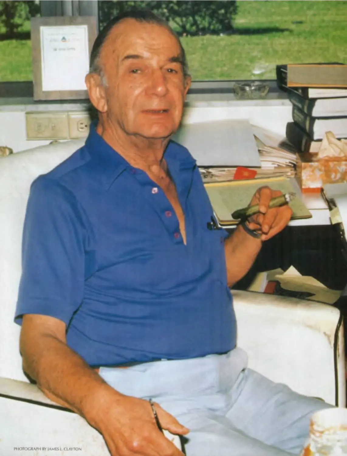
Frank H. Netter, M.D.
(1906-1991)
PHOTCK.RAPHBX
FOREWORD
"The release in 1989 of the first edition of Dr. Frank Netter's 'personal Sistine Chapel'—the
Atlas of Human Anatomy—was a major event in the history of the teaching and learning
of anatomy. Almost instantly, the Atlas of Human Anatomy became the top-selling
anatomical atlas in the world and clearly became the students' choice universally."
This quotation from the second edition of the Atlas of Human Anatomy still resonates
today as the beautifully conceived and rendered artwork of Dr. Netter continues to
capture the admiration of students and healthcare professionals.
The skillful editing and publishing teamwork of Novartis Medical Education, in
consultation with many of the 20th century's outstanding anatomists and physicians,
have made the first and second editions of the Atlas the standard against which all other
atlases are compared. This third edition owes much to the Consulting Editors of the
earlier editions, Drs. Sharon Colacino (Oberg) (first edition) and Arthur F. Dailey II
(second edition), who shepherded their editions with great skill and uncompromising
professionalism and thus made my task significantly easier. I am honored to serve as the
Consulting Editor of the third edition and to continue the Netter tradition in partnership
with Icon Learning Systems as the Atlas of Human Anatomy enters the new century.
Perceptive student and faculty response to the earlier editions prompted greater
attention to several concepts that are critical to the anatomical basis of clinical practice.
Notably, each regional section now begins with a surface anatomy plate, skillfully drawn
by Carlos A.G. Machado, M.D. These surface anatomy plates remind us of the value of
careful observation in clinical medicine and draw attention to surface features that por-
tend the underlying anatomy.
We also have included a significant number of normal radiographic images in
this edition, reflecting the importance of diagnostic imaging in clinical anatomy and
medicine. These new images are not meant to be comprehensive of the radiographic
knowledge available to today's clinicians, but rather are starting points for further
exploration into the richness of anatomical detail that imaging can provide. I am grateful
to Matthew Cham, M.D., and John Wandtke, M.D., of our Department of Radiology for
selecting these images and permitting us to use them in this edition.
We balanced the addition of new surface and radiographic anatomy plates largely
by eliminating several plates that contributed little to the quality of the Atlas. Several
plates from The Netter (formerly CIBA) Collection of Medical Illustrations were added
and several plates were altered slightly to correct anatomical errors consistent with our
current knowledge. In sum, the third edition has grown by 30 images. Finally, the
References and the Index have been updated.
The anatomical terminology is consistent throughout the Atlas and conforms to
the International Anatomical Terminology (Terminologia Anatomica) approved in 1998
by the International Federation of Associations of Anatomists. Common eponyms are
retained parenthetically, and the leader lines and labels have been checked and, where
necessary, corrected to ensure their accuracy. For reviewing this material I thank
Wojciech Pawlina, M.D., Leonard J. Cleary, Ph.D., Daniel O. Graney, Ph.D., and Brian R.
MacPherson, Ph.D. Their meticulous examination of each plate has greatly enhanced
the accuracy of this atlas.
For the proofreading, editing and designing, I am indebted to Jennifer Surich,
Erika Gehringer and Greg Otis at Icon Learning Systems; their keen eyesight, dedication
and professionalism buoyed me when I began to drift. A special thank you is reserved
for Paul Kelly, Executive Editor, who continues to listen patiently and is a true believer in
the power of the visual image to teach.
To all students, past, present and future, the legacy of Dr. Netter's artwork lives in
you; his unique ability to "clarify rather than intimidate" is his gift to you. I am both privi-
leged and humbled to have the opportunity to carry forward this tradition of excellence
and to contribute to your understanding of human anatomy.
Finally, to my wife, Paula, thank you for giving me the freedom to write and the
support to sustain me—in those respects, this book was co-edited.
John T Hansen, Ph.D.
Professor of Neurobiology and Anatomy
University of Rochester School of Medicine and Dentistry
Rochester, New York
CONTENTS
Section 1 HEAD AND NECK
SURFACE ANATOMY BONES AND LIGAMENTS SUPERFICIAL FACE NECK NASAL REGION ORAL REGION PHARYNX THYROID GLAND AND LARYNX ORBIT AND CONTENTS EAR MENINGES AND BRAIN CRANIAL AND CERVICAL NERVES CEREBRAL VASCULATURE REGIONAL SCANS Plate 1 Plates 2—18 Plates 19—22 Plates 23—31 Plates 32—46 Plates 47—58 Plates 59—69 Plates 70-76 Plates 77—86 Plates 87—93 Plates 94—109 Plates 110—129 Plates 130—141 Plates 142—144
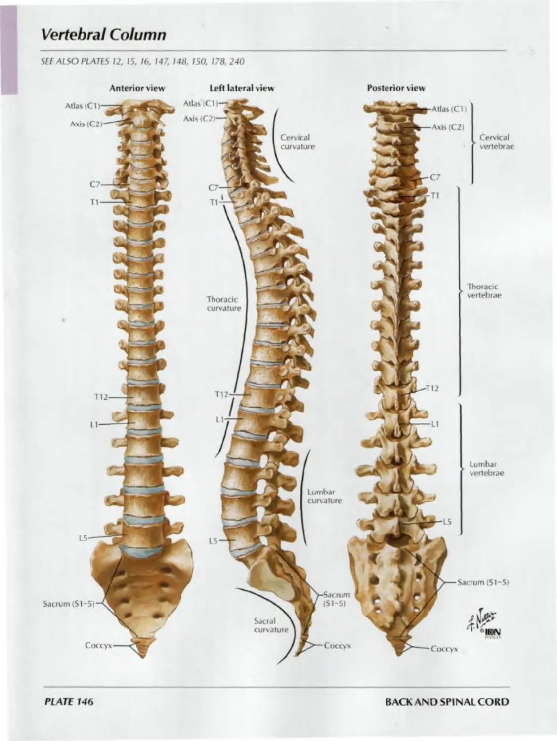
Section II BACK AND SPINAL CORD
SURFACE ANATOMY BONES AND LIGAMENTS SPINAL CORD MUSCLES AND NERVES Plate 145 Plates 146—152 Plates 153—166 Plates 167-173
Section III 1 FHORAX
SURFACE ANATOMY MAMMARY GLAND BODY WALL LUNGS HEART MEDIASTINUM REGIONAL SCANS Plate 174 Plates 175—177 Plates 178-191 Plates 192—206 Plates 207—225 Plates 226—237 Plate 238
Section IV ABDOMEN
SURFACE ANATOMY BODY WALL PERITONEAL CAVITY VISCERA (GUT) VISCERA (ACCESSORY ORGANS) VISCERAL VASCULATURE INNERVATION KIDNEYS AND SUPRARENAL GLANDS ABDOMINAL SECTIONS REGIONAL SCANS Plate 239 Plates 240—259 Plates 260-266 Plates 267-277 Plates 278—289 Plates 290—307 Plates 308—318 Plates 319—334 Plates 335—337 Plate 338
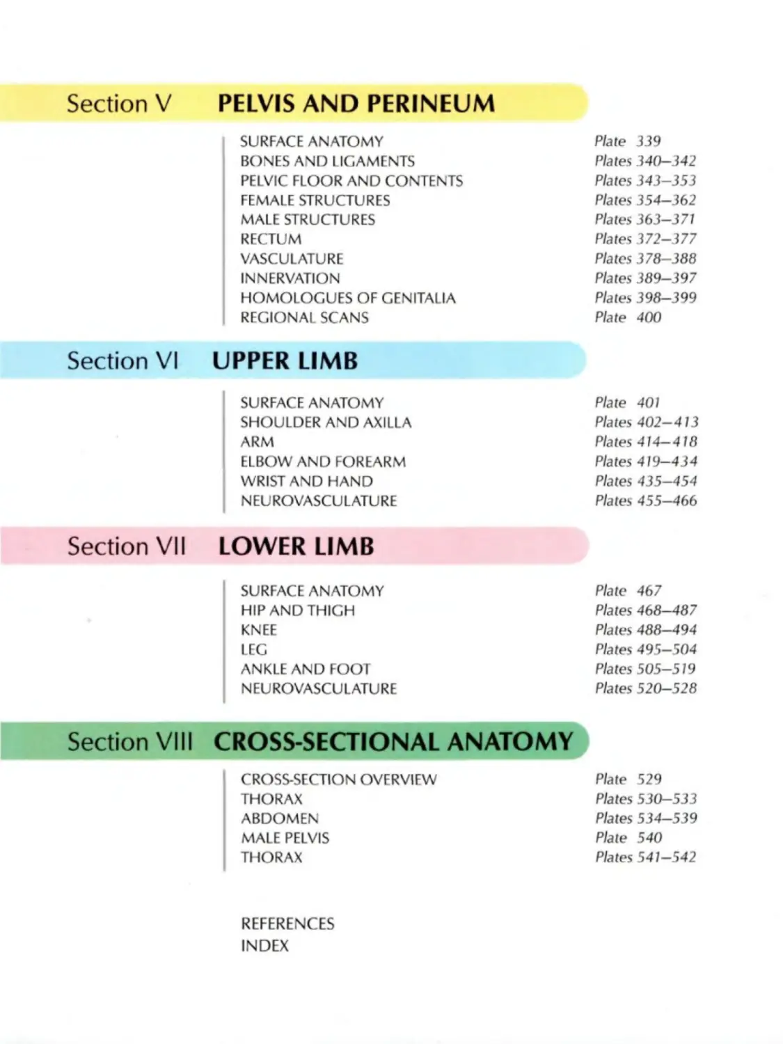
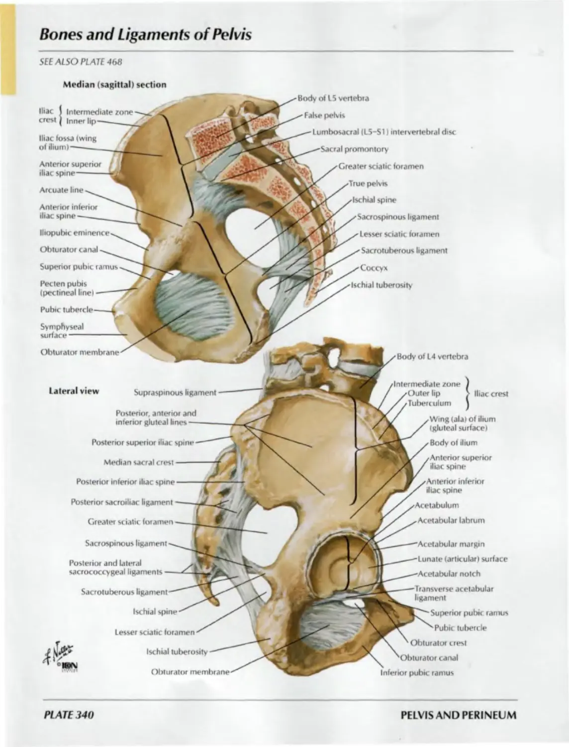
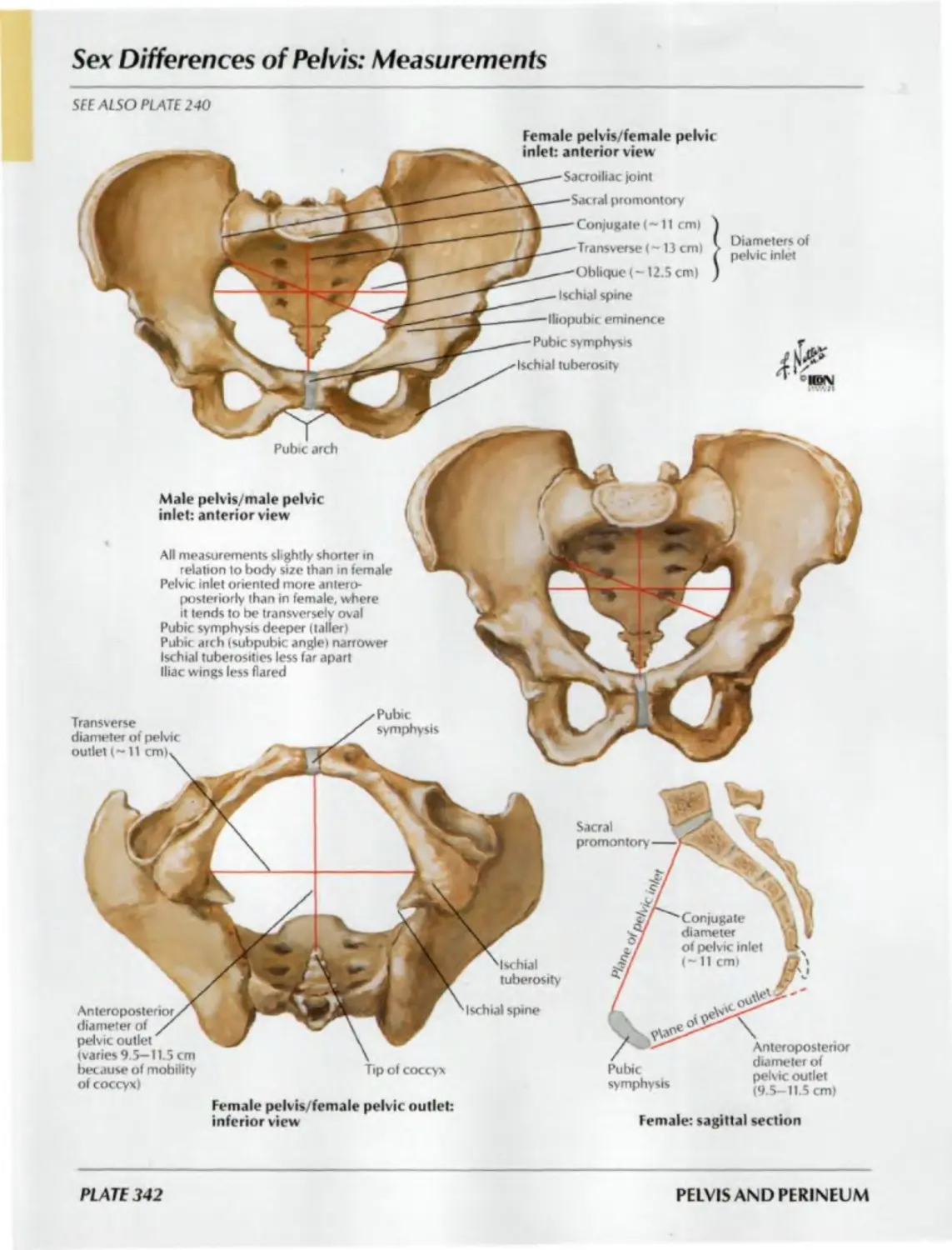
Section V PELVIS AND PERINEUM
SURFACE ANATOMY Plate 339
BONES AND LIGAMENTS Plates 340—342
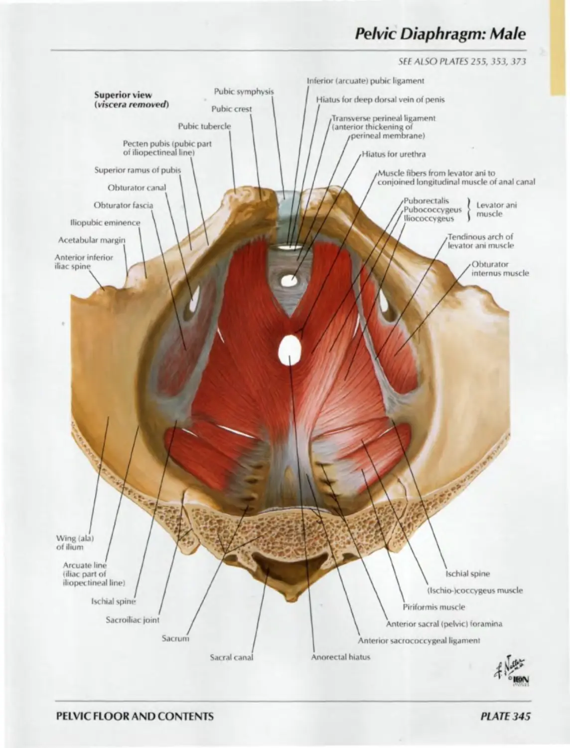
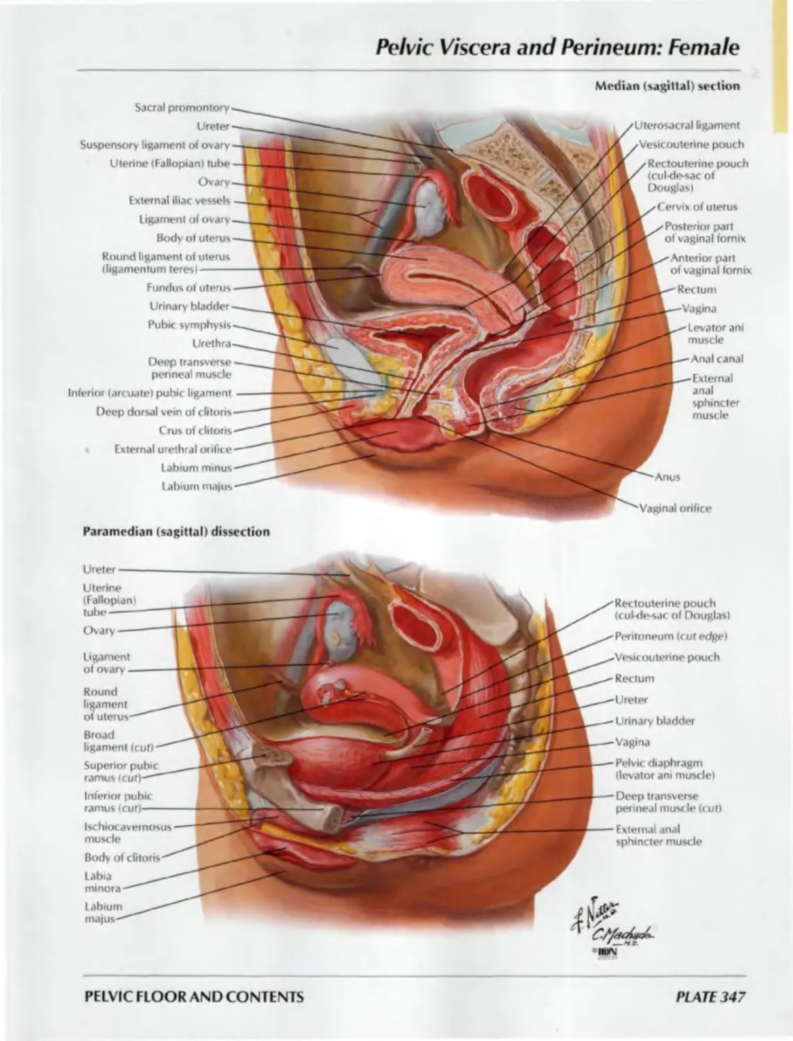
PELVIC FLOOR AND CONTENTS Plates 343—353
FEMALE STRUCTURES Plates 354—362
MALE STRUCTURES Plates 363-371
RECTUM Plates 372—377
VASCULATURE Plates 378—388
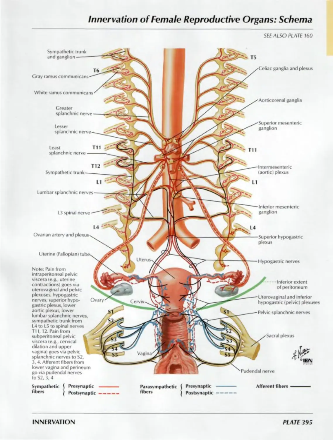
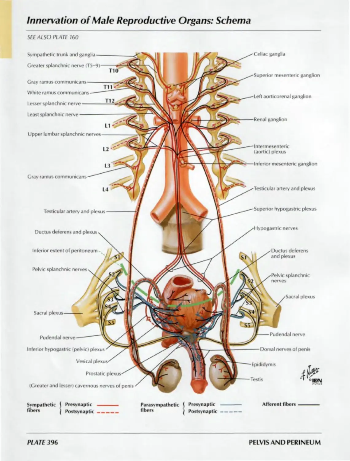
INNERVATION Plates 389-397
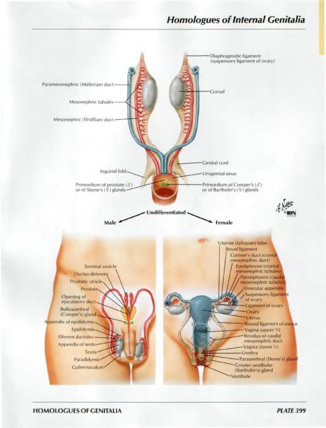
HOMOLOGUES OF GENITALIA Plates 398—399
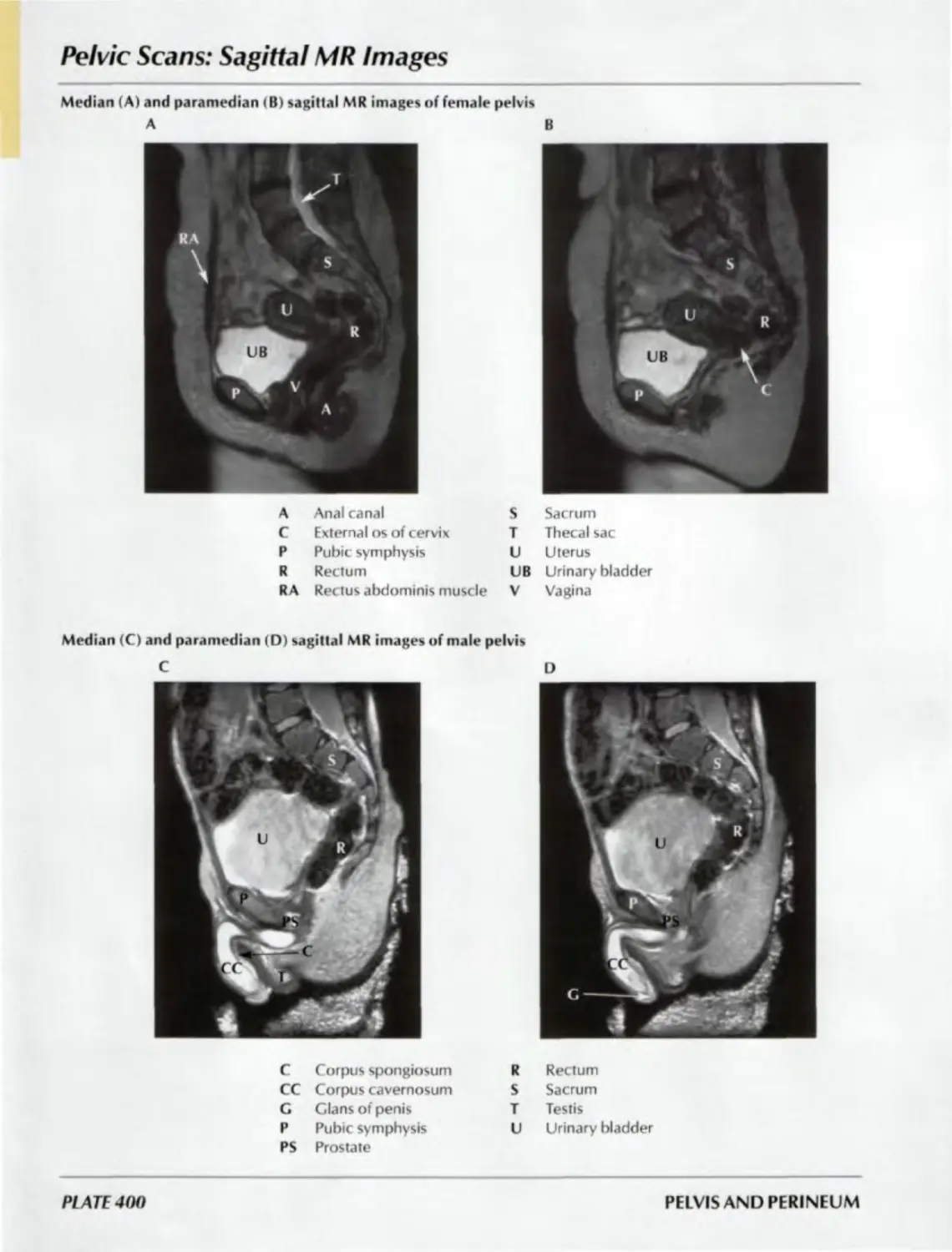
REGIONAL SCANS Plate 400
Section VI UPPER LIMB
SURFACE ANATOMY SHOULDER AND AXILLA ARM ELBOW AND FOREARM WRIST AND HAND NEUROVASCULATURE Plate 401 Plates 402—413 Plates 414—418 Plates 419—434 Plates 435—454 Plates 455-466
Section VII 1 _OWER LIMB SURFACE ANATOMY HIP AND THIGH KNEE LEG ANKLE AND FOOT NEUROVASCULATURE Plate 467 Plates 468—487 Plates 488—494 Plates 495—504 Plates 505—519 Plates 520—528
Section VIII CROSS-SECTIONAL ANATOMY
CROSS-SECTION OVERVIEW Plate 529
THORAX Plates 530—533
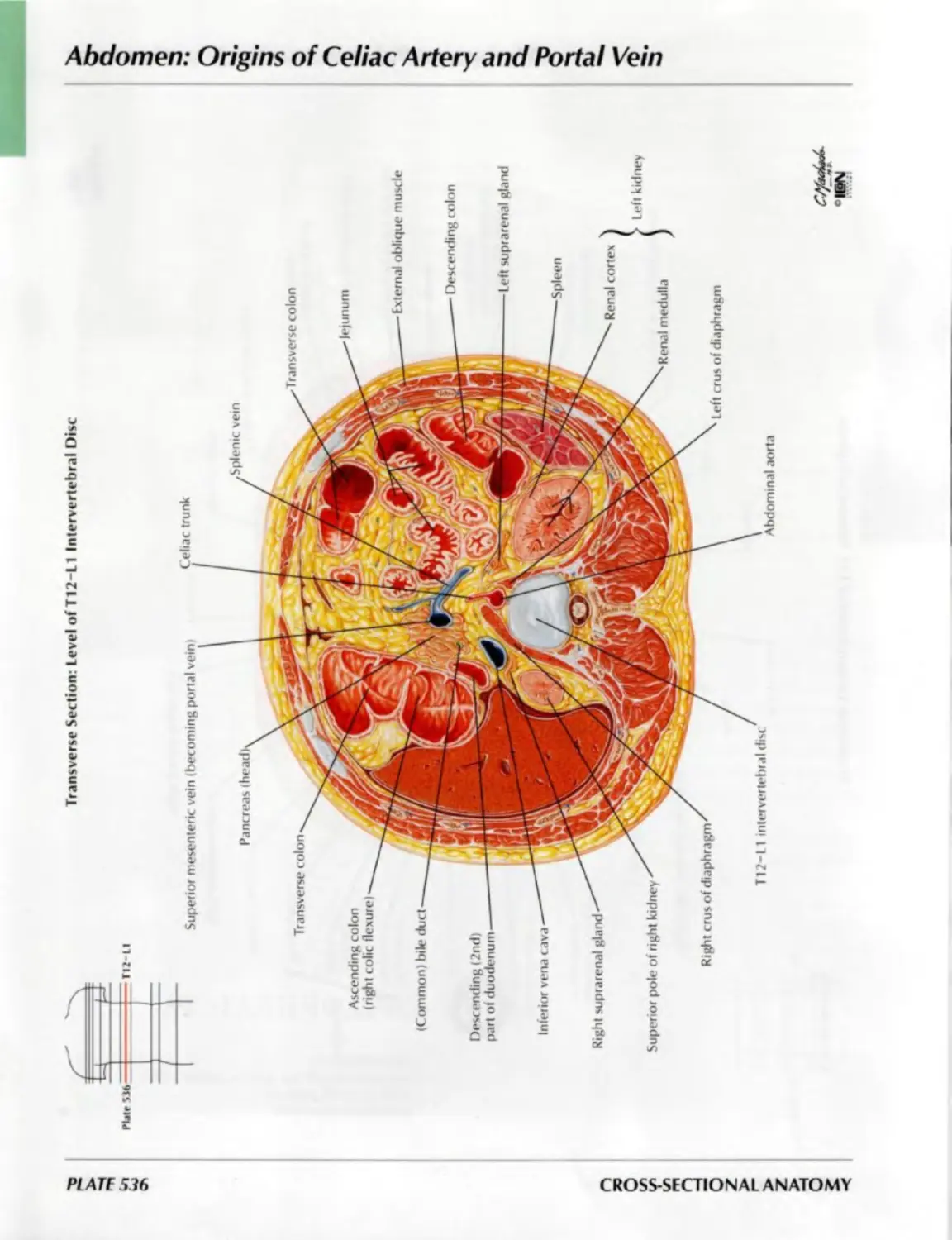
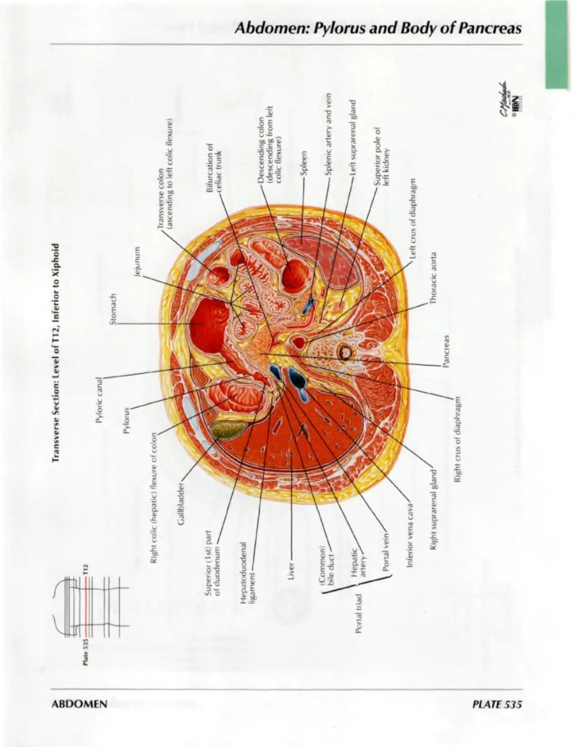
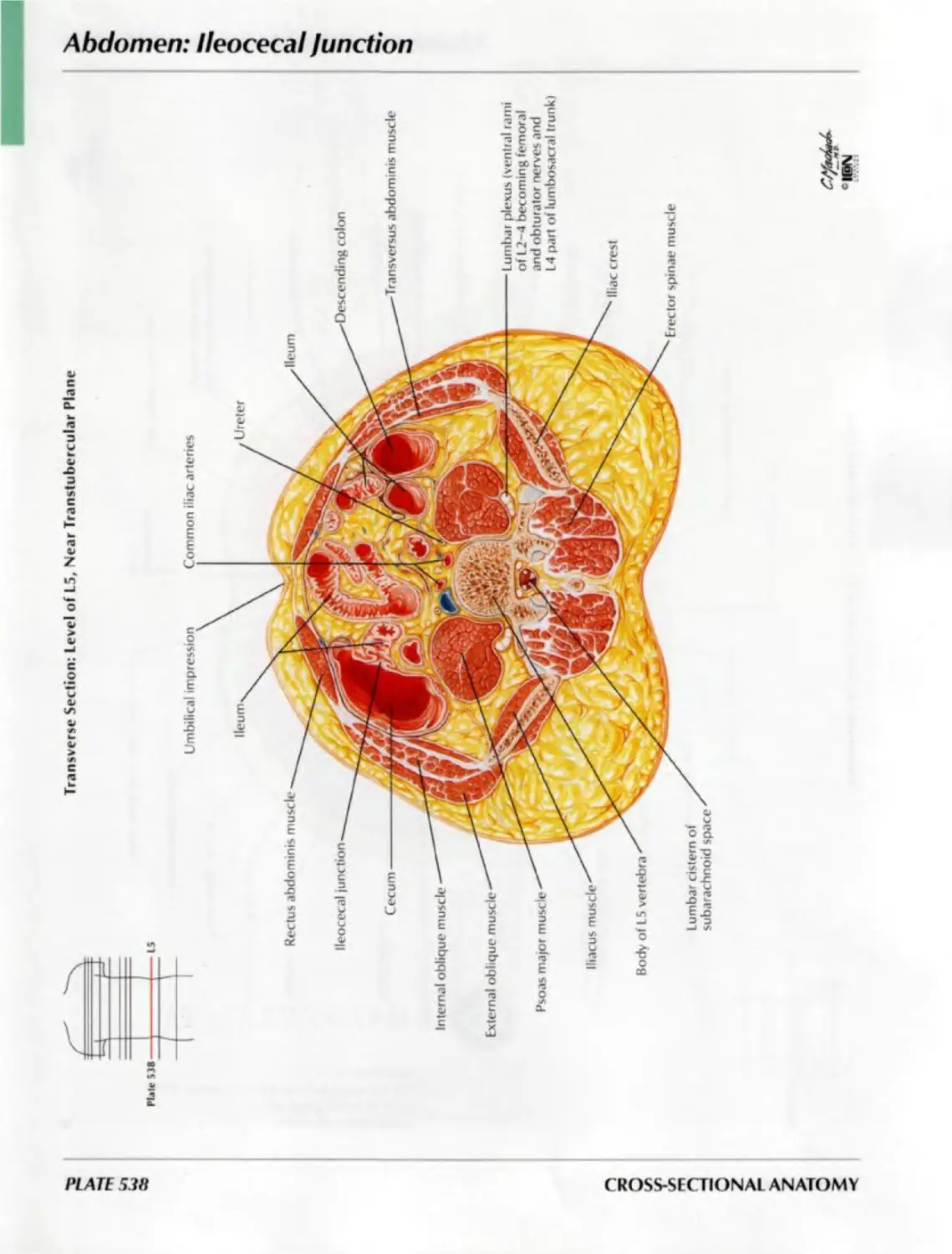
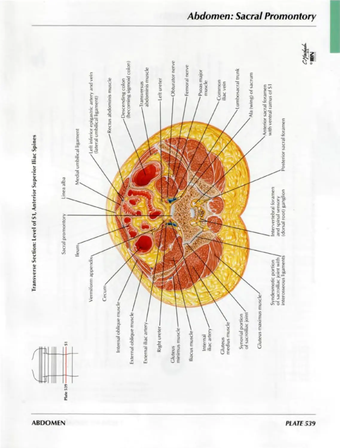
ABDOMEN Plates 534-539
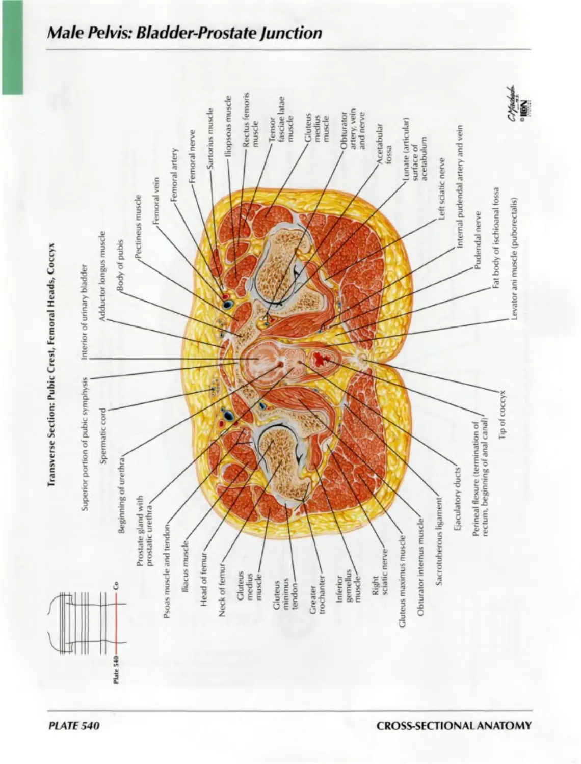
MALE PELVIS Plate 540
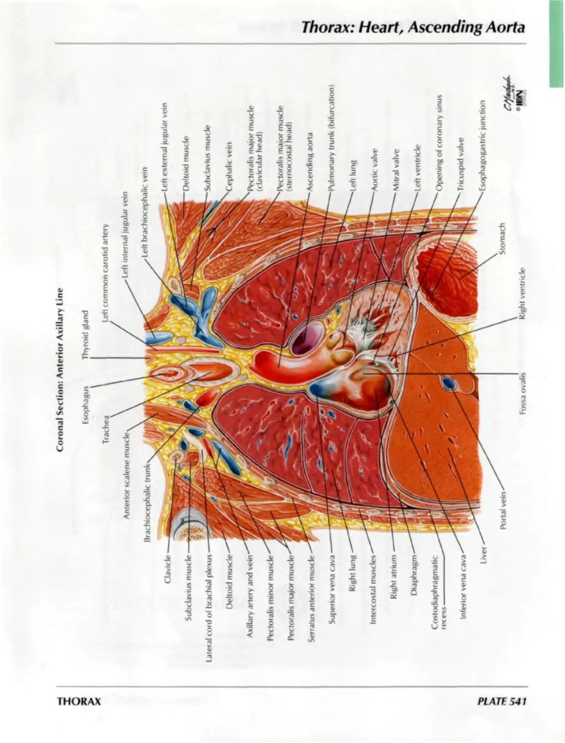
THORAX Plates 541—542
REFERENCES
INDEX
Section I
HEAD AND
NECK
SURFACE ANATOMY
Plate 1
1. Head and Neck
BONES AND LIGAMENTS
Plates 2-18
2. Skull: Anterior View
3. Skull: Anteroposterior Radiograph
4. Skull: Lateral View
5. Skull: Lateral Radiograph
6. Skull: Midsagittal Section
7. Calvaria
8. Cranial Base: Inferior View
9. Bones of Cranial Base: Superior View
10. Foramina of Cranial Base:
Superior View
11. Skull of Newborn
12. Bony Framework of Head and Neck
13. Mandible
14. Temporomandibular Joint
15. Cervical Vertebrae: Atlas and Axis
16. Cervical Vertebrae (continued)
17. External Craniocervical Ligaments
18. Internal Craniocervical Ligaments
SUPERFICIAL FACE
Plates 19-22
19. Superficial Arteries and Veins
of Face and Scalp
20. Cutaneous Nerves of Head and Neck
21. Facial Nerve Branches and
Parotid Gland
22. Muscles of Facial Expression:
Lateral View
ATLAS OF HUMAN ANATOMY
SECTION I: HEAD AND NECK
NECK
Plates 23-31
23. Muscles of Neck: Lateral View
24. Muscles of Neck: Anterior View
25. Infrahyoid and Suprahyoid
Muscles
26. Scalene and Prevertebral Muscles
27. Superficial Veins and Cutaneous
Nerves of Neck
28. Cervical Plexus In Situ
29. Subclavian Artery
30. Carotid Arteries
31. Fascial Layers of Neck
NASAL REGION
Plates 32-46
32. Nose
33. Lateral Wall of Nasal Cavity
34. Lateral Wall of Nasal Cavity
{continued)
35. Medial Wall of Nasal Cavity
(Nasal Septum)
36. Maxillary Artery
37. Arteries of Nasal Cavity:
Nasal Septum Turned Up
38. Nerves of Nasal Cavity:
Nasal Septum Turned Up
39. Nerves of Nasal Cavity (continued)
40. Autonomic Innervation of Nasal Cavity
41. Ophthalmic (V,) and
Maxillary (V2) Nerves
42. Mandibular Nerve (V3)
43. Nose and Paranasal Sinuses:
Cross Section
44. Paranasal Sinuses
45. Paranasal Sinuses (continued)
46. Paranasal Sinuses:
Changes With Age
ORAL REGION
Plates 47-58
47. Inspection of Oral Cavity
48. Roof of Mouth
49. Floor of Mouth
50. Muscles Involved in Mastication
51. Muscles Involved in Mastication
(continued)
52. Teeth
53. Teeth (continued)
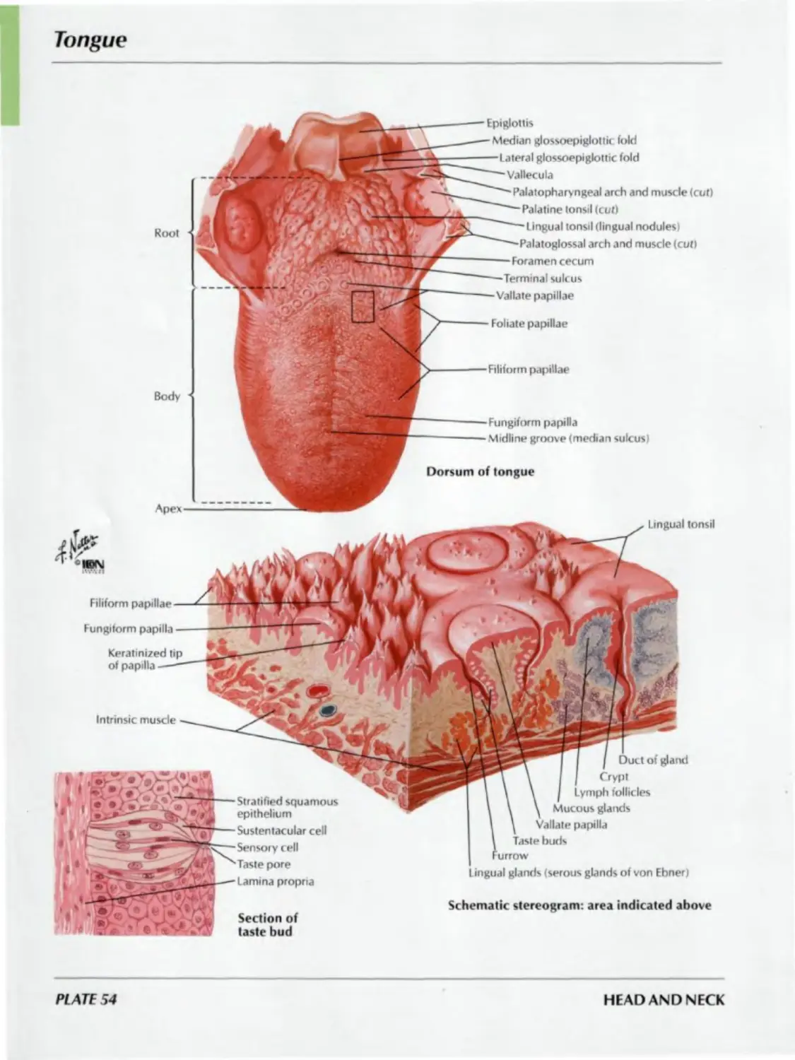
54. Tongue
55. Tongue (continued)
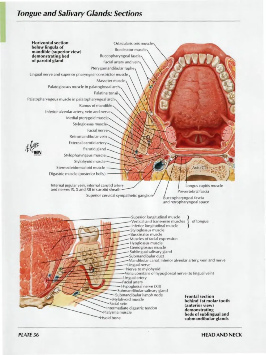
56. Tongue and Salivary Glands: Sections
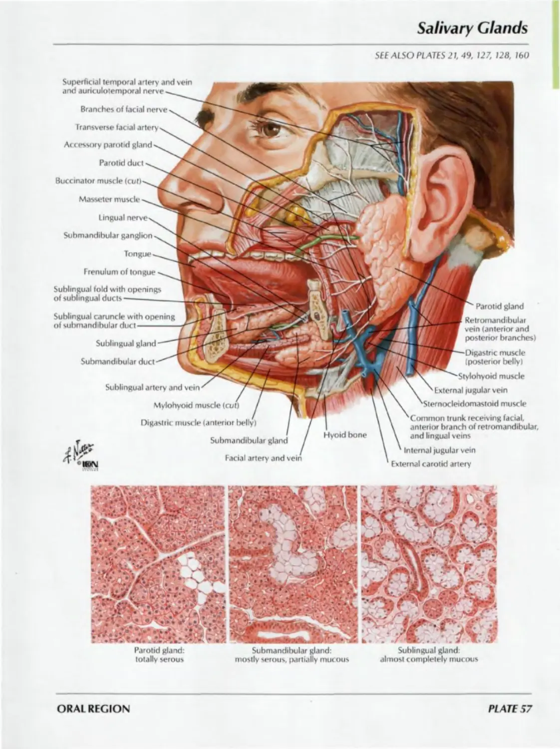
57. Salivary Glands
58. Afferent Innervation of Mouth
and Pharynx
PHARYNX
Plates 59-69
59. Pharynx: Median Section
60. Fauces
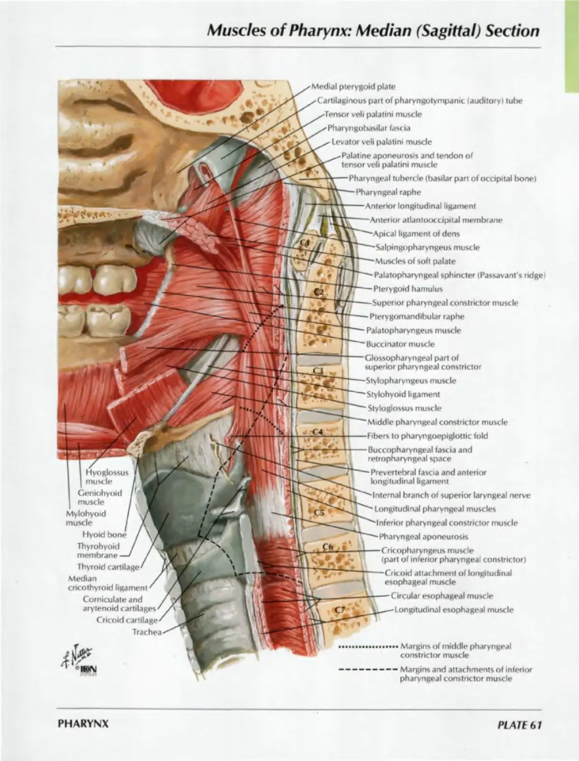
61. Muscles of Pharynx: Median (Sagittal)
Section
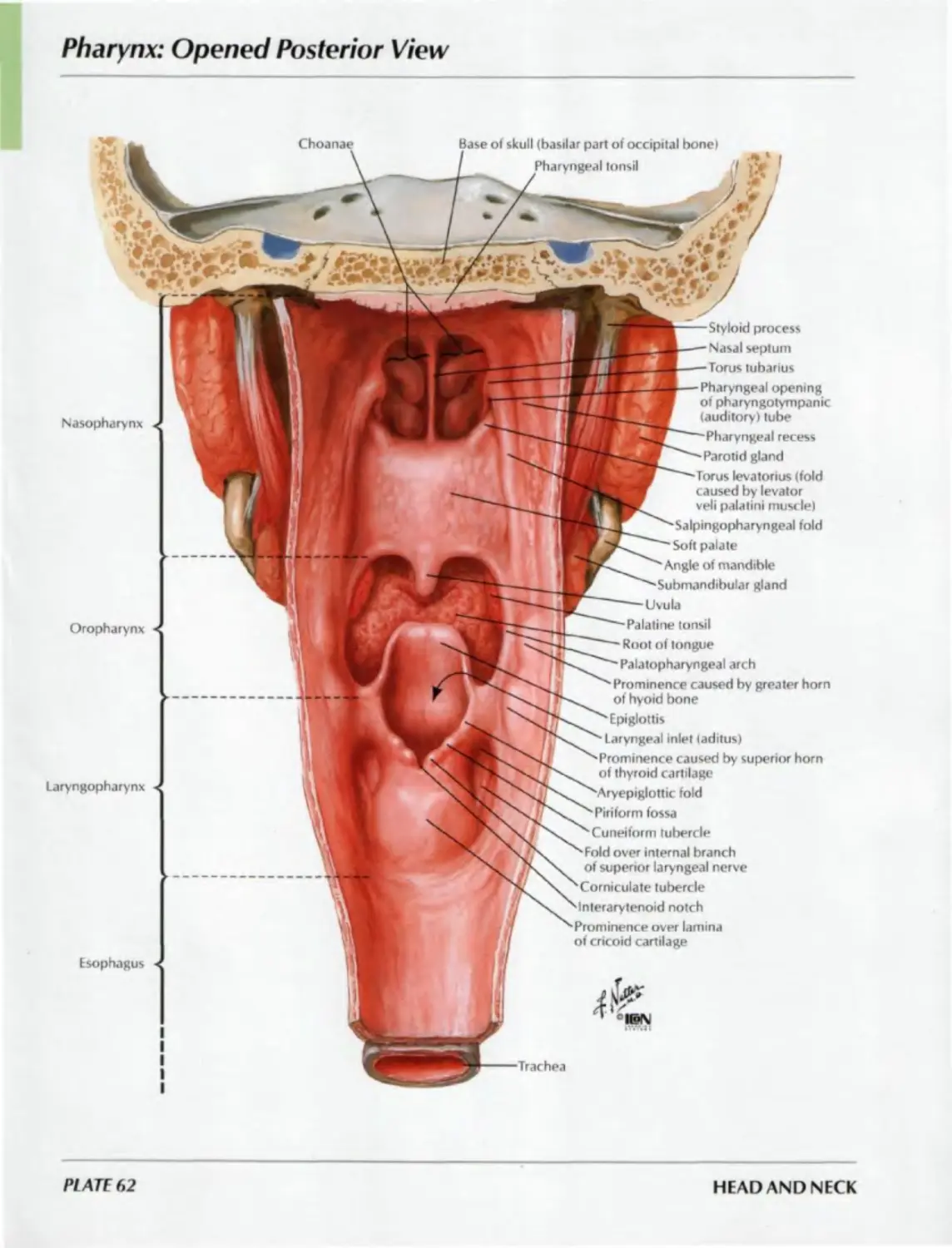
62. Pharynx: Opened Posterior View
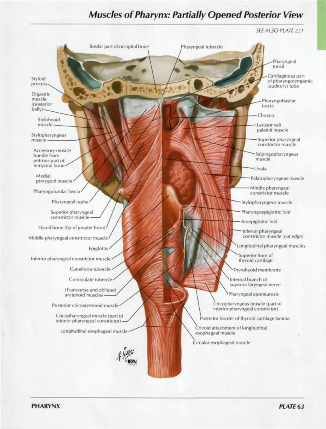
63. Muscles of Pharynx: Partially Opened
Posterior View
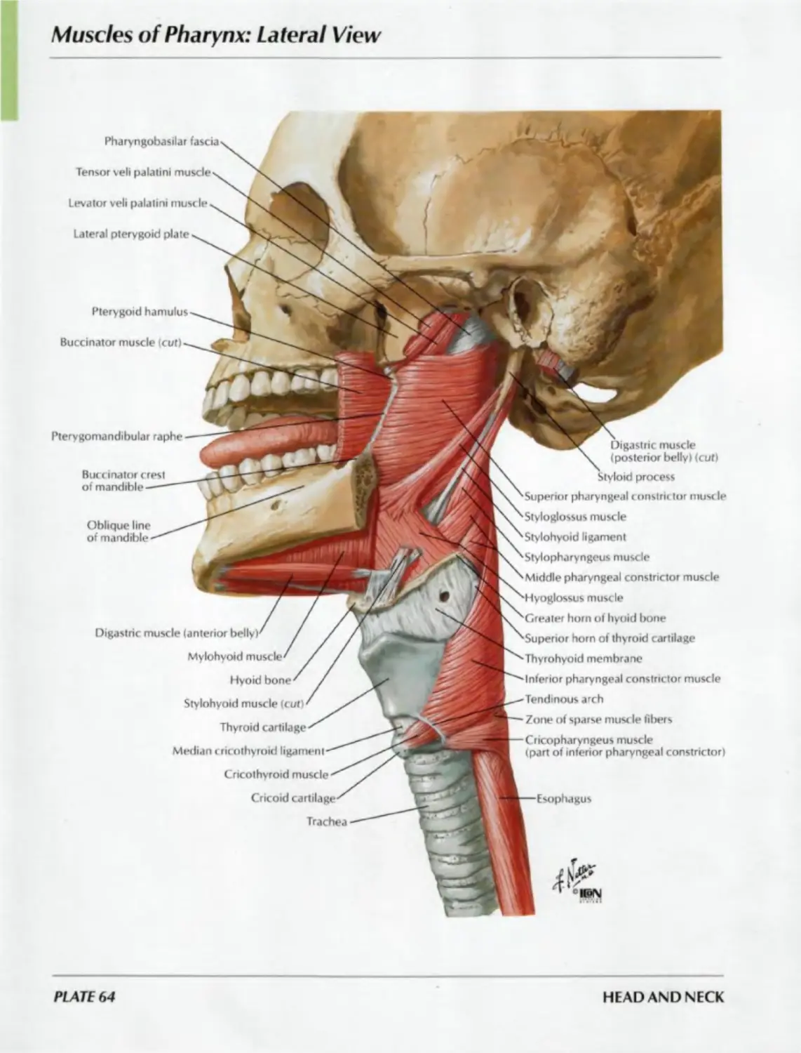
64. Muscles of Pharynx: Lateral View
65. Arteries of Oral and
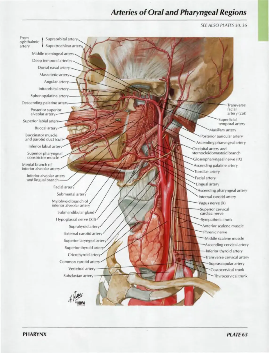
Pharyngeal Regions
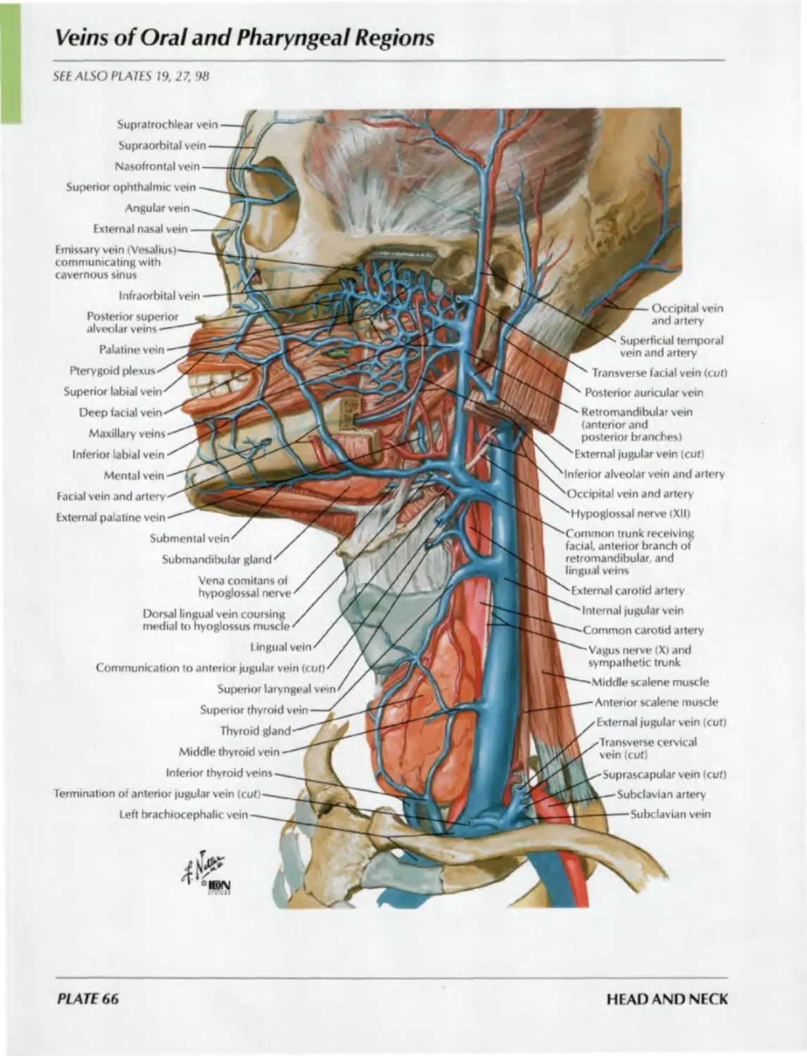
66. Veins of Oral and Pharyngeal Regions
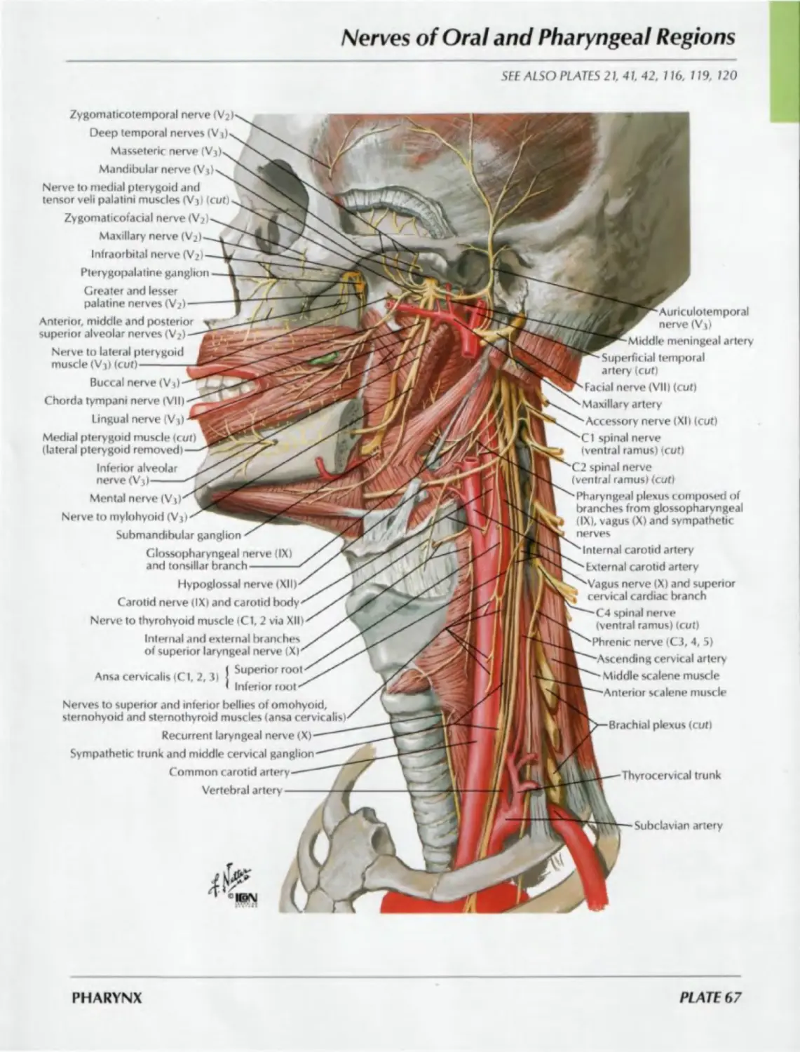
67. Nerves of Oral and
Pharyngeal Regions
ATLAS OF HUMAN ANATOMY
SECTION I: HEAD AND NECK
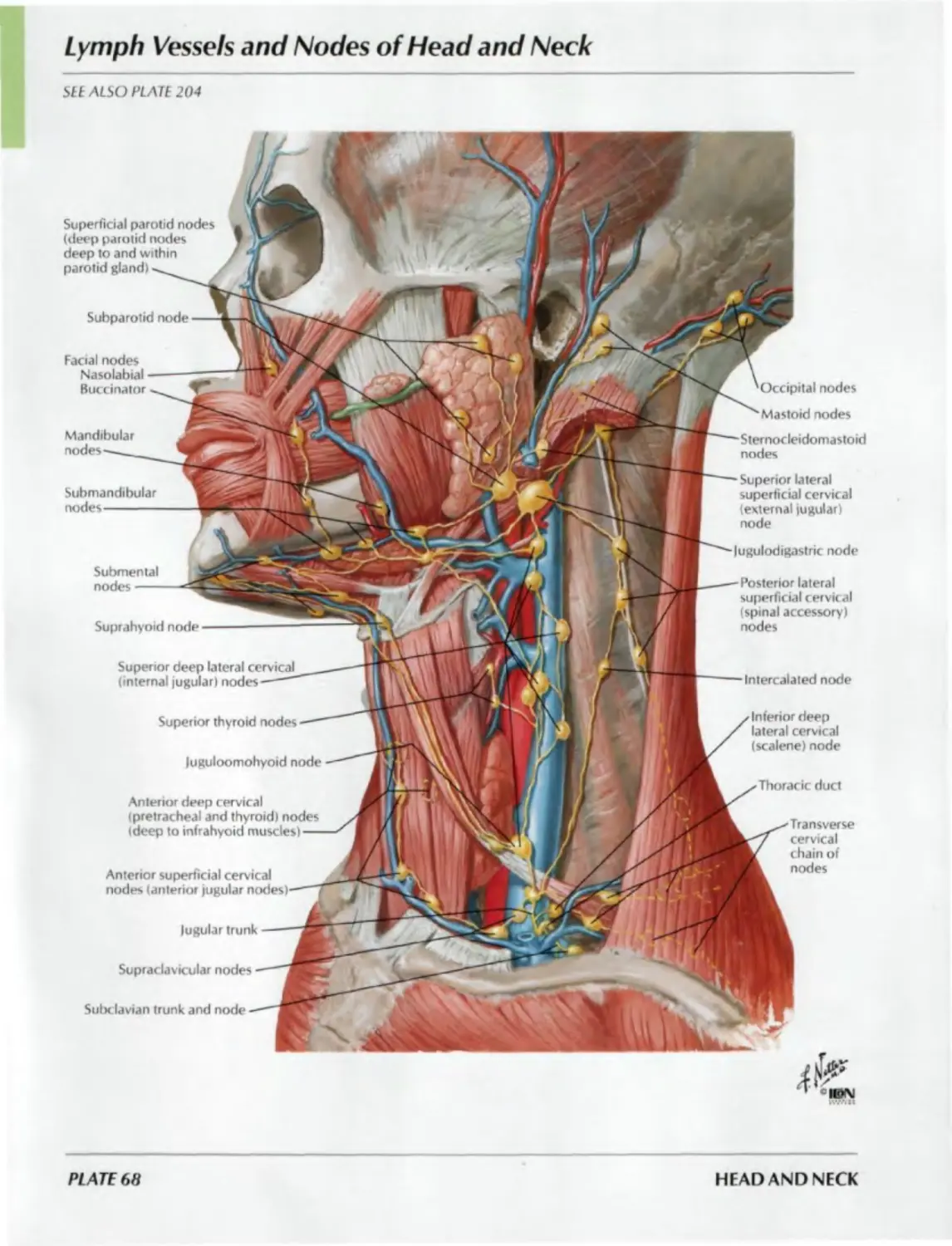
68. Lymph Vessels and Nodes of
Head and Neck
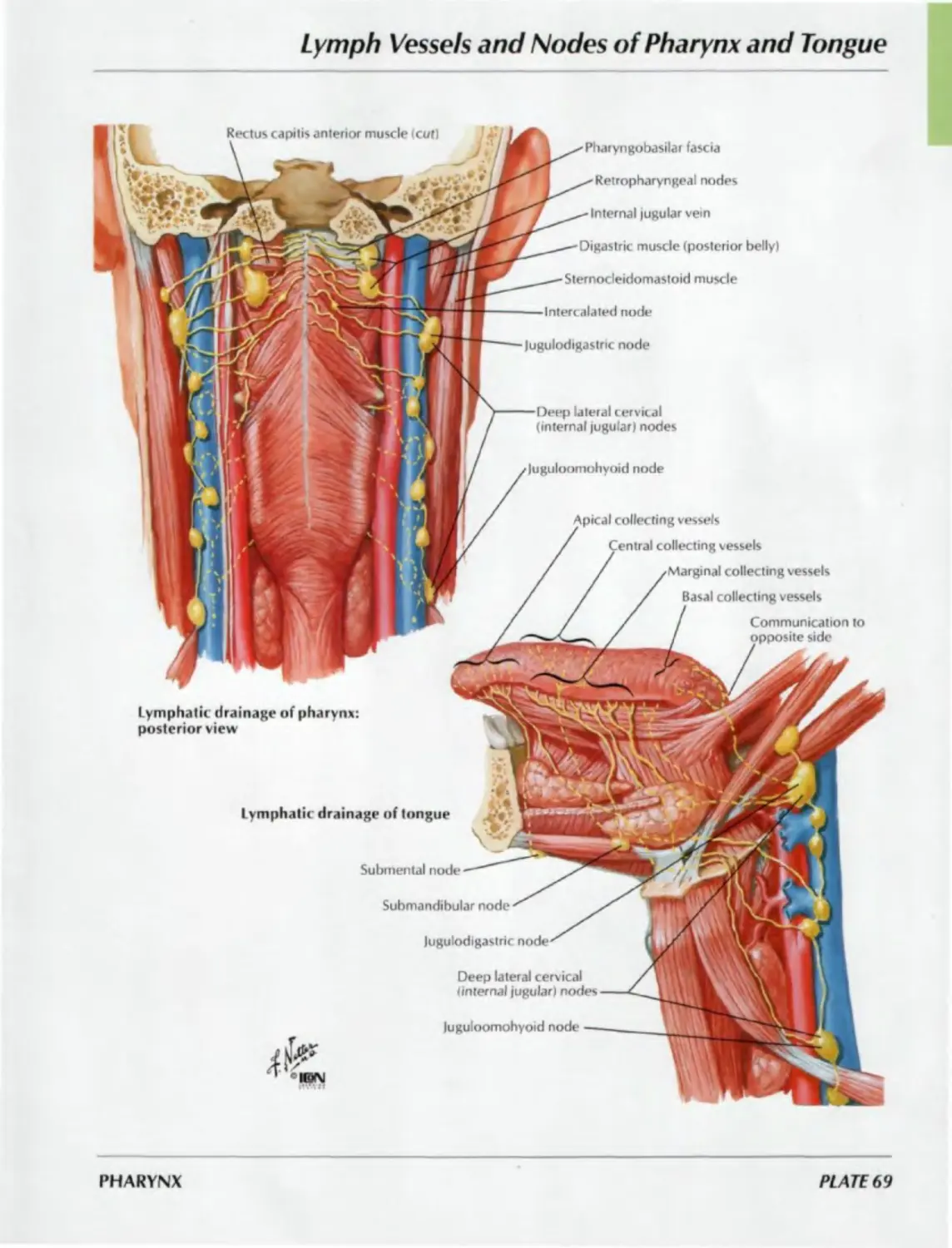
69. Lymph Vessels and Nodes of
Pharynx and Tongue
THYROID GLAND AND LARYNX
Plates 70-76
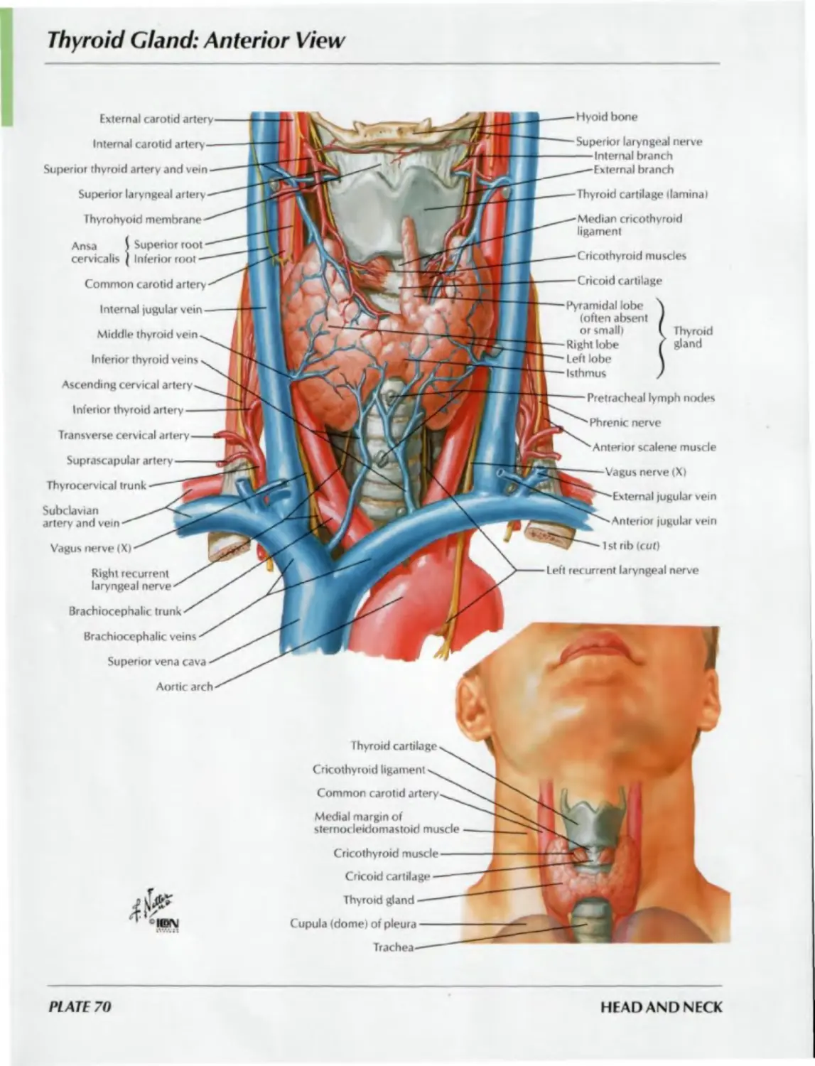
70. Thyroid Gland: Anterior View
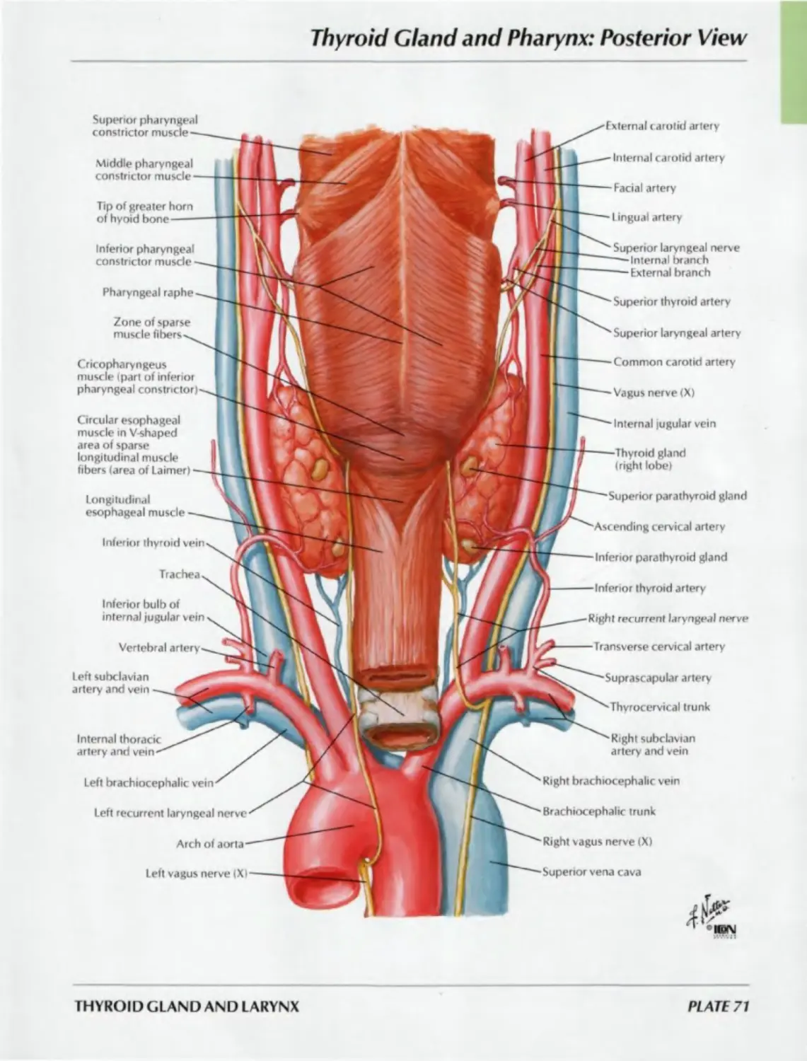
71. Thyroid Gland and Pharynx:
Posterior View
72. Parathyroid Glands
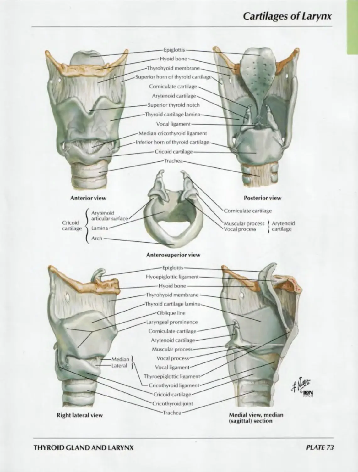
73. Cartilages of Larynx
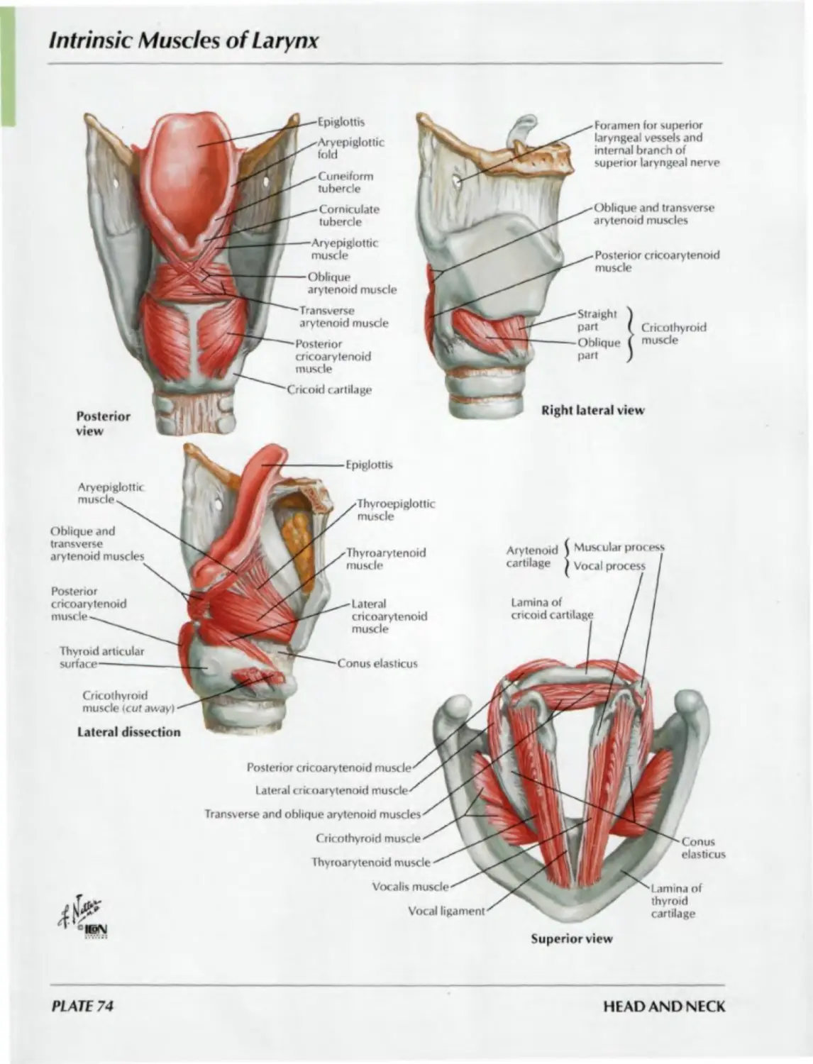
74. Intrinsic Muscles of Larynx
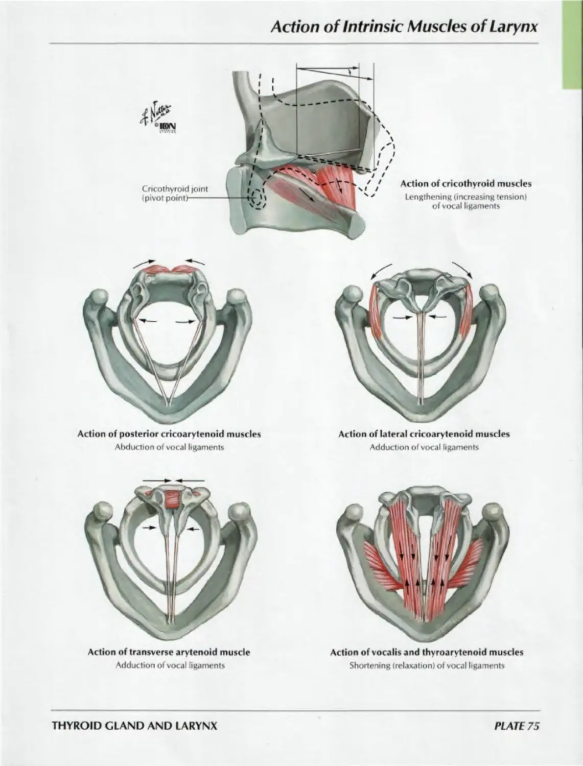
75. Action of Intrinsic Muscles of Larynx
76. Nerves of Larynx
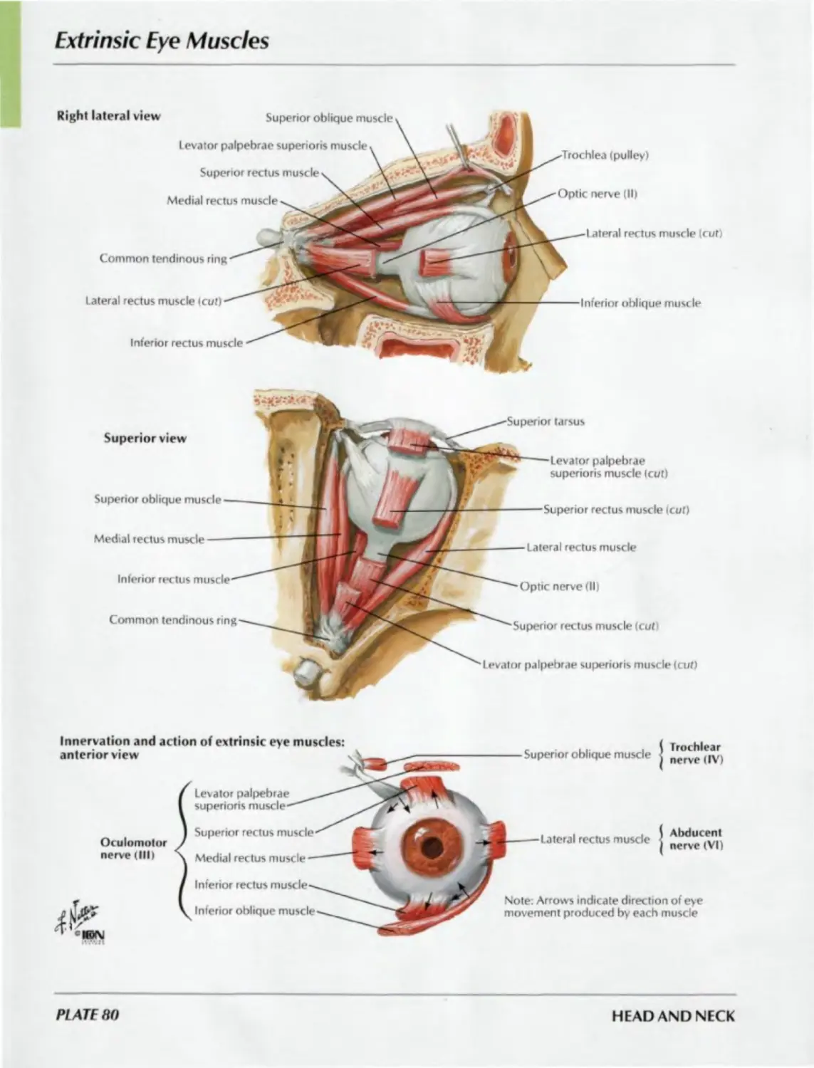
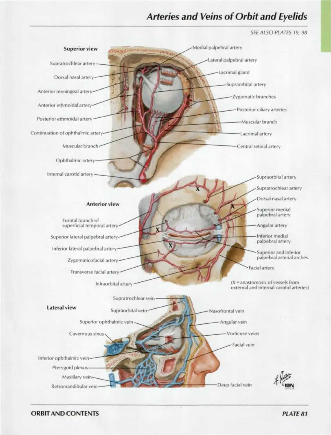
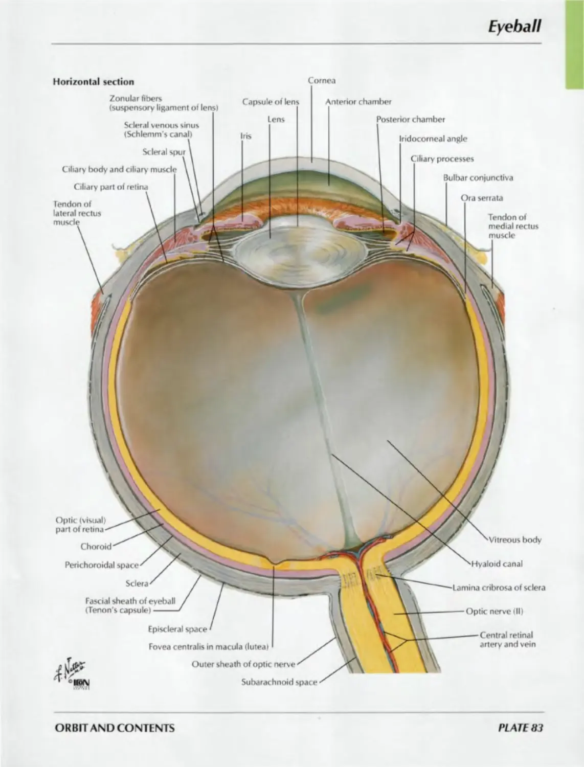
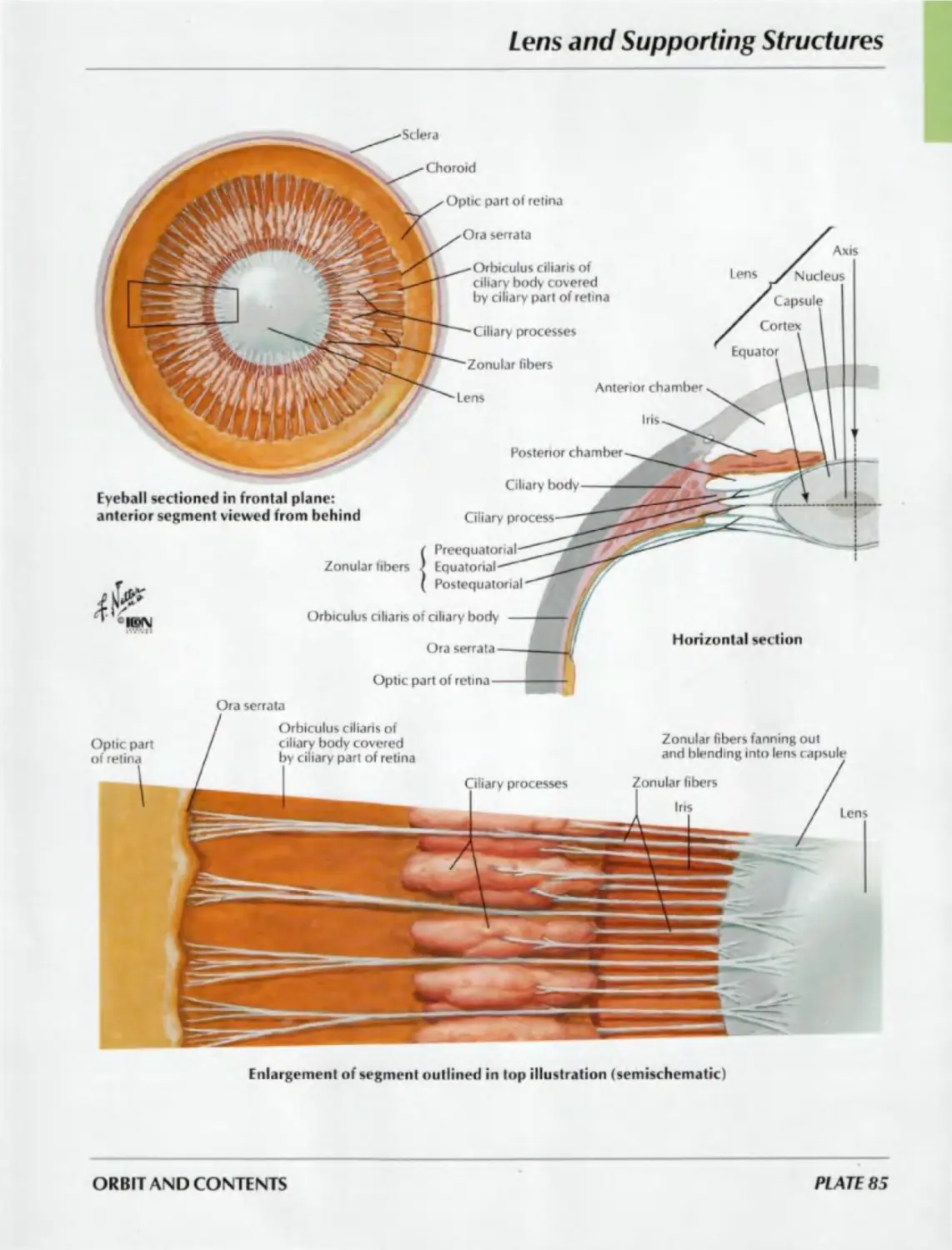
ORBIT AND CONTENTS
Plates 77-86
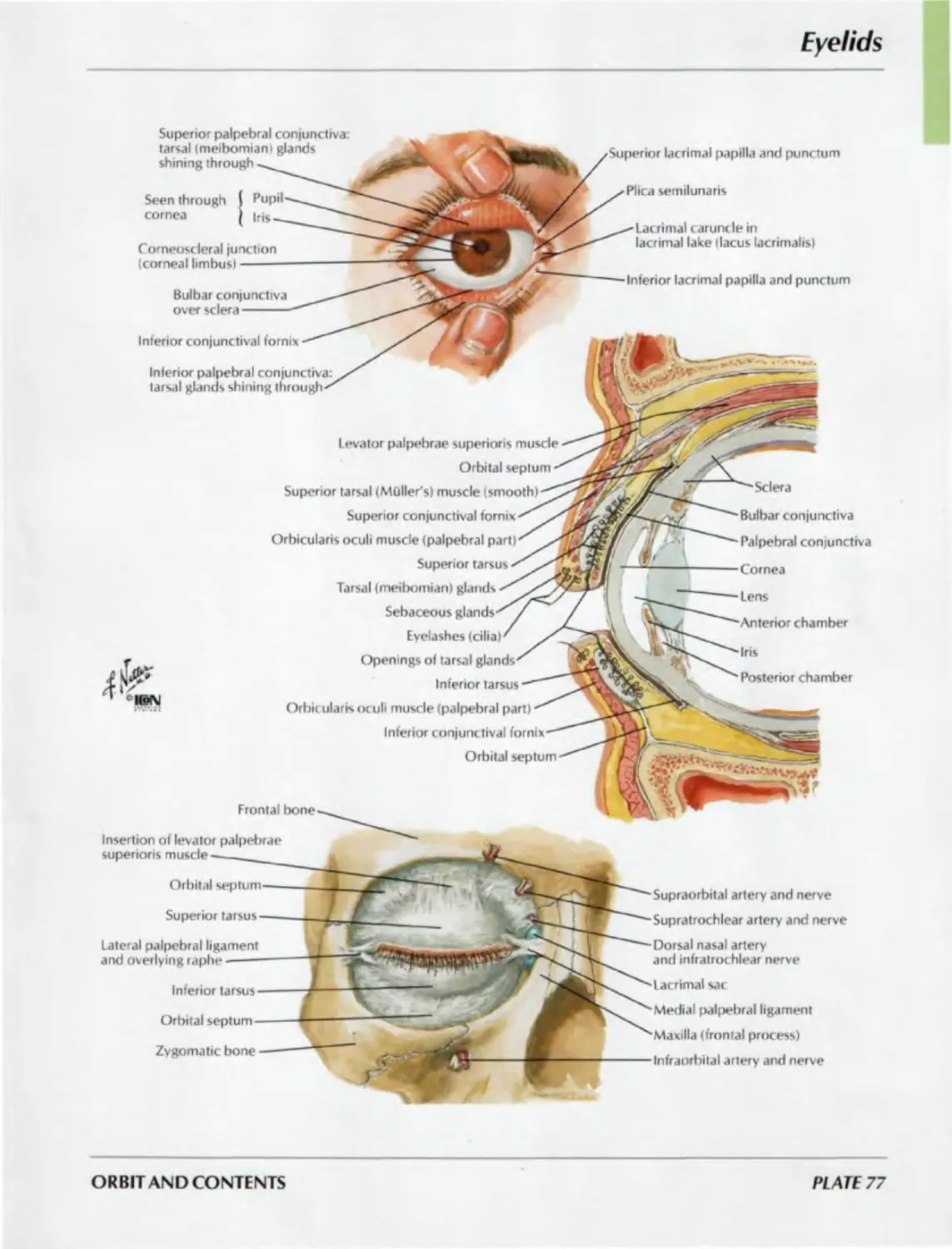
77. Eyelids
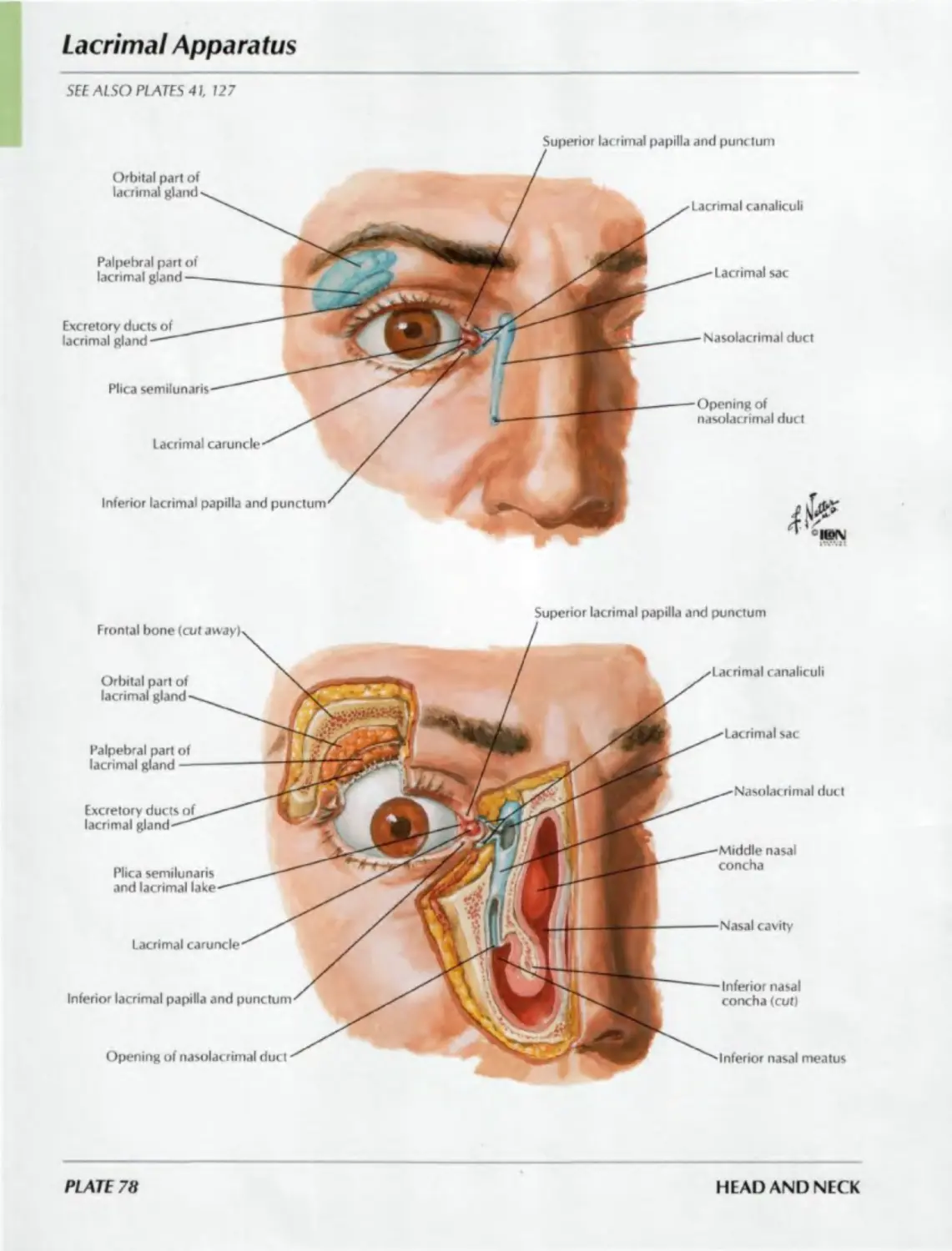
78. Lacrimal Apparatus
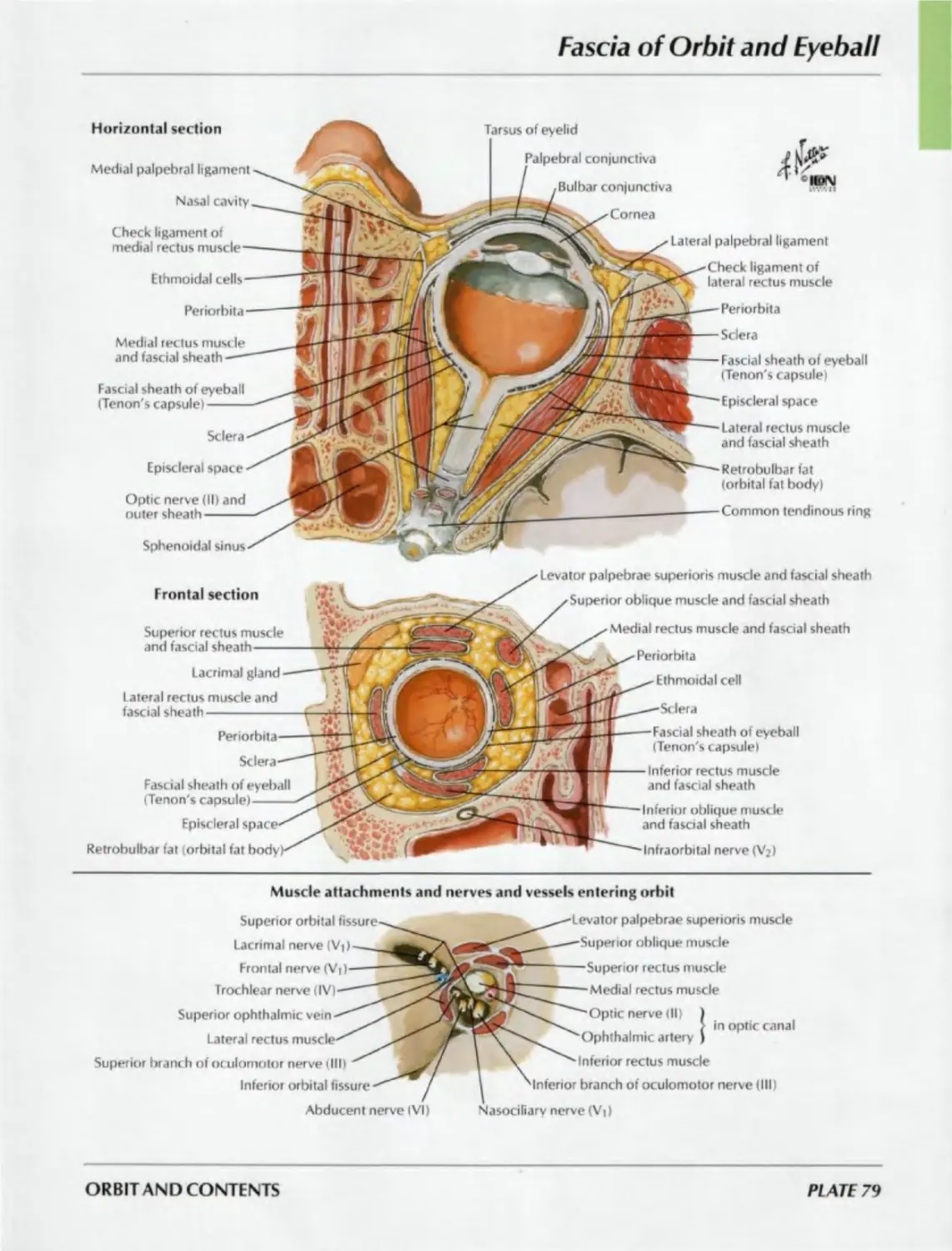
79. Fascia of Orbit and Eyeball
80. Extrinsic Eye Muscles
81. Arteries and Veins of Orbit
and Eyelids
82. Nerves of Orbit
83. Eyeball
84. Anterior and Posterior
Chambers of Eye
85. Lens and Supporting Structures
86. Intrinsic Arteries and Veins of Eye
EAR
Plates 87-93
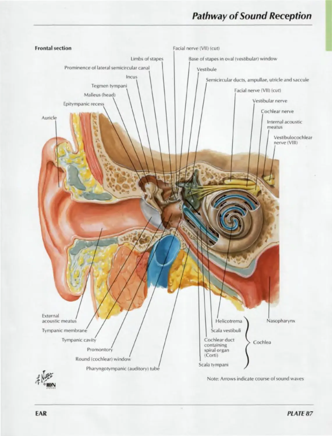
87. Pathway of Sound Reception
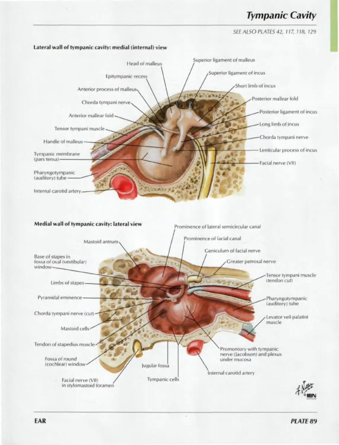
88. External Ear and Tympanic Cavity
89. Tympanic Cavity
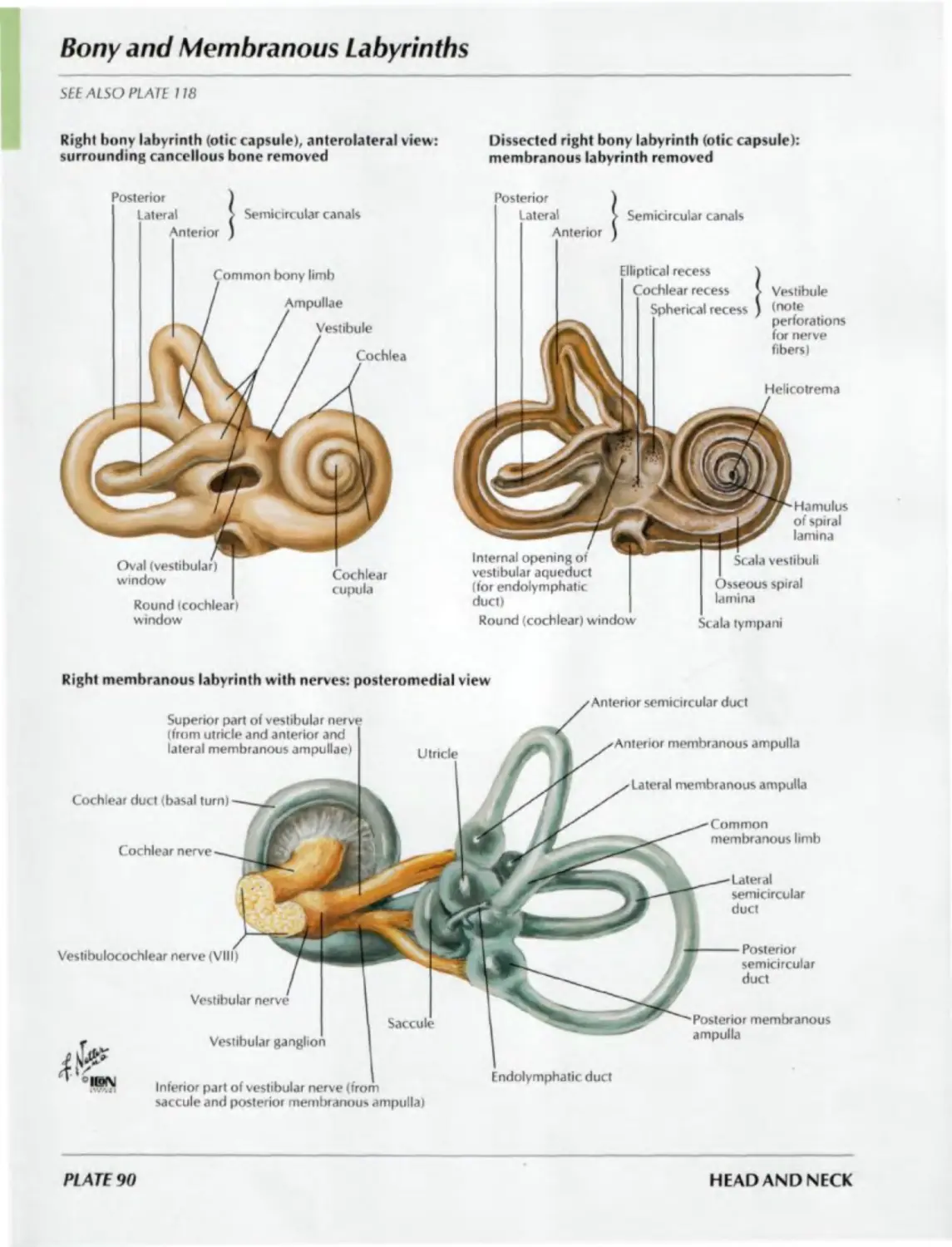
90. Bony and Membranous Labyrinths
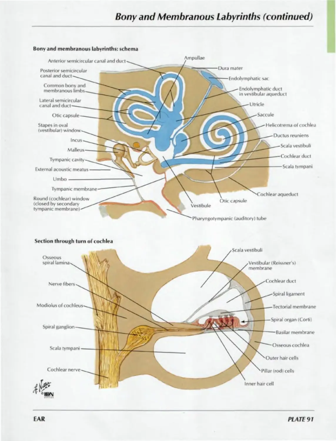
91. Bony and Membranous Labyrinths
(continued)
92. Orientation of Labyrinth in Skull
93. Pharyngotympanic (Auditory) Tube
MENINGES AND BRAIN
Plates 94-109
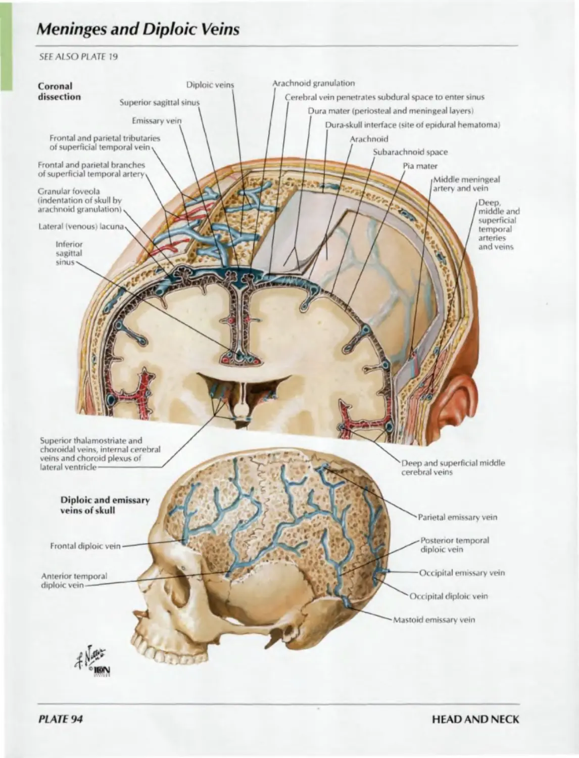
94. Meninges and Diploic Veins
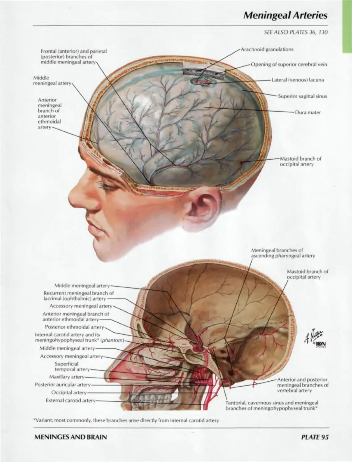
95. Meningeal Arteries
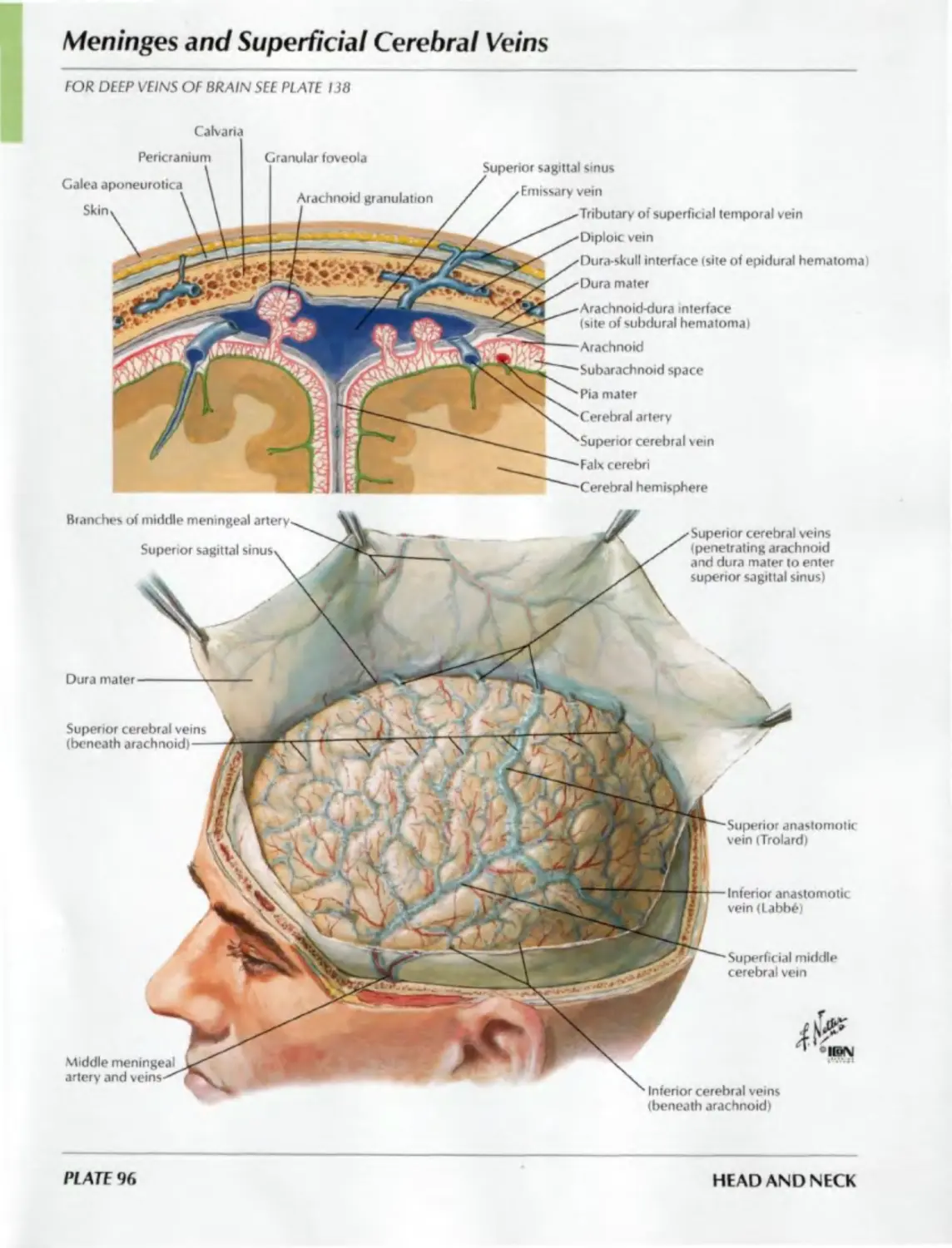
96. Meninges and Superficial
Cerebral Veins
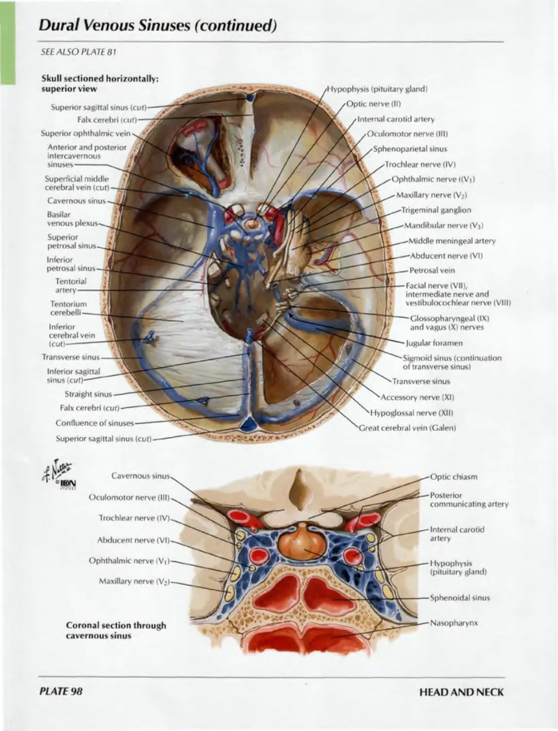
97. Dural Venous Sinuses
98. Dural Venous Sinuses
(continued)
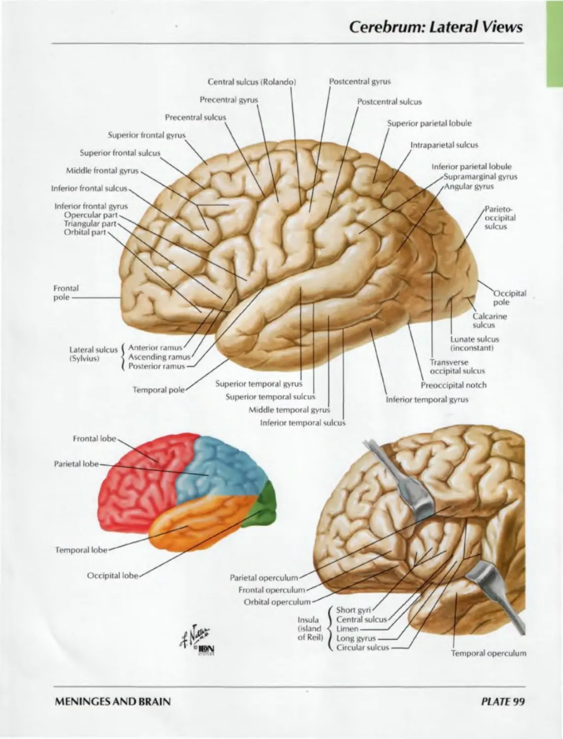
99. Cerebrum: Lateral Views
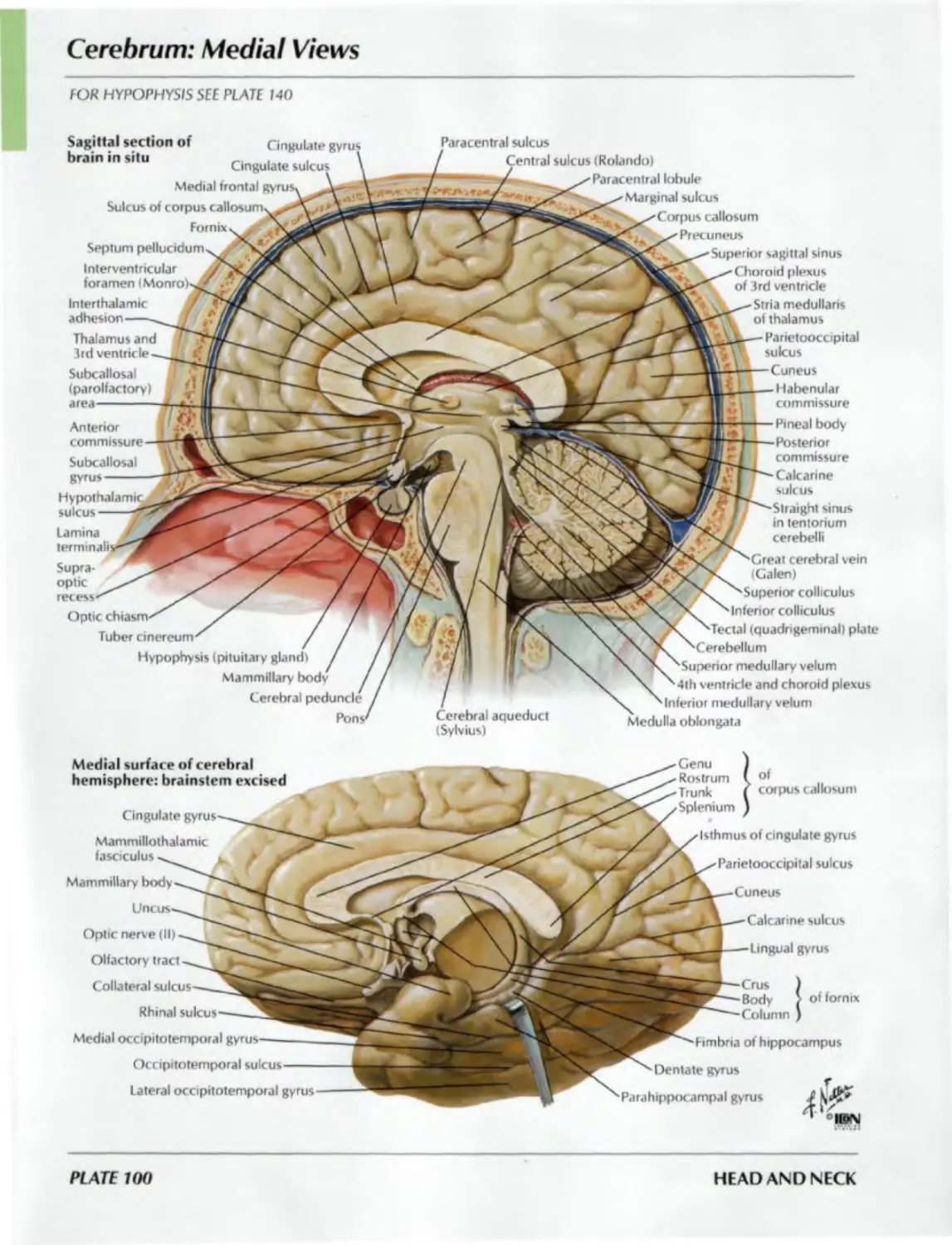
100. Cerebrum: Medial Views
101. Cerebrum: Inferior View
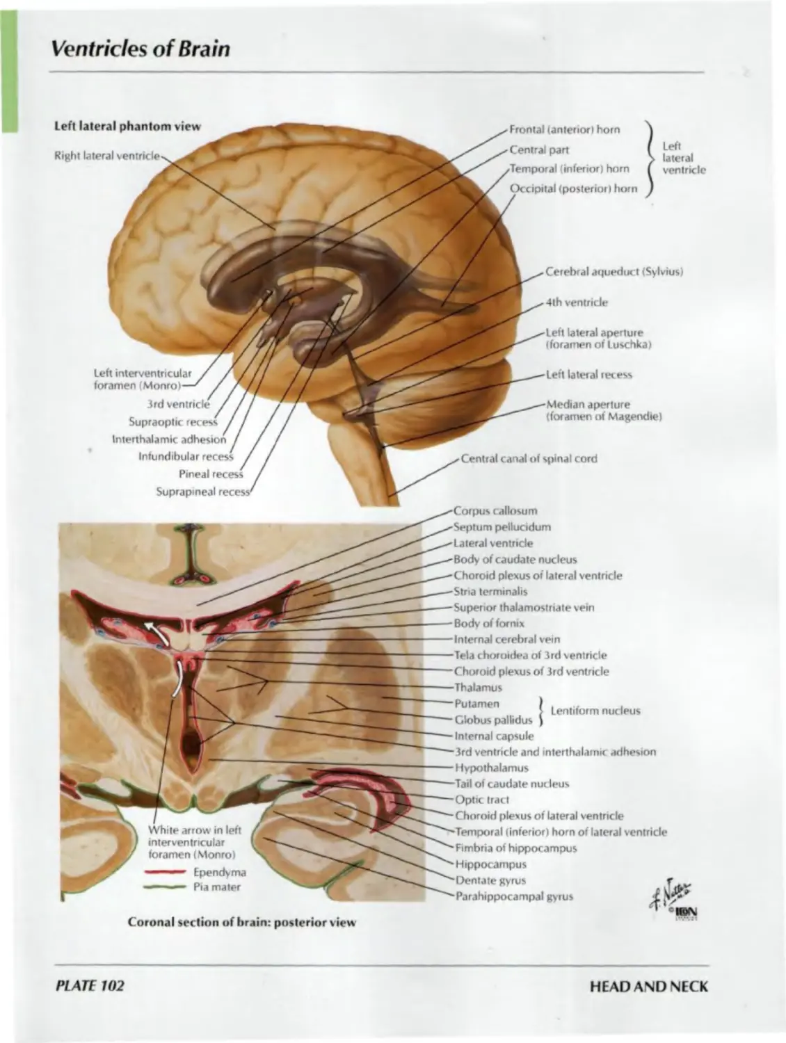
102. Ventricles of Brain
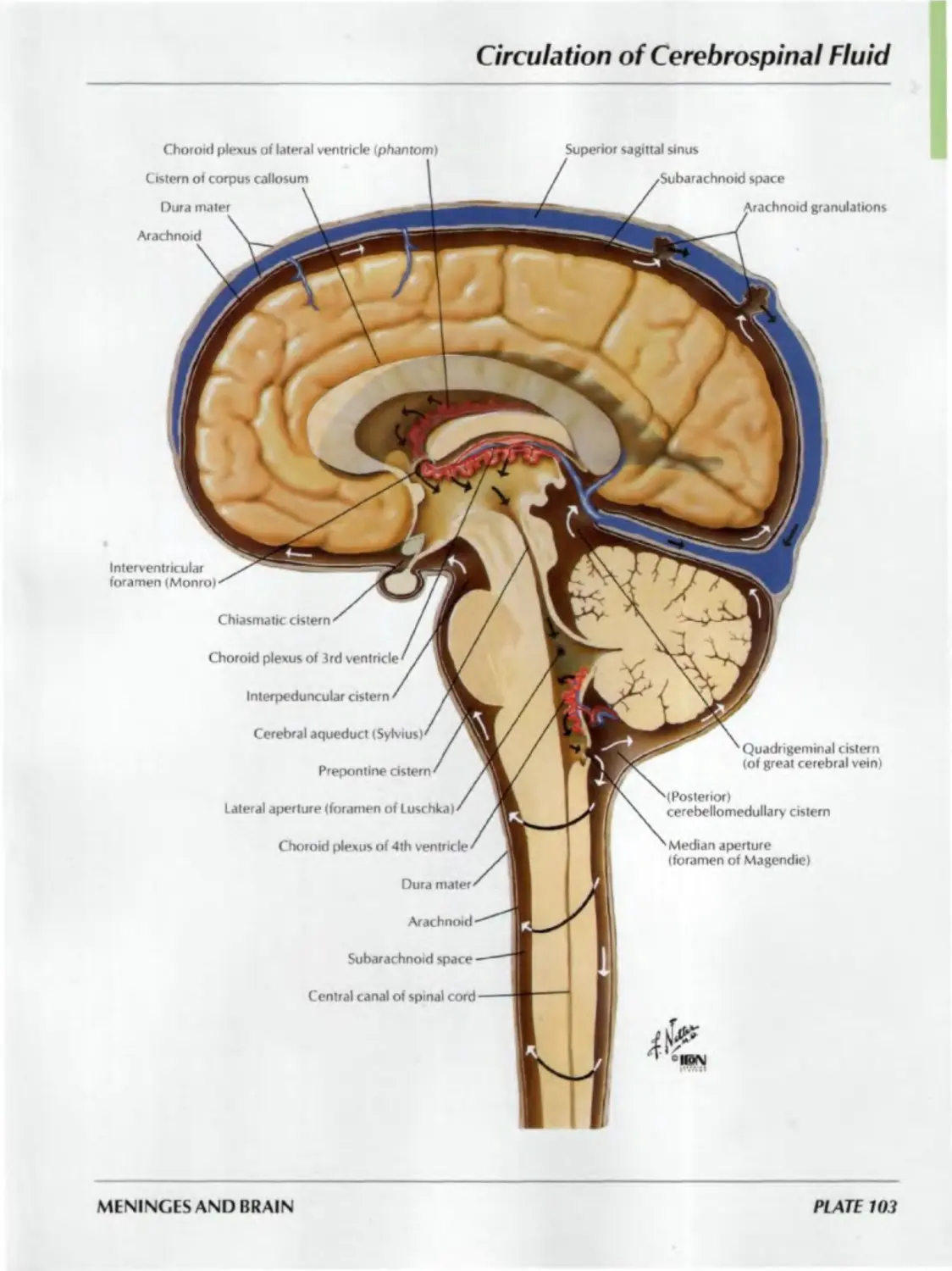
103. Circulation of Cerebrospinal Fluid
104. Basal Nuclei (Ganglia)
105. Thalamus
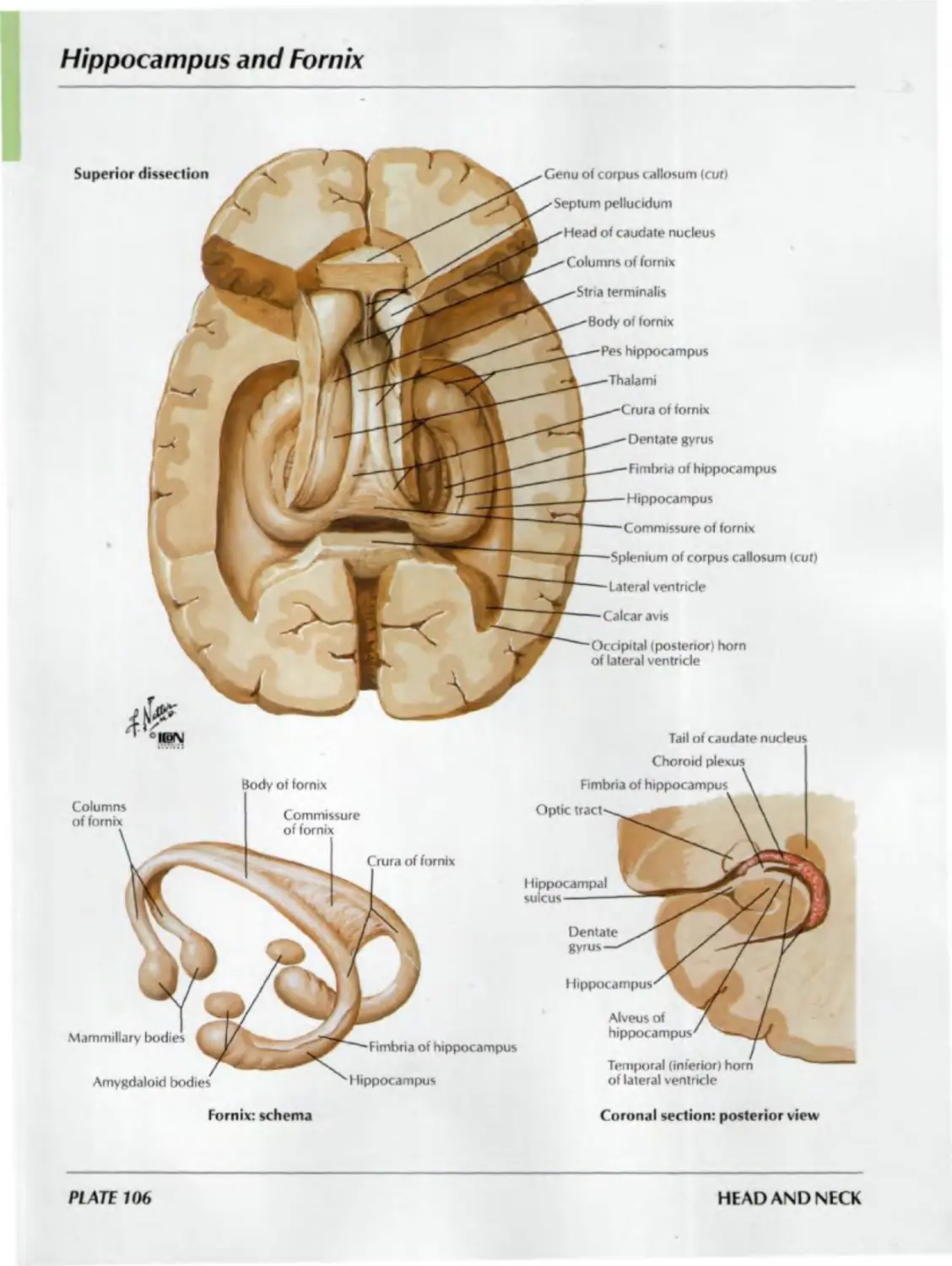
106. Hippocampus and Fornix
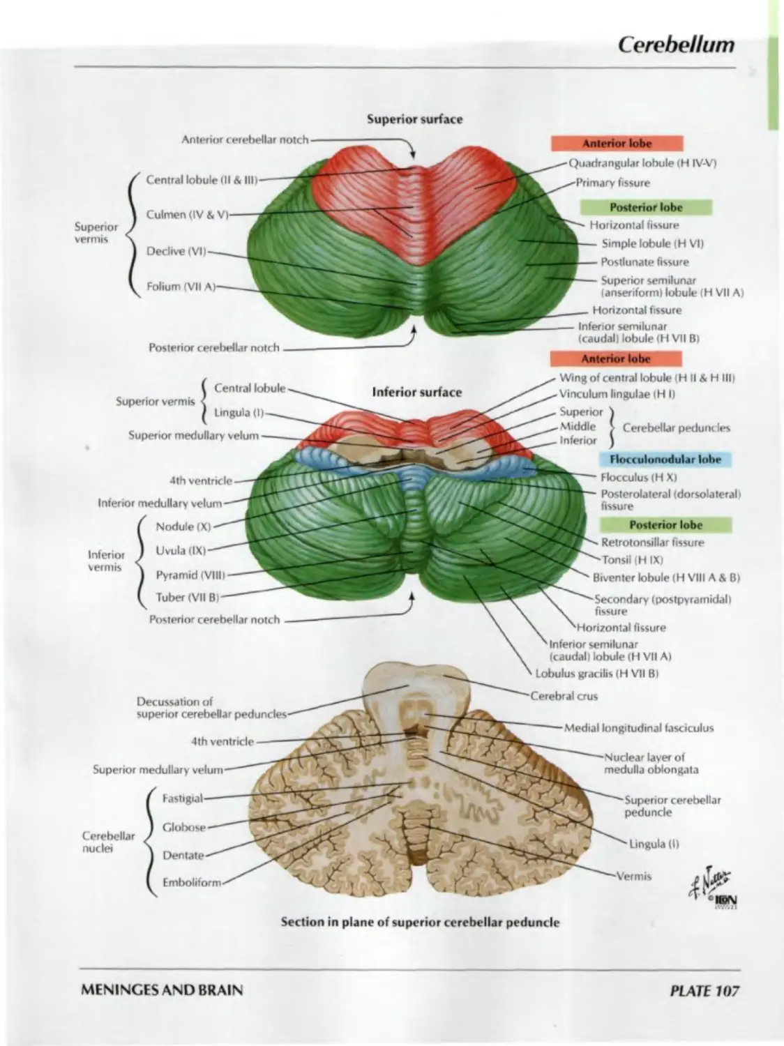
107. Cerebellum
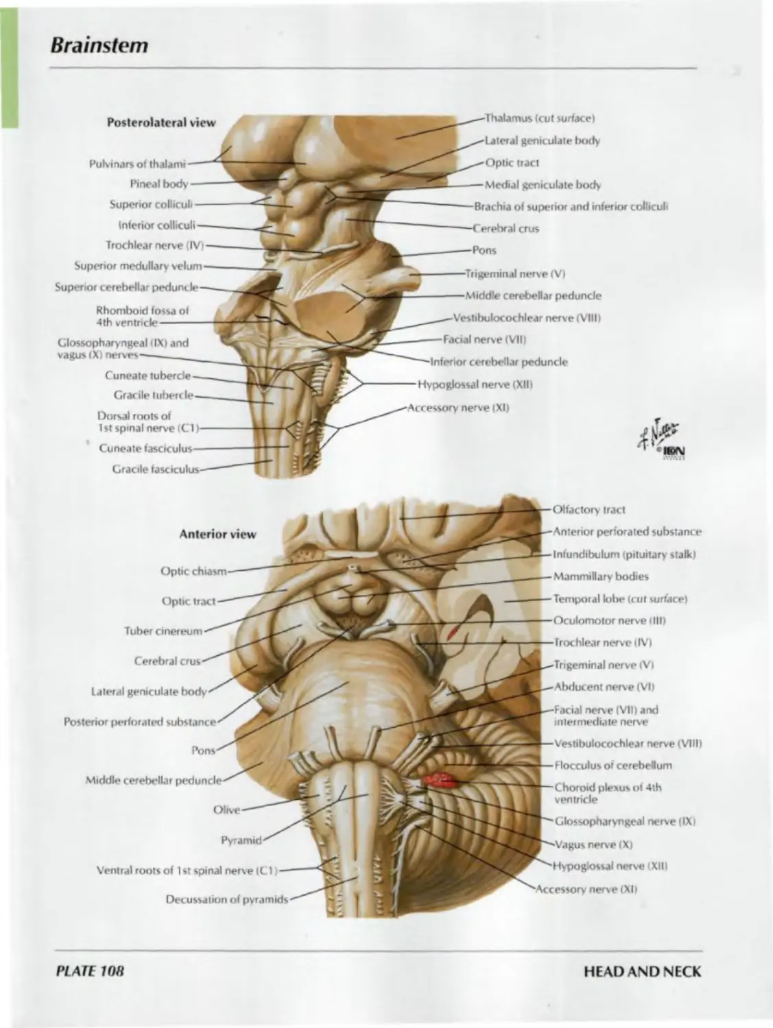
108. Brainstem
109. Fourth Ventricle and Cerebellum
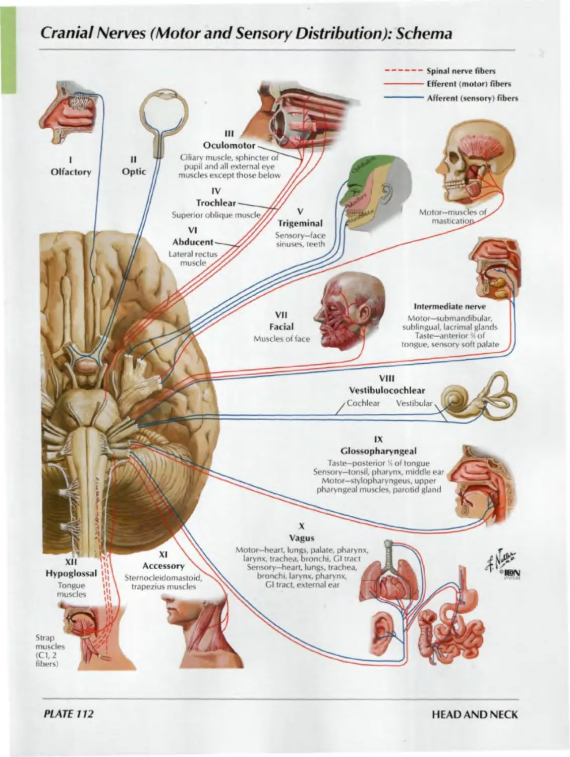
CRANIAL AND CERVICAL NERVES
Plates 110-129
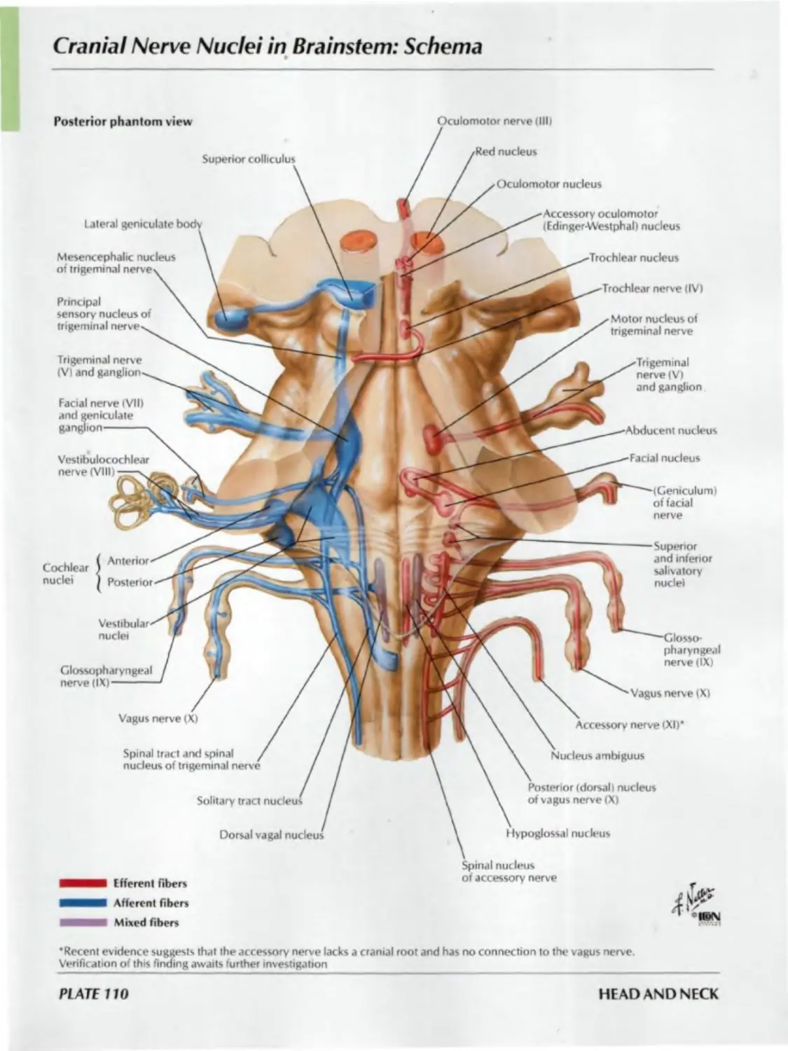
110. Cranial Nerve Nuclei in Brainstem:
Schema
111. Cranial Nerve Nuclei in Brainstem:
Schema (continued)
112. Cranial Nerves (Motor and Sensory
Distribution): Schema
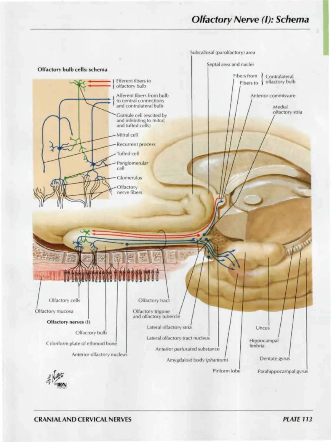
113. Olfactory Nerve (I): Schema
ATLAS OF HUMAN ANATOMY
SECTION I: HEAD AND NECK
114. Optic Nerve (II) (Visual Pathway):
Schema
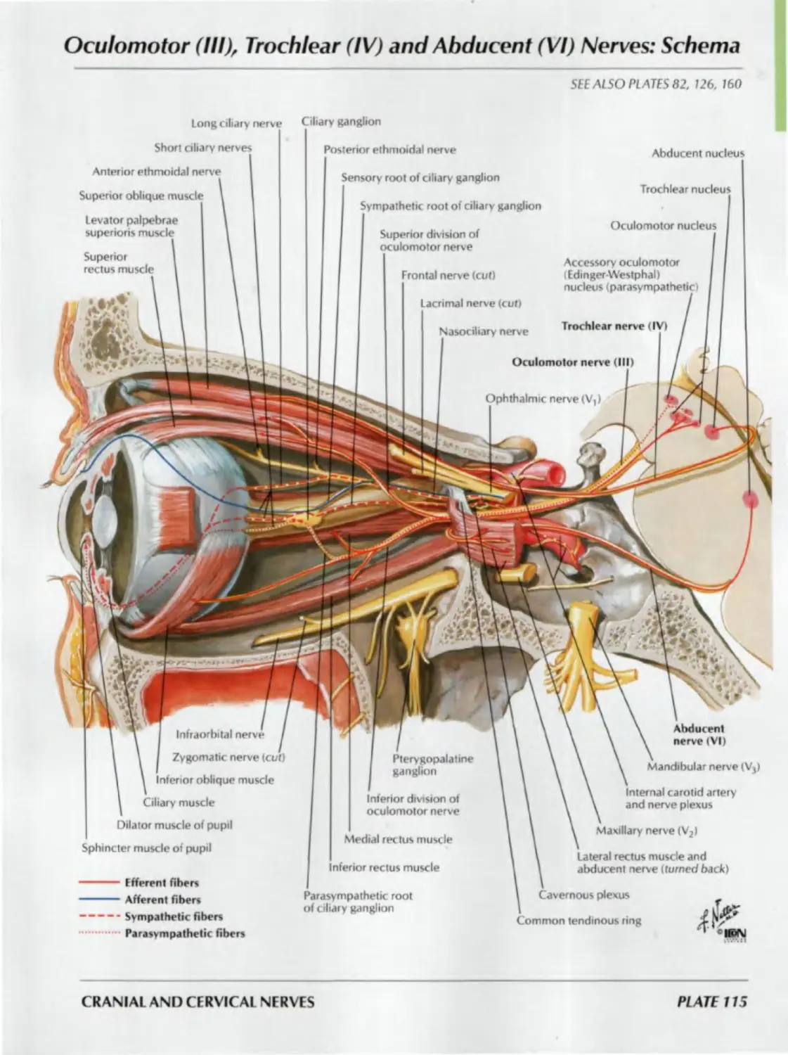
115. Oculomotor (III), Trochlear (IV) and
Abducent (VI) Nerves : Schema
116. Trigeminal Nerve (V): Schema
117. Facial Nerve (VII): Schema
118. Vestibulocochlear Nerve (VIII):
Schema
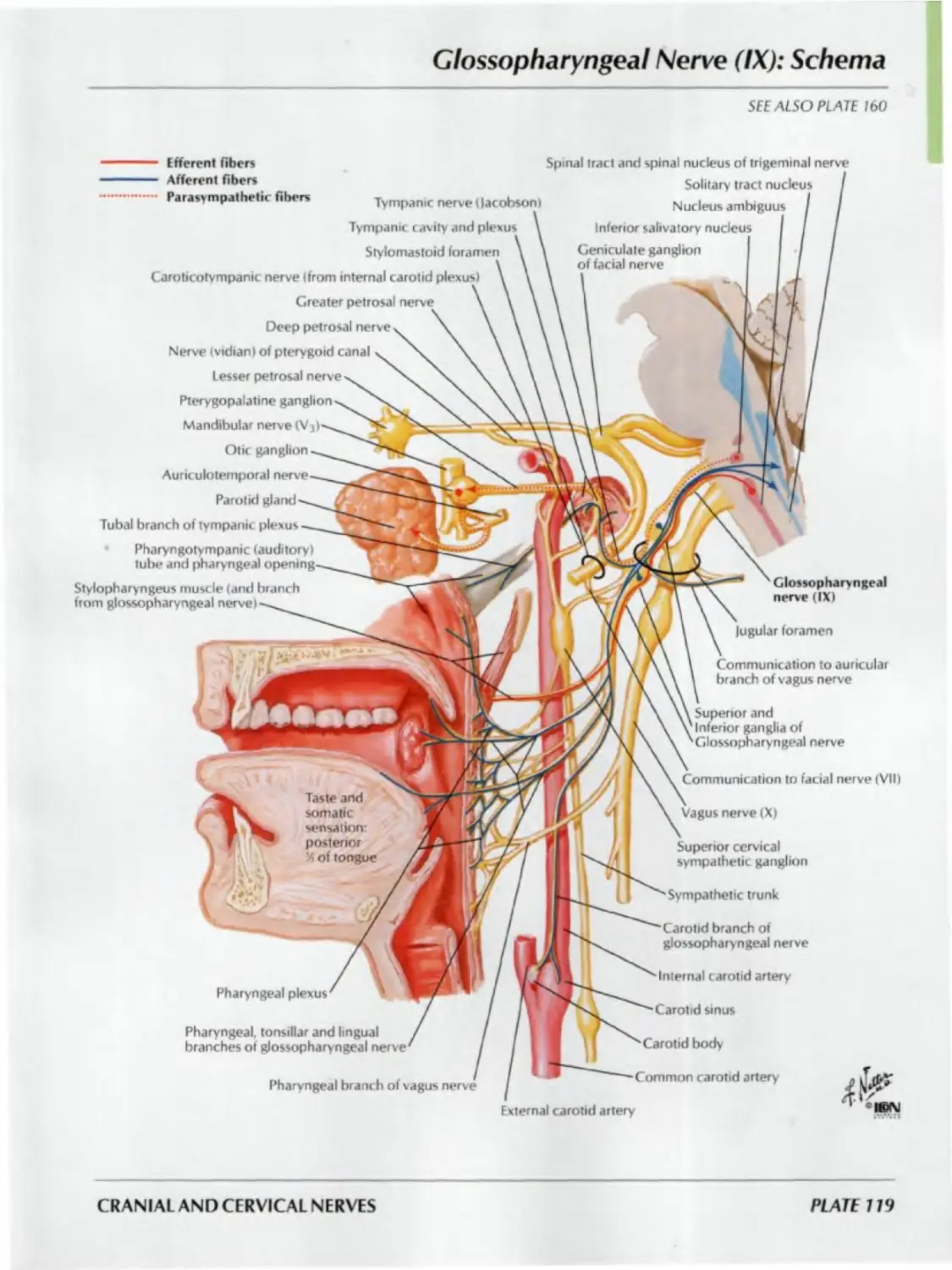
119. Glossopharyngeal Nerve (IX): Schema
120. Vagus Nerve (X): Schema
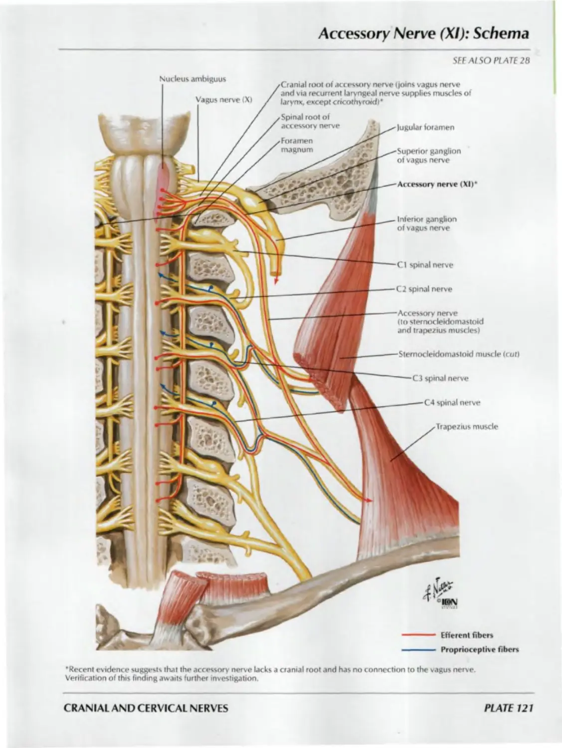
121. Accessory Nerve (XI): Schema
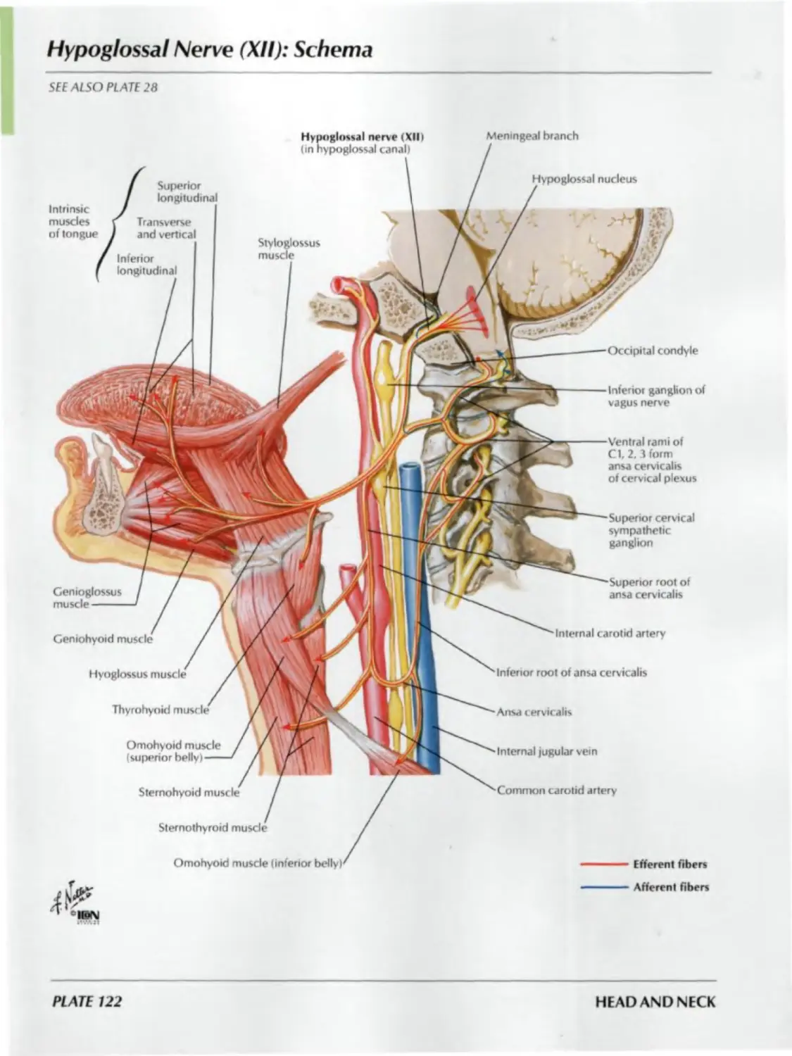
122. Hypoglossal Nerve (XII): Schema
123. Cervical Plexus: Schema
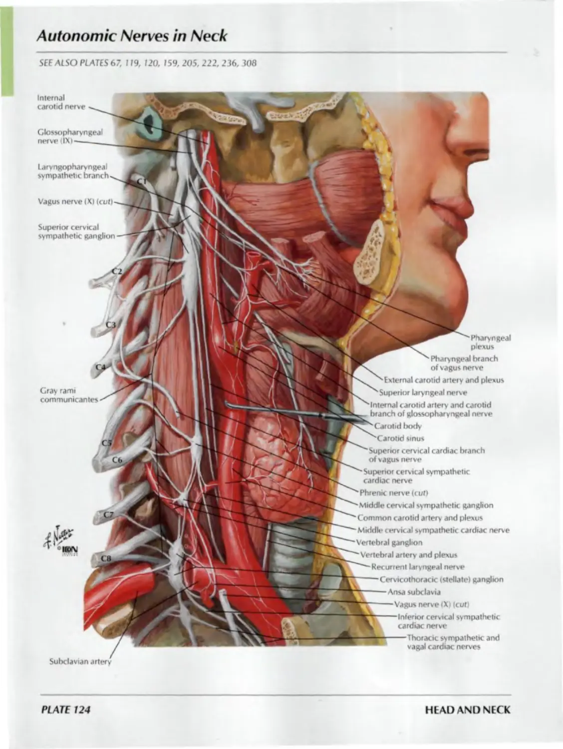
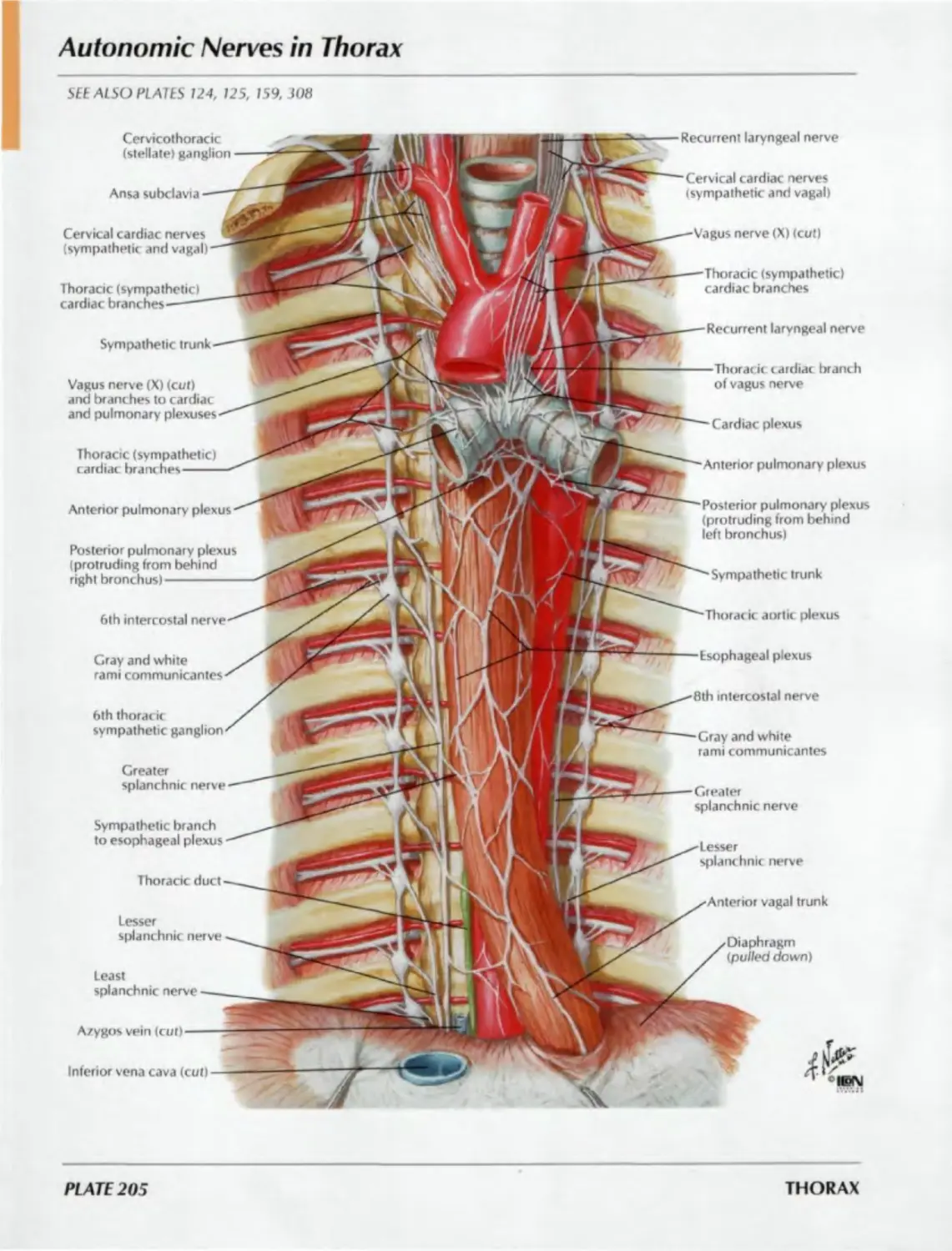
124. Autonomic Nerves in Neck
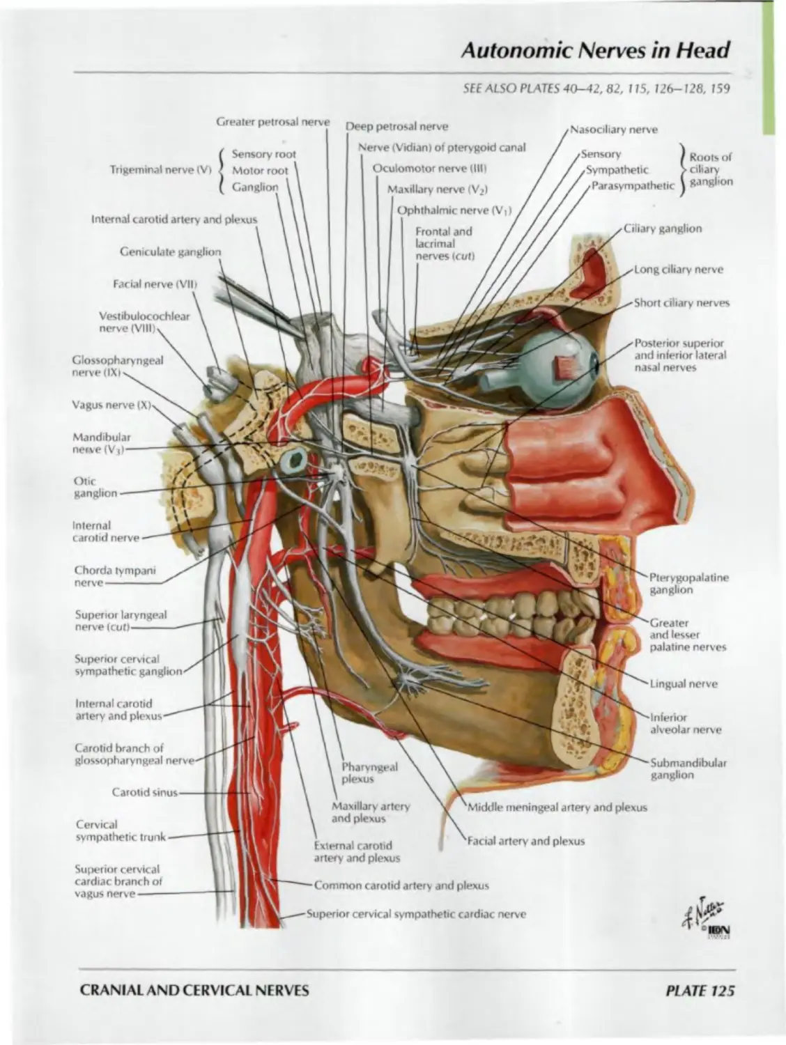
125. Autonomic Nerves in Head
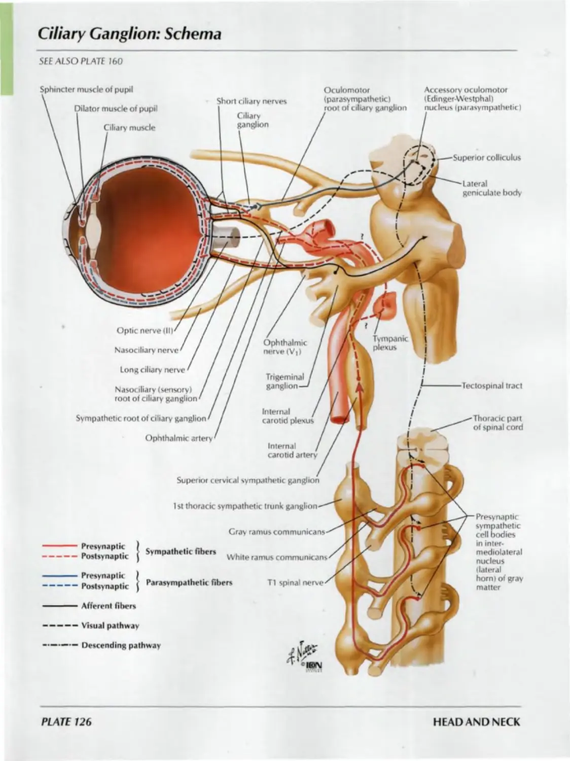
126. Ciliary Ganglion: Schema
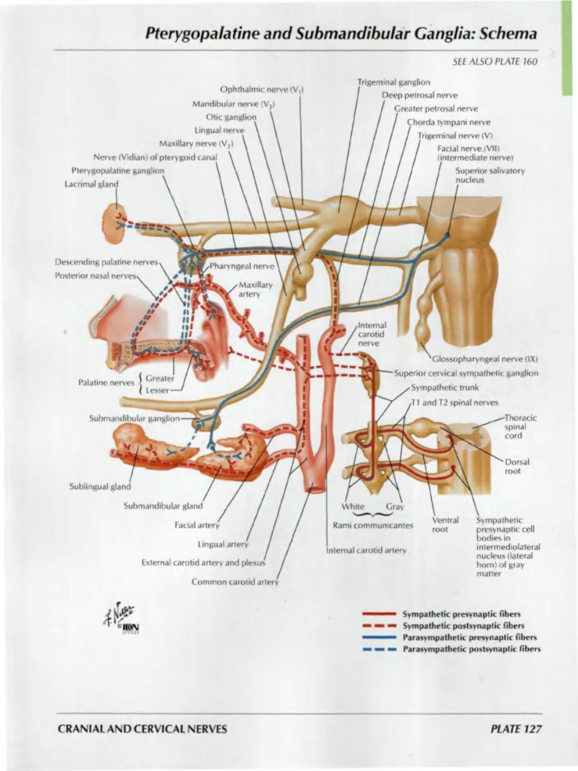
127. Pterygopalatine and Submandibular
Ganglia: Schema
128. Otic Ganglion: Schema
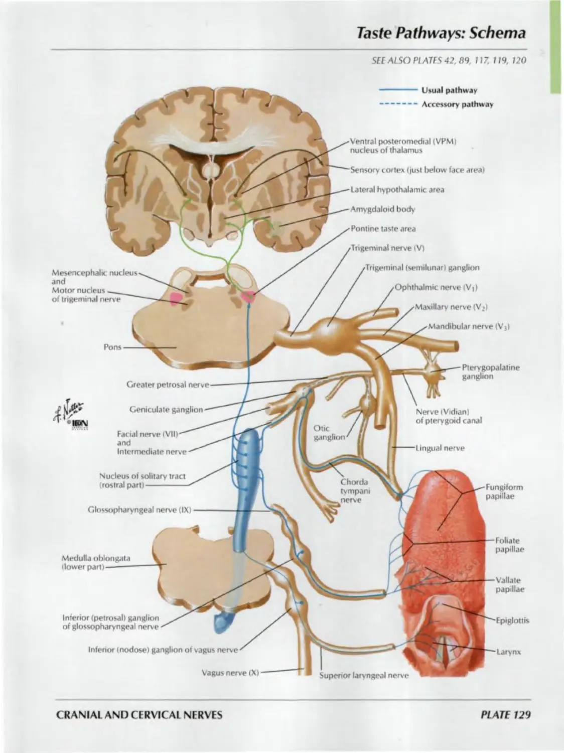
129. Taste Pathways: Schema
CEREBRAL VASCULATURE
Plates 130-141
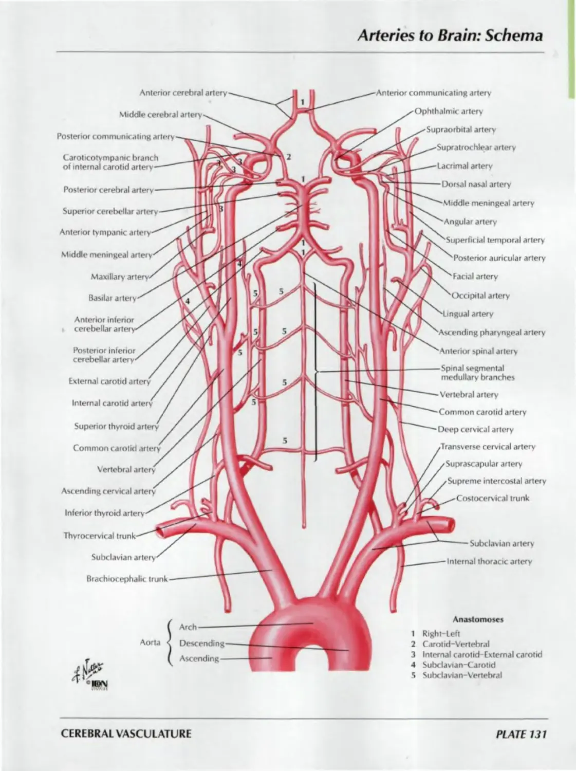
130. Arteries to Brain and Meninges
131. Arteries to Brain: Schema
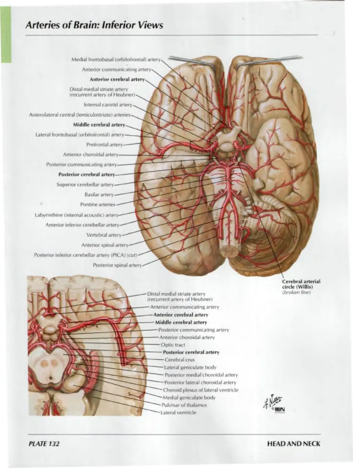
132. Arteries of Brain: Inferior Views
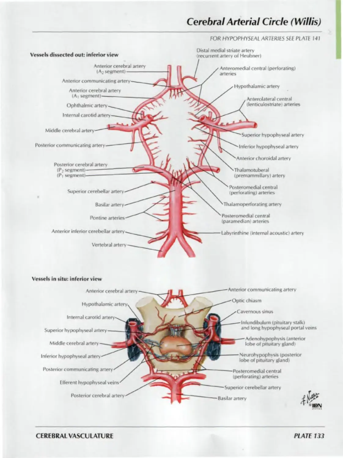
133. Cerebral Arterial Circle (Willis)
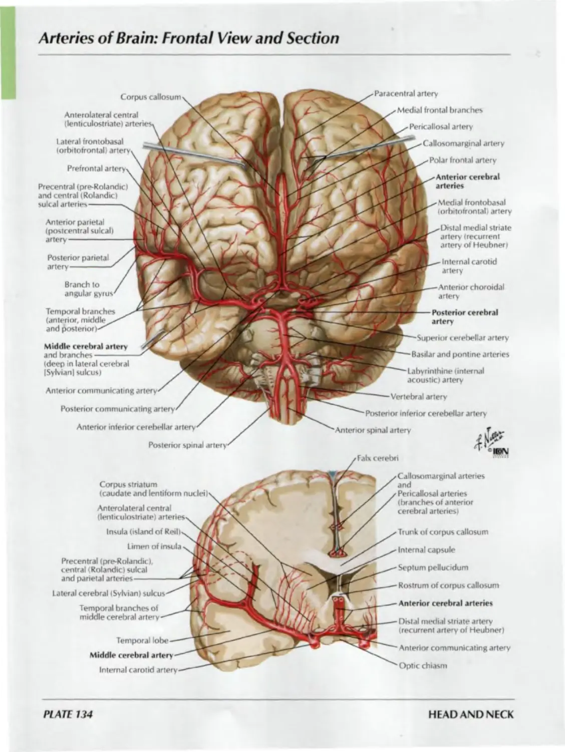
134. Arteries of Brain: Frontal View
and Section
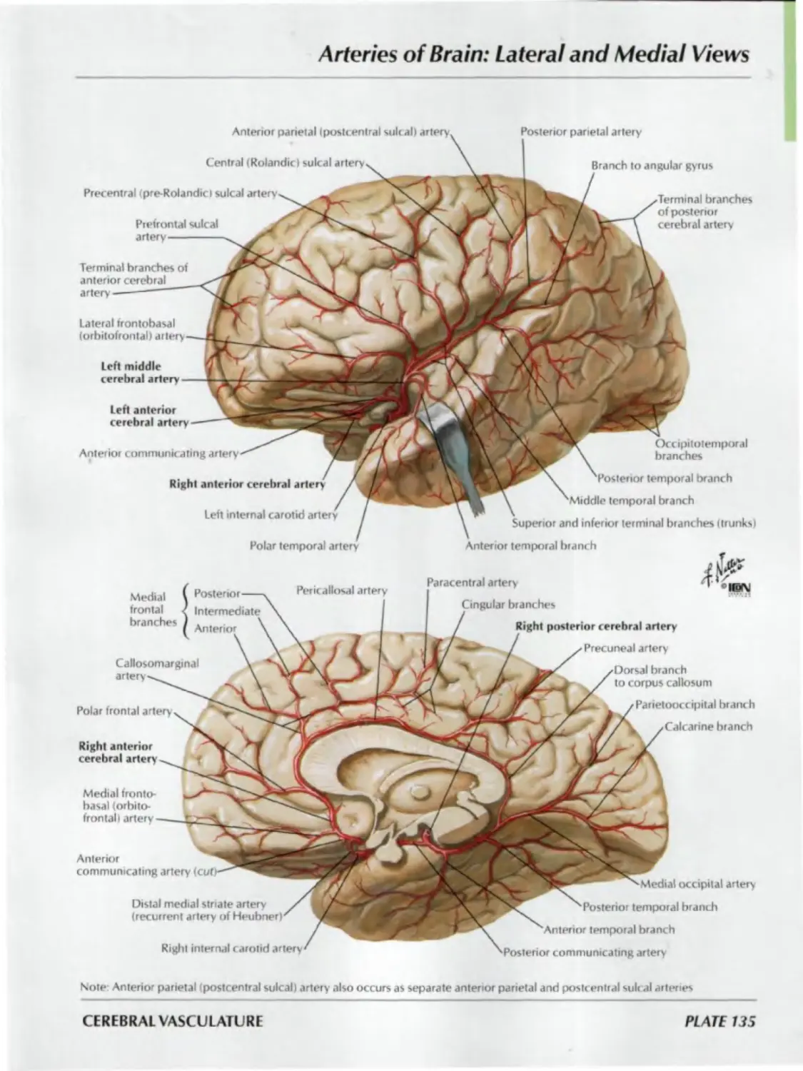
135. Arteries of Brain: Lateral
and Medial Views
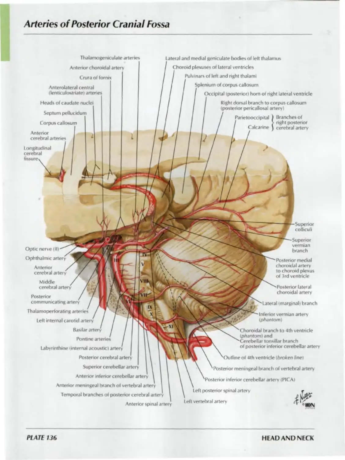
136. Arteries of Posterior Cranial Fossa
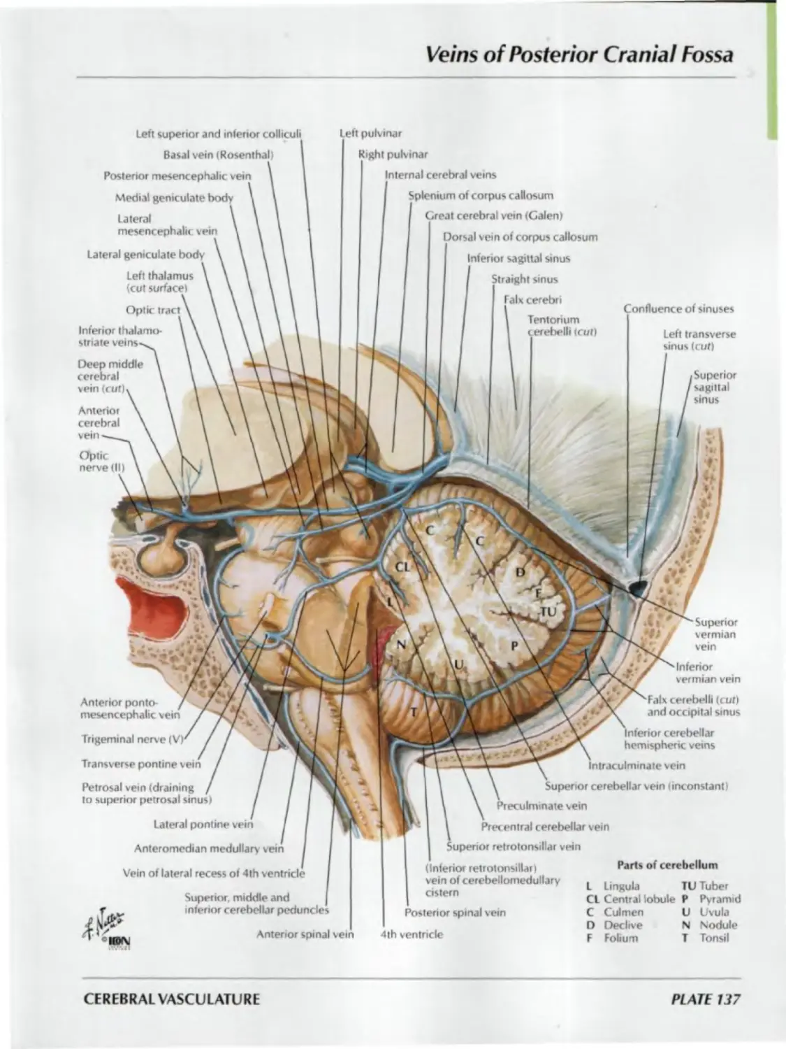
137. Veins of Posterior Cranial Fossa
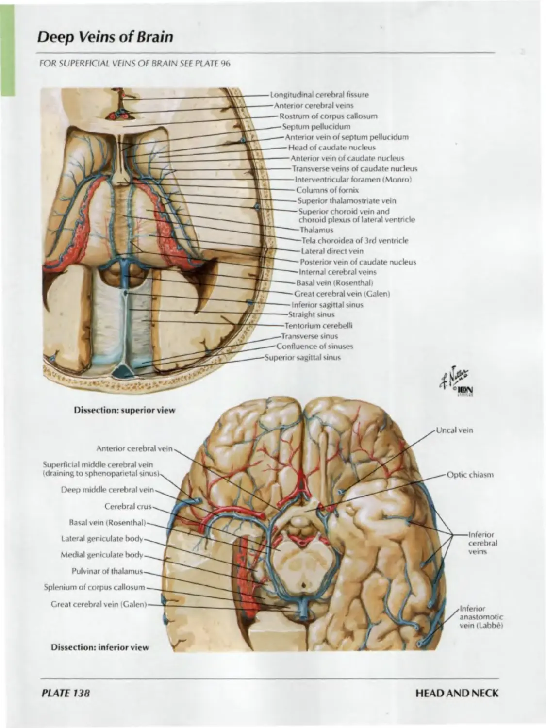
138. Deep Veins of Brain
139. Subependymal Veins of Brain
140. Hypothalamus and Hypophysis
141. Arteries and Veins of Hypothalamus
and Hypophysis
REGIONAL SCANS
Plates 142-144
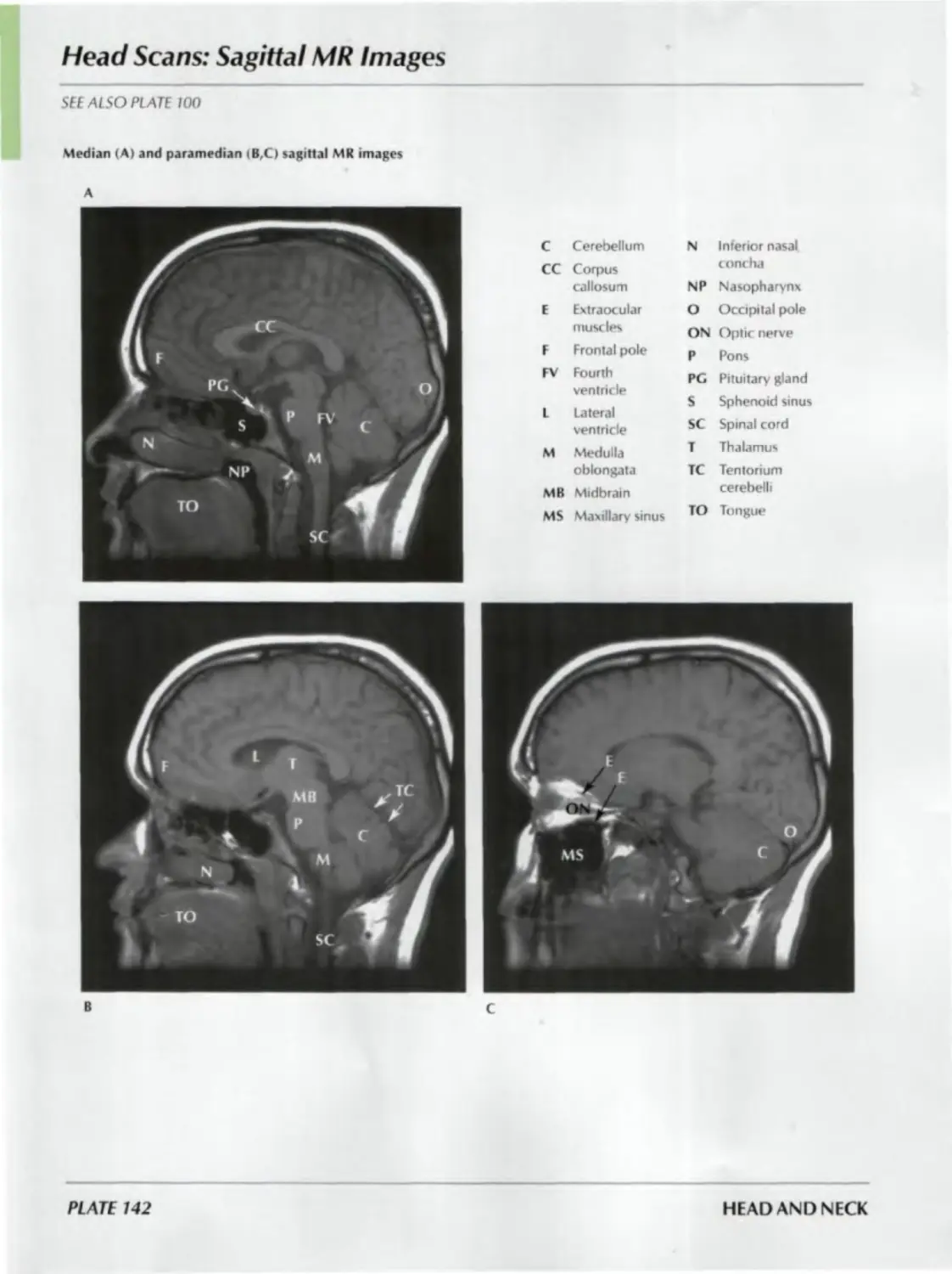
142. Head Scans: Sagittal MR Images
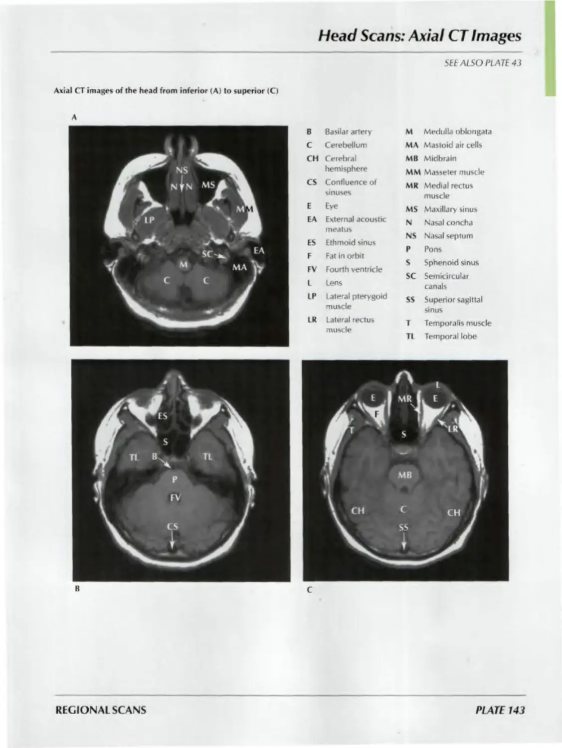
143. Head Scans: Axial CT Images
144. Head Scans: Coronal CT Images
ATLAS OF HUMAN ANATOMY
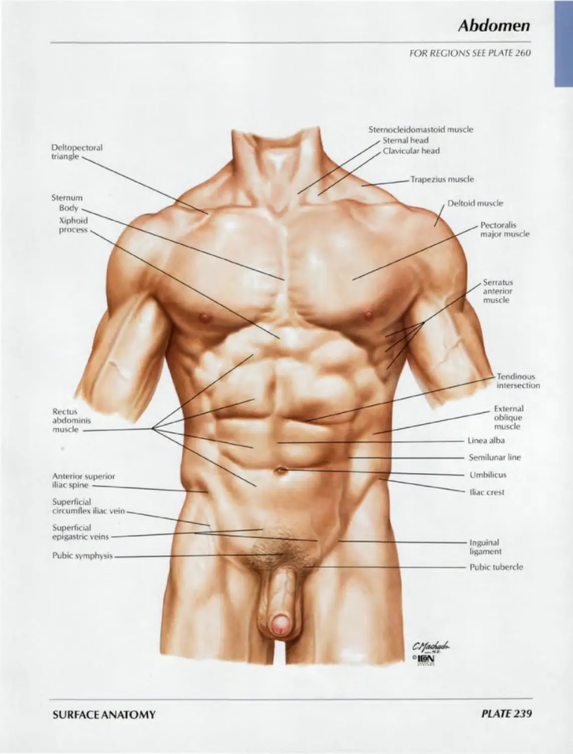
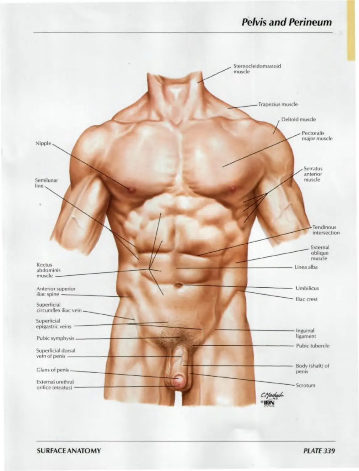
Head and Neck
Infraorbital margin
Zygomatic bone
Helix
Tragus
Antihelix
Antitragus
Ala of nose
Lobule
Commissure of lips
Angle of jaw (mandible!
Submandibular gland
External jugular vein
Inferior belly of omohyoid muscle
Brachial plexus
Trapezius muscle
Clavicle
Frontal
bone
Nasal
bone
Philtrum
Mental
protuberance
Nasolabial
sulcus ——
Tubercle of
superior lip
Thyroid cartilage
Supraorbital
notch
Anterior nares
(nostril) <=—_
lugular notch
Clavicular head of
sternocleidomastoid muscle
sternocleidomastoid muscle
чем’'
SURFACE ANATOMY
PLATE 1
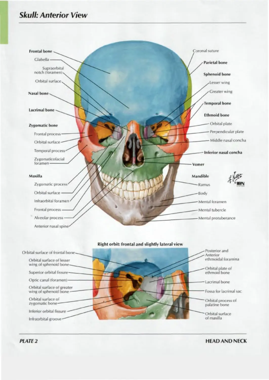
Skull: Anterior View
Frontal bone
Glabella
Supraorbital
notch (foramen)
Orbital surface
Nasal bone
Lacrimal bone
oronal suture
Parietal bone
Inferior nasal concha
Vomer
Mandible
Ramus
Body
Mental foramen
Mental tubercle
Mental protuberance
Zygomatic bone
Frontal
Orbital surface
Temporal process
Zygomaticofacial
foramen
Maxilla
Zygomatic
Orbital surface
Infraorbital foramen
Frontal process
Alveolar process
Anterior nasal
Sphenoid bone
wing
Greater wing
Temporal bone
Ethmoid bone
Orbital plate
Perpendicular plate
Middle nasal concha
Right orbit: frontal and slightly lateral view
Orbital surface of frontal
Superior orbital
Inferior orbital fissure
Orbital surface of lesser
wing of sphenoid
Optic canal (foramen) —
Orbital surface of greater
wing of sphenoid bone
Orbital surface of
zygomatic
Infraorbital groove
Posterior and
Anterior
ethmoidal foramina
Orbital plate of
ethmoid bone
Lacrimal bone
Fossa for lacrimal sac
Orbital process of
palatine bone
Orbital surface
of maxilla
PLATE 2
HEAD AND NECK
Skull: Anteroposterior Radiograph
SEE ALSO PLATE 2
A Angle of mandible
C Crista galli
E Ethmoid sinus
F Frontal sinus
G Greater wing of sphenoid
L Lesser wing of sphenoid
M Maxillary sinus
R Ramus of mandible
BONES AND LIGAMENTS
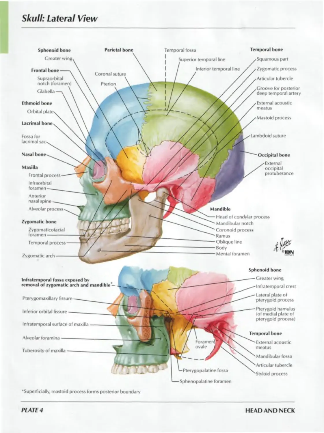
Skull: Lateral View
Parietal bone
Sphenoid bone
Lacrimal bone
Fossa for
lacrimal
Nasal
Temporal fossa
Zygomatic arch
suture
Mandible
Frontal bone
Supraorbital
notch (foramen)
Glabella
Ethmoid bone
Orbital
Maxilla
Frontal process
Infraorbital
foramen
Anterior
nasal spine
Alveolar process
Zygomatic bone
Zygomaticofacial
foramen
Temporal process
Infratemporal fossa exposed by
removal of zygomatic arch and mandible —
Alveolar foramina
Temporal bone
Squamous part
Zygomatic process
Articular tuberc le
Groove for posterior
deep temporal artery
External acoustic
meatus
Mastoid process
Occipital bone
External
occipital
protuberance
Head of condylar process
Mandibular notch
Coronoid process
Ramus
Oblique line
Body
Mental foramen
Tuberosity of maxilla--------
Pterygomaxillary fissure
Inferior orbital fissure
Infratemporal surface of maxilla
— Sphenopalatine foramen
Sphenoid bone
Greater wing
Infratemporal crest
Lateral plate of
pterygoid process
Pterygoid hamulus
(of medial plate of
pterygoid process)
Temporal bone
External acoustic
meatus
Mandibular fossa
Articular tubercle
process
‘Superficially, mastoid process forms posterior boundary
PLATE 4
HEAD AND NECK
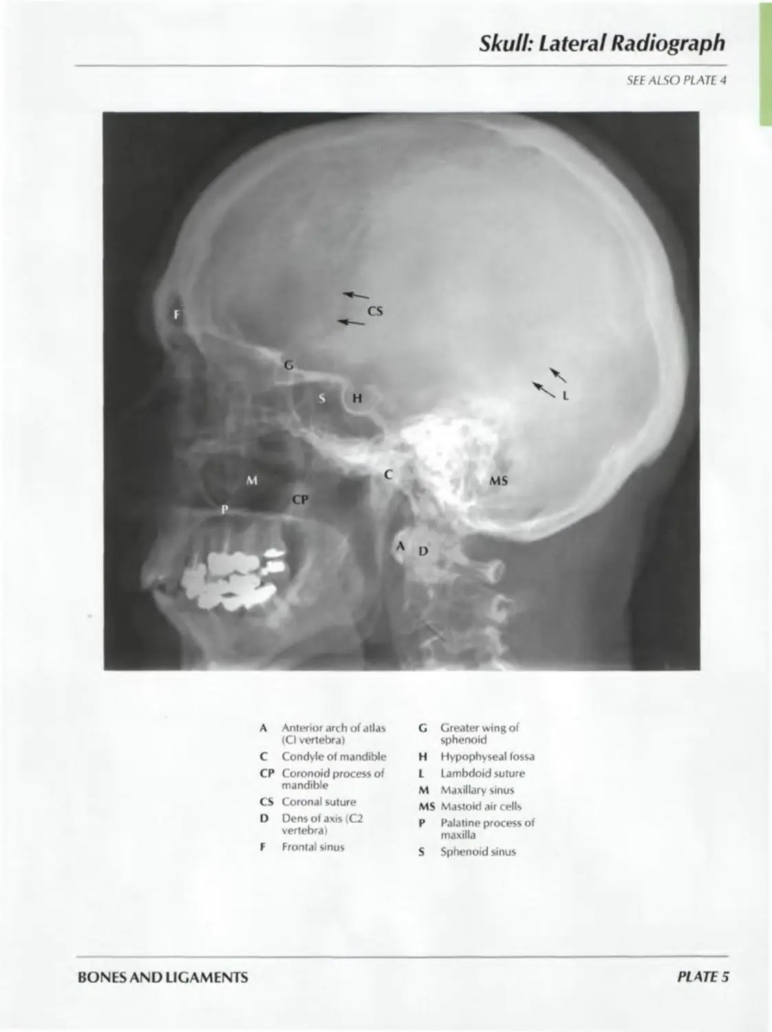
Skull: Lateral Radiograph
SEE ALSO PLATE 4
A Anterior arch of atlas
(Cl vertebra)
C Condyle of mandible
CP Coronoid process of
mandible
CS Coronal suture
I) Dens of axis (C2
vertebra)
F Frontal sinus
C Greater wing of
sphenoid
H Hypophyseal fossa
L Lambdoid suture
M Maxillary sinus
MS Mastoid air cells
P Palatine process of
maxilla
S Sphenoid sinus
BONES AND LIGAMENTS
PLATE 5
Skull: Midsagittal Section
Sphenoid bone
Parietal bone
Temporal bone
Greater wing-----
Lesser wing------
Anterior clinoid
process----------
Optic canal------
Sella turcica--->
Sphenoidal sinus<
Body------------,
Medial and lateral
plates of
pterygoid
process----------
Coronal
suture
Grooves tor
branches of middle
meningeal vessels
Squamous part
Petrous part
j Internal acoustic meatus
< / /Groove for superior
Z.'. / / f^riosal sinus
/ /External opening of
/ vestibular aqueduc t
; / /Groove
/ sigmoid
Frontal bone -
Frontal sinus
Lambdoid suture
Ethmoid bone
Occipital bone
Crista galli----
Cribriform plate
Perpendicular
plate-----------
Groove for
transverse sinus
protuberance
Nasal bone
Jugular foramen
Inferior nasal
concha---------
Groove for inferior
petrosal sinus
Hypoglossal canal
Maxilla
Anterior /
nasal spine'
Nasal surface—'
Incisive canal—'
Palatine process'
Alveolar process-
Foramen magnum
Palatine bone
Vomer
Occipital condyle
Basilar part
Lacrimal bone
Inferior nasal concha
Nasal bone
Frontal bone
Ethmoid bone
Sphenoid bone
Body
Plates of pterygoid process
Pterygoid hamulus
Medial /
Lateral J
Opening of sphenoidal sinus
Sphenopalatine foramen
Cribriform plate
Superior nasal concha
Middle nasal concha
Maxilla
Nasal
Palatine
Alveolar
plate )
. Palatine bone
Horizontal plate )
PL Are 6
HEAD AND NECK
Calvaria
BONES AND LIGAMENTS
PLATE 7
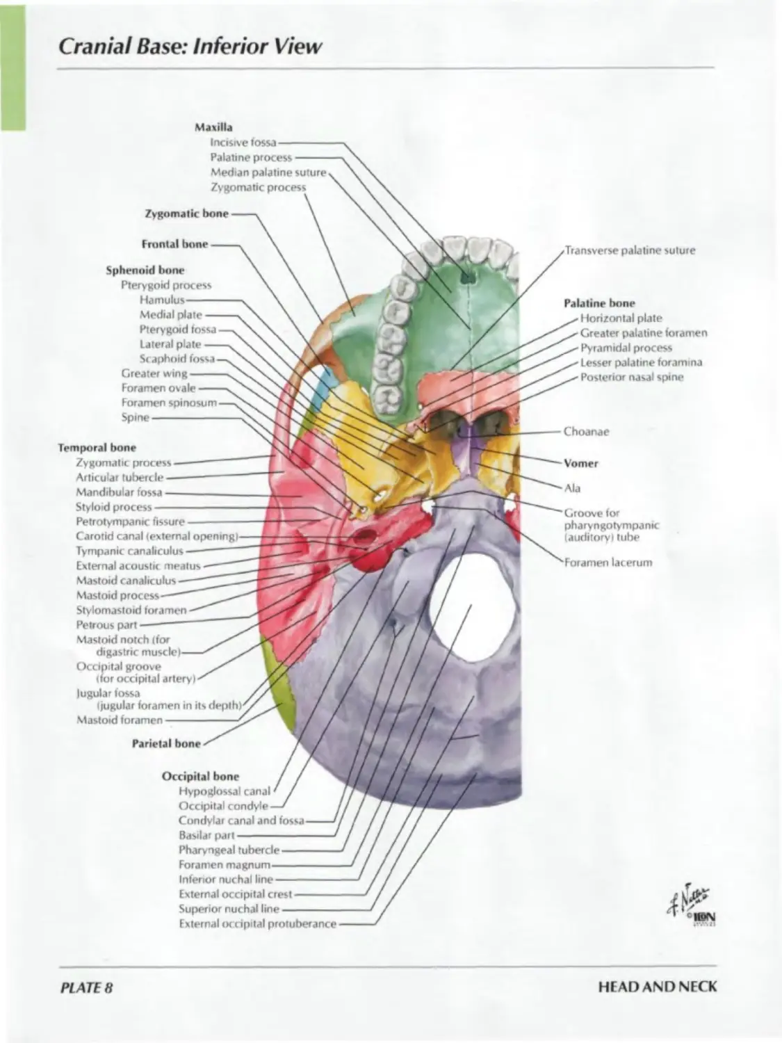
Cranial Base: Inferior View
Maxilla
Incisive fossa —
Palatine process
Median palatine suture
Zygomatic
Zygomatic bone
Frontal bone
Parietal bone
palatinesuture
Choanae
Sphenoid bone
Pterygoid process
Hamulus—
Medial plate
Pterygoid fossa
lateral plate
Scaphoid fossa
Greater wing
Foramen ovale
Foramen spi nosum
Spine-----------
Temporal bone
Zygomatic process
Articular tubercle
Mandibular fossa
Styloid process —
Petrotympanic fissure
Carotid canal (external openings
Tympanic canaliculus
External acoustic meatus
Mastoid canaliculus
Mastoid process
Stylomastoid foramen
Petrous part
Mastoid notch (for
digastric
Occipital groove
(for occipital artery)
Jugular fossa
(jugular foramen in its
Mastoid foramen---------
Occipital bone
Hypoglossal canal
Occipital condyle
Condylar canal and
Basilar part
Pharyngeal tubercle
Foramen
Inferior nuchal line
External occipital crest
Superior nuchal line
External occipital protuberance
Palatine bone
Horizontal plate
Greater palatine foramen
Pyramidal process
Lesser palatine foramina
Posterior nasal spine
Vomer
Ala
Groove for
pharyngotympanic
(auditory) tube
Foramen lacerum
PL ATE 8
HEAD AND NECK
Bones of Cranial Base: Superior View
Frontal bone
Groove for superior sagittal sinus--
Frontal crest-----------------------
Groove for anterior meningeal vessels
Foramen cecum-----------------------
Superior surface of orbital part----
Ethmoid bone
Crista galli-------------------------
Cribriform plate---------------------
Sphenoid bone
Lesser wing--------------------------
Anterior clinoid process----------
Greater wing-------------------------
Groove for middle meningeal
vessels (frontal branches)--------
Body
jugurn----------------------------
Prechiasmatic groove--------------
/ Tuberculum sellae-------
Sella ) Hypophyseal fossa---------
turcica j Dorsum sellae-----------
’ Posterior clinoid process
Carotid groove (for int. carotid a )-
Clivus-------------------------------
Middle
cranial
fossa
Anterior
cranial
fossa
Temporal bone
Squamous part---------------------
Petrous part
Groove for lesser petrosal nerve —
Groove for greater petrosal nerve •
Arcuate eminence----------------
Trigeminal impression-----------
Groove for superior petrosal sinus
Groove for sigmoid sinus--------
>
I
Posterior
cranial
fossa
Parietal bone
Groove for middle meningeal
vessels (parietal branches) —
Mastoid angle---------------
Occipital bone
Clivus--------------------------
Groove for inferior petrosal sinus
Basilar part-----------------
Groove for posterior meningeal
Condyle-------------------
Groove for transverse sinus
Groove for occipital sinus
Internal occipital crest
Internal occipital protuberance
Groove for superior sagittal sinus
BONES AND LIGAMENTS
PLATE 9
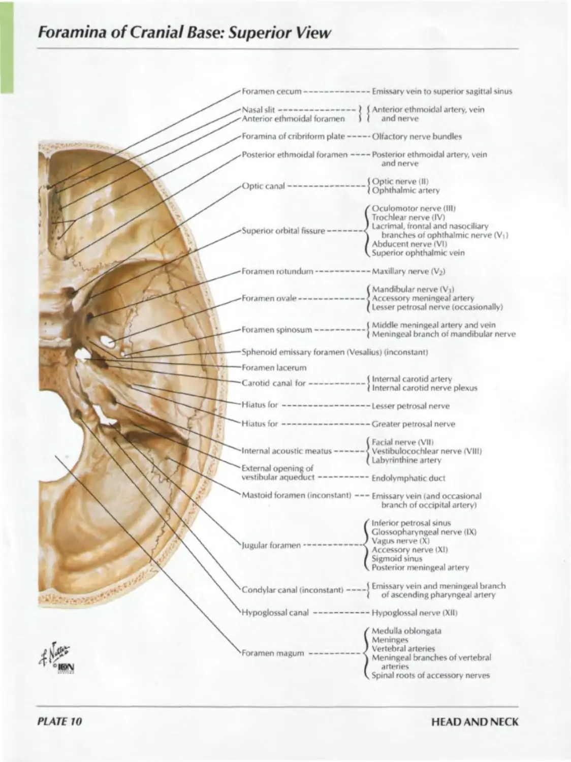
Foramina of Cranial Base: Superior View
Foramen cecum--------------------Emissary vein to superior sagittal sinus
Nasal slit-------------------| I Anterior ethmoidal artery, vein
Anterior ethmoidal foramen > < and nerve
Foramina of cribriform plate-----Olfactory nerve bundles
Posterior ethmoidal foramen------Posterior ethmoidal artery, vein
and nerve
Optic canal
Superior orbital fissure
Foramen spinosum
Foramen rotundum-----------
Foramen ovale —------------
Optic nerve III)
Ophthalmic artery
Oculomotor nerve (III)
Trochlear nerve (IV)
Lacrimal, frontal and nasociliary
branches of ophthalmic nerve (Vi)
Abducent nerve IVI)
Superior ophthalmic vein
Maxillary nerve (V?)
Mandibular nerve (Vj)
Accessory meningeal artery
Lesser petrosal nerve (occasionally)
(Middle meningeal artery and vein
Meningeal branch of mandibular nerve
Sphenoid emissary' foramen (Vesalius) (inconstant)
Foramen lacerum
Carotid canal for---------------1
Hiatus for —---------------------
Hiatus for -—--------------------
Internal acoustic meatus--------
External opening of
vestibular aqueduct--------------
Mastoid foramen (inconstant)-----
lugular foramen----------------
Condylar canal (inconstant)-----|
Hypoglossal canal----------------
Foramen magum------------------<
Internal carotid artery
Internal carotid nerve plexus
Lesser petrosal nerve
Greater petrosal nerve
Facial nerve (VIII
Vestibulocochlear nerve (VIII)
Labyrinthine artery
Endolymphatic duct
Emissary vein (and occasional
branch of occipital artery)
Inferior petrosal sinus
Glossopharyngeal nerve (IX)
Vagus nerve (X)
Accessory nerve tXI)
Sigmoid sinus
Posterior meningeal artery
Emissary vein and meningeal branch
of ascending pharyngeal artery
Hypoglossal nerve (XII)
Medulla oblongata
Meninges
Vertebral arteries
Meningeal branches of vertebral
arteries
Spinal roots of accessory nerves
PLATE 10
HEAD AND NECK
Skull of Newborn
Ethmoid bone
Anterior ethmoidal
foramen-----
Orbital plate
Lateral view
Anterior fontanelle
Parietal bone
Lacrimal bone
Nasal bone
Maxilla------------------
Infraorbital foramen
Palatine bone
Pyramidal process
Sphenoidal fontanelle
Frontal bone
Squamous part —
Supraorbital notch
plate of pterygoid
process
Superior view
(eminence)
suture
Posterior fontanelle
Lambdoid suture
Occipital bone
Mastoid fontanelle
Zygomatic bone
Zygomaticofacial foramen
Temporal bone
part
Petrosquamous fissure
Petrous part
(mastoid process absent)
Tympanic part (bony external
acoustic meatus absent)
(vestibular) window
Round (cochlear) window
Styloid process
Mandibular fossa
Zygomatic process
Frontal bone
Anterior fontanelle
Coronal suture
Parietal bone
Sagittal suture
Posterior fontanelle
Occipital bone
Lambdoid suture
BONES AND LIGAMENTS
PLATE 11
Bony Framework of Head and Neck
Temporal bone
Sphenoid bonex
Temporal fossa x
Zygomatic archx
Condylar process of mandible >_
Mandibular notchx
Coronoid process of mandible^
Lateral pterygoid plalex.
(broken line)
Hamulus of medial pterygoid plate x. у
(broken line) fi
Pterygomandibular raphex.
(broken linei t
! Ramus
Angle -
Body -
Hyoid bone
Spine of sphenoid bone
Foramen
Foramen
Body--------
Lesser horn—
Greater horn
Mastoid
process
External
acoustic meatus
Allas (Cl)
Styloid process
Axis(C2)
Stylomandibular
ligament
C3 vertebra
Alveolar process of maxilla
Lateral plate )
Hamulus )
Epiglottis '
Thyroid cartilage*''
Cricoid cartilage'
Trachea
Sphenopalatine foramen
Pterygopalatine fossa
(posterior nares)
Pyramidal process of palatine bone
^--C7 vertebra
T1 vertebra
1 si rib
Pl ATE 12
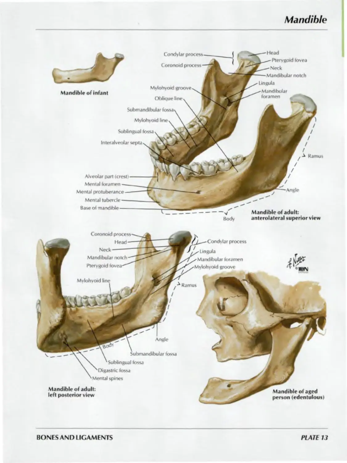
HEAD AND NECK
Mandible
Mandible of aged
person (edentulous)
Angle
\ Submandibular fossa
' Sublingual fossa
'Digastric fossa
Mental spines
Mandible of adult:
left posterior view
Coronoid process
Head —
Neck--------
Mandibular notch —
Pterygoid fovea—
Mylohyoid line
Ramus
Condylar process
I ,
• X Lingula
XMandibular foramen
/Mylohyoid groove
BONES AND LIGAMENTS
PLATE 13
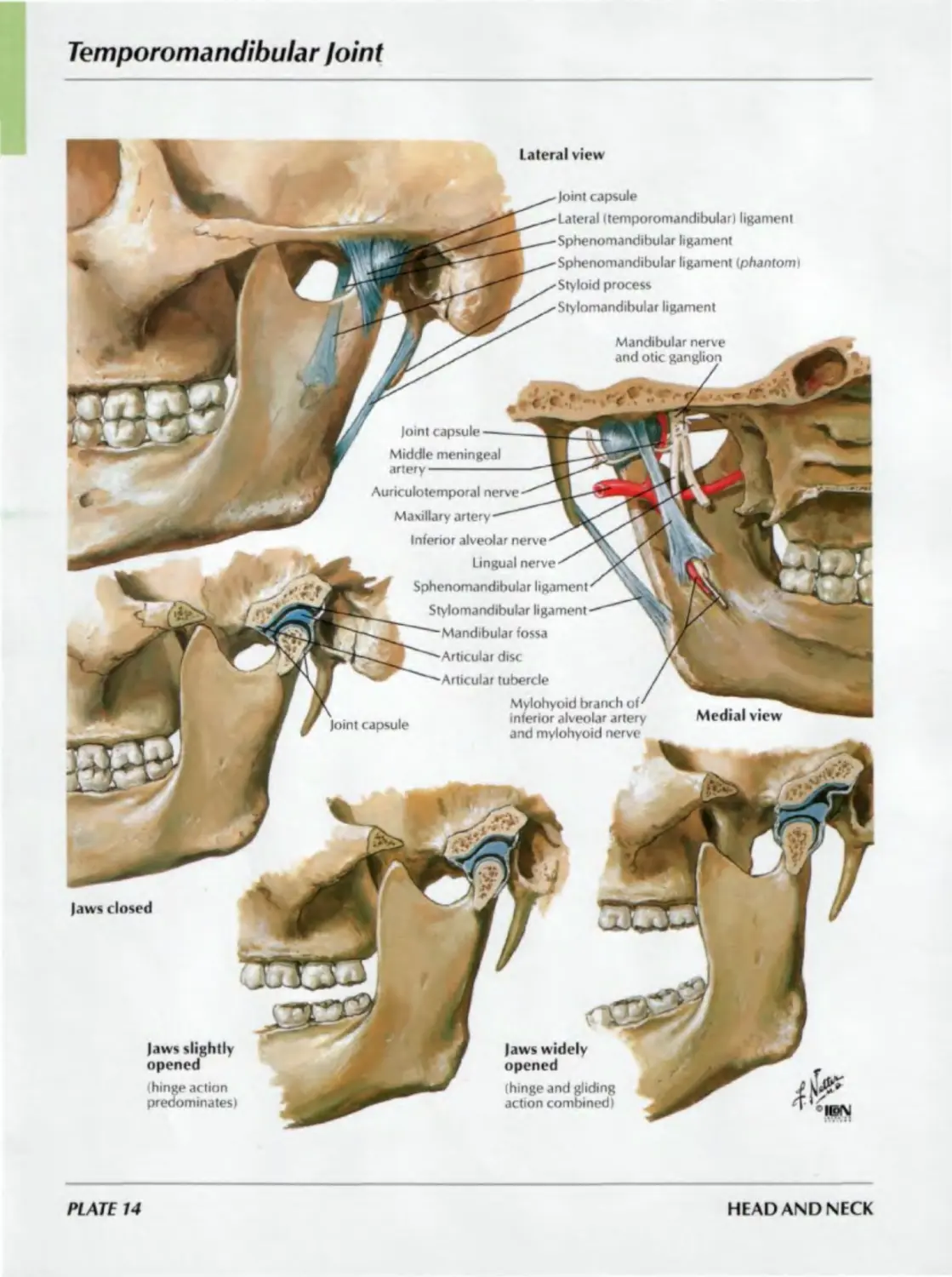
Temporomandibular /oint
Lateral view
Medial view
Joint capsule
laws closed
Mandibular nerve
and otic ganglion
laws slightly
opened
(hinge action
predominates)
Joint capsule
Lateral (temporomandibular) ligament
Sphenomandibular ligament
Sphenomandibular ligament (phantomi
Styloid process
Stylomandibular ligament
laws widely
opened
Ihingeand gliding
action combined)
Joint capsule----—
Middle meningeal
ar:eг у-------------
Auriculotemporal nerve
Maxillary artery—"
Inferior alveolar nerveyK jJ
Ungual nerve ' JmC *
Sphenomandibular ligamenr \|k
Stylomandibular ligament-"" 1
—Mandibular fossa
Articular disc
'"Articular tubercle
Mylohyoid branch or
inferior alveolar artery
and mylohyoid nerve
PLATE 14
HEAD AND NECK
Cervical Vertebrae: Atlas and Axis
SEE ALSO PLATES 12, 14h
Transverse
process
Anterior tubercle
Transverse
foramen
Superior articular
of lateral mass for
occipital condyle
Anterior
Vertebral
foramen
arch
Groove for vertebral artery
mass
Axis (C2): anterior view
Atlas (Cl): superior view
Posterior
Transverse
process
foramen
Articular facet
for dens
Anterior tubercle
Atlas (Cl): inferior view
Transverse
Inferior articular
surface of lateral
mass for axis
Axis (C2): posterosuperior view
Superior
articular
surface
for occipital
condyle
Posterior
articular facet
(for transverse
ligament
of atlas)
Dens
Axis(C2)
C3
Atlas (CD
Upper cervical
vertebrae, assembled: C
posterosuperior view
Radiograph of atlantoaxial joint
A Lateral masses of atlas (Cl vertebra)
D Dens of axis lC2 vertebra)
BONES AND LIGAMENTS
PLATE 15
Cervical Vertebrae (continued)
SEE ALSO PLATES 12. 146
Dens
Spinous
processes
C7 !
Costal facets
(for 1st rib)
Intervertebral joint
(symphysis)
(disc removed)
Zygapophyseal
joints
t Cervical curvature
Intervertebral
foramina for
spinal nerves
Articular
pillar
formed by
articular
processes
and
interarticular
parts
Cervical vertebrae: lateral radiograph
2nd cervical Io 1 st thoracic vertebrae:
right lateral view
A Anterior arch of atlas
P Posterior arch of atlas
S Spinous process
T Transverse process
V Vertebra prominens
(spinous process of C7)
Z Zygapophyseal joint
Bodies of 2—7 cervical vertebrae
are numbered
PLATE 16
HEAD AND NECK
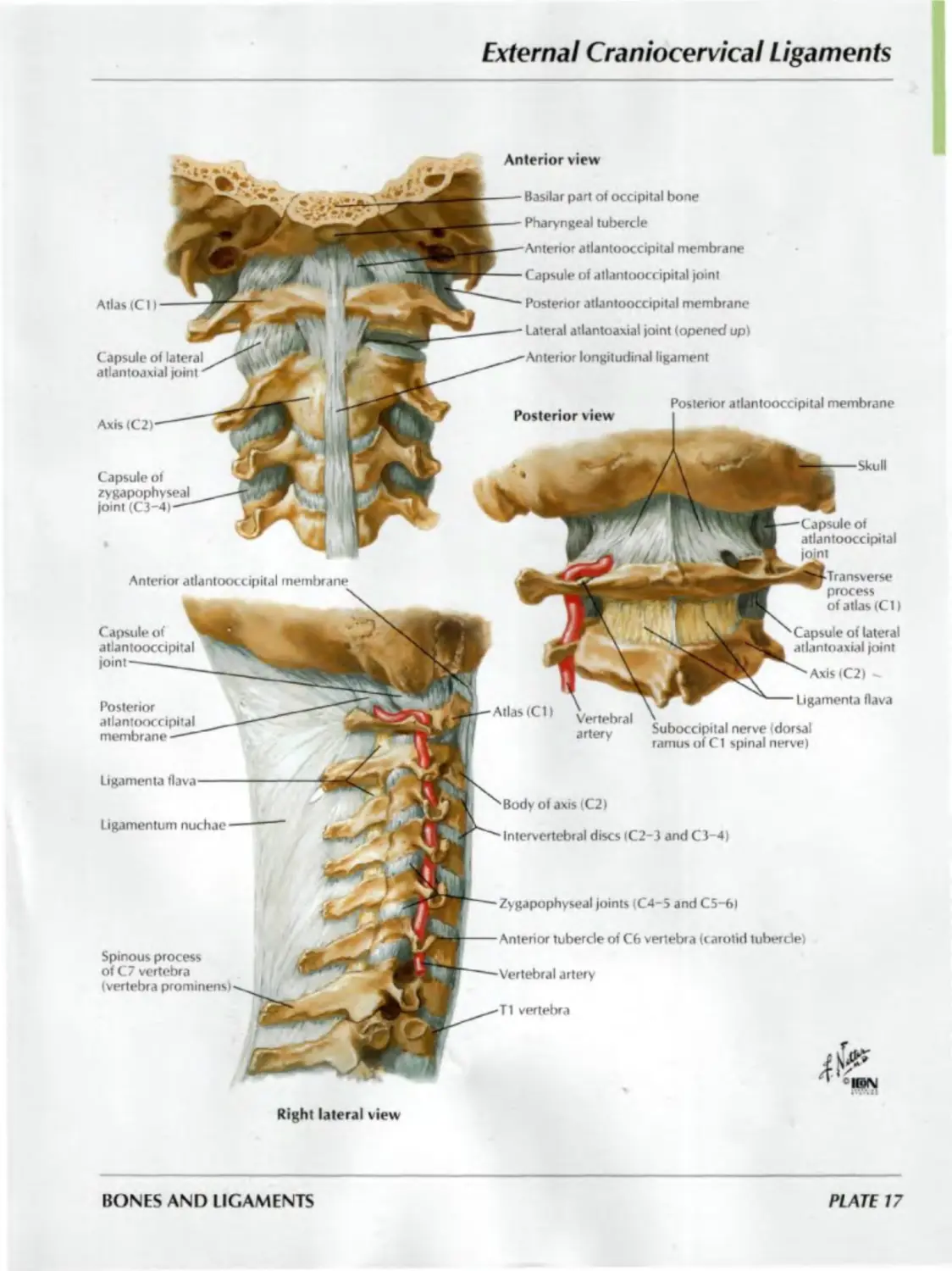
External Craniocervical Ligaments
Capsule of
zygapophyseal
joint (C3-4)
Atlas (Ct I
Capsule of lateral
atlantoaxial joint
Axis
Posterior view
Anterior view
Basilar part of occipital bone
Pharyngeal tubercle
Anterior atlantooccipital membrane
Capsule of atlantooccipital joint
Posterior atlantooccipital membrane
Lateral atlantoaxial joint (opened up)
Anterior longitudinal ligament
Posterior atlantooccipital membrane
Skull
Capsule of
atlantooccipital
Anterior atlantooccipital membrane
process
of atlas (Ct I
C apsule of
atlantooccipital
joint
antoaxial joint
Axis(C2) -
Posterior
atlantooccipital
membrane
Allas (Cl I
Vertebral
artery
Ligamenta flava
Ligamentum nuchae
Spinous process
of C7 vertebra
(vertebra prominensi
Right lateral view
Ligamenta flava
Suboccipital nerve Idorsal
ramus of Cl spinal nerve)
Body of axis (C2)
Intervertebral discs (C2-3 and C3-4)
Zygapophyseal joints (C4-5 and C5-f>)
Anterior tubercle of C6 vertebra (carotid tubercle)
Vertebral artery
T1 vertebra
BONES AND LIGAMENTS
PLATE 17
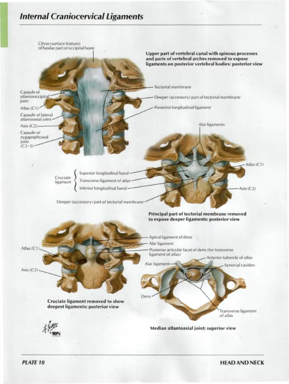
Internal Craniocervical Ligaments
Clivus (surtace feature)
of basilar part of occipital bone
Tectorial membrane
Deeper (accessory) part of tectorial membrane
Posterior longitudinal ligament
Alar ligaments
Capsule of
Deeper (accessory) part of tectorial membrane
Cruciate
ligament
Superior longitudinal band
Transverse ligament of atlas
Inferior longitudinal band
Atlas (CD
Axis (C 2)
Principal part of tectorial membrane removed
to expose deeper ligaments: posterior view
ioint
Atlas (C1)
Capsule of lateral
atlantoaxial joint
Axis (C2)
Capsule of
zygapophyseal
joint
(C2-3)
Atlas (CD
Axis (C2)
Cruciate ligament removed to show
deepest ligaments: posterior view
Upper part of vertebral canal with spinous processes
and parts of vertebral arches removed to expose
ligaments on posterior vertebral bodies: posterior view
Alar ligament
Apical ligament of dens
Alar ligament
Posterior articular facet of dens (for transverse
ligament of atlas) , ,
_____-—Anterior tubercle of atlas
Synovial cavities
Dens
Transverse ligament
of atlas
Median atlantoaxial joint:
superior view
PLATE 18
HEAD AND NECK
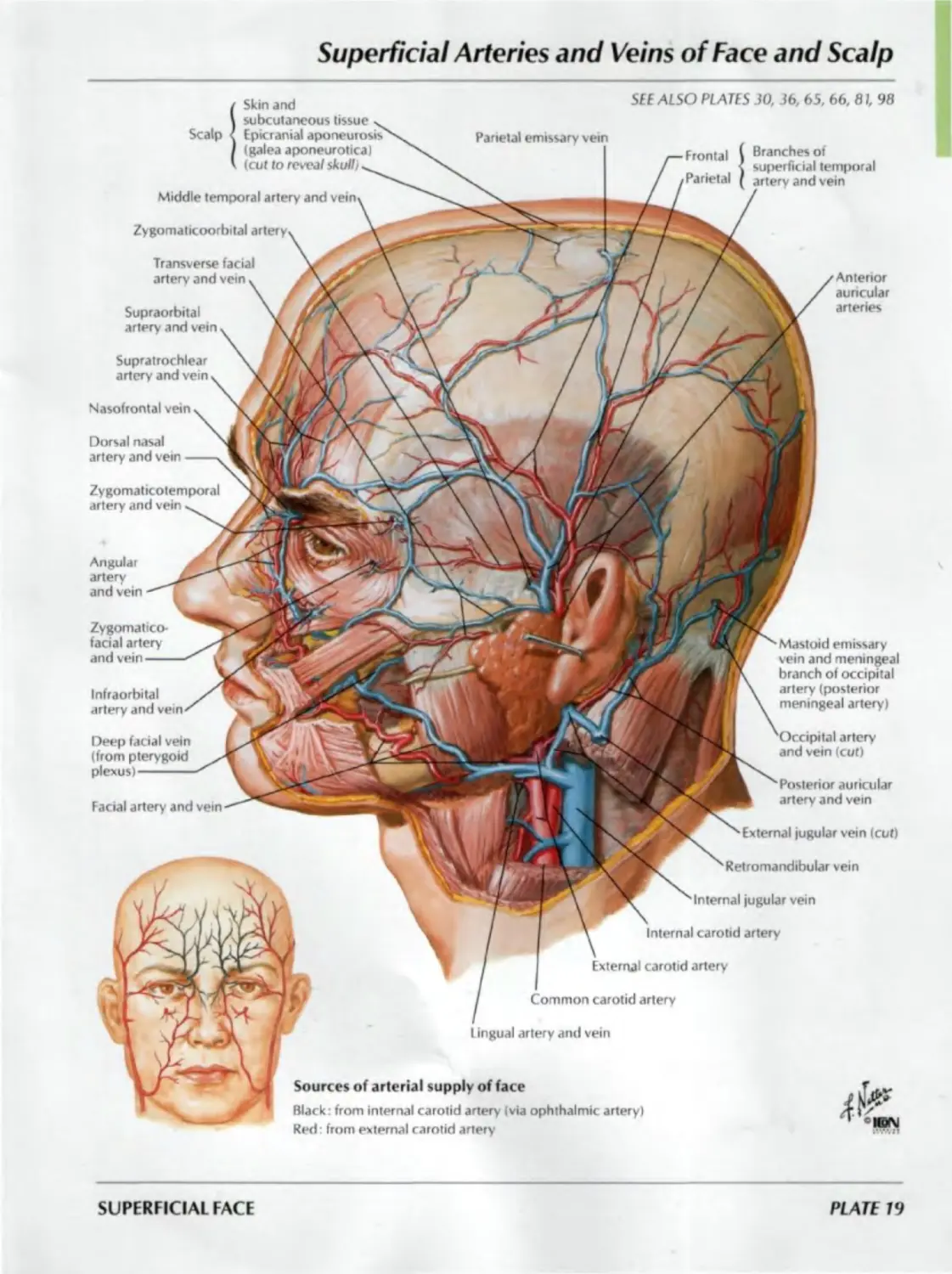
Superficial Arteries and Veins of Face and Scalp
SEE ALSO PLAJES 30, 36, 65, 66, 61, 98
Skin and
Angular
artery
and vein
Scalp
Middle temporal artery and vei
Zygomaticoorbital
Nasofrontal vein
Supratrochlear
artery and vein
Transverse facial
artery and vein
Dorsal nasal
artery and vein
Supraorbital
artery and vein
subcutaneous tissue
Epicranial aponeurosis
(galea aponeurotica)
. (cut to reveal skull)
Facial artery and vein
Infraorbital
artery and vein
Sources of arterial supply of face
Black: from internal carotid artery I via ophthalmic artery)
Red: from external carotid artery
Anterior
auricular
arteries
Branches of
superficial temporal
and vein
Zygomaticotemporal
artery and vein
Zygomatico-
facial artery
and vein
Deep facial vein
Mastoid emissary
vein and meningeal
branch of occipital
artery (posterior
meningeal artery)
artery
and vein (cut)
Posterior auricular
artery and vein
External jugular vein (cut)
Retromandibular vein
Internal jugular vein
Internal carotid artery
External carotid artery
Common carotid artery
Lingual artery and vein
SUPERFICIAL FACE
PLATE 79
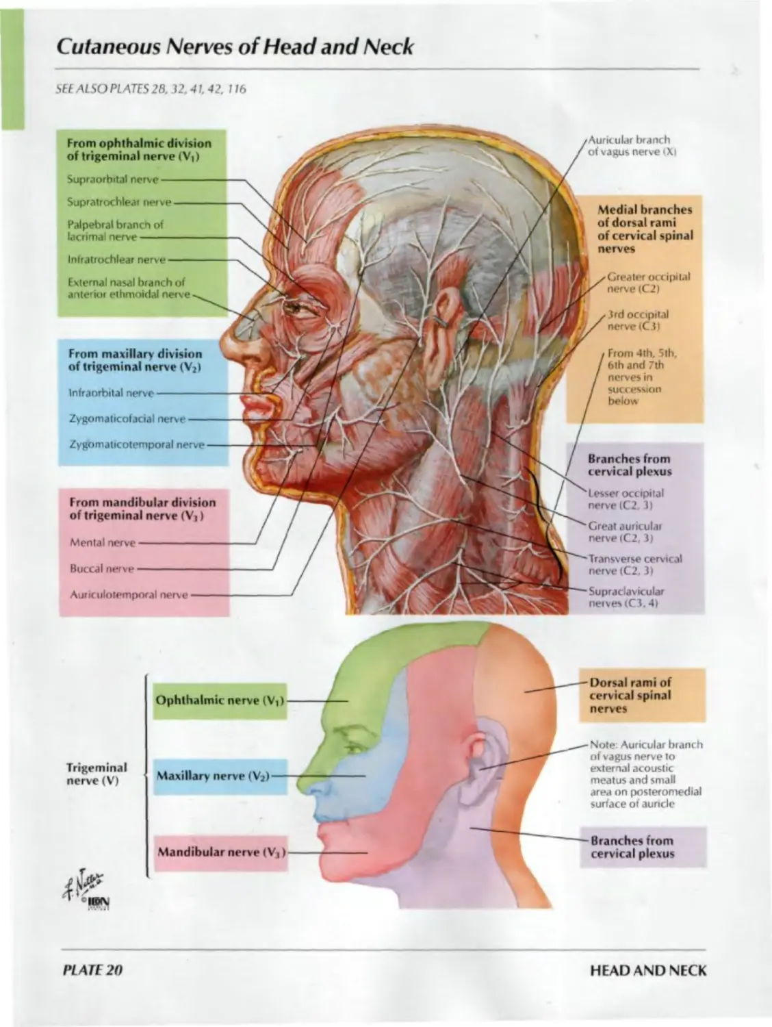
Cutaneous Nerves of Head and Neck
SEE ALSO PLATES 28. 32. 41. 42. 116
From mandibular division
of trigeminal nerve (Vj)
Mental nerve--------------
Buccal nerve--------------
Auriculotemporal nerve----
Medial branches
of dorsal rami
of cervical spinal
nerves
Greater occipital
nerve (C2)
3rd occipital
nerve (C3)
From 4th. 5th,
6th and 7th
nerves in
succession
below
Branches from
cervical plexus
Lesser occipital
nerve (C2, 3)
Great auricular
nerve (C2, 3)
Transverse cervical
nerve (C2, 3)
Supraclavicular
nerves (C3,4)
From ophthalmic division
of trigeminal nerve (Vi)
Supraorbital nerve —
Supratrochlear nerve
Palpebral branch of
lacrimal nerve-----
Infratrochlear nerve
External nasal branch of
anterior ethmoidal nerve
branch
of vagus nerve (Xi
From maxillary division
of trigeminal nerve (V2)
Infraorbital nerve-------
Zygomaticofacial nerve
Zygomaticotemporal
Ophthalmic nerve (Vi)
Mandibular nerve (Vj)
Dorsal rami of
cervical spinal
nerves
Trigeminal
nerve(V)
Maxillary nerve (V2)
Note: Auricular bianch
of vagus nerve to
external acoustic
meatus and small
area on posteromedial
surface of auricle
Branches from
cervical plexus
PLATE 20
HEAD AND NECK
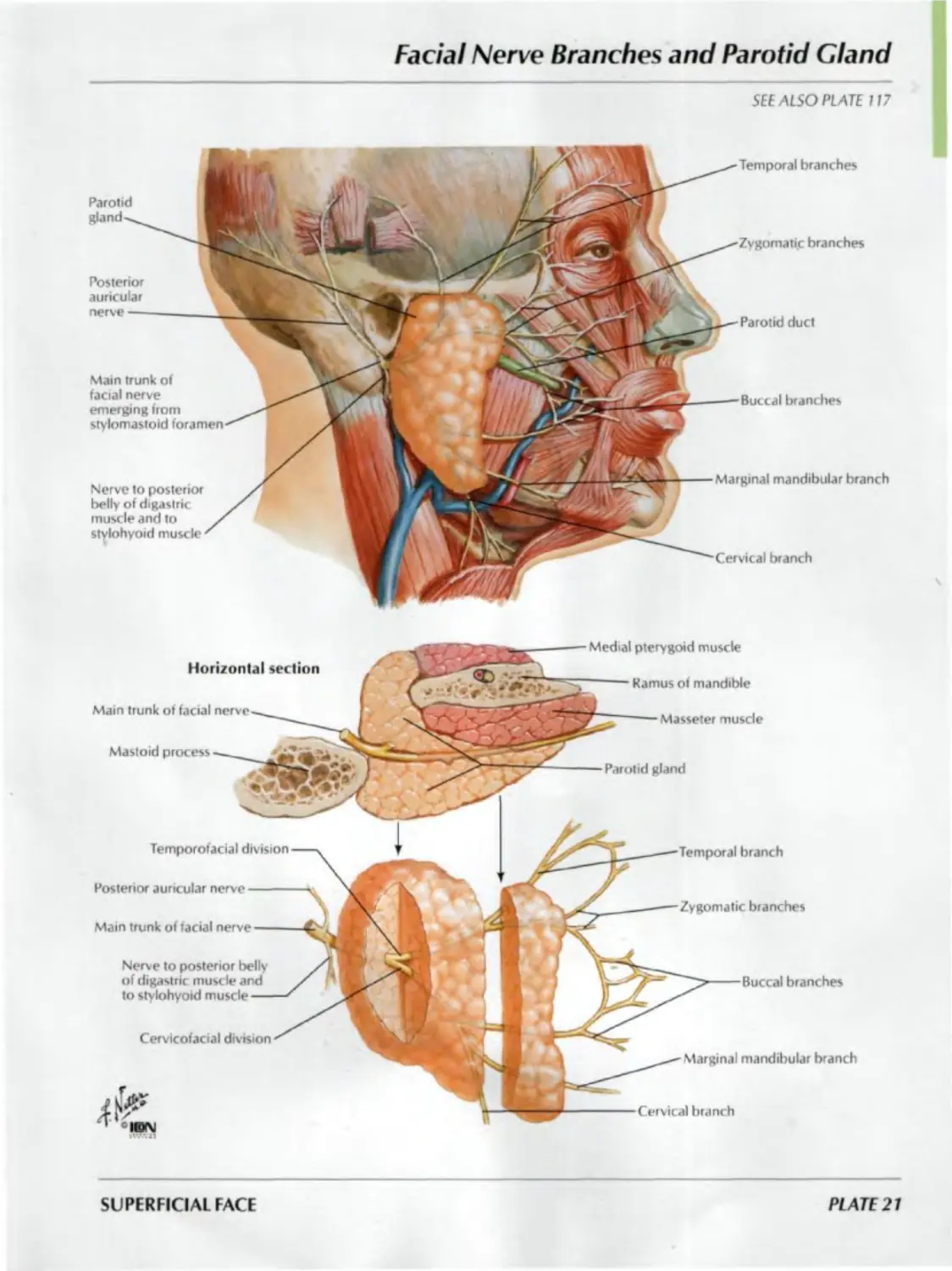
Facial Nerve Branches and Parotid Gland
SEE ALSO PLATE 117
Main trunk of
facial nerve
emerging from
stylomastoid
Nerve to posterior
belly of digastric
muscle and to
stylohyoid muscle
Parotid
gland
Posterior
auricular
nerve —
Temporal branches
Buccal branches
Marginal mandibular branch
branch
Zygomatic branches
Parotid duct
Parotid gland
Horizontal section
Main trunk of facial
Mastoid process
Medial pterygoid muscle
Ramus of mandible
Masseter muscle
Posterior auricular nerve
Main trunk of facial
Cervicofacial division
Temporofacial division
branch
Zygomatic branches
branches
Marginal mandibular branch
Cervical branch
Nerve to posterior belly
of digastric musc le and
to stylohyoid
SUPERFICIAL FACE
PLATE 21
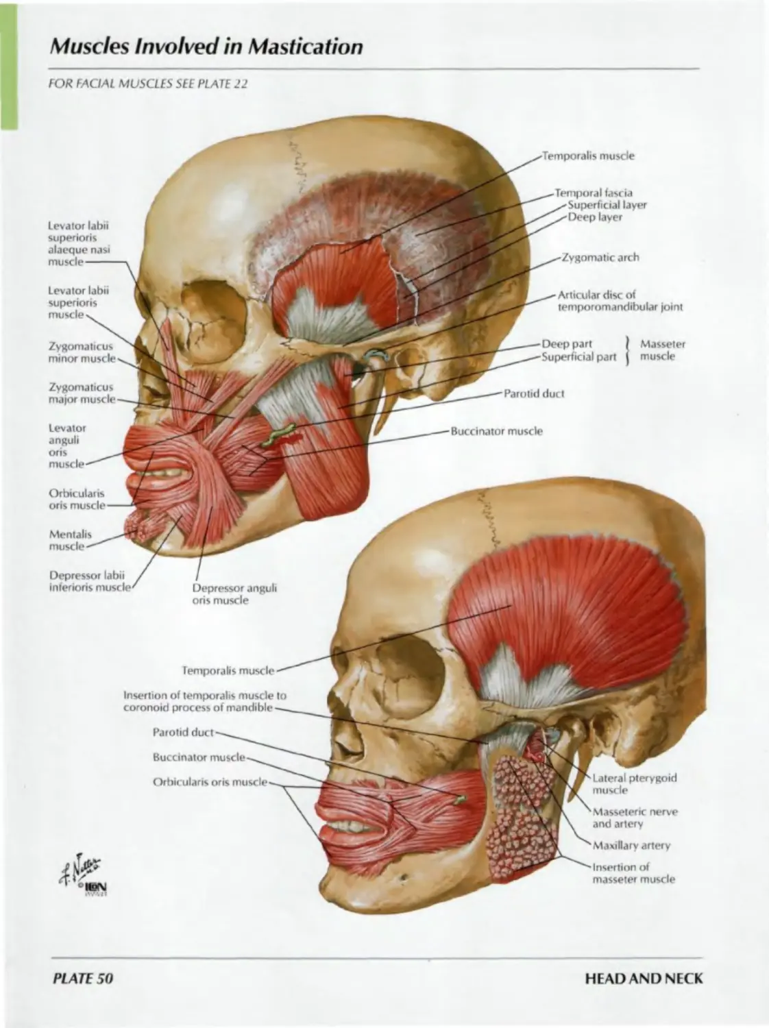
Muscles of Facial Expression: Lateral View
Levator labii superiors
alaeque nasi muscle
(partially cut
Nasalis
muscle
Temporal fascia
Orbicularis oculi muscle
Frontal belly (frontalis) of epicranius muscle
Procerus muscle
Levator labii superiors muscle
Orbicularis oris muscle
Zygomaticus minor muscle
Zygomaticus major muscle
Orbicularis oris muscle
Mentalis muscle
Depressor labii inferioris muscle
Depressor anguli oris muscle
Buccinator muscle
Clavicle
Orbital part
Palpebral part
Corrugator supercilii muscle (frontalis
and orbicularis oculi, partially cut away)
( Skin and
Scalp < subcutaneous tissue-------------------
( Epicranial aponeurosis (galea aponeurotica I
Auricularis anterior muscle
Auricularis superior muscle
Auricularis posterior muscle
Occipital belly
(occipitalis! of
epicranius muscle
Deltoid
fascia
Parotid
fascia
Risorius
Platysma muscle
Sternum
Pectoralis major fascia
Investing layer of
(deep) cervical fascia
Masseteric fascia
Transverse part
Alar part
Depressor septi
nasi muscle
PLATE 22
HEAD AND NECK
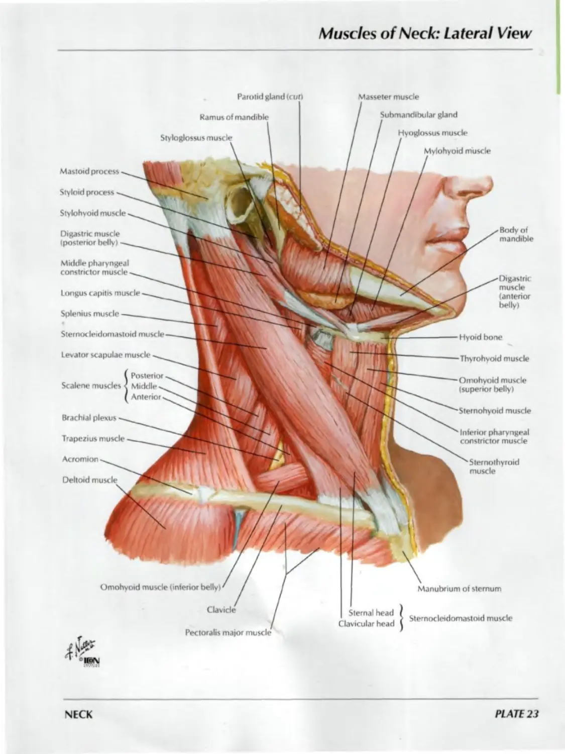
Muscles of Neck: Lateral View
Parotid gland (cut) Masseter muscle
Ramus of mandible
Styloglossus muscle
Submandibular gland
Hyoglossus muscle
/ Mylohyoid muscle
Mastoid process
Styloid process
Stylohyoid muscle
Digastric muscle
(posterior belly)
Body of
mandible
Middle pharyngeal
constrictor muscle
Longus capitis muscle
Splemus muscle
Digastric
muscle
(anterior
belly)
Sternocleidomastoid muscle
Levator scapulae muscle
! Posterior
Middle^,
Anterior.
Brachial plexus
Trapezius muscle
Acromion
Deltoid muscle
Hyoid bone
•Thyrohyoid muscle
Omohyoid muscle
(superior belly)
Sternohyoid muscle
Inferior pharyngeal
constrictor muscle
Sternothyroid
muscle
Omohyoid muscle (inferior belly)' /
Clavicle
Pectoralis major muscle
41^
Sternal head
Clavicular head
Manubrium of sternum
Sternocleidomastoid muscle
NECK
PLATE 23
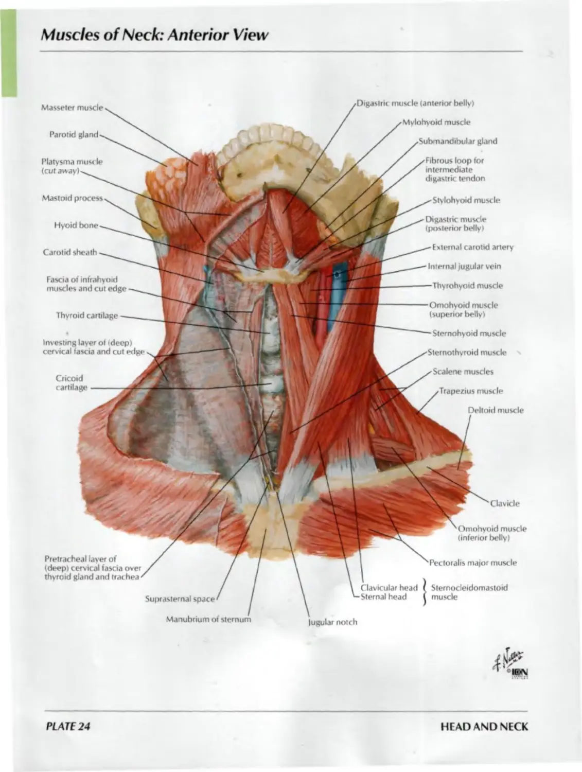
Muscles of Neck: Anterior View
Omohyoid muscle
(inferior belly)
Clavicle
lugular notch
major muscle
Clavicular head
Sternal head
Sternocleidomastoid
muscle
Masseter muscle
Parotid gland
Platysma muscle
(cut away!
Mastoid process
Hyoid bone
Carotid sheath
Fascia of infrahyoid
muscles and cut edge
Thyroid cartilage
Investing layer of (deep)
cervical fascia and cut edge
Cricoid
cartilage
Pretracheal layer of
(deep) cervical fascia over
thyroid gland and trachea
Suprasternal space
muscle (anterior belly)
Mylohyoid muscle
gland
loop for
intermediate
digastric tendon
Stylohyoid muscle
Digastric muscle
(posterior belly)
External carotid artery
Internal jugular vein
-Thyrohyoid muscle
Omohyoid muscle
(superior belly)
-Sternohyoid muscle
muscle v
Scalene muscles
Trapezius muscle
Deltoid muscle
PLATE 24
HEAD AND NECK
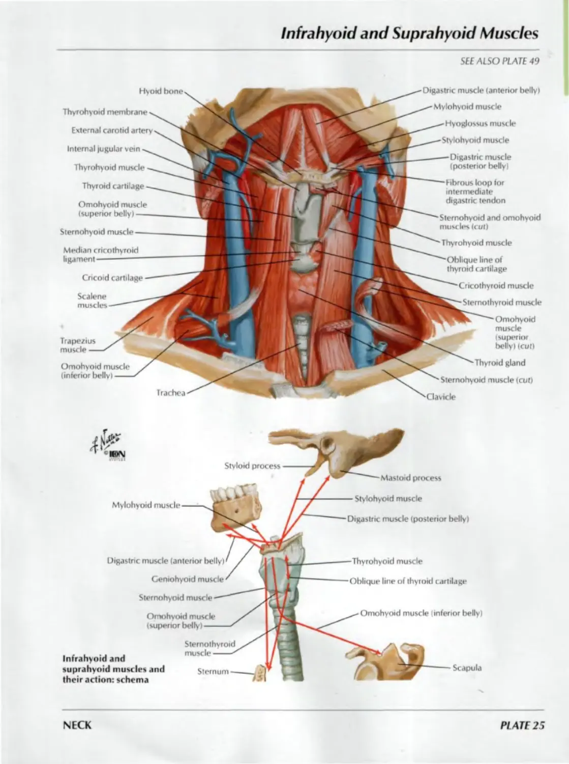
Infrahyoid and Suprahyoid Muscles
SEE ALSO PLATE 49
Digastric muscle (anterior bellyi
Mylohyoid muscle
Hyoglossus muscle
Stylohyoid muscle
line of
Digastric muscle
(posterior belly)
Hyoid bone
Thyrohyoid membrane
External carotid artery
Internal jugular vein
Thyrohyoid muscli
Thyroid cartilage
Omohyoid muscle
(superior belly)
Sternohyoid muscle
Median cricothyroid
ligament
Cricoid cartilage
Scalene
muscles
Thyrohyoid muscle
Cricothyroid muscle
Sternothyroid muscle
Thyroid gland
Sternohyoid muscle (cut)
Fibrous loop tor
intermediate
digastric tendon
Omohyoid
muscle
(superior
belly) (cut)
Sternohyoid and omohyoid
muscles (cut)
Irachea
Trapezius
muscle
Omohyoid muscle
(inferior belly)
Styloid process
Mylohyoid
Digastric muscle (anterior belly)
Geniohyoid muscle
Sternohyoid muscle
Infrahyoid and
suprahyoid muscles and
their action: schema
Mastoid process
Stylohyoid muscle
Digastric muscle (posterior belly)
Omohyoid muscle (inferior belly)
Scapula
Thyrohyoid muscle
line of thyroid cartilage
Omohyoid muscle
(superior belly)
Sternothyroid
muscle
Sternum
NECK
PLATE 25
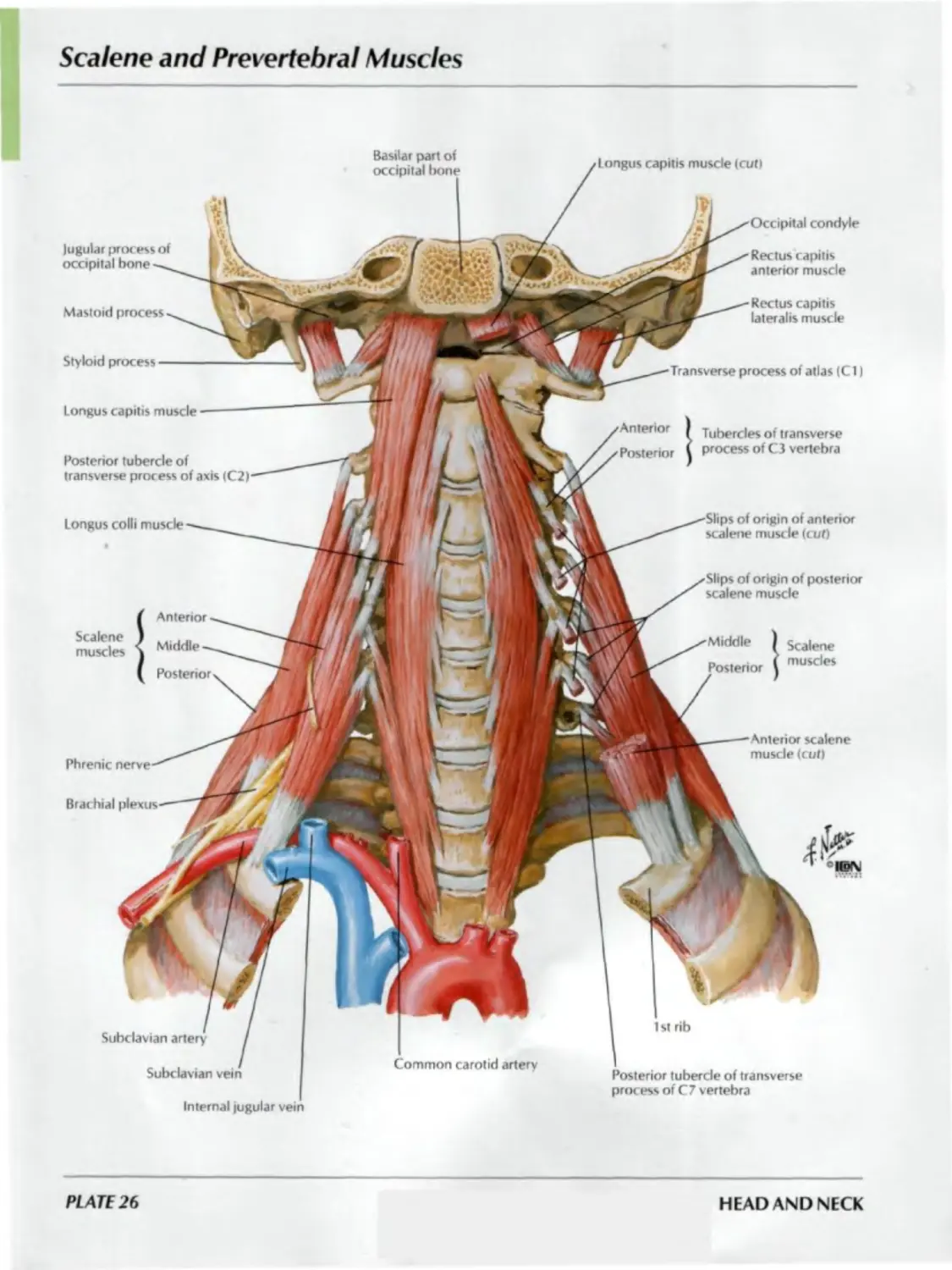
Scalene and Prevertehral Muscles
Basilar part of
occipital bone
I ongus capitis muscle (cut)
Jugular process of
occipital bone
Mastoid process
Occipital condyle
Rectus capitis
anterior muscle
Rectus capitis
lateralis muscle
Styloid process
Longus capitis muscle ---------
Posterior tubercle of
transverse process of axis (C2)
Longus colli muscle
Anterior
7
Transverse process of atlas (Cl)
Anterior
Posterior
Tubercles of transverse
process of C3 vertebra
•Slips of origin of anterior
scalene muscle (cut)
Slips of origin of posterior
scalene muscle
Scalene
muscles
Middle
Middle
Posterior
Scalene
muscles
Phrenic nerve
Anterior scalene
muscle (cut)
Brachial plexus
Subclavian artery
Subclavian vein
Internal jugular vein
1 st rib
Common carotid artery
Posterior tubercle of transverse
process of C7 vertebra
PLATE 26
HEAD AND NECK
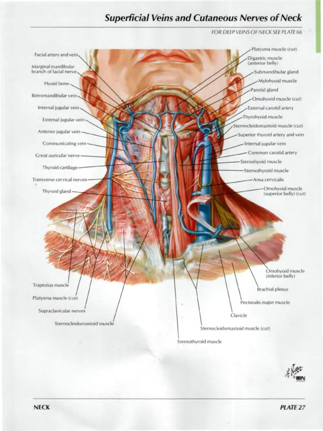
Superficial Veins and Cutaneous Nerves of Neck
FOR DEEP VEINS OF NECK SEE PLATE 66
Facial artery and vein.
Platvsma muscle (cut)
Marginal mandibular
branch of facial nerve
Hyoid bone
Retromandibular vein
Digastric muscle
(anterior belly)
__^Submandibular gland
^-Mylohyoid muscle
,x Parotid gland
/Omohyoid muscle (cut)
Internal jugular vein
External jugular vein
JJF'x External carotid artery
'"T/Thyrohyoid muscle
Sternocleidomastoid muscle (cut)
Anterior lugular vein
Communiiating vein
Great auricular nerve
Superior thyroid artery and vein
Internal jugular vein
___Common carotid artery
Transverse cervical nerves---
Sternohyoid muscle
—Sternothyroid muscle
______Ansa cervicalis
Thyroid gland
Omohyoid muscle
(superior belly) (cut)
Omohyoid mus< le
(inferior belly)
Trapezius muscle
Brachial plexus
Platysma muscle (cut)
Pectoralis major muscle
Supraclavicular nerves
Clavicle
Sternocleidomastoid muscle
Sternocleidomastoid muscle (cut)
Sternothyroid muscle
NECK
PLATE 27
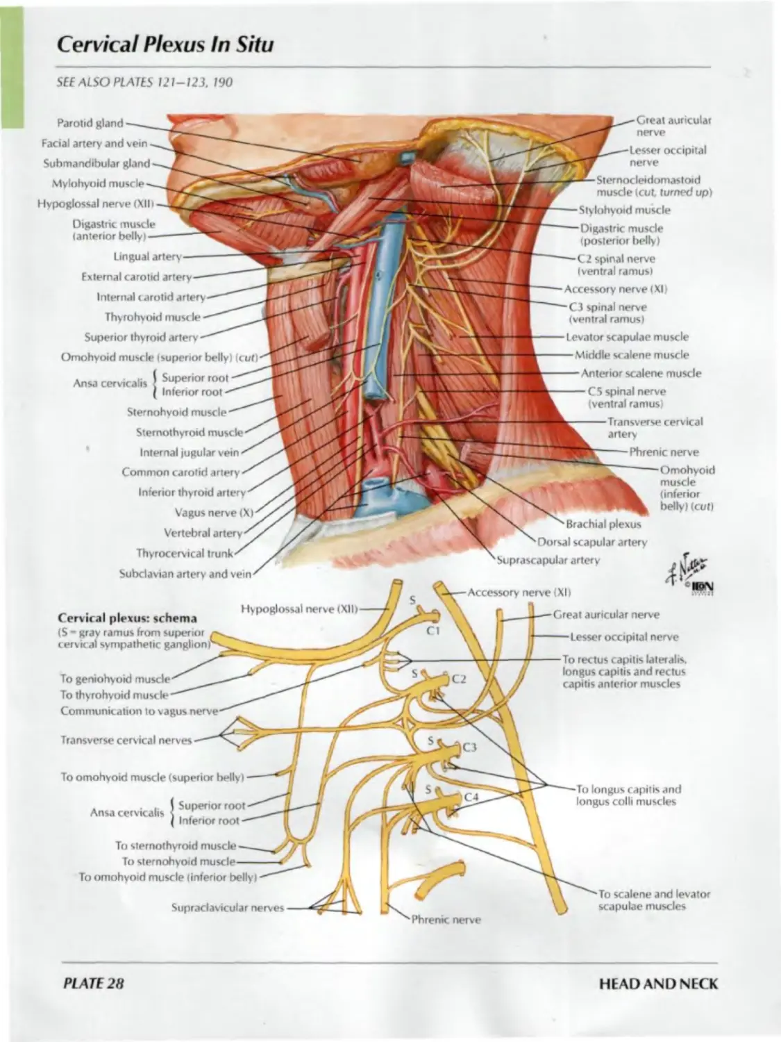
Cervical Plexus In Situ
SEE ALSO PLATES 121-123, 190
(inferior
belly) (cut)
Brachial plexus
Dorsal scapular artery
Suprascapular artery
Accessory nerve (XI)
Great auricular nerve
Parotid gland
Facial artery and vein
Submandibular gland
Mylohyoid muscle
Hypoglossal nerve (XII।
Digastric muscle
(anterior belly)
Lingual artery
External carotid
Internal carotid artery
Thyrohyoid muscle
Superior thyroid artery
Omohyoid muscle (superior belly) (cut)
Ansa cervkalis f ^P™o"oot
( Inferior root
Sternohyoid muscle
Sternothyroid muscle
Internal jugular vein
Common carotid artery
Inferior thyroid artery
Vagus nerve (X)
Vertebral
Thyrocervical trunk
Subclavian artery and vein
Great auricular
nerve
Lesser occipital
nerve
Sternocleidomastoid
muscle (cut turned up)
Stylohyoid muscle
Digastric muscle
(posterior belly)
C2 spinal nerve
(ventral ramus)
Accessory nerve (XI)
C3 spinal nerve
(ventral ramus)
Levator scapulae muscle
Middle scalene muscle
Anterior scalene muscle
C5 spinal nerve
(ventral ramus)
Transverse cervkal
artery
Phrenic nerve
Cervical plexus: schema
( S - gray ramus from superior
cervical sympathetic gangli
Transverse cervical nerves
To omohyoid muscle (superior belly)
Ansa cervkalis
Superior root
Inferior root
Hypoglossal nerve (XII)
Supraclavicular
To scalene and levator
scapulae muscles
Lesser occipital nerve
To rectus capitis lateralis,
longus capitis and rectus
capitis anterior muscles
To geniohyoid
To thyrohyoid muscle
Communication to vagus nerve
To sternothyroid muscle
To sternohyoid
To omohyoid muscle (inferior belly)
longus capitis and
longus colli muscles
PL ATE 28
HEAD AND NECK
Subclavian Artery
SEE ALSO PLATE 410
Right anterior dissection
Internal jugular vein
Common carotid
Ascending cervical artery
Phrenic nerve —
Anterior scalene musde
Inferior thyroid artery
Transverse cervical artery
Suprascapular artery
Dorsal scapular artery
Costocervical trunk
Thyrocervical trunk
Subclavian artery and vein
Right lateral schematic view
Vertebral artery
Costocervical trunk
Scapula
External carotid artery
Internal carotid artery
Ascending cervical artery
Inferior thyroid artery
Transverse cervical artery
Common carotid artery
Thyrocervical trunk
Suprascapular artery
Internal thoracic artery
Deep cervical artery (ascending
to anastomose with descending
branch of occipital artery)
Supreme intercostal
artery
1st posterior
intercostal
artery
2nd posterior
intercostal
artery
Thyroid gland
(reflected)
Middle cervical
sympathetic ganglion
Vagus nerve (X)
Vertebral artery
Common carotid artery
Recurrent laryngeal
netve
Brachiocephalic trunk
Internal jugular vein
(cut)
artery
(1st part medial to,
2nd part posterior to,
3rd part lateral to
anterior scalene muscle)
NECK
PLATE 29
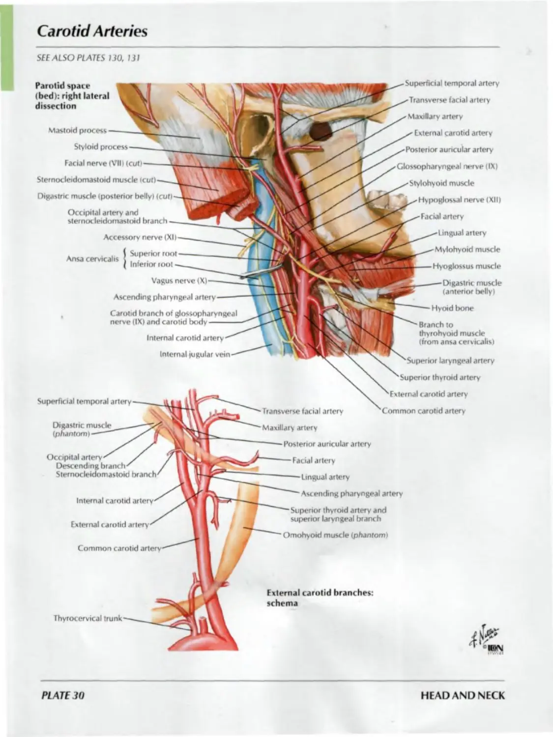
Carotid Arteries
SEE ALSO PLATES 130. 131
Superficial temporal artery
Transverse facial artery
Maxillary artery
External carotid artery
Posterior auricular artery
Stylohyoid muscle
Facial artery
Parotid space
(bed): right lateral
dissection
Glossopharyngeal nerve (IX)
Hypoglossal nerve (XII)
artery
Mylohyoid muscle
Hyoid bone
laryngeal artery
Superior thyroid artery
artery
Common carotid artery
Transverse facial artery
Digastric muscle
(anterior belly)
Branch to
thyrohyoid muscle
(from ansa cervicalis)
Mastoid process
Styloid process
Facial nerve (VII) (cut)
Sternocleidomastoid muscle icut)
Digastric muscle (posterior belly) (cut)
Occipital artery and
sternocleidomastoid branch
Accessory nerve (XI)
{Superior roo
. Г •
Inferior root
Vagus nerve (X)
Ascending pharyngeal artery
Carotid branch of glossopharyngeal
nerve (IX) and carotid body
Internal carotid artery
Internal jugular vein
Hyoglossus muscle
Maxillary artery
Superficial temporal artery
Digastric rr
(phantom)
Posterior auricular artery
Facial artery
Lingual artery
Ascending pharyngeal artery
Omohyoid muscle (phantom)
Superior thyrord artery and
superior laryngeal branch
Thyrocervical trunk
External carotid branches:
schema
Occipital
Descending
Sternocleidomastoid
Internal carotid
External carotid
Common carotid
PLATE 30
HEAD AND NECK
Fascial Layers of Neck
FOR CONTENTS OF CAROTID SHEATH SEE PLATES bS-67
Investing layer of (deep) cervical fascia
Platysma muscle
Alar fascia
Muscular portion of pretracheal layer of
(deepl cervical fascia (of infrahyoid
Visceral portion of pretracheal
layer of (deep) cervical fascia
Buccopharyngeal
(visceral) fascia
Carotid sheath
Subcutaneous tissue
Investing layer
of (deep) cervical
fascia rooting
posterior triangle
Fat in
poster ic
triangle
Prevertebral layer
•of (deep)
cervical fascia
muscle
Trachea
Sternothyroid muscle
Thyroid gland
Esophagus
Omohyoid muscle
Sternocleidomastoid
muscle
Recurrent laryngeal
nerve
Common carotid artery
Internal fugular vein
Vagus nerve (X)
Phrenic nerve
Anterior scalene muscle
Sympathetic trunk
Spinal nerve
Middle and posterior
scalene muscles
Longus colli muscle
Levator scapulae muscle
Trapezius muscle
Deep cervical muscles
Cross section Retropharyngeal Subcutaneous Cervical vertebra (C7)
space tissue
Sagittal section
Pharynx----------
Buccopharyngeal
fascia-----------
Retropharyngeal
space------------
Alar fascia------
Prevertebral fascia
Trachea----------
Esophagus--------
Mandible
Geniohyoid muscle
Geniohyoid fascia
Investing layer of (deep)
cervical fascia
Fascia of infrahyoid muscles
Pretracheal fascia
Thyroid gland
Subcutaneous tissue
Suprasternal space
Manubrium of sternum
Aorta
Pericardium
NECK
PLATE 31
Nose
Anterolateral view
Frontal
Nasal
Frontal process of maxilla
Lateral process of
septal nasal cartilages
Septal cartilage
Minor alar cartilage
Accessory nasal cartilage
(Lateral crus
Medial crus
Septal nasal cartilage
Anterior nasal spine of maxilla
Alar fibrofatty
Infraorbital foramen
Inferior view
Major alar cartilage
Lateral Medial
crus crus
cartilage Intermaxillary
suture
Frontalis muscle
Supraorbital artery
Supratrochlear artery and
Procerus muscle
Corrugator supercilii muscle
Dorsal nasal
Infratrochlear nerve
Angular artery
External nasal artery and nerve
Nasalis muscle (transverse
Infraorbital artery and nerve
Lateral nasal
Transverse facial artery
Nasalis muscle talar part)
Depressor septi nasi muscle
Orbicularis oris muscle
Facial artery
PLATE 32
HEAD AND NECK
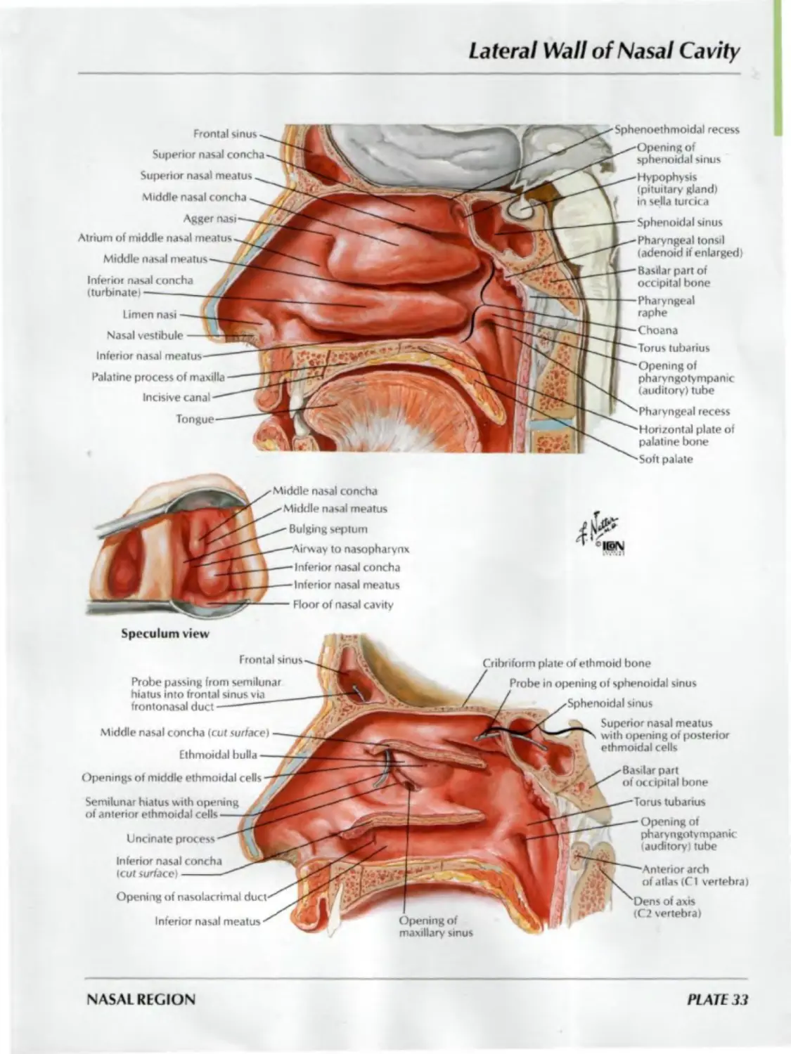
Lateral Wall of Nasal Cavity
Frontal sinus
Superior nasal concha
Superior nasal meatus
Middle nasal concha
Agger nasi
Atrium of middle nasal meatus-.
Middle nasal meatus
Inferior nasal concha
(turbinatei -----------
Limen nasi
Nasal vestibule
Inferior nasal meatus
Palatine process of maxilla
Incisive canal
Tongue
•Sphenoethmoidal recess
-Opening of
j sphenoidal sinus
^—Hypophysis
(pituitary gland)
/ in sella turcica
k-— Sphenoidal sinus
B—- Pharyngeal tonsil
(adenoid if enlarged)
1—• Basilar part of
occipital bone
4—Pharyngeal
raphe
T"—Choana
Г—Torus tubarius
""'‘Opening of
pharyngotympanic
(auditory) tube
^"Pharyngeal recess
'"Horizontal plate of
palatine bone
^•Soft palate
Speculum view
Probe passing from semilunar
hiatus into frontal sinus via
frontonasal duct
Semilunar hiatus with opening
of anterior ethmoidal cells
Inferior nasal concha
(cut surface)
Middle nasal concha
Middle nasal meatus
Bulging septum
to nasopharynx
Inferior nasal concha
Inferior nasal meatus
Floor of nasal cavity
Frontal sinus
Middle nasal concha (cut surface)
Ethmoidal bulla
Openings of middle ethmoidal cells
Uncinate process
Opening of nasolacrimal
Inferior nasal meatus
№
’ l©M
Cribriform plate of ethmoid bone
Probe in opening of sphenoidal sinus
sinus
Superior nasal meatus
with opening of posterior
ethmoidal cells
Basilar part
of occipital bone
tubarius
Opening of
pharyngotympanic
(auditory) tube
Anterior arch
of atlas (Cl vertebra)
of axis
(C2 vertebra)
NASAL REGION
PLATE 33
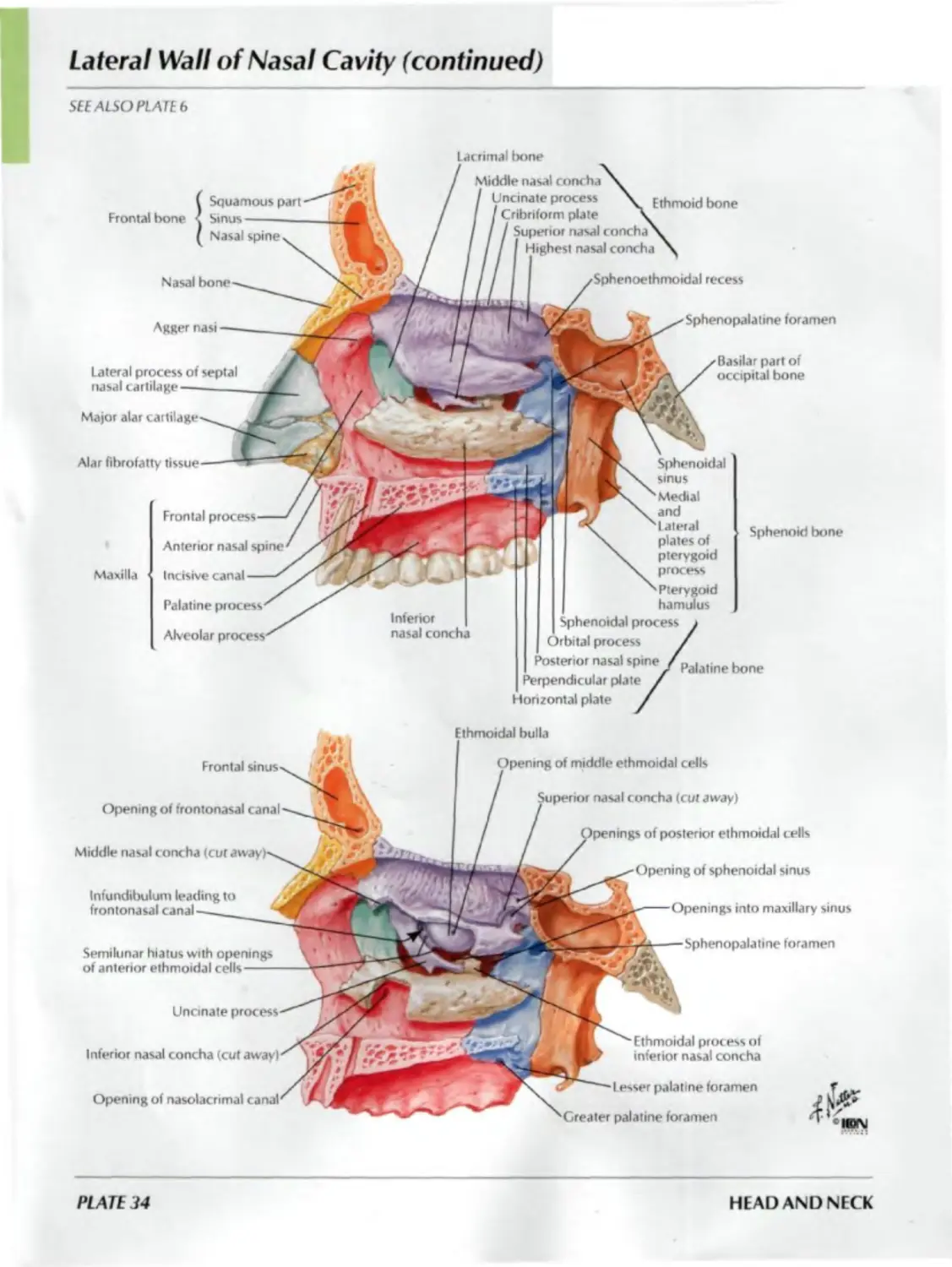
Lateral Wall of Nasal Cavity (continued)
SEE ALSO PLATE b
Lacrimal bone
Agger nasi
Alar fibrofatty
Maxilla
Nasal bone
Ethmoid bone
Sphenopalatine foramen
Basilar part of
occipital bone
Squamous part
Frontal bone • Sinus--------
Nasal spine
Lateral process of septal
nasal cartilage
Major alar
Frontal
Anterior nasal spine
Incisive canal
Palatine process
Alveolar
Middh oncha \
process
Cribriform plate \
nasal concha
4 nasal concha
Sphenoidal
sinus
Medial
and
I ateral
plates of
pterygoid
process
Pterygoid
hamulus
Sphenoid bone
Sphenoidal process у
Orbital process /
Posterwr nasal spine ZPalatjne
Perpendicular plate /
Horizontal plate /
Infundibulum leading to
frontonasal canal
Semilunar hiatus with openings
of anterior ethmoidal cells
Opening of frontonasal canal
Middle nasal concha
Uncinate
Inferior nasal concha (cut away)
Opening of nasolacrimal
Ethmoidal bulla
middle ethmoidal cells
nasal concha (cut away)
?rior ethmoidal cells
Opening of sphenoidal sinus
into maxillary sinus
Sphenopalatine foramen
Ethmoidal process of
inferior nasal concha
Lesser palatine foramen
Greater palatine foramen
PLATE 34
HEAD AND NECK
Medial Wall of Nasal Cavity (Nasal Septum)
-----Crista galli
Cribriform plate
pendicular plate
Nasal
Vomer
Ethmoid bone
Crest
Body
Sphenoidal sinus
Medial,
Anterior nasal spine
Nasal crest
Palatine
Greater palatine foramen
plates of
pterygoid
process
( Squamous part
Frontal bone
( Nasal
Vomerine groove
(for nasopalatine nerve
and vessels)------
Septal
Major alar
cartilage t medial crus)
Maxilla
Sphenoid
bone
Basilar part of
occipital bone
Pharyngeal
tubercle
Perpendicular plate
Nasal crest
Posterior nasal spine
Horizontal plate
Lesser palatine
foramen
Palatine
bone
NASAL REGION
PLATE 35
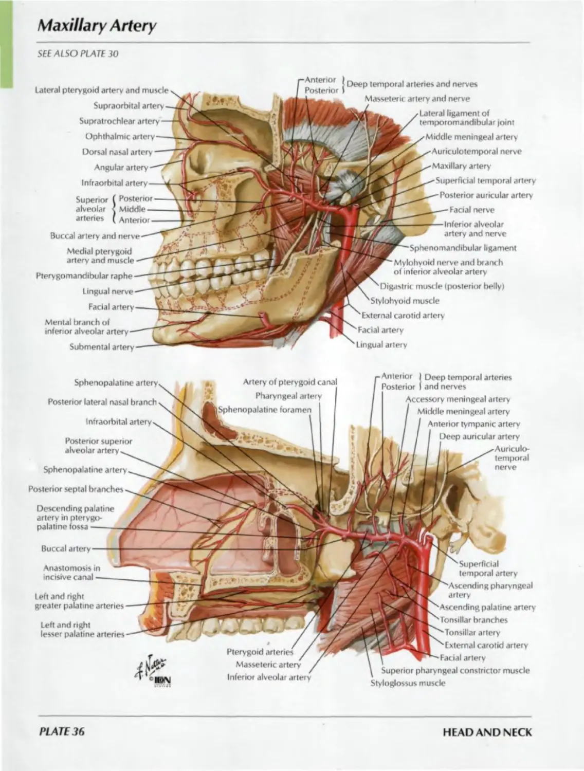
Maxillary Artery
SEE ALSO PLATE 30
Lateral pterygoid artery and muscle
Supraorbital artery
Supratrochlear
Ophthalmic artery
Dorsal nasal artery
Angular artery
Infraorbital artery
Superior ( Posterior -
alveolar
arteries
| Deep temporal arteries and nerves
Masseteric artery and nerve
Lateral ligament of
temporomandibular joint
Middle meningeal artery
Auriculotemporal nerve
Maxillary artery
temporal artery
Posterior auricular artery
Facial nerve
Inferior alveolar
artery and nerve
ligament
Mylohyoid nerve and branch
of inferior alveolar artery
Digastric muscle (posterior belly)
muscle
External carotid artery
Facia) artery
Lingual artery
Buccal artery and
Medial pterygoid
artery and muscle
Pterygomandibular raphe
Lingual nerve
Facial artery
Mental branch of
inferior alveolar
Submental artery
Middle -
Anterior
Anterior
Posterior
Sphenopalatine artery
Posterior lateral nasal branch
Artery of pterygoid canal
Pharyngeal artery
[Sphenopalatine foramen
Infraorbital artery
Posterior superior
alveolar artery
Sphenopalatine artery
Anterior
Posterior
Accessory meningeal artery
/ Middle meningeal artery
I I Anterior tympanic artery
Deep auricular artery
I^^Auriculo-
I ID temporal
* nerve
I Deep temporal arteries
I and nerves
Posterior septal branches
Descending palatine
artery in pterygo-
palatine fossa---
Buccal artery
Anastomosis in
incisive canal —
Left and right
greater palatine arteries
Left and right
lesser palatine arteries —
Pterygoid arteries /
Masseteric artery
Inferior alveolar artery
artery
' Am
Tonsillar
Tonsillar
^External carotid artery
Г" Facial artery
\ Superior pharyngeal constrictor muscle
Styloglossus muscle
PLATE 36
HEAD AND NECK
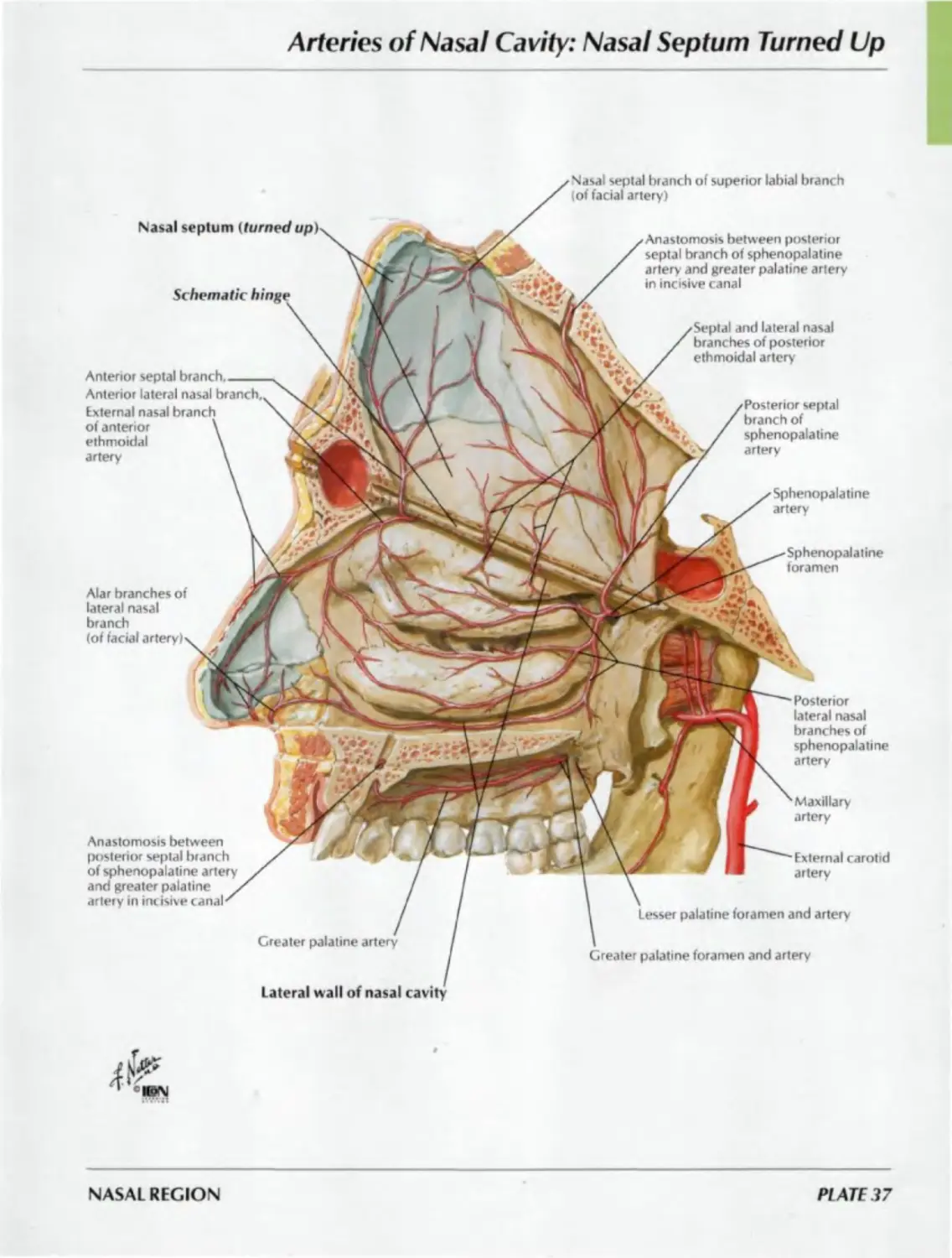
Arteries of Nasal Cavity: Nasal Septum Turned Up
Nasal septum
Schematic
Nasal septal branch of superior labial branch
(of facial artery)
Posterior
lateral nasal
branches of
sphenopalatine
artery
Sphenopalatine
foramen
Greater palatine artery
Lateral wall of nasal
Maxillary
artery
External carotid
artery
Lesser palatine foramen and artery
Greater palatine foramen and artery
Anastomosis between posterior
septal branch of sphenopalatine
artery and greater palatine artery
in incisive canal
Septal and lateral nasal
branches of posterior
ethmoidal artery
Posterior septal
branch of
sphenopalatine
artery
Sphenopalatine
artery
Anterior septal
Anterior lateral nasal
External nasal branch
of anterior
ethmoidal
artery
Alar branches of
lateral nasal
branch
(of facial
Anastomosis between
posterior septal branch
of sphenopalatine artery
and greater palatine
artery in incisive canal
NASAL REGION
PLATE 37
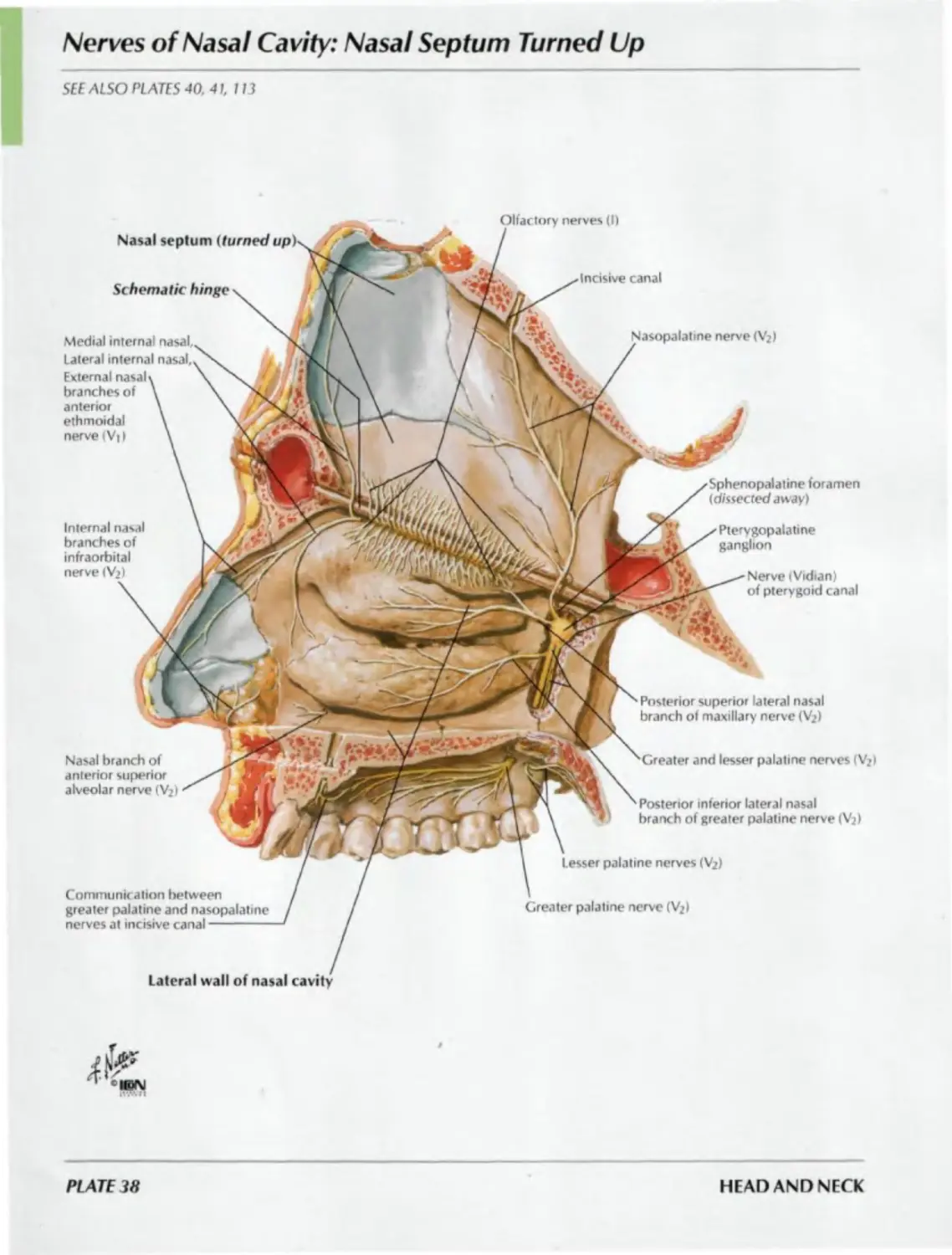
Nerves of Nasal Cavity: Nasal Septum Turned Up
SEE ALSO PLATES 40. 41, 113
Olfactory nerves (I)
Nasal septum (turned up)
Schematic hinge
Incisive canal
Medial internal nasal.
Nasopalatine nerve <V?)
Lateral internal nasal.
External nasal
branches of
anterior
ethmoidal
nerve (V) I
Sphenopalatine foramen
(dissected away)
Internal nasal
branches of
infraorbital
nerve < Vj)
Pterygopalatine
ganglion
Nerve (Vidian)
of pterygoid canal
Posterior superior lateral nasal
branch of maxillary nerve (Vj)
Nasal branch of
anterior superior
alveolar nerve (V?)
Greater and lesser palatine nerves (Vj >
Posterior inferior lateral nasal
branch of greater palatine nerve (V2)
Lesser palatine nerves (V2)
Communication between
greater palatine and nasopalatine
nerves at incisive canal---
Greater palatine nerve (Vj)
Lateral wall of nasal cavity
PLATE 38
HEAD AND NECK
Nerves of Nasal Cavity (continued)
SEE ALSO PLATE 113
Lateral
wall of
nasal
cavity
Distribution of olfactory
mucosa {shaded blue)
Lateral wall
of nasal cavity
External nasal branch of anterior ethmoidal nerve (V0
Lateral internal nasal branch of anterior ethmoidal nerve (V,)
Olfactory bulb
plate of ethmoid bone
Olfactory tract
Posterior superior lateral nasal branches
from maxillary nerve (Vj)
Maxillary nerve < Vj) (sphenopalatine
foramen dissected away)
Pterygopalatine ganglion
petrosal nerve
Deep petrosal nerve
Nerve (Vidian) of pterygoid canal
Phary ngeal branch of maxillary nerve (Vj)
Nasopalatine nerve (Vj) passing to septum (cut)
Posterior inferior lateral nasal branch
from greater palatine nerve (V>)
bulb
Cribriform plate
tract
Olfactory nerves (I)
Palatine nerves (Vi) .
I Lesser
Medial internal nasal branch
of anterior ethmoidal nerve
Olfactory nerves (I)
Nasopalatine nerve (Vj)
Incisive canal
Nasal
NASAL REGION
Pl ATE 39
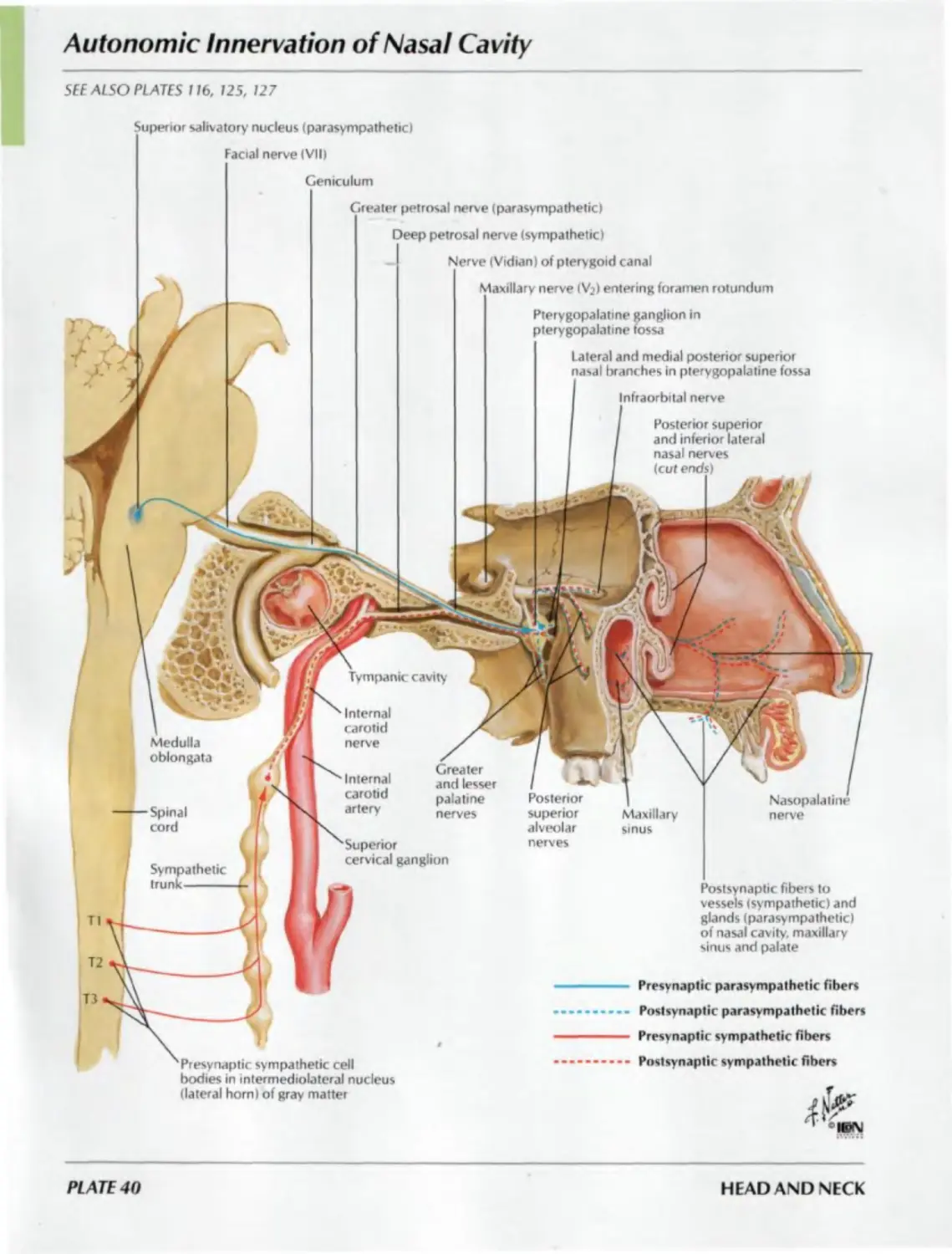
Autonomic Innervation of Nasal Cavity
SEE ALSO PLATES 11b. 125, 127
Superior salivatory nucleus {parasympathetic)
Facial nerve (VII)
Geniculum
Greater petrosal nerve (parasympathetic)
Deep petrosal nerve (sympathetic)
Nerve (Vidian) of ptery goid canal
Maxillary nerve (Vj) entering foramen rotundum
Posterior superior
and inferior lateral
nasal nerves
(cut ends)
Pterygopalatine ganglion in
pterygopalatine fossa
lateral and medial posterior superior
nasal branches in pterygopalatine fossa
Infraorbital nerve
Tympanic cavity
T2
П
Greater
and lesser
palatine
nerves
Posterior
superior
alveolar
nerves
Maxillary
sinus
Postsynaptic fibers to
vessels (sympathetic) and
glands (parasympathetic)
of nasal cavity, maxillary
sinus and palate
Nasopalatine
nerve
Presynaptic sympathetic cell
bodies in intermediolateral nucleus
(lateral horn) of gray matter
Presvnaptic parasympathetic fibers
Postsynaptic parasympathetic fibers
Presvnaptic sympathetic fibers
Postsynaptic sympathetic fibers
Superior
cervical ganglion
Internal
carotid
nerve
Medulla
oblongata
Spinal
cord
Internal
carotid
artery
Sympathetic
trunk------
PLATE 40
HEAD AND NECK
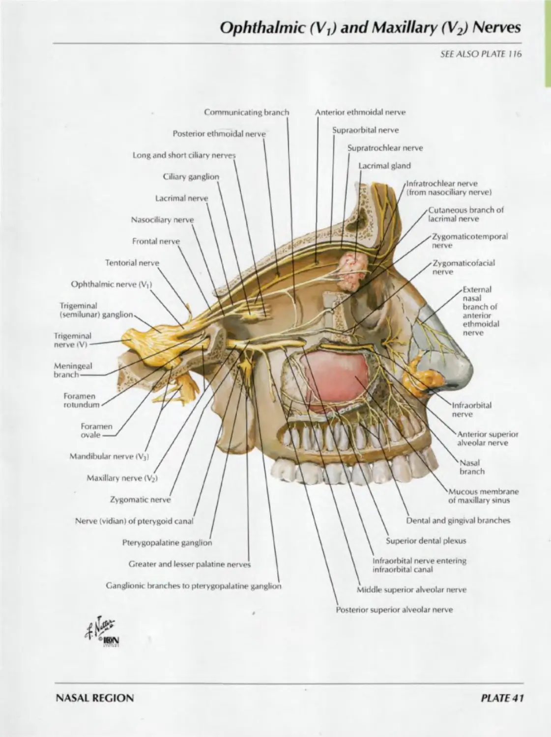
Ophthalmic (VJ and Maxillary (V2) Nerves
SEE ALSO PLATE 116
Communicating branch
Anterior ethmoidal nerve
Posterior ethmoidal nerve
Supraorbital nerve
Long and short ciliary nerves
Supratrochlear nerve
Lacrimal gland
Ciliary ganglion
Lacrimal nerve
-Intratrochlear nerve
(from nasociliary nerve)
Nasociliary nerve
Cutaneous branch of
lacrimal nerve
Frontal nerve
Zygomaticotemporal
nerve
Tentorial nerve
Zygomaticofacial
nerve
Ophthalmic nerve (V|>
Trigeminal
(semilunar) ganglion
Trigeminal
nerve (V) -
External
nasal
branch of
anterior
ethmoidal
nerve
Meningeal
branch---
Foramen
rotundum
Infraorbital
nerve
Foramen
ovale —
Anterior superior
alveolar nerve
Mandibular nerve <Vj)
Maxillary nerve (V^)
Nasal
branch
Zygomatic nerve
Mucous membrane
of maxillary sinus
Nerve (vidian) of pterygoid canal
Dental and gingival branches
Pterygopalatine ganglion
Superior dental plexus
Greater and lesser palatine nerves
Infraorbital nerve entering
infraorbital canal
Ganglionic branches to pterygopalatine ganglion
Middle superior alveolar nerve
Postenor superior alveolar nerve
NASAL REGION
PLATE 41
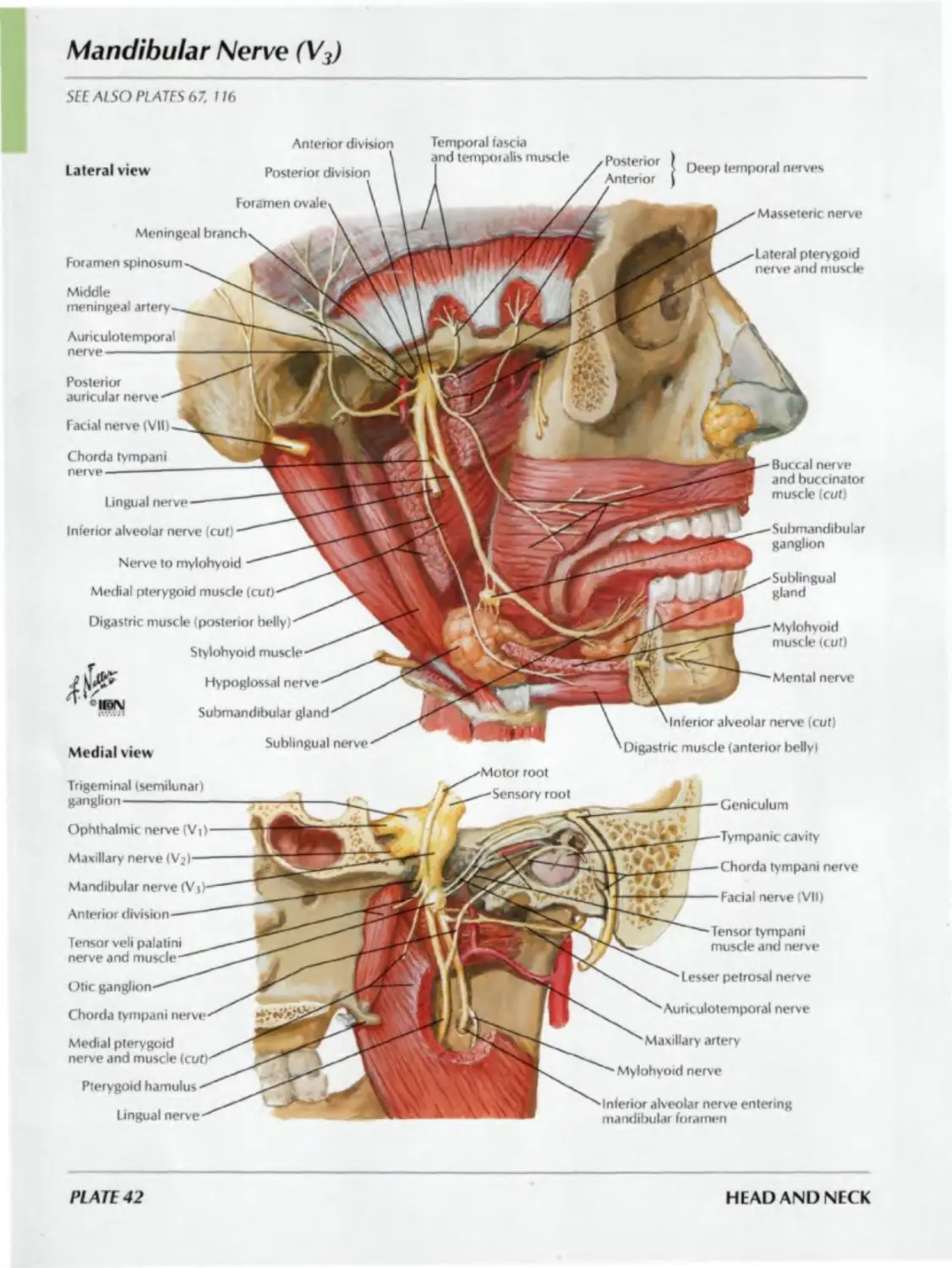
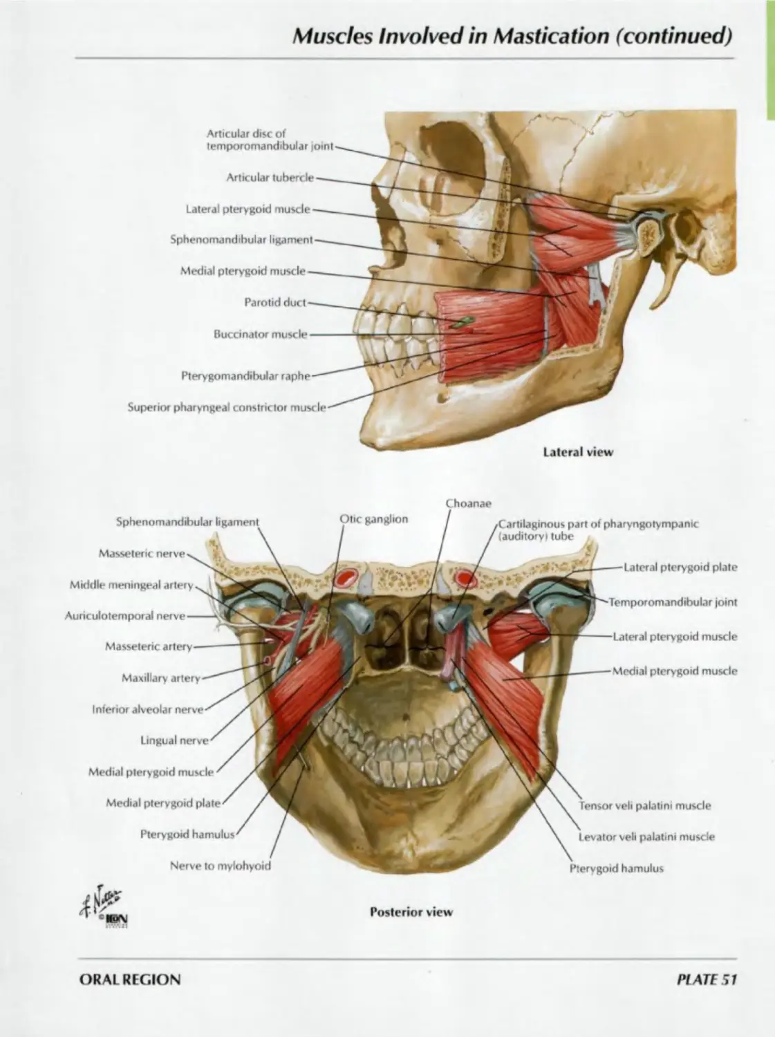
Mandibular Nerve (V3)
SEE ALSO PLATES 67. 116
Anterior
Lateral view
Posterior division
Foramen
Meningeal
Foramen spinosum
Facial nerve (VII)
Lingual nerve
Inferior alveolar nerve (cut)
Posterior
auricular nerve
Auriculotemporal
nerve-----------
Chorda tympani
nerve---------
Middle
meningeal artery
fascia
Posterior
Anterior
Nerve to mylohyoid
Medial pterygoid miisde (cut)
Digastric muscle (posterior helly)
Stylohyoid muscle
Hypoglossal nerve
Submandibular gland
Sublingual nerve
alveolar nerve (cut)
Digastric muscle (anterior belly I
Deep temporal nerves
Masseteric nerve
Lateral ptery goid
nerve and muscle
Buccal nerve
and buccinator
muscle (cut)
Submandibular
ganglion
Sublingual
gland
Mylohyoid
muscle (cut)
Mental nerve
Medial view
Trigeminal (semilunar)
ganglion-------------
Ophthalmic nerve (V|)
Maxillary nerve
Mandibular nerve
Anterior
Tensor veli
nerve and
Otic
Chorda tympani
Medial pterygoid
nerve and muscle
Pterygoid hamulus
Lingual nerve
root
Ceniculum
Chorda tympani nerve
Facial nerve (VII)
Lesser petrosal nerve
Auriculotemporal nerve
Maxillary artery
Mylohyoid nerve
Tensor tympani
muscle and nerve
Interior alveolar nerve entering
rnandilsular foramen
lie cavity
PLATE 42
HEAD AND NECK
Nose and Paranasal Sinuses: Cross Section
Lateral
pterygoid
muscle
Cartilage
of
auditory
tube
Major alar
cartilage
Vomer
Inferior nasal concha
Facial artery
Maxillary bone
Nasal vestibule
Nasal septal cartilage
Facial vein
Maxillary sinus
Masseter muscle
Coronoid
process of
mandible
Neck of
mandible
Parotid
gland
Styloid
process
Accessory
nerve
Facial nerve
Internal carotid
artery
Vagus nerve
Hypoglossal nerve
Retroman-
dibular
vein
Glosso
pharyngeal
nerve
Auricular
cartilage
Mastoid air cells
Sphenoid bone
Medial pterygoid
muscle
Lateral pterygoid
plate
Levator veli
palatini muscle
Pharyngeal recess
Longus capitis
muscle
Rectus capitis
anterior
muscle
Superficial
temporal
artery
Sympathetic
trunk
Styloglossus
muscle
Stylohyoid
muscle
Medulla oblongata
°«M
NASAL REGION
PLATE 43
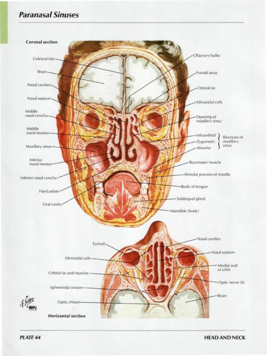
Paranasal Sinuses
Coronal section
Celebral falx
Olfactory bulbs
Brain
Frontal sinus
Nasal cavities
Orbital fat
Nasal septum
Ethmoidal cells
Middle
nasal concha
Opening of
maxillary sinus
Middle
nasal meatus
Maxillary sinus
Infraorbital
Zygomatic
Alveolar
Recesses of
maxillary
sinus
Inferior
nasal meatus
Buccinator muscle
Inferior nasal concha
Alveolar process of maxilla
Hard palate
Body of tongue
Sublingual gland
Oral cavity
Mandible (body)
Nasal cavities
Fy eball
Nasal septum
Fthmoidal cells
Orbital fat and muscles —
Sphenoidal sinuses----
Optic chiasm------
Horizontal section
Medial wall
of orbit
Optic nerve (II)
Brain
PLATE 44
HEAD AND NECK
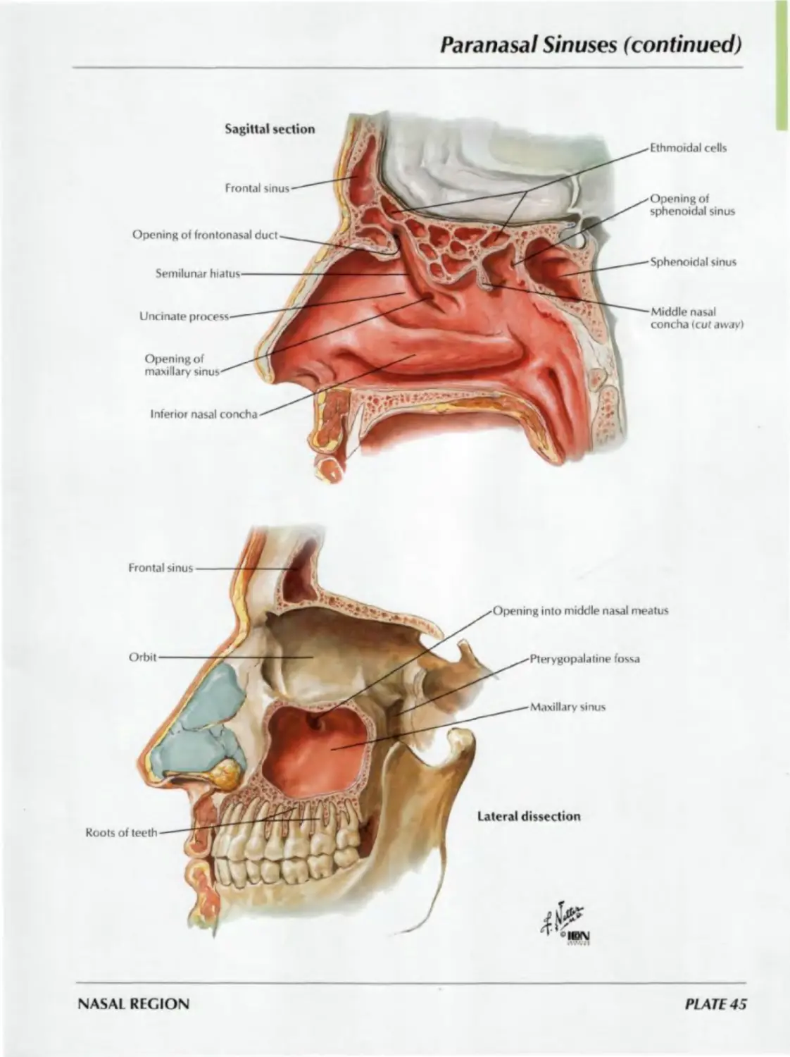
Paranasal Sinuses (continued)
Opening of
maxillary
Sagittal section
Frontal sinus
Opening of frontonasal duct
Semilunar hiatus
Uncinate
Inferior nasal concha
Ethmoidal cells
Opening of
sphenoidal sinus
Sphenoidal sinus
Middle nasal
concha (cut away)
NASAL REGION
PLATE 45
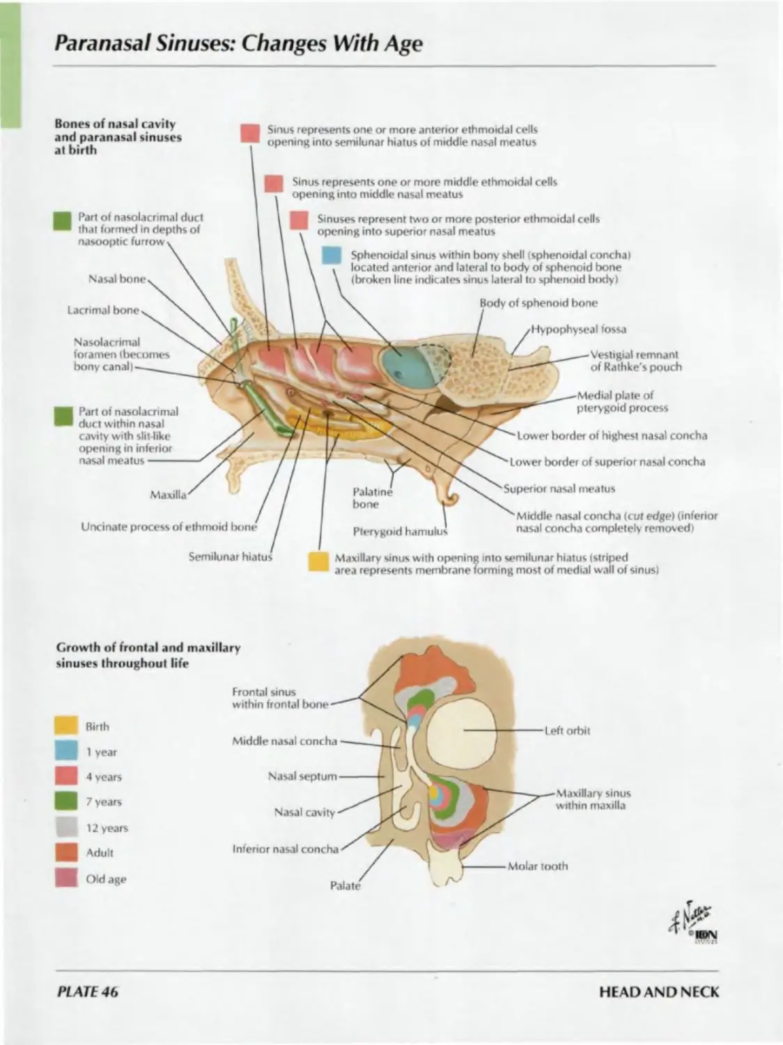
Paranasal Sinuses: Changes With Age
Bones of nasal cavity
and paranasal sinuses
at birth
Nasal bone
Body of sphenoid bone
Lacrimal bone
Hypophyseal fossa
Sinus represents one or more anterior ethmoidal cells
opening into semilunar hiatus of middle nasal meatus
Part of nasolacrimal
duct within nasal
cavity with slit-like
opening in inferior
nasal meatus-------
Sinuses represent two or more posterior ethmoidal cells
opening into superior nasal meatus
Sinus represents one or more middle ethmoidal cells
opening into middle nasal meatus
Lower border of highest nasal concha
Lower border of superior nasal concha
Superior nasal meatus
Maxilla
Uncinate process of ethmoid bone
Pterygoid hamulus
Semilunar hiatus
Vestigial remnant
of Rathke's pouch
Palatine
bone
Middle nasal concha (cut edge) (inferior
nasal concha completely removed)
Maxillary sinus with opening into semilunar hiatus (striped
area represents membrane forming most of medial wall of sinus)
Part of nasolacrimal duct
that formed in depths of
nasooptic furrow
Nasolacrimal
foramen (becomes
bony canal)
Growth of frontal and maxillary
sinuses throughout life
Birth
Sphenoidal sinus within bony shell «sphenoidal concha)
located anterior and lateral to body of sphenoid bone
(broken line indicates sinus lateral to sphenoid body)
Medial plate of
pterygoid process
1 year
4 vears
7 years
12 years
Adult
Old age
Middle nasal concha
Nasal septum
Nasal cavity
Inferior nasal concha
Frontal sinus
within frontal bone
Left orbit
Molar tooth
Palate
Maxillary sinus
within maxilla
PLATE 46
HEAD AND NECK
Inspection of Oral Cavity
Posterior wall of pharynx
Parotid papilla with
opening of parotid duct
Palatoglossal arch
Palatine tonsil
- Philtrum of lip
-Soft palate
- Palatopharyngeal arch
Uvula
Frenulum of upper lip——
Lingual minor salivary gland
Deep lingual artery and veins
and lingual nerve *>
Fimbriated told-—.J
Submandibular duct——1
Sublingual gland——1
Frenulum of tongue—-j
Sublingual fold with openings
of sublingual ducts----------
Sublingual caruncle with opening
of submandibular duct----------
Frenulum of lower lip—
ORAL REGION
PLATE 47
Roof of Mouth
Incisive papilla
Transverse
Palatine raphe
Palatine process
of maxilla
Palatine glands
Horizontal plate
of palatine bone
Palatine
aponeurosis
(from tensor
veli palatini
muscle)
Uvular muscle
Molar minor
salivary glands
Palatopharyngeus
muscle----------
fossa
Greater palatine artery and nerve
Greater palatine foramen
Lesser palatine foramen
Lesser palatine artery and nerves
of tensor veil
palatini muscle
Pterygoid hamulus
Interdigitating fibers of
levator veli palatini muscle
Buccinator muscle
Pterygomandibular raphe
Superior pharyngeal
constrictor muscle
Palatoglossus muscle
Palatine tonsil
Anterior view
Basilar part of
occipital bone
Cartilaginous part of
pharyngotympanic
(auditory) tube
Levator veli palatini muscle (cut)
tensor veli palatini muscle
Pharyngobasilar
fascia
Medial pterygoid muscle (cut)
Levator veli palatini muscle
Medial pterygoid plate
Choanae
Pterygoid hamulus
Pterygoid hamulus
Tendon of tensor veli palatini muscle-
Superior pharyngeal
constrictor muscle (cut)
Levator veli palatini muscle (cut)
Palatopharyngeus muscle
Palatopharyngeus muscle (cut)'
Uvular muscle
Posterior view
PLATE 48
HEAD AND NECK
Floor of Mouth
SEE ALSO PLATE 25
Lateral, slightly inferior view
Hyoglossus muscle
Mylohyoid muscle
Fibrous loop for intermediate digastric tendon
Digastric muscle (anterior belly)
Median raphe between mylohyoid muscles
Mastoid
process
Styloid process
Digastric muscle
(posterior belly)
muscle
horn I
Lesser horn ' Hyoid bone
Thyrohyoid muscle
muscle
Sternohyoid muscle
Hyoglossus
muscle
Digastric muscle (anterior belly)
Mylohyoid muscle
Stylohyoid muscle
muscle (posterior belly i
Fibrous loop for intermediate digastric tendon
Hyoglossus
muscle (cut)
Lesser
• Body-
Greater
Posterosuperior view
Inferior alveolar nerve and artery
Mylohyoid nerve and artery
Submandibular gland and duct
Mylohyoid muscle
Geniohyoid muscle—
Hyoid bone
mental spine for
origin of genioglossus muscle
ORAL REGION
PLATE 49
Muscles Involved in Mastication
FOR FACIAL MUSCLES SEE PLATE 22
Zygomaticus
major muscle
Mentalis
muscle--
Depressor labii
inferioris muscle'
Zygomatic arch
Parotid duct
Buccinator muscle
Articular disc of
temporomandibular joint
Deep part ( Masseter
Superficial part i muscle
Maxillary artery
Masseteric nerve
and artery
Insertion of
masseter muscle
Lateral pterygoid
muscle
Depressor anguli
oris muscle
Temporalis muscle
Insertion of temporalis muscle to
coronoid process of mandible-____
Parotid duct—
Buccinator muscle-.____
Orbicularis oris muscle—
Levator labii
superioris
alaeque nasi
muscle-------
Temporal fascia
/Superficial layer
/Deep layer
Levator labii
superioris
muscle \
Zygomaticus
minor muscle
Levator
anguli
oris
muscle-—'
Orbicularis
oris muscle
PLATE 50
HEAD AND NECK
Muscles Involved in Mastication (continued)
Masseter к nerve
Middle meningeal artery
Auriculotemporal nerve
Masseteric artery---
Maxillary artery
Inferior alveolar nerve
Lingual nerve
Medial pterygoid muscle
Medial pterygoid plate
Pterygoid hamulus
Nerve to mylohyoid
Articular tubercle
Lateral pterygoid muscle
Sphenomandibular ligament
Medial pterygoid muscle
Parotid duct
Buccinator muscle
Superior pharyngeal constrictor muscle
Articular disc of
temporomandibular joint
Lateral view
Otic ganglion
Choanae
Posterior view
'Cartilaginous part of pharyngotympanic
(auditory) tube
Ml
Lateral pterygoid plate
Lateral pterygoid muscle
—Medial pterygoid muscle
tensor veli palatini muscle
Levator veli palatini muscle
Pterygoid hamulus
ORAL REGION
PI ATE 57
Teeth
Deciduous
(primary)
Usual age of
eruption
Central incisor
(8-10 months)
Lateral incisor
(8-10
1 st molar
(15-21 months)
2nd molar
(20-24 months)
2nd molar
(20-24 months)
1 st molar
(15-21 months)
Canine I cuspid I
(16-20 months)
Lateral incisor
(15-21 months)
Permanent
(colored blue)
Usual age of
eruption
Canine (cuspid)
(16-20
Central incisor (7th year)
Lateral incisor (8th year)
Canine (cuspid) (11th—12th year)
premolar (9th year)
2nd premolar (10th year)
1st molar (6th year)
2nd molar (12th-13th year)
- 3rd molars (17th-25th year)
Central incisor
(6-9
2nd molar (12th-l 3th year)
1st molar (6th year)
2nd premolar (10th year)
I st premolar (9th year)
Canine (cuspid) (11th-12th year)
I aterai incisor (Sth year)
Central incisor (7th year)
Horizontal
plate of
palatine
bone
Incisive fossa
Palatine process
of maxilla
Central incisors
Lateral incisors
Canines
1 st premolars
1 st molars
2nd molars
palatine foramina
Upper permanent teeth
lower permanent teeth
PLATE 52
HEAD AND NECK
Teeth (continued)
Enamel
Dentine and dentinal tubules
Crown
Neck
Root
Interglobular spaces
Odontoblast layer
Interproximal spaces
Dental pulp containing vessels
and nerves
Gingival (gum) epithelium (stratified)
Gingival groove
Lamina propria of gingiva (gum)
(mandibular or maxillary periosteum)
Periodontium
(alveolar periosteum)
Papilla
'Cement
Root (central) canals
containing vessels and nerves
•Bone
•Apical foramina
Left lower
permanent
teeth:
labiobuccal
view
Incisors Canines Premolars Molars
(cuspids)
ORAL REGION
PLATE 53
Tongue
Root
Body -
Midline groove (median sulcus)
Dorsum of tongue
Filiform papillae
Foliate papillae
Epiglottis
Median glossoepiglottic fold
glossoepiglottic fold
Vallecula
alatopharyngeal arch and muscle (cut)
Palatine tonsil (cut)
Lingual tonsil (lingual nodules)
Palatoglossal arch and muscle (cut)
-Foramen cecum
sulcus
Vallate papillae
Apex
PLATE 54
HEAD AND NECK
Tongue (continued)
SEE ALSO PLATES 30, 42, 49, 122
Palatopharyngeus muscle
Palatoglossus muscle
Inferior longitudinal muscle of longue \
Genioglossus muscle
Mylohyoid muscle (cut)
Geniohyoid muscle
\ Mastoid pint ess
Digastric muscle iposterior
\ bells in up
\ Styloid process
\ Pharyngohasilar fascia
\ 'Superior pharyngeal constrictor muscle
'Stylohyoid ligament
Styloglossus muscle
Glossophary ngeal part of superior
X. x pharyngeal constrictor
'Stylopharyngeus muscle
Stylohyoid muscle
Middle pharyngeal constrictor muscle
4? jr 'ч ^Digastric muscle (posterior belly! (cut)
Hyoglossus muscle
\ Intermediate tendon of digastric muscle (cut)
^Fibrous loop for intermediate digastric tendon
Hyoid bone
Lingual nerve
Submandibular ganglion
Deep lingual artery
and venae comitantes
Artery to frenulum
Superior pharyngeal
constrictor muscle
/Styloglossus muscle
/-Palatoglossus muscle
^/Stylohyoid ligament
/-Stylopharyngeus muscle
/Hyoglossus muscle (cut)
S/-Lingual artery
/•External carotid artery
>7 /-Internal jugular vein
__--Retromandibular vein
Submandibular duct
Facial vein
Common trunk for facial,
retromandibular and
lingual veins
Sublingual artery and vein
\ . ^Lingual vein
Suprahyoid artery
Geniohyoid muscle
Hyoid bone
Hypoglossal nerve
Dorsal lingual artery and vein
Vena comitans of hypoglossal nerve
ORAL REGION
PLATE 55
Tongue and Salivary Glands: Sections
Horizontal section
below lingula of
mandible (superior view)
demonstrating bed
of parotid gland
Orbicularis oris muscle
Buccinator muscle^
Buccopharyngeal fascia
Facial artery and vein^
Pterygomandibular raphe»
Lingual nerve and superior pharyngeal constrictor muscle. \
Masseter muscle» \ \
Palatoglossus muscle in palatoglossal arch v \ \
Palatine tonsil \ \ \ \
Palatopharyngeus muscle in palatopharyngeal arch» \\ \ \
Ramus of mandible » X \\л'
Inferior alveolar artery, vein and nerve». X» \ ХлД
Medial pterygoid muscle» \
Styloglossus muscle» X/nflS
Facial nerve »^ ЛмДИ
Retromandibular vein *» \
External carotid artery
| Parotid gland
Stylopharyngeus muscle
Stylohyoid muscle
Sternocleidomastoid muscle
Digastric muscle (posterior belly)
i. Axis(C$
Internal jugular vein, internal carotid artery
and nerves IX, X and XII in carotid sheath -—
Superior cervical sympathetic ganglion
/ / Longus capitis muscle
/ Prevertebral fascia
Buccopharyngeal fascia
and retropharyngeal space
-----------Superior longitudinal muscle
Vertical and transverse muscles
Inferior longitudinal muscle
Styloglossus muscle
Buccinator muscle
Muscles of facial expression
Hyoglossus muscle
Genioglossus muscle
Sublingual salivary gland
Submandibular duct
Mandibular canal, inferior alveolar artery, vein and nerve
nerve
Nerve to mylohyoid
comitans of hypoglossal nerve (to lingual vein)
lingual artery
Facial artery
Hypoglossal nerve (XII)
Submandibular salivary gland
Submandibular lymph node
Mylohyoid muscle
Facial vein
of tongue
digastric tendon
Platysma muscle
bone
Frontal section
behind 1st molar tooth
(anterior view)
demonstrating
beds of sublingual and
submandibular glands
PLATE 56
HEAD AND NECK
Salivary Glands
SEE ALSO PLATES 21. 49. 127, 128. 160
Superficial temporal artery and vein
and auriculotemporal nerve
Branches of facial nerve
Transverse facial artery
Accessory parotid gland
Parotid duct
Buccinator muscle (cut)
Masseter muscle
lingual nerve
Submandibular ganglion
Tongue —
Frenulum of tongue
Sublingual fold with openings
of sublingual ducts----------
Sublingual caruncle with opening
of submandibular duct---------
Sublingual gland'
Submandibular duct 1
Sublingual artery and vein
Mylohyoid muscle (cut)
Digastric muscle (anterior belly)
Submandibular gland
Hyoid bone
Facial artery and vein
Parotid gland
Retromandibu
vein (anterior and
W posterior branches)
L’lgasfrt muscle
Stylohyoid muscle
v \ ' External jugular vein
\ 'Sternocleidomastoid muscle
. 'Common trunk receiving facial,
\ anterior branch of retromandibular,
\ and lingual veins
' Internal jugular vein
External carotid artery
Parotid gland:
totally serous
Submandibular gland:
mostly serous, partially mucous
Sublingual gland:
almost completely mucous
ORAL REGION
PLATE 57
Afferent Innervation of Mouth and Pharynx
SEE ALSO PLATES 42. 48. 55, 60, 116, 117, 119, 127. 129
Trigeminal (V) (maxillary Vj)
Via superior alveolar
nerves
Via pterygopalatine
ganglion and nasopalatine
and greater and lesser
palatine nerves
Trigeminal nerve (V) - —
Glossopharyngeal nerve (IX)
Vagus nerve (X) 'у'/'у'/У
Facial nerve (VII)
Facial (VII) (intermediate nerve)
greater petrosal nerve, pterygopalatine
ganglion and greater and lesser palatine nerves
Glossopharyngeal (IX)
pharyngeal plexus
tonsillar branches
plus general sensation
via lingual branches
Trigeminal (V) (maxillary Vj)
pharyngeal branch of
pterygopalatine ganglion
Facial (VII) (intermediate nerve)
Via greater petrosal nerve and
pterygopalatine ganglion
Vagus(X)
Via internal branch of
superior laryngeal nerve
Glossopharyngeal (IX)
Via pharyngeal plexus
Via tonsillar branches
Taste plus general
sensation via
branches
Vagus(X)and
pharyngeal (IX)
Via pharyngeal
plexus
Trigeminal (V)
(maxillary V2)
Via pterygopalatine
ganglion and
nasopalatine and
greater and lesser
palatine nerves
Via superior
alveolar nerves
Vagus(X)
Via internal
branch of
superior
laryngeal
Trigeminal (V)
(mandibular Vj)
Via buccal nerve
inferior
alveolar nerve
Trigeminal (V)
(mandibular Vy)
lingual
Facial (VII) (intermediate nerve)
via chorda tympani and lingual nerve
РЕМЕ 58
HEAD AND NECK
Pharynx: Median Section
Frontal sinus
Sphenoidal sinus -
Nasal septum—^
Nasopharynx -___
Soft palate
Palatine glands x,
I lard palate
Oral cavity
Incisive canal
Palatine tonsil —_
Body of tongue—x
Oropharynx x
Foramen cecum -
Lingual tonsil-
Genioglossus
muscle-—-
Root of tongue
Epiglottis
Mandible
(,eniohvoid mus< l<-----*—-rjs •—
Hyoid bone------——" ’
Hyoepiglottk ligament -- -——
Thyrohy oid membrane—' '
Laryngophary nx
laryngeal inlet (aditus)-^
Thyroid cartilage
Vocal fold
Sella turcica
Pharyngeal opening of pharyngotympanic
(auditory) tube
«Sphenooccipital synchondrosis
/ «Pharyngeal tonsil
/ / .Pharyngeal tubercle of occipital bone
III /Pharyngeal raphe
к/ / / /Anterior longitudinal ligament
/L / /Anterior atlantooccipital membrane
/Apical ligament of dens
Anterior
arch of
atlas (Cl
vertebra)
Dens of
axis (C2
vertebra)
Pharyngeal
constrictor
muscles
—
Bucco-
pharyngeal
fascia
Retro
pharyngeal
space
Prevertebral
fascia and
anterior
longitudinal
ligament
-Vertebral
bodies
Transverse arytenoid muscle-1
Cricoid cartilage
Trachea-
Esophagus'^
Esophageal muscles-'"^
Thyroid gland''
Investing layer of (deep) cervical fascia*
Pretracheal fascia
Suprasternal space'
Manubrium of sternum -
PHARYNX
PL ATE 59
Fauces
Medial view
Median (sagittal) section
Triangular fold
Tongue (drawn forward and downward)
Lingual tonsil
Epiglottis
Vallecula
Pharyngeal mucosa removed
Cartilaginous part of pharyngotympanic (auditory) tube
Medial pterygoid plate
Tensor veli palatini muscle and tendon
Levator veli palatini muscle
Ascending palatine
Pharyngeal branch of ascending pharyngeal artery
Lesser palatine artery
Salpingopharyngeus muscle
Pterygoid hamulus
Pterygomandibular raphe
Tonsillar branch of lesser palatine artery
Superior pharyngeal constrictor muscle
Tonsillar branch of ascending pharyngeal artery
Palatoglossus muscle
Palatopharyngeus muscle
Tonsillar branch of ascending palatine artery
Tonsillar branch of facial artery
Tonsillar branch of dorsal lingual artery
Glossopharyngeal nerve (IX) and tonsillar branch
Stylohyoid ligament
Hyoglossus muscle
Middle pharyngeal constrictor muscle
Stylopharyngeus muscle
Sphenoidal sinus
Pharyngeal tonsil
Torus tubarius
tuberde (basilar part of occipital bone)
Pharyngeal raphe
Pharyngeal opening of pharyngotympanic (auditory) tube
Hard palate
Pharyngeal recess
Salpingopharyngeal fold
Soft palate
Palatine glands
Uvula
Semilunar fold
Supratonsillar fossa
Palatine tonsil
Palatopharyngeal arch
Palatoglossal arch
PLATE 60
HEAD AND NECK
Muscles of Pharynx: Median (Sagittal) Section
I Hyoglossus IBR
I muscle / YA is
/>
Geniohyoid /
I muscle /
Mylohyoid /
muscle / д W
Hyoid bone / \ Л
Thyrohyoid / /aj
membrane —' / \
Thyroid cartilage' j
Median /
cricothyroid ligament'
Corniculate and ,
arytenoid cartilages'
Cricoid cartilage'1
Trachea
Medial pterygoid plate
^Cartilaginous part of pharyngotympanic (auditory* tube
-•Tensor veli palatini muscle
-xPharyngobasilar fascia
Levator veli palatini muscle
у --Palatine aponeurosis and tendon of
•ensor veli palatini muscle
V»-----Pharyngeal tubercle (basilar part of occipital bone)
Sy -------Pharyngeal raphe
Anterior longitudinal ligament
“Anterior atlantooccipital membrane
— -Apical ligament of dens
'—Salpingopharyngeus muscle
— Muscles of soft palate
Palatopharyngeal sphincter (Passavant's ridgei
— Pterygoid hamulus
— Superior pharyngeal constrictor muscle
- Pterygomandibular raphe
Palatopharyngeus muscle
Buccinator muscle
Glossopharyngeal part of
superior pharyngeal constrictor
•Stylopharyngeus muscle
•Stylohyoid ligament
' Styloglossus muscle
'Middle pharyngeal constrictor muscle
-Fibers to pharyngoepiglottic fold
-Buccopharyngeal fascia and
retropharyngeal space
“Prevertebral fascia and anterior
longitudinal ligament
Internal branch of superior laryngeal nerve
^•Longitudinal pharyngeal muscles
''"-Inferior pharyngeal constrictor muscle
" -Pharyngeal aponeurosis
I---Cricopharyngeus muscle
(part of inferior pharyngeal constrictor)
t“---Cricoid attachment of longitudinal
I esophageal muscle
A------Circular esophageal muscle
, y-—'Longitudinal esophageal muscle
Margins of middle pharyngeal
constrictor muscle
Margins and attachments of inferior
pharyngeal constrictor muscle
PHARYNX
P/.A7F67
Pharynx: Opened Posterior View
Nasopharynx
Oropharynx
<
Laryngopharynx
Esophagus <
Base of skull (basilar part of occipital bonet
I Pharyngeal tonsil
— stvloid
Ш I UL—— Nasal septum
—• Torus
; j f ———I —Pharyngeal opening
I o’pl'.irvnuotsmp.inii
r —X______.у (auditory) tube
~Ph,iu nge.il io
Parotid gland
Tor as les ,1 tor I US tolii
caused by levator
veli palatini muscle)
^'Salpingopharyngeal fold
w > _ '"Soft palate
'' Angle of mandible
''Submandibular gland
Uvula
" 'Palatine tonsil
~~ Root of tongue
Palatopharyngeal arch
Prominence caused by greater horn
of hyoid bone
Epiglottis
Laryngeal inlet (aditus)
^'Prominence caused by superior horn
of thyroid cartilage
^'Aryepiglottic fold
^Piriform fossa
^Cuneiform tubercle
''Fold over internal branch
of superior laryngeal nerve
''Corniculate tubercle
'Interarytenoid notch
Prominence over lamina
of cricoid cartilage
•Trachea
PLATE 62
HEAD AND NECK
Muscles of Pharynx: Partially Opened Posterior View
Basilar part of occipital bone
Pharyngeal tubercle
SEE ALSO PLATE 231
Pharyngeal
tonsil
Styloid
process
Cartilaginous part
of pharyngotympanic
(auditory) tube
Digastric
muscle
i posterior
belly)-—_
Pharyngobasilar
fascia
Stylohyoid
muscle-—
Choana
Stylopharyngeus
muscle---------
Levator veli
palatini muscle
Superior pharyngeal
constrictor muscle
Accessory muscle
bundle from
petrous part of
temporal bone-^
Salpingopharyngeus
muscle
Uvula
Medial
pterygoid muscle
Palatopharyngeus muscle
Pharyngobasilar fascia
Middle pharyngeal
constrictor muscle
Pharyngeal raphe
Stylopharyngeus muscle
Superior pharyngeal
constrictor muscle —
Pharyngoepiglottic told
Hyoid bone (tip of greater horn)
Aryepiglottic fold
Middle pharyngeal constrictor muscle
Inferior pharyngeal
constrictor muscle (cut edge)
Epiglottis
Longitudinal pharyngeal muscles
Inferior pharyngeal constrictor muscle
Superior horn of
thyroid cartilage
Cuneiform tubercle
Thyrohyoid membrane
Corniculate tubercle
(Transverse and oblique)
arytenoid muscles •
Internal branch of
superior laryngeal nerve
Pharyngeal aponeurosis
Posterior cricoarytenoid muscle
Cricopharyngeus muscle (part of
inferior pharyngeal constrictor)
Cricopharyngeal muscle (part of
inferior pharyngeal constrictor) -
Posterior border of thyroid cartilage lamina
Longitudinal esophageal muscle
Cricoid attachment of longitudinal
esophageal muscle
Circular esophageal muscle
PHARYNX
PMTf 63
Muscles of Pharynx: Lateral View
Pharyngobasilar fascia
Tensor veli palatini muscle\
Levator veli palatini muscle*
Lateral pterygoid plate
Pterygoid hamulus
Buccinator muscle tcut)
Pterygomandibular raphe —
Digastric muscle (anterior belly)
Mylohyoid muscle
Hyoid bone
Stylohyoid muscle (cut)
Thyroid cartilage
Median cricothyroid ligament
Digastric muscle
\ (posterior belly I (cut)
Styloid process
Superior pharyngeal constrictor muscle
Styloglossus muscle
Stylohyoid ligament
Stylopharyngeus muscle
Middle pharyngeal constrictor muscle
Hyoglossus muscle
Greater horn of hyoid bone
Superior horn of thyroid cartilage
Thyrohyoid membrane
Inferior pharyngeal constrictor muscle
Tendinous arch
Zone of sparse muscle fibers
Cricopharyngeus muscle
(part of inferior pharyngeal constrictor)
Cricothyroid muscle
Cricoid cartilage
Esophagus
Trachea
PLATE 64
HEAD AND NECK
Arteries of Oral and Pharyngeal Regions
SFF ALSO PLATES 30, 36
. ( Supraorbital artery^
ophthalmic <
artery ( Supratrochlear artery,
Middle meningeal artery-s.
Deep temporal arteries v.
Dorsal nasal artery-»^.
Masseteric artery-_ <'
Angular artery-\
Infraorbital artery-Vs
Sphenopalatine artery---Ж
Descending palatine artery----
Posterior superior
alveolar artery—---
Superior labial artery-—
Buccal artery—^
Buccinator muscle
and parotid duct icut)-^«^c^
Inferior labial artery-^
Superior pharyngeal—
constrictor muscle
Mental branch of
inferior alveolar artery-'
Inferior alveolar artery У
and lingual branch —S
Facial artery
Submental artery / /
Mylohyoid branch of / /
inferior alveolar artery /
Submandibular gland'' /
Hypoglossal nerve (XII и /
Suprahyoid artery/
External carotid artery'
Superior laryngeal artery
Superior thyroid artery'
Cricothyroid artery'
Common carotid artery^ I
Vertebral artery-Ж
Subclavian artery---
—Transverse
facial
artery (cut)
“Superficial
temporal artery
'''“Maxillary artery
^Posterior auricular artery
Ascending pharyngeal artery
Occipital artery and
sternocleidomastoid branch
“Glossopharyngeal nerve (IX)
'Ascending palatine artery
•"Tonsillar artery
Facial artery
''“Lingual artery
^Ascending pharyngeal artery
^Internal carotid artery
""Vagus nerve (X)
---Superior cervical
cardiac nerve
"Sympathetic trunk
к ^"Anterior scalene muscle
У~~ - Phrenic nerve
Middle scalene muscle
—Ascending cervical arte'v
—Inferior thyroid artery
* tn—;Mibn.,.'ti i1.
Suprascapular artery
Costocervical trunk
LJ^B^'Thvrocervica trunk
PHARYNX
«A7F65
Veins of Oral and Pharyngeal Regions
SEE ALSO PLATES 19. 27.98
vein
Supraorbital vein
Nasofrontal vein-
Superior ophthalmic vein —
Angular vein \
External nasal vein-----
Fmissary vein (Vesalius)
communicating with
cavernous sinus
Infraorbital vein
Posterior superior
alveolar veins ------
Palatine vein
Pterygoid plexus-*
Superior labial vein'
Deep facial vein-
Maxillary veins
Inferior labial vein
Mental vein -
Facial vein and artery
External palatine vein-*"
Submental vein
Submandibular gland ' /
Vena comitans of / /
hypoglossal nerve ' /
Dorsal lingual vein coursing /
medial to hyoglossus muscle ' /
lingual vein'
Communication to anterior jugular vein (cut)'
Superior laryngeal vein
Superior thyroid vein--/
Thyroid gland-""’/ i
Middle thyroid vein---- t
Inferior thyroid veins —- fjk
Termination of anterior jugular vein (rirfi—/J
Left brachiocephalic vein —
— < к
Superfu uil temporal
vein and artery
Transverse facial vein (cut)
Posterior auricular vein
Retromandibular vein
(anterior and
posterior branches)
xExternal jugular vein (cut)
Inferior alveolar vein and artery
'Occipital vein and artery
^Hypoglossal nerve (XII)
“Common trunk receiving
facial, anterior branch of
retromandibular, and
lingual veins
4 External carotid artery
Internal jugular vein
' Common carotid artery
Vagus nerve (X) and
sympathetic trunk
Г"—'Middle scalene muscle
L____Anterior scalene muscle
A у External jugular vein (cut)
НТ у Transverse cervical
vein (cut)
Suprast apular vein к uti
' . Jy —Subclavian artery
ЦЖ1---------Subclavian vein
PLATE 66
HEAD AND NECK
Nerves of Oral and Pharyngeal Regions
SFF ALSO PLATES 21,41, 42, 116, 119, 120
Zygomaticotemporal nerve (VjK
Deep temporal nerves (V j)>,
Masseteric nerve (V
Mandibular nerve (Vy)Г
Nerve to medial pterygoid and
tensor veli palatini muscles (Vj) (cut)-^
Zygomaticofacial nerve (Vj)»^.
Maxillary nerve (VJ—/
Infraorbital nerve (VM— V
Pterygopalatine ganglion -—k_
Greater and lesser \
palatine nerves (Vj)---------
Anterior, middle and posterior
superior alveolar nerves (VJ—
Nerve to lateral pterygoid
muscle (Vil (cut)---------
Buccal nerve (V d •'***7 1 JI
Chorda tympani nerve (VII)
Lingual nerve (Vi)
Medial pterygoid muscle (cur)
(lateral pterygoid removed)—
Inferior alveolar
nerve (Vj)---------------—
Mental nerve (Vj)x
Nerve to mylohyoid (Vji'z^^^W
Submandibular ganglion
Glossopharyngeal nerve (IX) /
and tonsillar branch-------' /
Hypoglossal nerve (XII)'
Carotid nerve (IX) and carotid body' S',
Nerve to thyrohyoid muscle (Cl, 2 via XII)' У
Internal and external branc hes s'
of superior laryngeal nerve (Xr .S
Superior root'\x''
Inferior root-'
Nerves to superior and inferior bellies of omohyoid,
sternohyoid and sternothyroid muscles (ansa cervicalis)
Recurrent laryngeal nerve (X)—----’
Sympathetic trunk and middle cervical ganglion''
Ansa cervicalis (Cl, 2, 3) |
Auric lllc И.ТИр. .r I
nerve (Vyi
Mui die
Superficial temporal
* artery (cut)
^^^4Facial nerve (VII) (cut)
Maxillary artery
Accessory nerve (XI) (cut)
^C1 spinal nerve
Б» (ventral ramus) (cut)
1 ^C2 spinal nerve
4 (ventral ramus) (cut)
I Pharyngeal plexus composed of
S branches from glossopharyngeal
Бй (IX), vagus (X) and sympathetic
Bba nerves
И Internal carotid artery
External carotid artery
Vagus nerve (X) and superior
cervical cardiac branch
Щк/' C4 spinal nerve
(ventral ramus) (cut)
^Phrenic nerve (C3, 4, 5)
1 '''•Ascending cervical artery
Middle scalene muscle
Anleruir s< alene must le
Brachial plexus (cut)
artery
Thyrocervical trunk
Vertebral artery
Subclavian artery
PHARYNX
РМ7Г 67
Lymph Vessels and Nodes of Head and Neck
SEE ALSO PLATE 204
Superficial parotid nodes
(deep parotid nodes
deep to and within
parotid gland) —
Subparotid node
Facial nodes
Nasolabial
Buccinator
Occipital nodes
Mastoid nodes
Mandibular
nodes-—-.
Sternocleidomastoid
nodes
Submandibular
nodes--------
Superior lateral
superficial cervical
(external jugular)
node
Jugulodigastric node
Submental
nodes----
Suprahyoid node--------------------
Superior deep lateral cervical
(internal jugular) nodes——'
Posterior lateral
superficial cervical
(spinal accessory)
nodes
Intercalated node
Superior thyroid nodes •—'
luguloomohyoid node
Anterior deep cervical
(pretracheal and thyroid) nodes
(deep to infrahyoid muscles) —
Inferior deep
lateral cervical
(scalene) node
Thoracic duct
Anterior superficial cervical
nodes (anterior jugular nodes)'
Transverse
cervical
chain of
nodes
lugular trunk -
Supraclavicular nodes —
Sulxlavian trunk and node
PLATE 68
HEAD AND NECK
Lymph Vessels and Nodes of Pharynx and Tongue
vessels
collecting vessels
Lymphatic drainage of pharynx:
posterior view
I ymphatic drainage of tongue
luguloomohyoid node
Submental node
Submandibular node
Jugulodigastric
luguloomohyoid node
Pharyngobasilar fascia
Retropharyngeal nodes
Internal jugular vein
Digastric muscle (posterior belly)
Sternocleidomastoid muscle
— Intercalated node
Jugulodigastric node
lateral cervical
(internal tugular) nodes
Basal collecting vessels
Communication to
side
Deep lateral cervical
(internal jugular) nodes
PHARYNX
PLATE 69
Thyroid Gland: Anterior View
External carotid artery'
Internal carotid artery
Superior thyroid artery and vein
Superior laryngeal artery
Thyrohyoid membrane
Ansa | Superior root
cervicalis | Inferior root
Common carotid artery
Internal jugular vein
Middle thyroid vein
Inferior thyroid veins
Ascending cervical artery
Inferior thyroid
Transverse cervical artery
Suprascapular artery
Thyrocervical trunk
Subclavian
artery and vein
Vagus nerve (X)
Right recurrent
laryngeal nerve
Brachiocephalic trunk
Brachiocephalic veins
Superior vena cava
Aortic arch
Hyoid bone
id muscles
jugular vein
Thyroid
gland
Thyroid cartilage (lamina)
Pretracheal lymph nodes
Phrenic nerve
Anterior scalene muscle
Vagus nerve (X)
Anterior jugular vein
1st rib (cut)
recurrent laryngeal nerve
Median cricothyroid
ligament
Superior laryngeal nerve
Internal branch
branch
Pyramidal lobe
(often absent
or small)
Right lobe
Left lobe
Isthmus
Thyroid cartilage
Cricothyroid ligament
Common carotid artery
Medial margin of
sternocleidomastoid muscle
Cricothyroid muscle
Cricoid cartilage
Thyroid gland
Cupula (dome) of pleura
Trachea
PL ATE 70
HEAD AND NECK
Thyroid Gland and Pharynx: Posterior View
Superior pharyngeal
constrictor muscle—
External carotid artery
Middle pharyngeal
constrictor muscle-
Internal carotid artery
Facial artery
Tip of greater horn
of hyoid bone------
Lingual artery
Inferior pharyngeal
constrictor muscle
Superior laryngeal nerve
—’Internal branch
— External branch
Pharyngeal raphe
Superior thyroid artery
Zone of sparse
muscle fibers^.
Superior laryngeal artery
Cricopharyngeus
muscle (part of inferior
pharyngeal constrictor)-.
Circular esophageal
muscle in V-shaped
area of sparse
longitudinal muscle
fibers (area of Laimerl —
Longitudinal
esophageal muscle------
Inferior thyroid vein
Common carotid artery
Vagus nerve
Internal jugular vein
Thyroid gland
(right lobe!
Superior parathyroid gland
Ascending cervical artery
Inferior parathyroid gland
Trachea
Inferior thyroid artery
Inferior bulb of
internal jugular vein
Right recurrent laryngeal nerve
Vertebral artery
Transverse cervical artery
Left subclavian
artery and vein
Suprascapular artery
Thyrocervical trunk
Internal thoracic
artery and vein-'
Right subclavian
artery and vein
Left brachiocephalic vein
Right brachiocephalic vein
Left recurrent laryngeal nerve
Brachiocephalic trunk
Arch of aorta
Right vagus nerve (X)
Left vagus nerve (X)
Superior vena cava
THYROID GLAND AND LARYNX
PLATE 71
Parathyroid Glands
SEE ALSO PLATE 76
Posterior view
External carotid artery
Hyoid bone
Internal carotid arterv
Superior laryngeal nerve''
Internal branch---------
External branch-------
—Superior thyroid artery
Superior laryngeal artery
Vagus nerve (X)
Epiglottis-
Superior thyroid artery-""
Common carotid artery—
Fibrous capsule of
thyroid gland (cut)-
Superior parathyroid gland
Left lobe of thyroid gland —
Ascending cervical artery—"
Inferior parathyroid gland —
Recurrent laryngeal nerve —
Esophagus------
Subclavian artery-—__
Trachea-----
Right lateral view
External carotid
Superior
Superior thyroid artery (cut)
Inferior pharyngeal constrictor muscle
Common carotid
Internal jugular vein
Inferior thyroid artery
Recurrent laryngeal nerve
Esophagus
Inferior pharyngeal
constrictor muscle (cut)
Common carotid artery
Fibrous capsule of
thyroid gland (cut)
Cricopharyngeus muscle (part of
inferior pharyngeal constrictor)
Superior parathyroid gland
Right lobe of thyroid gland
Inferior parathyroid gland
(may be more caudally located,
even within mediastinum)
Inferior thyroid artery
Recurrent laryngeal nerve
•Transverse cervical arterv
Suprascapular artery
-Thyrocervical trunk
Vertebral artery
Subclavian artery
Brachiot ephalk trunk
Internal branch of
superior laryngeal nerve
External branch of
superior laryngeal nerve
Superior parathy roid gland
Thyroid gland (right lobei
(retracted anterror/yt
Inferior parathyroid gland
if#
PLATE 72
HEAD AND NECK
Cartilages of Larynx
Arytenoid
articular surface
Posterior view
Corniculate cartilage
________E piglot t is--------
________— Hyoid bone-------
..^Thyrohyoid membrane _________
Superior horn of thyroid cartilage.
Corniculate cartilage^.
Arytenoid cartilage^.
__—Superior thyroid notch
Thyroid cartilage lamina----------
Vocal ligament----------
--Median cricothyroid ligament
• Inferior horn of thyroid cartilage-
________Cricoid cartilage_
________-Trachea—_________
Anterior view
Arch
Cricoid )
cartilage S Lamina
Muscular process
Vocal process
Arytenoid
cartilage
Anterosuperior view
Median
Lateral
Right lateral view
—— E pi glottis ----
Hyoepiglottic hgament-
------Hyoid bone-------
-Thyrohyoid membrane -
-Thyroid cartilage lamina
^^--Oblique line
-Laryngeal prominence
Corniculate cartilage —
Arytenoid cartilage—
Muscular process-—
Vocal process—"'”"
Vocal ligament-""^
Thyroepiglottic ligament
— Cricothyroid ligament-
Cricoid cartilage-—
^^Cricothyroid joint
^"--T r ac hea—
Medial view, median
(sagittal) section
THYROID GLAND AND LARYNX
PLATE 73
Intrinsic Muscles of Larynx
Cuneiform
tubercle
Corniculate
tubercle
Posterior
view
Oblique
arytenoid muscle
Transverse
arytenoid muscle
Postenor
cricoarytenoid
muscle
cartilage
Foramen for superior
laryngeal vessels and
internal branch of
superior laryngeal nerve
and transverse
muscles
Posterior cricoarytenoid
muscle
Straight j
part ( Cricothyroid
Oblique ( muscle
part )
Right lateral view
Epiglottis
Thyroepiglottic
muscle
Aryepiglottic
muscle
Oblique and
transverse
arytenoid muscles
Cricothyrord
muscle <cut away)
Lateral dissection
muscle
Lateral
cricoarytenoid
muscle
Conus elasticus
Posterior
cricoarytenoid
muscle
Thyroid articular
Lamina of
cricoid
Arytenoid
cartilage
Muscular
Vocal
Superior view
Conus
elasticus
Lamina of
thyroid
cartilage
Posterior cricoarytenoid
Lateral cricoarytenoid
Transverse and oblique arytenoid muscles
Cricothyroid muscle
Thyroarytenoid muscle
Vocalis muscle
Vocal ligament
PLATE 74
HEAD AND NECK
Intrinsic Muscles of Larynx
Posterior
view
Cuneiform
tubercle
Corniculate
tubercle
Oblique
arytenoid muscle
Transverse
arytenoid muscle
Posterior
cricoarytenoid
muscle
Cricoid cartilage
and transverse
id muscles
Posterior cricoarytenoid
muscle
Straight j
part I Cricothyroid
Oblique ( muscle
part )
Right lateral view
Foramen for superior
lary ngeal vessels and
internal branch of
superior laryngeal nerve
Thyroid articular
surface----—--------
Cricothyroid
muscle (cut away)
Lateral dissection
Epiglottis
Thyroepiglottic
muscle
Aryepiglottic
muscle
Oblique and
transverse
arytenoid muscles
Posterior
cricoarytenoid
muscle
Thyroarytenoid
muscle
Lateral
cricoarytenoid
muscle
Conus elasticus
Arytenoid ) Muscular
cartilage ) vocal
Superior view
Conus
elasticus
Lamina of
thyroid
cartilage
Posterior cricoarytenoid
Lateral cricoarytenoid
Transverse and oblique arytenoid muscles
Cricothyroid muscle
Thyroarytenoid muscle
Vocalis muscle
Vocal
PLATE 74
HEAD AND NECK
Action of Intrinsic Muscles of Larynx
Cricothyroid joint
(pivot point)----
Action of cricothyroid muscles
Lengthening (increasing tension)
of vocal ligaments
Action of posterior cricoarytenoid muscles
Abduction of vocal ligaments
Action of lateral cricoarytenoid muscles
Adduction of vocal ligaments
Action of vocalis and thyroarytenoid muscles
Shortening (relaxation) of vocal ligaments
Action of transverse arytenoid muscle
Adduction of vocal ligaments
THYROID GLAND AND LARYNX
PLATE 75
Nerves of Larynx
SEE ALSO PLATES 70-72, 231
Inferior pharyngeal
constrictor muscle
Cricothyroid muscle
Cricopharyngeus muscle
(part of inferior
pharyngeal < onstriclor)
Recurrent
laryngeal nerve
-Superior
laryngeal nerve
Internal branch
External branch
of Galen
Sensory branches to larynx
Aryepiglottic muscle
Thyroepiglottic muscle
nd muscle
Vocalis muscle
lateral cricoarytenoid muscle
Posterior cricoarytenoid muscle
articular surface
Recurrent laryngeal nerve
Right lateral view:
Internal branch of
superior laryngeal nerve
and oblique
arytenoid muscles
and posterior branches of
inferior laryngeal nerve
Anomalous (retroesophageal)
right subclavian
Right lateral view
Right vagus nerve (X)
Anomalous right inferior
laryngeal nerve (not recurrent)
Right common carotid artery
Left subclavian artery
Anomalous (retroesophageal)
right subclavian artery originating
from left side of aortic arch
Left recurrent laryngeal nerve
Arch of aorta
thyroid cartilage lamina removed
Left vagus nerve (X)
I eft common carotid artery
Left inferior laryngeal nerve
Left recurrent laryngeal nerve
Anterior view:
anomalous right
inferior laryngeal
nerve, not recurrent,
associated with
anomalous right
subclavian artery
PLATE 76
HEAD AND NECK
Eyelids
Superior palpebral conjunctiva:
tarsal (meibomianI glands
shining through
C orneoscleral junction
(corneal limbus)
Bulbar conjunctiva
over sclera
Superior lacrimal papilla and punctum
Plica semilunaris
Lacrimal caruncle in
lacrimal lake ilacus lacrimalis)
Inferior lacrimal papilla and punctum
Interior conjunctival fornix
Interior palpebral
tarsal glands shinii
Seen through
cornea
Frontal bone
Orbital s
Superior tarsus
Interior tarsus
Orbital septum
Zygomatic bone
lacrimal sac
Medial palpebral ligament
Infraorbital artery and nerve
Supraorbital artery and nerve
Supratrochlear artery and nerve
Dorsal nasal artery
and infratrochlear nerve
Bulbar conjunctiva
Palpebral conjunctiva
Lens
Anterior chamber
Posterior chamber
I evator palpebrae superiors musde
Orbital septum
Superior tarsal (Moller's) muscle (smooth)
Superior conjunctival fomix
Orbicularis oculi muscle (palpebral part)
Superior tarsus
Tarsal (meibomian) glands
Eyelashes (cilia!
Openings of tarsal glands
Inferior tarsus
Orbicularis oculi muscle (palpebral part)
Inferior conjunctival fornix
Orbital septum
Insertion of levator palpebrae
superioris muscle
Lateral palpebral ligament
and overlying raphe
ORBIT AND CONTENTS
PLATE 77
Lacrimal Apparatus
SEE ALSO PLATES 41, 127
Orbital part of
lacrimal gland
Plica
lacrimal papilla and punclum
lacrimal caruncle
Inferior lacrimal papilla and
Lacrimal canaliculi
Lacrimal sac
Nasolacrimal duct
Opening of
nasolacrimal duct
Palpebral part of
lacrimal gland
Excretory ducts of
lacrimal gland
Superior lacrimal papilla and punctum
Frontal bone
Inferior lacrimal papilla and
Orbital part of
lacrimal gland
Palpebral part of
lacrimal gland —
Lacrimal caruncle
Opening of nasolacrimal duct
lacrimal canaliculi
Lacrimal sac
Nasolacrimal duct
Inferior nasal meatus
Inferior nasal
concha (cut)
Middle nasal
concha
Excretory ducts of
lacrimal
Plica semilunaris
and lacrimal
PLATE 78
HEAD AND NECK
Fascia of Orbit and Eyeball
Tarsus of eyelid
conjunctiva
Cornea
Horizontal section
Medial palpebral ligament
Nasal cavity
Ethmoidal cells
Periorbita
Sclera
Episcleral space
Sphenoidal sinus
Bulbar conjunctiva
Periorbita
Sclera
Common tendinous ring
Retrobulbar fat
(orbital fat body)
Fascial sheath of eyeball
(Tenon's capsule)
Episcleral space
Lateral rectus muscle
and fascial sheath
IKXXJ
Check ligament of
medial rectus
Lateral palpebral ligament
Check ligament of
lateral rectus muscle
Medial rectus muscle
and fascial sheath
Fascial sheath of eyeball
(Tenon's capsule)
Optic nerve (II) and
outer sheath
Levator palpebrae superioris muscle and fascial sheath
Superior oblique muscle and fascial sheath
Medial rectus muscle and fascial sheath
Periorbita
Ethmoidal cell
sheath of eyeball
(Tenon's capsule)
Inferior rectus muscle
and fascial sheath
Inferior oblique muscle
and fascial sheath
Infraorbital nerve (Vj)
Fascial sheath of eyeball
(Tenon's
Episcleral
Retrobulbar fat (orbital fat body
Frontal section
Superior rectus muscle
and fascial sheath----
Lacrimal gland
Lateral rectus muscle and
fascial sheath-------------
Muscle attachments and nerves and vessels entering orbit
Superior orbital
Lacrimal nerve IV»)
Frontal nerve (V|)
Trochlear nerve (IV)
Superior ophthalmic vein
Lateral rectus
Superior branch of oculomotor nerve (III)
Inferior orbital fissure
Abducent nerve lVI)
nerve (Vi)
palpebrae superioris muscle
oblique muscle
rectus muscle
Medial rectus muscle
Optic nerve (II) )
artery )
Inferior rectus musde
branch of oculomotor nerve I III)
in optic canal
ORBIT AND CONTENTS
PL ATE 79
Extrinsic Eye Muscles
Superior view
Superior oblique muscle
Medial rectus muscle —
Inferior rectus
Common tendinous ring
tarsus
palpebrae
superioris muscle (cut)
Superior rectus muscle (cut)
Lateral rectus muscle
Optic nerve (II)
Superior rectus muscle (cut)
Levator palpebrae superioris muscle (cut)
Innervation and action of extrinsic eye muscles:
anterior view
Trochlear
nerve (TV)
Superior oblique muscle
Lateral rectus muscle ’
Note: Arrows indicate direction of eye
movement produced by each muscle
Oculomotor
nerve (III)
Levator palpebrae
superioris
Superior rectus muscle
Medial rectus muscle
Inferior rectus
Inferior oblique muscle
PLATF 80
HEAD AND NECK
Arteries and Veins of Orbit and Eyelids
SEE ALSO PLATES 14, 98
Superior view Supratrochlear artery- fl P* Dorsal nasal artery — ,» f W ¥BBj Anterior meningeal artery-^"^ ' Jgf jp| J B* Anterior ethmoidal artery-"'^ A • L^^B Posterior ethmoidal arterv—| 1 Continuation ot ophthalmic artery-— Muscular branch-""' Ophthalmic artery Internal carotid artery ДК J Anterior view Frontal branch of ^^B superficial temporal artery-"-^ ^KJL. Superior lateral palpebral artery-'"""’^ Ж Inferior lateral palpebral artery Zygomaticofacial artery , Transverse facial artery-''"’^ Infraorbital artery Supratrochlear vein Lateral view , ... . Supraorbital vein-"^ Superior ophthalmic vein Cavernous sinus к f^^B >Жви Inferior ophthalmic vein —— Pterygoid plexus —>У Maxillary vein-—- Cl -H- Retromandibular vein - --Medial palpebral artery r. -^-Lateral palpebral artery у —Lacrimal gland Zf 1 здучК- ч Supraorbital artery IjJ y \ -—-Zygomatic branches 1 Posterior ciliary arteries —“Ta <f|n Muscular branch B₽-— КЯ^ — Lacrimal artery ш Central retinal artery j . -^Supraorbital artery --Supratrochlear artery --Dorsal nasal artery Superior medial palpebral artery -"‘‘PL. Angular artery -—^4»^- r~ 1 palpebral artery jjT Superior and inferior ~ palpebral arterial arc hes ш Facial artery ' f/ /ж вя (X = anastomosis of vessels from external and internal carotid arteries) — Nasofrontal vein jz / ! Angular vein 3 Vorticose veins в* A i \ J > \ Facial vein vjiKUfff Deep facial vein *•' |©r\j
ORBIT AND CONTENTS
PLATE 81
Nerves of Orbit
SEE ALSO PLATES 41, 115, 12b
Medial branch
branch
Supraorbital nerve
palpebrae superioris muscle
rectus muscle
Lacrimal gland
Lacrimal nerve
Lateral rectus muscle
Frontal nerve
Mandibular nerve tVj)
Lesser petrosal nerve
Greater petrosal nerve
Meningeal branch
of mandibular nerve
Maxillary nerve (V?)
Meningeal branch
of maxillary nerve
Trigeminal (semilunar) ganglion
-Tentorial nerve (meningeal)
branch of ophthalmic nerve
Superior view
Supratrochlear
Medial rectus muscle
Superior oblique muscle
Infratrochlear nerve
Nasociliary nerve
Trochlear nerve (IV)
Common tendinous ring
Ophthalmic nerve < V| >
Optic nerve (II)
Internal carotid artery
and nerve plexus
Oculomotor nerve (III)
Trochlear nerve (IV)
Abducent nerve (VI)
Tentorium cerebelli
Long ciliary nerves
Short ciliary nerves
Lacrimal nerve
Superior view:
levator palpebrae superioris,
superior rectus and superior
oblique muscles partially
cut away
nerve (VI)
Lacrimal nerve
Frontal nerve (cut)
Ophthalmic nerve (V,)
Inferior branch of
oculomotor nerve (III)
Sensory root of ciliary ganglion
(from nasociliary nerve)
-----Branches to inferior
and medial rectus muscles
Supratrochlear nerve (cut)
Supraorbital nerve branches (cut;
Infratrochlear nerve
Anterior ethmoidal nerve
Optic nerve (III
Posterior ethmoidal nerve
Superior branch of
oculomotor nerve (III)
Nasociliary
Internal carotid plexus
Trochlear nerve IIV) (cut)
Oculomotor nerve (III)
Abducent nerve (VI)
Ciliary ganglion
Parasympathetic root of ciliary
ganglion (from inferior branch
of oculomotor nerve)
root of ciliary
ganglion (from internal
carotid plexus)
PLATE 82
HEAD AND NECK
Eyeball
Horizontal section
Cornea
Zonular fibers
(suspensory ligament of lens)
Capsule of lens
Anterior chamber
Scleral venous sinus
Posterior chamber
(Schlemm's canal)
Sclerals
Ciliary body and ciliary muscle
Ciliary part of retina
Optic (visual)
part of retina
Iridocorneal angle
Ciliary processes
Bulbar conjunctiva
Ora serrata
Choroid
Perichoroidal space
Sclera
Episcleral space
Fovea centralis in macula (luteal
Outer sheath of optic nerve
Subarachnoid space
body
cribrosa of sclera
Optic nerve (II)
Central retinal
artery and vein
Tendon of
medial rectus
muscle
Tendon of
lateral rectus
muscle
Fascial sheath of eyeball
(Tenon's capsule)
ORBIT AND CONTENTS
PLATE 03
Anterior and Posterior Chambers of Eye
Schwalbe's line
Trabecular meshwork and spaces
of iridocorneal angle (Fontana)
Scleral venous smus (Schlemm's canal)
Posterior limiting lamina
(Descemet's membrane)
Endothelium of anterior chamber
Cornea
Note: For clarity, only single plane of zonular fibers shown;
actually, fibers surround entire circumference of lens
PL ATE 84
HEAD AND NECK
Lens and Supporting Structures
Ora serrata
Sclera
Choroid
Optic part of retina
Eyeball sectioned in frontal plane:
anterior segment viewed from behind
Zonular fibers
( Postequatorial
Orbicules ciliaris of ciliary body
Ora serrata
Optic part of retina
Anterior chamber
Lens
Iris
Posterior chamber
Ciliary body
Ciliary
Orbiculus ciliaris of
ciliary body covered
by ciliary part of retina
processes
Zonular fibers
Horizontal section
Ora serrata
Enlargement of segment outlined in top illustration (semischematic)
ORBIT AND CONTENTS
PLATE 85
Intrinsic Arteries and Veins of Eye
SEE ALSO PLATE 81
Vitreous chamber
Retina
Choroid
Anterior chamber
Scleral venous sinus
iSchlemm's
Cornea
Minor arterial
artery and vein
Retinal artery and vein
Muscular artery and vein
Extrinsic eye muscle
Long posterior ciliary artery
Vorticose vein
circle of iris
arterial circle of iris
Blood vessels of ciliary body
conjunctiva and
conjunctival vessels
Anterior ciliary artery and vein
Iridocorneal
angle
Posterior
Zonular
Sclera
Long posterior ciliary artery
posterior ciliary arteries
retinal artery and vein
Optic nerve <111
Macula and
fovea centralis
Superior temporal
retinal arteriole
Optic disc
Superior macular
arteriole and venule
Superior nasal
retinal arteriole
and venule
Inferior nasal
retinal arteriole
and venule
Inferior temporal
retinal arteriole
and venule
Inferior macular
Kight retinal vessels: ophthalmoscopic view
arteriole and venule
PLATE Я6
HEAD AND NECK
Pathway of Sound Reception
Frontal section
Facial nerve (VII) (cot)
Limbs of stapes
Prominence of lateral semicircular canal
Base of stapes in oval (vestibular) window
Vestibule
Incus
Semicircular ducts, ampullae, utricle and saccule
Auricle
Tegmen tympani
Malleus (head)
Epitympanic recess
External /
acoustic meatus
Tympanic membrane
Tympanic cavity
Promontory
Round (cochlear) window
Pharyngotympanic (auditory) tube
Facial nerve (VII) (cot)
Helicotrema
Scala vestibuli
Cochlear duct
containing
spiral organ
(Corti)
Scala tympani
Vestibular nerve
Cochlear nerve
Internal acoustic
meatus
Vestibulocochlear
nerve (VIII)
Nasopharynx
Cochlea
Note: Arrows indicate course of sound waves
EAR
PLATE 87
External Ear and Tympanic Cavity
Helix
Scapha
Antihelix
Lobule
Crura of
Triangular fossa
Crus of helix
Tragus
Intertragic
notch
Concha
of auricle
Right auricle
Auricular
tubercle
Long limb of incus
Umbo
of light
mallear fold
Pars flaccida
Anterior mailear fold
process
of malleus
Pars tensa
Handle of malleus
Right tympanic membrane (eardrum)
viewed through speculum
Coronal oblique section of external acoustic meatus and middle ear
tympani nerve
limb of incus
Stapes in oval
(vestibular) window
Lateral process
of malleus
Tendon of tensor
tympani
muscle
Handle
of malleus
Promontory
of stapedius
muscle
Fossa of round (cochlear) window
View into tympanic cavity after
removal of tympanic membrane
Malleus:
lateral view
Auditory ossicles
Ossicles articulated: medial view
PL ATE 88
HEAD AND NECK
Tympanic Cavity
SEE ALSO PLATES 42. 117, 118, 129
Lateral wall of tympanic cavity: medial (internal) view
Head of malleus
Superior ligament of malleus
Epitympanic recess
Superior ligament of incus
Anterior process of malleus-
Short limb of incus
Chorda tympani nerve
Posterior mallear fold
Anterior mallear fold
Posterior ligament of incus
Tensor tympani muscle
Long limb of incus
Handle of malleus
Chorda tympani nerve
Tympanic membrane
(pars tensal--------
Lenticular process of incus
Facial nerve (VII)
Pharyngotympanic
(auditory) tube--
Internal carotid artery
Medial wall of tympanic cavity: lateral view Prominence of lateral semicircular canal
Mastoid antrum
facial canal
Greater petrosal nerve
Tensor tympani muscle
i tendon cut)
Tendon of stapedius muscle
Base of stapes in
fossa of oval (vestibular)
tube
Levator veli palatini
muscle
Tympanic cells
Geniculum of facial nerve
Promontory with tympanic
nerve (lacobson) and plexus
under mucosa
carotid artery
Pyramidal eminence-----
Chorda tympani nerve (cut)
Mastoid
Fossa of round
(cochlear)
Facial nerve (VII I
in stylomastoid foramen
EAR
PLATE 89
Bony and Membranous Labyrinths
SEE ALSO PLATE 118
Right bony labyrinth (otic capsule), anterolateral view:
surrounding cancellous bone removed
Dissected right bony labyrinth (otic capsule):
membranous labyrinth removed
Posterior
Lateral
Anterior
Semicircular canals
Posterior
Lateral
Anterior
Semicircular canals
window
Vestibule
Elliptical recess
Cochlear recess
Spherical recess
Internal opening
vestibular aqueduct
(for endolymphatic
duct)
(note
perforations
for nerve
fibers)
Hamulus
of spiral
lamina
veslibuli
spiral
lamina
Round (cochlear) window
Scala tympani
Helicotrema
Right membranous labyrinth with nerves: posteromedial view
Superior part of vestibular nerve
(from utricle and anterior and
lateral membranous ampullae)
Anterior semicircular duct
Anterior membranous ampulla
Cochlear duct (basal turn)
Cochlear nerve
Vestibulocochlear nerve
Lateral membranous ampulla
semicircular
duct
Vestibular
Vestibular ganglion
Inferior part of vestibular nerve
Common
membranous limb
Endolymphatic duct
Posterior
semicircular
duct
saccule and posterior membranous ampulla)
membranous
ampulla
PLATE 90
HEAD AND NECK
Bony and Membranous Labyrinths (continued)
Bony and membranous labyrinths: schema
Anterior semicircular canal and duct
Otic capsule
Stapes in oval
Incus
Tympanic cavity
External acoustic meatus
Umbo
Tympanic
Common fumy and
membranous limbs
Posterior semicircular
canal and duct
mater
Endolymphatic sac
Endolymphatic duct
in vestibular aqueduct
Utricle
Saccule
Helicotrema of cochlea
Ductus reuniens
Scala vestibuli
duct
Scala tympani
aqueduct
Otic capsule
Pharyngotympanic (auditory) tube
Lateral semicircular
canal and duct
Round (cochlear) window
(closed by
tympanic
Section through turn of cochlea
Nerve fibers
Spiral ganglion
Osseous
spiral la
Scala vestibuli
duct
Vestibular (Reissner's)
membrane
Scala tympani
Cochlear nerve
Tectorial membrane
Spiral organ (Corti)
Basilar membrane
Outer hair cells
Pillar (rod) cells
Inner hair cell
EAR
PLATE 91
Orientation of Labyrinth in Skull
Facial nerve (VIII
External opening of
vestibular aqueduct
(for endolymphatic duct!
Cochlea
Cochlear nerve
Superior projection of
right bony labyrinth
on floor of skull
Lateral semicircular
canal
for greater petrosal nerve
Geniculum of facial nerve
Plane of anterior
semicircular canal
'Plane of posterior
semicircular canal
Internal acoustic
opening
Vestibulocochlear nerve (VIIII
Vestibular nerve
Petrous part
of temporal bone
Lateral projection of right membranous labyrinth
Geniculum of facial
Trigeminal nerve
Anterior
semicircular duct
Cerebellum
Superior
petrosal smus
Confluence
of sinuses-*
Sigmoid sinus
Transverse sinus
Vestibulocochlear
nerve (VIII)
Cochlear duct
Cochlear nerve
part of
vestibular nerve
Internal jugular vein
Inferior part of
vestibular nerve
semicircular duct
Posterior semicircular duct
PL ATE 92
HEAD AND NECK
Pharyngotympanic (Auditory) Tube
Cartilaginous part of
pharyngotympanic
(auditory) tube at base
of skull: inferior view
Pterygoid hamulus and
medial pterygoid plate
Lateral pterygoid plate
Scaphoid fossa
Foramen
Foramen spinosum
Spine of sphenoid
Internal carotid artery
entering carotid canal
Mastoid process-
SEE ALSO PLATES 4fl, 51. 61
Palatine process of maxilla
Horizontal plate of palatine bone
Choana
i of cartilaginous part of
Lateral lamina ( pharyngotympanic
Medial lamina | (auditory) tube
Foramen lacerum
Petrous part of temporal bone
Occipital condyle
Foramen magnum
Section through cartilaginous part of
pharyngotympanic (auditory) tube,
with tube closed
Section through cartilaginous part of
pharyngotympanic (auditory) tube,
with tube open
Trigeminal
Internal carotid artery
in carotid canal
Dura mater--------
Lateral lamina
of cartilage
Medial lamina
of
Auditory tube lumen
Tensor veil palatini
muscle
Levator veli palatini
muscle
Salpingopharyngeus
muscle
Nasopharynx
Trigeminal ganglion
Internal carotid artery in carotid canal
Dura mater
Lateral lamina
of cartilage
Medial lamina
of cartilage
Auditory tube lumen
Tensor veli palatini
muscle
Levator veli palatini
muscle
Salpingopharyngeus
muscle
Nasopharynx
Pharyngotympanic (auditory) tube closed by
elastic recoil of cartilage, tissue turgidity and
tension of salpingopharyngeus muscles
Lumen opened chiefly when attachment
of tensor veli palatini muscle pulls wall
of tube laterally during swallowing
EAR
PLATE 9?
Meninges and Diploic Veins
SEE Al SO PLATE 19
Coronal
dissection
Diploic veins
Superior sagittal sinus
Emissary vein
Frontal and parietal tributaries
of superficial temporal vein v
Frontal and parietal branches
of superficial temporal artery.
Granular foveola
(indentation of skull by
arachnoid granulation)
Lateral (venous) lacuna
Inferior
sagittal
sinus \
Arachnoid granulation
I Cerebral vein penetrates subdural space to enter sinus
/ / Dura mater (periosteal and meningeal layers)
/ Dura-skull interface (site of epidural hematoma)
/ / I Arachnoid
I I I / Subarachnoid space
- i- / P,a mater
~ iMiddle meningeal
."1 /artery and vein
Deep,
middle and
superficial
temporal
arteries
and veins
Superior thalamostriate and
choroidal veins, internal cerebral
veins and choroid plexus of
lateral ventricle----------------
Deep and superficial middle
cerebral veins
Diploic and emissary
veins of skull
Parietal emissary vein
Frontal diploic vein
Г
Posterior temporal
diploic vein
Anterior temporal
diploic vein------
----Occipital emissary vein
Occipital diploic vein
Mastoid emissary vein
rJtxhU
PLATE 94
HEAD AND NECK
Meningeal Arteries
SEE ALSO PLATES 36, 130
Frontal (anterior) and parietal
(posterior) branches of
middle meningeal
Middle
meningeal
Mastoid branch of
occipital artery
Arachnoid granulations
Opening of superior cerebral vein
I ateral (venous) lacuna
Superior sagittal sinus
Dura mater
Meningeal branches of
ascending pharyngeal artery
Mastoid branch of
occipital artery
Middle meningeal artery------
Recurrent meningeal branch of
lacrimal (ophthalmic) artery------
Accessory meningeal artery-—
Anterior meningeal branch of
anterior ethmoidal artery--s.
Posterior ethmotdal artery
Internal carotid artery and its
meningohypophyseal trunk* (phantom)
Middle meningeal artery-----
Accessory meningeal artery—
Superficial
temporal artery--------
Maxillary artery--------L
Posterior auricular artery----
Occipital artery-------
External carotid artery---
Anterior and posterior
meningeal branches of
vertebral artery
Tentorial, cavernous sinus and meningeal
branches of meningohypophyseal trunk*
'Variant; most commonly, these branches arise directly from internal carotid artery
MENINGES AND BRAIN
PLATE 95
Meninges and Superficial Cerebral Veins
FOR DEEP VEINS OF BRAIN SEE PLATE 138
Calvaria
Pericranium
Galea
Granular foveola
artery
vein
Arachnoid-dura interface
(site of subdural hematoma»
Arachnoid
cerebri
Cerebral hemisphere
lor sagittal sinus
Emissary vein
Tributary of superficial temporal vein
vein
Dura-skull interface (site of epidural hematoma)
mater
space
Pia mater
Branches of middle meningeal artery
Superior sagittal
Dura mater
Inferior cerebral veins
Superior cerebral veins
(penetrating arachnoid
and dura mater to enter
superior sagittal sinus)
Inferior anastomotic
vein (Labbe)
Superficial middle
cerebral vein
Superior anastomotic
vein (Trolard)
(beneath arachnoid)
Superior cerebral veins
(beneath
Middle meningeal
artery and
PL ATE 96
HEAD AND NECK
Dural Venous Sinuses
Superior sagittal sinus
Sagittal section
Straight sinus
Great cerebral vein (Galen I
Tentorium cerebelli
Superior sagittal sinus
Falx cerebri
Inferior sagittal sinus
Sphenoparietal sinus
Anterior and posterior
intercavernous sinuses
Superior
petrosal sinus
Basilar
venous plexus.
Inferior
petrosal sinus
To jugular
foramen
Sigmoid sinus
Transverse sinus
Occipital sinus
Falx cerebelli
Confluence of sinuses
MENINGES AND BRAIN
PLATE 47
Dural Venous Sinuses (continued)
SEE ALSO PLATE 81
Skull sectioned horizontally:
superior view
Superior sagittal sinus (cut)—
Falx cerebri (cut)—-
Superior ophthalmic veinx^/jd
Anterior and posterior
intercavernous
sinuses-----------
Hypophysis (pituitary gland)
/Optic nerve (II)
Internal carotid artery
У Oculomotor nerve (III)
Sphenoparietal sinus
Superficial middle
cerebral vein (cut)
Cavernous sinus -
Trochlear nerve (IV)
у Ophthalmic nerve ((Vi)
Maxillary nerve (VJ
Basilar
venous plexus-^,
Superior
petrosal sinus—_
Inferior
petrosal sinus—4
Tentorial
artery— *
Tentorium
cerebelli—
•Trigeminal ganglion
•Mandibular nerve (Vj)
Middle meningeal artery
-Abducent nerve (VI)
Petrosal vein
Facial nerve (VII),
intermediate nerve and
vestibulocochlear nerve (VIII)
Inferior |
cerebral vein
(cut)------------7
Transverse sinus—Ь
Inferior sagittal______
sinus (cut)-"
Straight sinus —
Falx cerebri (cut)
Confluence of sinuses
Superior sagittal sinus (cut)—
Cavernous
Oculomotor nerve (III)
Trochlear nerve (IV)
Abducent nerve (VI)
Ophthalmic nerve < V>
Maxillary nerve
Glossopharyngeal (IX)
and vagus (X) nerves
— Jugular foramen
"'Sigmoid sinus (continuation
of transverse sinusl
Transverse sinus
Accessory nerve (XI)
^Hypoglossal nerve (XII)
Great cerebral vein (Galen)
Coronal section through
cavernous sinus
Optic chiasm
communicating artery
Internal carotid
artery
I (ypophysis
(pituitary gland)
Sphenoidal sinus
Nasopharynx
PLATE 98
HEAD AND NECK
Cerebrum: Lateral Views
Central sulcus (Rolando) Postcentral gyrus
Precentral gyrus
Postcentral sulcus
Precentral sulcus
Superior parietal lobule
Superior frontal gyrus
Superior frontal sulcus
Intraparietal sulcus
Middle frontal gyrus
Inferior frontal sulcus
Inferior parietal lobule
.✓Supramarginal gyrus
"/Angular gyrus
Inferior frontal gyrus
Opercular part
Triangular part v
Orbital partv
-Parieto-
occipital
sulcus
Frontal
pole —
। Occipital
k pole
Calcarine
sulcus
Lateral sulcus
(Sylvius)
Anterior ramus'
Ascending ramus
Posterior ramus —
Temporal pole1
Superior temporal gyrus
Superior temporal sulcus
Middle temporal gyrus
Inferior temporal sulcus
Parietal
Frontal lobe
Temporal
Occipital
Parietal operculum
Frontal operculum
Orbital operculum
Lunate sulcus
(inconstant)
\ Transverse
\ occipital sulcus
Preoccipital notch
Inferior temporal gyrus
Temporal operculum
Insula
(island
of Reil)
Short gyri
Central
Limen
Long gyrus
Circular sulcus
MENINGES AND BRAIN
WATT 99
Cerebrum: Medial Views
FOR HYPOPHYSIS SEE PLATE 140
Sagittal section of
brain in situ
Cingulate gyrus
Cingulate sulcus \
Medial frontal gyrus. V—-
Sulcus of corpus callosumvxj^n?^
Fornix < .
Septum pellucidum^r^K^rX \ \
Interventricular / Л>лГ\ 1 1
foramen i Monro)^£ .'/лРС
Interthalamic / Гм?
adhesion----
Thalamus and
ltd ventricle--f l <f Я
Subcallosal
(parolfactory) £? Iff л
area----------1 • Ц >. s___________ V
Anterior
commissure----
Subcallosal •
gyrus--------•ДДу
Hypothalam
sulcus-----
Lamina
lerininalis"^JH
Supra-
optic
recess'^
Optic chiasm>'^
Tuber cinereum
Hypophysis (pituitary gland) /
Mammillary body /
Cerebral peduncle
Pons'
Paracentral sulcus
' Central sulcus (Rolando)
" -JP______________ x* Paracentral lobule
Marginal sulcus
s^x'Corpus callosum
IX .s Prec u neus
|m^L.Superior sagittal sinus
•Choroid plexus
of 3rd ventricle
Stria medullaris
к of thalamus
A.— Parietooccipital
yt sulcus
Д——Cuneus
Ц_—Habenular
commissure
fH—t— Pineal body
—t----Posterior
commissure
{Jr— Calcarine
J7 sulcus
ty Straight sinus
/ in tentorium
cerebelli
^Creat cerebral vein
(Galen)
^Superior colliculus
Inferior colliculus
X^ X. '4Tectal (quadrigeminal! plate
X. X. ^Cerebellum
x uR ^Superior medullary velum
X. '4th ventricle and choroid plexus
' Inferior medullary velum
Medulla oblongata
Cerebral aqueduct
(Sylvius)
Isthmus of cingulate gyrus
Genu
Rostrum
Trunk
Splenium
of
corpus callosum
Calcarine sulcus
Lingual gyrus
Body } of fornix
Column )
Fimbria of hippocampus
gyrus
Parahippocampal gyrus
Medial surface of cerebral
hemisphere: brainstem excised
Cingulate
Mammillothalamic
fasciculus
Mammillary body
Optic nerve (II)
Olfactory tract
Collateral
Rhinal
Medial occipitotemporal
Occiprtotemporal sulcus----
Lateral occipitotemporal gyrus
PLATE 100
HEAD AND NECK
Cerebrum: Inferior View
FOR HYPOPHYSIS SEE PLATE 140
Sectioned brainstem Frontal pole of cerebrum Straight gyrus \ Olfactory sulcus \ \ Orbital sulci \ \ \ Orbital gyri X' X \ 'Л Temporal pole. \ f \ Br sulcus (Sylvius* \ / \ \ W \ J Interior temporal sulcus 's \ y. 1 \ \ X I Interior temporal gvrus \ ЛИГ V. 1 \ \ Г / у Jy\ 4ш ol < r cOruri— i^W I y^< l.nrus--^ Ж / ' / к > x~. /1 / JV) 1 Bl hit rd or уЖд t, ! (><> । \l -< fn 1/1 \ ' / 11 * \ 1 Xx / у' 1 s t 1 . \ 1 ( )i 1 " lilol.-f |>U',i к /Л К \ Д / | 4ul< .k'-W к / / / Iх" Lateraloccipito / jxll temporal gyrus' \ / / / / i Collateral sulcus' 7^" * / X/ лС Parahippocampal gyrus / / Medial occipitotemporal gyrus 1 Calcarine sulcus Isthmus of cingulate gyrus Longitudinal cerebral fissure / Genu of corpus callosum I 1 Lamina terminalis Il / Olfactory bulb I 1 £ / Г /Olfactory tract I'^V optic ! BjL / ✓Optic nerve II: leut ✓^Hypophysis pituitary gland) ^^'Anlenor 1 fl peilotated substanr e - — optic Tuber cinereum лг 1 M a m m 111 a г у bod у — Posterior perforated v V Ж \ iritfipedu-i ul<u A ’ ''^3 1 ^B* '• crebral crus \ lateral geniculate \7-^vV X X'Jt5L body \ J\ ’^Ал эиС"Ч.‘^Substantia nigra I ^Г\ \ 1 >v Medial geniculate I . / \ \ / A. XT body \ ^Red nucleus Pulvinar of thalamus ! \ \ Superior colliculus (of corpora quadrigeminal I \ \ \ Cerebral aqueduct 1 \ \ Splenium of corpus callosum \ Apex of cuneus Occipital pole of cerebrum Longitudinal cerebral fissure
MENINGES AND BRAIN
PLATE 101
Ventricles of Brain
Left lateral phantom view
Right lateral
Frontal (anterior I horn
Coronal section of brain: posterior view
Central part
(inferior) horn
horn
Left
lateral
ventricle
Cerebral aqueduct (Sylvius)
4th ventricle
Left lateral aperture
(foramen of Luschka)
Lett lateral recess
aperture
(foramen of Magendie)
Central canal of spinal cord
Left interventricular
foramen
3rd
Supraoptic
Interthalamic
Infundibular
Pineal
Suprapineal
callosum
Septum pellucidum
Lateral ventricle
of caudate nucleus
Choroid plexus of lateral ventricle
Stria terminalis
Superior thalamostriate vein
Body of fornix
Internal cerebral vein
Tela choroidea of 3rd ventricle
Choroid plexus of 3rd ventricle
•Thalamus
Putamen ) , ..
> Lentiform nucleus
Globus pallidus )
Internal capsule
3rd ventricle and interthalamic adhesion
Hypothalamus
ail of caudate nucleus
Optic tract
Choroid plexus of lateral ventricle
(inferior) horn of lateral ventricle
Fimbria of hippocampus
Hippocampus
Z _ntate gyrus i
Parahippocampal gyrus £ Л
PLATE 102
HEAD AND NECK
Circulation of Cerebrospinal Fluid
Choroid plexus of lateral ventricle (phantom!
Cistern of corpus callosum
Dura mater
Arachnoid
ior sagittal sinus
space
granulations
Interventricular
foramen (Monro)
Chiasmatic cistern
Choroid plexus of 3rd ventricle
Interpeduncular cistern
Cerebral aqueduct (Sylvius
Prepontine cistern
Lateral aperture (foramen of Luschka)
Choroid plexus of 4th ventricle
Dura mater
Arachnoid
Subarachnoid space
Central canal of spinal cord
Quadrigeminal cistern
(of great cerebral vein)
cerebellomedullary cistern
Median aperture
(foramen of Magendie)
MENINGES AND BRAIN
PLATE 103
Basal Nuclei (Ganglia)
Horizontal sections
through cerebrum
Genu of corpus callosum
lateral ventricle
Septum pellucidum
Column of fornix
Insula
(island of Reil)
Interthalamic
adhesion-----
Habenula
of internal
capsule
Retrolenticular part
of internal capsule
Tail ot caudate nucleus
Splenium of
corpus callosum
Crus of fornix
Choroid plexus
of lateral ventricle
Hippocampus and fimbria
Occipital (posteriori horn
of lateral ventricle
Pineal body
Organization of
basal nuclei (ganglia)
of caudate nucleus
Anterior limb
Genu
Posterior limb
Putamen
Globus pallidus i nucleus
3rd ventricle
External capsule
Caudate Putamen Globus
nucleus pallidus
Cleft for internal capsule
Lentiform
Striatum nucleus
Corpus
striatum
Caudate I Body
nucleus ) Head
Thalamus
Basal nuclei
(ganglial
Levels of ( д
sections j _ J
above ( ** “
Pulvinar
Lentiform nucleus
(globus pallidus medial
to putamen)
Medial geniculate body
Lateral geniculate body
Amygdaloid body
Tail of caudate nucleus
Interrelationship of thalamus, lentiform nucleus, caudate
nucleus and amygdaloid body (schema): left lateral view
PLATE 104
HEAD AND NECK
Thalamus
Corpus callosum (cut)
Interventricular foramen (Monro)
Head of caudate nucleus
Septum pellucidum
3rd ventride
Columns of fornix
Anterior tubercle
Stria termmalis
Interthalamic adhesion
Lamina affixa
Stria medullaris
Habenular trigone
Pulvinar (retracted)
Hippocampus
Lateral geniculate body
Fimbria of hippocampus
Medial geniculate body
Posterior commissure
Brachium of superior colliculus
Habenular commissure
Brachium of inferior colliculus
Superior colliculus
Inferior colliculus
Cerebellum
Calcarine sulcus'
3rd ventricle
Pulvinar
VPl
Reticular nucleus
Thalamic nuclei
CM
Centromedian
Lateral dorsal
Interthalamic
adhesion-----
External
medullary
lamina —
Internal
medullary
lamina
Intralaminar
nuclei
Occipital (posteriori horn
of lateral ventricle-------
Tela choroidea (cut edge)
of 3rd ventricle----<
Pes hippocampi
Temporal (inferior) hom
of lateral ventricle «м_
Choroid plexus^
Superior thalamostriate vemx
Internal cerebral vein
Dentate gyrus
Collateral eminence-
Pineal body-'
Collateral trigone—
Calcar avis'
\ ^Pulvinar
'Lateral geniculate body
Medial geniculate body
Median nuclei
LP Lateral posterior
M Medial
Schematic representation of thalamus
Schematic section
through thalamus
(at level of broken
line shown in figure
at right)
MD Medial dorsal
VA Ventral anterior
VI Ventral intermedial
VL Ventral lateral
VP Ventral posterior
VPL Ventral posterolateral
VPM Ventral posteromedial
(external medullary lamina and
reticular nuclei removed)
lateral nuclei
Medial nuclei
Anterior nuclei
MENINGES AND BRAIN
PLATE 105
Hippocampus and Fornix
Columns
of fornix
Superior dissection
Genu of corpus c allosum (cut)
Septum pelluctdum
Head of caudate nucleus
Columns of fornix
terminalis
of fornix
Pes hippocampus
Dentate gyrus
Fimbria of hippocampus
Commissure of fornix
Splenium of corpus callosum (cut)
Lateral ventricle
Calcar avis
Occipital (posterior) horn
of lateral ventricle
Crura of fornix
Body of fornix
Commissure
of fornix
Mammillary
Amygdaloid
Fornix: schema
of hippocampus
Hippocampus
Tail of caudate nucleus
Coronal section: posterior view
PLATE 106
HEAD AND NECK
Cerebellum
Superior surface
Anterior cerebellar notch
Central lobule III & III)—
Culmen (IV & V>
Declive (VI)
Folium (VII A
Posterior cerebellar notch
Anterior lobe
Quadrangular lobule (H IV-V)
Primary fissure
Superior
vermis
Posterior lobe
Horizontal fissure
Simple lobule (H VI)
Postlunate fissure
Superior semilunar
(anseriform) lobule (H VII A)
Horizontal fissure
Inferior semilunar
(caudal) lobule (H VII B)
! Central lobule
Lingula (1)
Superior medullary velum
4th ventricle
Inferior medullary velum
Nodule (X)
Uvula
Pyramid (VIIII
Tuber (VII B)
Posterior cerebellar notch
crus
Medial longitudinal fasciculus
Superior cerebellar
peduncle
Lingula (I)
Section in plane of superior cerebellar peduncle
Inferior
vermis
Decussation of
superior cerebellar peduncles
4th ventricle
Superior medullary
Wing of central lobule (H II & H III)
Vinculum lingulae iH I)
Superior )
Middle > Cerebellar peduncles
Inferior )
Flocculonodular lobe
Flocculus (H X)
Posterolateral (dorsolateral)
fissure
Posterior lobe
Retrotonsillar fissure
Tonsil (H IX)
Biventer lobule (H VIII A & B)
(postpyramidal)
fissure
Inferior semilunar
(caudal) lobule (H VII A)
Lobulus gracilis (H VII Bl
layer of
medulla oblongata
MENINGES AND BRAIN
PLATE 107
Brainstem
Posterolateral view
lateral geniculate body
Pulvinars of thalami----*
Pineal body------
Superior colliculi---
Inferior colliculi-
Trochlear nerve (IVi—
Superior medullary velum—
Superior cerebellar peduncle—
Rhomboid fossa ol .
4 th ventricle I
Glossopharyngeal t IX) and
vagus (X) nerves----------------
Cuneate tubercle------
Gracile tubercle—.
Dorsal roots of
1 st spinal nerve (Cl)—
Cuneate fasciculus------
Gracile fasciculus—•
Optic tract
—Medial geniculate body
Brachia of superior and interior colliculi
Cerebral crus
Pons
-Trigeminal nerve (V)
Middle cerebellar peduncle
Vestibulocochlear nerve (VIII t
Facial nerve (VI11
Inferior cerebellar peduncle
Hypoglossal nerve (XII t
Accessory nerve (XI)
Anterior view
Optic chiasm—
Optic tract
Tuber cinereum
Cerebral
Lateral geniculate body
Posterior perforated substance
Middle cerebellar
Olive
Pyramid
Ventral roots of 1 st spinal nerve (CD
Decussation of pyramids
Olfactory tract
Anterior perforated substance
Infundibulum (pituitary stalk)
Mammillary bodies
Temporal lobe (cut surface)
Oculomotor nerve (III)
Trochlear nerve i IV)
nerve (V)
Abducent nerve (VI)
nerve (VII) and
intermediate nerve
nerve (VIII)
Flocculus of cerebellum
Choroid plexus of 4th
ventricle
Glossopharyngeal nerve (IX)
gus nerve (X)
nerve (XII)
nerve (XI)
PLATE wa
HEAD AND NECK
Posterior view
3rd ventricle
Pulvinar of thalamus
Pineal body
Superior collkulus-
Inferior colliculus-
Trochlear nerve (IV)
Superior medullary velum—'
( Superior
Middle -
Inferior -
Cerebellar peduncles
Lateral recess-
Superior fovea
Sulcus limitans
Interior fovea
Trigeminal tubercle ' у
Hypoglossal trigone''
Vagal trigone'
Obex
Median sagittal section
Body of fornix—^
Thalamus (in
3rd ventricle)-—-__
Interventricular
foramen i Monro i-------y*-'*
Ante'ior commissure-------- L
Laminatermmalis---------
Hypothalamic sulcus—------
Cerebral peduncle-----=я' 1__
Cerebral aqueduct (Sylvius)—~~ Лк—-—/ У*1
Superior colliculus—
Tectal (quadrigeminal) plate--- '
Interiorcolliculus-^’^^z'*^Ж
Pons-''
Medial longitudinal fasciculus'^
4 th ventricle-' уХД
Choroid plexus of 4th ventricle' /\
Medulla oblongata'
Median aperture (foramen of Magendie)*'^
Decussation of pyramids'^.
Central canal of spinal cord'
Fourth Ventricle and Cerebellum
Habenular trigone
Medial
Lateral
Geniculate bodies
Dorsal median sulcus
Superior cerebellar peduncle
x Locus ceruleus
Medial eminence
Facial colliculus
•Vestibular area
Dentate nucleus
of cerebellum
Striae medullares
Taenia of 4th ventricle
Cuneate tubercle
Gracile tubercle
Dorsal median sulcus
Lateral funiculus
Cuneate fasciculus
Gracile fasciculus
Interthalamic adhesion
Posterior commissure
Habenular commissure
Pineal body
Splemum of corpus callosum
Great cerebral vein (Galen)
Lingula (I)
Central lobule (II4II)
Culmen (IV-V)
Declive (VI)
Folium (VII Al
Vermis of
cerebellum
•Superior medullary velum
Inferior medullary velum
•Tuber (VII B)
Pyramid (VIII)
Uvula (IX)
Modulus (X)
Vermis of
cerebellum
Choroid plexus of 4th ventricle
tonsil of cerebellum
MENINGES AND BRAIN
PM7f 109
Cranial Nerve Nuclei in Brainstem: Schema
Posterior phantom view
Oculomotor nerve (Illi
Red nucleus
Oculomotor nucleus
lateral geniculate body
Accessory oculomotor
(Edinger-Westphal) nucleus
Mesencephalic nucleus
of trigeminal nerves
•Trochlear nucleus
Principal
sensory nucleus of
trigeminal nerve x.
Trochlear nerve (IV)
Motor nucleus of
trigeminal nerve
Trigeminal nerve
(V) and ganglion-
•Trigeminal
nerve (V)
and ganglion
Facial nerve (VII)
and geniculate
ganglion---------
•Abducent nucleus
Vestibulocochlear
nerve (VIII)——x.
Facial nucleus
(Geniculum)
of facial
nerve
Cochlear
nuclei
Anterior'
Posterior
Superior
and inferior
salivatory
nuclei
Vestibular
nuclei
Glossopharyngeal
nerve (IX)------
Glosso-
pharyngeal
nerve (IX)
Vagus nerve (X)
Vagus nerve (X)
Accessory nerve (XI)*
Spinal tract and spinal j
nucleus of trigeminal nerve
Nucleus ambiguus
Solitary tract nucleus
Posterior (dorsal) nucleus
of vagus nerve (X)
Dorsal vagal nucleus
Hypoglossal nucleus
Efferent fibers
Afferent fibers
Mixed fibers
Spinal nucleus
of accessory nerve
•Recent evidence suggests that the accessory nerve lacks a cranial root and has no connection to the vagus nerve.
Verification of this finding awaits further investigation
PLATE 110
HEAD AND NECK
Cranial Nerve Nuclei in Brainstem: Schema (continued)
Medial dissection
Oculomotor nut leus
nucleus
nerve (IV)
Accessory oculomotor
(Edinger-Westphal) nucleus
Red nucleus
Oculomotor nerve
Mesencephalic nucleus
of trigeminal nerve
Trigeminal
nerve(V)
and ganglion
Abducent nucleus
Facial nucleus
Vestibular nuclei
Solitary tract nucleus
nucleus
Nucleus ambiguus
Spinal nucleus of accessory nerve
Efferent fibers
Afferent fibers
Internal genu
of facial nerve
Anterior and posterior
cochlear nuclei
Superior and
inferior salivatory
nuclei
Principal
sensory nucleus of
trigeminal
Motor nucleus of
trigeminal nerve
Facial nerve (VII)
Vestibulocochlear nerve (VIII)
Abducent nerve (VI)
Glossopharyngeal nerve (IX)
Hypoglossal nerve (XII)
Vagus nerve (X)
Accessory nerve (XI)
(dorsal) nucleus
of vagus nerve (X)
Spinal tract and spinal
nucleus of trigeminal nerve
Mixed fibers
•Recent evidence suggests that the accessory nerve lacks a cranial root and has no connection to the vagus nerve
k'erification of this finding awaits further investigation
CRANIAL AND CERVICAL NERVES
PLATE 111
Cranial Nerves (Motor and Sensory Distribution): Schema
Spinal nerve fibers
Efferent (motor) fibers
Afferent (sensory) fibers
Motor—muscles of
masticate
Facial
Muscles of face
Vagus
Motor—heart, lungs, palate, pharynx,
larynx, trachea, bronchi, Gl tract
Sensory—heart, lungs, trachea, .
bronchi, larynx, pharynx,
Gl tract, external ear
Trigeminal
Sensory—face
sinuses, teeth
XI \ \
Accessory \
Sternocleidomastoid,
trapezius muscles
XII
Hypoglossal «’•
Tongue th
muscles
IX
Glossopharyngeal
Taste-posterior 'A of tongue
Sensory—tonsil, pharynx, middle ear
Motor—stylopharyngeus, upper
pharyngeal muscles, parotid gland
I
Olfactory
Oculomotor
IV
Trochlear-------
Superior oblique muscle,
VI
Abducent —
Lateral rectus /Уу
muscle у/у
II
Optic
Ciliary muscle, sphincter of
pupil and all external eye
muscles except those below
Intermediate nerve
Motor—submandibular,
sublingual, lacrimal glands
Taste—anterior ‘A of
tongue, sensory soft palate
VIII
Vestibulocochlear
Cochlear Vestibular
Strap
muscles
(Cl, 2
fibers)
PLATE 112
HEAD AND NECK
Olfactory Nerve (I): Schema
Subcallosal (parolfactory) area
Septal area and nuclei
Olfactory bulb cells: schema
Anterior commissure
Mitral cell
Recurrent process
Tufted cell
Glomerulus
Olfactory
nerve fibers
Penglomerular
cell
Medial
olfactory stria
Granule cell (excited by
and inhibiting to mitral
and tufted cells)
Fibers from I Contralateral
Fibers to ) olfactory bulb
Olfactory cells
Olfactory tract
Olfactory mucosa
Olfactory nerves (I)
Lateral olfactory stria
Uncus
Olfactory bulb
Lateral olfactory tract nucleus
Cribriform plate of ethmoid bone
Anterior perforated substance
Anterior olfactory nucleus
Dentate gyrus
Amygdaloid body (phantom}
Olfactory trigone
and olfactory tubercle
Hippocampal
fimbria
! Efferent fibers to
olfactory bulb
. Afferent fibers from bulb
1 to central connections
’ and contralateral bulb
Piriform lobe Parahippocampal gyrus
CRANIAL AND CERVICAL NERVES
PLATE 113
Optic Nerve (II) (Visual Pathway): Schema
Periphery
Macula
Optic radiation
Optic radiation
Projection on
right retina
Calcarine
sulcus^
Projection on left
occipital lobe
Overlapping
visual fields
Calcarine
sulcus
Projection on
left retina
Projection on right
occipital lobe
Central darker
circle represents
macular zone
Lighter shades
represent
monocular fields
Each quadrant
a different color
Projec tion on right
dorsal lateral
geniculate nucleus
Projection on left
dorsal lateral
geniculate nucleus
Optic nerves (II)
A Optic chiasm j
x s /
Structure of retina: schema
A Amacrine cells
В Bipolar cells
C Cones
G Ganglion cells
H Horizontal cells
P Pigment cells
R Rods
PLMt 114
HEAD AND NECK
Oculomotor (III), Trochlear (IV) and Abducent (VI) Nerves: Schema
Long ciliary nerve Ciliary ganglion
Short ciliary nerves
Anterior ethmoidal nerve
Superior oblique muscle I
Levator palpebrae I
superioris muscle
Superior I I
rectus muscle | I
Infraorbital nerve
Zygomatic nerve (cut)
Inferior oblique muscle
Ciliary muscle
Dilator muscle of pupil
Sphincter muscle of pupil
-------Efferent fibers
------- Afferent fibers
-------Sympathetic fibers
Parasympathetic fibers
Posterior ethmoidal nerve
Sensory root of ciliary ganglion
Sympathetic root of ciliary ganglion
Superior division of
oculomotor nerve
Frontal nerve (cot)
Lacrimal nerve (cut)
Nasociliary nerve
I Pterygopalatine
I ganglion
Inferior division of
oculomotor nerve
Medial rectus muscle
Inferior rectus muscle
Parasympathetic root
of ciliary ganglion
SEE ALSO PLATES 82. 126. 160
Abducent nucleus
Trochlear nucleus
Oculomotor nucleus
Accessory oculomotor
(Edinger-Westphal)
nucleus (parasympathetic)
Trochlear nerve (IV)
Oculomotor nerve (III)
Ophthalmic nerve (V,)
Abducent
nerve (VI)
Mandibular nerve (V3)
Internal carotid artery
and nerve plexus
Maxillary nerve (V2)
Lateral rectus muscle and
abducent nerve (turned back)
Cavernous plexus
Common tendinous ring
CRANIAL AND CERVICAL NERVES
PLATE 115
Trigeminal Nerve (V): Schema
SEE ALSO PLATES 20. 3B, 39, 41, 42. 160
------Efferent fibers
Afferent fibers
......Proprioceptive fibers
......Parasympathetic fibers
------Sympathetic fibers
Trigeminal nerve (V) ganglion and nuclei
Motor nucleus
\ Mesencephalic nucleus
\ \ Principal sensory nucleus
\ \ I Spinal tract and nucleus
Ophthalmic nerve (Vt)
Tentorial (meningeal) branch
Nasociliary nerve
Lacrimal nerve
Sensory root of I •
ciliary ganglion I
Frontal nerve, \ 1
Ciliary ganglion. \ \ \
Posterior ethmoidal nerve, \ \ \ \
Long ciliary nerve v \ \ \ X \
Short ciliary nerves \ \ \ X \ j \ / I
Anterior ethmoidal nerve,. \ \ \ \ A A / X ' К
Supraorbital nerve, \ \/\\Y >L/\ / I x
Supratrochlear nerve. \\ ХлХ у\/\ |
Infratrochlear nerve. \\\. \ \ I
Internal nasal branches--A \ vkx ^\\W \ \ \
and \\ \ уиГ/ \\Xd\ \ \ 1
External nasal branches--y\\ \Y\i \ \K\W \
of anterior ethmoidal nerve \\ \ V 'K \w<\ \\ I
Maxillary nerve (V2)<
Meningeal branch —
Zygomaticotemporal \
nerve--------------_____
Zygomaticofacial nerve —.,
Zygomatic nerve —-----------
Infraorbital nerve—-
Pterygopalatine ✓
ganglion--------------------
Superior
alveolar
branches of
infraorbital nerve ——'
Nasal branches
(posterior superior
lateral, nasopalatine
and posterior
superior medial»'^
Nerve (Vidian) of
pterygoid canal
(from facial nerve [VIII
and carotid plexus)—.
Pharyngeal branch—
Greater and lesser s'
palatine nerves —'
facial nerve (VII)
Chorda
tympani nerve
Deep temporal nerves /
(to temporalis muscle)' Z ^ZZ
Lateral pterygoid Z
and masseteric nerves' Z
Tensor veli palatini and / Z /
medial pterygoid nerves' Z /
Buccal nerve / ,
Mental nerve /
Inferior dental plexus
Lingual nerve
Submandibular
ganglion
Mylohyoid nerve
Mandibular nerve (V4)
Inferior \
alveolar nerve
Otic ganglion \
Tensor tympani nerve
\ \ \ Superficial
\ \ \ temporal branches
\ \ Articular branch
\ \ and anterior
\ \ \ auricular nerves
\ \ Auriculotemporal nerve
\ Parotid branches
। Meningeal branch
Lesser petrosal nerve (from
glossopharyngeal nerve [IX|)
PLATE 116
HEAD AND NECK
Facial Nerve (VII): Schema
SEE ALSO PLATES 21 160
Greater petrosal nerve
Deep petrosal nerve (from internal carotid plexus) ’
Lesser petrosal nerve \
Nerve (Vidiani of pterygoid canal \ \
Otic ganglion \ \ \
Pterygopalatine ganglion \ \
Facial nerve (VII)
Geniculate ganglion
Internal carotid plexus
(on internal carotid artery )
Internal acoustic meatus
Intermediate nerve
Motor nucleus of facial nerve
Superior salivatory nucleus
Solitary tract nucleus
Facial muscles
Frontal belly (frontalis)
of occipitofrontalis
Orbicularis oculi------
Corrugalor supercilii
Zygomaticus major.
Zygomaticus minor
Procerus
Levator labii
su periolis—
levator labii
superiors
alaeque nasi
Levator
anguli oris
Nasalis %.
Depressor
septi nasi'
Orbicularis
oris--------
Depressor
anguli oris
Depressor labii
inferioris--------
MentaliS'
(Risorius)
(not shown)
Buccinator
br**M
Platysma
------ Efferent fibers
------ Afferent fibers
------ Parasympathetic fibers
------Sympathetic fibers
Sublingual gland AM |
Submandibular gland' ’Д | "
Submandibular ganglion
Lingual nerve (from trigeminal nerve)
Chorda tympani nerve
Occipital
belly
(occipitalis) of
occipitofrontal);
muscle
i j. I | \ i Occipital
/ с 4 Y ll'l till branch of
L^/l/l JI I \ \ I posterior
-J 1 \ЛГ I 1 \ \ \ auricular •
? /J JC I i \ \ \ nerve
till Branches to auricular muscles
lit Posterior auricular nerve
1 I Nerve to stapedius muscle
| I I Stylomastoid foramen
Tympanic plexus
I Tympanic nerve (Jacobson)
(from glossopharyngeal nerve)
I Glossopharyngeal nerve (IX) e
I J Digastric muscle (posterior belly)
I Stylohyoid muscle
Caroticotympanic nerve (from internal carotid plexus)
CRANIAL AND CERVICAL NERVES
PLATT 117
Vestibulocochlear Nerve (VIII): Schema
------ Afferent fibers
Facial canal
Tympanic cavity
Geniculum of facial nerve
(site of geniculate ganglion)
Greater petrosal nerve
Cochlear (spiral) ganglion
Vestibular nerve
Cochlear nerve
Motor root of facial nerve
and intermediate nerve \
Vestibulocochlear
nerve (VIII)---4
Medulla oblongata
i cross section )4
Medial
Internal
acoustic
meatus
Superior-
Inferior
Lateral
Anterior
Posterior
Cochlear
nuclei
Inferior cerebellar
peduncle (to cerebellum)
Vestibular ganglion
Chorda tympani nerve
Head of malleus
/ Incus
Ampulla of lateral
semicircular duct
Ampulla of superior
semicircular duct
I Utricle
Ampulla of posterior
semicircular duct
Saccule
Superior division J
J of vestibular nerve
— Inferior division i
PLATE 118
HEAD AND NECK
Glossopharyngeal Nerve (IX): Schema
SEE ALSO PLATE 160
- Efferent fibers
- Afferent fibers
• Parasympathetic fibers
Tympanic nerve (lacobson)
Tympanic cavity and plexus \
Stylomastoid foramen \ ’
Caroticotympanic nerve (from internal carotid plexus) \ \
Greater petrosal nerve \ \ \
Deep petrosal nerve v \ \ \ ’
Nerve t vidian) of pterygoid canal x 's \ \ \
Lesser petrosal nerve x. ч. N. \ \ \
Pterygopalatine ganglion^^ ^ч \ \ \
Mandibular nerve (Vj)-x^^ X.
Otic ganglion______
Auriculotemporal nerve----- Д\ ^ч Я
Parotid gland
Tubal branch of tympanic plexus 4 A
Pharyngotympanic (auditory) У»
tul»e and pharyngeal opening---- 1
Stylopharyngeus muscle (and branch
from glossopharyngeal nerve )~^_ gflk WL A I
Spinal tract and spinal nucleus of trigeminal nerve
Solitary tract nucleus I
Nucleus ambiguus / /
Inferior salivatory nucleus II I
। Geniculate ganglion I III
\ of facial nerve I I I
Glossopharyngeal
nerve (IX)
Jugular foramen
Communication to auricular
branch of vagus nerve
Superior and
Inferior ganglia of
Glossopharyngeal nerve
Communication to facial nerve (VII)
Taste and
somatic
sensation:
posterior
И of tongue
Pharyngeal plexus
Vagus nerve (X)
Superior cervical
sympathetic ganglion
Sympathetic trunk
Carotid branch of
glossopharyngeal nerve
Internal carotid artery
Carotid sinus
Pharyngeal, tonsillar and lingual
branches of glossopharyngeal nerve
Carotid body
Pharyngeal branch of vagus nerve
Common carotid artery
External carotid artery
CRANIAL AND CERVICAL NERVES
PLATE 7 79
Vagus Nerve (X): Schema
SEE ALSO PLATE 160
Glossopharyngeal nerve (IX)
Posterior nucleus of vagus
nerve (parasympathetic
and visceral afferent)
Meningeal branch of vagus nerve
Solitary tract nucleus (visceral
afferent* including taste)
Auricular branch of vagus nerve
Pharyngotympanic (auditory) tube
Spinal tract and spinal
I nucleus of trigeminal nerve
* (somatic afferent)
Levator veli
palatini muscle
Salpingopharyngeus
muscle------------
Palatoglossus muscle
Palatopharyngeus
muscle-----------
\ Nucleus ambiguus
\\ (motor to pharyngeal
‘‘J and laryngeal muscles)
''Cranial root of
accessory nerve*
(see next plate)
Vagus nerve (X)
Superior pharyngeal
constrictor muscle-'
lugular foramen
Superior ganglion of vagus nerve
Stvlopharyngeus muscle
Inferior ganglion of vagus nerve
Middle pharyngeal constrictor muscle
Inferior pharyngeal constrictor muscle
Cricothyroid muscle
Pharyngeal branch of vagus nerve (motor to muscles of
palate and lower pharynx; sensory to lower pharynx)
Communicating branch of vagus nerve to
carotid branch of glossopharyngeal nerve
Trachea
Pharyngeal plexus
Esophagus
Right subclavian artery
Superior laryngeal nerve:
— Internal branch (sensory and parasympathetic)
— External branch (motor to cricothyroid musdel
Right recurrent laryngeal nerve
Superior cervical cardiac branch of vagus nerve
Heart
Inferior cervical cardiac branch of vagus nerve
Hepatic branch of anterior
vagal trunk (in lesser omentum)
Thoracic cardiac branch of vagus nerve
Celiac branches from anterior
and posterior vagal trunks
to celiac plexus-------------4.
left recurrent laryngeal nerve (motor to muscles of larynx
except cricothyroid; sensory and parasympathetic to
larynx below vocal folds, parasympathetic, efferent and
afferent to upper esophagus and trachea)
Celiac and superior mesenteric
ganglia and celiac plexus
Hepatic plexus
Cardiac plexus
Gallbladder
and bile ducts
Esophageal plexus
ISM
Liver
Pyloric branch
from hepatic plexus
Pancreas
Duodenum
Ascending colon
Cecum
Appendix
Anterior vagal trunk
Gastric branches of anterior vagal trunk
(branches from posterior trunk behind stomach)
'Vagal branches (parasympathetic motor,
secretomotor and afferent fibers) accompany
superior mesenteric artery and its branches
usually as far as left colic (splenic) flexure
Small intestine
Efferent fibers
Afferent fibers
Parasympathetic fibers
PLATE 120
HEAD AND NECK
Accessory Nerve (XI): Schema
SEE ALSO PLATE 28
Nucleus ambiguus
Vagus nerve (XI
Cranial root of accessory nerve (joins vagus nerve
and via recurrent laryngeal nerve supplies muscles of
larynx, except cricothyroid)f
Spinal root of
accessory nerve
Jugular foramen
Foramen
magnum
Superior ganglion
of vagus nerve
Accessory nerve (XI)*
Interior ganglion
of vagus nerve
Cl spinal nerve
C2 spinal nerve
•Accessory nerve
(to sternocleidomastoid
and trapezius muscles)
Sternocleidomastoid musc le (cut)
C3 spinal nerve
C4 spinal nerve
Trapezius muscle
Efferent fibers
Proprioceptive fibers
•Recent evidence suggests that the accessory nerve lacks a cranial root and has no connection to the vagus nerve.
Verification of this finding awaits further investigation.
CRANIAL AND CERVICAL NERVES
PLATE 121
Hypoglossal Nerve (XII): Schema
SEE ALSO PLATE 28
Hypoglossal nerve (XII)
(in hypoglossal canal)
Meningeal branch
Intrinsic
muscles
of tongue
Superior
longitudinal
Hypoglossal nucleus
Transverse
and vertical
Inferior
longitudinal
Styloglossus
muscle
Occipital condyle
Inferior ganglion of
vagus nerve
Ventral rami of
Cl. 2. 3 form
ansa cervicalis
of cervical plexus
Superior cervical
sympathetic
ganglion
Genioglossus
muscle------
Superior root of
ansa cervicalis
Geniohyoid muscle
Internal carotid artery
Hyoglossus muscle
Inferior root of ansa cervicalis
Thyrohyoid muscle
Ansa cervicalis
Omohyoid muscle
(superior belly)-
Internal jugular vein
Sternohyoid muscle
Common carotid artery
Sternothyroid muscle
Omohyoid muscle (interior belly)
Efferent fibers
---------Afferent fibers
PLATE 122
HEAD AND NECK
Cervical Plexus: Schema
SEE ALSO PLATE 28
Great auricular nerve
Lesser occipital nerve
Greater occipital nerve
(from dorsal ramus of C2)
Sternocleidomastoid muscle (cut)
Accessory nerve (XI)
Hypoglossal nerve (XII)
Geniohyoid muscle
Supraclavicular nerves (medial,
intermediate and lateral)
Efferent fibers
Nerves to anterior and lateral
rectus capitis and longus
capitis and colli muscles
Nerves to longus capitis and
colli and levator scapulae muscles
muscle
Nerves to longus capitis and
colli, levator scapulae and
middle scalene muscles
Communication to
brachial plexus
Phrenic nerve
Sternohyoid
Ansa
Sternocleidomastoid muscle (cut)
Omohyoid muscle (inferior belly)
Thyrohyoid muscle
Superior root
of ansa cervicalis
Inferior root
of ansa cervicalis
Transverse cervical nerve
Omohyoid muscle
(superior
Sternothyroid muscle
Afferent fibers
Proprioceptive fibers
CRANIAL AND CERVICAL NERVES
PLATE 123
Autonomic Nerves in Neck
SEE ALSO PLATES 67, 119, 120, 159, 205, 222, 236, 108
Internal
carotid nerve
Glossopharyngeal
nerve (IX) ———
Laryngopharyngeal
sympathetic branch
Vagus nerve (X) (cut)
Superior cervical
sympathetic ganglion
Gray rami
communicantes
plexus
Pharyngeal branch
of vagus nerve
X. ^External carotid artery and plexus
^Superior laryngeal nerve
^-Internal carotid artery and carotid
branch of glossopharyngeal nerve
Carotid body
^Carotid sinus
Superior cervical cardiac branch
of vagus nerve
Superior cervical sympathetic
cardiac nerve
- Phrenic nerve (cut)
-Middle cervical sympathetic ganglion
Common carotid artery and plexus
• Middle < ervical sympathetic cardiac nerve
Vertebral ganglion
-Vertebral artery and plexus
Recurrent laryngeal nerve
Г------Cervicothoracic (stellate) ganglion
V------Ansa subdavia
Г—--------Vagus nerve (X) (cut)
----Inferior cervical sympathetic
cardiac nerve
-*--------- horacic sympathetic and
vagal cardiac nerves
Subclavian artery
PLATE 124
HEAD AND NECK
Autonomic Nerves in Head
Trigeminal nerve (V)
SEE ALSO PLATES 40—42, 82, 115, 126-128, 159
Greater petrosal nerve Deep peIroM| nerve
Nerve (Vidian) of pterygoid canal
Oculomotor nerve (III)
Nasociliary nerve
f Sensory root
< Motor root '
( Ganglion \
Internal carotid artery and plexus
Geniculate ganglion
Maxillary nerve (Vj)
(Jphthalmic nerve (V।)
Frontal and /
lacrimal /
nerves (cut) / /
Sensory'
> Sympathetic
/Parasympathetic \ Ranghon
Roots of
ciliary
Ciliary ganglion
Long ciliary nerve
Facial nerve (VII)
Vestibulocochlear
nerve (VIII) v
Short ciliary nerves
Glossopharyngeal
nerve (IXlx.
Posterior superior
and inferior lateral
nasal nerves
Vagus nerve (X)
Mandibular
nerve (Vd-
Otic
ganglion
Internal
carotid nerve
Chorda tympani
nerve----------
^Pterygopalatine
L ganglion
Superior laryngeal
nerve (cut)--------
Superior cervical
sympathetic ganglion
^Greater
l and lesser
I palatine nerves
lingual nerve
Internal carotid
artery and plexus
Inferior
alveolar nerve
Carotid branch of
glossopharyngeal nerve
Carotid sinus
Pharyngeal
plexus
Submandibular
ganglion
Cervical
sympathetic trunk
Maxillary artery
and plexus
Middle meningeal artery and plexus
Superior cervical
cardiac branch ot
vagus nerve------
External carotid
artery and plexus
Facial artery and plexus
Common carotid artery and plexus
Superior cervical sympathetic cardiac nerve
CRANIAL AND CERVICAL NERVES
PE Mt 125
Ciliary Ganglion: Schema
SEE ALSO PLATE 160
Sphincter muscle of pupil
Dilator muscle of pupil
Short ciliary nerves
Ciliary muscle
Ciliary
ganglion
Oculomotor
(parasympathetic)
root of ciliary ganglion
Accessory oculomotor
(Edinger-Westphal)
nucleus Iparasympatheticl
Lateral
geniculate body
Optic nerve (II)
Nasociliary nerve
Ophthalmic
nerve (V,)
tympanic
plexus
long ciliary nerve
Nasoc iliary (sensory)
root of ciliary ganglion
Trigeminal
ganglion—
•Tectospinal tract
Sympathetic root of ciliary ganglion
Internal j
carotid plexus
Thoracic part
of spinal cord
Ophthalmic artery
Internal /
carotid artery
Superior cerviial sympathetic ganglion
1st thoracic sympathetic trunk ganglion
Cray ramus communicans
Presynaptic
Postsynaptic
Sympathetic fibers .. ,
White ramus communicans
Presynaptic
Postsynaptic
Parasympathetic fibers
T1 spinal nerve
Presynaptic
sympathetic
cell bodies
in inter-
mediolateral
nucleus
I lateral
horn) of gray
matter
Afferent fibers
Visual pathway
Descending pathway
PLMt 126
HEAD AND NECK
Pterygopalatine and Submandibular Ganglia: Schema
Ophthalmic nerve (V,J
Mandibular nerve (Vj)
Otic ganglion \
Lingual nerve \ \
Maxillary nerve (V2) \ \ \
Nerve (Vidian) of pterygoid canal \ \ \ \
Pterygopalatine ganglion \ \ \ \ \
Lacrimal gland \ \ \ \ \ \
Descending palatine nerves
Posterior nasal nerves^
laryngeal nerve
Maxillary
artery
SEE ALSO PLATE 160
Trigeminal ganglion
I Deep petrosal nerve
/ Greater petrosal nerve
/ / Chorda tympani nerve
/ / / Trigeminal nerve (V)
Illi Facial nerve (VII)
Illi (intermediate nerve)
III/ / Superior salivatory
III / nucleus
Internal
carotid
nerve
Palatine nerves J Grca,er
( Lesser—
Submandibular ganglion
Sublingual gland
Submandibular gland
Facial artery
Lingual artery
External carotid artery and plexus
Common carotid artery
'Glossopharyngeal nerve (IX)
Superior cervical sympathetic ganglion
Sympathetic trunk
ЛI and T2 spinal nerves
yl ^^---Tboracic
spinal
cord
White Gray
Rami communicantes
Internal carotid artery
Ventral
root
Dorsal
root
Sympathetic
presynaptic cell
bodies in
intermediolateral
nucleus (lateral
horn) of gray
matter
Sympathetic presynaptic fibers
“ “ “ Sympathetic postsynaptic fibers
Parasympathetic presynaptic fibers
— — •“ Parasympathetic postsynaptic fibers
CRANIAL AND CERVICAL NERVES
PLATE 127
Otic Ganglion: Schema
SEE ALSO PLATE 160
Trigeminal ganglion
Mandibular nerve (V,)
Lesser petrosal nerve
Otic ganglion
Chorda tympani nerve
Ophthalmic nerve IV0
Trigeminal nerve (VI
Maxillary nerve (V2)
Facial nerve (VII)
Glossopharyngeal nerve (IX)
Auriculotemporal nerve
Inferior salivatory nucleus
Superficial temporal artery
Parotid gland
Maxillary
artery
Inferior
alveolar nerve
Pons
Inferior ganglion (IX)
Lingual nerve
External carotid artery
Dorsal root
Internal carotid artery
Common carotid artery
Rami communicantes
Medulla
oblongata
Tympanic
nerve
(Jacobson I
---tympanic
plexus
White Gray
Superior cervical sympathetic ganglion
Sympathetic trunk
T1 and T2 spinal nerves /Thoracic
spinal cord
Sympathetic presynaptic fibers
Sympathetic postsynaptic fibers
Parasympathetic presynaptic fibers
Parasympathetic postsynaptic fibers
Ventral root Sympathetic
presynaptic
cell bodies in
intermediolateral
nucleus (lateral
horn I of gray
matter
PLATE 128
HEAD AND NECK
Taste Pathways: Schema
SEE ALSO PI ATES 42, Я9, 117, 119, 120
Usual pathway
Accessory pathway
Pterygopalatine
ganglion
Fungiform
papillae
Foliate
papillae
Vallate
papillae
Glossopharyngeal nerve (IX)
Interior (petrosal) ganglion
of glossopharyngeal nerve
Medulla obi
I lower part)
Nerve tVidian)
of pterygoid canal
lingual nerve
Larynx
Inferior I nodose) ganglion of vagus nerve
Vagus nerve (X)
Mesencephalic nucleus
and
Motor nucleus
of trigeminal nerve
Greater petrosal
Geniculate ganglion
Facial nerve (VII)
and
Intermediate nerve
Nucleus of solitary tract
(rostral part)
Ventral posteromedial (VPM)
nucleus of thalamus
cortex (just lx*low face area)
Lateral hypothalamic area
Amygdaloid body
Pontine taste area
nerve (V)
(semilunar) ganglion
Ophthalmic nerve )Vj)
Maxillary nerve (V2)
Mandibular nerve (Vj)
CRANIAL AND CERVICAL NERVES
PLATE 129
Arteries to Brain and Meninges
SEE ALSO PLATES 29, 30
Left middle meningeal artery Middle cerebral artery
Posterior cerebral artery
Superior cerebellar artery
Basilar artery
Left labyrinthine
(internal acoustic) artery,
Mastoid branch of
left occipital artery
Anterior inferior
cerebellar artery
Posterior meningeal
branch of left ascending
pharyngeal artery
Posterior inferior
cerebellar artery •
Left and right
vertebral arteries
(intracranial parti
Posterior meningeal
branch of vertebral
artery---------------
Anterior meningeal
branch of vertebral artery
Posterior auricular artery
Occipital artery
Internal carotid artery
Carotid sinus--^
Carotid body------- '
Vertebral artery<
(cervical part)-''^
Transverse process of C6
Deep cervical artery^
Supreme intercostal
artery---------------—
Costocervical trunk
Subclavian artery
Anterior cerebral artery
Anterior communicating artery
Ophthalmic artery
Posterior
communicating
artery
^—Cavernous sinus
Middle
meningeal
artery
Maxillary
artery
—Superficial
temporal
artery
External
carotid
artery
-Facial artery
— Lingual artery
Ascending pharyngeal artery
Superior laryngeal artery
Superior thyroid artery
Common carotid artery
Ascending cervical artery (cut)
Inferior thyroid artery
Thyrocervical trunk
— Brachiocephalic trunk
Internal thoracic artery
PLATE 130
HEAD AND NECK
Arteries to Brain: Schema
Anterior cerebral artery Middle cerebral artery*. x Posterior communicating artery-—J Caroticotympanic branch of internal carotid artery у r'z’xj'w/" )) ll Posterior cerebral artery i Superior cerebellar artery I Ц |₽ J я Anterior tympanic artery-*^ AT II / I s /. Middle meningeal artery-^ /u A Maxillary artery/ ^zTAjJrl ' 4 \ 1 y/' A//'»/’! 1 15/ 5-, Basilar artery-^ Y4 ' L/t Anterior inferior >/ /^ / /\ cerebellar artery/ ./ X*'A/y \ J 5I 5 Posterior inferior ./ / / /4 1 MF5 । cerebellar artery/ / //\\ 1 1 X External carotid artery / / url 1/ J 5 Internal carotid artery / / |l/| Д Я 5 Superior thyroid artery / / I 4 / / L/П I 11 5 Common carotid artery^/ ix fj 1 Vertebral artery / 1 jyA / 1 yTi \ ж Ascending cervical artery 1 A \ Inferior thyroid artery-/ if Thyrocervicaltrun к Subclavian artery/ '1 \ \ Brachiocephalic trunk ^B U J f Aorta < Descending e ( Ascending w—-Л 41^ II _,_«^Anterior communicating artery t ЛЯВГ ^Ophthalmic artery fl ^'Supraorbital artery (f Supratrochlear artery U ILJa 'Tftl Lacrimal artery x.X x"“* Л Dorsal nasal artery • II | ^'"-Middle meningeal artery / r—-^. |1 J Al Angular artery Xjs. >4Superfi« ial temporal artery ! 't4** fN м/ V——i 1 1 x. xPosterior auricular artery- I X. Zr 1 1 ^Facial artery к к ^Occipital artery I p jf JnJI Xingual artery |l ’J) 'Ч ^Ascending pharyngeal artery Hl 1 B ' ^Anterior spinal artery _ 1 a I И— Spinal segmental к _ 1 1 medullary branches Xs- I ’ -Vertebral artery yy 1 1 \ ^^Common carotid artery I Ж 1, — Deep cervical artery __ /ж n 'A /Transverse cervical artery ^yif \ / /Suprascapular artery yfly |l </ /Supreme intercostal artery ш _ ^Costocervical trunk /я ! Ш —* Subclavian artery ЯШ Л Internal thoracic artery к /Я/ Я ь! к я Я1Я II К. Anastomoses Як 1 Right-Left Carotid-Vertebral 3 Internal carotid-External carotid 4 Subclavian-Carotid 5 Subclavian-Vertebral
CEREBRAL VASCULATURE
PLATE 131
Arteries of Brain: Inferior Views
Medial frontobasal (orbitofrontah
Anterior
Anterior cerebral
Internal carotid artery
Anterolateral central (lenticulostriate) arteries
Middle cerebral artery
Lateral frontobasal (orbitofrontah artery
Prefrontal
Anterior choroidal artery
Posterior communicating artery
Posterior cerebral
Superior cerebellar
Basilar artery
Pontine arteries
Labyrinthine (internal acoustic)
Anterior inferior cerebellar
Vertebral
Anterior spinal
Posterior spinal artery
Distal medial stnate artery
(recurrent artery of Heubner)
Posterior inferior cerebellar artery (PICA) (cut)
Distal medial striate artery
(recurrent artery of Heubner)
Anterior communicating artery
Anterior cerebral artery
Middle cerebral artery
Posterior communicating artery
choroidal artery
Optic tract
- Posterior cerebral artery
crus
Lateral geniculate body
Posterior medial choroidal artery
Posterior lateral choroidal artery
Choroid plexus of lateral ventricle
Medial geniculate body
Pulvinar of thalamus
Lateral ventricle
Cerebral arterial
circle (Willis)
{broken line)
PLATE 132
HEAD AND NECK
Cerebral Arterial Circle (Willis)
FOR HYPOPHYSEAL ARTERIES SEE PLATE 141
Vessels dissected out: inferior view
Distal medial striate artery
(recurrent artery of Heubner)
Anterior cerebral artery
(A; segment)
Anterior communicating
Anterior cerebral artery
(A, segment)----------
Ophthalmic artery
Internal carotid
Middle cerebral
Posterior communicating artery
Basilar
Superior cerebellar artery
Inferior hypophyseal artery
Anterior choroidal artery
Thalamoperforating artery
labyrinthine (internal acoustic) artery
Posteromedial central
(perforating) artenes
Posteromedial central
(paramedian) arteries
Thalamotuberal
(premammillary) artery
Pontine
Anterior inferior cerebellar artery
Vertebral artery
Anteromedial central (perforating)
arteries
Hypothalamic artery
Anterolateral central
(lenticulostriate) arteries
artery
Posterior cerebral artery
(Pi
(P, segments
Vessels in situ: inferior view
Anterior communicating artery
Optic chiasm
sinus
Superior cerebellar artery
Basilar artery
Adenohypophysis (anterior
lobe of pituitary gland)
Neurohypophysis (posterior
lobe of pituitary gland)
Infundibulum (pituitary stalk)
and long hypophyseal portal veins
central
(perforating) arteries
Anterior cerebral artery
Hypothalamic
Internal carotid
Superior hypophyseal artery
Middle cerebral artery
Inferior hypophyseal artery
Posterior communicating artery
Efferent hypophyseal veins
Posterior cerebral artery
CEREBRAL VASCULATURE
PLATE 133
Arteries of Brain: Frontal View and Section
Corpus callosum
Prefrontal
Branch to
angular gyrus
I ateral frontobasal
(orbitofrontali
Anterolateral central
(lenticulostriate)
Anterior parietal
(postcentral sulcal)
artery-------------
Posterior parietal
artery------------
Anterior communicating
Posterior communicating artery
Anterior inferior cerebellar
Posterior spinal
Precentral (pre-Rolandic)
and central (Rolandic)
sulcal
Temporal branches
(anterior, middle
and posterior
Middle cerebral artery
and branches
(deep in lateral cerebral
|Sylvian| sulcus)
Insula (r
Limen of insula
Corpus striatum
(caudate and lentiform
Anterolateral central
(lenticulostriate)
Precentral (pre-Rolandic),
central < Rolandic) sulcal
and parietal arteries
Lateral cerebral (Sylvian)
Temporal branches of
middle cerebral artery
Temporal lobe
Middle cerebral
Internal carotid
Paracentral artery
Medial frontal branches
Pericallosal artery
Callosomarginal artery
Polar frontal artery
Anterior cerebral
arteries
Medial frontobasal
(orbitofrontali artery
Distal medial striate
artery (recurrent
artery of Heubner I
Internal carotid
artery
Anterior choroidal
artery
Posterior cerebral
artery
cerebellar artery
Basilar and pontine arteries
Labyrinthine (internal
acoustic) artery
Vertebral artery
Posterior inferior cerebellar artery
spinal artery
Falx cerebri
Callosomarginal arteries
and
Pericallosal arteries
(branches of anterior
cerebral arteries)
Trunk of corpus callosum
Internal capsule
Septum pellucidum
Rostrum of corpus callosum
Anterior cerebral arteries
Distal medial striate artery
(recurrent artery of Heubner)
Anterior communicating artery
Optic chiasm
Pl ATE 134
HEAD AND NECK
Arteries of Brain: Lateral and Medial Views
Central (Rolandic) sulcal artery
Precentral (pre-Rolandici sulcal artery
Prefrontal sulcal
artery
Anterior parietal (postcentral sulcal)
Posterior parietal artery
Branch to angular gyrus
Anterior communicating artery
Right anterior cerebral
Left internal carotid
Polar temporal artery
Left anterior
cerebral artery
Lateral frontobasal
(orbitofrontal) artery
Left middle
cerebral artery
branches
of posterior
cerebral artery
Terminal branches of
anterior cerebral
artery
Occipitotemporal
branches
temporal branch
temporal branch
Superior and inferior terminal branches (trunks)
Anterior temporal branch
Medial t'ronlo-
basal (orbito-
Iron tab artery
Medial
frontal <
branches
Polar frontal artery
Right anterior
cerebral artery
Paracentral artery
branches
Right posterior cerebral artery
Precuneal artery
Right internal carotid artery
Parietooccipital branch
Calcarine branch
Callosomarginal
artery
Anterior
communicating artery (cut
Distal medial striate artery
(recurrent artery of
branch
to corpus callosum
Medial occipital artery
Posterior temporal branch
r temporal branch
communicating artery
Note Anterior parietal (postcentral sulcal) artery also occurs as separate anterior parietal and postcentral sulcal arteries
CEREBRAL VASCULATURE
PLATE 135
Arteries of Posterior Cranial Fossa
Thalamogeniculate arteries
Lateral and medial geniculate bodies of left thalamus
Anterior choroidal artery
Crura of fornix
Anterolateral central
(lentic uloslriate) arteries
Heads of caudate nuclei
Septum petlucidum
Corpus callosum
Anterior
cerebral arteries
Choroid plexuses of lateral ventricles
I Pulvinars of left and right thalami
’ I Splenium of corpus callosum
/ / Occipital (posterior) horn of right lateral ventricle
III Right dorsal branch to corpus callosum
/II (posterior pericallosal artery)
/ I I I Parietooccipital ) Branchesof
right posterior
cerebral artery
Calcarine
Longitudinal
cerebral
fissure^
Superior
colliculi
Optic nerve (11)'^ / __ /УпР * -L
Ophthalmic artery У / ' i щ
Anterior / ' / 11-Ц jkf, I
cerebral arterv / /М F V I
Middle / / . *
cerebral artery /' £/ J.
/ I I г i-. .
Posterior / \/ f'
communicating artery / |W /wBS
/ /
Thalamoperforatingarteries / 7
Left internal carotid arlery / / 7 J>
Basilar artery / / I
Pontine arteries /
Labyrinthine (internal acoustic) artery |
Posterior cerebral artery
Superior cerebellar artery
Anterior inferior cerebellar artery I I
Anterior meningeal branch of vertebral artery 1
Temporal branches of posterior cerebral artery
_____________________________________________i... 1 Left vertebral arterv
Anterior spinal artery
Superior
vermian
branch
Posterior medial
choroidal artery
to choroid plexus
of 3rd ventricle
Posterior lateral
choroidal artery
Lateral (marginal) branch
Inferior vermian artery
(phantom)
Choroidal branch to 4th ventricle
{phantom) and
Cerebellar tonsillar branch
of posterior inferior cerebellar artery
Outline of 4th ventricle {broken line)
Posterior meningeal branch of vertebral artery
Posterior inferior cerebellar artery (PICA)
Left posterior spinal artery
PLATE 136
HEAD AND NECK
Veins of Posterior Cranial Fossa
Left superior and inferior colliculi
Basal vein (Rosenthal!
Posterior mesencephalic vein
Left pulvinar
Right pulvinar
Internal cerebral veins
Medial geniculate body
Lateral \
mesencephalic vein
Lateral geniculate body
Left thalamus 1
(cut surface)
Optic tract
Inferior thalamo- '
striate veins-^
Deep middle
cerebral
vein (cut).
Anterior
cerebral
vein —_
Optic
nerve (II)
Anterior ponto- /
mesencephalic vein
Trigeminal nerve (V)'
Transverse pontine vein
Petrosal vein (draining /
to superior petrosal sinus)
Lateral pontine vein
Anteromedian medullary vein
Vein of lateral recess of 4th ventricle
Superior, middle and
inferior cerebellar peduncles
Splenium of corpus callosum
I Great cerebral vein (Galen)
Dorsal vein of corpus callosum
Inferior sagittal sinus
I Straight sinus
I I Falx cerebri
I Tentorium
I cerebelli (cut)
Confluence of sinuses
Left transverse
sinus (cut)
Superior
sagittal
sinus
Superior
vermian
vein
Inferior
vermian vein
^Falx cerebelli (cut)
and occipital sinus
r Inferior cerebellar
hemispheric veins
Intraculminate vein
. Superior cerebellar vein (inconstant)
Preculminate vein
i Precentral cerebellar vein
Superior retrotonsillar vein
(Inferior retrotonsillar)
vein of cerebellomedullary
cistern
Parts of cerebellum
Posterior spinal vein
Anterior spinal vein 4th ventricle
L Lingula
CL Central lobule P
C Culmen
D Declive
F Folium
TU Tuber
Pyramid
Uvula
Nodule
Tonsil
U
N
CEREBRAL VASCULATURE
PLATE 137
Deep Veins of Brain
FOR SUPERFICIAL VEINS OF BRAIN SEE PLATE 9b
Longitudinal cerebral fissure
Anterior cerebral veins
Rostrum of corpus callosum
Septum pellucidum
Anterior vein of septum pellucidum
of caudate nucleus
Anterior vein of caudate nucleus
veins of caudate nucleus
Interventricular foramen (Monro)
Columns of fornix
Superior thalamostriate vein
Superior choroid vein and
choroid plexus of lateral ventricle
Thalamus
choroidea of 3rd ventricle
Lateral direct vein
Posterior vein of caudate nucleus
Internal cerebral veins
Basal vein (Rosenthal)
Great cerebral vein (Galen)
Inferior sagittal sinus
sinus
cerebelli
sinus
Confluence of sinuses
sagittal sinus
Dissection: superior view
Anterior cerebral vein
Deep middle cerebral vein
Cerebral
Basal vein (Rosenthal)
Lateral geniculate body
Medial geniculate body
Pulvinar
Splenium of corpus callosum
Great cerebral vein
Dissection: interior view
Optic chiasm
Uncal vein
cerebral
veins
Superficial middle cerebral vein
(draining to sphenoparietal
PLATE 138
HEAD AND NECK
Subependymal Veins of Brain
Posterior veins of septum pellucidum
Lateral direct vein
Superior thalamic veins
Superior choroid vein
Posterior terminal vein ol caudate nucleus
(posterior part of thalamostriate vein)
Transverse veins of caudate nucleus
Internal cerebral veins (right and left)
Superior thalamostriate vein
Lateral ventricle
Anterior vein of
caudate nucleus
Anterior vein of
septum pellucidum
Genu of
corpus
callosum
Medial (atrial) vein of lateral ventricle
I aleral (atrial) vein of lateral ventricle
Splenium of corpus callosum
Great cerebral vein
I (Galen)
। Dorsal vein of corpus
c allosum
(Inferior sagittal
smus
Internal
occipital vein
Straight sinus
Occipital
(posterior)
horn of
lateral
ventricle
Interventricular
foramen (Monro)
Anterior commissure
Interthalamic adhesion
Anterior cerebral vein
Optic chiasm
3rd ventricle
Cerebellum
Deep middle cerebral vein
Inferior thalamostriate veins'
Basal vein (Rosenthal)
Temporal (inferior) horn of lateral ventricle
Posterior mesencephalic vein
Superior vermian vein
Hippocampal and inferior ventricular veins
Cerebral aqueduct
4th ventricle
Lateral and median apertures of 4th ventricle
eee Veins on lateral wall of ventricle
Veins on medial wall and floor of ventricle
All other veins
CEREBRAL VASCULATURE
PLATE 139
Hypothalamus and Hypophysis
SEE ALSO PLATFS 100. 101
Septum pellucidum
Thalamus
Fornix
Hypothalamic sulcus
Anterior commissure
Paraventricular
Posterior
Principal
nuclei of
hypothalamus
Dorsomedial
Supraoptic
Ventromedial-----
Arcuate _________
(infundibular)
Mammillary body
Optic chiasm
Infundibulum ipituitary stalk)
Mammillothalamic tract
Dorsal longitudinal
fasciculus and other
descending pathways
Pars tuberalis
Fibrous
Pars intermedia
Pars distalis---
Hypothalamic
sulcus
Cleft
Mammillary
body
Arcuate (infundibular) nucleus
Neurohypophysis
(posterior lobe
of pituitary gland)
eminence
of tuber cinereum
Infundibular
stem
Infundibular
process
Lamina terminalis
Paraventricular hypothalamic nucleus
Supraoptic hypothalamic
Supraopticohypophyseal tract
Tuberohypophyseal tract
Hypothalamohypophyseal trad
Infundibulum (pituitary stalk I
Adenohypophysis
(anterior lobe of
pituitary gland)
Pl ATE 140
HEAD AND NECK
Arteries and Veins of Hypothalamus and Hypophysis
SEE ALSO PLATE 133
Trabecula (fibrous tissue)
Long hypophyseal portal veins
veins
Primary plexus of
hypophyseal portal system
Neurohypophysis
(posterior lobe of
pituitary gland)
Efferent hypophyseal
veins to cavernous sinus
Inferior hypophyseal artery
Superior
hypophyseal
' artery
Artery of
Efferent hypophyseal vein
to cavernous sinus
hypophyseal vein
to cavernous sinus
Secondary plexus
of hypophyseal
portal system
Adenohypophysis
(anterior lobe of
pituitary gland)
hypophyseal vein
to cavernous sinus
CEREBRAL VASCULATURE
PLATE 141
Head Scans: Sagittal MR Images
SEE ALSO PLATE 100
Median (A) and paramedian IB,C) sagittal MR images
C Cerebellum
CC Corpus
callosum
E Extraocular
muscles
F Frontal pole
FV Fourth
ventricle
L Lateral
ventricle
M Medulla
oblongata
MB Midbrain
MS Maxillary sinus
N Inferior nasal
concha
NP Nasopharynx
О Occipital pole
ON Optic nerve
P Pons
PC Pituitary gland
S Sphenoid sinus
SC Spinal cord
T Thalamus
TC Tentorium
cerebelli
TO Tongue
В C
PLATE 142
HEAD AND NECK
Head Scans: Axial CT Images
SEE ALSO PLATE 43
Axial CT images of the head from inferior (A) to superior (C)
A
В Basilar artery
C Cerebellum
CH Cerebral
hemisphere
CS Confluence of
sinuses
E Eye
EA External acoustic
meatus
ES Ethmoid sinus
F Fat in orbit
FV Fourth ventricle
L Lens
LP Lateral pterygoid
muscle
LR Lateral rectus
muscle
M Medulla oblongata
MA Mastoid air cells
MB Midbrain
MM Masseter muscle
MR Medial rectus
muscle
MS Maxillary sinus
N Nasal concha
NS Nasal septum
P Pons
S Sphenoid sinus
SC Semicircular
canals
SS Superior sagittal
sinus
T Temporalis muscle
Tl Temporal lobe
REGIONAL SCANS
PLATE 143
Head Scans: Coronal CT Images
SEE ALSO PLATES 44. 79
Coronal CT images of the head from anterior (A) Io posterior (C)
A
A Anterior clinoid
process
C Crista galli
E Eye (vitreous
chamber)
ES Ethmoid sinus
I Inferior nasal
concha
IB Inferior rectus
muscle
L Lateral rectus
muscle
LS Levator palpebrae
superiors and
superior rectus
muscles
M Maxillary sinus
MR Medial rectus
musde
N Nasal septum
О Optic nerve
ОС Oral cavity
S Sphenoid sinus
PLATE 144
HEAD AND NECK
Section II
BACK AND
SPINAL CORD
SURFACE ANATOMY
Plate 145
145. Back
BONESAND LIGAMENTS
Plates 146-152
146. Vertebral Column
147. Thoracic Vertebrae
148. Lumbar Vertebrae
149. Lumbar Vertebrae: Radiographs
150. Sacrum and Coccyx
151. Vertebral Ligaments:
Lumbar Region
152. Vertebral Ligaments:
Lumbosacral Region
SPINAL CORD
Plates 153-166
153. Spinal Cord and Ventral Rami In Situ
154. Relation of Spinal Nerve Roots
to Vertebrae
155. Motor Impairment Related to Level of
Spinal Cord Injury
156. Lumbar Disc Herniation:
Clinical Manifestations
157. Dermatomes
158. Spinal Cord Cross Sections:
Fiber Tracts
159. Autonomic Nervous System:
General Topography
160. Autonomic Nervous System:
Schema
ATLAS OF HUMAN ANATOMY
SECTION II: BACK AND SPINAL CORD
161. Cholinergic and Adrenergic Synapses:
Schema
162. Spinal Membranes and Nerve Roots
163. Spinal Nerve Origin: Cross Sections
164. Arteries of Spinal Cord: Schema
165. Arteries of Spinal Cord: Intrinsic
Distribution
166. Veins of Spinal Cord and
Vertebral Column
MUSCLES AND NERVES
Plates 167-173
167. Muscles of Back: Superficial Layers
168. Muscles of Back: Intermediate Layers
169. Muscles of Back: Deep Layers
170. Nerves of Back
171. Suboccipital Triangle
172. Lumbar Region of Back:
Cross Section
173. Typical Thoracic Spinal Nerve
ATLAS OF HUMAN ANATOMY
Back
Trapezius muscle
External occipital protuberance
Spine of scapula
Deltoid muscle
Deltoid muscle
Spine of scapula
Iliac crest
Sacrum
Gluteal fold
Latissimus
dorsi muscle
Thoracolumbar
fascia--------
Inferior angle of
scapula
Gluteus maximus
muscle
Posterior superior
iliac spine-------
Greater trochanter
of femur
Gluteus medius
muscle
Infraspinatus
muscle \
Spinous process of
T12 vertebra
Teres
major muscle
Medial border of
scapula
Intergluteal (natal)
deft________
Ligamentum nuchae
___________Spinous process of
__C7 vertebra
Triceps brachii
muscle
Long head —
Lateral head ।
Tendon
SURFACE ANATOMY
PLATE /45
Vertebral Column
SEE ALSO PLATES 12, 15, 16, 147, 148, 150, 178,240
PLATE 146
BACK AND SPINAL CORD
Thoracic Vertebrae
SEE ALSO PLATE 180
Superior articular
process and facet
Pedicle
Transverse
costal facet
Superior
costal facet
Inferior
costal
process
Inferior
articular
process
Inferior
vertebral notch
T6 vertebra:
superior view
T6 vertebra:
lateral view
Vertebral canal
Superior articular
process and facet
7th rib
Spinous process (T9)
Inferior articular process |T9)
Transverse process of
T9 vertebra
Body
Superior articular
process and facet
Costal
process
Inferior articular
process and
process
process of
T7 vertebra
T12 vertebra:
lateral view
T7, T8 and T9 vertebrae:
posterior view
BONES AND LIGAMENTS
PLATE 147
Lumbar Vertebrae
Vertebral body
Vertebral foramen
Pedicle
Transverse
Nucleus
pulposus
Anulus
Intervertebral disc
Superior articular process
Mammillary process
Accessory process
Spinous prix ess
L2 vertebra:
superior view
Superior articular process
Mammillary process
Transverse process
Spinous process
Vertebral canal---------%
Superior
articular
Mammillary
Transverse
process
Vertebral body
Intervertebral disc
Vertebral body
Accessory
Spinous
Lamina
Interior
articular
L.3 and L4 vertebrae:
posterior view
Inferior
articular
process
Inferior
vertebral notch
Intervertebral
foramen
Superior
vertebral
notch
facet
(or sacrum
Lumbar vertebrae, assembled:
left lateral view
PLATE 148
BACK AND SPINAL CORD
Lumbar Vertebrae: Radiographs
Anteroposterior radiograph
Lateral radiograph
В Body of L3 vertebra
IA Inferior articular process of L1 vertebra
I Lamina of 14 vertebra
P Pedicle of L3 vertebra
S Spinous process of L4 vertebra
SA Superior articular process of L1 vertebra
SF Sacral foramen
T Transverse process of L3 vertebra
D Intervertebral disc space
I Intervertebral foramen
IN Inferior vertebral notch of L4 vertebra
P Pedicle of L3 vertebra
S Spinous process of L3 vertebra
SA Sacrum
SN Superior vertebral notch of L5 vertebra
Note: The lumbar vertebral bodies are numbered
BONES AND LIGAMENTS
PLATE 149
Sacrum and Coccyx
SEE ALSO PLATES 146, 152, 240, 340-342
Base of sacrum
Ala (wing)
Sacral hiatus
Transverse
Median sagittal section
Coccyx
Pelvic surface
Auricular surface
Sacral tuberosity
Lateral sacral crest
Median sacral crest
Median sacral crest
Intermediate sacral crest
Sacral canal
Sacral hiatus
Dorsal surfac
’Sacral
canal
Lumbosacral
articular surface
Anterior (pelvic)
sacral foramina
Sacral part
of pelvic brim
(linea terminalis)
Superior
articular
process
Posterior
sacral
foramina
Anterior (pelvic)
sacral foramen
Superior
articular process
Transverse process
of coccyx
Ala (lateral
part к
Coronal section
through S2 foramina
Posterior
sacral foramen
Sacral comu
(horn)
Intervertebral
foramen^
Facets of
superior articular
processes x
Anterior inferior
view
Coccygeal cornu
(horn)
Transverse process
of coccyx
Posterior superior view
PLATE 150
BACK AND SPINAL CORD
Vertebral Ligaments: Lumbar Region
Left lateral view
(partially sectioned
in median plane)
‘ / 1
Anterior
longitudinal ligament
lumbar vertebral body
Intervertelwal disc
Anterior
longitudinal ligament
2~.
Intenor articular process
•Capsule of zygapophyseal joint
(partially opened)
Superior articular process
-Transverse process
Spinous process
-Ligamentum tlavum
-Interspinous ligament
‘Supraspinous ligament
-Intervertebral foramen
Posterior
longitudinal ligament
Posterior vertebral segments:
anterior view
Anterior vertebral segments:
posterior view
(pedicles sectioned)
tlavum
Inferior articular facet
BONES AND LIGAMENTS
PLATE 151
Vertebral Ligaments: Lumbosacral Region
Anterior
longitudinal
ligament
Body of
LI vertebra
Intervertebral
discs
L2 spinal
nerve
Superior articular process
Transverse process
Lamina
Inferior articular process
Pedicle
Intervertebral foramen
Spinous process
Interspinous ligament
Supraspinous ligament
Pedicle (cull
Posterior
longitudinal
ligament
Body of
L5 vertebra
L5 spinal nerve
Auricular surface of sacrum
(for articulation with ilium)
Sacrum
Left lateral view
Superior articular processes;
facet tropism (difference in
facet axis) on right side----
Spinous process
Lamina
Inferior articular process
Ligamentum flavum
Iliolumbar ligament
Iliac crest
Posterior
superior
iliac spine
Posterior
inferior
iliac spine
Posterior sacroiliac ligaments
Greater sciatic foramen
Spine of ischium-
Sacrospinous ligament
Lesser sciatic foramen
Sacrotuberous ligament
Ischial
tuberosity
Lateral,
Posterior
sacro
coccygeal
ligaments
Posterior view
PLATE 152
BACK AND SPINAI CORD
Spinal Cord and Ventral Rami In Situ
Base of skullv^ И Cl spinal new — C2 vertebra (axis)— 1 IraSk 4 C8 spinal nerve wMW T1 spinal nerve 1 rib —ЯМ / ’ ~~— *k 4 Д 1 к«Г tA . / У iijh MB nerves— ,. Г Ш И d>j,z ^,5^ д’” ——Л Я |ГГ t T12 spinal [ 1 • Ж ~-«^9. V‘I?W nerve PF / f пЗЯ */ 1 yrk HU w / Ф 12thrib^ 11 Ш । I' V-^C a ’BL Жу^слЖЯгУ1 j v Subcostal BB nerve * * *// ‘'i|)cW Conus medullaris--^^-— Vfluf 4ЦцД JBfMTTw? LI spinal ne'\e-"-^ ^ J j , IVn Cauda equina F / lummL L5 spinal nerve f/ ^~~~~~^Р^ЖГГСТЬс __ж^^>_4иВ|жНиВВ S1 spinal nerve | 7 * / ЩВ *\Д Sacrum (cut away)- vS Internal terminal filum ipial part) Termination ot dural sac-^ fl 4 -г'" ' />ЖД Г---^ » [sternal tetrunal filuin idural partial ШЛ^Г1 л( S5 spinal ncrve-''| Ц Coccygeal nerve-'''Лt \ Ж\ 1 I Wv 1 By ^zCt vertebra (atlas) КнИ^ x Cervical plexus / sC7 vertebra wrtebra И yr >-Brachial plexus Spinal dura \ hlaments roob |T" and THl ;3^^№ 11 \ 1 /Т12 \ srrtrbra iic^ 1 1 1 ^-11 Jrll vertebra Ak £ F, Iliohypogastric 4 Я' -^у*Л " Ilioinguinal л^^МЯВ|К Jjf F ж " 1 umbar plexus _ Lj vertebra 2^ f emoial * Bl / Sacral plexus Superior and inferior gluteal nerves '—Sciatic nerve Я 1 Я Posterior cutaneous nerve of thigh |/Ж>иГ 1 ^'"Pudendal nerve 1 j//H^'coccvx • IBM
SPINAL CORD
PLATE 153
Relation of Spinal Nerve Roots to Vertebrae
Base
of
Cervical
enlargement-----
Internal terminal
filum (pial part)
Lumbar
enlargement
External
terminal filum
(dural part) —
Sacrum
of
dural sac
Cl spinal nerve exits
above С1 vertebra
Conus medullaris
(termination of
spinal cord)
I
T12
C8 spinal nerve
exits below
C7 vertebra
(there are 8 cervical
nerves but only
7 cervical vertebrae)
T’° ТЮ
LJ
Cauda equina
L4
L4
ВИВ Cervical nerves
Thoracic nerves
Lumbar nerves
Mi Sacral and coccvgeal nerves
Lumbar disc protrusion does not usually affect
nerve exiting above disc. Lateral protrusion
at disc level L4-5 affects L5 spinal nerve, not
L4 spinal nerve. Protrusion at disc level L5-S1
affects SI spinal nerve, not L5 spinal nerve
Coccygeal nerve
Medial protrusion at disc level L4-5 rarely affects
L4 spinal nerve but may affect L5 spinal nerve
and sometimes SI-4 spinal nerves
P/A7T 754
BACK AND SPINAL CORD
Motor Impairment Related to Level of Spinal Cord Injury
Great toe Extensor halloas
extension longus
Function Muscles Segments
Inspiration Diaphragm C3.4, 5
Shoulder abduction Deltoid C5
Eltww flexion Biceps brachii Brachialis C5,6
Wrist extension Extensor carpi radialis longus and brevis C6, 7
Elbow extension Triceps brachii C7, 8
Finger flexion Flexor digitorum superficial^ and C8
Finger abduction and adduction Interossei C8 T1
Thigh adduction Adductor longus and brevis L2, 3
Knee extension Quadriceps 13,4
Ankle dorsiflexion Tibialis anterior L4, 5
Ankle plantar flexion Gastrocnemius Soleus SI, 2
Anal contraction Sphrnctei am externus 52, 3,4
SPINAL CORD
PLATE 155
Lumbar Disc Herniation: Clinical Manifestations
Clinical features of herniated lumbar nucleus pulposus level of herniation Pain Numbness Weakness Atrophy Reflexes
lip нл lls' L5 L4—5 disc; Sth lumbar nerve root ? Over 1 । sacro- v iliac joint, 1 hip. lateral \ thigh and leg b У1 / Л [ Lateral leg first 3 toe' Dorsiflexion of great toe and foot difficulty walking on heels; foot , drop may occur Minor Changes uncommon in knee and ankle jerks, but internal hamstring reflex diminished or absent
I lv4 /((A L5-S1 disc; I VS- 1st sacral nerve root Over sacro I iliac | joint, V hip, postero- I lateral \ thigh and leg i to heel 1 J Back of calf, later; heel, foot to toe 1 ..... flexion of ' foot and great toe maybe affected; difficulty . walking on 11 toes Gastrocnemi- us and soleus Ankle jerk diminished or absent
PLATE 156
BACK AND SPINAL CORD
Dermatomes
SEE ALSO PLATES 465, 525; FOR MAPS OF CUTANEOUS NERVES SEE PLATES 20, 455, 457-459, 4Ы, 464. 520-524
Schematic demarcation of dermatomes
(according to Keegan and Garrett)
shown as distinct segments. There is
actually considerable overlap between
any two adjacent dermatomes. An
alternative dermatome map is that
provided by Foerster (see References).
Levels of principal dermatomes T10 Level of umbilicus
C5 Clavicles T12 Inguinal or groin regions
C5, 6, 7 Lateral parts of upper limbs LI, 2, 3, 4 Anterior and inner surfaces of lower limbs
C8,T1 Medial sides of upper limbs L4, 5, SI Foot
C6 Thumb L4 Medial side of great toe
C6, 7, 8 Hand SI, 2, L5 Posterior and outer surfaces of lower limbs
C8 Ring and little fingers SI Lateral margin of foot and little toe
T4 Level of nipples S2, 3, 4 Perineum
SPINAL CORD
PLATE 157
Spinal Cord Cross Sections: Fiber Tracts
Sections through spinal cord at various levels
Dorsal median sulcus
Dorsal intermediate sulcus
Lateral horn of gray matter
Dorsal lateral sulcus
Commissure
Ventral lateral sulcus
Cray
matter
White
matter
Gelatinous
substance
Ventral
(anterior» horn
Dorsal
(posterior) horn
Anterior
white
commissure
Ventral median fissure
Principal fiber tracts
of spinal cord
Ascending pathways
Descending pathways
» Fibers passing in both directions
Dorsal
column
system
Gracile
Cuneate
Septomarginal fasciculus (oval bundle)
Interfascicular (semilunar) fasciculus
Lateral corticospinal
(pyramidal) tract
Rubrospinal tract
Medullary (lateral)
reticulospinal tract
tract
Vestibulospinal tract
•Anterior (ventral)
corticospinal tract
Tectospinal tract
Medial longitudinal (sulcomarginal) fasciculi
Fasciculi propru
Dorsolateral fasciculus
(tract of Lissauer)----
Posterior (dorsal)
spinocerebellar tract
Anterior (ventral)
spinocerebellar tract
Anterolateral system (Al S)
includes
spinothalamic,
spinoreticular,
spinomesencephalic
(spinotectal) and
spinohypothalamic
Spinoolivary tract
PLATE 158
BACK AND SPINAL CORD
Autonomic Nervous System: General Topography
Oculomotor nerve Illi)
Facial nerve
Glossopharyngeal nerve
Vagus nerve
Internal carotid nerve and plexus
Superior cervical sympathetic
C4 spinal nerve
Middle cervical sympathetic ganglion
Vertebral
Cervicothoracic (stellate) ganglion
Sympathetic trunk
Thoracic (sympathetic) cardiac
Sympathetic
6th thoracic sympathetic ganglion
Gray and white rami communicantes
Greater splanchnic
Lesser splanchnic nerve
Least splanchnic nerve
Aorticorenal ganglion
Lumbar splanchnic nerves (sympathetic)
Gray rami
Sacral splanchnic nerves
Sciatic nerve
Cervical (sympathetic)
cardiac nerves
Pelvic splanchnic nerves
(sacral parasympathetic
outflow)--------------
Superior
Middle
Inferior
Anterior
Posterior
6th intercostal nerve
(ventral ramus of T6
spinal nerve)
Ciliary ganglion
Pterygopalatine ganglion
Otic ganglion
Chorda tympani nerve
Lingual nerve
Submandibular ganglion
Pharyngeal and superior laryngeal branches
of vagus nerve
Recurrent laryngeal branch of vagus nerve
cervical / Cardiac branches
:ervtcal ? of vagus nerve
Thoracic 1
Cardiac plexus
Pulmonary plexuses
Esophageal plexus
Thoracic aortic plexus
Anterior vagal trunk
Posterior vagal trunk
Celiac ganglion
trunk and plexus
Superior mesenteric ganglion
Superior mesenteric artery
and plexus
Intermesenteric
(abdominal aortic) plexus
Inferior mesenteric ganglion
Inferior mesenteric artery
and plexus
Superior hypogastric plexus
Parasympathetic branch from
inferior hypogastric plexus to
descending colon
Hypogastric nerves
Rectal plexus
Vesical plexus
Prostatic plexus
Inferior hypogastric
(pelvic) plexus
Sympathetic fibers
Parasympathetic fibers
SPINAL CORD
PLATE 159
Autonomic Nervous System: Schema
Oculomotor nerve (III)
Facial nerve (VII).
Glossopharyngeal nerve (IX),
Medulla oblongata_______J
Vagus nerve (X) _
Intracranial vessels
------------------Ciliarv ganglion
Pterygopalatine ganglion
Lacrimal glands
-------------------Otic ganglion
Parotid glands
_____________Submandibular ganglion
Sublingual and
submandibular glands
Peripheral cranial
Larynx
Trachea
Bronchi
Lungs
Sweat
gland
Pulmonary plexus
Cardiac plexus
.Hear!
Peripheral
blood vessel
prca,er I Splanchnic
L*ss*' 1
I Least I .
Celiac ganglion
Stomach
----------Aorticorenal ganglion
fT Liver
~ Gallbladder
Bile ducts
Lumbar
splanchnic
nerves /
Pancreas
Arrector
(smooth) T12^
muscle of
hair follicle ( ^4
Note: Above three
structures are LZ**
shown at only
one level but L3‘
occur at all levels
L4
Note: Blue-shaded
areas indicate
zones of
parasympathetic
outflow from
CNS
S5
Coccygeal —
Sympathetic
fibers
Presvnaptic
Postsynaptic — —
Intermesenteric
plexus---------
Hypogastric
nerves
Sacral
splanchnic
nerves
Pelvic
splanchnic
nerves
Parasympathetic
fibers
' ------Superior
mesenteric
'Suprarenal glands ganglion
Kidneys
Inferior
mesenteric
ganglion
Intestines
. Descending
(colon
Sigmoid colon
Superior
hypogastric
plexus
Rectum
lUnnary
bladder
Prostate
External genitalia
Presvnaptic -----------
Postss naptic----— — —
Inferior
hypogastric
plexus
Antidromic
conduction
PLATE 160
BACK AND SPINAL CORD
Cholinergic and Adrenergic Synapses: Schema
Parotid
gland
Medulla
oblongata
I
I
j Internal carotid nerve
Larynx
Trachea
Bronchi
Lungs
Vagus nerve (X)
Heart
Cervical
sympathetic
ganglia-----
Striated muscle
Sweat glands
Thoracic
part of
spinal cord
Upper
lumbar
part of
spinal cord
(LI-2 |3|)
Gray ramus
communicans
White ramus
communicans
Celiac
ganglion
Superior
mesenteric
ganglion —
Suprarenal
gland-----
Hair follicles
Peripheral arteries
Visceral
arteries
Gastrointestinal
tract
Interior
mesenteric
ganglion —
Sacral
part of
spinal cord
Pelvic splanchnic nerves
Urinary bladder
C Cholinergic synapses
A Adrenergic svnapses
Л Urethra
IQ Prostate
Sympathetic Presynaptic
fibers | Postsynaptic
Presynaptic -----------
Postsynaptic-----------
Parasympathetic
fibers
Somatic fibers —-—
Antidromic conduction
SPINAL CORD
PLATE 161
Spinal Membranes and Nerve Roots
Arachnoid
mater
Mesothelial septum in dorsal median
Dura mater
Posterior view
Membranes removed: anterior view
[greatly magnified)
Gray matter
White matter
and white
communicantes
Ventral root of spinal
Dorsal root of spinal nerve
Spinal sensory (dorsal root) ganglion
White and gray rami communicantes
to and from sympathetic trunk
Ventral ramus of spinal nerve
Dorsal ramus of spinal nerve
Subarachnoid
Pia mater overlying spinal cord
Filaments of dorsal root
Denticulate
Filaments of dorsal root
root of spinal nerve
Filaments of ventral root
sensory (dorsal root* ganglion
rsal ramus of spinal nerve
Ventral ramus ot spinal nerve
root of spinal nerve
nerve
PLATE 162
BACK AND SPINAL CORD
Section through thoracic
vertebra
Aorta
Body of vertebra
Dura mater
Arachnoid mater
Subarachnoid space
Pia mater
Recurrent
meningeal
branches of spinal
Pleura
Lung
Spinal Nerve Origin: Cross Sections
SEE ALSO PLATES 161. 162, 165. 166
Fat in epidural space
Sympathetic ganglion
Ventral root
White and gray rami
communicantes
Spinal nerve
Ventral ramus
(intercostal nerve)
Dorsal ramus
Spinal sensory
(dorsal root) ganglion
Dorsal root
Lateral horn of
gray matter of spinal cord
Medial branch
of dorsal
ramus
of spinal
nerve
Internal vertebral
(epidural) venous plexus
Section through lumbar vertebra
Lateral branch
SPINAL CORD
PLATE 163
Arteries of Spinal Cord: Schema
SEE ALSO PLATE 131
Anterior view
Posterior view
Posterior cerebral artery
Posterior inferior cerebellar artery
Superior ceretiellar artery
Basilar artery
Posterior spinal arteries
Antenor interior
cerebellar artery
Vertebral artery
Posterior inferior
cerebellar artery'
Posterior segmental
medullary arteries
Anterior spinal artery
Deep cervical artery
Vertebral artery
Anlerior segmental
medullary arteries-
Ascending cervical artery
Ascending cervical
artery----------
Deep cervical artery
Subclavian artery
Subclavian artery
Anterior segmental
medullary artery —
Posterior segmental
medullary arteries
Posterior intercostal artery
Ptal plexus
Major anterior
segmental medullary
artery (artery of
Adamkiewicz)------
Posterior intercostal artery
Anterior segmental
medullary artery ----“
Lumbar artery
Anastomotic loops to
posterior spinal arteries'
Cauda equina arteries'
Lateral sacral
arteries------
Thoracic
vertebrae *
Lumbar
* vertebrae'
► Sacrum <
Posterior
intercostal arteries
Posterior segmental
medullary arteries
•Anastomotic loops to
anterior spinal artery
Lumbar arteries
Lateral
sacral arteries
Notp All spmjl nerve roots have
JMOClaled radicular or tegmental
medullan arteries Most roots have
radicular arierres isee Plate 1651. Both
types oi arteries run along roots, bol
radicular arteries end before reaching
anterior or posterior spinal arteries;
larger segmenlal medullary arteru-s
continue on io supply a segment of
these arteries
PLATE 164
BACK AND SPINAL CORD
Arteries of Spinal Cord: Intrinsic Distribution
Posterior spinal arteries
Anterior spinal artery
Anterior radicular artery
Posterior radicular artery
Spinal branch
Posterior intercostal artery
Paravertebral anastomoses
Prevertebral anastomoses
thoracic (descending) aorta
Anterior segmental
medullary artery
Dorsal branch of posterior
intercostal artery
Branch to vertebral body
and dura mater
Section through thoracic level:
anterosuperior view
artery
Peripheral branches from pial plexus
Left posterior spinal artery
Posterior radicular artery
Pial arterial plexus
Sulcal (central I brane hes to
right side of spinal cord
Zone supplied by central branches
Posterior radicular artery
arterial plexus
Anterior segmental
medullary artery
Zone supplied by penetrating
branches from pial plexus
Zone supplied by both
central branches and branches
from pial plexus
Sulcal (central) branches to left side
of spinal cord
Anterior segmental
medullary artery
Anterior and posterior
radicular arteries
Anterior spinal artery
Arterial distribution: schema
Note All sprul nerve foots have jssocuted radicular or
tegmental medullary arlerte» Most root» have racbculaf artrnc
Holb lv|»-> <11 arteries run along mots. hut radicular a-teries end
betide reaching anterior or posterior spinal arteries Larger segmental
meduilaiv arteries continue on to suppiv a segment of these arteries
SPINAL CORD
PLATE 165
Veins of Spinal Cord and Vertebral Column
Posterior internal vertebral (epidural) venous plexus
Intervertebral vein
Posterior external vertebral venous plexus
Basivertebral vein
Anterior external vertebral venous plexus
Basivertebral vein
Basivertebral vein
Anterior spinal vein
Intervertebral vein
Anterior segmental medullary/radicular vein
Posterior segmental medullary/radicular vein
Pial venous plexus
Posterior sulcal (central) vein
Posterior spinal vein
Posterior internal vertebral (epidural) venous plexus
Anterior sulcal
(central) vein
Posterior internal vertebral
(epidural) venous plexus
Anterior internal
vertebral lepidural)
venous plexus--------
Anterior internal vertebral
(epidural) venous plexus
Anterior and posterior
segmental medullary/
radicular veins
-Anterior internal vertebral
(epidural) venous plexus
Anterior external
vertebral venous
plexus-----------
Posterior external vertebral
venous plexus
Intervertebral
vein
PLATE 766
BACK AND SPINAL CORD
Muscles of Back: Superficial Layers
Superior nuchal line of skull
Semispinalis capitis muscle
Spinous process of C2 vertebra
Splenius capitis must le
Sternocleidomastoid muscle
Spinous process of C7 vertebra
Posterior triangle of neck
Splenius cervicis muscle
Trapezius muscle.
Levator scapulae muscle
Spine of scapula
•Rhomboid minor muscle (cut)
Deltoid muscle.
Infraspinatus
fascia-----
Teres minor
muscle—__
Teres major
muscle----
Latissimus
dorsi muse le
Spinous process
of T12 vertebra -
Thoracolumbar fascia
External
oblique muscle
Internal oblique
muscle in lumbar triangle
Iliac crest
Gluteal aponeurosis
lover gluteus medius muscle)
Gluteus maximus muscle
•Supraspinatus
muscle
Serratus
posterior
superior muscle
Rhomboid
major muscle
(cut)
Infraspinatus
fascia (over
infraspinatus
muscle I
Teres minor
and major
muscles
Latissimus dorsi muscle (cutI
Serratos anterior muscle
Serratus posterior inferior
muscle
12 th rib
Erector spinae muscle
External
oblique muscle
Internal
oblique muscle
MUSCLES AND NERVES
PLATE 167
Muscles of Back: Intermediate Layers
Superior nuchal line of skull
Rectus capitis posterior minor muscle
Posterior tubercle of atlas (C1)
Obliquus capitis superior muscle
Longissimus capitis muscle
Rectus capitis posterior major muscle
Semispinalis capitis muscl
Obliquus capitis inferior muscle
Longissimus capitis muscle
Splenius capitis and
splenius cervicis muscles
Semispinalis capitis muscle (cut)
Serratus posterior superior muscle
Spinalis cervicis muscle
lliocostalis muscle
Spinous process of C7 vertebra
Longissimus cervicis muscle
Erector
spi пае
muscle
Longissimus muscle
Spinalis muscle
Serratus posterior
inferior muscle—
Tendon of origin of
transversus abdominis muscle
Internal oblique
muscle----------
External oblique
muscle (cut»'----
Iliac crest
lliocostalis cervicis muscle
lliocostalis thoracis muscle
Hook
Spinalis thoracis muscle
Longissimus thoracis muscle
lliocostalis lumborum muscle
Spinous proc ess of T12 vertebra
Transversus abdominis
muscle and tendon
of origin
Thoracolumbar fascia
(cut edge)
PLATE /6»
BACK AND SPINAL CORD
Muscles of Back: Deep Layers
Superior nuchal line of skull-—.
Mastoid process-—
Posterior tubercle of atlas
(Cl vertebra)-------------
Spinous process of axis
(C2 vertebra)----------
Semispinalis capitis muscle
Spinous process
of C7 vertebra
Rectus < apitis (josterior minor muscle
Obliquus capitis superior muscle
Rectus capitis posterior major muscle
Transverse process of atlas (C1)
Obliquus capitis inferior muscle
Longus
Brevis
Rotatores cervicis muscles
Interspmalis cervicis muscle
External intercostal must les
Levator costae muscle
Longus J Rotatores
Brevis ) thoracis muscles
Semispinalis thoracis muscle
Brevis
Longus
levatores
costarum muscles
Multifidi muscles
Thoracolumbar fascia
(anterior layer) —_
Interspmalis lumborum muscle
Thoracolumbar fascia
(posterior layer) (cut)-
Lateral intertransversarius muscle
Transversus abdominis muscle
and tendon of origin---------
Quadratus lumborum muscle
Iliac crest
Multifidi muscles
Erector spinae muscle (cut)
Multifidi must les (cut)
MUSCLES AND NERVES
PLATE 769
Nerves of Back
SEE ALSO PLATES 173, 187, 24b, 250
Accessory nerve (XI)
Trapezius muscle (reflected)
Levator scapulae muscle
Superficial cervical artery
(descending branch! and vein
Greater occipital nerve (dorsal ramus of
C2 spinal nerve)
•3rd occipital nerve (dorsal ramus of
C3 spinal nerve)
•Lesser occipital nerve
•Great auricular nerve
Cervical plexus
(ventral rami of
C2, 3 spinal nerves)
Trapezius muscle
Subtrapezial plexus
•Posterior cutaneous branches (from medial
branches of dorsal rami of C4-T6 spinal
nerves; C7, 8 are minimal)
. ✓Deltoid muscle
Supraspinatus
muscle-------
Spine of __
scapula-—
Infraspinatus
fascia-------
Infraspinatus fascia
Teres minor and
major muscles
___— Superior lateral brachial
cutaneous nerve (from
t axillary nerve C5,6)
Posterior cutaneous
branches (from lateral
branches of dorsal rami
of T7-12 spinal nerves)
Rhomboid
minor muscle
Rhomboid
major musde'
Lateral cutaneous branches of
ventral rami (intercostal nerves)
Latissimus dorsi muscle
Spinous process of
T12 vertebra---------'
Thoracolumbar fascia —
External oblique muscle
Iliac crest
Iliohypogastric nerve
(ventral ramus of LI spinal
nerve)
Superior cluneal nerves
(dorsal rami of LI-3 spinal
nerves)
Gluteus maximus
muse le--------
Medial cluneal nerves
(dorsal rami of S1- 3 spinal
nerves)
Inferior cluneal nerves from
posterior femoral cutaneous
nerve (sacral plexus Sl-3)
PLATE 170
BACK AND SPINAL CORD
Suboccipital Triangle
Rectus
capitis posterior minor muscle
Rectus capitis posterior major muscle
Epicranial aponeurosis
(galea aponcuroticaK
Occipital belly (occipitalis) of
occipitofrontalis muscle------- л
Semispinalis capitis muscle
(cut and reflected)
Vertebral artery
(atlantic part)
Greater occipital nerve
(dorsal ramus of C2
spinal nerve)---------
Obliquus capitis
superior muscle
Occipital artery
Suboccipital nerve
(dorsal ramus of Cl
spinal nerve)
3rd (least! occipital nerve
(dorsal ramus ofC3
spinal nerve)—_____
Posterior arch of
atlas (Cl vertebra)
Occipital artery
Semispinalis capitis-
and splenius capitis-
muscles in posterior
triangle of neck
•Obliquus capitis
inferior muscle
Greater occipital
nerve (dorsal ramus
of C2 spinal nerve)
Posterior auricular artery-—
Splenius capitis muscle
(cut and reflected)
Great auricular nerve
(cervical plexus C2, 3)'
•3rd (least) occipital nerve
(dorsal ramus of C3
spinal nerve)
Longissimus capitis muscle
Lesser occipital nerve
(cervical plexus C2, 3)
Splenius cervicis muscle
Semispinalis cervicis muscle
Sternocleidomastoid muscle
Trapezius muscle'
nispinalis capitis muscle (cut)
Posterior cutaneous branches
of dorsal rami of C4-6
spinal nerves-----------------'
Splenius capitis muscle (cut)
MUSCLES AND NERVES
PLATE 171
Lumbar Region of Back: Cross Section
Interior vena cava
External
oblique
muscle
Tendon of origin of
transversus abdominis and
internal oblique muscles
Spinous process
of 11 vertebra
Supraspinous ligament
Erector spiпае muscle
Thoracolumbar fascia (posterior layer)
Quadratus lumborum muscle
Internal
oblique muscle
Transversus abdominis
muscle
Serratus posterior inferior muscle
Latissimus dorsi muscle
Thoracolumbar fascia (middle layer)
Thoracolumbar fascia (anterior layer-
quadratus lumborum fascia l
Descending (2nd) part of duodenum
Kidney
Renal fascia (anterior and posterior layers)
Penrenal fat
Pararenal fat
Peritoneum
Extraperitoneal
(subserous) fascia
(areolar tissue)
Transversalis
fascia
Pancreas
Anterior longitudinal ligament
Crura of diaphragm
Superior mesenteric artery and vein
Mesentery
Abdominal aorta
Duodenojejunal
junction
Body of
L2 vertebra
Psoas major
muscle
and fascia
Lumbar
spinal nerve
Transverse
process
Spinal
dura mater
Cauda
equina
PLATE 172
BACK AND SPINAL CORD
Typical Thoracic Spinal Nerve
SEE ALSO PLATES 187, 250
Anterior cutaneous branch
Medial branch
Lateral branch
Sternum
External /
intercostal membrane
Transversus thoracis
muscle
External intercostal muscle
Internal intercostal muscle
Innermost intercostal muscle
Ventral root
Dorsal root
Intervertebral
foramen
Spinal sensory (dorsal
root) ganglion
Anterior branch
Lateral cutaneous branch
«^-Posterior branch
Sympathetic ganglion
Cray and white rami communicantes
Ventral ramus (intercostal nerve)
Serratus anterior muscle
Latissimus dorsi muscle
Subsea pulaos muscle
Teres major muscle
Scapula
Dorsal ramus / /
Medial branch /
Lateral branch |
Posterior cutaneous branch
Trapezius muscle
Infraspinatus muscle
Rhomboid major muscle
Erector spinae muscle
Internal intercostal membrane
Note: In lower thoracic
region, lateral branch of
dorsal ramus is longer,
motor and cutaneous;
medial branch is shorter
and motor only
MUSCLES AND NERVES
PLATE 173
Section III
THORAX
SURFACE ANATOMY
Plate 174
174. Thorax
MAMMARY GLAND
Plates 175-177
175. Mammary Gland
176. Arteries of Mammary Gland
177. Lymph Vessels and Nodes of
Mammary Gland
BODY WALL
Plates 178-141
178. Bony Framework of Thorax
179. Ribs and Sternocostal Joints
180. Costovertebral Joints
181. Cervical Ribs and Related Anomalies
182. Anterior Thoracic Wall
183. Anterior Thoracic Wall (continued)
184. Anterior Thoracic Wall: Internal View
185. Posterior and Lateral Thoracic Walls
186. Posterior Thoracic Wall
187. Intercostal Nerves and Arteries
188. Diaphragm: Thoracic Surface
189. Diaphragm: Abdominal Surface
190. Phrenic Nerve
191. Muscles of Respiration
LUNGS
Plates 192-206
192. Topography of Lungs: Anterior View
193. Topography of Lungs:
Posterior View
194. Lungs In Situ: Anterior View
195. Lungs: Medial Views
196. Bronchopulmonary Segments
197. Bronchopulmonary Segments
(continued)
198. Trachea and Major Bronchi
199. Nomenclature of Bronchi: Schema
ATLAS OF HUMAN ANATOMY
SECTION III: THORAX
200. Intrapulmonary Airways: Schema
201. Intrapulmonary Blood Circulation:
Schema
202. Great Vessels of Superior
Mediastinum
203. Bronchial Arteries and Veins
204. Lymph Vessels and Nodes of Lung
205. Autonomic Nerves in Thorax
206. Innervation of Tracheobronchial Tree:
Schema
HEART
Plates 207-225
207. Heart In Situ
208. Heart: Anterior Exposure
209. Radiograph of Chest
210. Heart: Base and Diaphragmatic
Surfaces
211. Pericardial Sac
212. Coronary Arteries and Cardiac Veins
213. Coronary Arteries and Cardiac Veins:
Variations
214. Coronary Arteries: Arteriographic
Views
215. Coronary Arteries: Arteriographic
Views (continued)
216. Right Atrium and Ventricle
217. Left Atrium and Ventricle
218. Valves and Fibrous Skeleton of Heart
219. Valves and Fibrous Skeleton of Heart
(continued)
220. Atria, Ventricles and
Interventricular Septum
221. Conducting System of Heart
222. Nerves of Heart
223. Innervation of Heart: Schema
224. Innervation of Blood Vessels: Schema
225. Prenatal and Postnatal Circulation
MEDIASTINUM
Plates 226-237
226. Mediastinum: Right Lateral View
227. Mediastinum: Left Lateral View
228. Esophagus In Situ
229. Topography and Constrictions
of Esophagus
230. Musculature of Esophagus
231. Pharyngoesophageal Junction
232. Esophagogastric Junction
233. Arteries of Esophagus
234. Veins of Esophagus
235. Lymph Vessels and Nodes
of Esophagus
236. Nerves of Esophagus
237. Mediastinum: Cross Section
REGIONAL SCANS
Plate 238
238. Chest Scans: Axial CT Images
ATLAS OF HUMAN ANATOMY
Thorax
Sternocleidomastoid muscle i
Sternal head—<
............
Biceps
brachii
muscle
L Triceps
f brachii
muscle
Axilla
Clavicle
Nipple
Body of
sternum
Deltoid
muscle
lugular
notch
Linea alba
ISM
Tendinous
intersection
Posterior
axillary fold
Anterior
axillary fold
Rectus abdominis
muscle
Serratus
anterior muscle
Serratus
anterior muscle
Latissimus dorsi
muscle
Xiphoid process
of sternum
External oblique
muscle
Pectoralis major
muscle
Clavicular head
Sternal head
Cephalic
vein
SURFACE ANATOMY
PEA7F /74
Mammary Gland
Anterolateral
dissection
Suspensory
ligaments
(Cooper)
Areolar
glands
Pectoralis
major muscle
(deep to
pectoral
fascia)
Serratus anterior muscle
External
oblique muscle
Clavicle
2nd rib
Pectoralis
major
muscle—
Areola
Pectoral
fascia—
Nippli
Intercostal
must les—
Gland lobules
Lactiferous ducts
Lactiferous sinus
Intercostal
vessels
and nerve-
Suspensory ligaments (Cooper)
Lactiferous duct
Lactiferous sinus
Lung'
Gland lobules
fat (subcutaneous
tissue layer)
6th rib
Sagittal section
ЮА1
PLATE 175
THORAX
Arteries of Mammary Gland
Lateral thoracic
and
Lateral mammary
branches---------
Subclavian artery
Internal thoracic
artery and its
perforating branches
Medial mammary branches
Axillary
Brachial plexus
Brachial
Lateral mammary
branches of
lateral cutaneous
branches of posterior
intercostal arteries
MAMMARY GLAND
PLATE 176
Lymph Vessels and Nodes of Mammary Gland
Interpectoral (Rotter's)
Central axillary
Brachial
(lateral i
axillary
nodes
Subscapular
(posterior)
axillary
nodes-----
Parasternal
Apical (subclavian)
Pathway to anterior
mediastinal nodes
Pathways to opposite
Pathways to inferior phrenic (subdiaphragmatic) nodes and Ir
Pectoral
lanteriorl
axillary
PLATE 177
THORAX
Bony Framework of Thorax
SEE ALSO PLATE 240
BODY WALL
PLATE J 78
Ribs and Sternocostal Joints
Subclavius muscle
Muscle attachments
Head
Origins
Neck
Insertions
Head
Head
Neck
Tubercle
•Tubercle
•Angle
Angle
Interclavicular ligament
Articular disc
Costal groove
Articular cavity
Manubriosternal joint
Articular cavities
Costal cartilage'
Costochondral joints
Costal cartilages
Interchondral joints
Middle
scalene
muscle
Tubercle
(no angle)
Articular facets
for vertebral body
Grooves for
subclavian
vein and artery
Posterior
scalene
muscle
Intraarticular
sternocostal
ligament
Sternocostal articulations:
anterior view
Anterior
scalene
muscle'
Note: The head of a typical rib
articulates with the superior
costal facet of the thoracic
vertebra of the same number (by
its inferior articular facet), the
inferior costal facet of the
vertebra numbered one number
lower (by its superior facet) and
the intervertebral disc between
the two vertebrae. The costal
tubercle articulates with the
transverse process of the
vertebra of the same number
Costoclavicular
ligament
Articular facet
for transverse
process of
vertebra
Radiate sternocostal
ligaments--------
Middle rib:
posterior view
Serratus
anterior
muscle
.(1st digitation)
Superior Inferior
2nd left rib:
superior view
1st left rib:
superior view
/ Xiphoid
/ process
Costoxiphoid
ligament
Serratus anterior muscle'
(2nd digitation)
Clavicle
w. *" Manubrium
PM7T /79
THORAX
Costovertebral Joints
SEE ALSO PLATE 147
Anterior longitudinal
ligament
Left lateral view
Lateral costotransverse ligament
Intertransverse ligament
Superior costotransverse ligament
Inferior costal
facet (for head of
rib one number
higher)
Interarticular
ligament of
head of
Superior costal
facet (for bead of
rib of same
ligament of
head
Superior costal
facet (for head of rib
of same number)
Transverse process
ligament
Right posterolateral view
ligament
Lateral costotransverse ligament
costotransverse ligament
facet tfor tubercle of rib of same number as vertebra)
Superior costotransverse ligament (cut)
Superior articular
facet of rib head
Radiate
ligament
of head
of rib
Intraarticular ligament
Synovial
cavities
Costotransverse ligament
lateral costotransverse ligament
Transverse section: superior view
Radiate ligament of head of rib
BODY WALL
PLATE 180
Cervical Ribs and Related Anomalies
SEE ALSO PLATES 29, 412,413
Scalene muscles
An tenor ч
Middle^ \
Cervical rib
compresses
subclavian
artery:
poststenotic
dilation
Cervical rib adheres
1 st thoracic rib by
dense fibrous band
to
Inferior trunk of
brachial |>lexus
elevated by
cervical rib
C5
Rudimentary
1 st thoracic rib
with postfixed
brachial plexus
Normal morphology
PLATE 181
THORAX
Anterior Thoracic Wall
Sternocleidomastoid muscle
Posterior triangle of neck
Sternothyroid muscle
/ Sternohyoid muscle
' / Omohyoid muscle
Invested by
cervical tascia
Trapezius muscle
Clavicle
Perforating branches of internal
thoracic artery and anterior cutaneous
branches of intercostal nerves
Subclavius muscle invested by
clavipectoral fascia
Pectoralis major muscle
[Thoracoacromial artery i pectoral
branch) and lateral pectoral nerve
Cephalic vein
Costocoracoid ligament
Acromion
Coracoid process
Deltoid muscle
Medial
pectoral
nerve
Long thoracic nerve
and lateral thoracic artery
Latissimus dorsi muscle1
Pectoralis minor
muscle invested by
Clavipectoral fascia
Digitations of
serratus anterior muscle
Digitations of serratus
anterior muscle
Lateral cutaneous branches of
intercostal nerves and
posterior intercostal arteries '
External intercostal
membranes anterior to
internal intercostal
muscles
External oblique
muscle----------
External intercostal muscles
Body and xiphoid process
of sternum
Anterior layer of rectus sheath
internal oblique muscle
Sternalis muscle (inconstant)
Rectus abdominis muscle
Linea alba
Cutaneous branches of thoracoabdominal
(abdominal portions of interr ostal) nerves
and superior epigastric artery
BODY WALL
PLATE 182
Anterior Thoracic Wall (continued)
Omohyoid, sternothyroid and sternohyoid
in.M les
Internal jugular vein
Levator scapulae muscle
Subdavius muscle
Trapezius muscle
Thoracoacromial artery
Coracoid process
Cephalic vein
Pectoralis major
muscle (cut)»
Deltoid
muscle»
Intercostobrachial nerve
Long thoracic nerve у
and lateral thoracic artery
Pectoralis minor muscle
Digitations of
serratus anterior muscle
Lateral cutaneous
branches of intercostal
nerves and posterior
intercostal arteries-----
External intercostal muscles
External intercostal membranes j
anterior to internal intercostal muscles
Internal oblique muscle
Rectus abdominis muscle and sheath (cut)
Anterior 1
' Middle > Scalene muscles
/ Posterior i
'hrenic nerve
Brachial plexus
Thoracic duct
Subclavian
artery and vein
Axillary
artery and vein
'Superior thoracic artery
^''Internal thoracic
artery and veins
'External intercostal muscle
'Internal intercostal
muscle (cut)
'Transversus thoracis
muscle
Anterior intercostal
branches of internal
thoracic artery
Intercostal nerve
Musculophrenic artery
and vein
Transversus abdominis muscle
Superior epigastric arteries and veins
PLMt 183
THORAX
Anterior Thoracic Wall: Internal View
Sternothyroid muscle
Sternohyoid muscle I
Manubrium of sternum
/ Common carotid artery
Internal jugular vein
Inferior thyroid artery
Anterior scalene muscle
Subclavian artery and vein.
Clavicle (cut)
Brachiocephalic vein
Phrenic nerve and
pericardiacophrenic
artery and vein----
Internal thoracic
artery and vein —
Anterior intercostal
arteries and veins
and intercostal nerve-—’
Perforating branches
of internal thoracic
artery and vein and
anterior cutaneous
branch of
intercostal nerve
Collateral
branches of
intercostal artery
and vein----------
Body of sternum
Sternocostal
triangle--------
Diaphragm —'
Slips of
costal origin
of diaphragm--
Transversus
abdominis
muscle-----
Sternal part of diaphragm
Xiphoid process
-Vertebral artery
Brachiocephalic trunk
Subclavian artery and vein
Brachiocephalic vein
Internal thoracic
artery and vein
Anterior intercostal
arteries and veins
and intercostal nerve
Internal intercostal
muscles
Innermost
intercostal
muscles
Transversus
thoracis
muscle
Musculophrenic
artery and vein
Transversus
abdominis
muscle
internal thoracic artery and veins
Superior epigastric artery and veins
BODY WALL
PLXVE 184
Posterior and Lateral Thoracic Walls
SEE ALSO PLATES 167-169
Spinous process
ofTI vertebra
External
intercostal
muscles
Transversus
abdominis
muscle-----
Spinous process
of L2 vertebra
Phrenic
Splenius capitis and cervicis muscles
Posterior scalene muscle
Serratus posterior superior muscle
External intercostal muscles
Thoracolumbar fascia over
erector spinae muscle
Erector spinae muscle cut away to reveal
levatores costarum and transversospinales muscles
Serratus posterior inferior muscle
Digitations of external oblique muscle
Internal oblique muscle
Tendon of origin of transversus abdominis muscle
scapulae muscle
nerve (XI)
{Anterior
Middle
Posterior
Brachial plexus
Subclavian artery and vein
Superior thoracic artery
External intercostal membrane
anterior to internal intercostal muscle
Perforating branch of internal
thoracic artery and anterior
cutaneous branch of intercostal nerve
Intercostobrachial nerve
External intercostal muscle
(retracted!
muscle
Teres major muscle
Long thoracic nerve
I ateral thoracic artery
Lateral cutaneous branches
of intercostal nerves and
posterior intercostal arteries
Serratus anterior muscle
PLATE 185
THORAX
Posterior Thoracic Wall
Superior nucha I line
Splemus capitis muscle
External occipital protuberance
Accessory nerve (XI)
Posterior triangle of neck
levator scapulae muscle
Sternocleidomastoid muscle
Trapezius muscle <.
Spine of scapula '
Infraspinous fascia \
Rhomboid minor muscle
Rhomboid major muscle
Supraspinatus muscle
Infraspinatus muscle
Deltoid muscle
Spine and
•Acromion of scapula
Teres minor
muscle —_____
•Teres minor
muscle
Teres major
muscle----
Teres major
muscle
Spinous
processes of
thoracic
vertebrae
Latissimus dorsi
muscle (cut)
latissimus dorsi muscle
lower digitatioos of
serratus anterior muscle
External
oblique muscle
'—Digitations of external
oblique muscle
Lumbar triangle i Petit) with
internal oblique _____
muscle in its floor----------
Serratus posterior interior muscle
Iliac crest
Thoracolumbar fascia over deep
muscles of back (erector spinae
I Medial
Lateral
Posterior cutaneous branches (from
medial and lateral branches of dorsal
rami of thoracic spinal nerves)
BODY WALL
PLATE 186
Intercostal Nerves and Arteries
SEE ALSO PLATES 173, 2SO
Ventral root
Spinal sensory
(dorsal root) ganglion
Erector spinae muscle
Dorsal ramus of thoracic nerve , branch
\ ( Lateral branch I
Costotransverse ligament \ \ \
Intercostal nerve (ventral \ \ I \
ramus of thoracic spinal nerve) \ \ \ \
— Spinal (radicular, or segmental medullary)
branch of posterior intercostal artery
Dorsal branch of posterior intercostal artery
/ Trapezius muscle
/ / Rhomboid major muscle
II/ Erector spinae muscle
I / Pos,er'or intercostal artery
II// Internal intercostal membrane
Internal intercostal membrane
deep to external intercostal
muscle--------------------------
Serratus anterior muscle v
Scapula
Infraspinatus muscle
/ Subscapularis muscle
у Serratus
у anterior
muscle
Cray and white z
rami communicantes
Sympathetic trunk and ganglia
Thoracic aorta
Innermost intercostal muscle
Internal intercostal muscle
Right posterior
intercostal
arteries (cut)
Innermost intercostal muscle
Internal intercostal muscle
External intercostal muscle
External intercostal muscle
Transversus
thoracis muscle
Sternum
Internal thoracic artery
Lateral
cutaneous
branch of
intercostal
nerve
Anterior branch \
of lateral cutaneous \
branch of \
intercostal nerve
External oblique
muscle
Perforating
branch-----
Pectoralis major muscle
Superior
epigastric artery
Anterior cutaneous branch of intercostal nerve
Rectus abdominis muscle
External
oblique
muscle
Lateral cutaneous
branch of posterior
intercostal artery
External intercostal membrane
Anterior intercostal arteries
PLATE 187
THORAX
Diaphragm: Thoracic Surface
SEE ALSO PLATES 226, 227
Thoracic (descending^
T8-9 intervertebral
Right sympathetic trunk
Costal part of
parietal pleura
Right greater
splanchnic nerve
Right costomediastinal
recess of pleural cavity
Diaphragmatic
part of parietal
pleura (cut away)
Neck of rib
sympathetic trunk
Left greater
splanchnic nerve
Hemiazygos vein
Mediastinal pleura
Inferior vena cava (receiving hepatic veil
Right internal thoracic artery and veins
Anterior
Sternum
Mediastinal part of parietal
pleura and pericardium (cu
Azygos
vein
Thoracic
duct
Right leaflet of
central
Right phrenic nerve and
pericardiacophrenic
artery and vein
Right costodiaphragmatic
recess of pleural
Diaphragmatic
part of
parietal pleura
(cut away)
Left leaflet of
central tendon
Mediastinal part of
parietal pleura and
pericardium (cut)
Left phrenic nerve and
pericardiacophrenic
artery and vein
Pericardium
Left costomediastinal
recess of pleural cavity
Transversus thoracis muscle
Left internal thoracic artery and veins
5th costal cartilage
Middle leaflet of central tendon covered by pericardium
BODY WALL
PLATE 188
Diaphragm: Abdominal Surface
SEE ALSO PLATES 245, 255, 262
Sternal part of diaphragm
Sternocostal triangle
Anterior branch of left phrenic nerve
Costal part of diaphragm
Central tendon of diaphragm
Esophageal hiatus
Fibers from right crus passing
to left of esophageal hiatus
Left inferior phrenic artery
Recurrent branch to esophagus
Anterior branch
Lateral branch
Left superior
suprarenal artery
Left phrenic nerve
Ascending lumbar
(becomes
hemiazygos) vein
Left crus of
diaphragm
Anterior branch ot right phrenic nerve
Right inferior phrenic artery
Caval opening
Right crus ot diaphragm
Greater splanchnic nerve
Lesser splanchnic nerve
Ascending lumbar
(becomes azygos) vein
Least splanchnic
nerve
Costal part
of diaphragm
Lumbocostal triangle
12 th nb
Lateral arcuate ligament
Lumbar part of diaphragm
Medial arcuate ligament
Psoas major muscle
Transverse process of L1 vertebra
Aortic hiatus
Sympathetic trunk
Median arcuate ligament
Abdominal aorta
Celiac trunk
Transversus
abdominis
muscle
Quadratus
lumborum muscle
PLATE 184
THORAX
Ventral rami
Anterior scalene muscle-----
Right common carotid artery
Brachial plexus
Right phrenic nerve -
Right subclavian artery
Right vagus nerve (X)
Right internal
thoracic artery (cut)
Brachiocephalic trunk
Right pericardiaco-
phrenic artery (cut)
Superior vena cava
Root of tight lung-'"
Pericardial branch
of phrenic nerve
Mediastinal part of
parietal pleura---
Diaphragmatic
part of parietal
pleura (cut)—
Phrenicoabdominal branches
of phrenic nerves to inferior
surface of diaphragm
Phrenic Nerve
SFF ALSO PLATES 28. 123
Ventral rami
Anterior scalene muscle
Brachial plexus
Left phrenic nerve
"Left subclavian artery
Left vagus nerve (X)
Lett common carotid artery
Left internal
thoracic artery (cut)
Thoracic cardiac nerves
I eft pericardiaco-
phrenic artery (cut)
Left recurrent
laryngeal nerve
''Root of left lung
'"Mediastinal part of
parietal pleura
Costal part of
parietal pleura
Phrenic nerves
(efferent and
afferent fibers)
Lower intercostal nerves-------
(afferent only from peripheral
part of diaphragm)
BODY WALL
PLATE 190
Muscles of Respiration
FOR ADDITIONAL MUSCLES OF INSPIRATION SEE PLATES /69, /Я5
Muscles of inspiration
Muscles of expiration
Accessory
Sternocleidomastoid
(elevates sternum! —
Quiet breathing
Expiration results from
passive recoil of lungs
and rib cage
Scalenes
Anterior-----
Middle-------
Posterior —
(elevate and fix
upper ribs)
Principal
External intercostal*
(elevate ribs, thus
increasing width of
thoracic cavity)
Active breathing
Internal intercostals,
except interchondral
part
Interchondral part-----
of internal intercostal*
(also elevates ribs)
Diaphragm-----------
(domes descend, thus
increasing vertical
dimension of thoracic
cavity; also elevates
lower ribs)
Abdominals
(depress lower ribs,
compress abdominal
contents, thus pushing
up diaphragm)
— Rectus abdominis
,—External oblique
у Internal oblique
у Transversus
/ abdomints
PLATE 191
THORAX
Topography of Lungs: Anterior View
Thyroid cartilage Thyroid gland Cervical (cupula, or dome, of) I parietal pleura . \ Sternoclavicular joint. 1 \ Clavicle. \ Ж \ 1st rib and costal cartilage. \ i Right border of heart. \ /и| \ \ \ \ \ I \ \ V/v • Horizontal C——|. >4>—... ^r 2 of right zC/ lungs. nipple /А x. ~ —”7-'*2х‘еу x /Л Oblique fissure /' \ ' right lung——' I LтНВ Г*"1 n \ Costodiaphragmatic /< \ |^ДЦ / I i Rfife / z I cavity X /Ъ*П * к\J 7 у. | \ vcTf/ мДУД / Inferior border of right lung / / Pleural reflection / Gallbladder Right dome of diaphragm Liver Cricoid cartilage / Trachea / 1 Jugular (suprasternal) notch II / Apex of lung 1 1 .J Н/ Arch ot aorta \ 1» / Ж / Cardiac notch of left lung / Left border of heart “s у л ! / / Z i -^-Left ’1 , nipple \ x V 1 \ Zl у/ ~( ostodiaphragmatn И recess \ x r^J\ Spleen Inferior border of left lung Left dome of diaphragm Pleural reflection 1 Stomach Bare area of pericardium Xiphoid process •
LUNGS
PI ATE 192
Topography of Lungs: Posterior View
Spinous process of T1 vertebra Apex of left lung \ 1 st rib 1 \ Oblique fissure of left lung \ L \ \ \ l Ж ( Spine of scapula \ \ । Ld Left border of costal \ \ \^Иаг J. V xJ parietal pleuta \ \ Jr' 1 \ rs \ \ \ I 1 A //У' < S' Vd I Zf/ V И re< ess /Ту! < .Mt. । ЯКХ Л l1 \(// Zz I -AM. Spleen jSlj 40 x —J. . Pleural reflection' ~ ' ( [''"'J Interior border of left lung 1 Left kidney rjf^ rz* Left dome of diaphragm ’ IR>X Left suprarenal gland Cervical (cupula, or dome, of) parietal pleura 1 st rib I Oblique fissure of right lung . / / Clavicle IBB I II Horizontal fissure ^B / I I °friRht lung / / I / / / /Right border i / / /of costal •Г x* 1 / parietal pleura 7/_ JcZ. / 1 / ' IJpA •./ / /7~тг~ч~7\ / I 1/ ; -f C~^C?^A. A} \ C zL 'V \ \\ T0irv z 1 ЛлчПх \ Liver <h A lw T i ] Ж 1^? \ t Pleural reflection I Inferior border of right lung Right kidney Right dome of diaphragm Right suprarenal gland
PLATE 193
THORAX
Lungs In Situ: Anterior View
Thyroid gland
Omohyoid, sternothyroid and sternohyoid muscles
Manubrium of sternum
Sternocleidomastoid muscle
External jugular vein
Trachea and inferior thyroid veins
Common carotid artery
(Internal jugular vein
I Phrenic nerve
/ / Anterior scalene muscle
Costal part of parietal pleura (cut away)
Thoracic duct
Clavicle
Pectoralis ma)or muscle
Pectoralis
minor mu;
Brachial plexus
/Subclavian artery and vein
/Internal thoracic artery
/ and vein
Intercostal
muscles
Axillary artery and vein
/Cardiac notch
/ of left lung
Superior lobe,
Middle lobe/
Inferior lobe '
of right lung
Superior lobe,
Interior lobe
of left lung
Oblique fissure / I I
Horizontal fissure ‘ / /
of right lung / Il
Diaphragmatic / / /
part of parietal pleura / I /
Diaphragm I /
ifjgy 7th costal cartilage /
Xiphoid process
— - Pleural reflections
Oblique fissure
Musculophrenic artery
Lingula of superior lobe of left lung
Internal thoracic artery
Mediastinal part of parietal pleura
Fibrous pericardium
LUNGS
PLA7F 794
Lungs: Medial Views
Right lung
Groove for subclavian artery
Groove for brachiocephalic vein
Groove for I st
Groove for superior vena cava
Superior lobe
Area for thymus and fatty
tissue of anterior
Anterior border
Hilum
Horizontal fissure
Cardiac impression
Oblique fissure
Middle lobe
Groove for inferior vena cava
Diaphragmatic surface
Left lung
Area for trachea and esophagus
Oblique
Groove for arch of aorta
Pleura (cut edge)
Left pulmonary artery
Left bronchial arteries
Left main bronchus
Left superior pulmonary veins
Bronchopulmonary (hilar)
lymph nodes
Inferior lobe
Left inferior pulmonary
Groove for descending aorta
Inferior border.
Apex
for trachea
Area for esophagus
Groove for azygos vein
Oblique fissure
Pleura (cut edge)
Right superior lobar
(eparterial) bronchus
Right pulmonary arteries
Right bronchial artery
Right intermediate bronchus
Right supetior pulmonary veins
Bronchopulmonary (hilar)
lymph nodes
Right inferior pulmonary veins
Inferior lobe
for esophagus
ligament
Inferior border
for subclavian artery
for left
brachiocephalic vein
for 1 si rib
Anterior border
for thymus and fatty
tissue of anterior mediastinum
Superior lobe
Hilum
impression
Pulmonary ligament
Cardiac notch
fissure
Groove for esophagus
Diaphragmatic surface
Lingula
IfoA.
PLATE 195
THORAX
Bronchopulmonary Segments
Anterior view
Right lung Superior lobe _ 'jXF r~ Apical (Sb- ' ! \ ' Anterior (S3) , i s 1/ Postenor (S2>———_ .ZH~’'s' — / Ж V \ 's z ш 4 х-ч 2 Middle lobe // s 4s » ii \ *4 Lateral tS4)-^^ J h. Medial (S5)«w ~ 1 Interior lobe Л I \ v 4 / 1 * \ X — - - - * . । ' \\ x ^чк ’ Anterior । \ '1 M basalts8) Г V '' ( Medial basal (S'l у \ \ Ini Lateral । \ >4V basal S - . Posterior „* basal (S’0)-—" \ 7 *'* r'~** Left lung । Superior lobe t' (Apico- ' ar \* Superior 1 posterior V- division < (oilmen) j Anterior IS3 к ^^B L|n*ul‘H ! Superior Г division ) \5 У у i x r ' x 1 / / ' X 1 » JBu \Br Superior iS" ^ ф* ,* Lateral Posterior ^7^'''Z z'q basal iS -'^7ZBZ xz У У 1 • ,' f' —Left lung Superior lobe B. / > Apico- ) r --И ? \ У <|fcx ts>-2) ?51,vr‘on1 > ’’ i I (culmen) ' '' J—7 т Anterior (SJ) ) Sj А /д._ —n —У /^' /Superior । S ‘i 1 L|ngular — sl \ X—'''' / ; X' Interior IS ’ j div.sron '1 ' z" F ' /’ 1/ --I г- F-'/'Z ~Z^y-> V- "" ' /'' 4: '.'-"'x №>/ жЖ ' zz / 1 s f/ ' 1 Interior lobe a \ X' • ; У /А1 1 1 /7 /у[ basalts'’8) ' '' г } ' ' B4^L-.* -T-’ Posterior ^Ml^BI < A '' 'л! ! basalts10) Wr x lateral Posterior view \ Right lung "-v^a T '--J '''’- ~' , \ z>'1' « 4~—Superior lobe F"" ~~ Apir al IS’ । — ^*‘'*’*"*W* J” / Posterior (S-l — "«. —M~| Yzz'-'''' 1 1 Anterior(SJ) '"2'7' ,?J Z-L-C \ 7 Middle lobe Ь t 1 w 1 । :s; > |‘"^l(s4) .''-T'x ' --'I'x ~ ''j Inferior lobe l afeial basal<s’' „z-чА- 'z> Жч- Posterior ' 'X 4x '''•'- basa,,s’°‘
LUNGS
PLATE 196
Bronchopulmonary Segments (continued)
Lateral views
Superior lobe
Apical (S’)
Posterior (S2)
Anterior (S3)
Middle lobe
Lateral (S*>
Medial
Right lung
Inferior lobe
Superior (S6)
Anterior
basal (Se)
Lateral
basal (S9)
Left lung
Lingular
division
medial
basal(S7*e)
Lateral
basal (S’)
Superior lobe
Apico-
posterior
(S’*2)
Anterior (S3)
Superior (S4)
(SS)
Superior
division
(culrnen)
Inferior lobe
Superior (Sbl
Medial views
Right lung
Superior lobe
Apical (S’)—
Posterior <S2)
Anterior (S3)
Middle lobe
Medial (S5
Inferior lobe
Superior IS6)
Medial
basal (S7)
Anterior
basal(Se)
Lateral
basal (S9)
Posterior basal <S’°)
Left lung
Superior lobe
Apico-
poslenoc
(S’*2)
Anterior (S3)
Superior (S4)
Inferior (S5)
Superior
► division
(culrnen)
Lingular
division
Inferior lobe
Superior <Sfc)
Anteromedial
basal (S7*e)
Lateral
basal (S9)
Posterior basal (S’°)
PLATE 197
THORAX
Trachea and Major Bronchi
Thyroid cartilage —
Median cricothyroid
ligament--------------
Cricoid cartilage
Connective tissue sheath
(visceral layer of pretracheal fascia)
Tracheal cartilage (ring)
Elastic fibers
Connective tissue
sheath (visceral layer
of pretracheal fascia)
(cut awayi —---------
Small artery
Lymph vessels
Nerve
Anular (intercartilaginous)
ligaments---------------------------
Tracheal cartilages
Mucosa of posterior tracheal
wall shows longitudinal folds
formed by dense collections
of elastic fibers-------------
•
To
superior -«
lobe
Superior lobar
leparterial)
bronchus
Superior lobar
bronchus
Nerve
Small arteries
Gland
Elastic fibers
Trachealis (smooth)
muscle
Esophageal muscle
Epithelium
Lymph vessels
B1
Superior division bronchus
Lingular bronchus
Middle
lobar bronchus
To
superior
lobe
Right and left
main bronchi
To f B4у
middle <
lobe I у
'Intermediate bronchus
Infenor lobar
bronchus
To
lingula
Inferior lobar,
bronchus
B*>
B>e
Гв^Т
To
inferior -<
lobe
To
* inferior
lobe
Intrapulmonary
Extrapulmonary
-------Y-------
Intrapulmonary
LUNGS
PLATE /98
Nomenclature of Bronchi: Schema
Btbi з
В1Ьй“. \
•Trachea
Bl*2bria
\ gI ♦ 2bii В
B«x ₽.
Bl.2ai.₽
B2ai,^>
B2ai-^
_ B24
B2*»
B’*2b"
В2Ь"
Bla"^gU. В‘ьХ^
В-' (posterior)
В1418
B’ (apical)
Superior lobar
(epaderiali /
bronchus /
Superior
lobar
bronchu'
B,42b,Z
Bl-2b
Superior
division
bronchus
Wy^'U"
Г/в'*2 4
у B’*2d
^^В’***
в,42с
/ В*’2 (apicoposterior)
в><-Л в14 в,а‘
в,л"
В’4"8^/ В^
Blaii 4^
В ’ (anterior)
Right and left
main bronchi
B* (anterior)
BJb* BJb* Bb*i--S
Bbb; В"'
Bw;
Intermediate
bronchus
Bb (superior,
lower lobe) —
lingular bronchus
(superior)
Middle lobar bronchus-
B4 (lateral)
,B44 \
(inferior)
(medial)
B4b"
-Interior lobar
bronchus
Inferior lobar
bronchus-—‘
B4b"
B" (medial basal)
B7*8 (anteromedial basal)
Lr-87’8* ^-B7'®41
B8 (anterior basal)
В®4
B4 (lateral basal)
В10 (posterior basal)
В81"
В4 (lateral basal)
В9.ио В9^
В^-В
(posterior basall
lOai
BIOan
ЦЧлн a
B94"8 Дч.».
BIOc
BtOci
B9h.i 3
glObi
glObi
Bl(k.
g tObai
B)0b.
•-F----B’r8aii
B7’8b
-----
. u7*6bii
B9<MI
B9bi 3
Nomenclature in common usage for bronchopulmonary
segments (Plates 196 and 197) is that of Jackson and
Huber, and segmental bronchi are named accordingly.
Ikeda proposed nomenclature (as demonstrated here) for
bronchial subdivisions as far as 6th generation. For simpli-
fication on this illustration, only some bronchial subdivisions
are labeled as far as 5th or 6th generation. Segmental
bronchi (Bi are numbered from 1 to 10 in each lung,
corresponding to pulmonary segments. In left lung.
B1 and B2 are combined as are B7 and BH. Subsegmental,
or 4th order, bronchi are indicated by addition of lower -
case letters a, b or c when an additional branch is present
Fifth order bronchi are designated by Roman numerals i
(anterior) or ii (posterior) and 6th order bronchi by Greek
letters a or В Several texts use alternate numbers (as
proposed by Boyden) for segmental bronchi.
Variations of standard bronchial pattern shown here
are common, especially in peripheral airways.
PIAtt 799
THORAX
Intrapulmonary Airways: Schema
X
<=
о
X
Subdivisions of intrapulmonary airways
Structure of intrapulmonary airways
LUNGS
PLATE 200
Intrapulmonary Blood Circulation: Schema
PLATF 201
THORAX
Great Vessels of Superior Mediastinum
Cric old cartilage Thyroid cartilage
Thyroid
Trachea
Common carotid artery
Common carotid artery
Vagus nerve (X)
nerve (X)
Anterior scalene
Anterior scalene muscle
Phrenic nerve
nerve (cut)
Internal jugular vein
External jugular vein
ic duct
Brachial
Brachial plexus
Subclavian artery and vein
artery and vein
Brachiocephalic trunk
Left brachiocephalic vein
Right brachiocephalic vein
Internal thoracic artery
Phrenic nerve and
pericardiacophrenic
artery and vein (cut)
Arch of aorta
Vagus nerve (X)
Superior vena cava
Right superior lobar
(eparterial)
bronchus-------------
Recurrent
laryngeal nerve
arteriosum
Right pulmonary
artery--------
Pulmonary
Right
pulmonary
Left
pulmonary
artery
Left
pulmonary
veins
Mediastinal
part of
parietal pleura
(cut edge)
Costal part of
parietal pleura
(cut edge)
Diaphragmatic
part of
parietal pleura
Right costo-
diaphragmatic
recess of
pleural
Right intermediate
Phrenic nerve (cut)
Left main bronchus
Azygos vein
Pericardium (cut edge)
Thoracic
Diaphragm
Inferior vena cava
Esophagus and esophageal plexus
Mediastinal
part of
parietal pleura
(cut edge)
Costal part of
parietal pleura
(cut edge)
Phrenic nerve
(cut)
Diaphragmatic
part of
parietal pleura
and cut edge
LUNGS
PLATE 202
Bronchial Arteries and Veins
Trachea (pulled to
Right main bronchus
3rd right posterior
intercostal artery
Right bronchial artery
Left main bronchus
(pulled to right by hook)
Esophageal
Esophagus
Aorta (pulled aside
by hook)
Superior left
bronchial artery
Inferior left
bronchial artery
Esophageal branch
of bronchial artery
Variations in bronchial arteries
Bronchial veins
Right
bronchial
Azygos
Left main
bronchus (turned
up by hook)
bronchial vein
Accessory
hemiazygos vein
Right main bronchus (pulled to
left and rotated by hook)
Right and left bronchial
arteries originating from
aorta by single stem
Only single bronchial artery
to each bronchus «normally,
two to left bronchus)
PLATE 203
THORAX
Lymph Vessels and Nodes of Lung
SEE ALSO PLATES 68, 177, 235
Right paratracheal nodes
Left paratracheal nodes
Right superior tracheobronchial nodes
Bronchomediastinal lymphatic trunk
Bronchomediastinal lymphatic trunk
Brachiocephalic vein
Brachiocephalic vein
Inferior deep cervical
(scalene) node
Inferior deep cervical
(scalene) nodeK
Jhoracic duct
Internal jugular vein
and jugular lymphatic trunk
Left superior
tracheobronchial nodes
Right lymphatic duct
I Aortic arch) node of
ligamenlum arteriosum
Subclavian vein
and subclavian
lymphatic trunk
Bronchopulmonary
(hilar) nodes
Bronchopulmonary
(hilari nodes
Pulmonary (intra-
pulmonary) nodes
Pulmonary (intra-
pulmonary) nodes
Subpleural
lymphatic
plexus
Subpleural
lymphatic
plexus----
Interlobular
lymph
vessels
Interlobular
lymph
vessels-—~~
Drainage
follows
bronchi,
arteries
and
veins-'^"’
Inferior
tracheobronchial
(carinal) nodes
Drainage
follows
bronchi,
arteries
and veins
1
Pulmonary
ligaments'"
Routes to
mediastinum
Drainage routes
Right lung: All lobes drain to pulmonary and broncho-
pulmonary (hilar) nodes, then to inferior tracheobronchial
(carinal) nodes, right superior tracheobronchial nodes
and to right paratracheal nodes on way to brachiocephalic
vein via bronchomediastinal lymphatic trunk and/or
inferior deep cervical (scalene) node
Left lung: Superior lobe drains to pulmonary and broncho-
pulmonary (hilari nodes, inferior tracheobronchial (carinal)
nodes, left superior tracheobronchial nodes, left paratracheal
nodes and/or (aortic arch) node of ligamentum arteriosum,
then to brachiocephalic vein via left bronchomediastinal trunk
and thoracic duct. Left inferior lobe drains also to pulmonary
and bronchopulmonary (hilar) nodes and to inferior tracheo-
bronchial (carinal) nodes, but then mostly to right superior
tracheobronchial nodes, where it follows same route as lymph
from right lung.
LUNGS
PLATE 204
Autonomic Nerves in Thorax
SEE ALSO PLATES 124, 125, 159, 308
Cervicothoracic
(stellate) ganglion
Recurrent laryngeal nerve
Ansa subdavia
Cervical cardiac nerves
(sympathetic and vagal)
Cervical cardiac nerves
(sympathetic and vagal)"
—Vagus nerve (X) (cut)
Thoracic (sympathetic)
cardiac branches-——
Thoracic (sympathetic)
cardiac branches
Sympathetic trunk
-Recurrent laryngeal nerve
Vagus nerve (X) (cut)
and branches to cardiac
and pulmonary plexuses
— Thoracic cardiat branch
of vagus nerve
Cardiac plexus
Thoracic (sympathetic)
cardiac branches------
Anterior pulmonary plexus
Antenor pulmonary plexus
Posterior pulmonary plexus
(protruding from behind
left bronchus)
Posterior pulmonary plexus
(protruding from behind
right bronchus)------------
Sympathetic trunk
6th intercostal nerve
Thoracic aortic plexus
Gray and white
rami communicantes
8th intercostal nerve
6th thoracic
sympathetic ganglion
Gray and white
rami communicantes
Greater
splanchnic nerve —
—Greater
splanchnic nerve
Sympathetic branch
to esophageal plexus -
Lesser
splanchnic nerve
Thoracic duct
Anterior vagal trunk
Lesser
splanchnic nerve
Diaphragm
(pulled down)
Least
splanchnic nerve
Azygos vein (cut)
Inferior vena cava (cut)
PLATE 205
THORAX
Innervation of Tracheobronchial Tree: Schema
SEE ALSO PLATE 160
Glossopharyngeal nerve (IX)
Larynx
Carotid sinus
Carotid body
Sympathetic trunk
Pulmonary plexus
Cough receptors
Irritant receptors
Stretch receptors (Hering-Breuer reflex)
From hypothalamic
and higher centers -
Superior cervical
sympathetic ganglion
Descending tracts
in spinal cord-
Vagus nerve (X)
(cholinergic; efferent to smooth
muscle and glands; afferent
from aorta, tracheobronchial
mucosa and alveoli)
Thoracic
spinal cord
Cough
receptors
Afferent nerves from
nose and sinuses (via
trigeminal |V| and
glossopharyngeal (IX|
nerves) may also initiate
reflexes in airways
Sympathetic nerves
(adrenergic)
Arch of
aorta-~_
Common
carotid
artery—
Recurrent
laryngeal
nerve v .
Superior
laryngeal
nerve
A Adrenergic terminals
(norepinephrine and/or
epinephrine)
C Cholinergic terminals
(acetylcholine)
Parasympathetic fibers
Sympathetic fibers
Afferent fibers
LUNGS
PLATE 206
Heart In Situ
Inferior thyroid veins Trachea
Thymus gland
Left common carotid artery
Brachiocephalic trunk
Left brachiocephalic vein
Right brachiocephalic vein
Phrenic nerve
Anterior scalene muscle
Brachial plexus
External jugular vein
Subclavian artery and vein
Superior vena cava
1st rib
Phrenic nerve and
репса rdiacophren ic
artery and vein
Pericardium
Internal jugular vein
Vagus nerve (X)
Arch of aorta
•Thoracic duct
Recurrent laryngeal nerve
Internal thoracic artery
Phrenic nerve and
pericardiacophrenic
artery and vein
Mediastinal part
of parietal pleura
Left lung
Diaphragm
i Mediastinal
1 Parts of
parietal
pleura
I ine of fusion
of fibrous
pericardium
to diaphragm
Internal
thoracic
artery
Superior
epigastric
artery
Musculophrenic
artery
PLATE 207
THORAX
Heart: Anterior Exposure
Pericardium (cul edge) at site
of reflection from great vessels
Brachiocephalic trunk
I
Right internal jugular vein
Left brachiocephalic vein
Right brachiocephalic vein
Arch of aorta
Phrenic nerve
Subclavian artery and vein
Internal thoracic
artery and vein
Phrenic nerve and
pericardiacophrenic
artery and vein.
Superior vena cava
Right
pulmonary artery.
Transverse
pericardial sinus
Right superior
pulmonary vein
Hilum of
right lung
Mediastinal
pleura
(cut edge)-
Pericardium
(cut edge)—
Right J
ventricle
Left common carotid artery
Left internal jugular vein
Left vagus nerve (X)
Ligamentum arteriosum
Recurrent laryngeal nerve
Pulmonary trunk
Left pulmonary
artery
•Transverse
pericardial sinus
Left
superior
pulmonary
vein
Hilum
of left
lung
Mediastinal
part of
parietal
pleura
(cut edge)
Pericardium
(cut edge)
NON
Right atrium
Right /
auricle /
Coronary sulcus and
right coronary artery
Conus arteriosus
Anterior interventricular
sulcus and anterior
interventricular branch
of left coronary artery
Auricle
of left
atrium
Inferior
border
(acute
margin)
of heart
Apex
o( heart
Left
ventricle
Left border
(obtuse margin)
of heart
HEART
PLATE 208
Radiograph of Chest
A Apex of heart
AA Aortic arch
C Clavicle
CP Coracoid process of scapula
D Dome of diaphragm (right)
IV Inferior vena cava
PA Pulmonary artery (left)
RA Right atrium
S Spine of scapula
SV Superior vena cava
T Trachea (air)
V Left ventricle
PLATE 209
THORAX
Heart: Base and Diaphragmatic Surfaces
Right pulmonary
Left pulmonary
Left auricle
Left superior
pulmonary
Left atrium
Left inferior
pulmonary vein
Pericardial reflection
Oblique vein
of left atrium
Coronary
Left ventricle
Apex
Base of heart: posterior view
Arch of aorta
Right auricle
Superior vena cava
Right superior
pulmonary vein
Right atrium
groove
(sulcus terminalis)
Right inferior
pulmonary vein
Coronary sulcus
Inferior vena cava
ventricle
Right ventricle
Base and diaphragmatic surfaces:
posteroinferior view
Left subclavian artery
Left common carotid artery
Left pulmonary artery
Left superior pulmonary vein
Left auricle
Left inferior pulmonary vein
Oblique vein of left atrium
Left atrium
Pericardial
Coronary sinus
Left ventricle
Brachiocephalic trunk
Superior vena cava
Arch of aorta
Right pulmonary artery
Right superior pulmonary vein
Right inferior pulmonary vein
groove
(sulcus terminalis)
Right atrium
Inferior vena cava
Coronary sulcus and
right coronary artery
Posterior interventricular sulcus
and posterior interventricular
branch of right coronary artery
(posterior descending artery)
HFART
PLATE 210
Pericardial Sac
Arch of aorta
Pulmonary trunk
Superior vena
Pericardium (cut edge)
Ascending aorta
Left auricle
Left ventricle
Coronary
Posterior
interventricular sulcus
Right ventricle
Diaphragmatic part
of pericardium
Sternocostal part of
pericardium (cut
Heart draw n out of opened
pericardial sac: left lateral view
pericardial sinus
pulmonary veins
vein of left atrium
Oblique pericardial sinus
Left atrium
Right pulmonary vein
Esophageal prominence
Right atrium
Inferior vena cava
Superior vena cava
Transverse pericardial sinus
Superior vena cava
Right pulmonary veins
Pericardium (cut edge)
Inferior vena cava
Right phrenic nerve and
pericardiacophrenic vessels
Mediastinal part of
parietal pleura (cut edge)
Pericardial sac with
heart removed: anterior view
Arch of aorta
Ascending aorta
trunk (bifurcation)
I eft lung
pulmonary veins
Pericardium (cut edge)
Esophageal prominence
Diaphragmatic part
of pericardium
Left phrenic nerve and
pericardiacophrenic vessels
Mediastinal part
of parietal pleura (cut edge)
sinus
Line of fusion
of fibrous pericardium
to diaphragm
PLATE 211
THORAX
Coronary Arteries and Cardiac Veins
Right coronary artery
Sinuatrial (SA)
nodal branch
Left auricle (cut)
Anterior cardiac veins
of right ventricle-----
Small cardiac vein
Right marginal branch
of right coronary artery
Interventricular septal branches
Sternocostal surface
Left coronary artery
Circumflex branch of
left coronary artery
Great cardiac (anterior
interventricular) vein
Anterior interventricular
branch (left anterior
descending) of left
coronary artery
Atrial branch of right
coronary artery
Oblique vein of left atrium
(Marshall)
Posterior left
Middle cardiac
(posterior
interventricular) vein
Sinuatrial (SA) nodal branch
coronary artery
Posterior interventricular
branch (posterior descending)
of right coronary artery
(SA)node
cardiac vein
Right marginal branch
Diaphragmatic surface
Great cardiac (anterior
interventricular)
Circumflex branch of
left coronary artery
Left marginal branch
Coronary sinus
Posterior vein of
left ventricle
Interventricular
septal branches
HEART
PLATE 212
Coronary Arteries and Cardiac Veins: Variations
Anterior interventricular (left anterior descending)
branch of left coronary artery very short Apical part
of anterior (sternocostal) surface supplied by branches
from posterior interventricular (posterior descending)
branch of right coronary artery curving around apex
Posterior interventricular (posterior descending)
branch derived from circumflex branch
of left coronary artery instead of from
right coronary artery
Posterior interventricular (posterior descending)
branch absent. Area supplied chiefly by small
branches from circumflex branch of left
coronary artery and from right coronary artery
Posterior interventricular (posterior descending)
branch absent. Area supplied chiefly by elongated
anterior interventricular (left anterior descending)
branch curving around apex
PLATE 213
THORAX
Coronary Arteries: Arteriographic Views
Right coronary artery: left anterior oblique view
Arteriogram
Posterior interventricular branch (posterior descending artery)
Right coronary artery: right anterior oblique view
Sinuatrial (SA) nodal branch
Conus (arteriosus) branch
Right marginal
branch
Right
coronary artery
Branches to back of left ventricle
Right marginal branch
Sinuatrial (SA)
nodal branch
Atrioventricular
(AV) nodal
branch----------
Right
coronary artery
Arteriogram
Atrioventricular (AV) nodal branch
Posterior interventricular branch
(posterior descending artery)
Right posterolateral branches (to back
of left ventricle)
HEART
PLATE 214
Coronary Arteries: Arteriographic Views (continued)
PLATE 215
THORAX
Right Atrium and Ventricle
Ascending aorta
Pulmonary trunk
Superior vena cava
Right auricle
Right pulmonary artery'
Conus arteriosus
Pericardial reflection
Crista terminalis
Right superior
pulmonary vein
Atrioventricular part
of membranous
septum
Left atrium
•Septal cusp
of tricuspid valve
Right inferior
pulmonary vein
Pectinate muscles
Interatrial septum-—"
•Opening of
coronary sinus
Limbus of fossa ovalis
Fossa ovalis-—"
Valve
(Thebesian)
of coronary
sinus
Valve (Eustachian)
of inferior vena cava
Inferior vena cava
Tricuspid
valve
Pericardial reflection
Aorta
Superior vena cava
Right auricle
Right atrium
Parietal limb
Anterior
Membranous part of
interventricular septum
Transverse
pericardial sinus
Septal
Chordae tendineae
Posterior papillary muscle
Anterior papillary muscle
Opened right atrium: right lateral view
Moderator
band
Septal
papillary muscle
Interventricular septum
(muscular part)
Septal I
limb ( Septomarginal
trabecula
Opened right ventricle: anterior view
Pulmonary trunk
pericardial sinus
Anterior semilunar cusp )
Right semilunar cusp > Pulmonary
( valve
Left semilunar cusp J
Conus arteriosus
Supraventricular crest
Posterior
cusp
Trabeculae carneae
HEART
PLATE 216
Left Atrium and Ventricle
Ligamentum
arteriosum
Pericardial
Arch of aorta
pulmonary veins
Left atrium
sinus
Inferior
vena cava
Left pulmonary
artery
Right pulmonary
artery
Right pulmonary
veins
Flap opened in posterolateral
wall of left ventricle
Mitral
valve
Transverse pericardial sinus
Fold of left vena cava
(ligament of Marshall)
Left auricle
Oblique vein of left atrium
Posterior cusp
Anterior cusp
Anterior papillary muscle
Chordae tendineae
Posterior papillary muscle
Left auricle
Muscular part of
interventricular
Section through left atrium and ventricle
with mitral valve cut away
Note: Broken line
indicates level
of origin of
tricuspid valve
Conus
f Left semilunar cusp
Aortic ) .
valve \ Right semilunar cusp
( Posterior semilunar cusp
( Interventricular
Membranous
septum .
J Atrioventricular
Mitral valve
(cut
Arch of aorta
pulmonary
artery
pulmonary
artery
Left superior
pulmonary vein
of
foramen ovale
Right pulmonary
veins
Left atrium
sinus
Inferior vena cava
PLATE 217
THORAX
Valves and Fibrous Skeleton of Heart
Anterior semilunar cusp
Left semilunar
Right (coronary)
semilunar
Left fiorous ring
(of mitral valve
Pulmonary .
valve Right semilunar cusp
Aortic
valve
Circumflex
Mitral
valve
Aortic
valve
Left (coronary)
semilunar cusp'
Posterior
< noncoronary)
semilunar
Commissural
cusps
Posterior
Heart in diastole:
viewed from base with atria removed
Conus arteriosus
Left fibrous trigone
Right coronary artery
Interventricular )
part (broken line) I
I Membranous
Atrio- / septum
ventricular I
part J
Anterior \
cusp I
Septal V Tricuspid
cusp f valve
Posterior \
cusp )
Right fibrous ring
(of tricuspid valve)
Right fibrous trigone
Atrioventricular (AV)
nodal branch*
Posterior interventricular branch*
Anterior
cusp
Septal
cusp
Right (coronary)
semilunar cusp—
Circumflex
Mitral
valve
Interventricular
part (broken line)
Atrio-
ventricular
part
Heart in
systole
cusp
fibrous ring
Left fibrous ring
(of mitral valve)
Posterior interventricular branch’
viewed from base with atria removed
{Anterior semilunar cusp
Right semilunar
Left semilunar
Conus arteriosus
fibrous trigone
Right coronary artery
Membranous
septum
Left (coronary)
semilunar
Posterior
(noncoronary)
semilunar
Anterior
cusp
Posterior
Tricuspid
valve
Right fibrous trigone
Atnoventncular (AV) nodal branch*
•Of left coronary artery
*Of right coronary artery
HEART
PLATE 218
Valves and Fibrous Skeleton of Heart (continued)
Ascending aorta
Aortic sinuses (Valsalva)
Opening of
right coronary artery
( Interventricular
Membranous ) Pjrl------------------
septum 1 Atrioventricular
\ part
Broken line indicating level
of origin of tricuspid valve
on opposite side of septum
Muscular part of
interventricular septum
Nodule of semilunar valve
(body of Arantius)
Anterior cusp
of mitral valve
Aortic valve
Opening of left coronary artery
Left \
semilunar cusp J
Posterior \ Aortic
semilunar cusp f valve
Right 1
semilunar cusp /
Anterior papillary muscle
Posterior
papillary musde
(sectioned)
Right atrium
Chordae tendineae
Opening of
coronary sinus
Septal
papillary muscle
Opening of
inferior vena cava
Tricuspid (right atrioventricular) valve
Posterior cusp
Anterior cusp
Septal cusp
Interventricular
part (behind va/ve)
Septal band of
septomarginal trabecula
Anterior
papillary muscle
(sectioned)
Left atrium
Chordae tendineae
Posterior
papillary muscle
Mitral (left atrioventricular) valve
Anterior cusp
Commissural
cusps
Anterior papillary
muscle (sectioned)
Posterior cusp
Membranous
septum
. Tricuspid
1 valve
Posterior papillary
muscle (sectioned)
Anterior papillary
muscle
Mitral
valve
(Albinis)
nodules
PLATE 219
THORAX
Atria, Ventricles and Interventricular Septum
! Posterior cusp
Anterior cusp
Pulmonary trunk
Right superior pulmonary vein
Anterior cusp of
mitral valve
Openings of coronary arteries
I Ascending aorta
Aortic sinus (Valsalva)
Left atrium
Right auricle
Left auricle
Ascending aorta
Left semilunar cusp
Aortic ) „
valve \ Posterior
I semilunar (non-
\ coronary) cusp
Left
pulmonary
veins
Superior
vena cava
□ / Atno-
p. I ventric-
\ ular
3 7 part
Inter-
ventric-
ular
part->^
Note: *
Broken
line
indicates
line of
attach-
ment of
septal
leaflet
of
tricuspid
valve
Right atrium
Anterior .
cusp /
(retracted)
Septal
cusp-
Posterior cusp
Left I
ventricle
Right ventricle
Left posterior
papillary muscle
Left semi-
lunar cusp
Right semi
lunar cusp
Moderator
band
Septal band
Aortic
valve
Supraven-
tricular
crest
Outflow
to
pul
monary
trunk
' xConus
J arteriosus
' 'Right
anterior
l papillary
\ muscle (cut)
Right
ventricle
Septo-
marginal
trabecula
Septal I medial) papillary muscle
Right anterior /
papillary muscle (cut)
Left anterior /
papillary muscle
Right posterior papillary muscle
Left ventricle
Plane of section
Muscular part of interventricular septum
HEART
PLATE 220
Conducting System of Heart
Superior vena cava
Sinuatrial
(SA) node
Crista terminal)*
Inferior vena cava
Right side
papillary muscle
Subendocardial
branches
(Purkinje fibers)
aorta
Atrioventricular part )
' Membranous septum
Interventricular part |
valve
(AVI node
(AV)
bundle (His)
Right bundle
fibers
Septomarginal
trabecula
(moderator band)
Sinuatrial (SA)
nodal
Note: Specific
internodal conduction
pathways in the atria
may exist based on
strong physiological
and some anatomical
evidence
Right fibrous ri
(of tricuspid
Membranous J Interventricular part
septum )
( Atrioventricular part
Left bundle
Anterior
papillary muscle
Subendocardial
branches
(Purkinje fibers)
Posterior
papillary muscle
Aortic
valve
Posterior semilunar
Left semilunar cusp
Right semilunar
Left side
Arch of aorta
Pulmonary trunk
Superior vena cava
Left auricle
Right
pulmonary veins
Mitral valve
(cut away)
Muscular part of
interventricular septum
l®N
PLATE 221
THORAX
Nerves of Heart
SEE ALSO Pl ATES 124, 159,205
Superior cervical
sympathetic ganglion
Superior cervical
sympathetic ganglion
Vagus nerve (X)
Vagus nerve (X)
(Conjoined sympathetic and
vagal) superior cervical cardiac nerves
Superior cervical
(sympathetic) cardiac nerve
Middle cervical
sympathetic ganglion.
Superior cervical
(vagal) cardiac nerve
Middle cervical
(sympathetic) cardiac nerve
Middle cervical
sympathetic ganglion
Phrenic nerve
Phrenic nerve
Inferior cervical
(vagal) cardiac nerve
Middle cervical
(sympathetic) cardiac nerve
Vertebral ganglion
Inferior cervical
(vagal) cardiac nerve
Inferior thyroid artery
Vertebral ganglion
Vertebral artery __
Cervicothoracic
(stellate! ganglion
Cervicothoracic
(stellate) ganglion
Ansa subclavia
Inferior cervical
sympathetic
cardiac nerves
Recurrent
laryngeal nerve
Inferior cervical
(sympathetic) cardiac nerves'
3rd thoracic
sympathetic ganglion
Thoracic cardiac branch
of vagus nerve---------
Thoracic (sympathetic!
cardiac branches
4th thoracic
sympathetic ganglion
Thoracic (sympathetic)
cardiac branches-------
Thoracic cardiac
branch of
vagus nerve
Cardiac plexus
Recurrent laryngeal
nerve
Phrenic nerve (cut)
HEART
PLATE 222
Innervation of Heart: Schema
SEE ALSO PLATES 120, lf>0
Posterior nucleus
Superior cervical
sympathetic ganglion
of vagus nerve
Nucleus of
solitary tract
Superior cervical
(sympathetic) cardiac nerve
Middle cervical
sympathetic ganglion
Middle cervical
(sympathetic) cardiac nerve.
Vertebral ganglion (variation)^
Ansa subclavia
Cervicothorack
(stellate) ganglion
1 st intercostal
nerve----------
Inferior cervical
(sympathetic) cardiac nerve
Thoracic cardiac
branch of vagus nerve
2nd thoracic
sympathetic ganglion
White ramus communicans
Gray ramus cornmunicans
Thoracic (sympathetic!
cardiac branches------
4th thoracic sympathetic ganglion
Medulla /
oblongata
Vagus nerves
Superior cervical
(vagal) cardiac nerves
Interior cervical
(vagal) cardiac nerves
Ascending I
connections I
Cardiac
plexus
Sympathetic presynaptic
Sympathetic postsynaptic
Parasympathetic presynaptic
Parasympathetic postsynaptic
Visceral afferent
accompanying sympathetic fibers
KM
Visceral afferent accompanying
parasympathetic fibers
PLATE 223
THORAX
Innervation of Blood Vessels: Schema
SEE ALSO PLATE 160
Cervical sympathetic trunk ganglia
Internal carotid nerve
Spinal nerves
White ramus communicans
Gray rami communicantes
Cervical cardiac nerves
Brachial plexus
Spinal nerves
'Upper thoracic
sympathetic
trunk ganglia
Cardiopulmo- w
nary splanchnic
and aortic nerves
Intercostal
nerves
Spinal nerves
Lower thoracic
sympathetic
trunk ganglia^
Abdominopetvic
splanchnic
nerves
Upper lumbar
sympathetic
trunk gangliaz
Ubers direct
to vessels
Spinal nerves
Gray ramus communicans
Lower lumbar
sympathetic
trunk ganglia z
Sympathetic fibers
- Presynaptic
—Postsynaptic
Postsynaptic sympathetic
fibers to lumbar and
sacral plexuses and
to nerves of lower limb —
HEART
PLATE 224
Prenatal and Postnatal Circulation
FOR OCCLUDED PART OF UMBILICAL VESSELS SEE PLATE 245
Prenatal circulation
Pulmonary
Superior vena cava
Right pulmonary
Right pulmonary
Foramen ovale
Hepatic vein
Ductus venosus
Liver
Hepatic
portal vein
Umbilical vein
Inferior vena cava
Aorta
Umbilical arteries
Ductus arteriosus
Left pulmonary artery
Left pulmonary vein
Fossa ovalis
(obliterated
foramen
trunk
ligamentum venosum
(obliterated ductus venosus)
Ligamentum teres
(round ligament) of liver
(obliterated umbilical vein)
Medial umbilical ligaments
(occluded part of umbilical arteries)
Postnatal circulation
Superior mesenteric artery
Kidney
Ligamentum
arteriosum
(obliterated
ductus
arteriosus)
PLATE 225
THORAX
Mediastinum: Right Lateral View
Cervical (cupula, or dome, of) parietal pleura
and suprapleural membrane (Sibson's fascial
Anterior scalene muscle and phrenic nerve
Brachial plexus
1st rib
Right subclavian artery and vein
Trachea
Clavicle
Right vagus nerve (X)
Subclavius muscle
1st rib
Sympathetic trunk
Right and left
brachiocephalic veins
Right superior intercostal vein
4th thoracic vertebral body
Arch of azygos vein----
Right main bronchus
and bronchial artery --
Azygos vein---------
Right internal thoracic
artery
Thymus gland (seen
through mediastinal pleural
Superior vena cava
Posterior intercostal
vein and artery and
intercostal nerve -—‘
Internal intercostal
muscle--------------
Internal intercostal
membrane deep to
external intercostal
muscle---------------
Gray and white
rami communicantes
Costal part of /
parietal pleura /
(cut edge)-----'
Greater thoracic /
splanchnic nerve'
Esophagus and
esophageal plexus
Bronchopulmonary
(hilarI lymph nodes'
Pulmonary ligament
(cut)
•Nerve and vessels commonly run independently
" Phrenic nerve and
pericardiacophrenic
artery and vein*
Right pulmonary
artery
Mediastinal part
of parietal pleura
(cut edge)
Fibrous pericardium
over right atrium
Right pulmonary
veins
Inferior vena cava
(covered by
mediastinal part
of parietal pleura)
Diaphragm (covered
by diaphragmatic part
of parietal pleura)
Costal part of
parietal pleura (cut edge)
Costodiaphragmatic recess
of pleural cavity
MEDIASTINUM
PLATE 226
Mediastinum: Left Lateral View
Anterior scalene muscle and phrenic nerve
Brat hial plexus
Cervical (cupula, or dome, of) parietal pleura and
suprapleural membrane (Sibson's fascia)
,1st rib
left subclavian vein and artery
Esophagus
Subclavius muscle
Thoracic duct
Clavicle
Left vagus nerve (X)
Left brachiocephalic vein
I eft superior
intercostal vein
Lett internal thoracic artery,
Arch of aorta
Thymus gland (seen through
mediastinal pleura)---^.
Recurrent laryngeal nerve
Ligamentum arteriosum -
Left pulmonary artery-—
Left phrenic nerve and
pericardiacophrenic
artery and vein*----—
Accessory
hemiazygos vein
Bronchopulmonary
(hilar) lymph nodes
—Posterior intercostal
vein and artery and
intercostal nerve
Mediastinal part of
parietal pleura
I I ut ct/c'-'i----•’*
Internal intercostal
muscle
Fibrous pericardium
Left pulmonary
veins--------
Internal intercostal
membrane deep to
external intercostal
muscle
Pulmonary ligament
(cut)
Cray and white
rami communicantes
Fat pad
Costal pleura
(cut edge)
Esophagus and
esophageal plexus
(covered by mediastinal
part of parietal pleura)—
Sympathetic trunk
Greater
splanchnic nerve
Diaphragm (covered
by diaphragmatic part
of parietal pleura)-
Thoracic
(descending) aorta
Costodiaphragmatic
recess of pleural cavity
Left main bronchus
and bronchial artery
Costal part of
parietal pleura (cut edge) ''''
•Nerve and vessels commonly run independently
PLATE 227
THORAX
Esophagus In Situ
Common carotid artery
Anterior scalene muscle
Phrenic nerve
Posterior scalene muscle
Brachial plexus
Subclavian
Thyrocervical trunk
Vagus nerve (
1st rib (cut
Brachio-
cephalic
trunk
Superior lobar
bronchus
Thoracic part
of esophagus-
Arch of
aorta
Inferior
vena cava
I lepatic
veins (cut)
Esophageal
plexus----
Antenor
vagal trunk---
Inferior
vena cava
(cut)---------
Arch of
azygos vein
Right main
bronchus
Inferior phrenic arteries--------
Celiac trunk-----
Abdominal aorta
Abdominal
part of
esophagus
Mediastinal
part of
parietal pleura
(cut edge)
Longus colli muscle
part of esophagus
laryngeal nerves
iracic duct
Internal tugular vein (cut)
Subclavian vein (cut)
Left brachiocephalic vein (cut)
Internal thoracic artery (cut)
Phrenic nerve (cut)
Common carotid artery
Subclavian artery
Vagus nerve (X)
Left recurrent
laryngeal nerve
Bifurcation
of trachea
Costal
pleura
(cut edge)
main
bronchus
Thoracic
(descending)
aorta
Mediastinal part of
parietal pleura
(cut edge)
Pericardium
(cut edge)
Diaphragm
Diaphragmatic
part of
parietal pleura
Right and
left crura of
MEDIASTINUM
Pl ATE 22Я
Topography and Constrictions of Esophagus
Average length in centimeters
23
•38
40
Incisor teeth
Pharyngo-
esophageal
constriction
of aorta
Left main
bronchus
Cricopharyngeus (muscle)
part of inferior pharyngeal
constrictor
Cricoid cartilage
Cricopharyngeus
(muscle) part of
inferior
pharyngeal
constrictor
Epiglottis
Piriform fossa
Thoracic
(aortobronchial)
constriction
Diaphragmatic
constriction
(inferior
esophageal
"sphincter")—
Abdominal
Cardiac part
of
Arch of aorta
Sternum
Inferior pharyngeal
constrictor muscle
Cricoid
Fundus of stomach
Lateral view
PLATE 229
THORAX
Musculature of Esophagus
Inferior pharyngeal constrictor muscle
Pharyngeal raphe.
Zone of sparse muscle fibers
Trachea
Hook
Bare area on ventral surface of esophagus
Lateral mass of longitudinal muscle
Window cut in longitudinal muscle layer
Circular muscle layer
Main longitudinal muscle bundle
passing upward and ventrally to
attach to middle of posterior
surface of cricoid cartilage----_
Circular muscle layer with
sparse longitudinal fibers
in V-shaped area (LaimerP
•Accessory muscle bundle from postero-
lateral surface of cricoid cartilage---
Thyroid
cartilage.
fibroelastic membranes
with sparse muscle fibers
Left main
bronchus
Cricoid
cartilage
Cncopharyngeus (muscle)
part of inferior pharyngeal
constrictor-----------
Additional fibers from contralateral
side of cncopharyngeus (muscle)
part of inferior pharyngeal
constrictor-------------------
MEDIASTINUM
PLATE 230
Pharyngoesophageal /unction
SEE ALSO PLATE 63
—Superior pharyngeal constrictor muscle
- Root of tongue
-Epiglottis
— Middle pharyngeal constrictor muscle
— Palatopharyngeus muscle
-Stylopharyngeus muscle
—Pharyngoepiglottic fold
-Laryngeal inlet (aditus)
—Thyroid cartilage (superior horn)
—Thyrohyoid membrane
—Internal branch of superior laryngeal
nerve and superior laryngeal artery
and vein
[Longitudinal
pharyngeal
muscles
Posterior view with
pharynx opened and
mucosa removed
" Oblique arytenoid muscle
Transverse arytenoid musr le
'yt--------Thyroid cartilage
Sf Posterior cricoarytenoid muscle
'—Inferior pharyngeal constrictor muscle
\ -Pharyngeal aponeurosis (cut away)
^-Zone of sparse muscle fibers
^Cricopharyngeus (muscle) part of inferior
phary ngeal constrictor
^Cricoid cartilage (lamina)
'‘Cricoesophageal tendon
(attachment of longitudinal esophageal muscle)
4Circular esophageal muscle
Fsophageal mucosa and submucosa
-Circular muscle in V-shaped area (Laimert
‘Right recurrent laryngeal nerve
Longitudinal esophageal muscle
Window cut in longitudinal muscle
exposes circular muscle layer
PLATE 231
THORAX
Esophagogastric Junction
ongitudinal esophageal muscle
Esophageal
mucosa----
Submucosa
Circular esophageal muscle
Gradual slight muscular thickening
or upper limb)
Supradiaphragmatic fascia
Infradiaphragmatic (Iransversalis) fascia
Phrenoesophageal ligament
(descending limb)
Subhiatal
fat ring
Peritoneum
Zigzag (Zl line:
juncture of
esophageal and
gastric
Cardiac notch
Cardiac part
(cardia)
of stomach
Longitudinal esophageal muscle (cut)
Gastric folds
(rugae)
Circular esophageal muscle
(shown here as spiral)
notch
Fundus of
stomach
Collar of
Window cut tn middle circular muscle layer of stomach
Innermost oblique muscle layer of stomach
(forms sling)
Outer longitudinal muscle layer of stomach (cut)
маги
MEDIASTINUM
PLATE 232
Arteries of Esophagus
SEE ALSO PLATES 290. 291
Esophageal branch of
Inferior thyroid artery,
Common
carotid artery.
Subclavian
artery-—_
Esophageal branch of
Inferior thyroid artery
---’Cervical part of esophagus
Thyrocervical trunk
-----— Subclavian artery
----Vertebral artery
“Internal thoracic artery
-------Common carotid artery
Brachiocephalic trunk
Trachea
Arch of aorta
3rd right posterior intercostal artery
Right bronchial artery
Superior left bronchial artery
Esophageal branch of right bronchial artery
Inferior left bronchial artery and esophageal branch
Thoracic (descending) aorta
Esophageal branches of thoracic aorta
Л A
Jhoracic part
of esophagus
Abdominal part
of esophagus
Diaphragm
Stomach
Inferior phrenic arteries'
Common hepatic artery (cut)
----- I SOph.lgl'.ll
branch of left
gastric artery
Left gastric artery
' Celiac trunk
Splenic artery (cut)
Common variations: Esophageal
branches may originate from left
inferior phrenic artery and/or
directly from celiac trunk.
Branches to abdominal esophagus
may also come from splenic or
short gastric arteries
PLATE 233
THORAX
Veins of Esophagus
Inferior thyroid vein
Internal jugular vein
External jugular vein"
Subclavian vein
Vertebral vein
Right
brachiocephalic vein
Superior vena cava
Right superior
intercostal vein
Esophagus
6th right posterior
intercostal vein""
Azygos vein"
Junction of hemiazygos
and azygos veins----
SEE ALSO PLATE 302
Inferior thyroid vein
Internal jugular
’ vein
Subclavian
vein
Thoracic
duct
"''Left brachio-
cephalic vein
Left superior
intercostal vein
Esophageal veins
(plexus)
Accessory
hemiazygos vein
Venae comitantes
of vagus nerve
Hemiazygos
vein Submucous venous plexus
/Left inferior phrenic vein
Inferior vena cava (cut)-
Diaphragm
Short gastric veins
liver
Hepatic
veins—
Inferior
vena
cava ""
Hepatic
portal
vein-""
Splenic
vein
suprarenal
vein
Right renal vein
Left gastric vein
Lett renal
vein
Omental
(epiploic)
veins
Left gastro-omental
(gastroepiploic) vein
Right gastric vein
Esophageal branches of left gastric vein
/ Superior mesenteric vein Inferior mesenteric vein
Right gastro-omental (gastroepiploic) vein
MEDIASTINUM
PLATE 234
Lymph Vessels and Nodes of Esophagus
SEE ALSO PLATES 204. 258
PLATE 235
THORAX
Nerves of Esophagus
SEE ALSO PLATES 159, 205
Anterior
view
Esophagus
Recurrent laryngeal
nerves
Right recurrent
laryngeal nerve
Ansa subclavia
3rd
intercostal
nerve-^.
Gray and
white rami
communic antes
3rd thoracic
sympathetic
ganglion —
Thoracic
sympathetic
trunk-------
Right greater
splanchnic nerve
Sympathetic
fibers along
left inferior
phrenic artery
Branch of
posterior vagal
trunk to celiac
plexus--------->
Greater
splanchnic
nerves—
Sympathetic
fibers along
esophageal
branch of left
gastric artery-
Celiac plexus
and ganglia —
Superior ganglion of vagus nerve
Superior cervical sympathetic ganglion
Inferior ganglion of vagus nerve
Pharyngeal branch of vagus nerve
Vagus nerve (X)
Superior lary ngeal nerve
Cervical sympathetic trunk
Middle cervical sympathetic ganglion
Cervical (sympathetic and vagah cardiac nerves
/Vertebral ganglion of cervical sympathetic trunk
_________^-Ansa subclavia
____— Branch to esophagus and recurrent
nerve from stellate ganglion
'—Cervicothoracic (stellate)
ganglion
Left recurrent laryngeal nerve
Thoracic (vagal
and sympathetic)
cardiac branches
Cardiac plexus
Pulmonary plexuses
Esophageal plexus
(anterior portion)
Branches to esophageal
plexus from sympathetic
trunk, greater splanchnic
nerve and thoracic
aortic plexus
Left greater splanchnic nerve
•Anterior vagal trunk
Vagal branch to hepatic plexus
via lesser omentum
Principal anterior vagal branch
to lesser curvature of stomach
Vagal branch to fundus
and body of stomach
agal branch
to celiac plexus
Posterior vagal
branch to
lesser
curvature
Esophageal
plexus
(posterior
portion)
Posterior
vagal trunk
Posterior
view
Vagal branch
to fundus and
cardiac part
of stomach
MEDIASTINUM
PLATE 236
Mediastinum: Cross Section
SEE ALSO PLATE 533
Internal thoracic artery and vein
Transversus thoracis muscle
Pectoralis major muscle
Pleural cavity
Right atrium
Phrenic nerve and
pericardiacophrenic
artery and vein
Branches of right
main bronchus
Right inferior
pulmonary vein
Right ventricle
Interventricular part.
Atrioventricular part
of membranous
septum
Superior vena cava
Right lung
(middle lobei
Oblique
pericardial sinus
Esophagus and
esophageal plexus
Sternum
5th left costal cartilage
Papillary muscle
Branches of
left main bronchus
Muscular part of
interventricular
septum
Left ventricle
Phrenic nerve and
pericardiacophrentc
artery and vein
Anterior )
сия» ( Mitral
Posterior ( valve
cusp )
Hemiazygos vein
Left sympathetic trunk
Left greater splanchnic nerve
Azygos vein
T8 vertebra
Thoracic duct
f Septal cusp
Tricuspid ( Posterior cusp
varve
Anterior cusp
Pericardial cavity
Mediastinal part of
parietal pleura
Left atrium
Coronary sinus
Left lung
(superior lobe)
Left inferior
pulmonary vein
Thoracic
(descending) aorta
Bth rib
PLATE 237
THORAX
Chest Scans: Axial CT Images
Series of chest axial CT images from superior (A) to inferior (C)
A
A Aorta
AZ Azygos vein
В Body of vertebra
IV Inferior vena cava
L Liver
LB Left bronchus
LSL Left superior lobe of lung
LV Left ventricle
PA Pulmonary artery
R Rib
RB Right bronchus
RSI Right superior lobe of lung
RV Right ventricle
S Sternum
SC Spinal cord
SP Spinous process of vertebra
SV Superior vena cava
I Trachea (bifurcation)
C
В
REGIONAL SCANS
PLATE 238
Section IV
ABDOMEN
SURFACE ANATOMY
Plate 239
239. Abdomen
BODY WALL
Plates 240-259
240. Bony Framework of Abdomen
241. Anterior Abdominal Wall:
Superficial Dissection
242. Anterior Abdominal Wall:
Intermediate Dissection
243. Anterior Abdominal Wall:
Deep Dissection
244. Rectus Sheath: Cross Sections
245. Anterior Abdominal Wall:
Internal View
246. Posterolateral Abdominal Wall
247. Arteries of Anterior Abdominal Wall
248. Veins of Anterior Abdominal Wall
249. Nerves of Anterior Abdominal Wall
250. Thoracoabdominal Nerves
251. Inguinal Region: Dissections
252. Femoral Sheath and Inguinal Canal
253. Inguinal Canal and Spermatic Cord
254. Indirect Inguinal Hernia
255. Posterior Abdominal Wall:
Internal View
256. Arteries of Posterior Abdominal Wall
257. Veins of Posterior Abdominal Wall
258. Lymph Vessels and Nodes of
Posterior Abdominal Wall
259. Nerves of Posterior Abdominal Wall
PERITONEAL CAVITY
Plates 260-266
260. Regions and Planes of Abdomen
261. Greater Omentum and
Abdominal Viscera
ATLAS OF HUMAN ANATOMY
SECTION IV: ABDOMEN
262. Mesenteric Relations of Intestines
263. Mesenteric Relations of Intestines
(continued)
264. Omental Bursa: Stomach Reflected
265. Omental Bursa: Cross Section
266. Peritoneum of Posterior
Abdominal Wall
VISCERA (GUT)
Plates 267-277
267. Stomach In Situ
268. Mucosa of Stomach
269. Musculature of Stomach
270. Duodenum In Situ
271. Mucosa and Musculature
of Duodenum
272. Mucosa and Musculature
of Small Intestine
273. Ileocecal Region
274. Ileocecal Region (continued)
275. (Vermiform) Appendix
276. Mucosa and Musculature
of Large Intestine
277. Sigmoid Colon: Variations in Position
VISCERA (ACCESSORY ORGANS)
Plates 278-289
278. Topography of Liver
279. Surfaces and Bed of Liver
280. Liver In Situ and Variations in Form
281. Liver Segments and Lobes: Vessel
and Duct Distribution
282. Intrahepatic Vascular and Duct
Systems
283. Liver Structure: Schema
284. Intrahepatic Biliary System: Schema
285. Gallbladder and Extrahepatic
Bile Ducts
286. Variations in Cystic and
Hepatic Ducts
287. Junction of (Common) Bile Duct
and Duodenum
288. Pancreas In Situ
289. Spleen
VISCERAL VASCULATURE
Plates 290-307
290. Arteries of Stomach, Liver and Spleen
291. Arteries of Stomach, Duodenum,
Pancreas and Spleen
292. Arteries of Liver, Pancreas,
Duodenum and Spleen
293. Celiac Arteriogram
294. Arteries of Duodenum and Head
of Pancreas
295. Arteries of Small Intestine
296. Arteries of Large Intestine
297. Arterial Variations and Collateral
Supply of Liver and Gallbladder
298. Variations in Colic Arteries
299. Veins of Stomach, Duodenum,
Pancreas and Spleen
300. Veins of Small Intestine
301. Veins of Large Intestine
302. Hepatic Portal Vein Tributaries:
Portocaval Anastomoses
303. Variations of Hepatic Portal Vein
304. Lymph Vessels and Nodes of Stomach
305. Lymph Vessels and Nodes
of Small Intestine
ATI AS OF HUMAN ANATOMY
SECTION IV: ABDOMEN
306. Lymph Vessels and Nodes
of Large Intestine
307. Lymph Vessels and Nodes
of Pancreas
INNERVATION
Plates 308-318
308. Autonomic Nerves and Ganglia
of Abdomen
309. Nerves of Stomach and Duodenum
310. Nerves of Stomach and Duodenum
(continued)
311. Innervation of Stomach and
Duodenum: Schema
312. Nerves of Small Intestine
313. Nerves of Large Intestine
314. Innervation of Small and Large
Intestines: Schema
315. Autonomic Reflex Pathways: Schema
316. Intrinsic Autonomic Plexuses of
Intestine: Schema
317. Innervation of Liver and Biliary
Tract: Schema
318. Innervation of Pancreas: Schema
KIDNEYS AND SUPRARENAL GLANDS
Plates 319-334
319. Kidneys In Situ. Anterior Views
320. Kidneys In Situ: Posterior Views
321. Gross Structure of Kidney
322. Renal Artery and Vein In Situ
323. Intrarenal Arteries and
Renal Segments
324. Variations in Renal Artery and Vein
325. Nephron and Collecting Tubule:
Schema
326. Blood Vessels in Parenchyma of
Kidney: Schema
327. Ureters
328. Arteries of Ureters and Urinary
Bladder
329. Lymph Vessels and Nodes of Kidneys
and Urinary Bladder
330. Nerves of Kidneys, Ureters and
Urinary Bladder
331. Innervation of Kidneys and Upper
Ureters: Schema
332. Renal Fascia
333. Arteries and Veins of Suprarenal
Glands In Situ
334. Nerves of Suprarenal Glands:
Dissection and Schema
ABDOMINAL SECTIONS
Plates 335-337
335. Abdominal Wall and Viscera: Median
(Sagittal) Section
336. Schematic Cross Section of Abdomen
atT12
337. Schematic Cross Section of Abdomen
at L3, 4
REGIONAL SCANS
Plate 338
338. Abdominal Scans: Axial CT Images
ATLAS OF HUMAN ANATOMY
Abdomen
FOR REGIONS SEE PLATE 2bO
Sternocleidomastoid muscle
Sternal head
Clavicular head
Trapezius muscle
Rectus
abdominis
muscle —
Deltoid muscle
Pubic tubercle
Inguinal
ligament
Pectoralis
major muscle
Tendinous
intersection
Deltopectoral
triangle
Sternum
Body
Xiphoid
process
Serratus
anterior
muscle
External
oblique
muscle
— Linea alba
— Semilunar line
— Umbilicus
— Iliac crest
Anterior superior
iliac spine---------
Superficial
circumflex iliac vein
Superficial
epigastric veins
Pubic symphysis
SURFACE ANATOMY
PLATE 239
Bony Framework of Abdomen
Body of
sternum
Costal
cartilages
Xiphoid
process
12th rib
Inner lip
Sacral
promontory
Transverse processes of lumbar vertebrae
Iliac
crest
Intermediate
zone--------
Iliac tuberosity
Outer lip
Iliac crest
Tuberculum
Wing (ala) of ilium
Greater
sciatic
notch
Arcuate line
Anterior superior
iliac spine------
Ischial spine
Anterior inferior
iliac spine-----
Sacrum
Lesser
sciatic notch
Iliopubic
eminence
Coccyx
Greater trochanter
of femur
Superior
pubic ramus
Pecten pubis
(pectineal line)
Obturator
foramen —
Pubic symphysis
Pubic tubercle
Ischial tuberosity
Inferior
pubic ramus
Inferior pubic ligament
Pubic arch
•Lesser trochanter
of femur
PLATE 240
ABDOMEN
Anterior Abdominal Wall: Superficial Dissection
Serratus
anterior muscle
Latissimus
dorsi muscle
External
oblique
muscle
Muscular
part----
• *
Aponeurotic
part—-
Anterior superior
iliac spine s.
Inguinal ligament
(Poupart )^
Intercrural fibers
Superficial
inguinal ring
External spermatic
fascia on
spermatic cord------
Cribriform fascia
in saphenous
openi n g------—
Fascia lata —-—'
Great .
saphenous vein-^”^
Superficial
dorsal vein of penis
Pectoralis major
muscle
----Xiphoid process
Rectus sheath
Linea alba
•Subcutaneous tissue
(superficial fascial
of abdomen
'Thoracoepigastric
vein
Camper's (fatty) layer,
Scarpa's (membranous)
layer of subcutaneous
tissue of abdomen
(turned back)
.Attachment of
Scarpa's layer to
fascia lata
Superficial
circumflex
iliac vessels
Superficial
epigastric vessels
—Superficial external
pudendal vessels
Fundiform ligament
Superficial fascia of
penis and scrotum
(dartos) (cut)
Deep i Buck's)
fascia of penis with
deep dorsal vein of
penis showing through
BODY WALL
PLATE 241
Anterior Abdominal Wall: Intermediate Dissection
Latissimus
dorsi muscle
Serratus
anterior muscle
External
oblique muscle
(cut away) -----
External
intercostal muscles'
External oblique
aponeurosis
(cut edge)-----
Rectus sheath
Internal
oblique muscle-----------
Anterior superior
iliac spine
Inguinal ligament
(Poupart )^_
Cremaster muscle
(lateral origin)—
Inguinal falx
(conjoint tendon)—
Reflected
inguinal ligament
Femoral vein
(in femoral sheath)
Saphenous
opening----
Cremaster muscle
(medial origin}
Fascia lata
Great saphenous vein
"Pectoralis major
muscles
^Anterior layer of
rectus sheath
(cut edges)
x-Linea alba
/Rectus abdominis
muscle
/External
oblique muscle
(cutaway)
,x Tendinous intersection
/Internal oblique
muscle
zPyramidalis muscle
Jnguinal falx
f (conjoint tendon)
.Inguinal ligament
f (Poupart)
/Anterior superior
' iliac spine
. External oblique
' aponeurosis (cut and
turned down)
/Pectineal ligament
(Cooper)
x-Lacunar ligament
(Gimber nat)
— Reflected inguinal
ligament
— Pubic tubercle
— Suspensory ligament
of penis
—Cremaster muscles
and cremasteric fascia
"Deep (Buck’s*
fascia of penis
''External spermatic
fascia (cut)
"Superficial (dartos)
fascia of penis and
scrotum (cut)
PLATE 242
ABDOMEN
Anterior Abdominal Wall: Deep Dissection
Superior
epigastric vessels
Serratus
anterior muscle
External
oblique muscle
(cutaway)-—__
Anterior layer of
rectus sheath (cut)
Rectus abdominis
muscle —______
Linea alba
External oblique
aponeurosis (cut)
Anterior layer of
rectus sheath
Transversus abdominis
muscle (cut)
Internal oblique
aponeurosis (cut)
Transversus
abdominis musc le
Internal
oblique muscle (cut)
•Transversalis fascia
(opened on left)
Peritoneum and
extraperitoneal
(subserous) fascia
(areolar tissue)
Posterior layer
of rectus sheath
Medial umbilical
ligament (occluded part
of umbilical artery)
Arcuate line
Umbilical prevesical
fascia
Interior
epigastric vessels
Arcuate line
Anterior superior
iliac spine —___
Inferior epigastric
artery and vein (cut)
Inguinal ligament
Superficial
circumflex iliac.------
Superficial epigastric,
Superficial
external pudendal —
arteries (cut)
Inguinal falx
(conjoint tendon)
Pectineal ligament
(Cooper >--------
Site of deep inguinal
ring (origin of internal
spermatic fascia)
•Cremasteric and pubic
branches of inferior
epigastric artery
Femoral sheath
(contains femoral
artery and vein)
Inguinal ligament
(Poupart)
Lacunar ligament
(Gimbetnai)
Lacunar ligament
(Gimbernat)-----
Reflected
inguinal ligament
Pectineal ligament
(Cooper)
Fat in retropubic space
(Retzius)
Fascia lata
Pectineal fascia
Sartorius muscle
Pubic tubercle
Cremaster muscle and fascia
External spermatic fascia (cut)
Deep (Buck's)
fascia of penis'
Superficial (dartos)
fascia of penis and
scrotum (cut)
• ^Internal spermatic fascia
Cremaster muscle and fascia (cut)
External spermatic fascia (cut)
BODY WALL
PLATE 243
Rectus Sheath: Cross Sections
Section above arcuate line
Aponeurosis of
external oblique muscle
Aponeurosis of
internal oblique muscle
Aponeurosis of transversus
abdominis muscle
Anterior layer of rectus sheath
I Rectus abdominis muscle
Linea alba
External
oblique muscle
Skin
Internal
oblique muscle
Transversus
abdominis muscle
Peritoneum
Posterior layer
of rectus sheath
Falciform
ligament
Extraperitoneal fascia Transversal» fascia
Subcutaneous
tissue (fatty layer)
Aponeurosis of internal oblique muscle splits to form anterior and posterior layers of rectus sheath
Aponeurosis of external oblique muscle joins anterior layer of sheath; aponeurosis of transversus abdominis
muscle joins posterior layer Anterior and posterior layers of rectus sheath unite medially to form linea alba
Section below arcuate line
Aponeurosis of
external oblique muscle
Anterior layer of rectus sheath
External
oblique muscle
Aponeurosis of
internal oblique muscle
Aponeurosis of transversus
abdominis muscle
Rectus abdominis muscle
Skin
Internal
oblique muscle
Transversus
abdominis muscle
Peritoneum
Extraperitoneal fascia
Transversal» fascia
Urachus
(in median
umbilical
fold!
Medial umbilical
ligament and fold
Umbilical
prevesical
fascia
Subcutaneous
tissue (fatty and
membranous layers)
Aponeurosis of internal oblique muscle does not split at this level but passes completely anterior to
rectus abdominis muscle and is fused there with both aponeurosis of external oblique muscle and that
of transversus abdominis muscle. Thus, posterior wall of rectus sheath is absent below arcuate line
and rectus abdominis muscle lies on transversalis fascia
PLATE 244
ABDOMEN
Anterior Abdominal Wall: Internal View
Diaphragm
Diaphragmatic
fascia -*_
Falciform
ligament -
Peritoneum
(cut edges)»
Transversalis
fascia and
its cut edge >
Arcuate line
Rectus
abdominis
muscle».
Inferior
epigastric
vessels —
Inguinal
triangle
(Hesselbach)
Transversalis
fascia (cut)—
Interfoveolar
ligament*
Deep
circumflex
iliac vessels
Deep
inguinal ring
Cremasteric -*J
and pubic
branches of
inferior
epigastric artery
External Hiac vessels
Spermatic cord
Femoral ring
Femoral sheath
Lacunar ligament (Gimbernat)'
Pectineal ligament (Cooper)'
Inguinal lalx (conjoint tendon)
Umbilical artery /
(occluded part distal to this point)
Obturator nerve and vessels
Obturator canal
Ureter (cut)
Anterior recess of ischioanal fossa
Superior vesical artery |
Ductus (vas) deferens
FOR UMBILICAL VESSELS SEE PLATE 225
Seminal vesicle
Parietal
pleura
Round
ligament
(liga mentum
teres) of liver
and
paraumbilical
veins
•Umbilicus
Peritoneum
/Transversalis
fascia
External and
Internal
oblique
muscles
'Transversus
abdominis
muscle
•Left medial
umbilical
ligament
(obliterated
left umbilical
artery)
Right medial
umbilical fold
Median
umbilical
ligament
(obliterated
urachus) and
paraumbilical
veins in median
umbilical fold
1 Umbilical
prevesical fascia
Lateral umbilical fold
ИИЯЛ \ (contains inferior
epigastric vessels)
\ \ 'Femoral nerve
д \ 'Iliopsoas fascia
\ 'Iliopsoas muscle
» 'External iliac vessels
'Supravesical fossa
'Transverse vesical fold
'“Obturator internus muscle
'Tendinous arch of
'Levator ani muscle
Bulbourethral (Cowper's) gland embedded
in deep transverse perineal muscle
Prostate and sphincter urethrae muscle
Urinary bladder
Perineal membrane
BODY WALL
PL ATE 245
Posterolateral Abdominal Wall
SEE ALSO PLATES 167. 170. 172. 173.250
Latissimus dorsi muscle
Superior cluneal nerves (lateral cutaneous branches
of dorsal rami of LI—3 spinal nerves)
— Gluteus maximus muscle
Latissimus dorsi muscle (cut and turned back)
Serratus posterior inferior muscle
of costal origin of latissimus dorsi muscle
Digitations of costal origin of external oblique muscle
External oblique muscle (cut and turned back)
Tendon of origin of transversus abdominis muscle
— Internal oblique muscle
—'Lateral cutaneous branch of subcostal nerve (ventral ramus of T12>
-----Lateral cutaneous branch of iliohypogastric nerve (LI)
Serratus anterior muscle ----1
Teres major muscle------1
Infraspinous fascia--"1
Rhomboid major muscle-—"1
Auscultatory triangle—-—1
Lateral cutaneous branch ot dorsal ramus of T7 spinal nerve—
Medial cutaneous branch of dorsal ramus of T7 spinal nerve—
Trajiezius muscle
Latissimus dorsi muscle —'
External oblique muscle —
Thoracolumbar fascia (posterior layer) —
Lateral cutaneous branch of
subcostal nerve (ventral ramus of T12) —
Lumbar triangle (PetitI —
Iliac crest—
Lateral cutaneous branch of iliohypogastric nerve (LI) —
Superior cluneal nerves (lateral cutaneous
branches of dorsal rami of LI — 3 spinal nerves) —
Gluteal aponeurosis over gluteus medius muscle —
Gluteus maximus muscle —
Tensor fasciae latae muscle —
PLATE 246
ABDOMEN
Arteries of Anterior Abdominal Wall
Axillary artery,
Subclavian artery
Lateral thoracic artery.
Internal thoracic arteries
Subscapular artery.
Serratus
anterior
muscle -
Pericardiaco-
phrenic
artery with
phrenic nerve
Thoracodorsal artery
•Transversus thoracis
muscle
Latissimus dorsi muse
Branch to falciform
ligament of liver
Anterior
intercostal arteries
Musculophrenic
arteries
Intercostal ( External""
muscles \ Internal"-
( Innermost
Diaphragm
Anastomoses with lower
intercostal, subcostal
and lumbar arteries-—.
External,-
Internal-
oblique
muscles
Transversus abdominis muscle
Ascending branch of
deep circumflex iliac artery—
Superficial
circumflex iliac artery-_____
Superficial
epigastric artery (cut)— -I
Superficial external
pudendal artery-------------
'J
I----Superior epigastric
arteries
'Diaphragm
—Transversus abdominis
muscle and aponeurosis
—-Rectus abdominis
muscles
'"-'Internal
oblique muscle
^“External
oblique muscle
—- Posterior layer of
rectus sheath
----Arcuate line
----Inferior epigastric
artery
____-Superficial epigastric
artery
|—Superficial circumflex
iliac artery
f— Femoral artery
Femoral artery —
Superficial external
pudendal artery
Deep external
pudendal artery-
' Deep external
pudendal artery
Cremasteric and testicular
arteries and artery to ductus
deferens in spermatic cord"’
Dorsal arteries of
penis ideep to
Buck's fascia)
BODY WALL
PLATE 247
Veins of Anterior Abdominal Wall
Subclavian vein
Axillary vein
— External )
— Internal i Jugular veins
^Anterior )
Cephalic vein
Intercostal
tributaries
to axillary
vein--------
Lateral thoracic vein ——
Cephalic vein
Anterior
intercostal veins—"~
Axillary vein
Internal thoracic vein
lateral thoracic vein
Musculophrenic vein----
Areolar venous plexus
Superior epigastric veins
Thoracoepigastric vein
Perforating tributaries
to internal thoracic
vein
Paraumbilical veins in
round ligament of liver
—Thorac oepigastric vein
Inferior epigastric veins v
^^Tributaries of
paraumbilical veins
Tributaries to deep
circumflex iliac veins
Thoracoepigastric vein (cut!
Superficial epigastric vein
Superficial
circumflex iliac vein—
•Superficial circumflex
iliac vein
Superficial epigastric vein ____
External pudendal vein
^'External pudendal vein
Saphenous opening----
Cribriform fascia over
saphenous opening
Femoral vein —
Great saphenous vein
WON
|---Great saphenous vein
Pampiniform (venous) plexus—
— Anterior scrotal veins
Deep dorsal vein of penis
Superficial dorsal vein of penis
PLATE 248
ABDOMEN
Nerves of Anterior Abdominal Wall
SEE ALSO Pl ATES 170, 246, 259, 480
Supraclavicular nerves
(medial, intermediate, lateral)
Pectoralis
major muscle
Medial cutaneous
nerve of arm----
Intercostobrachial
nerve (T1,2)------
Serratus anterior muscle
Long thoracic nerve
b
External
oblique muscle (cut)
Latissimus dorsi muscle
Posterior layer of
rectus sheath
Serratus anterior muscle
Anterior layer of
rectus sheath (cut)
Lateral cutaneous branches
of intercostal nerve (T2 11)
Anterior cutaneous branches
of intercostal nerve (T1 -11
Lateral cutaneous branch
of subcostal nerve tT12 > —
Lateral cutaneous branch
of iliohypogastric nerve (LI)
Anterior cutaneous branch
of subcostal nerve (T12)'-—
Lateral cutaneous
nerve of thigh--
Anterior cutaneous branch
of iliohypogastric nerve (LI )•
Femoral lx ant hes of
genitofemoral nerve IL1, 2)
Anterior scrotal branch
of ilioinguinal nerve (LI )•''
Genital branch of
genitofemoral nerve (L1,2)
Rectus abdominis muscle
•Transversus abdominis
muscle
Internal oblique muscle
and aponeurosis (cut)
Anterior and lateral
cutaneous branches
of subcostal nerve (T12)
Anterior branch of
iliohypogastric nerve (LI)
Ilioinguinal nerve (LI)
External oblique
aponeurosis (cut!
Anterior cutaneous branch
of iliohypogastric nerve (LI)
Ilioinguinal nerve (LI)
Cremasteric muscle
of spermatic cord
External spermatic fascia
of spermatic cord
BODY WALL
PLATE 244
Thoracoabdominal Nerves
SEE ALSO PLATES 173, 187
Spinal nerve trunk
Meningeal branch I
Spinal sensory \ (
(dorsal root) ganglion i
Dorsal root \ \
Ventral root \ \ \
Trapezius muscle
I Erector spinae muscle
/ I Medial branch,
I I / Lateral branch of
l/l Dorsal (posterior)
I / I ramus
Ventral (anterior) ramus of spinal nerve (intercostal nerve)
' Collateral branch
/ External intercostal muscle
/ / Internal intercostal muscle
f / / Innermost intercostal muscle
Latissimus dorsi
muscle
Subcostal
muscles
Serratus
anterior
muscle
Window cut
in innermost /
intercostal muscle
Lateral
cutaneous
branch
Communicating
branch
Greater and lesser / /
splanchnic nerves' /
Sympathetic trunk
Gray and white rami communicantes
Rectus abdominis muscle
Linea alba
Internal intercostal
membranes anterior to
external intercostal
muscles
Superior costotransverse
ligaments
Internal
intercostal
muscle
Collateral branch
reioining intercostal nerv •
Transversus abdominis muscle
Slip of costal part of diaphragm
Costal cartilage
Innermost
intercostal
muscle
Internal
intercostal
muscle
External intercostal
muscle
External intercostal
membrane
External
oblique muscle
Anterior cutaneous
branch
PLATE 250
ABDOMEN
Inguinal Region: Dissections
External oblique
muscle and---------
aponeurosis--------
Anterior superior iliac spine %.
Internal oblique muscle—-
(cut and reflected)
Transversus abdominis muscle —
Deep inguinal ring
(in transversalis fascia)——
Cremaster muscle
(lateral origin)----------
Inferior epigastric vessels
(deep to transversalis fascia)•—1
Inguinal ligament (Poupart)----
Lacunar ligament (Cimbernat)------
Cremaster muscle
(medial origin)------
Superficial inguinal ring'—'
Lateral crus-—’
Medial crus-—'
Pubic crest-'
-Linea alba
Rectus sheath
(anterior layer)
Transversalis
fascia within
inguinal triangle
(site of direct
inguinal hernia)
Inguinal falx
(conjoint
tendon)
Reflected
inguinal
ligament
Intercrural
fibers
-External
spermatic fascia
on spermatic
cord exiting
"•Superficial
inguinal ring
Fundiform
ligament of penis
Anterior view
Posterior (internal) view
Rectus sheath (posterior layer)
Arcuate line
superior iliac spine
Transversalis fascia Icut away)
Rectus abdominis muscle
Linea alba
Iliopubic tract
epigastric vessels
Inguinal (Hesselbach's) triangle
Deep inguinal ring
Testicular vessels and genital branch of
genitofemoral nerve
Iliopsoas fascia (covering femoral nerve)
Iliopsoas muscle
External iliac vessels
Inguinal falx (conjoint tendon)
Femoral ring (dilated) (broken line)
Lacunar ligament (Cimbernat)
Obturator-pubic arterial anastomosis
Ductus (vas) deferens
ligament (Cooper)
Superior pubic ramus
Obturator artery Г „
Pubic symphysis <N*JP
BODY WALL
PLATE 251
Femoral Sheath and Inguinal Canal
Ureter
nerve
Transversalis
fascia forms
anterior wall of
femoral sheath
(posterior wall
formed by
iliopsoas fascia)
Urinary
bladder
Pectineal
ligament
(Cooper
Lacunar
Transversalis fascia icut edge)
Umbilical prevesical fascia (cut edge)
fascia
Parietal peritoneum
Median umbilical ligament (urachus)
Medial umbilical ligament (occluded part
of umbilical artery)
Inferior epigastric vessels
Iliac fascia
Deep circumflex iliac vessels
Testicular vessels
Cremasteric artery
Ductus (vas) deferens
External iliac vessels
Pubic (obturator anastomotic) vessels
External oblique aponeurosis (cut)
Internal spermatic fascia on spermatic cord
Femoral nerve (deep to iliopsoas fascia)
Femoral vessels in femoral sheath
Pectineal fascia
Falciform margin of saphenous opening
(cut and reflected)
Lateral cutaneous nerve of
Iliac fascia
Genital branch of genitofemoral nerve
Femoral branch of genitofemoral
Testicular vessels
External iliac
Inferior epigastric vessels
Ductus (vas) deferens and cremasteric
Pectineal ligament
Femoral ring
Transversalis fascia forms
anterior wall of femoral sheath
Lacunar ligament (Gimbernat)
Inguinal ligament (Poupart
Lymph node (Cloquet's) in femoral canal
Femoral sheath (cut
Pectineal
PLATE 252
ABDOMEN
Inguinal Canal and Spermatic Cord
Testicular vessels covered by peritoneum
External oblique muscle
Testicular vessels and genital branch of genitofemoral nerve
Internal oblique muscle
Ductus (vas) deferens
Transversus abdominis muscle
Cremasteric vessels
Transversalis fascia
External iliac vessels covered by peritoneum
Ductus (vas) deferens covered by peritoneum
Fxtraperitoneal
fascia (loose
connective tissue)
Inferior epigastric vessels
Peritoneum
Medial umbilical ligament
(occluded part of umbilical artery)
Umbilical prevesical fascia
Urinary bladder
Rectus abdominis muscle
Pvramidalis muscle
Median umbilical
ligament (urachus)
Superficial
inguinal rings
Anterior
superior
iliac spine
Origin of internal
spermatic fascia
from transversalis
fascia at deep
inguinal ring
Ilioinguinal nerve
Pubic
symphysis —
(covered by
intermingling
fibers of
external
oblique
aponeurosis)
Spermatic
cord i
Femoral
vessels
Pubic tubercle
External spermatic fascia
enveloping spermatic cord
Cremaster muscle
and cremasteric
fascia on
spermatic cord
Inguinal falx (conjoint tendon)
Inguinal ligament (Poupart)
Intercrural fibers
BODY WALL
PLAIT 253
Indirect Inguinal Hernia
COMPARE TO PLATE 25 3
Loop of bowel
entering hernial sac
Ductus (vas) deferens, testicular vessels
and genital branch of genitofemoral nerve
entering spermatic cord
Neck of hernial sac
Inferior epigastric
vessels
Origin of
internal spermatic
fascia from
transversalis fascia at
deep inguinal ring
Peritoneum
Extraperitoneal
fascia
Transversalis fascia
Deep inguinal ring
Superficial
Inguinal ring
Hernial sac
Ductus (vas) deferens and
vessels of spermatic cord
External
spermatic
fascia
Cremaster
muscle and
fascia
Internal spermatic
fascia
PLATE 254
ABDOMEN
Posterior Abdominal Wall: Internal View
FOR DIAPHRAGM SEE ALSO PLATE 189
___— Caval opening
——Diaphragm
l__--Central tendon of diaphragm
——-Esophagus arxl vagal Irunks
-Right crus of diaphragm
___Left crus of diaphragm
.—Median arcuate ligament
-—•Aorta and thoracic duct
——Greater, lesser and least
splanchnic nerves and
ascending lumbar vein
--Medial arcuate ligament
--Lateral arcuate ligament
---Sympathetic trunk
---Anterior longitudinal
ligament
---Quadratus lumborum
musde
11
Anterior
inferior
iliac spine
----Psoas minor musde
---Psoas major musde
' -Transversus abdominis
muscle
^-Internal oblique muscle
‘'-‘External oblique muscle
~~-lliacus musde
----Anterior sacrococcygeal
ligament
-Anterior superior iliac spine
Piriformis muscle
—Ilschio-icoccygeus musde
—Ischial spine
----Inguinal ligament (Poupart)
Obturator internus muscle
Rectococcygeus muscle
'-'Tendinous arch of
levator ani muscle
Obturator membrane /
Pecten pubis (pectineal line) >
Pubic tubercle
\ Pubic symphysis
Perineal
membrane
LJrethra and rectoperinealis musde
Rectum
' ^Opening for femoral vessels
^Pectineal ligament (Cooper)
Lacunar ligament (Cimbernat)
^Levator ani muscle
Lesser trochanter of femur
BODY WALL
PLATE 2.5.4
Arteries of Posterior Abdominal Wall
Inferior phrenic arteriei
Celiac trunk with common hepatic,
left gastric and splenic arteries
Recurrent branch
to esophagus
Superior suprarenal arteries
Middle suprarenal artery
Interior
suprarenal artery
Right
renal artery
Superior suprarenal arteries
Middle suprarenal artery
/ /Superior mesenteric
/ О' artery
/Inferiorsuprarenal
Bf artery
v г Left renal artery
✓Il'stU ul.ll
If arteries
Psoas major
muscle (cut)—.
Abdominal aorta
Quadratics
lumborum
muscle-------
Subcostal artery I
1st to 4th right
lumbar arteries-I
Common
iliac arteries
Inferior mesenteric
artery
Left colic artery
Sigmoid arteries
Superior rectal
artery
*5th lumbar
arteries
Iliolumbar
artery —
Internal
iliac artery
Lateral
sacral arteries
External
iliac artery
Median sacral
artery
.-Internal iliac
artery
-- Superior gluteal
artery
External iliac
artery
Testicular
(ovarian) artery
.Umbilical (patent
part) artery giving
rise to superior
vesical arteries
Ascending
branch of-
deep
circumflex
iliac artery
— Obturator artery
Superficial
circumflex
iliac artery
—Inferior vesical
artery and artery to
ductus deferens
Middle rectal
artery
Inferior
epigastric
artery-------'
^"'Internal pudendal
artery
Pubic < /
Cremasteric '
branches of inferior
epigastric artery
Superficial epigastric artery (cut) / I
Femoral artery /
Ductus deferens and testicular artery (cut) I
Superficial external pudendal artery
Deep external pudendal artery
Transversalis
fascia (cut)'''
Anterior
abdominal wall
(turned down)
^Inferior gluteal
artery
Cremasteric,
PuInc (obturator
anastomotic)
branches of
Inferior epigastric
artery
'Ductus (vas) deferens
‘Medial umbilical ligament
(occluded part of umbilical
artery)
l(<>\
PLATE 256
ABDOMEN
Veins of Posterior Abdominal Wall
Inferior phrenic
Inferior vena cava
Right renal
Subcostal
Iliolumbar vein
Common
iliac vein
Superficial
circumflex
iliac vein
Lateral
sacral
Ovarian
(testicular)
veins and
ureter (cut)
Internal
iltac vein
Right inferior
suprarenal
Median
sacral vein
Quadralus
lumborum muscle
Round ligament of uterus
Superficial epigastric vein (cut)
Pubic (obturator anastomotic) vein
Femoral vein
Great saphenous vein
External pudendal vein
wall (turned down)
Psoas major
muscle
1 st to 4th right
lumbar
Deep circumflex
iliac vein
Inferior
epigastric vein
(cut)
Hepatic veins
Esophagus
Left suprarenal veins and
anastomosis with inferior
phrenic vein
Left renal vein
Ovarian (testicular)
veins
Ascending
lumbar veins
Common iliac
vein
Internal iliac
vein
Superior gluteal
vein
Inferior gluteal
vein
Internal
pudendal vein
External iliac
vein
Middle rectal
vein
Uterine vein
Superior vesical
vein
Deep circumflex
iliac vein
Obturator vein
Round ligament
of uterus
vein
Inferior epigastric
vein
venous
plexus
Uterovaginal venous
plexus
venous plexus
Pudendal plexus
BODY WALL
PLATE 257
Lymph Vessels and Nodes of Posterior Abdominal Wall
SEE ALSO PLATE 387
Intercostal nodes
Thoracic duct
Cisterna chyli
Right lumbar trunk
Intestinal trunk
Left lumbar trunk
Common iliac nodes
Middle sacral nodes
Lateral sacral nodes
Node of Cloquet,
or Rosenmuller
(highest deep
inguinal node)
Deep
inguinal
nodes
Right jugular
Right subclavian trunk
Right bronchomediastinal trunk
Posterior
mediastinal nodes
Tracheobronchial nodes
Inferior phrenic nodes
Celiac "N
nodes
Visceral
(preaortici
lymph
nodes
Inferior
mesenteric
node
Superior
mesenteric
nodes
Lumbar nodes
(lateral aortic nodes)
Common iliac nodes
iliac nodes
External iliac nodes
Left jugular trunk
Thoracic duct
Left subclavian trunk
Left bronchomediastinal trunk
Inferior
epigastric
node
Superficial
inguinal
nodes
(superior,
horizontal
and
inferior
(vertical!
groups)
PLATE 258
ABDOMEN
Nerves of Posterior Abdominal Wall
Greater, lesser and least
splanchnic nerves»
Celiac superior mesenteric
and aorticorenal ganglia
Greater, lesser anti least
splanchnic nerves
Sympathetic trunks
Caval opening
Subcostal nerve (T12)
Iliohypogastric nerve (LI)
Ilioinguinal nerve (LI)
Iliohypogastric nerve
(LI)
Muscular branches
from lumbar plexus
/Subcostal nerve (T12)
Ilioinguinal
nerve (LD-
Psoas major
muscle (cut)
Genitofemoral
nerve (LI, 21s.
LzQuadratus
j lumborum
I muscle
Transversus
abdominis
muscle (cut)
Subcostal
nerve |T 12)
and its
lateral
cutaneous
branch-----
B^lliohspogastric
nerve (LI)
L-Ilioinguinal
T nerve (LI)
В— Genitofemoral
nerve (LI, 2)
I -Lateral cutaneous
nerve of thigh (L2,3)
Intermesenteric
(paraaortic)
plexus-»^
Gray and
white rami
communicantes
(only)
B- Femoral branch,
B- Genital branch
I of genitofemoral
I nerve (LI, 2)
Lumbosacral
trunks (L4, 5)
Gray rami
communicantes
(only)
Obturator nerves
Accessory obturator
nerve (L3,4)
Lateral
cutaneous
nerve
of thigh
(12,31---
Femoral nerve
Sacral plexus (L4-S4)
Pelvic splanchnic nerves
Femoral
nerve
<L2—4)
Pudendal nerve (S2, 3, 4»
Anterior
cutaneous
branches of
femoral
nerve
(L2, 3)----
Nerves to (ischio-)coccygeus
and levator ani muscles (S3, 4)
^Obturator nerve (L2, 3, 4)
Dorsal nerve of penis (clitoris)
Anterior cutaneous branch of iliohypogastric nerve (11)
Anterior scrotal (labial) branches of ilioinguinal nerve ill)
Genital branch
Femoral branches
of genitofemoral nerve (LI, 2)
BODY WALL
РМГГ259
Regions and Planes of Abdomen
Right
lateral rectus plane
(semilunar line)
Left
lateral rectus plane
(semilunar line)
Epigastric
Right hypochondrium
(hypochondriac region
Transpyloric plane
Right |
midclavicular i
line ।
Left
midclavicular
line
Left
hypochondrium
(hypochondriac
region)
Subcostal plane
Umbilical region
Right flank (lumbar region)
Left flank (lumbar
region)
1п1еНиЬегслйаг_р]апе
Interspinous plane
Right groin (inguinal
Pubic hypogastric
region---------------
groin (inguinal
region)
PLATE 260
ABDOMEN
Greater Omentum and Abdominal Viscera
SEE ALSO PLATES 267. 335. 337
PERITONEAL CAVITY
PLATE 261
Mesenteric Relations of Intestines
Abdominal aorta
Transverse mesocolon
Superior duodenal fold
Superior duodenal fossa
Lett colic (splenic) flexure
Paraduodenal fossa
Inferior duodenal fossa
duodenal fold
Mesentericoparietal recess (fossa)
mesenteric artery and vein
—-Transverse
colon (e/evatecf)
Exposure of suspensory
muscle of duodenum
(ligament of Treitz)
mesenteric artery
in root of mesentery
Esophagus
crus of diaphragm
(part passing to right
of esophageal hiatus)
Kight crus of diaphragm
(part passing to left
of esophageal hiatus)
Left crus of diaphragm
trunk
Suspensory muscle of
duodenum (ligament of
Treitz)
Superior mesenteric artery
Duodenojeiunal flexure
(4th) part
Jejunum
(horizontal, or
3rd) part of duodenum
—- Descending (2nd) part
of duodenum
PLATE 262
ABDOMEN
Mesenteric Relations of Intestines (continued)
Transverse colon
(elevated)
Greater
omentum
(elevated)
-Transverse mesocolon
(e/evated over pancreas)
Jejunum (cut)
Free taenia
(taenia libera)
Mesentery (cut and
small intestine removed)
Omental
(epiploic)
appendices—'"'
Left colic (splenic) flexure
Right colic
(hepatic) flexure —
Descending colon
Right paracolic
gutter----------
left paracolic gutter
Ascending colon—
Sigmoid mesocolon
Terminal part of
ileum (cut)----
•Sigmoid colon
Cecum
Rectum
Retrocecal
recess----
(Vermiform)
appendix —
Sigmoid colon (reflected)
Sigmoid теми olon
Intersigmoid recess
Ureter
Parietal
peritoneum
Common and external iliac vessels'
PERITONEAL CAVITY
PLATE 263
Omental Bursa: Stomach Reflected
Inferior vena cava (retroperitoneal)
Common hepatic artery
(in peritoneal fold)
Hepatoduodenal ligament
(right margin of lesser omentum)
Probe in omental
(epiploic) foramen
Gallbladder
Diaphragm
Liver
Gastro-omental (gastroepiploic) arterial anastomosis (enclosed
in greater omentum)
Stomach (posterior surface)
Caudate lobe of liver
Probe in superior recess of omental bursa
Left gastric artery (in gastropancreatic fold)
// /Left inferior phrenic artery
/ (retroperitoneal)
/Gastrophrenic ligament
/left supiaienal gland and stipr-not
/ pole of kidney iretroperit meab
•Gastro-
splenic
lienal)
ligament
Spleen
Right colic /
(hepatic) flexure
Left colic (splenic) flexure
Kidney (retroperitoneali
Descending (2nd I part of duodenum
Phrenicocolic ligament
Splenorenal (lienorenal) ligament
Right gastro-omental I gastroepiploic)
artery (covered by peritoneum)
Anterior superior pancreatico-
tail of pancreas (intraperitoneal)
Posterior layers I t
, . । , > of greater omentum
Anterior layers (cut |
duodenal artery (retroperitoneal)
Head of pancreas (retroperitoneal)
transverse mesocolon
Body of pancreas (retroperitoneal)
PLATE 264
ABDOMEN
Omental Bursa: Cross Section
Right kidney
Vertebral body of LI
Inferior vena cava
Diaphragm
Abdominal aorta
Splenic vein
SEE ALSO PLATE 3 36
Left kidney
Spleen
Parietal I
peritoneum
Transverse colon
Transverse mesocolon
Duodenum
Omental (epiploic)
foramen (Winslow)
(Common) bile duct
Portal
triad
Hepatic portal vein
Hepatic artery proper
Gastrosplenic
(gastrolienal)
ligament
Greater omentum
Visceral peritoneum (cut edges I
Stomach
Pancreas
Omental bursa (lesser sac)
Lesser omentum (hepatoduodenal
and hepatogastric ligaments)
PERITONEAL CAVITY
PLATE 265
Peritoneum of Posterior Abdominal Wall
Interior vena cava Abdominal aorta and celiac trunk
(Common) bile duct and
hepatic artery proper.
Coronary ligament of liver \
Right suprarenal gland \ \
Omental (epiploic) foramen \ \
behind right free margin \ \ \
of lesser omentum\ \ V,
a \ \
Hepatic
veins
Falciform ligament
Superior recess of omental bursa (lesser sac)
/ Attachment of lesser omentum and
/ left gastric artery
/ Esophagus
Right triangular
ligament------.
Attachment
ot greater
omentum
and right
gastro-omental
(gastro-
epiploic)
vessels-.
Duodenum
Right kidney.
Parietal
peritoneum,.
Transversalis
fascia-,__
Root of
mesentery
Site of
ascending
colon
Common iliac
artery (retro-
peritoneal)—.
External iliac
artery (retro-
peritoneal)—,
Testicular
vessels (retro-
peritoneal)—
Ureters (retro-
peritoneal)—
Site of deep
inguinal ring,.
Median
umbilical
fold
(contains
urachus)'--
Rectum
Urinary -
bladder
-Left triangular
ligament
of liver
Gastrophrenic
ligament and
left inferior
phrenic artery
Short gastric
vessels
Splenorenal
(lienorenal)
ligament and
splenic vessels
Phrenic oc oik
ligament
Pancreas and
splenic artery
(retroperitoneal)
Attachment of
transverse
mesocolon
Superior
mesenteric
vessels
Site of
descending
i colon
Attachment of
sigmoid
mesocolon
and sigmoid
vessels
Superior
rectal vessels
Sacrogenital
fold (ligament)
Lateral
umbilical fold
(contains
inferior epi-
gastric vessels)
Medial
umbilical fold
(contains
occluded part
of umbilical
artery)
PLATE 266
ABDOMEN
Stomach In Situ
Left lobe of liver
Falciform
ligament.
Hepatoduodenal ligament I ,
I > Lesser omentum
/ Hepatogastric ligament )
Round
ligament
of liver----
(obliterated
umbilical
vein)
Abdominal part Cardiac notch (incisure)
O,esophaBU; / Fundus of stomach
Inferior
border
of liver-
Quadrate
lobe oG-*
liver
Cardiac
pat I of
stomach
Right lobe
of liver—'
Gallbladder -
Omental
(epiploic)
foramen**
(Winslow)
//
Angular notch G
Body
of stomach
<1Пв1
Pylorus-——
Duodenum
ЗЧ- /
z s,ornach
Right kidney
(retro-
peritoneal)*'
Right colic
(hepatic)
flexure —
Left colic (splenic) flexure
Variations in
position and
contour of
stomach in
relation to
body habitus
Greater omentum
Hypertonic stomach
Orthotonic stomach
Hypotonic stomach
Atonic stomach
VISCERA (GUT)
PLATE 267
Mucosa of Stomach
SEE ALSO PLATE 232
PI ATE 268
ABDOMEN
Musculature of Stomach
Longitudinal muscle of esophagus
Middle circular muscle layer of stomach
Circular muscle
of duodenum
Longitudinal
muscle of duodenum
Longitudinal
muscle of
duodenum
Windows cut in
middle circular
muscle layer
Outer longitudinal muscle layer of stomach
(concentrated chiefly at lesser and greater
curvatures and at pyloric part)
Outer longitudinal muscle
layer (cut away)
Helvetius
(middle circular and
innermost oblique
fibers blend here)
Section
through
pylorus
(composed
chiefly of
thickened
circular
muscle)
Middle circular
muscle
Innermost
oblique muscle
layer
VISCERA (GUT)
PLATE 269
Duodenum In Situ
Hepatic portal vein
Portal triad
Hepatic artery proper
(Common) bile duct
Head of pancreas
Superior mesenteric vessels
Celiac trunk
Splenic arterv
Right free margin of lesser
omentum (hepatoduodenal ligament)
Liver (cut)
Suprarenal gland
Kidney
I Transverse mesocolon
I and its cut edges
I Transverse colon (cut)
I Left colic
I (splenic)
J flexure
Suprarenal
glands.
Pylorus
Kidney.
Transverse
mesocolon
and its
cut edgesx
Right
colic
(hepatic)
flexure^.
Transverse
colon (cut).
Ascending 4
colon -
Psoas
major
muscle
Superior (1st) part I / /
Descending (2nd) part / /
Inferior (horizontal, / /
or 3rd) part--------1 / ।
Ascending 14th) part----' /
Root of mesentery (cut edges)
I Descending colon
Inferior duodenal fold and fossa
Duodenojejunal flexure and jejunum (cut)
Inferior mesenteric artery
Abdominal aorta
Inferior vena cava
PLATE 270
ABDOMEN
Mucosa and Musculature of Duodenum
Hepatic portal vein
Hepatic artery proper
(Common) bile duct
'Gastroduodenal artery
Right free margin of lesser
omentum (hepatoduodenal ligament)
Right gastric artery
Common hepatic artery
Pyloric orifice
Superior duodenal flexure
(Common) bile duct
Superior (1st) part
(ampulla, or
duodenal cap)
(no circular folds)
Accessory pancreatic
duct (Santorini)
Descending
(2nd! part—
—(Principal) pancreatic
duct (Wirsung)
Minor duodenal
papilla (inconstant)
Duodenojejunal flexure
Circ ular folds
(valves of
Kerckring)^
Jejunum
Major duodenal
papilla (Vater)
Longitudinal fold
'Ascending (4th) part
Head of pancreas
Superior mesenteric artery and vein
Inferior duodenal flexure
Inferior (horizontal,
or 3rd) part --------
Outer
longitudinal
muscle layer
(with window
cut)
Inner
circular
muscle
layer
[with
window
cut)
Submucosa
with duodenal
(Brunner's)
glands
Barium radiograph of stomach,
duodenum and proximal jejunum
В Body of stomach
D Duodenal cap
D2 Descending (2nd) part
of duodenum
Fundus of stomach
Proximal jejunum
Pylorus
Pyloric antrum
P
PA
VISCERA (CUT)
PLATE 271
Mucosa and Musculature of Small Intestine
(ejunum
Solitary
lymphord nodule
Barium radiograph of jejunum
Anastomotic loop
(arcade) of
jejunal
Straight arteries
(artenae
Serosa (visceral
peritoneum)
Longitudinal
muscle layer
Circular
muscle layer
Submucosa
Mucosa
Circular folds
(valves of
Ileum
Anastomotic loops
(arcades) of
ileal arteries —
Straight arteries
larteriae
Serosa (visceral
periloneur
Longitudinal
muscle layer
Circular
muscle layer
Submucosa
Mucosa
Circular
Solitary lymphoid
Aggregate lymphord
nodules (Peyer's
PLATE 272
ABDOMEN
Ileocecal Region
Retrocecal recess
External iliac vessels
(retroperitoneal)
Retrocecal recess
Cecal folds
Right paracolic gutter
Omental taenia
Posterior cecal artery
Cecal folds
ocolic artery
Colic branch
Ileal branch
Superior mesenteric artery
cecal artery
artery
Anterior cecal artery
—Vascular fold of cecum
Superior ileocecal recess
Ileocecal fold (bloodless fold of Treves)
Terminal part of ileum
Inferior ileocecal recess
Mesoappendix
-----Appendicular artery
Free taenia
artery
Mesocohc taenia
Some variations in posterior peritoneal attachment of cecum
of
posterior
peritoneal
reflection
Lines of
posterior
peritoneal
reflection
VISCERA (GUT)
PLATE 273
Ileocecal Region (continued)
Free taenia (taenia libera)
Ileocecal lips: labial form of ileal orifice
(as seen commonly postmortem
and occasionally in vivo)
Terminal part of ileum
Orifice of (vermiform) appendix
(Vermiform)
Ileal papilla: papillary
form of ileal orifice
(found most commonly
in vivo)
Free taenia
(taenia libera)
muscle of colon
Mesocolic taenia
Fibers to papilla
Fibers to ileum
Fibers around ileocecal junction
to tenia ) Longitudinal
Fibers fo papilla ) muscle °' ileum
muscle of ileum
Schema of muscle fibers at ileal orifice
PLATE 274
ABDOMEN
(Vermiform) Appendix
Variations in position of appendix
Mesoappendix
ierosa (visceral peritoneum I
Longitudinal muscle
Circular muscle
•Submucosa
Crypts of Lieberkuhn
Barium radiograph of unusually long
appendix <A Appendix; C Cecum)
•Aggregate lymphoid
nodules
Me Burney's
point (on
spinoumhilical
line)\
Fixed retrocecal
appendix
VISCERA (CUT)
PLATE 275
Mucosa and Musculature of Large Intestine
FOR RECTUM AND ANAL CANAL SEE PLATES 372-377
Greater omentum (cut away)
Right colic
(hepatic) flexure
Transverse mesocolon
Transverse colon
Left colic
(splenic)
flexure
Omental taenia
Omental
(epiploic)
appendices-
Haustra
Peritoneum
(cut away)^
Omental
taenia
(exposed
by hook)-
Ascending
colon-----
Peritoneum
(cut away)
♦—Descending
colon
Free taenia
(taenia libera)
Haustra
Semilunar folds
Mesocolic
taenia
(exposed
by hook)
Ileum
Rectosigmoid junction
Itaeniae spread out and
unite to form longitudinal
muscle layer)
Free taenia
(taenia libera)
Ileal
orifice
Cecum
(Vermiform) appendix
Rectum
Sigmoid colon
Sigmoid mesocolon
Levator ani muscle
External anal sphincter muscle
PLATE 276
ABDOMEN
Sigmoid Colon: Variations in Position
FOR RECTUM SEE PLATES 347, 348, 372-375
I ooping to right side
Ascending high into abdomen
VISCERA (GUT)
PLATE 277
Topography of Liver
Liver
covered by
diaphragm,
pleura and
lung
(percussion
dullness)
Liver
covered by
diaphragm
and pleura
(percussion
flatness)
Liver
covered by
diaphragm
(percussion
flatness or
intestinal
resonance)
Transpyloric
liver covered
Liver covered
Diaphragm
Liver
covered by
diaphragm,
pleura and
lung
Krcussion
Iness)
Liver
covered by
diaphragm
and pleura
(percussion
flatness)
by diaphragm,
pleura
and lung
by diaphragm
and pleura
(percussion
border of liver
PLATE 278
ABDOMEN
Surfaces and Bed of Liver
Diaphragm tpulled up)
Right
triangular
ligament
Inferior
border
of Itver
Costal
Left triangular ligament
appendix of liver
Left lobe of liver
Falciform ligament
Inferior border of liver
Anterior view
Round ligament (ligamentum teres) of liver
(obliterated umbilical vein) forming free
border of falciform ligament
Gastric
Esophageal
impression
Fissure for
ligamentum venosum
Caudate lobe
Hepatic portal vein
Fissure for ligamentum teres
Porta
Right lobe
of liver
Gallbladder
(fundus)
Fibrous appendix
of liver
Left triangular
ligament
Papillary process
Caudate process
Hepatic artery
—Hepatic veins
nferior vena cava
Bare area
Suprarenal impression
Hepatorenal portion of
coronary ligament
Right triangulai ligament
(Common) bile duct
Common hepatic duct
Cystic duct
Renal impression
Duodenal impression
lobe
Gallbladder
Falciform ligament
Round ligament of liver
Colic impression
Falciform
Bare area
Suprarenal gland
Right kidney
Right triangular
Groove for (inferior) vena
Posterior view
Left
triangular I
ligament—*
Fissure for
ligamentum
venosum
left triangular
Inferior vena cava
Supenor recess
of omental bursa
Duodenum
Transverse colon-------
Stomach
Bed of liver
VISCERA (ACCESSORY ORGANS)
PLATE 279
Liver In Situ and Variations in Form
Round ligament digamentum teres) of liver
(obliterated umbilical vein)
Quadrate lobe.
— Gallbladder
Left
lobe of liver
Caudate lobe seen through lesser
omentum (hepatogastric ligament)
Kight lobe of liver
Window cut in lesser omentum
(hepatoduodenal ligament)
Omental
(epiploic)
foramen
(Winslow)v,
I lepatic artery proper
(Common) bile due I
Hepatic portal vein
Kidney
I retro
peritoneal)
Duodenum
Greater
omentum
Right colic
(hepatic)
flexure-—
Variations in form of liver
.— Lesser
omentum
(hepatogastric
ligament)
Spleen
Stomach
Lett colic
(splenic)
flexure
•-Greater
F omentum
Transverse, "saddlelike" liver,
relatively large left lobe
Very deep renal
impression and
'corset constriction'
grooves
PLMt 280
ABDOMEN
Liver Segments and Lobes: Vessel and Duct Distribution
Anatomic J
lobes I
Right lobe
Left lobe
Functional
surgical
segments
Posterior
lateral
segment
All
(Posterior
superior
area)—'
Right
anterior
lateral
segment
(VI)
(Posterior
inferior
area)
Right (part of) liver
Left (part of) liver
lateral division
Medial
division
Lateral Medial division
division
(Anterior
inferior
area)
Parietal
surface
Division into segments is
based upon ramifications of
bile ducts and hepatic vessels.
It does not entirely correspond
with division into anatomic lobes
Posterior medial
segment (VIII)
(Anterior
superior area)
Lateral segment ill)
(I ateral superior area)
Left anterior
lateral segment
illl)
(Lateral
inferior
area)
Anterior
medial
segment
(Anterior
inferior
area»
Right lobe
Visceral
surface
--------------v
Anterior medial
segment (V
Right Left
1
2
3
Caudate
lobe
i Medial
superior
area)
Distribution of vessels
and ducts
Medial
segment (IV)
I afetal
segment (II)
(lateral superior
area)
Right
anterior
lateral
segment IVI
(Posterior
inferior areai
i Medial
inferior
area)
Quadrate
lobe
Left lobe
Phantom division / I
between anterior and / inferior vena cava
posterior superior areas
Posterior lateral
segment (VII)
(Posterior superior
J1
l eft
interior
lateral
segment (III
lateral
inferior area)
Caudate
process
Common hepatic duct, hepatic portal
vein and hepatic artery proper
S
f>
7
it
9
tn
11
12
13
14
IS
16
Right branch
Left branch
Anterior segmental
Medial segmental
Posterior segmental
Lateral segmental
Anterior inferior area
Medial inferior area
Anterior superior area
Medial superior area
Posterior inferior area
Lateral inferior area
Posterior superior area
Lateral superior area
Caudate lobe
(right and left)
Caudate process
VISCERA (ACCESSORY ORGANS)
PLATE 281
Intrahepatic Vascular and Duct Systems
Hepatic artery branch
Portal vein branch
Sinusoids
Central veins (tributary to hepatic vein
via sublobular veins)
Normal lobular pattern of liver
Portal triad
'plates'
PLATE 282
ABDOMEN
Liver Structure: Schema
Connective tissue Lymph vessel
Central vein
Limiting plate
of portal space
Periportal
space I Mall)
Portal arteriole
Peri|M>rtal arteriole
Intralobular arteriole
Branch of /
hepatic artery
Central vein
Sublobular vein (tributary to hepatic vein)
Perisinusoidal spaces (Disse)
Sinusoids
Central vein
Intralobular bile ductule (cholangiole)
Periportal bile ductule (canal of Hering)
Inlet venule
Distributing vein
Periportal
bile ductule
(canal of Hering) мВ
Central vein
Branch of
portal vein
Bile duct
VISCERA (ACCESSORY ORGANS)
PLATE 283
Intrahepatic Biliary System: Schema
Branch of portal vein
Bile canaliculi
Sinusoid
Bile ducts
Periportal bile ductules (canals of Hering)
Intralobular bile dur titles
Branch of portal vein
Bile ducts
Bile ductules
Bile canaliculi
Branch of hepatic artery
I ow-power section of liver
Note In above illustration,
bile canaliculi appear as
structures with walls of their
own However, as shown in
histologic section at right,
boundaries of canaliculi are
actually a spec ialization of
surface membranes of adjoining
liver parenchymal cells
PLATE 284
ABDOMEN
Ciallbladder and Extrahepatic Bile Ducts
Right and left
hepatic
Right and left hepatic arteries
artery proper
Common
hepatic duct
Cystic
artery-----
Cystic duct
Hepatic
portal vein
liver
Gallbladder
Head of
pancreas
Right gastric artery
Common hepatic artery
artery
Stomach
Anterior layer of
lesser omentum (cut edge!
Hepatic ducts
Right Left
Cystic duct
(Common)
bile duct
Superior (1st)
part of
duodenum
Transverse
colon
Gallbladder
Hepatopancreatic
ampulla (
Major duodenal papilla (Vater)
Neck
fundus <
Descending
(2nd) part of
duodenum
Infundibulu
(Hartmann's
pouch)
Smooth
(Common)
bile duct
hepatic
duct
Gland
orifices
Pancreatic
duct
VISCERA (ACCESSORY ORGANS)
PLATE 285
Variations in Cystic and Hepatic Ducts
Variations in cystic duct
Low union
with common
hepatic duct
Cystic duct
absent or
very short
High union
with common
hepatic duct
Anterior spiral
joining common
hepatic duct on
left side
Posterior spiral
joining common
hepatic duct on
left side
Adherent to
common
hepatic duct
PLATE 286
ABDOMEN
/unction of (Common) Bile Duct and Duodenum
Minor
duodenal
Circular
Major
duodenal
papilla
Longitudinal
told
Longitudinal
duodenal
muscle seen
through
opening
in circular
muscle
Fibers to
longitudinal
bundle
Interior of descending
(2nd) part of duodenum
(Common) bile duct
(ductus choledochus)
Longitudinal muscle
of duodenum
Reinforcing
libers--------
Pancreatic duct
Dissection
muscle
of duodenum
duct (choledochus)
bundle
Sphincter of pancreatic
duct (inconstant)
Reinforcing fibers
Duodenal muscle fibers to
longitudinal bundle
of hepatopancreatic
(Common) bile duct
Pancreas
(Common) bile duct
Longitudinal bundle
Section, level A
(C ommon) bile duct
Pancreatic duct
Longitudinal bundle
Pancreatic duct
Section, level В
Reconstruction
Variations in union of bile and pancreatic ducts
Section, level C
Sphincter
of ampulla
Sphincter of (common)
bile duct (choledochus)
Sphincter of
ampulla
Sphincter of
hepatopancreatic
ampulla
Sphincter of
hepatopancreatic
ampulla—-------
Sphincter of pancreatic
duct (inconstant)
Sphincter of
hepatopancreatic
ampulla --------
Major
duodenal papilla
(Common)
bile duct
Long common
channel
(Common)
bile duct
Hepatopancreatic
ampulla
< (common
\ channel)
Longitudinal I Duodenal
Circular J muscle
No common
c hannel
Pancreatic
duct
Short common
channel
Pancreatic
duct
Fibers to
longitudinal bundle
Duodenal
wall
Duodenal mucosa—
(Common) bile duct
Sphincter of
(common) bile duct-
I ongitudinal bundle
Pancreatic duct'---
Duodenal muscle —
Pancreas-----------
VISCERA (ACCESSORY ORGANS)
PLATE 287
Pancreas In Situ
Inferior vena cava
Hepatic portal vein
Abdominal
aorta
Celiac trunk
Splenic artery
Stomach (cut)
Portal triad
Hepatic artery projter
•Spleen
(Common) bile duct
Right free margin of lesser omentum
Suprarenal gland
Duodenum
Right kidney
(retroperitoneal)
Pancreas
Body
Attachment
of transverse
mesocolon
Left
colic
(splenic)
flexure
Right colic
t hepatic)
flexure».
transverse
colon (cut)
Transverse
colon (cut)'
Left kidney (retroperitoneal)
Attachment of
transverse mesocolon
Inferior mesenteric vein
(retroperitoneal)
Middle colic artery and vein /
Superior mesenteric artery and vein
Uncinate process of pancreas
lejunum (cut)
Duodenojejunal flexure
Root of mesentery (cut)
Low-power
section
of pancreas
Islets
(Langerhans)
Interlobular duct
Interlobular septum
PLATE 288
ABDOMEN
Spleen
Gastrosplenic ligament
Superior border
Hilum
Gastric
impression
Posterior
extremity
Short
gastric
vessels
Renal
impression
Inferior
border
Splenorenal
(lienorenal)
ligament
Splenic artery
Left gastro-omental
Splenic
Colic
Anterior
Visceral surface
Diaphragmatic
surface
Posterior
extremity
Inferior
border
oterior extremity
Serosa (visceral
peritoneum)
Splenic pulp
Splenic
trabeculae
Fibrous
capsule
Splenic vein
and
Cross section
Short gastric vessels
in gastrosplenic ligament
Suprarenal gland
Left
Splenic artery and
Diaphragm
Spleen
9th rib
Stomach (cut)
Transverse colon
Tail of pancreas
Splenorenal (lienorenal) ligament
Attachment of transverse mesocolon (cur)
Spleen
in
situ
VISCERA (ACCESSORY ORGANS)
PLATE 289
Arteries of Stomach, Liver and Spleen
Right and left inferior phrenic arteries
Left gastric artery
Abdominal aorta
Celiac trunk
Left hepatic artery
Right hepatic artery I
Splenic artery
Esophageal branch of left gastric artery
Recurrent branch of left inferior
phrenic artery to esophagus
Short
gastric
arteries
Cystic
artery.
Cystic
duct—
I lepatic
artery
proper -
(Common)
bile duct'''
г
Hepatic
portal vein
Right
gastric
artery'
Supra-
duodenal
artery--
Posterior superior, /
Anterior superior---1
pancreaticoduodenal arteries
Common hepatic artery
Right gastro-omental
(gastroepiploic)) artery
/ Splenic branches of
/ splenic artery
Left gastro-omental
(gastroepiploic) artery
Gastroduodenal artery
Dorsal pancreatic artery
PLATE 290
ABDOMEN
Arteries of Stomach, Duodenum, Pancreas and Spleen
Right and left inferior phrenic
arteries (shown here from common trunk)
Left gastric artery
Esophageal branch of left gastric artery
Celiac trunk
Splenic artery
Common hepatic artery
Right gastric artery,
Recurrent branch
of left inferior
phrenic artery
to esophagus
Hepatic artery proper,
Supraduodenal artery.
Right
gastro-omental
(gastroepiploic)
artery----------
Gastroduodenal
artery-------ч
Anterior superior
pancreaticoduodenal
artery -
Posterior superior
pancreatic oduodenal
artery--------
Anterior superior
pancreaticoduodenal
artery-------------
Posterior branch of interior
pancreaticoduodenal ,
artery (phantom) '
Anterior branch of inferior j
pancreaticoduodenal artery
Inferior pancreatic oduodenal artery
View with stomach
reflected cephalad
Left gastroomental
(gastroepiploic)
artery
Short
gastric
arteries
Left gastroomental
(gastroepiploic) artery
Artery to tail of pancreas
, (partially in phantom)
Greater pancreatic artery
Inferior pancreatic
artery (phantom)
Dorsal pancreatic artery
Middle colic artery (cut)
^"•Superior mesenteric artery
VISCERAL VASCULATURE
PLATE 291
Arteries of Liver, Pancreas, Duodenum and Spleen
Intermediate hepatic artery
Hepatic artery proper
eft
hepatic artery
Hepatic portal vein
Right and left inferior phrenic
arteries (shown here from
common stem)
Right hepatic artery
Common hepatic
artery
Celiac trunk
Cystic artery
Abdominal aorta
Gallbladder
Left
gastric
artery
Short
gastric
arteries
Cystohepatii
triangle
(Calot)-------
Cystic duct
Common hepatic duct'
(Common) bile duct
Right gastric artery
Supraduodenal
artery-------
Left gastro-omental
«gastroepiploic) artery
Gastroduodenal
artery---------
Artery to tail of pancreas
Greater pancreatic artery
Splenic artery
Dorsal pancreatic artery
Inferior pancreatic artery
“Anastomotic branch
Middle colic artery (cut)
Posterior superior
pancreaticoduodenal
artery (phantom)---
’ Superior mesenteric artery
Inferior pancreaticoduodenal artery
Anterior superior /
pancreaticoduodenal artery
Posterior branch of inferior
pancreaticoduodenal artery
Right gastro-omental /
(gastroepiploic) artery
Anterior branch of inferior
pancreaticoduodenal artery
PLATE 292
ABDOMEN
Celiac Arteriogram
SLL ALSO PLATL 240-292
CH Common hepatic artery
CT Celiac trunk
C Left gastric artery
CD Gastroduodenal artery
H Hepatic artery proper
RC Right gastric artery
S Splenic artery
VISCERAL VASCULATURE
PI ATE 293
Arteries of Duodenum and Head of Pancreas
Duodenum and head of pancreas reflected to left
(CommonI bile duct
Gastroduodenal artery---
Superior mesenteric artery
Inferior pancreaticoduodenal artery
Posterior branch of inferior
pancreaticoduodenal artery
Anterior branch of inferior
pancreaticoduodenal artery
(partially in phantom)
Posterior superior pancreaticoduodenal artery
Anterior superior pancreaticoduodenal artery
{phantom)
Splenic artery
Greater pancreatic
Inferior
pancreatic artery
Dorsal
pancreatic artery
Superior
mesenteric
Celiac
Inferior
pancreaticoduodenal
Hepatic artery proper
Common hepatic artery
Supraduodenal artery
Gastroduodenal artery
(partially in phantom)
Posterior superior
pancreaticoduodenal artery
(Common) bile duct
Right gastro-omental
(gastroepiploic) artery
(phantom, cut)
superior
pancreaticoduodenal artery
(phantom)
Anastomotic branch
Posterior branch of
inferior pancreatico-
duodenal artery
branch of
inferior pancreatico-
duodenal artery (phantom)
Posterior view
PLATE 294
ABDOMEN
Arteries of Small Intestine
Common hepatic artery
Right gastric artery
Supraduodenal artery
Anterior superior
pancreaticoduodenal
Inferior
pancreatico-
duodenal
arteries
Right gastro-omental
(gastroepiploic)
Gastroduodenal artery
Posterior superior
pancreaticoduodenal artery
Right colic
artery-----
Superior
mesenteric
arterv----
Anterior
cecal artery
Posterior
cecal artery
Middle colic
artery (cut)
jefunal and ileal (intestinal)
Anastomotic
Straight arteries (arteriae rec
pancreatic artery
Inferior
pancreatic artery
Superior mesenteric
artery and vein
Left gastro-omental
(gastroepiploic) artery
and left inferior phrenic arteries
gastric artery
Celiac trunk
artery and vein
gastric arteries
(Common portion)
Posterior
Anterior
Anastomosis
(inferior
pancreatico-
duodenal to
1st jejunal
artery)----
Ileocolic
artery—
Colic branch
Ileal
Appendicular
artery
VISCERAL VASCULATURE
PLATE 295
Arteries of Large Intestine
FOR ARJERItS Of RfClUM Sit ALSO PLAH 378
Middle colic artery
Superior mesenteric artery
Marginal artery.
Ileocolic
Sigmoid
mesocolon
1st fejunal artery
Marginal artery
Jejunal and ileal
(intestinal) arteries
Sigmoid
arteries
Marginal
artery
Inferior
mesenteric
artery
'Straight
arteries
(arteriae
rectae)
\ 'Superior rectal artery
\ 'Rectosigmoid arteries
'Bifurcation of
superior rectal artery
'Internal pudendal artery
in pudendal canal tAlcock)
Interior rectal artery с l
Straight arteries
(arteriae rer lae).
Anterior
Transverse
mesoc olon
Marginal
artery—
Appendicular
artery-------
Internal iliac artery' V
Obturator artery' J
Median sacral artery /
(from abdominal aortal'
Superior vesical artery (from /
patent part of umbilical artery)'
Inferior vesical artery'
Middle rectal artery'
Branch ot superior rectal artery
Marginal
artery
f (Common
,nfenor, \ portion),
pancreatico ) >
duodenal ) Posterior x
arteries /
Kight coin
artery .
artery—^.
Colic
branch
Ileal
branch
Anterior
cecal artery—
Posterior
cecal artery—
Left colic
artery
z Ascending
' branch
z Descending
' brane h
PLATE 246
ABDOMEN
Arterial Variations and Collateral Supply of Liver and Gallbladder
Umbilicus (turned up)
-Internal thoracic arteries
Right, middle and left hepatic arteries
Falciform and round ligaments with arteries
Cystic
artery
Short gastric arteries
Intercostal
arteries
Inferior
phrenic
artery -
Gastroduodenal artery
Common hepatic artery'
Posterior superior
pancreaticoduodenal artery-
Anterior superior
pancreaticoduodenal artery
Right gastric artery
Right gastro-omental
(gastroepiploic) artery
Inferior pancreaticoduodenal artery
' I eft
gastro-omental
(gastroepiploic)
artery
Left gastric
artery
^Celiac trunk
< Splenic artery
A ^Dorsal
\ pancreatic
\ artery
'Superior
mesenteric
I artery
Inferior pancreatic
artery
Omental (epiploic) arteries-
Omental (epiploic) arterial arc
C ystic
Capsular
Accessory or replaced arteries
1. Right or common hepatic
2. Left hepatic
3. Right hepatic
4. Cystic
Anastomoses of corresponding arteries
5. Inferior phrenic/left gastric «-> left hepatic
6. Right
left hepatic
Right
gastric
Gastroduodenal
Effects of hepatic I * Zone of relative safety
artery obstruction B. Zone of questionable effects
I C. Zone of inevitable infarction
VISCERAL VASCULATURE
PLATE 297
Variations in Colic Arteries
Arc of Riolan
Ileocolic
artery—
Middle colic
artery absent
Ileocolic
artery-*
Middle
colic
artery
Right
colic
artery
Ileocolic
artery —
Right colic
artery absent
Ileocolic
Left colic
artery
Ileocolic
artery —
Right
colic
artery
from left
colic artery
Absence of middle colic
artery replaced by large
branch from left colic
artery
Middle colic
arter у —
Arc of Riolan between
middle colic and
left colic arteries
Inferior mesenteric
artery
Middle
colic artery
Right colic
artery —
Middle colic
artery^
Superior
mesenteric
artery
Right colic
artery-----
Common trunk for
right colic and middle
colic arteries
Common trunk for
right colic and
ileocolic arteries
Superior
mesenteric artery
Absence of right
colic artery
PLATE 298
ABDOMEN
Veins of Stomach, Duodenum, Pancreas and Spleen
vena cava
gastric vein and esophageal tributary
Hepatic
Right
gastric ve
Hepatic portal
Hepatic portal vein
Right gastric vein
Posterior superior
pancreaticoduodenal vein
Superior mesenteric vein
Right gastro-omental
(gastroepiploic) vein-
Anterior superior pancreaticoduodenal
Tributary from transverse colon
Anterior inferior pancreaticoduodenal
Posterior inferior pancreaticoduodenal vein
of
tail of
pancreas
Left gastro-omental
(gastroepiploic) vein
(Great) pancreatic vein
mesenteric vein
Middle colic vein (cut)
gastric
veins
I eft gastro-
omental
(gastro-
epiploic)
vein
gastric vein
gastric
veins
Posterior
inferior
pancreatico-
duodenal
Posterior
superior
pancreatico-
duodenal
vein
Prepyloric
Anterior
superior
pancreatico-
duodenal
vein
Right
gastro-omental
(gastroepiploic!
vein---------
Anterior
inferior
pancreatico-
duodenal vein
Middle colic vein
Superior mesenteric
VISCERAL VASCULATURE
PLATE 299
Veins of Small Intestine
Middle
colic
vein
Right
gastric
Right
colic
vein
Ileocolic
vein-—'
I lepatic portal vein
Lett gastric vein
Splenic vein
Superior mesenteric vein
Jejunal and ileal (intestinal) veins
Anastomotic loops
veins (venae rectae)
Transverse
Superior mesenteric artery and
Relations of superior
mesenteric vein and artery
in root of mesentery
Transverse colon
(elevated)
lejunal and ileal (intestinal)
PLATE 100
ABDOMEN
Veins of Large Intestine
FOR VEINS OF RECTUM SEE ALSO PLATE 379
Left gastric vein
Short gastric
veins
Splenic vein
(Great)
pancreatic
vein
sacral vein
Superior
rectal vein
Rectosigmoid
vein
Tributaries of left
and right superior
rectal veins
Perimuscular rectal venous plexus
Left middle rectal vein
Left internal pudendal vein
in pudendal canal (Alcock)
External rectal venous plexus
Hepatic
portal
Kight gastric vein
(cut»
Posterior
superior
pancreatico-
duodenal
vein
Prepyloric
vein--------
Superior
mesenteric
Right gastro-
omental (gastro-
epiploic)
Anterior superior
pancreaticoduodenal vein
Tributary from colon
Posterior inferior
pancreaticoduodenal vein
Anterior inferior
pancreaticoduodenal
Middle colic vein (cut
Kight colic
Ileocolic
Anterior cecal vein
Posterior cecal vein
Appendicular
Right testicular
(ovanan)
External iliac
Internal iliac vein
Superior gluteal vein
Obturator
Right inferior gluteal
Right internal pudendal vein
Right vesical, prostatic and
deferential (vesical, uterine and
vaginal) veins
Right middle rectal vein
Right inferior rectal vein
(tn internal pudendal vein)
gastro-omental
(gastroepiploic)
vein
(Dorsal or superior)
pancreatic vein
Inferior
mesenteric vein
lejunal and ileal
(intestinal) veins
Left colic vein
Left testicular
(ovarian) vessels
Inferior
mesenteric vein
Sigmoid veins
VISCERAL VASCULATURE
PLATE 301
Hepatic Portal Vein Tributaries: Portocaval Anastomoses
Anterior,
Posterior
cecal
veins
Falciform ligament and
round ligament of liver
Paraumbilical veins
Right
gastric
vein
veins
Blood from superior
mesenteric vein
Blood from splenic, gastric
and inferior mesenteric veins
Mixture of above two
Caval tributaries
Hepatic
portal
vein
Left gastric vein
Middle
colic vein
Right
colic vein
Superior mesenteric vein
Middle rectal veins
Sigmoid and
rectosigmoid
veins
Levator am muscle
Portocaval anastomoses
1 Esophageal 3 Rectal
2 Paraumbilical 4 Retroperitoneal
Inferior rectal veins
Short
gastric
veins
Posterior,
Anterior
superior
pancreatico-
duodenal
veins
Posterior, —
Anterior—;
inferior ।
pancreatico-
duodenal
veins
Ileocolic
Appendicular
vein
Left
gastro-
omental
(gastro-
epiploic I
vein
Splenic vein
gastro-
I gastro
epiploic) vein
Inferior
mesenteric
vein
Left colic
vein
and right
superior
rectal veins
PLATE 302
ABDOMEN
Variations of Hepatic Portal Vein
Typical arrangement
Cystic vein
Hepatic
portal vein
Right
gastric
vein
I 04 cm
Esophageal vein
Lett gastric
vein
Splenic
vein
Short
gastric
veins
Posterior superior
pancreaticoduodenal vein
Superior mesenteric vein*
Right gastro-omental
(gastroepiploic) vein-^
Interior pancreatico- Уу
duodenal veins------'/A
Middle colic vein' /
Right colic vein' Л
Ileocolic vein'
Pancreatic veins
Lett gastro-omental
(gastroepiploic) vein
VS4 Interior mesenteric vein
11 ' Left colic vein
lejunal and ileal
(intestinal) veins
High intestinal veins
Splenic vein
Inferior
mesenteric vein
Inferior mesenteric vein
may enter superior
mesenteric vein
Hepatic
portal
Superior
mesenteric
Left gastric vein
Left gastric vein
often enters junction
of splenic and superior
mesenteric veins
Superior
mesenteric
Hepatic
portal
vein
VISCERAL VASCULATURE
PLATE 303
Lymph Vessels and Nodes of Stomach
Right
superior
pancreatic
node
Celiac nodes
Nodes around cardia
Left gastric nodes
Right gastro-omental
(gastroepiploic) nodes
Supra-
pyloric
nodes—
To cisterna
chyli
Sub
pyloric
nodes—.
Hepalu
nodes
Right gastro-omental (gastroepiploic) nodes
Celiac nodes
Superior mesenteric nodes
Right superior pancreatic node
Splenic
nodes
Splenir
nodes
Left gastro-
omental
(gastro-
epiploic)
node
Left gastro-
omental
(gastro-
epiploic)
node
Suprapyloric. retropyloric and
suhpvloric nodes--------------
Left superior pancreatic
nodes
Left gastric nodes
Nodes around
cardia
Zones and
pathways of
gastric lymph
drainage (zones
not sharply
demarcated)
PLATE 304
ABDOMEN
Lymph Vessels and Nodes of Small Intestine
VISCERAL VASCULATURE
PLATE 305
Lymph Vessels and Nodes of Large Intestine
Epicolic nodes
Left colic nodes
Preaortic nodes
Paracolic nodes
Inferior mesenteric nodes
I eft colic
nodes
Common iliac
Internal iliac nodes
External iliac nodes
Middle rectal
Superficial inguinal
Perineal lymph vessels
(drain largely to inguinal
Superior rectal nodes
Paracolic nodes
mesentenc nodes
Preaortic
nodes
Superior
rectal
nodes
Middle
rectal
nodes
Paracolic
nodes
Middle
colic nodes
Supericw
mesenteric
nodes
(central
superior
group)
Right
colic nodes
Ileocolic
nodes
Prececal
nodes—
Appendicular
nodes
ir<»\
PLATE 306
ABDOMEN
Lymph Vessels and Nodes of Pancreas
Hepatic nodes
Anterior view
gastric nodes
Posterior view
Pyloric
nodes
Celiac nodes
Hepatic
portal
Pancreaticoduodenal
Hepatic nodes
around bile ducts
and hepatic
artery proper
nodes
Splenic nodes
Superior pancreatic nodes
nodes
Superior mesenteric nodes
(central superior group!
Cystic node
(Calot)
Superior pancreatic noties
Splenic vein
Inferior mesenteric vein
Superior mesenteric
artery and vein
Superior mesenteric nodes
(central superior group)
VISCERAL VASCULATURE
PLATE 307
Autonomic Nerves and Ganglia of Abdomen
SEL ALSO PLAEE 159
Right sympathetic trunk
Thoracir duct—-----
Right greater
and lesser
splanchnic
nerves-------------
Right phrenic nerve
Inferior phrenic
arteries and plexuses
Right greater
and lesser
splanchnic
nerves-------------
Right suprarenal
plexus---------
Right aortico-
renal ganglion —
Right least
splarfchnic nerve
Right renal
artery and plexus
Right
sympathetic trunk
White and gray
rami communicantes
Cisterna chyli
Gray ramus communicans
3rd lumbar ganglion
of sympathetic trunk
2nd and 3rd lumbar
splanchnic nerves
Right ureter and plexus
Right testicular (ovarian)
artery and plexus
4th lumbar splanchnic nerve
1st sacral ganglion
of sympathetic trunk
Gray rami communicantes
Anterior,
Posterior
vagal trunks
Left gastric artery
and plexus
Celiac ganglia
Left greater
splanchnic nerve
Lett lesser
splanchnic nerve
Splenic artery
and plexus
Common hepatic
artery and plexus
Superior mesenteric
ganglion and plexus
Left aorticorenal
ganglion
I eft sympathetic
trunk
Intermesenteric
(aortic) plexus
Inferior mesenteric
ganglion
Left colic artery
and plexus
Inferior mesenteric
artery and plexus
Left common iliac
artery and plexus
Superior rectal
artery and plexus
Superior hypogastric
plexus
Internal and external
iliac arteries and
plexuses
Right and left
hypogastric nerves
to inferior hypo-
gastric (pelvic) plexo
Left sacral plexus
Pelvic splanchnic
nerves
PLATE 308
ABDOMEN
Nerves of Stomach and Duodenum
SEE ALSO PLATE /59
Right and lett interior phrenic arteries and plexuses
Hepatic branch of anterior vagal trunk
Hepatic plexus-----
Anterior and posterior layers of lesser omentum
Branch from hepatic plexus to
cardia via lesser omentum
Right greater splanchnic \
Celiac branch of anterior vagal trunk
Left gastric artery and plexus
Anterior vagal trunk
/ Celiac branch ot posterior vagal trunk
'Anterior gastric
branch of anterior
vagal trunk
Left greater splanchnic
nerve
Left lesser splanchnic
nerve
Splenic artery and plexus
Celiac ganglia and plexus
Plexus on gastro-omental
(gastroepiploic) arteries
Superior mesenteric artery and plexus
Plexus on inferior pancreaticoduixienal artery
Plexus on first jejunal artery
Plexus on anterior superior and anterior
inferior pancreaticoduodenal arteries
(posterior pancreaticoduodenal arteries
and plexuses not visible in this view)
Vagal
branch
from
hepatic
plexus
to
pyloric
part of
stomach
Right gastric
artery and plexus
INNERVATION
PLATE 309
Nerves of Stomach and Duodenum (continued)
SEE ALSO PLATE /59
Plexus on gastro-omental
(gastroepiploic) artenes-
Hepatic plexus» -
Right gastric \
artery and
plexus^ '
Posterior gastric branch of posterior vagal trunk
Hepatic branch of anterior vagal trunk via lesser omenturn
Branch from hepatic plexus to cardia via lesser omentum
Right inferior phrenic artery and plexus
Posterior vagal trunk
Celiac branch of posterior vagal trunk
Celiac branch of anterior vagal trunk
Left gastric artery and plexus
Left inferior phrenic artery and plexus
Celiac ganglia and plexus
Greater, lesser and least
splanchnic nerves
Aorticorenal ganglia
Splenic artery and plexus
Greater,
Lesser.
Least
splanchnic
nerves
Right phrenic nerve
/Phrenic ganglion
/ «Branch from right inferior phrenic
/ / plexus to cardia of stomach
/ I Right and left inferior phrenic
' / arteries and plexuses
/ I Anterior vagal trunk
/ г / Posterior vagal trunk
View
with
stomach
reflected
cephalad
Plexus on
anterior superior
and anterior inferior
pancreatico-
duodenal arteries —
Plexus on gastroduodenal artery
Plexus on posterior superior i
and posterior inferior /
pancreaticoduodenal arteries
Right greater,
lesser
and least
splanchnic nerves
Celiac branches
of anterior
and posterior
vagal trunks
Celiac ganglia
Superior mesenteric ganglion and plexus
Right aortico-
renal ganglion
"'"Left gastric
artery and plexus
Lett greater,
lesser and least
splanchnic nerves
Superior mesenteric ganglion and plexus
Left aorticorenal ganglion
PLATE 310
ABDOMEN
Innervation of Stomach and Duodenum: Schema
SEE ALSO PLATE 160
Right bth thoracic
ganglion of
sympathetic
trunk-^_
7th right
inter costal nerve
Gray,-------------
White-------------
rami communicantes
Left 6th thoracic
ganglion of
sympathetic trunk
Esophageal plexus
Left greater
splanchnic nerve
Aortic plexus
Spinal sensory
(dorsal root)
ganglion-----
Anterior
(ventrail root
of spinal nerve
Left 9th thoracic
ganglion of
sympathetic trunk
Posterior vagal trunk
and celiac branch
Right greater
splanchnic nerve
Right lesser
splanchnic nerve
Celiac ganglia
Least
splanchnic nerve
(inconstant)
•Anterior vagal trunk
and celiac branch of
vagus nerve (X)
Left gastric artery
✓Celiac trunk
Common
hepatic artery
Splenic artery
Hepatic
artery proper
Short
gastric
arteries
Superior
mesenteric
ganglion-».
Aorticorenal
ganglia-----
Right gastric artery
Right renal artery
Gastroduodenal artery.
Posterior and anterior
superior pancreatico- J
duodenal arteries^
Superior
mesenteric
artery ___
Right gastro-
omental
(gastroepiploic)
arteries
Posterior
and anterior
inferior
pancreatico-
duodenal
arteries——'
Sympathetic fibers
Presynaptic --------
Postsynaptic ----
Parasympathetic fibers
Presynaptic ————
Postsynaptic ------
Afferent fibers ----------
INNERVATION
PLATE 311
Nerves of Small Intestine
SEE ALSO PLATE 159
Recurrent branch of left inferior phrenic
artery and plexus to esophagus----
Anterior vagal trunk
Posterior vagal trunk
Hepatic branch of anterior
vagal trunk (courses in lesser
omentum, removed here) —
Celiac branches of anterior
and posterior vagal trunks-
Inferior phrenic arteries and plexuses
Left gastric artery and plexus
Hepatic plexus---
Greater splanchnic nerves
Right gastric artery and plexus (cut)
Celiac ganglia and plexus
Gastroduodenal artery and plexus-*
Lesser splanchnic nerves
Least splanchnic nerves
Aorticorenal ganglia
Superior mesenteric ganglion
Intermesenteric (aortic) plexus
Inferior pancreaticoduodenal
arteries and plexuses------
Superior mesenteric
artery and plexus—
Middle colic artery and plexus (cut)
Right colic artery and plexus
Ileocolic artery and plexus'
Superior mesenteric
artery and plexus —
Peritoneum (cut edge)
branches
Mesoappendix (contains
appendic ular artery and
nerve plexus)—
PIATE312
ABDOMEN
Nerves of Large Intestine
FOR NERVES OF RECTUM SEE PLATES F59. 390-393
Anterior vagal trunk and hepatic branch
Marginal artery and plexus
Posterior vagal trunk
Esophagus
Celiac branches of anterior
and posterior vagal trunks v j
\ J
Left inferior phrenic
artery and plexus
Right inferior phrenic artery and plexus
Left gastric artery
and plexus
Right greater
splanchnic nerve
Celiac ganglia and plexus
Right lesser and
least splanchnic
nerves-----
Left greater
splanchnic
nerve
Left suprarenal
plexus
Right aortico-
renal ganglion -
Left lesser and
least splanchnic
nerves
Superior
mesenteric
ganglion —
Left aorticorenal
ganglion
Middle colic
artery
and plexus—
I eft renal artery
and plexus
1 st left lumbar
splanchnic nerve
Inferior
pancreatico-
duodenal
arteries
and plexuses
Left lumbar
sympathetic trunk
Intermesenteric
(aortic) plexus
Right colic
artery
and plexus
~ Left colic artery
and plexus
Ileocolic
artery
and plexus
" ~~ Inferior
mesenteric
c ganglion, artery
к and plexus
Cecal and
appendicular
arteries
and plexuses'
Sigmoid
arteries
and plexuses
Right internal
iliac artery
and plexus (cut)
K' Superior
В hypogastric
plexus
Sacral sympathetic trunk'
Right sacral plexus'”
Superior
rectal
artery
and plexus
Pelvic splanchnic nerves^
Middle rectal artery and plexus
Right and left
hypogastric
nerves
Right inferior hypogastric
(pelvic) plexus----------
Rectosigmoid
artery and
plexus
Vesical plexus
Rectal plexus
Urinary bladder
Nerves from interior hypogastric (pelvic)
plexuses to sigmoid colon, descending
coion and left colic (splenic) flexure
INNERVATION
PLATE 313
Thalamus
Posterior (dorsal)
nucleus of vagus nerve
Hypothalamus
(red sympathetic part
blue parasympathetic part)
Abdominal aorta
Celiac ganglia
Myelencephalon
(medulla
oblongata)
Vagus nerve (X)
Celiac
trunk
Spinal sensory
(dorsal root I ganglion
Rami
communicantes
Sympathetic trunk
White
Gray/ I
! Greater
Lesser.
Least \
T10
/Superior
mesenteri
— ganglion
T11
Aorticoren;
ganglion
T12
Inter me sent
(aortic) plex
Thoracolumbar
spinal cord
Inferior
mesenteric
ganglion
L5
Sacral
spinal cord
Abdominopelvic splanchnic nerves
► Lumbar
splanchnic
nerves*
Sacral
► splanchnic
nerves*
Pelvic splanchnic nerves
Pudendal nerve
Superior
hypogastr
plexus
Hypogastric
nerves
Inferior
hypogastric
(pelvic) plex
PLATE 314
ABDOMEN
Innervation of Small and Large Intestines: Schema
SEE ALSO PLATE lf>0
Sympathetic efferents ------—
Parasympathetic efferents------—
Somatic efferents —
Afferent* and CNS connections ---------
Indefinite paths — — — — -
Superior mesenteric
artery and plexus
Ю (I lj
T10-12
T10-I2
Superior rectal artery and nerves
Rectal plexus
Inferior anal (rectal) nerve
External anal
sphincter muscle
Inferior mesenteric
and left colic-
arteries and plexuses
Levator
ani muscle
T9, 10
T12.il
Chief segmental sources
of sympathetic fibers
innervating different
regions of intestinal
tract are indicated.
Numerous afferent fibers
are carried centripetally
through approximately
the same sympathetic
splanchnic nerves
that transmit
presynaptic fibers
INNERVATION
PLATE 314
Autonomic Reflex Pathways: Schema
Sympathetic trunk
Ganglion of
sympathetic trunk
Spinal nerve
to vessels and
glands of skin,
White ramus communicans
Gray ramus communicans
Ganglion of t
sympathetic trunk
Pain
Ganglion of
sympathetic trunk
Sympathetic
fibers
Presynaptic----------
Postsynaptic -----
Spinal
sensory
(dorsal
root)
ganglion
root Thoracic part
of spinal cord
Anterior (ventral) root
Meningeal branch to spinal
meninges and spinal perivascular
plexuses (usually arises from
spinal nerve) ..
Abdominopelvic
(greater, lesser
and least)
splanchnic nerves
Celiac >
ganglion
Superior
mesenteric
ganglion
Parasympathetic
fibers
Presynaptic
Postsynaptic
Stretch
(distention)
Vagus
nerve (X)
Enteric
plexuses
of gut ~~
lateral horn
of gray matter
Afferent fibers ---------
Antidromic conduction
PLATE 315
ABDOMEN
Intrinsic Autonomic Plexuses of Intestine: Schema
Peritoneal
layers of
mesentery
Branch of
straight
artery
(arteriae rectae)
to intestine and
accompanying
nerves-------
’Subserous plexus
Longitudinal intramuscular plexus
Myenteric (Auerbach's) plexus
Circular intramuscular plexus
Submucous (Meissner's) plexus
Periglandular plexus
Visceral peritoneum (serosal
Subserous connective tissue
longitudinal muscle
Intermuscular stroma
Circular muscle
Submucosa
Submucosal glands
Muscularis mucosae
Mucosa and intestinal glands
Lumen
Note: Intestinal wall is shown
much thicker than in actuality
INNERVATION
PLATE 316
Innervation of Liver and Biliary Tract: Schema
SEF Al SO PLATE 160
Sympathetic trunk
Anterior vagal trunk
Diaphragm
Phrenic ganglion
Celiac ganglia
Common hepatic artery
Splenic artery
Aorta
Anterior hepatic plexus
Posterior hepatic plexus
Hepatic branch of
anterior vagal trunk
Gastroduodenal
artery and plexus
Common
areas of
referred
pain in
biliary
disease
Left greater
splanchnic nerve
Sympathetic fibers
Presynaptic —
Postsynaptic -•
Sphincter of
hepatopancreatic ampulla
Spinal sensory
(dorsal root i ganglion
•Thoracic part of spinal
cord
Parasympathetic fibers
Presynaptic —
Postsynaptic —
Afferent fibers ______
Ramification of nerve
fibers around fine branch
of hepatic artery
Right greater splanchnic nerve-
Posterior vagal trunk-
Right phrenic nerve Io diaphragmatic
part of parietal (and visceral?) peritoneum
PLATE 317
ABDOMEN
Innervation of Pancreas: Schema
SEE ALSO PLATE 1Ы)
Common areas of
pancreatic pain
Sympathetic fillers
Presynaptic ---------
Postsynaptic -----
Parasympathetic fibers
Presynaptic —
Postsynaptic —
Afferent fibers -------
INNERVATION
PLATE 318
Kidneys In Situ: Anterior Views
Diaphragm
Right suprarenal
gland------
Right kidney-—-_._
Right renal
artery and vein
Right subcostal nerve
Transversus
abdominis muscle
Quadratus
lumborum muscle
Iliac crest
Psoas major muscle-----
Hiatus muscle
Right ureter
Right tommon
iliac artery-
Right external
iliac artery--
Right internal
iliac artery—
Urinary bladder
I ____Esophagus
--Left suprarenal gland
Celiac trunk
—left kidney
—-Left renal artery
and vein
-------Superior mesenteric
artery (cut)
------Subcostal nerve
—Abdominal aorta
-Iliohypogastric nerve
' -Ilioinguinal nerve
^'"•Lateral cutaneous
nerve of thigh
^""Genitofemoral nerve
^Lett testicular
(ovarian) artery and vein
inferior mesenteric
гч artery (cut)
L Peritoneum (cut)
^Sigmoid mesocolon (cut)
^^Rectum
Esophagus
Inferior vena cava
Area for bare
area of liver---
Right
suprarenal gland
Peritoneum
(cut)
Area for liver
Duodenum -
Peritoneum
(cut)-------
Area for colon
Area for
small
ligament
suprarenal gland
ligament
for stomach
Area for spleen
Tail of pancreas
Transverse mesocolon
Area for small intestine
Area for descending colon
Anterior relations
of kidneys
PLATE 319
ABDOMEN
Kidneys In Situ: Posterior Views
Latissimus
dorsi muscle
Serratus posterior
inferior muscle—
External
oblique muscle
Aponeurosis of
transversus
abdominis muscle
Internal
oblique muscle
Thoracolumbar fast ia
(posterior layer)--
Iliac crest-------------------
Erector spinae muscle
Gluteal aponeurosis
(over gluteus medius
muscle)------------
Pleura (costo-
diaphragmatic recess)
Lumbocostal ligament
Quadratus lumborum
muscle (cut)
Diaphragm
Subcostal nerve
Right kidney
•Ascending colon
•Transversus
abdominis muscle
'Iliohypogastric nerve
'Ilioinguinal nerve
'Quadratus lumborum
muscle (cut)
'•Psoas major muscle
'Iliolumbar ligament
Gluteus maximus
muscle-----------z
Area for aponeurosis
of transversus
abdominis muscle
Aorta Inferior vena cava
Projection of 11 th rib
Area for diaphragm
Projection of 12th
Area for quadratus
lumborum muscle
Area for
psoas major muscle
Posterior relations
of kidneys
Area for diaphragm
of 12th rib
Area for quadratus
lumborum muscle
Area for psoas
major muscle
for aponeurosis
of transversus
abdominis muscle
KIDNEYS AND SUPRARENAL GLANDS
PLATE 320
Gross Structure of Kidney
Superior pole
Lateral border
Anterior surface
of right kidney
Inferior pole
Medial border
Hilum
Renal artery
Renal vein
Renal pelvis
Medial border
Ureter
Fibrous capsule
(cut and peeled back)
Stellate veins visible
Suprarenal gland
and tabulated
kidney of infant
Cortex
Medulla (pyramids)
Renal papilla
Renal column
(Bertin)---------
Base of pyramid
Right kidney sectioned in
several planes, exposing
parenchyma and renal pelvis
Fibrous capsule
Minor calices
Blood vessels
entering renal
parenchyma
Renal sinus
Major calices
Renal pelvis
Fat in renal sinus
Minor calices
Ureter
PLATE 321
ABDOMEN
Renal Artery and Vein In Situ
Right inferior
suprarenal
Inferior vena cava
Right and left inferior phrenic arteries
Celiac trunk
Right suprarenal
Right superior
suprarenal
Right middle
suprarenal
Esophagus
Left inferior phrenic vein
Left superior suprarenal arteries
Left middle
suprarenal artery
suprarenal vein
Left inferior
suprarenal artery
Inferior mesenteric artery
Superior mesenteric artery (cut)
Ureteric branch of
left renal artery
Left 2nd lumbar vein and communication
to ascending lumbar vein
Left renal
artery and vein
Left testicular (ovariam
artery and vein
Right renal artery and vein
Right testicular (ovarian) artery and vein
Inferior vena cava
Abdominal aorta
Ureteric branch
of right renal artery
KIDNEYS AND SUPRARENAL GLANDS
PLATE 322
Intrarenal Arteries and Renal Segments
Superior (apical) segmental
Anterior superior
segmental artery
Capsularand
perirenal branches
Inferior
suprarenal artery
Anterior branch
of renal artery
Renal
Posterior branch
of renal artery
(posterior segmental artery )
Pelvic and ureteric branches
Frontal section of left kidney:
anterior view
Interlobar
arteries
Anterior inferior
segmental artery
Posterior segmental arteries
Inferior segmental artery
Cortical radiate
(interlobular!
arteries
(Capsular)
perforating
radiate artery
arteries
PLATE 323
ABDOMEN
Variations in Renal Artery and Vein
A Low accessory right renal
artery may pass anterior to inferior
vena cava instead of posterior to it
В Inferior phrenic artery with
superior suprarenal arteries may arise
from renal artery (middle suprarenal
artery absent»
KIDNEYS AND SUPRARENAL GLANDS
PLATE 324
Nephron and Collecting Tubule: Schema
Renal
medulla <
(pyramid)
Ascending
hmb------
Descending
limb------
Cribriform area of renal papilla
Openings
of papillary
ducts
PLATE 325
ABDOMEN
Blood Vessels in Parenchyma of Kidney: Schema
Renal medulla (pyramid) Renal cortex
Afferent glomerular arteriole
Cortical glomerulus
Efferent glomerular arteriole
Fibrous capsule,
(uxtamedullary glomerulus
(efferent glomerular arteriole
descends into renal pyramid)
Medullary
capillary
plexus
Henle's loop
of nephron
Arcuate
artery
and vein
Renal
column
i Bertini
Cortical
capillary
jlexus
Interlobar
artery
and vein
Collecting
tubule
Stellate
veins
Afferent
glomerular
arteriole
from arcuate
artery
Vasa rectae
vera (from
arcuate and
cortical radiate
arteriqs)
Venulae
rectae
Vasa rectae
spuria (from
luxtamedullary
efferent
glomerular
arterioles!
Afferent glomerular
arteriole \
Interlobar
artery and
vein in
perirenal
fat of
Subcapsular
zone
Efferent glomerular
arteriole (joins cortical
capillary plexus)---
Corticomedullary glomerulus
/' Efferent glomerular arteriole
ijoins cortical capillary plexus
and also descends into renal pyramid)
/Afferent glomerular arteriole
I / /Aglomerular arteriole to capillary plexus
I / / «Capsular and perforating veins
I J- -f УCortical
— S' radiate
—4. J i4 ___~ (interlobular)
L >, arteries
/ Jri' к '/ V Рч. and veins
renal sinus
KIDNEYS AND SUPRARENAL GLANDS
Pl ATE 326
Ureters
SEE ALSO PLATES 349, 350, 354
Diagonal course
of ureter
through bladder
wall
Ureter
Ureter
Intersigmoid recess
Sigmoid mesocolon
Ureters in female:
superior view
Ureters in male:
anterior view
Right kidney
Duodenum-
Superior
mesenteric artery
Right colic artery
Right ureter----
Ileocolic artery—
Testicular vessels
Common iliac
Internal iliac
External iliac artery
Middle rectal artery
Broad ligament
Uterosacral
(sacrogenital)
fold
Left kidney
Left ureter
Inferior
mesenteric artery
Left colic artery
arteries
Superior rectal
artery (cut)
nerve
Obturator artery
and nerve
Superior vesical artery
Inferior vesical artery and
artery to ductus deferens
Occluded part
of umbilical artery
Ductus deferens
Urinary bladder
Urinary bladder
Medial umbilical
ligament (occluded
part of umbilical artery)
Round ligament
of uterus
Superior vesical artery
Uterine artery
artery
and nerve
Ureter
Inferior vesical and
vaginal artery
iliac artery
vessels
iliac artery
Common iliac artery
Root of mesentery
PLATE 327
ABDOMEN
Arteries of Ureters and Urinary Bladder
•Abdominal aorta
.—-Superior mesenteric
artery
—Interior
suprarenal artery
— Renal artery and vein
— Ureteric branch
from renal artery
.Ovarian (testicular)
artery
—Ureter
Psoas major muscle
---Inferior
mesenteric artery (cut)
---Ureteric branch from aorta
-Ureteric branches from
ovarian and common
iliac arteries
^Common iliac artery
---Median sacral artery
Internal iliac artery
--•Iliolumbar artery
---Superior gluteal artery
.lateral sacral artery
---Inferior gluteal and
internal pudendal arteries
___Umbilical artery (patent part)
---Middle rectal artery
-—-Uterine artery
^--Obturator artery
___Vaginal artery
—-•Inferior vesical artery
and ureteric branch
Superior vesical arteries
Inferior epigastric artery
Ureteric branch from
superior vesical artery
Occluded part
of umbilical artery
KIDNEYS AND SUPRARENAL GLANDS
PLAFf328
Lymph Vessels and Nodes of Kidneys and Urinary Bladder
SEE ALSO PLATES 386, 388
lymph vessels along cortical
radiate (interlobularl arteries
Lymph vessels along arcuate arteries
Lymph vessels along interlobar arteries
Lateral aortic (lumbar), precaval
and postcaval nodes
Subcapsular
lymphatic plexus
Note. Arrows
indicate direction
of flow
Lumbar lymph trunks to
cisterha chyli and thoracic duct
Promontorial 'middle sacral) node
Internal iliac nodes
External iliac nodes
Lymph vessels from dorsal
part and trigone of bladder
Lymph vessels from superior
and anterior parts of bladder
Lateral vesical and
prevesical nodes-
Common iliac nodes
PLATE 329
ABDOMEN
Nerves of Kidneys, Ureters and Urinary Bladder
5f£ ALSO PLATE 159
Anterior vagal trunk
Posterior vagal trunk
Greater splanchnic nerve
Celiac ganglia and plexus
Lesser splanchnic nerve
Superior mesenteric ganglion
Least splanchnic nerve
Aorticorenal ganglion
Renal plexus and ganglion
2nd lumbar splanchnic nerve-
Renal and upper ureteric branches
from intermesenteric plexus-------
intermesenteric (aortic) plexus
Testicular (ovarian) artery and plexus
Inferior mesenteric ganglion----
Sympathetic trunk and ganglion -
Middle ureteric branch —
Superior hypogastric plexus
Sacral splanchnic nerves
(branches from upper sacral
sympathetic ganglia to
hypogastric plexus)------
Gray ramus communicans
Hypogastric nerves
Sacral plexus
Pudendal nerve
Pelvic splanchnic nerves
Inferior hypogastric (pelvic) plexus
with periureteric loops and
branches to lower ureter-------------
Rectal plexus
Vesical plexus
Prostatic plexus
KIDNEYS AND SUPRARENAL GLANDS
PLATE 330
Innervation of Kidneys and Upper Ureters: Schema
SEE ALSO PLATES 160, 397
Spinal sensory (dorsal
root | ganglion^
Gray ramus communicans
Ventral ramus of T10
(intercostal nerve) —
Ganglia of sympathetic trunk
T12
1 st lumbar splanchnic nerve
•Nucleus of solitary tract
Posterior (dorsal) nucleus
of vagus nerve
Medulla oblongata
Vagus nerve (X)
‘Descending fibers
•Ascending fibers
•Spinal cord segments
T1O-L1
Lesser
splanchnic nerve
-Least
splanchnic nerve
Celiac ganglia and plexus
Superior mesenteric
ganglion
Aorticorenal ganglion
Renal artery,
plexus and
ganglion
Intermesenteric plexus
Superior hypogastric
plexus
Sympathetic fibers
Presynaptic — -
Postsynaptic -----
Parasympathetic fibers
Presynaptic --------
Postsynaptic -----
Afferent fibers ------
Sacral
Pelvic
splanchnic
nerves----
S3
—
Hypogastric nerve
Inferior hypogastric
(pelvic) plexus
PLATE 331
ABDOMEN
Duodenum
Right kidney
Right colic (hepatic) flexure
External
and internal
oblique
muscles
Liver
Duodenojejunal flexure
Superior mesenteric
vein and artery Abdomina|
aorta
Inferior
vena cava
Renal Fascia
Left renal vein and arterv
left kidney
Peritoneum
Descending
colon
Tendon of.
origin of
transversus
abdominis
muscle
Transversalis
fascia
Latissimus dorsi muscle
Pararenal fat
(retroperitoneal I
Quadratus lumborum muscle
Transverse section through 2nd
lumbar vertebra demonstrates
horizontal disposition of fascia
Crura of diaphragm
Psoas major muscle and fascia
Erector spinae muscle
Perirenal fat
Fibrous capsule of kidney
Anterior and posterior
layers of renal fascia
Diaphragm------
Diaphragmatic fascia
(continuation of
transversalis fascia)—
12th rib
Pararenal fat
(retroperitoneal)
Transversalis
Iliac crest
Costodiaphragmatic
recess-------------
Suprarenal gland —
Perirenal fat
Quadratus lumborum
Lung
Liver
Bare area of liver
Fibrous capsule of kidney
Peritoneum
Anterior and posterior
layers of renal fascia
Right kidney
Right colic (hepatic) flexure
Sagittal section through right kidney
and lumbar region demonstrates
vertical disposition of renal fascia
KIDNEYS AND SUPRARENAL GLANDS
PLATE 332
Arteries and Veins of Suprarenal Glands In Situ
Liver {retracted superiorly)
Inferior vena cava {retracted medially)
Peritoneum {cut edge)
Aort<
Renal fascia
Suprarenal gland
Abdominal exposure of right suprarenal gland
Right kidney
{pulled down)
Superior suprarenal arteries
(from inferior phrenic artery)
Branches of middle suprarenal
arteries (from abdominal aorta)
Peritoneum
(cut edges)
•Capsule
Cortex
Medulla
Duodenum
{pulled
down)
Left renal artery
and vein
Cross section
through
suprarenal
gland
Pancreas and spleen
(retracted superiorly)
t eft inferior phrenic artery
/ /Superior suprarenal
/ / arteries
Suprarenal gland
ME / kidnes
Infenor suprarenal artery
(from renal artery)
Duodeno-
jejunal flexure'
Left colic (splenic) /II
flexure {pulled medially)' I /
Middle suprarenal artery /
Suprarenal vein /
Inferior suprarenal artery
Splenic
vem^
Abdominal exposure of left suprarenal gland
PLATE 333
ABDOMEN
Nerves of Suprarenal Glands: Dissection and Schema
SEE ALSO PLATES 159. 160
left phrenic nerve
Posterior vagal trunk
Left inferior phrenic
artery and plexus
Left suprarenal gland
Aorticorenal
ganglia
Lett sympathetic trunk
Left 1st lumbar splanchnic nerve
Right sympathetic trunk
Right 1st lumbar splanchnic nerve
Left
renal
ganglion
and
plexus
Celiac plexus
and ganglia
Left least
splanchnic
nerve
Superior mesenteric ganglion
Right phrenic nerve
Anterior vagal trunk
Right inferior phrenic
artery and
Right suprarenal
greater
splanchnic nerve
Right lesser
splanchnic
Right least
splanchnic
Right renal
ganglion and
lesser
splanchnic nerve
T10
Medulla
Postsynaptic
fibers supply
blood vessels
Celiac,
aorticorenal
and renal
ganglia
Intermediolateral cell column
(lateral horn of gray matter)
Г1
T12 -
Spinal cord
Presynaptic
fibers ramify
around cells
Sympathetic
trunk
of medulla
Suprarenal gland
Abdomino-
pelvic
splanchnic
nerves
(presynaptic
fibers)
Cortex
KIDNEYS AND SUPRARENAL GLANDS
PLATE 334
Abdominal Wall and Viscera: Median (Sagittal) Section
Sternum------
Diaphragm (central tendon)
Inferior diaphragmatic
fascia and -------------
Parietal peritoneum ----
Coronary ligament
enclosing bare area
of liver
Liver
—Esophagus
—Superior recess of omental
bursa (lesser sac)
Lesser omentum
Diaphragm (right crus)
Hepatic portal vein and
hepatic artery proper
in right margin of
lesser omentum---------
Omental bursa _________
(lesser sac)------'
Stomach----------------
Middle colic artery----
Transverse mesocolon —
Parietal peritoneum (of
anterior abdominal wall)
Transverse colon-------
Greater omentum
Small intestine
Rectus abdominis
muscle_______
Rectus sheath
Arcuate line___„
Left gastric artery
Omental (epiploic)
foramen (Winslow)
Celiac trunk
| -Splenic vessels
f “ Renal vessels
Pancreas
Superior mesenteric artery
Inferior (horizontal, or 3rd) part
of duodenum
i"- Inferior mesenteric artery
"-Abdominal aorta
Parietal peritoneum (of
posterior abdominal wall)
Transversalis fascia 4
Umbilical prevesical
fascia------------
Median umbilical
ligament (urachus) —
Fatty layer of
subcutaneous tissue
(Camper's fascia) —
Mesentery of small intestine
Anterior longitudinal ligament
-^Vesical fascia
Rectal fascia
Presac ral fascia
Rectovesical pouch
Membranous layer
of subcutaneous tissue
(Scarpa's fascia)---
— Rectum
Rectoprostatic
iDenonvilliers'l fascia
Urinary bladder----------**
Fundiform ligament of penis*-
Pubic bone----------------
Suspensory ligament of penis
Retropubic (prevesical)
space (cave of Retzius) у
Deep (Buck's) fascia /
of penis--------------'
Superficial (dartos) fascia .
of penis and scrotum-------'
Levator ani muscle
Prostate
--Deep
---Superficial
---Subcutaneous
External
anal
sphincter
muscle
"Deep and superficial transverse
perineal muscles
" Bulbospongiosus muscle
Superficial perineal (Colles') fascia
Perineal membrane and bulbourethral (Cowper's) gland
Tunica vaginalis testis
Testis
Puborectalis muscle (thickened medial
edge of left levator ani muscle)
ABDOMEN
Schematic Cross Section of Abdomen atT12
SEE ALSO PLATES 4 34-437
Falciform ligament
Liver Lined alba
Supenor epigastric vessels
Hepatic nerve plexuses
Transversalis fascia \
Parietal peritoneum \ \
Visceral peritoneum of liver \ \ t
Diaphragm \ \ \ \
Inferior diaphragmatic fascia \ \ \ \ \
I lepatic artery proper \ \ \ \ \ \
(bifurcation) \ \ \ \ \
Common hepatic duct \ \ 'Т*сА_и1
Gallbladder \
Cystic duct \
Hepatic —
portal vein
Costo- —
diaphrag-
matic
recess of
pleural
cavity
Pleura
Rectus sheath
Rectus abdominis muscle
lesser omentum
/ Left gastric artery and vein
/ I External oblique aponeurosis
/ I / Transversus abdominis muscle
I I I / 8th costal cartilage
/ / / / / Diaphragmatic slip of origin
pr~ T~ ./ / / / / 7th costal cartilage
r®" J External
oblique muscle
/ Dia phragm
, 5*<’ '
Gastrosplenic
ligament and
short gastric
vessels
, 8th nb
, Spleen
Inter-
costal | IVKiu WW’Lz'*
vessels \
and ‘
nerve 1
On* it.il
(vpiploi
foramen \ V*
I Winslow \/X **S
Common-----------'X-vyjlfcf
hepatic artery
(retroperitoneal! ^SCV*'
Inferior vena cava z
Omental bursa (lesser sac)
Right lesser and least splanchnic nerves Ji III
Right sympathetic trunk / / /
Right crus of diaphragm |
Azygos vein J
Thoracic duct
Anterior longitudinal ligament
Celiac ganglia
Abdominal aorta
Body of 112 vertebra
/ Serratus
Xj Af, \X anterior
HRSMBmOOHF z \ must
\ Splenorenal ligament
I \ \ \ \ \ \ 'v'fh splenic vessels
I \ \ \ \ \ '— Parietal peritoneum
/ \ \ \ \ \ on posterior wall
/ \ \ \ \ \ of omental bursa
I \ \ \ \ l-eh gastric artery
/ \ \ \ Left kidney
I \ \ Left suprarenal gland
/ \ 12th rib Г
I Latissimus dorsi muscle < Nz *
lliocostalis ), Qin»»
Long.ss.mus > Efec,?< ,8МЧ
Spinairs Jmu$<tc
muscle
ABDOMINAL SECTIONS
PLATE 336
Schematic Cross Section of Abdomen at L3, 4
Round ligament (ligamentum teres) of liver Linea alba
Transverse colon
Branches of inferior epigastric vessels
Rectus sheath
Transversus abdominis aponeurosis
External oblique aponeurosis
Internal oblique
Mesentery of small intestine
Superior mesenteric vessels
Small intestine (ileum)
Lymph
Ascending
colon
Right
paracolic
gutter
Tendon
of origin of
transversus
abdominis
muscle
Iliohypogastric nerve
Ilioinguinal nerve
Layers of ( Combined
thoracolumbar / Middle
fascia / Posterior
Right colic vessels and branches
Right genitofemoral nerve
Transverse process of L4 vertebra
Inferior vena cava
Anterior longitudinal ligament
Ligamentum flavum
Abdominal aorta
Spinous process of L3 vertebra
latissimus
dorsi muscle
Parietal peritoneum
Psoas major muscle
Inferior mesenteric
and I st sigmoid vessels
Testicular (ovarian) vessels
Psoas minor muscle
Left sympathetic trunk
Left lumbar plexus ventral rami
of L2, 3 spinal nerves
Erector spinae musde
Superior articular process of L4 vertebra
Intervertebral disc between L3 and L4 vertebrae
Left
ureter
Quadratus
lumborum
musde
Supraspinous ligament
Intermesenteric (aortic । plexus
Rectus abdominis muscle
Omental appendices
Greater omentum
Parietal peritoneum
Transversalis fascia
Transversus abdominis muscle
Internal oblique
External oblique
muscle
Small intestine
(jejunum)
Descending
colon
Left
paracolic
gutter
Pl ATE 337
ABDOMEN
Abdominal Scans: Axial CT Images
Series of abdominal axial Cl images from superior (A) to inferior ID)
A Aorta M Deep back muscles
AC Ascending colon P Psoas muscle
В Body of vertebra R Rib
DC Descending colon RA Rectus abdominis muscle
IL Ileum Rl Right common iliac artery
IV Inferior vena cava RL Right lobe of liver
LA Linea alba S Spleen
LI Left common iliac artery ST Stomach
LK Left kidney TC Transverse colon
LL Left lobe of liver
REGIONAL SCANS
PLATE 338
Section V
PELVIS AND
PERINEUM
SURFACE ANATOMY
Plate 339
339. Pelvis and Perineum
BONES AND LIGAMENTS
Plates 340-342
340. Bones and Ligaments of Pelvis
341. Bones and Ligaments of Pelvis
(continued)
342. Sex Differences of Pelvis:
Measurements
PELVIC FLOOR AND CONTENTS
Plates 343-353
343. Pelvic Diaphragm: Female
344. Pelvic Diaphragm: Female
(continued)
345. Pelvic Diaphragm: Male
346. Pelvic Diaphragm: Male
(continued)
347. Pelvic Viscera and Perineum:
Female
348. Pelvic Viscera and Perineum: Male
349. Pelvic Contents: Female
350. Pelvic Contents: Male
351. Endopelvic Fascia and Potential Spaces
352. Urinary Bladder: Orientation and
Supports
353. Urinary Bladder: Female and Male
FEMALE STRUCTURES
Plates 354-362
354. Pelvic Viscera: Female
355. Uterus, Vagina and Supporting
Structures
356. Uterus and Adnexa
357. Uterus: Age Changes and
Muscle Pattern
358. Uterus: Variations in Position
359. Perineum and External
Genitalia (Pudendum or Vulva)
360. Perineum (Superficial
Dissection)
ATLAS OF HUMAN ANATOMY
SECTION V: PELVIS AND PERINEUM
361. Perineum and Deep Perineum
362. Female Urethra
MALE STRUCTURES
Plates 363-371
363. Perineum and External Genitalia
(Superficial Dissection)
364. Perineum and External Genitalia
(Deeper Dissection)
365. Penis
366. Perineal Spaces
367. Prostate and Seminal Vesicles
368. Urethra
369. Descent of Testis
370. Scrotum and Contents
371. Testis, Epididymis and
Ductus Deferens
RECTUM
Plates 372-377
372. Rectum In Situ: Female and Male
373. Ischioanal Fossae
374. Rectum and Anal Canal
375. Anorectal Musculature
376. External Anal Sphincter Muscle:
Perineal Views
377. Actual and Potential Perineopelvic
Spaces
VASCULATURE
Plates 378-388
378. Arteries of Rectum and Anal Canal
379. Veins of Rectum and Anal Canal
380. Arteries and Veins of Pelvic Organs:
Female
381. Arteries and Veins of Testis
382. Arteries and Veins of Pelvis: Female
383. Arteries and Veins of Pelvis: Male
384. Arteries and Veins of Perineum
and Uterus
385. Arteries and Veins of Perineum: Male
386. Lymph Vessels and Nodes of Pelvis
and Genitalia: Female
387. Lymph Vessels and Nodes of
Perineum: Female
388. Lymph Vessels and Nodes of Pelvis
and Genitalia: Male
INNERVATION
Plates 389-397
389. Nerves of External Genitalia: Male
390. Nerves of Pelvic Viscera: Male
391. Nerves of Perineum: Male
392. Nerves of Pelvic Viscera: Female
393. Nerves of Perineum and External
Genitalia: Female
394. Neuropathways in Parturition
395. Innervation of Female Reproductive
Organs: Schema
396. Innervation of Male Reproductive
Organs: Schema
397. Innervation of Urinary Bladder and
Lower Ureter: Schema
HOMOLOGUES OF GENITALIA
Plates 398-399
398. Homologues of External Genitalia
399. Homologues of Internal Genitalia
REGIONAL SCANS
Plate 400
400. Pelvic Scans: Sagittal MR Images
ATLAS OF HUMAN ANATOMY
Pelvis and Perineum
Sternocleidomastoid
muscle
Trapezius muscle
Deltoid muscle
Nipple
Superficial
circumflex iliac vein
Pubic symphysis
Superficial dorsal
vein of penis-----
Tendinous
intersection
Pectoralis
major muscle
Umbilicus
Iliac crest
Inguinal
ligament
Pubic tubercle
Superficial
epigastric veins
Clans of penis
External urethral
orifice (meatus)
Semilunar
tine
Rectus
abdominis
muscle
Anterior superior
iliac spine
Serratus
anterior
muscle
External
oblique
muscle
linea alba
Body (shaft) of
penis
Scrotum
SURFACE ANATOMY
PLATE 339
Bones and Ligaments of Pelvis
SEE ALSO PLATE 468
Median (sagittal) section
Supraspinous ligament
Posterior superior iliac spine
Median sacral crest
Posterior inferior iliac spine
Greater sciatic foramen
Ischial tuberosity
Body of L5 vertebra
False pelvis
Lumbosacral (L5-S1) intervertebral disc
promontory
Greater sciatic foramen
Body of L4 vertebra
Lateral view
Posterior sacroiliac ligament
Sacrospinous ligament
Sacrotuberous ligament
Ischial spine
Lesser sciatic foramen
Obturator
Posterior and lateral
sacrococcygeal ligaments
Acetabular labrum
notch
Obturator crest
Inferior pubic ramus
Iliac ) Intermediate zone
crest | Inner
Iliac (ossa
of
Anterior superior
iliac spine
Arcuate line
Anterior inferior
iliac spine
Iliopubic
Obturator canal
Superior pubic ramus
Pecten pubis
(pectineal line)
spine
Sacrospinous ligament
Lesser sciatic foramen
Sacrotuberous ligament
Coccyx
Ischial tuberosity
Symphyseal
surface —
Obturator
Postenor. anterior and
inferior gluteal lines
zone 1
Outer lip J- Iliac crest
Wing (ala) of ilium
(gluteal surface)
Body of ilium
superior
iliac spine
inferior
iliac spine
margin
Lunate (articular) surface
acetabular
ligament
Superior pubic ramus
Pubic tubercle
PLATE 340
PELVIS AND PERINEUM
Bones and Ligaments of Pelvis (continued)
Iliolumbar ligament
Iliac crest
SFF ALSO PLATE ISO
Supraspinous ligament
—Posterior superior iliac spine
_____Posterior sacroiliac ligaments
Tuberculum of iliac crest
Posterior sacral foramina
reater sciatic foramen
t----Anterior superior iliac spine
^Sacrospinous ligament
Sacrotuberous ligament
I esser sciatic foramen
Acetabular margin
Ischial tuberosity
/Tendon of long head of
biceps femoris muscle
Anterior
longitudinal
ligament
Iliolumbar
Iliac
Posterior
sacrococcygeal
I Deep
Superficial | ligaments
Iliac crest
Outer lip---
Intermediate
Lateral sacrococcygeal
ligament
Posterior view
Inner lip
Linea
terminahs - -
Anterior sacroiliac ligament
-------Sacral promontory
Greater sciatic foramen
Anterior superior iliac spine
Sacrotuberous ligament
Sacrospinous ligament
Anterior inferior iliac spine -
Ischial spine
----------------------------Arcuate line
Lesser sciatic foramen
Iliopubic eminence
Superior pubic ramus —
----------------Pecten pubis
(pectineal line)
Obturator
Inferior pubic ramus
Anterior view Pubic
- Iliopectineal
line
Coccyx
sacrococcygeal
symphysis
BONES AND LIGAMENTS
PLATE 341
Sex Differences of Pelvis: Measurements
SEE ALSO PLATE 240
Female pelvis/female pelvic
inlet: anterior view
Pubic arch
Ischial spine
Diameters of
pelvic inlet
Ischial
tuberosity
Pubic
symphysis
Male pelvis/male pelvic
inlet: anterior view
Conjugate
diameter
of pelvic inlet
<-11 cm)
Anteroposterior
diameter of
pelvic outlet
(9.5—11.5 cm)
Anteroposterior/^
diameter of /
pelvic outlet
(varies 9.5-11.5 cm
because of mobility
of coccyx)
All measurements slightly shorter in
relation to body size than in female
Pelvic inlet oriented more antero-
posteriorly than in female, where
it tends to be transversely oval
Pubic symphysis deeper (taller)
Pubic arch (subpubic angle) narrower
Ischial tuberosities less far apart
Iliac wings less flared
Sacral
promontory
Pubic
symphysis
Transverse
diameter of pelvic
outlet (~ 11 cm).
—-Sacroiliac joint
——Sacral promontory
Л Ж------Conjugate (—11 cm)
f Jr Transverse (- 13 cm)
Oblique (-12.5 cm)
— Ischial spine
-----Iliopubic eminence
— Pubic symphysis
Ischial tuberosity
Female pelvis/female pelvic outlet:
inferior view
Female: sagittal section
PLATE 342
PELVIS AND PERINEUM
Pelvic Diaphragm: Female
SEE ALSO PLATES 255, 353, 355, 373
Coccyx -
Anterior sacro-
coccygeal ligament
Medial view
Ischial
spine
coccygeus
muscle
Left levator ani
muscle (cut)
External anal
sphincter muscle
Superficial and deep
transverse perineal muscles
muscle
Superior view
Pubic symphysis
Inguinal ligament
(Poupart)
Arcuate line of ilium--
Obturator internus muscle
and obturator fascia
Tendinous arch of levator ani muscle
Obturator canal
lliococcygeus muscle
(part of levator ani muscle)
Rectum
Urethra
Pubococcygeus muscle
(part of levator ani muscle)
Vagina
Sphincter urethrae muscle------
Left puborectalis muscle and
perineal
Compressor urethrae and urethrovaginal
sphincter (portions of sphincter urethrae muscle)
Inferior pubic ligament
dorsal vein of clitoris
perineal ligament
Fascia of deep perineal muscles
Urethra
Obturator canal
Obturator fascia
(over obturator
internus muscle)
Puborectalis and
pubococcygeus muscles
(part of levator ani
muscle,
—Tendinous arch of
levator am muscle
—Rectum
-----lliococcygeus muscle
(part of levator ani muscle)
----Ischial spine
Levator plate (median raphe)
of levator ani muscle
chio-)coccygeus muscle
muscle
PELVIC FLOOR AND CONTENTS
PLATE 343
Pelvic Diaphragm: Female (continued)
FOR UROGENITAL DIAPHRAGM SEE PLATE 361
Inferior view
Musculofascial extensions to urethra
Pubic symphysis
Musculofascial extensions to vagina
Inferior (arcuate)
pubic ligament —
Interdigitating fibers of perineum
Deep dorsal
vein of clitoris
Puborectalis muscle
(part of levator ani muscle)
Inferior pubic ramus
Pubococcygeus muscle
(part of levator ani muscle I
Urethra
Tendinous arch of levator ani muscle
Vagina
Obturator internus muscle
Rectum
lliococcygeus muscle
(part of levator ani muscle)
Ischial tuberosity
Ischial spine
Obturator internus
tendon
Sacrospinous ligament
Sacrotuberous ligament
Piriformis muscle
iTschio-koccygeus
muscle------——' /
Piriformis muscle (cut) / /
Sacrospinous ligament (cut)' /
Sacrotuberous ligament (cut)'
Sacrum
Levator plate (median raphe)
of levator ani muscle
Anococcygeal body (ligament)
(attachment of external anal sphincter muscle)
Tip of coccyx
Lateral view
Median sacral crest
Sacrotuberous ligament
4th posterior (dorsal) sacral
(Ischio-)coccygeus
Sacrospinous ligament (c
Coccyx
Piriformis muscle
Greater sciatic foramen
spine
lliococcygeus muscle
(part of levator am muscle)
arch of levator ani muscle
Pubococcygeus muscle
(part of levator ani muscle)
Puborectalis muscle
(part of levator ani muscle)
(attachment of
anal sphincter muscle)
bone
(cut surface)
Deep dorsal vein
of clitoris
Urethra
PLMP344
PELVIS AND PERINEUM
Pelvic Diaphragm: Male
SEE ALSO PLATES 255, 353, 373
Inferior (arcuate) pubic ligament
Superior view
(viscera removed)
Pubic symphysis
Pubic crest
Hiatus for deep dorsal vein of penis
Pubic tubercle
Pecten pubis (pubic part
of iliopectineal line)
.Transverse perineal ligament
(anterior thickening of
/perineal membrane)
Hiatus for urethra
Superior ramus of pubis
Obturator canal
.Muscle fibers from levator ani to
conjoined longitudinal muscle of anal canal
Obturator fascia
Iliopubic eminence
Puborectalis ) Levatorani
Pubococcygeus ,
lliococcygeus I
Acetabular margin
.Tendinous arch of
levator ani muscle
Anterior inferior
iliac spine
Obturator
mternus muscle
\
Wing (ala)
of ilium
Arcuate line
(iliac part of
iliopectineal line)
Ischial spine
Sacroiliac joint
Sacrum
Ischial spine
(Ischio-)coccygeus muscle
\ Piriformis muscle
. Anterior sacral (pelvic) foramina
Anterior sacrococcygeal ligament
Sacral canal Anorectal hiatus
PELVIC FLOOR AND CONTENTS
PLATE 345
Pelvic Diaphragm: Male (continued)
FOR. UROGENITAL DIAPHRAGM SEE PLATE 366
Inferior view
Deep dorsal veins of penis
Fat in retropubic (prevesical) space
№
Rectoprostatic (Denonvilliers'j fascia
Obturator
internus
tendon
Medial border (pillar)
of levator ani muscle
Perineal membrane (cut away)
Ischiopubic
ramus
Sphincter urethrae muscle ascending
anterior aspect of prostate-----
Urethra
Ischial tuberosity
Sacrotuberous ligament (cut)
Obturator internus muscle
Sac rospinous ligament (cut)
Obturator internus muscle
(lschio-)co< cygeus muscle
Levator ani muscle
Sacrospinous ligament (cut)
Sacrotuberous ligament (cut)
of anorectal junction
Tip of coccyx
Tendinous arch of
levator ani muscle
Gluteus maximus
muscle
Circular muscle layer
Conjoined longitudinal muscle
lliococcygeus
Pubococcygeus
Puborectalis
Pubic symphysis
Inferior pubic ligament
Fibromuscular extensions of levator
ani muscle to prostate
Pubic tubercle
Rectourethralis superior muscle
Prerectal muscle fibers (Luschka)
from levator ani muscle
Muscle fibers from levator
ani to conjoined longitudinal
muscle of anal canal
PLATE 346
PELVIS AND PERINEUM
Pelvic Viscera and Perineum: Female
Median (sagittal) section
Sacral promontory
Ureter
Suspensory ligament of ovary
Uterine (Fallopian) tube
Ovary
External iliac vessels
Ligament of ovary
Body of uterus
Round ligament of uterus
(ligamentum teres)------
Fundus of uterus
Urinary bladder
Pubic
Levator ani
muscle
Anal canal
Uterosacral ligament
Vesicouterine pouch
Rectouterine pouch
(cul-de-sac of
Douglas)
Cervix of uterus
Posterior part
of vaginal fornix
Anterior part
of vaginal fornix
Rectum
Deep transverse
perineal muscle
Inferior (arcuate) pubic ligament
Deep dorsal vein of clitoris
Crus of clitoris
External urethral orifice
Labium minus
Labium majus
Paramedian (sagittal) dissection
Rectouterine pouch
(cul-de-sac of Douglas)
Peritoneum (cut edge)
pouch
Rectum
Urinary bladder
Vagina
Pelvic diaphragm
(levator ani muscle)
Deep transverse
perineal muscle (cut)
External anal
sphincter muscle
Ureter------------------
Uterine
(Fallopian)
tube
Ovary
Ligament
of ovary
Broad
ligament (cut)
Inferior pubic
ramus (cut)—
Labium
Body of clitoris
Labia
minora
Round
ligament
ol
Superior pubic
ramus
muscle
PELVIC FLOOR AND CONTENTS
PLATE 347
Pelvic Viscera and Perineum: Male
Paramedian (sagittal) dissection
External iliac vessels
Peritoneum
Rectus abdominis
muscle
Anterior layer
of rectus sheath
Transversalis fascia
Umbilical
prevesical fascia
Sub- (fatty (Camper'
cutaneous < membranous
tissue ((Scarpa'-
Superior pubic ramus
Fundiform
ligament of penis
Suspensory
ligament of
Areolar tissue and vesical
venous plexus in retro-
pubic (prevesical) space
Deep dorsal vein of penis
Corpus cavernosum
Deep (Buck's)
fascia of
Corpus spongiosum
Superficial (dartos) fascia
of penis and scrotum
Septum of scrotum
Ischiocavernosus
Urinary bladder
Median (sagittal) section
Urachus--------
Apex------a3
Fundus
Body------—
Trigone-----I
Neck-------1
Pubic symphysis----
Fundiform ligament of penis---
Suspensory ligament of penis----
Inferior (arcuate) pubic ligament-—’
Transverse perineal ligament (anterior
thickening of perineal membrane -----
Superficial perineal space-j
Corpus cavernosum
Corpus spongiosum-'
Superficial (dartos) fascia of penis and scrotum-'
Deep (Buck's) fascia of penis'
Prepuce**^
Glans of penis and external urethral meatus**
Ductus (vas) deferens
bladder and fascia
(cut)
Seminal vesicle
Rectovesical pouch
Rectoprostatic
(Denonvilliers') fascia
Prostate (covered by fascia)
Ischiopubic ramus (cut)
diaphragm
(levator ani muscle)
Deep transverse
perineal muscle
Perineal body
Deep ) External anal
sphincter
muscle
Deep perineal (investing or
Gallaudet's) fascia
perineal (Colles') fascia
fascia of superficial
perineal space)
Superficial (dartos) fascia of scrotum
External spermatic fascia
Vesical fascia
' у Rectovesical pouch
/Rectum
Seminal
vesicle
f Prostate
and
. capsule
Rectoprostatic
(Denonvilliers)
/*'/» fascia
—~~ Sphincter
, urethrae
muscle
— Bulbourethral
(Cowper's) gland
-------- Perineal body
Bulbospongiosus muscle
Deep perineal (investing or
Gallaudef s) fascia
\ Superficial perineal (Colles') fascia
Buck's fascia
Septum of scrotum ТиЛ"
Navicular fossa 5.
PLATE 348
PELVIS AND PERINEUM
Pelvic Contents: Female
-
Uterus (fundus)
ligament of ovary I
Ovary
Uterine (Fallopian) tube
Round ligament of uterus
Broad (lateral uterine) ligament I
linea terminalis of pelvis I i
Femoral ring \ ll
Deep inguinal ring \ I II
Iliopubic tract \ \ \ I I A
(covered by peritoneum) \ \ \ Il ’-----
External iliac vessels \ \ \ BcV
Iliac fossa \ \ .V
ifalse pelvis) \ A VAzl
Lett \
Superior view
linea alba
Median
umbilical
fold and
ligament
(urachus)
Urinary bladder and transverse vesical fold
/ Rectus abdominis muscle
/ Medial umbilical fold and ligament
I (occluded part of umbilical artery)
/ / Rectouterine pouch
/ / (cul-de-sac of Douglas)
' / / Rectum
/«Inferior epigastric vessels and
I lateral umbilical fold
zUlerosacral fold
ill / /Ureteric fold
J. Il II /Suspensory ligament
-n I I /of ovary (contains
/ / I ovarian vessels)
A' / A ». ! 1 I (Vermiform) appendix
/ ' /. V / .J / / Cecum
/ / Cecal folds
/ Il I R'ght
Sigmoid colon
/ Quadratus
I lumborum
I (I muscle
Descending colon
Transversus t
abdominis f Abdominal
Internal oblique / muscles
External oblique \
Psoas major
and minor
muscles
Sympathetic trunk
Anterior I I
longitudinal ligament /
Body of 13 vertebra
Ovarian \
vessels '
Pararectal fossa
Inferior vena cava
Sacral promontory
Abdominal aorta
1
Superior hvpogastric plexus
\ \ / transversalis
\ \ / fascia
\ \ I Parietal peritoneum
\ \ Ascending colon
\ terminal ileum
i Ureter
Root of mesentery
PELVIC FLOOR AND CONTENTS
PLATE 349
Pelvic Contents: Male
Superior view
Rectum Linea alba
Urinary bladder and transverse vesical fold
Seminal vesicle (subperitoneal)
Ductus deferens (in peritoneal fold)
Linea terminalis of pelvis
Femoral ring
Pubic branch of inferior epigastric
artery (obturator anastomotic)
Deep inguinal ring I
External iliac vessels I 1
Testicular vessels \ \ \ -
(in peritoneal fold) 111 ""j
Iliac fossa (false pelvis) \ VA 'V-j ЙД
Transversalis fascia I V'
Parietal \ \
peritoneum. , Л
Lett I ( \ \ ’ \ I
\ V y/1 <•
У/ял \ i >4 * Л
Median
umbilical
fold and
ligament
(urachus)
Medial umbilical fold and ligament
(occluded part of umbilical artery)
/ Rectus abdominis muscle
I I Rectovesical pouch
/Inferior epigastric vessels and
lateral umbilical fold
/ / / Vesicosacral
/ / (sacrogenital)fold
III Pararectal fossa
/ / / / /Ureteric fold
/ III (Vermiform) appendix
даЕг*>ч/ II I Iliopubic tract
1 / / (extraperitoneal)
-sJ I Cecum
I Cecal
/I / / lumborum
I / muscle
/ Descending colon
Transversus
abdominis
Internal oblique
External oblique
Quadratus Sigmoid colon / / /
Psoas minor / /
and major / /
muscles / /
Sympathetic trunk I
Anterior I i
longitudinal ligament /
Body of L3 vertebra
Abdominal
muscles
Testicular
vessels
Inferior
vena cava
Sacral promontory
Abdominal aorta
Right paracolic
gutter
i Ascending colon
Terminal ileum
Superior hypogastric plexus
l Ureter
Root of mesentery JjfcA'
‘ ° ISA.
PLATE 350
PELVIS AND PERINEUM
Endopelvic Fascia and Potential Spaces
Female: superior view (peritoneum and loose areolar tissue removed)
Umbilical prevesical fascia
Median umbilical ligament (urachus)
Occluded part of umbilical arterv (medial umbilical ligament)
Urinary bladder and vesical fascia I
Rectus abdominis muscle I
Superior vesical artery in lateral ligament of bladder
Linea terminal)» of pelvis
Femoral ring
Inferior epigastric artery
and I
Pubic branch
Deep inguinal ring
Iliopubic tract
Transversalis
fascia
Pubic symphysis
I Inferior (arcuatel pubic ligament
/' I Deep dorsal vein of clitoris
/ Retropubic (prevesical) space
I I .Transverse perineal ligament (anterior
/ / /thickening of perineal membrane)
/ I /Medial pubovesical ligament
/ / /Lateral pubovesical ligament
I I I I / / /Vesicocervical fascial fibers
/' ' ' ' ' ' ' Superior fascia of pelvic
diaphragm (superior
levator ani fascia)
1 / / JL / /Obturator canal and
' / ooturatoi
External
iliac
vessels
Femoral
nerve
(under
fascia)»
Deep
circumflex
iliac
vessels-—
Iliac
fascia
Ovarian
vessels in
suspensory
ligament
of ovary
1 1 I * S / / ' \ i
Ureter-^ . / / /
Psoas maior muscle- 7 f r~y
Internal iliac vessels /f*1 ' / J» • Z / Л
Inferior vesical / / / • у , < Ji • /
and vaginal arteries / / '' *» тС’/-*/,
Hypogastric (neurovascular) sheath j / / 7^^ '
Middle rectal artery / / /
Cervix of uterus and uterine fascia / / / /
Superior rectal artery (left branch) III
Rectum and rectal fascia / /
Rectovaginal (potential) space / /
Presacral (potential) space (spread open) /
д Median sacral artery
^Obturator
V-.- internus
A fascia
Tendinous arch of
. levator ani muscle
Tendinous arch of
pelvic fascia
Sacrouterine ligament
\ Cardinal (transverse cervical or
I \ Mackenrodt's) ligament with uterine artery
I Vaginorectal fascial fibers
Presacral fascia (pulledaway)
I I
I Anterior sacrococcygeal ligament
Vesicocervical and vesicovaginal (potential) spaces
PELVIC FLOOR AND CONTENTS
PLATE 351
Urinary Bladder: Orientation and Supports
Female: midsagittal section
Peritoneum
Transversalis fascia
Median umbilical ligament (urachus)
Umbilical prevesical fascia
Uterus (fundust
•Vesicouterine pouch
Fundus \
•Apex I
Ureteric orifice f Urinary bladder
-Trigone \
-Neck /
Rectum
External anal
sphincter muscle
Perineal body
Pubic symphysis
Inferior (arcuate) pubic ligament
Deep dorsal vein of clitoris
Medial pubovesical ligament
(medial puboprostatic ligament in male)
Transverse perineal ligament (anterior
thickening of perineal
Tendinous arch of levator ani muscle
Obturator canal
Lateral pubovesical ligament
(lateral pulioprostatic ligament in male)
Tendinous arch of pelvic fascia
Superior fascia of pelvic diaphragm
(covering levator ani muscle)----
Obturator fascia over
obturator internus muscle
LJrinary bladder pulled up and back
(vesical fascia removed)
Median umbilical ligament (urachus) (cut)
Inferior vesical and vaginal arteries
Ureter—
______Pubic symphysis
_______Retropubic (prevesical) space and
venous plexus
-------Inferior larcuatel pubic ligament
-------- Deep dorsal vein of clitoris
--------Transverse perineal ligament
Sphincter urethrae and
Sphincter urethrovaginalis
muscles
^’"'—Perineal membrane
Urethra
"^Vagina
Labium minus
Labium majus
Superior view with
peritoneum
and vesical
fascia
removed
PLATE 352
PELVIS AND PERINEUM
Urinary Bladder: Female and Male
SEE ALSO PLATES 329, 347, 348, 3S2, 380, 382, 383, 397
Vagina
Round
ligament
of
Body of bladder------
Fundus of bladder
Ductus (vas) deferens
Interureteric crest
Right ureteric orifice
Trigone of bladder
Neck of bladder
Paravesical endopelvic fascia
and vesical venous plexus---------
Tendinous arch of levator ani muscle
Uvula of bladder
Obturator internus muscle----------------
Levator ani muscle---
Capsule of prostate
Lateral puboprostatic ligament
Prostate and prostatic urethra
Seminal
Tendinous arch of pelvic fascia
Bulbourethral (Cowper's) gland
Perineal membrane and sphincter urethrae
Bulbous portion of spongy urethra
Corpus spongiosum and bulbospongiosus muscle
'az -Z J Deep perineal (investingor Gallaudet's)
Female: frontal section
----Peritoneum
Body of bladder
Fundus of bladder
Interureteric crest
Left ureteric orifice
Trigone of bladder
Neck of bladder
Paravesical endopelvic fascia
and vesical venous plexus
Vesical fascia
Tendinous arch of levator ani muscle
Obturator internus muscle
Levator ani muscle
of bladder
—Tendinous arch of pelvic fascia
Lateral pubovesical ligament
Urethra
Sphincter urethrae muscle
-Perineal membrane
Inferior pubic ramus
Crus of clitoris and ischiocavernosus muscle
Bulb of vestibule and bulbospongiosus muscle
Deep perineal (investing or Callaudet's) fascia
perineal (Colles ) fascia
Male: frontal section
Peritoneum
Internal
urethral
sphincter
Anterior
recess of
ischio-
anal
fossa
Inferior
pubic
ramus
Crus of
penis and
ischiocaver-
nosus muscle
perineal
(Colles') fascia
PELVIC FLOOR AND CONTENTS
PLATE 353
Pelvic Viscera: Female
Superior view with peritoneum intact
--------------Median umbilical fold (urachus)
Sigmoid colon
Sacral promontory
Abdominal aorta
Paravesical
fossa------
Vesicouterine
pouch —
Ovary
_____— Rectouterine pouch (cul-de-sac
of Douglas)
___----- Rectouterine fold
-Uterine (Fallopian) tube
-----External iliac vessels
-Pararectal fossa
— Suspensory ligament of ovary
(contains ovarian vessels)
Ureteric fold
•Urinary bladder
Fundus of uterus
^x’Deep inguinal ring
Round ligament of uterus
Body of uterus
Broad ligament
Ligament of ovary
____ Mesosalpinx (of broad ligament)
Cervix of uterus
Urinary bladder
/Vesical fascia (cut edge)
/ Obturator fascia
Obturator canal----
Obturator artery —
Superior fascia of pelvic diaphragm--
Cervix of uterus and uterovaginal fascia-
Uterine vessels---
Cardinal (transverse cervical
or Mackenrodt's) ligament----'
Rectocervical and rectovaginal (potential) spaces-
Rectal fascia (cut edge)—
. JjJ*» Rectouterine (sacrouterine)
L VZ iment---------------—'
External iliac vessels-
Ureter —-
Sacral promontory-----
Superior view with peritoneum and uterus removed
PLATE 354
PELVIS AND PERINEUM
Uterus and Adnexa
Suspensory ligament of ovary Posterior view
Mesosalpinx (of broad ligament)
| EpoOphoron
ligament of ovary
I Fundus of
f uterus
Vesicular
appendix 1
i hydatid of
Morgagni)-J
Uterine (Fallopian) tube — — >
\ Isthmus Ampulla Infundibulum
Fimbriae
Corpus luteum
Abdominal
ostium
Body
of uterus.
Mesometrium (of
broad ligament |"
Ureter
jf Suspensory ligament
of ovary
К 'Ovary
'Mesovarium (of
broad ligament)
Ligament of ovary
Uterosacral ligament
Uterine (Fallopian) tube
Rectouterine pouch
(cul-de-sac of Douglas)
Fundus of uterus
Body
of uterus
Uterine
ostium
Uterine
part
Isthmus
Infun-
dibulum
Folds of
uterine
tube
Isthmus
of
uterus
I igament
of ovary
Internal
os—_
Cervix
of
uterus
External
os-------
Endometrium
Myometrium
Mesometrium (of
broad ligament)
Uterine vessels
C ardinal (transverse
cervical or
Mackenrodt's)
ligament
•Cervical canal
with palmate folds
Vaginal fornix
Vagina
Suspensory ligament
of ovary (contains
ovarian vessels!
I Vesicular appendix
I (hydatid of Morgagni)
EpoOphoron
I Follicle A
I i Graafian i /
\ Corpus albicans 7 o,ovarY
Corpus luteum 1
Frontal section
PLATE 35b
PELVIS AND PERINEUM
Uterus: Age Changes and Musde Pattern
FEMALE STRUCTURES
PLATE 357
Uterus: Variations in Position
(normal and severe)
PLATE .358
PELVIS AND PERINEUM
Perineum and External Genitalia (Pudendum or Vulva)
SEE ALSO PLATES 384, 386. 387. 393
Mons pubis
Anterior commissure ot
labia majora
Prepuce of clitoris
Pudendal cleft (groove or
space between the
labia majora)
Glans of clitoris
Frenulum of clitoris
External urethral orifice
labium minus
Labium majus
Openings of paraurethral
(Skene's) ducts
Vestibule of vagina
(cleft or space surrounded
by labia minora)
Vaginal orifice
Opening of greater
vestibular (Bartholin's) gland
Hymenal caruncle
Vestibular fossa
Frenulum of labia minora
Posterior commissure of
labia majora
Perineal raphe
(over perineal body)
Anus
Anular hymen
Septate hymen
Cribriform hymen
Parous inlroitus
FEMALE STRUCTURES
PLATE 359
Perineum (Superficial Dissection)
Subcutaneous
tissue
Superficial fatty (Camper's) layer
—-Deeper membranous
(Scarpa's) layer
—Pectus sheath (anterior layer)
Aponeurosis of external oblique musde
^Superficial inguinal ring
Anterior superior iliac spine
Round ligament of uterus and coverings (cut)
'^-Inguinal ligament (Poupart)
Pubic tubercle
— Pubic symphysis
— Saphenous opening
—Suspensory ligament of clitoris
|T_____—Fascia lata of thigh
Ischiopubtc ramus
^^-Superficial perineal (Colles') fascia
L—------- (cut away) to open superficial
perineal space
I ____—-Ischiocavernosus musde
F .------bulbospongiosus muscle (covers
bulb of vestibule)
---------Perineal membrane
_________Deep perineal (investing or Gallaudet's)
fascia tpartially cut away)
---------Superficial transverse perineal musde
---------Ischial tuberosity
---------Superficial perineal (Colles') fascia
. (cut edge turned down)
r--------Fat body of ischioanal fossa
Superficial
perineal (Colles')
fascia
Round ligament
of uterus and
coverings
Peritoneum
-Urachus
—Transversalis fascia
-Rectus .
abdominis
muscle , / .
.Vesical fascia
Uterovaginal fascia
/ Rectal fascia
Subcutaneous ( Fatty------
tissue I Membranous'^
Rectus sheath (anterior layer (''y Ink
Pubic symphysis — \ |
Inferior (arcuatei pubic ligament vL
Transverse perineal ligament
Suspensory ligament of clitoris—
Sphincter urethrae and sphincter urethrovaginalis muscles-—^
Perineal membrane--------------------------------------------
Superficial perineal space--
1» fl Superficial perineal (Colles') fascia-—
Hz Perineal body
Superior fascia of pelvic diaphragm-'"'^
Inferior fasda of pelvic diaphragm'
Levator ani musde
’ Anococcygeal
body
External anal
sphincter muscle
PLATE 360
PELVIS AND PERINEUM
Perineum and Deep Perineum
Superficial perineal space
(pouch or < ompartment
Ischiopubic ramus
with cut edge of
Perineal
membrane
Ischial
tuberosity
Bulbospongiosus muscle
with deep perineal (investing
or Gallaudet's) fascia
partially removed
Sacro-
tuberous
ligament
Obturator
fascia
Tendinous arch of
levator ani muscle
Inferior fascia of
pelvic diaphragm (cut)
Levator ani muscle
External anal sphinc ter muscle
Anococcygeal (ligament) body
Bulbospongiosus
muscle
(cut away)
Coccyx
Urethra
urethrae
muscle
Superficial
transverse
perineal
muscle
Perineal
body
Ischioanal fossa
Ischio-
pubic
Crus of
Bulb of
ligament of clitoris
Ischiocavernosus muscle
of vestibule
membrane
Greater vestibular
i Bartholin's) gland
Perineal membrane
Perineal membrane
(cut and reflected)
urethrae
muscle
Sphincter urethrovaginalis
muscle
Vagina
transverse
perineal muscle
’ I6A,
FEMALE STRUCTURES
PLATE 361
Female Urethra
SEE ALSO PLATES 347, 352
Ureteric orifice
of urinary bladder
Neck of urinary bladder
Detrusor muscle of bladder wall
Lacunae and
openings of
urethral
Openings of
paraurethral (Skene's)
Frontal section, anterior view
Cavernous venous plexus of urethra
Levator ani muscle and
Ftbromuscular extension
Urethra
Sphincter urethrae muscle
Perineal membrane
Bulb of vestibule
Bulbospongiosus muscle and deep
perineal (investingor Gallaudet's) fascia
Round ligament of uterus (terminal part)
Superficial perineal (Colles') fascia
Labium majus
l abium minus
Urethral
Urethra
External urethral orifice
Paraurethral (Skene's) duct
Opening of paraurethral (Skene's)
Vaginal orifice
Schematic reconstruction
PLATE 362
PELVIS AND PERINEUM
Perineum and External Genitalia (Superficial Dissection)
Superficial
fatty (Camper's)
and deeper
membranous
(Scarpa's)
of subcutaneous
tissue
Deep (Buck's)
fascia of penis
Investing
fascia
over external
oblique
muscle
Urogenital triangle
Fat body in ischioanal fossa
Pubic symphysis
Regions (triangles)
of perineum:
surface topography
pubic
ramus
Anus
Ischiopubic ramus
Ischial tuberosity
----------Anal triangle
Tip of coccyx
Superficial (dartos)
fascia of penis and
scrotum
Saphenous
opening and
femoral
artery
and
Great
saphenous
vein
Superficial
perineal
(Colles’)
fascia
Broken line
indicates
line of
attachment
of Colles'
and Scarpa's
fasciae with
deep fascia
spermatic
fascia
Anterior
superior
iliac spine
Inguinal
ligament
(Poupart)
Fascia lata
(deep fascia)
of thigh
MALE STRUCTURES
PLATE 363
Perineum and External Genitalia (Deeper Dissection)
SEE ALSO Pl ATES 383, 385, 388-39/, 396
Skin of penis
Superficial (dartos)
fascia of penis
Aponeurosis of
external oblique
muscle
Deep membranous (Scarpa's)
layer of subcutaneous
tissue (cut away)
Inguinal ligament
(Poupart)
Deep (Buck's)
fascia of penis
ring
External spermatic fascia
investing spermatic cord
fascia lata (deep fascia) of thigh
Bulbospongiosus muscle
— Ischiocavernosus muscle
covers crus of penis
Superficial perineal (Colles') fascia (cut
away to open superficial perineal space)
Perineal membrane
Deep perineal (investing or Gallaudet's)
fascia (partially cut away) covers
muscles of superficial perineal space
Superficial transverse perineal muscle
Ischial tuberosity
Superficial perineal (Colles') fascia (cut edge)
Levator ani muscle roofing ischioanal fossa
anal sphincter muscle
Gluteus maximus muscle
Superficial dorsal vein
Dorsal artery and nerve
Lateral superficial vein
Corpus cavernosum and its
tunica albuginea
Deep artery
spongiosum and its
tunica albuginea
Skin
Deep dorsal vein
Superficial (dartosi fascia
Deep (Buck's) fascia
Intercavernous septum of deep
Section through body of penis
PLATE 364
PELVIS AND PERINEUM
Penis
Frenulum
Skin
Ischiopubic
ramus
External urethral orifice (meatus!
---Glans of penis
Corona of glans
Neck of glans
Opening of preputial (Tyson's) gland
Superficial (dartos) fascia of penis
Deep (Buck's) fascia of penis
External spermatic fascia investing spermatic cord (cut)
Superficial perineal (Colles') fascia (cut away to open
superficial perineal space)
Deep penneal (investing or Gallaudefs) fascia
(cut away) over muscles of superficial perineal space
(cutaway)
Superficial transverse perineal muscle
Anus
Glans of penis
cavernosa
of penis
Intercavernous
septum of deep
(Buck's) fascia
Corpus spongiosum
Pubic tubercle
Superior
pubic ramus
Ischial
Perineal
membrane
Gluteus maximus muscle
Levator am muscle
and inferior fascia
of pelvic diaphragm
roofing ischioanal fossa
External anal
sphincter muscle
Perineal
body
Ischiopuhic
ramus
Bulb of penis
Tip of coccyx
Crus of penis
Perineal
membrane
Ischial
tuberosity
Perineal
External anal
sphincter muscle
MALE STRUCTURES
PLATE 365
Perineal Spaces
Pubic bone
Pubic
Ischial tuberosity
Inferior views
Dorsal artery and nerve of penis
Deep artery of penis
Urethral
Bulbourethral gland
Artery of bulb of penis
Perineal membrane (cut edge)
Internal pudendal artery
and dorsal nerve of penis
Internal pudendal artery and perineal branch
Levator ani muscle
Sphincter
urethrae
muscle
Superior
pubic
ramus
Inferior
(arcuate)
pubic
ligament
Deep dorsal
vein of penis
Ischiopubic ramus
Perineal
Perineal membrane
Deep transverse perineal muscle
Deep perineal (investing
or Galiaudet's) fascia
Corpus cavernosum (crus of penis)
and deep (Buck's) fascia of
Ischiocavernosus muscle
Superficial perineal (Colles') fascia
(closes superficial perineal space)
Frontal section, anterior view of perineum: schema
perineal ligament (anterior
thickening of perineal membrane)
Dorsal artery and nerve of penis
Deep artery of penis
Urethral artery
Duct of bulbourethral gland
Artery of bulb of penis
Deep perineal (investing or Gallaudet’s) fascia over
ischiocavernosus, bulbospongiosus and
superficial transverse perineal muscles (cut away)
Superficial perineal (Colles') fascia (cutaway)
transverse perineal muscle
Urinary bladder
Prostate
Obturator internus muscle
Ischiopubic ramus
Sphincter urethrae muscle
Bulbourethral gland (Cowper)
Ischiocavernosus muscle
Bulb of penis (corpus spongiosum) and
deep । Buck's) fascia of penis
Bulbospongiosus muscle
PLATE 366
PELVIS AND PERINEUM
Prostate and Seminal Vesicles
Uvula
of bladder
Urethral
crest
Openings
of prostalic
ducts in
prostatic
SEE ALSO PLATES 348. 350, 353, 383, 390
Ureteric
orifice
Trigone
of
(Cowper's) gland
Opening of bulbourethral duct
Perineal body
Internal
urethral
sphincter
Prostate
Seminal colliculus
Prostatic utricle
duct
Sphincter urethrae
Frontal section, anterior view
of bulbous portion of spongy
urethra schematically extended
Rectovesical or rectoprostatic
(Denonvilliers) fascia
Sagittal section
Urinary bladder
Ductus deferens
Ampulla
of ductus
Prostatic
urethra \
Seminal
colliculus'
Beginning
of ejaculatory
duct
Inferolateral
lobe*.
Prostate
Ischiopubic
Deep
perineal muscle
and fascia
Seminal
vesicle
Bulbourethral iCowper's) glands i
Posterior view
Rectum
and
rectal
fascia
Seminal
vesicle
!__H.9
Anterior muscular band of skeletal muscle
(continuation of sphincter urethrae)
Isthmus and fibromuscular
stroma (smooth muscle)
Prostatic utricle
^.Median lobe
Inferolateral
lobe
Prostatic /
sinus-----'
Ejaculatory ducts
Capsule of
prostate
Inferoposterior lobe
Cross section through prostate
MALE STRUCTURES
PLATE 367
Urethra
Prostatic
urethra
Intermediate
part of
(membranous)
urethra-------
Bulbous
portion
Internal
urethral
sphincter
Pendu-
lous
(penile)
portion
Bulb of penis
Crus of penis
Deep artery
Lacuna rnagna (inconstant)
Navicular (ossa
Glans penis
Urethral lacunae (Morgagni)
and urethral glands (Littr£)
Intercavernous septum
of deep (Buck's) fascia
Corpus cavernosum
—Tunica albuginea
C orpus spongiosum
— Tunica albuginea
Prostatic utricle
Sphincter urethrae
Prostatic
•igone of
urinarv bladder
'o/M.
i»n
—Uvula of bladder
Urethral crest
Openings of
prostatic ducts
Openings of
ejaculatory ducts
Bulbourethral
(Cowper's) gland
Openings of bulbourethral
ducts
External urethral orifice (meatus)
Roof of
urethra
Floor of
urethra
Pl ATE 368
PELVIS AND PERINEUM
Descent of Testis
Gubernaculum
Urinary bladder
8 weeks
(22.5-mm crown rump)
Suprarenal
Suspensory
(diaphragmatic*
ligament
Gonads
Suprarenal gland
Kidney
Suspensory
(diaphragmatic)
ligament
(atrophic)
Testes
Epididymis
Gubernaculum
Deep inguinal
Urinary bladder
MALE STRUCTURES
PLATE 369
Scrotum and Contents
Skin of penis
Superficial
fascia of penis
Deep (Buck's)
Superficia If • i T —'Г, inguinal ring- f/ External Ak Ыд spermatic ta-aia V\ Wl ' П | Лшк _. V i A * * W. У fl - ( remaster mus: le \\\ ’ W 1 Ш ^f and tascia~-^_ JcMr M Ш ^^^я Jgg/fn *1 %\fl | Г /г?Г 1 \ Wk ^цМ 1 bJ"JD V' \\ г ш Ш кМИ^^Вк^^^В'^^ЕИМКг fl (I.rrti!--'.i-.i 1. 1 YV-.'< ЛГ vAt । т/ F — Superficial Л t<isi la il№ ot sr rnfuni^^ r.'T I’^Z 1 "At * W Лк S к । n < i s< r r > t m W Superficial idartos) fascia of scrotun . j». / ЗНт! External spermatic fascia Cremaster muscle and fascia ^EflE Internal spermatic fascia Epididymis-""^ । 1л?/ у Testis (covered by visceral laver of tunica vaginalis) f Skin of scrotum-" fascia of penis 2( 1 .^Testir ul.ir artery k' w .^Ductus deferens —Artery to ductus deferens —-Genital branch of genitofemoral nerve E— Pampiniform (venous) plexus —fpididymis д Appendix of epididymis ту Appendix of testis И Testis (covered by visceral laver of tunica vaginalis) [ —-Parietal layer of tunica vaginalis 1 г I ^,4 * 51- r - ~ / -w / J -^r
PLATE 370
PELVIS AND PERINEUM
Testis, Epididymis and Ductus Deferens
Schema
Lobules oi testis
Scrotal septum (dartos)
Septa
Rete testis (in
mediastinum testis)
Ductus deferens
Skin of scrotum
Superficial (dartos) fascia
External spermatic fascia
Cremaster muscle and fascia
Internal spermatic fascia
Parietal layer of tunica vaginalis
Visceral layer of tunica vaginalis
Tunica albuginea of testis
Sinus of epididymis
Epididymis
Cross section through scrotum and testis
MALE STRUCTURES
PLATE.371
Rectum In Situ: Female and Male
Male
Sigmoid colon
Sigmoid mesocolon-^
Free taenia
(taenia liberal
Rectosigmoid junction
Peritoneal reflection
Ductus (vas)
deferens (cut)
Rectovesical pouch
Ureter (cut)
Rectum and
rectal fascia
Urinary
bladder
Levator ani muscle
(pelvic diaphragm)
--Seminal
vesicle
Rectovesical or
rectoprostatic
(Denonvilliers'i fascia
Puborectalis part of
levator ani muscle —
Prostate
External anal
sphincter muscle*
Deep---------
Superficial--
Subcutaneous
Ischiocavernosus
muscle and deep
perineal (investing
or Gallaudet's)
fascia {partially
cut away]
Female
Sigmoid
mesocolon— I
Perineal
body
Deep transverse
perineal muscle
and perineal
membrane
Rectosigmoid
junction
^^^^'Superficial transverse
perineal muscle
and deep perineal
(investing) fascia
Superficial perineal (Colles') fascia
Peritoneal
reflection
Rectal fascia
and rectum
Rectouterine
pouch
(cul-de-sac
of Douglas I —
Coccyx
Levator am
muscle
(pelvic
diaphragm)
Puborectalis 1
part of levator
ani muscle —
External anal
sphincter muscle’
Deep----------
Superficial---
Subcutaneous
’Parts variable and often indistinct
Sigmoid colon
Free taenia (taenia liberal
"Uterus
•Vesicouterine pouch
Ureter (cut)
-Vagina and vaginal fascia
Urinary bladder and vesical fascia
Ischiocavernosus muscle and
deep perineal (investing or
Gallaudet's) fascia
Deep transverse perineal muscle
and perineal membrane
Superficial perineal (Colles’) fascia
Superficial transverse perineal muscle and deep
perineal (investing or Gallaudet's) fascia
Perineal body
PLATE 372
PELVIS AND PERINEUM
Ischioanal Fossae
SEE ALSO PLATES 306, 390-393
Sigmoid
colon
lliacus
muscle
and fascia
External
iliac
vessels
lliopectineal
line
Obturator
internus
muscle and
fascia
-Pudendal canal
(Alcock) contains
internal pudendal
vessels, pudendal
nerve and
perineal nerve
Ischial
tuberosity
Ureter
Tendinous arch of levator ani muscle
Extraperitoneal |
(supralevator) space
(fibrofatty tissue)
Sacrogenital fold (uterosacral
in female)
Fat body of
ischioanal
fossa
Deeper part I
Superficial |
(perianal) part
Levator ani muscle and superior and
inferior fascia of pelvic diaphragm
Peritoneum (cut edge) forming floor of pararectal fossa
Transverse fibrous septum
of ischioanal fossa
Submucous space (internal venous plexus)
Perianal space (external venous plexus)
Intersphmctetic groove (anocutaneous line)
Rectal fascia
External anal sphincter muscle
Conjoined longitudinal muscle
Internal anal sphincter muscle
RECTUM
PLATE 373
Rectum and Anal Canal
15-17 cm
11-13 cm
8-9 cm
5-b cm
4-5 cm----r----
2.5-3 cm
0.5-1 cm
Sweat glands and
hairs in perianal skin
Anal verge /
Pecten .
Anoderm
"Parts variable and often indistinct
Sigmoid colon
Rectosigmoid junction
Superior
Middle
Interior
Transverse folds
of rectum (valves
of Houston)
Peritoneal reflection
___—Rectal fascia
-----Longitudinal muscle layer
-Circular muscle layer
-Muscularis mucosae
«—'levator ani muscle
Anorectal line
•Anal columns (Morgagnii
Anal sinus
•Pectinate (dentate) line
Internal rectal venous plexus
in submucous space
Deep part’ of external
sphincter muscle
Conjoined longitudinal muscle
Internal sphincter muscle
Anal valve
Anal crypt
Superficial part" of external
sphincter muscle
'Anal glands
•Transverse fibrous septum
Muscularis mucosae of anal canal
'''Perianal space
Subcutaneous part’of external sphincter muscle
Corrugator cutis ani muscle
External rectal venous plexus in perianal space f
Intersphincteric groove (anocutaneous line) J kJ?*
l(<»X
PLATE 374
PELVIS AND PERINEUM
Anorectal Musculature
Rectosigmoid
junction
Sigmoid colon
taenia (taenia libera)
Levator am muscle
septum
Perianal skin
Parts* of
external anal
sphincter muscle
Deep
Superficial
Subcutaneous
Corrugator cutis
ani muscle
Anterior view
of taenia spread out to form longitudinal muscle layer of rectum
from longitudinal muscle join circular muscle layer
Window cut in longitudinal muscle layer to expose underlying
muscle layer
Frontal section
Superior fascia of pelvic diaphragm' >
Inferior fascia of pelvic diaphragm' / У
Levator ani muscle' / /
Rectal fascia' S' >
Longitudinal muscle of rectum' .
Circular muscle of rectum' x'
Muscularis mucosae of rectum'
Deep part* of external anal sphincter muscle'
Internal rectal venous plexus'"'"^_<-
Conjoined longitudinal muscle'"'^
Superficial part* of external anal sphincter muscle-'"’^
Internal anal sphincter muscle'' >
Muscularis mucosae of anal canal'''^^^-
Transverse fibrous septum of ischioanal fossa
Subcutaneous part* of external anal sphincter muscle-—"
Corrugator cutis ani muscle
External rectal venous plexus
Intersphincteric
groove
(anocutaneous
line)
’Parts variable and often indistinct
RECTUM
PLATE 375
External Anal Sphincter Muscle: Perineal Views
Male
Female
Clitoris
External urethral
orifice
Ischiopubic
ramus
Parts* of external anal
sphincter muscle
Levator ani muscle
Parts*of external anal
sphincter muscle
"Parts variable and often indistinct
Superficial perineal (Colles') fascia (cut edge)--
Ischiocavernosus muscle with deep perineal
(investing or Gallaudet's) fascia removed
Bulbospongiosus muscle with deep perineal
(investing or Gallaudet's) fascia removed
Fibers from superficial external anal
sphincter muscle to ischiopubic ramus
Perineal membrane
Superficial transverse perineal muscle with
deep perineal (investing or Gallaudet's)
fascia removed------------------------------
Superficial perineal (Colles') fascia (cut edge)
Perineal body
Crossed fibers from superficial and deep external
sphincters to superficial transverse perineal muscle
[ Deep
! Superficial
[ Subcutaneous
Pubococcygeus
Puborectalis
lliococcygeus
Gluteus maximus muscle
Anococcygeal body (ligament) (posterior extensions
of superficial external anal sphincter muscle)
scrotal idartos) fascia
of scrotum
(Buck's) fascia of penis
Bulbospongiosus muscle with deep perineal
(investing or Gallaudet's) fascia removed
Ischiocavernosus muscle with deep perineal
(investing or Gallaudet's) fascia removed
membrane
Ischiopubic ramus
Perineal body
Superficial transverse perineal muscle with deep
perineal (investing or Gallaudet's) fascia removed
Superficial
Deep
Superficial perineal (Colles') fascia (cut edges)
fibrous septum of ischioanal fossa icuf)
tuberosity
ligament
Pubococcygeus )
> Levator ani muscle
Anococcygeal body (ligament) (posterior extensions
of superficial external anal sphincter muscle)
Gluteus maximus muscle
of coccyx
PLATE 376
PELVIS AND PERINEUM
Actual and Potential Perineopelvic Spaces
Sagittal section
Peritoneum—
-Vesical fascia
Rectal
l fascia к .
Rectal
fascia
Presacral
fascia
Retropubic '4 v J
(prevesical)
space (Retzius)-^ X
Retrovesical-'
Prerectal------
Retroprostattc
Presacral
space
Rectovesical
or rectoprostatic
(Denonvilliers')
fascia (septum)
Deep—~
Superficial--
Subcutaneous
Levator am muscle
and fascia of
pelvic diaphragm
Deep postanal space
Anococcygeal body (ligament)
Recto-
vesical
space
Sphincter urethrae
and deep transverse
perineal muscle------
External
anal
sphincter
muscle*
Deep (Buck's) fascia of penis'^
Superficial perineal (Colles') fascia
Superficial postanal space (part
of perianal space)
•Parts variable and
often indistinct
Submucous space
Perianal space
Bulbospongiosus muscle and deep perineal iGallaudet's) fascia
Superficial perineal compartment (space or pouch)
Perineal membrane,
Deep transverse perineal muscle and
Superficial transverse perineal muscle
(cut away) to expose
Anterior recess of ischioanal fossa
-Preanal communication (inconstant) between
right and left ischioanal fossae
Pus in ischioanal fossa
— Posterior communication between right and left
ischioanal fossae via deep postanal space
deep to anococcygeal body (ligament)
—- Gluteus maximus muscle and
----Sacrotuberous ligament
(cut away) to expose
----Posterior recess of ischioanal fossa
Perineal view
Spread of perineal abscess in perineal spaces
RECTUM
PLATE 377
Arteries of Rectum and Anal Canal
Posterior view Left colic artery Interior mesenteric artery
Ascending branch
Descending branch
Sigmoid arteries I
Marginal artery \ I
Rectosigmoid arteries \ \ I
Superior \ \ \ |
rectal \ \ \ J
artery s. \ U \ u I
Abdominal aorta
Inferior vena cava
Median sacral artery
/ Common iliac artery
/ / External iliac artery
_ / / Internal iliac artery
/ / Lateral sacral arteiy
/ /I Superior gluteal
/ / / artery
Middle
rectal
artery
Inferior
epigastic
artery
Inferior
gluteal
artery
Ischial sptne
Obturator artery
Obturator
internus muscle
Internal pudendal
artery
Internal /
pudendal artery
Umbilical artery
Inferior vesical artery
Pudendal canal (Alcock)
Ischial tuberosity
Anus
‘Artery to ductus deferens
Superior vesical artery
(Ischio-icocrygeus muscle
External anal
sphincter muscle
Middle rectal artery
Inferior rectal artery
Levator ani muscle
Puborectalis muscle
(severed)
Inferior rectal artery
PLATE 378
PELVIS AND PERINEUM
Veins of Rectum and Anal Canal
Anterior view
Inferior mesenteric vein (to portal vein
via splenic vein)
Inferior vena cava
Sigmoid veins
Common iliac veins
Rectosigmoid vein
Median sacral vein
Superior rectal vein (bifurcation)
External iliac vein
/л
Internal iliac vein
Obturator vein
b'i
Superior vesical and
uterine veins (from
uterine venous plexusl
Middle rectal vein
Internal pudendal vein
Communication between internal
and perimuscular rectal plexuses
Vaginal vein (from vaginal
venous plexus)
Inferior vesicular vein
Internal pudendal vein in
pudendal canal (Alcock)
Inferior rectal vein
Communication between internal
and external venous plexuses
Perimuscular
rectal venous plexus
Internal rectal plexus
External rectal plexus
VASCULATURE
PLATE 379
Arteries and Veins of Pelvic Organs: Female
Anterior view
Psoas major
muscle -
Suspensory
ligament
of ovary
contains
ovarian
vessels
Kidney
Ovanan
artery
and vein
Ureter
Inferior
vena cava
Rectum
Ovary
Uterine
(Fallopian)
tube------,
Uterus
Vagina——.._
Renal
artery
and vein
Peritoneum
(cut edge)
Urinary
bladder
Medial
umbilical fold
Renal artery and vein
Ovarian artery and vein
Abdominal aorta
Ureter
Inferior mesenteric artery
Psoas major muscle
Common iliac artery
Median sacral vessels
Superior rectal artery (cut)
ligament of uterus (cut)
artery
iliac artery
Anterior division
External iliac artery
Obturator artery and nerve
Umbilical artery
(patent parti
Middle rectal artery
Uterine artery
artery
Inferior vesical artery
Round ligament
of uterus (cut)
Superior vesical arteries
Inferior epigastric
artery and vein
Umbilical artery
(occluded part)
Superior vesical arteries
Medial umbilical ligament
Median umbilical ligament (urachus)
Round
ligament
of uterus----
PLATE 380
PELVIS AND PERINEUM
Arteries and Veins of Testis
Anterior view
Renal vessels
Inferior vena cava
Abdominal aorta
•Testicular vessels
Ureter
Inferior
mesenteric artery
Common iliac vessels
Internal iliac vessels
External iliac vessels
Inferior vesical
artery
Inferior epigastric
vessels
Artery to ductus
deferens
Cremasteric
vessels
Testicular vessels
in spermatic cord
Femoral
vessels
’Superficial
external
pudendal
vessels (cut-pass
superficial to
spermatic cord)
Deep external
pudendal
vessels
Pampiniform
venous plexus
Deep dorsal vein
and dorsal
arteries of
penis under
deep ( Buck's)
fascia of penis
VASCULATURE
PLATE 381
Arteries and Veins of Pelvis: Female
Median sacral
artery x.
Iliolumbar
artery-.
•Abdominal aorta
Common iliac
arteries
Right
Left
Lateral
sacral
arteries.
Superior
gluteal
artery—
Piriformis
muscle.—
Interior
gluteal
artery».
Internal
pudendal
artery----
zInternal iliac artery
Posterior division
Anterior division
x External iliac artery
z Obturator artery
zUmbilical artery (patent part)
z Middle rectal artery
zUterine artery
zVagmal artery
z Inferior vesical artery
-•Superior vesical arteries
- Medial umbilical ligament
(occluded part of umbilical artery)
Pubic
(lschio-1
coccygeus
muscle —
Sagittal section
Sacrotuberous
ligament-----
Right paramedian section:
lateral view
Obturator fascia
(of obturator internus muscle)
Abdominal aorta
Internal pudendal artery
in pudendal canal I Alcock)
Levator ani muscle (cut edge)1
Inferior vena cava
•Right common iliac artery
'Left common iliac artery
,z Ureters
External iliac artery (cut)
Superior gluteal artery»
Obturator artery»
Umbilical artery (patent part)
Piriformis muscle »
Internal iliac artery
—Posterior division
Anterior division
Internal pudendal artery-».
Inferior gluteal artery
llschio-icoccygeus muscle
Middle rectal artery ———
Uterine artery—
Vaginal artery-'"'^
Inferior vesical artery—
Internal pudendal artery-^ .
Inferior rectal artery-"^ z
Superior vesical arteries'
Medial umbilical ligament
(occluded distal part of umbilical artery)
. I <1
2g/ canal
г'-чObturator
internus
muscle
Levator ani
muscle
PELVIS AND PERINEUM
Arteries and Veins of Pelvis: Male
Left paramedian section:
lateral view
Inferior vena cava
Abdominal aorta
Right obturator vessels,
Common iliac vessels
Right umbilical artery,
(patent part)
Median sacral vessels
Superior vesical artery,
External iliac vessels (cor)
Umbilical artery
(occluded part).
Internal iliac vessels
Iliolumbar artery
Deep
circumflex
iliac
vessels-^U
I ateral sacral artery
Superior gluteal artery
Inferior
epigastric
Obturator artery
Umbilical artery
Ductus
deferens
and its artery
Ureter tcut)
Inferior gluteal artery
Medial umbilical
ligament--------
Inferior vesical artery
—Middle rectal artery
Median umbilical
ligament (urachus)
Internal pudendal
artery
Umbilical artery
(occluded part)-
Ductus deferens and
its artery
Superior
vesical artery-'''
Superficial
dorsal vein
of penis—
— Prostatic branches of
inferior vesical artery
Inferior rectal artery
Deep dorsal vein
and dorsal 7
artery of penis'
Deep (Bu< k’s)
fascia of penis.
Testicular artery
Posterior
scrotal
arteries
^"Prostatrc venous plexus
^'Sphincter urethrae muscle
Perineal artery
Internal pudendal artery (dorsal artery of penis)
on superior aspect of perineal membrane
Pampiniform
(venous) plexus
Artery to ductus deferens
Vesical (retropubic) venous plexus
Inferior
vesical
Branch to
Urethral
•H<>(\
Capsular
Hyperplastic middle lobe
Hyperplastic lateral lobe
Sphincter urethrae mucle
Arterial supply of prostate
(Frontal section, anterior view of
specimen with benign hyperplasia)
VASCULATURE
PlATE 383
Arteries and Veins of Perineum and Uterus
Posterior
labial artery
Superficial
perineal space
Perineal
membrane
Perineal
arterv—
Inferior
rectal
Tubal
Branches
of
uterine
artery
Perineal
Note: Deep perineal (investing or
GaHaudet'si fascia removed from
muscles of superficial perineal space
External anal sphincter muscle
Ischiocavernosus
muscle
Bulbospongiosus
muscle
Superficial
transverse
perineal
muscle
Internal
pudendal
artery in
pudendal
canal
(Alcock
Ovarian vessels
Tubal branches of ovarian
Uterine vessels
Ureter
Vaginal branches of uterine
Vaginal
Levator ani muscle
Perineal
Internal pudendal
Perineal
Superficial perineal space
Superficial perineal (Col les'I fascia
Dorsal artery of clitoris
Deep artery of clitoris
Bulb of vestibule
Compressor urethrae muscle
Artery to bulb of vestibule
Greater vestibular (Bartholin's) gland
Deep transverse perineal muscle
Internal pudendal artery
Perineal membrane (cut away)
Perineal artery (cut)
Superficial permeal (Colles')
fascia (cut and reflected*
to open superficial perineal space
Internal pudendal artery
in pudendal canal (Alcock)
Inferior rectal artery
PLATE 384
PELVIS AND PERINEUM
Arteries and Veins of Perineum: Male
External
spermatic
fascia over
testis
and
spermatic cord
Bulbospongiosus
muscle
Perineal
membrane
Perineal
Superficial
transverse
perineal
muscle—
Ischiocavernosus
muscle
Transverse
perineal
Superficial (dartos) fascia of scrotum
Septum of scrotum
Posterior scrotal arteries
Deep (Buck's) fascia of penis
Superficial perineal (Colles') fascia (cut edge)
Superficial perineal space (opened)
Perineal artery and vein
Internal pudendal artery passes superior
to perineal membrane
Superficial transverse perineal
muscle and transverse perineal
artery (cut and reflected)
Internal pudendal vessels and
pudendal nerve (cut) in pudendal
canal (Alcock) (opened up)
Inferior rectal artery
fascia of pelvic diaphragm
(roof of ischioanal fossa)
Superficial
perineal
(Colles') fascia
Pudendal
canal
Note: Deep fierineal
(investing or
Gallaudet's) fascia
removed from muscles
of superficial perineal space
Deep artery of penis
Deep dorsal vein of penis
Dorsal artery and nerve of penis
Transverse perineal ligament (anterior
thickening of perineal membrane)
Deep artery of penis
Dorsal artery of penis
Urethral
Perineal membrane (cut
Artery of bulb of penis
Internal pudendal artery
Perineal artery
Internal pudendal vessels in pudendal canal (Alcock)
Superficial perineal (Colles') fascia (cut edge)
VASCULATURE
PLATE 385
Lymph Vessels and Nodes of Pelvis and Genitalia: Female
laleral aortic (lumbar)
nodes
Preaortic lymph nodes
Promontorial (middle sacral) nodes
Common iliac nodes
(Lateral) sacral node
Internal iliac node1
Lateral (superior)
external iliac node
Medial (inferior)
external iliac nodes
Obturator node
Superficial
inguinal
nodes
Highest
deep
inguinal
node
(Cloquet)
Deep
inguinal
nodes
kS
к
PLATE 386
PELVIS AND PERINEUM
Lymph Vessels and Nodes of Perineum: Female
VASCULATURE
PLATE 387
Lymph Vessels and Nodes of Pelvis and Genitalia: Male
Preaortic nodes
Pathways from testes along
testicular vessels
iliac nodes
Promontorial (middle sacrah nodes
sacral node
Internal iliac nodes
iliac nodes
Highest deep inguinal
node (Cloquet)
Deep inguinal nodes
Superficial inguinal nodes
Preaortic node
Promontorial node
Presymphyseal
Common iliac
iliac nodes
External iliac nodes
(Middle
and lateral!
sacral nodes
Pathway along inferior vesical vessels
to internal iliac nodes (principal pathway)
Pathway over bladder to external iliac nodes
Pathway
alongside
rectum to
(middle and
lateral) sacral
nodes
Prevesical plexus and pathway (broken line)
to external iliac
CMDN
Lymphatic drainage
from prostate
Pathway (broken line) from lower
prostate and membranous urethra
along internal pudendal vessels (beneath
pelvic diaphragm) to internal iliac nodes
PLATE 388
PELVIS AND PERINEUM
Nerves of External Genitalia: Male
SEE ALSO PLATES 159, 308
Splanchnic
nerves
Greater
Lesser—
east —,
Celiac ganglia
Superior mesenteric ganglion
Subcostal nerved 12)
•Aorlicorenal ganglia
Iliohypogastric nerve
lumbar splanchnic nerves
Ilioinguinal nerve
Sympathetic trunk «Jj
and ganglia-----------
Inferior
mesenteric
ganglion
Genitofemoral
nerve--------
—Intermesenteric
(aortic) plexus
Lateral
cutaneous nerve
of thigh-------
Femoral branch-----
and
Genital branch------
of genitofemoral
nerve
Femoral nerve
Lateral
cutaneous
nerve of thigh
Femoral
branches of
genitofemoral
nerve--------
Anterior
cutaneous
branches of
femoral nerve
Anterior cutaneous /
branch of iliohypogastric nerve
Dorsal nerves of penis
Genital branch of genitofemoral nerve
Anterior scrotal branch of ilioinguinal nerve
Testicular arferv
and plexus
HB------Superior
hypogastric
plexus
I-----Hypogastric
I nerves
Inferior
hypogastric
(pelvic)
plexus
Ductus
deferens
and plexus
•Femoral
nerve
Testicular
artery
and plexus
INNERVATION
Pl ATE 389
Nerves of Pelvic Viscera: Male
SEE ALSO PLATES 159. 308
Anterior vagal trunk
T10 spinal nerve (anterior ramus)
Posterior vagal trunk
and
Celiac branch--------
Inferior phrenic arteries and plexuses
•White and gray rami communicantes
Greater
Lesser
Least
Splanchnic
nerves
Left gastric arterv and gastric plexus-—
Celiac ganglia, plexus and trunk
Left aorticorenal ganglion
Left renal artery
and plexus
Superior mesenteric ganglion
------LI spinal nerve
(anterior ramus)
Superior mesenteric artery and plexus-
intermesenteric laortic) plexus----
Gray
White
Rami
communicantes
Inferior mesenteric ganglion,
artery and plexus
Ureter arxl ureteric plexus
Superior hypogastric plexus
Superior rectal artery and plexus
Hyjxigastric nerves
Nerve from inferior
hypogastric plexus
to sigmoid and descending
colon (parasympathetic)--.
Sacral splanchnic nerves
(sympathetic)--------
Inferior hypogastric
(pelvic) plexus-—^.
1st. 2nd, 3rd lumbar splanchnic nerves
Gray rami communicantes
Sympathetic trunk and ganglia
Sth lumbar splanchnic nerve
LS spinal nerve (anterior ramus)
Lumbosacral trunk
Gray rami communicantes
^-S1 spinal nerve
(anterior ramus)
/Pelvic splanchnic
nerves
iparasympathetic)
Obturator nerve
and artery------
Sacral plexus
--Piriformis muscle
Ductus deferens
and plexus------
Vesical plexus
^Gluteus maximus
J muscle and sacro
r tuberous ligament
Rectal plexus
Prostatic plexus
(Ischio)coccygeus
muscle and
sacrospinous ligament
Cavernous nerves
— Pudendal nerve
Levator ani muscle
Inferior anal (rectal) nerve
Perineal nerve
Dorsal nerve of penis
IIpX
Posterior scrotal nerves
PLATE 390
PELVIS AND PERINEUM
Nerves of Perineum: Male
SEE ALSO PLATE J96
Posterior scrotal nerves
Dorsal nerve of penis
Perineal
nerves
Superficial
Deep \
Perineal membrane (cut to show
neurovascular structures on superior
aspect ol perineal membrane)
Dorsal nerve of penis
(continuation of
pudendal nerve supplying
muscles on superior aspect
of perineal membrane)
'Superficial perineal
(Colles') fascia (cut
to open superficial
perineal space)
Superficial and deep
branches of
perineal nerve
Dorsal nerve of penis
(passes superior to
perineal membrane)
Perineal nerve
Obturator fascia
(of obturator
internus muscle)
Pudendal canal
i Alcock) (opened up)
Pudendal nerve
Inferior
cluneal nerves
Piriformis muscle
Inferior anal (rectal) nerves
(Ischio-)coccygeus muscle
Perforating /
cutaneous nerve
Ischial spine
Pudendal nerve
Anococcygeal nerve
Levator ani muscle
Obturator internus muscle
Sacrotuberous ligament
(cut)
Inferior anal (rectal) nerve
Gluteus maximus muscle (cut)
Perineal nerve
INNERVATION
PLATE 391
Nerves of Pelvic Viscera: Female
SEE ALSO PLATES /59, 308
Sympathetic trunk
and L2 ganglion^.
White and
gray rami
communicantes
^'Abdominal aorta-
Inferior vena cava
Extraperitoneal
(subserous) A
Kx fascia - -“
Peritoneum
Lumbar
splanchnic
nerves----
Gray rami
communicantes
L5 spinal
nerve \
Common
iliac vessels
and plexus -—
Ureter-----
Intermesenteric
(aortic) plexus
Ovarian artery
and plexus
Superior
hypogastric j
plexus z<Z
Right
hypogastric
nerve (cut)s
Sacral *
promontory']
-Superior
I hypogastric
I plexus
Right and
left sacral
sympathetic
trunks and
ganglia \
p Common
I iliac artery
I and plexus
Ureter
----Internal
iliac artery
and plexus
Piriformis
muscle —
"External
iliac artery
and plexus
Sacral
splanchnic
nerves
(sympathetic)
\ \ colon
\ Right and left
\ hypogastric nerves
I Uterus
Right sympathetic trunk
Lett hypogastric nerve
Inferior hypogastric (pelvic) plexus
Pudendal
nerve---
Uterine (Fallopian) tube
Ovary
Pelvic W
splanchnic
nerves
(parasympathetic)
Symphyseal surface of pubis
^Urinary bladder
Vesical plexus
(Ischio-icoccygeus muscle
Rectal plexus
Uterus (retracted)
Rectum [retracted)
PLATE 392
PELVIS AND PERINEUM
Nerves of Perineum and External Genitalia: Female
Sff ALSO PLATE 395
Anterior labial brani h
of ilioinguinal nerve
Dorsal nerve of clitoris
Posterior labial nerves
Superficial
Deep
Branches
of perineal
nerve
•Perineal branch of
posterior
cutaneous nerve
of thigh
Dorsal nerve of
clitoris passing
superior to
perineal membrane
Perineal nerve
Pudendal nerve in
pudendal canal
(Alcock) (dissected)
•Inferior cluneal
nerves
Gluteus maximus
muscle (cut away)
Sacrotuberous ligament
Perforating cutaneous nerve
Interior anal (rectal) nerves
Anococcy geal nerves
Ischial tuberosity
spine
Pudendal nerve block,
perineal approach
INNERVATION
PLATE 393
Neuropathways in Parturition
StEALSOPLAIt 159
Splanchnic
nerves
Greater
Lesser x.
Least x.
T7 spinal
nerve lanterior
ramusi
Celiac ganglion
Sympathetic trunk
and ganglia
Aorticorenal ganglion
Superior mesenteric
ganglion -------
•T11 spinal
nerve (anterior
ramus)
Rami communicantes
Intermesenteric
(aortic) plexus—
Subcostal nerve (112)
Lumbar
splanchnic
nerves-—.
Inferior
mesenteric ganglion
Intermesenteric
(aortic) plexus
Superior hypogastric
plexus
—SI spinal nerve
(anterior ramus)
Iliohypogastric
nerve(LI)-------
|T~— Hypogastric nerves
Ilioinguinal nerve (L1) —
Pelvic splanchnic
nerves
Vesical plexus
Interior hypogastric
(pelvic) plexus
Dorsal nerve of clitoris------------
Uterovaginal plexus
Posterior labial nerves
Pudendal nerve (S2, 3, 4)
Interior anal (rectal) nerve
-------Sensory fibers from uterine
body and fundus accompany sympathetic
fibers via hypogastric plexuses to T11, 12 (LIЛ
-------Motor fibers to uterine body
and fundus (sympathetic)
....... Sensory fibers from cervix and upper
vagina accompany pelvic splanchnic nerves
(parasympathetic) to S2, 3, 4
..... Motor fibers to lower uterine segment,
cervix and upper vagina (parasympathetic)
— — — - Sensory fibers from lower vagina
and perineum accompany somatic fibers
via pudendal nerve to S2, 3, 4
Motor fibers to lower vagina and
perineum via pudendal nerve (somatic)
PLATE 394
PELVIS AND PERINEUM
Innervation of Female Reproductive Organs: Schema
SEE ALSO PLATE 160
Sympathetic trunk
and ganglion--------
T6
Cray ramus communicans
Celiac ganglia and plexus
White ramus communicans
Greater
splanchnic nerve-----
Lesser
splanchnic nerve—____
Least T11
splanchnic nerve-------
T12
Sympathetic trunk------------
LI
Lumbar splanchnic nerves-------.
L3 spinal nerve "
Ovarian artery and plexus
Uterine (Fallopian) tube.
Note: Pain from
intraperitoneal pelvic
viscera (e.g., uterine
contractions) goes via
uterovaginal and pelvic
plexuses, hypogastric
nerves, superior hypo- <
gastric plexus, lower
aortic plexus, lower
lumbar splanchnic nerves,
sympathetic trunk from
L4 to L5 to spinal nerves
Til, 12. Pain from
subperitoneal pelvic к J
viscera (e.g., cervical
dilation and upper
vagina) goes via pelvic w
splanchnic nerves to S2,
3, 4. Afferent fibers from
lower vagina and perineum
go via pudendal nerves
Cervix-
Vagina'
to S2, 3, 4
Sympathetic
fibers
Presynaptic ----------
Postsynaptic----------
Parasympathetic | Presynaptic
fibers ) Postsynaptic
Aorticorenal ganglia
Superior mesenteric
ganglion
Intermesenteric
(aortic) plexus
Inferior mesenteric
ganglion
Superior hypogastric
plexus
Hypogastric nerves
--—Inferior extent
of peritoneum
• Uterovaginal and inferior
hypogastric (pelvic) plexuses
•Pelvic splanchnic nerves
✓ Sacral plexus
Pudendal nerve
Afferent fibers
NoM
INNERVATION
PLATE 395
Innervation of Male Reproductive Organs: Schema
SEE ALSO PLATE 160
Sympathetic trunk and ganglia Greater splanchnic nerve (TS-91 >•* T10 Gray ramus communicans ' T1_M^ -'Sj White ramus communicans T12 Lesser splanchnic nerve Least splanchnic nerve LI Upper lumbar splanchnic nerves L2<" ^=4 L 3 Cray ramus communicans—1Л L4 ' r/| Testicular artery and plexus у Ductus deferens and plexus » 1 Inferior extent of peritoneum» / /4^ If ''Lj S 1 \J Pelvic splanchnic nerves» / f Sacral plexus Pudendal nerve —' '^»>' Viz Inferior hypogastric (pelvic) plexus' ' ml Vesical plexus' Prostatic plexus'^ (Greater and lesser! cavernous nerves of penis ' Sympathetic t Presynaptic fibers 1 Postsynaptic _____ >4» ,1 1». S Celiac ganglia * 'ч в fJ S * \ J! ^—Superior mesenteric ganglion Xi Г ; -~Left aorticorenal ganglion Renal ganglion _> Z: 5»ll|\l \\— 1S Intermesenteric 3 (aortic) plexus / 111 \\ fr * 3 III 1 \v z I '-V'''- / 1( III W 4 ✓•Testicular artery and plexus / V V - ' г 111 V / u -Superior hypogastric plexus Ж у ✓Hypogastric nerves II / \ S' _ ✓Ductus deferens ^-^7 ’ and P*exus / 1 X /Ту ✓PoIsk splani him IHBv/r У|^^ИК. JbX д92 y' nerves Tax. / r /1 \y' k. <_ l JI! X \ /Sacral plexus гЛ1 / (l| У/ Pudendal nerve Dorsal nerves of penis Epididymis VI Aj fcje. Testis I Kjg\ Parasympathetic | Presynaptic Afferent fibers fibers ) Postsynaptic — —
PLATE 396
PELVIS AND PERINEUM
Innervation of Urinary Bladder and Lower Ureter: Schema
Spinal sensory ganglia
Posterior (dorsal) root
Anterior (ventral) root
Renal ganglion
SEE ALSO PLATE 1вО
Lumbar part >
of spinal cord
Sacral part
of spinal cord
Ascending
fibers---
White
/ Gray
Rami
communicantes
Celiac ganglia
Superior mesenteric ganglion
•Aorticorenal ganglion
Renal artery and plexus
Intermesenteric
(aortic) plexus
1st and 2nd lumbar
splanchnic nerves
Interior mesenteric ganglion
L2 spinal
nerve (ventral
ramusi
Sympathetic trunk
Descending
fibers
Gray rami
communicantes
Sacral plexus
Pelvic splanchnic nerves
Sympathetic | Presynaptic -----------
fibers | Postsynaptic----------------
Ureter
Sacral
splanchnic
nerves from
sacral
sympathetic
trunk to
inferior
hypogastric
(pelvic)
plexus
Pudendal nerve
Vesical plexus
Prostalic plexus
Sphincter urethrae muscle у
superior to perineal membrane
Parasympathetic ( Presynaptic
fibers i Postsynaptic
Supemx hy|M>gaslri< plexus
Hypogastric nerves
Inferior hypogastric
(pelvic) plexus
Urinary bladder
Bulbospongiosus muscle
Somatic efferent fibers-----------
Afferent fibers ---------
INNERVATION
PLATE 397
Homologues of External Genitalia
Undifferentiated
45-50 mm
(-10 weeks)
45 50 mm
(-10 weeks)
------Epithelial tag----------
------Coronal sulcus----------
Site of future origin of prepuce
-------- Urethral fold--------
-------Urogenital groove------
------Lateral tubercle--------
(shaft or corpus)
Labioscrotal swelling—
Urethral folds partly-----
fused (urethral raphe)
Anal tubercle-—------
'— Anus ------------
External urethral orifice
Glans penis
Prepuce
Body (shaft)
of penis
Fully developed
Raphe
of penis
Scrotum
Body of
clitoris-
Prepuce
Glansof
clitoris-”
External
urethral
orifice -
I abium
minus
Labium
majus
Vaginal
orifice
Fully
developed
Posterior
commissure
-Perineal raphe —
Perianal tissues —
(including external
anal sphincter
muscle)
PLATE 398
PELVIS AND PERINEUM
Homologues of Internal Genitalia
Uterine (fallopian) tube
Broad ligament
Ж Seminal vesicle
Gubernaculum
f " Ductus
/ Proslatk
Prostate
I Opening of
ejaculatory duct
Bulbourethral
(Cowper's) gland
Appendix of epididymis
Efferent ductules
Appendix of
Gartner’s duct (cranial
mesonephric duct) t
Epoflphoron (cranial
tubules)
Parodphoron (caudal
mesonephric tubules)
appendix
of ovary J
Ligament of ovary
Ovary
Uterus Ж
Round ligament of uterus
Vagina lupper S) A
Residua of caudal
mesonephric duct
Vagina (lower I .)
Urethra
Paraurethral (Skene's) gland
Greater vestibular
(Bartholin's) gland
Vestibule
HOMOLOGUES OF GENITALIA
PLATE 399
Pelvic Scans: Sagittal MR Images
Median (A) and paramedian (B) sagittal MR images of female pelvis
A
В
A Anal canal S
C External os of cervix T
P Pubic symphysis U
R Rectum UB
RA Rectus abdominis muscle V
Sacrum
Thecal sac
Uterus
Urinary bladder
Vagina
Median (C) and paramedian (D) sagittal MR images of male pelvis
C
D
C Corpus spongiosum R
CC Corpus cavernosum S
C Glans of penis T
P Pubic symphysis U
PS Prostate
Rectum
Sacrum
Testis
Urinary bladder
PLATE 400
PELVIS AND PERINEUM
Section VI
UPPER
LIMB
SURFACE ANATOMY
Plate 401
401. Upper Limb
SHOULDER AND AXILLA
Plates 402-413
402. Clavicle and Sternoclavicular Joint
403. Humerus and Scapula: Anterior Views
404. Humerus and Scapula:
Posterior Views
405. Shoulder: Anteroposterior Radiograph
406. Shoulder (Glenohumeral) Joint
407. Muscles of Shoulder
408. Muscles of Rotator Cuff
409. Scapulohumeral Dissection
410. Axillary Artery and
Anastomoses Around Scapula
411. Pectoral, Clavipectoral
and Axillary Fasciae
412. Axilla (Dissection): Anterior View
413. Brachial Plexus: Schema
ARM
Plates 414-418
414. Muscles of Arm: Anterior Views
415. Muscles of Arm: Posterior Views
416. Brachial Artery In Situ
417. Brachial Artery and Anastomoses
Around Elbow
418. Arm: Serial Cross Sections
ELBOW AND FOREARM
Plates 419-434
419. Bones of Elbow
420. Elbow: Radiographs
421. Ligaments of Elbow
422. Bones of Forearm
423. Individual Muscles of Forearm:
Rotators of Radius
424. Individual Muscles of Forearm:
Extensors of Wrist and Digits
425. Individual Muscles of Forearm:
Flexors of Wrist
ATLAS OF HUMAN ANATOMY
SECTION VI: UPPER LIMB
426. Individual Muscles of Forearm:
Flexors of Digits
427. Muscles of Forearm (Superficial
Layer): Posterior View
428. Muscles of Forearm (Deep Layer):
Posterior View
429. Muscles of Forearm (Superficial
Layer): Anterior View
430. Muscles of Forearm (Intermediate
Layer): Anterior View
431. Muscles of Forearm (Deep Layer):
Anterior View
432. Forearm: Serial Cross Sections
433. Attachments of Muscles of
Forearm: Anterior View
434. Attachments of Muscles of
Forearm: Posterior View
WRIST AND HAND
Plates 435-454
435. Carpal Bones
436. Movements of Wrist
437. Ligaments of Wrist
438. Ligaments of Wrist {continued)
439. Bones of Wrist and Hand
440. Wrist and Hand:
Anteroposterior Radiograph
441. Metacarpophalangeal and
Interphalangeal Ligaments
442. Wrist and Hand: Superficial
Palmar Dissections
443. Wrist and Hand: Deeper
Palmar Dissections
444. Flexor Tendons, Arteries and Nerves
at Wrist
445. Bursae, Spaces and Tendon Sheaths
of Hand
446. Lumbrical Muscles and Bursae,
Spaces and Sheaths: Schema
447. Flexor and Extensor Tendons
in Fingers
448. Intrinsic Muscles of Hand
449. Arteries and Nerves of Hand:
Palmar Views
450. Wrist and Hand: Superficial Radial
Dissection
451. Wrist and Hand: Superficial Dorsal
Dissection
452. Wrist and Hand: Deep Dorsal
Dissection
453. Extensor Tendons at Wrist
454. Fingers
NEUROVASCULATURE
Plates 455-466
455. Cutaneous Innervation of Wrist
and Hand
456. Arteries and Nerves of Upper Limb
457. Musculocutaneous Nerve
458. Median Nerve
459. Ulnar Nerve
460. Radial Nerve in Arm and Nerves of
Posterior Shoulder
461. Radial Nerve in Forearm
462. Cutaneous Nerves and Superficial
Veins of Shoulder and Arm
463. Cutaneous Nerves and Superficial
Veins of Forearm
464. Cutaneous Innervation of Upper Limb
465. Dermatomes of Upper Limb
466. Lymph Vessels and Nodes
of Upper Limb
ATLAS OF HUMAN ANATOMY
Upper Limb
Deltoid
Cephalic vein
Biceps brachii muscle
Cubital fossa
Cephalic vein
Median cubital vein
Brachioradialis muscle
Flexor carpi
radialis tendon
Thenar eminence
Radial1
longitudinal
crease
Common namo
of digits
I Thumb
2 Index
) Middle
4 Ring
5 Little
Trapezius muscle
Clavicle
Acromion
muscle
Anterior view
Pectoralis major
muscle
Posterior view
Intermediate (medial)
antebrachial vein
Extensor carpi
radialis brevis
muscle
Triceps
brachii muscle
Long head
Lateral head
Tendon
anterior
muscle
Triceps brachii
muscle (long head)
vein
Extensor pollicis
longus tendon
Deltoid
muscle
Extensor digitorum tendons
Site of proximal
interphalangeal (PIP) joint
Site of distal
interphalangeal (DIP) joint
Flexor carpi ulnaris muscle
Extensor carpi ulnaris muscle
Cephalic vein
Olecranon of ulna
Palmaris longus
tendon
------Flexor digitorum
superficialis tendons
Flexor carpi ulnaris tendon
Hypothenar eminence
Proximal palmar crease
Proximal digital crease
Middle digital crease
Distal digital crease
Extensor indicis
tendon
Brachioradialis
and extensor
carpi radialis
longus
SURFACE ANATOMY
PLATE 401
Clavicle and Sternoclavicular Joint
Sternoclavicular joint
Anterior sternoclavicular ligament
Clavicle
Interclavicular
ligament
Articular disc of
sternoclavicular pint
Articular
Subclavius muscle
sternoclavicular joint
Costoclavicular ligament
Costoclavicular ligament
Costal cartilages
Synchondrosis of 1 st rib
Manubrium
2nd nb
Sternocostal
(synovial) joint
Radiate sternocostal ligament
Manubriosternal synchondrosis
PLATE 402
UPPER LIMB
Humerus and Scapula: Anterior Views
SEE ALSO PLATE 178
Superior angle
Superior border
Anatomical neck
Suprascapular notch
Greater tubercle
Neck
Lesser tubercle
Medial border
Surgical neck
Subscapular fossa
Infraglenoid tubercle
Lateral border
Inferior angle
Scapula
Humerus
Omohyoid muscle
Deltoid tuberosity'
Deltoid muscle
Supraspmatus muscle
Subscapularis muscle
(oracobrachialis muscle
Condyles
Pectoralis major muscle
latissimus dorsi muscle
( apitulum
Teres major muscle
Deltoid muscle-
Coronoid fossa
Trochlea
Coracobrachialis muscle
Brachialis muscle
Medial epicondyle
Brachioradialis muscle
Extensor carpi radialis longus muscle
Pronator teres muscle (humeral head)
Brachialis muscle
Pronator teres muscle (ulnar head)
Supinator muscle
Flexor pollicis longus muscle (ulnar head)
Biceps brachii muscle
Medial
Lateral'
Radial
fossa-
Serratus
anterior
muscle
Lateral
epicondyle
Intertubercular
sulcus---------•
Crest of
lesser tubercle
Biceps brachii muscle
(long head)
Triceps
brachii
muscle
(long head)
Supraglenoid
tubercle
Biceps brachii muscle
(short head)
Flexor digitorum superficialis muscle
(humeroulnar head)
Lateral
supracondylar ridge
Medial
supracondylar ridge
Subscapularis
muscle
Common flexor tendon
(flexor carpi radialis, palmaris longus,
flexor carpi ulnans and flexor digitorum
superficialis [humeroulnar headl muscles)
Crest of
greater tubercle
I Glenoid
I cavity of
I scapula
Head of
humerus
•Trapezius muscle
/Pectoralis minor muscle
Common extensor tendon
(extensor carpi radialis brevis, extensor
digitorum with extensor digit! minimi
and extensor carpi ulnaris muscles)
Muscle attachments
^1 Origins
I Insertions
Coracoid process . Clavicle (cut)
Acromion
Acromial angles
SHOULDER AND AXILLA
PLATE 403
Humerus and Scapula: Posterior Views
SFF ALSO PLATE 178
Suprascapular notch
Superior border
Clavicle (cut)
✓Coracoid process
Acromion
Supraspinatus muscle
Levator
scapulae
пцм |e —
Rhomboid
minor
muscle—*"
Rhomboid
major
muscle-"
Infraspinatus
muscle-------
Latissimus dorsi
muscle (small
slip of origin)—J
Supraspinous fossa
Spine
Neck.
Infraspinous fossa
Medial border
Lateral border
Inferior angle
Groove for
circumflex
scapular
vessels
Trapezius
muscle
Scapula
Humerus
Deltoid muscle
-Supraspinatus muscle
-Infraspinatus muscle
-Teres minor muscle
Triceps
brachii
muscle
(long head)
Teres minor
muscle
Teres major
muscle
Triceps brachii muscle
Common flexor tendon
•Triceps brachii muscle
(lateral head)
Deltoid muscle
Brachialis muscle
-Triceps brachii muscle
(medial head)
Acromial angle
- Notch connecting
supraspinous and
infraspinous fossae
Greater tubercle
Head of humerus
'Anatomical neck
Surgical neck
Infraglenoid tubercle
Deltoid tuberosity
Radial groove
Medial
supracondylar ridge
Lateral
supracondylar ridge
Olecranon fossa
Lateral epicondyle
Trochlea
Groove for ulnar nerve
Medial epicondyle
Muscle attachments
i Origins
ИЯ Insertions
Common extensor tendon
Anconeus muscle
PLATE 404
UPPER LIMB
Shoulder: Anteroposterior Radiograph
SEE ALSO PLATE 403
A Acromion
AN Anatomical neck of humerus
C Clavicle
CP Coracoid process
C Glenoid cavity of scapula
GT Greater tubercle
H Humerus
HH Head of humerus
S Scapula (lateral border)
SN Surgical neck of humerus
SS Spine of scapula
SHOULDER AND AXILLA
PLATE 405
Shoulder (Clenohumeral) Joint
Anterior view
Clavicle
Coracoacromial ligament
Supraspinatus tendon (cut)
Coracohumeral ligament
Coracoid process
Transverse humeral ligament
Subscapularis tendon (cut)
Biceps brachii tendon (long head)
Supiaspinatus muscle
Subscapularis muse le
Coracoacromial ligament
Acromion
Coracoid process
Coracohumeral ligament
Subdeltoid bursa
Glenoid cavity (cartilage)
Synovial membrane (cut edge)
Capsular
ligament
Capsular
ligaments
Glenoid
labrum
Trapezoid
ligament
Infraspinatus tendon
(fused to capsule)
Deltoid
muscle
Superior transverse
scapular ligament and
suprascapular notch
Supraspinatus tendon,
(fused to capsule)
Subscapularis tendon
(fused to capsule)
Coraco-
clavicular
ligament
Teres minor tendon
(fused to capsule)—
Deltoid
muscle
(reflected)
Openings of subtendinous
bursa of subscapularis----
Conoid
ligament
Greater tubercle and
Lesser tubercle---_
of humerus
Biceps brachii tendon
(longhead)
Superior glenohumeral
ligament
Inferior glenohumeral
ligament
Middle glenohumeral
ligament
Broken line indicates
position of subtendinous
Irursa of subscapularis
Subdeltoid bursa fused with
subac romial bursa
Intertubercular tendon sheath--—
(communicates with synovial cavity)
Communications of
subtendinous
bursa of subscapularis
Anterior
view
Subdeltoid bursa
\ Supraspinatus tendon
\ \ Capsular ligament
I Synovial membrane
Acromion
\ \ / .Acromioclavicular
I I / /joint
Acromioclavicular joint capsule
(incorporating acromioclavicular ligament)
Acromion
I Glenoid ’
I cavity of
I scapula
Axillary recess
Joint opened: lateral view
Coronal section through joint
PLATE 406
UPPER LIMB
Muscles of Shoulder
SEE ALSO PLATES 23, 167, 182, 183, 185, 186
Semispinahs capitis muscle
minor muscle
major muscle
Splenius capitis muscle )
process of C7 vertebra
Levator scapulae muscle
Posterior view
Trapezius muscle
Deltoid
muscle
Infraspinatus
fascia-------
Triangle of auscultation
of scapula
Teres minor muscle
lores maior muscle
long head
Lateral head
Sternum
Tra|>ezius must le
Clavicular
head
Sternocostal
head
Abdominal
part
Anterior view
Deltopectoral triangle
Deltoid muscle
Deltoid branch of
thoracoacromial artery
Cephalic vein
Biceps ( long
brachii <
muscle ( Short head
Triceps brachii muscle
(lateral head)--------
Latissimus dorsi muscle
Serratus anterior muscle
External oblique muscle
Not connected
to upper limb
muscle
muscle
Latissimus dorsi muscle
Triceps
brachii
muscle
process of
T12 vertebra
Omohyoid muscle and
investing layer of
deep cervical fascia
Sternocleidomastoid
• muscle
Pectoralis
major
muscle
6th costal cartilage
layer of
rectus sheath
SHOULDER AND AXILLA
PLATE 407
Muscles of Rotator Cuff
Subscapularis tendon
Coracoacromial ligament
-Acromioclavicular joint
Acromion
Infraspinatus muscle
Clavicle
Spine of scapula
Superior border of scapula
Supraspinatus muscle
Subscapularis muscle
Supraspinatus muscle
Acromion
Spine of scapula
Acromion
Axillary nerve
Subscapularis
muscle
Infraspinatus
tendon
Supraspinatus
tendon
Supraspinatus
tendon
Teres minor
tendon
Supraspinatus
tendon %.
Biceps
brachii
tendon
(long
headi^
Superior c„MOC,ivlcuU„.eame„, J Trap.™,
( Conoid ligament I
Infra-
spinatus
muscle
Teres
minor
muscle
Coracoacromial ligament
/Coracoid process
/ /Superior transverse
/ scapular ligament and
J suprascapular notch
IfcAI
Anterior view
Posterior view
PL ATE 408
UPPER LIMB
Scapulohumeral Dissection
SEE ALSO PLATE 460
Anterior view
Acromion
Supraspinatus tendon
Greater tubercle of humerus
Subscapularis tendon
Intertubercular tendon sheath
Anterior circumflex humeral artery
Biceps brachii tendon
(long head) (cut)
Axillary nerve and
posterior circumflex
humeral
Quadrangular space
Radial
Biceps ( Long bead
brachii <
muscle ( Short bead
Coracobrachialis muscle
Triangular
Latissimus dorsi muscle
iial ligament
process
Suprascapular artery and nerve
Pectoralis minor tendon (cut)
Subscapular artery
Circumflex scapular artery
Subscapularis muscle
Teres major muscle
Lower subscapular nerve
(to teres major muscle)
Superior transverse scapular
ligament and suprascapular
notch
Thoracodorsal artery and
nerve (to latissimus dorsi
muscle)
brachii tendon
(short head) (cut) and
coracobrachialis tendon
(cut)
Posterior view
Teres major muscle
minor muscle
Quadrangular space
transmitting axillary
nerve and posterior
circumflex humeral artery
Superior lateral
cutaneous nerve of arm
Superior transverse scapular
ligament and suprascapular
notch
Supraspinatus muscle (cut)
Spine of
Infraspinatus muscle (cut)
Triangular space with
circumflex scapular artery
deep to space
Suprascapular artery and nerve
Acromion
Infraspinatus tendon (reflected)
Joint capsule of shoulder
Deltoid muscle (reflected!
Deep artery of arm and
Radial nerve
shown between
Lateral head and
Long head of
triceps brachii muscle
SHOULDER AND AXILLA
PLATE 409
Axillary Artery and Anastomoses Around Scapula
SEE ALSO PLATES 29, 417
Anterior view
Transverse cervical
Suprascapular
Acromion and
acromial anastomosis
Dorsal scapular
Coracoid
Anterior circumflex
humeral artery
Posterior circumflex
humeral artery-----
Subscapular
Circumflex
scapular
Brachial artery
Thoracodorsal
Lateral thoracic
Ascending cervical artery
Inferior thyroid artery
trunk
Subclavian artery
Anterior scalene muscle
Clavicle (cut)
Superior thoracic
artery
Thoracoacromial artery
branch
Acromial branch
Deltoid branch
Pectoral branch
1, 2, 3 indicate 1st,
2nd and 3rd parts of
axillary artery
Omohyoid muscle (inferior belly)
Suprascapular artery
Acromial branch of
thoracoacromial artery
I evator scapular muscle
Acromion and
acromial plexus
Dorsal scapular artery
Supraspinatus muscle (cut)
Infraspinous
branch of
suprascapular
artery
Superior transverse
scapular ligament
and suprascapular notch
Spine of scapula
Posterior
circumflex
humeral
artery (in
quadrangular
space)and
ascending and
descending
branches
Infraspinatus muscle (cut)-
Circumflex
scapular artery
Teres minor muscle (cut)
Teres major muscle
Lateral head ) Triceps
> brachii
Longhead | muscle
Posterior view
It&M
PLATE 410
UPPER LIMB
Pectoral, Clavipectoral and Axillary Fasciae
SEE ALSO PLATE 182
Thoracoacromial
Coracoid
Cephalic vein
Deltoid
muscle-
Pectoralis
major
Biceps
brachii
muscle
Short head
Long head
Anterior view
Trapezius muscle
muscle invested by
fascia of infrahyoid muscles
Fascia investing
subclavius muscle*
Costocoracoid ligament*
Costoclavicular ligament
membrane*
Fascia investing pectoralis
minor muscle*
Suspensory ligament of axilla*
Pectoralis major muscle and
pectoral fascia (superficial
and deep layers)
Latissimus dorsi fascia
Serratus anterior fascia
Axillary fascia (anterior part)
Brachial fascia
(cut edge! over
coracobrachial's
and biceps brachii
muscles
Brachial plexus
Lateral cord
Posterior cord
Medial cord
Supraspinatus muscle
c । I Spine
Scapuld j Body
Infraspinatus muscle
! Central—
Pectoral
(anterior)
Oblique parasagittal section of axilla
Trapezius muscle
Subscapularis muscle
teres minor muscle
Teres major muscle
Latissimus dorsi muscle
muscle
muscle and fascia
Costocoracoid ligament
ial artery and cephalic vein
Costocoracoid membrane
Lateral pectoral nerve
Axillary artery and vein
Pectoralis major muscle and fascia
Pectoralis minor muscle and fascia
Medial pectoral nerve
fascia (fenestrated)
of axilla
•Components of clavipectoral fascia
SHOULDER AND AXILLA
PLATE 411
Axilla (Dissection): Anterior View
Pectoralis minor tendon (cub
Corar oid process
Acromion
Thoracoacromial artery
\ Acromial branch
\ \ Deltoid branch
\ \ \ Clavicular branch
\ \ \ I Pectoral branch
Trapezius
muscle.
Suprascapular artery and nerve
Dorsal scapular artery and nerve
/
/ Transverse cervical artery
Cephalic vein
Axillary artery
Muse uloc ulaneous nerve
Anterior scalene
muscle
Anterior circumflex humeral artery
Axillary nerve and posterior
circumflex humeral artery s
Pectoralis major muscle (cutk
Coracobrachialis muscle» '
Clavicle and
subciavius
muscle
(cut)
Sternocleidomastoid
muscle
Phrenic
nerve
Omo-
hyoid
muse le
Deltoid muscle
Biceps brachii muscle
Musculocuta-
neous nerve
Brachialis
muscle
Deep
artery
of arm
Radial nerve
Triceps
brachii
muscle
Brachial veins
Ulnar nerve
Median nerve
Brachial artery
Medial cutaneous nerve
of the forearm
Ulnar nerve
Medial cutaneous
nerve of arm
Intercostobrachial
nerve
Circumflex /
scapular arterv
Lower sub- I
scapular nerve
Teres major muscle
Subscapular artery
I atissimus dorsi muscle
Thoracodorsal artery and nerve
Upper subscapular nerve
Basilic vein
Serratus anterior muscle
Lateral thoracic artery and long thoracic nerve
Pectoralis minor muscle (cut)
I
1 st rib
Subclavian
artery and vein
Brachial plexus
Superior thoracic artery
Lateral pectoral nerve
Medial pectoral nerve
PLATE 412
UPPER LIMB
Brachial Plexus: Schema
Note: Usual composition shown.
Prefixed plexus has large C4
contribution but lacks T1.
Postfixed plexus lacks C5 but
has T2 contribution
Suprascapular
nerve (C5,6)
sV»»*43
Dorsal scapular
nerve (C5)
L To phrenic I
\ nerve4 l
Contribution
from C4
Dorsal
ramus
Lateral pectoral
nerve (C5, 6, 7)
To subclavius
muscle (C5,6)
Muse ulocu taneous
nerve (C5, 6, 7 k
1 st rib
Contribution
from T2
Axillary
nerve (C5, 6)
Radial nerve
(C5,6, 7. 8, T1)
to longus colli
and scalene
muscles
i (C5,6, 7, 8)
1st intercostal nerve
Median nerve
|C\6, 7.8.T1)
Long thoracic
nerve (C5,6, 7)
Inconstant contribution
Medial pectoral nerve (СВ, T1)
Medial cutaneous nerve of arm <T 1)
Medial cutaneous nerve of forearm (C8, T1 >
Upper subscapular nerve <C5, 6)
Thoracodorsal (middle subscapular) nerve (Cb, 7,8)
Lower subscapular nerve (C5,6)
SHOULDER AND AXILLA
PLATE 41.3
Muscles of Arm: Anterior Views
SEE ALSO PLALE 457
Deltoid muscle
(reflected)
Coracoacromial ligament
Subdeltoid bursa
Greater tubercle
Lesser tubercle
of humerus
Intertubercular
tendon sheath
Pectoralis major
muscle {reflected}
Brachial artery (cut)
Median nerve
Brachialis muscle
Lateral cutaneous
nerve of forearm
Bicipital aponeurosis
Brachioradialis
Flexor carpi
radialis muscle
Coracobrachialis muscle
Musculocutaneous
Deltoid muscle
Superficial layer
Biceps brachii
tendons (cut)
Short head
Biceps brachii tendon
Pronator teres muscle
Acromion
Coracoid process
minor tendon (cut)
Subscapularis muscle
Musculocutaneous nerve (cull
Coracobrachialis muscle
artery (cut)
Teres major muscle
Latissimus dorsi muscle
Anterior circumflex
humeral
Biceps ( Long head
brachii s
muscle I Short
l ateral intermuscular
lateral epicondyle of humerus
Lateral cutaneous nerve of
Head of
Biceps brachii
Radial tuberosity
Deep layer
Medial
intermuscular
septum
Medial
epicondyle
of humerus
Tuberosity of ulna
Brachialis muscle
PLATE 414
UPPER LIMB
Muscles of Arm: Posterior Views
•Acromion
Superficial layer
Greater tubercle of humerus
Infraspinatus musde
Teres minor musde
Axillary nerve and posterior
circumflex humeral artery
Deltoid muscle (cut and reflected)
Superior lateral cutaneous nerve of arm
Long head
Lateral head
Tendon
Triceps brachii muscle
Bracbioradialis musde
Teres major musde
Posterior cutaneous
nerve of arm
(from radial nerve I /
Medial inter-
muscular septum
Ulnar nerve
Medial epicondyle
of humerus ------
Teres
major
musde
Olecranon of ulna —
Flexor carpi
ulnaris muse le
Anconeus musde
Extensor carpi
radialis longus muscle
Extensor carpi ulnaris muscle
Posterior cutaneous
nerve of forearm
(from radial nerver
Long head of
triceps brachii muscle
Extensor digitorum muscle
lateral head of triceps
brachii musde (cut)
SEE ALSO PLATE 460
of shoulder joint
tendon
Infraspinatus and
minor tendons (cut)
nerve
circumflex
humeral artery
lateral
cutaneous nerve of arm
Deep artery of arm
Radial nerve
Middle collateral artery
collateral artery
lateral cutaneous
nerve of arm
intermuscular
septum
Extensor carpi radialis brevis muscle
Medial head of
triceps brachii muscle
Nerve to anconeus and
lateral head of
triceps brachii muscle
Deep layer
Medial epicondyle
of humerus
Ulnar nerve
Posterior
cutaneous nerve
of forearm
Olecranon of ulna
Lateral epicondyle
of humerus
Anconeus muscle
ARM
PM7T475
Brachial Artery In Situ
Coracoid process
—Axillary artery
Deltoid muscle
Pectoralis minor muscle (cut)
Anterior circumflex
humeral artery'----
Humerus
Lateral cord,
Medial cord
of brachial plexus
Pectoralis major
muscle and tendon (cut)
" Musculocutaneous nerve
Subscapularis muscle
Biceps
brachii
muscle
Long head-
Short head
'Anterior and posterior
circumflex humeral arteries
Coracobrachialis muscle
Teres major muscle
Latissimus dorsi muscle
Brachial artery-
Deep artery of arm
Muscular branch
Medial cutaneous nerve of arm
Median nerve-—
Ulnar nerve
Muscular branch
Medial cutaneous nerve of forearm
Biceps brachii muscle
4ong head
-Medial head
Triceps brachii muscle
— Superior ulnar collateral artery
Brachialis muscle
Medial intermuscular septum
Radial recurrent artery.
-------Inferior ulnar collateral artery
Biceps brachii tendon
Medial epicondyle of humerus
Bicipital aponeurosis
Radial artery
------Pronator teres muscle
-Ulnar artery
Flexor carpi radialis muscle
Brachioradialis muscle
PLATE 416
UPPFRLIMB
Brachial Artery and Anastomoses Around Elbow
SEE ALSO PLATE 410
Clavicular branch
Pectoral branch
Acromial branch
Deltoid branch
Thoracoacromial
Middle collateral artery
Radial
Recurrent interosseous
Brachial
Deep artery of arm
Radial collateral artery
Posterior
interosseous
ulnar collateral artery
Inferior ulnar collateral artery
Level of lower margin
of teres major muscle
is landmark for name
change from axillary
to brachial artery
Circumflex scapular
artery
Thoracodorsal artery
Axillary
Anterior circumflex humeral artery
Posterior circumflex humeral artery-
Radial
Superior thoracic artery
thoracic artery
Subscapular artery
Anterior ulnar recurrent artery
Posterior ulnar recurrent artery
interosseous artery
Anterior interosseous artery
artery
ARM
PLATE 417
Arm: Serial Cross Sections
Pectoralis major muscle and tendon
Cephalic vein
Short
Biceps brachii muscle
Coracobrachialis
Deltoid
Biceps brachii muscle
Musculocutaneous
Brachialis muscle
Cephalic vein
Radial
Radial collateral
Middle collateral
Lateral intermuscular septum
Biceps brachii muscle
Cephalic vein
Brachialis muscle
Brachioradialis muscle
Radial nerve
Posterior cutaneous nerve
of forearm (from radial nerve)
Triceps
brachii muscle
Medial head
Lateral head
Long head
Posterior
nerve
Median nerve
Brachial artery and veins
vein
Deep artery of arm
Ulnar nerve
Radial nerve
Latissimus dorsi tendon
Teres magir muscle
Brat hial fascia
Median nerve
Brachial artery and veins
Basilic vein
Medial cutaneous nerve of arm
Neurovascular compartment
Ulnar nerve
Superior ulnar collateral artery
Medial intermuscular septum
Brachial fascia
Medial cutaneous nerve of forearm
Basilic vein
Median nerve
Ulnar nerve
lial artery and veins
intermuscular septum
Humerus
brachii musde and tendon
Lateral intermuscular septum
Medial cutaneous
nerve of forearm
Lateral cutaneous nerve of forearm
(from musculocutaneous nerve)
Medial cutaneous
nerve of forearm
Medial cutaneous
nerve of arm
! Lateral head
Long
Extensor carpi
radialis longus
muscle
cutaneous
nerve of
forearm
(from radial
nerve)
PLATE 418
UPPER LIMB
Bones of Elbow
Right elbow
Condyle
I Later
Lateral
supracondylar
Radial fossa
Lateral epicondyle
Capitulum
Tuberosity
Medial
supracondylar ridge
Coronoid
fossa
Medial epicondyle
Net к
Radius
Radial notch of ulna
Tuberosity
process
In extension: anterior view
Humerus Humerus---•—
Olecranon
fossa
Groove for
ulnar nerve
Ulna
Lateral epicondyle
Olecranon
Head
Neck
Tuberosity
Radius
In extension: posterior view
In extension: lateral view
In extension: medial view
In 90° flexion: lateral view
In 90° flexion: medial view
ELBOW AND FOREARM
PLATE 419
Elbow: Radiographs
SEE ALSO PLATE 419
Lateral radiograph
C Capitulum
CP Coronoid process of ulna
H Humerus
HR Head of radius
L Lateral epicondyle
О Olecranon
R Radius
T Trochlear notch
U Ulna
Anteroposterior radiograph
C Capitulum
CP Coronoid process of ulna
H Humerus
HR Head of radius
L I ateral epicondyle
M Medial of epicondyle
NR Neck of radius
О Olecranon
OF Olecranon fossa
R Radius
T Trochlea of humerus
RT Radial tuberosity
U Ulna
Pl ATE 420
UPPER LIMB
Ligaments of Elbow
In 90 flexion: lateral view
In 90° flexion: medial view bursa
Humerus
Opened joint:
anterior view
Radius
Humerus
Opened joint:
posterior view
ELBOW AND FOREARM
PLATE 421
Bones of Forearm
Styloid process
Radial tuberosity
Right radius and ulna
in supination: anterior view
Head
Neck
Radius
Anterior
Anterior border
Interosseous border
Interosseous
membrane
Oblique cord
Ulna
Anterior surface
Olecranon
tuberosity
Groove for extensor
digitorum and extensor
indicts muscles
process
ulna
Anterior border
Interosseous
border
Groove for extensor
pollicis longus
process
Radial notch of ulna
Radius
ir notch
Interosseous
membrane
Ulnar tuberosity
Ulna
Lateral surface
Right radius and
ulna in pronation:
anterior view
Posterior border
Posterior surface
Dorsal tubercle
Groove for extensor
carpi radialis longus
and brevis muscles
Area for extensor
pollicis brevis and
abductor pollicis
longus muscles
Styloid process
Coronal section of
radius demonstrates
how thickness of
cortical bone of
shaft diminishes
to thin layer over
cancellous bone
at distal end
PLATE 422
UPPER LIMB
Individual Muscles of Forearm: Rotators of Radius
Right forearm: anterior view
ELBOW AND FOREARM
PLATE 423
Individual Muscles of Forearm: Extensors of Wrist and Digits
Note: Anconeus muscle not shown
because it is extensor ot elbow
Extensor
indicis
tendon
Olecranon
I ateral
epicondyle
Extensors of thumb
Extensor digitorum
and extensor digiti
minimi tendons
Ulna
Extensor carpi
epicondyle
Right forearm:
posterior
(dorsal) views
Extensors of wrist
Lateral epicondyle
extensor
tendon
digitorum
and extensor
digiti minimi
(cut away)
Interosseous
membrane
Lina
Extensor carpi
radialis longus
Extensor carpi
radialis brevis
Common
extensor
Extensors of digits
(except thumb)
digitorum
digiti minimi
Extensor
Abductor pollicis longus
Extensor pollicis brevis
Extensor pollicis
PLATE 424
UPPER LIMB
Individual Muscles of Forearm: Flexors of Wrist
Note: Brachioradialis muscle not shown because it is flexor of elbow lateral epicondyle-. vsAC" Vi V у Д— Common flexor tendon
1 9 Flexor carpi radialis— Palmaris longus -J— 1 W Flexor carpi ulnaris * "T J Radius — 1 /1 I /fl ul \a Right forearm: anterior (palmar) view f f Ulna // Lp " Pisiform » L Hook of hamate ГХ Palmar aponeurosis (cut)
ELBOW AND FOREARM
PLATE 425
Individual Muscles of Forearm: Flexors of Digits
Flexor pollicis longus
Flexor digitorum superficialis
tendons (cut away)
Ulna
ommon flexor
Right forearm:
anterior (palmar) views
Flexor digitorum superficialis
Medial
Lateral epicondyle
C oronoid process
Interosseous
membrane
Radius
Radius
Medial
lateral epicondyle
Coronoid process
Interosseous membrane
Flexor digitorum profundus
Ulna
PLATE 426
UPPER LIMB
Muscles of Forearm (Superficial Layer): Posterior View
SEE ALSO PLA1ES 453,461
Superior ulnar collateral artery»
(anastomoses distally with \
posterior ulnar recurrent artery)
Triceps brachii muscle
Brachioradialis muscle
Ulnar nerve
Extensor carpi radialis longus muscle
Medial epicondyle of humerus
Common extensor tendon
Olecranon of ulna-
Anconeus muscle
I ——Extensor carpi radialis brevis muscle
Flexor carpi ulnaris muscle
Extensor digitorum muscle
Extensor carpi ulnaris muscle-
Extensor digiti minimi muscle
Abductor pollicis longus muscle
Extensor pollicis brevis muscle
Extensor retinaculum
(compartments numbered)
Extensor pollicis longus tendon
•Extensor carpi radialis brevis tendon
Extensor carpi radialis longus tendon
Dorsal branch of ulnar nerve
Superficial branch of radial nerve
Extensor carpi ulnaris tendon
Extensor digiti minimi tendon
Extensor digitorum tendons
Extensor indicis tendon
•Abductor pollicis longus tendon
•Extensor pollicis brevts tendon
Extensor pollicis longus tendon
•Anatomical snuffbox
Sth metacarpal bone
ELBOW AND FOREARM
PLATE 427
Muscles of Forearm (Deep Layer): Posterior View
SEE ALSO PLATES 453. 461
Branches of
brachial artery
Superior ulnar collateral
Inferior ulnar collateral
(posterior branch)
Linar nerve
Posterior ulnar recurrent artery
Medial epicondyle of humerus
Triceps brachii tendon (cut)
Olecranon of ulna
Anconeus muscle
Flexor carpi ulnaris muscle
Recurrent interosseous
Posterior interosseous
Extensor pollicis longus muscle
Extensor indicis muscle
Anterior interosseous artery (termination)
Extensor carpi ulnaris tendon (cut)
Extensor digiti minimi tendon (cut)-
Extensor digitorum tendons (cut)
Extensor retinaculum
(compartments numbered)
Medial intermuscular septum
Ulna
5th metacarpal bone
Middle collateral branch of
deep artery of arm
Lateral intermuscular septum
Brachioradialis muscle
Extensor < aqx radialis longus must le
Lateral epicondyle of humerus
Common extensor tendon (partially cull
Extensor carpi radialis brevis muscle
Supinator muscle
branch of radial nerve
teres muscle (slip of insertion)
Radius
interosseous nerve
pollicis longus muscle
pollicis brevis muscle
Extensor carpi radialis brevis tendon
carpi radialis longus tendon
artery
I st metacarpal bone
2nd metacarpal bone
I st dorsal
interosseous muscle
PLATE 428
UPPER LIMB
Muscles of Forearm (Superficial Layer): Anterior View
Palmar aponeurosis
Biceps brachii muscle
Brachial artery and median nerve
Brar hialis muscle
Biceps brachii tendon —
Radial artery
Bicipital aponeurosis
Brachioradialis muscle
Radial artery
Median nerve
Thenar muscles
Lateral cutaneous nerve of forearm
(terminal musculocutaneous nerve)
Flexor pollicis longus
muscle and tendon
Extensor carpi
radialis longus muscle
Palmar carpal ligament
(continuous with
extensor retinaculum)
Extensor carpi
radialis brevis muscle
Medial cutaneous nerve of forearm
Ulnar nerve
Triceps brachii muscle
Medial intermuscular septum
Ulnar artery
Medial epicondyle of humerus
Common flexor tendon
Pronator teres muscle
Palmaris longus tendon
Ulnar artery and nerve
Flexor digitorum superficialis tendons
Pisiform
Palmar branch of median nerve
Hypothenar muscles
Flexor carpi
ulnaris muscle
Flexor carpi
radialis muscle
Palmaris longus
muscle
SEE ALSO PLATES 458, 459
Flexor digitorum
superficialis muscle
Superficial
flexor
muscles
Dorsal branch of ulnar nerve
ELBOW AND FOREARM
PLATE 429
Muscles of Forearm (Intermediate Layer): Anterior View
SEE ALSO PLATES 458, 459
Biceps brachii muscle
Ulnar nerve
Brachialis muscle
I Median nerve
Brachial artery
Lateral cutaneous nerve of forearm (cut)
(from musculocutaneous nerve)
Medial intermuscular septum
Radial nerve-----
Deep branch------
Superficial branch----
Pronator teres muscle (humeral head)
(cut and reflected)
Biceps brachii tendon
Flexor carpi radialis and palmaris
longus tendons (cut)
Radial recurrent artery
Anterior ulnar recurrent artery
Radial artery
Flexor digitorum superficialis
muscle (humeroulnar head)
Ulnar artery
Supinator muscle
Common interosseous artery
Brachioradialis muscle
Pronator teres muscle (ulnar head) (cut)
‘Anterior interosseous artery
Pronator teres muscle (cut)'
Flexor carpi ulnaris muscle
Flexor digitorum superficial)*
muscle (radial head)------------
Flexor digitorum superficialis muscle
Ulnar artery
Flexor pollicis longus muscle
Ulnar nerve and dorsal branch
Palmar carpal ligament
(continuous with extensor
retinaculum) with palmaris
longus tendon (cut and reflected)
Median nerve
Palmar branches of median and ulnar nerves (cut)
Pisiform
Flexor < aipi radialis
tendon (cut)-----------
Deep palmar branch of ulnar artery
and deep branch of ulnar nerve
—Superficial branch of ulnar nerve
Superficial palmar branch
of radial artery-----------
Transverse carpal ligament
(flexor retinaculum)
Pl ATE 430
UPPER LIMB
Muscles of Forearm (Deep Layer): Anterior View
Brachialis muscle -
Musculocutaneous nerve-----
(becomes)
Lateral cutaneous nerve of forearm-
SEE ALSO PLATES 458, 459
Ulnar nerve
Median nerve
-----Brachial artery
Lateral intermuscular septum
Medial intermuscular septum
Radial nerve-.
Lateral epicondyle
— Pronator teres muscle
(cut and reflected)
Biceps brachii tendon (cut)
•Anterior ulnar recurrent artery
Radial recurrent artery
'Medial epicondyle of humerus
Radial artery —
Supinator muse le
Posterior and anterior
interosseous arteries-—
Flexor carpi radialis,
palmaris longus,
flexor digitorum superficialis
(humeroulnar head) and
flexor carpi ulnaris
muscles (cut)
Flexor digitorum superficialis
muscle (radial head) (cut) —
Posterior ulnar recurrent artery
Ulnar artery
Pronator teres muscle
(cut and reilected) —
‘Common interosseous artery
Radial atlery
'Pronator teres muscle (ulnar head) (cut)
Flexor pollicis longus
muscle and tendon (cut1--------------
Radius
Pronator quadratus muscle
Brachioradialis tendon (cut)
Radial artery and
superficial palmar branch -
Flexor pollicis longus tendon (cut)
Median nerve (cut)
Flexor digitorum profundus muscle
‘Anterior interosseous artery and nerve
—Ulnar nerve and dorsal branch
Palmar carpal branches of radial and ulnar arteries
Flexor carpi ulnaris tendon (cut)
Pisiform
Flexor carpi radialis tendon (cut) —
Abductor pollicis longus tendon
--Deep palmar branch of ulnar artery
and deep branch of ulnar nerve
Extensor pollicis brevis tendon-—
Hook of hamate
5th metacarpal bone
ELBOW AND FOREARM
PLATE 431
Forearm: Serial Cross Sections
Median antebrachial vein
Pronator teres muscle
Radial artery and superticial branch of radial nerve
Radius
Brachioradialis muscle
Cephalic vein and lateral cutaneous nerve
of lorearm (from musculocutaneous nerve)
Supinator muscle
Deep branch of radial nerve
Extensor carpi radialis longus muscle
Extensor carpi radialis brevis muscles
Extensor digitorum muscle
Extensor digiti minimi muscle
Extensor carpi ulnaris muscle
Flexor carpi radialis muscle
Brachioradialis muscle
Radial artery and superficial
branch of radial nerve
Flexor pollicis longus muscle
Extensor carpi radialis
longus muscle and tendon
Radius'
Extensor carpi radialis
brevis muscle and tendon
Abductor pollicis longus muscle
Extensor digitorum muscle
Extensor digiti minimi muscle
Extensor carpi ulnaris muscle
Flexor carpi radialis tendon'
Radial artery
Brachioradialis tendon'
Abductor pollicis longus tendon
Superficial branch of radial nerve'
Extensor pollicis brevis tendon^
Extensor carpi radialis
longus tendon--------
Extensor carpi radialis brevis tendon / Fr
Flexor pollicis longus muscle /
Fxtensor pollicis longus tendon
Radius
Flexor digitorum superficialis
V muscle (radial head)
Ш /Anterior branch of medial
Я / cutaneous nerve of forearm
/J / /Flexor pollicis longus muscle
/ ’< //Interosseous membrane
/ /1/ //Flexor carpi radialis muscle
/ / //Ulnar artery and median nerve
/ / /jAf'//Palmaris longus muscle
f / ///Flexor digitorum superficialis
/ yZ muscle i humeroulnar headi
///s Common interosseous artery
/ /Ulnar nerve
KX /^/Flexor carpi ulnaris muscle
Basilic vein
/ Flexor digitorum profundus muscle
- ;'^-*Ulna and antebrachial fascia
Anconeus muscle
___-^Posterior cutaneous nerve
of forearm I from radial nerve)
Palmaris longus muscle
— Flexor digitorum superficialis muscle
-Median nerve
------------Ulnar artery and nerve
кЛ---------Flexor carpi ulnaris muscle
fry-------Anterior interosseous artery and nerve
(from median nerve)
* "Flexor digitorum profundus muscle
Ulna and antebrachial fascia
s-----Interosseous membrane and
extensor pollicis longus muscle
^Posterior interosseous artery and nerve
(continuation of deep branch of radial nerve)
'"Palmaris longus tendon
''Median nerve
Flexor digitorum superficialis muscle and tendons
/ \ xFlexor carpi ulnaris muscle and tendon
\ ' Ulnar artery and nerve
\ ' Dorsal branch of ulnar nerve
\ 'Flexor digitorum profundus muscle and tendons
к 'Antebrachial fascia
'Ulna
'Extensor carpi ulnaris tendon
Pronator quadratus muscle and interosseous membrane
F \ Extensor indicis muscle and tendon
Extensor digiti minimi tendon
Fxtensor digitorum tendons (common tendon
to digits 4 and 5 at this level)
PLATE 432
UPPER LIMB
Attachments of Muscles of Forearm: Anterior View
Pronator quadratus muscle
Brachioradialis
Abductor pollicis longus muscle
Flexor pollicis
longus muscle
Radius
Flexor carpi radialis muscle
Brachialis muscle
Brachioradialis muscle
Extensor carpi radialis longus
Bra< hialis muscle
Biceps brachii muscle
Supinator muscle
Flexor digitorum superficialis
muscle f radial headi------------
Pronator teres muscle
Flexor pollicis longus muscle
Extensor carpi radialis brevis,
extensor digitorum extensor
digiti minimi, extensor carpi
ulnaris muscles
extensor
tendon
digitorum profundus muscle
Ulna
Pronator quadratus muscle
Note: Attachments of intrinsic
muscles of hand not shown
Flexor digitorum superficialis muscle
teres muscle (humeral head!
Pronator teres muscle (ulnar head)
Pronator teres, flexor
carpi radialis. palmaris
longus. flexor carpi ulnaris,
flexor digitorum superficialis
(humeroulnar head) muscles
Flexor digitorum profundus muscle
Origins
flexor
tendon
digitorum superficialis muscle
(humeroulnar head)
carpi ulnaris muscle
Extensor carpi ulnaris muscle
Insertions
ELBOW AND FOREARM
PLATE 433
Attachments of Muscles of Forearm: Posterior View
Note: Attachments of intrinsic muscles of hand not shown Flexor carpi ulnaris muscle (humeral origin via common flexor tendon)- flexor carpi ulnaris muscle (ulnar origin) Flexor digitorum profundus muscle 1’Ж| 1 Extensor carpi ulnaris muscle (ulnar origin) BJI M Extensor pollicis longus muscle <U1H Extensor indicis muscle ''v. Ulna L H| Extensor carpi radialis longus muscle^ X. \ Extensor carpi radialis brevis muscle*»^ * тг* Extensor carpi ulnaris muscle _ 4 J» Extensor digitorum muscle (central bands)>^ . f Extensor digiti minimi muscle-s. \f 1 \>s Extensor digitorum muscle (lateral bands) —) I I Origins MH Insertions — Triceps brachii muscle (medial head) к .^-Triceps brachii tendon -"Anconeus muscle "Biceps brachii muscle Supinator muscle A —Abductor pollicis longus muscle LS Pronator teres muscle Extensor pollicis brevis muscle 1 -4 Radius Л1 □ ж —-"Brachioradialis muscle i I V к ч -Abductor pollicis longus muscle ' Ж \ W —Extensor pollicis brevis muscle / <—4 , ? -±.—Extensor pollicis longus muscle \ Extensor indicis muscle IT н
Pl ATE 434
UPPER LIMB
Carpal Bones
WRIST AND HAND
PL ATI 435
Movements of Wrist
Position of carpal bones with hand in abduction:
anterior (palmar) view
Position of carpal bones with hand in adduction:
anterior (palmar) view
Dorsum —*
Radius
Carpometacarpal
joint
3rd metacarpal bone
(radiocarpal)
joint
disc
of wrist joint
Lunate
Midcarpal
joint
Palm
Hand in
anatomical
position
Sagittal sections through wrist and
middle finger
Radius
Hand in
flexion
PLATE 436
UPPER LIMB
Ligaments of Wrist
Carpal tunnel: palmar view
Radius
Palmaris longus tendon
Palmar carpal ligament
(thickening of deep antebrachial
fascia) (cut and reflected)-----
Radial artery and
superficial palmar branch
Flexor carpi radialis tendon
Flexor pollicis longus tendon
Median nerve
Palmar aponeurosis
Tubercle of scaphoid
Tubercle of trapezium
Transverse carpal ligament
(flexor retinaculum)
Ulna
Interosseous membrane
Ulnar artery and nerve
Flexor carpi ulnaris tendon
Flexor digitorum profundus tendons
Flexor digitorum superficialis tendons
Pisiform
Deep palmar branch of ulnar artery
and deep branch of ulnar nerve
Hook of hamate
Metacarpal bones
Flexor retinaculum removed:
palmar view
Palmar
radiocarpal
ligament
Radial collateral
Interosseous membrane
Radiocapitale
Space(of
Capitate
Tubercle of scaphoid
Tubercle of trapezium
Articular capsule of
carpometacarpal joint of thumb
Capitotriquetral ligament
(part of radiate capitate ligament)
Ulna
Palmar radioulnar ligament
Area of articular disc
Ulnolunate part I Palmar ulno-
Ulnotriquetral part j carpal ligament
Ulnar collateral ligament
Flexor carpi ulnaris tendon icut)
Pisiform
Lunate
Pisornetacarpal ligament
Pisohamate ligament
Hook of hamate
Palmar metacarpal ligaments
Palmar carpometacarpal
Metacarpal hones
WRIST AND HAND
PLATE 437
Bones of Wrist and Hand
Head
< Carpal
' bones
Distal
phalanges
Carpal
hones
Scaphoid
and
Tubercle
Trapezium
and
Tubercle
Head
Lunate
Right hand:
anterior (palmar) view
Metacarpal bones
Distal J Shafts
phalanges ) Tuberosity
Head
Base 1
Shafts > Middle phalanges
Head J
Right hand:
posterior (dorsal) view •<
Shafts S Proximal phalanges
Head |
Middle phalanges
Pisiform
Shafts > Metacarpal bones
Head I
and
Hook
Sesamoid
Isones
Shafts
Tuberosity
Head
I Pisiform
Carpal bones < Triquetrum
Base
{Base
Shafts
Base
WRIST AND HAND
PLATE 439
Wrist and Hand: Anteroposterior Radiograph
SEE ALSO PLATE 439
BM Base of 5 th metacarpal P Pisiform
BP Base of 4th proximal phalanx PP Proximal phalanx of thumb
c Capitate PPI Proximal phalanx of index finger
DP Distal phalanx of thumb R Radius
DPR Distal phalanx of ring finger S Scaphoid
H Hamate SH Shaft of 5th metacarpal
HH Hook of hamate SP Styloid process of ulna
HM Head of 5th metacarpal SPR Styloid process of radius
HP Head of proximal phalanx T Triquetrum
L lunate TP Trapezoid
M Metar arpal of index finger TZ Trapezium
MP Middle phalanx of middle finger и Ulna
PI ATE 440
UPPER LIMB
Metacarpophalangeal and Interphalangeal Ligaments
Flexor digitorum
superficialis tendons (cut)
Anterior (palmar) view
loint capsule
Collateral
Cut margins of
digital fibrous sheaths
Palmar ligaments
(plates)
Metacarpal bone
In extension:
medial view
digitorum profundus tendons
Distal interphalangeal
(DIP) joint
Proximal
Middle
Distal
Pisiform
Hook of hamate
Palmar carpometacarpal ligaments
metacarpal ligaments
Deep transverse
metacarpal ligaments
Metacarpophalangeal (MP) joint
i„;„. Proximal interphalangeal (PIPi joint
Palmar ligament (plate)
WRIST AND HAND
PLATE 441
Metacarpophalangeal and Interphalangeal Ligaments
Anterior (palmar) view < Trapezium-"'"'’’ loint capsule s. / I I Collateral ligaments^. / Ui St?L /1 v/ ч! r 1441 я T гШ > > /Л Cut margins of / ДЛт digital fibrous sheaths' АпЯЛН i X А' У \ Flexor digitorum S' superficialis tendons (cut)' Л jj I f if ' > Metacarpal bone^ \ Dorsal surface In extension: Ж - -. - - medial view M ? Palmar surface Palmar ligament (plate)' in medial view | T" Jr Palmar ligament (plate) Л P'S' lor m 'Hook of hamate Palmar carpometacarpal ligaments ][A V Palmar metacarpal ligaments \ 1 ’ VI * 1 I * Deep transverse \ metacarpal ligaments -T| 1 [ V “Palmar ligaments ’ll 11 > (ft «1// (plates) \I1 1 A (' К1/ 'i Лл w® 11 ^'"“Flexor digitorum profundus tendons ' _ / /Metacarpophalangeal (MP) joint / Joint capsule /Proximal interphalangeal (PIP) joint / / ^Collateral / > Distal interphalangeal ligament / /(DIP)joint Proximal Middle Distal Phalanges XiJ-^ Joint capsule “—Collateral ligament в °»N Note: Ligaments of metacarpophalangeal —V and interphalangeal joints are similar
WRIST AND HAND
PLATE 441
Wrist and Hand: Superficial Palmar Dissections
Palmaris longus tendon
Branch of superficial
radial nerve to skin of
lateral thenar area —
Palmar carpal ligament
(thickening of deep
antebrachial fascia
continuous with
extensor retinaculum)'
Palmar branch of
median nerve —
Thenar muscles
Palmar branch of ulnar nerve
Pisiform
Deep palmar branch of ulnar artery and deep
branch of ulnar nerve
Superficial branch of ulnar nerve
Ulnar artery
Palmaris brevis muscle
Hypothenar muscles
•Palmar aponeurosis
Recurrent
(motor)
branch of fl
median
nerve to I
thenar
muscles' f
Minute fasciculi
attach palmar
aponeurosis Io dermis
/Palmaris brevis
muscle (reflected)
Anterior (palmar) views
Palmar aponeurosi'
Transverse fasciculi
Palmar digital arteries and nerves
Superficial transverse metacarpal ligaments
Palmar digital
nerves from
superficial
branch of ulnar
nerve to 5th and
medial half of
4th fingers
PIATF442
UPPER LIMB
Wrist and Hand: Deeper Palmar Dissections
Radial artery and venae comitantes
Flexor carpi radialis tendon
Tendinous sheath of
flexor pollicis longus____.
(radial bursa)—
Median nerve--------------
Palmaris longus tendon and
palmar carpal ligament----
Transverse carpal
ligament (flexor_______
retinaculum)-—------
Thenar muscles--------1
Proper palmar
digital nerves
of thumb---------
(Synovial)------1
tendinous
sheath of flexorJ
pollicis longus I
(radial bursa) ,*
Probe in 1 st /
lumbrical A
fascial sheath'
Ulnar artery with venae comitantes and ulnar nerve
•Flexor carpi ulnaris tendon
Common flexor sheath (ulnar bursa) containing
superficialis and profundus flexor tendons
Pisiform
Deep palmar branch of ulnar artery and deep
branch of ulnar nerve
Superficial branch of ulnar nerve
Palmar digital nerves to 5th finger and
medial half of 4th finger
Median nerve
Common flexor sheath (ulnar bursa)
Superficial palmar arterial and venous arches
2nd, 3rd and 4th lumbrical muscles (in fascial sheaths)
(Synovial) flexor tendon sheaths of fingers
Common palmar
digital artery------
Proper palmar /
digital arteries'
Septa from palmar
aponeurosis forming canals
Z/\/Superticial
palmar branch
V of radial artery
/ and recurrent
branch of
median nerve
to thenar
muscles
Palmar aponeurosis
(reflected)
Ulnar artery
and nerve
Anterior (palmar) views
Common palmar
digital branches
of median nerve
Hypothenar
muscles
Proper palmar digital nerves of thumb
Fascia over adductor pollicis muscle
Common flexor
sheath (ulnar
bursa)
1st dorsal interosseous muscle
Probe in dorsal extension of thenar
space deep to adductor pollicis muscle
5th finger
(synovial)
tendinous sheath
Thenar space (deep to flexor tendons
and 1st lumbrical muscle)------------
Septum separating thenar from midpalmar space
Common palmar digital artery
Proper palmar digital arteries and nerves
Probe in
midpalmar space
'Midpalmar space
(deep to flexor
tendons and
lumbrical muscles)
Insertion of
flexor digitorum
superficialis tendon
Anular and cruciform parts of fibrous sheath
IBM over (synovial) flexor tendon sheaths
Insertion of flexor digitorum
profundus tendon
WRIST AND HAND
PLATE 44.i
Flexor Tendons, Arteries and Nerves at Wrist
Palmar view
Median duo
Palmaris longus tendon
Median nerve
Adductor pollicis muscle
Ulnar artery
Ulnar nerve
Flexor carpi ulnaris
tendon
Abductor digiti minimi muscle
Opponens digiti minimi muscle
Superficial palmar (arterial) arch
lumbrical muscles
Flexor digitorum superficialis
tendons and
flexor digitorum profundus
tendons
sheath
(ulnar bursa)
/ Radial artery
\ Flexor carpi radialis
Radial trio Z ,endon
) Flexor pollicis longus
/ tendon in tendon
\ sheath (radial bursa)
Palmar carpal ligament (reflected)
(Svnoviall tendon sheath
Transverse carpal ligament
(flexor retinaculum)
Trapezium
1st metacarpal bone
Opponens pollicis muscle
Abductor pollicis
brevis muscle
Flexor pollicis
brevis muscle
(reflected)
Transverse
(flexor
Two
tendon
quartets
Ulnar trio
Pisiform
Flexor digiti minimi brevis muscle
Two tendon
quartets in
common
flexor sheath
Transverse cross section of wrist demonstrating carpal tunnel
Ulnar
trio
Ulnar artery
and nerve
Capitate
Median
duo
Trapezoid
Flexor carpi
radialis tendon
artery
Flexor pollicis
longus tendon
in tendon sheath
longus
tendon
Median nerve'
Flexor carpi
ulnaris
Flexor
digitorum
superficialis
tendons
Flexor
digitorum
profundus
tendons
Radial
trio
Simple method of demonstrating
arrangement of flexor digitorum superficialis
tendon quartet within carpal tunnel
•Contents of carpal tunnel
PLATE 444
UPPER LIMB
Bursae, Spaces and Tendon Sheaths of Hand
Thenar space
(deep to flexor tendon
and 1st lumbrical
Pronator quadratus muscle
Flexor carpi radialis
Tendinous sheath of flexor pollicis longus (radial
Transverse carpal ligament (flexor retinaculum)
Flexor digitorum profundus
Tendinous sheath of flexor pollicis
longus (radial bursa)
Fascia of adductor pollicis
Common
flexor sheath
(ulnar bursa)
Flexor digitorum
superficialis tendons
Fibrous and synovial (tendonI
sheaths of finger (opened)
Midpalmar space
(deep to flexor tendons
and lumbrical muscles)
Flexor digitorum profundus
tendon
Flexor digitorum superficialis
tendon
Lumbrical muscles in
fascial sheaths
(Synovial) tendon
sheath of finger
Lumbrical muscles in fascial sheaths
(cut and reflected)
Anular and cruciform parts
(pulleys) of fibrous sheath (over
synovial sheath) of finger
Palmar
Common palmar digital
artery and nerve
Lumbrical muscle
in its fascial sheath
Flexor tendons to 5th
digit in common flexor
sheath (ulnar bursa)—
Hypothenar muscles
Dorsal interosseous fascia
Dorsal subaponeurotic space
Dorsal fascia of hand
Dorsal subcutaneous space
flexor sheath
(ulnar bursa) (opened)
Septa forming canals
Profundus and superficialis flexor tendons to 3rd digit
between midpalmar and thenar spaces
Thenar space
Midpalmar
Extensor pollicis
longus tendon
Flexor pollicis longus
tendon in tendon
sheath (radial bursa)
Adductor pollicis muscle
interosseous fascia
Palmar interosseous muscles
Dorsal interosseous muscles
Extensor tendons
WRIST AND HAND
PLATE 445
Lumbrical Muscles and Bursae, Spaces and Sheaths: Schema
Usual arrangement
Common variation
Common flexor sheath
(ulnar bursa)
Lumbrical muscles
(in fascial sheaths)
(Synovial) tendon
sheaths of fingers
Midpalmar space
Tendinous sheath of flexor
pollicis longus (radial bursa)
Thenar space
Intermediate bursa
(communication
between common
flexor sheath lulnar
bursal and tendinous
sheath of flexor pollicis
longus Iradial bursal)
Lumbrical muscles: schema
Flexor digitorum
profundus tendons
1 st and 2nd lumbrical muscles
lumpennate)
Camper chiasm
Note: Flexor digitorum superficialis and
profundus tendons encased in synovial
sheaths are bound to phalanges by fibrous
digital sheaths made up of alternating
3rd and 4th lumbrical muscles
(bipennate)
Flexor digitorum
superficialis tendons (cut)
strong anular (A) and weaker cruciform
(C) parts (pulleys)
Tendons of
flexor digitorum
superficialis
and profundus
muscles
Al C1 A2 C2 A3 C3 A4 C4 A5
(Synovial) tendon sheath
PLATE 446
UPPER LIMB
Flexor and Extensor Tendons in Fingers
Insertion of central band of extensor
tendon to base of middle phalanx
Triangular aponeurosis
Posterior
(dorsal)
view
Extensor
Insertion on extensor ter
to base of distal phalanx
Long extensor
tendon
Metacarpal bone
Lumbrical
lateral
Interosseous muscles
of interosseous tendon
passes to base of proximal
phalanx and joint capsule
Insertion of extensor tendon
to base of middle
Finger in
extension:
lateral view
band
Extensor expansion (hood)
Long extensor tendon
Metacarpal bone
superficialis tendon
Insertion of extensor
tendon to base
of distal phalanx
Collateral
Interosseous muscles
l umbrical muscle
IBM
Attachment of interosseous muscle to
base of proximal phalanx and joint capsule
Collateral ligament
Extensor tendon
Insertion of lumbrical
muscle to extensor
Finger in flexion:
lateral view
superficialis tendon (cut)
Collateral ligaments
ligament (plate)
Flexor digitorum
profundus tendon (cut)
rotation of joints
Insertion of small deep slip of extensor
tendon to proximal phalanx and joint capsule
Interosseous muscles
Lumbrical muscle
Note: Black arrows indicate
pull of long extensor tendon;
red arrows indicate pull
of interosseous and
lumbrical muscles; dots
indicate axis of
WRIST AND HAND
PLATE 447
Intrinsic Muscles of Hand
Radial artery and palmar carpal
Superficial palmar branch of radial
Transverse carpal ligament
(flexor retinaculum)
Opponens pollicis muscle
Branches of median nerve
to thenar muscles and to 1st
and 2nd lumbrical muscles
Abductor pollicis
brevis muscle (cut)
Flexor pollicis
brevis muscle
1 st dorsal
interosseous muscle
Median nerve
Pronator quadratus muscle
Ulnar nerve
Anterior (palmar) view
Lumbrical muscles
Adductor pollicis
muscle
Branches from deep
branch of ulnar nerve
to 3rd and 4th lumbrical
muscles and to all
interosseous
artery
Ulna
Posterior
(dorsal) view
Abductor digiti
minimi muscle
Dorsal interosseous
muscles (bipennate)
pollicis
brevis muscle
ям"
Ulnar artery and palmar carp.il branch
carpi ulnaris tendon
Palmar carpal arterial arch
Pisiform
Abductor digiti minimi muscle (cuti
palmar branch of ulnar artery
branch of ulnar nerve
Flexor digiti minimi brevis muscle (cut)
Opponens digiti minimi muscle
Deep palmar (arterial) arch
Palmar metacarpal arteries
Common palmar digital arteries
Deep transverse metacarpal ligaments
Palmar interosseous
muscles (unipennate)
Deep transverse
Radius metacarpal ligaments
Ulna
Anterior
(palmar) view
Tendinous slips
to extensor
expansions
(hoods)
Note: Arrows indicate action of muscles
PLATE 448
UPPER LIMB
Arteries and Nerves of Hand: Palmar Views
Radial artery
Median nerve and palmar branch
Superficial palmar branch ot radial artery
Abductor pollicis brevis muscle (cut)
Opponens pollicis muscle
Flexor pollicis brevis muscle
Recurrent (motor) branch of
median nerve to thenar muscles-
Proper digital nerves
and arteries to thumb
Adductor pollicis muscle
Branches of median nerve to
1 st and 2nd lumbrical muscles
Ulnar artery and nerve
'Palmar carpal ligament (continuous
with extensor retinaculum)
Transverse carpal ligament
(flexor retinaculum)
Deep palmar branch of ulnar artery
and deep branch of ulnar nerve
•Superficial branch of ulnar nerve
-Common flexor sheath
(ulnar bursa)
"Superficial palmar (arterial) arch
"-Common palmar digital nerves
x and arteries
^Communicating branch of
median nerve with ulnar nerve
----Proper palmar digital nerves
and arteries
Flexor tendons, synovial
and fibrous sheaths----
Superficial palmar branch of radial artery
Deep palmar (arteriali arch and
deep branch of ulnar nerve
Princeps pollicis artery
Proper digital arteries
and nerves of thumb-
Distal limit of
superficial palmar arch
Proper palmar digital nerves from median nerve
Branches of proper palmar
digital nerves and
arteries to dorsum of
middle and distal
phalanges
artery and nerve
Radial
Median
Common palmar digital
Palmar carpal branches of
radial and ulnar arteries
Branches to hypothenar
muscles
Superficial branch of
ulnar nerve
Hook of hamate
Proper palmar digital
nerves from ulnar nerve
Radialis
Palmar metacarpal
Proper palmar digital arteries
palmar branch of
ulnar artery and deep
branch of ulnar nerve
Deep palmar branch of
ulnar nerve to 3rd and
4th lumbrical, all
interosseous, adductor
pollicis and deep head
of flexor pollicis
brevis muscles
branch
nerve with
ulnar nerve
WRIST AND HAND
PLATE 449
Wrist and Hand: Superficial Radial Dissection
Lateral (radial) view
Extensor
retinaculum
1 st metacarpal bone
Extensor carpi
tendon
1st dorsal interosseous muscle----
Deep fascia
Insertion of extensor
pollicis longus tendon
Insertion of abductor
pollicis longus tendon
Radial artery
Dorsal carpal branch
of radial artery
Extensor carpi
radialis longus
Superficial branch of radial nerve
branch
branch
Dorsal digital branches of radial nerve
5c aphoid
Radial artery in anatomical snuffbox
of extensor
pollicis brevis tendon
PLATE 450
UPPER LIMB
Wrist and Hand: Superficial Dorsal Dissection
Posterior (dorsal) view
Posterior cutaneous nerve of forearm
(from radial nerve)
Cephalic vein
Superficial branch of radial nerve
Extensor retinaculum (thickening
of posterior antebrachial fascia)
Communicating branches of
radial and ulnar nerves
Basilic vei
Dorsal branch of ulnar nerve
Dorsal venous
network of hand
Dorsal metacarpal veins
Probe in dorsal
subaponeurotic space
(between opened
dorsal fascia of hand and
dorsal interosseous
fascia)
Intercapitular veins
Dorsal digital nerves and veins
Dorsal branches of
proper palmar digital nerves'
Note: Lymphatic pathways shown in black;
arrows indicate direction of drainage
WRIST AND HAND
PMTF457
Wrist and Hand: Deep Dorsal Dissection
Medial cutaneous
nerve of forearm -
Lateral cutaneous nerve of forearm
(terminal part ot musculocutaneous nerve)
Superficial branch of radial nerve
Radial artery in anatomical snuffbox
Dorsal digital arteries
Posterior cutaneous
nerve of forearm
(branch of radial
Extensor retinaculum
Dorsal branch of ulnar
Dorsal carpal branch
of ulnar
Extensor carpi ulnaris
Dorsal carpal (arterial) arch
Dorsal metacarpal arteries
Dorsal digital branches
of dorsal branch
of ulnar
Dorsal branches of proper
palmar digital branches
of ulnar nerve and of
proper palmar digital
arteries to dorsum
of middle and
distal phalanges of
5th and ulnar half
of 4th
digitorum, extensor digiti minimi
and extensor indicts tendons (cut)
pollicis longus tendon
Extensor pollicis brevis tendon
pollicis longus tendon
Extensor carpi radialis longus
tendon
Extensor carpi radialis brevis
tendon
digital branches of superficial
branch of radial nerve to 1st 2nd.
3rd and radial half of 4th fingers
Dorsal branches of proper palmar digital
branches of median nerve and of
proper palmar digital arteries to
dorsum of middle and distal phalanges
Of 2nd, rid and r.idi.il lull of 5th
fingers
PLATE 452
UPPER LIMB
Extensor Tendons at Wrist
Posterior (dorsal) view
Extensor carpi ulnaris - Compartment 6
Extensor digiti minimi Compartment 5
Extensor pollicis longus - Compartment 3
Abductor digiti
minirni muscle
Intertendinous connections
Transverse fibers of
extensor expansions (hoods)
Extensor retinaculum
Compartment 4
Abductor pollicis longus
Extensor pollicis brevis
digitorum
Extensor indicis
Extensor carpi radialis brevis ) „
; Compartment 2
Extensor carpi radialis longus )
Plane of cross section
shown below
Compartment I
Radial artery in anatomical snuffbox
Dorsal interosseous muscles
Cross section of most distal portion of forearm
Ulna
Radius
WRIST AND HAND
PLATE 453
Fingers
Epiphysis
Nail matrix
Nail root
Synovial membrane
Articular cartilage
/ Middle phalanx
Extensor digitorum tendon
Sagittal section
Eponychium
(cuticleK
Lunule
Nail bed
Body of nail
Flexor
digitorum
superficialis
tendon
Distal phalanx
Cross section
through distal
phalanx
Nerves Arteries
Septa
v
Distal anterior closed space i pulp)
\ 'Fibrous tendon
\ \ sheath of finger
\ \ Synovial (flexor tendon)
\ \ sheath of finger
\ Flexor digitorum profundus tendon
i Palmar ligament (plate)
Articular cavity
Subungual space
Minute arteries
Fine nerves
Nail bed
Distal phalanx
Fibrous septa and areolar
tissue in anterior
closed space (pulp)
Body of nail
PLATE 454
UPPFR1IMB
Anterior (palmar) view
Musculocutaneous
nerve
Radial
nerve
Median
nerve
Cutaneous Innervation of Wrist and Hand
► Ulnar nerve
Posterior (dorsal) view
Medial cutaneous
nerve of forearm
Palmar
branch
Palmar
branch
Medial cutaneous
nerve of forearm
Posterior
cutaneous
nerve of
forearm
Superficial
branch
Lateral
cutaneous
nerve of
forearm
I ateral
cutaneous
nerve of
forearm
Palmar
digital
branches
Musculocutaneous
nerve
Ulnar nerve <
Median
nerve
Radial
nerve
Proper
palmar
digital
branches
Proper
palmar
digital
branches
Palmar
digital
branches
Division between ulnar and radial
nerve innervation on dorsum of
hand is variable; it often aligns
with middle or 3rd digit instead
of 4th digit as shown
Dorsal
branch
and
dorsal
digital
branches
Superficial
branch
and
dorsal
digital
branches
NEUROVASCULATURE
PLATE 455
Arteries and Nerves of Upper Limb
Anterior view
Deltoid muscle
Coracobrachialis muscle
Biceps brachii muscle
Short head (cutl
Intercostobrachial nerve
Long head (cut)
Musculocutaneous nerve
Medial cutaneous
nerve of arm
Brachialis muscle
Radial nerve
Biceps brachii muscle (cut) and tendon
Ulnar nerve
Lateral cutaneous nerve of forearm
(from musculocutaneous nervek
Medial cutaneous
nerve of forearm
Radial nerve
Deep branch
Superficial branch
Median nerve
Supinator muscle
Brachial artery
Brachioradialis muscle
Bicipital aponeurosis
Radial atlery.
Humeral head (cut)
Pronator teres muscle (partially cut)
Ulnar head
Pronator teres muscle
Median nerve
Flexor carpi radialis muscle (cut)
Flexor pollicis longus muscle
Humeroulnar head
Flexor carpi radialis
tendon (cut)-------1
Radial head
Flexor digitorum
superficialis muscle (cut)
Flexor digitorum profundus muscle
Transverse carpal ligament
(flexor retinaculum)-----,
Flexor carpi ulnaris muscle
Superficial branch
of radial nerve4
Ulnar artery and nerve
Recurrent (motor)
branch of
median nerve to
thenar muscles^.
Dorsal branch of ulnar nerve
Flexor digitorum superficialis tendons (cut)
Deep palmar branch of ulnar artery and deep branch of ulnar nerve
Superficial branch of ulnar nerve
Superficial palmar arch (cut)
Common palmar
digital branches
of median nerve'
Common palmar digital branch of ulnar nerve
Communicating branch of median nerve with ulnar nerve
Proper palmar
digitial branches
of median nerve
Proper palmar digital branches of ulnar nerve
PLATE 456
UPPER LIMB
Anterior view
Musculocutaneous Nerve
Musculocutaneous nerve (C5, 6, 7)
Coracobrachialis muscle
Brachialis muscle
Articular branch
Anterior branch
Posterior branch
Lateral cutaneous
nerve of forearm
Cords of
brachial
plexus
Biceps brachii muscle
(retracted)
Medial cutaneous
nerve of forearm
Medial cutaneous
nerve of arm
Anterior (palmar) view
Posterior (dorsali view
Note: Only muscles innervated
by musculocutaneous
nerve shown
Ulnar nerve
Median nerve
Radial nerve
Axillary nerve
Medial
Posterior
Lateral
Cutaneous
innervation
(via lateral
cutaneous
nerve of
forearm)
NEUROVASCULATLIRE
PI ATE 457
Median Nerve
Anterior view
Median nerve (CS, 6, 7, 8, T1)
Inconstant contribution
Medial
Posterior
Lateral
Axillary nerve
Radial nerve
Ulnar nerve
Note: Only muscles innervated by median nerve shown
Musculocutaneous nerve
Pronator teres musde (humeral head»
Articular branch
Flexor carpi radialis muscle
Palmaris longus muscle
Pronator teres muscle (ulnar head)
Cords of
brachial
plexus
1st and 2nd
lumbrical muscles
Flexor digitorum superficialis musde
(turned up)
Dorsal branches to
dorsum of middle and
distal phalanges
Medial cutaneous
nerve of forearm
Proper palmar
digital nerves
Common palmar
digital nerves
Medial cutaneous
nerve of arm
Communicating branch
of median nerve with
ulnar nerve
Posterior (dorsal) view
Flexor digitorum profundus muscle
(lateral part supplied by median
lanterior interosseous) nerve;
medial part supplied by ulnar nerve)
Anterior interosseous nerve
Flexor pollicis longus muscle
Pronator quadratus muscle
Palmar branch of median nerve
! Abductor pollicis brevis
Opponens pollicis
Superficial head of
flexor pollicis brevis
(deep head
supplied by
ulnar nerve)
Palmar view
PI ATE 458
UPPER LIMB
Ulnar Nerve
Anterior view
Note: Only muscles innervated
by ulnar nerve shown
Adductor pollicis muscle
Medial epicondyle
Dorsal branch of ulnar nerve
Palmar branch
Superficial branch
Deep branch
Hypothenar muscles
Common palmar digital nerve
Palmar and dorsal interosseous muscles
3rd and 4th lumbrical muscles I turned down)
Dorsal branches to dorsum of middle and distal phalanges
Articular branch
(behind condyle)
Flexor digitorum profundus
muscle (medial part only:
lateral part supplied by
anterior interosseous
branch of median nerve)
Proper palmar digital nerves
(dorsal digital nerves are from dorsal branch)
Flexor carpi ulnarts muscle
(drawn aside)
Communicating branch of median nerve with
ulnar nerve
Palmaris brevis
Abductor digiti minimi
Flexor digiti minimi brevis
Opponens digiti minimi
Ulnar nerve (C7, 8, T1)
(no branches above elbow)
Inconstant contribution
Flexor pollicis brevis muscle
(deep head only; superficial \
head and other thenar muscles
supplied by median nerve)
NEUROVASCULATURE
PLATE 459
Radial Nerve in Arm and Nerves of Posterior Shoulder
Infraspinatus
ll-A.
scapular nerve (C5)
Posterior view
muscle
nerve (C5# 6)
Levator scapulae
muscle (supplied
also by branches
from C3 and C4r
Rhomboid
minor muscle
Rhomboid
major muscle
muscle
Axillary nerve (C5, 6)
Inferior lateral
cutaneous nerve of arm
Posterior cutaneous
nerve of forearm
lateral intermuscular
septum
Brachialis muscle (lateral
part; remainder of muscle
supplied by musculo-
cutaneous nerve!
Brachioradialis muscle
Extensor carpi radialis
longus muscle
Extensor carpi
radialis brevis
muscle
Teres major
( Long head
Triceps brachii muscle < lateral
( Medial
Triceps brachii
Medial epicondyle
Olecranon
Anconeus muscle
Extensor digitorum muscle.
Extensor carpi ulnaris muscle.
Superior lateral
cutaneous nerve of arm
Radial nerve
(C5, 6, 7,8, T I)
Inconstant contribution
Lower subscapular nerve (C5,
minor muscle
Posterior cutaneous nerve of arm
(branch of radial nerve in axilla
PLATE 460
UPPER LIMB
Radial Nerve in Forearm
SEE ALSO PLATE 450
— Radial nerve (C5, 6, 7, 8, TI) Inconstant
—Superficial (terminal) branch
— Deep (terminal) branch
-----Lateral epicondyle
------Anconeus muscle X
------Brachioradialis muscle '
-------Extensor carpi radialis longus muscle
-------Supinator muscle ш
'Extensor carpi radialis brevis muscle I
----—Extensor carpi ulnaris muscle
Ex tensor digitorum muscle and /
extensor digiti minimi muscle I
Extensor indicis muscle 1
.^Extensor pollicis longus muscle
/Abductor pollicis longus muscle
/Extensor pollicis brevis muscle /
Posterior interosseous nerve
(continuation of deep branch of
radial nerve distal to supinator muscle)
Superficial branch of radial nerve
contribution
Posterior view
Extensor- supinator
group of muscles
\ Superior lateral
From axillary nerve < cutaneous nerve
I of arm-----------------------------
Dorsal digital nerves
Posterior cut.
nerve of ami
Cutaneous innervation from
radial and axillary nerves
Inferior lateral
cutaneous nerve
of arm
Posterior cutaneous
nerve of forearm
Superficial branch of
radial nerve and dorsal
digital branches
NEUROVASCULATURE
PLATE 461
Cutaneous Nerves and Superficial Veins of Shoulder and Arm
Anterior view
Posterior view
Supraclavicular nerves
I from cervical plexus I
Acromial branches of
thoracoacromial vein
Medial cutaneous
nerve of
Cephalic
Inferior lateral
Median cubital vein
Intermediate (median)
antebrachial vein
Branches of medial
cutaneous nerve
of forearm
vein
veins
Cephalic vein
Branches of
lateral cutaneous
nerve of forearm
(terminal part of
musculocutaneous
Basilic vein
Lateral cutaneous
nerve of forearm
(terminal part of
musculocutaneous nerve)
cutaneous
nerve of
Intercostobrachial
nerve
Superior lateral
cutaneous nerve
of arm (from
axillary nerve)
cutaneous nerve
of arm (from
radial
Posterior
cutaneous
nerve of
forearm (from
radial nerve
Accessory
supraclavicular nerve
Branches
of medial
posterior
circumflex
humeral vein
Intercosto-
brachial nerve
Medial cutaneous
nerve of arm
Superior lateral
cutaneous
nerve of arm
(from axillary
nerve)
Inferior lateral
cutaneous nerve
of arm (from
radial nerve)
Posterior
cutaneous
nerve of arm
(from radial nerve)
Posterior
cutaneous
nerve of
forearm
(from radial
nerve)
branch of
thoracoacromial vein
PLATE 462
UPPER LIMB
Cutaneous Nerves and Superficial Veins of Forearm
Anterior (palmar) view
Cephalic vein
Lateral cutaneous
nerve of forearm
(from musculo-
cutaneous nerve)
Accessory
cephalic
Cephalic
Intercapitular veins
Intermediate
(median)
antebrachial vein
Proper palmar digital nerves
and palmar digital veins
vein
Basilic vein
Intermediate
(median)
basilic vein
Dorsal branch
of ulnar nerve
Palmar carpal
ligament
(continuous
with extensor
retinaculum)
Palmar
aponeurosis
Superficial
transverse
metacarpal
ligament
Posterior cutaneous
nerve of forearm
(from radial
Intermediate
(median)
cephalic
Note: In 70% of cases,
an intermediate
cubital vein I tributary to
basilic vein) replaces
intermediate cephalic
and intermediate basilic
veins (see Plate 462)
Superficial branch
of radial
Palmar branch
of medial
branch
of ulnar nerve
Posterior (dorsal) view
Basilic
branch
Perforating
veins
Posterior branch
of medial
cutaneous
nerve of
forearm
aponeurosis
Dorsal digital nerves and veins
Cephalic vein
Extensor
retinaculum
I branch
nerve
Dorsal
venous
network
Posterior
cutaneous
nerve of
forearm (from
radial nerve)
Posterior
branch
of medial
cutaneous
nerve of
forearm
cephalic
vein
Posterior
branch of
lateral
cutaneous
nerve of
forearm (from
musculocu-
taneous nerve)
Dorsal branch
of ulnar
Dorsal
metacarpal
Intercapitular
NEUROVASCULATURE
PLATE 463
Cutaneous Innervation of Upper Limb
Anterior (palmar) view
Posterior (dorsal) view
Medial
cutaneous
nerve of
forearm
(C8.T1)
Supraclavicular nerves
(from cervical
plexus-C3( 4)
Supraclavicular nerves
(from cervical
plexus~C3, 4)
Axillary nerve
Superior lateral
cutaneous nerve
ofarm(C5,6)—
Radial nerve
Inferior lateral
cutaneous nerve
of arm (C5, 6)—
Lateral cutaneous
nerve of forearm
(C5.6I7I)
(terminal part of
musculocutaneous
nerve)
Radial nerve
Superficial branch
(C6-8)
Median nerve
Palmar branch
and
Palmar
digital branches
(C6-8)
Axillary nerve
Superior lateral
cutaneous nerve
of arm (C5,6)
Intercosto-
brachial
nerve (T2)
and medial
cutaneous
nerve of
arm
(C8, Tl, 2)
Radial nerve
Posterior cutaneous
nerve of arm (C5-8)
Inferior lateral cutaneous
nerve of arm
Posterior cutaneous
nerve of forearm
(C|5|, 6-81
Lateral cutaneous
nerve of forearm
(C5,6,|7|)
(terminal part of
musculocutaneous
nerve)
Radial nerve
Superficial branch
and dorsal digital
branches(C6-8)
Median nerve
Proper palmar
digital branches
Note: Division variable between ulnar and radial
innervation on dorsum of hand and often aligns with
middle of 3rd digit instead of 4th digit as shown
PLATE 464
UPPER LIMB
Dermatomes of Upper Limb
Note: Schematic demarcation of dermatomes
(according to Keegan and Garrett! shown as distinct
segments. There is actually considerable overlap
between adjacent dermatomes. An alternative
dermatome map is that provided by Foerster
Anterior view
C2
C3
C5
T1
C8
C5
Posterior view
C3
C5
C2
C4
C6
C7
C8
T1
C7
C8
NEUROVASCULATURE
PLATE 465
Lymph Vessels and Nodes of Upper Limb
SEE ALSO PLATES 177. 451
PLATE 466
UPPER LIMB
Section VII
LOWER
LIMB
SURFACE ANATOMY
Plate 467
467. Lower Limb
HIP AND THIGH
Plates 468-487
468. Hip (Coxal) Bone
469. Hip Joint
470. Hip Joint: Anteroposterior Radiograph
471. Femur
472. Bony Attachments of Muscles of Hip
and Thigh: Anterior View
473. Bony Attachments of Muscles of Hip
and Thigh: Posterior View
474. Muscles of Thigh: Anterior Views
475. Muscles of Thigh: Anterior Views
(continued)
476. Muscles of Hip and Thigh:
Lateral View
477. Muscles of Hip and Thigh:
Posterior Views
478. Psoas and lliacus Muscles
479. Lumbosacral and Coccygeal
Plexuses
480. Lumbar Plexus
481. Sacral and Coccygeal Plexuses
482. Arteries and Nerves of Thigh:
Anterior Views
483. Arteries and Nerves of Thigh:
Anterior Views (continued)
484. Arteries and Nerves of Thigh:
Posterior View
485. Nerves of Hip and Buttock
486. Arteries of Femoral Head and Neck
487. Thigh: Serial Cross Sections
ATLAS OF HUMAN ANATOMY
SECTION VII: LOWER LIMB
KNEE
Plates 488-494
488. Knee: Lateral and Medial Views
489. Knee: Anterior Views
490. Knee: Interior
491. Knee: Cruciate and
Collateral Ligaments
492. Knee: Anteroposterior Radiograph
493. Knee: Posterior and Sagittal Views
494. Arteries of Thigh and Knee: Schema
LEG
Plates 495-504
495. Tibia and Fibula
496. Tibia and Fibula (continued)
497. Attachments of Muscles of Leg
498. Muscles of Leg (Superficial
Dissection): Posterior View
499. Muscles of Leg (Intermediate
Dissection): Posterior View
500. Muscles of Leg (Deep Dissection):
Posterior View
501. Muscles of Leg (Superficial
Dissection): Anterior View
502. Muscles of Leg (Deep Dissection):
Anterior View
503. Muscles of Leg: Lateral View
504. Leg: Cross Sections and Fascial
Compartments
ANKLE AND FOOT
Plates 505-519
505. Bones of Foot
506. Bones of Foot (continued)
507. Calcaneus
508. Ankle: Radiographs
509. Ligaments and Tendons of Ankle
510. Ligaments and Tendons of Foot:
Plantar View
511. Tendon Sheaths of Ankle
512. Muscles of Dorsum of Foot:
Superficial Dissection
513. Dorsum of Foot: Deep Dissection
514. Sole of Foot: Superficial Dissection
515. Muscles of Sole of Foot: First Layer
516. Muscles of Sole of Foot: Second Layer
517. Muscles of Sole of Foot: Third Layer
518. Interosseous Muscles and Deep
Arteries of Foot
519. Interosseous Muscles of Foot
NEUROVASCULATURE
Plates 520-528
520. Femoral Nerve and Lateral
Cutaneous Nerve of Thigh
521. Obturator Nerve
522. Sciatic Nerve and Posterior
Cutaneous Nerve of Thigh
523. Tibial Nerve
524. Common Fibular (Peroneal) Nerve
525. Dermatomes of Lower Limb
526. Superficial Nerves and Veins of
Lower Limb: Anterior View
527. Superficial Nerves and Veins of
Lower Limb: Posterior View
528. Lymph Vessels and Nodes
of Lower Limb
ATLAS OF HUMAN ANATOMY
Lower Limb
Anterior view
Posterior view
Anterior
superior ill
spine
Iliac crest
Iliotibial tract
Tensor fasciae
latae muscle
Fibularis
(peroneus)
longus muscle
Rectus femoris
muscle -------
Vastus
lateralis muscle
Tibialis
anterior muscle
Lateral malleolus
Extensor digitorum
longus tendons
Iliac crest
Inguinal
ligament
Gluteus
medius
muscle
Sartorius muscle
Great
saphenous
vein
Semitendi-
nosus
muscle
Adductor
magnus
muscle
Vastus
medialis
muscle
Gracilis
tendon
Rectus femoris
tendon (becoming
part of quadriceps
femoris tendon)
Patella
Patellar ligament
Tibial tuberosity
Great saphenous
vein
Anterior border
of tibia
Medial
malleolus
Gluteus
maximus
muscle
Greater
trochanter
of femur
Iliotibial
tract
Gluteal
fold
Lateral malleolus
Calcaneal
tuberosity
Small
saphenous
vein
Fibulans
(peroneus) longus
and brevis tendons
Great
saphenous
vein
Calcaneal (Achilles)
tendon
Medial malleolus
Plantar surface
of foot
Extensor hallucis
longus tendon
Bicep femoris
muscle
Long head
Short head
Popliteal
fossa
Gastrocnemius
muscle
Medial head
Lateral head
SURFACE ANATOMY
PLATE 467
Hip (Coxal) Bone
SEE ALSO PLATES 240, 340, 341
Lateral view
Gluteal
lines
Anterior>
Inferior \
Posterior
Intermediate zone
.—Tuberculum
__—Outer lip
Iliac crest
Winglalalof ilium
(gluteal surface)
Posterior
superior
iliac spine
•Anterior superior iliac spine
Anterior inferior iliac spine
Posterior
inferior
iliac spine
Greater sciatic notch
Body of ilium
Ischial spine
Lesser sciatic notch
Body of ischium
Acetabulum
у Lunate surface
^Margin (limbus) of acetabulum
'y Acetabular notch
Superior pubic ramus
•Pubic tubercle
Ischial tuberosity
Ramus of ischium
Obturator
foramen
Obturator crest
Inferior pubic ramus
Intermediate zone I Iliac
lip ( crest
iac tuberosity
Ilium
Ischium
Pubis
Anterior superior iliac spine
Wing (ala) of ilium
(iliac fossa)
Anterior inferior iliac spine
Arcuate line
Iliopubic
Superior pubic ramus
Pecten pubis
(pectineal
Pubic
Symphyseal surface
Obturator groove
Inferior pubic
Posterior
superior
iliac spine
surface
(for sacrum)
Posterior
inferior
iliac spine
Greater sciatic notch
spine
Body of ilium
Lesser sciatic notch
of ischium
tuberosity
of ischium
Medial view
PLATE 468
LOWER LIMB
Hip Joint
Anterior view
Iliofemoral ligament (Y ligament of Bigelow)
lliopectineal bursa (over gap in ligaments)
Pubofemoral ligament
Obturator crest
Supenor pubic ramus
Posterior view
Iliofemoral liagment
Ischiofemoral ligament
Zona orbicularis
Lesser trochanter
Intertrochanteric line
Lesser trochanter
Anterior superior iliac spine
•Antenor inferior iliac spine
Iliopubic eminence
Acetabular labrum (fibrocartilaginous)
Articular cartilage
Head of femur-
Neck of femur
Obturator membrane
Intertrochanteric
Transverse acetabular ligament
Ischial tuberosity
Lesser trochanter
Greater
trochanter
Greater
trochanter
Joint opened
lateral view
Obturator artery
—Anterior branch
—Posterior branch
—Acetabular branch
fat in acetabular fossa (covered by
synovial membrane)
Lunate (articular)
surface of
acetabulum__________
Ischial
tuberosity
Greater
trochanter
Protrusion
of synovial
membrane
Anterior inferior
iliac spine-----
Ligament of
head of femur
(cut)--------
Intertrochanteric
crest
Anterior
superior
iliac spine
Ischial
spine7'
HIPANDTHIGH
PLATE 469
Hip Joint: Anteroposterior Radiograph
SEE ALSO PLA7ES 4ЬЧ,471
A Acetabulum
F Femur
G Greater trochanter
H I lead of femur
I Ischium
IC Intertrochanteric crest
IL Ileum
IT Ischial tuberosity
L lesser trochanter
О Obturator foramen
P Pubis (superior ramus)
PL ATE 470
LOWER LIMB
Femur
Anterior view
Posterior view
Greater
trochanter
Head
Trochanteric fossa
Head
Greater
trochanter
Fovea for
ligament
of head
Fovea
ligament
of head
" Lesser trochanter
Intertrochanteric line
Neck'1
Intertrochanteric crest'
Calcar
Lesser trochanter
Pectineal line'
Gluteal tuberosity'1
Linea aspera
Medial lip
Lateral lip-
Nutrient
foramen
Shaft (body)
Shaft (body)
Line of attachment of border of
synovial membrane
Line of reflection of synovial membrane
Line of attachment of fibrous capsule
Line of reflection of fibrous
capsule (unattached)
Adductor tubercle^
Popliteal surface
Lateral epicondyle
Medial epicondyl<
Lateral epicondyle
Lateral condyle
lateral condyle
Patellar surface
Medial condyle'
Intercondylar
fossa
HIPANDTHIGH
PLATE 471
Bony Attachments of Muscles of Hip and Thigh: Anterior View
lliacus muscle
Sartorius muscle
Rectus femoris muscle
Obturator internus-----
and superior and inferior
gemellus muscles
Piriformis muscle
Gluteus minimus muscle
Origin of psoas major muscle
from sides of vertebral bodies,
intervertebral discs and
transverse processes (T12-L4)
Piriformis muscle
Pectineus muscle
Adductor longus muscle
•Adductor brevis muscle
Gracilis muscle
Vastus lateralis muscle
Obturator externus muscle
Iliopsoas muscle
Adductor magnus musde
Vastus medialis muscle'
Quadratus femoris muscle
Vastus intermedins muscle
Origins
Insertions
Artic ularis genus muscle
•Adductor magnus muscle
Iliotibial tract
Biceps femoris muscle
Sartorius muscle
Quadriceps femoris muscle
(rectus femoris,
vastus lateralis,
vastus intermedins
and vastus medialis
via patellar ligament)
Gracilis muscle / Pes anserinus
Semitendinosus muscle
PLATE 472
LOWER LIMB
Bony Attachments of Muscles of Hip and Thigh: Posterior View
Gluteus medius muscle
Gluteus minimus muscle
Tensor fasciae latae muscle
Gluteus maximus muscle
Sartorius muscle
Rectus femoris muscle
Superior gemellus muscle
Obturator externus muscle
Inferior gemellus muscle
Gluteus medius muscle
Quadratus femoris muscle
Quadratus femoris muscle
Obturator internus muscle'
Iliopsoas muscle
Adductor magnus muscle
Gluteus maximus muscle
Biceps femoris (long head)
and semitendinosus muscles'
Vastus lateralis muscle
•Adductor magnus muscle
Semimembranosus muscle
Adductor brevis muscle
Origins
Insertions
Pectineus muscle
Vastus medialis muscle^"
Adductor longus muscle—"
Vastus intermedius muscle
Biceps femoris muscle (short head)
Adductor magnus muscle
Vastus lateralis muscle
Adductor magnus muscle
•Plantaris muscle
Gastrocnemius muscle (lateral head)
Gastrocnemius muscle (medial head)
•Popliteus muscle
Semimembranosus muscle
Popliteus muscle
Note; Width of zone of attachments Io posterior aspect
of femur (linea aspera) is greatly exaggerated
HIPANDTHICH
Pl ATE 473
Muscles of Thigh: Anterior Views
•Anterior superior iliac spine
•lliacus muscle
Anterior superior iliac spine
Psoas major muscle
—•Gluteus medius muscle
Sartorius muscle (origin)
Inguinal ligament
—Pubic tubercle
Anterior inferior
iliac spine
Ligaments
of hip joint
•Iliopsoas muscle
•Tensor fasciae latae muscle
Pectineus
muscle
Pectineus muscle
Tensor fasciae latae muscle (origin
Rectus femons muscle (origin)
Greater trochanter
Iliopsoas muscle Icut)
Adductor longus muscle
Gracilis muscle
Sartorius muscle
Rectus femons muscle
•Vastus lateralis muscle
Vastus intermedius muscle
•Vastus medialis muscle'
Iliotibial tract
Rectus femoris tendon (becoming part
of quadriceps femoris tendon)
Lateral patellar retinaculum
•Medial patellar retinaculum
Anteromedial
intermuscular
septum-------
Patellar ligament
Sartorius tendon
Grac ilis tendon
Pes anserinus
•Semitendinosus tendon
•Tibial tuberosity
Iliotibial tract (cut)'
Rectus femoris tendon (cut)'
Quadriceps femoris tendon
Patella
Lateral patellar retinae ulum
Medial patellar retinaculum
Head of fibula1
Patellar ligament
Tibial tuberosity
Sartorius
tendon
•Muscles of quadriceps femoris
PLATE 474
LOWER LIMB
Muscles of Thigh: Anterior Views (continued)
Pectineus muscle (cut and reflected)
ramus of pubis
longus muscle (cut and reflected)
brevis том le (< ut)
tubercle
Gracilis muscle (cut)
Obturator externus muscle
Quadratus femoris muscle
Gracilis muscle (cut)
Tibial collateral ligament
Medial patellar retinaculum
Sartorius tendon (cut)
tendon
Adductor minimus part of
Adductor magnus muscle
Openings lor perforating branches
of deep artery of thigh
Deep dissection
Anterior superior iliac spine
Anterior inferior iliac
ligaments of hip joint
Greater trochanter of femur
Iliopsoas muscle (cuti
Pectineus muscle
(cut and reflected!
Adductor brevis muscle
(cut and reflected)
Vastus intermedius muscle
Adductor longus muscle
(cut and reflected)
Femoral artery and vein
passing through hiatus
of adductor magnus muscle
Vastus medialis muscle (cut)
Rectus femoris tendon (cut as it
becomes part of quadriceps tendon)
Vastus lateralis muscle (cut)
Lateral epicondyle of femur
Patella
Lateral patellar retinaculum
Fibular collateral ligament-
Head of fibula
Patellar ligament
Tibial tuberosity
of adductor magnus muscle
inserting on adductor tubercle
on medial epicondyle of femur
Pes anserinus
HIPANDTHIGH
PLATE 475
Muscles of Hip and Thigh: Lateral View
Vastus lateralis muscle
Iliotibial tract
Semimembranosus
Fibular collateral ligament
Plantaris muscle
Gastrocnemius muscle (lateral
Fibulans (peroneus) longus muscle
Iliac crest
Gluteal aponeurosis
over gluteus medius muscle
Gluteus maximus muscle
External oblique
muscle
Anterior superior
iliac spine
muscle
tensor fasciae latae muscle
Rectus femoris muscle
I lead of fibula
digitorum longus muscle
Patellar ligament
Tibialis anterior musde
Lateral condyle of tibia
and Gerdy's tubercle
patellar retinaculum
SLong head
Short head
PLATE 476
LOWER LIMB
Muscles of Hip and Thigh: Posterior Views
FOR PIRIFORMIS AND OBTURATOR INTERNUS SEE ALSO PLATES 343. 344; FOR OBTURATOR EXTERNUS SEE PLATE 483
Superficial dissection
Deeper dissection
Iliac crest
Gluteus minimus muscle
Gluteus maximus
Piriformis muscle
Sciatic nerve
Semitendinosus muscle
Greater trochanter
Iliotibial tract
Gracilis
Semitendinosus muscle
Sartorius muscle
Gluteal aponeurosis over
Gluteus medius muscle —
Sacrospinous ligament
Superior gemellus muscle
Obturator internus muscle
-Popliteal vessels and tibial nerve
Common fibular (peroneal) nerve
------------------'Plantaris muscle
Plantaris tendon
Interior gemellus muscle
Sacrotuberous ligament
Quadratus femoris musde
Ischial
Biceps femoris muscle (long head)
----- Adductor minimus part of
""-Adductor magnus muscle
——Semimembranosus muscle
w
Biceps femoris muscle
------Short head
------Long head
Semimembranosus muscle
Gastrocnemius muscle
-----Medial head
' Lateral head
Popliteus muscle
Tendinous arch of
Soleus muscle
HIPANDTHIGH
PLATE 477
Psoas and lliacus Muscles
SEE ALSO PLATE 2ЧЧ
Iliopsoas muscle
passes backward
to insertion on
lesser trochanter
Psoas minor
muscle-----
I umbar plexus
Superior pubic
12th rib
Genitofemoral
lliacus
Femoral nerve
Abductors
I gluteus medius
and
minimus muscles)
Note: Arrows indicate
direction ot action
of iliopsoas muscle
Adductors
trunk
Iliac crest
Iliopectineal bursa
Iliofemoral
ligament of
hip joint
(Y ligament
of Bigelow)
Shaft (body) of femur
Quadratus lumborum
muscle
Transversus abdominis
muscle
Iliohypogastric
Ilioinguinal nerve
Origin of psoas major
muscle from vertebral
bodies, transverse
processes and
intervertebral discs
(T12-L4) and origin
of psoas minor muscle
from vertebral bodies
(T12, LI)
Psoas major
muscle
Lateral cutaneous
nerve of
superior
iliac spine
Greater trochanter
of femur
PLATE 478
LOWER LIMB
Lumbosacral and Coccygeal Plexuses
Intercostal nerve (I111
Subcostal nerve (112)
Ilioinguinal nerve (LI)
Genitofemoral nerve (LI, 2)
Lumbosacral trunk
Nerve to piriformis (S1, 2I
Obturator nerve (L2, 3, 41
Inferior gluteal nerve (L5, SI, 2)
Sciatic nerve
To psoas major and
psoas minor muscles
Nerve to quadratus
femoris (and inferior
gemellus) (L4, 5, 81)
Superior gluteal
nerve<14, 5,SI)
Sciatic
nerve
Iliohypogastric nerve
(T12.L1I
Pudendal nerve (82, 3,
Posterior cutaneous
nerve of thigh (SI, 2, 3)
Tibial nerve
(L4, 5, SI, 2, 3)
Lumbar plexus
Sacral plexus
Coccygeal plexus
nerve of thigh
«Anterior division
Posterior division
Sympathetic trunk
Lateral cutaneous nerve
of thigh (L2, 3)
Genital branch
Femoral branch
of genitofemoral nerve
To psoas major and
ihacus muscles
Anterior
and
Lateral
of subcostal and
iliohypogastric nerves
Nerve to obturator
internus (and superior
gemellus) (L5, SI,
Accessory obturator nerve
(I 3, 4) (inconstant
Femoral nerve (L2, 3,
Pelvic splanchnic
nerves
Perforating cutaneous
nerve (S2, 3)
Nerve to levator ani
and coccygeus (S3,4)
branch of
4th sacral nerve
Anococcygeal nerves
Obturator nerve
Inferior anal
(rectal) nerve
Dorsal nerve of pems/ditoris
Perineal nerve and Posterior scrotal/labial branches
Common fibular
(peroneal) nerve
(L4, 5, SI, 2)
HIPANDTHICH
PLATE 479
Lumbar Plexus
Schema
Gray rami
Subcostal nerve (T12)
White and gray rami communicantes
Iliohypogastric
Ilioinguinal
Genitofemoral nerve
Femoral nerve
Muscular branches
to psoas and iliacus muscles
L2
L4
Lateral cutaneous
nerve of
Accessory obturator nerve (often absent)
Obturator nerve
Lumbosacral
Diaphragm (cut)
Subcostal nerve (T12)
Sympathetic trunk
Iliohypogastric nerve
Ilioinguinal nerve
Genitofemoral nerve (cut)
Lateral cutaneous
nerve of thigh
Femoral
Obturator nerve
Psoas major muscle (cut)
Lumbosacral trunks
Inguinal ligament (Poupart)
T12 Л
Ventral
rami of
spinal
nerves
Anterior division
Posterior division
White and gray
rami communicantes
Subcostal nerve (T12)
Iliohypogastric nerve
Ilioinguinal nerve
Transversus abdominis
muscle
lumborum muscle
Psoas major muscle
rami communicantes
nerve
Iliacus muscle
Lateral cutaneous
nerve of thigh
nerve
branch and
Femoral branch of
genitofemoral nerve
Obturator nerve
PLATE 480
LOWER LIMB
Sacral and Coccygeal Plexuses
Schema
Tibial nerve
Lumbosacral trunk
Gray rami communicantes
Pelvic splanchnic nerves
(parasympathetic to
inferior hypogastric
(pelvicl plexus)
|! Anterior division
Posterior division
Superior gluteal
Inferior gluteal nerve
Nerve to
Sciatic nerve
J Common fibular
( (peroneal) nerve
Coccygeal nerve
Anococcygeal nerve
Perineal branch of 4lh sacral nerve
Nerve to levator am and
(ischio-)coccygeus muscles
nerve
Perforating cutaneous nerve '
Posterior cutaneous nerve of thigh > f
Nerve to quadratus femoris (and inferior
Nerve to obturator internus (and superior gemellus)
Lumbosacral
Psoas major muscle
Superior gluteal artery and nerve
Obturator nerve
lliacus muscle —
Inferior gluteal
Nerve to quadratus femoris
Internal pudendal
Nerve to obturator internus
Pudendal nerve
Obturator internus muscle
Superior pubic ramus
Piriformis muscle
(lschio-)coccygeus muscle
Nerve to levator ani muscle
Levator ani
Topography: medial and slightly anterior view
of hemisecied pelvis
Gray rami communicantes
splanchnic
nerves (cut)
(parasympathetic
to inferior
hypogastric
I pelvic I plexus)
splanchnic
nerves (cut)
(sympathetic
to inferior
hypogastric
[pelvicl plexus)
HIP AND THIGH
PLATE 481
Arteries and Nerves of Thigh: Anterior Views
SEE ALSO PLATE 520
Anterior superior iliac spine
Inguinal ligament
Iliopsoas muscle
Superficial circumflex
iliac vessels
Superficial
epigastric vessels
Vastus medialis
muscle
Superficial dissections
Tensor fasciae latae
muscle (retractecfl
Lateral cutaneous nerve of thigh (cut)
Sartorius muscle (cut)
Femoral
sheath
Iliopsoas muscle
Vastus
medialis
muscle
Pectineus
muscle
Deep artery
of thigh
Gracilis
muscle
Adductor
longus
muscle
Sartorius
muscle
4C
Rectus
femoris
Deep
artery
of thigh
Adductor
longus muscle
Adductor canal
(opened by
removal of
sartorius
muscle l
Saphenous
nerve
'Femoral nerve,
artery and
vein
Pectineus
muscle
Nerve to
vastus medialis
muscle
Adductor
magnus muscle
Sartorius
muscle (cub
Superior medial
genicular
artery (from
popliteal artery)
Inferior medial
genicular
artery I from
popliteal artery)
Femoral nerve,
artery and vein
Gluteus
minimus
and medius
-----Superficial and
[- Deep external
pudendal vessels
Fascia lata (cut)
Rectus femoris muscle
Vastus lateralis muscle
Tensor fasciae latae muscle
Lateral
circumflex
femoral
Vastus
lateralis
Saphenous
nerve and
saphenous
branch of
descending
genicular
artery
Articular
of descending
genicular artery
(emerges from
vastus medialis
muscle)
Patellar
Infrapatellar
branch of
Saphenous nerve
Anteromedial
intermuscular
septum
covers
entrance of
femoral vessels
to popliteal fossa
(adductor hiatus)
PLATE 482
LOWER LIMB
Arteries and Nerves of Thigh: Anterior Views (continued)
Deep circumflex iliac artery
Lateral cutaneous
nerve of thigh--
Sartorius muscle (cut)
Tensor fasciae latae
muscle (retracted)-z
Gluteus medius and
minimus muscles—
Femoral nerve'
Rectus femoris muscle (cut)
Ascending, transverse and /
descending branches of-----'
Lateral circumflex femoral artery
Medial circumflex femoral artery
Pectineus muscle (cut)
Deep artery of thigh'
Perforating branches
Adductor longus muscle (cut)
Vastus lateralis muscle
Vastus intermedius muscle
Rectus femoris muscle (cut)
Saphenous nerve
Anteromedial intermuscular
septum (openedl------------
Vastus medialis muscle
Quadriceps femoris tendon
Patella and patellar
anastomosis -----
Medial patellar retinaculum
Patellar ligament
SEE ALSO PLATES 520, 521
Deep dissection
External iliac artery and vein
Inguinal ligament (Poupart)
Femoral artery and vein (cut)
Pectineus muscle <cu()
— Obturator canal
Obturator externus muscle
Adductor longus muse le (cut)
Anterior branch and
‘Posterior branch of
obturator nerve
Quadratus femoris muscle
Adductor brevis muscle
Branches of posterior branch
of obturator nerve
Adductor magnus muscle
Gracilis muscle
Cutaneous branch of obturator nerve
Femoral artery and vein (cut)
Descending genicular artery
—Articular branch
— Saphenous branch
Adductor hiatus
Sartorius muscle (cut)
•Adductor magnus tendon
•Adductor tubercle on
medial epicondyle of femur
Superior medial genicular artery
(from popliteal artery)
Infrapatellar branch of
Saphenous nerve
Inferior medial genicular artery
(from popliteal arteryl
HIPANDTHIGH
Pl ATE 483
Arteries and Nerves of Thigh: Posterior View
SEE ALSO PLATE 522
Nerve to obturator internus
(and superior gemellus)
Posterior cutaneous
nerve of thigh
Deep dissection
Superior cluneal nerves
Gluteus maximus muscle (cut)
Medial cluneal nerves
Inferior gluteal artery and
Pudendal nerve
Sacrotuberous ligament
Ischial tuberosity
Inferior cluneal nerves
Adductor magnus muscle
Gracilis
Sciatic nerve
Muscular branches of sciatic nerve
Semitendinosus musde (retractedi
Semimembranosus muscle
Sciatic nerve
Articular branch
Adductor hiatus —
Popliteal vein and artery-
Superior medial genicular
Medial epicondyle of femur
Gastrocnemius muscle (medial head)
Medial sural cutaneous nerve
Iliac crest
Tibial nerve
Small saphenous vein
—Superior gluteal artery and nerve
minimus muscle
Tensor fasc iae latae muscle
Piriformis muscle
Gluteus medius muscle (cut)
Superior gemellus muscle
Greater trochanter of femur
internus muscle
Inferior gemellus muscle
maximus muscle (cut)
Quadratus femoris muscle
magnus muscle
Superior lateral genicular artery
fibular (peroneal) nerve
Plantaris muscle
Gastrocnemius muscle (lateral head)
Lateral sural cutaneous nerve
Adductor minimus part of
adductor magnus muscle
Gluteal aponeurosis and
gluteus medius muscle (cut)
2nd and 3rd perforating arteries
(from deep artery of thigh)
4th perforating artery (termination
of deep artery of thigh)
Medial circumflex femoral
artery
Long head (retracted)
Short head
st perforating artery (from
deep artery of thigh)
lateralis muscle
and iliotibial tract
Biceps femoris
muscle
PLATE 484
LOWER LIMB
Nerves of Hip and Buttock
Iliac crest
Gluteus maximus muscle (cut)
Superior gluteal nerve
Gluteus medius muscle (cut)
Sciatic nerve
Inferior gluteal nerve
Posterior cutaneous
nerve of thigh-------<
Nerve to obturator internus
(and superior gemellus) s.
Pudendal nerve.
Ischial spine
Sacrospinous
ligament"-^
Perforating
cutaneous nerve
Sacrotuberous
ligament-------
Interior anal
(rectal) nerve---
Dorsal nerve
of penis-----
Perineal
nerve—
Posterior
scrotal nerve
Perineal branches
of posterior
cutaneous nerve
of thigh----------
Ischial tuberosity
Semitendinosus muscle
Biceps femoris muscle (long head)
(covers semimembranosus muscle)
Gluteus minimus muscle
Piriformis muscle
Superior gemellus muscle
Tensor fasciae latae
muscle
Gluteus medius
musde (rut)
Obturator
internus muscle
Nerve to
quadratus
femoris (and
inferior
gemellus)
supplying
articular branch to
hip joint
Greater trochanter
of femur
Intertrochanteric
crest
Interior gemellus
muscle
Quadratus femoris
muscle
Gluteus maximus
muscle (cut)
Sciatic nerve
Posterior cutaneous nerve of thigh
Inferior duneal nerves
HIPANDTHIGH
PLATE 485
Arteries of Femoral Head and Neck
SEE ALSO PLATE 494
Anterior view
Posterior view
Retinacular
arteries
(subsynovial)
Superior
Anterior,
Inferior.
Acetabular
branch of
obturator artery
(often minute)
Superior ) Retinacular
Posterior ’ arteries
Inferior ) (subsynovial)
Anastomosis
between medial
and lateral
circumflex
femoral
Anastomosis
arteries
ischiofemoral
ligament and
joint capsule
Iliofemoral (Y)
ligament and
joint capsule x
Iliopsoas
tendon
Medial
circumflex
femoral artery
A scend in
Transverse,
Descending"'"’^
branches of
Lateral circumflex
femoral artery-
Medial circumflex
femoral artery
Deep artery
of thigh
Lateral
circumflex /
femoral artery
Nutrient artery
of femur
Nutrient artery
of femur
Anterior view in situ
Coronal section
Medial circumflex
femoral artery
Acetabular labrum
Iliopsoas
muscle-
Anastomosis
Ligamentsand
joint capsule.
Lateral circumflex
femoral artery
artery
Synovial
membrane
muscle
Retinacular
arteries.
branches
Acetabular
branch
Obturator
artery
Medial
circumflex
femoral
artery
Deep artery
of thigh
Epiphyseal
plate
Medial circumflex
femoral artery
circumflex femoral artery
Iliopsoas tendon
circumflex femoral artery
femur of child: anterior view
PLATE 48b
LOWER LIMB
Sartorius muscle
Deep artery and vein of thigh
Pectineus muscl.
Iliopsoas muscle
Rectus femoris muscle
Vastus medialis muscle
Lateral cutaneous nerve of thigh
Vastus intermedius muscle
Femur
Vastus lateralis muscle
Tensor fasciae latae muscle
Iliotibial tract
Gluteus maximus muscle
Vastus medialis muscle
Rectus femoris muscle
Vastus intermedius muscle
Vastus lateralis muscle
Iliotibial tract
Lateral intermuscular
septum of thigh-----
Biceps
femons
muscle
Short head
Long head
Semitendinosus muscle
Semimembranosus muscle
Rectus femoris tendon—
Vastus intermedius muscle
Iliotibial tract*'
Vastus lateralis muscle'
Articularis genus muscle z
Lateral intermuscular
septum of thigh-------
Femur-
Biceps femoris muscle z
Common fibular
(peroneal) nerve-*
Tibial nerve*1
Thigh: Serial Cross Sections
Fascia lata
Branches of femoral nerve
Femoral artery and vein
Adductor longus muscle
Great saphenous vein
Obturator nerve (anterior branch)
Adductor brevis muscle
Obturator nerve
(posterior branch)
Gracilis muscle
Adductor magnus muscle
Sciatic nerve
Posterior cutaneous
nerve of thigh
Semimembranosus muscle
Semitendinosus muscle-
Biceps femoris muscle (long head)
•Medial intermuscular septum of thigh
Sartorius muscle
Nerve to vastus
medialis muscle
in adductor
——Saphenous nerve Z canal
----Femoral artery and vein )
~Great saphenous vein
Adductor longus muscle
'•Gracilis muscle
Adductor brevis muscle
4Deep artery and vein of thigh
'Adductor magnus muscle
'Posterior intermuscular septum of thigh
Sciatic nerve
Vastus medialis muscle
Sartorius muscle
^Saphenous nerve and descending genicular artery
'Great saphenous vein
Gracilis muscle
'Adductor magnus tendon
'Popliteal vein and artery
Semimembranosus muscle
Semitendinosus muscle
HIPANDTHICH
PLATE 487
Knee: Lateral and Medial Views
Biceps femoris
muscle
Lateral view
Iliotibial tract
Bursa deep to iliotibial tract
Plantaris muscle
Head of fibula
Gastrocnemius muscle
Soleus muscle
Bi< eps femoris tendon and its
inferior subtendinous bursa
Common fibular
(peroneal) nerve
Long head
Short head
Fibulans (peroneus)
longus muscle------
Vastus lateralis muscle
Quadriceps femoris tendon
Patella
Lateral patellar retinaculum
loint capsule of knee
Patellar ligament
Tibial tuberosity
Tibialis anterior muscle
Fibular collateral ligament
and bursa deep to it
Sartorius musde
Gracilis muscle
of semitendinosus musde
Semimembranosus muscle and tendon
Adductor magnus tendon
bursa
Pes
anserinus
Gradlis and
Sartorius tendons
Tibial
collateral
ligament
Gastrocnemius musde
Soleus musde
Patellar
Tibial tuberosity
Medial view
Vastus medialis muscle
Quadriceps femoris tendon
Medial epicondyle
of femur
Patella
Medial patellar retinaculum
fibers
Oblique fibers
Anserine bursa
deep to
PLATE 488
LOWER LIMB
Knee: Anterior Views
Right knee in extension
Vastus intermedius muscle
Vastus lateralis muscle
Iliotibial tract
Lateral patellar retinaculum
Lateral epicondyle of femur
Fibular collateral ligament and
Biceps femoris tendon and its
inferior subtendinous bursa
Broken line indicates
bursa deep to iliotibial tract
Insertion of iliotibial tract to Gerdy's
tubercle and oblique line of tibia
Common fibular (peroneal) nerve
Head of
Fibularis (peroneus) longus muscle
Extensor digitorum longus
Tibialis anterior muscle
Femur
Articular is genus muscle
Vastus medialis muscle
Rectus femons tendon (becoming
quadriceps femoris tendon)
Patella
Medial epicondyle of femur
Medial patellar retinaculum
Tibial collateral ligament
’tendons j a™*™
bursa
Medial condyle of tibia
Patellar ligament
Tibial tuberosity
Gastrocnemius muscle
Articularis genus muscle
Synovial membrane (cut
Lateral condyle of femur
Origin of popliteus tendon
(covered by synovial membrane
Subpopliteal recess
Lateral
Fibular collateral ligament
Head of fibula
Patella (articular surface
on posterior aspect)
Vastus lateralis muscle
(reflected inferiorly]
Joint opened,
knee slightly in flexion
Suprapatellar (synovial) bursa
Cruciate ligaments (< overed by synovial membrane)
Medial condyle of femur
Infrapatellar synovial fold
Medial meniscus
folds (cut)
Infrapatellar fat pads (lined try synovial membrane)
Suprapatellar (synovial) bursa (roof reflected)
Vastus medialis muscle (reflected inferiorly)
' ISN
KNEE
PLATE 484
Knee: Interior
Subpopliteal
Popliteus tendon
Fibular collateral ligament
Inferior view
Iliotibial tract blended into lateral
patellar retinaculum and capsule
Lateral condyle of femur
Anterior cruciate ligament
Arcuate popliteal ligament
----Patellar ligament
Medial patellar retinaculum
blended into joint capsule
Suprapatellar synovial bursa
membrane (cut edge)
Infrapatellar synovial fold
Posterior cruciate ligament
Tibial collateral ligament
(superficial and deep parts)
Medial condyle of femur
Oblique popliteal ligament
Semimembranosus tendon
Superior view
- Semimembranosus tendon
Medial menisc us
Synovial membrane
loint capsule
Anterior cruciate ligament
Patellar ligament
Superior articular surface
of tibia (medial facet)
Tibial collateral ligament
(deep part bound to
medial meniscus)
Posterior meniscofemoral ligament
Arcuate popliteal ligament
Fibular collateral ligament
Popliteus tendon
Subpopliteal recess
Lateral
Superior articular surface
of tibia (lateral facet)
Iliotibial tract
blended into capsule
Infrapatellar fat pad
Anterior aspect t
Oblique popliteal ligament
Posterior cruciate ligament
Superior view': ligaments and cartilage removed
Attachment of posterior cruciate ligament
Attachment of synovial membrane
Superior articular surface
of tibia (lateral facet) —
Attachments of homs
of lateral
Attachments of horns of medial
Intercondylar eminence
Attachment of synovial membrane
articular surface
of tibia (medial facet)
Attachment of anterior
cruciate ligament
Tibial tuberositv
. t
Anterior aspect
PLATE 490
LOWER LIMB
Knee: Cruciate and Collateral Ligaments
Right knee in flexion: anterior view
Lateral meniscus
Transverse
ligament of knee
Anterior cruciate ligament
Posterior cruciate ligament
I ateral condyle of
femur (articular
Medial condyle of femur (articular surface)
Popliteus tendon
Medial meniscus
ligament
Right knee in extension:
posterior view
Medial condyle ot tibia
I ibial tuberosity
’ IBM
I lead of
Gerdy's tubercle
|
Adductor tubercle on
medial epicondyle of femur
Medial condyle of femur (articular surface!
Medial meniscus
Tibial collateral ligament
Medial condyle of
Fibular collateral
ligament
cruciate ligament
с cruciate ligament
Posterior meniscofemoral
ligament
Lateral condyle of
femur (articular
surface)
Popliteus tendon
Fibular collateral
ligament
Lateral meniscus
of fibula
KNEE
PLATE 491
Knee: Anteroposterior Radiograph
F FE HF ICE I LCF LCT M MCF MCT P T Fibula Femur Head of fibula Intercondylar eminence Lateral epicondyle Lateral condyle of femur Lateral condyle of tibia Medial epicondyle Medial condyle of femur Medial condyle of tibia Patella Tibia
PLATE 492
LOWER LIMB
Knee: Posterior and Sagittal Views
Right knee: posterior view
Adductor magnus tendon
Medial head of gastrocnemius
muscle and subtendinous bursa
Tibial collateral ligament
Semimembranosus tendon
Oblique popliteal
(tendinous expansion
semimembranosus muscle i
Semimembranosus bursa
deep to tendon (broken
Popliteus muscle
Femur (popliteal surface!
Attachment of joint capsule
muscle
Lateral head of gastroc nemius muscle
subtendinous bursa
Fibular collateral ligament
and its inferioi
bursa
Biceps femoris tendon
lursa beneath it
popliteal ligament
(edge of capsule that arches
over popliteus musde!
Head of fibula
Posterior ligament of
fibular head
Femur
of joint capsule
Articularis
genus muscle
Quadriceps
femoris tendon
Suprapatellar fat body
Interosseous membrane
Tibia
Lateral
subtendinous
bursa of
gastrocnemius
muscle
Suprapatellar (synovial) bursa
Patella
Subcutaneous prepatellar bursa
•Articular cavity
Synovial membrane
Patellar ligament
Synovial
membrane
Infrapatellar fat pad
Subcutaneous infrapatellar bursa
Deep (subtendinous! infrapatellar bursa
lateral meniscus
Articular cartilages
•Tibial tuberosity
Tibia
Sagittal section
(lateral to midline of knee)
KNEE
PLATE 443
Arteries of Thigh and Knee: Schema
Perforating
Deep circumflex iliac
Femoral artery
Superficial
circumflex ill
Femoral artery
Muscular branches
Femoral artery passing
through adductor hiatus
Superior lateral genicular artery
Patellar anastomosis
Circumflex fibular branch
Anterior tibial
Interosseous membrane
Inferior lateral genicular
(partially in phantom)
Posterior tibial recurrent
(phantom)
artery
Saphenous branch
Superior medial genicular artery
Popliteal artery (phantom)
Middle genicular artery (phantom)
Inferior medial genicular artery
(partially in phantom)
Anterior tibial recurrent artery
Posterior tibial artery (phantom)
Fibular (peroneab artery (phantom)
Ascending branch,
Transverse branch,
Descending branch of
Lateral circumflex
femoral
Deep artery
of thigh
External iliac artery
Inferior epigastric artery
Superficial epigastric artery
Superficial external pudendal artery
Obturator artery
Deep external pudendal artery
Medial circumflex femoral arteiy
PLATE 494
LOWER LIMB
Tibia and Fibula
Bones of
right leg
Anterior view
Posterior view
/— Intercondylar eminence—
/—Intercondylar eminence—у
Lateral Medial
Fibula---------I
intercondylar
Anterior
intercondylar area
Lateral condyle
Apex,
Head,
Neck
of
fibula
I ateral
surface
Anterior
border —
Interosseous
border------
Medial surface
Lateral
Articular facet
of lateral
malleolus______
Medial
condyle
intercondylar
tubercle
Tibia
Medial
malleolus
surface
Posterior
intercondylar
area
tubercle
(insertion of
iliotibial tract*
Tibial tuberosity
Lateral surface
Anterior border
Interosseous
border
Medial surface
Medial border
Medial Lateral
Tibia
Soleal
intercondylar intercondylar
tubercle tubercle
Interosseous
border .
Posterior
surface —
Medial border
I ateral surface
Malleolar fossa
of lateral
malleolus
Fibula
Posterior
border
Nutrient
foramen
Posterior
surface
Medial crest
Fibular notch
malleolus
surface
Superior
articular surfaces
(medial and
lateral facets)
Lateral condyle
Apex,
Head,
Neck
of
fibula
Groove for
tibialis posterior
and flexor digitorum
longus
Articular facet of
medial malleolus
Medial
Articular I
of medial
tor
insertion of
semimembranosus
tendon
LEG
PLATE 495
Tibia and Fibula (continued)
Anterior view with ligament attachments
Superior view
Iliotibial tract
Apex
Head
Posterior
Fibula
Tibia
Medial condyle
Anterior
Tibial tuberosity
Cross section
Lateral surface
Anterior border
Interosseous membrane
Medial surface
Interosseous border
Tibia
Anterior border
Medial border
Lateral surface
Posterior surface
Fibula
Tibia
Fibula
Medial surface
Medial crest
Posterior border
Posterior surface
Inferior view
Anterior
Fibula
Tibia
Posterior
Lateral
surface
Inferior articular
surface of tibia
Gerdy's
tubercle
Anterior
border
Articular
facet of
medial
malleolus
Medial
malleolus
Lateral
malleolus
Anterior
border—
Medial
malleolus
Medial
(deltoid)
ligament
of ankle
Head of
fibula—
Biceps
femoris
tendon-
Anterior
ligament
of fibular
head-----
Lateral
surface
’Anterior
talo-
fibular
ligament
Anterior ligament
of fibular head___
Lateral
condyle
Articular facet
of lateral malleolus.
Anterior
tibiofibular
ligament.
Patellar
ligament
Tibial
collateral
ligament
Posterior ligament
of fibular head
Fibular
collateral
ligament.
Interosseous
border------
Interosseous
border
Malleolar
fossa of
lateral malleolus
•Interosseous
membrane
Superior
articular surface
(lateral facet)—
Superior
articular surface
(medial facet)
Calcaneofibular
ligament-------
Posterior
intercondylar area
(origin of posterior
cruciate ligament)
•Tibial
tuberosity
Posterior I
tibiofibular ligament
Anterior
tibiofibular ligament
Anterior
intercondylar area
(origin of anterior
cruciate ligament)1
Intercondylar
eminence
'Medial | Intercondylar
Lateral ( tubercles
Posterior ( Cruciate
Anterior I ligaments
Interosseous border
PLATE 496
LOWER LIMB
Attachments of Muscles of Leg
Anterior view
Iliotibial tract
Biceps femoris muscle
Fibularis
I peroneus)
longus muscle
Fibularis
(peroneus)
tertius muscle
Extensor
digitorum
longus muscle
Fibularis
(peroneus)
brevis muscle
Fibularis
(peroneus)
tertius muscle
Fibularis
(peroneus)
brevis muscle
Note: Attachments
of intrinsic muscles
of foot not shown
Extensor
digitorum
longus muscle
Tibialis anterior
muscle
Quadriceps femoris
muscle via
patellar ligament
Extensor
hallucis
longus muscle
Sartorius muscle
Gracilis muscle у Рм anserinus
Semitendinosus
muscle
Extensor hallucis
longus muscle
Origins
Insertions
Posterior view
Plantaris muscle
Soleus
muscle
Flexor hallucis
longus muscle
Flexor
digitorum
longus muscle
Gastrocnemius muscle
(medial head)--------
Semimembranosus
muscle----------
Tibialis
posterior muscle
Tibialis
anterior muscle
Popliteus muscle
Plantaris muscle
flexor hallucis
longus muscle
Fibularis
(peroneus)
longus
musde
Flexor
digitorum
longus
muscle
Soleus and
gastrocnemius
muscles via
calcaneal (Achilles)
tendon
Fibularis (peroneus)
brevis muscle
Gastrocnemius muscle
(lateral head)
Tibialis posterior
muscle
Popliteus
muscle
LEG
PLATE 447
Muscles of Leg (Superficial Dissection): Posterior View
SEE ALSO PLATE 522
Semitendinosus muscle "1 ЧУ1! Semimembranosus muscle —~~~~~fl "IT— Gracilis muscle Popliteal artery and vein | .. j Sartorius muscle Jef 1 j Superior medial genicular artery-^ Gastrocnemius muscle (medial head)'— Jijrj Nerve to soleus muscle 'Ж *. ,'1 1 Small saphenous veinml ^U' L j Gastrocnemius muscle-— Soleus muscle № Plantaris tendon- Flexor digitorum longus tendon Ш Tibialis posterior tendon III f Posterior tibial artery and vein .Wli Tibial nerve Medial malleolus Flexor hallucis longus tendon X Ж1 Flexor retinaculum-' f f Calcaneal branch of |1д£5Г posterior tibial artery' HL T Iliotibial tract -* -**— Biceps femoris muscle Tibial nerve Common fibular 1 peroneal) nerve '"Superior lateral genicular artery kf 1 AU\ ^"""Plantaris muscle ^“Gastrocnemius muscle (lateral head) ateral sural cutaneous nerve (cut) Ж ^Medial sural cutaneous nerve (cut) , ш 1 1 7110 Soleus muscle Ш _^--'Fibularis (peroneus) longus tendon -Fibularis (peroneus) brevis tendon —Calcaneal (Achilles) tendon j —Lateral malleolus Superior Fibular (peroneal) retinaculum ^"'“Fibular (peroneal) artery "Calcaneal branr hes of fibular (peroneal) artery "Calcaneal tuberosity
PLATE 498
LOWER LIMB
Muscles of Leg (Intermediate Dissection): Posterior View
SEE ALSO PLATE 423
Gastrocnemius muscle
(medial head) (cut)
Tibial nerve
Adductor magnus tendon
Popliteal artery and vein
Superior medial genicular artery
Tibial collateral ligament
Semimembranosus tendon (cut)
Inferior medial genicular artery
Popliteus muscle
Plantaris tendon
Gastrocnemius muscle (cut)
Flexor digitorum longus tendon
Tibialis posterior tendon
Posterior tibial artery and vein
Medial malleolus
Flexor hallucis longus tendon
Flexor retinaculum
Calcaneal (Achilles) tendon
Calcaneal branch of
posterior tibial artery
Soleus muscle inserting into
calcaneal (Achilles) tendon
Tendinous arch of
Soleus muscle —
Superior lateral genicular artery
Gastrocnemius muscle (lateral bead) (cut)
Fibular collateral ligament
Biceps femoris tendon (cut)
Plantaris muscle
Inferior lateral genicular artery
Head of fibula
Common fibular (peroneal) nerve (cut)
Nerve to soleus muscle
Fibularis (peroneus) longus muscle
Soleus muscle
Fibularis i|M*roneus) longus tendon
Fibularis (peroneus) brevis tendon
Tibial nerve
Lateral malleolus
Superior fibular (peroneal) retinaculum
Fibular (peroneal) artery
Calcaneal tuberosity
Calcaneal branches
of fibular (peroneal i artery
—Common fibular (peroneal) nerve (cut)
Lateral and medial sural cutaneous nerves icut)
LEG
PLATE 494
Muscles of Leg (Deep Dissection): Posterior View
SEE ALSO PLATE 523
Superior medial genicular artery-
Gastrocnemius muscle (medial head) (cut)'
Sural (muscular) branches-
Popliteal artery and tibial nerve
Tibial collateral ligament*
Semimembranosus tendon (cut)
Inferior medial genicular artery-
Popliteus muscle -
Posterior tibial recurrent artery*
Tendinous arch of soleus muscle-
Superior lateral genicular artery
Plantaris muscle (cut)
Gastrocnemius muscle (lateral head) (cut)
Fibular collateral ligament
Biceps femoris tendon (cut)
Inferior lateral genicular artery
Head of fibula
Common fibular (peroneal) nerve
•Soleus muscle (cut and reflected)
Posterior tibial artery
Anterior tibial artery
Flexor digitorum longus muscle
Tibial nerve
Tibialis posterior muscle
Calcaneal I Achilles) tendon (cut)-
Flexor digitorum longus tendon
Tibialis posterior tendon
Medial malleolus and posterior medial
malleolar branch of posterior tibial artery.
Flexor retinaculum
Medial calcaneal branches of
posterior tibial artery and tibial nerve
Iibialis posterior tendon
Medial plantar artery and nerve
Lateral plantar artery and nerve
Flexor hallucis longus tendon
1 st metatarsal bone
Fibular (peroneal) artery
Flexor hallucis longus muscle (retracted)
Fibular (peroneal) artery-
interosseous membrane
Perforating branch | of fibular
-Communicating branch j (peronealI artery
-Fibularis (peroneus) longus tendon
-Fibularis (peroneus) brevis tendon
-Lateral malleolus and posterior lateral
malleolar branch of fibular iperoneal) artery
•Superior fibular (peroneal) retinaculum
Lateral calcaneal branch
of fibular (peroneal) artery
Lateral calcaneal branch of sural nerve
Inferior fibular (peroneal) retinaculum
Fibularis (peroneus) brevis tendon
Fibularis (peroneus) longus tendon
Flexor digitorum longus tendon
5 th metatarsal bone
PLATE 500
LOWER LIMB
Muscles of Leg (Superficial Dissection): Anterior View
Stt ALSO PLATE 524
Superficial fibular
(peroneal! nerve
Extensor digitorum longus muscle
Dorsal digital nerves
Soleus muscle
Extensor hallucis longus muscle
Medial malleolus
>ialis anterior tendon
Extensor hallucis longus tendon
Extensor hallucis brevis tendon
medialis muscle
Patella
Superior medial genicular artery
Tibial < ollateral ligament
Medial patellar retinaculum
medial genicular artery
Joint capsule
of sartorius muscle
tuberosity
Tibia
Infrapatellar branch (cut) of
Saphenous nerve (cut)
Gastrocnemius muscle
Vastus lateralis muscle
Rectus femoris tendon (becoming
quadriceps femoris tendon)
Iliotibial tract
Superior lateral genicular artery
Lateral patellar
Biceps femoris tendon
Inferior lateral genicular
Common fibular (peroneal) nerve
Head of
Fibularis (peroneus) longus muscle
Tibialis anterior
Fibularis (peroneus)
brevis muscle
Fibula
Superior extensor retinaculum
Lateral malleolus
Inferior extensor retinaculum
Extensor digitorum longus tendons
Fibularis (peroneus) tertius tendon
Extensor digitorum brevis tendons
Medial branch of deep tibular (peroneal) nerve
digital branches
of deep fibular (peroneal) nerve
LEG
PLATE 501
Muscles of Leg (Deep Dissection): Anterior View
SEE ALSO PLATE 524
Superior lateral genicular artery
Superior medial genicular artery
Fibular collateral ligament
Quadriceps femoris tendon
Lateral patellar retinaculum
Tibial collateral ligament
Iliotibial tract (cut)'
Medial patellar retinae ulum
Biceps femoris tendon (cut)
Infrapatellar branch of saphenous nerve (cut)
Inferior lateral genicular artery
Inferior medial genicular artery
Common fibular (peroneal) nerve
Saphenous nerve (cut)
Head of fibula
Patellar ligament
Fibularis (peroneusl longus muscle (cutl
Insertion of sartorius tendon
Anterior tibial artery
Extensor digitorum longus muscle (cut)-"'
Anterior tibial recurrent artery and
recurrent branch of deep peroneal nerve
Superticial fibular (peroneali nerve
Deep fibular (peroneal) nerve
Fibularis (peroneus! longus muscle
Extensor digitorum longus muscle
Fibularis (peroneus)
brevis muscle and tendon
Fibularis (peroneus) longus tendon
Perforating branch of
fibular (peroneal) artery.
Anterior lateral malleolar artery-
Lateral malleolus and arterial network
Lateral tarsal artery and lateral
branch of deep fibular (peroneal) nerve
Extensor digitorum brevis and
extensor hallucis brevis muscles (cut)
Fibularis (peroneus) brevis tendon
Posterior perforating branches
from deep plantar arch--------
Extensor digitorum longus tendons (cut)
Interosseous membrane
Tibialis anterior muscle (cut)
Gastrocnemius muscle
Soleus muscle
—Tibia
------Superficial fibular (peroneal) nerve (cut)
Extensor hallucis longus muscle and tendon (cut)
Interosseous membrane
Anterior medial malleolar artery
Medial malleolus and arterial network
Dorsalis pedis artery
Tibialis anterior tendon
Medial tarsal artery
Medial branch of deep fibular (peroneali nerve
Arcuate artery
Deep plantar arterv
Dorsal metatarsal arteries
Extensor digitorum brevis tendons (cut)
Extensor hallucis longus tendon (cut)
Dorsal digital arteries
Extensor hallucis brevis tendon (cut)
Branches of proper plantar
digital arteries and nerves -
Dorsal digital branches of
deep fibular (peroneal) nerve
PLATE 502
LOWER LIMB
Muscles of Leg: Lateral View
SEE ALSO PLATE 524
Biceps
femoris
muscle
Long head-
Short head
Tendon^
Vastus lateralis musde
Iliotibial tract
Quadriceps femoris tendon
Superior lateral genicular artery
Patella
Fibular collateral ligament
I alec al patellar retinaculum
Common fibular
(peroneal) nerve
Inferior lateral
genicular artery
- Lateral condyle of tibia
Patellar ligament
Tibial tuberosity
Gastrocnemius muscle
Tibialis anterior muscle
Soleus muscle
Extensor digitorum longus muscle
Fibularis (peroneus) longus
muscle and tendon--------
Superficial fibular
(peroneal) nerve (cut)
Extensor digitorum longus tendon
Fibularis (peroneus) brevis
muscle and tendon--------
Extensor hallucis longus muscle and tendon
Superior extensor retinaculum
Fibula
Inferior extensor retinaculum
Lateral malleolus
Fxtensor digitorum brevis muscle
Calcaneal (Achilles) tendon
xtensor hallucis longus tendon
(Subtendinous) bursa
of tendocalcaneus^
Extensor digitorum longus tendons
Fibularis (peroneus) brevis tendon
Superior fibular
(peroneal) retinaculum
Fibularis (peroneus) tertius tendon
Inferior fibular
(peroneal) retinaculum
5th metatarsal
bone
Fibularis (peroneus) longus
tendon passing to sole ot foot
LEG
PLATE 503
Leg: Cross Sections and Fascial Compartments
Interosseous membrane
Tibia
Anterior intermuscular septum
Posterior intermuscular septum
Deep fascia of
Deep fascia of leg
intermuscular septum
Anterior compartment
Extensor muscles
Tibialis anterior
Extensor digitorum longus
Extensor hallucis longus
Fibularis (peroneus) tertius
Anterior tibial artery and veins
Deep fibular (peroneal) nerve
Lateral compartment
Fibularis (peroneus) longus muscle
Fibularis (peroneus) brevis muscle
Superficial fibular (peroneall nerve
Deep posterior compartment
Deep flexor muscles
Flexor digitorum longus
Tibialis posterior
Flexor hallucis longus
Popliteus
Posterior tibial artery and veins
Tibial nerve
Fibular (peroneal) artery' and veins
posterior compartment
Superficial flexor muscles
Soleus
Gastrocnemius
Plantaris (tendon)
tibial artery and veins and
deep fibular (peroneal) nerve
Tibialis anterior
Extensor hallucis longus muscle
Extensor digitorum longus muscle
Superficial fibular (peroneal)
Anterior intermuscular septum
Deep tascia ol leg
Fibularis iperoneus)
longus muscle
Fibularis (peroneus)
brevis musde
Posterior intermuscular septum
Cross section just above middle of leg
Lateral sural cutaneous
Transverse intermuscular
Soleus muscle
Gastrocnemius muscle (lateral head)
Sural communicating branch
of lateral sural cutaneous nerve
Interosseous membrane
-----‘Great saphenous vein
and saphenous nerve
Tibialis posterior muscle
digitorum longus
muscle
Fibular (peroneal) artery
and veins
Posterior tibial artery
and veins and tibial nerve
Flexor hallucis longus
muscle
Deep fascia of leg
Plantaris tendon
muscle (medial head)
Medial sural cutaneous nerve
Small saphenous vein
lt‘A
PLATE 504
LOWER LIMB
Bones of Foot
Dorsal view
Calcaneus
Phalanges
Proximal
Middle
Tarsal sinus
Transverse
tarsal joint
Lateral tubercle
Neck
tubercle
Shaft (body)
Head
Base
Shaft (body)
Head
Base
Tuberosity
ot 5th
metatarsal
bone
Metatarsal
bones
for
tendon of
flexor hallucis
longus
Navicular
Tuberosity
Lateral I
Intermediate >
Posterior
process
► Talus
Cuneiform bones
Tarsometatarsal joint
Base
Plantar view Calcaneus
Talus •
Posterior
Navicular
Cuneiform bones
Lateral
tubercle
Medial
tubercle
Transverse
tarsal joint
Tarsometatarsal joint
Metatarsal bones
Phalanges
Sesamoid bones
Medial
Lateral
Middle
Cuboid
Fibular (peroneal)
trochlea
Distal
Lateral------
Intermediate
Medial
process
process
Sustentaculum tali
for
tendon of flexor
hallucis longus
fibularis
(peroneus)
longus tendon
Tuberosity
ot Sth
metatarsal
bone
Base
(body)
Head
Base
(body)
I lead
Base
Head
Base
Tuberosity
ANKLE AND FOOT
PLATE 505
Bones of Foot (continued)
Lateral view
Talus
Transverse tarsal joint
Navicular
Tarsal sinus
Phalanges
Calcaneus s
Tuberosity
Trochlea
Lateral
Posterior
Tuberosity of
5th metatarsal bone
Groove for fibularis
(peroneus) longus
Intermediate I _
. > Cuneiform bones
Lateral I
Tarsometatarsal joint
Metatarsal bones
Fibular
(peroneal)
trochlea
Groove for
fibularis
(peroneus)
longus
Medial view
PLATE 506
LOWER LIMB
Calcaneus
Right foot
Body,
Tuberosity
Sustentaculum tali
Body
Calcaneal sulcus
Middle talar articular surface
Tuberosity
Superior view
Middle talar articular surface
Tuberosity
Sustentaculum tali
Sustentaculum tali
Fibula
Tibia
Tuberosity
Interosseous membrane
Posterior view
Talus,
Functional relations of calcaneus
Articular surface
for cuboid bone
Posterior talar
articular surface
Anterior talar
articular surface,
Anterior talar
articular surface
Posterior talar
articular surface
Critical
angle
Medial
(deltoid)
ligament
of ankle -
Lateral process
of tuberosity
Fibular (peroneal)
tendons in inferior
fibular (peroneal)
retinaculum
Fibular
(peroneal)
trochlea
Articular surface
for cuboid bone
Calcaneufibular
ligament
Medial
process of
tuberosity
Groove for
tendon of flexor
hallucis longus-
Posterior
talocalcaneal
ligament-----
Posterior view
with ligaments
Posterior
tibiofibular
ligament
Lateral
process of
tuberosity
Posterior talar
articular surface
Posterior
talar
articular
surface
Posterior
talofibular
ligament
Fibular
(peroneal)
trochlea
Fibular
(peroneal)
trochlea
Medial
process of
tuberosity-
Middle talar
articular surface
Articular
surface
for cuboid
bone-------
Groove for fibulans
(peroneus) longus tendon
Lateral view
Middle talar
articular surface
Anterior talar
articular surface
Groove for tendon
of flexor hallucis longus
Medial view
ANKLE AND FOOT
PLATE 507
Ankle: Radiographs
Lateral view
C Calcaneus
CB Cuboid
F Fibula
HI Head of talus
LC Lateral cuneiform
N Navicular
PT Posterior process of talus
ST Sustentaculum tali of calcaneus
T Tibia
TC Tuberosity of calcaneus
TM Tuberosity of 5th metatarsal
TT Trochlea of talus
Anterior view
F Fibula
LM Lateral malleolus
MM Medial malleolus
T ПЫа
TA Talus
PLATE 508
LOWER LIMB
Ligaments and Tendons of Ankle
Tibia
Right foot: lateral view
Fibula
Posterior talofibular ligament
/Calcaneofibular ligament
/Anterior talofibular ligament
Components of lateral
(collateral) ligament
of anlde
Anterior and
Posterior
tibiofibular
ligaments
Superior fibular (peroneal)
retinaculum
Calcaneal (Achilles) л
tendon (cut)——
Interosseous talocalcaneal ligament
Dorsal talonavicular ligament
/ Calcaneonavicular ligament |
/ /Calcaneocuboid ligament | Bifurcate ligament
/ / /Dorsal cuboideonavicular ligament
/ / / /Dorsal cuneonavicular ligaments
LZ/ / /Dorsal intercuneiform ligaments
/Dorsal tarsometatarsal ligaments
Inferior
fibular
Iperoneal)
retinaculum
Lateral talocalcaneal ligament / /
Long plantar ligament / I
Fibularis (peroneus) longus tendon
Fibularis (peroneus) brevis tendon
Dorsal metatarsal ligaments
( Dorsal cuneocuboid ligament
Cuboid bone
Dorsal calcaneocuboid ligament
Right foot: medial view
•Tibia
Medial (deltoid)
ligament of ankle
Posterior tibiotalar part
Tibiocalcaneal part-----
Tibionavicular part —
Anterior tibiotalar part
Medial talocalcaneal ligament
Posterior process of talus
Dorsal talonavicular ligament
Navicular bone
Dorsal cuneonavicular ligaments \
Medial cuneiform bone \ \
Dorsal intercuneiform ligaments. \
Dorsal tarsometatarsal ligaments—.__v-
Posterior talocalcaneal
ligament
Calcaneal
(Achilles)
tendon
(cut)
1st metatarsal bone
Tuberosity
Tibialis anterior tendon'
Tibialis posterior tendon
/ Short plantar >
/ ligament
Plantar calcaneo-
navicular (spring) ligament
Sustentaculum
Long plantar ligament
ANKLE AND FOOT
PLATE S09
Ligaments and Tendons of Foot: Plantar View
Flexor digitorum longus tendon to 2nd toe (cut)
Distal phalanx ot great toe
Flexor digitorum brevis tendon to 2nd toe (cut)
Interphalangeal (IPI joint
4th distal phalanx
Flexor hallucis longus tendon (cut)
4th middle phalanx
Proximal phalanx of great toe
Deep transverse metatarsal ligaments
Metatarsophalangeal (MP) joint
4th lumbrical tendon (cut)
Sesamoid bones
Abductor digiti minimi
and flexor digiti minimi
brevis tendons (cut) —
Abductor hallucis and medial head
of flexor hallucis brevis tendons (cut)
Plantar ligaments (plates)
Adductor hallucis and lateral head
of flexor hallucis brevis tendons (cut)
Interosseous muscles (cut)
1 st metatarsal bone
Sth metatarsal bone
Plantar tarsometatarsal ligaments
Plantar metatarsal ligaments
Medial cuneiform bone
Tuberosity of 5th metatarsal bone
•Tibialis anterior tendon (cut)
Fibularis (peroneus)
brevis tendon-----
Plantar cuneonavicular ligament
Cuboid bone
—-Plantar cuboideonavicular ligament
Fibularis (peroneus)
longus tendon------
•Tuberosity of navicular bone
Tuberosity of cuboid bone
Plantar calcaneonavicular (spring) ligament
Long plantar ligament
Tibialis posterior tendon
Plantar calcaneocuboid
(short plantar) ligament*
Flexor digitorum longus tendon (cutl
Calcaneus-
Sustentaculum tali
Medial process-
and
Lateral process
of
Tuberosity
of calcaneus—
Flexor hallucis longus tendon (cut)
Posterior process of talus
(medial and lateral tubercles)
ll<A.
Capsules and ligaments of
metatarsophalangeal and
interphalangeal joints:
lateral view
Collateral ligaments Plantar ligament (plate)
PLATE 510
LOWER LIMB
Tendon Sheaths of Ankle
Soleus muscle
Lateral view
Fibularis (peroneus)
longus muscle------
Extensor digitorum longus muscle
Superior extensor retinaculum
Fibularis (peroneus)
brevis muscle------
-Tendinous sheath of tibialis anterior
Lateral malleolus and subcutaneous bursa
Calcaneal (AchillesI tendon
Inferior extensor retinaculum
Common tendinous sheath
of fibularis (peroneus)
longus and brevis--------
-Tendinous sheath of extensor digitorum
longus and peroneus tertius
Subcutaneous calcaneal bursa
•Tendinous sheath of
extensor hallucis longus
(Subtendinous) bursa
of calcaneal tendon -
Superior and
Inferior---------
fibular (peroneal)
retinacula
Calcaneus
Extensor digitorum brevis muscle
Abductor digiti minimi muscle
Fibularis
(peroneus)
longus
Fibularis
(peroneus)
brevis
Eibulans (peroneus) tertius tendon
tendon
tendon
Tuberosity of Sth metatarsal bone
Medial view
Tibialis anterior tendon and sheath
Calcaneal (Achilles)
tendon
Tibia
Sheath of tibialis posterior tendon
•Tendinous sheath of
flexor digitorum longus
Supenor extensor retinaculum
Medial malleolus and subcutaneous bursa
Posterior tibial artery
and tibial nerve
Inferior extensor retinaculum
Tibialis posterior tendon and sheath
Tendinous sheath of
flexor hallucis longus
Tibialis anterior tendon and sheath
Tendinous sheath
of extensor hallucis longus
Subcutaneous
calcaneal bursa
(Subtendinous) bursa
of calcaneal tendon
Flexor retinaculum
1 st metatarsal bone
Tendinous sheath of flexor hallucis longus
Medial plantar nerve
Tendinous sheath of
flexor digitorum longus
Abductor hallucis muscle (cut)
x Plantar aponeurosis (cut)
Flexor digitorum brevis muscle (cut)
ANKLE AND FOOT
PLATE 511
Muscles of Dorsum of Foot: Superficial Dissection
Superficial fibular Iperoneal) nerve (cut)
Fibularis (peroneus) brevis muscle
Fibularis (peroneus) longus tendon
Extensor digitorum
longus muscle and tendon
Superior extensor retinaculum
Fibula
Perforating branch of
fibular (peroneal) artery
—Tibialis anterior tendon
—Anterior tibial artery and
deep fibular (peroneal) nerve
""Tibia
"-Extensor hallucis longus tendon
__^Tendinous sheath of
extensor digitorum longus
Medial malleolus
Tendinous sheath of
tibialis anterior
Lateral malleolus and anterior
lateral malleolar artery----
Tendinous sheath of extensor
hallucis longus
Inferior extensor retinaculum -—
Anterior medial malleolar artery
Lateral tarsal artery and lateral
branch of deep peroneal nerve
(to muscles of dorsum of foot)
Dorsalis pedis artery and medial branch
of deep fibular (peroneal) nerve
Medial tarsal artery
Fibularis (peroneus) brevis tendon
Arcuate artery
Tuberosity of 5th metatarsal bone
Fibularis (peroneus) tertius tendon
Deep plantar artery passing between
heads of 1st dorsal interosseous
muscle to join deep plantar arch
Extensor digitorum brevis and
extensor hallucis brevis muscles
Extensor hallucis longus tendon
Extensor digitorum
longus tendons----
Extensor expansions
Lateral dorsal cutaneous
nerve (continuation of
sural nerve) (cut)-----'
Dorsal digital branches of
deep fibular (peroneal) nerve
Dorsal metatarsal arteries
Dorsal digital arteries
Dorsal digital branches
of superficial fibular
Iperoneal) nerve
Dorsal branches of
proper plantar digital
arteries and nerves —
«A7F572
LOWER LIMB
Dorsum of Foot: Deep Dissection
Superficial fibular (peroneal) nerve Icut) 4|\w 11 ' Fibularis iperoneusl longus tendon 'AfX\ i Fibularis Iperoneus) brevis У I 111 Jxi muscle and tendon w Extensor digitorum longus ✓ IB—AJ muscle and tendon 1 {JP1 Fibula—J Жв|д [ Л Perforating branch of IfflMB l^Yil fibular (peroneal) artery^ F\ К И| I \ In II \ Anterior lateral malleolar artery J aVKB J Lateral malleolus^. / V X WJ Lateral branch of deep peroneal Г jKf'* ! V1 nerve (to muscles of dorsum J11 V- of foot) and lateral tarsal artery-^^ / It V Fibularis । peroneus <ongus tendon (c ut i • Ul/^ Extensor digitorum brevis and extensor . > ''' \ hallucis brevis muscles (cut) <Дп ml Fibularis (peroneus brevis tendon (cut)-—'''"""""'ИКСЛВр ИМ Fibularis (peroneus) tertius tendon (cut) \,1 ж * / 4 ** — Abductor digiti minimi muscle Л f Bl Dorsal metatarsal arteries в—ДЯЩ—ЗШЯ JHJ <!\вд1 Metatarsal bones ‘ rTr-f- — -If Dorsal les 11 Lateral dorsal cutaneous Г/ Т1ЛТ ill jlW nerve (continuation of 'ihЛ—ьМк-СЧ J sural nerve) (cutl ll 1 Уд\ Anterior perforating branches !JJHWLa4 h "1 from plantar metatarsal arteries-''''"f.! |jNJdK*-a|KYi Au\ ' j ' / \ । f 11’4 f \ Dorsal digital arteries < ДжИТмД/ уИ||г Dorsal branches of У «Т ’> proper plantar digital / \У Jr, Л jlll 11 jrB'T/t arteries and nerves —J ' 1 C / '^5 — Soleus muscle Tibialis anterior rnusr le and tendon j ^^-Tibia -Anterior tibial artery and B*-" deep fibular (peroneal) nerve ^x'Extensor hallucis longus muscle and tendon .✓Anterior medial malleolar artery ✓ Medial malleolus у Dorsalis pedis artery 1 ЗЛ у Medial branch of deep fibular / (peroneal) nerve L/S / Medial tarsal arteries Ml / BrVZ .✓-Tuberosity of navicular bone мВ[ у _xArcuate artery ✓✓Posterior perforating branches from deep plantar arch JH Deep plantar artery to deep plantar arch ^_^'Abductor hallucis muscle 1 j —-Extensor hallucis longus tendon Extensor halluc is bievis tendon ьДF (cut) тдГрв№ Extensor digitorum brevis В ' * УК i tendons (cut) Я V >\1L| —Extensordigitorum 1 Wi Л longus tendons (cut) l\\l ?/;< "Extensor expansions Ws * Д h uAlr IrK ~~Dorsal digital branches of deep fibular (peroneal! nerve 1 """Dorsal digital branches ' ol superficial fibular (peroneal) nerve
ANKLE AND FOOT
PLATE 513
Sole of Foot: Superficial Dissection
Superficial transverse
metatarsal ligaments
Proper plantar digital
arteries and nerves
Superficial branch of
medial plantar artery
Transverse fasciculi
Digital slips of
plantar aponeurosis
Lateral plantar fascia
Medial plantar fascia
Cutaneous branches
of lateral plantar
artery and nerve-
Cutaneous branches of medial
plantar artery and nerve
Lateral band of plantar aponeurosis
icalcaneometatarsal ligament)
Tul>erosity of calcaneus
with overlying fat
pad (partially cut away)'
Plantar aponeurosis
Medial calcaneal branches of tibial
nerve and posterior tibial artery
PLATE 514
LOWER LIMB
Muscles of Sole of Foot: First Layer
Proper plantar digital branches
of lateral plantar nerve
Proper plantar digital branches
of medial plantar nerve
Proper plantar digital arteries
Common plantar digital arteries
from plantar metatarsal arteries
Lumbrical muscles
Fibrous sheaths
of flexor tendons
Superficial branch of
medial plantar artery
Flexor digitorum brevis tendons-
overlytng
Flexor digitorum longus tendons
Lateral head
and
Medial head
of flexor hallucis
brevis muscle
Plantar metatarsal branch of
lateral plantar artery------
Flexor hallucis longus tendon
Abductor hallucis muscle
and tendon
Flexor digiti minimi brevis muscle
Flexor digitorum brevis muscle
Abductor digiti minimi muscle
(deep to lateral plantar fascia)
Plantar aponeurosis (cut)
Medial process
and
Lateral process
of
Tuberosity
of calcaneus —-
Medial calcaneal branches of tibial
nerve and posterior tibial artery
ANKLE AND FOOT
PLATE 515
Muscles of Sole of Foot: Second Layer
Proper plantar digital branches
ot medial plantar nerve
Flexor digitorum longus tendons
Proper plantar digital branches
of lateral plantar nerve
Flexor digitorum brevis tendons
Fibrous sheaths (opened)
Sesamoid bones
Common plantar digital
nerves and arteries
Lumbrical muscles
Flexor digiti minimi
brevis muscle-------
--Lateral head
and
— Medial head of
flexor hallucis brevis muscle
Flexor hallucis longus tendon
Superficial branch
and
Deep branch--------
of lateral
plantar nerve
Abductor hallucis tendon
and muscle (cut)
Flexor digitorum longus tendon
Lateral plantar nerve and artery
Superficial and deep branches
of medial plantar artery
— Medial plantar artery and nerve
Quadratus plantae muscle —
Tibialis posterior tendon
Abductor digiti minimi muscle (cut)
— Flexor hallucis longus tendon
Nerve to abductor digiti minimi muscle
(from lateral plantar nerve)
Posterior tibial artery and
tibial nerve (dividing)
Flexor retinaculum
Flexor digitorum brevis muscle
and plantar aponeurosis (cut)^
‘Abductor hallucis muscle (cub
Lateral calcaneal nerve and artery (from
sural nerve and fibular Iperoneall artery)
Medial calcaneal artery and nerve
Tuberosity of calcaneus
PLAIT 5 J6
LOWER LIMB
Muscles of Sole of Foot: Third Layer
Proper plantar digital branches of medial plantar nerve
Proper plantar digital branches
of lateral plantar nerve
Proper plantar digital branch
of superficial branch
of medial plantar artery
Anterior perforating arteries
to dorsal metatarsal arteries
Tendons of lumbrical muscles (cut)
Sesamoid bones
Flexor digitorum
longus tendons '
Flexor digitorum
brevis tendons (cut)
Transverse head
and
Oblique head of
adductor hallucis muscle
Flexor digiti minimi
brevis muscle-------
Medial head
and
Lateral head of
flexor hallucis brevis muscle
Plantar metatarsal arteries
Plantar interosseous muscles
-Superficial branches of medial
plantar artery and nerve
Superficial branch of
lateral plantar nerve-
Flexor hallucis longus tendon (cut)
Abductor hallucis muscle (cut)
Deep plantar arterial arch and
deep branches of lateral plantar nerve
Deep branches of medial
plantar artery and nerve
Tuberositv of 5th metatarsal bone
Flexor digitorum longus tendon (cut)
Peroneus brevis tendon
•Tibialis posterior tendon
Peroneus longus tendon
and fibrous sheath---
Medial plantar artery and nerve
Quadratus plantae muscle
(cut and slightly retracted)'
mX'.
Flexor hallucis longus tendon
-----Flexor retinaculum
Lateral plantar artery and nerve
Abductor hallucis muscle (cut)
Abductor digiti minimi muscle tout)'
Flexor digitorum brevis muscle
and plantar aponeurosis (cut)
Lateral calcaneal artery and nerve
•Medial calcaneal artery and nerve
Tuberosity of calcaneus
ANKLE AND FOOT
PLATE 517
Interosseous Muscles and Deep Arteries of Foot
Fibularis (peroi
longus tendon
Fibularis (peroi
brevis tendon
C uboid bone
Lateral tarsal artery
Tuberosity of 5th metatarsal bone
Fibularis (peroneus)
tertius tendon
Posterior perforating branches
(from deep plantar arterial arch)
Dorsal metatarsal arteries
Extensor digitorum
longus tendons
Extensor expansions
Anterior perforating branches
(from plantar metatarsal arteries)
Dorsal digital arteries
Dorsal view
Navicular bone
tarsal artery
Lateral )
/ Cuneiform bones
Medial )
Dorsal tarsometatarsal ligaments
Dorsal metatarsal ligaments
artery
Deep plantar artery passes to
contribute to deep plantar arch
interosseous muscles
Metatarsal bones
hallucis longus tendon (cut)
Extensor digitorum brevis and
extensor hallucis brevis tendons (cut)
Plantar view
Proper plantar digital arteries
Common plantar digital arteries
Lumbrical muscles (cut)
Deep transverse metatarsal ligament
and plantar ligaments (plates)
Interosseous muscles
Flexor digiti minimi brevis
Deep plantar
Lateral plantar artery (cut)
Tuberosity of 5lh metatarsal bone
Calcaneocuboid (short plantar) ligament
Abductor digiti minimi muscles
Plantar metatarsal ai
Fibularis (peroneus) longus tendon
Fibularis (peroneus) brevis tendon (cut)
Tuberosity of cuboid bone
Anterior perforating branches
(to dorsal metatarsal arteries)
Sesamoid bones
nsertion of adductor hallucis
and lateral head of flexor hallucis
brevis muscles (cut)
Insertion of abductor hallucis
and medial head of flexor hallucis
brevis muscles (cut)
Medial origin of flexor hallucis
brevis muscle (cut)
Deep plantar artery (from dorsalis
pedis artery)
Posterior perforating branches
(to dorsal metatarsal arteries)
Plantar metatarsal ligaments (between
bases of metatarsal bones)
Medial cuneiform bone
lis anterior tendon (cut)
Lateral origin of flexor hallucis
brevis tendon (cut)
iity of navicular bone
Tibialis posterior tendon (cut)
Flexor hallucis longus tendon (cut)
ntar calcaneonavicular
(spring) ligament
PLATE 518
LOWER LIMB
Interosseous Muscles of Foot
Plantar view
5th metatarsal bone
Distal phalanx of great toe
Proximal phalanx of great toe
Sesamoid bones
1st metatarsal bone
Little
(5th)
toe
Distal phalanx
Middle
Proximal phalanx
Plantar interosseous musdes
iunipennale)
Tuberosity ot Sth metatarsal bone
Lateral I
Intermediate > Cuneiform bones
ANKLE AND FOOT
PLATE 519
Femoral Nerve and Lateral Cutaneous Nerve of Thigh
Lateral cutaneous nerve of thigh (L2, 3)
Femoral nerve (12, 3, 4)
Obturator nerve
Iliacus muscle
Psoas major muscle (lower part)
Articular branch
Sartorius muscle
(cut and reflected)
Pectineus muscle
Rectus femoris
muscle (cut
and reflected]
Quadriceps
femoris
muscle
Vastus
intermedius muscle
Vastus
lateralis musde
Articulans genus muscle
Note: Only muscles
innervated by femoral
nerve shown
Vastus
medialis muscle
L2
L3
L4
Lumbosacral trunk
Saphenous nerve
Lateral cutaneous
nerve of thigh
Anterior cutaneous
branches of
femoral nerve
Medial cutaneous nerves
of leg (branches of
saphenous nerve)------
ТГ2
11 1
Lumbar plexus
Infrapatellar branch
of saphenous nerve
Sartorius muscle
(cut and reflected)
II
Cutaneous
innervation
PLATE 520
LOWER LIMB
Obturator Nerve
Iliohypogastric nerve
Ilioinguinal nerve
Genitofemoral nerve
Femoral nerve
Posterior branch
Anterior
Posterior branch
Cutaneous branch
Note: Only muscles
innervated by obturator
nerve shown
>• Lumbar plexus
Lumbosacral trunk
Obturator externus muscle
Cutaneous
innervation
Adductor hiatus
Articular branch
to knee joint-----
Adductor brevis muscle
Lateral
cutaneous nerve
of thigh
Obturator nerve
(L2, 3,4)
Adductor longus muscle (cut)
Adductor magnus muscle
(ischiocondylar, or
"hamstrings,'' part supplied
by sciatic | tibial) nerve)
Gracilis muscle A
NEUROVASCULATURE
PLATE 521
Sciatic Nerve and Posterior Cutaneous Nerve of Thigh
Inferior cluneal nerves
Perineal branches
Semitendinosus muscle
Semimembranosus muscle
Tibial nerve
Articular branch
Plantaris muscle
Gastrocnemius muscle
Sural nerve
Soleus muscle
Tibial nerve
Tibial division
of sciatic nerve
Adductor magnus muscle
(also partially supplied
by obturator nerve)
Medial sural
cutaneous nerve
Medial
calcaneal branches
Articular
branch
Lateral dorsal
cutaneous nerve
Common fibular
(peroneal) nerve
Lateral sural
cutaneous nerve
Lateral calcaneal
branches
Sural
communicating
branch
From sciatic
nerve
Posterior cutaneous
nerve of thigh
(SI, 2,3)
Long head (cut) of
biceps femoris muscle
Medial and lateral
plantar nerves
Greater sciatic foramen
Sciatic nerve (L4, 5, SI, 2, 3)
Common fibular (peroneal)
division of sciatic nerve
Short head of
biceps femoris muscle
Long head (cut)
of biceps femoris
muscle
Tibial nerve
via medial
calcaneal
branches
Cutaneous innervation
Sural nerve
Superficial fibular
(peroneali nerve
Common fibular
(peroneal) nerve
via lateral sural
cutaneous nerve
Medial sural
cutaneous nerve
Posterior
cutaneous nerve
of thigh---------
PL ATE 522
LOWER LIMB
Tibial Nerve
Lateral sural cutaneous nerve (cut)
Articular branches
Plantaris muscle
Nerve to popliteus muscle
Popliteus muscle
Cutaneous innervation of sole
Interosseous nerve of leg
Tibialis posterior muscle
Sural nerve (cut).
Lateral calcaneal branch
Flexor retinaculum (cut)
Flexor digitorum-
longus muscle
Gastrocnemius
muscle (cut) —
Common and
Proper plantar
digital nerves
Flexor hallucis
longus muscle
From
tibial nerve
Soleus muscle (cut and-
partly retracted}
Medial calcaneal
branches----------
(SI, 2)
Tibial nerve
(L4, 5, SI, 2, 3)
Medial
plantar nerve
(L4, 5)
Medial
calcaneal branch
Lateral
plantar nerve
<S1, 2)
Flexor digiti minimi
brevis muscle
Medial sural
cutaneous nerve (cut)
Lateral dorsal
cutaneous nerve
'Common fibular (peroneal) nerve
Articular branch
Lateral calcaneal
branch ot sural nerve
^Lateral plantar
nerve
Flexor
retinaculum
(cut)»
Tibial
nerve» Г'"'А
Medial
calcaneal Zlbfl
branch —<\\
Saphenous nerve'
(L3,4) у
Sural nerve '
(SI, 2) via
lateral calcaneal
and lateral dorsal
cutaneous
branches
Nerve to abductor
digiti minimi muscle
•
г Quadratus plantae
muscle and nerve
/Abductor digiti
minimi muscle
Deep branch to
interosseous
muscles,
✓ 2nd, 3rd and 4th
lumbrical muscles
and
Adductor hallucis
v muscle
^Superficial
branch to
4 th interosseous
v muscle
Medial plantar
nerve----------
Flexor digitorum
brevis muscle
and nerve------'
Abductor hallucis
muscle and nerve
Flexor hallucis
brevis muscle
and nerve____
1 st lumbrical
muscle and
nerve —_____
Common
plantar
digital /
nerves''
Proper У
plantar /
digital /
nerves^
Note: Articular branches not shown
NEUROVASCULATURE
PLATT 523
Common Fibular (Peroneal) Nerve
SEE ALSO PLATES 502, 503
Common fibular
(peroneal) nerve
(phantom)
Fibularis (peroneus)
longus muscle
Intermediate dorsal
cutaneous nerve
Biceps femoris tendon
Head of fibula
Medial dorsal cutaneous nerve
Dorsal digital nerves
Superficial fibular
(peroneal) nerve
Fibularis (peroneus)
longus muscle (cut
Branches of lateral
sural cutaneous nerve
Lateral dorsal cutaneous nerve
(branch of sural nerve)----
Fibularis (peroneus)
brevis muscle------
Common fibular (peroneal)
nerve (L4, 5, SI, ---------
Inferior extensor
retinaculum (partially cut)
Extensor hallucis
longus muscle
Lateral sural cutaneous nerve (phantom)
Articular branches
Recurrent articular nerve
Extensor digitorum longus muscle (cut)
Deep fibular (peroneal) nerve
Tibialis anterior muscle
Extensor digitorum
longus muscle
Lateral sural
cutaneous nerve
Superficial fibular
(peroneal) nerve
Lateral branch of
deep fibular
(peroneal) nerve to
Extensor hallucis brevis
and
Extensor digitorum brevis
muscles
Medial branch of
deep fibular
(peroneal) nerve
Cutaneous innervation
Deep
fibular
(peroneal)
nerve
Sural nerve
via lateral dorsal
cutaneous branch
PLATE 524
LOWER LIMB
Dermatomes of Lower Limb
Schematic
demarcation of
dermatomes
(according to
Keegan and
Garrett) shown as
distinct segments.
There is actually
considerable
overlap between
any two adjacent
dermatomes. An
alternative
dermatome map
is that provided
by Foerster (see
References).
Anterior view
T12
I i
Til
NEUROVASCULATURE
PLATE 525
Superficial Nerves and Veins of Lower Limb: Anterior View
Lateral cutaneous
branch of subcostal nerve---
Inguinal ligament (Poupart)-—
Superficial circumflex iliac vein-*""
Femoral branches
of genitofemoral nerve
Lateral cutaneous nerve of thigh
Saphenous opening (fossa ovalis)-^
Fascia lata^
Anterior femoral cutaneous
nerves of thigh (from femoral nerve)
Superficial epigastric vein
Ilioinguinal nerve (scrotal branch) (usually
passes through superficial inguinal ring)
Genital branch of genitofemoral nerve
Femoral vein
Superficial external pudendal vein
•Accessory saphenous vein
Great saphenous vein
Cutaneous branches of obturator nerve
Patellar nerve plexus
Infrapatellar branch of saphenous nerve
Saphenous nerve (terminal branch
of femoral nerve)
Branches of lateral sural cutaneous nerve
(from common fibular Iperoneal! nervei-
Great saphenous vein
Deep fascia of leg
Superficial fibular (peroneali nerve
Medial dorsal
cutaneous branch —
Intermediate dorsal
cutaneous branch—_______
Small saphenous vein and lateral
dorsal cutaneous nerve (from sural nerve)
Lateral dorsal digital nerve
and vein of 5th toe —--------
d*
Dorsal digital nerves < |(q\
Dorsal venous arch
Dorsal metatarsal veins
Dorsal digital nerve and vein of medial side of great toe
Dorsal digital branch of deep fibular (peroneal) nerve
Dorsal digital nerves and veins
PLATE 526
LOWER LIMB
Superficial Nerves and Veins of Lower Limb: Posterior View
Medial cluneal nerves (from
dorsal rami of SI, 2, 3)
Perforating cutaneous nerve
(from dorsal rami of SI, 2, 3)
Branch of anterior
cutaneous nerve of thigh
Cutaneous branch of obturator
Great saphenous vein
Small saphenous vein
Branches of saphenous
Medial calcaneal branches of tibial nerve
Plantar cutaneous branches
of medial plantar nerve-
Iliac crest
Branches of posterior
cutaneous nerve of thigh
Accessory saphenous vein
Sural nerve
calcaneal branches of sural nerve
Branches of lateral cutaneous
nerve of thigh
Terminal branches of posterior cutaneous
nerve of thigh
Lateral dorsal cutaneous nerve
(continuation of sural nerve)
Plantar cutaneous branches of
lateral plantar nerve
Lateral cutaneous branch
of iliohypogastric nerve
Lateral sural cutaneous nerve (from
common fibular | peroneal | nerve)
communicating nerve
cluneal nerves (from
dorsal rami of LI, 2, 3)
cluneal nerves (from
posterior cutaneous nerve of thigh)
Medial sural cutaneous nerve (from tibial nerve)
NEUROVASCULATURE
PLATE 527
Lymph Vessels and Nodes of Lower Limb
SEE ALSO PLATES 387, 388
Popliteal vein
Fascia
Femoral nerve
Femoral ring
Ductus (vas) deferens
Femoral sheath
Great saphenous vein
Deep inguinal
Superficial
lymph vessels
Superficial
inguinal
nodes
Popliteal
lymph nodes
Inguinal ligament
(Poupart)-----—
Femoral artery
and vein-----
Lacunar
ligament
(Gimbernat)
External iliac
lymph nodes
Deep
fascia
of leg
Femoral canal
(opened)
Small
saphenous vein
Horizontal group:
—-Superolateral
nodes
—-Superomedial
nodes
Vertical group:
— Inferior nodes
Great
saphenous vein
Cribriform
fascia
within
saphenous
opening —
Great
saphenous vein
lymph nodes
PLATE 528
LOWER LIMB
Section VIII
CROSS-SECTIONAL
ANATOMY
CROSS-SECTION OVERVIEW
Plate 529
529. Key Figure for Cross Sections
THORAX
Plates 530-533
530. Thorax: Brachiocephalic Vessels
531. Thorax: Aortic and Azygos Arches
532. Thorax: Pulmonary Trunk
533. Thorax: Four Chambers of Heart
ABDOMEN
Plates 534-539
534. Abdomen: Esophagogastric junction
535. Abdomen: Pylorus and Body of
Pancreas
536. Abdomen: Origins of Celiac Artery
and Portal Vein
537. Abdomen: Renal I lilum and Vessels
538. Abdomen: Ileocecal junction
539. Abdomen: Sacral Promontory
MALE PELVIS
Plate 540
540. Male Pelvis: Bladder-Prostate
Junction
THORAX
Plates 541-542
541. Thorax: Heart, Ascending Aorta
542. Thorax: Tracheal Bifurcation,
Left Atrium
ATLAS OF IIUMAN ANATOMY
Key Figure for Cross Sections
CROSS-SECTION OVERVIEW
PLATE 524
PLATE530 CROSS-SECTIONAL ANATOMY
Transverse Section: Upper Level of T4, Sternoclavicular Joint
Mediastinal lymph node Manubrium
Sternoclavicular joint (articular disc)
Clavicle
Right brachiocephalic vein
Costal cartilage of 1st rib
Pectoralis major muscle
Right lung
Axillary fossa
Tendon of long
head of biceps
brachii muscle
Surgical neck
of right
humerus------у
Deltoid
muscle-
Teres
minor
muscle
Intercostal
muscles —
Subscapularis muscle
Phrenic nerve
Right vagus nerve (X)
Esophagus
Rhomboid major muscle
Costotransverse joint
Trapezius muscle
Body of T4 vertebra
Spinal cord
Brachiocephalic trunk
Trachea
Left brachiocephalic vein
Left common carotid artery
Left vagus nerve (X)
Pectoralis minor muse le
Left subclavian
artery
Shaft of left
humerus
Long
head of
triceps
brachii
muscle
Teres
minor
muscle
Scapula
Left lung
Infraspinatus muscle
Serratus anterior muscle
Left recurrent laryngeal nerve
Thoracic duct
Erector spinae muscle
Rhomboid major muscle
Transversospinalis muscle
Thorax: Brachiocephalic Vessels
THORAX PL АГЕ 531
Transverse Section: T4-5 Intervertebral Disc, Manubrium
Superior tracheobronchial lymph node Trachea
Superior vena cava
Right phrenic nerve
Arch of azygos vein
Right lung
Intercostal muscles
Serratus anterior muscle
Tendon of long head of
biceps brachii muscle ч
Latissimus dorsi
tendon
Shaft of
humerus
Deltoid
muscle
Long
head of
triceps
brachii
muscle-
Teres
major
muscle
Teres minor muscle
Scapula
Esophagus
Thoracic duct
Spinal cord
T4-5 intervertebral disc
Rhomboid muscle
Trapezius muscle
Thymus gland
Left recurrent |
laryngeal nerve
Manubrium
Arch of aorta
left vagus nerve (XL
Internal thoracic artery and vein
Left lung
Ribs
Pectoralis major muscle
Pectoralis minor muscle
Axillary fossa
Combined short head
of biceps brachii and
coracobrachialis muscles
Tendon of long
head of biceps
brachii muscle
Shaft of
humerus
Lateral
head of
triceps
brachii
musde
Long head of
triceps brachii
muscle
Left latissimus dorsi tendon
(inserting onto humerus)
Teres major muscle
Subscapularis muscle
Teres minor muscle
I Scapula
Infraspinatus muscle
Oblique fissure (of left lungi
l©\
Thorax: Aortic and Azygos Arches
PLATE 532 CROSS-SECTIONAL ANATOMY
Phle S3 2
TS-fc
Transverse Section: TS-6 Intervertebral Disc, Sternal Angle
Inferior tracheobronchial lymph nodes
Ascending aorta
Right pulmonary artery
Superior vena cava
Pectoralis major muscle
Right lung (at hilum)
Intercostal muscles
Axillary fossa
Tendon of long
head of biceps
brachii musde
Coracobrachialis
muscle'-.___
Deltoid
muscle
Lateral
head of
triceps
brachii
musde'
Latissimus
dorsi y,
tendon^
Long head of triceps
brachii musde---------
Teres major muscle
Scapula
Infraspinatus muscle
Rhomboid major muscle
Right main bronchus
Azygos vein
Esophagus
Manubriosternal junction
Internal thoracic artery and vein
2nd costal cartilage
Pulmonary trunk
Left pulmonary artery
Left lung (at hilum)
Pectoralis minor muscle
Ribs
Long head
of biceps
brachii musde
Shaft of
humerus
Latissimus
dorsi
tendon
Teres major
muscle
Serratus anterior
musde
Subscapularis muscle
Left main bronchus
Descending aorta
I lemiazygos vein
Trapezius musde
Thoracic duct
T5-6 intervertebral disc
Thorax: Pulmonary Trunk
THORAX PLATE 533
Transverse Section: Level ofT7, 3rd Interchondral Space
Orifice of tricuspid valve
Groove for coronary artery
Internal thoracic artery and vein
Left atrium
Right atrium
Interatrial septum
Superior vena cava
Right inferior
pulmonary vein
tsophagus
Azygos vein
Serratus
anterior muscle
Latissimus
dorsi muscle
Inferior
angle of scapula
triangle of auscultation
Neck of 7th rib
Zygapophyseal (facet) joint
Sternum (body)
Right ventricle
Anterior interventricular groove
Pec loralis major muscle
Apex of heart
Interventricular septum
Papillary muscle
left ventricle
Leaflet of mitral valve
Coronary groove
Coronary sinus
Oblique
pericardial sinus
Descending aorta
Thoracic duct
Latissimus dorsi muscle
Triangle of auscultation
Head of 7th rib
Body of T7 vertebra
Trapezius muscle
IBM
Thorax: Four Chambers of Heart
PLATE 534 CROSS-SECTIONAL
Right crus ot diaphragm
(forming right boundary
of esophageal hiatus*
Inferior lobe of right lung in
costodiaphragmatic
Transverse Section: level of T10, Esophagogastric function
Diaphragm
Xiphoid process
Rectus abdominis muscle
Liver
Interior vena cava
Serratus
anterior musde
Azygos vein
Diaphragm
Latissimus dorsi muscle
cartilages
Esophagogastric junction
Body
Trapezius
Gastric folds (rugae)
Fundus of stomach
I eft crus of
diaphragm
Spleen
Inferior lobe of left lung in
costodiaphragmatic recess
Erector spinae muscle
Thoracic duct
Right crus of diaphragm
(forming left boundary
of esophageal hiatus)
|
Thoracic aorta
Abdomen: Esophagogastric /unction
PLATE 536 CROSS-SECTIONAL ANATOMY
PUlr S lb— ———- — тiz-11
Transverse Section: Level of T12-L1 Intervertebral Disc
Superior mesenteric vein (becoming portal vein)
Pancreas
Transverse colon
(Common) bile duct
Ascending colon
(right colic flexure)
trunk
Splenic vein
colon
Interior vena cava
Right suprarenal gland
Superior pole ot right kidney
Right crus
T12-I1 intervertebral disc
Jejunum
External oblique muscle
Descending colon
Left suprarenal gland
Renal cortex
► Left kidney
Renal medulla
Lett crus of diaphragm
Abdominal aorta
Descending (2nd)
part ot
Abdomen: Origins of Celiac Artery and Portal Vein
ABDOMEN PLATE 535
Hepatoduodenal
ligament
Transverse Section: Level <>fT12, Inferior to Xiphoid
Liver
(Common)
bile duct
Pyloric
Right colic (hepatic) flexure of colon
Gallbladder
Superior (1st) part
of duodenum
Stomach
Jejunum
Pancreas
Portal vein
Inferior vena cava
Right suprarenal gland
Right crus
Transverse colon
(ascending to left colic flexure)
Bifurcation of
trunk
Descending colon
(descending from left
colic flexure)
Spleen
Splenic artery and vein
Left suprarenal gland
Superior pole of
left kidney
Left crus of diaphragm
Thoracic aorta
Abdomen: Pylorus and Body of Pancreas
ABDOMEN PLATE 537
PUlr SJ7
Junction of 2nd and 3rd
parts of duodenum
Inferior vena
Right crus of diaphragm
Psoas major muscle
Lt-2 intervertebral
Conus medullaris and cauda
Transverse Section: Level of LI-2 Intervertebral Disc
Superior mesenteric vein
Greater omentum
Pancreas with uncinate process
Transverse colon
Ascending colon
Liver
Right renal vein
(entering inferior
vena caval-------
Right kidney
Abdominal
aorta
mesenteric artery
colon
Ileum
lejunum
fat
Ureteropelvic junction
Descending colon
Renal fascia
kidney
Minor calyx and renal pelvi
Pararenal fat
renal artery
Left renal vein (entering
inferior vena cava)
Left crus of diaphragm
Abdomen: Renal Hilum and Vessels
PLATE 538 CROSS-SECTIONAL ANATOMY
Male ill
Transverse Section: Level of L5, Near Transtubercular Plane
Rectus abdominis muscle
Ileocecal
Cecum
Internal oblique muscle
External oblique muscle
Psoas major muscle
lliacus muscle
Body of 15 vertebra
Umbilical ii
Common iliac arteries
Ureter
Lumbar cistern of
subarachnoid space
Lumbar plexus (ventral rami
of L2-4 becoming femoral
and obturator nerves and
L4 part of lumbosacral trunk!
Iliac crest
Erector spinae muscle
Transversus abdomints muscle
Abdomen: Ileocecal Junction
ABDOMEN PLATE 539
PUle SJS
Transverse Section: Level of SI, Anterior Superior Iliac Spines
Sacral promontory
Ileum.
Vermiform appendix
Internal oblique muse le
External oblique muscle
External iliac artery——_
Right ureter
Gluteus
minimus muscle-
lliacus muscle-"
Internal
iliac artery
Gluteus
medius muscle
Synovial portion
of sacroiliac join!
Gluteus maximus muscle
Syndesmotic portion ,
of sacroiliac joint with /
interosseous ligaments
Linea alba
Intervertebral foramen
and spinal sensory
(dorsal root) ganglion
Medial umbilical ligament
Left inferior epigastric artery and vein
(lateral umbilical ligament)
Rectus abdominis muscle
Descending colon
(becoming sigmoid colon)
•Transversus
abdominis muscle
Left ureter
Obturator nerve
Femoral nerve
Psoas major
muscle
Common
iliac vein
lumbosacral trunk
Ala (wing) of sacrum
Anterior sacral foramen
with ventral ramus of SI
Posterior sacral foramen
Abdomen: Sacral Promontory
PLATE 540 CROSS-SECTIONAL ANATOMY
PUlr 540
Transverse Section: Pubic Crest, Femoral Heads, Coccyx
Superior portion of pubic symphysis Interior of urinary bladder
Spermatic cord
Beginning of urethra
Prostate gland with
prostatic urethra x
Psoas muscle and tendon
Iliacus muscle
Head of femur
Neck of femur
Gluteus
medius
muscle-
Gluteus
minimus
tendon -
Greater
trochanter
Inferior
gemellus
muscle-^
Right
sciatic nerve
Gluteus maximus muscle
Obturator internus muscle
Sacrotuberous ligament
Ejaculatory ducts
Perineal flexure (termination of
rectum, beginning of anal canal)
T ip of coccyx
Adductor longus muscle
Body of pubis
Pectineus muscle
Femoral vein
Femoral artery
Femoral nerve
Sartorius muse le
Iliopsoas muscle
Rectus femoris
muscle
Tensor
fasciae latae
muscle
Gluteus
medius
muscle
Obturator
artery, vein
and nerve
•Acetabular
fossa
I unate (artic ular)
surface of
acetabulum
Left sciatic nerve
Internal pudendal artery and vein
Pudendal nerve
Fat body of ischioanal fossa
Levator ani muscle (puborectalis)
Male Pelvis: Bladder-Prostate Junction
Coronal Section: Anterior Axillary Line
THORAX PLATE 541
Clavicle
Subclavius muscle
Lateral cord of brachial plexus
Deltoid muscle
Axillary artery and vein'
Pectoralis minor muscle-
Pectoralis major muscle-
Serratus anterior muscle
Superior vena cava
Right lung
Intercostal muscles
Right atrium
Diaphragm-
Costodiaphragmatic
recess----------------
Inferior vena cava
Liver
Esophagus
Trachea
Anterior scalene muscle
Brachiocephalic trunk-
Portal vein
Fossa ovalis
Thyroid gland
Left common carotid artery
Left internal jugular vein
Right ventricle
Left brachiocephalic vein
Stomach
-Left external jugular vein
Deltoid muscle
Subclavius muscle
Cephalic vein
L ^Pectoralis major muscle
| (clavicular head)
[^Pectoralis major muscle
F (sternocostal head)
Ascending aorta
Pulmonary trunk (bifurcation)
Left lung
Aortic valve
' Mitral valve
" Left ventricle
Opening of coronary sinus
Tricuspid valve
Esophagogastric junction
Thorax: Heart, Ascending Aorta
PLATE 542 CROSS-SECTIONAL ANATOMY
Coronal Section: Midaxillary Line
Carina of trachea
Arch of azygos vein
Trapezius muscle
Supraspinatus muscle
Subscapularis muscle
Oblique fissure of right lung
Middle lobe of right lung
Right pulmonary artery
Right intermediate bronchus
Horizontal fissure of right lung
Left atrium
Coronary sinus
Diaphragm
Esophagus
Costodiaphragmatic recess
Liver
Right crus of diaphragm
Right suprarenal glamr
Thoracic duct
Esophagus
Left vertebral artery
Middle scalene muscle
Abdominal aorta
Pancreas
__-Anterior scalene muscle
—- Posterior scalene muscle
— Trachea
— Subscapularis muscle
— Axillary artery
Axillary vein
Arch of aorta
Axillary lymph nodes
— 'Left pulmonary artery
Superior lobe ot left lung
"'"Oblique fissure of left lung
"" Left main bronchus
"" Intercostal muscles
^Inferior lobe of left lung
Xeft auricle
^Entry of left inferior
pulmonary vein
"" Stomach
"""Spleen
Frank H. Netter, M.D.
Frank H. Netter was born in 1906 in New York City. He studied art
at the Art Student's League and the National Academy of Design
before entering medical school at New York University, where he
received his M.D. degree in 1931. During his student years. Dr.
Netter's notebook sketches attracted the attention of the medical
faculty and other physicians, allowing him to augment his income by
illustrating articles and textbooks. He continued illustrating as a
sideline after establishing a surgical practice in 1933, but he ultimately
opted to give up his practice in favor of a full-time commitment to art.
After service in the United States Army during World War II, Dr. Netter
began his long collaboration with the CIBA Pharmaceutical Company
(now Novartis Pharmaceuticals). This 45-year partnership resulted in
the production of the extraordinary collection of medical art so
familiar to physicians and other medical professionals worldwide.
Icon Learning Systems acquired the Netter Collection in luly 2000
and continues to update Dr. Netter's original paintings and to add
newly commissioned paintings by artists trained in the style of Dr.
Netter.
Dr. Netter's works are among the finest examples of the use of
illustration in the teaching of medical concepts. The 13-book Netter
Collection of Medical Illustrations, which includes the greater part of
the more than 20,000 paintings created by Dr. Netter, became and
remains one of the most famous medical works ever published. The
Netter Atlas of Human Anatomy, first published in 1989, presents the
anatomical paintings from the Netter Collection. Now translated into
11 languages, it is the anatomy atlas of choice among medical and
health professions students the world over.
The Netter illustrations are appreciated not only for their aesthetic
qualities, but more importantly, for their intellectual content. As Dr.
Netter wrote in 1949, "... clarification of a subject is the aim and goal
of illustration. No matter how beautifully painted, how delicately and
subtly rendered a subject may be, it is of little value as a medical
illustration if it does not serve to make clear some medical point." Dr.
Netter's planning, conception, point of view, and approach are what
inform his paintings and what makes them so intellectually valuable.
Frank H. Netter, M.D., physician and artist, died in 1991.
REFERENCES
Plate 54 Plate 221
Braus H. Anatomie des Menschen. Berlin, Verlag von Julius Springer, 1924 James TN. The internodal pathways of the human heart. Prog Cardiovas Dis 2001:43:495
Plate 85 Plates 245, 353, 355. 362, 366
Nishida S. The Structure of the Eye. New York, Elsevier North-Holland, 19B2 Myers RP, King BF, Cahill DR. Deep perineal "space" as defined by magnetic resonance imaging. Presented at 14th Annual Scientific Session of the American Association of Clinical Anatomists,
Plates 110, 111, 121 Honolulu, HI, 1997. (Abstract:Clin Anat 1998:11)
lachman N, Acland RD, Rosse C. Anatomical evidence for the absence of morphologically distinct cranial root of the accessory nerve in man. Clin Anat 2002,15:4 Plate 274 DiDio LJA. Anatomo-Fisiologia do Piloro ileo-ceco- colica no homen, Aetas das Primeiras Jornadas Inter- universitarias Argentinas de Gastroenterologia, Rosario, 1954
Plates 157, 465, 507, 525 . Dados anatomicos sobre о "piloro" ileo-
Foerster O. The dermatomes in man. Brain 1933;56:1 ceco-colico. (Com observacao direta in vivo de "papila" ileo-ceco-colica.) (English summary).
Garrett FD. The segmental distribution of the cutaneous nerves in the limbs of man. Anat Rec 1948:102:409 Thesis, Fac Med, Univ de S3o Paulo, 1952
Keegan ||. | Neurosurg 1947:4:115 Plate 282 Healey JE Jr, Schroy. Anatomy of the biliary duct within the human liver; analysis of the prevailing
Plate 165 pattern of branchings and the major variations of the biliary ducts. Arch Surg 1953:66:599
Turnbull IM. Blood supply of the spinal cord. In Vinken P|, Bruyn GW (eds). Handbook of Clinical Neurology, XII. Amsterdam, North-Holland, 1972, pp 478-491 , Sdrensen. The intrahepatic distribution of the hepatic artery in man. I Int Coll Surg 1953:20:133 Plates 283, 284
Plates 196, 197 Elias H. Liver morphology. Biol Rev 1955:30:263
lackson CL, Huber JF. Correlated applied anatomy of the bronchial tree and lungs with a system of nomenclature. Dis Chest 1943;9:319 . Origin and early development of the liver in various vertebrates. Act Hepat 1955;3:1 . Morphology of the liver. In "Liver Injury," Trans 11th Conference. New York, Macy Foundation, 1953
Plate 199 . A re-examination of the structure of the
Ikeda S, Ono Y, Miyazawa S, et al. Flexible broncho-fiberscope. Otolaryngology (Tokyo) 1970:42:855 mammalian liver; the hepatic lobule and its relation to the vascular and biliary system. Am | Anat 1949,85:379
. A re-examination of the structure of the mammalian liver; parenchymal architecture. Am J Anat 1949:84:311 Plates 297, 298 Michels NA. Blood Supply and Anatomy of the Upper Abdominal Organs, With a Descriptive Atlas. Philadelphia, JB Lippincott, 1955 Plate 316 Thomas MD. In The Ciba Collection of Medical Illustrations, Vol 3, Part II. Summit NJ, CIBA, p 78 Plates 335, 348, 367, 377, 394 Stormont T|, Cahill DR. King BF, Myers RP. Fascias of the male external genitalia and perineum. Clin Anat 19*34:7:115 Plates 343, 347, 352, 355, 360, 361 Oelrich TM. The striated urogenital sphincter muscle in the female. Anat Rec 1983,205:223 Plates 346, 348 Myers RP, Goellner JR, Cahill DR. Prostate shape, external striated urethral sphincter and radical prostatectomy: the apical dissection. 1 Urol 1987;138:543 Plates 346, 348, 366, 367 Oelrich TM. The urethral sphincter muscle in the male. Am J Anat 1980:158:229 Plate 383 Flocks RH, Kerr HD, Elkins HB, et al. Treatment of carcinoma of the prostate by interstitial radiation with radio-active gold (Au198):A preliminary report. J Urol 1952:68(21:510 Plate 465 Keegan JJ, Garrett FD. The segmental distribution of the cutaneous nerves in the limbs of man. Anat Rec 1948,102:409 Plate 525 Keegan JJ. Neurological interpretation of dermatome hypalgesia with herniation of the lumbar intervertebral disc. J Bone Joint Surg 1944,26:238 Last RJ. Innervation of the limbs. J Bone Joint Surg 1949;31 (B):452
INDEX
References лге lo plate numbers; numbers
in bold refer to primary sources. In most
cases, structures are listed under singular
nouns.
A
Abdomen
arteries of 247, 256-see also arteries of
individual organs
bony framework of 240
cross sections of 514-539
lymph vessels and nr,des of 258. 304-306.
329-see also lymph nodes of individual
organs
muscles ot 241-246. 255
nerves of 249. 250. 259-see also nerves of
individual organs
autonomic 308
planes of 260
quadrants of 260
regions of 260
veins of 248. 257-see also veins of
individual organs
viscera <rt 261, 267-277-see a.so individual
organs
Acetabulum 344. 468. 470
Achilles-see Tendon, Ach'lles: calcaneal
Acinus 200
Acromion 23, 178. 182, 186, 401. 403-410,
412. 414. 415
Action ot
extrinsic eye muscles 80
infrahyoid muscles 25
intnnsir laryngeal muscles 75
jaws 14
suprahyoid muscles 25
wrist 436
Adamkiewicz-see Artery, medullary.
segmental, major anterior (Adamkiewicz)
Adam's apple-see Prominence, laryngeal
Adenohypophysis 133. 140
Adenoid-see Tonsil, pharyngeal
Adhesion, interthalamic 100. 102. 105, 109.
I <9
Adilus of larynx 59, 62. 231
Adnexa
of orbit-see Apparatus, lacrimal: Eyelids
of uterus 3 56
Adrenergic 161
synapsis-see Synapsis, adrenergic
terminal see Terminal, adrenergic
Agger nasi f 3, 34
Airway, intrapulmonary 200
Ala I wing)
of central cerebellar lobule-see Wing, of
central (cerebellar) lobule
ilium 240. 340, 345. 468
of nose I
sacrum 150. 539
vomer 8
Abini-see Nodule, fibrous, of mitral valve
(Albini*
ALS-see System, anterolateral (ALSt
Alveolus of
lung 200, 201
pancreas 288
Alveus ol hippocampus 106
Ampulla 271
of duodenum 271, 285
hepatopancreatic 287
of inner ear 87
labyrinthine 90. 118
of lactiferous duct 175
of semicircular duct 90. 118
of uterine tube 356
of Vater-see Ampulla, hepatopancreatic
of vestibule 91
Anastomosis
around
elbow 417
eye/orbit 81
knee 494
scapula 410
between
angular and dorsal nasal arteries 36. 81
carotid and vertebral arteries 131
cervical and occipital arteries 29
circumflex femoral arteries 486
external and internal carotid arteries 81.
131
inferior phrenic and left gastric arteries
297
intercostal and lumbar arteries 226, 247
lacrimal and middle meningeal arteries 95
median ami ulnar nerves 458. 459, 478
pancreatic arteries 292-295
posterior septal branch of sphenopalatine
artery and greater palatine artery 37
pubic branches of obturator and
interior epigastric arteries 251, 350,
351
•ight and left carotid arteries 131
right arxl left hepatic arteries 297
subclavian and carotid arteries 131
subclavian and vertebral artenes 131
superior and inferior mesenteric arteries
I arc of Riolan) 298
paravertebral 165
portacaval 302
prevertebral 165
scapular 410
Anesthesia perineal 393
Angiogram, coronary-see Arteriogram
of coronary arteries
Angle
anterior chamber of eye-see Angle,
iridocorneal
iridocorneal 83, 84. 86
of jaw 1
of Louis-see Angle, sternal
of mandible 12. 13, 62
of mastoid 9
of rib 178, 179
Angle cont
of scapula 403, 404, 533
sternal 178
subpubic 342
tuber 507
Ankle
bones of 505-507
ligaments of 509
lymph nodes of 528
tendinous sheaths of 511
tendons of 509
Anoderm 374
Anomaly of
cervical ribs 181
right inferior laryngeal nerve 76
right subclavian artery 76
Ansa
cervicalis 27, 28, 30, 67. 70. 122, 123
subdavia 124, 205, 222, 223, 236
Anteflexion ot uterus 358
Antihelix of external ear 1. 88
Antilragus of external ear 1, 88
Antrum
mastoid 89
pyloric 267, 271
tympanic 89
Anulu* fibrosus
of heart-see Ring, fibrous, of heart
of intervertebral disc 148
Anus 347, 359, 363, 365. 376, 378, 398
Aorta 238, 338
abdominal 172, 189, 225, 228. 255. 256,
262, 265, 266, 270, 288, 290. 292,
299, 314, 317, 319, 320, 322, 328, 332.
333, 335-337, 349. 350, 354, 378.
380-383, 392, 536. 537, 542
ureteric branches 328
arch of 31, 70, 71. 76, 131, 192. 2UJ, 201
206-211, 216, 217. 221, 225, 227, 228,
229,233,531. 542
ascending 131, 211. 216. 219. 220, 532. 541
descending 131. 188, 203, 227, 228. 233,
2 17 532 i
thoracic 163, 165, 187, 225, 227, 229, 233,
534. 535
esophageal branches 233
ureteric branch 328
Aperture see also Opening
lateral, of 4th ventricle (Luschka) 102, 103.
139
median, of 4th ventricle (Magendie) 102.
103. 109, 139
Apex of
bladder 348
fibula 495, 496
heart 208 210, 533
lung 192, 193, 195
sacrum 150
tongue 54
Aponeurosis
bicipital 414, 416. 429, 456, 463
epicranial (galea aponeurotica) 19, 22, 96.
171
ATLAS OF HUMAN ANATOMY
APONEUROSIS
2
Aponeurosis cont
external abdominal oblique 241-244, 249,
252, 336, 337
gluteal 320. 476, 477. 4Я4
internal abdominal oblique 243, 244, 249,
337
oblique
external 254
internal 254
palatine 48, 61
palmar 425, 429, 430, 437. 442, 443. 445,
463
pharyngeal 61.63, 231
plantar 511, 514-517
ol scalp see Aponeurosis, epicranial I galea
aponeurotical
transversus abdominis 168, 169, 172, 244.
246, 247, 320, 332. 335
Apparatus
lacrimal 78
Appendage, atrial see Auricle, of left atrium; of
right atrium
Appendix
epididymidis 370, 399
epiplo»c-see Appendix, omental
fibrous, ot liver 279
omental (epiploic) 263. 276, 337
of testis 374, 406
vermiform 120, 263. 274. 275, 276. 349.
350, 539
vesicular 356, 399
Aqueduct
cerebral 100, 102.103, 109. 139
cochlear 91
of Sylvius (midbrain aqueduct) 100. 102.
103. 109
vestibular 90-92
Arachnoid
granulation 94-96. 103
mater 94, 96, 103, 162, 163
Arantius. nodules of-see Nodule, of semilunar
valve
Arc
omental (epiploic) arterial 297
of Riolan-see Anastomosis, between
superior and inferior mesenteric arteries
Arch
anterior, of atlas 15, 16. 59
aortic 209
lumbocostal, of diaphragm 189.
255-see also I igament. arcuate,
of diaphragm
of obturator fascia-see Arch, tendinous, of
levator am muscle
palatoglossal 47. 54. 56. 60
palatopharyngeal 47, 54. 56. 60.62
of pelvic diaphragm-see Arch, tendinous, of
levator am muscle
of pelvic fascia-see Arch, tendinous, of
pelvic fascia
pharyngopalatme-see Arch.
palatopharyngeal
posterior, of atlas 15. 16, 171
Arch cont.
pubic 240, 342
of soleus muscle-see Arch, tendinous, of
soleus muscle
superciliary 1
tendinous
of levator am muscle 245, 255, 343 346,
351-353,355, 361,373
of pelvic fascia 47, 353
of soleus muscle 499, 500
zygomatic 4, 12, 50
Arch (arterial)
of aorta 70, 71, 76, 131. 202, 206-208, 210,
211, 217, 227, 228, 233, 531, 542
deep
palmar 448, 449
plantar 502, 513, 518
dorsal carpal 452
palmar carpal 448
palpebral 81
superficial palmar 443, 444, 449, 456
Arch (venous)
of azygos vein 531. 542
dorsal
of foot 526
of hand 455, 463
superficial palmar 443
Area
bare
of diaphragm 266, 279, 319
of liver 279, 319, 332, 335
of pericardium 192
cribriform, of kidney 329
intercondylar, of tibia 495, 496
of Laimer 7\ 230, 231
of liver-see Segment, of liver
parolfactory 100, 113
pontine taste 129
of referred pain
biliary 317
pancreatic 318
septal l pellucidum I 113
subcallosal 100, 113
vestibular, of 4th ventricle 109
Arm
artenes of 416. 417, 456
bones of 403,404
cross sections of 418
fascia/fascial compartments 418, 462
lymph nodes and vessels of 466
muscles of 414, 415
nerves of 456, 457, 460. 462
Arteria recta-see Artery, straight of small
intestine
Arteriogram of coronary arteries 214. 215
Arteriole
of kidney
afferent 325, 326
aglomerular 326
cortical radiate 323, 326
efferent 325, 326
interlobular-see Arteriole, of kidney,
cortical radiate
Arteriole conf.
of liver
intralobular 283
periportal 283
portal 283
macular (of retina) 86
nasal (of retina) 86
temporal I of retinal 86
Artery
of abdominal wall 247, 256
accessory
hepatic 297, 298
left gastric 297
meningeal 36, 95
middle colic 300
renal 324
acetabular-sec Artery, obturator, acetabular
branch
acoustic, internal-see Artery, labyrinthine
acromial-see Artery, thoracoacromial.
acromial branch
of Adamkiewicz-see Artery, medullary,
segmental, major anterior
alveolar
anterior superior 36
inferior 36. 49. 56, 65, 66
lingual branch 65
mental branch 36. 65
mylohyoid branch 36. 65
middle superior 36
posterior superior 36, 65
of anal canal 378
angular (of facel 19, 32. 36,65, 81, 131-see
also Artery, cerebral, middle, branch to
angular gyrus
anomalous right subclavian 76
anterolateral central (lentkukistriate)
132-134, 136
anteromedial central (perforating) 133
aortic intercostabsee Artery, intercostal,
posterior
appendicular 273, 295. 296. 302, 312
arcuate
ol arm 416, 417, 456
of foot 502, 512,513, 518
ot kidney 323, 326
ascending
branch-see under Artery, iliac, circumflex,
deep; femoral, circumflex, lateral;
humeral, circumflex, posterior
cervical-see Artery, cervical, ascending
trontal-see Artery, prefrontal
palatine-see Arterv. palatine, ascending
pharyngeal-see Artery, pharyngeal,
ascending
atrial, anterior nght-see under Artery,
coronary, right
atrioventricular-see under Artery, coronary,
left, circumflex branch of
of atrioventricular node-see Artery,
coronary, right, atrioventricular (AV)
nodal branch
ATLAS OF HUMAN ANATOMY
3
ARTERY
Artery cont
auric ular
anterior-see Artery, temporal, superficial,
anterior auricular branch
deep 16
posterior 14. 30. 36. 63, 95. 130. 131, 171
axillary 176. 183, 194. 247, 410-412,
416, 541. 542
basal 143
basilar 130-134. 136. 164
brachial 176, 410, 412.414, 416-418
429-431.456
deep-see Artery, deep, ot arm (deep
brachial) muscular branches of 416
brachioc ephalic-see Trunk i arterial I.
brachiocephalic
ofbrain 130-136. 141
bronchial 195, 201. 203, 226, 227, 233
esophageal branches 233
variations in 20.3
buccal 36.65
of bulb
of penis 385
of vestibule 355. 384
calcaneal 498, 499
lateral-see Artery, fibular (peroneal).
I lateral) calcaneal branch
medial-see Artery, tibial, posterior,
calcaneal branch
cakanne-see Artery, cerebral, posterior,
calcarine branch
callosomarginal 134.135
medial trontal branches 134. 13S
candelabra-see Artery, prefrontal
capsular
of kidney 326
of liver 297
caroticotympanic 131
carotid 30
common 19, 26-30, 65-67. 70-72. 76.
119. 122. 124. 125. 127, 128, 130,
131, 184, 190. 194. 202, 206, 207.
210. 228. 2 31. 51(1, 542
external 19. 24. 25, 27-30, 36. 37, 55-57,
65-67, 70-72. 95. 119, 124. 125.
127. 128. 130, 131
internal 19. 28-30, 31. 40, 43. 56. 65, 67,
70-72, 81. 82. 89. 93, 95. 98 11 5
117. 119, 122. 124-128.130,
131-136
cavernous branch of 95
meningeal branch of 95
meningohypophyseal trunk of 95
tentorial branch of 95, 98
carpal-see carpal branches under Arterv,
radial; ulnar
of cauda equina 164
of cavernous sinus-see Arterv. carotid,
internal, cavernous branch ot
cecal 273. 295. 296. 302. 313
celiac-see Trunk (arterial), celiac
central
anterolateralilenticulostrratei 132, 134,136
anteromedial (perforating) 133
Artery cont.
posteromedial (perforating) 133
of retina see Artery, retinal, central
sulcal (Rolandic), of brain-see Artery,
sulcal, central (Rolandic)
cerebellar
anterior inferior 130-134. 136, 164
posterior inferior 130-132, 134, 136,
164
rerehelar tonsillar branch 136
choroidal branch to 4th ventricle 136
superior 130-134, 136. 164
superior vermian branch 136
cerebral
anterior 130-136
terminal branches 135
middle 130,132-136
branch to angular gyrus 134,135
temporal branches 135
terminal branches I trunks) 135
posterior 130-136
calcarine branch 135,136
dorsal branch to corpus callosum 135,
136
parietooccipital branch 135. 136
temporal branches 135
terminal branches 135
cervical
ascending 29. 65, 67, 70-72, 130. 131,
164
deep 29, 130, 131. 164
transverse 28, 29,65, 70-72, 131, 170,
410.412
choroidal
anterior 132-134.136
posterior
lateral 132. 136
medial 132, 136
to 4th ventricle-see Artery, cerebellar,
posterior inferior, choroidal branch to
4th ventricle
ciliary
anterior 86
posterior 81,86
singular 135
ot Circle of Willis 133
circumflex
coronary-see Artery, coronary, left,
circumflex branch of
femoral-sec Artery, femoral, circumflex
fibular-see Artery, tibial, posterior,
circumflex fibular branch
humeral-see Artery, humeral, circumflex
iliac-see Artery, iliac, circumflex
scapular-see Artery scapular, circumflex
clavicular-see Artery, thoracoacromial,
clavicular branch
of clitoris
deep 384
dorsal 384
colic 298
branch of ileocolic see Artery, ileocolic,
colic branch
Artery conr.
left 256, 296, 298, 308, 313, 314, 327,
378
large branch replacing middle colic 298
middle 288. 291, 292, 295, 296, 298, 312,
313, 335
common trunk with right colic 298
right 295, 296. 298, 312, 313, 327, 337
common trunk
with ileocolic 298
with middle colic 298
variations in 298
collateral
of gallbladder 297
of liver 297
medial 415, 417, 428
radial 415. 417. 418
ulnar
inferior 416, 417, 428
superior 416-418, 428
communicating
anterior 130-135
posterior 98. 130-136
of conjunctiva 86
of conus artenosus-see Artery, coronary (in
general), right, conus branch
coronary (in general) 212, 213
left 212, 213, 215
anterior descending (LAD)-see Artery,
coronary (in general), left, anterior
interventricular branch of
anterior interventricular branch of 208.
212,213,215
interventricular septal branch 212
circumflex branch of 212, 213, 215
atrioventricular branch of 215
lateral branch ot 215
posterolateral branch of 215
marginal-see Artery, coronary (in
general), right, tight marginal branch
posterior descending-see Artery,
coronary (in general), right, posterior
interventricular branch
right 208. 210. 212-214
anterior right atrial branch 212
conus branch 214
posterior interventricular branch 210,
212,214
atrioventricular (AV) nodal branch
214.218
interventricular septal branch 212
right marginal branch 212, 214
sinuatrial (SA) nodal branch 212, 214
right marginal-see Artery, coronary (in
general), right, right marginal branch
cortical radiate (interlobular) of kidney 323,
326
costocervical-see Trunk (arterial),
costocervical
of cranial fossa, posterior 136
cremasteric 243, 245, 247, 252, 253. 256.
381
ATLAS OF HUMAN ANATOMY
ARTER}'
4
Artery cont.
cncolhyroid-see Artery. tbryoid. superior.
cricothyroid branch
cyslk 285. 290. 292. 297
deep
ot aim (deep brachial) 409. 412, 415, 418
ot clitoris 384
ot penis 364, 366. 368, 385
ot thigh (deep femoral) 482-484, 486.
487. 494
muscular branches 494
perforating blanches 484, 494
deferential-see Artery, to ductus deferens
deltoid-see Artery, thoracoacromial, deltoid
branch
descending
branch ot
lateral femoral circumflex-see Artery
femoral, circumflex, lateral
posterior humeral circumflex-see
Artery, humeral, circumflex,
posterior
coronary-see Artery, coronary, left,
anlerioi interventricular branch: right,
posterior interventricular branch
genicular-see Artery, genicular.
descending
palatine-see Arterv, palatine, descending
digital (tout)
common plantar 515, 516. 518
dorsal 502. 512, 513, 518
proper plantar 51)2. 512-515, 518
digital (Handl
common palmar 442, 443, 445. 448. 449
dorsal 452, 454
proper palmar 442, 443, 445, 449,454
dorsal
branch to cocpus callosum-see Artery,
cerebral, posterior, dorsal branch to
corpus callosum
of clitoris 184
ot foot 502, 512, 513, 518
of (terns 24 7, 364. 366. 181. 383
scapular-see Artery, scapular, dorsal
dorsalis perils-see Artery, dorsal of foot
of Drummond-see Arterv, marginal
fjuxtacolM. of Drummond'
to ductus deferens 247, 256, 327, 370, 378.
381. 38 t
branch from interior vesicle artery 256.
327, 378, 381
of duodenum 288, 289
of elbow 417, 456
epigastric
inferior 243, 245. 247, 251. 252, 256. 266.
328, 337, 349-351. 378, 380, 381,
383, 494
cremasteric branch-see Artery,
cremasteric
pubic branch 243. 245. 252. 256, 350.
351
superficial 241. 243, 247. 256, 482, 494
superior 182-184. 187, 207, 243, 247, 336
episcleral 86
Arterv r ont
esophageal branch from
aorta-see Aorta, thoracic, esophageal
branch
bronchial-see Artery, bronchial,
esophageal branch
inferio' phremc-see Artery, phrenic,
inferior, esophageal branch
infenor thyroid-see Arterv, thyroid,
inferior, esophageal branch
left gastric-see Artery, gastric, left,
esophageal branches
ethmoidal
anterior 37. 81, 95
anterior meningeal branch 81. 95
anterior septal branch 37
nasal (external and anterior lateral)
branch 37
posterior 81. 95
lateral nasal branch 37
septal branch 37
of eye 86-see also Artery, ophlhalmic
of eyelids 81
facial 19. 27. 28. 30. 32. 36. 43. 56, 57.65.
66. 71.81, 125,127, 130, 131
inferior labial branch 65
lateral nasal branch 37
alar branch 37
superior labial branch 65
nasal septal branch 37
tonsillar branch itonsillar artery) 36. 60,
65
transverse 19. 30. 32. 57. 65,81
femoral 243, 247, 252. 253. 256. 363, 381.
387. 475 482, 483, 486. 487 494, 528,
540
circumflex
lateral 482. 483. 486. 494
ascending branch 486, 494
descending branch 486. 494
tranverse branch 486, 494
medial 483. 484, 486. 494
deep-see Artery, deep, of thigh (deep
femoral)
muscular branch 494
of proximal femur 486
fibular Iperoneal' 494, 498-500. 504. 512
communicating branch 500
'lateral) calcaneal branch 498-500, 516,
517
perforating branch 500, 501
posterior lateral malleolar branch 500
of toot 513, 515-518
of forearm 456
foveolar-see Aitery, obturator, acetabular
branch to ligament ot femoral head
frontal
ascending (middle cerebral)-see Artery.
prefrontal
branch ot middle meningeal-see Artery,
meningeal, middle, frontal branch
branch of superficial temporal-see Artery,
temporal, superficial, frontal branch
medial frontal branch-see Artery.
Artery cont
callosomarginal, medial frontal branches
polar (frontopolar) 134, 135
frontobasal (orbitofrontali 132. 134, 135
frontopolar-see Artery, frontal, polar
(frontopolar)
gastric
accessory left 297
let! 233. 256, 264, 266. 290. 291-293.
295. 297, 308-313, 335, 336, 390
esophageal branch 233. 236. 290, 291
right 271, 285, 290-293, 295, 297,
109-312
short 266, 289-292, 295, 297, 336
gastroduodenal 271. 285. 291-295. 297.
299. 310-312, 317
gastroepiploic-see Artery, gastroomental
l gastroepiploic)
gastroomentali gastroepiploic)
left 264, 288-292, 295, 309-311
right 264. 266, 290-292. 295. 297,
309-311
genicular
descending 482. 483. 487. 494
articular branch 482. 483, 494
saphenous branch 482, 483. 494
lateral
inferior 494, 498-503
superior 484, 494, 498-503
medial
interior 482. 483, 494, 499-502
superior 482 484,498-502
middle 494
gluteal
interior 256, 328, 378. 382. 383, 481. 484
superior 256. 128, 378, 182, 383, 481, 484
of hand 449. 452. 456
of head of femur 486
of heart 212-215
hemorrhoidal-see Artery, rec tal
hepatic
accessory 297
common 256. 264, 271. 285, 290-297,
308. 311, 317.336. 535
intermediate 292
left 285, 292. 297
accessoryireplaced 297
proper 266, 270. 271. 279-282, 285. 288.
290 292, 293, 294. 297. 307. 311,
335. 336
replaced 297
right 285. 292, 297
accessory/replaced 297
nt Heubner-see Artery, striate, distal medial
(recurrent artery ot Heubner)
of hip 486. 494
humeral
circumflex
anterior 410, 412. 4)4. 416. 417
posterior 410. 412, 415-417
ascending and desc ending branches
410
hypogastric-see Artery, rtrac internal
hypophyseal 133, 141
ATLAS OF HUMAN ANATOMY
5
ARTERY
Artery < ол(
hytxithalamic 1 11, 141
ileal 273. 295. 296. JOO
brane h from ileoc oik -see under Artery.
ileocolic
ileocolic 273, 295. 296. 29Я,
<12 IB, 327
colic branch 273, 295. 296
ileal branch 273. 295. 296
ol ileum 295
iliac-
circumflex
ascending branch-see under Artery,
iliac, circumflex, deep
deep 245, 247. 252. 256. 351. 383 483.
494
ascending branch 247.256
superficial 241. 243. 247. 256. 482 494
common 256. 263. 266. 308. 319, 327,
128, 118. 378, 180-183, 192, 538
ureteric branch 328
external 245. 251-253, 256, 263. 266.
273. 301. 308. 319, 327, 347-351.
354. 373. 378, 380-383. 392. 483,
494, 539
internal 256. 296. 308. 313, 319 327,
329, 351, 355, 378, 380-382, 383,
392. 539
iliolumbar 256, 382, 383
infraorbital 19. 32, 36. 65. 77, 81
of infratemporal fossa 36
innominate-see Trunk (arterial),
brachiocephalic
intercostal 165. 182. 183. 185. 187. 203.
226. 233, 247, 297, 341
anterior 184, 187, 247
highest-see Artery, intercostal, supreme
lateral cutaneous blanch of 182. 183. 185.
203
posterior 164. 165, 176. 182 183, 185.
187. 203, 226. 227. 233, 247
dorsal branch of 165, 187
supreme 29. 130, 131
interlobar, ol kidney 323, 326
interlobular, of kidney-see Artery of kidney.
cortical radiate
internal acoustic-see Artery, labyrinthine
(internal acoustic)
internal mammary-see Artery, thoracic.
internal
internal spermatic-see Artery, testicular
interosseous (forearm)
anterior 417, 428. 430-432
common 417, 430-412
posterior 417, 428, 431, 432
recurrent 417, 428
intcrventncular-see Artery, coronary (in
general), left, anlenor interventricular
branch ol; right, posterior interventricular
branch
intestinal-see Artery, ileal; jejunal: of small
intestine
Artery cont.
inlrahepatic 283. 284
intrapulmonary 201
intrarenal-see cortical radiate; interlobar
Artery, of kidney
of ins 84 86
greater arterial circle 84. 86
lesser arterial circle 84, 86
jejunal 295-297, 300. 309
tuxtacolic-see Artery, marginal (juxtacolc, of
Drummond)
of kidney 323, 326-see also Artery, renal
arcuate 323, 326
cortical radiate 323, 326
interlobar 323, 326
interlobular-see Artery, of kidney, cortical
radiate
segmental 323
of knee 494
Libi al branches-see labial hranr hes undet
Artery facial, perineal
labyrinthine (internal acoustic 1130.
i32-134 136
lacrimal 81. 131
anastomotic branch with middle
meningeal 95
zygomatic branches 81
laryngeal, superior 30. 65, 70-72, 130.
231
of larynx 70. 72
of leg 498-502
lenticulostriate-see Artery, anterolateral
central (lenticulostriatel
of ligament of femoral head-see Artery,
obturator, acetabular branch (to
ligament oi femoral bead)
lingual 19. 28. 30. 36. 55. 56. 65. 71 127.
130, 131
deep 47. 55
dorsal 55
tonsillar branch 60
suprahyoid branch 55,65
of liver 290-292-sce also Artery, hepatic
lumbar 164, 247, 256
of lung 202-see also Artery, pulmonary
macular-see Arteriole, macular tof retina)
maleolat-see also malleolar branch under
Artery, fibular; tibial, posterior
anterior
lateral 502, 512. 513
medial 502. 512, 513
mammary 176-see mammary branch under
Artery, thoracic, internal; lateral
internal-see Artery, thoracic, internal
of mammary gland 176
maiginal
branch ot right coronary-see Artery,
coronary (in general), right, right
marginal branch
(juxtacolic, oi Drummond) 296. 301, 313
masseteric 36. 50, 51. 65
mastoid-see Artery, occipital, mastoid
branch
Artery cont
maxillary 14. 30. 36, 37.42. 50. 51, 65. 67.
95. 125.127, 128. 130, 131
pterygoid branch 36
medullary
segmental 131, 164. 165. 187
major anterior ( Adamkiewicz) 164
meningeal
accessory 36, 95
branch-see under Artery, carotid, interna
ethmoidal, anterior; occipital;
pharyngeal, ascending; vertebral
branch of lacrimal-see Artery, lacrimal.
anastomotic branch with middle
meningeal
middle 14. 36. 42. 51,65.67,94-96. 98.
125, 130. 131
frontal branch 95
parietal branch 95
mental-see Artery, alveolar, inferior, mental
branch
mesenteric
inferior 159, 256. 262. 270, 271, 296, 298,
308 31 3, 314. H9, 322. 127
335. 317, 378. 380. 381, 390
superior 159, 172. 225. 256. 262. 266,
270. 271. 273, 288. 291. 292,
294-298, 300. 307. 309, 311. 312.
314. 318. 319, 322, 327, 328. 332.
335. 337, 390. 537
ileal branch 295, 296, 300
jejunal branch 295. 296. 300
metacarpal
dorsal 452
palmar 448, 449
metatarsal
dorsal 502. 512, 513, 518
plantar 515, 517, 518
anterior perforating branches of. to
dorsal metatarsal arteries 513, 517,
SI8
of mouth 65
musculophrenic 183, 184, 194, 207. 247
mylohyoid see Artery, alveolar, inferior,
mylohyoid branch
nasal
dorsal 19, 32, 36. 65. 77, 81. 131
external-see Artery, ethmoidal, anterior,
nasal (external and anterkx lateral)
branch
lateral-sec* lateral nasal branch under
Artery, ethmoidal, posterior; facial,
sphenopalatine
septal see nasal septal btanch under
Artery, ethmoidal (anterior and
posterior); facial, superior labial
branch
of nasal cavity 36. 17
ol neck 29, 30.65
of femur 486
obliterated umbilical-see Artery, umbilical,
occluded part
obturator 245, 251, 256, 296. 327. 328, 351,
354, 178. 180. 382, 183. 190. 469,
486, 494, 540
ATLAS OF HUMAN ANATOMY
ARTERY
Ь
Artery rant
acetabular branch (to ligament of femoral
head l 469. 486
pubic branch (anastomosis with interior
epigastric pubic branch» 251. 350.
351
occipital 19. 30.66. 95. 130. 131. 171
descending branch 29
mastoid branch 95, 130
medial 135
meningeal branch 19
sternocleidomastoid branch 30. 65
omental lepgiloic) 297
ophthalmic 36, 79, 81. 126. 130. 131. 133,
136
ol orbit 81
orbitofrontal-see Artery, frontobasal.
(twbitotrontall
ovarian 256. 301, 308, 319 322. 327, 328.
330. 337. 349. 351, 354, 356 380. 383.
392. 395
branch from uterine-see Artery, uterine,
ovarian branch
tubal branch 384
ureteric branch 328
palatine
ascending 36,60. 65
tonsillar branch 36.60
descending 36, 65
greater 36, 37. 48
lesser 36. 37. 48. 60
tonsillar branch 60
palmar
carpal-sec palmar carpal branch under
Arterv. radial: ulnar: see also Arch
(arterial:, palmar carpal
deep-see Artery, ulnar, deep palmar
branch o': see also Arch (arterial?,
deep, palmar
digital 449
common see Artery, digital (foot and
hand), common palmar
proper-see Arterv. digital (foot and
hand), proper palmar
metacarpal-see Arterv. metacarpal.
palmar
superficial-sec Arch (arterial), superficial
palmar
palpebral 81
pancreatic 291-294. 297
caudal 291, 292
dorsal (superior) 290-295, 297. 301
great 291-294
inferior 291-295. 297
superior-see Artery, pancreatic, dorsal
(superior)
transverse 291-295
pane real icoduodenal
common inferior 295, 296
inferior 291. 292, 294. 297, 309. 312, 313
anterior branch 291. 292. 294-297.
309-311
posterior branch 290-292, 294-296.
310, Ml
superior
Arterv cont
anterior 264. 290-292. 294. 295. 297.
309-311
posterior 290-292. 294. 295, 297. 310.
311
paracentral 134, 135
Cingular branch 135
paramedian -see Artery , posteromedial
central (perforating)
of parathyroid glands 72
parietal 134.135
branch of middle meningeal-see Artery,
meningeal, middle, parietal branch
branch of superficial temporal-see Artery,
temporal, superficial, parietal branch
parietooccipital 135. 136
pectoral-see Artery, thoracoacromial.
pectoral branch
of pelvic viscera 380. 382-385
of penis 247. 364 381 383. 385
of bulb-see Artery, of bulb, of penis
deep-see Artery, deep, of penis
dorsa. see Artery, dorsal, of penis
perforating-see also Artery, posteromedial
central; posteromedial (perforating)
branch-see Perforating branch under
Artery, deep, of thigh, fibular
(peroneal); metatarsal, plantar;
thoracic, internal
of Circle of Willis 133
pericallosal 134. 135
posterior-see Artery, cerebral,
posterior, dorsal branch to corpus
callosum
pericardiacophrenic 184. 188. 190. 202.
207,208, ’ll. 226 227,237,247
perineal 355. 366, 383-385
posterior labial branch 384
posterior scrotal branch 383. 385
transverse 384. 385
of perineum 372. 384. 385
peroneal-see Artery, tibular tpe'oneall
pharyngeal, ascending 30. 36. 65. 130.
131
meningeal branch 95, 130
tonsillar branch 36. 60
of pharyngeal region 65
phrenic, inferior 189, 228. 233, 236, 256,
264. 266. 290-292. 295. 297. 108- M0.
312,313, 322, 324. 333. 334, 390
recurrent esophageal branch 189. 233.
236,290. 291, М2
plantar
deep 502. 512. 513, 518
digital-see Artery, digital (foot), common
plantar: proper plantar
lateral 500. 516-518
medial 500, 514-516
metatarsal-see Artery, metatarsal, plantar
polar frontal 134, 135
pontine 132-134. 136
popliteal 477, 484. 487, 494. 498-500
sural (musculari branch 500
postcentral sulcal-see Artery, sulcal.
postcentral
Arterv cont
precentral sulcal-see Arterv, sulcal.
precentral (pre-Rolandicl
precuneal 135
prefrontal 132, 134. 135
pre-Rolandic-see Artery, sulcal, precentral
(pre-rolandic)
princeps pollicis 449
profunda
brachii-see Artery, deep, of arm ideep
brachial)
temoris-see .Artery, deep, ot thigh (deep
femoral I
prostatic 383
pterygoid-see Artery, maxillary, ptervgoid
branch
of ptervgoid canal 36
pubic
branch of inferior epigaslnc—see under
Artery, epigastnc. inferior
branch of obturator 251 -see also
Anastomosis, between pubic
branches of obturator and inferior
epigastrc arteries
pudendal
external
deep 247, 256. 381, 482, 494
superficial 241, 243. 247. 256. 381 482.
494
internal 256. 296. 328 366. 378. 379.
382-385, 481, 540
pulmonary 195, 201. 202. 208-210. 216,
217.225-227,238
radial 416, 417, 428-430, 431. 432. 437,
443. 444, 448-450, 453. 456
dorsal carpal branch 450
palmar carpal branch 431, 448, 449
superficial palmar branch of 430, 431,
437. 443. 448, 449
radialis indicis 449
radicular 164. 165. 187
rectal
inferior 296. 378, 382-385
middle 256. 296. 313, 327, 328. 351.
369. 380. 382. 383
superior 256 266, 296, 308, 313. 314.
327, 351, 378, 380, 390
rectosigmoid 296. 313, 378
recurrent
branch of inferior phrenic -see Artety.
phrenic, inferior, recurrent
esophageal branch
esophageal branch-see Artery, phrenic.
interior, recurrent escgihageal branch
of Heubner-see Artery, striate, distal
medial (recurrent artery of Heubnerl
interosseous-see Artery, interosseous
(forearm), recurrent
meningeal branch ot lacnmal-see Artery,
lacrimal, anastomotic branch with
middle meningeal
radial 416. 417. 430. 431
tibial
anterior 494, 502
posterior 494, 500
ATLAS OF HUMAN ANATOMY
7
ARTERY
Artery cont.
ulnar
anterior 417. 430, 431
posterior 416, 427. 42». 431
renal 256, 308. 311, 313. 319. 321. 322-324.
128. 331-333, 335, 380, 381, 390. 397.
537
accessory 324
pelvic branch 323
ureteric branch 322, 323, 328
variations 324
retinacular (to femoral neck head) 486
retinal
central 81,83,86
macular-see Arteriole, macular
nasal see Arteriole, nasa
temporal-see Arteriole temporal lot retina)
retroesophageal right subclavian 76
Rolandic-see Artery, sdlcal. central (Rolandic)
uf round ligament
of femur-see Artery, obturator, acetabular
branch Ito ligament of femoral head!
of uterus see Arterv. uterine, branch Io
round ligament
sacral
lateral 164, 256. 378, 382. 383
middle 164, 256. 296. 351. 354. 378. 380.
382. 383
sapbenous-see Artery, genicular,
descending, saphenous branch
scapular
circumflex 409. 410, 412, 414 417
dorsal 28. 29. 410, 412
scrotal, poster ни-see Arterv. perineal.
posterior scrotal branch
segmental
medullary-see Artery. medullary.
segmental
septal, nasal-see septal branch under Artery,
ethmoidal (anterior and posteriori;
facial, superior labial branch;
sphenopalatine
of shoulder 29
sigmoid 256. 266. 296. 313. 327, 337, 378
sinuatrial-see Artery, coronary, right.
sinuatrial <5A> nodal branch
of small intestine 295
sphenopalatine 36, 37, 65
posterior lateral nasal branch 36. 37
posterior septal branch 36. 37
spermatic, internal-see Artery, testicular
spinal
anterior 131, 132. 134, 136, 164, 165
posterior 132. 134, 136, 164. 165
splenic 233. 256. 266, 270, 288 290.
291-295. 297, 308-311. 317, 318.
335, 336. 535
splenic branch 290
sternocleidomastoid see Artery, occipital,
sternocleidomastoid branch
o’ stomach 290, 291
straight (arteria recta) of small intestine 272.
295. 296, 316
Artery cont.
striate, distal medial (recurrent artery of
Heubne'i 132 135
subclavian 26. 28, 29, 65-67, 70-72, 76.
120,124, 130,131,164, 176, 181.
183-185, 190, 194, 202, 207, 208, 210,
226-228. 233, 247. 410, 412, 530
retroesophageal right 76
subcostal 247, 256
sublingual 55, 57
submental 36,65
subscapular 247, 409, 410. 412, 417
sulcal
central (Rolandic) 134, 135
postcentral 135
precentral (pre-Rolandic) 134, 135
of spinal cord 165
superior vena cava-see Artery, coronary,
right, sinuatrial (SAI nodal branch
supraduodenal 290-295
suprahyoid-see Artery, lingual, suprahyoid
branch
supraorbital 19. 32, 36, 65. 77.81. 131
suprarenal
inferior 256. 322. 323. 328. 333
middle 256, 322. 333
superior 189, 256. 322, 333
suprascapular 28, 29.65, 70-72, 131, 109,
410,412
infraspinous branch 410
supratrochlear 19. 32. 36. 65. 77. 81. 131
ol tail of pancreas-see Artery, pancreatic,
caudal
tarsal
lateral 502, 512, 513, 518
medial 502, 512, 513,518
of teeth 36
temporal
branch of
middle cerebral-see Artery, cerebral,
middle, temporal branches
poslenor cerebral-see Artery, cerebral
posterior, temporal branches
deep 36. 65, 94
superficial 19. 30. 36, 43, 57,65-67. 81.
94. 95. 128, 130, 131
anterior auricular branch 19
frontal branch 19. 81 94
parietal branch 19, 94
tentorial see Artery, carotid, internal,
tentorial branch: Trunk (arterial),
meningohypophyseal
terminal-see Terminal branches under
Artery, cerebral, anterior; middle:
posterior
testicular 247, 251-253, 256, 266. 301. 308,
319. 322. 327, 328, 3 30. 337, 350. 370.
381, 383, 389. 396
thalamogeniculate 136
thalamoperforating 133, 136
thalamotuberal 133
Of thigh 486-488, 498
thoracic
Arterv tool
internal 29, 71, 130. 131. 176,
182 184, 187, 188. 190, 194. 202,
207, 208, 226-228. 23.3. 237, 247.
297.531-533
medial mammary branch 176
perforating branch 176, 184, 185, 187
lateral 176. 182. 183. 185. 247, 410, 412,
417
lateral mammary branch 176
superior 183, 185, 410, 412, 417
thoracoacromial 182, 183, 410-412. 417
acromial branch 410, 412, 417
clavicular branch 410, 412. 417
deltoid branch 407, 410, 412. 417
pectoral branch 410, 412, 417
thoracodorsal 247, 409, 410, 412, 417
thy rocervical-see Trunk (arterial).
thyrocervical
thyroid
inferior 28. 29.65. 70-72, 130, 131, 184,
222. 233, 410
esophageal branch 233
superior 27, 28. 30.65, 70-71 130, 131
cricothyroid branch 65
of thyroid gland 70. 72
tibial
anterior 494. 500, 502. 504. 512, 513
posterior 494. 498-500, 504, 511,
515-517
cakancal branch 498-500, 514-517
circumflex fibular branch 494
medial malleolar branch 500
of tongue 55-see also Artery, lingual
tonsillar-see tonsilar branch under Artery,
facial: lingual, dorsal; palatine,
(ascending and lesser I; pharyngeal,
ascending-see also Artery, cerebellar,
posterior, inferior, cerebellar tonsillar
tubal-see tubal branch under Arterv,
ovarian; uterine
tympanic, anterior 36, 131
ulnar 416. 417,427-431, 432. 437, 442-444.
448 449, 456
deep palmar branch of 430, 431, 437,
442. 443, 448, 449. 456
dorsal carpal branch of 452
palmar carpal branch 431 438, 448
umbilical 225
occluded part 243,245. 252, 253. 266.
327. 349-351. 380, 382, 383
patent part 245. 256. 378. 380, 382, 383
uretenc-see uretenc branch under Aorta.
abdominal; and under Artery, that.
common; ovarian; renal; vesical
(inferior and superior!
urethral 366, 383, 385
of urinary bladder 328
uterine 327, 328. 351, 354-356. 380, 382.
384
branch to round ligament 384
ovarian branch 384
tubal branch 384
vaginal branch 384
ATI AS OF HUMAN ANATOMY
ARTERY
8
Artery coni
vaginal 327, 328. 351. 352. 355, 380 382.
384
vermian-see Artery, cerebellar. superior,
superior vcrrwan branch
vertebral 17. 28. 29.65.67. 71. 72. 95. 124.
130-134, 136. 164, 171, 184. 222. 233.
542
groove lor 15
meningeal branch 95. 130. 136
vesical
inferior 256. 296. 327. 328. 351. 352. 178,
380. 382, 383
ureteric branch 328
superior 245. 256. 296. 327, 328, 351,
378, 380, 382, 383
□retene branch 332
of wrist 4 56
zygomatic branch from lacrimal-see Artery,
lacrimal, zygomatic branches
zygomaticofacial 19, 81
zygomaticoorbital 19
zygomaticolengxiral 19
Articular facel-see Atlas; Dens
Articulation-see Joint
Arvtenoid-see Cartilage, arytenoid
Atlas (C11 12.15, 17, 18. 33. 59. 146
anterior arch-see Arch, anterior, ot Atlas
anterior articular facet 15
Cl vertebra 5
inferior view 15
lateral masses 15
posterior arch-see .Arch, posterior, of Atlas
superior articular facet 15
superior view’ 15
transverse ligament
posterior articular facet 15
transverse ligament tubeide 15
Atrium
left 210. 211, 216, 217, 219, 220. 237. 533.
54.’
right 208-211 216, 219, 220, 226, 237. 533.
541
Attachment ot
ascetHiing colon 266
cecum 273
descending colon 266
greater omentum 266
lesser omentum 266
muscles-see origins and insertions ot
individual muscles
sigmoid mesocolon 266
transverse mesocolon 26G, 288
Auerhach-see Plexus (nerve), myenteric
(Auerbach's)
Auricle
of ear 87, 88
of left atrium 208 210. 211 217. 220. 221.
542
of right atrium 208. 210. 216. 220
Autonomic nervous system-see System,
nervous, autonomic (ANS)
Axilla 174, 414-417-see also Fossa. axillary
Axilla corn
c ross sections of 411. 530 532, 541. 542
fold
anterior axillary 174
posterior axillary 174
Axis (C2) 12. IS, 18. 56
anterior view 15
body 15
C2 vertebra-see Dens, of axis |C2
vertebra)
celiac-sec Trunk (arterial. celiac
lateral mass
inferior artic ular surface 15
ot lens 85
posterosuperior view 15
В
Back
bones of 146-150
cross section (of lumbar region। 172
joints of 151. 152
ligaments of 151, 152
muscles of 167-169, 171
nerves of 170
Band-see also Fibe'
central. of extensor (digitorum) tendon 434.
447
lateral
of extensor idigitorum) tendon 4 34. 44 7
of plantar aponeurosis 518
longitudinal, of c ruciate ligament 18
moderator 216, 220. 221
parietal, of supraventricular errs I-see limb.
oi supraventncular crest
septal 216. 219. 220
Barthnlin-see Dud. sublingual Gland,
vestibular, greater
Base
cranial 8 10
bones of 9
foramina of 10
of (Sth) metacarpal bones 440
of heart 210
of metacarpal bones 439
of metalarsal bones 505
ot phalanges (foot) 505
of phalanges ihaixl) 439
ot phalanx <4th proximal) 440
ot renal pyramid 321
of sacrum 150
of skull-see Base, cranial
Basiocciput-scc Part, basilar, of occipital bone
Bed
of liver 279
of nail 453
ot parotid gland 30, 56
of sublingual gland 56
ol sulimancSbular gland 56
Bertin-see Column, renal
Biceps-see Muscle, biceps
Bigelow-see ligament iliofemoral
Bladder, urinary 161. 245, 253, 261, 313, 319,
327. 335, 347-353, 354, 360. 362, 366.
367, 369, 372, 380, 197, 400, 540
Blood supply-see Artery; Vein
Blood vessels, innervation of 224
Body
adipose-see Fat. pararenal
amygdaloid 104.106.113
anococcygeal 360, 361
of Aranlius-see Nodule, of semilunar valve
(Arantius)
of axrs 15
of bladder 348. 353
carotid 30.67, 119. 124. 130, 206
o( caudate nucleus 102. 104
cell, of presynaptic sympathetic neurons
126-128
ciliary 83 86
of clitoris 347, 398
of epididymis 371
fat
infrapatellar 493
ol ischioanal fossa 360, 363. 373, 540
of orbit 44, 79
oi lemur 471. 478
ot fibula 495. 496
of lornix 100, 102, 106
of gallbladder 285
gastric-see Bodv, of stomach
geniculate
lateral 101 104, 105. 108-110, 114. 126,
112.136-118
medial (05, 108. 109, 132, 136-138
of hyoid bone 12, 49
of ilium 344, 468
of rsch"um 468
of 13 vertebra 149
mammillary 100. 101, 106. 108. 140
of mandible 2. 4. 12, 13. 23
of metacarpal bones 439
of metatarsal bones 505
of pancreas 288
of penis 339. 404
perineal 148 352. 359-361, 365-367. 372,
376, 38 5
of phalanges
fool 505
hand 4 39
pineal 100. 104, 105, 108, 109
ol pubis 540
of nb 178
of sc apula 411
of sphenoid bone 6, 35, 37. 46
of slernum 174, 178, 182, 184, 239, 240,
533
of stomach 267, 268, 271
of tibia 495, 496
ol tongue 44, 54
of uterus .347, 354, 356
of vertebra 318
vertebral 16, 147, 148. 151, 152, 163. 172.
238. 530. 533. 534, 538
C4 vertebra 16
C7 vertebra 16
13 vertebra 149
vitreous, of eye 8 I
ATLAS OF HUMAN ANATOMY
9
BRANCH
Bone
of abdomen 240
of ankle 495. 496, 505-507
of atm 403, 404
auditory see Ossicle, audilory
of back 146-140, 150
breast-see Sternum
calcaneal-see C alcaneous
capitate-see Capitate
carpal 435-459
clavicle-see Clavicle
conchal-see Concha, nasal
coxal-see Bone, hip (coxal)
of cranial base 9
cuboid-see Cuboid
cuneiform-see Cuneiform
of ear-see Ossicle, auditory
of elbow 424
ethmoid 2, 4, 6. 9-11, 35-55, 39. 46
femur-see Femur
fibula-see Fibula
of foot 505-507
of forearm 422
frontal I. 2, 4,6-9, 11, 32. 34. 35, 77, 78
"funny" see Nerve, ulnar
hamate-see Hamate
of hand 444
hip (coxal) 140-342, 468, 469, 471-473
hyoid 12, 23-25, 27, 30. 49. 55 57. 59,61.
63, 64. 70. 72, 73
ilial-see Ilium
ischial-see Ischium
lacrimal 2, 4, 6. 11, 34, 46
of leg 495. 496
lunate-see lunate
mandibular-see Mandible
maxillary 4 5-see also Maxilla
metacarpal 421-441. 444, 447, 450
(index finger) 440
shaft 440
metatarsal 500 503, 505, 506, 509-511.
513,518, 519
nasal 1. 2. 4. 6. 11. 32. 34, 35, 46
of nasal cavity 34. 46
navir ular-see Navicular
of neck 12-16
occipital 4-9. 11. 17. 26. 33-35, 59-63
basilar part-see Pan. basilar, of occipital
bone
palatine 2,6.8, 11, 12, 34, 43. 46
panetai 2 9. 11
of pelvis 340, 341-see also Pelvis, bony
phalanx
distal
(ring finger) 440
(thumb) 440
middle (middle finger) 440
proximal
(index finger) 440
(thumb) 440
prsiform-see Pisiform
pubic see Pubis
scaphoid-see Scaphoid
sesamoid 439. 505, 506. 512, 516-519
Bone cont
of shoulder 402-404
sphenoid 2-6, 8.9,11,12, 34. 35. 43
greater wing 3. 5
lesser wing 3
sternum-see Sternum
talus-see Talus
tarsal-see Bone, of foot
temporal 2-6, 8. 9. 11. 12
of thigh 469, 487
of thorax 178, 179
tibia-see Tibia
trapezium-see Trapezium
trapezoid-see Trapezoid
triquetrum-see Triquetrum
vertebra-see Vertebra
vomer-see Vomer
of wrist 435, 436, 439
zygomatic 1. 2, 4, 8. 11. 77
Bony framework-see Framework, bony
Border
anterior
of fibula 495, 496
of radius 422
ot tibia 495. 496
of ulna 422
of fibula-see Border, anterior: interosseous;
posterior, of fibula
of heart-see Border, inferior: left; right; of
heart
inferior
of heart 208
of liver 278, 279
of lung 192, 193
interosseous
of fibula 495, 496
of radius 422
of tibia 495 496
of ulna 422
lateral, of scapula 403. 404
left, of heart 192,208
of liver-see Border, inferior, of liver
of lung-see Border, inferior, of lung
medial, of scapula 404. 406
posterior
of fibula 495, 496
of radius 422
of ulna 422
ol radius-see Border, anterior; interosseous;
posterior; of radius
right, of heart 192
of scapula-see Border, lateral; medial;
superior; of scapula
superior, of scapula 404, 406
of tibia-sec Border, anterior; interosseous;
of tibia
of ulna-see Border, anterior; interosseous;
posterior; of ulna
Bowel-see Colon; Intestine, large; small
Bowman-see Capsule, glomerular
Boyden-see Sphincter, of (common) bile
duct
Brachium-see also Arm
of inferior colliculus 105, 108
of superior colliculus 105, 108
Brain 96. 99-109
arteries of 130-135
schema 131
meninges of 94-96
veins of
deep 138
subependymal 139
superficial 96
venous sinuses 97, 98
Brainstem 108, 110. Ill
Branch
bundle, of conducting system of heart-see
Bundle, left; right; ot conductive fibers
of head
subendocardial, of conductive system of
heart 221
Branch (of artery)-see under parent artery
acetabular-see under Artery, obturator.
acetabular branch
acromial-see under Arterv, suprascapular;
thoracoacromial
alar-see under Artery, facial, lateral nasal
branch
anastomotic, of lacrimal with middle
meningeal artery 95
angular-see Artery, cerebral, middle, branch
to angular gyrus
to angular gyrus-see under Artery, cerebral,
middle, branch to angular gyrus
attic ular-see under Artery, genicular,
descending, articular branch
ascending-see ascending branch under
Artery, iliac, circumflex, deep; femoral,
circumflex, lateral
atrial-see under Artery, coronary, right
atrioventricular-see under Artery, coronary,
left, circumflex branch of.
atrioventricular branch of
atrioventricular (AV) nodal-see under Artery,
coronary, right, posterior
interventricular branch
auricular-see Artery, temporal, superficial,
anterior auricular branch
calcaneal
lateral-see under Artery, fibular (peroneal)
medial-see under Artery, tibial, postenor
cakanne see under Artery, cerebral,
posterior
carpal-see under Artery, radial; ulnar
cavernous-see under Arterv. carotid,
internal
cerebellar tonsillar-see under Artery,
cerebellar, posterior inferior
choroidal-see under Artery, cerebellar,
posterior inferior
circumflex-see under Artery, coronary, left;
femoral, tibial, posterior
clavicular-see under Artery, thoracoacromial
colic-see under Artery, ileocolic
communicating-see under Arterv. fibular
(peroneal l
ATLAS OF HUMAN ANATOMY
BRANCH
10
Branch (of artery) cant
conus-see unde' Artery, coronary, right
Io corpus callosum (dorsall-see under
Artery, cerebral posterior
cremasteric-see Artery, cremasteric
cricothyroid-see under Artery, thyroid,
superior
deterential-see under Artery, to ductus
deferens
deltoid-see under Artery, thoracoacromial
descending-see under Arterv, coronary, left;
Artery, femoral, circumflex, lateral;
occipital
esophageal see under Aorta, thoracic:
Artery, bronchial; gastric, left phrenic,
inferior; thyroid, inferior
frontal-see under Artery, men-ngeal. middle;
temporal, superficial
medial-see under Artery, callosomarginal
genicular-see Arterv genicular
ileal-see under Artery, ileocolic
infraspinous-see under Arterv,
suprascapular
interventricular, posterior-see under Artery,
coronary, right
labial
inferior and superior-see under Artery,
facial
posterior-see under Artery, per neal
lateral-see under Artery, coronary, left,
circumflex branch of
malleolar-see under Artery, fibular
(peroneali tibial posterior-see also
Arterv. malleolar, anterior
mammary-see under Artery, thoracic,
internal; lateral
marginal-see under Artery, coronary, right
mastoid-see under Artery, occipital
meningeal-see under Artery, carotid,
internal; ethmoidal, anterior; posterior,
occipital; pharyngeal, ascending;
vertebral
mental-see under Artery, alveolar, inferior
muscular-see under Artery, brachial; femoral
mylohyoid-see under Artery, alveolar,
inferior
nasal, lateral-see under Artery, ethmoidal
(anterior and posteriori; facial
sphenopalatine
nodal-see Artery, coronary, right, posterior
interventricular branch, atrioventricular
(AVI nodal branch; sinuatrial (SA) nodal
branch
palmar, deep-see under Artery, ulnar
parietal see under Artery, meningeal.
middle; temporal, superficial
parietooccipital-see under Artery, cerebral.
posterior
pectoral-see under Artery, thoracoacromial
pelvic-see under Artery, renal
pertoralmg-sce under Artery, deep, of thigh,
fibular (peroneali; metatarsal, plantar
thoracic, internal
Branch (of arterv) cont
posterolateral-see under Artery, coronary,
left, circumflex branch of
pterygoid-see under Artery, maxillary
pubic-see under Artery, epigastric, inferior;
obturator
to round ligament-see under Artery, uterine
saphenous-see under Artery, genicular.
descending
scrotal, posterior-see under Artery, perineal
septal
interventricular-see under Artery,
coronary, left, anterior
interventricular branch; coronary,
right, posterior interventricular
branch
nasal-see under Artery, ethmoidal
(anterior and posteriori; facial,
superior labial branch;
sphenopalatine
sinuatrial (SA) nodal-see under Artery,
coronary, right
splenic-see under Artery, splenic
sternocleidomastoid-see under Artery,
occipital
suprahyoid-see under Artery, lingual
sural (muscular)-see under Artery, popliteal
temporal-see under Artery, cerebral, middle;
posteuor
tentorial-see under Artery, carotid, internal
terminal-see under Artery, cerebral
anterior: middle; posterior
tonsillar-see under Artery, facial: lingual,
dorsal, palatine (ascending and lesser);
pharyngeal, ascending-see also Arterv,
tonsillar under Artery, cerebellar,
posterior inferior, cerebellar tonsillar
branch
transverse see under Artery, femoral,
circumflex, lateral
tubal-see under Artery, ovarian
ureteric-see under Aorta, abdominal; and
Artery, iliac, common; ovarian; renal;
vesical (inferior and superior!
vermian-see under Artery, cerebelar,
superior
zygomatic-sec under Artery, lacrimal.
Branch (of nerve)-sec under parent nerve
anterior-see under Nerve, obturator
anterior cutaneous-see under Nerve,
femoral
articular-see under Nerve, auriculo-
temporal: frbular (peroneal), common:
median; obturator; tibial ulnar
auricular-see under Nerve, vagus
buccal see under Nerve, fac ial-see also
Nerve, buccal (of mandibular nerve)
calcaneal-see under Nerve, sural; tibial
carotid (to carotid sinus and body) see
under Nerve, glossopharyngeal
Branch (of nerve) cont.
celiac-see under Nerve, vagus
cervical-see under Nerve, facial
communicating
of cervical plexus
Io brachial plexus 123
to vagus 28
of glossopharyngeal nerve
with auricular branch of vagus 119
with chorda tympani of facial 119
or intercostal with intercostal 250
of median nerve with ulnar nerve 458
between nasopalatine and greater
palatine 38
ot vagus wtth glossopharyngeal nerve 120
of zygomatic to lacrimal 41, 11b, 127
cutaneous-see under Nerve, femoral;
iliohypogastric; intercostal; plantar
(lateral and medial); subcostal
deep-see under Nerve, plantar, lateral-
radial: ulnar
dental-see under Nerve, maxillary
dorsal-see under Nerve, digital, proper
(palmar and plantari; radial; ulnar
external-see under Nerve, accessory;
laryngeal superior
femoral-see under Nerve, genitofemoral
ganglionic see Nerve, maxillary, branches to
pterygopalatine ganglion
gastric-see under Nerve, vagus
genital-see under Nerve, genitofemoral
gingival-see under Nerve, maxillary
hepatic see under Nerve, vagus
infrapatellar-see under Nerve, saphenous
internal-see uridet Nerve, accessory;
laryngeal, superior
intestinal-see under Nerve, vagus
labial-see under Nerve, ilioinguinal; perineal
laryngopharyngeal-see under Ganglion,
cervical sympathetic, superior
lingual-see under Nerve, glossopharyngeal
marginal mandibular-see under Nerve.
facial
meningeal-see under Nerve,
hypoglossal; mandibular; spinal;
vagus-see also Nerve, ophthalmic,
tentorial (meningeal! branch
motor, to thenar muscles-sce under Nerve.
median
nasal-see under Ganglion, pterygopalatine:
and under Nerve, alveolar, superior;
ethmoidal, anterior; infraorbital:
maxillary; palatine, greater
occgiilal-see under Nerve, auricular.
posterior
palmar-see under Nerve, median; ulnar
palpebral see under Nerve, lacrimal
parotid-see under Nerve, auriculotemporal
pericardial-see under Nerve, phrenic
perineal-see under Nerve, cutaneous,
ATLAS OF HUMAN ANATOMY
11
CAPILLARY
Branch (of nerve) cont
of thigh pusteno*; spinal. sacral
pharyngeal-see under Ganglion,
pleiygopalatine: anti under Nerve.
glossopharyngeal, vagus
posierior-see under Nerve, obturator
pterygopalatine see Nerve, maxillary,
ganglionic branch to pterygopalatine
ganglion
pyloric-see under Nerve, vagus, hepatic
branch
recurrent-see Nerve, spinal, (recurrent!
meningeal branch
scrotal
anterior-see under Nerve, ilioinguinal
posteoor-see under Nerve, perineal
stylohyoid-see under Nerve, facial
stylopharyngeal-see under Nerve,
glossopharyngeal
superficial-see under Nerve, plantar, lateral;
radial; ulnar
tempotal-see under Nerve, facial
tentorial-see under Nerve, ophthalmic
tonsillar-see under Nerve, glossopharyngeal
zygomatic-see under Nerve, facial-see also
Nerve, zygomatic
zygomaticofacial-see under Nerve.
zygomatic
zygomaticotemporal-see under Nerve,
zygomatic
Breast 175-177
artery of 176
cross section of 175
mammary gland 175
lymph vessels and nodes 177
Bregma 7
Brim, pelvic 150, 355-see also linea,
terminal is (of pelvis)
Bronchiole 200. 201
Bronchus
arteries of 205
cardiac (left lung) 199
eparlerial 195, 198 199. 202, 228
extrapulmonary 198
innervation of 206
intermediate 199, 202
intrapulmonary 198, 200
left 238
lingular 198, 199
lobar
inferior 195. 198. 199
lower-see Bronchus, lobat, inferior
middle 198. 199
superior
left 198, 199
right (cparterial) 195, 198, 199, 202
upper-see Bronchus, lobar superior
main 195.198, 199, 202, 203, 205,
226-230, 237. 532, 542
nomenclature of 199
Bronchus cont.
2nd order-see Bronchus, lobar
3rd order-see Bronchus, segmental
4th order-see Bronchus, subsegmental
primary-see Bronchus, main
right 238
segmental 199, 200
subsegmental 199, 200
superior division 198. 199
veins of 203
Brunner-see Gland, duodenal
Buck-see Fascia, deep. (Buck's! of penis
Bulb see also Eyeball
of corpus spongiosum 365
duodenal 268. 271
olfactory 39. 101 113
of penis 365, 366
urethral-see Urethra, male, spongy, bulbous
portion
of vestibule 353, 355, 360-362, 384
Bulla, ethmoidal 33, 34
Bundle
atrioventricular (Hisl 221
of His-see Bundle, atrioventricular
left, of conductive fibers of heart 221
oval-see Fasciculus, septomarginal
right, ot conductive fibers of heart 221
Burns-see Space, suprasternal (Burns)
Bursa
anserine 488. 489
of biceps femoris tendon-see Bursa,
subtendinous. inferior, of biceps
femoris
of fibular collateral ligament 488-490,
493
of gastrocnemius muscle-see Bursa,
subtendinous. ot gastrocnemius
muscle
iliopectineal 469, 478
of iliotibial tract 488-490
olecranon-see Bursa, subcutaneous.
olecranon
omental 264. 265, 335, 336
cross section of 265
inferior recess of 335
superior recess of 264, 266, 279. 335
radial-see Sheath, synovial, of flexor pollicis
longus
semimembranosus 488. 493
subcutaneous
calcaneal 511
infrapatellar 493
lateral malleolar 511
medial malleolar 511
olecranon 421
prepatellar 493
subdeltoid 410, 414
subscapular-sec Bursa, subtendinous. of
subscapularis
subtendinous
calcaneal 503. 511
of gastrocnemius muscle 493
inferior, of biceps femoris 488, 489, 493
Bursa coni.
infrapatellar 493
of subscapularis 406
suprapatellar 489, 490, 493
ulnar-see Sheath, flexor, common
Buttock nerves of 484. 485. 527
c
Calcaneus 505-507, 508. 510, 511-seealso
Sustentaculum tali; Tuberosity
Calcar avis 105. 106
Calot-see lymph node, cystic; Triangle of
Calot
Calvaria 7. 96
Calyx of kidney 321, 537
Camera-see Chamber of eye
Camper-see Tissue, subcutaneous, fatty layer,
of abdomen
Canal
adductor 482. 483. 487
Alcock's-see Canal, pudendal
anal 347, 373. 374, 400-see also
Rectum/anal canal
carotid 8, 93
central, of spinal cord 102, 103, 109
cervical, of uterus 356
condylar 8, 10
dental root S3
facial 118
femoral 252. 528
gastric 268
of Hering see Ductule, bile
Hunter-see Canal, adductor
hvaloid, of eye 83
hvpogjossal 6, 8. 10. 122
incisive 6, 33-38. 39
infraorbital 41
inguinal 252
mandibular 56
nasofrontal 34
nasolacrimal 34
obturator 245, 340, 343, 345, 351, 352. 354.
382
optic 2,6,10, 79
pudendal 296, 305, 373, 378, 379, 382.
384, 385. 391, 393
pyloric 267
sacral 150. 345
Schlemm's-see Sinus, venous, scleral
semicircular 90-92, 14 f
spinal-see Canal, vertebral
vertebral 16, 147, 148
Canaliculus
bile 284
lacrimal 78
mastoid 8
tympanic 8
Cap of duodenum 268. 271
Capillary
intrapulmonary 201
subpleural 201
ATLAS OF HUMAN ANATOMY
CAPITATE
12
Capitate 435, 439 440
Capitulum of humerus 403, 419, 420
Capsule-чч- also Ligament: loini
atlantoaxial-see Capsule, pint, atlantoaxial
atlantcxrccipital-see Capsule, joint,
atlantooccipital
of basal nuclei
external 104
internal 102. 104, 134
Bowman's-see Capsule, glomerular
fat, penrcnal-see Fat. perirenal
fibrous, of thyrord gland 72
glomerular 325
joint
atlantoaxial 17. 10
atlantooccipital 17,18
temporomandibular 14
zygapophyseal 17. 10, 151
of kidney 321, 325, 32b. 332
lens 83-85
of liver 282
of suprarenal gland 333
temporomandibular-see Capsule, joint,
temporomandibular
Tenon’s-see Sheath, fascial, of eyeball
of vertebral joint, lateral-see Capsule, joint,
zygapophyseal
zygapophyseal-see Capsule, joint,
zygapophyseal
Caput of mandible-see Head, ot mandible
Carina, ol trachea 542
Carpal-see Bone, carpal
Cartilage
alar 32. 34, 35
alar (major) 43
arytenoid 61. 73. 74
ot auditory tube 43, 48, 51,61,63,93
auricular 43
corniculate 61, 73
costal 178, 179. 188, 192. 240. 250. 530.
531, 534
cricoid 12. 24, 25. 59. 61, 64. 70. 73. 74.
192, 198. 202.229-231
laryngeal 73
nasal
lateral 32. 34, 35
septal 32,35,43
thyroid I. 12. 24, 25, 27. 59. 61, 64, 70. 73.
74, 192. 198. 202, 229 231
trarheal 198
Caruncle
hymenal 355, 359
lacrimal 77, 78
sublingual 47, 57
Cauda equina 153. 154, 163. 172, 537
Cavity
of concha 88
glenoid, ot scapula 178. 403, 406
nasal 13-46. 78, 79
arteries of 37
autonomic innervation of 40
nerves of 38-39
paranasal sinuses 44 46
oral 44. 47, 144-see also Mouth
Cavity conf
inspection of 47
pericardial 237
peritoneal 260-266
pleural 237
tympanic 40, 42, 87 89,91, 118, 119
Cavum ronchae-see Cavity, of concha
Cecum 120, 261, 263. 276, 149. 350. 538-539
Cell
amacrine, of retina 114
bipolar, of retina 114
hody-see Body. cell, of presynaptic
sympathetic neurons
epithelial of stomach 268
ethmoidal (air I 44-46, 79
ganglion, of retina 114
granule, of olfactory bulb 113
hair, of spiral organ 91
horizontal, of retina 114
mastoid (air) 5, 43,89, 143
mitral, of olfactory bulh 113
mucous, of stomach 268
neuroendocrine G. 268
olfactory 113
parietal, of stomach 268
periglomerular, of olfactory bulh 113
pigment, of retina 114
pillar, of spiral organ 91
of retina 114
rod. of spiral organ 91
of spiral organ 91
of stomach 268
of taste bud, sustentacular 54
tufted, of olfactory bulb 113
tympanic (air) 89
zymogenic, of stomach 268
Cementum 53
Cerebellum 92. 100, 105, 107, 109, 137, 139.
142, 143-see also Tentorium cerebelli
Cerebrospinal fluid (CSFl 103
Cerebrum 96. 99-104
Cervix of uterus 347. 351. 154-356, 395
os, external, 400
Chamber of eye
anterior 77. 83-86
posterior 77. 83-86
vitreous 86
Chiasm, optic 98, 100, 101, 108. 114, 133.
134, 138-140
Choana 8, 12. 35.48. 51.62.63. 93
Cholangiole 283. 284
Cholinergic
synapses 161
terminals 206
Chorda tympani-see Nerve, chorda
tympani
Chordae tendineae 216, 217. 219
Choroid 83,85,86, 114
Cilia of eyelid see Eyelash
Circle, arterial
cerebral (Willis) 132, 133
major, of iris 84
minor, ot iris 84
of Willis see Circle, arterial, cerebral (Willis)
Circulation
of cerebrospinal fluid (CSF) 103
intrapulmonary (schema) 201
postnatal 225
prenatal 225
Cistern
cerebellomedullary (posterior) 103
chiasmatic 103
chyle-see Cisterna, chyli
of corpus callosum 103
of great cerebral vein-see Cistern.
quadrigeminal
interpeduncular 103
lumbar 538
prepontine 103
quadrigeminal 103
Cistema
chyli 258. 305, 308
magna-see Cistern, cerebellomedullary
(posterior)
Claustrum 104
Clavicle 1, 23-25,27, 174, 178. 179, 182 184.
192-194. 209, 226, 227. 401 402-408,
410-412, 530, 541
Cleft
nlergluleal (natal) 145
Clitoris 157. 359, 361. 376. 398
Clivus 9, 18
Cloquef-see Lymph node, deep, inguinal.
highest
Coccyx 146, 150. 152-154, 240, 340-346,
361, 363, 365, 372,376. 540
Cochlea 87. 89-92
Collar of Helvetius 232, 269
Colles-see Fascia, perineal, superficial
Colliculus
facial, of 4lh ventricle 109
inferior I0O, 105, 108, 109, 137
seminal 353, 367
superior 100, 101. 105. 108-110. 126 136.
137
Colon see also Intestine, large
ascending 120 261, 263, 270. 276, 320.
337, 338. 349, 350, 536, 537
descending 261. 263, 270, 276, 332. 337.
338, 349, 350, 535-538
flexure of-see Flexure, edit
sigmoid 261, 263, 276, 27 7, 349, 350.
354, 372 375. 392, 537
transverse 261-263, 265, 270. 276, 279.
285, 288, 289, 304, 335, 337, 338,
535-537
Column
anal 374
ol Bertin-see Column, renal
of fornix 100, 104-106, 138
ot Moigagni-see Column, anal
rectal-see Column, anal
renal 321, 326
spinal-see Column, vertebral
vertebral 146
arteries of 165
relationship to spinal cord/nerves 153,
154
veins of 166
ATLAS OF HUMAN ANATOMY
13
CROSS SECTION
Commissure
cerebral
anterior 100, 109, IB, 139, 140
posterior 100, 105, 109
of fornix 106
of gray matter 158
habenular 100. 105, 109
labial 359
of lips 1
white, of spinal cord 158
Compartment
of extensor tendons at wrist 427. 428,
453
fascial, of
arm 418
lorcarm 432
leg 504
thigh 487
perineal, superficialsee Space, perineal
(superficial)
Compressor urethrae 143, 355. 361, 184
Concha
of external ear 88
nasal 143
inferior 2, 6, 33, 34, 44 46, 78, 144
middle 2, 6, 33. 34. 44-46, 78
superior 6, 33. 34, 46
sphenoidal 46
Conduction fiber-see Bundle, atrioventricular;
lefl; right
Conducting system see System, conducting
Condyle
of femur 471, 489-491
lateral 492
medial 492
of humerus 403, 419
OCdpittl*, 8,9.26. 93 122
of tibia 476. 489. 491, 495, 496. 503
lateral 492
medial 492
Cone
Of light 88
medullary-see Conus, medullans
ol retina 114
Confluence of sinuses 97, 98, 137, 138
Conjunctiva 77, 79. 83. 84. 86
Constrictions of esophagus 229
Constrictor of pupil-see Muscle, sphincter, of
pupil
Conus
arteriosus 208, 216-218, 220
elasticus 74
medullans 15 3, 154, 163, 537
Cooper-see Ligament, pectineal; suspensory.
of breast
Cord
of brachial plexus 413, 457, 458, 541
genital 399
oblique of radioulnar syndesmosis 421. 422
spermatic 241, 245, 247. 249. 251 254, 381,
385, 540
spinal 40, 126-128, 142,153, 158, 161. 163,
206, 238, 314, 315, 317, 318, 331, 334,
Cord < onl.
397, 530-537
arteries of 164. 165
cross sec Hons of 158
fiber tracts of 158
in situ 153, 163
relationship to vertebrae 154
veins of 166
tendinous-see Chordae tendineae
vocal-see Ligament, vocal
Cornea 77, 79. 83. 84, 86
Cornu-see also Horn
coccygeal 150
sacral 150
Corona
ol glans 365
Corpus-see also Body
adiposum-see Body, fat
albicans 359, 362
Arantii-see Nodule, of semilunar valve
(Arantius)
callosum 100-102, 104-106. 134, 136,
138, 139, 142
cavemosum 348, 364-366. 368. 400
cbtorrs-see Body, of clitoris
of gallbladder-see Body, or gallbladder
luteum 356
of penis-see Body, of penis
spongiosum 348. 364-366, 368, 400
of stomac h-see Body, of stomach
striatum 104, 134
of ulents-see Body, of uterus
Corpuscle
cortical renal 325
juxtamedullary renal 325
Malpighian-see Corpuscle, renal
renal 325
Cortex
of kidney 321. 325. 326. 536
of lens 85
of suprarenal gland .333, 334
Corti-see Organ, spiral, of Corti
Cough receptors 206
Cowper-see Gland, bulbourethral (Cowper's)
Coxa-see Bone, hip icoxall
Cranium 2,4, 6 12-see also Base, cranial
neonatal II
view
anterior 2
inferior 8
lateral 4
median section 6
superior 7
Crease
digital
distal 401
middle 401
proximal 401
longitudinal, radial 401
palmar, proximal 401
Crest
frontal 7. 9
of humeral tubercle 403
Crest cont.
iliac 145, 152. 167-170, 186, 239, 240. 246,
319, 320. 332, 339-341, 467, 468,
476-47B, 484, 485, 527, 538
infratemporal 4
intertrochanteric 469-471, 485
nasal
of maxilla 35
of palatine bone 35
obturator 340,469
occipital
external 8
internal 9
pub< 251, 345
sacral 150, 340. 344
of sphenoid bone 35
supracondylar 403. 404, 419
supraventricular 216, 220
urethral 367, 368
Cricoid-see Cartilage, cricoid
Crista
galli 3,6,9,35, 144
terminalis 216, 221
Cross section
abdomen
abdominal wall, anterior (transverse) 244
abdominal wall, lateral (coronal! 245
abdominal wall and viscera (median) 335
alxlomtnal wall and viscera at LI-2
(transverse) 537
abdominal wall and viscera at L2-3
(transverse) 337
abdominal wall and viscera at L5
(transverse) 538
abdominal wall and viscera at SI
(transverse) 539
abdominal wall and viscera at T10
(transverse) 534
abdominal wall and viscera at T12
(transverse) 336, 535
abdominal wall and viscera at T12-LI
(transverse) 536
appendix (transverse) 275
duodenum (coronal) 271
esophagogastric junction (coronal) 232
galbladder/bHiary tract (sagittal) 285
kidney (muhisectional) 325
kidney with arteries (coronal) 323
omental bursa (transverse) 265
pylorus (coronal) 269
retroperitoneum, kidney, suprarenal gland
(sagittal I 332
retroperitoneum al level of renal bikini
(transverse) 332
stomar h (coronal) 268
back
lumbar region of back (transverse) 172
lumbar verlebra/spinal cord (transverse)
163
spinal cord with arteries (transverse and
median) 165
ATI AS OF HUMAN ANATOMY
CROSS SECTION
14
Cross section cool.
spinal cord with veins (transverse! 166
thoracic vertebra/spinal cord (transverse)
16)
thoracic vertebra/spinal cord with arteries
(transverse) 165
head and neck
brain/cranial cavity
twain with arteries (coronal) 134
brain with arteries (median) 135
brain with basal nuclei (transverse) 104
cavernuus'sphenoidal sinuses (coronal)
98
cerebellum (in pane of cerebellar
peduncle) 107
cerebellum/brainstem (median) 109
hemisected brain,'bramstem'spinal cord
with subarachnoid space 103
hemisected brain minus brainstem 100
hemisected cranium and brain 100
hemisected cranium with
menmges/dural sinuses 97
thalamus (schema) 105
ventricles of brain (coronal) 102
ear
cochlea. section through turn 91
external, middle, internal (schema) 87
labyrinth (bony, membranous) 91
pharyngotympanic (auditory) tube 93
hemisections (median sections)
hemisected cranium 6
hemisected cranium with meningeal
arteries 95
hemisected head and neck 59
mouth
oral cavity? oropharynVparotid region
(transverse) 56
tongue and floor of mouth I coronal) 56
nasal cavity
arteries (hinged bony specimen) 37
lateral wall (bony, median) 6, 34
lateral wall (median) 33
nerves (hinged bony specimen) 38
paranasal sinuses (coronal) 44
paranasal sinuses (transverse) 44
paranasal sinuses (sagittal) 45
septum (bony, mediani 35
septum (median) 35
neck
fascial layers (schematic, median) 31
fascial layers (schematic, transverse) 31
orbits
anterior orbit (sagittal) 77
eyeball, whole (transverse! 83
anterior/posterror chambers 84
blood vessels (schema) 86
left orbit'maxillary sinus (sagittal) 45
left and right orbits (coronal) 44
left and right orbits (transverse) 44
middle of orbit icoronab 79
right orbit (sagittal) with veins 81
Cross section coni
right orbit (transverse) 79
parotid region
(transverse) 21, 56
pharynx
mucosa (median) 59
muscles (median) 61
tongue
(coronal) 56
(sagittal) 59
tooth 53
lower limb
hip joint with arteries (coronal) 486
knee (sagittal lateral to midline) 385
leg (above middle) (transverse) 504
thigh (serial transversel 487
pelvis and perineum
ischioanal fossae Icoronal) 373
pelvic viscera and perineum: female
(median) 347, 356
pelvic viscera and perineum: male
(median) 348
pelvic wall and floor (coronal) 245
pelvic wall and viscera at level of femoral
heads, tip of coccyx 540
perineopelvic spaces < median) 377
perineum and urethra: male (coronal) 366
prostate (transverse) 367
prostate with arteries (coronal) 383
prostatic, seminal vesicles and penile
urethra (median) 367
prostatrc and bulbous urethra (coronal)
367
rectum (coronal) 374
scrotum and testis icoronab 371
testis (coronalI 371
urethra: female (coronal) 362
urethra: male (coronal) 368
urinary bladder and urethra: iemale
(coronal) 353
urinary bladder and urethra: male
(coronal! 353
uterus: age changes (coronal) 357
uterus and adnexa (coronal) 356
vagina and endopelvic tascia (coronal)
253
wall of anal canal (coronal) 375
thorax
breast (sagittal) 175
heart atria, ventricles and interventricular
septum (oblique) 220
mediastinum (transverse) 237
ribs and sternocostal joints (coronal) 179
thoracic wall/spinal nerve (transverse) 166
(schema), 250
thoracic wall and viscera. T4 level
(transverse) 530
thoracic wall and viscera, T4-5 level
(transverse) 531
thoracic wall and viscera. T5-6 level
(transverse) 532
Cross section t ool
thoracic wall and viscera. T7 level
(transverse) 533
thoracic wall and viscera at anterior
axillary line (coronal) 541
thoracic wall and viscera at mtdaxillary
line (coronal) 542
trachea (transverse) 198
upper limh
arm (serial transverse) 418
axilla (sagittal) 411
finger, distal (sagittal) 454
finger, distal (transverse) 454
forearm (serial transverse) 432
hand at midpalm (transverse) 445
shoulder (coronali 406
sternoclavicular joint (coronal) 402
wrist'carpal tunnel (transverse) 444
wrist joints (coronal) 438
Crown of tooth 53
Crus-see also limb
of antihelix 88
cerebral 100, 101, 107, 109, 132, 138
of clitoris 347, 353, 355, 361
of diaphragm-see under Diaphragm.
thoracic, crura
of fornix 100. 104, 106, 136
of helix 88
of incus 88. 89
of penis 353. 365. 366. 368
of stapes 87-89
of superficial inguinal ring 251
Crypt
anal 378
of intestinal glands (Lieberkuhn) 275
of Lieberkuhn-see Crypt, of intestinal glands
of lingual tonsil 54
Cuboid 505. 506. 508-510, 518. 519
Cuff
musculotendinous-see Cuff, rotator
rotator 410, 413, 412
Cul-de-sac of Douglas 347. 349, 351. 354, 356.
372
Cui men
of cerebellum 107, 109, 137
or left lung 196. 197
Cuneiform 505, 506. 508-510, 518. 519
Cuneus 100. 101
Cupula of
cochlea 90
pleura 70. 192, 193, 226,227
Curvature
cervical 16
lumbar 146
sacral 146
of stomach 267
thoracic 146
of vertebral column 146
Cusp
anterior
of mitral valve 217-220, 237
ATLAS OF HUMAN ANATOMY
/5
DUCTULE
Cusp i <x'<
of pulmonary valve-see Cusp, semilunar,
anterior, of pulmonary valve
of tricuspid valve 218-220. 237
of aortic valve-see Cusp, semilunar, left;
posterior: right; of aortic valve
commissural, of mitral valve 218, 219
coronary-see Cusp, semilunar, left; right; of
aortic valve
mitral valve-see Cusp, anterior; commissural;
posterior; of mitral valve
noncoronary-see Cusp, semilunar, posterior,
of aortic valve
posterior
of aortic valve-see Cusp, semilunar.
posterior, of aortic valve
of mitral valve 217-220. 237
of tricuspid valve 216, 218-220, 237
of pulmonary valve-see Cusp, semilunar.
anterior, left: right; of pulmonar y valve
semilunar
anterior, of pulmonary valve 216. 218
left
of aortic valve 217-2 21
of pulmonary valve 216. 221
posterior (noncoronary), of aortic valve
217-221
right
of aortic valve 217-221
of pulmonary valve 216. 221
septal, of tricuspid valve 216. 218-220. 237
of tricuspid valve-see Cusp anterior;
posterior; septal; of tricuspid valve
Cymba < onchae of external ear 88
D
Dartos-see Fascia, superficial (dartos). of
penis; of scrotum
Darwin-see Tubercle, auricular
Declive ot cerebellum 107, 109, 137
Decussation
of pyramids 108, 109
ot superior cerebellar peduncles 107
Deltoid-see Musc le, deltoid
Denonvilliers’-see Fascia.
rec tovesical/rec loprosta tic
iDenonvilliers )
Dens 5. IS. 16
articular facet 1S
of axis IC2 vertebra) 5.15, 16. 18, 33, 59
Dentin 53
Depression tor trigeminal ganglion-see
Impression, trigeminal
Depressor-see Muscle, depressor
Dermatomes-see also Plate 20 for zones of
head and neck
of lower limb 157, 525
of neck 465
of trunk 157
of upper limb 157. 465
Descemet-see lamina, posterior limiting (of
cornea)
Descendens
cervkalis-see Root of ansa cervicalis,
inferior
hypoglossisee Root, of ansa cervicalis.
superior
Descent of testis 369
Diameters of pelvic inlet 342
Diaphragm 155-see also Dome
pelvic 343 348. 372. 373. 375-see also
Muscle, levator ant
female 343, 344
male 345, 346
thoracic 184, 188, 189, 191, 192. 194. 202.
205. 207. 226-229. 232-234. 245. 247.
250, 255. 257. 264. 265, 267, 278. 279.
289. 317. 319, 320, 332. 335, 336. 390.
480
arteries 189, 256
central tendon 188. 189.255
crura 172, 189. 255. 262, 332, 336,
534-537, 542
function 191
innervation (schema) 190
nerves 189, 190
surface
abdominal 189
projection 192
thoracic 188
veins 257, 326
Diastole 218
Dilator of pupil-see Muscle, dilator, of pupil
DIP-see )oinl
Diploe 7
Disc
articular
of sternoclavicular joint 179, 402, 530
of temporomandibular joint 14, 51
of wrist 4 16-438
intervertebral 17. 148. 151, 152, 188, 531,
532, 536, 537
lumbar, herniation 156
optic 86
space, intervertebral 149
Disse-see Space, perisinusoidal (Dissel
Division
of brachial plexus 413
of left lung-see Culrnen, of left lung; lingula,
of left lung
of mandibular nerve 42
of oculomotor nerve 79. 82, 115
of vestibular nerve 9(1, 118
Dome
of diaphragm 192. 193, 209
of pleura-see Cupula of. pleura
Dorsum
of foot 501, 502, 512, 513
of hand 445. 451-453. 455, 464
sellae 9
of tongue 54
Douglas-see line, arcuate, of rectus sheath;
Pouch, rectouterine
Drummond-see Artery, marginal (juxtacolic, of
Drummond)
Duct-see also Opening
alveolar 200
Bartholm-see Duct, sublingual
bile
(common) 265, 266. 270, 271, 280. 285.
287, 288. 290. 292. 294. 299 307,
535, 536
intrahepatic 283, 284
bulbourethral 370
cochlear 87, 90-92
cystic 279. 285. 286, 290, 292, 336
parts of 285
variations of 286
ejaculatory 367. 399. 540
endolymphatic 91, 92
epididymal 371
Gartner's-see Duct, longitudinal, of
epoophoron (Gartner!
hepatic
accessory 286
common. 279. 281, 282. 285, 286. 292.
298, 299. 336
of labyrinth ‘30-92. 118
of lacnmal gland 78
lactiferous 175
longitudinal, of epodpboron (Gartner) 399
lymph-see also Duct, thoracic; Trunk
(lymphatic)
right 204
mesonephric (wolffian) 369. 399
miillerian-see Duct, patamesonephric
(mullerian)
nasofrontal see Canal, nasofrontal
nasolacrimal 33, 46, 78-see also Canal,
nasolacrimal
pancreatic 285, 287
accessory 271. 288
interlobular 288
principal 271. 288
paramesonephric (rniilleriam 369, 399
paraurethral 362
parotid 21. 50,51, 57.65
of SantorinHsee Duct, pancreatic.
accessory
semicircular, of labyrinth 90-92, 118
Skene’s-see Duct, paraurethral
Stenson s-see Duct, parotid
sublingual 57
submandibular 47, 49. 55-57
thoracic 68, 183. 188. 194 202,204,205,
207, 227. 228. 234, 235, 237, 255, 258.
305, 308. 336. 530-534. 542
Wharton's-scc Duct, submandibular
of Wirsung-see Duct, pancreatic,
principal
wolffian-see Duct, mesonephric (wolffianl
Ductule
bile 283, 284
of testis 371, 399
ATL AS OF HUMAN ANATOMY
DUCTUS
16
Ductus
arteriosus 225
choledochus-see Duct, bile (common)
deter. .1-254, 256. 327. 348,
350. 353, 367, 369-371, 372. 383, 389,
390. 396. 399. 528
reuraens 91
venosus 225
Duodenojejunal tlcxure-sce Flexure.
duodenojejunal
Duodenum 120, 265, 266, 270, 271. 288, 294.
119 127
ampulla 268, 271
arteries of 291 -294
ascending (4th) part 262
cap of 268, 271
cross sections 332, 335, 535, 536
descending 12nd) part 172, 264, 267, 285,
287, 312 516, 537
flexures ol 271
inferior (horizontal-3rd) pari 262. 335. 537
innervation ot ( schema > 311
in situ 270
mucosa ol 271
musculature of 271
nerves of 309-311
superior (1st) part 265, 267, 268. 279. 285.
535
veins of 299
Dura 156
Dura mater 15,93-97, 103, 153 154. 162,
163, 172
E
far 87-89
drum-see Membrane, tympanic
external 88
inner 87
lobe-see Lobule, of auricle
middle 87, 88-see also Cavity, tympanic
Ebner-see Gland, serous (Ebner)
Edinger Westphal-sec Nucleus, oculomotor,
accessory
Elbow
arteries of 460
bones of 419
collateral circulation at-see Anastomosis,
around elbow
joint-see loint, elbow
ligaments of 421
Eminence
collateral 105
hypothenar 401
ihopectineal-see Eminence, iliopubic
iliopubic 240. 140-342. 345, 468. 469
intercondylar, of tibia 490, 492, 495, 496
medial, ot 4th ventricle 109
Eminence cont.
median, of tuber cinereum 140
pyramidal, in tympanic cavity 89
thenar 401
Enamel of tooth 53
Endometrium 156
Endothelium
of anterior chamber (cornea) 84
Ependyma 102
Epicondyle
of femur
lateral 420, 471, 475, 492
medial 420, 471, 475, 483, 484, 488. 491.
492
of humerus
lateral 403, 404, 414, 415, 419. 421.
423-426. 428, 431,461
medial 403, 404, 414-416, 4)9, 421,
423-431, 459 460
Epididymis 369-371, 396. 399
Epiglottis 54. 59. 60. 62, 63 72-74. 129. 229,
231
Epithelium
gingival 53
pigment 84
of longue 54
Epoophoron 356, 399
Equator ot lens 85
Eruption of teeth 52
Esophagus 51. 59.61-64, 71. 72.120, 188,
202. 203 226-230, 211 2M, 2 56. 237.
255, 257, 262, 266, 267, 269, 313. 319.
322. 335
arteries of 233
constrictions 229
cross section 237, 530-534. 541, 542
in situ 228
junctions
esophagogastric 232, 534
pharyngoesophageal 231
lymph vessels and nodes 235
mucosa,'submucosa 231, 232
musculature 230, 231, 269
nerves 236
Ethmoid-see Bone, ethmoid
Eustachian-see Tube, pharyngotympanic
(auditory); Valve, of inferior vena cava
Expiration 191
Eye 44. 77. 80. 83-86, 115, 143 160-see also
Orbit
muscle-see Muscle, of eye
vitreous chamber 144
Eyeball 44. 83-86
arteries of 86
chambers of 84
extrinsic muscles of 80
lens and supporting structures of 85
veins of 86
Eyelashes 77
Eyelids 77
Face
artenes of 19, 36, 65
lymph nodes of 68. 69
muscles of 22. 50
nerves of 20, 21, 67
veins of 19,66
zones of sensory innervation 20
Facet
articular-see also Atlas; Dens; Surface.
lumbosacral articular; of tibia, articular
anterior (of dens for atlas) 15
for dens 15
of axis
for atlas-see Facet, articular, anterior (of
dens for atlas):
for C3 vertebra-see Facet, articular,
infenor lof axis) for C3
for transverse ligament-see Facet,
articular, posterior (of dens for
transverse ligament)
costal
(1st rib) 16
superior (for head of rib) 16, 147, 180
transverse (for tubercle of rib) 16, 147,
180
of cricoid cartilage for thyroid cartilage 76
of dens for transverse ligament-see Facet,
articular, posterior (of dens for
transverse ligament)
inferior (of axis) for C3 15
inferior articular see under Vertebra, C3
vertebra
posterior (of dens for transverse ligament)
15, 18
posterior articular see under Atlas,
transverse ligament
(of rib)
for transverse process 179
for vertebral body 179
lof sacrum) for vertebra ISO
superior articular 16
(of transverse process) for rib 147, 180
(of vertebra)
for rib-see Facet, articular, costal
superior, (for tread of rib);
transverse (for tubercle of rib)
for sacrum 148
troprsm 152
Fallopius-see Tube, ulenne
False
pelvis-see Pelvis, false
Falx
cerebelli 97.137
cerebri 35. 96-98, 134, 137
inguinal (conjoint tendon) 242. 243, 245,
251
ATLAS OF HUMAN ANATOMY
17
FAT
Fascia
alar 31. 32, 34
antebrachial 432, 463
of arm-see Fascia, brachial
axillary 411, 462
brachial 418, 462
buccopharyngeal 31, 56, 59, 61
Buck's see Fascia, deep. (Buck’s), of penis
Camper s-see tissue, subcutaneous, tatty
layer, of abdomen
cervical 22, 24, 31, 59. 182, 407
investing 'aye< 22, 24. 27. 31. 59
pretracheal layer 24. 31. 59
prevertebral layer 31, 56, 59, 61
superficial layer (ot deep:-see Fascia,
cervical, investing layer
clavipectoral 182.411
Colles' see Fascia, [terineal. superficial
(Colles')
cremasteric 242. 243. 253. 254, 370, 37!
crrbnfoim 241, 528
cturahsee Fascia, deep, of leg
dartos-see Fascia, superficial, (dartos), of
peris; of scrotum
deep
of arm-see Fascia, brachial
(Buck's), ot penis 241-243, 247, 248, 335,
348, 363-366. 368. 370, 376. 377,
381, 383, 385
of deep perineal muscles 143
of forearm-see Fascia, antebrachial
of hand-see Aponeurosis, palmar; Fascia,
dorsal, of hand
of leg 504. 526-528
deltoid 22
Denonvilliers’-see Fascia,
rectovesical/rectoprostalic
(Denonvilliers’i
dorsal, of hand 445. 451
diaphragmatic 232, 245, 332. 335, 336
endopelvic 351
paravesical 353
extrapentoneal 172. 243, 244. 252. 253,
392-see also Fascia, endopelvic
of eyeball-see Sheath, fascial, of eyeball
Gallaudet's-see Fascia, perineal, deep
(invesfing-Gallaudefs)
geniohyoid 31
gluteus medius see Aponeurosis.
gluteal
iliac 245,251-253.351,373
infradiaphragmatic 232
infraspinatus 167, 170, 186. 246. 407
intercavernous of deep fascia of penis
(Buck's) 365
investing-see also under names of 'ndrvidual
muscles or regions
Fascia cont.
cervical-see Fascia, cervical, investing
layer
of perineum (Gallaudet's)-see Fascia,
perineal, deep (investing
Gallaudet's)
lata 241-243, 355, 359, 363, 364,
387. 482, 487, 526. 528
levator ani. superior-see Fascia, of pelvic
diaphragm superior
lumbodorsaHee Fascia, thoracolumbar
masseteric 22
nasal fibrofatty 32. 34
of neck-see Fascia, cervical
obturator 343. 345. 351. 352. 354. 355, 361.
373. 382, 391
of orbit 79
paravesical-see under Fascia, endopelvic
parotid 22
pectineal 243, 252
pectoral 22, 175,415
pelvic-see Fascia, endopelvic
tendinous arch of-see Arch, tendinous, of
pelvic fascia
of pelvic diaphragm
deep-see Fascia, of pelvic diaphragm.
superior
inferior 355. 360. 361, 373, 375, 377, 385
superficial-see Fascia, of pelvic
diaphragm, inferior
superior 351. 352, 354, 355. 360, 373,
375, 377
of penis
deep (Buck's)-see Fascia, deep. (Buck's),
of penis
superficial (dartos)-see Fascia, superficial,
(dartos), of penis
perineal
deep (investing-Gatlaudel’si 348. 353,
355, 360. 36k 362, 364-366. 372,
377
superficial (Colles') 335, 348. 353, 355,
160-362, 363-366. 372, 376. 377,
384. 385. 391
pharyngobasilar 33, 48. 55, 59-61, 63, 64,
69
plantar 518
presacral 335, 351. 377
pretracheal-see Fascia, cervical, pretracheal
layer
prevertebral-see Fascia, cervical,
prevertebral layer
prostatic 348, 353, 367
psoas 172, 332
rectal 335, 351. 354. 360, 367, 372-375.
377
rectovesical/rectoprostatic (Denonvilliers')
335. 346, 348, 367, 372,377
renal 172, 332,333, 537
Scarpa s-see Tissue, subcutaneous.
membranous layer, of abdomen
Fascia cont.
of scrotum-see Fascia, superficial. (dartos),
of scrotum
Sibson’s-see Membrane, suprapleural
spermatic 254
external 241-243. 251. 253. 254, 348,
363-365, 370, 371, 376
internal 243. 252-254. 370. 371
subcutaneous-see Tissue, subcutaneous
of abdomen 241, 244 3 35. 348. 360, 363.
364
subserous 172, 243, 244, 252, 253, 392
superficial
of abdomen-see Tissue, subcutaneous,
fatty layer: membranous layer; of
abdomen
cervical (investingj-see Fascia, cervical,
investing layer
(dartos), of penis 241 243, 335, 348,
363-365, 370
(dartos), of scrotum 241-243, 335, 348,
363, 370, 371, 376, 385
of penis-see Fascia, superficial; (dartos),
of penis, deep. (Buck's), of penis
supradiaphragmatic 232
temporalis 22. 42, 50
of thigh-see Fascia lata
thoracolumbar 145, 167-170. 172, 185, 186,
246. 320. 337
transversalis 172, 232, 243-245, 251-254,
256, 266, 332, 335-337, 348-352, 360
trapezius 22
umbilical prevesical 243 245, 252. 253,
335. 348. 351, 352
of urogenital diaphragm
inferior-see Membrane, perineal
uterine 351
uterovaginal 354. 355. 360
vaginal 354, 372
vesical 335. 348. 351, 353, 354. 360. 372.
377
Fascicle-see Fiber
Fasciculus-see also Fiber
cuneate 108. 109. 158
dorsal longitudinal 140
dorsolateral 158
gracile 108, 109, 158
interfascicular 158
mammillothalamic 100
medial longitudinal 107, 109. 158
propru 158
semilunar-see Fasciculus, interfascicular
septomarginal 158
sulcomarginal-see Fasciculus, medial
longitudinal
transverse, of
palmar aponeurosis 442
plantar aponeurosis 514
Fat-see also Body, fat
of anterior abdominal wall-see Tissue.
subcutaneous, fatty layer, of abdomen
of breast 175
in epidural space 163
ATLAS OF HUMAN ANATOMY
FAT
/8
Fat coot
extraperitoneal 392
hilar 330
in ischioanal fossa-see also Body. tat. of
ischioanal fossa
orbital 143-see Body, fat of orbit
pararenal 172, 332, 537
pencardral 226, 227
perirenal 172, 325, 332. 537
in prevesical spare 243
subcutaneous-see Tissue, subcutaneous
of subhiatal ring 232
Fauces 60
Femur 240, 470, 471, 4B6. 4Я7. 489, 492,
493-sce also Condyle; Head; of femur
Fenestra
cochleae-see Window, round
vestibulr-see Window, oval
Fiber-see also Fasciculus
of ciliary zonule 83-86
of His-see Bundle, atrioventricular
intercrural inguinal 241, 253
of Luschka see Muscle, levator ani,
pretectal fibers
nerve
afferent (sensory) 110-113, 115-120. 122.
123, 126. 206. 314. 315, 317, 318,
331, 395-397
vagal 223, 311
antidromic conduction 160, 161, 315
efferent I motor) 110-112, 115-117,
119-123, 314-see also Fiber, nerve
somatic efferent
mixed 110. 111
motor-see F4>er. nerve, efferent
olfactory 113
parasympathetic 40, 115-117. 119. 120,
159. 206. 314
presynaptic'postsynaptic 126-128, 160.
161, 311, 315, 317, 318
proprioceptive 116, 121. 123
sensory-see Fiber, nerve, afferent
somatic efferent 161, 314, 397
spinoreticular 158
sympathetic 115-117. 159, 206, 314
presynaptic'postsynaptic 126-128, 160,
161, 223.224.311,315.317,318.
331 334, 395. 397
to olfactory bulb 113
Purkinje-see Branch, subendocardia:, of
conducting system of heart
transverse, of antebrachial fascia (palmar
carpal ligament) 429. 430. 437,
442-444. 449, 463
vaginorectal fascial 351
vesicocervical fascial 351
zonular 85. 86-see also Fiber, of ciliary
zonule
Fibula 492. 495, 496, 501. 503. 504, 507 509.
512, 513-see also Head, nf fibula
Filum terminale (dural and piall 153, 154
Fimbria
of hippocampus 100, 102 104-106, 113
of uterine tube 356
Finger 447. 454
Fissure-see also Sulcus
ol cerebellum 107
cerebral
lateral-see Sulcus, cerebral, lateral
longitudinal 101, 136, 138
Sylvian-see Sulcus, cerebral; lateral
for round ligament of liver 279
for ligamentum venosum 279
ot lung 192-195. 531, 542
orbital
inferior 2, 4. 79
superior 2, 10, 79
petrosquamous 11
petrotympanic 8
pterygomaxillary 4
ventral median, of spinal cord 158
Flexure
colic
hepatic-see Flexure, colic, right
left (splenic) 261-264. 267. 270, 276. 280.
288. 329
right (hepatic) 261, 263. 264 267, 270,
276, 280, 288, 332
splenic-see Flexure, colic, left
duodenojejunal 172, 247, 262, 270, 271,
288, 333
of duodenum 271
intenor 271
superior 271
hepatic-see Flexure, colic, right
perineal (of rectum and anal canall 540
splenic-see Flexure, colic, left
Flocculus of cerebellum 107. 108
Floor
of mouth 49
of nasal cavity 33
of urethra 372
Fluid, cerebrospinal 103
Foerster 465, 525
Fold-see also Ligament. Plica
alar, ol knee 494
aryepiglottic 62, 63. 74
axillary
anterior 174
posterior 174
bloodless, of Treves-see Fold, ileocecal
cecal 273, 349, 350
circular 271
of duodenum 271, 287
ileum 2 72
jejunum 272
duodenal 262. 270
duodenojejunal-see Fold, duodenal
fimbriated, of tongue 47
gastnc 232. 268, 534
gastropancreatic 264
glossoepiglottic 54
gluteal 145, 467
ileocecal 273
infrapatellar 494
inguinal 399
interureteric 353
Fold cont
of iris 84
of Kerckring-sec Fold, circular, of
duodenum; ileum; jejunum
of left vena cava 211, 217
longitudinal 271
mallear 88, 89
palatine, transverse 48
palmate, of cervical canal 356
pharyngoepiglottic 61, 63,231
pubovesical 245
rectal-see Fold, transverse, of tectum
rectosacral-see Fold, sacrogenital;
ulerosacral
sacrogenital 266, 350, 373-see also Fold,
ulerosacral
salpingopharyngeal 60,62
semilunar
of conjunctiva 77, 78
of fauces 60
of large intestine 276
sublingual 47, 57
transverse, of rectum 374
triangular, of fauces 60
umbilical
lateral 245. 266, 349, 350, 539
medial 244, 245. 266, 349. 350. 380
median 244, 245. 266, 349, 351, 354
ureteric 349, 350, 354
urethral 398
urogenital 398
of uterine tube 356
ulerosacral 327, 349, 354, 373-see also
Fold, sacrogenital
vascular cecal 273
vesical, transverse 245. 349, 350
vesicosacral 350
vocal 59
Folium of cerebellum 107. 109 137
Follicle
Graffian-see Follicle, vesicular ovarian
hair 160. 161
lingual-see Tonsil, lingual
lymph
of appendix-see Lymph nodule, of
appendix
of small intestine-see Lymph nodule,
aggregate of small intestine
of stomach-see Lymph nodule, of
stomach
ovarian-see Follicle, vesicular ovarian
vesicular ovanan 3 56
Fontana-see Space, of iridocorneal angle
Fontanelle 11
Foot
arteries of 518
bones of 505-507
ligaments of 509. 510
lymph nodes of 528
muscles of 512-519
nerves of 522, 523
ATLAS OF HUMAN ANATOMY
/9
CALLBLADDER
Foot COM.
plantar surface 467
sole of 514-517
tendons of 510
Footplate of stapes, 87, 89
Foramen-see also Aperture; Opening
alveolar, of maxilla 4
apical dental 53
cecum of
frontal bone 9, 10
longue 54. 59
of cranial base 10
cribriform 10
of emissary veins 7-see also Canal.
condylar; Foramen. masto>d: parietal;
sphenoidal emissary
epiploic-see Foramen, omental
ethmoidal
anterior 2, 10, 11
posterior 2. 10
infraorbital 2. 4, 11, 32
interventricular (Monrol 100. 102. 10.3, 105,
109. 138. 139
intervertebral 16, 148-152. 173, 539
for spinal nerves-see Nerve, spinal
ischiatk-see Foramen, sciatic
jugular 6, 8.9, 97,98. 119-121
lacerum 8, 10, 93
of Luschka-see Aperture, lateral of 4th
ventricle
of Magendie-see Aperture, median, of 4th
ventricle
magnum 6. 8,10.93.121
mandibular 13. 42
mastoid 8, 10
mental 2. 4. 13
of Monro-see Foramen, interventricular
(Monro)
nasolacrimal 46
nutrient, of femur 471. 495
obturator 240. 341, 468, 470, 483
omental (epiploic) 264-267. 280. 335, 336
optic 2
ovale of
cranium 4, 8. 10. 12,41, 42. 93
heart 217, 225
palatine
greater 8. 34, 35, 37. 48. 52
lesser 8. 34, 35. 37. 48, 52
parietal 7
rotundum 10. 40, 41
sacral 149
anterior (ventrall 150, 341, 345, 539
posterior 150, 341, 344, 539
sciatic
greater 152, 340, 341. 344, 522
lesser 152, 340, 341
sphenoidal emissary 10
sphenopalatine 4. 6. 12. 34. 36, 37
spinosum 8, 10, 12, 42, 93
stylomastoid 8. 21.89,117,119
Foramen coni
supraorbital 2, 4, 11
of transverse process 15. 16
vena caval-see Opening, caval
vertebral 15, 16. 147, 148
of Vesalius-see Foramen, sphenoidal
emissary
of Winslow-see Foramen, omental (epiploic)
zygomaticofacial 2, 4.11
Forearm
arteries of 456
bones of 422
cross sections of 432
fasciadascial compartments of 432, 463
lymph nodes of 466
muscles of 427-431
nerves of 458, 461
cutaneous 463
superficial veins of 462
Foreslun-see Prepuce
Fornix
cerebral 100, 105 106, 138. 140
body of 102, 106, 109
column of 104. 106, 138
commissure of 106
crus 104. 106. 136
conjunctival 77
vaginal 347, 356
Fossa-see also Olecranon
acetabular 340, 468. 469. 540
axillary 530-532-see also Axilla
of cochlear window-see Fossa, of round
window
condylar, of occipital bone 8
coronoid, of humerus 403, 424
cranial 9
cubital 401
digastrk, of mandible 13
duodenal 262, 270
glenoid-see Cavity, glenoid, of scapula
hypophyseal of sphenoid bone 5.9,46
ileocecal 273
ileocolic-see Recess, ileocecal
iliac 340, 341, 349. 350, 468
incisive 8. 48. 52
infraspinous 178, 404
infratemporal 4, 12
intercondylar, of femur 471
interpeduncular 101
intersigmoid 263
ischioanal 355, 360. 361 363-365, 373,
375, 377, 385
anterior recess of 245 353,355
jugular 8. 89
for lacrimal sac 2. 4
mandibular 4, 8, 11, 14
mesentericoparietal 262
navicular, of urethra 348. 368
olecranon, of humerus 404, 419
of oval window 89
ovalis
of fascia lata-see Opening, saphenous
heart 216. 225, 541
Fossa cont
paraduodenal-see Recess, paraduodenal
pararectal 350
parotid-see Bed, of parotid gland
piriform 62, 229
popliteal 467
pterygoid
of mandible-see Fovea, pterygoid, of
mandible
of sphenoid bone 8
pterygopalatine 4, 12, 36, 40
radial, of humerus 403, 419
retrocecal-see Recess, retrocecal
rhomboid, of 4th ventricle 108
of round window 88. 89
scaphoid
of external ear 88
of sphenoid bone 8. 93
sublingual 13
submandibular 13
subscapular 178, 403
supraspinous 178, 404
supratonsillar 60
supraveskal 245
temporal 4,12
triangular, of ear 88
trochanteric, of femur 471
of vestibular window-see Fossa, of oval
window
Fourchette-see Commissure, labial
Fovea
caudal-see Fovea, inferior, of 4th ventricle
centralis, in macula (lutea) 83, 86
of head of femur 471
inferior, of 4th ventricle 109
pterygoid, of mandible 13
superior, of 4th ventricle 109
Foveola, granular 7, 94. 96
Framework, bony
of abdomen 240
ot head and neck 12
of thorax 178
Frenulum
of cklofts 359
of ileal orifice 274
of lip 47
of penis 365
of tongue 47. 57
Fundus
of gallbladder 279, 285
of stomach 229, 232, 267. 268, 534
of urinary bladder 348. 353
of uterus 347, 349, 354, 356
Funiculus, lateral 109
c
Galea aponeurolica-see Aponeurosis,
epkranial
Galen-see Vein, cerebral, great (Galen)
Gallaudet-see Fascia, perineal, deep
Gallbladder 120, 160, 192, 261, 264, 267,
ATLAS OF HUMAN ANATOMY
GALLBLADDER
20
Gallbladder cont
278-280, 285, 286. 292. 336, 535
arterv (cystic) ot 290, 292
innervation I schema) 321
parts 285
referred pain from 321
surface projec tion of 278
Ganglion
aorlicorenal 159, 160, 259, 308,
310-316. 330, 331, 334, 389, 390.
394-397
basal-see Nucleus, basal
celiac 120, 159-161, 236, 259. 308,
309-314, 315, 317. 318. 330, 334. 336.
389. 390, 394-397
cervical sympathetic
inferior-see Ganglion, cervicothoracic
(stellate)
middle 67. 124, 159, 161, 222, 223, 236
superior 29, 40, 56. 119, 122, 124,
125-128, 159, 161, 206. 222. 223,
236
laryngopharyngeal branch 129
cervicothoracic (stellate) 124, 159, 205. 222,
223, 236
ciliary 41, 82, 115,116,125,126, 159. 160
cochlea (spiral) 91. 118
dorsal root-see Ganglion, spinal sensory
geniculate 40, 42,110, 117-119, 125. 129
of glossopharyngeal nerve
inferior 119, 128. 129
superior 119
inferior
of glossopharyngeal nerve 119, 128, 129
of vagus neive 120-122, 129, 236
tugular-see Ganglion, superior, of
glossopharyngeal nerve; of vagus nerve
lumbar sympathetic 163, 224, 308 330, 331,
389, 390, 392 394-396
mesenteric
inferior 159-161, 308, 313, 314.330. 389.
390, 394-397
superior 120. 159-161. 259. 308.
310-312, 313-115, <18. 3 31) 111
334, 389, 390, 394 397
nodose-see Ganglion, inferior, of vagus
nerve
otic 14. 42. 51, 116 117. 119 125, 127. 128,
129, 159, 160
petrosal-see Ganglion, inferior, of
glossopharyngeal nerve
phrenic 310, 317
pterygopalatine 38 41, 58.67. 115-117, 119,
125, 127, 129, 159. 160
nasal branches 38-40.125,127
pharyngeal branch 39, 58, 116, 127
renal 330, 331, 334, 396. 397
sacral sympathetic 161, 308
semilunar-see Ganglion, trigeminal
spinal sensory (dorsal root) 162, 163, 173.
187, 250, 311, 314. 315, 317, 318. 331.
397
sprral-see Ganglion, cochlear (spiral)
stellatc-sce Ganglion, cervicothoracic
Istellaie)
Ganglion cont
submandibular 42, 55, 57,67, 116. 117, 125,
127, 159. 160
superror
of glossopharyngeal nerve 119
of vagus nerve 120. 121, 236
of sympathetic trunk
cervical
inferior see Ganglion, cervicothoracic
(stelate)
middle 67, 124. 159. 161, 222, 223,
236
superior 29, 40, 56, 119. 122,124,
125-128, 159, 161, 206, 222, 223.
236
lumbar 163, 224, 308. 330, 331. 389, 390.
392. 394-397
sacral 161, 308
thoracic 126, 159, 163, 187, 205. 222,
223, 236, 237, 311, 315. 395, 396
vertebral 124. 159, 222. 223, 236
thoracic sympathetic 126, 159, 163, 187,
205. 222, 223. 236, 237. 311. 315. 395,
396
trigeminal 41,42, 82, 93, 98, 110, 111, 116,
125-129
of vagus nerve-see Ganglion, inferior;
superior; of vagus nerve
vertebral sympathetic 124. 159, 222. 223,
236
vestibular 90, 118
Garlner-see Duct, longitudinal, of
epobphoron (Gartner)
Gasserian ganglion-see Ganglion,
trigeminal
Geniculum of facial nerve 89. 92. Ill, 118
Genitalia
female 359. 398
homologues of 398. 399
male 363-365. 370. 398
Genu
of corpus callosum 100, 101, 104, 106, 139
of facial nerve
exlernal-see Ganglion, geniculate
internal-see Geniculum of facial nerve
of internal capsule 104
Gerdy-see Tubercle, Gerdy's
Gimbernat see Ligament, lacunar
Gingiva (gum) S3
Glabella 1. 2. 4
Gland
adrenal-see Gland, suprarenal
anal 374
areolar 175
Barlholm's-see Gland, vestibular, greater
Brunner's 271-see also Gland, duodenal
bulbourethral (Cowper's) 245, 335, 348,
353. 366-368. 399
Cowper's-see Gland, bulbourethral
duodenal 271
gastric 268
intestinal 316
lacrimal 41. 78. 79, 81, 82, 127, 160
Gland cont.
lingual 47, 54
of littre-see Gland, urethral male
mammary 175-see also Breast
meibomian-see Gland, tarsal
mucous, ot longue 54
palatine 48. 59. 60
parathyroid 71, 72
parotid 21. 23, 24, 27. 43. 56. 57. 62. 68.
119, 128, 160. 161
accessory 57
pineal-see Body, pineal
pituitary 142-see also Hypophysis (pituitary
gland)
preputial 365
prostate-see Prostate
pyloric 268
retromolar 48
salivary 56. 57-see also Gland, parotid;
sublingual; submandibular
sebaceous 77
serous (Ebner) 54
Skene's-see Gland, urethral, female
Stenson's-see Gland, parotid
sublingual 42, 47, 49, 56, 57, 117, 127,
160
submandibular 1. 23, 24. 27. 28, 42, 49, 56.
57,62, 65,66, 117, 127, 160
suprarenal 160, 161. 193, 264. 266. 270.
279. 288. 289, 319. 321. 332 333-336,
369
arteries of 322. 333
cross section of 333, 535. 536, 542
of infant 321
innervation of (schema) 334
nerves of 334
veins of 333
sweat 160, 161, 374
tarsal (meibomian) 77
thymus 207. 226. 227. 531
thyroid 24. 25. 27, 29, 31. 59, 66 70, 71, 72,
192, 194, 202, 541
arteries of 65. 70
veins ot 66. 70
Tyson's-see Gland, preputial
urethral
female 362, 399
male 368, 399
vestibular, greater 361, 384, 399
Wharloo's-see Gland, submandibular
Glans of
clitoris 359, 398
penis 339, 348, 363, 368. 398, 400
Glisson-see Capsule, of liver
Globe-see Eyeball
Globus paRidus 102, 104
Glomerulus ot
kidney 325, 326
olfactory nerves 113
ATLAS OF LEUMAN ANATOMY
21
HORN
Gonad 369, 399-see also Ovary; Testis
Graffian follicle see Follicle, vesicular ovarian
Granulation, arachnoid 94-96, 103
Groove-see also Sulcus
for anterior meningeal vessels 9
atnovenlncular-see Sulcus, coronary
of auditory tube-see Sulcus, of
pharyngotympanic I auditory) tube
chiasmatic-see Sulcus, prechiasmatic
for circumflex scapular vessels 4(34
coronary (for coronary artery) 533
costal 179
for digastric muscle-see Notch, mastoid
gingival 53
for greater petrosal nerve 9, 92
lor inferior petrosal sinus 6,9
tor inferior vena cava, on bare area of liver
279
infraorbital 2
intermuscular anal-see Groove.
intersphinctenc
for internal carotid artery-see Sulcus, carotid
intersphinctenc 373-375
intertubercular, of humerus-see Sukus.
intertubercular, ot humerus
interventricular 533
for lesser petrosa! nerve 9
mastoid-see Notch, mastoid
for middle meningeal vessels 6. 7,9
for middle temporal artery 4. 7
mylohyoid 13
for nasopalatine nerve and vessels-scc
Groove, vomerine
obturator 473
occipital 8
for occipital artery-see Groove, occipital
for occipital smus 9
for posterior meningeal vessels 9
for radial nerve 404
for sigmoid sinus 6.9
for spinal nerve 16
for subclavian artery 179
for subclavian vein 179
for superior petrosal sinus 6, 9
for superior sagittal sinus 7,9
for transverse sinus 6,9
for ulnar nerve 404. 419
urogenital 398
for vertebral artery 15
vomerine (for nasopalatine nerve and
vessels) 35
Gubernaculum 369. 399
Cum-мт Gingiva (gum)
Gutter, paracolic 263. 273. 337. 349. 350
Gyrus
angular 99
cingulate 100.101
dentate 100, 102, 105, 106, 113
frontal 99. 100
of insula 99
lingual 100, 101
marginal 100
Gyrus coni,
occipitotemporal 100, 101
orbital 101
parahippocampal 100-102. 113
paraterminal-see Gyrus, subcallosal
postcentral 99
precentral 99
straight 101
subcallosal 100
supramarginal 99
temporal 99, 101
H
Habenula 104
Hair
follicle 160,161
Hamate 435, 436, 438-440~see also Hook of
Hamate
Hamulus
of cochlea-see Hamulus, of spiral lamina (of
cochlea)
of hamate-see Hook of hamate
pterygoid 4. 6, 8, 11. 12, 34. 42, 46. 48. 51.
M 61. 64. 93
of spiral lamina (of cochlea) 90
Hand
arteries of 449. 452, 456
bones of 439
ligaments of 441
lymph nodes of 466
muscles (intrinsic) of 442-448. 453
nerves of 449-453, 455. 456, 458. 459,
461
veins of 451, 46 3
Handle of malleus 88
Hartmann-sec Infundibulum, of
gallbladder
Haustra of large intestine 276
Head-see also individual regions; Muscle
of caudate nucleus 104-106,136, 138
of epididymis 371
of femur 469-471, 540
of fibula 474-476, 488. 489, 491-493. 495,
496, 499-503, 524
of humerus 403, 405. 406
of malleus 89.118
ot mandible 4, 13
of metacarpal bones (5th) 439, 440
of metatarsal bones 505
of pancreas 271, 288, 536
of phalanges (foot) 501
of phalanges (hand) 439
of phalanx (proximal) 440
of radius 414.419.420,422
ofnb 178, 179, 533
of talus 506. 508
Heart 120. 160, 161, 207, 208, 210, 212, 213,
217-219, 220-223. 533, 541
arteries of 212-215
chambers of 216, 217, 220
Heart coni.
conducting system of 221
cross section of 220, 237, 533, 541. 542
exterior features 208. 210
in situ 207
innervation of (schema) 223
internal features 216-220
nerves of 222
pericardial sac for 207, 211
surfaces of 208.210
valves of 218, 210
veins of 212
Helicotrema of cochlea 87, 90, 91
Helix 1 see also Antihelix
of external ear 88
HeKetius-see Collar, of Helvelius
Hematoma
site of epidural-sec Interface, dura-skull
site of subdural-see Interface, arachnoid-
dura
Hemisphere, cerebral 96, 99-101, 104. 14 3
Henle-see Loop, nephronic (Henle's)
Hering-see Ductule, bile
Henng-Breuer-see Reflex, Hering-Breuer
Hernia sac-see Neck, of hernia sac
Hesselbach-see Tnangle. inguinal
Hiatus see also Opening
adductor 475, 483, 484, 494. 521
anorectal 345
of deep dorsal vein of penis 345
esophageal 189
for greater petrosal nerve 10
for lesser petrosal nerve 10
sacral 150
saphenous-see Opening, saphenous
semilunar, of ethmoid tone 33, 34, 45.
46
for urethra 345
Hilton-see Groove, intersphincleric
Hilum
of kidney 321, 537
of lung 195. 532
Hip
artenes ot 486
bone-see Bone, hip (coxal)
loint 469
ligaments of 469, 471
muscles of 476-478
nerves of 485
Hippocampus 102, 104-106
His-see Bundk-. atrioventricular (His)
Homologues
external gcnitafia 398
internal genitalia 399
Hook of Hamate 425, 431, 435, 437, 439. 440
Hom-see also Cornu
anterior, of lateral ventncle-see Horn.
frontal, of lateral ventricle
coccygeal-see Cornu, coccygeal
frontal (anterior), of lateral ventricle 102
of gray matter 158
ot hyoid bone
greater 12, 64
AILAS Ol- HUMAN ANATOMY
HORN
22
Hom cont.
lesser 12
inferior, of lateral ventricle-see Horn,
temporal, of lateral ventricle
lateral, of gray matter 158. 163-see also
Nucleus, intcrmcdiolateral HMD
of lateral ventricle-see Horn, frontal;
occipital; temporal; of lateral ventricle
occipital, of lateral ventricle 102.104-106,
H6.139
posterior, of lateral ventricle-see Horn,
occipital, of lateral ventricle
sacral see Comu, sacral
temporal, of lateral ventricle 102, 105, 106.
139
of thyroid cartilage 64. 73. 231
Houston-see Fold, transverse, of rectum
Huebner-see Artery, striate, distal medial
Humerus 403, 404 405. 416, 418-421. 433,
4.34 see also Head; Neck; Trochlea, of
homerus
Hunter-see Canal, adductor
Hydatid of Morgagni-see Appendix, vesicular
Hymen of vagina 355, 359
Hyoid-see Bone, hyoid
Hypopharynx 62-see also Laryngopharynx
Hypophysis 'pituitary gland) 33. 98 100, 101,
140
arteries and veins of 141
Hypothalamus 102, 129. 140, 141. 314
I
Ileum 261, 263, 272-274. 276, 337, 338. 349.
350.470. 537-539
Ilium 240, 340-342, 345. 468-see also
individual landmarks
Impression
of liver 279
of lung 195
trigeminal 9
umbilical 538
Incisure-see Notch
angular, of stomach-see Note h. angular, of
stomach
cardiac see Notch, cardiac, of stomach
Incus 87.88, 91. 118
Intundibuliform-see Fascia, spermatic.
internal
Infundibulum
of ethmoid twine-see Infundibulum,
ethmoidal
ethmoidal 34
of galbladder 285
of neurohypophysis iprtuilary stalk) 108.
133, 140
Innervation
of abdominal viscera 312-322
afferent, of mouth and pharynx 58
autonomic
of blood vessels 224
of nasal cavity 40
of biliary tract 317
of blood vessels 224
cutaneous
Innervation coni.
of abdomen 249, 250
of back 170, 186, 187
of back of neck and scalp 170, 171
of head and neck 20
of lower limb 520-527
of thorax 173, 182, 187, 249, 250
of upper limb 455, 457-459, 461-465
of duodenu m 309, 311
of extrinsic eye must les 80
of heart 223
of intestines 314
of kidneys 331
of liver 317
of pancreas 318
of reproductive organs
female 395
male 396
segmental, of lower hmb movements 525
segmental, of skin-see Dermatomes
of stomach 315
of tracheobronchial tree 206
of ureters 331, 397
of urinary bladder 397
Inscriptions, tendinous, of rectus abdominis
muscle 242
Insertion of
arm muscles 403, 404
forearm muscles 433, 434
hip muscles 472,473
leg muscles 497
scalene muscles 26,179
shoulder muscles 402-404
thigh muscles 472. 473
Inspection of
mouth 47
Inspiration 191
Insula 99. 104. 134
Interface
arachnoid-dura 96
dura-skull 96
Intersection, tendinous set- Tendon.
interseclion, tendinous
Intestine
innervation of (schema) 314
intrinsic autonomic plexuses of 316
large 261. 263. 276-see also Colon
arteries of 296, 297
lymph vessels/nodes of 306
mesenteric relations of 263
mucosa of 276
musculature 276
nerves of 313
veins of 301
small 120. 261. 272. 335. 337-see also
Duodenum; Ileum; Jciunum
arteries of 295
lymph vessels/nodes of 305
mesenteric relations ot 262
mucosa of 272
musculature of 272
nerves of 312
autonomic (schema) 316
veins of 300, 301
Iris 77,83.85,86
Ischium 468. 470-see also individual
landmarks
Island
of Langerhans-see Islet, pancreatic
of Reil-see Insula
Islet, pancreatic (island ol Langerhans) 288
Isthmus of
cingulate gyrus 100,101
prostate 367
thyroid gland 70
I
)acobson-see Nerve, tympanic
laws 14-see also Mandible
angle of-see Angle of jaw
lejunum 261 263. 270-272. 337 see also
Intestine, small
proximal 271
loint
acromioclavicular 408
atlantoaxial 15, 17. 18
atlantooccipital 17,18
of back 151, 152
carpometacarpal 436, 438
costotransverse 180. 530
costovertebral 180
cramovertebral-see Joint, atlantoaxial;
atlantooccipital
elbow 419. 421, 456
facet-see loint, zygapophyseal
glenohumeral 406. 409, 415-see |omt,
shoulder
hip 469, 474, 475, 478. 486
intercarpal 436. 438
intermctacarpal 438
interphalangeal
distal I DIP' 401
proximal (PIP) 401
interphalangeal (finger! 441. 447. 454
interphalangeal (toe) 510
intervertebral (symphysisi 16
knee 488-493
manubriosternal 179
metacarpophalangeal 441. 510
midcarpal 436. 438
radiocarpal-see Joint, wrist
radioulnar
distal 435-438
proximal 419, 421, 422
sacroiliac 342, 345, 539
shoulder (glenohumeral) 406. 409, 415
sternoclavicular 179. 192. 402. 530
sternocostal 179, 402
slernomanubrial-see loint, manubriosternal
tarsometatarsal 505. 506
temporomandibular 14, 36. 50, 51
of thorax 179, 180
transverse tarsal 505. 506
ATLAS OF HUMAN ANATOMY
2.3
LIGAMENT
Joint cont.
vertebral body 16
wnst (radiocarpall 435-438
zygapophyseal (facet) 16-18, 151. 533-see
also Facet, articular, costal, superior
(for head of rib)
Jugum of sphenoid bone 4
Junction
anorectal 346-see also Line, anorectal:
pectinate of
atlantooccipital 1f»-see Joint,
atlantooccipital
of bile duct with duodenum-see Junction,
choledochoduodenal
choledochoduodenal 287
duodenojeiunal 172-see also Flexure,
duodenofejunal
esophagogastric 232, 268. 534, 541
ileocecal 2 73. 274. 5 38
manubriosternal 179, 532
pharyngoesophageal 231
rectosigmoid 276, 372, 374, 375
ureteropelvic 537
К
Kerckring-see Fold, circular, of
duodenum, ileum; jejunum: Valve of
Kerckring
Kidney 172. 193. 225. 264-267. 270, 279. 280.
289,319-321, 327, 332, 333, 336, 369
380, 535-537
arteries 326
internal (intrarenal) 323
variations 324
cross sections 321. 331
in situ 319, 320
inne-vation (schema» 331
lymph vessels anal nodes 329
microvasculature 326
nerves 330
relations 319, 320, 332
segments 323
structure
gross 321
internal 325, 326
vasculature 322-see also Kidney,
microvasculature
Knee
arteries of 494
collateral circulation at-see Anastomosis,
around knee
cross section of 493
joint-see Joint, knee
lymph nodes of 528
lymph vcssels/nodes at see Lymph node
popliteal
L
13 vertebra see Body, of L3 vertebra. Pedicle,
vertebra; Process, spinous. of vertebra
Labbe-see Vein, anastomotic, inferior
Labium-see also Lip
majus 347. 352. 355, 359, 362, 398
minus 347. 352, 355. 359, 362, 398
Labrum
of acetabulum 340, 469. 486
glenoid, of scapula 410
Labyrinth
bony 90 92
membranous 90-92
osseous-see Labyrinth, bony
Lacertus fibrosus-see Aponeurosis.
bicipital
Lacuna
lateral (venous), of superior sagittal sinus 94.
95
of Morgagni-see Lacuna/urethral
of urethral gland-see Lacuna/urethral
urethral 362, 368
Laimer-see Area, of Laimer
Lake, lacrimal 77, 78
lambda 7
Lamina 16-see also Layer; Plate
afkxa 105
cnbrosa. of sclera 83
of L4 vertebra 149
medullary, ot thalamus 105
osseous spiral, of cochlea 91
posterior limiting, of cornea (Descemel's
membrane) 84
propria, of gingiva 53
quadrigeminal-see Plate, tecta!
tectal-see Plate, tectal
terminals 100, 101. 109, 140
of thyroid cartilage 63, 73
of vertebra 16. 147. 148. 151. 152
Langerhans-see Islet, pancreatic (of
Langerhans)
t aryngopharynx 58. 59, 62
Larynx 129, 161, 206
arteries of 72
cartilages (skeleton) of 73
muscles of 74, 75
extrinsic (infrahyoid and suprahyoid
muscles) 25
intrinsic 74
action of 75
nerves of 70, 72. 76
Layer-see also I amina
nuclear, of medulla oblongata 107
odontoblast 53
of rectus sheath 244
1*8
arteries of 498-502
bones of 495. 496
cross sections of 504
deep fascia of 504, 526-528
lymph nodes of 528
muscles of 498-503
Leg cont
nerves of 498*502. 522-524
veins of 498-502
tens 77.83-86. 143
Leptomeninges 163
Ligament-see also Fold: Raphe
acetabular, transverse 340, 469
alar 18
of ankle 496, 509. 511
anococcygeal 344. 360. 361. 396, 397
anterior, of head of fibula 496
anular
of fibrous digital sheath-see Part, anular,
of fibrous digital tendon sheath ot
fingers
of radius 421
of trachea 198
apical, of dens 18, 59, 61
arcuate
of diaphragm 189, 255
popliteal 490.493
pubic-see Ligament, pubic, inferior
(arcuate)
of atlas 18
bifurcate (of transverse tarsal joint) 509
broad 327, 347, 349, 354-356, 399
calcaneocuboid 509, 518
dorsal 509
plantar 510
calcaneofrbular 496, 507. 509
calcaneometatarsal-see Band, lateral, of
plantar aponeurosis
calcaneonavkular, plantar (spring) 509, 510.
518
capitotriquetral 4)7
cardinal (transverse cervical or
Mackenrodt’s! 351. 354-357
carpal 429. 430. 437. 441-444. 449, 463
palmar-see Fiber, transverse, of
antebrachial fascia
transverse (flexor retinaculum) 430.437,
443-445, 448, 449, 456
carpometacarpal 437, 438, 441
cervical
lateral-see ligament, cardinal
transverse-see Ligament, cardinal
check, nf rectus muscles 79
collateral
of ankle 509
of elbow 421
fibular tot knee) 475. 476, 488-493, 496,
499, 500, 502. 503
of finger-see Ligament, collateral, of
interphalangeal joint, of hand
of interphalangeal joint
of foot 510
of hand 441, 447
of knee 475, 476, 488*491, 493, 496,
499-503
of metacarpophalangeal joint 441
of metatarsophalangeal joint 510
radial
of elbow joint 421
ATLAS OF HUMAN ANATOMY
LIGAMENT
24
Ligament rone
ot wrist joint 437, 438
tibial (of knee) 475, 488-493, 499-502
of юс-see Ligament, collateral, of
interphalaogeal joint, of foot
ulnar
of etxiw 421
of wrist joint 437, 438
of wrist 437. 438
conoid 406, 408
Coopers-see I igament. pectineal,
suspensory, of breast
coracoar romial 406. 408, 409, 414
coracoclaviculat 406. 408
coracohumeral 406
coronary, ot liver 266, 279. 335
costoclavicular 179, 402. 411
costocoracoid 182, 411
costotransverse 180. 187, 250
costovertebral 180
costoxiphoid 179
craniocervical 17, 18
cricothyioid, median 25,61. 64. 70. 73. 198
cruciate
ot atlas 18
ot fibrous digital sheath-see Hart, cruciate,
of fibrous digital tendon sbeath ot
fingers
of knee 489 491, 496
cuboideonavicular
dorsal 509
plantar 510
cuneocuboid, dorsal 509
cuneonavicular
dorsal 509
plantar 510
deep
posterior sacrococcygeal 341
transverse metacarpal 441, 448
transverse metatarsal 510, 518
deltoid-see Ligament, medial (collateral I, of
ankle
denticulate 162
diaphragmatic iof gonad)-see t gament,
suspensory, ol gonad
of elbow 421
falciform 244, 245, 261, 266, 267, 279,
297. 302. 336
fibular collateral-see Ligament, collateral,
fibular
of foot 509, 510
fundiform, of penis 241, 251. 335. 348
gastrolienal-see LigamenL gastrosplenic
gastrophrenic 264, 266. 319
gastrosplenic 264-266, 289, 319, 336
Gimbernat's-see Ligament, lacunar
glenohumeral 406
ot hand 441
ol bead of femur 469
ot head of fibula 493, 496
hepatoduodenal 264, 267, 270, 271,
280. 535
bepatogastnc 267, 280
of hip 469. 474 475, 486
Ligament cont
humeral, transverse 406
hyoepiglottic 59, 73
iliofemoral 469, 478. 486
iliolumbar 152, 320, 341
of incus 89
infundibulopelvic-see Ligament,
suspensory, of ovary
inguinal (Poupart'sl 239, 241-243, 251, 252,
253, 255. 339. 343. 349-351. 360. 363.
364, 387, 467, 474 480. 482. 483, 526,
528
intercarpal. interosseous 438
intercartilaginous. of trachea 198
interdavicular 179, 402
intercuneiform, dorsal 509
intertoveolar 245
interosseous-see under Ligament,
intercarpal; sacroiliac; talocalcaneal
-see also Membrane interosseous of
interphalangeal
of foot 510
of hand 441,447
intersprnous 151. 152
intertransverse 180
ischiofemoral 469. 486
ot knee 474-476, 488 494, 496, 499, 500,
503
lacunar iGimbernat’s) 242, 243, 245, 251,
252, 255, 528
lateral
(collateral) of ankle 509
of temporomandibular ioint 14. 36
lienorenal-see Ligament, splenorenal
longitudinal
ante-ior 17. 59,61, 151. 152, 172, 180.
255, 335-337, 341, 349, 350
posterior 18. 151, 152
lumbocostal 320
Mackenrodt's-see Ligament, cardinal
of malleus 89
of Marshall-see fold, of left vena cava
medial (collateial:. of ankle 496. 507, 509
meniscofemoral, posterior 490, 491
metacarpal
dorsal 438
palmar 437, 441
transverse
deep 441 448
superficial 442, 463
metacarpophalangeal 441 447
metatarsal
dorsal 509. 518
plantar 510,514.518
transverse
deep 510, 518
superficial 514
nuchal-see Ligamentum, nuchae
oblique popliteal 490, 493
of ovary 347, 349, 354-357, 399
palmar
carpal 429. 430, 437, 442-444, 449,
463
ligament cont.
carpometacarpal 437, 441
digital 441. 447. 454
metacarpal 437, 441
radiocarpal 437
radioulnar 437
ulnocarpal 437
palpebral 77. 79
patellar 439. 467, 474-476, 483, 490, 493.
496, 501-501
pectineal (Cooper's! 242, 243, 245, 251,
252. 255
oi pelvis 340, 341
perineal, transverse 343, 345, 348, 352,
360. 366. 385
phrenicocolic 264, 266
phrenoesophageal 232
pisohamate 437
pisometacarpal 437
plantar
calcaneocuboid 510
calcaneonavicular 509, 510, 518
cuboideonavicular 510
cuneonavicular 510
long 509, 510, 518
metatarsal 510, 518
short 492 518
tarsometatarsal 510
popliteal
arcuate 490, 493
oblique 490, 493
Poupart's-see Ligament, inguinal
proper ovarian-see I igament. of ovary
pubic, inferior (arcuate) 240. 347, 348, 351,
352. 360. 366
pubofemoral 469
puboproslattc 152. 353
pubovesical 351-353
pulmonary 195. 204, 226, 227
radial collateral-see Ligament, collateral,
radial, of elbow joint, of wrist joint
radiate
carpal 437
of head of rib 180
sternocostal 402
radiocarpal 438
radioulnar 438
reclosacral-see Ligament, ulerosacral
reflected inguinal 242. 243, 251
round
of femur-see Ligament, of head of femur
of liver 225, 245. 267. 279. 280. 297, 302,
337
of uterus 347. 349, 353-355, 357, 360,
362. 380, 399
sacrococcygeal
anterior 255, 341, 343, 345, 351
lateral 152, 340, 341
posterior 152, 340, 341
sacrogenhal-see Ligament, ulerosacral; Fold
sacrogenital
ATLAS OF HUMAN ANATOMY
25
LIVER
Ligament cont.
sacroiliac
anterior 341
dorsal-sec Ligament, sacroiliac,
posterior
interosseous 539
posterior 152, 340, 341
sacrospinous 1S2, 340, 341, 344. .346, 390.
477. 485
sacrotuberous 152, 340, 341, 344, 346. 361,
376. 377. 382, 390. 391. 393, 477. 484.
485. 540
of scapula 406, 408-410
scapular, transverse
inferior 409
superior 406, 408, 409, 410
sphenomandibular 14. 36, 51
spiral, of cochlea 91
splenorenal 264, 266, 289, 319. 336
spring-see Ligament, calcaneonavicular,
plantar (spring!
sternoclavicular 402
sternocostal 179,402
stylohyoid 12. 55. 60. 61, 64
stylomandibular 12, 14
supraspinous 15L 152. 172, 337, 340. 341
suspensory
of axilla 411
of breast 175
of clitoris 360, 361
ol gonad 369. 399
ot lens-see Zonule, ciliary; see also Ffcer.
zonular
ot ovary 347, 349, 351, 354-356. 380,
399
of penis 242. 335, 348
talocalcaneal 507, 509
talocrural
lateral-see Ligament, collateral, of
ankle
medial-see Ligament, medial (collateral),
of ankle
talofibular 496, 507. 509
talonavicular 509
tarsometatarsal
dorsal 509. 518
plantar 510
temporomandibular-see I igament. lateral,
of temporomandibular joint
thyroepiglottic 73
tibial collateral-see Ligament, collateral.
tibial, (of knee)
tibiocalcaneal-see Part, ot medial (collateral)
ligament of ankle
tibiofibular 496. 507, 509
Irbronavicular-see Part, of medial (collateral)
ligament of ankle
tibiotalar-see Part, of medial (collateral)
ligament of ankle
transverse-see also Atlas (Cl I
acetabular-see LigamenL acetabular,
transverse
of atlas 18
cervical-see Ligament, cardinal
Ligament conf.
humeral-see I igament. humeral,
transverse
of knee 491
metacarpal-see Ligament, metacarpal,
transverse, deep; superficial
metatarsal-see Ligament, metatarsal,
transverse, deep; superficial
perineal 343, 345, 348. 351. 352, 360,
366 385
posterior articular facet-see under Atlas
(Cl), transverse ligament
scapular see Ligament, scapular,
transverse
trapezoid 406. 408
ot Treitz-see Muscle suspensory, of
duodenum
triangular, of liver 266. 279
ulnar collateral-see LigamenL collateral,
ulnar, of elbow; of wrist joint
ulnocarpal 438
umbilical-see also Fold, umbilical
medial 225, 243 245, 252, 253. 256. 327,
349-351. 380. 382, 383, 539
median 245. 252, 253, 335. 351. 352.
380, 383
uterine, lateral see Ligament, broad
uterosacral 347, 351, 354, 356, 357
of vertebral column 151,152
vocal 73, 74
of wrist 437,438
Y, of Bigelow-see Ligament, iliofemoral
Ligamentum
arteriosum 202, 208, 217, 225, 227
capitis femoris-see Ligament, of head of
femur
flavum 17, 151. 152. 337
nuchae 17. 145
teres-see Ligament, round, of liver; of uterus
venosum 225
Limb
of incus 88, 89
lower 468-528
of stapes 87,88
of supraventiicular crest (parietal and septal)
216
upper 402-466
Limbus
of acetabulum-see Margin, acetabular
of corned 77
of fossa ovalis 216
Limen
of insula 99, 134
nasal 33
line-see also Plane
anocutaneous-see Groove, intersphincteric
anorectal 374
arcuate
of ilium 240, 340. 341, 343, 345. 468
of rectus sheath 243, 245, 247. 251, 335
dentate, of anal canal-see Line, pectinate, of
anal canal
gluteal 340, 468
line cont
Hilton's white-see Groove, intersphincteric
iliopectineal 341, 345, 373
intermediate, of iliac crest 240, 340, 341,
468
intersphincteric anal-see Groove.
intersphincteric
intertrochanteric, of femur 469, 471
mylohyoid 13
nuchal
inferior 8
superiors. 167-169
oblique, of
mandible 4,13. 64
thyroid cartilage 25, 73
tibia 489. 495
pectinate, of
anal canal 374
femur 475
pubis 240, 255. 340, 341, 345. 468
pectineal
of femur 471
ot pubis-see Pecten, pubis
Schwalbe's 84
semilunar, of abdomen 239, 260, 339
soleal of tibia 495
temporal 4
transverse sacral-see Ridge, transverse, of
sacrum
white, of Hilton-see Groove, intersphincteric
zigzag (Z), of stomach 232. 268
Linea
alba 174, 182, 239 241-244. 250, 251,
336-339, 349, 350, 539
aspera of femur 471
semicircularis-see Line, arcuate, of rectus
sheath
semilunaris-see Line, semilunar, of abdomen
terminals (of pelvis) 150. 141, 149-351.
355
Lingula
of cerebellum 107, 109, 137
of left lung 194-197
of mandible 13
of pancreas-see Process, uncinate, of
pancreas
Lip 47-see also Labium
of bicipital groove-see Crest, of humeral
tubercle
of iliac crest 240, 340, 341
superior
tubercle-see tubercle, of superior lip
Lips see Commissure
Lissauer-see Fasciculus, dorsolateral
Littre see Gland, urethral I male)
liver 120 192, 193, 225, 234, 238, 261, 264,
267, 270, 278. 279-281. 283, 285, 332,
333, 335, 336
arteries of 290. 292, 297
bed of 279
collateral supplv of 297
cross sections of 534-537, 541. 542
histology of 282-284
ATLAS OF HUMAN ANATOMY
LIVER
26
Liver cont.
in situ 280
innervation of {schema) it 7
intrahepatic vascular and duct systems 281.
282
lobes J 38
segments and lobes 281
structure (schema) 28.3
surfaces 279
topography 2 78
variations in form 280
lobe
cerebellar 107
cerebral 99
ear-see Lobule, of auricle
eparteriaL-see Bronchus, eparterial
frontal 99
of liver 267, 281
of lung-see I obe. pulmonary
occipital 99, 114
parietal 99
piriform 113
of pituitary gland 133. 140
of prostale 367. 383
pulmonary 194-197, 237, 534, 542
temporal 99, 134, 143
of thyroid gland 70, 72
Lobule 1
of auricle 88
cerebellar 107, 109, 137
of external ear-see Lobule, of auricle
ot mammary gland 175
paracentral, of cerebrum 100
parietal, ot cerebrum
inferior 99
superior 99
of testis 371
locus ceruleus 109
loop, nephronic (Henle's) 325, 326
lunate 435-439. 440
of acetabulum see Surface, lunate
Lung 161 192-195, 196. 197, 204, 207. 208.
237,278, 332
bronchopulmonary segments 196, 197
cross sections 237, 530-534, 541, 542
external features 192-195
in situ 194
intrapulmonary circulation (schema) 201
lobe
left superior 238
right superior 238
lymph vessels and nodes 204
topography 192. 193
lunula
of fingernail 454
of semilunar cusp 219
Luschka-see Aperture, lateral, of 4th ventricle;
Muscle, of levator ani, prerectal fibers of
Lymph follicle see Lymph nodule
lymph node-see also lymph vessel
of abdominal cavity 258
aortic
Lymph node cont.
arch 204
lateral 253. 325,382
apical axillary-see I ymph node, axillary.
apical
appendicular 306
axillary 68. 177,411, 542
anterior-see Lymph node, axillary,
pectoral
apical 68. 177
brachial 177
central 177,411
lateral-see Lymph node, axillary, brachial
pectoral 177,411
posterior-see Lymph node, axillary.
subscapular
subscapular 177
of bladder 329
brachial (lateral) axillary-see Lymph node.
axillary, brachial
of breast 177
bronchopulmonary 195, 204, 226. 227
buccinator 68
of Calot see lymph node, cystic
cardiac (of stomachi-see Lymph node.
gastric, left
carinal-see Lymph node, tracheobronchial
cecal 306
celiac 235, 258, 304, 305, 307
central axillary-see lymph node, axillary.
central
cervical 68, 69. 204, 235
Cloquet s-see lymph node, inguinal, deep,
highest
colic 306
cubital 466
cystic 307
dcllopcctoral 466
diaphragmatic 235
eprcolic 306
epigastric
interior 258
superficial 258, 387
of esophagus 235
facial 68’
gastnc, left 235. 304, 307
gastroepiploic-see Lymph nixie, gastro-
omental
gastroomental 304
of genitalia 388
hepatic 300. 307
hilar-see Lymph node, bronchopulmonary
hypogastric-see Lymph node, iliac,
internal
ileocolic 306
iliac
common 258, 306, 329, 386, 388
external 258, 306, 329, 386, 388,
528
internal 258. 306, 329, 386, 388
infracardiac 235
infrapyioric-see Lymph node, subpylonc
inguinal
Lymph node cont.
deep 258. 386-388, 5140
highest 258, 386-388
superficial 258. 306, 386-388. 528
intercalated 68, 69
intercostal 235, 258
internal thoracic-see Lymph node,
parasternal
interpectoral 177
intestinal-see Lymph node, mesenteries.
superior
intrapulmonary-see Lymph node.
pulmonary
tugular
anterior-see Lymph node, cervical
external-see Lymph node, cervical
internal-see Lymph node, cervical
jugulodigastric 68, 69
luguloomohyoid 68, 69
of kidney 329
of large intestine 306
of limb
lower 528
upper 466
of liver-see Lymph node, hepatic
lumbar 258, 329. 386
of lung 204
mandibular 68
mastoid 68
mediastinal 235, 258. 530
mesenteric
inferior 258, 306, 307
superior 258. 304-307
of mouth 68, 69
nasolabial 68
of neck 68, 69
obturator 386
occipital 68
of oral region 68. 69
pancreatic 304. 307
pancreaticoduodenal 307
paracolic 306
parasternal 177
paralracheal 204, 235
paravesical 329
parotid 68
pectoral (anterior) axillary-see Lymph node,
axillary, pectoral
of pelvis 386, 388
of perineum 38f>- 388
of pharynx 68,69
phrenic, inferior 177, 258
popliteal 528
preaortic 258. 306, 388
presacral-see Lymph node,
sacral, middle
pretracheal 68. 70
promontorial 329, 386, 388
pulmonary 204
ATLAS OF HUMAN ANATOMY
27
MEMBRANE
lymph node cont
pyloric 107
rectal 306
retrocardiac 235
retropharyngeal 69
retropyioric 304
of Rosenmuller-see Lymph nixie, inguinal.
deep, highest
Rotter's-see I ymph node, interpectoral
sacral
lateral 258. 386, 38»
middle 258. 329. 386. 388
scalene-see Lymph node, cervical
sigmoid 306
of small intestine 305
spinal accessory-see lymph node, cervical
splenic 304, 307
sternocleidomastoid 68
of stomach 304
nodes around cardia 235, 304
subcfavian-see lymph node. axillary apical
subinguinal-see Lymph node, inguinal
submandibular 56, 68. 69
submental 68, 69
subparotid 68
subpylonc 304
sunscapular (posteriori axillary-see Lymph
node, axillary, subscapular
supraclavicular 68
suprahyoid 68
suprapylonc 304
thoracic, internal-see lymph node.
parasternal
thyroid 68
of tongue 69
tracheal-see Lymph node, paratracheal
tracheobronchial 204, 226.227, 235.
258. 531,532
transverse cervical 68
of urinary bladder 329, 388
vesical 329, 388
Lymph nodule
aggregate of small intestine 272
of appendix 275
of stomach 268
Lymph trunk
bronchomediastinal 204, 258
intestinal 258, 305
lugular 68, 204, 258
lumbar 258. 305, 329
subclavian 68, 204. 258
Lymph vessel-see also Ivmph node: lymph
nodule
anal 306
of breast 177
of esophagus 235
of genitalia
female 386
male 388
of hand 466
of intestine
large 306
small 305
lymph vessel coot
of kidneys 329
of liver 283
of lower limbs 528
of lung 204
of mammary gland 177
of oral region 68
of pancreas 307
perianal 306
of pelvis
female 386
male 388
of perineum ifemale! 387
of pharynx
of posterior abdominal wall 258
prostatic 388
of stomach 304
testicular 388
of trachea 198
of upper limb 466
of urinary bladder 329
Lymphatic duct-see also Cisterna, chyli; Duct,
thoracic; Trunk (lymphatic!;
•ight 204. 305
lymphoid nodule
aggregated
of vermiform appendix 275
of ileum (Peyer's patches) 272
solitary
of ileum 272
of stomach 268
M
Mackenrodl-see ligament, cardinal
Macula lulea 83. 86. 114
Magenstrasse-see Canal, gastric
Mall see Space, periportal lMalli
Malleollus 495, 496, 498-503, 511-513
lateral 467, 508
medial 467, 508
Malleus 87-89, 91. 118
Malpighian-see Corpuscle, renal
Mandible I. 2, 4, 12 13, 14. 23. 31. 49. 56. 59,
62,64-see also Neck
angle 3
condyle 5
coronoid process-see Process
ramus 3
Manubrium-see also Handle
malleus-see Handle, of malleus
sternum 23. 24. 31. 59. 178, 179, 184, 194.
402, 530, 531
Margin-see also Border
acetabular 341, 468
of heart see also Border, inferior; left: right;
of heart
acute 208
obtuse 208
infraorbital 1
of pharyngeal constnctor muscles 61
Marshall s ligament see Vein, oblique, of left
atrium
Mass
lateral 15
Cl vertebra-see under Atlas (Cl I. anterior
arch
inferior articular surface-see under Axis
(C2) lateral mass
superior articular surface-see Condyle
Mater
arachnoid 94, 96. 103, 162, 163
dura 35,91,93-97, 103, 153, 154, 162. 163,
1'2
pra 94.96. 154. 162, 163
Matter
gray 158, 162
white 158, 162
Maxilla 2, 4. 6, 8. 11, 12, 32-35. 44, 46, 48,
52, 77. 93
palatine process 5
McBurney-see Point, McBumey's
Measurement oi pelvis 342
Meatus 339
acoustic
external 4, 8. 12, 87,88. 91, 143
internal». 10, 87, 92, 117, 118
urethral, external see Onfice, urethral,
external
nasal
inferior 33. 44. 78
middle 33. 44, 46
superior 33, 46
Medial-see Radius
Mediastinum 226. 237
cross section of 237
lateral views 226. 227
testis 371
Medulla
of kidney 321. 325, 326, 536
oblongata 40. 43. 100. 109. 118. 128, 129,
142. 143, 160. 161, 223, 314, 331
spmal-see Cord, spinal
of suprarenal gland 333. 334
Meibomian see Cland. tatsal
Merssner-see Plexus (nerve!, myenteric
Membrane
atlantooccipital
anterior 17, 59. 61
postenor 17
basilar, of cochlea 91
costocoracoid 411
cricothyroid-see Ligament, cricothyroid.
median
Descemet's-see lamina, posterior limiting,
of cornea
intercostal
external 173. 182, 183. 185, 187. 250
internal 173, 187, 226. 227, 250
interosseous, of
forearm 422, 424. 426, 432, 437, 438
leg 493. 494. 496, 500, 502, 504, 507
obturator 255, 340, 355, 469
perineal 335. 343, 346. 353, 355, 360-362.
364-366, 372, 375, 377, 383-385
pharyngobasilar-see Fascia,
ATLAS OF HUMAN ANATOMY
MEMBRANE
28
Membran» c ont
pharyngobasilar
Reissner's-see Membrane, vestibular
suprapleural 226. 227
tectorial, ol
atlantoaxial joint 18
cochlea 91
thyrohyoid 25, 59.61, 63,64, 70, 72. 73,
231
tympanic 87-89, 91
secondary 91
vestibular (Reissner's) 91
Meninges 94-96, 103, 162, 163
arteries 95
veins 94-96
Meniscus of knee 489 493
Mesentery 172, 261-266, 270. 272. 300. 316,
337,335,349, 350
relation to intestines 262, 263
Meshwork. trabecular, of iridocorneal angle 84
Mesoappendix 273. 275. 312
Mesocolon
sigmoid 263, 276, 296, 319, 327, 372
transverse 261-265, 270, 276, 296, 300,
319, 335
Mesometrium (of broad ligament) 356
Mesosalpinx (of broad ligament) 354, 356
Mesovarium (of broad ligament) .354-356
Metacarpal-see Base, of metacarpal bones;
Bone, metacarpal
Metatarsal-see Bone, metatarsal; Tuberosity if
metatarsal bone
Midbrain 142, 143
Modiolus, cochlear 91
Monro-see Foramen, interventricular (Monrui
Mons pubis 359
Morgagni-see Appendix, vesicular; Column.
anal; Lacuna, urethral
Mouth
floor of 49
glands of 57
inspection of 47
lymph nodes of 68,69
muscles of 49, 56
nerves of 58,67
roof of 48
veins ol 66
Movement see Action of
Mucosa
of colon 276
of duodenum 271, 287
of esophagus 231, 232
of ileum 272, 316
of intestine
large 276, 316
small 272, 316
of jejunum 272. 316
olfactory 39, 113
pharyngeal Ы)
of stomach 232. 268
of tongue 54
of trachea 198
MUller-see Muscle, tarsal, superior (Muller)
Mullerian duct-see Duct, paramesonephrir
(miiller ian)
Muscle
abdominal oblique-see Muscle, oblique.
external: internal
of abdominal wall 241-246. 255
abductor
digiti minimi (foot) 510. 511, 513.
SI 5-518, 523
digiti minimi (hand) 444, 448, 453,
459
hallucis 510, 511, 513, 515-518. 523
pollicis brevis 444, 448, 449, 458
pollicis longus 424. 427, 428, 432, 450,
452,453,461
adductor
brevis 155, 475. 483, 487,521
hallucis 510. 517, 519. 523
longus 155, 474, 475, 482. 483. 487, 521,
540
magnus 467, 475, 477, 482-484, 487,
488, 493, 499, 521, 522
hamstrings (ischiocondyiart part ol 521
minimus 475, 477, 484
pollicis 443-445, 448. 449. 459
anal sphincter-see Sphincter, anal
anconeus 415. 427, 428, 432, 460, 461
of arm 414, 415
articularis, genus 487, 489. 493, 520
aryeprglottic 74. 76
arytenoid
oblique 63, 74. 76, 231
transverse 59.63, 74-76, 231
auricularis 22
of back 167-169, 171
deep 338
biceps
brachii 155, 174, 401. 406-409. 411, 413,
414, 416, 418, 421. 429-431. 456,
457, 530-532
femoris 467, 476, 477, 484, 485, 488,
489, 493, 496, 498-503, 522, 524
long bead 467
short head 467
brachialis 155. 412, 414, 416, 418, 429-431,
456, 457. 460
brachioradialis 401. 414-416. 418, 427, 428,
429-432, 456, 460, 461
buccinator 22, 42, 44, 48. 50, 51, 56. 57,61.
64, 65. 117
bulbocavernosus-see Muscle, bulbo-
spongiosus
bulbospongiosus 335, 348, 353. 355,
360-362. 364, 366. 376, 377, 384, 385.
397
ciliary 83. 84, 115. 126
circular
of duodenum 271
esophageal 61, 63-71, 230-232
intestinal 316
of rectum 346. 374, 375
of stomach 232
coccygeus-see Muscle, (ischio-icoccygeus
Muscle cont
compressor-see under Compressor urethrae
constrictor
pharyngeal
inferior 23.61,63, 64, 71, 72, 76. 120,
229-231
cricopharyngeal part see Muscle,
cncopharyngeus
middle 23. 55.60. 61. 63,64. 71. 120,
231
superior 36. 48, 51, 55, 56. 60. 61,
63-65, 71. 120, 231
glossopharyngeal part 55,61
of pupil-see Muscle, sphincter, of pupil
coracobrachialis 409, 413. 414, 416, 418,
456,457,531,512
corrugator
cutis ani 374, 375
superc ilii 22, 32, 117
cremaster 242, 243, 251, 253, 370, 371
cricoarytenoid
lateral 74-76
posterior 63, 74-76,231
cncopharyngeus 61, 63, 64. 71, 72. 76.
229-231
cricothyroid 25. 64, 70. 74-76, 120
deltoid 23, 24. 145. 155. 167, 170. 174, 182.
183, 186, 239, 339, 401, 406. 407, 409.
411. 413-416. 418. 456. 460. 530, 532.
541
deltopectoral triangle 239
depressor
anguli oris 22. 50, 117
labii inferioris 22, 50. 117
septi nasi 22, 32. 117
detrusor see Muscle, of urinary bladder,
intrinsic
digastric
anterior belly 23-25, 27, 28. 30. 42. 49,
57,64
posterior belly 23-25. 28, 30, 36.42. 49,
55-57,63, 64,69, 117
dilator, of pupil 84, 115, 126
of duodenum see Muscle, suspensory, of
duodenum
epicranius-see Muscle, occipitofrontalis
erector spinae 167, 168, 169, 172. 173,
185 187, 250. 320, 332, 336. 337.
530-539
of esophagus
circular 61.63, 71. 230-232
longitudinal 61, 63, 71, 230-232
of expiration 191
extensor
carpi radialis brevis 155. 401, 415, 424,
427-429. 432, 450, 452, 453. 460.
461
carpi radialis longus 155, 401, 415. 418,
424, 427-429, 432, 450, 452, 453,
460, 461
carpi ulnaris 401, 415, 424, 427, 428, 432,
452, 453, 460, 461
ATLAS OF HUMAN ANATOMY
29
MUSCLF
Muscle cont
digiti minimi 424, 427, 428. 432, 452,
453, 461
digitorum 415, 424, 427. 432. 445, 447,
452-454, 460, 461
digitorum brevis 501-503, 511, 512, 513,
518, 524
digitorum longus 476, 489, 501-504.
511-513, 518, 524
haluos brevis 501,502, 512. 513.518,524
ItaHuus longus 155. 501-504, 511-513,518.
524
indicis 424 428, 432, 452, 453, 461
pollicis brevis 424, 427. 428. 431, 432,
450, 452, 453, 461
pollicis longus 424, 427, 428. 432. 445,
450, 452, 453, 461
extraocular 142
extrinsic
oblique
inferior 79,80. 115
superior 79, 80. 82. 115
rectus
inferior 79, 80, 115
lateral 79,80,82.83. 115
medial 79,80.82,83. 115
superior 79. 80. 82, 115
intrinsic
ciliary 88. 84. 115. 126
dilator, of pupil 84. 115, 126
sphincter, of pupil 84. 115, 126
of eye 80, 115
of facial expression 22, 50, 117
fibularis (peroneus)
brevis 498-504, 507-513, 517. 518, 524
longus 467. 476. 488, 489, 498-503, 504,
507-513, 517,518, 524
tertius 501. 503, 504. 509, 511-513. 518
flexor
carpi radialis 414. 416. 425, 429-432.
437, 441 445. 456, 458
carpi ulnaris 401 415. 425, 427-432. 437,
443, 444. 448, 456, 459
digiti minimi brevis (hand) 445. 448, 459
digiti minimi brevis (foot) 510, 515-518,
523
digitorum brevis 510, 511, 515-517, 523
digitorum longus 498-500. 504. 511, 523
digitorum profundus 155, 426, 431, 432,
437, 441. 443-447, 454 456, 458,
459
digitorum superficialis 155. 426, 429-432,
437, 441, 443-447, 454, 456, 458
hallucis brevis 504, 515-518. 523
hallucis longus 500, 504, 523
pollicis brevis 431, 444. 448. 449, 458.
459
pollicis longus 426,429-432.437,
443-445,456, 458
of floor of mouth 49
of foot 512-519
Muscle cont.
of forearm 423-431
frontalis-see also Muscle, occipitofrontalis.
frontal belly
gastrocnemius 155, 467. 476, 477, 484, 488,
489. 493, 498-504, 522. 523
lateral head 467
medial head 467
gemellus
interior 477, 484, 485. 540
superior 477. 484, 485
genioglossus 55, 56. 59. 122
geniohyoid 25. 31. 49, 55. 59.61. 122. 123
glossopbaryngeus-see Muscle, constrictor.
pharyngeal superior, glossopharyngeal
part
gluteus
maximus 145, 167. 170. 246. 320, 346,
356, 361. 364. 376. 377. 390. 391,
393. 467, 476, 477, 484, 485, 487,
539, 540
medius 145, 167, 246. 320, 467. 474, 476,
477, 478, 482-485, 539. 540
minimus 477, 478. 482 485. 539. 540
gracilis 474, 475, 477-482, 487-489, 498,
521
of hand 442-448 453-see also
individual muscles
of hip 476-478
hyoglossus 23, 25, 30, 49. 55. 56,60,61,64.
122
hypothenar 442-445, 448. 459
iliacus 245, 2 5 5, 319, 351, 373, 474. 478,
480. 481, 520, 538-540
iliococcygeus 343-346, 355, 376
ihocostalis 168, 341
iliopsoas 245, 251, 474, 475. 478. 482. 483.
486, 487, 540
infrahyoid 25-see also individual
muscles
infraspinatus 145, 167, 173, 186, 187,
406 411,415, 460. 530-532
of inspiration 191
intercostal 530-537, 541, 542
external 169, 173, 182, 183.185, 187, 191,
194. 226. 227, 242. 247. 250
innermost 173, 184, 187, 247, 250
internal 119. 173, 183. 184. 187, 191. 194,
196.226.247,250
interossei 155
interosseous
dorsal (of foot) 510, 512, 513, 518. 519, 523
dorsal (of hand) 428. 443. 445. 447, 448,
450. 453,459
palmar 445, 447, 448. 459
plantar 510,517-519. 523
mterspinalis
cervicis 169
lumborum 169
intertransversarius. lateral 169
ischiocavernosus 347, 348. 353, 355, 360.
361, 364-366. 372, 376. 184, 385
Muscle cont
lischio-lcoccygeus 255. 343-346, 378. 382,
390-392, 481
of larynx 74
latissimus dorsi 145. 167, 170, 172-174, 182.
186, 241, 242. 246. 247, 249, 250. 320.
332 Hh. 337, 407, 409. 411, 412, 414,
416,418. 531-534
layer
inner circular 271
outer longitudinal 271
of leg 498-503
levator
anguli oris 22. 50. 117
am 255, 276. 302, 314. 335, 34 3-348.
353. 355, 360-362. 364-366.
372-378, 382. 384. 390, 391, 481,
540
prerectal fibers of 346
costorum 169.185
labu superioris 22, 50,62,117
alaeque nasi 22, 50. 117
palpebrae superioris 77, 79,80, 82, 115
scapulae 23. 26. 31.167. 170, 183. 185.
186, 407, 410, 460
veli palatini 43. 48. 51, 60, 61,63. 64 88.
89,93, 120
longissimus 168. 341
capitis 168, 171
cervicis 168
thoracis 168
longitudinal
ol duodenum 271
esophageal 61. 71
intestinal 320
pharyngeal 61. 231
of rectum 346. 373-375
of stomach 232
longus
capitis 23. 26. 43. 56
colli 26, 31, 228
lumbrical
(of foot) 510, 515-518, 523
(ol hand) 443-448.458.459
of Luschka
masseter 21. 23. 24, 43, 50, 57, 56, 143
of mastication 50, 51
mentalis 22, 50, 117
of mouth 49-51
Moller s-see Muscle, tarsal, superior
iMiiller)
mttltifidus 169
mylohyoid 22-24, 27, 28. 30. 42. 49,
55-57. 61. 64
nasalis 22, 32, 117
of neck 24-26
oblique
external 167, 168. 170, 172, 174, 175,
182-187, 191,239.241-247.
249-251, 253, 255, 320, 332. 336,
337, 339. 349. 350. 363, 364, 407.
476
ATLAS OF HUMAN ANATOMY
MUSCLE
30
Muscle cont
inferior 79, 80, 115
internal 167. 168. 172, 182. 183. 185, 186.
191.242-247, .'49. 251. 255, 120,
332, 337, 349, 350
superior 79,80, 82, 115
obliquus capitis
inferior 168, 169, 171
superior 168. 169, 171
obturator
externus 475, 483, 521
internus 245. 255, 343-346, 353, 355,
373. 378. 382, 391, 477, 481. 484.
485, 540
occipitalis-see Muscle, occipitofrontalis.
occipital belly
occipitofrontalis
frontal belly 22. 32.117
occipital belly 22, 117, 171
omohyoid
infenor belly 1. 23-25, 27, 28. 122, 123,
182. 183. 407. 410-412
superior belly 23-25, 27. 28, 31. 49. 122,
123. 182. 183, 194
opponens
digiti minimi 444, 448, 459
polk-is 444. 448. 449. 458
orbicularis
oculi 22. 77. 117
oris 22,32,50,56,117
palatoglossus 48, 54-56, 60, 120
palatopharyngeus 48, 54-56, 60, 61, 63,
120. 231. 249
palmaris
brevis 442. 459
longus 425, 431, 432. 437, 442-444,
458
papillary 237, 533
anterior 216. 217, 219-221, 237
conahsee Muscle, papillary, septal
medial 216, 219. 220
posterior 216. 217, 219-221, 237
septal 216. 219, 220
pectinate 216
pectineus 474, 475, 482, 483, 486. 487,
520. 540
pectoralis
major 23. 24, 27. 174, 175, 182. 183. 187,
194, 237, 239. 241. 242, 339, 401,
407. 411412. 414. 416, 41«
530-533. 541
clavicular head 174
sternal head 174
minor 182, 183, 194. 409 411, 412. 414
416. 530-532. 541
perineal-see also individual muscles of
perineum
transverse
deep 335, 343, 347. 352. 355. 361, 366.
Л 184
superficial 343. 344. 360, 361,
364-366. 372, 376. 377. 384. 385
peroneus 467-see also Muscle, fibularis
(peroneus i
Muscle cont
pharyngeal constrictor-see Muscle.
constrictor, pharyngeal
of pharynx 60.61. 63, 64, 70. 231
piriformis 255, 343-345, 382. 390 192.
477, 481. 484, 485
plantaris 476. 477, 484, 488 493, 498. 499,
500, 504 522. 523
platysma 22,24. 27, 31, 117
popliteus 477. 489-491 493, 499, 500, 504.
523
preverlebral 26
procerus 22, 32. 117
pronator
quadratus 423,431.432,444.445,448.458
teres 414. 416, 423, 428-432. 456, 458
psoas 338
mapr 172. 189. 245. 255-257. 259, 270.
319, 320. 328, 332. 337, 349-351,
380, 474. 478, 480. 481. 520,
537-540
minor 255, 337. 349, 350. 478
pterygoid
external-see Muscle, pterygoid,
lateral
internal-see Muscle, pterygoid, medial
lateral 36. 42, 43. 50. 51. 143
medial 21. 36. 42. 43. 48. 51. 56.63. 67
pubococcygeus 343-346, 355, 376
puborectats 335. 343-346.372.376. 378. 540
pyramidalis 242, 253
quadratus
femoris 475, 477, 483-485
lumborum 169. 172 189,255-257,
259, 319, 320. 332, 337, 349, 350.
478. 480
planiae 516, 517. 520, 523
quadriceps femoris 474. 52O-see also
Muscle, rectus femoris; vastus
intermedins; vastus lateralis;
vastus medialis
rectococcygetis 255
rectourethralis 255, 346
of rectum 346. 373-375
rectus
abdominis 174. 182. 183. 187, 191, 239,
242-245, 247. 249. 250. 251, 253.
335-400.534-539
capitis
anterior 26, 43. 69
lateralis 26
posterior (major and minor) 168.169, 171
of eye
inferior 79. 80, 115, 144
lateral 79. 80. 82, 83. 115, 143. 144
medial 79, 80, 82, 83, 115, 143, 144
superior 79. 80. 82. 115, 144
lemons 467,474-476,482,483. 487, 520, 540
of respiration 191
rhomboid
major 167. 170, 173, 186, 187, 246. 407,
460, 530-532
Muscle com.
minor 167, 170, 186. 411. 464
risorius 22. 117
sacrospinahs-see Muscle, erector spinae
salpingopharyngeus 60, 61. 63. 93. 120
sartorius 243, 467. 474-477, 482, 483,
487-489. 498, 501. 502. 520. 540
scalene
anterior 23-25.26, 28, 29. 31.65-67, 70.
181. 183-185, 190, 191, 194, 202.207,
226-228t 410,412,541, 542
middle I medius) 23-26, 28, 31, 65-67,
181. 183, 185. 191. 228. 542
posterior 23-26, 31. 183, 185. 191. 542
semimembranosus 476. 477, 484, 487. 488,
490. 493, 498-500. 522
semispinalis
capitis 167-169, 171,407
cervicis 171
thoracis 169
semitendinosus 467 474 475. 477, 484,
485,487 489.498.522
serratus
anterior 167. 173 175,182.183,
185-187, 239, 241-243, 246, 247,
249, 250, 336. 339, 401, 407, 411,
412, 530-534, 541
posterior inferior 167, 168. 172, 185, 186,
246, 320
posterior superior 167, 168, 185
of shoulder 407-409-see also individual
muscles
of soft palate6l
soleus 155, 477, 488, 498 504, 511. 513,
522, 523
sphincter
anal-see Sphincter, anal
am externus 155
of pupil 84. 115, 126
urethrae 245. 343. 346, 348. 352, 353,
360-362. 366-368. 383, 384, 397
spinalis 168. 341
splenius
capitis 23.167, 168, 171,185, 186, 407
cervids 167, 168, 171, 185
slapedius 88. 89
slernalts 182
sternocleidomastoid 1. 23, 24. 27. 28. 30. 31.
56, 57, 69, 121, 123, 167, 171, 174, 182,
186. 191. 194, 239. 339. 407. 412
clavicular head 1, 174, 239
sternal head 1. 174, 239
sternohyoid 23-25, 27. 28. 31. 49, 122, 123,
182-184, 194
sternothyroid 23-25, 27, 28, 31, 122, 123,
182-184, 194
of stomach 232. 269
styloglossus 23. 36. 43, 55, 56.61, 64, 122
stylohyoid 23-25, 28. 30. 36, 42, 43, 49.
55-57.63. 64. 117
stylopharyngeus 55. 56. 60. Ы. 63, 64,
119. 120.231
ATLAS OF HUMAN ANATOMY
31
NERVE
Muscle rant
subclavius 182, 181. 226. 227. 407. 411. 412.
541
subcostal 250
suhmucosae ani 374, 375
subscapularis 173. 185, 187, 406. 408. 409.
411, 414. 416, 530-532, 542
supinator 423. 428, 430*432, 456. 461
suprahyoid 25-see also individual muscles
supraspinatus 167, 170, 186. 406-408.
409-411,415,460, 542
suspenso'y, of duodenum 262, 269. 271.
287
tarsal, superior l Miillerl 77
temporalis 42, 50, 14 3
tensor
fasciae latae 246. 467. 474, 476, 482-485,
437, 540
tympani 42, 88, 89
veii palatini 42, 48, 51. 60. 61.64. 91
teres
major 145, 167, 170. 173, 185. 186. 246.
407, 409-412, 414-416, 418. 460,
531, 532
minor 167, 170, 186, 406-408, 409-411,
415, 460, 530, 531
thenar 429. 442. 443, 448 449. 458
of thigh 474-478
of thoracic wall 182-186. 191
thyroarytenoid 74-76
thyroepiglottic 74. 76
thyrohyoid 23-25, 27, 28. 49, 122, 123
tibialis
anterior 155, 467, 476, 488, 489.
501-504, 509, 510-513, 518, 524
postenor 498-500, 504. 509. 510, 511,
516-518, 523
of tongue 54-56, 122
trachealis 198
transverse perineal-see Muscle, pcrmeal.
transverse
transversospinalis 185, 530-<ee also Muscle,
longissimus; multifidus: semispinalis
transversus
abdominis 163, 169. 172, 183-185. 189.
191, 243-247, 249-251. 253-255,
259. 319, 320, 336. 337. 349, 350,
478. 480, 538. 539
thoracis 173,183.184.187.188,237.247
trapezius I, 23-25. 27. 31. 121, 123, 145,
167,170, 171,173, 182. 183, 186. 187,
239. 246, 250, 339. 401, 407, 411, 412.
530-534, 542
triceps brachii 145, 149, 174, 401. 407, 409.
410. 412. 415, 416,418.421, 427,429,
460. 530-532
lateralhead 145, 401
longhead 145. 401
of urinary bladde'. intrinsic 352-354, 362,
367. 168
of urogenital diaphragm 361, 367
uvular 48
vastus
Muscle cont.
irnlermedius 474,475,483, 487.489. 520
lateralis 467. 474-476, 482-484.
487-489. 501. 503, 520
medialis 46 7, 474, 475, 482, 483.
487-489. 501. 520
vocalis 74-76
zygomaticus
ma|0t 22, 50, 117
minor 22, 50, 117
Muscle layer
of appendix 275
of colon 274
of duodenum 269. 271. 287
of esophagus 63. 71, 269
of ileum 272, 274
of jejunum 272
of large intestine 276
of stomach 269
Muscularis mucosae
of intestine 316
of rectum 374, 375
of stomach 268
Musculus
uvulae-see Muscle, uvular
Myometrium 156
N
Nares (nostrils), anterior I
Naris, posterior-see Choana
Nasopharynx 33. 58. 59, 62, 87. 9.3. 98. 142
Navicular 505, 506. 508, 509. 518. 519
Navicula' bone-see Navicular
Neck
arteries of 29. 30, 65
of bladder 348. 353, 362
bones of 12,15, 16
cutaneous nerves of 27
fascial layers ot 31
ol femur 469, 471, 540
of fibula 495
ot gallbladder 285
of hernia sac 254
of humerus 403, 404, 530
anatomical 405
surgical 405
ligaments of 17, 18
lymph nodes ol 68. 69
of mandible 13. 43
muscles of 22-26
nerves of 20. 27. 28,67. 123, 124
of pancreas 288
of radius 419, 420,422
of rib 178. 179, 188, 533
of scapula 178, 403. 404
surgical-see Neck, of humerus
of tooth 53
veins of
superficial 27
deep 66
Nephron of kidney 325, 326
Nerve
of abdominal wall 249, 250, 259
abducent 79. 82, 98, 108,111112,115
accessory 28. 30. 43.67, 98, 108. 110-112,
120, 121, 123, 170. 185. 186
external branch 121
internal branch 121
obturator 259, 479, 480
alveolar
inferior 36. 42, 49. 51. 56, 58. 67, 116,
125. 128. 751
superior 40, 41. 58. 67, 116
nasal branch 38
anal Irectal). inferior 314, 390, 391, 393,
394, 479, 485
anococcygeal 391, 393. 479, 481
antebrachial cutaneous-see Nerve,
cutaneous, of forearm
anterior cutaneous branch of femoral-see
Nerve, femoral, anterior cutaneous
branch
anterior drvision of
mandibular 42
ubfurator-see Nerve, obturator, anterior
branch
of arm 456, 457, 460-462
ol arteries 224
articular-see articular branch under Nerve,
auriculotemporal: hrxilar iperoneal);
median; obturator; tibial; ulnar
auricular
anterior 116
great 20, 27, 28, 123, 170, 171
posterior 21, 42, 117
occrpital branch 117
to auricular muscles 117
auriculotemporal 14. 20, 36 42, 51. 57, 67,
116, 119, 128
articular branch 116
parotid branch 116
autonomic 40. 110-112, 115-117, 119, 120,
124-128. 159-161 205,206. 221 224,
236. 308-318, 330, 331, 334, 389. 390,
vtj. 394-397
in abdomen 312
in head 125
in neck 124
in thorax 205
axillary 408 409. 412, 413. 415. 457, 458.
460, 464
of back 170
of bile ducts 160. 321
of blood vessels 224
brachial cutaneous-see Nerve, cutaneous.
of arm
of bronchus 160, 206
buccal (of mandibular nerve) 20, 36. 42. 58,
67,116
branch ol facial-see Nerve, facial, buccal
branch
branch of mandrliular nerve-see Nerve,
ATLAS OF HUMAN ANATOMY
NERVE
32
Nerve cont.
buccal (of mandibular nerve)
calcaneal
lateral-see Nerve, sural, calcaneal branch
(lateral)
medial-see Nerve, tibial, cakaneal branch
(medial)
cardiac (sympathetic)
cervical 65. 124, 125, 159. 205,
222-224, 236
thoracic 124. 159. 190, 205. 222-224,
236
cardiac ivagal)
cervical 65.67. 120, 124, 125. 159. 205.
222,223,216
thoracic 120. 124, 159.190, 205. 222.
223,236
caroticotympanic 117. 119
carotid
internal 40, 124. 125, 127,159, 161, 224
sinus branch-see Nerve,
glossopharyngeal, carotid branch
cavernous 390, 396
celiac see Nerve, vagus, celiac branch
cervical
branch ot facial-see Nerve, facial, cervical
branch
transverse 20, 27, 28, 123
chorda tympani 42, 58. 67, 88. 89. 116-118,
125, 127-129, 159
ciliary
long 41,82. 115, 116. 125. 126
short 41, 82,115, 116, 125, 126
of clitoris 259, 393, 394. 479. 485
cluneal
inferior 170. 391. 397, 484. 485. 522.
527
medial 170, 484. 527
superior 170, 246, 484, 527
coccygeal 153, 154. 481
to coccygeus muscle 259, 479, 481
cochlear 87. 90-92, 118
communicating-see Artery, fibular
(peroneal), communicating branch
cranial 110-122, 125-129-see also
individual nerves
crural, medial cutaneous 524
cutaneous see also individual nerves
antebrachial-see under Nerve, cutaneous,
of forearm
of anterior abdominal wall 249. 250
of arm 461, 462, 464
lateral
inferior 415, 460-462. 464
superior 170. 409, 415, 460-462.
464
medial 249,412,413.416,418,
456-458, 462, 464
posterior 415. 460, 461. 464
brachial-see under Nerve, cutaneous, of
arm
Nerve coot
branch-see under Nerve, femoral;
iliohypogastric: intercostal: spinal;
subcostal
dorsal
intermediate (superficial fibular) 524,
526
late'al (sural) 512, 513. 522-524. 526,
527
medial (superficial fibular) 524, 526
of face 20
femoral-see under Nerve, cutaneous, of
thigh
of foot 523, 524. 526.527
of forearm 457, 461. 463, 464
lateral 414, 418, 429-432. 452,
455-457. 462-464
medial 412. 413, 416. 418, 429, 432,
452, 455-458. 462-464
posterior 415, 418, 432, 451, 452, 455,
460-464
of hand 455, 458. 459, 462, 464
of head 20
of leg 520, 524, 526, 527
of lower limb 526, 527
of neck 20, 27
perforating 391, 393, 479. 481, 485, 527
of shoulder 462, 464
sural
lateral 498, 499, 522-524
median 498. 499. 522. 523
of thigh 520-522, 526. 527
lateral 249. 252, 259, 319, 389.
478-480. 482. 483. 487. 520, 521,
526. 527
posterior 153. 393. 479. 481. 484. 485,
487, 522, 527
pudendal branch 393,485, 522
deep-see under Nerve, fibular (peroneal):
petrosal; temporal-see also Deep
branch under Nerve, plantar, lateral:
radial, ulnar
dental branch-see under Nerve, maxillary
to descending colon 160
of diaphragm 190
digastric-see digastric branch under Nerve,
facial
digital
dorsal (finger) 450-452, 454, 455. 461,
463. 464
dorsal (toe) 501, 502, 512, 513, 524,
526
palmar
common 442, 443, 445, 449, 455,
456. 458, 459
proper 442, 443, 449, 454-456, 458.
459, 463, 464
dorsal branch to dorsum of middle
and distal phalanges 458, 459
proper (thumb) 443. 449
Nerve cont.
plantar
common 516, 523
p'ope' 502, 512-517, 523
dorsal branch ro dorsum of middle
and distal phalanges 502, 512
to dilator of pupil 115, 160
dorsal
branch of-see under Nerve, radial; ulnar
of cbtorrs 259. 393, 394, 479, 490
ol perns 259. 364. 366. 385. 389-391.
396, 479, 485
scapular-see Nerve, scapular, dorsal
of duodenum 308-311
of esophagus 236
ethmoidal
anterior 20, 38, 39. 41, 82, 115, 116
nasal branch 14, 32. 28. 39. 41. 116
posterior 41, 82, 115. 116
external branch-see under Nerve.
accessory ; lary ngeal, superior
to eye muscles 79,82. 112, 115, 126
facial 21, 27. 30. 36. 40. 42. 43. 56. 57. 67,
87. 89,92,98, 108. 110-112.
117-119, 125, 127-129. 159. 160
buccal branch 21. 117
cervical branch 21, 117
digastric branch (to posterior belly of
digastric muscle) 21
marginal mandibular branch 21. 27, 117
stylohyoid branch 21
temporal branch 21, 117
zygomatic branch 21. 117
femoral 245, 252. 259, 351. 387, 389.
478-480. 482, 483. 487, 520, 521,
528, 538-540
anterior cutaneous branch 259, 389. 520.
526. 527
branch-see under Nerve, genitofemoral
lateral, cutaneous-see Nerve, cutaneous,
of thigh, lateral
posterior, cutaneous-see Nerve,
cutaneous, of thigh, posterior
fibular iperoneal)
common 477, 479, 481, 484
487-489, 498-503, 522-524
articular branch 522
deep 501, 502, 504, 512, 513, 524
dorsal digital branch 512
lateral and medial branch 502, 512, 522
superficial 501-504. 512. 513. 522,
524, 526
dorsal digital branch 512
of fool 512-517, 522, 523
of forearm 456. 458, 459, 461
frontal 41. 79,82, 115. 116, 125
of gallbladder 160
ganglionic-see Nerve, maxillary, ganglionic
branch to pterygopalatine ganglion
gastric branch-see Nerve, vagus, gastric
branch
ATLAS OF HUMAN ANATOMY
33
NERVE
Nerve coni.
to geniohyoid muscle 28
genital-see Nerve, genitofemoral, genital
branch
of genitalia 160, 389, 393. 395, 396
genitofemoral 249, 252, 259, 319, 327. 337.
389, 478-480. 521, 526
femoral branch 249, 252. 259. 389. 479.
480. 526
genital branch 249, 251-254, 259, 370.
389, 479. 480. 526
gingival-see Nerve, maxillary, gingival
branch
glossopharyngeal 30. 43. 58. 60. 65. 67, 98,
108, 110-112, 117,119, 120. 124. 125,
127-129, 159-161, 206
branch to stylopharyngeus 119
carotid branch 119. 124, 125
lingual branch 58,119
pharyngeal branch 119
tonsillar branch 58, 60. 67. 119
gluteal (inferior and superior) 153,479, 481,
484, 485
of hair follicle 160
of hand 449-452, 454 456. 458. 459. 461.
463
of heart 160,222,223
hepatic-see Nerve, vagus, hepatic branch
of hip 485
hypogastric 159, 160, 308. 313, 314, 330,
331 389, 390. 392, 194. 395-397
hypoglossal 28. 30. 42. 43. 55. 56. 66.67,
98. 108, 111, 112, 122, 123
meningeal branch 122
to rliacus muscle 479. 480
iliohypogastric 153, 170. 246, 249. 259. 319.
320. 337, 389, 394 478-480, 521, 527
cutaneous branch 246, 249, 393
ilioinguinal 153. 249. 253, 259, 319, 320,
337, 389, 393.394, 478-480, 521. 526
anterior labial branch of 259. 393
anterior scrotal branch of 249. 259, 389,
526
to inferior gemellus muscle 479, 481, 485
infraorbital 20, 32, 38. 40. 41. 67. 77. 79,
IIS, П6
nasal branch 38, 40. 41
infrapatellar-see Nerve, saphenous;
infrapatellar branch
infratrochlear 20, 32, 41. 82. 116
intercostal 153, 159, 161. 173. 182-185,
187. 190, 205. 223, 224. 226. 227. 236.
249. 250, ill, 331, 336, 413, 479-see
also Nerve, spinal, thoracic
cutaneous branch 182-185, 187, 249, 250
intercostobrachial 183, 185, 249. 412, 456,
462. 464
intermediate 58. 98. 108, 117.118. 129
intermesenteric-see Plexus (nerve),
intermesenteric
internal branch-see under Netve, accessory;
laryngeal superior
Nerve cont.
internal carotid 40, 124. 125, 127, 159,
161. 224
interosseous
anterior (median) 431, 432, 458
of leg (tibial) 523
posterior (radial) 428, 432, 451, 461
inlestinaf-see Intestinal branch under Nerve.
vagus
of intestine 320
large 120, 160, 161,313. 314,316
small 120. 160. 161. 312. 314. 316
ischiatic-see Nerve, sciatic
of lacobson-see Nerve, tympanic
of kidney 160. 334, 336
labial-see under Nerve, genitofemoral,
genital branch: ilioinguinal; perineal
lacrimal 20. 41. 79, 82, 115, 116, 125
palpebral branch 20
oi lacrimal gland 160
laryngeal
inferior, right (anomalous) 76
recurrent 29, 31. 67. 70-72, 76, 120.
124, 159,190. 202, 205, 207, 208,
222. 227, 228, 231, 236
superior 58, 61,63.67, 70-72, 76. 120,
124. 125, 129. 159. 206, 231. 236
external branch 67, 70. 72, 76. 120
internal branch 61. 67, 70. 72. 76. 120,
231
laryngopharyngeal branch of superior
cervical ganglion-see under Ganglion,
cervical sympathetic, superior
of larynx 70. 72, 76, 160
to lateral pterygoid muscle 42. 67
of leg 498-502, 520-524
Io levator ani muscle 259. 479.481
Io levator scapulae muscle 28. 123
lingual 14. 36. 42. 47, 49. 51. 55-58.
67, 116, 117, 125. 127-129, 159
branch-see under Nerve,
glossopharyngeal
of liver 160, 317
long thoracic 182, 183. 185. 249, 412, 413,
J ,n
to longus capitis muscle 28, 123
to longus coll' muscle 28, 123, 413
lumbar splanchnic 159, 160. 308. 313.
314, 330, 331. 334. 389, 390. 392,
394-397
of lung 160, 205, 206
mandibular 14. 20 41. 42, 58.67, 82. 98.
112, 115, 116, 119, 125, 127-129
meningeal branch 42. 82
marginal mandibular branch-see under
Nerve, far ial
masseteric 36, 42, 50. 51, 67,116
maxillary 20. 39-41, 42, 58.67, 82. 98.
112, 115, 116, 125, 127-129
dental branch 41
Nerve cont
ganglionic branch to pterygopalatine
ganglion 41
gingival branch 41
mandibular branch 41. 82, 116
nasal branch 116
to medial pterygoid muscle 42, 67, 116
median 412-414. 416, 418, 429-432, 437,
442-444, 448. 449 455-458,463,464
articular branch 458
motor branch to thenar muscle 438. 448.
456, 495
palmar branch 429. 430. 442. 443, 449.
450, 455, 458, 463,
meningeal branch-see under Nerve.
hypoglossal; mandibular; maxillary;
spinal; vagus see Nerve, ophthalmic
mental 20, 42, 67, 116
motor
branch-see under Nerve, median, motor
branch to thenar muscle
root of tngeminal-see Nerve, trigeminal,
motor and sensory roots
of mouth 58. 67
musculocutaneous 412-414, 416, 418, 431.
455-457, 458
to mylohyoid 14, 36.4Z 49. 51. 56.67,116
nasal-see nasal branch under Nerve,
alveolar, superior; ethmoidal, anterior;
infraorbital; maxillary; palatine, greater;
also under ganglion, pterygopalatine
of nasal cavity 38-40
nasociliary 41. 79,82, 115,116. 125,126
nasopalatine 38-40, 116
of neck 24. 27. 28. 67. 123. 124
obturator 245, 259, 327, 380, 390,
479-481. 483, 487, 520, S21, 526,
527, 538-540
accessory 259, 479. 480
anterior branch 483, 521
articular branch 521
posterior branch 483, 521
to obturator internus muscle 479,481,
484, 485
occipital
branch-see under Nerve, auricular,
posterior
greater 20. 123,170,171
lesser 20. 28. 123. 170, 171
third 20, 170, 171
oculomotor 79, 80, 82. 98. 108, 110-112,
115, 125. 159. 160
olfactory 18. 39. 112. 113
to omohyoid muscle 28-see also Ansa
cervicalis
ophthalmic 20.41, 42. 82. 98, 112, 11S. 116,
125-129
meningeal branch-see Nerve, ophthalmic,
tentorial (meningeal) branch
tentorial imeningeall branch 41, 82. 116
ATLAS OF HUMAN ANATOMY
NERVE
34
Nerve coot
optic 44, 79, 80, 82, 83, 86, 98, 101, 112,
114, 126, 136, 137, 142. 144
of orbit 41, 82
palatine
descending 127
greater 38-41, 48. 58, 67, 116, 125,127
nasal branch 38, 39, 125, 127
lesser 38-41. 48, 58.67. 116, 125. 127
palmar-see palmar branch under Nerve,
median: ulnar
palpebral-see Nerve, lacrimal, palpebral
branch
of pancreas 160. 321, 322
parasympathetic-see Fiber nerve.
parasympathetic
parotid-see Nerve, auriculotemporal,
parotid branch
of parotid gland 128
pectoral
lateral 182,411-413
medial 182, 411-413
pelvic splanchnic-see Nerve, splanchnic.
pelvic
of pelvic viscera 390, 392, 395. 396
of penis 259, 364, 366, 385, 389-391, 396,
479, 485
perforating cutaneous-see Nerve.
cutaneous, perforating
pericardial-see Nerve, phrenic, pericardial
branch
perineal
branch-see under Branch, perineal, sacral
posterior labial branch of 393, 479, 485
posterior scrotal branch ol 390, 391, 479,
485
of perineum 391. 393, 395. 396
of peripheral blood vessels 160
peroneal-see Nerve, fibular (peroneal)
petrosal
deep 39. 40. 117, 119, 125, 127
greater 39.40, 58,82.89,117-119, 125,
127, 129
lesser 42,82. 116, 117, 119, 128
pharyngeal-see pharyngeal branch under
Nerve, glossopharyngeal, vagus; also
under ganglion, pterygopalatine
of pharynx 58. 67
phrenic
abdominal portion 189, 308.310, 317, 334
cervical portion 26. 28. 29, 31, 65, 67, 70,
123, 124, 183, 185, 190, 194, 202,
207, 2U8, 222, 228
pericardial branch 190
thoracic portion 184, 188, 190, 207, 208,
211, 226, 227, 237, 247, 416, 530,
531
to piriformis muscle 479. 481
plantar
lateral 500, 514. 516. 517. 522. 523. 527
cutaneous branch 514
Nerve cont
deep branch 516, 517, 523
superficial branch 516. 517. 523
digital branch (common and proper)
>15-517
medial 500. 511, 514-517, 522, 523, 527
cutaneous branch 514
digital branch (common and proper)
515-517
posterior
division of
mandibular 42
obturator-see Nerve, obturator.
posterior branch
presacral-see Plexus (nerve), hypogastric.
superior
of prostate 160
to psoas muscle 479. 480
of pterygoid canal (Vidian) 38-41. 116, 117,
119, 125, 127,129
to pterygoid muscle 42.67.116
pudendal 153, 259, 314, 330. 390-397, 479.
481. 484. 485. 540
block (anesthesia) of 393
perineal branch-see Branch, penneal
of pupil
dilator 115, 160
sphincter 115, 160
pyloric-see pyloric branch under Nerve.
vagus, hepatic branch
lo quadratus femoris musde 479, 481,
485
quadratus plantae 523
radial 409, 412, 413, 415. 418. 427, 428,
430-432, 442, 450-452, 455-458.
460, 461, 464
deep branch 428, 430-432, 456, 459
dorsal branch 432. 461
superficial branch 427, 430, 432. 442.
450-452. 455. 456, 461. 463, 464
rectal
inferior-see Nerve, anal (rectalI. inferior
superior 314
to rectus capitis muscle 28, 123
of rectum 160
recurrent
branch-see also Nerve, sprnal (recurrent)
meningeal branch
laryngeal-see Nerve, laryngeal, recurrent
root-see Root
sacral splanchnic-see under Nerve,
splanchnic
saphenous 482 483, 487, 501. 502. 504,
520, 523, 526. 527
infrapatellar branch 482. 483,520
medial cutaneous branch 520
to scalene muscle 28, 123. 413
scapular, dorsal 412, 413, 460
sciatic 153, 159, 477. 479, 481, 484. 485,
487, 522, 540
Nerve cont
muscular branch 484
scrotabsee Nerve, genitofemoral, genital
branch; ilioinguinal, anterior scrotal
branch; perineal posterior scrotal
branch
to sebaceous gland 160
sensory
root of tngeminal-see Nerve, trigeminal,
motor and sensory roots
of shoulder 460-462
of sigmoid colon 160
of small intestine 160, 312, 314, 316
lo sphincter ol pupil 115, 160
spinal
cervical 20. 28, 31.67. 108. 121, 153. 154.
159, 170, 171. 173. 190,223.224
cutaneous branch 170, 171, 173, 246. 250
groove 16
intervertebral foramina 16
lumbar 152-154, 159. 163, 170, 172, 173,
223. 224, 246. 250. 390. 395. 397
(recurrent) meningeal branch 163, 250.
315
sacral 153.154, 159, 170, 173, 394
perineal branch of 56. 483. 485
thoracic 126-128, 153. 154, 159 lb.’.
163, 170.173, 186, 187, 223, 224.
246. 250, 315-seealso Nerve,
intercostal
spinosus-see Nerve, mandibular meningeal
branch
splanchnic
greater 159, 160, 188. 189 205, 224, 226,
2’7. 236. 237. 250. 255, 259,
308-315,317. 318, 330, 334, 389,
390, 394-396
least 159. 160. 189. 205, 224, 255. 259,
308 315, 330. 331. 334, 336. 389,
390, 394-396
lesser 159, 160, 189, 205, 224, 250, 255,
259. 308-315, 330. 331, 334 336,
389. 390, 394-396
lumba' 159, 160. 308. 313, 314. 330. 331.
334. 389, 390, 392, 394-397
pelvic 159-161, 259. 308, 313. 330. 331.
390, 392, 394-397, 479, 481
sacral 159. 160, 313, 314. 330, 390, 392,
397, 481
thoracic -see Nerve, cardiac
(sympathetic), thoracic; splanchnic
greater; least; lesser
to stapedius muscle 117
to sternohyoid muscle 28-see also Ansa
cervicalis
to sternothyroid muscle 28-see also Ansa
cervicalis
of stomach 160, 308-311
to stylohyoid muscle-see Nerve, facial,
stylohyoid branch
to stylopharyngeus muscle-see Nerve,
glossopharyngeal, branch to
stylopharyngeus
ATI AS OF HUMAN ANATOMY
35
NUCLEUS
Nerve cont.
to subclavius muscle 417
subcostal 153, 246. 249. 259. 319, 320, 389,
394. 479. 480, 526
cutaneous branch 246, 249, 259
sublingual 42
of sublingual gland 160
of submandibular gland 160
suboccipital 17, 171
subscapular
lower 409, 412. 413. 460
upper 412. 413
supert'icial branch-see under Nerve, plantar,
lateral; radial; ulnar
to superior gemellus muscle 479. 481.
484, 485
supraclavicular 20. 27. 28. 123, 249. 462,
464
supraodiital 20. 32, 41. 77.82. 116
of suprarenal gland 160, 334
suprascapular 409, 412, 413. 460
supratrochlear 20. 32. 41, 77, 82, 116
sural 522. 523. 527
calcaneal branch (lateral) 500
communicating branch 518
cutaneous branch
lateral 484, 498, 499, 504, 522-527
lateral dorsal 512, 513, 522-524.
526, 527
medial 484, 498, 499 504. 522, 523,
527
of sweat gland 160
sympathetic see Fiber, nerve, sympathetic
of teeth 41, 42
temporal
branch of faoaHee Nerve, facial,
temporal branch
deep 36, 42, 67, 1)6
Io tensor tympani muscle 42, 116
to tensor vek palatini muscle 42,67, 116
tentorial see Nerve, ophthalmic, tentorial
I meningeal) branch
of thigh 482-484. 520-522
thoracic, long 182, 183. 185. 249, 412,
413
of thoracic wall 187
thoracoabdominal 250
thoracodorsal 409, 412, 413
to thyrohyoid muscle 28. 30-see also Ansa
cervkalis
of thyroid gland 70
tibial 477. 479, 481, 484, 487. 4'38-500, 504,
511.516. 522. 523
articular branch 522
calcaneal branch (medial) 514-517, 522
ot tongue 55, 58
tonsrilar-see Nerve, glossopharyngeal.
tonsillar branch
of fracbea 160, 206
of tracheobronchial tree 206
transverse cervical-see Nerve, cervical
transverse
Nerve coot
trigeminal 20, 41. 42, 58, 92. 108.
110-112, 116, 125, 127-129, 137
motor and sensory roots 42, 125
trochlear 79. 82. 98. 108-112, 115
tubal, of tympanic plexus 119
tympanic (lacobson I 89. 117, 119, 128
ulnar 412, 413. 415, 416, 418, 427-432, 437,
443. 444. 448 449, 451, 455-459, 464
articular branch 459
deep branch 430. 431, 437. 442. 443.
448, 449, 456. 459
dorsal branch 427, 429-431, 451, 452,
455, 459, 463
palmar branch 430. 442, 443, 449. 455,
463, 464
superficial branch 430. 442. 443, 453,
456, 459
of ureter 330. 331. 397
of urinary bladder 160. 330, 397
vagus 28-31. 43, 58. 65-67. 70-72. 76.98.
108. 110-112, 119, 120, 121, 124. 125.
129. 159-161. 190. 202, 205-208. 222.
223, 226-228, 236. 308 315, 331, 530,
531-see also Nerve, laryngeal
anterior vagal trunk 120, 228, 308-313
auricular branch 20, 120
cardiac branch
cervical 120. 124. 125. 205. 222, 223,
228
thoracic 120, 205, 222, 223. 228
cehac branch 120, 228. 312, 313
esophageal branch
gastric branch 120. 308-313
hepatic branch 120. 308. 309
pyloric branch 120, 309
intestinal branch 120
meningeal branch 120
pharyngeal branch 119, 120. 124. 159,
236
posterior vagal trunk 228. 308-313
pulmonary branch 206
vestibular 87. 90,92. 118
vestibulocochlear 87, 90, 92. 98. 108,
110 112. 118, 125
Vidian-see Nerve, ol pterygoid canal
(Vidian)
of wrist 455
zygomatk 41, 115, 116
branch of facial nerve-see under Nerve.
facial, zygomatic branch
branch of maxillary nerve-see Nerve,
zygomatk
zygomatkcifacial branch 20. 41,67, 116
zygomatkotemporal branch 20.41 67,116
Nervi erigentes-see Nerve, splanchnic, pelvic
Nervous system-see System, nervous
Nervus
intermedius-sce Nerve, intermediate
spinosus-see Nerve, mandibular, meningeal
branch
Neurohypuphysis ) 33, 140
infundibulum of-see Infundibulum, of
neurohypophysis
Newborn i neonate)
circulation of-see Circulation, postnatal
cranium of 11
Nipple of breast 174, 175, 192, 339
Node
atrioventricular <AV) 221
lymph-see Lymph node
sinuatrial (SA) 212, 221
Nodule
Albini's-see Nockile. fibrous, of mitral valve
Arantir-see Nodule, of semilunar valve
of cerebellum 107. 137
fibrous, ol mitral valve (Albinat 219
lymph-see Ly mph nodule
ol semilunar valve (Arantius) 219
Nodulus of cerebellum 109
Nose-see Nares
cartilage ot 32
concha of 33-35
external 32
lateral wall of 33, 34
medial wall of 35
septum of 35
Nostrils -see Nares
Notch
acetabular 340, 468
angular, of stomach 267
cardiac, of
of left lung 192, 194. 195
of stomach 232, 267
cerebellar (anterior and posteriori 107
fibular, of tibia 495
inferior see under Notch, vertebral
interarytenoid 62
intertragk 88
ischiatk-see Notch, sciatic
jugular 1, 24. 174. 178. 192
mandibular 4. 12, 13
mastoid 8
pancreatic 288
preoccipttal 99
radial, ol ulna 419, 422
scapular-see Notch, suprascapular
sciatic (greater and lesser) 240, 468
of stomach-see Notch, angular; cardiac; of
stomach
superior-see under Notch, vertebral
supraorbital 1, 2.411
suprascapular 178, 403. 406, 408-410
suprasternal 192-seealso Notch, jugular
of thyroid cartilage 73
trochlear, of ulna 419, 420. 422
of ulna-see Notch, radial; of ulna; trochlear,
of ulna
ulnar, ot radius 422
vertebral (inferior and superior! 147. 148
inferior, of L4 vertebra 149
superior, of L5 vertebra 149
Nucleus
abducent 110. 111, 115
accessory oculomotor-see Nucleus
oculomotor, accessory
ambiguus 110. HI. 119-121
basal ibasal ganglia) 104
ATLAS OF HUMAN ANATOMY
NUCLEUS
ЗЬ
Nudeus <r»it
caudate 102. 104-106, 134. 138
cerebellar (dentate. emboliform, tastigial.
globose) 107
cochlear (anterior and posterior) ПО. I11,
118
of cranial nerve 110, 111, 45-122
dentate, of cerebellum 107,104
dorsal
cochlear 110, 111, 118
vagal see Nucleus, posterior I dorsal), of
vagus nerve
dorsomedial. of hypothalamus 140
Edinger-Westphal-see Nucleus, oculomotor
accessory
emboliform, of cerebellum 107
facial 110. Ill
fastigial, of cerebellum 107
geniculate, lateral 114
globose, ot cerebellum 107
hypoglossal 110, 111, 122
hypothalamic ldorsomedial, mammillary;
paraventricular, posterior; supraoptic;
tuberal; ventromedial) 140
mtermediolateral (IML) 126-128 see also
Horn, lateral, of gray matter
intralaminar, of thalamus 105
of lens 84, 85
lentiform 102. 104, 134
mammillary, of hypothalamus 140
mesencephalic, of trigeminal nerve 110.
III. lib, 124
motor
of facial nerve 117
of trigeminal nerve 110, 111, 116. 129
oculomotor 115
accessory 110. 111. 115. 126
olfactory 113
paraventricular, oi hypothalamus 140
pontine, of trigeminal nerve-see Nucleus,
principal sensory, of trigeminal nerve
posterior ldorsal)
of hypothalamus 140
ol vagus nerve ПО. II I, 120, 223, 314,
331
principal sensory, of trigeminal nerve 110,
111. 116
pulposus 148, 156
red 101, 110, 111
reticular 105
ruber-see Nucleus, red
salivatory
inferior 110, HI. 119. 128
superior 40. 110, 111, 117, 127
sensory, principal, of trigeminal nerve 110.
Ill, 116
septal (pellucidurr) 113
of solitary trad 110. 111. 117, 119. 120, 129,
223, 336
spinal
of ac cessory nerve 110, 111
of trigeminal nerve 110, 111, 116. 119,
120
Nucleus cont
supraoptic, of hypothalamus 140
thalamic 105
tngeminal-see Nucleus, motor; sensory,
principal; spinal; of trigeminal nerve
trochlear 110, 111. 115
luberal, of hypothalamus 140
vagal, dorsal-see Nucleus, posterior (dorsal),
of vagus nerve
of vagus nerve, posterior idorsah-see
Nucleus, posterior idorsal). of vagus
nerve
ventromedial, of hypothalamus 140
vestibular 110. Ill, 118
inferior, lateral, medial and superior 118
О
Oddi see Sphincter, of hepatopancreatic
ampulla
Olecranon of ulna 401, 415, 414, 420, 422,
424 42/. 428, 460
fossa 420
Omentum
greater 261. 264, 265, 267, 280, 335, 337
lesser 267. 280, 288. 309 310, 335, 336,
537
right free margin 271
Opening w-e also Hiatus
oi alveolar duct 200
ot auditory tube-see Opening, pharyngeal,
of pharyngotympanic (auditory) tube
of Bartholin's gland 359
of «adder 3 52
ol bulbourethral duct 367, 368
ol coronary arteries 219. 220
oi coronary sinus 216
of ejaculatory duct 367, 368. 399
of ethmoidal cells 33, 34. 46
of greater vestibular gland 359
ileocecal-see Orifice, ileal
internal acoustic 92
of maxillary sinus 33. 34, 44-46
of nasolacrimal duct 33, 34. 45. 78
of paraurethral duct 359, 362
of parotid duel 47
pharyngeal of pharyngotympanic (auditory)
tube 33. 59.60. 62. 63, 119
of preputial gland 365
of prostatic duct 367, 168
of pyforus-see Orifice, pylooc
saphenous 241, 242, 248 252, 360, 363,
526. 528
of Skene’s ducts 359, 362
of sphenoidal sinus 33, 34, 45
of stomach
cardiac-see Orifice, cardiac, of stomach
pyloric-see Orifice, pyloric
of sublingual duct 47, 57
of submandibular duct 47, 57
of superior cerebral vein 95
of Tyson's glands 369
Opening cont
of ureter-see Orifice, ureteric
of urethra-see Orifice, urethral, external
of urethral gland 366
of uterine lube 359
of uterus-see under Os
of vagina-see Orifice, vaginal
of vermiform appendix-see Orifice, of
(vermiform) appendix
of vestibular canaliculus 90
Ora serrata 83. 85. 86
Orbit 2, 4. 11. 44, 46. 77-82
anterior adnexa of 77. 78
arteries of 81
fascia of 79
muscles of 80
nerves of 41. 82
veins of 81
Organ
reproductive
female, innervation of 395
male, innervation of 196
spiral, of Corti 91
Orifice-see also Opening
cardiac, of stomach 268
pyloric 271
of tricuspid valve 533
ureteric 353, 362, 367
urethral, external 339, 347. 348, 359, 362,
365, 368. 372, 398
vaginal 347, 359
ot tvermiform) appendix 274
Origin of
arm muscles 403 404
cremaster muscle 242
diaphragm 189
forearm muscles 433, 434
gemoglossus musde 49
hip muscles 472, 473
leg muscles 497
scalene muscles 26
sertatus anterior muscle 179
shoulder muscles 179
subclavius muscle 179
thigh muscles 472, 473
Oropharynx 31. 47. 58-60, 62. 63
Os-see also Bone
of uterus (external, internal) 359
Ossicle, auditory 88
Ostium-see Opening
Ovary 351. 353, 358, 359, 362, 384. 396,
399. 406
Ovum 162
P
Pain, referred, in
biliary disease 321
pancreatic disease 322
Palate 35. 44. 46
Palatine bone-see Bone, palatine
Palatine process-see under Maxilla
ATLAS OF IIUMAN ANATOMY
37
PLS
Palpebrae 77
levator superioris 144
Pancreas 102. 172. 264-266. 270. 271. 285.
287, 288, 294, 318. 335, 343,
535-537-see Head
arteries ol 291-294
cross sections of 265. 535*537
duct of 285, 287
histology of 288
in situ 288
innervation of (schema) 160. 322
intrinsic nerve supply 318
lymph vessels and nodes of 311
notch of 288
veins of 303
Papilla
circumvallate see Papilla, vallate
duodenal
major 271, 285, 287
minor 271, 287
filiform 54
foliate 54. 129
fungiform 54. 129
gingival S3
incisive 48
lacrimal 77, 78
of parotid duct 47
renal 325
vallate 54, 129
of Vater-see Papilla, duodenal, major
Paradidymis 399
Parietal bone-see Bone, parietal
Paroophoron 199
Pars
distalrs, of adenohypophysis 140
flaccida, of tympanic membrane 88
infundibularis, of adenohypophysis 140
intermedia, of adenohypophysis 140
nervosa-see Part, optic (visual), of retina
plana, ot ciliary body 85
tensa, of tympanic membrane 88
tuber alts, of adenohypophysis 140
Part
anular, of fibrous digital tendon sheath of
fingers 438. 441.442, 445
atrioventricular, of membranous septum
217-221
basilar, of occipital bone 6, 8, 9, 17, 18,
26. 33-35, 48, 61-63
cardiac, of stomach 232
cavernous, of internal carotid nerve
plexus-see under Plexus (nerve), of
carotid artery, internal
central, of lateral ventricle 102
ciliary, of retina 8.3,85
cricopharyngeal, of inferior pharyngeal
consinclor-see Musrfe,
cricopharyngeus
cruciate, of fibrous digital tendon sheaths of
fingers 438. 441. 442, 445
of duodenum 262. 270, 335
ascending (4th) 271
desi ending (2nd) 271
Part cont.
horizontal (3rd) 271
superior (1st) 271
glossopharyngeal, of superior pharyngeal
constrictor-see under Muscle, constrictor,
pharyngeal, superior
hamstrings, of adductor magnus muscle 521
of hypothalamus 314
inferior 271
of infenor frontal gyrus 99
interarticular, of cervical vertebrae 15, 16
articular pillar 16
interchondral, of internal intercostal
muscle 191
of mtervenlncular septum 217-221, 237
ischiocondyfar-see Part, hamstrings, of
adductor magnus muscle
of lacrimal gland 78
of medial (collateral! ligament ol ankle
tibiocalcaneal 509
tibionavicular 509
tibiotalar (anterior and posteriori 509
membranous, of interventricular septum
217-221. 237
muscular, of interventricular system 216,
217, 219-221
optic (visual), ot retina 83
of temporal bone
petrous 6, 8. 92
squamous 4, 6
Passavant-see Ridge, Passavant's
Patella 467, 474-476. 483. 488, 489, 49 2 493.
501, 503
Pathway
ascending, of spinal cord 158
autonomic reflex 319
descending, of spinal cord 126, 158
lymphatic
lo anterior mediastinal nodes 177
to inferior phrenic nodes and liver 177
to opposite breast 177
of sound reception 87
taste 129
visual 114. 126, 126
Pecten
anal 374
pubis 240. 255, 340, 341, 345. 468
Pedicle of
axis 15, 16
vertebra 15, 147-149. 151, 152
Peduncle
cerebellar
infenor 107-109, 118, 137
middle 107-109, 137
superior 107-109, 137
cerebral 100, 101, 109, 132, 138
Pelvis
arteries of 378, 380-385
female 380, 382, 384
male 383
bony 240, 340 342, 468
diameters of 342
Pelvis coni
diaphragm of 344-346
false 340, 349, 350
gender differences of 342
of kidney-see Pelvis, renal
ligaments of 340. 341
lymph vessels and nodes of 333, 386-388
female 386
male 388
measurements of 342
muscles of-see Diaphragm, pelvic
nerves of 390, 395. 396
female 392, 394
male 396
renal 321, 537
true 340
veins of 380, 382-385
viscera of
female 347, 349, 353-358
male 148. 150. 353. 367
Penis 363-365, 368, 370, 398-sce Body;
Glans; Vein; of pents
Pericardium 31. 188 194. 202, 207, 208. 211,
226-229
Pericranium 96
Perineum 347. 348. 359-361, 363, 364 366,
385. 387, 391, 393
(female)
arteries of 384
external genitalia (vulva) 359
deep dissection ol 361
lymph vessels and nodes 386. 387
nerves of 393
superficial dissection of 360
urethra 362
veins of 384
(male)
arteries of 381. 385
external genitalia 363
deep dissection of 366
lymph vessels and nodes 388
nerves of 389, 391
scrotum and contents 370
superficial dissection of 364, 365
testis, epididymis 371
urethra 368
veins of 381. 385
Periodontium 53
Periorbita 79
Peritoneum 172, 232, 243 245. 253, 254,
276. 312, 319, 332. 333, 347-350, 352,
353, 355, 360. 372, 373, 377, 380,
392-see also Cavity peritoneal
parietal 252, 263. 265, 266. 335-337. 350
of posterior aspect of anterior abdominal
wall 266
visceral 265, 272. 275. 316
Pes
ansennus 474, 488, 489
hippocampi 105, 106
Petit-see Triangle, lumbar
Peyer's-see Lymph nodule, aggregate of small
intestine
ATLAS OF HUMAN ANATOMY
PHALANGES
38
Phalanges 433, 414. 439. 441. 447. 454, 505.
506. 510, 519
Phalanx-see under Base: Bone
Pharynx pharyngeal region 31, 33. 47, 58-60,
62. 63. 87, 93, 98
arteries of 65
lymph vessels and nodes of 68. 69
mucosa of 62
muse les of 60. 61. 63. 64, 70
nerves of 58.67
veins of 66
Philtrum I. 47
Pia mater 94. 96. 102. 162. 163
PIP -see |omt
Pisiform 429-431, 435-444. 448, 449
Pit, anal 398
Plane
of abdomen 260
interspinous 260
intertubercular 260
lateral rectus 260, 278
middavicular 260
midmguinal-see Plane, lateral rectus
semilunar-see Plane, lateral rectus
subcostal 260
transpyloric 260, 278
Plate-see also Lamina; Surface
cribriform, of ethmoid bone 6. 9. 10, 34, 35,
39, 113
horizontal, of palatine bone 6. 8. 34, 35, 93
lateral pterygoid of sphenoid hone 4, 6. 8,
11, 12, 34, 35, 51. 64.93
levator 347, 348
limiting, of portal space 283, 284
medial pterygoid of sphenoid bone 6. 8, 34,
35. 43. 46. 48. 51, 60 61
orbital, of ethmoid bone 2, 4. 11
palmar digital 445, 451. 454
perpendicular, of
ethmoid bone 2, 6, 35
palatine bone 6, 34, 35
plantar digital 510, 518
quadrigeminal-see Plate, tectal
teclal 100. 109
Pleura
cervical 192.193
costal 188, 192-194, 202. 207,
226-228. 245, 278. 324, 336
cupula 192, 193
diaphragmatic 188. 190, 194. 202, 207,
226-228, 245, 278, 336
dome 192, 193
mediastinal 188, 190. 194, 202, 207, 208,
211. 226-228, 237
parietal-see Pleura, cervical; costal;
diaphragmatic; mediastinal
pericardial-see Pleura, mediastinal
visceral 201
Plexus (arterial!
maleolar 502
patellar 482, 483, 494
pial 164. 165
Plexus (capillary), ol kidney 326
Plexus (choroid) of
lateral ventricle 94, 102, 104-106, 132,
136. 138
3rd ventricle 100, 102. 103, 136
4th ventricle 103, 108, 109
Plexus (lymph), subpleural 204
Plexus (nerve)
alveolar, superior-see Plexus nerve, dental,
superior
aortic 159, 205, 259, 308, 311, 330, 390
392-397-see also Plexus (nerve),
intermesenteric
of appendicular artery 304 313
Auerbach's-see Plexus (nerve), myenteric
autonomic, intrinsic, of intestines (schema)
316
brachial 1. 23, 26-28.67. 153, 176, 181, 183.
185, 190, 194, 202, 207. 224, 226-228
411-413,416
cardiac 120, 159, 160, 205. 222, 223, 236
of carotid artery
common 124,125
external 124.125.127
cavernous part 115
internal 82.115, 117,119,125.126,159
cavernous-see under Plexus (nerve), of
carotid artery, internal
of cecal arteries 317
celiac 120. 159. 236 308-311, 312. 313,
330,331,334, 390. 395
cervical 20, 28, 122, 123, 153-see also Ansa
cervical is
coccygeal 479.481
of colic arteries
left 312-314
middle 312, 313
right 312, 313
dental
inferior 116
superior 41
of ductus deferens 389, 390, 396
enteric 315, 316
esophageal 120. 159, 202, 205, 226-228,
236,237,311
of facial artery 125
of gastric artenes
left 308-313, 390
right 309-312
of gastroduodenal artery 310, 312. 317
of gastro-omental artery 309. 310
of hepatic artery 120. 308-310. 312, 317,
B(>
hypogastric
inferior 159, 160, 300, 313, 314, 330, 331,
389, 390, 392, 394-397
superior 159, 160, 308, 313, 314, 330,
331, 349. 350, 389, 390, 392,
394-397
of ileocolic artery 312, 313
of iliac arteries
common 308, 392
external 308, 392
internal 305, 308, 392
Plexus (nerve) cont.
intermesenteric 159. 160. 259, 308,
312-314, 330. 331, 337, 389, 390, 392,
394-397
intrinsic, ot intestines (schema) 316
lejunal 309
lumbar 153, 224, 259, 3.37, 478, 479, 480.
520, 521, 538
lumbosacral 479
of marginal artery 313
of maxillary artery 125
Meissner's-see Plexus (nerve), submucous
mesenteric
inferior 159. 308, 313. 314, 390
superior 159, 308-310 312. 314, 318, 390
of middle meningeal artery 125
myenteric l Auerbach's) 316
ovarian 308, 330. 392. 395
of pancreaticoduodenal arteries
anterior inferior 309. 310
anterior superior 309, 310
inferior 309, 312, 313
posterior inferior 310
posterior superior 310
parotid, of facial nerve 21
patellar 526
pelvic 159, 308, 313, 314, 328, 331, 389,
390, 392, 394-397
periglandular 316
perivascular 315-see also name of specific
arterv under Plexus (nerve)
pharyngeal 58 67 119. 120. 124. 125
phrenic, interior 308-310, 312. 313, 334,
390
prostatic 159, 330, 390, 396, 397
pulmonary 120, 159, 160, 205, 206. 236
pylonc 309
rectal 159, 313, 314, 330, 390, 392
of rectal arteries
middle 313
superior 308, 313, 390
ot rectosigmoid artery 313
renal 308, 313, 330. 331. 334. 390. 397
sacral 153, 224, 259, 308, 31 1. HO, 331,
390, 395-397 479, 481
of sigmoid arteries 313
of splenic artery 308-310
submucous (Meissner's) 316
subserous 316
suprarenal 308, 313
testicular 308, 330. 389, 396
tympanic 89, 117, 119, 126, 128
ureteric 308. 330, 390
uterine 392, 394, 395
uterovaginal 392, 394, 395
vaginal 392, 394, 395
of vertebral artery 124
vesical 159, 313, 330, 390, 392, 394, 396.
397
Plexus (venous)
anal 373
areolar 248
ATLAS OF HUMAN ANATOMY
39
QUADRICEPS
Plexus (venous) com
basilar 97, 98
epidural-see Plexus (venous I, vertebral,
internal
esophageal 214
pampiniform 248. J 70, 181. 183
paravesical 15 J
pial if>(>
prevesical 152, 383
prostatic 383
pterygoid 66. 81
pudendal 257
rectal
external 305, 374, 375, 379
internal 374. 375, 379
perimuscular 257, 305, 379
retropubic 352, 381
submucous 234
urethral 162
vertebral
external 166
internal (epidural) 163. 166
vesical 148
vesicourethral 257
Plica-see also Fold
semilunaris, ol conjunctiva 77, 78
Point
anthropometry 2. 4, 7
central, oi perineum-sec Body, perineal
McBurney’s 275
Poirier-see Space, oi Poirier
Pole ol cerebrum 99, 101
frontal 142
Ы kidney 321. 535, 536
occipital 142
Pons 100, 108 109. 128. 129, 142. 143
Pore
interalveolar (KohnI 200
Porta bepatis 279. 282
Position of
appendix 275
stomach 267
uterus 358
Postnatal circulation 225
Pouch
of Douglas-see Pouch, rectouterine
Rathke's 46
rectouterine 347, 349. 351. 354, 356, 372
superficial perineal see Space, perineal
l superficial I
vesicouterine 351, 155. 356, 376
Poupart-see Ligament, inguinal (Poupart s)
Precuneus 100
Prenatal circulation 225
Prepuce ol
clitoris 160. 398
penis 348. 398
Pre-Rolandic-see Artery, sulcal, precentral
Process
accessory, of lumbar vertebra 148
alveolar, of maxilla 2. 4. 6. 12. 34, 44
Process cont
articular, of
articular pillar 16
inferior, of LI vertebra 149
sacrum, superior 150
vertebra
inferior 15,16, 147, 148. 151, 152
superior 147, 148, 151, 152
caudate, of liver 279. 281
ciliary 83, 84. 85
clinoid
anterior 6, 9. 144
posterior 9
condylar, of mandible 4, 12, 13
coracoid, of scapula 178. 182, 183, 209.
403-406, 408 412. 414. 416
coronoid, of
mandible 4, 5,12.13, 43
ulna 419 420, 422. 426
ethmoidal, ol inferior nasal concha
34
frontal, of
maxilla 2. 4, 32, 34, 77
zygomatic bone 2
inferior articular 15, 16
infundibular, of neurohypophysis
140
jugular, of occipital bone 26
lenticular, of incus 89
of malleus anterior 89
mammillary, of lumbar vertebra 148
mastofo, of temporal bone 4, 8. 12. 21.
23-26. 30, 49, 55,93. 169
muscular, of arytenoid cartilage 73, 74
odontoid-see Dens, of axis
orbital, of palatine bone 2, 34
palatine, of maxilla 6. 8. 33-35, 48, 93
papillary, of liver 279
pterygoid, of sphenoid bone 4, 6. 8. II. 12.
34, 35
pyramidal, of palatine bone 8, 11, 12
sphenoid, of palatine bone 14
spinous. of 15, 16
axis 169
sphenoid bone-see Spine, ot sphenoid
bone
vertebra 15-17, 147, 148, 151, 152, 167,
169 170. 172, 185. 193,238
C7 vertebra 16, 145
13 vertebra 149
14 vertebra 149
T12 vertebra 145
styloid, of 43
radius 426, 439. 440
temporal bone 4. 8. 11.12, 14, 23, 25. 26.
30, 49. 55. 63,61
ulna 422,435, 440
ot talus, posterior 508-510
temporal, of zygomatic bone 2. 4
transverse of 15. 16
atlas 15. 26. 169
Process cont
axis 15. 26
cervical vertebra 15, 16. 26, 130
coccyx 150
lumbar vertebrae 148. 149. 151, 152. 172,
189,240
thoracic vertebrae 147,180
uncinate, ol
cervical vertebrae 16
ethmoid bone 33, 34, 45, 46
pancreas 288. 537
vocal, of arytenoid cartilage 73, 74
xiphoid 178. 179. 182. 184. 192, 194.
240, 241, 514
of sternum 174. 239
zygomatic, of
maxilla 2.8
temporal bone 4. 8. 11
Prominence
of facial canal 89
laryngeal 73
of lateral semicircular canal 87. 89
Promontory
sacral 150. 240. 340-343, 347, 349, 350,
354, 392, 539
of tympanic cavity 87-89
Prostate 161, 245, 335. 346. 353, 366, 367.
368. 372, 191. 199, 400. 540
Protuberance
mental I, 2, 13
occipital
external 4, 6. 8,145,186
internal 9
Pterion 4
Pubis-see Ramus, pubic
Pulley-see Part, anular: cruciate; of fibrous
digital tendon sheaths of fingers
Pulp, dental 53
Pulposus-see also Nucleus, pulposus
herniated lumbar nucleus, clinical features
156
Pulvinar 101. 104, 105, 108, 109, 112.
1 tb-U8
Puncta, lacrimal 77, 78
Pupil 77
Purkinje-see Branch, subendoc ardial, of
conductin system of heart
Putamen 102. 104
Pylorus 267-271. 307. 309, 535
Pyramid of
cerebellum 107,109, 137
kidney 321, 325, 326
medulla 108
Q
Quadrants of abdomen 260
Quadriceps 155
ATLAS OF HUMAN ANATOMY
RADIOGRAPH
40
R
Radiograph of
appendix 275
ileum 272
tejunum 272
Radius 419-422, 423-426, 428. 431-438,
440, 444, 448, 453-see also Head; Neck,
of radius
medial ol 420
Ramus
communicans
gray 28, 124, 126-128, 159-163, 173,
187, 205, 223,224, 226,227, 236.
250, 259, 308. 311, 314, 315, 330,
331, 390, 392, 394-397. 479-481
white 126-128, 159-161. 162. 163, 173,
187, 205, 223. 224, 226, 227. 236.
250. 259, 308, 311 314, 315, 331,
390, 392, 394 397,479. 480
ischiopubic 346, 348, 360. 361. 363-367
of ischium 468
of lateral sulcus 99
of mandible 2. 4 12. 13. 21. 23. 56
pubic
inferior 240, 340. 341, 344, 347. 353. 355.
468
superior 240. 251, 340. 341, 345, 347,
348, 365, 366, 468-470, 475, 478.
481
of spinal nerve
dorsal 20. 162, 163. 171, 173, 187. 250
ventral 162,163,173,187, 250, 331-see
also Nerve, intercostal; Plexus
(nervel. brachial; cervical; lumbar;
sacral
Raphe see also ligament
iliococcygeal 343. 344
median, of
levator ani muscle 343, 344
mylohyoid muscle 49
of palate 48
penoscrotal 398
perineal 359, 398
pharyngeal 63. 71, 230
pterygomandibular 12. 36. 48, 51. 56, 60.
61. 64
urethral 398
Rathke-see Pouch, Rathke's
Ray, medullary, ol kidney 325
Recess-see also Fossa; Pouch; Space
anterior, of ischioanal fossa-see Fossa.
ischioanal, anterior recess of
cochlear, of vestibule 90
costodiaphragmatic 188, 192, 193,202.
226. 227, 320, 332, 336, 534, 541, 542
costomediastinal 188
duodenal 262
elliptical, of vestibule 90
epitympanic 87-89
ileocecal 273
ileocolic-see Recess, ileocecal
infundibular, of 3rd ventricle 102
intersigmoid 263. 327
Recess < ont
lateral, of 4th ventricle 102, 109
of lesser sac-see Rec ess. of omental bursa
of maxillary sinus 44
mesentericoparietal 262
of omental bursa (superior) 264. 266, 279,
335
optic, of 3rd ventricle-see Recess,
supraoptic of 3rd ventricle
paraduodenal 262
pharyngeal 33. 43.60. 62
pineal, of 3rd ventricle 102
piriform-see Fossa, piriform
retrocecal 259, 269
sphenoethmoidal 33. 34
spherical, of vestibule 90
subpopliteal 489, 490
supraoptic, of 3rd ventricle 100, 102
suprapineal, of 3rd ventricle 102
of 3rd ventnde 100, 102
of 4th ventricle, lateral 102, 109
vesicouterine-see Pouch, vesicouterine
of vestibule 90
Rectum anal canal 160, 255, 263, 266, 276.
319. 335, 343, 344, 346-352, 360, 367.
372-377, 378-380. 392. 400
arteries 378
external sphincter (female and male) 376
in situ (female and male) 372
veins of 379
Reflex
autonomic 315
cough 206
Henng-Breuer 206
sneeze 206
Region
of abdomen 260
epigastric 260
femoral 252
flank 260
groin 260
hypochondriac 260
hypogaslrrc-see Region pubic
iliac-see Region, groin
iiiocecal 273, 274
inguinal 251-253-see also Region, groin
lateral, of abdomen-see Region, flank
lumbar, of back 260
oral/pharyngeal
arteries of 65
lymph vessels and nodes of 68. 69
muscles of 63.64
nerves of 67
veins of 66
pubic 260
umbilical 260
Reil-see Insula
Reissner-see Membrane vestibular (Reissner’s)
Relations of spinal nerve roots to vertebrae
154
Respiration 191
Rete-see Plexus
Rete testis 371
Retina 83, 84. 86. 114
Retinaculum
extensor
inferior 501. 503, 511. 512, 524
superior 501, 503, 511. 512
of wrist 427, 428. 450-453
Fibular
inferior 500. 503, 507. 509, 511
superior 498-500, 503, 509, 511
flexor, of
ankle 498-500, 511, 516, 517, 523
wrist-see ligament, carpal, transverse
patellar
lateral 474-476 488-490. 501-503
medial 474, 475, 483, 488-490, 501,
502
peroneal-see Retinaculum, fibular
Retrocession of uterus 158
Retroversion of uterus 358
Retzius-see Space, of retropubic
Rib 178, 179, 181, 188. 192, 193, 238, 240,
338, 530-537, 541, 542
cervical 181
Ridge-see also Crest; Fold
Passavanf s 61
transverse, of sacrum 150
Ring
common tendinous 79. 80, 82, 115
femoral 245. 251. 252, 349-351, 528
fibrous, of head 218
inguinal
deep 243, 245, 251, 254, 349 351,
354, 369
superficial 241, 251, 253, 254, 360,
364, 369, 370
Riolan-see Arc of Riolan
Rod of retina 114
Rolandic artery-see Artery, sulcal, central
Roof of mouth 48
Root
of accessory nerve
cranial 120.121
spinal 121
ot ansa cervicalis
cervical-see Root, of ansa cervixairs
inferior
hypoglossal-see Root, of ansa cervicalis,
superior
inferior 28, 30,67. 70. 122. 123
superior 28, 30,67. 70, 122, 123
of brachial plexus 413
of ciliary ganglion
nasociliary-see Root, of cikary ganglion.
sensory
oculomotor-see Root, of ciliary ganglion,
parasympathetic
parasympathetic 82, 115
sensory 82, 115, 116,125,126
sympathetic 82, 115, 125, 126
of lung 190. 208
of mesentery 262, 266, 270. 288. 300, 327
of pterygopalatine ganglion-see Nerve,
maxillary, ganglionic branch to
pterygopalatine ganglion
of spinal nerve 156
ATLAS OF HUMAN ANATOMY
41
SINUS
Root ont
arteries of 164, 165
compression 156
dorsal 108, 127, 128, 154, 162, 163, 173,
250, 315, 347
relation of. to meninges 162. 163
relation of, to vertebrae 154 163
veins of 166
ventral 108, 127, 128, 154, 162, 163, 173,
187. 250. 311. 315, 397
of tongue 54, 59, 62, 231
of tooth 53
of trigeminal nerve
motor 42, 125
sensory 42. 125
Rosenmuller-see Lymph node, inguinal, deep,
highest
Rosenthal-see Vein, of basal
Rostrum of corpus callosum 100. 134, 138
Rotter-see Lymph node, interpectoral
Ruga-see also Fold
gastric 232, 268. 534
vaginal 355
s
Sac-see Neck
alveolar 200
endolymphatic 91
hernial 254
lacrimal 77. 78
lesser peritoneal-see Bursa, omental
scrotal 373
thecal 400
Saccule of internal ear 87, 90. 91. 118
Sacrum 145, 146, 149, 150, 152, 154, 240,
340. 344. 345, 400
Santorini-see Duct, pancreatic,
accessory
Scala
tympani of cochlea 87, 90, 91
vestibuli of cochlea 87,90. 91
Scapha of external ear 88
Scaphoid 435-439. 440 450
Scapula 25. 29, 173, 178, 185. 187, 403, 404,
411, 530-533, 542-see also Spine
inferior angle 145
(lateral border) 405
medial border 145
Scarpa-see 1 issue, subcutaneous,
membranous layer, of abdomen (Scarpa's!
Schlemm-see Sinus, venous, scleral
Schwalbe see Line, Schwalbe's
Sclera 77, 79. 83-86
Scrotum 139. 348. 369-371, 376, 385. 398
Segment
bronchopulmonary 196, 197
of kidney-see Segment, renal
of liver 281
of lung-see Segment, bronchopulmonary
renal 327
Segmentation, dermal-see Dermatomes
Sella turcica 6. 9,33,35, 59
Septum
atrioventricular 218, 221, 237
Denonvilliers'-see Fascia,
rectovesical/rectoprostatic
interalveolar 13
interatrial 216, 533
intercavernous, of Buck's fascia 364
interlobular, of pancreas 288
intermuscular
anterior, of leg 504
lateral
of arm 414, 415,418, 428, 431, 460
of thigh 487
medial
of arm 414-416. 418. 428-431
of thigh 487
posterior, of leg 504
transverse, of leg 504
interventricular 216-221, 533
of lung 201
nasal 33, 35, 38, 39. 44. 46, 59, 62, 143, 144
orbital 77
pellucidum 100. 102, 104-106 134, 136,
138. 140
of penis 368
rectovesical-see Fascia,
rertovesical/rectoprostalic
of scrotum 348. 370, 371, 376, 385
transverse fibrous, of ischioanal fossa
373-376
Serosa 116-see also Peritoneum, visceral
of appendix 275
of small intestine 272
Sesamoid see Bone, sesamoid
Shaft
of clitoris see Body, of clitoris
of femur 471, 478
of humerus 530-532
of penis-see Body, of penis
Sheath
bulbar-see Sheath, fascial, of eyeball
carotid 24. 31. 56
fascial, of eyeball 79,83
femoral 242, 243, 245, 252. 482, 528
flexor-see also Sheath, tendon, fibrous
digital; synovial
common (ulnar bursa) 443-446
of optic nerve 79, 83
rectus 183. 241-244, 251, 335-337
anterior layer of 182, 249. 348. 360, 407
posterior laver of 247, 249
tendon
of ankle 511
fibrous digital
of foot 515, 516
of hand 441, 443-446. 449, 454
anular and cruciform parts of 443,
445, 446
synovial
textensor) of wrist/dorsum of hand 453
of tlexor carpi radialis 444
(flexor) ol fingers 441, 443, 445, 446.
449, 454
Sheath cont.
ot tlexor pollicis longus (radial bursa)
443-445
of foot 504. 516.
of thyroid gland-see Capsule, fibrous of
thyroid gland
Shoulder
arteries of 29. 410
bones of 402-404
cross sections ot 406. 530-532
fasciae of 411
joints of 402, 406
lymph vessels and nodes of 466
muscles ot 407-409
nerves of 460
Sibson-see Membrane, subpleural
Sinus see also Recess
anal 374
aortic 219. 220
carotid 119. 124, 125, 130,206
cavernous 81. 98. 130, 133
confluence of 97. 98. 137, 138, 143
coronary 210-212, 217, 219. 237, 533, 541,
542
dural 97.98, 137, 138
of epididymis 371
ethmoid 3, 143, 144-seeCell. ethmoidal
frontal 3, 5. 6. 33-35, 44-46, 59
intercavernous (anterior and posterior) 97
maxillary 3. 5, 40. 41, 43-46, 142-144
occipital 97,137
paranasal 44-46-see also individual
paranasal sinuses
changes with age 46
pericardial
oblique 211, 237,533
transverse 208, 211, 216, 217
pet'osal
inferior 97, 98
superior 92. 97,98
prostatic 367. 368
pyloric 267
rectal-see Sinus, anal
renal 321
sagittal
inferior 94. 97,98, 137-139
superior 94-98. 100, 103, 137, 138. 143
sigmoid 92,97. 98
sphenoidal 5. 6. 33-35. 44-46. 59. 60, 79,
98. 142-144
sphenoparietal 97,98
straight 97,98, 100, 137-139
tarsal 505, 506
transverse, of dura 97,98. 137, 138
urethral-see Sinus, prostatic
urogenital 399
of Valsalva-see Sinus, aortic
venosus sclera-see Sinus, venous, scleral
venous 97, 98-see also individual venous
sinuses
dural 97, 98
scleral 84
ATLAS OF HUMAN ANATOMY
SINUSOID
42
Sinusoid of liver 282-284
Skeleton-see Bone
Skene-see Duct, paraurethral; Gland, urethral,
female
Skin 244
of penis 364, 365,370
oi scrotum 370
Skull-see Cranrum
Slit, nasal 10
Sneeze 206
Snuffbox, anatomical 427, 450, 452. 453
Space-see also Pouch; Fossa; Recess
of Bums-see Space, suprasternal
of Disse-see Space, pensinusoidal
endopelvic 351
epidural 94,96, 163
cranial-see Interlace, dura-skull
vertebral 163
episcleral 79,83
of Fontana-see Space, of iridocorneal angle
intergiobuiar 53
rnterproximal, between teeth 53
intervaginal. of optic nerve 83
of iridocorneal angle I Fontana) 84
lumhar
inferior-see Triangle, lumbar
superior-see Triangle, auscultatory
of Mall-sec Space, periportal I Mall I
midpalmar 443. 445, 446
paravesical 353
perianal 3 73, 374, 3 77
perichoroidal 83, 84
perineal (superficial) 348 352, 355, 360,
364-366, 377, 384. 385, 391
perineopelvic 377
periportal (Mall) 283
pensinusoidal (Disse) 283
of Poirier 437
postanal 377
prerectal 377
presacral tpotential) 351. 377
prevesical 243, 335, 346 348, 351, 352, 377
quadrangular 409, 410
rectovaginal (potential) 351
rectovesical 348. 377
retropharyngeal 31, 56, 59.61
retroprostatic 377
retropubic 335, 346. 348, 351. 352. 377
of Retzius see Space, retropubic
subarachnoid 94, 96. 103, 162. 163
subdural 94. 96
submucous 373, 377
suprachoroidal 83. 84
suprasternal (Burns) 24, 31, 59
thenar 443, 445. 446
triangular 409. 410
vesicocervical (potential) 351, 354
vesicovaginal (potential) 351, 354
Sphenoid-see Bone, sphenoid
Sphincter
anal
external 276, J14, 335, 343, 347, 348.
Sphincter coot
352. 360, 361, 364-366. 372-378.
384, 398
internal 373 375
of Boyden-see Sphincter, of (common) bile
duct
ot (common) bile duct 287
esophageal
inferior 229
external anal-see Sphincter, anal, external
of hepatopancreatic ampulla 287, 317
ileocecal 274
of Oddi-see Sphincter, of hepatopancreatic
ampulla
palatopharyngeal 61
of pancreatic duct 287
of pupillae-see Muscle, sphincter, of pupil
pyloric-see Pylorus
urethrae-see Muscle, sphincter urethrae
tat
urethral
external-see Muscle, sphincter urethrae
internal 353, 367. 368
urethrovaginaks 343, 352, 355, 360, 361
Spine see also Column, vertetxal
iliac
anterior
inferior 240. 255. 340, 341, 345, 468,
469, 474, 475
superior 239. 240*243. 251. 253, 255.
339, 340, 341. 361. 363, 467, 468,
469, 474-476. 478, 482
posterior
inferior 152. 340, 468
superior 145. 152. 340. 341 468
ischial 152, 240, 255, 340-345, 378, 391,
393. 468, 469. 485
mental 13. 49
nasal
antenor, of maxilla 2. 4, 6, 32, 34, 35
of frontal bone 34, 35
posterior, of palatine booe 8. 34. 35
of scapula 145, 167. 170,178, 186, 193,
209. 404. 405, 407-411
of sphenoid bone 8, 12,93
Spleen 192, 193, 264 265, 267, 280, 288,
289. 333. 336. 338, 534-536, 542
arteries of 290-292
cross sections 289, 534-536, 542
in situ 289
surfaces 289
veins of 299, 302
Splcnium of corpus callosum 100, 101, 104,
106, 109, 136-139
Spur, scleral 83
Stage of development
of paranasal sinuses 46
Stalk, pituitary-see Infundibulum, of
neurohypophysis
Stapes 87-89. 91
Siem, infundibular, of neurohypophysis
140
Stensen-see Duct, parotid
Sternum 25, 59, 173, 178, 182, 184, 187. 188.
229. 237. 238. 335, 407. 534. 535. 541.
542-see also Body, of sternum; Process
Stomach 192 228. 229 2 12 2 11 261 264.
265. 267-269, 279. 280, 285, 288, 289,
336. 338-see also Body, of stomach
arteries of 290, 291
bed of 266, 270
cross sections of 265, 268, 534, 535, 541,
542
innervation of ischemal 311
in situ 267
mucosa of 268
musculature of 269
nerves of 309, 310
variations in form of 267
veins of 30 3
Stria
medullaris of thalamus 100, 105,109
olfactory 113
terminalis 102, 105. 106
Striatum 104
Submucosa 271
of esophagus 231, 232
of ileum 272
intestinal 316
of jejunum 272
of stomach 268
Substance
gelatinous 158
interpeduncular-see Substance, perforated,
posterior
perforated
anterior 101, 113
posterior 108
Substantia
adamantina 53
eburnea S3
gelatmosa see Substance, gelatinous
nigra 101
ossea 53
Sulcus-see also Groove
calcaneal 525
calcarine (cerebral) 99-101, 105, 114
carotid 9
central (cerebral) 99, 100
cerebral 99. 101, 134
cingulate (cerebral) 10O
collateral (cerebral) 10O. 101
coronal, of glans, penis 398
coronary 208, 210
of corpus callosum 100
frontal (cerebral) 99
hippocampal 106
hypothalamic 100. 109, 140
for inferior vena cava on liver-sec Groove,
for inferior vena cava, on bare area of
liver
of insula 99
ATLAS OF HUMAN ANATOMY
43
THORAX
Sulcus cont.
interparietal (cerebral) 99
intertubercular, of humerus 403
interventricular, of heart
anterior 208
posterior 210. 211
lateral (cerebral) 99. 101. 134
limitans of 4th ventricle 109
lunate, of cerebrum 99
4th ventricle 109
nasolabial I
occipital, transverse (cerebral) 99
occipitotemporal (cerebral) ICO. 101
olfactory (cerebral) 101
orbital (cerebral! 101
paracentral (cerebral) 100
paracolic-see Gutter, paracolic
parieto-occipital (cerebral) 99, 100
of pharyngotympanic (auditory) tulie Я
postcentral (cerebral) 99
precentral (cerebral) 99. 100
prechiasmatic 9
rhinal icerebrah 100. 101
of Rolando-see Sulcus, central (cerebral)
of spinal cord
dorsal
intermediate 158
lateral 158
median 109. 158. 162
ventral lateral 158
Sylvian-see Sulcus, lateral (cerebral)
temporal 99, 101
terminal's 54, 210
transverse occipital 99
of 4th ventricle 109
Surface-see also Plate
of heart 208. 210
ot liver 279
diaphragmatic 279, 281
panetal-see Surface, of liver,
diaphragmatic
visceral 279, 281
lurnbosar ral articular 150
lunate, of acetabulum 469
of lung 195
costal 192-194
diaphragmatic 195
mediastinal 195
nasal, of maxilla 6
orbital, of frontal bone 9
patellar, of femur 471
of sacrum
auricular 150. 152
dorsal 150
pelvic 150
ol tibia, articular 490. 495, 496
Sustentaculum tali 506-510
Suture
coronal 2, 4-7,11
frontoparietal'see Suture, coronal
Suture cont.
intermaxillary 32
interparietal-see Suture, sagittal
lambdoid 4-7,11
median palatine 8
parielooccipilal-see Suture, lambdoid
sagittal 7. 11
sphenooccipital-see Synchondrosis,
sphenooccipital
squamous 11
transverse palatine 8
Swelling, labioscrotal 398
Sylvian sulcus-see Sulcus, lateral (cerebral)
Symphysis, pubic 16, 239, 240, 251, 253, 255,
339. 340-346. 348. 351. 352. 360. 363,
366. 382, 400, 540
Synapsis-see also Terminal, nerve
adrenergic 161
cholinergic 161
Synchondrosis
of first rib 402
manubriosternal 179. 402
sphenooccipital 59
xiphisternal 179
System
anterolateral (AIS) 158
biliary, intrahepatic (schema) 284
conducting, of heart 221
dorsal column 158
nervous, autonomic (ANS) 40, 115-117,
119, 120, 124-128. 159-161, 205, 206.
223, 224. 236, 308-318, 330, 331. 334,
389. 390. 392, 394-397
schema 160
synapses of 161
topography of 159
nervous, parasympathetic 40. 115-117, 119,
120, 126-128. 159-161,206,223,311,
314, 315, 317, 318, 331, 394-397
nervous, sympathetic 40. 115-117, 126-128,
159-161. 206, 223, 224. 311. 314, 315,
317,318,331,334, 394-397
reproduclive-see Organ, reproductive
Systole 218
T
T12 vertebra-see Process, spinous, vertebra,
T12 vertebra
Taenia
of colon
free 263, 273, 274. 276. 372, 375
libera see Taenia, of colon, free
mesocolic 273. 274, 276
omental 273, 276
of 4th ventricle 109
Tag, epithelial, of glans 398
Tail of
caudate nucleus 102, 104. 106
pancreas 288, 289, 319
Talus 505-508-see also Head; Process;
Trochlea; of talus
Tarsal bone-see Bone, of foot
Tarsus-see Bone, of foot
ol eyelid 77. 79, 80
Taste 54. 58. 117, 119, 129
Tectum-see Plate, tectal
Teeth 46, 52. 53,229
Tegmen tympani 87
Tela choroidea of 3rd ventricle 102, 105, 138
Tendon 145, 401 -see also individual muscles
Achilles 467, 498-500, 503, 509, 511
of ankle 509. 511-513
anula*. common-see Ring, common
tendinous
calcaneal 467, 498-500, 503, 509, 511
central, of
diaphragm 188, 189, 255
perineum-see Body, perineal
common
anular-see Ring, common tendinous
extensor 424,427, 428
flexor 425, 426, 429
conjoincd-scc Falx, inguinal
digastric-see Tendon, intermediate, of
digastric muscle
extensor, common 424, 426. 428
digitorum 401
longus 467
hallucis longus 467
indacis 401
pollicis longus 401
flexor, common 425, 426, 429
carpi radialis 401
carpi ulnaris 401
digitorum, superficialis 401
gracilis 467
intermediate, of digastric muscle 24, 25, 49,
55, 56
intersection, tendinous 174, 239, 339
palmaris longus 401
patellar-see Ligament, patellar
plantaris 477. 498, 499. 504
popliteal 489-491
quadneeps femons 467. 483, 487 489, 493,
501-503
rectus femoris 467
Tenon-see Sheath, fascial, of eyeball
Tentorium cerebelli 82.97.98.100. 137,138,142
Terminal, nerve-see also Synapsis
adrenergic 206
cholinergic 206
Testis 335, 348, 369-371, 385, 396. 399. 400
Thalamus 100. 102, 104. 105, 106, 108, 109,
137. 138. 140, 142, 314
Thebesian-see Valve, of coronary sinus;
Thigh
arteries of 482-484, 494
bones of 471-473
cross section of 487
lymph nndes of 528
muscles of 474-478
nerves of 482-484. 520-522
Thorax
arteries of 184, 187, 233-see also
arteries of individual organs
ATLAS OF HUMAN ANATOMY
THORAX
44
Thorax cont.
bones ot 178, 179
cross sections of 237. 530-533. 541. 542
joints of 179, 180
lymph vessels and nodes of 177. 204. 235
muscles of 182-186, 191
nerves of 182, 186. 187, 205-see also
nerves of individual organs
veins of 184, 234-see also veins of
individual organs
viscera of-see individual organs
Tibia 490, 492. 493, 495, 496, 501, 475, 504,
507-509, 511-513-see also Condyle, of
tibia
anterior 467
Tissue
adipose 31, 241 -see also Tissue,
subcutaneous
alar fibrofatty 32, 34
areolar
extrapentoneal 254
fatty, of anterior mediastinum 195
subcutaneous
fatty (Camper's) layer, of abdomen 241,
244, 335, 348, 360, 363
membranous layer
or abdomen (Scarpa's) 241, 244, 360
ot perineum (Colles'l-see Fascia.
perineal, superficial (Colles’)
Tongue 33. 35. 44. 54-57, 59,60. 62. 69. 119.
122, 129, 142
arteries of 55
cross sections of 56, 59
dorsum of 54
histology of 54
lymph vessels and nodes of 69
muscles of 55
veins of 55
Tonsil
ot cerebellum 107. 109, 137
lingual 54, 59. 60
palatine 47, 48. 54. 56, 59, 60, 62
pharyngeal 33, 59, 60, 62. 63
Topography of
abdominal viscera 260
autonomic nervous system 159
esophagus 229
gallbladder 278
kidney 320
liver 278
lung 192, 193
perineum 363
spinal nerve 153,173
thyroid gland 70
Torus tubarius 33, 60. 62
Trabecula
carneae 216
fibrous, of adenohypophysis 140
of iridocorneal-see Meshwoik, trabecular,
ot mdocorneal angle
septomarginal 216, 219. 220
Irachea 12, 24. 25, 31, 59,61,64. 70-73. 120,
161, 192, 194. 198, 199, 203, 207, 209.
Trachea cont
226, 228-230, 233, 530, 531, 541, 542
(bifurcation) 238
Tract
biliary 285-287
innervation (schema) 317
comma 158
corticospinal 158
dorsolateral 158
fiber, pnnciple, of spinal cord 158
gastrointestinal 161
hypothalamohypophyseal 140
iliopubic 251, 351
iliotibial 467, 474, 476, 477, 484. 487-490,
496. 498, 501-503
of Lissauer-see Fasciculus, dorsolateral
mammillothalamic 140
olfactory 39. 100, 101, 108, 113
optic 101, 102, 106, 108. 114, 132. 137
pontoreticulospinal 158
pyramidal 158
reticulospinal 158
rubrospinal 158
spinal, of trigeminal nerve 110. Ill, 116.
119, 120
ol spinal cord 158
spinocerebellar 158
spinohypothalamic 158
spinomescnccphalic 158
spinoolivary 158
spinoreticular 158
spimgei tai 158
spinothalamic 158
supraoptic ohypophyseal 140
tectospinal 126,158
tuberohypophyseal 140
vestibulospinal 158
Tragus I -see also Antitragus
of external ear 88
Trapezium 435. 436. 439-441, 444, 450
Trapezoid 435, 436, 438-440
Treves-see Fold, ileocecal
Triad, portal 282, 535
Triangle
anal 363
auscultatory 246, 395. 533
of Calot 292-see Triangle, cystohepatic
cervical 22, 31, 167,171, 182. 186
cystohepatic 292. 299
dehopectoral 239. 407
Hesselbach's-see Triangle, inguinal
inguinal 245, 251
lumbar 167, 186, 246
lumbocostal-see Triangle, lumbar
peiineal-see Triangle, urogenital
of Petit-see Triangle, lumbar
sternocostal 189
suboccipital 171
urogenital 363
Trigone-see also Triangle
colateral 105
fibrous, of heart 218
Trigone cont
habenular 105, 109
hypoglossal 109
lumbocostal 189
olfactory 113
ot urinary bladder 348, 352, 353. 362,
367, 368
vagal 109
vertebrocostal 189
Triquetrum 435-440
Trochanter of femur
greater 145, 240, 46 7, 469-471, 474, 475,
477, 478, 484. 485. 540
lesser 240. 255, 470, 474, 471, 478
Trochlea
fibular 505-507
of humerus 403. 404, 419, 420
peroneal-see Trochlea, fibular
of superior oblique muscle 80
of talus 505, 506, 508
Trolard-see Vein, of Trolard
Tropism-see Facet, tropism
Trunk (arterial)
brachiocephalic 29, 70-72, 130, 131, 184,
190.202,207-210, 228,233
celiac 159. 189, 225, 228. 233, 256. 262,
266, 270, 288, 290-295, 297 299, 301,
311, 314, 318, 319, 322, 335, 390, 535,
536
celiacomesenteric 297
of corpus callosum 100, 134
costocervical 28, 29,65,130. 131
gastrophrenic 297
hepatogastric 297
hepaldienomesenteric 297
hepatomesenleric 297
licnogaslnc 297
lienomesenteric 297
meningohypophyseal 95
pulmonary 202. 208, 211. 216. 220, 221,
225, 532, 542
thyrocervical 28-30,65,67. 70-72, 130,
131, 228,233.410
Trunk (lymphalichsee also Duct, thoracic
bronchomediastinal 204, 258
intestinal 258. 305
jugular 68. 204, 258
lumbar 258, 305, 308. 333
subclavian 68, 204, 258
Trunk (nerve)
of brachial plexus 413
lumbosacral 259, 390, 478 481, 520, 521,
538, 539
sympathetic 43
cervical 31, 40. 65. 67, 119, 124-128, 159,
224, 236
lumbar 159, 189. 224, 255, 259, 308, 313,
314, 330. 334-337. 349, 350, 389,
390, 392. 394-396, 479-481
sacral 159, 334, 479.481
thoracic 40, 126-128, 159, 187,188, 205,
224. 226, 227, 236, 237, 250, 308,
311, 314, 315, 317, 318, 394-396
ATLAS OF HUMAN ANATOMY
45
VARIATION
Trunk (nerve) cont,
vagal
anter-or 120, 159, 205, 226. 236, 255,
308-313, 317, 318, 330, 334, 390
posterior 120, 159, 236, 255. 308-313.
317, 318, 330, 334, 390
Tube
auditory-see Tube, pharyngotympanic;
Cartilage, ot auditory tube
Eustachian see Tube, pharyngotympanic
Fallopian-see Tube, uterine
pharyngotympanic (auditory) 8. 33. 48, 51.
59-63, 87-89, 91. 93, 120, 206
uterine 347, 349, 354-357, 380. 392. 395,
399
Tuber
of cerebellum 107, 109. 137
cinereum 100, 101. 108, 140
parietal 11
Tubercle
adductor 471, 475, 483, 484. 488. 491
anal 398
anterior 15, 16
of atlas 15,18, 33
of C6-see Tubercle, carotid
thalamic 105
of transverse process of cervical
vertebra 16
articular, of temporal bone 4.8. 14. 51
of atlas, for transverse ligament 15
auricular 88
carotid 17
conoid, of clavicle 402
corniculate 62, 63, 74
cuneate 108, 109
cuneiform 62. 63, 74
dorsal, of radius 435
of external ear-see Tubercle, auricular
genital 398
Gerdy's 476, 489. 491. 495
gracile 108. 109
of humerus
greater 403-406, 409, 414, 415
lesser 403. 404. 414
of iliac crest-see Tuberculum, of iliac crest
infraglenoid of scapula 403, 404
intercondylar, of tibia 495, 496
menial, of mandible 2, 13
olfactory 113
pharyngeal, of basilar part ol temporal bone
8.17,35,59-61,63
posterior 15, 16
of atlas 168, 169
of transverse process of cervical vertebra
16
pubic 239, 240, 242, 243, 253, 255,
339-341. 345, 346 360, 365, 468 474
475
of radius-see Tubercle, dorsal, of radius
of rib 178, 179
of scaphoid 435. 437, 439
of superior lip 1
Tubercle cont
supraglenoid, of scapula 403
of talus 505
transverse ligament-see Atlas IC1),
transverse ligament, lubercle
of trapezium bone 435, 437. 439
trigeminal 109
of Vater-see Papilla, duodenal, major
Tuberculum sellae 9
of iliac crest 240, 468
Tuberosity
calcaneal 467. 498, 499, 506, 507, 510.
514-517
of calcaneus 508
of cuboid bone 506. 510, 518
deltoid 403. 404
gluteal, of femur 471
iliac 240, 468
ischial 152. 240, 340-342. 344, 346, 360.
361, 363 366, 373, 376, 378, 393 468,
469, 470, 477, 484, 485
maxillary 4, 12
of metatarsal bone
1st 506, 510-512
5th 505, 506, 508, 517-519
navicular 505. 506. 510, 513. 518
parietal-see Tuber, parietal
radial 414.419, 420. 422
sacral 150
tibial 467, 474. 475. 488-491. 493, 495, 496,
501, 503
of ulna 414, 419, 422
Tubule
collecting, of kidney 325. 326
dentinal 53
mesonephric 399
seminiferous 371
Tunic dartos-see Fascia, superficial, tdartos); of
penis; of scrotum
Tunica
albuginea
of corpora cavernosa penis 364, 368
of corpus spongiosum penis 364, 368
of penrs-see Tunica, albuginea, of corpora
cavernosa penis; of corpus
spongiosum penis
testis 371
vaginalis, of testis 335, 370, 371
Tunnel, carpal 437
Turbinate-see Concha, nasal
Tyson-see Gland, preputial
u
Ulna 419-422, 42 3-428, 432-438. 440. 444
448, 453-see also Olecranon (of ulna)
Umbilicus 239. 245, 297 339
Umbo of tympanic membrane 88
Uncus
of brain 100. 101, 106. 113
of cervical vertebrae 16
Urachus 244 245, 252, 253, 266, 335,
148-352. 354, 360, 380, 383
Ureter 245, 252. 257, 263, 266, 308. 319, 321.
327, 329, 337. 347-352, 354-356, 367,
372, 373, 380-384, 390, 392, 397,
537-539
Urethra 161, 255, 343. 344. 346, 352. 353,
360-362, 364, 366-368, 399, 540
female 352, 353, 360, 361, 362
male
bulbous portion-see under Urethra,
spongy
cavernous part-see Urethra, spongy
intermediate part 368
membranous part-see Urethra.
intermediate part
pendulous (penilei portion-see under
Urethra, spongy
prostatic 353, 368, 540
spongy 367, 368
bulbous portion 353, 367, 368
pendulous (penile) portion 367, 368
Urinary bladdet-see Bladder, urinary
Uterus 147. 34# 152. IS i 356, 357. 358,
360, 372. 380. 392. 394, 395, 399, 41Ю
Utricle
prostatic 367. 368, 399
ol vestibular labyrinth 87,90. 91. 118
Uvula 47, 60, 62, 63
of bladder 353, 367,368
of cerebellum 107, 109, 137
V
Vagina 343, 344. 347, 352, 353, 355-357,
360- 362, 372, 376, 380. 395. 398- 400
Vallecula, epiglottic 54, 60
Valsalva see Sinus, aortic
Valve
anal 374
aortic 217-221, 541
atrioventricular 219
of coronary sinus 216
eustachian-see Valve, of inferior vena cava
of foramen ovale 217
of heart 218.219
Houston*s-see Fold, transverse, of rectum
of inferior vena cava 216
of Kerckring 271
mitral 217-221, 237,533, 541
pulmonary 216. 218, 221
rectal-see Fold, transverse, oi rectum
tbebesian-see Valve, of coronary sinus
tricuspid 216, 218-220, 237, 533, 541
Variation in
arteries
bronchial 203
cardiac-see Variation in, arteries,
coronary
celiac-see Variation in, trunk, celiac
colic 298
coronary 213
esophageal 233
hepatic 297
renal 324
blood supply of heart 213
ATLAS OF HUMAN ANATOMY
VARIATION
4b
Variation in com
colon, sigmoid 277
ducts •*
bile 287
cystic 286
hepatic 286
pancreatic 287
liver, form of 280
sigmoid colon, position of 277
stomach, form of 267
trunk, celiac 297
uterus, position ot 358
veins
cardiac 213
portal 303
renal 324
Vas deferens see Ductus, deferens
Vasa rectae
of kidney 326
of small intestine-see Artery, straight (a'leria
rectal
Vasculature visceral 290-311
Vater-see Papilla, duodenal, major
Vein-see also Sinus, venous
of abdominal wall 244. 252
accessory
cephalic 462, 511
hemiazygos 203. 227, 234
saphenous 526, 527
alveolar
inferior 56, 66
posterior superior 66
of anal canal379
anastomotic
infenor (Labbe) 96, 138
superior (Trolard) 96
angular 19. 66. 81
antebrachial, intermediate tmedian) 401.
432. 462, 463
appendicular 305, 306
arcuate, ol kidney 326
ascending lumbar 257, 322
atrial 139
auricular, posterior 19. 66
axillary 183, 194, 248. 411, 466, 541, 542
azygos 188, 189, 202. 203. 205. 226. 228,
234, 237, 238. 255, 336, 531-534
basal (Rosenthal) 137-139
basilic 401. 412 418, 412, 451. 462. 461
466
basivertebral 166
brachial 412, 418
lateral 137
brachiocephalic 66, 70. 71. 184, 202. 204,
207, 208, 226-228, 234. 530. 541
of brain 137-139, 141
bronchial 203
capsular, of kidney 326
cardiac 212. 213
caudate 138, 139
cecal 301. 102
Vein cont
central
of liver 282. 283
retinal 83. 86
of spinal cord 166
cephalic 174, 182, 183, 248, 401, 407, 41I.
412. 418. 432, 451. 462, 463. 466, 541
cerebellar 137
cerebral
anterior 137-139
deep 94. 96.98. 137-139
direct lateral 138, 139
great (Galen) 97.98. 100, 109. 137-139
internal 94, 102. 105, 137-139
superficial 94-96, 98, 138
cervical, transverse 66, 170
choroidal, superior 94. 138. 139
ciliary, anterior 84. 86
of ciliary body 86
circumflex humeral 462
of clitoris, deep dorsal-see Vein, dorsal,
deep, of clitoris
coke
left 301-303
middle 288, 299-303
right 300-303, 337
of colon 301
communicating between
anterior and internal jugular veins
27,66
lumbar and hemiazygos veins 322
of conjunctiva 86
coronary 234-see also Vein, gastric.
left
of cranial fossa, posterior 137
cremasteric 253, 381
cubital, median 401, 462, 466
cystic 303
deep, of thigh 487
deferential 301
digital
dorsal (foot) 526
dorsal (hand) 451, 463
palmar 463
diploic 94, 96
dorsal
deep
of clitoris 257. 343. 344. 347. 351, 352
of penis 348, 364, 366, 381, 383, 385
superficial, of penis 339, 364
of duodenum 299
emissary 94.96
mastoid 19,94
occipital 94
parietal 19,94
of Vesalius 66
epigastric
inferior 243, 245. 248, 251 253. 257,
266, 337, 349. 350, 380, 381, 383
superficial 239, 241, 248. 257, 339, 482,
526
superior 183, 184. 243, 248, 336
episcleral 86
Vein cont
esophageal 234, 299, 302, 303
of eye 86
of eyelids 81
facial 19, 27. 28 43, 55-57, 66, 81
deep 19,66
transverse 19, 66
femoral 242, 243, 248, 252, 253, 257, 363,
381, 387, 475 482, 483. 487 526. 528,
M0
deep-see Vem. deep, of thigh
fibular (peroneal) 498, 504
of Galen-see Vein, cerebral, great iGalen)
gastric
left 234. 299-303, 336
right 234, 299-303
short 234, 266, 289. 299, 301-303, 336
gastroepiploic see Vein, gastroomental
gastroomental 234, 289, 299, 301-303
gluteal 257. 301
of hand 451,463
of head, superficial 19
of heart, 212,213
hemiazygos 188, 189, 234, 237, 532
accessory 203. 227, 234
hemordioidal-see Vem, of anal canal rectal
hepatic 225, 228, 234, 257. 266. 279. 282.
299
portal-see Vein, portal (hepatic)
hippocampal 139
hypophyseal 133, 141
hypothalamic 141
leal 300. 301. 303
leocolic 300-303
iliac
common 257, 263. 379, 381, 383, 392,
539
deep circumflex 245. 252. 257 351. 383
external 245, 251-253. 257, 263, 273,
301, 347-351 354, 373. 379. 381.
383, 483
internal 257, 301, 351, 355. 379. 381. 383
superficial circumflex 239, 241. 248. 257,
339, 482, 526
iliolumbar 257
infraorbital 19,66
innervation of 224
innominate-see Vein, brachiocephalic
intercalated, of liver-see Vein, of liver,
sublobular
intercapitular 451, 463
intercostal
anterior 184, 248
highest-see Vein, intercostal, superior
posterior 226, 227, 234, 248. 336
superior 226, 227, 234
interlobar, of kidney 326
interlobular, of kidney 326
intervertebral 166
intestinal 300. 301. 303
large 301
small 300
intraculminate 137
ATLAS OF HUMAN ANATOMY
47
VEIN
Vein cont
intrapulmonary 201
jejunal 300. 301, 303
jugular 43
anterior 27,66, 70. 248
external t 19. 27, 57,66. 70, 194. 202,
207,234, 248. 541
internal 19, 24-31, 55-57. 66, 69-72, 122,
183. 184. 194. 202, 204, 207, 208.
228, 234, 248. 541
ol kidney 321, 322, 326-see also Vein,
renal
of labbe-see Vein, anastomotic, inferior
labial ЬЬ
laryngeal, superior 66, 231
of left ventricle, posterior 212
of leg 498-502
lingual 19, 55-57,66-see also Vena
comitans. of hypoglossal nerve
deep 47, 55. 66
of liver
central 282, 283
nterc alateif-see Vein, of liver,
sublobular
sublobular 282. 283
of lower limb, superficial 526, 527
lumbar 257, 322
ascending 257, 322
of lung 202
macular-see Venule, macula'
Marsliall-see Vein, oblique, of left atrium
maxillary 66
medullary, anterior 137
meningeal, middle 94. 96
mental 66
mesencephalic 137, 139
mesenteric
inferior 234, 262, 288. 299. 301 303,
307, 137 379
superior 172, 234, 266. 270, 271, 288,
295. 299, .300-1113, 307. 332. 337,
536. 537
metacarpal, dorsal 451, 463
metatarsal, dorsal 526
of mouth 66
musculophrenic 183, 184, 248
nasal
dorsal 19
external 66
of retina see Venule, retinal, nasal
nasofrontal 19. 66. 81
of neck 27, 66
oblique of left atrium 210-212. 217
obturator 234, 245, 257, 379. 383
occipital 19.66
internal 139
ophthalmic
inferior 81
superior 66. 79, 81, 98
of orbit 81
ovarian 257, 301, 319, 322. 327. 337. 349,
351, 354. 356. 380, 384
Vein cont
tubal branch of 383
palatine, external 66
pancreatic 299, 301, 303
pancreaticoduodenal 299 301-303
paraumbilical 245, 248. 302
of pelvic viscera 380, 382-385
of penis
dorsal
deep 241, 248. 257, 346, 348. 364. 366,
381, 383, 385
superficial 241, 248, 364. 383
lateral superficial 364
pericallosal 137, 139
pericardiacophrenic 184, 188. 202. 207,
208, 211, 226, 227, 237
perineal 385
peroneal-see Vein, fibular
petrosal 98. 137
of pharynx 66
phrenic, inferior 234, 257. 266, 322
pontine, transverse 137
pontomesencephalic, anterior 137
popliteal 477, 484, 487, 498, 499, 528
portal (hepatic) 225, 234, 265. 270, 271.
279-282. 285. 288, 290, 292,
299 302, 303, 307, 335, 336. 535, 536
anastomoses (portacavall 302
branch ol 283, 284
variations and anomalies of 303
of posterior cranial fossa 137
precentral 137
preculminate 137
prepyloric 299, 301
profunda femoris-see Vein, deep, ol thigh
prostatic-see Plexus (venousI. prostatic
pubic 252, 257
pudendal
external 248, 257
deep 381. 482
superficial 241, 381, 387. 493. 526
internal 257. 301, 373. 379. 385, 540
pulmonary 195. 201, 202, 208, 210, 211,
216. 217, 220. 221. 225-227, 237, 303,
533, 542
radicular 166
rectal 379
inferior 301. 302. 379
middle 257, 301, 302, 379
superior 266, 301. 302, 379
rectosigmoid 301. 302, 379
renal 234, 257. 289. 319. 321. 322, 324.
328. 332, 333, 335. 380. 381, 537
of retina-see Venule, retinal
relromandrbular 19. 27, 43, 55-57, 66, 81
retrotonsillar 137
of Rosenthal-see Vein, basal Rosenthal I
sacral
lateral 257
median 257, 301, 354, 379. 380. 383
saphenous
accessory 526, 527
Vein cont.
great 241, 242. 248. 257. 363. 387 467.
487, 504, 526-528
small 467, 484, 498. 504. 526-528
of scalp 19
scrotal, anterior 248
septal 138, 139
sigmord 266, 301, 302, 337, 379
of skull 94
spermatic
external-see Vein, cremasteric
internal-see Vein, testicular
spinal 137. 166
splenic 234. 265, 266. 289. 295. 299-303.
307. 333. 335. 336. 535, 536
stellate, of kidney 321, 326
of stomach 299
straight 300
subclavian 26, 28, 29. 66. 70, 71,
183-185, 194, 202, 204, 207. 208.
226-228, 234, 248. 412
subcostal 257
subependymal 139
sublingual 55. 57
sublobular, of liver 282. 283
submental 66
sulcal lanterror and posteriori 166
superficial
of aim 462
cerebral 96
of head 19
of lower limb 526, 527
of neck 27
ot shoulder 474
of trunk 248
supraorbital 19,66, 81
suprarenal 234, 257. 322,333
suprascapular 66
supratrochlear 19,66, 81
temporal
deep 94
middle 19, 94
of retma-see Venule, retinal, temporal
superficial 19, 57, 66. 94 96
termrnal-see Vein, thalamostriate,
superior
testicular 251-253. 257, 266, 301, 319.
322, 327, 337, 350, 381
thalamic, superior 139
thalamostriate 94. 139
caudate 139
inferior 137, 139
superior 102. 105, 138, 139
of thigh 526, 527
deep 487
thoracic
internal 71, 183, 184. 188. 194. 208.
237.248. 531-533
lateral 248
(horacoauomial 462
thoracoepigastric 241, 248
thvroid
interior 66, 70, 71, 194, 207, 234
ATLAS OF HUMAN ANATOMY
VEIN
48
Vein tonL
middle 66. 70
superior 27. 66. 70
tibial
anterior 504
posterior 498. 499. 504
of Trolard 96
of trunk, superficial 248
tubal branch of ovarian-see Vein, ovarian.
tubal branch
umbilical 225
uncal 138
uterine 257. 301. 354-356, 379. 384
vaginal 305. 383
vermian 137, 139
of vertebrae 166
vertebral 234
vesalian-see Vein, emissary, ol VesaKus
vesical 257, 301. 379
vorticose 81. 86
zygomaticofacial 19
zygomaticotemporal 19
Velum
medullary, of cerebellum
inferior 100, 107
superior 100, 107-109
of palate 47, 62
Vena cava
inferior 172, 188, 202, 205. 209-211, 216,
217, 219, 221, 225, 226, 228, 234, 237,
238. 257, 264-266. 270. 279. 281. 288,
299, 303. 319, 320, 322, 324, 332, 333,
336-338, 349. 350, 378-383, 392,
534-538. 541
supenor 70, 71, 190, 202, 207-211, 216,
220. 221,225. 226, 234, 531 533,541
Vena comitans
of hypoglossal nerve 55, 56. 66
of radial artery 443
of ulnar artery 443
of vagus nerve 234
Vena recta-see Vein, straight
Vena terminalis-see Vein, thalamostriate,
superior
Ventricle
of brain 102
lateral 94.102, 104, 106.132,136,139,
142
3rd 102, 105, 109, 139
4lh 100, 102. 107, 109, 136, 137, 139, 143
ol heart
left 208 211, 217, 220,237, 238, 533, 542
right 208, 210, 211, 216, 220, 237, 238.
533, 542
Venule
macular 86
rectae, of renal parenchyma 330
retinal 83
nasal 86
temporal 86
Vermis of cerebellum 107, 109
Vertebra-see Body, vertebral, C4 vertebra; C7
vertebra
Cl-see Allas (Cl)
Cl vertebra-see under Atlas (Cl)
C2-see Dens
C2 vertebra-see Dens
C3 vertebra 15
inferior articular facet 15
C4 vertebra 15, 16
C7 vertebra 16-see also Process, vertebra,
C7 vertebra
cervical 12, 15-17, 26. 31, 146, 153, 154.
193, 229
coccygeal-see Coccyx
inferior articular facet-see Vertebra, C3
vertebra
lumbar 146, 148, 151-154. 163.229
notch, ol 14 vertebra
inferior vertebral 149
prominens 16. 17
sacral-see Sacrum
thoracic 12. 146. 147, 153, 154, 163, 229
Verumontanum see Colliculus, seminal
Vesalius, vein erf-see Vein, emissary (Vesalius)
Vesicle, seminal 348, 350, 367, 399
Vesttel-see Cord
blood-see Artery; Vein
epigrastric
inferior 254
lymph-see lymph vessel
testicular 254
Vestibule
of bony labyrinth 87,90, 91
nasal 33, 35.43
of vagina 355, 359, 399
Vidian-see Netve. of pterygoid canal
Vinculum 447
Viscera 267-277-see also individual organs
Vision 114
Vomer 2,6, 8, 35, 43
W
Wall
abdominal 240-259
anterior 241-245. 247-249
arteries of 247
dissection of 241 -243
deep 243
intermediate 242
superficial 241
internal view 245
nerves of 249
peritoneum of 245
veins of 248
posterior 255 259
arteries of 256
internal view 255
lymph vessels and nodes of 258
nerves of 259
peritoneum of 266
veins of 257
posterolateral 246
Wall cont.
nasal
lateral 33. 34
medial 35-see also Septum, nasal
thoracic 182*187
anterior 182-184
cross sections of 187, 530-534
cutaneous nerves of 182
muscles of 182 187
posterior 186
Whartoo-see Duct, submandibular;
Gland, submandibular
Window
cochlear-see Window, round
oval 11, 87-91
round 11, 87-91
vestibular-see W'indow, oval
Wing of
central (cerebellar) lobule 107
ilium 240. 340. 345. -|6«
sacrum 150
sphenoid bone
greater 2-6,8. 9,11
lesser 2,6,9
Winslow see foramen, omental (epiploic)
Wirsung-see Duct, pancreatic, principal
Wolffian duct see Duct, mesonephric
(Wolffian'
Wrist
arteries of, 444, 456
bones of 435. 439
dissection of
superficial 442, 450-452
deep 443
ligaments of 437. 438
movements of 436
nerves of 444, 455
tendons of 444, 453
X
X-ray-see Arteriogram of coronary arteries.
Radiograph
Xiplioid-see Pnx ess. xiphoid
Y
V ligament-see ligament, iliofemoral
z
Zitut-see Ring, common tendinous.
Zonule, ciliary I of Zinn I
Zona
orbicularis, of hip joint 469
Zone-see also Area: Region
of cutaneous innervation of face 20
of stomach 268
subcapsular. of kidney 325. 326
Zonule, ciliary (of Zinn) 83-86
ATLAS OF IIUMAN ANATOMY