/


Текст
MED
Thieme Flex!
A well-balanced combination of a full-fledged anatomic
atlas and textbook, eminently useful to students and
medical practitioners alike. Skilful visual approach to
anatomy, which is a must in every physician's education,
is happily wedded to a lucid text juxtaposed page by page
to magnificent multicolor illustrations in such a manner that
the concise description of the functional aspects of
anatomy provideslet a useful guide for the perceptive
student. Aspects of physiology and biochemistry are
included to the extent they have a bearing on the material
presented.
ISBN 3-13-533503-8 (Thieme)
ISBN 0-86577-251-7A!)
Color Atlas and Textbook
of Human Anatomy
in 3 Volumes
W. Kahle • H. Leonhardt • W. Platzer
Volume 3:
Nervous System
and Sensory Organs
Werner Kahle
3rd revised edition
W. Kahle • H. Leonhardt • W. Platzer
Color Atlas and Textbook
of Human Anatomy
in 3 Volumes
Volume 3:
Nervous System and
Sensory Organs
by Werner Kahle
Translated by H. L. and A. D. Dayan
3rd revised edition
178 color plates with 578 drawings
by Gerhard Spitzer
1986
Georg Thieme Verlag Thieme Inc.
Stuttgart ■ New York New York
Prof. Dr. mod Werner Kahle
Neurologisches Institut
(Edinger Institut) der Univer-
sitat Frankfurt/Main, FRG
Prof. Dr. med Helmut
LeonhardI
Direktor des Anatomischen
Instituts der UniversitSt Kiel.
FRG
Univ.-Prof. Dr med. univ
Werner Platzer
Vorstand des Anatomischen
Instituts der UniversitSt
Innsbruck, Austria
Gerhard Spitzer.
Frankfurt/Main. FRG
Hod/L. Dayan. M.B.. and
Anthony D. Dayan, M.D..
Beckenham, Kent. UK
1 St German edition 1976
2nd German edition 1978
3rd German edition 1979
4th German edilion 1984
5th German edition 1986
1st English edition 1978
2nd English edition 1984
1st Dutch edition 1978
2nd Dutch edition 1981 '
1st French edilion 1979
1 St Greek edition 1985
1st Italian edition 1979
1 St Japanese edition 1979
2nd Japanese edition 1981
3rd Japanese edition 1984
1st Spanish edition 1977
Library of Congress
Cataloguing in Publication Data
Kahle, W. (Werner)
Color atlas and tenXbook of
human anatomy
Translation of: Ta-
schenatlas der Anatomie.
Includes bibliographies
and indexes.
Contents: V 1 Locomotor
system /by Werner Platzer -
V 2. Internal organs / by
Helmut Loonhardt--v. 3
Nervous system and sensory
organs / Werner Kahle
I Anatomy, Human-Atlases.
I. Leonhardt. Helmut.
II Platzer. Werner III Title.
(DNLM: t. Anatomy-atlases
QS17K12tl
QM25.K3413 1986
611 • 022'2 86-5679
This book is an authorized translation from the 5th
German edition published and copyrighted iD 1976.
1986 by Georg Thieme verlag Stuttgart, Germany, and
may not be reproduced in part or in whole without written
permission from the publisher Title of the German
edition: Taschenatlas der Anatomie, Band 3. Nen/ensystem
und Sinnesorgane
Some of the product names, patents and registered
designs referred to in this book are in fact registered
trademarks or proprietary names even though specific
reference to this fact is not always made in the text
Therefore, the appearance of a name without
designation as propnetary is not to be construed as a
representation by the publishers that it is in the public domain
This book, including all parts thereof, is legally protected
by copyright. Any use. exploitation or commercialization
outskJe the narrow limits set by copyright legislation,
without the publisher's consent, is illegal and liable to
prosecution This applies in particular to photostat
reproduction, copying, mimeographing or duplication of any
kind, translating, preparation of microfilms, and
electronic data processing and storage
c 1978.1986 Georg Thieme Verlag, Rudigerstrasse 14,
D-7000 Stuttgart 30. FRG
Typesetting by Druckhaus Dorr. (Linotype System 5
B021)
Pnnted in West Germany by Druckhaus Dbrr.
D-7140 Ludwigsburg
ISBN 3-13-533503-8 (Georg Thieme Verlag. Stuttgart)
ISBN 0-86577-251 7 (Thieme Inc., New York)
1 2 3 4 5 6
Foreword
This pocket atlas is designed to provide a plain and clear compendium of
the essential facts of human anatomy for the student of medicine. It also
demonstrates the basic knowledge of the subject for students of related
disciplines and for the interested layman. For all students preparation for
their examinations and practice requires repetition of visual experiences.
Text and illustrations in this book have been deliberately juxtaposed to
provide visual demonstration of the topics of anatomy.
The pocket atlas is divided according to organ systems into three volumes:
Volume 1 deals with the locomotor system. Volume 2 with the internal
organs and skin and Volume 3 with the nervous system and the organs of
the special senses. The topographic relationships of the peripheral
pathways of nerves and vessels are considered in Volume 1, in so far as they
are closely related to the locomotor system: Volume 2 systematically
describes the distribution of the vessels. The floor of the pelvis (pelvic
cavity), which has a close functional relationship with the organs of the
lesser pelvis, and the relevant topography are incorporated in Volume 2.
The developmental anatomy (embryology) of the teeth is briefly mentioned
in Volume 2 because it aids unterstanding of the eruption of the teeth. The
common embryological origins of the male and female genital organs are
also discussed because it helps to explain their structure in the adult, as
well as their not infrequent variants and malformations. Certain problems
connected with pregnancy and childbirth are mentioned in the chapter on
the female reproductive organs. But these do not cover all the knowledge
of embryology required by students. The notes on physiology and
biochemistry are deliberately brief and only serve to provide better
understanding of structural details. Reference should be made to textbooks of
physiology and biochemistry. Finally, it must be emphasized that no
pocket atlas can replace a major textbook or the opportunity to examine
macroscopic dissections and microscopic preparations.
The reference list mentions textbooks and original papers as a guide to the
more advanced literature, and it also cites clinical textbooks of relevance
to the study of anatomy.
Those who require less detailed knowledge of the structure of the human
body will find clear illustrations, too, of the anatomic bases of the more
important methods of medical examination. To help the nonmedical
reader, everyday English terms for the major organs and their parts have
been supplied as far as feasible: these terms are also listed in the index.
Frankfurt/Main, Kiel, Innsbruck
The Editors
Foreword to the 3rd English Edition of Volume 3
In this revision of the third edition a number of illustrations have been
changed. Latin terminology has been brought into line with current
international nomenclature and the results of recent research have been included
in the text. The index has been completely revised. As far as possible, the
Latin terms have been supplemented by English names, so that even the
nonmedical reader may rapidly find important key words.
I wish to thank all the readers whose suggestions have helped to improve
the text. I am particularly grateful to my colleagues Prof. Leonhardt for his
important suggestions, and to Prof. P/atzerfor reading the text and for his
many original preparations which have served as models for the
illustrations. Above all I thank the publisher Dr. h. c. G. Hauff and his colleagues
for generously enabling me to make all the changes I wanted.
Frankfurt. August 1985
Werner Kahle
Foreword to 1 st Edition of Volume 3
This volume provides not only an introduction to the basic structure of the
nervous system but also a simple, short review of our present state of
knowledge which has been so greatly increased during the last decades
by the results of electron microscopy, histochemistry and electrophysiol-
ogy. Although a strict morphologist may view the inclusion of
electrophysiological findings with suspicion, so far as they help to explain the function
of neural structures, or permit a topological classification of morphological
organisation, they must be taken into account. Such studies are known as
"electroanatomy" and form part of neuroanatomy.
After reading this volume a medical student should be able to pass the first
part of the medical examination and, perhaps, a true interest in this
fascinating field will be aroused in some readers. It is hoped that the Latin
nomenclature will not deter the nonmedical reader from gaining insight into
the structure and function of the nervous system. Finally, professional
colleagues will, I am sure, be able to spend many happy hours finding
errors and mistakes which have inevitably been included in the first
edition.
It remains to express my thanks to those who have helped in this book:
first and foremost Mr. Gerhard Spitzer, whose masterly draftmanship has
made a decisive contribution to the success of the book. Then to all those
colleagues who have helped with advice, encouragement and criticism, to
Miss E. Klasmeier for her constant helpfulness, and to my wife who
compiled the index. Finally f must express my obligations to the staff of the
publishers without whose perseverance and patience this book would
probably never have been finished.
Frankfurt/Main, January 1976
Werner Kahle
Contents
Forewords Ill
Nervous System 1
Introduction 2
Development and Classification of the Nervous System 2
Position of the Nervous System in the Body 4
Development of the Brain 6
Brain 8
Evolution of the Brain 14
Histology 16
The Nerve Cell .... 16
Synapse 22
Transmitter, Axon Transport 26
Neuronal Circuits 30
Nerve Fiber 32
Development of the Myelin Sheath 34.
Myelin Sheaths in the Central Nervous System 34
Peripheral Nerve 36
Neuroglia 38
Blood Vessels 40
Spinal Cord and Spinal Nerves 42
Survey 42
Structure, Reflexes 44
Grey Matter and Intrinsic Pathways 46
Transverse Sections of the Spinal Cord 48
Ascending Tracts 50
Descending Tracts 52
Vessels of the Spinal Cord 54
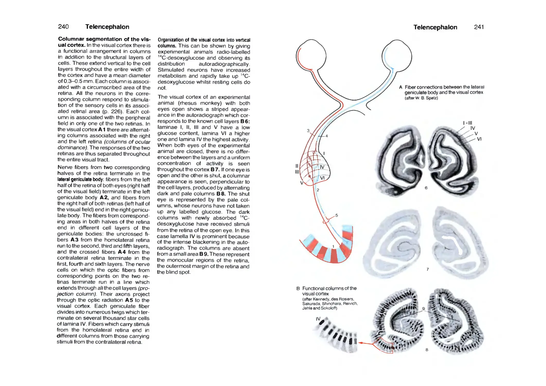
Spinal Ganglion and Posterior Roots 56
Coverings of the Spinal Cord 58
Radicular Innervation 60
Spinal Cord Syndromes 62
Peripheral Nerves 64
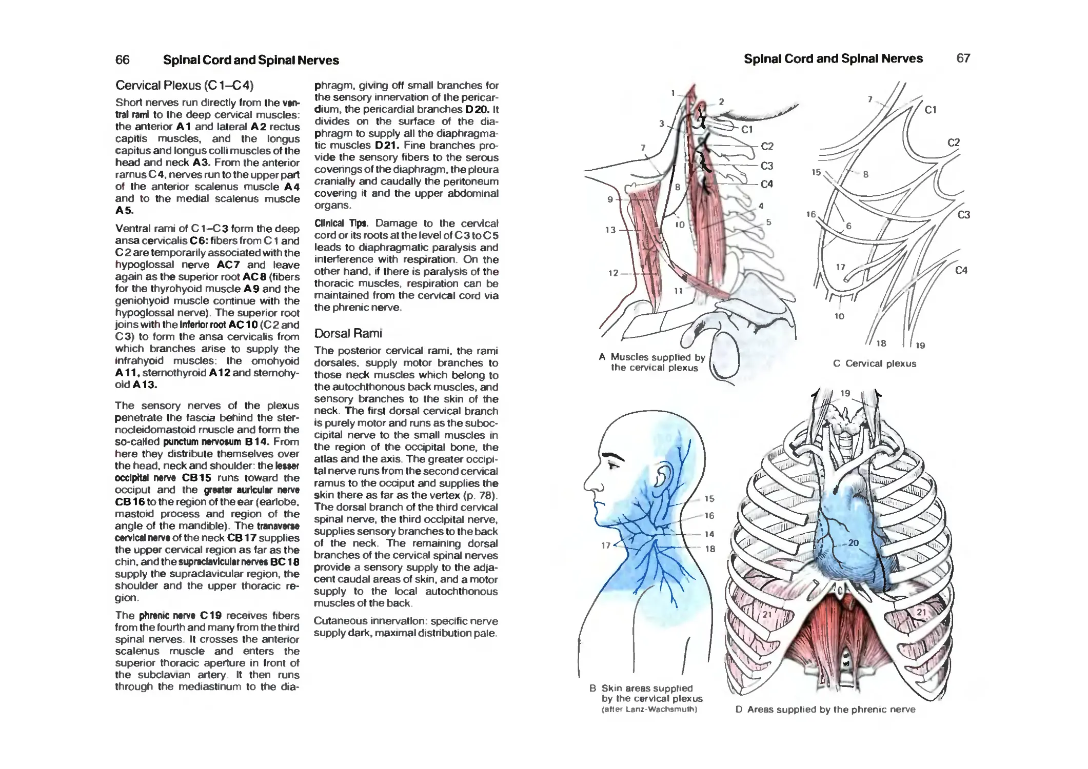
Cervicobrachial Plexus 64
Cervical Plexus 66
Dorsal Rami 66
Brachial Plexus 68
Supraclavicular Part 68
Infraclavicular Part 68
Lateral Cord 68
Medial Cord 72
Posterior Cord 74
Nerves of the Trunk 78
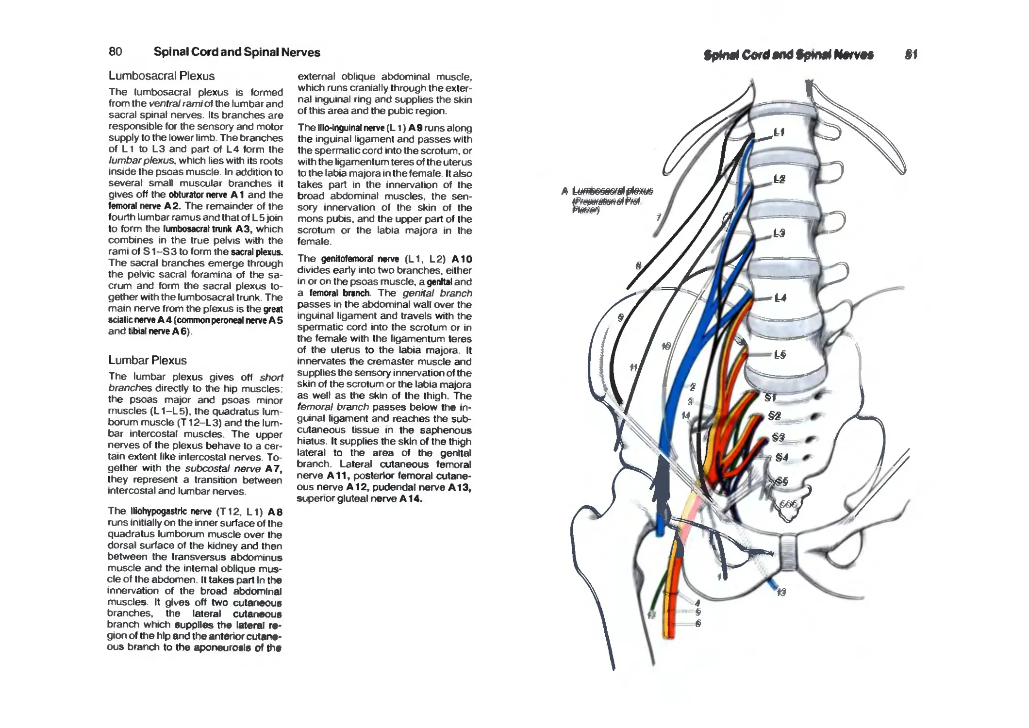
Lumbrosacral Plexus 80
Lumbar Plexus 80
Contents
VII
Sacral Plexus 84
Pudendal Nerve and Coccygeal Plexus 90
Brain Stem and Cranial Nerves 92
Survey 92
Organization 94
Cranial Nerves 94
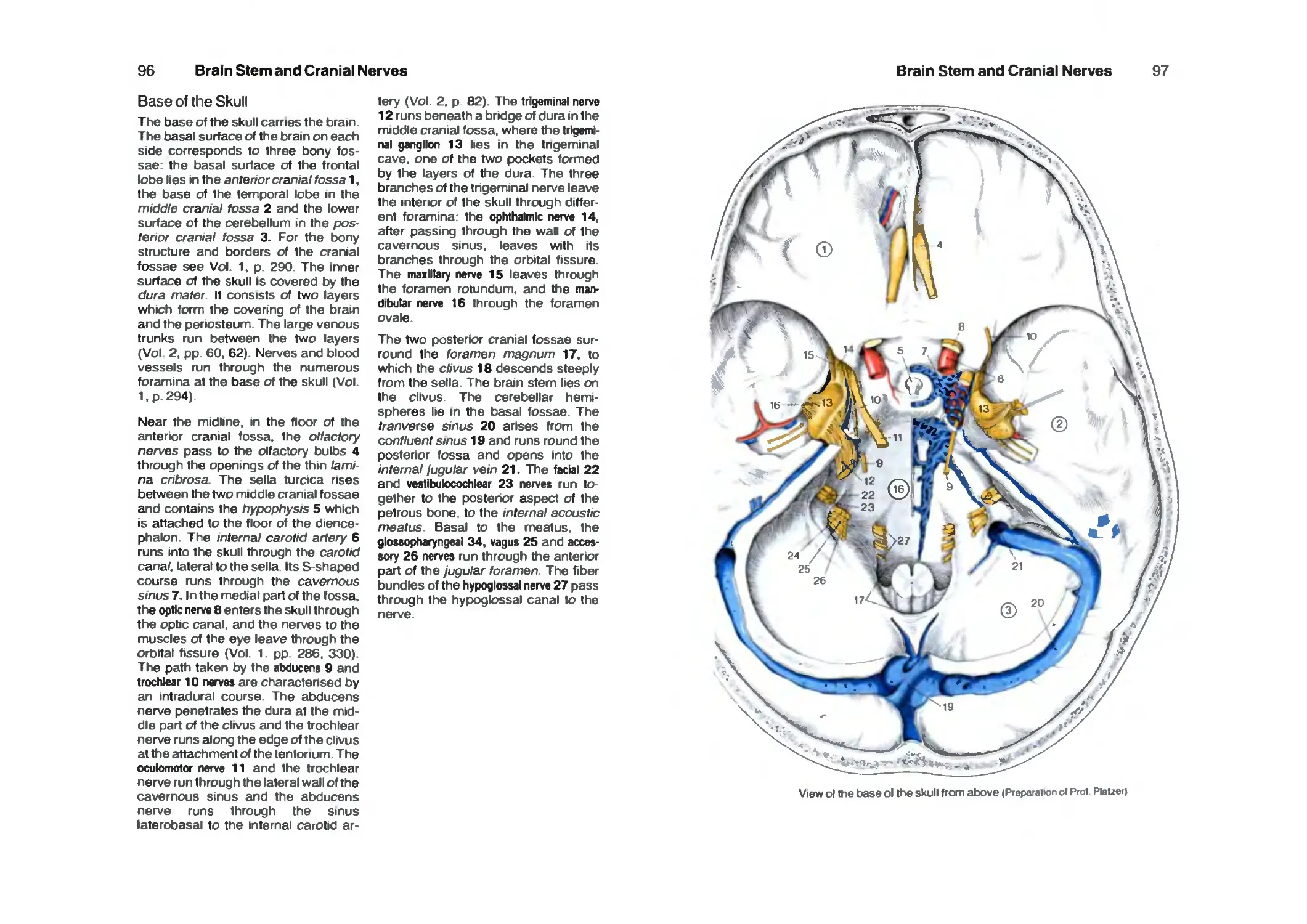
Base of the Skull 96
Cranial Nerve Nuclei 98
Medulla Oblongata 100
Pons 102
Hypoglossal Nerve 104
Accessory Nerve 104
Vagus Nerve 106
Glossopharyngeal Nerve 110
Vestibulocochlear Nerve 112
Facial Nerve 114
Trigeminal Nerve 116
Parasympathetic Ganglia 120
Midbrain 124
Red Nucleus and Substantia Nigra 128
Nerves to the Muscles of the Eye 130
Long Tracts 132
Reticular Formation 138
Histochemistry of the Brain Stem 140
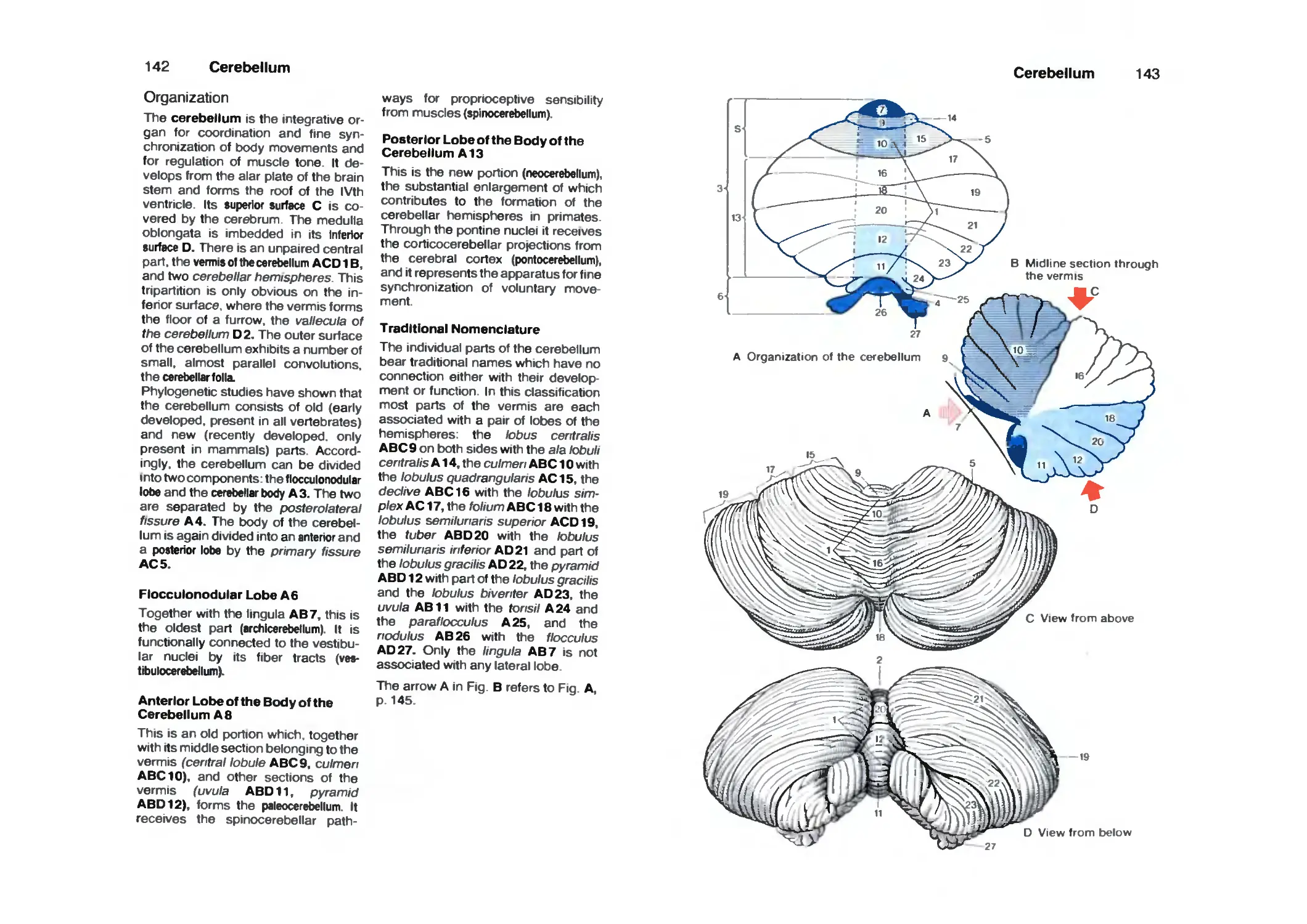
Cerebellum 142
Organization 142
Cerebellar Peduncles, Nuclei 144
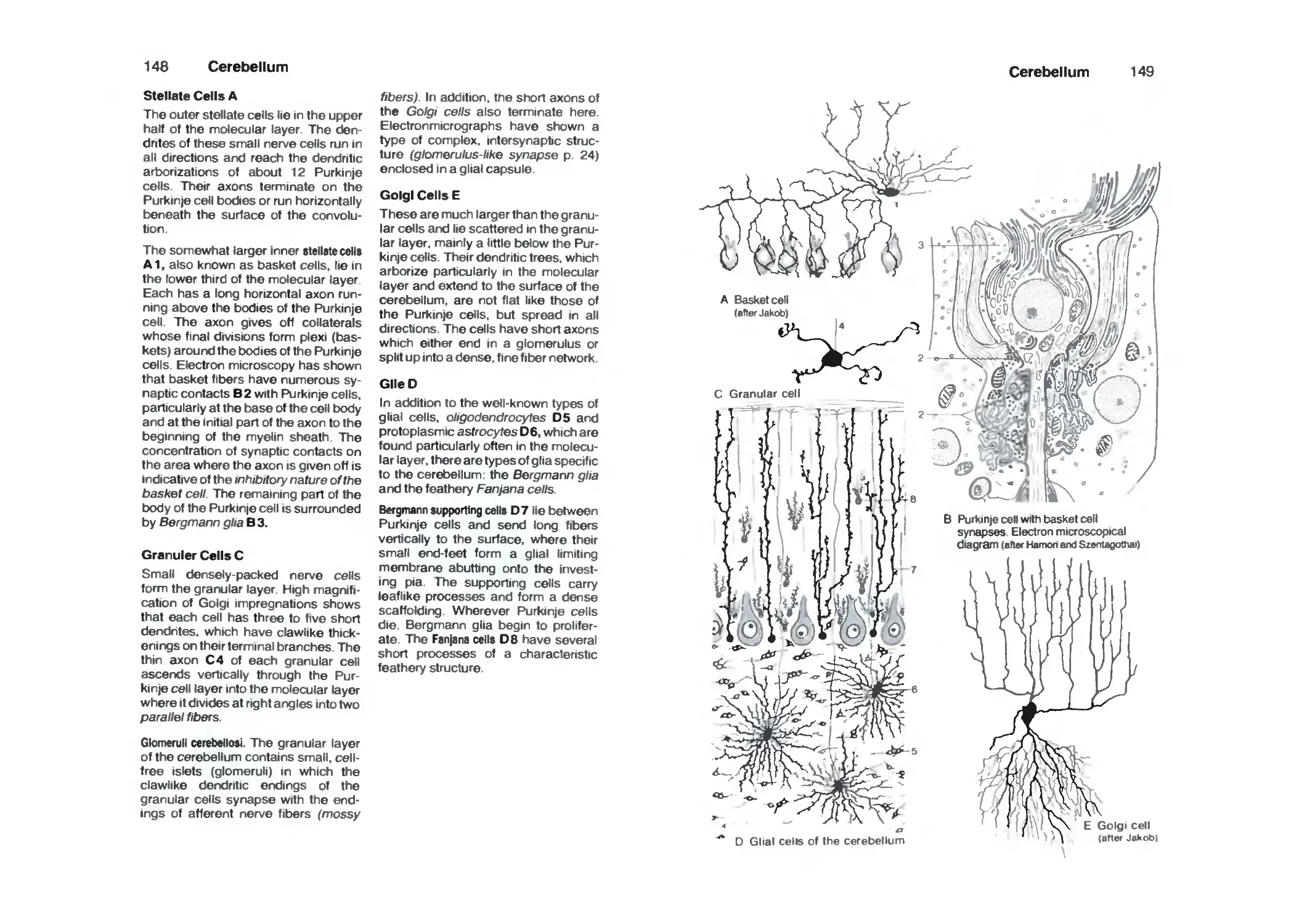
Cerebellar Cortex 146
Neuronal Circuits 150
Functional Organization 152
Nerve Tracts 154
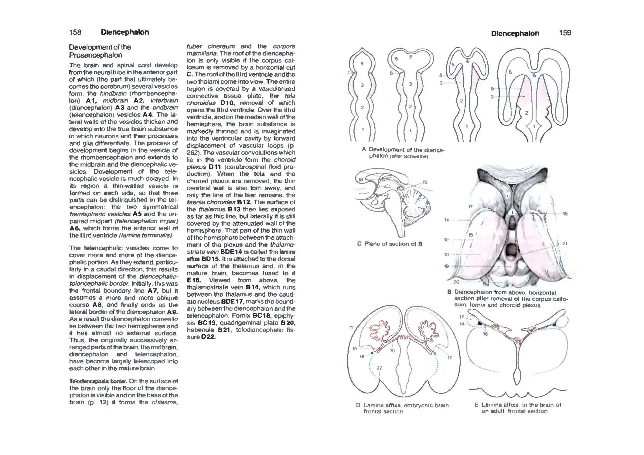
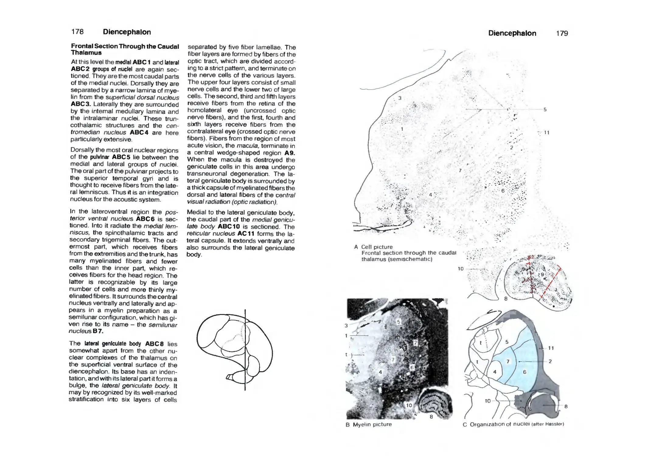
Diencephalon 158
Development of the Prosencephalon 158
Organization 160
Frontal Sections 160
Epithalamus 164
Dorsal Thalamus 166
Subthalamus (Ventral Thalamus) 180
Hypothalamus 182
Hypothalamus and Hypophysis 188
Neuroendocrine System 190
Telencephalon 1 ^4
Organization of the Hemispheres 194
Evolution 196
Cerebral Lobes '98
Frontal Sections 200
Horizontal Sections 206
VIII
Contents
Paleocortex 210
Amygdaloid Body 212
Fiber Connections of the Paleocortex 214
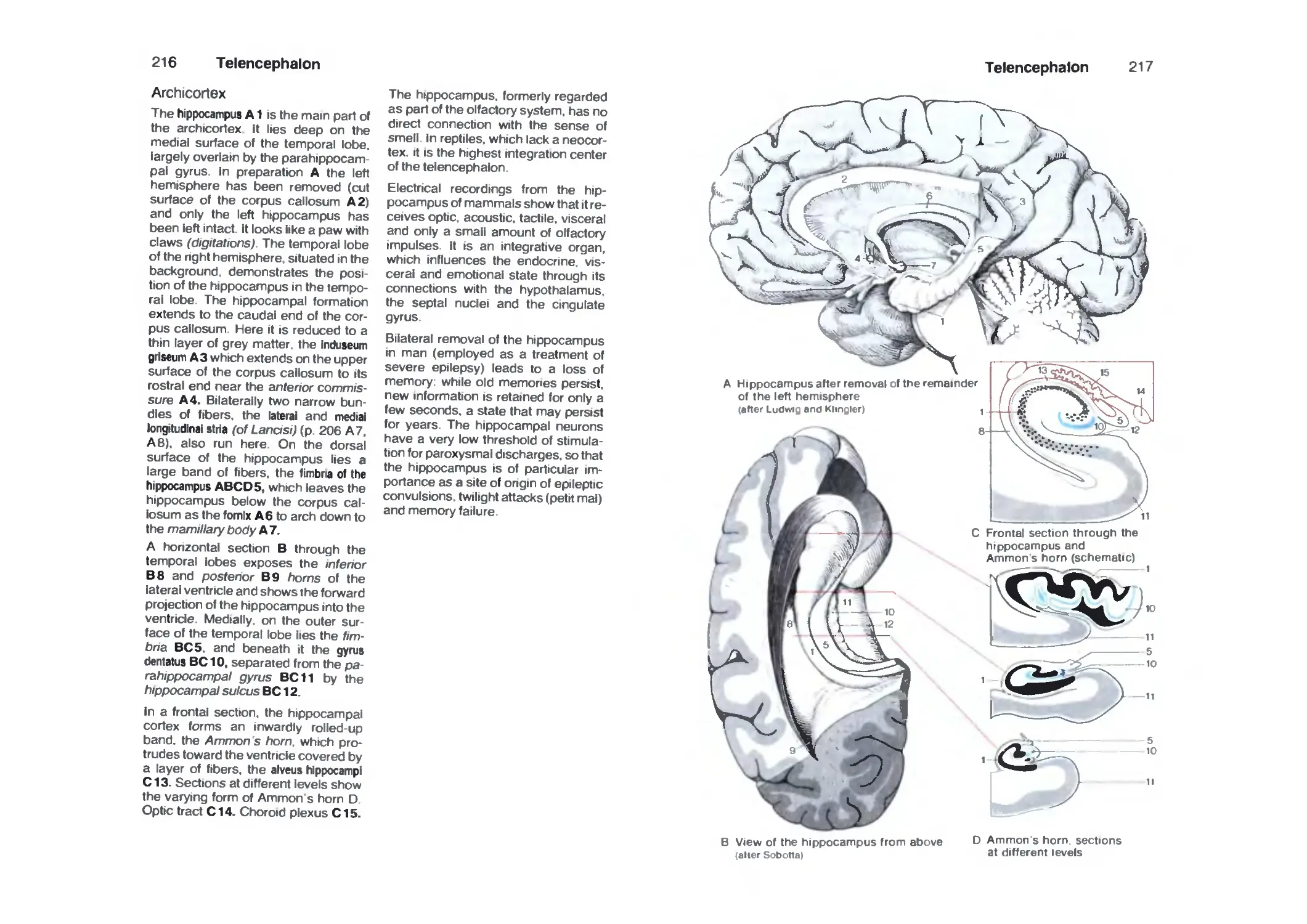
Archicortex 216
Fiber Connections 218
Cortex of the Hippocampus 220
Corpus Striatum 222
Insula 224
Neocortex 226
Cortical Fields 228
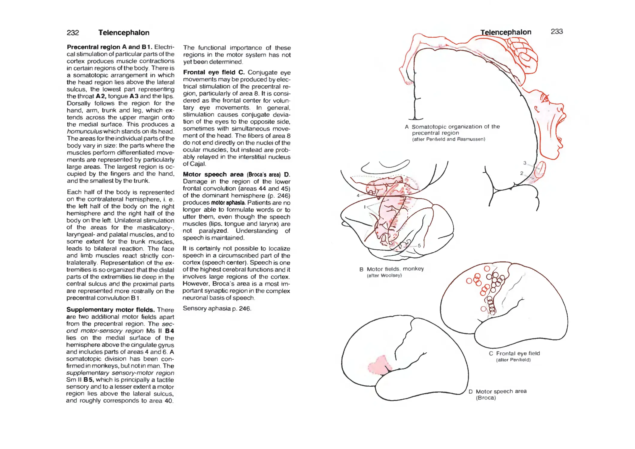
Frontal Lobe 230
Parietal Lobe 234
Temporal Lobe 236
Occipital Lobe 238
Fiber Tracts 242
Hemispheric Dominance 246
Histochemistry of the Forebrain 248
Vascular System 250
Arteries 25a
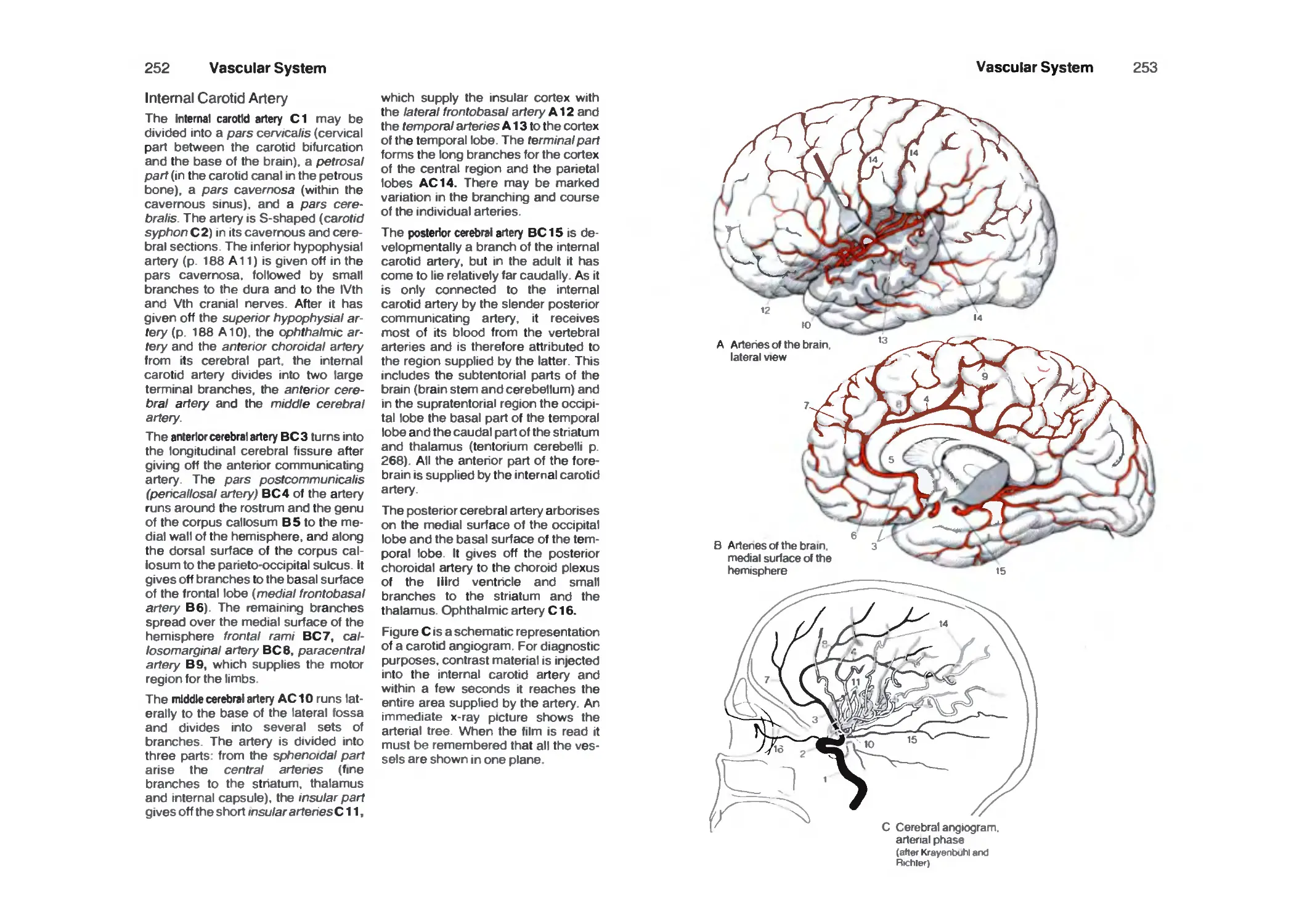
Internal Carotid Artery 252
Supply Areas 254
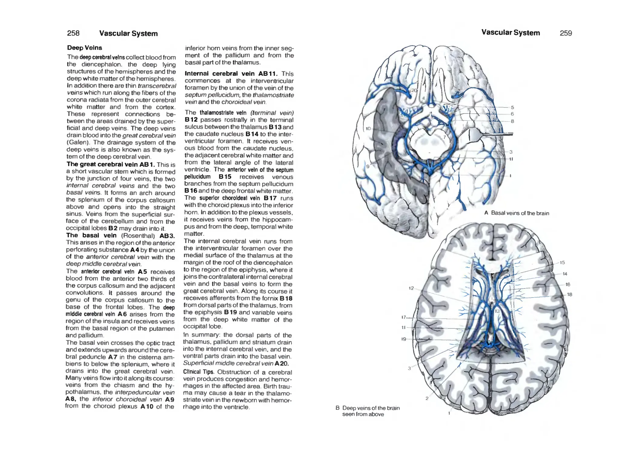
Veins 256
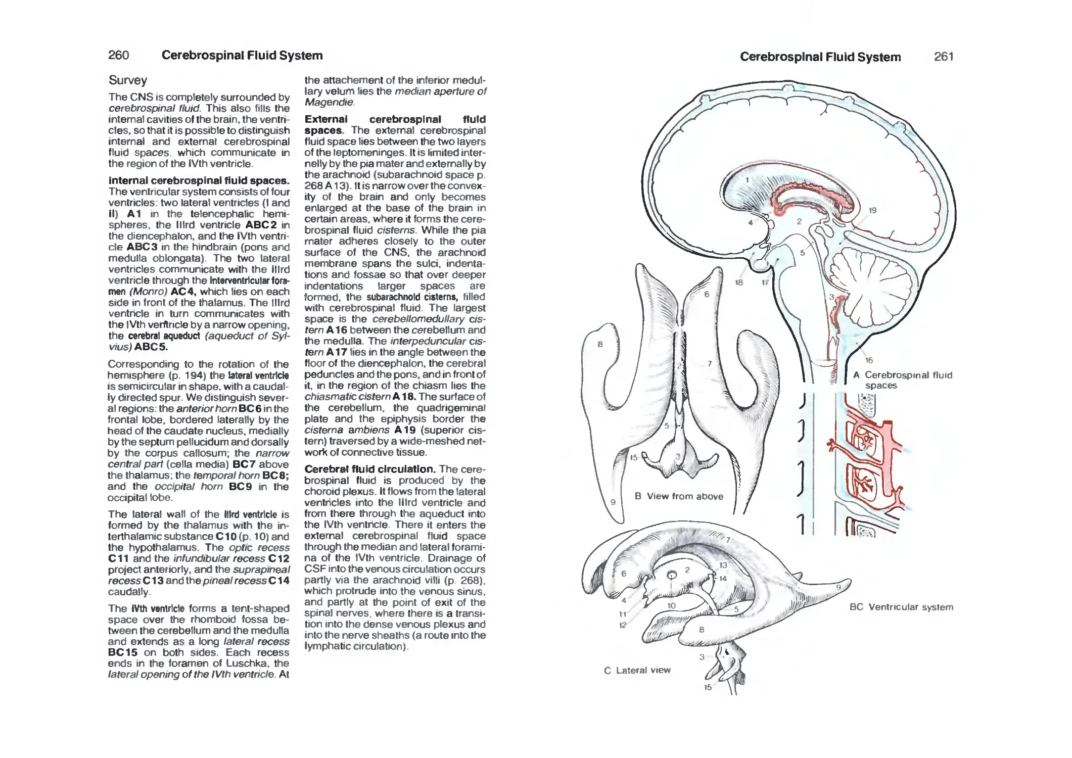
Cerebrospinal Fluid System 260
Survey 260
Choroid Plexus 262
Ependyma 264
Circumventricular Organs 266
Meninges 268
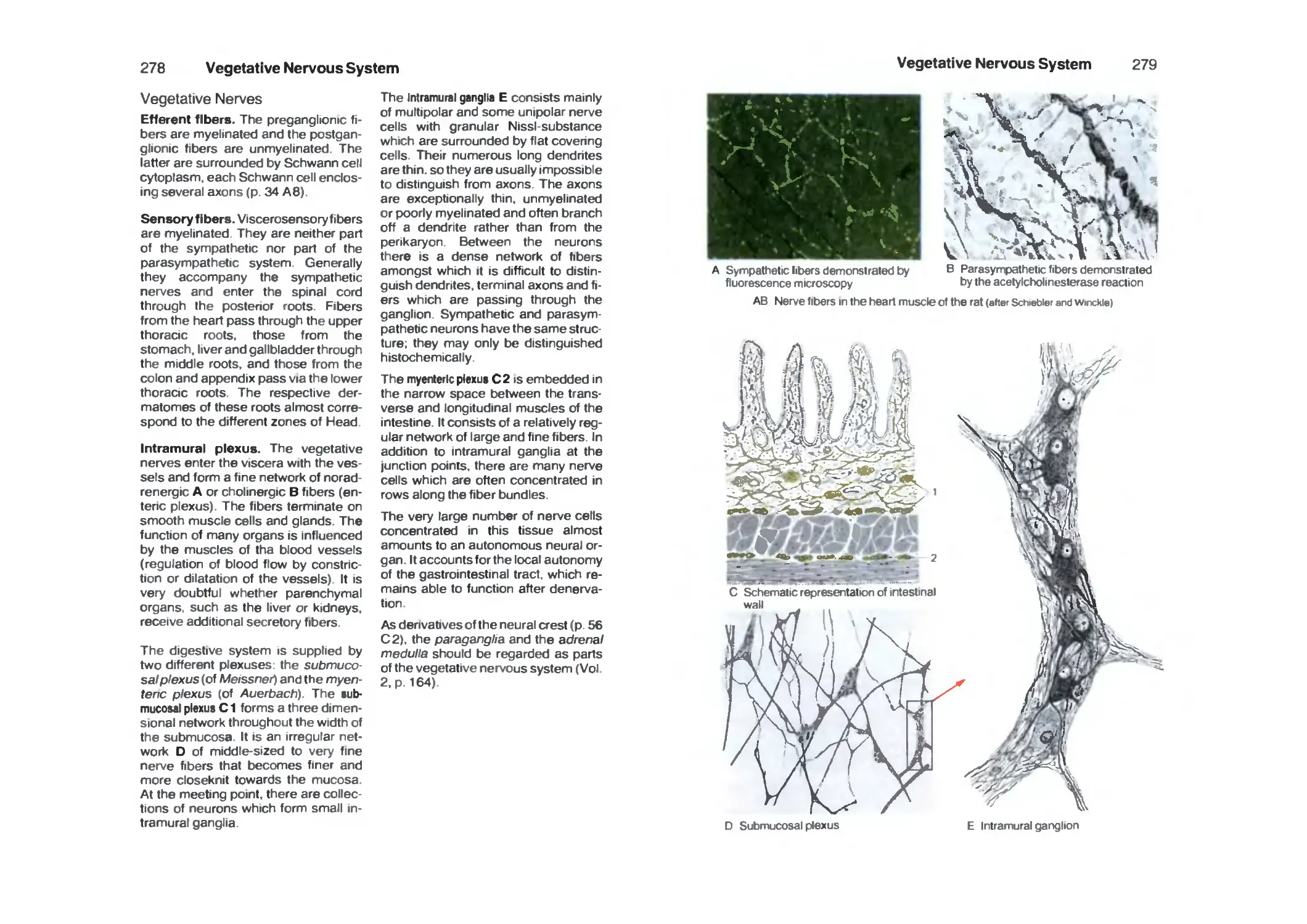
Vegetative Nervous System 270
Survey 270
Neuronal Circuits 274
Sympathetic Trunk 274
Vegetative Nerves 278
Functional Systems 282
Pyramidal Tract 282
Extrapyramidal Motor System 284
Motor End-Plates 286
Tendon and Muscle Receptors 286
Muscle Spindle 288
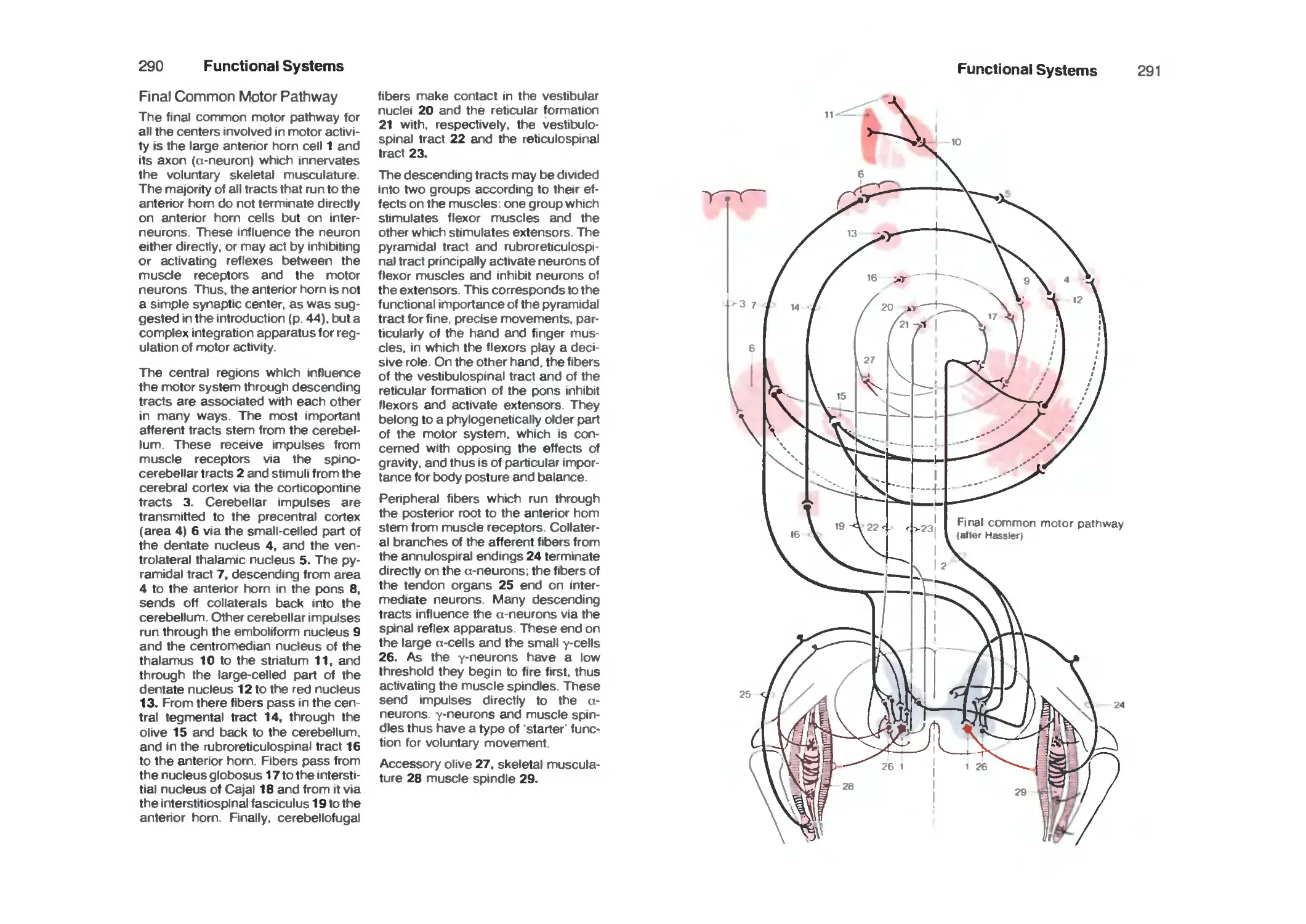
Final Common Motor Pathway 290
Cutaneous Sensory Organs 292
Pathway of Epicritic Sensibility 296
Pathway of Protopathic Sensibility 298
Organ of Taste 300
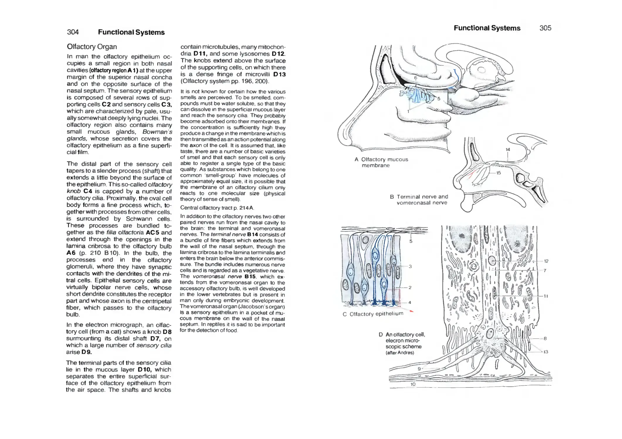
Olfactory Organ 304
Limbic System 306
Contents
IX
Sensory Organs 311
The Eye 312
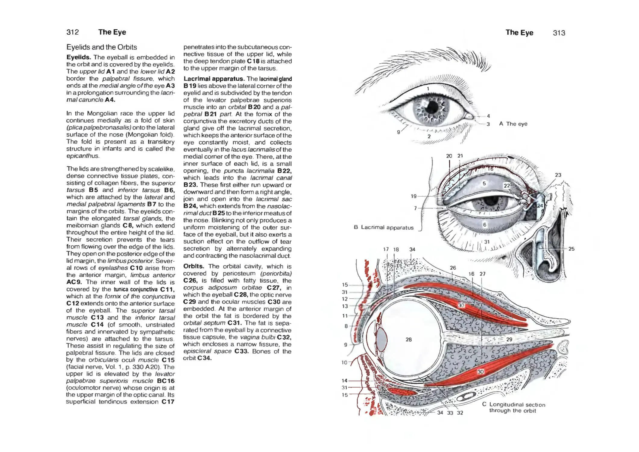
Eyelids and the Orbits 312
Eye Muscles 314
Survey 316
Anterior Part of the Eye 318
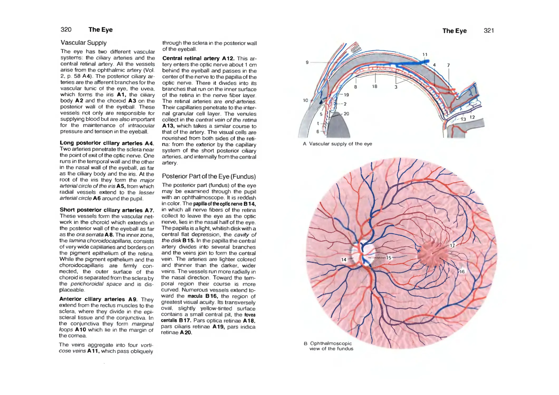
Vascular Supply 320
Posterior Part of the Eye (Fundus) 320
Retina ■. 322
Photoreceptors 326
Optic Pathway 328
Topistics of the Optic Tract 330
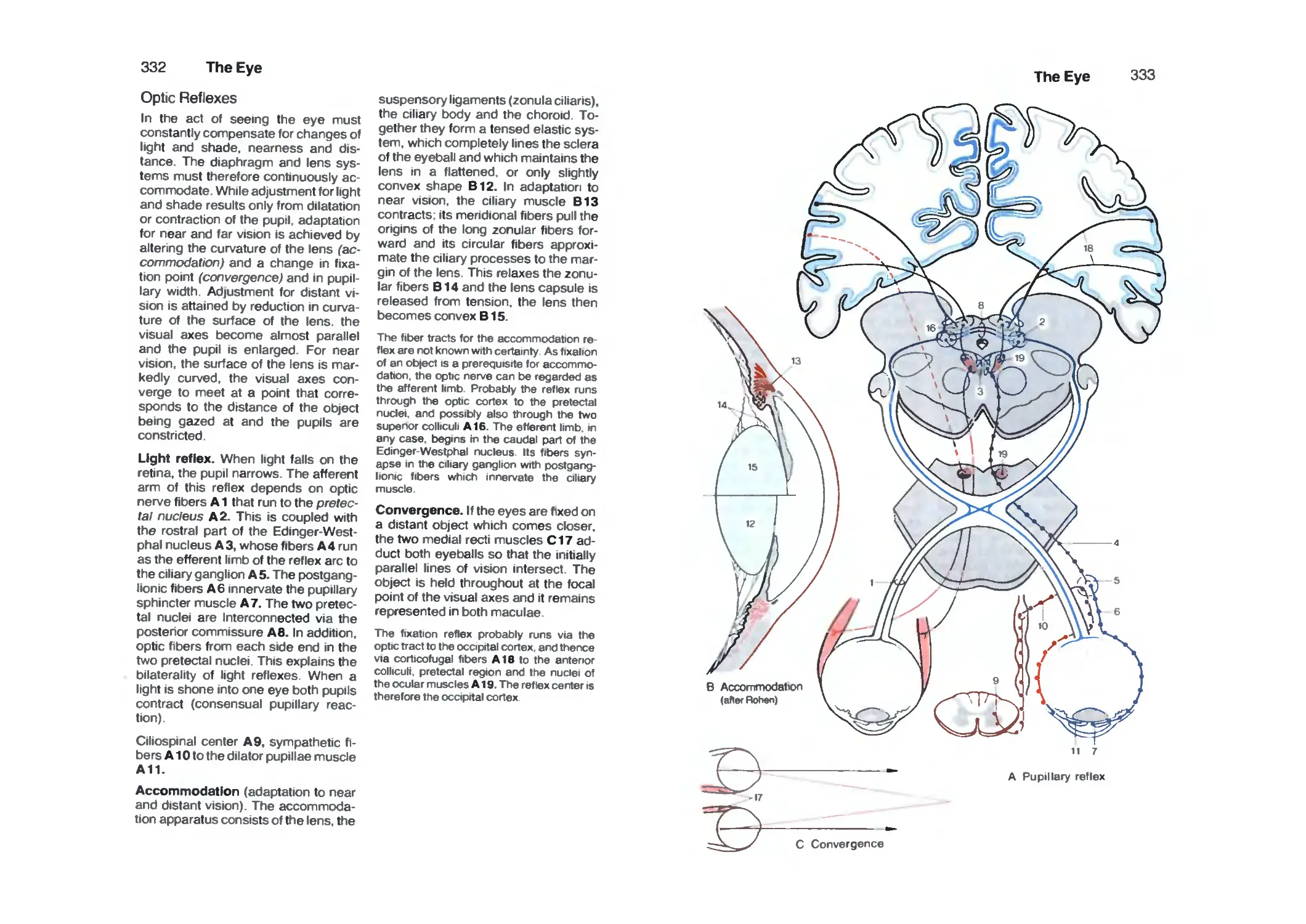
Optic Reflexes 332
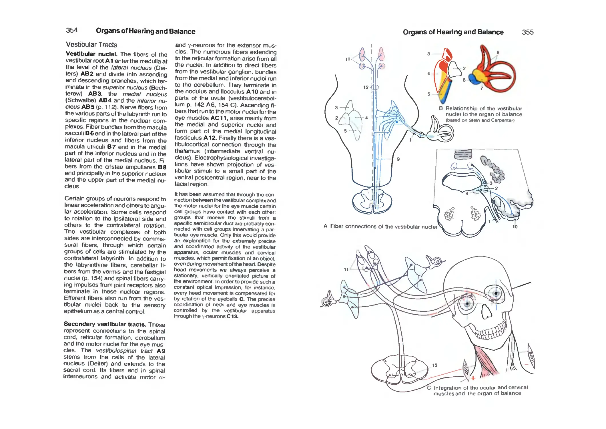
Organs of Hearing and Balance 334
Survey 334
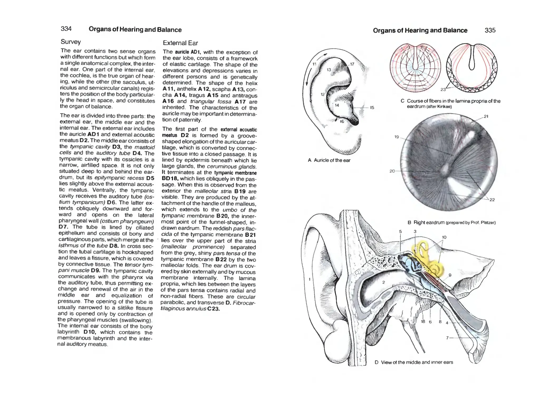
External Ear 334
Middle Ear 336
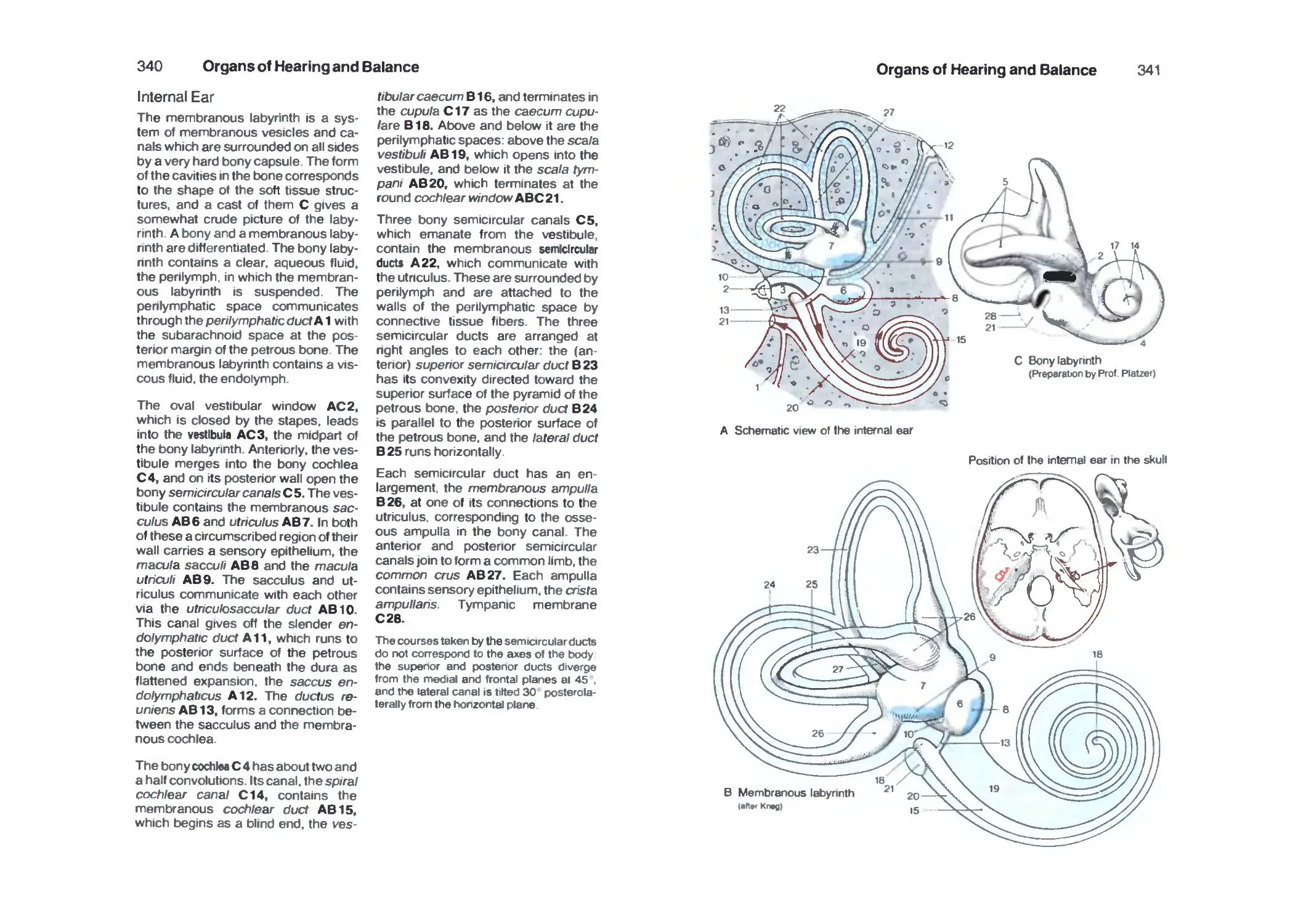
Internal Ear 340
Cochlea 342
Organ of Corti 344
Organ of Balance 346
Vestibular Sensory Cells 348
Spiral and Vestibular Ganglia 348
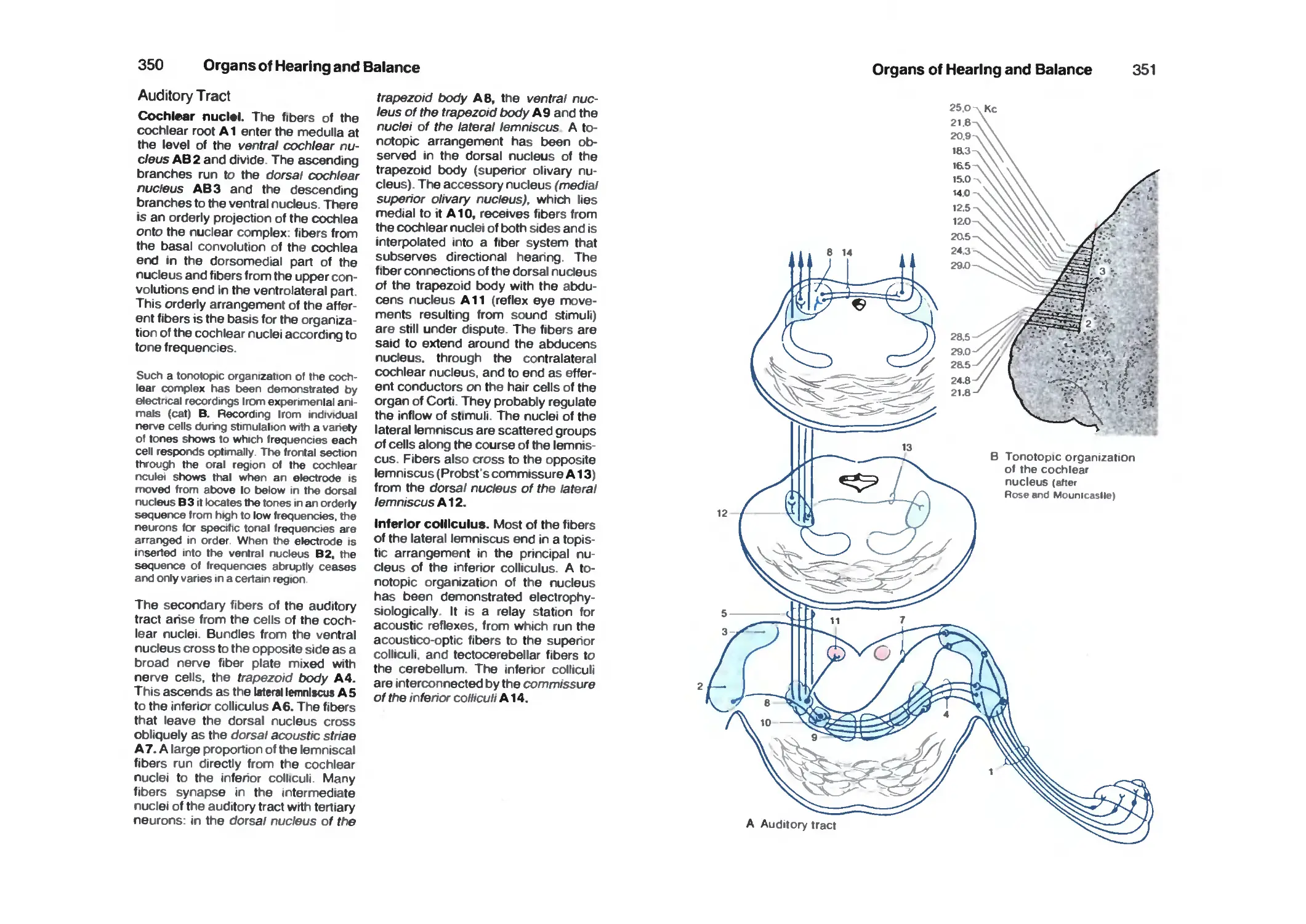
Auditory Tract 350
Vestibular Tracts 354
References 356
Index 362
Vol. 1: Locomotor System by W. Platzer
Vol. 2: Internal Organs by H. Leonhardt
Nervous System
Introduction
Development and Classification
of the Nervous System
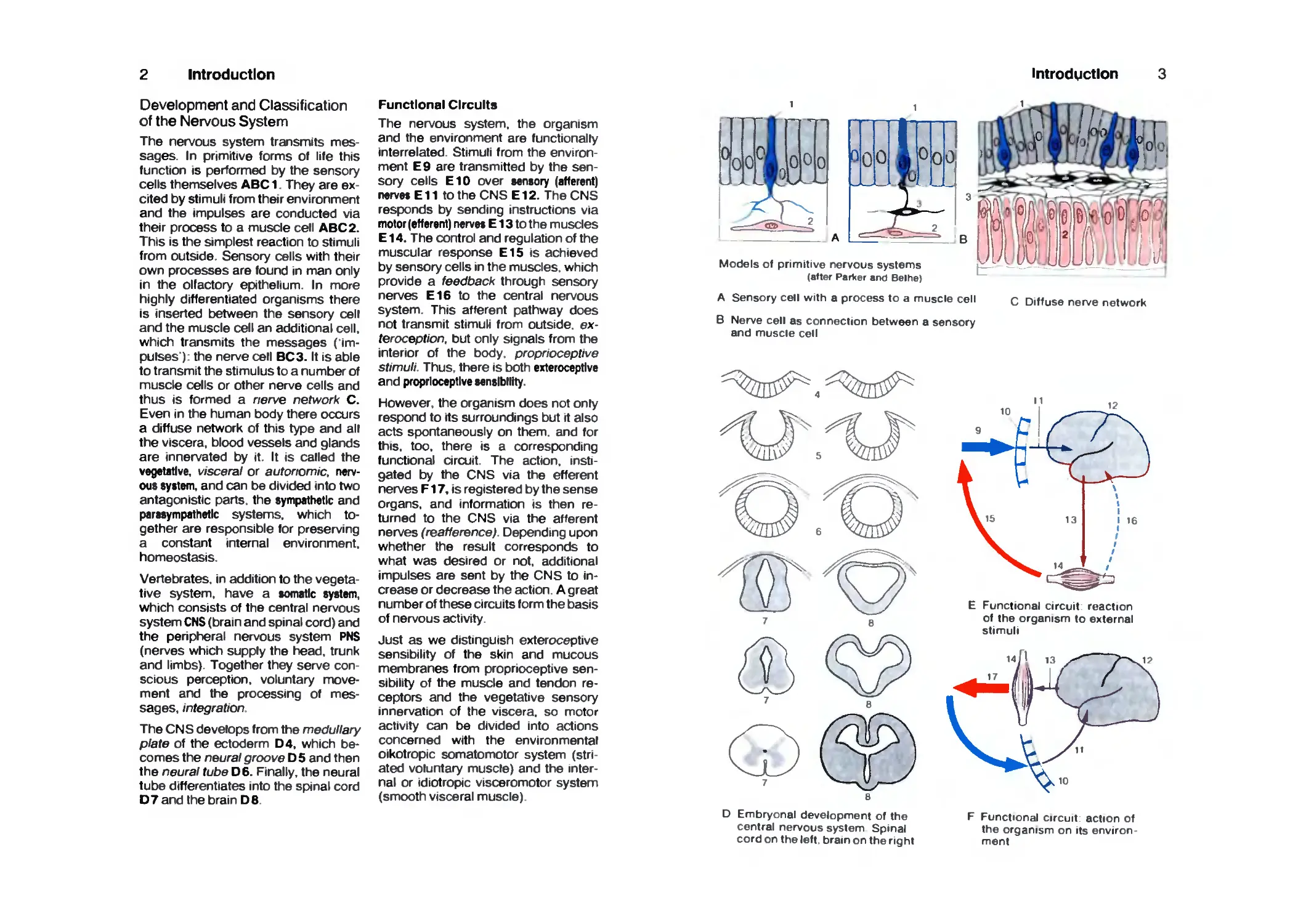
The nervous system transmits
messages. In primitive forms Of life this
function is performed by the sensory
cells themselves ABC 1. They are
excited by stimuli from their environment
and the impulses are conducted via
their process to a muscle cell ABC 2.
This is the simplest reaction to stimuli
from outside. Sensory cells with their
own processes are found in man only
in the olfactory epithelium. In more
highly differentiated organisms there
is inserted between the sensory cell
and the muscle cell an additional cell,
which transmits the messages
('impulses'): the nerve cell BC3. It is able
to transmit the stimulus to a number of
muscle cells or other nerve cells and
thus is formed a nerve network C.
Even in the human body there occurs
a diffuse network of this type and all
the viscera, blood vessels and glands
are innervated by it. It is called the
vegetative, visceral or autonomic,
nervous system, and can be divided into two
antagonistic parts, the sympathetic and
parasympathetic systems, which
together are responsible for preserving
a constant internal environment,
homeostasis.
Vertebrates, in addition to the
vegetative system, have a somatic system,
which consists of the central nervous
system CNS (brain and spinal cord) and
the peripfieral nervous system PNS
(nerves which supply the tiead, trunk
and limbs). Together they serve
conscious perception, voluntary
movement and the processing of
messages, integration
The CNS develops from the medullary
plate of the ectoderm D4, which
becomes the neural groove D 5 and then
the neural tube D6. Finally, the neural
tube differentiates into the spinal cord
D7 and the brain DB.
Functional Circuits
The nervous system, the organism
and the environment are functionally
interrelated. Stimuli from the
environment E9 are transmitted by the
sensory cells E10 over sensory (afferent)
nen^es E11 to the CNS E12. The CNS
responds by sending instructions via
motor (efferent) nerves E13 to the muscles
E14. The control and regulation of the
muscular response E15 is achieved
by sensory cells in the muscles, which
provide a feedback through sensory
nerves E16 to the central nervous
System. This afferent pathway does
not transmit stimuli from outside, ex-
teroception. but only signals from the
interior of the body, proprioceptive
stimuli. Thus, there is both exteroceptive
and proprioceptive sensibility.
However, the organism does not only
respond to its surroundings but it also
acts spontaneously on them, and for
this, too, there is a corresponding
functional circuit. The action,
instigated biy the CNS via the efferent
nerves F17, is registered by the sense
Organs, and information is then
returned to the CNS via the afferent
nerves (reafference). Depending upon
whether the result corresponds to
what was desired or not, additional
impulses are sent by the CNS to
increase or decrease the action. A great
number of these circuits form the basis
of nervous activity.
Just as we distinguish exteroceptive
sensibility of the skin and mucous
membranes from proprioceptive
sensibility of the muscle and tendon
receptors and the vegetative sensory
innervation of the viscera, so motor
activity can be divided into actions
concerned with the environmental
oikotropic somatomotor system
(striated voluntary muscle) and the
internal or idiotropic visceromotor system
(smooth visceral muscle)
Introdgctlon
Models of primitive nervous systems
(after ParVer and Beihe)
A Sensory cell with a process to a muscle cell c Diffuse nerve network
B Nerve cell as connection between a sensory
and muscle cell
^%#^
E Functional circuit reaction
of the organism to external
stimuli
D Embryonal development of the
central nervous system Spinal
cord on the left brain on the right
F Functional circuit action of
the organism on its environ
ment
Introduction
Position of the Nervous System in
the Body
The CNS Is divided into the brain,
(encephalon) A1 and the spinal cord
(SC) (medulla spinalis) A2. The brain lies
in the cranial cavity surrounded by a
bony capsule; the spinal cord lies in
the vertebral canal enclosed by Ihe
bony spinal column Both are covered
by cranial or spinal meninges which
enclose a space filled with cerebrospinal
fluid. In this way the CNS is surrounded
on all sides by bony walls and a fluid
cushion.
The penpheral nerves pass Ihrough
holes (foramina) in the base of the
skull (cranial nerves) and through the
intervertebral foramina (spinal nerves
A3), to run toward the muscles and
skin. In the limb regions they first form
nerve plexuses the bracliial plexus A 4
and the lumbosacral plexus A 5, in which
the fibers of the spinal nerves
intermingle so that the nerves to the limbs
come to contain fibers from several
spinal nerves. At the point of entry of
the afferent nerve fibers there are
ganglia A 6, small oval bodies which contain
sensory nerve cells. Cerebellum A7.
In descnbing the position of cerebral
structures the terms above and below
and in front and behind, are too inexact
as there are various brain axes As a
result of the upright posture in man.
there is a bend in the neural tube the
axis of the spinal cord runs almost
vertically, that of the forebrain
horizontally (Forel's axis, red), and the axis of the
lower parts of the brain obliquely (Mey-
nert's axis, blue). The terms for position
are in accordance with these axes- the
front end of the axis is called oral or
rostral (os mouth rostrum ship s
bow), the back end is the caudal part
(Cauda, tail), the lower part is basal or
ventral (venter, abdomen), and the
upper part dorsal (dorsum back)
The lower parts of the brain which
merge into the spinal cord are known
collectively as the brain stem, truncus
cerebri (white) BB. The anterior part is
called the forebrain. prosencephalon
(grey) B9. The vanous parts of the
brain stem have a uniform
architecture, consisting of a basal and alar
plate, similar to the spinal cord (see p
13). True penpheral nerves arise from
them, as they do from the spinal cord.
Like the cord, they both lie on the
chorda dorsalis during embryonal
development All these features
differentiate the brain stem from the forebrain.
This classification differs from the
official one, in which the diencephalon is
regarded as part of the brain stem.
The forebrain consists of two parts, the
dienceplvilon and the end brain,
telencephalon or cerebrum. In the mature
brain the telencephalon forms the two
hemispheres (cerebral hemispheres).
between which lies the diencephalon
introduction 5
A Position of the central nervous system within the body
B Brain axas median section
through the brain
Introduction
Development of the Brain
The closure of the neural groove into
the neural tube begins at the level of
the upper cervical cord. From there the
closure runs orally to the rostral end of
the brain (oral neuroporus.
subsequently lamina lerminalis) and in the
caudal direction to the end of the
spinal cord. Further development in the
CNS proceeds in the same directions.
Thus, different parts of the brain do not
mature at the same time, but in
different phases (heterochronous
maturation).
In the region of the head, the neural
tube expands into several vesicles.
The most rostral vesicle is the future
forebrain, prosencephalon (red and
yellow), and the posterior vesicles
form the future brain stem, cerebral
trunk (blue). At the same time two
curvatures of the neural tube develop,
the parietal flexure A1 and the cervical
flexure A2 Although at this early
stage the brain stem is still a uniform
structure, it is already possible to
identify the future areas of the medulla
oblongata ABCD3, pons, ABCD4,
cerebellum ABCD5 and meserce-
phalon (midbrain) ABC 6. The brain
stem develops more rapidly than the
prosencephalon. In the second month
of development, the telencephalon
(end brain) is still a thin-walled
vesicle, whilst differentiation of nerve cells
in the brain stem has begun
(emergence of the cranial nerves A7). The
optic vesicle develops from the di-
encephalon ABB (p. 316 A): optic
put A 9. In front of it lies the end brain
vesicle (telencephalon ABCD10)
which is initially unpaired
(telencephalon impar), but which soon extends
on both sides to form the two
telencephaiic hemispheres.
The prosencephalon B enlarges
during the third month. The end brain and
the diencephalon become separated
by the telodiencephalic sulcus B11.
The aniage of the olfactory bulb
BCD 12 develops from the
hemispheric vesicle and the hypophyseal
aniage B13 (p. 188 B) and the
mamillary eminence B14 develops from the
diencephalic floor. The pontine flexure
produces a deep transverse sulcus
B15 between the cerebellar aniage
and the medulla oblongata. The
undersurface of the cerebellum lies
against the thin, membrane-like dorsal
wall of the medulla (p. 263 E).
tn the fourth month, the telencephaiic
hemispheres begin to overgrow the
other parts of the brain C. The
telencephalon, which at first developed
more slowly than the rest of the brain,
now shows the most rapid growth (p.
158 A). The middle region of the lateral
face of the hemisphere grows less
rapidly and becomes overlapped by
adjacent areas. This is the insula
CD 16. In the sixth month the insula is
still visible on the surface D. The first
sulci and convolutions (gyri) appear on
the previously smooth surfaces of the
hemispheres. The walls of the neural
tube and cerebral vesicles, which
were thin at first become thickened
during the course of development.
They contain the nerve cells and
neural tracts and form the true substance
of the brain. Development of the
telencephalon p. 194.
Nerve fibers grow from one
hemisphere to the other through the
anterior wall of the telencephalon impar.
The commissural systems, which join
the two hemispheres, develop from
this thickened part of the wall, the
commissural plate. The largest of
them is the corpus callosum E. The
largely caudal direction of the increase
in size of the hemispheres, results in
further caudal extension during
development so that eventually the
diencephalon becomes completely
concealed.
Development of the spinal cord p. 56,
the brain stem pp. 94.124, the
diencephalon pp. 158. 160 and the
telencephalon p. 194
introduction
Brains ol human
embryos of different
crown-rump lengths
E Development ol
the corpus callosum
D Brain of a letus 33 cm long
Introduction
Brain
Synopsis
Each region of the brain contains
spaces of variable shape and width
The primary cavity of the neural tube
and cerebral vesicles becomes much
smaller during development due to
thickening of the walls. The central
canal is retained in the spinal cord in
lower vertebrates. In man the canal is
completely obliterated. In a cross
section of the spinal cord the position of
the previous central canal A1 is shown
only by a few cells of its former lining.
The cavity within the brain persists and
forms the ventricular system, which is
filled with a clear fluid, the liquor cere-
brospinalis or cerebrospinal fluid. The
founh ventricle AD 2 occurs in the
region of the medulla oblongata and the
pons. After a narrowing of the cavity in
the midbrain, the third ventricle CD 3
lies in the diencephalon. On both sides
the interventricular foramen (foramen
of Monro) DE4 opens from the side
wall of the third ventricle into the lateral
ventricle CE 5 (first and second
ventricles) ol both telencephalic
hemispheres.
The lateral ventricle is curved E and
cut through twice in a frontal section of
the hemisphere C. This shape is
produced by the growth of the
hemisphere, which does not expand
equally in all directions dunng development
but almost describes a semicircle (p.
194). The middle of the semicircle
forms the insula. This lies deep in the
lateral wall of the hemisphere on the
floor of the lateral fossa (C6) and is
covered by the adjacent parts, opercu-
la C 7, so that the surface of the
hemisphere shows only a deep fissure
sulcus lateralis (fissura lateralis, fissura
Sylvii) BC8. The hemisphere is
divided into several lobes: the frontal
lobe B9, the parietal lobe BIO, the
occipital lobe B11 and the temporal
lobe B12.
The diencephalon C (dark grey) and
the brain stem are largely covered by
the telencephalic hemispheres, so
that they are visible only at the base of
the brain or in a longitudinal section. A
mid-line section D shows the parts of
the brain stem, the medulla oblongata
D13, the pons D14, the midbrain D15
and the cerebellum D16. The fourth
ventricle D2 is shown in a longitudinal
view. The cerebellum rests on its
tentlike roof. The third ventricle CD3 is
opened along its entire width. In its
rostral part, the interventricular
foramen D4 opens into the lateral
ventricle. The corpus callosum D17, a plate
of fibers which joins the two
hemispheres is cut transversely and lies
above the third ventricle.
Weight of the Brain
The mean weight of the human brain
ranges between 1250 g and 1600 g. It
is related to the body weight: a heavier
individual generally has a heavier
brain. The mean weight of the male
brain is 1350 g and that of the female is
1250 g. The brain has attained its full
weight by the age of 20 years. In old
age. there is a decrease in the weight
of the brain due to age-related
atrophy. The weight of the brain does not
give any indication of the intelligence
of the individual. Studies of the brains
of eminent people (so-called elite
brains) have shown the usual
variations.
Introduction
©
Spinal cord Medulla ^^ ^ Pons
oblongata
A Sections through the spinal cord and brain stem m true relative sizes
Mid brain
D Schematic mid-line longitudinal
section through the brain
Schematic paramedian longitudinal
section through the brain
10
Introduction
Lateral Aspect
The two cerebral hemispheres overlie
all other parts of the brain, so that only
the cerebellum A1 and the brain stem
A2 are visible. The surface of the
cerebral hemisphere is characterized
by large numbers of grooves, sulci, and
convolutions gyrl. Beneath the surface
of the relief of the convolutions lies the
cerebral cortex, the highest nervous
organ, on whose integrity depends
consciousness, memory, thought
processes and voluntary activity. The
extent of the cerebral cortex is greatly
increased by the formation of sulci and
gyri. Only one third of the cerebral
cortex lies on the surface of the
hemisphere and two thirds lie deep in the
sulci. The hemispheres are separated
by a deep furrow, the longitudinal cerebral
fissure B 3. The lateral sulcus {sylvian) A 4
lies on the lateral surface. A frontal
section (pp. 8,200,202) clearly shows
that it is not a simple sulcus, and that in
its depths lies a space, the lateral fossa.
The anterior pole of the hemisphere is
called the frontal pole A 5 and the
posterior is called the occipital pole
A6. The hemisphere is divided into
different lobes: the frontal lobe A7,
which is separated by the central
sulcus AS from the parietal lobe A9, the
occipital lobe AID, and the temporal lobe
A11. The central sulcus separates the
prencetrel gyrus A12 (region of voluntary
motor control) from the postcentral gyrus
A13 (region of sensibility). The two
together are known as the central
region.
Median Section
The interbrain diencephalon lies
between the hemispheres, and above it
IS the corpus callosum CI5, which
connects the two hemispheres. A median
section through the brain reveals the
structures. The corpus callosum forms
a fiber plate, the oral arch of which
surrounds a thin section of the wall of
the hemisphere, the septum pallucidum
C16. The third ventricle C17 is
opened. Fusion of its two walls forms
tfie interthalamic adhesion CI 8,
above which arches the fornix CI9. In
the anterior wall of the third ventricle
lies the rostral commissure C20 (mainly
containing crossing fibers of the
olfactory cortex), on its floor is the optic
clilasm C21. the hypophysis C22, and
the paired mamillary bodies C23, and in
its caudal wall is the pineal gland or
epiphysis C 24.
The third ventricle is connected with
the lateral ventricle of the hemisphere
by the foramen Interventrtculare (Monroi)
C25. Caudally it continues into the
cerebral aqueduct (of Sylvius) C 26, which
expands beneath the cerebellum to
form the tent-shaped fourth ventricle
C 27. On the cut surface of the
cerebellum C 28, the furrows and convolutions
form the so-called arbor vitae. Rostral
to the cerebellum lies the quadrige-
minal lamina, lamina tecti C29 of the
midbrain (a relay station for the optic
and acoustic tracts). On the base of
the brain stem the pons C30 arches
forward and leads over into the medulla
oblongata C31, which is joined to the
spinal cord.
Introduction 11
A Lateral view
of the brain
B Dorsal view
Median section through the brain
medial surface of the right hemisphere
12
Introduction
Base of the Brain
The base of the brain affords a general
survey of the brain stem, the ventral
surface of the frontal A1 and temporal
A 2 lobes, and the floor of the dience-
phalon. The longitudinal cerebral
fissure A3 separates the two frontal
lobes, on whose basal surface lie
bilaterally the olfactory lobes with the
olfactory bulb A 4 and the olfactory tract A 5. In
the olfactory trigone A 6 the tract divides
into two olfactory striae, which delimit
the anterior perforated substance A 7
through which enter many blood
vessels. At the chiasm AS where the optic
nerves A 9 cross, the base of the
diencephalon begins with the
hypophysis A10 and the mamillary bodies All.
The pons A12 arches forward caudally
and is continuous with the medulla
oblongata A13. Many cranial nerves merge
from the brain stem. The cerebellum is
divided into the medial, deep-lying
cerebellar vermis A14 and the two
cerebellar hemispheres A15.
White and Grey Matter
if the brain is cut into slices, the cut
surfaces show the white and grey
matter, the substantia albe and grisea. The grey
matter consists of collections of nerve
cells and the white matter of fiber
tracts, i.e. the processes of the
neurons which appear light because of
their whitish covering, the myelin
sheaths. In the spinal cord the grey
matter lies centrally B16 and is
surrounded by white matter. In the brain
stem B17 and the diencephalon. the
grey and white matter are variously
distributed: the grey areas are called
nuclei. In the telencephalon BIS the
grey matter lies at the outer margin
and forms the cerebral cortex, while the
white matter lies internally; this
arrangement is the opposite of that in the
spinal cord
The arrangement in the spinal cord
represents a primitive condition found
in fishes and amphibia, where the
nerve cells are in a periventricular
position, even in the telencephalon.
The cerebral cortex represents the
highest level of organization, which is
fully developed only in mammals.
There are transitional formations
between nucleus and cortex.
Developmental Zones
During embryonal development, the
neural tube is divided into longitudinal
zones: the ventral half of the lateral
wall, which differentiates early, is
called the basal plate C19 and is considered
to be the site or ongin of the motor
nerve cells. The dorsal half of the
lateral wall, which differentiates later,
is called the alar plate C 20, and is
considered as the site of ongin of sensory
nerve cells. Between the alar and
basal plates lies an area C21 from which
autonomic nerve cells are said to
arise. Thus, a structural plan can be
recognized in the spinal cord and brain
stem, knowledge of which aids
understanding of the organization of the
various parts of the brain. In the
diencephalon and telencephalon the
derivatives of the basal and alar plates
are difficult to identify and many
authors do not accept such a
classification of the forebrain.
Introduction 13
9
8
10
11
A Basal view of the brain
16 ^ 'V ^17
B Distribution of white and grey matter
C Longitudinal zones of the CNS
14
Introduction
Evolution of the Brain
In the course of evolution the
vertebrate brain has developed into the
organ of human intelligence. As
precursors are extinct, the pattern
followed by evolution can only be
reconstructed with the help of information
from species which have retained a
primitive brain structure. In amphibia
and reptiles the telecephalon A1
appears as an appendage of the large
olfactory bulb A2. The midbrain A3
and diencephalon A4 lie free on the
surface. However, even in a primitive
mammal, e.g. the hedgehog, the
telencephalon extends across the
rostral parts of the brain stem, and in
the prosimii it completely overlies the
diencephalon and the midbrain. Thus,
phylogenetic development of the brain
consists primarily of increasing
enlargement of the telencephalon and
displacement of the highest
integrative functions into this part of the brain
- this is in fact a telencephalization.
Very ancient primitive structures are
still retained in the human brain and
are combined with newer highly
differentiated structures. If, then, we
speak of the older and newer parts of
the human brain, this relates to the
evolution of the brain. It is neither a
computer nor a thinking machine
based on rational principles of
construction, but an organ which has
developed in numberless variants over
millions of years.
Development of the form of the human brain
can be studied by making casts of
fossil intracranial cavities: a cast of the
cranial cavity (endocranial mould)
constitutes a gross impression of the
shape of the brain. In comparing the
casts the enlargement of the temporal
and frontal lobes is striking While
there is a distinct change from Homo
pekinensis via Neandertal man. the
first to use sharp flint knives, to
Cro-Magnon man, the cave painter,
there is no difference worth
mentioning between Cro-Magnon and modern
man.
During the process of phylogeny and
ontogeny, different parts of the brain
develop at different times. Those parts
which serve elemental vital functions
develop eariy and were already
present in primitive vertebrates. Those
parts concerned with higher, more
differentiated functions, develop late in
higher vertebrates. During their
development, they force the earlier
developed parts of the brain into the
depths as they themselves bulge
outwards, i. e. become increasingly
prominent.
Pressure by prominent parts of the
brain produces a negative imprint on
the inner surface of the bony cranium
the cerebral convolutions (gyri)
correspond to the impressiones gyrorum
in the skull. According to the theory of
H. Spatz, the impressions always
occur over those parts of the brain which
are in progressive development during
evolution. In modern man there are
particularly marked impressions of the
base of the skull. Here the basal
convolutions of the frontal and temporal
lobes, which are covered by the basal
neocortex, make their imprints. The
basal neocortex is a cortical region
that has developed very recently, and
it has only achieved full development
in man. Damage to it produces serious
changes in personality and character.
It seems possible that evolution of the
human brain is not yet completed and
that further progress will most
probably involve the basal neocortex,
which is characterized by its
prominence and Its ability to form
impressions, and which is concerned with
specifically human charactenstics.
Introduction 15
3
A Development of the vertebrate brain
Prosimius (Galago)
Gonlla
r.
Homo pekinensis
B Endocranial cast of a gorilla and fossil man
C Endocranial cast of homo sapiens lateral and basal view
16
Histology
The Nerve Cell
Nervous tissue consists of nerve cells
and glial cells (supporting and covering
cells), which arise from ectoderm.
Blood vessels and meninges do not
belong to neural tissue and are of
mesodermal origin. The nerve cell
(ganglion cell or neuron) A is the functional
unit of the nervous system. When
mature, neurons are unable to divide, so
that increase of their number or
replacement of old cells is impossible.
The number of nerve ceils in each
individual is constant from birth
throughout life.
The neuron consists of a cell body, the
perikaryon A1, its multibranched.
short processes, dendrites A 2 and a
single elongated process - the axon or
neurite ABCD3.
The perikaryon is the trophic center of
the cell, and processes which become
separated from it degenerate. It
contains the cell nucleus A 4 with a large,
chromatin-rich nucleolus A 5, which, in
the female, contains the Barr liody A 6
(sex chromatin from the second X
chromosome).
The dendrites, increase the external
surface of the cell by branching. The
processes of other neurons end on
them: they are the sites where nerve
impulses are received.
The axon conducts the nerve impulse.
First it forms the cone of origin or axon
hillock AD 7, the area where nerve
impulses arise. At a certain distance from
the perikaryon (initial segment) it
receives a covering, the myelin sheath
AS, which consists of lipid-containing
material (myelin). The axon gives off
branches (axon collaterals) A 9 and finally
divides (telodendron) A10 to terminate
with small endbulbs, the Iwutons ter-
mlnaux, on nerve or muscle cells.
Stimulus transmission to other cells
occurs at the bouton terminal, which
forms a synapse with the surface
membrane of the next cell in the
series.
Neurons are classed as unipolar,
bipolar ox multipolar ceWs according to the
number of their processes; the
majority are multipolar. Many have short
axons (Colgi type) and others have
axons more than 1 m long (Deiters'
type).
A neuron cannot be completely
stained by any one technique. The
methods used are complementary;
cell staining (e. g. NissI stain) shows
the cell nucleus and the perikaryon
B-D. The latter, including the bases of
its dendrites, is filled with chromophil
substance (Nissl's bodies or tigroid
bodies), and may contain pigments
(melanin or lipofuscin) D11. The axon
hillock IS free from NissI bodies. Motor
neurons have a large perikaryon and
large NissI bodies, whilst sensory
neurons are smaller and often contain
only NissI granules.
The processes of neurons can be
demonstrated by silver impregnation
(Colgi method), when the cells appear
as browny-black silhouettes B-D.
Other impregnation methods
selectively stain Ixiutons terminaux E or the
neurofibrils F, which run through the
perikaryon into the axon as parallel
bundles.
Histology
17
10
A Neuron
(schematic)
L
f
• f
I
E ♦
Impregnation of
boutons terminaux
(synapses)
Impregnation of
neurofibnis
B Nerve cell m
the brain stem
C Anterior horn nerve cell in the spinal cord
A
D Pyramidal
nerve cell in
the cerebral
cortex
B-D Equivalent pictures of nerve cells
cell staining (NissI) and silver impregnation (Golgi)
18
Histology
Functional Changes In Neurons
The appearance of a nerve cell differs
according to its current functional
state. The condition of the nucleolus,
deposits on the nuclear membrane,
and particularly the appearance of the
NissI txxjies may vary considerably.
These changes can be demonstrated
in animal experiments A:
Mice were made to swim in water for a
certain period of time and
subsequently, at definite intervals, the motor cells
of the spinal cord were examined. In
the resting animal, a large number of
nerve cells have strongly stained NissI
bodies (strong cells) A1, a smaller
number had poorly stained NissI
txxJies (intermediate cells) A 2, and a
few were pale cells (weak cells) A3.
After various periods of work it was
found that the number of strong cells
decreased rapidly and the number of
intermediate cells rose at the same
rate: the number of weak cells also
increased significantly. During the
phase of recovery it takes many hours
before the resting state is restored.
These studies show that during
functional stress there is breakdown of
NissI txxJies in the neuron and that
during the phase of recovery they are
restored, C. This applies not only to
motor neurons, but has also been
demonstrated for sensory neurons. D.
If experimental animals are exposed
to continuous sound there is loss of
NissI txxJies in the neurons of the
cochlear nucleus on which the fibers of
the auditory nerves terminate. They
take about a week to regenerate.
Estimates of the protein content of the
neurons also showed a dificit.
NissI txxJies are also lost when the
axon is cut The distal part of the axon
dies and the perikaryon undergoes
retrograde cell change. The perikaryon
swells, the NissI granules disintegrate
(tigrolysis) and the nucleus migrates to
the cell margin. The change is
reversible if the lesion is not close to the
perikaryon.
Apart from this, during a functional
load on neurons glial cells collect
around them, aatellitosls BC.
in a similar swimming experiment to
that described above, the numbers of
neurons with none, one, two or three
satellite cells were counted at hourly
intervals: dunng the experiment the
number of cells without satellites
rapidly declined, while there was a
simultaneous increase in the number
with satellite cells. The curve
representing neurons with only one satellite
fell sooner than the others: this can
only be explained by other satellite
cells migrating to join the solitary one.
During the recovery phase satellitosis
declines. There is probably an
exchange of metabolites between the
neuron and the satellite cells.
Histology
19
100
X
80
70
60
50
40
Work
1 1
, Recovery
j 1 1 1
1
L /
30
20
10
0
1
10 11 12 13 l-l ISh"
40
3S
30
2S
20
15
10
5
0
rrr
Work
Recovery
m
9 10 11 12 13 14 h
A Decrease of Nissl granules in
anterior horn cells in the treading
experiment (mouse) (after Kulenkampff)
B Increase in the number of satellite
cells around anterior horn cells in
the treading experiment (mouse)
(afler Kulenkampff)
C Changes in nerve cells under stress
3 Hours
0
Cell protein
0 Weeks
D Decrease and restoration of NissI substance in cells of the cochlear nucleus after
exposure to a continuous sound (guinea pig) (afier Hamberger arxl Myden)
20
Histology
Ultrastructure of the Nerve Cell
In the electron micrograph the cell
nucleus ABC 11S seen to be enclosed in a
double membrane A2. This has pores
BC3, which probably open only
temporarily The nucleus consists of
karyoplasma with fine chromatin
granules (DNA). The nucleolus ABC4.
a spongy structure, is formed of dense
granular and loose filamentous
components. It contains proteins and
ribonucleic acid. RNA.
In the cytoplssm the NissI bodies
appear as granular endoplasmic reticulum
ABC 5, a layered lamellar system of
membranes, which surround flat,
intercommunicating clefts (cisterT\s)
BC6. They have been shown to
contain cholinesterases and various other
substances To the outside of the
membrane adhere small granules the
ribosomes BC7 which serve protein
synthesis. To maintain the long axon,
up to 1 m in length, extremely active
protein synthesis is necessary in the
cells (structure metabolism) In
contrast to the ergastoplasm of secretory
cells, in the NissI txxjies a large
number of the nbosomes lie freely
between the cisterns. In the agranular
or smooth reticulum C 8 no ribosomes are
associated with the membranes The
endoplasmic reticulum communicates
with the perinuclear space BC9, and
with the marginal cisterns A10. below
the cell surface Marginal cisterns are
often found at sites adjacent to a
terminal bouton or glial cell process The
cytoplasm contains neurofilaments and
neurotubules ABC 11, which are
arranged in the axon as long, parallel
bundles
Metabolites are transported along the
neurofilaments and neurotubules The
neurofibrils are the light microscope
equivalent of neurotubules which have
been grouped together
Cell organelles. Neurons contain many
mitochondria ABC12. They are
surrounded by two membranes, from the
inner one of which cristae C13 project
into the intenor of the organelle
Mitochondria alter their shape continually
(e g in the perikaryon they are short
and plump, and in dendrites and axons
they are long and slim) and they are
always in motion along predetermined
cytoplasmic tracks between the NissI
txxJies.
They are regarded as the site of
cellular respiration and energy supply
Numerous enzymes are situated on
the inner membrane and in the inner
(matrix) space including those of the
citnc acid cycle and of
phosphorylation.
The Golgy apparatus consists of a
number of dictyosomes ABC 14, mul
tilayered. noncommunicating cisterns
surrounded by a membrane. On the
dictyosome a regenerative CI5 and a
secretory C16 side can be
distinguished, on which Golgi vesicles form
by constriction of the cisternal
margins
The Golgi apparatus is involved in the
synthesis and concentration of
secretory substances and membrane
production.
The large number of lysosomes ABC 17
contain acid enzymes and are
principally concerned with cell digestion
Pigment A18.
Histology
21
Electron microscopic
appearance of a nerve cell
(schematic)
\^ \V '^ '"^t^^^ part o, A
22
Histology
Synapse
The axon ends in a large number of
small clublil<e swellings, the boutons
terminaux. Together with the apposed
membrane of the adjacent neuron
they form the synapses, where
excitation is transmitted from one nerve cell
to another.
The synapse is divided into the
presynaptic part, bouton terminal AB1
with the presynaptic membrane BC2,
the synaptic gap 83, and the
postsynaptic part with the postsynaptic
membrane BC4 of the next neuron. The
bouton contains no neurofilaments of
neurotubules, but it does contain
mitochondria and mainly small, clear
vesicles BC5, which are clustered on
the presynaptic membrane (active
part of the boutons). The synaptic gap,
which often contains a dark stripe of
filamentous material, communicates
with the extracellular space. The pre-
and postsynaptic membranes are
covered by electron dense
condensations. Similar densities are found in a
variety of cell Junctions (zonula or
macula adherens). These are,
however, constructed symmetrically, i. e.
the zone of condensation is similar in
both of them: In contrast, the synapse
is asymmetrical, the dense zone of the
postsynaptic membrane 86 is usually
wider and more dense than that of the
presynaptic membrane.
Structure
According to Gray (B) two types of
synapses I and // can be distinguished
by the width of the synaptic gap and
the nature of its dense zone. In type I
synapsesihe gap is wider, the subsyn-
aptic membrane is denser and
extends over the entire area of
membrane contact. The gap in type II
synapses is narrower and dense zones
are restricted to a few points only; the
subsynaptic density is less well
developed so the asymmetry is not so
distinct. There are also transitional
types between these two.
Function
Excitatory and Inhibitory synapses can be
differentiated. Most excitatory
synapses lie on dendrites and the majority
of inhibitory synapses are localized on
the perikaryon or the base of the axon,
where excitation originates and can be
most effectively suppressed. While
synaptic vesicles are generally round,
some boutons contain oval or
elongated vesicles CIO. They are
considered characteristic of inhibitory
synapses.
Synapses can be classified according
to their localization, their structure,
their function, or according to the
neurotransmitter substances
contained in them.
Localization
Boutons may be apposed to the
dendrites AC 7 of a receptor neuron
(axodendritic synapses) A8C, the perikaryon
(axosomatic synapses) A 9, or to the axon
(axo-axonal synapses). Large neurons are
furnished with thousands of boutons.
Histology
23
;^<f^r^^V?^
C Transverse section of a dendrite surrounded by synapses (after Uchizono)
24
Histology
There are many variants of the simple
form of synapse. Synaptic contacts
between parallel axons and dendrites
are known as parallel synapses or
boutons en passage A1. Many
dendrites have spiny processes, which
form spinous synapses A 2 with boutons.
On the apical dendrites of large
pyramidal cells the terminal axon
enlargement encloses the entire spine,
which divides and bears a large
number of synaptic points of contact
B. Several axons and dendrites may
join together to form glomerulus-like
complexes C, in which the different
synaptic elements are Intertwined.
They probably affect each other In the
sense of modulation of the
transmission of excitation.
Each region of the brain has its typical
forms of synapses. Types I and //
(Gray) occur principally In the cerebral
cortex. Glomerulus-like synaptic
complexes occur in the cerebellar cortex,
the thalamus and the spinal cord.
Neurotransmitter
Excitation is transmitted by chemical
substances (chemical synapses).
Electrical synapses are only present in
invertebrates and fishes. The most
widespread transmitter substance in
the nervous system Is acetylcholine,
ACh. The effective substance in
inhibitory synapses is presumed to be
gamma amino butyric acid, GABA.
Catecholamines also act as
transmitters, e. g. noradrenaline (NA) and
dopamine (DA), as well as serotonin.
Many neuropeptides (releasing
factors of the hypothalamus Vol. 2,
p. 153) do not work only as hormones
In the blood stream, but also as
transmitters in excitatory synapses
(neurotensin, cholecystoklnin) and In
inhibitory synapses (somatostatin,
thyroliberin, motllin). The hormonal
and neural actions of these
substances supplement each other in
many ways. Oxytocin acts hormonally
as a milk releasing factor whilst the
stimulation of oxytocinergic fiber
systems produces brood nursing
behaviour. Luliberin acts as a hormone to
release gonadotropic hormone whilst
the stimulation of luliberlnergic fibers
In animal experiments produces
copulation behaviour.
It is believed that these substances
are formed in the perikaryon and are
stored in synaptic vesicles in the
terminal nerve endings. Often it Is only the
enzymes necessary for synthesis of
the transmitter that are produced in the
perikaryon and the substances
themselves are actually synthesized in the
boutons. The small, clear vesicles are
probably the carriers of ACh, the
elongated vesicles in inhibitory synapses
carry GABA. Noradrenaline is
contained in the small granular vesicles
and dopamine in the large granular
vesicles, DE.
Most vesicles lie near the presynaptic
membrane. Its diffuse density can be
demonstrated by special staining
methods as a grid consisting of
trabeculae F3, enclosing a hexagonal
Space. The vesicles pass through the
spaces as far as the synaptic
membrane, and during stimulation their
contents are emptied through stomata
F4 of the membrane Into the synaptic
space. The substances are given off in
definite amounts (quanta), so that the
Individual vesicles may be the
morphological correlate of the quanta. In
the synaptic cleft the substances
cause depolarization of the
postsynaptic membrane and thus
transmission of excitation. They are
immediately inactivated by enzyme systems
and partly are reabsorbed into the end
bulbs by pinocytosis F5.
Histology 25
^1
A Spiny synapse | \
B Complex synapse
C Glomerulus type of synapse
(after Szentdgothai)
I 1 1000A
00 o ACh
® O NA
F Model of a synapse (after Akeri. Pfenmnger, Sandn and Moor)
26
Histology
Transmitter, Axon Transport
Just as a gland cell normally secretes
only one specific material, so every
neuron only produces one particular
transmitter substance. According to
the substance produced, neurons may
be classified into cholinergic, cate-
cholaminergic {noradrenergic and
dopaminergic), serotonlnergic and
peptidergic neurons. Catecholaminergic and
serotonlnergic neurons can be
visualized directly by fluorescence
microscopy as the transmitter
substances are converted Into fluorescent
products when exposed to formalin
gas AB. Thus the axons may be
followed and the outline of the perikaryon
with its nonfluorescent nucleus can be
recognized. Fluorescence is least
marked in the axons, more definite in
the perikaryon and most marked in the
terminal swellings of axons, which is
the site of the highest concentration of
neurotransmitters. Demonstration of
cholinergic neurons can be done by a
histochemical test for an enzyme
important in acetylcholine metabolism,
acetylcholinesterase C. The different
neuropeptides of the peptidergic
nerve cells are distinguished by im-
munohistochemical reactions D.
Neuropeptides were demonstrated in
both catecholaminergic and
cholinergic neurones and some neurones
contained two different
neuropeptides. It may, therefore, be necessary
to revise the concept that each nerve
cell only produces one transmitter
substance.
Neurotransmitters are produced in the
smooth endoplasmic reticulum of the
perikaryon, from the cisterns of which
vesicles become detached. It is also
possible that the Golgl apparatus is
involved in the production of
transmitter substances. The substances travel
either by inactive transport, in storage
form as molecular particles, or as
vesicles in the axoplasm, as far as the
boutons terminaux.
The neurotubules DE1 are particularly
important in the transport mechanism.
If they are disrupted by the
administration of colchicine, intra-axonal
transport breaks down. Materials are
assumed to be transported along the
neurotubules. In the region of the
neurotubules, viscosity is less than in
the rest of the axon and this permits
cytoplasmic flow in the centrifugal
direction. The velocity of flow is greatest
along the walls of the tubules and
decreases as one moves further
away, (velocity profile E2). This
hypothesis may be used to explain the
different rates of transport of different
substances. ATP, which is hydrolysed
by the action of ATPase E3 in the
tubule wall provides the energy for
transport. The velocity of intra-axonal
transport of catecholamines has been
estimated at 5 to 6 mm per hour.
In addition to the rapid intra-axonal
transport, there is also a continuous,
but much slower axoplasmic flow of
about 1 mm in 24 hours.
This can be demonstrated by placing a
ligature on a single neuron F. Proximal
to the constricted region the axoplasm
is held back and the axon swells. This
plasma flow is not concerned with
transport of transmitter substances
but with continual supply of nutrients
for the elongated cell process. The
velocity of the plasma flow
corresponds to the speed of growth of the
axon during development. Thus, the
axon is not a type of rigid conducting
wire, but more like a plasma column,
which is being continuously renewed
in a centrifugal direction from the
perikaryon. There is also centripetal
intra-axonal transport, and in this
manner proteins, viruses and toxins can
travel from peripheral nerve endings to
the perikaryon.
Histology 27
C Peptidergic neuron,
reticular formation
(after Bar, Stumpf et a].}
A-B Catecholaminergic neurons in the brain stem
(after Dahlstrom and Fuxe)
D Cross section of an axon
Axonal transport along a microtubule
(after Kreutzberg)
^
E Longitudinal section 2
F Blockage of the axoplasm after axon ligation (after Weiss and Miscoe)
28
Histology
Neuron Systems
Groups of neurons that possess the same
transmitter substances and whose axons
form discrete fiber bundles are named after
their neurotransmitter as cholinergic,
noradrenergic, dopsminergic, serotoninergicor
peptidergic systems. The nerve impulse can
be transmitted to a neuron of the same type,
as well as to one with a different transmitter
substance. In the neuronal chain of the
parasympathetic system (p. 270), cholirier-
gic neurons conduct impulses from the
central nervous system to peripheral ganglia,
where they are again changed to cholinergic
neurons. In the sympathetic system (p.
270), neurons in the spinal cord are also
cholinergic, but in peripheral ganglia there is
transmission to noradrenergic r\erve cells.
It is not yet fully known which transmitter
substances are Involved in the central
neurons, particularly in the nerve cells of the
cerebral cortex. Noradrenergic,
dopaminergic and serotoninergic neurons lie in the
brain stem. Noradrenergic neurons form the
locus coeruleus A1 (p. 92 B28, 124 D18)
and groups of cells in the lateral part of the
reticular formation of the medulla oblongata
and the pons (fibers project to the
hypothalamus, to the limbic system, diffusely
in the neocortex and to the anterior and
lateral horns of the spinal cord).
Serotoninergic neurons lie in the nuclei of
the raphe A2 (p. 100 B28) particularly in the
dorsal nucleus of the raphe A3 (fibers
project to the hypothalamus, the olfactory
cortex and to the limbic system). The pars
compacta of the substantia nigra A 4 (p. 126
A17,128 AB1) from which the nigrostriatal
fibers extend to the striatum consists of
dopaminergic neurons.
Peptidergic neurons are found most
commonly in phylogenetically older regions of
the brain: in the central periaqueductal grey
matter of the mid brain AS, the reticular
formation A6, the hypothalamus AB7, the
olfactory bulb B8 and in the structures of the
limbic system (cingulate gyrus 89, hip-
pocampusBW, amygdaloidtx)dyB^^ and
habenular nucleus A12). They are also
found scattered as short interneurons in the
cerebral cortex, the thalamus and the
striatum. Many Purkinje cells in the cortex of
the vermis of the cerebellum B13 are
peptidergic as are many of the small neurons in
the spinal ganglia. In addition the
interpeduncular nucleus A14, the nucleus soli-
tarius A15 the locus coeruleus and the
nuclei of the raphe are rich in peptidergic
nerve cells. The most important peptidergic
fiber systems are the fornix, the terminal
stris and the medial forebrain bundle, the
medial telencephalic fascicle.
The structure, breakdown and storage of
neurotransmitters may be influenced by
drugs; in nerve cells an excess or adeficien-
cy may be produced and this leads to motor
or psychiatric changes. Certain compounds
(neuroleptics) produce sedation or tranquil-
lization, whilst others (stimulating amines)
produce an alert consciousness. Still other
substances, e. g. LSD, cause
hallucinations. With some of these substances the
changes produced in the neurons may be
demonstrated by fluorescence or electron
microscopy.
Neurotropic drugs attack the formation,
transport or storage of the transmitter
substances. For example, in the neurons of the
substantia nigra, the dopamine-containing
vesicles C16 move through the axons to the
boutons, where they lie in precise amounts
as stored dopamine. If through the
administration of a certain drug the enzyme
responsible for the breakdown of DA is inhibited,
DA in the axon endings will be increased
and likewise the vesicles become enlarged
and their number increases C17. The result
is increased transmission of impulses at the
synaptic membranes. Other substances,
e. g. reserpine, cause complete emptying of
the stores, the granular vesicles disappear
and there is no longer transmission of
impulses at the synapse.
Histology
29
dopaminergic neuron
^^1 mradrenergic neuron
I I serotoninergic neuron
[__U peptidergic neuron
AB Monoaminergic and peptidergic cell groups in the brain
C Changes of catecholamine metabolism (after Hassler und Bak)
mrmal metabolism
inhibition of catecholamine emptying of
breakdown catecholamine store
30
Histology
Neuronal Circuits
Nerve cells and their processes form a
network A, which is not a continuous
nerve fiber plexus (continuity theory).
but consists of countless individual
elements, the neurons (neuron
theory). The neuron is the basic
building block of the nervous system, an
anatomical, genetic, trophic and
functional unit.
In the nerve network neurons are
interconnected in a special way, namely by
synapses. Inhibitory synapses are just
as important as excitatory; as the
former can limit and select the
continual impulse inflow, i. e. important
signals are transmitted and unimportant
ones suppressed.
Inhibition
Postsynaptic inhibition does not stop the
transmission of an impulse across a
synapse, but the resulting discharge of
the neuron is inhibited. It depends on
contact of an Eixon collateral B1 with
an inhibitory interneuron B2,
stimulation of which inhibits the discharge of
the next cell B3. An inhibitory
influence may be exerted on the stimulated
cell itself B4. Such a negative recou-
piing (feedback) has a braking effect,
so that only strongly stimulated cells
will discharge and transmit a signal. A
collateral B6 may run to an inhibitory
neuron from the stimulating Eixon BS
before the terminal boutons. In this
way the transmission of signals in a
neuronal network may be limited and
narrowed down.
In presynapilc Inhibition the contact Is
shortly before the excitatory synapse
B 7 and prevents excitation of the
postsynaptic neuron at the outset.
Presynaptic boutons do not contain GABA
as a transmitter, but are instead
probably cholinergic.
Telodendron
The terminal branches of an axon C8
(telodendron) may be in contact with
more than a hundred postsynaptic
neurons. By this arrangement the
impulse leads not to a single but to
numerous synapses for transmission.
Many boutons terminate on the
neurons in the center of a telodendron,
so that as a result of spatial summation
on them there is discharge and
transmission of the impulse. As only a few
boutons terminate on nerve cells in the
marginal zone, there will be no
discharge there but only a subliminal
change in the postsynaptic membrane
(facilitation). Only additional
stimulation by some boutons of a neighboring
telodendron C9 then results in a
discharge. The central region of the
telodendron is the excitatory zone
CIO, the marginal area is the zone of
facilitationC tt.
If there is sufficient facilitation,
excitation of a few axons may spread over a
large number of neurons (divergence).
The interpolation of inhibitory inter-
neurons may act in the opposite way
and excitation of a large number of
axons may become concentrated onto
a few neuron chains (convergence).
Often stimulation of one neuron will
cause simultaneous inhibition of all
neighboring nerve cells D. The result
is 'focussing' and sharper delineation
of the stimulus ^confrasf formation).
Reverberatory Circuits
The central nervous system is
constructed of reverberating circuits
The simplest model of a recurrent
reverberatory circuit consists of a
recurrent Eixon collateral which causes
renewed discharge of its own neuron
El2. True feedback circuits will be
made up to chains of neurons and may
be very complex in their structure E13.
Histology 31
A Neuronal network in the cerebral cortex silverimpregnation
(alter Ctnal)
B Synaptic inhitxtion of nerve cells by intemeurons (after Ecdes)
<:
t
—r<:- --C ,
C Spatial summation
D Contrast formation E Reverberating circuits
32
Histology
Nerve Fiber
The axon AGH1 is surrounded by a
sheath In unmyelinated nerves this
consists of the cytoplasm of the sheath
cells, and in myelinated nerve fibers it
IS the myelin sheath ABH2. The axon
and its sheath together are called the
nerve filler. The myelin sheath starts at a
certain distance from the origin of the
axon and terminates just before rts
final branching It consists of myelin, a
lipoprotein, whrch is formed by sheath
cells In the central nervous system
the sheath cells are oligodendrocytes
and in the perrpheral nerves they are
Schwann cells, which are derived from
the neural crest (see p. 56) In fresh
unfixed nerve fibers the myelin sheath
IS highly refractile and structureless.
Its lipid content makes it birefrrngent in
polarized light After fixation, because
the lipids are removed, a denatured
protein framework remains as a lattice
structure (neurokeratin)D3.
At regular intervals of A-3 mm) the
myelin sheath is interrupted by deep
constrictions, the nodes of Ranvier
ABG4. In peripheral nerves, the gap
between two nodes of Ranvier. the
Interrradal or inferannu/ar segment G
corresponds to the extent of one
Schwann cell The Schwann cell
nucleus ADGS lies at the center of each
internode producing a slight bulge in
the myelin sheath The perinuclear
cytoplasm of the Schwann cell usually
contains small granules (n-granules)
E. Cytoplasm is also contained in
oblique indentations, the Schmldt-Lanterman
clefts CG6. The margins of the sheath
cells delimit the nodes of Ranvier. at
which the axon may branch F, give off
axon collaterals or where synapses
may lie in parallel contact
Ultrastructure of the Myelin Sheath
Around the axon HI, enclosed by an
elementary membrane the axolemma,
are seen very regular concentric dark
and light lamellae The width of each
lamella, as measured from one dark
line to the next, averages 120 A At
higher magnification it becomes
apparent that the light line is again divided
by a thin irregular, dotted line H7. We
distinguish therefore a dense primary
line and a weaker intermediate line
Investigations under polanzed light
and with x-rays have shown that the
myelin sheath is formed of alternating
layers of protein and lipid molecules
Accordingly, the dark layers (primary
and intermediate lines) are regarded
as consisting of protein molecules and
the light ones of lipid molecules
Histology 33
A A nerve liber schematic after v Moiiendom)
2 4
B Node of Ranvier
C Schmidt Lanterman incisures
D Perikaryon of a Schwann cell
E Pi-granules in the perikaryon
F Axon division (after Caial)
:zzzz^
CCS2?>
?te
\~7
G Internode (after Caiai)
H Electron micrograph of the
myelin sheath
34
Histology
Development of the Myelin
Sheath
The development of the myelin sheath
provides an indication of the construction
of Its lamellae. The body of the
Schwann cell A1 forms a furrow in
which the axon A 2 becomes
embedded. The furrow deepens, its margins
become approximated and eventually
meet, which results in duplication of
the cell membrane, the mesaxon A3.
This becomes spirally wound around
the axon, probably by movement of
the Schwann cell around the enclosed
axon.
The term wesaxon is based on the term
mesentery The peritoneum as a duplicated
thin membrane forms a suspensory
ligament around the viscera. In the same way
the Schwann cell forms a duplication
surrounding the axon
Like all elementary membranes, the
Schwann cell membrane consists of
an outer and an inner dense protein
layer with a light lipid layer in between.
At first, during membrane duplication,
the two outer protein layers lie against
one another and then fuse to form an
intermediate line A 4. Thus, from an
original six-layered double membrane
the five-layered myelin lamella
evolves. Subsequent winding brings
the inner protein layers of the cell
membrane together and they fuse to
form the primafy dense line AS. At the
end of the process the beginning of the
duplication lies on the inner side of the
myelin sheath (Inner meuxon) A 6 and its
end on the outer side (outer mesaxon)
A 7.
Unmyelinated nerve fibers AS are also
surrounded by Schwann cells, each of
which encloses several axons. The
margins of the furrows in them may
form a similar membrane duplication
(mesaxon), but without fusion of the
membrane layers.
Myelin Sheaths in the Central
Nervous System
Myelin sheaths in the central nervous
system B, show essential differences
from those in the peripheral nerves.
Whilst the Schwann cell myelinates
only a single axon, in the CNS an
oligodendrocyte B9 myelinates several
axons and subsequently will also
connect several intemodes by
protoplasmic bridges. If the internodes are
imagined as unrolled, the extension
and shape of the cell body becomes
that shown in C. The mechanism of the
process of myelination is not known.
From the protoplasmic bridge the
outer mesaxon forms an external
elevation BIO. The myelin lamellae
terminate at the nodes of Ranvier B11
(paranodal region). A longitudinal
section shows that the innermost lamellae
ends first and the outermost covers
the other endings and ends directly on
the node. At the ends of the lamellae
the dense primary lines widen to form
pockets filled with cytoplasm B12. The
axon of the central nerve fiber lies
completely free in the region of the
node of Ranvier. Schmidt-Lanlerman
incisures are not present in central
intemodes. Cytoplasm B13.
Histology 35
A Development of the myelin
sheath (after Hamilton Boyd and Mossman)
B Electron micro
scopical scheme of
a central nerve litier
(atter Bungel
C Oligodendrocyte with myelin
lamellae (afier Bunge)
36
Histology
Peripheral Nerve
The myelin sheath of a peripheral nerve
fiber is surrounded by the cytoplasm of
the Schwann cell A1. On its cell
membrane lies a basement membrane A 2,
which surrounds the entire internode
and acts as a strict barrier between the
nerve fibers. Nucleus of the Schwann
cell A3. The Schmidt-L^nterman incisures
A 4 appear rn longitudinal section as
cytoplasm-containing spaces in the
split-open dense line. In
three-dimensional reconstructions, they appear as
spirals in whrch the cytoplasm rs in
communication with the outside. At the
nodes of Ranvier B5, the processes
AB6 of the Schwann cell extend
across the paranodal region and the
axon ABD7. They interdigitate and
thus form a dense covering around the
nodes of Ranvier.
There is a regular relationship be
tween the circumference of the axon
the thickness of the myelin sheath, the
distance between the nodes of
Ranvier and the conduction velocity in
nerves. The greater the circumference
of the axon, the thicker the myelin
sheath and the greater the internodal
distance. If a myelinated nerve fiber is
still growing, e g. a nerve in a limb, the
internodes increase in length. The
longer the internode, the quicker the
conduction velocity in the fiber
Myelinated, poorly myelinated and
unmyelinated nerve fibers are also known as
A. B and C fibers Myelinated A fibers
have an axon diameter of 3-20 (im and
a conduction velocity of up to 120 m/
sec, poorly myelinated B fibers are up
to 3 ^m diameter and have a
conduction velocity of up to 15 m/s. Impulses
travel slowest in unmyelinated fibers
(up to 2 m/s) and there is a continuous
spread of the impulse In myelinated
fibers, on the other hand, the impulse
spreads by saltatory conduction, i e
in jumps The morphological basis of
saltatory conduction is the alternation
of myelinated internodes and exposed
nodes of Ranvier; the current jumps
intra-axonally from one node to the
next, and through changes in the
permeability of the axon membrane at the
nodes the current circuit is closed
each time. This mode of conduction is
much faster and requires less energy
than continuous spread of the
impulse.
Peripheral nerve fibers are surrounded by
longitudinal collagenous connective
tissue fibrils which, together with the
basement membrane form the
endoneural sheath. The fibers are
embedded in loose connective tissue, the
endoneurium D8. A variable number of
nerve fibers are collected together in
bundles or fasicles CIO by the
perineurium CD9, which contains mostly
circular fibers The innermost layer of
the perineurium is formed by
endothelial cells which surround the
endoneural space in several thin layers The
perineural endothelial cells have a
basal membrane on their perineural
and endoneural surfaces and are
joined to one another by zonuiae oc-
cludenles. They form a barrier
between the nerve and the surrounding
tissues, similar to the endothelium of
the cerebral capillaries (p. 40 D) The
mechanical strength of the peripheral
nerve is due to its content of circular
elastic fillers In the nerves of the
limbs, the perineunum is strengthened
in the region of the loints The
epineurium CD 11 is adjacent to the
perineurium Its inner layers form
concentric lamellae. They change into
loose connective tissue which
contains fatty tissue D12 and blood and
lymph vessels Nuclei of Schwann
cells D13 and capillaries D14.
Histology
37
■'.'V
B Node of Ranvier in a central and
peripheral fiber (alier Bunge)
C Transverse section of a
peripheral nerve
D Enlarged portion of C
38
Histology
Neuroglia
The supporting and covering tissue of
the CNS IS the neuroglia (glia glue),
which has all the functions of
connective tissue, namely mechanical
support, exchange of metabolites and, rn
pathological processes, catabolism
and scar formation. It is of ectodermal
origin In a Nissl-stained section only
cell nuclei and cytoplasm are visible
and cell processes can be shown only
by special methods of impregnation
There are three different types of glial
cells astroglia (macroglia), oligoden-
drogiia and microglia A.
Astrocytes have a large, pale nucleus
and a number of processes arranged
in star shape Protoplasmic astrocytes with
only a few processes are commonest
in the grey matter, and fil>rous astrocytes
with many processes occur mainly in
the white matter They are the fiber
builders and comprise the glial fibers
in the cell body and its processes
After damage to the brain they form
glial fibre scars The astrocytes are
regarded as the supporting elements
forming a three-dimensional
scaffolding At the outer surface of the brain
the scaffolding thickens to form a
dense fitter felt, the membrana gllae llml-
tans, which represents the outer limit of
ectodermal tissue adjacent to the
connective tissue membranes that cover
the brain Astrocyte processes extend
to the vessels and are probably
involved in metabolic exchange and the
nutrition of nerve cells
The oligodendrocytes have smaller,
darker nuclei and only a few
processes with sparse branching In the grey
matter they accompany neurons as
satellite cells B. In the white matter
they lie in rows between the nerve
fibers (interfascicular glia) and form
there the myelin sheaths
Microglial cells have oval or rod-shaped
nuclei and short thick, many-
branched processes They are
thought to possess amoeboid mobility
and to be able to move about in the
tissue If the tissue is destroyed, they
phagocytose dead matenal and so act
as scavenger cells, becoming
rounded (compound granular corpuscles or
gitter cells)
It IS often stated that the microglia
does not develop from ectoderm but
from mesoderm (mesoglia) but there
IS insufficient evidence to maintain this
view
Observations in tissue culture on the
different behaviour of the different
types of glia have supplemented these
predominantly histological findings, al
though the in vitro state cannot always
be directly equated with conditions in
intact tissue Astrocytes C show only
slight mobility They are the static
tissue element in contrast to the mobile
microglial cells, which change shape
and move rapidly among the branches
of the astrocytic processes Oligoden
drocytes D show pulsation as their cell
bodies contract and enlarge in a
regular rhythm
Histology 39
C o
protoplasmic
fibrillar astrocyte astrocyte
% (i
^
oligodendroglia microglia
A Equivalent pictures of neuroglia above Nissi staining below silvenmpregnation
O © © <4.,:D
B Oligodendrocytes as
satellite cells of a neuron
C Astrocyte in tssue culture
Expansion
0 S 10
D Pulsation of oligodendrocytes in tissue culture
(after Ijjmsden and Pomefal)
25 30 Minutes
40
Histology
Blood Vessels
Cerebral blood vessels are of meso-
dermal origin. Dunng development
they grow Into the brain substance
from mesodermal coverings Usually
they are surrounded by a narrow free
space (Virchow-Robin space), an
artefact which IS caused by trssue
shrinkage dunng histological
preparation. Artenes and arterioles are of the
elastic type, i. e their muscles are
poorly developed, and there is little
contractihty. The capillaries have a
closed, non-fenestrated endothelium
and a complete t)asement membrane
There are no lymph vessels in the
central nervous system.
Astrocyte processes extend toward
the capillaries and widen into vascular
endfe«t AB1. Electron microscopic
studies have shown that the capillaries
are completely surrounded by
vascular end feet. The capillary wall consists
of endothelial cells B 2, the cell
boundaries of which overlap one another like
roof tiles and are held together by
maculae occludentes. Then follows
the basement membrane B3 and the
astrocyte covenng B4, which is
comparable to the membrana gliae limi-
tans: both of them separate the
ectodermal tissue of the CNS from the
adjacent mesodermal tissue.
Brain tissue is effectively separated
from the rest of the body by the blood-
brain barrier. It is a selective barrier as
many substances are prevented from
passing from the blood stream through
the capillary wall into brain tissue
The turner was first demonstrated in
Goldmann's experiments C with
trypan blue If the dye is injected
intravenously into an expenmental
animal almost all organs become stained
blue (Goldmann's Isl experiment).
and only the brain and spinal cord
remained unstained. There was slight
blue staining only in the tuber
anereum C5, the area postrema and
Vne spinal ganglia TY^e choroid plexus
C6 and the dura C7 showed marked
staining. A similar appearance may be
observed in human jaundice: bile
pigment stains all organs yellow except
the central nervous system. If dye is
injected into the cerebrospinal fluid
space (Goldmann's 2nd experiment).
the brain and spinal cord are stained
diffusely from the surface and the
remainder of the body remains
unstained. There is. therefore, a barner
between the cerebrospinal fluid and
the blood, but not between the CSF
and the central nervous system. There
is a difference between the blood-
brain barrier and the blood-CSF tiar-
rier. They behave in different ways
A closed layer of endothelial cells D8
whose intercellular spaces are closed
by maculae occludentes D9 form the
wall of the cerebral capillaries D10.
Basal membrane D11.
The capillary endothelium is the site of
the blood brain barrier (Vol 2. p
40 CD). When peroxidase is injected
into the cerebral vascular system
electron-microscopic examination shows
that It IS held back at the endothelia
and the maculae occludentes D8. A
similar barner has been demonstrated
for a large numtier of isotopes There
may be either a complete barrier or
delayed passage. It is of great
practical importance to know the
permeability or impermeability of the barrier
to medicaments. Liver capillanes with
fenestration of the capillary wall D12.
Histology 41
B Blood vessel vwlh astrocyte processes
electron-microscopical scheme (atter woiff)
D Cerebral capillanes and liver capillary
electron-microscopical scheme
Blood brain tjarner in Goldmann s
first and second expenments in
the rabbit (atter Spatz)
42
Spinal Cord and Spinal Nerves
Survey
The spinal cord (medulla spinalis) lies in
the vertebral canal suorrunded by
cerebrospinal fluid. It has two spindle-
shaped enlargements, the cervical
enlargement (Intumescentiacervicalis)CI in
the cervical region and the lumbar
enlargement (Intumescentia lumtMlis) C2
in the lumbar region. At its lower end
the spinal cord tapers to form the conu*
medullarts BC3 and ends as a fine
thread, the filum termlnale C4. The
anterior median fissure on the anterior
surface and the posterior median sulcus BC 5
on the posterior surface mark the
t)Oundary between the two
symmetrical halves of the spinal cord. Nerve
fibers enter dorsolaterally and emerge
ventrolaterally from both sides of the
spinal cord and combine into dorsal
(posterior) roots and ventral (anterior)
roots which eventually unite to form
spinal nerves, short nerve trunks 1 cm
long Spinal ganglia 86 are intercalated
into the posterior roots. Only the
posterior root of the first cervical spinal
nerve has no associated spinal
ganglion or only a rudimentary one.
There are, in man. 31 pairs of spinal
nerves, which emerge from the
vertebral canal through the intervertebral
foramina. Each pair of spinal nerves
serves one body segment. The spinal
cord itself is not segmented. Only
because the nerve fibers form bundles at
their exit from the foramina is there an
impression of segmentation (p. 60)
The spinal nerves are divided into
cervical, thoracic, lumbar, sacral and
coccygeal nerves. There are eight
pairs of cervical nerves (the first pair
emerges between the occipital bone
and the atlas); 12 pairs of thoracic
nerves (the first pair emerges between
the first and second thoracic
vertebrae); five pairs of lumbar nerves (the
first pair emerges between the 1st and
2nd lumbar vertebrae); five pairs of
sacral nerves the first pair of which
emerges from the uppermost sacral
foramina: and one pair of coccygeal
nerves, which emerges between the
first and second coccygeal vertebrae.
Originally the spinal cord and vertebral
canal were the same length, so that
each spinal nerve emerged from the
foramen lying at its own level
However, dunng development the
vertebral column grows much more in
length than the spinal cord, so that the
lower end of the cord, in comparison
with the surrounding vertebrae comes
to lie higher and higher. In the newborn
the lower end of the cord lies at the
level of the III. lumbar vertebra, and in
the adult it lies at the level of the 1st
lumbar or Xllth thoracic vertebra. The
spinal nerves can now no longer
emerge at the same vertebral level of
origin from the cord, but instead their
roots run downward for a certain
distance in the vertebral canal before
reaching their point of exit. The more
caudal the origin of a root from the
spinal cord the longer is its course in
the vertebral canal. Thus, the level at
which a spinal nerve emerges from the
vertebral column in the adult ceases to
be at the corresponding level of the
spinal cord.
From the conus medullaris on, the
vertebral canal contains only a dense
mass of descending spinal roots,
known as the cauda equina B7.
Spinal Cord and Spinal Nerves
43
coccygeal nerve
A Lateral view showing spinal nerves
Cocc
B Dorsal view showing spinal ganglia
44
Spinal Cord and Spinal Nerves
Structure, Reflexes
The grey matter, substantia grtsea
(nerve cells), appears in a
cross-section of the spinal cord as a butterfly
configuration surrounded by the white
matter, substantia alba (fiber tracts).
We distinguish on either side a
posterior horn (comu dorsale) AB1 and
anterior horn (cornu ventraie) AB2. In a
longitudinal section of the spinal cord
they can be seen to form columns, the
anterior and posterior columns (see p.
47B). In between lies the central
intermediate substance A 3 with the
obliterated central canal A 4. In the thoracic
cord a lateral horn, comu lateraie ABS, is
interposed between the anterior and
posterior horns. The fibers of the
posterior root AB 7 enter the lateral posterior
sulcus A 6. Fibers of the anterior root
ABS leave the anterior side of the
spinal cord as fine bundles.
The posterior horn is denved from the
alar plate (sensory) and contains
neurons of the afferent system B. The
antenor horn, the basal plate
derivative (motor), contains motor anterior
horn cells, whose efferent fibers run to
the muscles. Vegetative neurons of
the sympathetic system lie in the
lateral horn (see p. 270)
The white matter is subdivided into
the posterior column, funiculus posterior
A 9 (from the posterior septum A10 to
the posterior horn), the lateral column,
funiculus lateralis All (from the posterior
horn to the anterior root), and the
anterior column, funiculus antenor A12
(from the anterior root to the anterior
fissure A13). The lateral and anterior
columns are combined as the
anterolateral column. The comitsura altM A14 joins
the two halves of the spinal cord.
from which signals pass to the brain C.
Synaptic relay may then occur in the
medulla oblongata. Afferent fibers
may also, however, run to the antenor
horn cells and transmit impulses
directly to them. The resulting muscular
reaction is called a reflex and the
neuronal circuit is l<nown as a re//ex
arc D. In general, afferent fibers do not
pass directly to a motor neuron to form
a monosynaptic reflex arc, but
intermediate neurons are interposed - mul-
tisynaptic reflex arc E.
The stretch reflex and the avoidance reflex
(flight reflex) are clinically important. In
the stretch reflex F, a muscle is
stretched t)y a tap on its tendon and it
responds to stimulation of its receptors
by a momentary contraction. The
reflex, at any one level of the spinal cord,
involves only a small number of
neurons. In the avoidance reflex G,
skin receptors are stimulated (pain)
and coordinated action by a number of
groups of muscles produces a
withdrawal movement. This involves
several levels of the spinal cord and
the assistance of many intermediate
neurons.
Reflex Arcs
The afferent fibers of the posterior
root, which arise from the neurons of
the spinal ganglia, transmit sensory
impulses to the posterior horn cells.
Spinal Cord and Spinal Nerves 45
A Cross-section of the spinal cord B Longitudinal zones in the spinal cord
C Afferent fibers, ascending tracts
D Simple reflex arc
E Multisynaptic reflex arc
F Stretch retlex
G Avoidance
(withdrawal)
reflex
46
Spinal Cord and Spinal Nerves
Grey Matter and Intrinsic
Patfiways
The ni/c/euspropnus (or dorsal
funicular group) A1 forms the greater part of
the posterior horn. The dorsal
nucleus (Clarke) A2\\es detached from It.
The sulMtantIa spongiosa A 4, whose
nerve cells are largely peptidergic, sits
like a cap on the substantia gelatlnosa of
Rolandi A3, which lies dorsal to the
nucleus propnus. The posterior hom is
separated from the surface of the
spinal cord by the dorsolateral tract of
Lissauer A 5. The intermediate
substance A 6 lies between the postenor
and anterior horns and lateral to it is
the lateral horn, cornu laterale A 7.
Between the posterior and lateral horns
the border of the white matter is less
dense (reticular formation) A8.
In the anterior horn motor neurons
are arranged in groups of nuclei. The
medial group includes the
ventromedial nucleus A 9 and the dorsomedial
nucleus A10; the lateral group
includes the ventrolateral nucleus All,
the dorsolateral nucleus /<M and the
retrodorsolateral nucleus A13; and
the central group in the cervical cord
Includes the phrenic nucleus and the
accessory nucleus
The antenor horn is subdivided
somatotopically, the medial group of
cells supplying the neck and back
muscles and the intercostal and
abdominal muscles B14. The cells of the
ventrolateral nucleus innervate the
muscles of the shoulder girdle and the
upper arm BIS, and the cells of the
dorsolateral nucleus supply the
muscles of the lower arm and hand B16.
Finally, the retrodorsolateral nucleus
contains a particularly large number of
motor elements, which supply the
small muscles of the fingers B17.
The nerve cells for the extensor
muscles B18 lie in the ventral part of the
antenor horn and dorsal to them are
the cells for the flexor muscles B19
The somatotopic arrangement does
not occupy a single plane of the
anterior horn, but is distributed over a
certain height in such a way that
neurons which innervate the shoulder
girdle lie at a higher level, below them
are the cells for the innervation of the
upper arm, and on a still more inferior
plane are those for the forearm and
hand. Diagram C shows the pattem of
innervation for the entire muscles of
the body
For an orderly, synchronised
movement during contraction of one muscle
group there must be simultaneous
relaxation of the antagonist muscles.
This IS achieved by inhibition of the
corresponding anterior horn cells: for
example, if a neuron that supplies an
extensor muscle D18 is activated,
then simultaneously impulses will
pass via an axon collateral to an
inhibitory interneuron, a Renshaw cell
D20, which inhibits the neurons of the
flexor muscles D19.
Intrinsic apparatus of the spinal
cord. Other intermediate neurons
control the spread of excitation over
several levels on the homo- or
contralateral side simultaneously. Their
ascending and descending fibers run
in the fasciculi proprti E 21, which border
on the grey matter
Spinal Cord and Spinal Nerves 47
D Neuronal relay in the
spinal cord
B Somatotopical distribution
of the grey matter in the
cervical cord
C Summary of somatotopical
organization of the grey matter (after Bossy)
E Fasciculi propni
48
Spinal Cord and spinal Nerves
Transverse Sections of the Spinal
Cord
There are marked variations in the
appearance of cross-sections of the
spinal cord at different levels It is
wider at the cervical and lumbar
enlargements than elsewhere, and is
largest at C4-C5 and L4-L5. In both
enlargements the large number of
neurons supplying the extremities
results in an increased amount of grey
matter. The greatest amount of white
matter is found in the cervical region
and it continuously decreases caudal-
ly. The ascending sensory tracts
increase from the sacral to the cervical
region due to the entry of additional
fibers, while the descending motor
tracts decrease in size from the
cervical to the sacral region because of the
termination of fibers at different levels.
The butterfly configuration of the grey
matter changes in shape at different
levels, as does that of the dorsolateral
tract of Lissauer ABCD1.
In the cervical cord, the posterior
horn is narrow and terminates in a
caplike zona spongiosa (r)ucleus dor-
somarginalis) A2. The lateral angle
between the posterior and anterior
horns is occupied by the reticular
formation AD 3. The pain fiber relay is in
the small cells of the substantia gelatlnosa
of Rolandi ABCD 4. Proprioceptive
sensory nerve fibers from the muscles
(muscle spindles) terminate in the
dorsal nucleus (Clarke) AS 5 where the
tracts to the cerebellum begin. The
reduced grey matter in the thoracic
cord possesses a slender posterior
horn with a prominent dorsal r)ucleus.
In the thick posterior horn of the
lumbar and sacral cord, the substantia
gelatinosa CD4 is much increased in
size, and is bordered dorsally by the
narrow band of the zona spongiosa
CD 2.
In the thoracic cord, the lateral horn
forms the Intennediolateral substance B6.
It contains mainly sympathetic nerve
cells, principally for the vasomotor
system, whose efferent fibers are said
to leave by the posterior roots.
Sympathetic nerve cells also lie medially in
the Intermedlomedlal nucleus 87. In the
sacral cord parasympathetic neurons
form the inlermediolaleral and mter-
mediomedial r)uclei D 8.
The anterior horn expands widely in the
cervical cord and contains several
motor nuclei; the medial group of
nuclei (ver)lroniedial nucleus A 9 and dor-
somedial nucleus A10) and the lateral
group of nuclei (the ventrolateral mjc-
leus All, the dorsolateral nucleus
A12 and the retrodorsolateral mjcleus
A13). In the region supplying the
upper limb, the anterior horn shows
much more differentiation than in the
thoracic cord, where only a few cell
groups can be identified. The
extensive and voluminous anterior hom in
the lumbar and sacral cord, which
supplies the lower limbs, again
contains several groups of nuclei.
Spinal Cord and Spinal Nerves 49
\
i-':fimA
A Cervical cord
B Thoracic cord
C Lumbar cord
1
2
A
3
5
13
12
10
11
9
1
2
A
5
7
6
11
9
O Sacral cord
50
Spinal Cord and Spinal Nerves
Ascending Tracts
Tracts of the Anterolateral
Funiculus
Lateral spinothalamic tract A1. Afferent,
thinly myelinated posterior root fibers
A2 A. neuron of the sensory tract),
divide in the dorsolateral tract and
terminate in the cells of the substar)tia
gelatinosa and the posterior horn B.
neuron). The fibers of the tract arise
from these cells, cross in the commis-
sura alba to the opposite side and
ascend to the thalamus in the lateral
funiculus. This is the pathway for pain
and temperature sensation and ex-
tero- and proprioceptive impulses. It is
divided somatotopically: the sacral
and lumbar fibers lie dorsolaterally
and the thoracic and cervical fibers lie
ventromedially. Fibers for pain
sensation probably lie superficially and
those for temperature sensation more
deeply.
The ventral splnottialamlc tract A3. The
afferent fibers A4 A. neuron) divide
into ascending and descending
branches and terminate on posterior
horn cells, whose axons cross to the
opposite side in the anterior funiculus
and ascend toward the thalamus B.
neuron). They transmit crude pressure
and touch sensation. Together with
the lateral tract they are considered as
the pathway for protopathic sensibility.
The spinotectal tract AS carries pain
fibers to the roof of the midbrain
(contraction of the pupils in pain).
Tracts of the Posterior Funiculus
Fasciculus grKllls (of Goli) C6 and
fasciculus cuneatus (of Burdach) C7. The
thick heavily myelinated fibers ascend
in the posterior columns without syn-
apsing. They belong to the first
neuron of the sensory tract and
terminate on the nerve cells B. neuron) of
the posterior funicular nuclei (p. 132
B5, 8). They transmit exteroceptive
and proprioceptive impulses of epicrftic
sensibility (exteroceptive: information
about the localisation and quality of
cutaneous sensibility: proprioceptive:
information about the position of the
limbs and body posture). The posterior
columns are arranged
somatotopically: the sacral fibers lie medially, toward
the lateral side are the lumbar and
thoracic tracts (fasciculus gracilis).
Fibers from T1 to C 2 lie further
laterally and form the fasciculus cur)eatus.
Short descending collaterals C8 leave
the ascending fibers. They terminate
on posterior horn cells and form
compact bundles - in the cervical cord
Schultze's comma D9, in the thoracic
cord Flechsig's oval field D10 and in
the sacral cord the Phillipe-Gombault
triangle D11.
CeretMllar Tracts of the Lateral
Funiculua
Dorsal spinocerebellar tract (Flechsig)
812. The afferent posterior root fibers
end in the cells of the dorsal nucleus
(Clarke) 813, from which the tract
originates. It runs to the cerebellum at
the margin of the ipsilateral lateral
funiculus and carries principally
proprioceptive impulses from joints,
tendons and muscle spindles.
Ventral splnocerebellartract ^Gotvers' tract)
814. The cells of origin lie in the
posterior horn. The fibers ascend on
the same and on the opposite side at
the ventrolateral margin of the spinal
cord to the cerebellum. They carry
extero- and proprioceptive impulses.
Both cerebellar tracts are arranged
somatotopically: sacral fibers lie dor-
sally and lumbar and thoracic ones are
ventral to them
The splno^llvary tract 815 and the
splnovestibular tract 816 arise from the
posterior fK>rn cells of the cervical cord
and carry mainly proprioceptive
impulses to the interior olive and the
vestibular nuclei.
Nerve cells in the spinal ganglion - A.
neuron) ABC 17 (p. 56, 58 A13, 64
A 7)
Spinal Cord and Spinal Nerves 51
Anterior and posterior
spinocerebellar tracts
A Lateral and ventral
spinothalamic tracts
10
O Descending fit)ers oT the
poslerior colunnns (or funiculi)
C Fasciculus gracilis
Fasciculus cuneatus
52
Spinal Cord and Spinal Nerves
Descending Tracts
Corticospinal Tract. Pyramidal
Tract A
The majority of fibers arise in the pre-
central gyrus and the cortex in front of
it (areas 4 and 6), but some are
supposed to come from the cortical
regions of the parietal lobe. Eighty
percent of the fibers cross to the
contralateral side in the lower medulla
oblongata, the pyramidal decussation A1, and
run in the lateral funiculus as the lateral
corticospinal tract A 2. The remainder run
uncrossed as the anterior corticospinal
tract A3 in the anterior funiculus, and
only cross at the level of their
termination. More than half of pyramidal tract
fibers terminate in the cervical cord to
supply the upper limb, and a quarter
end in the lumbosacral cord to supply
the lower limb. In the lateral funiculus
there is a somatotopic arrangement,
lower limb fibers running on the
penphery and those for the trunk and
the arm lying deeper. Most of the fibers
end on interneurons which transmit
the impulses for voluntary movement
to anterior horn cells. These fibers not
only transmit impulses to the anterior
horn cells, but they also transmit
cortical inhibition via interneurons (see pp.
282,290).
Extrapyramidal Tracts B
The extrapyramidal tracts include
descending systems from the brain stem
which influence the motor system- the
vestibulospinal tract B4 (balance,
muscle tonus), ventral and lateral
reticulospinal tract 85 from the pons,
lateral reticulospinal tract B6 from the
medulla oblongata and the tegmen-
tospinal trad B7 from the midbrain
The rubrospinal tract 88 (in man
largely replaced by the tegmentospi-
nal tract) and the tectospinal tract B9
terminate in the cervical cord and only
influence the differentiated motor
activity of the head and upper limb The
medial longitudinal fasciculus BIO
contains various hber systems of the
brain stem (p. 134).
Vegetative Tracts
The vegetative tracts C consist of
poorly myelinated and unmyelinated
fitters and only rarely form compact
bundles. The parependymal tract C11
runs on both sides of the central canal.
Its ascending and descending fibers
can be followed into the diencephalon
(hypothalamus) and carry impulses for
genital function. Ventral to the
pyramidal tract runs the descending tract for
vasoconstnction and sweat secretion
(Foerster) CI2, which is arranged
somatotopically in the same way as
the lateral pyramidal tract.
Representation of the Tracts
The various tracts are not
recognisable in transverse section of the
normal spinal cord Only through expen-
rnental lesions (section) and injuries to
the spinal cord, or during development
will some of them become visible. The
tracts become myelinated at different
times during development, and they
stand out from each other because of
this, e.g the pyramidal tract. D2,
which myelinates late After injury
distal fibers separated from their
perikaryon degenerate thus making their
area in the cord visible, e. g the
fasciculus gracilis El 3.
Spinal Cord and Spinal Nerves
53
A Anterior and lateral corticospinal
tracts pyramidal tract
C Vegetative tracts
Vi V
' I*
13 '
mm-
D Unmyelinated pyramidal tract E Oegeneration of the fasciculus
in an infant
gracilis after infury to the spinal cord
54
Spinal Cord and Spinal Nerves
Vessels of the Spinal Cord
The spinal cord is supplied with blood
from two sources, from the vertebral
arteries and from segmental arteries
(intercostal ax\ri lumbar arteries).
Before the union of the vertebral arteries
A1, two slender posterior spinal
arteries are given off that form a network
of small arteries on the dorsal surface
of the cord. At the level of the
pyramidal decussation, two additional
branches from the vertebral arteries
combine to form the anterior spinal artery
A 2 which runs on the anterior surface
of the spinal cord, at the entrance to
the anterior sulcus.
Segmental arteries C 3. Spinal rami C 5 arise
from the dorsal branches C4 of the
segmental arteries and from the
vertebral arteries. They pass through the
intervertebral foramina to supply the
spinal roots and the spinal meninges
by dividing into dorsal and ventral
radicular branches. Of the 31 pairs of
spinal arteries, only 8 to 10 extend as
far as the spinal cord and supply it with
blood The levels at which the
radicular arteries reach the spinal cord vary,
as do the sizes of the vessels. The
largest enters at the level of the lumbar
enlargement between Tl2 and L3
(A. radicularis magna) A 6.
The anterior spinal artery is widest at the
level of the cervical and lumbar
enlargement and is much reduced in the
midthoracic region. As the latter is also
a border region between the areas
supplied by radicular artenes, this part
of the spinal cord is most vulnerable to
circulatory disturbances. According to
the variations in the radicular arteries
this vulnerability may extend also to
other parts of the spinal cord.
A large number of small arteries, the
sulcocommissural arteries, D7, artse
from the anterior spinal artery in the
anterior sulcus. In the cervical and
thoracic cord they branch altemately
to the right and left halves of the spinal
cord at the commissura alba, while in
the lumbar and sacral cord they divide
into two branches. In addition,
anastomoses with the posterior spinal
artery arise from the anterior spinal
artery, so that the spinal cord is
surrounded by a vascular ring (vasocorona)
D 8, from which vessels extend into the
white matter. Injected preparations
show that the grey matter is much
more richly vascularized than the
white matter D.
The anterior spinal artery supplies the
anterior horns, the base of the
posterior horns and the greater part of the
anterolateral funiculus E9. The
posterior spinal arteries E10 supply the
posterior funiculi and the remainder of
the posterior horns. The marginal
zone of the anterolateral funiculi is
supplied by the plexus of the vaso-
coronaEII.
The spinal veins B form a network in
which the anterior spinal vein and the
two posterior spinal veins stand out.
The efferent veins run with the spinal
roots and open into the epidural
venous plexus (see Vol. 2, p. 64). As far as
their penetration through the dura the
spinal veins lack valves.
Spinal Cord and Spinal Nerves 55
C3 -1
T1
T3
T6
C Afferent blood pathways
N
T8
T10
L2
^k
k
L5
O Vascularization of the spinal cord
Arteries and veins of the
spinal cord
E Areas supplied by the arteries of the
spinal cord (atter Gilhlan)
56
Spinal Cord and Spinal Nerves
Spinal Ganglion and Postenor
Roots
Each posterior spinal root bears a
spindle-shaped enlargement, the
spinal ganglion A, a collection of sensory
neurons whose bifurcated processes
supply one branch to the periphery
and another to the spinal cord. They lie
as clusters of cells, or as rows of cells,
between the bundles of nerve fibers.
The neurons arise from the lateral
region of the neural plate C1, and are
not involved in the formation of the
neural tube but remain on both sides
as the neural crest C2. Thus, the spinal
ganglia may be regarded as the grey
matter of the spinal cord displaced
peripherally. Other derivatives of the
neural crest are the cells of the
vegetative ganglia, the paraganglia and the
adrenal medulla.
From the capsule A3 of the spinal
ganglion, which merges into the
perineurium of the spinal nerves,
connective tissue extends inward and
forms a covering for each neuron ^erj-
doganglionic connective tissue) B4.
The innermost covering is. however,
formed by ectodermal satellite cells
BE5, which are surrounded by a basal
membrane. They are comparable to
the Schwann cells of peripheral nerve.
The large neurons B6E, with
myelinated and glornerulus-like coiled
processes comprise only one third of the
cells: they convey impulses of epicritic
sensibility. The remainder consists of
middle-sized and small ganglion cells,
which are supposed to transmit pain
stimuli and sensations from the
intestines. There are also some multipolar
neurons.
The spinal ganglion cells are originally
bipolar cells. During development,
however, the two processes join to
form a single trunk which forks like a
■ T" They are called pseudounipolar
nerve cells D.
The posterior root is thicker than the
anterior root It contains fibers of
different calibres: two thirds are poorly
myelinated or unmyelinated. The fine,
poorly myelinated and unmyelinated
fibers, which carry the impulses of
protopathic sensibility, enter the spinal
cord through the lateral part of the root
F7. The large myelinated fibers carry
impulses of epicritic sensibility and
enter through the medial part of the
root into the spinal cord F8.
At their point of entry into the spinal
cord there is a narrow zone where the
myelin sheaths are thin, so that the
fibers appear unmyelinated. This zone
forms the boundary between the
central and the peripheral nervous
systems (Redllcli-Obersteiner'szone) G. In the
electron micrograph H the boundary is
not exactly analogous to the Redlich-
Obersteiner zone. The boundary for
each axon is the last node of Ranvier
before it enters the spinal cord. Up to
this point there is a peripheral myelin
sheath, surrounded by a basement
membrane which is absent from the
following internode. Even in
unmyelinated fibers the boundary is marked
by the basement membrane of the
surrounding Schwann cell. The
basement membrane forms a boundary
which IS penetrated only by the axon
Spinal Cord and Spinal Nerves 57
Spinal ganglion
B Enlargement from A
^ y» sio
"W",
C Development of the spinal ganglion
©
O Development of pseudounipolar ganglion cells
B
0
^1
12m ,
E Spinal ganglion cells
Posterior root
G Redlich Obersteiner s zone
■~.Mt^~* H Electron microscop
^^y*^ ical scheme of a
posterior root
(allef Andres)
58
Spinal Cord and Spinal Nerves
Coverings of the Spinal Cord
In the vertebral canal the spinal cord is
surrounded by three connective tissue
membranes: the tough pachymeninx
or dura mater A1 and the soft lep-
tomeninx, divided into spinal araclinold
A2 and spinal pla mater A3.
The spinal dura mater forms the
outermost covering which Is separated
from the periosteal lining of the
vertebral canal, the endorhachls A4, by the
epidural space cavum epidurale AS. The
space is filled with fatty tissue and
contains a profuse venous plexus, the
internal vertebral venous plexus (Vol. 2, p.
60 C). Caudally, the dura forms the
dural sac B6, which ensheathes the
Cauda equina B7 and ends as a
slender strand that runs as the fllum ter-
rnlnale extema as far as the
periosteum of the coccyx (filum terminate
durae matris spinalis B8). The dural
sac is only attached to the bone at its
oral end, the foramen magnum (os
occipltale). The epidural space forms
a resilient cushion for the dural sac,
which moves together with the
vertebral column and the head. Flexion of
the head pulls the dural sac upward,
causing mechanical stress on the
cervical cord. Flexion of the head also
stretches the roots and the vessels D 9
and dorsiflexion of the head shortens
them D10.
The arachnoid lies closely adherent to
the inner surface of the dura, it bounds
the subarachnoid space, which is filled
with cerebrospinal fluid AC11. The
subdural space, a capillary space
between the inner surface of the dura
and the arachnoid, only becomes a
true cavity under pathological
conditions (e. g. subdural hemorrhage). The
dura and the arachnoid accompany
the spina/ roots AC 12, enter with them
the intervertebral foramina and also
surround the spinal ganglion ABC 13.
The funnel-like root pockets contain
C. S. F. In their proximal part. The dura
merges into the ep/neurium A14, and
the arachnoid into the perineurium
A15 of the spinal nerves. That part of
the root leaving the vertebral canal,
the radicular nerve AC 16, runs
obliquely downward in the cervical and
lumtx>sacral regions and obliquely
upward in the midthoracic region C.
The spinal pla mater lies directly on the
outer glial layer of the spinal cord. It is
the border-zone between the
mesodermal coverings and the ectodermal
neural tissue. The pla contains many
small vessels, which penetrate into the
spinal cord from the surface. On both
sides of the spinal cord a connective
tissue plate, the denticulate ligament A17
extends from the pla to the dura and is
attached to the latter by Individual
dentate processes of fibrous tissue. The
ligament is present from the cervical
cord to the center of the lumbar cord. It
helps to fix the spinal cord in position,
suspended in cerebrospinal fluid.
Clinical Tips. Under aseptic conditions
cerebrospinal fluid may safely be
withdrawn for examination from the lower
segment of the dural sac, which only
carries fibers of the Cauda equina. In
order to obtain a sample of
cerebrospinal fluid, the patient Is tient forward
and a hollow needle is inserted deeply
between the processes of the second
to the fifth lumbar vertebrae until
C. S. F. drips out from It {lumbar punc-
tureC).
Spinal Cord and Spinal Nerves 59
D Cervical cord with the neck
flexed and extended (after Breig)
E Lumbar puncture
60
Spinal Cord and Spinal Nerves
Radicular Innervation
Metameres
The body of a vertebrate, with the
exception of the head, was onginally
subdivided into a number of segments
or metameres In man remnants of this
segmental subdivision may be seen in
the vertebrae, the ribs and the
intercostal muscles. Metamerism only
concerns mesodermal tissues {myotomes
and sc/erofomes) and not ectodermal
derivatives. Thus, there are no spinal
cord segments, but only levels at
which individual spinal nerve roots
enter and leave as individual spinal roots.
However, as spinal nerve fibers join to
form spinal nerves as they exit through
the intervertebral foramen, there is an
apparent secondary segmentation.
The sensory fibers of the spinal nerves
supply segmental areas of skin, which
are called dermatomes, but this, too, is a
secondary segmentation. It is only an
expression of radicular innervation.
Dermatomes are very important in the
diagnosis and location of damage to
the spinal cord. Sensory loss in certain
dermatomes indicates a particular
level in the spinal cord. Simple points
of reference include the line through
the nipples which is the border
between T4 and T5, and the groin which
is the boundary between Li and L2.
The first cervical spinal nerve has no
sensory representation on the body
surface since its dorsal root ganglion is
absent or is rudimentary.
There are somewhat different
segmental boundaries for the various
sensations I. e. touch and pain, as well as
for sweat secretion and piloerection.
Scheme A in the illustration opposite
shows sensory loss (hypesthesia)
produced by a prolapsed intervertebral
disk and indicates that dermatomes
extending around the trunk become
elongated in the extremities. They
may even partly lose their continuity
with the midline (C7, L5) During the
growth of the extremities in embryonic
development they are displaced to the
distal parts of the extremities C.
The dermatomes overlie one another
like tiles, as is shown by the
overlapping boundaries which indicate the
extent of the areas affected by
posterior root pain (hyperalgesia) B. Loss
of a single posterior root cannot be
demonstrated since its own
dermatome is also supplied from adjacent
dermatomes
Spinal Cord and Spinal Nerves
61
C3
4
5
6
A Dermatomes
(after Keegan and
Garrelll
C5
6
Overlap of dermatomes
(alter Forsler)
C Development of the dermatomes in the
extremities (alter Boik)
62
Spinal Cord and Spinal Nerves
Spinal Cord Syndromes
The anatomical structure of the spinal
cord results in a very special pattern of
functional loss after injury. According
to the site of the injury different
pathways are affected and therefore van-
ous functions will cease.
A complete transaction A divides the
descending motor tracts, so that there
IS complete paralysis below the lesion.
At the same time all ascending
sensory tracts are also severed and below
the lesion complete sensory loss will
occur. If the damage is above the
sacral cord, there is also loss of
voluntary control of unnation and
defecation. If the damage is above the lumbar
enlargement, both lower limbs
become paralyzed (paraplegia),
whereas, if it IS above the cervical
enlargement, both upper limbs are also
paralyzed (tetraplegia).
If there is only a hemlsactlon B of the
spinal cord, the result is the Brown-
S^quard syndrome. For example, a
left hemisection interrupts the lateral
pyramidal and anterior cortico-spinal
tracts B1 and produces a left-sided
paralysis. The interruption of the
vasomotor pathways produces ipsilateral
vasomotor paralysis, and severance
of the posterior funiculus 82 and the
lateral cerebellar tract B3 results in a
severe disturbance of deep sensibility
(sense of position). Moreover, on the
side of the lesion, there is also
hyperesthesia (light touch appears
painful) This IS due to loss of epicritic
sensibility (posterior funiculus) with
retention of protopathic sensibility
(crossing and ascending in the
anterolateral funiculus of the opposite
side) B4. Finally, on the intact nght
side, from the lesion downward, there
IS dissociated sensory loss, i e , loss
of temperature and pan sensation
(crossed and interrupted anterolateral
tract on the damaged side) with almost
normal touch perception 85. The
anesthetic zone B6, above the lesion
on the affected side, is due to
destruction of the posterior root zone of entry
at the level of the damage to the spinal
cord-
A lesion in the center of the spinal cord
also produces dissociated sensory
loss. At the relevant level epicritic
sensibility, which travels in the ipsilateral
posterior funiculus C2, is retained but
there is loss of pain and temperature
sensation (analgesia and therrnanes-
thesia): their fibers which cross over
the white commissure are interrrupted
C5.
Spinal Cord and Spinal Nerves 63
Complete transection of the
spinal cord
B Brown-S^quard syndrome in
hiemisection of the spinal cord
C Dissociated sensory loss associated with
damage to the center of the spinal cord
64
Spinal Cord and Spinal Nerves
Peripheral Nerves
The peripheral nerves may contain
four different types of fibers-
somatomotor (efferent) fibers A1 to
striated muscle, somatosensory
(afferent) fibers A 2 for cutaneous
sensibility, visceromotor fibers A3 to smooth
muscle, and viscerosensory fibers A4
from the internal organs. The spinal
nerves generally contain several types
of fibers; they are mixed nerves.
The different fibers run in the following
ways: somatomotor nerve fibers arise
frorn the anterior horn cells AS and
pass through the antenor root A 6, the
somatosensory and viscerosensory
fibers arise from the nerve cells of the
spinal ganglia A7, the visceromotor
fibers of the lateral horn cells A8
mainly run through the anterior root. The
anterior and posterior roots A 9 join to
form a spinal nerve A10 which
contains all types of fibers. This short
nerve trunk divides into four branches:
one recurrent branch, ramus meningeus
All, which runs to the meninges, a
ramus dorsalls A12. a ramus ventralls A13
and a ramus communicans A14. The
dorsal ramus carries the motor supply to
the deep (autochthonous) muscles of
the back and the sensory supply to the
skin on both sides of the vertebral
column (p. 78). The ramus ventralis
carries the motor supply to the
muscles of the antenor and lateral walls of
the trunk and the limb muscles, and
the sensory supply of the
corresponding regions of the skin. The ramus
communicans provides a
communication-with the ganglion of the
sympathetic chain A15 (vegetative nervous
system p. 270). It generally forms two
independent rami communicantes.
the unmyelinated ramus communi-
car)s gnseus A17 and the myelinated
ramus communicans albus A16. The
ramus albus carries the visceromotor
fibers to the sympathetic ganglion
where they communicate with
neurons whose axons re-enter the
spinal nerves as postganglionic fibers (p
274) partly through the ramus griseus.
Plexus
At the level of the limbs, the ventral
rami form the spinal nerve plexus in
which fibers from different spinal
nerves are exchanged. The nerve
trunks which then extend peripherally,
contain newly rearranged fibers,
which originate from different spinal
nerves.
The cervical plexus is formed from the
ventral rami of the first four spinal
nerves, and the brachial plexus from the
ventral rami of spinal nerves C5-T1.
They run through the scalenus
foramen into the posterior triangle of the
neck and form three primary trunks,
the superior B18 (C5 and C6). medial
B19 (C7) and Inferior trunks B20 (C8
andTl).
The plexus is divided into a
supraclavicular part and an infraclavicular
part, which consists of three
secondary cords. The latter are described
according to their relation to the
axillary artery: the lateral cord B 21 (from the
antenor divisions of the supenor and
medial trunks), the medial cord B22
(from the anterior division of the in-
fenor trunk) and the posterior cord B23
(from the dorsal divisions of all three
trunks).
Lesser occipital nerve B24, greater
auricular nerve 825, transverse
cervical nerve B26, supraclavicular nerves
B27, deep cervical ansa B28, phrenic
nerve B29. musculocutaneous nerve
B30, median nerve B31, medial
antebrachial cutaneous nerve 832,
medial cutaneous brachial nerve B33,
ulnar nerve B34, axillary nerve B35,
radial nerve B36, long thoracic nerve
B37, medial pectoral nerve B38,
hypoglossal nerve B39.
Spinal Cord and Spinal Nerves 65
B Cervical plexus and brachial plexus
(Preparation 01 Prof Platzer)
parts of the lateral cord
■■ parts oT the medial cord
^H parts of the posterrar cord
66
Spinal Cord and Spinal Nerves
Cervical Plexus (C1-C4)
Short nerves run directly from the vin-
tral rami to the deep cervical muscles:
the anterior A1 and lateral A 2 rectus
capitis muscles, and the longus
capitus and longus colli muscles of the
head and neck A3. From the anterior
ramus C 4, nerves run to the upper part
of the anterior scalenus muscle A 4
and to the medial scalenus muscle
AS.
Ventral rami of C1-C3 form the deep
ansa cervicalis C6: fibers from C1 and
C 2 are temporarily associated with the
hypoglossal nerve AC7 and leave
again as the superior root ACS (fibers
for the thyrohyoid muscle A 9 and the
geniohyoid muscle continue with the
hypoglossal nerve). The superior root
joins with the Inferior root AC 10 (C 2 and
C3) to form the ansa cervicalis from
which branches arise to supply the
infrahyoid muscles: the omohyoid
All, sternothyroid A12 and
sternohyoid A13.
The sensory nerves of the plexus
penetrate the fascia behind the
sternocleidomastoid muscle and form the
so-called punctum nervosum B14. From
here they distribute themselves over
the head, neck and shoulder: the leuer
occipital nerve CB15 runs toward the
occiput and the greater auricular nerve
CB16 to the region of the ear (earlobe.
mastoid process and region of the
angle of the mandible). The tranaverae
cervical nerve of the neck CB 17 supplies
the upper cervical region as far as the
chin, and the supraclavicular nerves BC18
supply the supraclavicular region, the
shoulder and the upper thoracic
region.
The phrenic nerve C19 receives fibers
from the fourth and many from the third
spinal nerves. It crosses the anterior
scalenus muscle and enters the
superior thoracic aperture in front of
the subclavian artery. It then runs
through the mediastinum to the
diaphragm, giving off small branches for
the sensory innervation of the
pericardium, the pericardial branches D20. It
divides on the surface of the
diaphragm to supply all the
diaphragmatic muscles D21. Fine branches
provide the sensory fibers to the serous
coverings of the diaphragm, the pleura
cranially and caudally the peritoneum
covering it and the upper abdominal
organs.
Clinical Tips. Damage to the cervical
cord or its roots at the level of C 3 to C 5
leads to diaphragmatic paralysis and
interference with respiration. On the
other hand, if there is paralysis of the
thoracic muscles, respiration can be
maintained from the cervical cord via
the phrenic nerve.
Dorsal Rami
The posterior cervical rami, the rami
dorsales. supply motor branches to
those neck muscles which belong to
the autochthonous back muscles, and
sensory branches to the skin of the
neck. The first dorsal cervical branch
is purely motor and runs as the
suboccipital nerve to the small muscles in
the region of the occipital bone, the
atlas and the axis. The greater
occipital nerve runs from the second cervical
ramus to the occiput and supplies the
skin there as far as the vertex (p. 78).
The dorsal branch of the third cervical
spinal nerve, the third occipital nerve,
supplies sensory branches to the back
of the neck. The remaining dorsal
branches of the cervical spinal nerves
provide a sensory supply to the
adjacent caudal areas of skin, and a motor
supply to the local autochthonous
muscles of the back.
Cutaneous innervation: specific nerve
supply dark, maximal distribution pale
Spinal Cord and Spinal Nerves 67
B Skin areas supplied
by the cervical plexus
(attef Lanz Wachsmuih)
O Areas supplied by the phrenic nerve
68
Spinal Cord and Spinal Nerves
Brachial Plexus (C 5-T1)
Peripheral sensory Innervation. The
sensory nerve supply to the skin via the
peripheral nerves from the brachial
plexus does not correspond to the
radicular innervation (p. 60). The
margins of the regions supplied by each
nerve overlap. The region which is
supplied by an individual nerve is the
specific autonomous region (darker
blue), and the total area supplied also
by adjacent nerves is the maximal
region (lighter blue).
Clinical Tips. After nerve section, the
autonomous area becomes
completely devoid of sensation (anesthetic).
and the marginal areas have impaired
sensation (hypesthesia).
Supraclavicular Part
The supraclavicular part supplies
motor nerves to the muscles of the
shoulder girdle.
Nerves to the dorsal and lateral
surfaces of the thorax include: the dorsal
scapular nerve A1, which supplies the
levator scapulae muscle C2 and the
rhomboideus major C4 and minor C 3
muscles, the long thoracic nerve AS,
whose branches terminate on the
lateral wall of the thorax on the slopes of
the serratus anterior muscle B6, and
the thoracodorsal nerve A 7, which
supplies the latissimus dorsi muscle C8.
The muscles on the dorsal surface of
the scapula, supraspinatous muscle
C9 and infraspinatous muscle CIO,
are supplied by the suprascapular nerve
All, and on the ventral surface, the
subscapular nerve A12 passes to the
teres major muscle C13.
The subclavlus rwrve A14 (to the sub-
clavius muscle BIS) and the pectoral
nerves (lateral A16 and medial A17)
which supply the pectoralis major B18
and pectoralis minor muscles B19
pass to the anterior surface of the
thorax
Clinical tips. Damage to the
supraclavicular part of the brachial plexus
leads to paralysis of the shoulder
girdle muscles and makes it impossible
to raise the arm. This type of upper
plexus paralysis (Erb's palsy) may be
produced of the arm during
anesthesia. Damage to the infraclavicular
part of the brachial plexus produces a
lower plexus paralysis (Klumpke 's
paralysis). This involves principally the
small muscles of the hand and
possibly also the flexor muscles of the
forearm
Infraclavicular Part Lateral Cord
The musculocutaneous and median
nerves stem from the lateral cord.
Musculocutaneous Nerve (C 5-7)
D-F
The nerve runs through the
coracobrachialis muscle between the biceps
and brachialis muscles as far as the
elbow. It gives off branches E20 to the
flexor muscles of the upper arm: to the
coracobrachialis muscle D21, the
short head D22 and the long head
D23 of the biceps brachii muscle and
the brachialis muscle D24.
The sensory fibers of the nerve at the
elbow pass through the fasaa onto the
surface and, as the lateral antebrachial
cutaneous nerve EF2S, supply the skin in
the lateral part of the forearm. Damage
to this nerve causes sensory loss in a
small region of the elbow: diminished
sensibility extends as far as the middle
of the forearm.
A Supraclavicular part of
Ihe brachial plexus
B Antenor view of the muscles
supplied by the supraclavicular part
Muscles supplied by
the
musculocutaneous nerve
(after Lanz-Wachsmuth)
Dorsal view of Ihe muscles supplied
by the supraclavicular part
^ E Sequence of
branches F
Nerve supply of
the skin
70
Spinal Cord and Spinal Nerves
Median Nerve (C6-T1)
The median loop AC 1 is formed on the
anterior surface of the axillary artery
by parts of the lateral and medial cord
which join to form the median nerve.
The nerve runs to the elbow in the
medial bicipital sulcus, superficial to
the brachial artery, where it reaches
the forearm between the two heads of
the pronator teres muscle. It extends
to the wrist between the flexor di-
gitorum superfidalis and the flexor di-
gitorum profundus. Before it enters the
carpal canal it lies superficially
between the tendons of the flexor carpi
radiaiis muscle and the palmaris lon-
gus. In the carpal canal it divides into
its terminal branches.
The muscular branches C2 of the
nerve supply the pronators and most of
the flexors of the forearm: pronator teres
A3, the flexor carpi radiaiis A4, the
palmaris longus AS and the radial
head A 6 and humero-ulnar head A 7
of the flexor digitorum superfiaalis
muscle. The anterlof antebrachial
Interosseous nerve C8 branches off at the elbow
and runs on the interosseous
membrane to the pronator quadratus A9. It
gives off branches to the flexor pollicis
longus muscle A10 and the radial part
of the flexor digitorum profundus
muscle.
In the lower third of the forearm, the
sensory palmar branch of the median
nerve BC11 passes to the skin of the
ball of the thumb, the radial side of the
wrist and the palm of the hand.
After passing through the carpal canal
the median nerve divides into 3
branches: the common palmar digital
nerves I, II, III C12, each of which divide
at the level of the proximal metacar-
p>ophalangeal joints into the two proper
palmar digital nerves BC 13. The first
common palmar digital nerve gives off a
branch to the ball of the thumb
(abductor pollicis brevis muscle A14,
superficial head of the flexor pollicis brevis
muscle A15 and the opponens pollicis
muscle A16). The common palmar
digital nerves supply the l-lll lumbrical
muscles A17. They run to the
interosseous spaces between the fingers and
divide in such a fashion that each pair
of proper palmar digital nerves
supplies the sensory innervation of the
lateral surfaces of one interdigltal
space. The first pair of nerves thus
supplies the ulnar side of the thumb
and the radial side of the index finger,
the second supplies the ulnar side of
the index finger and the radial side of
the middle finger, and the third
supplies the ulnar side of the middle finger
and the radial side of the ring finger.
The zone supplied by the proper
nerves includes on the dorsal side the
terminal phalanx of the thumb and the
terminal and middle phalanges of the
other fingers B.
The median nerve gives off branches
to the periosteum, the elbow joint, the
radiocarpal joint and the mediocarpal
joint. At the head of the wrist joint there
is always an anastomosis with the
ulnar nerve.
Clinical Tips. Pronation of the forearm is
impossible after damage to the nerve
and flexion is very restricted. In the
hand the terminal and middle
phalanges of the thumb, index and
middle fingers cannot be flexed, a
characteristic indication of median
nerve paralysis, the so-called hand of
berwdiction D.
Cutaneous innervation; specific nerve
supply dark, maximal distribution pale
Spinal Cord and Spinal Nerves
71
Muscles supplied by
■he median nerve
(afler Lanz-Wachsmulh)
D Median nerve paralysis
(aller Lanz-Wachsmuth)
C Sequence of branches
72
Spinal Cord and Spinal Nerves
Medial Cord
Ulnar N*rvc(C&-T1)
The ulnar nerve runa through th«
upper arm, firat In the medial l>lctp)tal
sulcua without gMng off any branchea.
On the ulnar aide of the arm H pasaea
down behind the medial Intermuacular
aeptum, covered by the medial head of
the triceps musda, and croaaea the
elbow joint on the extenaor aide In a
bony groove, the sulcu$ for the ulngr
netvg, on the medial epicondyle of the
humerus. Here the nerve may be fell
and pressure on It produces an '•leo'
trie" type of pain which radlatas to the
ulnar aide of the hand. The nerve
passes between the two heads of the
flexor carpi ulnaris muscle on the
flexor side of the forearm and then
runs below this musde to the wrist
iolnl, it doM not traverse the carpal
tunnel but passes ovor the flexor to-
tinacuium to the palmar surface of the
hand wtiere K divides into §up»rileiaf
anad*»pbr»n^i9t.
In the forearm it gives off branches CI
to the flexor carpi ulnans musde A3
and to the tUnof half of the flaxor
digitorum profundus musde Aa Jn the
middle of the forearm a sensory
branch is givan off, 0ie #»rasifewwli of
the lAisr mm ■C4, ivtiieh passas to
the lA^w mOo of the boek of the hand
where H §upo^to» tho Sl^, The icno
auppiad overlaps with that mnarvaiad
by the rtMi nerve. Another sensory
branch, the piMisr anmpv of the wfttf
wrw tc S, is given off In the diatai third
oftheforaarm H passes to the patm of
the hand and lupplias the ston of the
(nierossaou* apace between the nng
and MIe tkiifOf* ooa dMdes into the
^^^^^^ ^^A^^^g ^^^^^ ^^M^^^ ^K^'V AAJ^^.M^
p*vpw pvfwvp ^^nw nvfwv w // wnpon
aupply the sensory InnotvaHon of the
volar at«faca of the Mtla Anger and the
utnar sida of the ring finger, and on the
extensor side they extend also to the
terminal phalanges. Communicating
branch to the median nerve C0.
The dMp branch AC0 penetrates deep
Into ttie palm and forms a loop in the
region of the thenar eminence. H gives
off branches to all the muscles of the
hypothenar eminence CIO (abductor
musde of the fifth finger All, the
flexor brevis musde of the fifth finger
A12, and the opponens musde of the
fifth finger Aia;, all the dorsal and
palmar interosseous muscles A14,
the third and fourth iumbrlcal musdes
A10, the adductor poWcis musde
A16, and the deap head of the flexor
poiHcis brevis musde A17,
CMnleii Tips, Damaga to the ulnar nerve
produces a elsw htnd 0, where the
metacarpophalangeal )oifH§ Me in ex'
tension and the middle and terminai
(oimsare i(ap(flexed ThischaraderiS'
tic finger posiOoii is caused by paraiy'
ais of the interosseous and iumbncal
musdes which band the melacar'
pophalangeal joims and oxt^na the
middle oi^ i^ffntom ifiilofo^i$i4n^$$i
joinis Loss of the flexor* causes this
posHlon because of the unopposed
action of the extensor musdes, smee
the Ittie finger and the adductor* of the
ttmmb m» also paralyxed, the ttmmb
and Mtle finger ean no longer (ouch
each other,
•ion suppiv spedfic nerve atipply of
the nerve Mr1(, meximal area SMppied
90m
fpffMl Cord Mid fpfnaf N«rvM
73
e A*m§ si skm §ttfi0s4
74
Spinal Cord and Spinal Nerves
In addition to (he ulnar nerve, the
medial cord gives off the medial
brachial and antebrachial cutaneous
nerves, both purely sensory nerves
which supply the skin on the medial
side of the arm.
The nwdial brachial cutaneous rwve
(C8-T1) AB reaches the anterior
surface of the arm below the axillary
fossa. There it divides and supplies
the skin on the median surface
between the axilla and the elbow joint. It
sends ventral branches to the flexor
side arx) dorsal branches to the
extensor side of the arm. There are often
anastomoses with the intercostobra-
chial nerve.
The medial antebradilal cutaneous nerve
(C 8-T1) AC. This passes beneath the
fascia on the ulnar side of the upper
arm. in its lower third it pierces the
fascia with two branches, the anterior
branch AC1 and the ulnar branch AC 2.
The anterior branch supplies the
medial flexor side of the forearm, almost
to the midline, and the ulnar branch
supplies the upper region of the medial
extensor side, again almost to the
midline The region innervated by the
medial antebrachial cutaneous nerve
overlaps a little over the arm and the
hand.
At the same level the superior lateral
brachial cutaneous nerve EF5
branches off and passes under the
posterior border of the deltoid muscle
to the skin, which it supplies on the
lateral surface of the shoulder and the
arm. The trunk, which passes
anteriorly beneath the deltoid muscle, gives off
numerous branches F6 to the deltoid
muscle D7 and supplies its various
parts.
Clinical Tips. As a result of its position on
the capsule of the shoulder joint, the
nerve may be damaged by dislocation
or a fracture of the neck of the
humerus. In that case a region of skin
over the deltoid muscle becomes
anesthetic.
Cutaneous innervation: specific nerve
supply dark, maximal distribution pale
Posterior Cord
The posterior cord gives off the axillary
and radial nerves.
The Axillary Nerve (C5-C6) D-E
This passes deep in the axillary fossa,
across the capsule of the shoulder
joint and around the surgical neck on
the dorsum of the humerus. There it
passes through the lateral axillary
foramen and beneath the deltoid
muscle to the anterior margin of this
muscle.
Before the nerve penetrates the lateral
axillary foramen it gives off to the teres
minor muscle D4. a rrwtor branch F3.
Spinal Cord and Spinal Nerves 75
Sequence of branches of the
medial brachial and antebrachial
cutaneous nerve
B Medial brachial
cutaneous nerve
(afler Lanz-Wachsmuth)
C Medial antebrachial
cutaneous nerve
(after Lanz-Wachsmulh)
F Sequence of
branching of
the nerves
Muscles supplied by the
axillary nerve
(allar Lanz Wachsmuih)
E Region of skin
supplied by the
axillary nerve
(aller Lanz-Wachsmuth)
7§ §pinaiCof6and§pin»tn«r¥9§
tym§ m fits §tjl§ij§ ffff ffw fa^altmve
mxi.- m ffm ^tatst tfm4 §f fits appm
mm, il ^As§«§ @n »is tten^ §i4s ^-
SftjfiiStiUfmfti§^S§m*§8ilH(§eS(if§§
uiiJiywr ■www cwr Crref rrer^vT oiuu ow^r ui
JTfcj i^^r* jvif j^i 1J-* JOU X«7/7^ ^*'fc*' ^^^^'^ ^N J-A\ j-\^^ a-i*^ J^ ^ ^
fryffr&fovs fftys&Wm BfarreTFws sftpb ^
tOVAfi iSff #16 AAfiiAAAr tattdBiri ^»^gugyg
^§f¥§ AVGf wW6^ §©fl^ §#fl§6Vy fr"
fm^^ A§e§ m§§§ m ih§ y^^ mm
feKrfen CrWfWR^fl S^lRf Vt ffnr KfvfVf eWPPF
fefS6ffl©fflwWB§ ffW§6te Aw #TO Ih© ©!)i^
lirP r ff ref Lrouni cff fr ref p POT TCr ff ref Bfw^or nrnvr
IVflMBH Vv PV ^nrCfv fin IrPaf 08V9ff ff^^Bff
WNWS veil, W^JK^ §b|P^ 0!« fS^ISt
£12,
Th0 4lMp tKWWl) £ 19 ^^ @ff/fflU$e)!//#^
HfT . P P TCfP P P P RirPvyP tfr cfT PCrP refO' ref W PW Cf ATlff P^
§^§ §f ffm hefi4 m§ gm/m §(i, i §. i§
Iw ^iff^ft§^ W^PtofWW fi^fWfltfflPS
Al9/ Iw ^i(iSft§^ ^mPIP ffWWfft A16/
sMuet^ M^€i@ Isn^os Ali «n^ 0!«
««x(«n§@r MKi§ &ir«»f§ A19 #nus«|i^
Tn© WW w#ft§T? ©f In© fflflWS ^©wW-"
cHr7/ PP TCP ^^r^^^V^^T ^^^^VV^^^^^^^V ^^ff^W.' QPr CP5?
^ ^«if)«t9^ to f^ 9ji<toirf§^ M^S'^
torf^M§ ffWS€l^ A20 #ii^ f^ «]i!ldifii§^
ffw§^ ^ f^ und^ frf^ A2I,
P P TCT P TCfT WW 3^ff W'^y 3Tff ra^rr W tfPcITPCPPWcr PCf
MVPNi P(|p9.- t/8PpFlw^SF ffi' P1I6F rfteltfr wytm
mP^fs^m§fP^yp^mm€m§m
pSffWysfs S^ Ipp6p GWfwPsBT tfrys^99
WrF^rcyrF ^ffP^ffylW B^^ff^^St^Rffry ffTCP Prcff^H
Rr PRff ^rerjrjW^W,' P^S^ff TCPP WP TCTCf WPPRff JMWW
PTCff Wtct ^ffp^^ff pBWWw," oW^J WToTorrOTfiF (PpS'
Spinal Cord and Spinal Nerves 77
78
Spinal Cord and Spinal Nerves
Nerves of the Trunk
In the trunk region the original
metamerism of the body can still be seen in
the arrangement of the ribs and their
intercostal muscles. The thoracic
nerves, too, follow a segmental
pattern.
Each of the twelve thoracic spinal
nerves divides into a dorsal ramus A1
and a ventral ramus A 2.
Dorsal Rami
Each dorsal ramus divides into a
medial and a lateral branch, both of which
supply motor innervation of the
autochthonous deep back muscles.
The sensory innervation of the back is
supplied principally by lateral
branches of the dorsal rami AD 3. The
area supplied by the cervical dorsal
rami is extensive and includes the
occipital region (greater occipital rwve
D4). in the pelvic region the sensory
supply of the back comes from the
dorsal rami of the lumbar spinal nerves
L1-L3 and the sacral spinal nerves
Si-S3 (iuperlor dunaal nerve* D5 and
medial cluneal nerve* D6).
Ventral Rami
The ventral rami of the spinal nerves
run between the ribs as Intercostal
nerves, at first on the inner surface of
the thorax and then within the internal
intercostal muscles. The intercostal
nerves are divided into an upper and a
lower group.
The nerves of the upper group (T1-T6)
run to the sternum and supply the
Intercostal muscles C7, the superior
and inferior posterior serratus muscles
and the transverse thoracic muscles.
They give off sensory branches at the
anterior margin of the serratus anterior
muscle, to the skin of the thorax,
namely the lateral cutaneous branches
ADS, which divide into ventral and
dorsal rami, and close to the sternum,
the anterior cutaneoua bfinche* AD 9 are
given off, which also end as ventral
and lateral rami. The lateral and
medial cutaneous branches of ventral
rami 4-6 extend to the region of the
mammary gland and are called the
lateral and medial mammary
branches.
The nerves of the lower group (T 7-T12),
where the intercostal spaces do not
end at the sternum, pass across the
costal cartilages to the linea alba.
They follow an increasingly oblique
downward path and supply the
abdominal muscles C10,1, e. the oblique
abdominal muscles (external C11 and
internal CI2), the rectus abdominis
muscle CI3 and the pyramidalis
muscle.
Special Features: The first intercostal
nerve takes part in the formation of the
brachial plexus and only supplies a
slender branch to the Intercostal
space. The second intercostal nerve
(and often also the third) gives off its
lateral cutaneous branch to the arm
(IntercostobrKhlal nenre B14), where it
connects with the medial brachial
cutaneous nerve. The last intercostal
nerve, which passes inferior to the
12th rib, is known as the sul)costal nerve.
It runs obliquely downward over the
iliac crest.
The inguinal and hip regions receive
their sensory supply from the
uppermost branches of the lumbar plexus:
from the Iliohypogastric nerve D15 (lateral
and anterior branches), from the llk>-
Ingulnal nerve D16 and from the
genitofemoral nerve (genital branch D17
and femoral branch D18).
Spinal Cord and Spinal Nerves 79
C Muscles supplied by intercostal nerves D Cutaneous supply of the trunk
80 Spinal Cord and Spinal Nerves
Lumbosacral Plexus
The lumbosacral plexus is formed
from the ventral ram/of the lumbar and
sacral spinal nerves. Its branches are
responsible for the sensory and motor
supply to the lower limb. The branches
of LI to L3 and part of L4 form the
lumbar plexus, which lies with its roots
inside the psoas muscle. In addition to
several small muscular branches it
gives off the obturator nene A1 and the
femoral nene A 2. The remainder of the
fourth lumbar ramus and that of L 5 join
to form the lumbosacral trunk A3, which
combines in the true pelvis with the
rami of S1-S3 to form the sacral plexus.
The sacral branches emerge through
the pelvic sacral foramina of the
sacrum and form the sacral plexus
together with the lumbosacral trunk. The
main nerve from the plexus is the great
sciatic nerve A 4 (common peroneal nerve A 5
and tibial nerve A 6).
Lumbar Plexus
The lumbar plexus gives off short
branches directly to the hip muscles:
the psoas major and psoas minor
muscles (L1-L5), the quadratus lum-
borum muscle (T12-L3) and the
lumbar intercostal muscles. The upper
nerves of the plexus behave to a
certain extent like intercostal nerves.
Together with the subcostal nerve A 7,
they represent a transition between
intercostal and lumbar nerves.
The Iliohypogastric nerve (T12, LI) A8
runs initially on the inner surface of the
quadratus lumtxsrum muscle over the
dorsal surface of the kidney and then
between the transversus abdominus
muscle and the intemal oblique
muscle of the abdomen. It takes part In the
innervation of the broad abdominal
muscles. It gives off two cutaneous
branches, the lateral cutaneous
branch which supplies the lateral
region of the hip and the anterior
cutaneous branch to the aponeurosis of the
external oblique abdominal muscle,
which runs cranially through the
external inguinal ring and supplies the skin
of this area and the pubic region.
The lllo-lngulnal nerve (L1) A 9 runs along
the inguinal ligament and passes with
the spermatic cord into the scrotum, or
with the ligamentum teres of the uterus
to the labia majora in the female. It also
takes part in the innervation of the
broad abdominal muscles, the
sensory innervation of the skin of the
mons pubis, and the upper part of the
scrotum or the labia majora in the
female.
The genitofemoral nerve (L1, L2) A10
divides early into two branches, either
in or on the psoas muscle, a genital and
a femoral branch. The genital branch
passes in the abdominal wall over the
inguinal ligament and travels with the
spermatic cord into the scrotum or in
the female with the ligamentum teres
of the uterus to the labia majora. It
innervates the cremaster muscle and
supplies the sensory innervation of the
skin of the scrotum or the labia majora
as well as the skin of the thigh. The
femoral branch passes below the
inguinal ligament and reaches the sut>-
cutaneous tissue in the saphenous
hiatus. It supplies the skin of the thigh
lateral to the area of the genital
branch. Lateral cutaneous femoral
nerve All, posterior femoral
cutaneous nerve A12, pudendal nerve A13,
superior gluteal nerve A14.
fpfM(CofdMidfpirai<N«fVM 81
82
Spinal Cord and Spinal Nerves
Lateral Femoral Cutaneous Nerve
(L2-L3) A
This nerve passes over the iliacus
muscle to just below the anterior
superior iliac spine. It then extends
beneath the inguinal ligament, through
the lateral part of the muscular lacuna,
to the lateral surface of the thigh and
penetrates through the fascia lata of
the skin It is a purely sensory nerve
and supplies the skin on the lateral
surface of the thigh as far as the
popliteal fossa.
Femoral Nerve (L1-L4) BCD
The nerve passes on the lateral
margin of the psoas major muscle as far as
the inguinal ligament, and under the
latter and through the muscular lacuna
to the anterior surface of the thigh.
Beneath the inguinal ligament the
nerve trunk divides into a number of
branches: a primarily sensory ventral
group, anterior cutarwous branchas CD 1, a
lateral and medial group of motor
branches to the axtenaor muadas of the
thigh, and the aaphanout narve CD 2. The
saphenous nerve runs Into the
adductor canal. It penetrates the vasto-ad-
ductor membrane and runs on the
medial side of the knee joint and the
leg together with the great saphenous
vein to the medial malleolus.
In the true pelvis the femoral nerve
gives off a number of small branches
D3 to the psoas major muscle B4 and
the iliacus muscle B5. A branch D6
passes under the Inguinal ligament to
the pectlneus muscle B7. The anterior
cutaneous branches CD1 are given
off a little further distally, the largest of
them extending down the medial side
of the thigh to the knee. They supply
the sensory cutaneous Innervation to
the antenor and medial surfaces of the
thigh.
The lateral group of muscular rami D 8
consists of branches to the sartorlus
muscle B9, the rectus femoris BIO,
the vastus lateralis B11 and the
vastus intermedius B12; the branch,
ramus musculans D13, to the vastus
medialis muscle B14 passes along
the medial txsrder of the sartorius
muscle. The motor rami always divide into
several separate branches to the
proximal arid distal parts of the
muscle. In addition, the muscular branches
give off small sensory twigs to the
capsule of the knee joint and the
periosteum of the tibia. From the
branch to the vastus medialis,
branches are given off to the femoral
artery and vein.
The laphenoua rwve CD 2 is a purely
sensory nerve. Below the knee joint it
gives off an Intrapatellar branch CD 15, to
supply the skin below the patella. The
other branches, the medial cutanous
crural nerves, supply the skin on the
anterior and medial surfaces of the lower
leg. The area supplied extends on the
anterior side over the edge of the tibia
and may reach the medial margin of
the foot as far as the great toe.
Clinical Tips. Paralysis of the nerve
prevents extension of the knee joint. Flex-
Ion of the hip joint is restricted and the
patellar tendon reflex is absent.
Cutaneous innervation: specific nerve
supply dark, maximal distribution pale.
Spinal Cord and Spinal Nerves 83
A Lateral cutaneous
femoral nerve
(after Lanz Wachsmulh)
f^
C Area of skin supplied by the
femoral nerve
(allar Lanz Wachsmuth)
D Sequence of the branches
B Muscles supplied by the femoral narve (after Lanz wachsmulh)
64 fpiraiCofdwfdfpinclMtfrvM
emal threugh n^hisH N ^(lm4§ to 0!«
llifih. H §u|p^<i«s « mu§«utof ^flfNft to
lt!« e^UjfatGf 0ximflu§ museto AVI
#n^ tf)«n dM^9§ »nto « §(/IP«v1kM «n^
£^, &^ 6^ Wfmh Pi §(i|p^#^.- K «(^
l*w 9r«€ii$ Af tw§ets§, m4 tmtfy «
01^. Th# 4lMf ImmIi A97 p«§$9§
#w ffwo ^^@€0fl^ to f##w Iw #^
^li^Sto^ ffi$^fHi§ ffHt§^§ A9f
CWmW f^pftr P§fsfy9§ & 1^ 6Mw0to^
sute in (9SS e( «didu«tor mus€49 Iw^
^^-* ii^mti iiiiJikJi iJii»iAgiii 1111 li 1111 Ji I
PT^ #TO flWK©5 Pi wy^ossfete to ^6s§
JtiSk0i^es0er»(tfUfik(pmetL4m4
§9 ©SfflfcWW ©ft Iw #fltofl©f §Wm§^ Gf
w§.- w#ft6^?©§ fr^ffl Pi ^A$$ ^#fipy to
mws€49§ in 0!« psMs r«9i©n> f^
pwtoffw§^ 9©flww r^/ 6fctowtof wtof'
pws «nd qus^dws tom©n§ f 10 mws-
•uKWlor C(MmI Mnw <:L4-« 1 ;> f
This n0ry9p«§$9§©y#r 1^0 (/{^^«rm#f'
on el 0!« eintoirffiis ffluseto «nd d©r$«l'
V thrswan fjis §u^«^«ntofm torsm^
to SM^ 0!« pwetor iwj^rycDidn el ll!«
^Wflw 0t^^ii§ ffWSfil^ m An^
#u<9U§ flwwnus fflus€l« t13, H #)«»
aiawAwfl fin tfiyfi itfttSiGiSd lAS And WtifM
ffm tmHthif toa, ll!« elh«r §f^ el Hw
pelvis ^^^ (frtffxtetefitwgs §i^;^.
Intorlor C(MmI M*rv* (L&^Z) f
Tlw n0fV9 l##ve§ ths p0M§ threuph
pi§ pftiffifiprttofffl tef#ffp©fl ipft^ sti^p^l^^
^sint A. fl V) €lAAdina uo isf £liffliswM
W99^9nOf r0ff^0f§^ ClffMWMAf
» ©%JtrTT^^^ ^tWt? (t^^ ^Rf^^^^V off Wf ff ff^ff R^
wV©Wyl In© Jftff#pwl©fffl f©ffiff?©ff #ra
p«$s©§ &9«©w 1^ §M©ti§ m«jwn«^
tilM^. Mffc^dk<8>6tfytofMhtlhllWte6gfc8
J*^^^^ '^*^^ M\Ji^f*j^f *~*^^^*^^~* AA^j3 rinif^^^^^ ^^
PaVv apFVS pff9999 t/KJfry WIS frmMJftf Gf
llw0ifihtore«6l)ll!«peielPto«lto§§« H
i§ « purety s«nsdpy n0pv9«Nd^^ell
^#ngl)9§ to ll!« toM«r mmgrn et ttw
^AffR^v^^^ W^W ^^^^T^W w^^^^^^T^^ff^^^^off Rf W^
PI OT§d §<^Ppi©§ hW §0f^§^f^ AW#W#-"
D^ 6^ 1^ p^§l^n§f §wim^ 6l 1^
tfif^, l^^ffi 1^ r«gp^ ^ 1^ l^©r
#0CWSClW5©fllwpf®WfflSl©tfn#©9©l
Spinal Cord and fpiiurt N«fVM
85
^
0
Pfii|«iri>«ir 9l(ii|««l n«r««
86
Spinal Cord and Spinal Nerves
Sciatic Nerve (L4-S3) AC 1
Ttie nerve is formed from two
components, ttie common peroneal nerve (fibular
nerve) and ttie tibial nerve, whiicfi
present as a single trunk because they
run in thie true pelvis and tfie thiigh
surrounded by a common connective
tissue sfieatfi. Tfie sciatic nerve
leaves tfie pelvis tfirougfi tfie in-
frapiriform foramen and passes
toward tfie knee beneatfi the gluteus
maximus muscle and the biceps
muscle, and on the posterior surface of the
obturator internus, quadratus femoris
and adductor magnus muscles The
peroneal nerve and tibial nerve
separate above the knee joint. In the pelvis
within the connective tissue sheath the
peroneal nerve lies superior and the
tibial nerve inferior; in the thigh the
peroneal nerve lies laterally and the
tibial nerve medially. Both may run
completely separately in which case
the tibial nerve alone leaves the in-
frapiriform foramen and the peroneal
nerve perforates the piriform muscle.
Common Peroneal Nerve
(L4-S2)AC2
In the thigh, the peroneal portion of the
sciatic nerve gives off a muscular
branch to the short head of the biceps
femoris muscle A 3.
After division of the sciatic nerve, the
common peroneal nerve passes along
the biceps muscle at the lateral margin
of the popliteal fossa to the head of the
fibula. It winds around the neck of the
fibula toward the anterior surface of
the lower leg and enters the peroneus
longus muscle. Within the muscle it
divides (common peroneal nerve) into
the superficial AC4 and deep ACS
peroneal nerve*. The superficial peroneal
nerve, which is predominantly
sensory, passes between the peroneus
longus muscle and the fibula to the
dorsum of the foot. The deep peroneal
nerve, which is predominantly motor,
tums anteriorly to the extensor
muscles of the lower leg and passes to the
dorsum of the foot via the lateral
surface of the tibialis anterior muscle.
Two cutaneous branches leave the
common peroneal nen/e at the lateral
margin of the popliteal fossa, the lateral
sural cutaneous nerve ABC 6, which
supplies the skin of the lateral surface of
the lower leg, and the communicating
peroneal brancli C7, which together with
the medial sural cutaneous nerve
forms the sural nerve.
The superficial peroneal nerve gives
off muscular branches C8 to the
peroneus longus A 9 and brevis A10
muscles. The remainder of the nerve
is purely sensory; it divides into two
terminal branches, the medial dorsal
cutaneous nerve BC11 and the Inlermedl-
ata dorsal cutaneous nerve BC12, which
supply the skin of the dorsum of the
foot, with the exception of the space
between the great and second toes.
The deep peroneal nerve gives off
several motor branches C13 to the
extensor muscles of the lower leg and
the foot, to the tibialis anterior muscle
A14, the extensors of the digits
(longus A15 and brevis A16) and the
extensors of the great toe (longus A17
and brevis A18). The terminal branch
is sensory and supplies the adjacent
surfaces of the interspace between
the great and second toes B19.
Clinical Dps. Damage to the nerve
affects the extensors of the foot, which
can no longer be raised at the ankle
joint. The foot hangs down whilst
walking and the toes drag the floor. The
patient must compensate by lifting the
foot higher than normal, resulting in a
goose step.
Cutaneous Innervation: specific nerve
supply dark, maximal distnbution pale.
Spinal Cord and Spinal Nerves 87
N8 \
\ B Skin supplied by tfie common
peroneal nerve
13 (after Lara Wachsmuth)
\ C Sequence of
branches of
the nerves
A Muscles supplied by Ihe comrrxm peroneal nerve (after Lanz Wachsmutfi)
88
Spinal Cord and Spinal Nerves
Tibial Nerve (L4-S3)
In the thigh there are several motor
branches AC 1 from the tibial portion of
the sciatic nerve: to the proximal and
distal parts of the semitendinosus
muscle A 2, to the long head of the
biceps muscle A3 and one branch that
becomes subdivided to supply the
semimembranosus A4 and the medial
part of the adductor magnus AS
muscles.
After the division of the saatic nerve,
the tibial nerve runs vertically through
the middle of the popliteal fossa deep
to the gastrocnemius muscle, and
comes to lie beneath the tendinous
arch of the soleus muscle. It continues
distally between the flexor hallucis lon-
gus and flexor digitorum longus
muscles and turns around the back of the
medial malleolus between their
tendons. Below the malleolus it divides
into two terrnnal branches, the medial
and lateral plantar nerves.
The medial sural cutaneous nerve C 6
separates in the popliteal fossa and runs
downward between the two heads of
the gastrocnemius muscle to combine
with the peroneal communicating
branch to form the sural nerve BC 7. This
passes lateral to the Achilles tendon
and behind and around the lateral
malleolus to reach the lateral margin of
the foot. It gives off lateral calcaneal
branches BC8 to the skin of the lateral
side of the heel, and the lateral dorsal
cutaneous nerve BC9 to the lateral
margin of the foot
In addition, motor branches CIO arise
in the popliteal fossa for the flexors of
the lower leg: to both heads of the
gastrocnemius muscle All, the
soleus muscle A12 and the plantaris and
popliteal A13 muscles. The crural
interosseous nerve C14 is given off by
the popliteal branch and runs on the
posterior surface of the interosseous
membrane It supplies the sensory
innervation of the periosteum of the
tibia, the upper ankle joint and the
tibiofibular joint. During its course the
tibial nerve gives off muscular
branches CIS to the tibialis posterior
muscle A16. the flexor digitorum
longus muscle A17 and the flexor hallucis
longus A18. Before the nerve trunk
divides into its terminal branches, it
sends the medial calcaneal branches BC 19
to the medial cutaneous region of the
heel
The medial of the two terminal
branches, the medial plantar nerve CD 20,
innervates the abductor hallucis
muscle D21, the flexor digitorum brevis
muscle D22 and the flexor hallucis
brevis muscle D23. It divides into the
three common plantar digital nerves BC 24,
which supply lumbrical muscles 1 and
2 D2S. and divide further into the deep
digital plantar nerves BC26, that
supply the skin of the spaces between the
toes from the great to the fourth toe.
The second terminal branch, the lateral
plantar nerve CD 27. divides into the
superficial branch C28, which, together
with the common plantar C29 and
deep BC30 digital nerves, supplies
the skin of the region of the small toe.
and a deep branch CD 31 to the
interosseous muscles D32, the adductor
hallucis muscle D33 and the lateral three
lumbrical muscles. Flexor digiti minimi
brevis muscle D34.
Clinical Tips. Damage to the tibial nerve
results in paralysis of the flexors of the
toes and the foot The foot cannot be
moved in the plantar direction and the
patient is unable to stand on his toes.
Cutaneous innervation: specific nerve
supply dark, mciximal distribution pale
Spinal Cord and Spinal Nerves 89
, A Muscles supplied by
the tibial nerve
(alter Lanz Wachsmulh)
i
V?
Vi
;Al-1
m
12
12
L4
L5
S3-N
^
S2
^
/
B Areas of skin supplied by the
tibial nerve
(after Lanz Wachsmuth)
26 ^
C Sequence of
branches of the
nerves
D Foot muscles supplied by the
tibial nerve (after Lanz wachsmutti
90
Spinal Cord and Spinal Nerves
Pudendal Nerve and Coccygeal
Plexus
Pudendal Nerve (S2-S4) AB1
The nerve leaves the pelvis through
the infrapiriform foramen AB2 and
passes dorsally around the ischial
spine to enter the ischiorectal fossa
through the lesser sciatic foramen
AB 3. It then runs in the lateral wall of
the ischiorectal fossa in the pudendal
canal (Alcock's canal) to the lower
edge of the symphysis, sending its
terminal branch to the dorsal side of
the penis or clitoris. Before leaving the
pelvis it gives off a long branch from
S4 to supply the levator ani muscle
84. This branch passes obliquely
forwards on the upper surface of the
muscle.
Numerous branches leave the nerve
in the pudendal canal: the Interior rectal
nerves ABC 5, which may also arise
directly from the 2nd through 4th
sacral nerves, penetrate through the wall
of the canal to the perineum and
supply the motor innervation of the
external anal sphincter AB6 and the
sensory innervation of the skin around the
anus and the lower two thirds of the
anal canal.
The penr)eal nerves AB 7 divide into deep
and superficial branches. The deep
branches take part in the innervation
of the external anal sphincter. More
superficially they supply the bul-
bospongiosus and ischiocavemosus
muscles and the superficial
transverse perineal muscle. The superficial
branches supply the sensory
innervation of the posterior part of the scrotum
(posterior scrotal nerves) ACS in the
male, and the lebia majora (postenor
labial nerves) BC9 in the female, the
mucous membrane of the urethra and
the bulbus penis in the male, and the
external urethral opening and ves-
tibulum vaginae in the female.
The terminal branch, the dorsal nerve ol
the penis A10, or the dorsal nerve ol the
clitoris B11, sends motor branches to
the deep transverse perineal muscle
and the sphincter urethrae muscle
B12. After passing through the
urogenital diaphragm AB13 it gives oft a
branch to the corpus cavernosum of
the penis in the male, or the corpus
cavernosum of the clitoris in the
female. In the male it runs along the
dorsum of the penis and sends
sensory branches to the skin of the penis
and the glans. In the female it supplies
the sensory innervation of the clitoris,
including its glans.
Coccygeal Plexus (S4-C0) AB14
The ventral branches of the 4th and
5th sacral nerves and the coccygeal
nerve form a delicate plexus on the
coccygeal muscle. From it the
anococcygeal nerves are given off, which supply
the sensory innervation to the skin
between the coccyx and the anus
C14.
Sensory Innervation of the Pelvis
and Perineum C
In addition to the sacral and coccygeal
nerves, this area is also supplied by
the ilioinguinal and genitofemoral C15
nerves, and the iliohypogastric nerve
CI6, obturator nerve CI7, posterior
femoral cutaneous nerve CIS and the
inferior C^9 and medial C20 cluneal
nerves.
The external genitalia, urethra and
anus are other transitional zones
between involuntary smooth (visceral)
muscle and voluntary striated muscle,
where vegetative and somatomotor
fibers interdigitate. In addition to
sensory, somatomotor and sympathetic
fibers, the pudendal nerve also
contains parasympathetic nerves from the
sacral cord. Sympathetic fibers also
arise as the pelvic splanchnic nerves
(Nn. engentes) from the 2nd to 4th
sacral nerves.
Spinal Cord and Spinal Nerves 91
A Pudendal nerve in the male
C Sensory innervation of the perineum
(after Haymaker and Woodhall)
B Pudendal nerve in the temale
92
Brain Stem and Cranial Nerves
Survey
The brain stem (truncus cerebri) may be
divided into three sections the
medulla oblongata CI, the pons C2 and the
mesencephalon (midbrain) C 3.
This IS the part of the brain which lies
above the chorda dorsalis (notochord)
during embryonic development and
from which a number of true peripheral
(cranial) nerves emerge The
cerebellum, which belongs to it ontogenetical-
ly will be discussed separately
because of its special structure
The medulla oblongata, between the
pyramidal decussation and the lower
margin of the pons, forms the
transition from the spinal cord to the brain.
The anterior median fissure extends
as far as the pons, interrupted by the
pyramidal decussation A 4, and is
paralleled on both sides by the anterior
lateral sulcus ADS. The anterior
funiculi become thicker below the pons
and form the pyramids A 6. On both
sides the olives bulge lateral to them
AD 7.
The dorsal surface of the brain stem is
covered on each side by the
cerebellum C8. Upon removal of it the three
cerebellar peduncles on each side are
cut through- the inferior cerebellar
peduncle (restiform body) BD9,
medial cerebellar peduncle (brachium
pontis) BD10 and the superior
cerebellar peduncle (brachium conjunc-
tivum) BD11. This exposes the IVth
ventricle C^2, whose tentshaped roof
IS formed by the superior medullary
velum C^Z and the inferior medullary
velum C14. The floor of the IVth ventn-
cle, the rhomboid fossa B, is now
exposed The medulla and the pons,
which together are known as the
rhombencephalon, are named after it On
each side the posterior funiculi thicken
to form the tuberculum nuclei cuneati
B15 and the gracilis B16, which are
bordered in the midline by the pos-
tenor median sulcus B17, and on both
sides by the posterior lateral sulcus
B18.
The IVth ventricle forms bilaterally the
lateral recess B19, which
communicates with the external cerebrospinal
fluid space by the lateral aperture
(Luschkas foramen) B20. An
unpaired opening lies under the inferior
medullar velum (median aperture,
foramen of Magendie) The floor of the
rhomboid fossa shows bulges near the
median sulcus 821 over the nuclei of
the cranial nerves, medial eminence
822, facial colliculus B23, trigone of
the hypoglossal nerve B 24, tngone of
the vagus nerve 82S and the
vestibular area B26. The rhomboid fossa is
crossed by myelinated nerve fibers,
the stnae medullares B27. The
pigmented nerve cells of the locus
coeruleus B28 glimmer with a blue
color through the floor of the rhomboid
fossa They are mostly noradrenergic
and project to the hypothalamus, the
limbic system and the neocortex (p. 28
A1) The locus coeruleus also
receives peptidergic neurons (encepha-
lin, neurotensin)
Mesencephalon. Its ventral surface is
formed by the cerebral peduncles
AD29 (descending cerebral tracts)
The interpeduncular fossa A 30 lies
between the two peduncles Its floor is
perforated by large numbers of blood
vessels the postenor perforated
substance. On the dorsal surface of the
mesencephalon lies the quadngemi-
nal plate, lamina tecti. BD 31, with two
upper and two lower hillocks the
super/or D 32 and inferior 033 colliculi
Brain Stem and Cranial Nerves
93
^Vvii
A Basal view of the
brain stem
B Dorsal view of the brain stem,
rhomboid fossa
C Subdivisions
of the brain
stem
D Lateral view
of the brain
stem
94
Brain Stem and Cranial Nerves
Organization
The longitudinal organization of the
neural tube A1 is still recognizable in
the brain stem, although it is altered by
the enlargement of the central canal
into the IVth ventricle A2 and A3.
The ventrodorsal arrangement of the
motor basal plate A 4, viscero-motor
region AS, viscero-sensory region A6
and the sensory alar plate A 7 is
changed to a mediolateral
arrangement by flattening and opening out of
the neural tube on the floor of the
rhomtx}id fossa A2: the somatomotor
zone lies medially, next to it the
visceromotor zone, and the viscero
sensory and somatosensory zones are
displaced laterally The cranial nerve
nuclei in the medulla oblongata are
arranged according to this basic
pattern A3.
Cranial Nerves
According to the classical anatomists,
there are 12 pairs of cranial nerves,
although the first two are not true
nerves. The olfactory nerve (I) consists of
bundles of processes of sensory cells
of the olfactory epithelium which
together form the olfactory nerves and
enter the olfactory bulb B8. The optic
rwrve (II) is a cranial nerve pathway
The retina, the origin of the optic nerve
fibers, like the pigmentary epithelium
of the eyeball, is a projection of the
diencephalon (p 316) Optic chiasm
B9, optictractBIO.
The nerves which supply the muscles
of the eye are somatomotor nerves
the oculomotor nerve (III) leaves the brain
in the floor of the interpeduncular
fossa B11; the trochlear nerve (IV) emerges
from the dorsal surface of the midbrain
and runs to the basal surface around
the cerebral peduncles, the abducens
nerve (VI) emerges from the lower mar
gin of the pons
Five nerves have developed from the
branchial arch nerves of lower
vertebrates, the trigeminal nerve (V). facial nerve
(VII), glossopharyngeal nerve (IX). vagus
nerve (X) and accessory nerve (XI) The
muscles supplied by these nerves are
denved from the branchial arch mus
cles of the foregut Onginally they
were visceromotor nerves In
mammals the branchial arch muscles
became the striated muscles of the
pharynx, mouth and face They differ
from other stnated muscles by not
being entirely under voluntary control
(emotionally dependent, mimic
reactions of the facial muscles) The
vestibular part of the vestibulocochlear
nerve (VIII) has a phylogenetically old
association with the balance ap
paratus which is already present in
lower vertebrates
The trigeminal nerve (V) emerges
from the lateral part of the pons Its
sensory tiranch runs to the trigeminal
ganglion (semilunar ganglion, Gasser-
ian ganglior^) B12; its motor branch
B13 by-passes the ganglion The
facial nerves (VII) and the
vestibulocochlear nerves (VIII) leave the
medulla at the cerebellopontine angle
Taste fibers in the facial nerve emerge
as an independent nerve, the Inter-
medlus nerve B14. The
glossopharyngeal (IX) and vagus (X) nerves
anse dorsal to the olive Superior
ganglion of the vagus nerve B15. The
cervical roots of the accessory nerve
(XI) join to form the spinal root B16.
The supenor fibers cranial branches
which emerge from the medulla
oblongata run a short course in the nerve as
the internal branch B17 and then join
the vagus nerve
The hypoglossal nerve (XII) a soma
tomotor nerve, is developmentally a
remnant of several cervical nerves
which have secondarily become
included in the cerebral area and have
lost their sensory roots
Olfactory tract B18, lateral olfactory
stna B19, anterior perforated
substance B20, hypophysial stalk B21,
choroid plexus B 22 (p 262 D12)
Brain Stem and Cranial Nerves 95
A Longitudinal organization of the medulla oblongata (after Herrick)
B Cranial nerves
base of the brain
96
Brain Stem and Cranial Nerves
Base of the Skull
The base of the skull carries the brain.
The basal surface of the brain on each
side corresponds to three bony
fossae: the basal surface of the frontal
lobe lies in the anterior cranial fossa 1,
the base of the temporal lobe in the
middle cranial fossa 2 and the lower
surface of the cerebellum in the
posterior cranial fossa 3. For the bony
structure and borders of the cranial
fossae see Vol. 1, p. 290. The inner
surface of the skull is covered by the
dura mater. It consists of two layers
which form the covering of the brain
and the periosteum. The large venous
trunks run between the two layers
(Vol 2, pp. 60, 62). Nerves and blood
vessels run through the numerous
foramina at the base of the skull (Vol.
1,p.294).
Near the midline, in the floor of the
anterior cranial fossa, the olfactory
nerves pass to the olfactory bulbs 4
through the openings of the thin
lamina cribrosa. The sella turaca rises
between the two middle cranial fossae
and contains the hypophysis 5 which
is attached to the floor of the dience-
phalon. The internal carotid artery 6
runs into the skull through the carotid
canal, lateral to the sella. Its S-shaped
course runs through the cavernous
sinus 7. In the medial part of the fossa,
the optic nerve 8 enters the skull through
the optic canal, and the nerves to the
muscles of the eye leave through the
orbital fissure (Vol. 1. pp. 286, 330).
The path taken by the atxlucens 9 and
trochlear 10 nerves are characterised by
an intradural course. The abducens
nerve penetrates the dura at the
middle part of the clivus and the trochlear
nerve runs along the edge of the clivus
at the attachment of the tentorium. The
ocukxnotor nerve 11 and the trochlear
nerve run through the lateral wall of the
cavernous sinus and the abducens
nerve runs through the sinus
laterobasal to the internal carotid
artery (Vol. 2, p. 82). The trigeminal nerve
12 runs beneath a bridge of dura in the
middle cranial fossa, where the
trigeminal ganglion 13 lies in the trigeminal
cave, one of the two pockets formed
by the layers of the dura. The three
branches of the trigeminal nerve leave
the interior of the skull through
different foramina: the ophthalmic nerve 14,
after passing through the wall of the
cavernous sinus, leaves with its
branches through the orbital fissure.
The maxillary nerve 15 leaves through
the foramen rotundum, and the
mandibular nerve 16 through the foramen
ovale.
The two posterior cranial fossae
surround the foramen magnum 17, to
which the clivus 18 descends steeply
from the sella. The brain stem lies on
the clivus. The cerebellar
hemispheres lie in the basal fossae. The
trarwetse sinus 20 arises from the
confluent sinus 19 and runs round the
posterior fossa and opens into the
internal jugular vein 21. The facial 22
and vestibulocochlear 23 nerves run
together to the posterior aspect of the
petrous bone, to the internal acoustic
meatus. Basal to the meatus, the
glossopharyngeal 34, vagus 25 and
accessory 26 nerves run through the anterior
part of the jugular foramen. The fiber
bundles of the hypoglossal nerve 27 pass
through the hypoglossal canal to the
nerve.
Brain Stem and Cranial Nerves
97
View ol the tiase ol the skull from above (Preparaiion ot Pro! Plauer)
98
Brain Stem and Cranial Nerves
Cranial Nerve Nuclei
As in the spinal cord, where the ongin
of the motor fibers is in the anterior
horn, and the termination of the
sensory fibers IS in the posterior horn, in
the m^ulla are found cell
aggregations with efferent fibers (nuclei orlglnes)
and cell aggregations (nuclei termlneles)
upon which terminate the afferent
fibers whose pseudounipolar cells lie in
sensory ganglia outside the brain
stem
The somatomotor nuclei lie near the
midline, nucleus of the hypoglossal
nerve AB1 (tongue muscles), nucleus
of the abducens AB2, nucleus of the
trochlear nerve AB3, and nucleus of
the oculomotor nerve AB4 (eye
muscles)
Laterally follow the visceromotor
nuclei: the true visceromotor nuclei of the
parasympathetic system, and the
former visceromotor nuclei of the now
transformed branchial arch
musculature The parasympathetic nuclei
include the dorsal nucleus of the vagus
nerve AB5 (viscera), the interior
salivatory nucleus AB 6 (preganglionic
fibers it) the parotid), the superior
salivatory nucleus AB 7 (preganglionic
fibers to the submandibular and
sublingual glands), and the Edinger-
Westphal nucleus f^Be (preganglionic
fibers to the pupillary sphincter and
ciliary muscle of the eye)
The row of motor nuclei of the
branchial arch nerves begins caudally with
the spinal nucleus of the accessory
nerve AB9 (shoulder muscles), which
extends into the cervical cord as the
nucleus of the spinal root of the aces-
sory nerve It extends cranially with the
nucleus amtxguus AB10, the motor
nucleus of the vagus nerve, and of the
glossopharyngeal nerve (pharyngeal
and laryngeal muscles), and the nu
cleus of the facial nerve AB 11 (facial
muscles) The nucleus of the facial
nerve lies deeply, as do all the motor
nudei of the branchial arch nerves Its
fibers run a dorsally arched course, in
the floor of the rhomboid fossa (col-
liculus facialis), around the abducens
nucleus (faaal knee, genu internum of
the facial nerve A12), and then they
turn downward to the inferior margin of
the pons, where they leave the
medulla The most cephalic motor nucleus of
the branchial arch nerves is the mofor
nucleus of the trigeminal nerve AB 13
(masticatory muscles)
The sensory nuclei lie laterally the
most medial is the viscerosensory
nucleus of the tractus solitanus AB 14,
in which end sensory fibers from the
vagus and glossopharyngeal nerves,
and all taste fibers Further laterally
extends the region of the nuclei of the
trigeminal nerve, which, with the
pontine nucleus of the trigeminal nerve
(pnncipal nucleus) AB15, the
mesencephalic nucleus of the
trigeminal nerve AB16, and the spinal
nucleus of the trigeminal nerve AB17,
forms the largest expanse of all the
cranial nerve nuclei. All fibers of
exteroceptive sensation from the face,
mouth and sinuses end in this area
Finally, lying most laterally, is the area
of the vestibular B18 and cochlear
B19 nuclei, in which end the fibers of
the vestibular roof (organ of
equilibrium) and the cochlear root (auditory
organ) of the vestibulo-cochlear
nerve
Brain Stem and Cranial Nerves 99
Cranial nerve nuclei medial view
of a midline section through
the medulla oblongata
(after Braus and Eize)
B Dorsal view of the rhomboid fossa
showing the cranial nerve nuclei
100
Brain Stem and Cranial Nerves
Medulla Oblongata
Level of the Hypoglossal Nerve A
(semisfihematic: cell staining on the
left, fiber staining on the right)
In the dorsal part, the tegmentum, the
cranial nerve nuclei can be seen, and
the ventral portion shows the olive
AB1 and the pyramidal tract AS2.
Medially in the tegmentum lies the
large-celled nucleus of the
hypoglossal nerve AB3 and dorsal to it the
dorsal nucleus of the vagus nerve
AB4 and the nucleus solitarius AB5.
Which contains a large number of
peptidergic neurons. Dorsolaterally the
posterior funiculi of the spinal cord end
in the nucleus gracilis A 6 and the
cuneate nucleus AB7, from which
arise the secondary sensory pathway,
the lemniscus medialis. The nucleus
of the spinal trigeminal root, the
nucleus spinalis n. trigemini AB8 lies
ventral to the cuneate nucleus. The
large cells of the nucleus ambiguus
AB9 stand out in the center of this
area, they lie in the region of the
reticular formation, of which only the
somewhat denser lateral reticular
nucleus AB10 is designated. The olive
A1, whose fibers pass toward the
cerebellum, is accompanied by two
accessory nuclei, the dorsal olivary
nucleus AB 11 and the medial olivary
nucleus AB 12. At the ventral surface
of the pyramids appears the arcuate
nucleus AB13 in which collaterals of
the pyramidal tract synapse (ar-
cuatocerebellar tract p. 154 B18).
The fibers of the hypoglossal nerve
A14 cross the medulla to their exit
between the pyramid and the olive.
Dorsal to the hypoglossal nucleus lies
the dorsal longitudinal fasciculus
AB15, laterally the tractus solitarius
AB16, and ventrally the medial
longitudinal fasciculus AB17. The
internal arcuate fillers A18 radiate broadly
from the nuclei of the posterior
funiculus into the medial lemniscus
A19. The spinal tract of the trigeminal
nerve A 20 runs laterally, and dorsal to
the main olivary nucleus the central
tegmental tract A 21 (extrapyramidal
motor tract) passes downward. Fibers
of the olivocerebellar tract t^22 run
through the hilus of the olive, and
around the lateral margin pass the
superficial arcuate fibers A 23
(nucleus arcuatus-cerebellum). The
ventral region is occupied by the
pyramidal tract/K2.
Level of the Vagus Nerve B
The IVth ventricle has become
enlarged. The same nuclear columns are
found in its floor as in A. Ventral to the
hypoglossal nucleus B3 is /Poller's
nucleus B2i, and dorsally the
intercalate nucleus ofStaderini B 25; the fiber
connections of these two nuclei are
not known. In the lateral field the
posterior funicular nuclei are mostly
disappearing and are being replaced by the
vestibular nuclei {medial vestibular
nucleus B26). Decussating fibers in
the midline of the medulla, form a
raphe, B27. On both sides of this
raphe lie the small cell groups of the
raptie nucleus B28 whose sero-
toninergic neurons project to the
hypothalamus, the olfactory cortex
and the limbic system. The raphe
nuclei are also rich in peptidergic neurons
(VIP, encephalin, neurotensin and
thyroliberin). The spinal fibers which run
to the cerebellum aggregate laterally
in the inferior cerebellar peduncle
B 29. Fibers which enter and leave the
vagus nerve B30 traverse the
medulla. Ventral to them, on the lateral
border of the medulla, are the ascending
spinothalamic B3A and spinoceret)el-
lar B32 tracts. Dorsal to the olive, the
olivocerebellar fibers B33 collect to
extend toward the inferior cerebellar
peduncle.
Level ol section
Brain Stem and Cranial Nerves
101
\^ y^^'
'''"^^kJl^^^^^i>'■''^M-
B Cross-section through the medulla
oblongata at the level of the vagus nerve
102
Brain Stem and Cranial Nerves
Pons
Level of the Genu of tfie Facial
Nerve A
Semischematic; cell staining on the
left, fiber staining on the right)
Beneath the floor of the rhomboid
fossa lie the large cells of the nucleus of
the abducens nerve A1 and
ventrolateral to it the facial nerve nucleus A 2.
Between the abducens and the facial
nerve nuclei lies the visceral efferent
superior salivalory nucleus A3. The
lateral field contains the vestibular and
trigeminal nuclei: the medial vestibular
nucleus A4, lateral vestibular nucleus
AS and the spinal nucleus of the
trigeminal root A 6.
Fibers of the facial nerve bend around
the abducens nucleus A1 to form the
facial colliculus A7. We distinguish in
this bundle of fibers an ascending AS
limb and a descending limb, which lies
cranial to the level of the section
illustrated. Its vertex is the genu of the
facial nerve A 9. Fibers of the
abducens nerve A10 run downward
through the medial part of the
tegmentum. Medial to the abducens nucleus
lies the medial longitudinal fasciculus
AB11 and dorsal to it the dorsal
longitudinal fasciculus AB12. The cen-
tral tegmental AB13 and
spinothalamic A14 fracfs lie deep in the
tegmentum of the pons. Secondary fibers
of the auditory tract collect in a broad
band, the corpus trapezoideum AB15,
from the ventral cochlear nucleus and
cross to the opposite side ventral to
the medial lemniscus A16, where they
ascend in the lateral lemniscus B17.
They synapse partly in the adjacent
nuclei of the trapezoid body - ventral
A18 and dorsal (superior olive) AB 19.
The spinal tract of the trigeminal nerve
A 20 lies in the lateral field.
The base of the pons is formed by the
transverse pontine fibers A 21. These
are corticopontine fibers, which
synapse in the pontine nuclei A 22, and
pontocerebellar fibers, which are
postsynaptic and extend to the cerebellum
in the medial cerebellar peduncle
(brachium pontis) A 23. In the middle
of the longitudinally cut fiber bundle lie
the transversely cut fibers of the
pyramidal tract Ik2^.
Level of the Trigeminal Nerve B
The medial zone of the tegmentum of
the pons contains the tegmental
nuclei. The nuclei, of which only the
inferior central tegmental nucleus
(nucleus papilliformis) B 25 can readily be
distinguished, belong to the reticular
formation. In the lateral region the
trigeminal complex is most extensive:
laterally the pontire nucleus of the
trigeminal nerve B 26, medial to it the
motor nucleus of the trigeminal nerve
B 27, and dorsally the nucleus of the
mesencephalic trigeminal root A 28.
The afferent and efferent fibers
together form a thick trunk which leaves
at the ventral surface of the pons.
The lateral lemniscus B17 and the
trapezoid body B15, with the adjacent
dorsal nucleus of the trapezoid body
B19, are situated ventral to the
trigeminal nuclei. The dorsal longitudinal
812 and the medial longitudinal B^^
fasciculi and the central tegmental
tract B13 may be distinguished
among the descending and ascending
tracts. Tegmentum of the pons AB 29,
base of the pons AB 30.
Level of
section
Brain Stem and Cranial Nerves
11 10 9 7 12 8 Ji^^^'~
103
/
B Transverse section through
the pons at the level of the
trigeminal nerve
104
Brain Stem and Cranial Nerves
Hypoglossal Nerve
The Xllth cranial nerve is a purely
somatomotor nerve for the muscles of
the tongue. Its nucleus B1 forms a
column of large, multipolar nerve cells
in the floor of the rhomboid fossa
(trigone of the hypoglossal nerve). It
consists of a number of cell groups,
each of which supplies a particular
tongue muscle. The nerve fibers leave
between the pyramid and the olive and
form two bundles which combine into a
nerve trunk.
The nerve then leaves the skull
through the hypoglossal canal B 2 and
passes downward, lateral to the vagus
nerve and the internal carotid artery. It
describes a loop, the arcus n. hypoglossi
A 3, and reaches the root of the tongue
a little above the hyoid bone, between
the hypoglossal and mylohyoid
muscles, where it divides into its terminal
branches
The fibers of the 1st and 2nd cervical
nerves adhere to the hypoglossal
nerves. They form part of the ansa
cervicalls (branches to the infrahyoid
muscles), where they branch off again
as the superior root A4 and join the
Inferior root AS Bnd and 3rd cervical
nerves). The cervical fibers for the
geniohyoid muscle A 6 and the thyroid
muscle A 7 extend further in the
hypoglossal nerve. The hypoglossal nerve
gives off branches, the rami llnguales, to
the hypoglossal muscle A 8,
genioglossal muscle A 9, styloglossal
muscle A10 and the intrinsic muscles of
the tongue All. The nerve supply to
the tongue musculature is strictly
unilateral.
Clinical Tips. Damage to the hypoglossal
nerve produces hemiatrophy of the
tongue. When the tongue is thrust oui
it deviates to the damaged side, since
the action of the genioglossus muscle,
which moves the tongue forward,
predominates on the intact side
Accessory Nerve
The Xlth cranial nerve Is purely motor.
Its external branch supplies the
sternocleidomastoid muscles D12 and the
trapezius muscle D13. Its nucleus spinal
nucleus of the accessory nerve C14
forms a narrow column of cells from
Cl to C5or C6 The large, multipolar
nerve cells lie on the lateral margin of
the anterior horn. Cells in the caudal
part supply the trapezius muscle and
that of the cranial part of the
sternocleidomastoid muscle. The nerve
fibers leave at the lateral surface of the
cervical cord between the anterior and
posterior roots and combine to form a
single trunk which enters the skull as
the external (spinal) branch CIS through
the foramen magnum, alongside the
spinal cord. Here, fiber bundles from
the caudal part of the nucleus ambi-
guus join the nerve as the internal
(cranial) branch CI6. Both components
pass through the jugular foramen
C17. Immediately after their exit, the
fibers from the nucleus ambiguus form
the Internal branch CIS and join the
vagus nerve CI9. The fibers from the
cervical cord form the external branch
C20 which supplies the
sternocleidomastoid and trapezius muscles
as the accessory nerve. It passes
through the sternocleidomastoid
muscle and Its terminal branches reach the
trapezius muscle.
Clinical Tips. Damage to the nerve
results in a tilted position of the head and
the arm cannot be lifted above the
honzontal
Brain Stem and Cranial Nerves 105
A Muscles supplied by
the hypoglossal
nerve
B Region of the
nucleus and exit of
the hypoglossal
nerve
D Muscles supplied by
the accessory nerve
C Region of the nucleus
and exit of the accessory
nerve
106
Brain Stem and Cranial Nerves
Vagus Nerve
The xth cranial nerve supplies not only
regions of the head, as do the other
cranial nerves, but it also descends
into the thorax and abdomen where it
divides into a plexus for the viscera. It
IS the largest parasympathetic nerve of the
vegetative nervous system and thus
the most important antagonist ol ttw
sympathetic nervous system (p. 270).
It has the following components: motor
fibers (branchial arch muscles), ex-
leroceplive sensory fibers,
visceromotor and viscerosensory fibers
and taste fibers. The fibers leave just
behind the olive, combine into the
nerve trunk and leave the skull through
the jugular foramen B1. In the
foramen the nerve forms the superior
ganglion (jugular ganglion) B2 and after its
exit the considerably larger Inferior
ganglion (ganglion nodosum) B 3.
Fibers to the branchial arch muscles
AB4 come from the large multipolar
nerve cells of the nucleusamUguus AB 5.
The visceromotor fibers AB6 arise in
the small-celled dorsal nucleus of the
vagus nerve AB7, which lies lateral to the
hypoglossal nerve nucleus in the floor
of the rhomboid fossa. The
exteroceptive sensory fibers AB8 stem from
nerve cells in the superior ganglion.
They descend with the spinal
trigeminal root B 9 and terminate in the nucleus
ol the spinal tract of the trigeminal nerve BIO.
Cells of the viscerosensory fibers
AB 11 lie in the inferior ganglion. The
fibers run caudally as part of the tractus
solltarlus B12 and terminate at various
levels in the nucleus solitarius AB 13. The
nucleus contains many peptidergic
neurons (VIP, corticoliberin. dynor-
phin). Taste fibers AB14 also arise
from cells of the inferior ganglion and
end in the cranial part of the nucleus of
the tractus solitanus
Head Region
In addition to the meningeal branch
(sensory supply to the dura in the
posterior cranial fossa), the vagus
nerve gives off an auricular branch B15.
It branches off at the superior ganglion
and runs through the mastoid
canaliculus and reaches the external
auditory meatus through the
tympanomastoid fissure. It supplies the
skin of the dorsal and caudal region of
the meatus and a small area on the
lobe of the ear (exteroceptive sensory
component of the nerve) CD.
Cervical Part
The nerve descends in the neck in a
common connective tissue sheath
with the common carotid artery and
internal jugular vein and passes with
them through the superior aperture of
the thorax.
It gives off the following branches:
1. pharyngeal branches B16 at the level
of the inferior ganglion. They join in the
pharynx with fibers from the
glossopharyngeal nerve and the
sympathetic chain to form the pharyngeal
plexus. On the external surface ol the
muscles and in the submucosa of the
pharynx this forms a network of small
fibers and groups of nerve cells. The
vagal fibers provide the sensory
supply to the mucous membrane of the
trachea and the esophagus, including
the epiglottis EF. The taste buds which
lie on the epiglottis are also supplied
by the vagus nerve. Superior laryngeal
nerve B17.
Brain Stem and Cranial Nerves 107
E Tongue sensory
supply and taste
^\ Sensory supply of
the throat
108
Brain Stem and Cranial Nerves
Vagus Nerve, Cervical
Reglon(continued)
Pharyngeal branches Motor fibers of the
vagus nerve innervate the muscles of
the soft palate and the pharynx the
muscles of the tonsillar niche, the
levator veil palatini muscle and the
constrictor muscles of the pharynx B1.
2 Superior laryngeal nerve A 2. It arises
below the inferior ganglion and divides
at the level of the hyoid bone into an
external branch (motor supply to the
cricothyroid muscle) and an internal
branch (sensory supply to the mucous
membraine of the larynx down to the
vocal cords)
3 Recurrent laryngeal nerve A3. It
branches off in the thorax after the
vagus has deviated to the left across
the arch of the aorta A 4, or to the right
across the subclavian artery AS. It
passes on the left around the aorta
and the ligamentum arteriosum and on
the nght around the subclavian artery
and then ascends again on their
posterior side Between the trachea and
the esophagus, to which it gives off
tracheal A 6 and esophageal branches, it
passes to the larynx. Its terminal
branch, the Inferior laryngeal nerve A 7,
supplies the motor innervation to all
laryngeal muscles, apart from the
cricothyroid muscle, and the sensory
innervation to the laryngeal mucous
membrane below the vocal cords.
The motor fibers stem from the nucleus
amblguus C, whose cell groups show a
topographical arrangement from the
cranial part arise the fibers of the
glossopharyngeal nerve, including
those for the superior laryngeal nerve,
and caudally fibers for the inferior
laryngeal nerve The nerves
responsible for abduction and adduction of the
vocal cords are arranged one below
the other
4 Cervical cardiac branches,
(preganglionic parasympathic fibers) The
superior branches A 8 are given off at
various levels and pass with the large
vessels to the heart where they
terminate in the parasympathetic ganglia of
the cardiac plexus In one branch run
viscerosensory fibers which carry
information about aortic wall tension
Stimulation results in a fall of blood
pressure ( depressor nerve') The
inferior cervical cardiac rami A 9 are
given off from the recurrent laryngeal
nerve, or from the main trunk, and
terminate in the ganglia of the cardiac
plexus
Thoracic and Abdominal Parts
Here the vagus loses its identity as a
single nerve and expands as a visceral
nerve network It forms the pulmonary
plexus A10 at the hilus of the lung the
esophageal plexus All from which
the anterior vagal trunk A12 and the
posterior vagal trunk A13 extend to
ward the anterior and posterior
surfaces of the stomach, the anterior/<^4
and posterior gastnc branches
Hepatic branches A1S pass to the hepatic
plexus celiac branches A16 to the
celiac plexus and the renal branches
A17 to the renal plexus
The visceromotor (preganglionic para
sympathetic) fibers arise from the
dorsal nucleus of the vagus nerve in which a
topographic arrangement of the
supply to the viscera O can be
demonstrated
Clinical Tips. If the vagus nen/e is
damaged there is impairment of function in
the throat and the larynx (Vol 2, p
124). unilateral paralysis of the levator
veil palatini muscle F18 causes the
soft palate and the uvula to be
displaced to the intact side The vocal
cord on the affected side F19 will
remain immobile in the cadaver
position (recurrent laryngeal nerve palsy)
as a result of paralysis of the internal
laryngeal muscles
Brain Stem and Craniai Nerves 109
B Muscles supplied by
the vagus nerve
A Thoracic and abdominal
parts of the vagus nerve
(after Feneis)
^/
'^
-4
>-
Superior
laryngeal n
Crico
Ihyroid
muscle
Inferior
laryn eal
C
Somatotopic
organization of the
nucleus ambiguus
(afler Crosby
Humphrey Lauer)
E Normal position of the
soft palate and vocal
cords
Trachea and
esophagus
Somatotopic organization
of the dorsal nucleus of the
vagus nerve
(after Gelz and Sienes)
F Position of the soft palate
and vocal cords after
paralysis of the left
vagus nerve
110
Brain Stem and Cranial Nerves
Glossopharyngeal Nerve
The IXth cranial nen/e supplies the
sensory innervation of the middle ear
and parts of the tongue and pharynx.
and motor fibers to the muscles of the
pharynx. It contains motor,
visceromotor, visceroser\sory and taste
fibers. Behind the olive It emerges
from the medulla immediately atxive
the vagus nerve and then leaves the
skull together with the vagus nerve
through the jugular foramen B1. In the
foramen it forms the superior ganglion B 2
and, after passage through the
foramen, the larger Inferior ganglion
(ganglion petrosum) B3. Lateral to the
internal carotid artery and the pharynx
ft arches toward the root of the tongue,
where it divides into a number of
terminal branches.
The motor fibers AB4 stem from the
cranial part of the nucleus amblguus
AB5, and the visceroetterent
(secretory) fibers AB6 from the Inferior salivary
nucleus AB7. The cells of the v\s-
cerosensory fibers AB8 and the taste
fibers AB9 lie in the inferior ganglion
and the fibers descend in the tractus
solttarlus BIO to terminate at specific
levels of the nucleus solttarius AB11.
The first branch, the tympanic nerve B12,
arises from the inferior ganglion with
viscerosensory and preganglionic
secretory fibers in the petrosal fossa. It
passes through the inferior tympanic
canaliculus into the middle ear. where
it receives sympathetic fibers from the
plexus of the intemal carotid artery via
the caroticotympanic nerve, and forms
the tympanic plexus. It supplies sensory
fibers to the mucous membrane of the
middle ear and the auditory
(Eustachian) tube Secretory fibers extend to
the otic ganglion as the lesser petrosal
nerve (p. 122)
In addition to connections with the
vagus nerve, the facial nerve and the
sympathetic system, the inferior
ganglion gives off the brandi to the carotid
sinus B13 (viscerosensory), which
descends to the bifuraction of the
common carotid artery and terminates in
the wall of the carotid sinus B14 and in
thecarof/dbodyB15(Vol.2,p. 164C)
The nerve conducts impulses from the
mechanoreceptors of the sinus and
the chemoreceptors of carotid body to
the medulla, and via the collaterals to
the dorsal nucleus of the vagus
(afferent limb of the sinus reflexes). From
the nucleus of the vagus,
preganglionic fibers pass to groups of nerve
cells in the cardiac atria, whose axons
(postganglionic parasympathetic
fibers) end on the sinotnal and
atrioventricular nodes (efferent pathway of the
sinus reflexes). This system registers
and regulates blood pressure and
pulse rate.
Pluryngeal brandies B16 are also given
off and together with part of the vagus
nerve they form the pharyngeal
plexus, which takes part in the sensory E
and motor supply to the pharynx. A
motor branch, the branch to the
stylopharyngeal muscle B17, innervates
the stylopharyngeal muscle and a few
sensory tonsillar branclies 018 run to the
tonsils and the soft palate. Below the
tonsils, the nerve divides into the lingual
branclies 019, which supply the
posterior third of the tongue, including the
vallate papillae with sensory and taste
fibers 020.
Brain Stem and Cranial Nerves
111
A and B Region of ttie nucleus and
root of ttie
glossopharyngeal nerve
D Tongue sensory
supply and taste
nerves
E Sensory supply
of the pharynx
112
Brain Stem and Cranial Nerves
Vestibulocochlear Nerve
The Vlllth cranial nen/e is an afferent
nerve made up of two parts: the
cochlear branch from the organ of hearing
and the vestibular branch from the
organ of equilibnum.
Cochlear Division A
The nerve fibers arise from the bipolar
neurons of the spiral ganglion A1, a band
of cells that follows the spiral course of
the cochlea. The peripheral processes
of the cells end on the hair cells or the
organ of Corti, the central processes
form small bundles which combine as
the tractus spiralis foraminosus A 2
and join on the floor of the internal
acoustic meatus to form the cochlear
nerve A 3. This runs through the internal
acoustic meatus into the cranial cavity
together with the vestibular nerve,
surrounded by a common connective
tissue sheath. At the entrance of the
Vlllth cranial nerve into the medulla at
the cerebellopontine angle, the
cochlear component lies dorsally and the
vestibular component ventrally.
The cochlear fibers terminate in the
ventral A4 and dorsal AS cochlear nuclei.
Secondary fibers from the ventral
nucleus cross to the opposite side (corpus
trapezoideum A 6) and ascend, partly
synapsing in the trapezoid nuclei, as
the lateral lemniscus A 7 (central auditory
tract, p. 350). Fibers which arise from
the dorsal cochlear nucleus cross,
partly as dorsal acoustic striae, close
below the floor of the rhomboid fossa and
likewise ascend in the lateral
lemniscus.
Vestibular Division B
The nerve fibers arise from bipolar
cells of the vestibular ganglion B8 which
lies in the Internal acoustic meatus
The peripheral cell processes termi
nate on the sensory epithelium of the
semicircular ducts B9, the sacculus
BIO and the utrlculus B11 (p 348D)
Their central processes loin to form
the vestibular nerve B12 and, after
dividing into ascending and
descending branches, terminate in the
vestibular nuclei of the medulla Only a few
extend directly over the inferior
cerebellar peduncle into the cerebellum.
The vestibular nuclei lie in the floor of
the rhomboid fossa beneath the lateral
recess: superior nucleus (Bechterew)
B13, medial nucleus (Sctiwalbe) B14,
lateral nucleus (Deiters) BIS and the
Inferior nucleus B16. The majority of
primary vestibular fibers end in the
medial nucleus. Secondary fibers from
the vestibular nuclei pass to the
cerebellum, to the nuclei of the nerves to
the eye muscles and into the spinal
cord (vestibulospinal tract B17).
The function of the vestibular
apparatus IS of decisive importance for
balance and maintenance of an
upright posture. These functions are
subserved particularly by the tracts to the
cerebellum and the spinal cord The
vestibulospinal tract influences
muscle tone in various parts of the body
The vestibular apparatus is
particularly concerned with the control of
movements of the head and the fixation of
vision dunng movement (tracts to the
eye muscle nuclei)
Brain Stem and Cranial Nerves 113
A Vestitxilocochlear nerve: region of the
nuclei and projections of the cochlear
part
114
Brain Stem and Cranial Nerves
Facial Nerve
The Vllth cranial nerve contains motor
fibers to the mimetic muscles of the
face and in a separate bundle the
intermediate nerve which arises from
the brain stem, taste and viscero-efle-
rent (secretory) fibers. The motor
fibers AB1 stem from the large,
multipolar nerve cells of the facial nerve
nucleus AB2. They bend around the
abducens nucleus AB3 (genu of the
internal facial nerve) and exit from the
lateral surface of the medulla at the
lower border of the pons. The cells of
the preganglionic secretory fibers
AS 4 form the superior sallvatory nucleus
ABS. The taste fibers AB6, whose
pseudounipolar cells of origin lie in the
geniculate ganglion BC7, terminate in the
cranial part of the nucleus solltarlus AB 8.
Viscero-efferent and gustatory fibers
do not bend around the abducens
nucleus but join the descending limb of
the nerve and emerge as the
Intermediate nerve B 9, between the facial and the
vestibulocochlear nerves.
Both parts of the nerve pass through
the internal acoustic meatus (petrosal
part of the temporal bone, porus acus-
ticus internus Vol I, p. 290 A 34), and
enter the facial canal as a single trunk.
At the bend of the nerve in the petrous
bone (external facial genu) lies the
geniculate ganglion BC7. The canal
then runs in the medial wall of the
tympanic cavity (p. 338 A10) and turns
caudally to the stylomastoid foramen
BC10, through which the nerve leaves
the skull. It divides into its terminal
branches in the parotid gland (parotid
plexus Ell).
Within the facial canal, the nerve gives
off the greater petrosal nerve BC12, the
stapedius nerve BC13 and the chorda
tympeni BC14. The greater petrosal
nerve (preganglionic secretory fibers
to the lacrimal glands, nasal glands
and palatal glands) separates off from
the geniculate ganglion, extends
through the hiatus of the canal for the
greater petrosal nerve and into the
cranial cavity across the anterior
surface of the petrous bone, across the
foramen lacerum and finally through
the pterygoid canal to the
pterygopalatine ganglion CI5. The stapedius
nerve supplies the stapedius muscle
in the middle ear. The chorda tympani
(taste fibers to the anterior two thirds of
the tongue D and preganglionic fibers
to the submandibular and sublingual
glands) branches off above the
stylomastoid foramen, runs beneath the
mucous membrane through the
middle ear cavity (p. 337 A20) and
through the petrotympanic fissure,
finally to join the lingual nerve CI6.
Before It enters the parotid gland the
facial nerve gives off the posterior
auricular nerve E17 and branches to the
posterior belly of the digastric muscle E18
and to the stylohyoid muscle C19. The
parotid plexus gives off the temporal
branches E20, zygomatic branches
E 21, buccal branches E 22, the
marginal mandibular branch E23, and the
cervical branch E24 for the platysma.
These branches supply all the mimetic
muscles.
The ramifications of the cervical
branch beneath the platysma form a
plexus by anastomosing with
branches of the sensory transverse
cervical nerve (p. 66), the ansa cer-
vicalis superficialis. The small
branches given off by this plexus are
mixed sensorimotor nerves. Terminal
twigs from the temporal and buccal
branches and the marginal branch of
the mandible form similar plexuses
with branches of the trigeminal nerve.
Clinical Tips. Damage to the nerve
results in flaccid paralysis of all the
muscles on the affected side of the face.
The corner of the mouth hangs down
and the eye cannot be closed F and
hyperacusis is suffered (p. 338 C).
Brain Stem and Cranial Nerves 115
D Tongue taste
A and B Region of the nucleus and course of
ttie facial nerve
E Muscles supplied
by the facial nerve
F Left-sided facial
nerve paralysis
116
Brain Stem and Cranial Nerves
Trigeminal Nerve
The Vth cranial nerve contains
sensory fibers for the skin and mucous
membrane of the face, and motor
fibers for the muscles of mastication,
the mylohyoid and anterior belly of the
digastric muscles and probably also
for the tensor veli palatini and the
tensor tympani muscles It leaves the
pons as a thick trunk with a larger
sensory root (major part) and a thinner
motor root (minor part) and passes
anteriorly over the petrous bone on the
anterior surface of which it lies in a flat
sulcus, the trigeminal impression The
trigeminal ganglion (semilunar
ganglion of Gasser) lies in a dural pouch,
the cavum trigeminal and gives off
three main branches; the ophthalmic,
maxillary and mandibular nerves
The sensory fibers B1 stem from the
pseudounlpolar cells of the trigeminal
ganglion B2, whose central processes
end In the sensory trigeminal nucleus.
Fibers of epicritic sensation terminate
pnmanly in the pontine nucleus of the
trigeminal nen/e (princlpel nucleus) AS 3,
while fibers of protopathic sensation
end mainly in the spinal nucleus of the
trigeminal nerve B 4. As the spinal tract B 5
the fibers descend as far as the upper
cervical cord, where they end in a
somatotopic arrangement C: fibers for
the perioral region end cranially and
fibers for the adjacent cutaneous
areas terminate further caudally ( on-
ton-skin arrangement of central
sensory supply). The mesencephalic tract B 6
carries proprioceptive sensory
impulses from the muscles of
mastication.
The mesencephalic nucleus of the
trigeminal nerve AB7 consists of
pseudounlpolar nerve cells, whose
processes run through the semilunar
ganglion without interruption. These
are the only sensory fibers whose cells
of origin do not lie in a ganglion outside
the CNS, but In a nucleus of the brain
stem which, so to speak, represents a
sensory ganglion within the brain.
The motor fibers anse from large
multipolar nerve cells of the motor nucleus of
tlie trigeminal nerve AB 8.
Nerve Supply of the Mucous
Membrane
The ophthalmic nerve supplies the
frontal and sphenoidal sinuses and the
wall of the nasal septum 09, the
maxillary nerve supplies the maxillary
sinuses, nasal conchae, palate and
gingiva D10, and the mandibular nerve
supplies the lower part of the oral
cavity and the cheeks D11.
Ophthalmic Nerve E12
This nerve gives off a recurrent,
tentorial branch and divides into lacrimal
E13, frontal E14 and nasociliary E15
nerves. These branches pass through
the superior orbital fissure into the
orbital fossa the nasociliary nerve
passes through the medial part of the
fissure and the other branches
through its lateral part.
The lacrimal nerve runs to the lacrimal
gland E16 to supply the skin at the
lateral corner of the eye.
Postganglionic parasympathetic secretory
fibers from the zygomatic nerve pass
through a communicating branch to
innervate the lacrimal gland.
The frontal nerve divides into the supra-
troclilear nerve E17 (medial corner of the
eye) and the supraorbital nene El8,
which runs through the supraorbital
notch (the conjunctiva, upper eyelid
and skin of thie forehead as far as the
vertex)
The nasodllary nerve passes to the
medial corner of the eye, which it supplies
with its terminal branch, the Infratrocli-
lear nerve E19. It gives off the following
branches: a communicating branch to
the ciliary ganglion E20, the long ciliary
nerves E21 to the eye ball, the posterior
ethmoMal nerve E22 (sphenoidal sinus
and ethmoidal cells) and the snterlor
ethmoidal nene E23, which runs through
the ethmoid foramen to the ethmoid
plate, which it penetrates to enter the
nasal cavity. Its terminal branch, the
external nasal branch, supplies the
skin of the alae and tip of the nose
Brain Stem and Cranial Nerves 117
A and B Region of the nuclei and
the exit of the trigeminal
nerve
Somatotopic distribution
in the spinal trigeminal
nucleus (after Oeierme)
'* E Ophthalmic nerve
(after Feneis)
D Sensory supply of the
23 13 14 22 15 mucous membrane
F Area of skin
supplied by
the ophthalmic
nerve
118
Brain Stem and Cranial Nerves
Trigeminal Nerve (continued)
Maxillary Nerve A1
This nerve, after It has given off a
meningeal branch, passes through the
foramen rotundum A 2 Into the
pterygopalatine fossa where It divides into
the zygomatic nerve, the ganglionic
branches (pterygopalatine nerves)
and the infraorbital nerve.
The zygomatic nerve A3 passes through
the inferior orbital fissure onto the
lateral wall of the orbit. It gives off a
communicating branch
(postganglionic, parasympathetic secretory
fibers to the lacnmal gland from the
pterygopalatine ganglion) to the
lacrimal nerve and divides into a
zygomaticotemporal brandi A4 (temple) and
a zygomaticofacial branch AS (skin over
the zygomatic arch).
The ganglionic brancties A 6 are two to
three fine twigs which extend down to
the pterygopalatine ganglion. The
fibers carry a sensory supply to the
upper pharynx, the nasal cavity and
the hard and soft palates.
The Infraorbital nerve A 7 passes through
the inferior orbital fissure into the orbit,
and through the infraorbital canal A 8
to the cheek, where it supplies the skin
between the lower eyelid and the
upper lip B. It gives off the posterior superior
alveolar nerves A 9 (to the molars), the
medial superior alveolar nerve A10 (to the
premolars) and the anterior superior
alveolar nerve All (to the canine and
incisors). Above the alveoli, these
nerves form the superior alveolar plexus.
The pure motor branches leave the
mandibular nerve shortly after it has
passed through the foramen: the masseteric
nerve C13 to the masseter muscle F14,
the deep temporal nerves C15, to the
temporalis muscle F16 and the pterygoid
nerves C17 to the pterygoid muscles
F18. Motor fibers to the tensor tym-
pani and tensor veil palatini muscles
run to the otic ganglion and leave as
the nerve to the tensor tympani muscle and
the nerve to the tensor veil palatini muscle.
The aurlcutotemporal nerve C19 (to the
skin of the temporal region, external
auditory meatus and tympanic
membrane) usually arises from two roots,
which surround the middle meningeal
artery (p. 123), and then join to form
the nerve. The lingual nerve C20
descends in an arch to the base of the
tongue and supplies the sensory
innervation to the anterior two thirds of
the tongue O. It receives taste fibers
from the chorda tympani (facial nerve).
The Inferior alveolar nene C21 carries
motor fibers for the mylohyoid muscle
and the anterior belly of the digastric
muscle, in addition to sensory fibers
which enter the mandibular canal and
give off numerous Inferior dental branches
C22 to the teeth of the lower jaw. The
cutaneous branch of the nerve, the
mental nerve C23, exits through the
mental foramen and provides the
sensory supply for the chin, the lower lip
and the skin over the body of the
mandible E. The buccal nene C24 runs
through the buccinator muscle C25 to
supply the mucous membrane of the
cheek.
Mandibular Nerve C
After Its passage through the foramen
ovale, and after giving off a meningeal
branch C12, in the infratemporal fossa
the nerve divides into the
auriculotemporal, lingual and inferior alveolar
nerves, the buccal nerve and the
purely motor branches
Brain Stem and Cranial Nerves 119
A Maxillary nerve
(after Feneis)
B Region of skin
supplied by the
nerve
C Mandibular nerve
(after Feneis)
15 „,i/.?
F Motor innervation
(partial)
D Sensory supply to
the tongue
E Cutaneous innervation
120
Brain Stem and Cranial Nerves
Parasympathetic Ganglia
Fibers from the viscero-efferent
nuclei, both motor and secretory,
synapse in parasympathetic ganglia to
form postganglionic fibers In addition
to the parasympathetic root
(preganglionic fibers) each ganglion has a
sympathetic root, fibers of which have
their synapses in the ganglia of the
sympathetic chain, and a sensory root
whose fibers run straight through the
ganglion without interruption Thus, all
branches leaving the ganglion contain
sympathetic, parasympathetic and
sensory fibers
Ciliary Ganglion AB1
This IS a small, flat body which lies
lateral to the optic nerve in the orbit. Its
parasympathetic fibers, which arise
from the Edinger-Westphal nucleus in
the midbrain, run in the oculomotor
nerve AB2 and cross to the ganglion
as the oculomotor root AB3
(parasympathetic root). The preganglionic
sympathetic fibers arise in the lateral horn
of the spinal cord. C8-T2 (clllospinal
center 84) and synapse in the superior
cervical ganglion B5. The
postganglionic fibers ascend in the carotid
plexus B6 and pass to the ganglion as
the sympathetic root 87. Sensory
fibers anse from the nasociliary nerve
(nasociliary root) ABB.
The short ciliary nerves AB9 run to the
eyeball from the ganglion and
penetrate the sclera to reach the interior of
the bulb of the eye Their
parasympathetic fibers innervate the ciliary
muscle, which is responsible for
accommodation, and the sphincter
muscle of the pupil, and their sympathetic
fibers, innervate the dilator muscle of
the pupil
Clinical Tips. The pupil is
antagonistically innervated by parasympathetic
(pupillary constriction) and
sympathetic (pupillary dilatation) nerve fibers
Damage to the ciliospinal center or to
the spinal roots C6, T1 (paralysis of
the lower plexus p 68) produces ip-
silateral pupillary constnction
Rerygopalatlne Ganglion AB 10
The ganglion lies on the anterior wall
of the pterygopalatine fossa below the
maxillary nerve AB11, which gives off
the ganglionic branches (pterygopalatine
nerves) AB12 to the ganglion
(sensory root) The parasympathetic
secretory fibers from the supenor salivat-
ory nucleus travel in the facial nerve
(intermedius nerve) AB 13 to the genu
of the facial nerve where they separate
as the greater petrosal nerve AB14. This
nerve passes through the foramen
lacerum in the base of the skull and
through the pterygoid canal to the
ganglion (parasympathetic root)
Sympathetic fibers from the carotid
plexus form the deep petrosal nerve AB 15
(sympathetic root) and join with the
greater petrosal nerve to form the nerve
of the pterygoid canal AB 16.
The branches which are given off
supply secretory fibers to the lacrimal
gland and the glands of the nose The
parasympathetic fibers B17 to the
lacrimal gland AB18 synapse in this
ganglion. The postganglionic fibers
run in the ganglionic rami B12 and its
anastomosis A 20 to the lacnmal nerve
A 21 to reach the lacrimal gland
The remaining parasympathetic
secretory fibers run in the orbital branches
B22 to the postenor ethmoid bone
cells in the lateral, posterior nasal branches
B23 to the nasal conchae in the
nasopelatlne (incisivus) nerve over the
wall of the nasal septum and through
the incisor canal to the anterior part of
the palate and in the palatine nerves
AB 24 to the hard and soft palates
Taste fibers B2S for the soft palate run
in the palatine and greater petrosal
nerves Blue line sensory fibers, blue
dots parasympathetic fibers, red dots
sympathetic fibers, blue dashed line
taste fibers
Brain Stem and Cranial Nerves 121
9 13 8 2 15 14 1
^1 • ' I 5 '^ A Topography of the ciliary ganglion
j[__^,^^>—^ and pterygopalatine ganglion
L^::^^
■C' ■- ■ '
B Conduction pathways of the ciliary ganglion and the pterygopalatine
ganglion
122
Brain Stem and Cranial Nerves
Parasympathetic Ganglia
(continued)
otic Ganglion AB1
The ganglion is a fiat body lying below
the foramen ovale on the medial side
of the mandibular nerve A 2, from
which sensory and motor fibers
(sensory-motor root AB3) enter the
ganglion and pass through without sy-
napsing. The preganglionic
parasympathetic fibers stem from the inferior
salivatory nucleus. They run in the
glossopharyngeal nerve and pass with
the tympanic nerve from the inferior
ganglion of the glossopharyngeal
nerve in the petrosal fossa to the
tympanic cavity. The fibers leave the
tympanic cavity as a slender branch, the
lesser petrosal nerve AB4
(parasympathetic root) through the hiatus canal
for the lesser petrosal nerve. The
nerve extends beneath the dura on the
surface of the petrous bone and after
its passage through the foramen ovale
it reaches the otic ganglion. The fibers
of the sympathetic root AB 5 arise from
the plexus of the middle meningeal
artery.
Motor fibers in the motor root of the
trigeminal nerve which pass through
the ganglion, leave it in the tensor veil
palatine nerve B 6 (soft palate) and in the
tensor tympani nerve B7 (for the tensor
tympani muscle which tenses the
tympanic membrane). Motor fibers B8
from the facial nerve VII for the levator
veil palatini muscle run in the chorda
tympani AB9 and join the ganglion
through the communicating branch of
the chorda tympani AB 10. They pass
through it without synapsing and run
through a communicating branch All
to the greater petrosal nerve A12 with
which it reaches the pterygopalatine
ganglion A13. They reach the palate
in the palatine nerves A14.
The postganglionic parasympathetic
secretory fibers together with
sympathetic fibers cross into the
auriculotemporal nerve AB1S, and from
this nerve via an additional
anastomosis into the facial nerve AB16.
With branches of the latter they ramify
in the parotid gland ABA7. In addition
to the parotid gland they supply the
buccal and labial glands via the buccal
and inferior alveolar nerves.
Submandibular Ganglion AB18
The ganglion and a few small
subsidiary ganglia he in the floor of the mouth
above the submandibular gland ABA9
on the inferior side of the lingual nerve
AB20, with which it is connected by
several ganglionic branches. Its
preganglionic parasympathetic fibers
B21, which stem from the superior
salivary nucleus, run in the facial nerve
(intermedlus nerve) and leave it with
the taste fibers B22 in the chorda
tympani B9. They pass in the latter to
the lingual nerve B20 and with that
nerve to the floor of the mouth, where
they are transferred to the ganglion.
Postganglionic sympathetic fibers
from the plexus of the external carotid
artery reach the ganglion through the
sympathetic branch B23 which arises
from the plexus of the facial artery and
runs through it without synapsing.
The postganglionic parasympathetic
and sympathetic fibers pass partly in
glandular branches to the
submandibular gland and partly further in the
lingual nerve to the sublingual gland
AB24 and to the glands in the anterior
two thirds of the tongue Ciliary
ganglion A 25.
Red dashed line, motor fibers, blue
dashed line: taste fibers, blue dots,
parasympathetic fibers, red dots
sympathetic fibers
Brain Stem and Cranial Nerves 123
124
Brain Stem and Cranial Nerves
i\/1idbraln
Structure
The brain stem has a uniform structure
with certain modifications in the
medulla A1, pons A 2 and
mesencephalon A3. The phylogenetically old
part of the brain stem, which is
common to all three regions and which
contains the cranial nerve nuclei, is the
tegmentum A 4. At the level of the
medulla and the pons it is overlaid by
the cerebellum and in the midbrain by
the tectum, the quadhgeminalplate AS.
The basal part contains principally the
large descending tracts from the
telencephalon: in the medulla they form
the pyramids A 6, in the pons the
pontine bulbKT, and in the
mesencephalon, the cerebral peduncles A 8.
In the midbrain the ventricular system
IS greatly narrowed and forms the
cerebral aqueduct (of Sylvius) ABCD9. The
lumen of the neural tube becomes
increasingly narrowed during
development through the increase in volume
of the midbrain tegmentum B. The
primitive structure of the neural tube
remains intact; ventrally lie the motor
parts of the basal plate, the
oculomotor nerve nucleus BC10, the
trochlear nerve nucleus (extraocular
muscles), the red nucleus C11, and
the substantia nigra C12 (extrapyra-
midal-motor system); and dorsally lie
the sensory derivatives of the alar
plate, lamina tecta C13 (synaptic relay
station for acoustic and optic tracts).
Section Through the Midbrain D
(Level of the two infenor colliculi.) Dor-
sally lies the inferior colliculus with its
nucleus 014, (a relay station for the
central auditory tract). Basally lies the
zone of transition between the pons
and the cerebral peduncles and the
most caudal cell groups of the
substantia nigra 015. In the midzone of
the tegmentum below the aqueduct, is
the conspicuous large-ceiled nucleus
of the trochlear nerve 016, above
which lies the dorsal tegmental
nucleus 017. Further laterally lies the
nucleus of the locus coeruleus 018
(a pontine respiratory center with
adrenergic neurons extending into the
midbrain p. 93 B28). Above this
nucleus are scattered the relatively large
cells of the mesencephalic nucleus of
the trigeminal nerve D19. The lateral
field is occupied by the pedunculopon-
tine tegmental nucleus D20. At the
lower margin of the tegmentum lies the
interpeduncular nucleus D21, which
contains many peptidergic neurons. In
which terminates the retroflex tract
(Meynert), descending from the
habenular nucleus neurons
(especially encephalin) (p. 164 AB).
The lateral lemniscus 022 (p. 112,
350) spreads out into the ventral part
of the inferior collicular nucleus D14
(p. 351 A5). At the lateral margin the
fibers of the peduncle of the inferior
colliculus D23 aggregate before they
pass to the medial geniculate body
(central auditory tract). In the medial
field the medial longitudinal fasiculus
024 and the decussation of the
superior cerebellar peduncle 025 are
visible. Laterally lies the fiber tract of
the medial lemniscus 026. The fibers
of the cerebral peduncle 027 are cut
transversely and are interrupted by a
few longitudinal fibers of the pons.
Periaqueductal gray, griseum centrale
028.
Level of section
Brain Stem and Cranial Nerves
A Structure of the medulla oblongata pons
and midbrain
B Development of the midbrain
C Organization in the
basal and alar plates
D Transverse section ttirough ttie
midbrain at ttie level of the
postenor colliculi. myelin stain
and cell stain (NissI)
126
Brain Stem and Cranial Nerves
Section Through the Superior
Colllcull A
Dorsally both superior colliculi A1
have been sectioned. In the lower
vertebrates they are the most
important optic center with several cell and
fiber layers, but in man they are only a
synaptic relay station for reflex
movements of the eye and for pupillary
reflexes. They possess a rudimentary
layered structure. In the superticial
grey layer (stratum griseum super-
ficiale) A 2 fibers from the occipital
fields of the cerebral cortex, cor-
ticotectal tract A3 terminate. The
stratum opticumKA, which consists of
fibers of the optic tract in lower
vertebrates, consists in man of fibers from
the lateral geniculate body. The
deeper cell and fiber layers may be
grouped together as the stratum
lemnisci AS, in which end the spinotectal
tract, fibers from the medial and lateral
lemnisci. and fiber bundles from the
inferior colliculi.
The aqueduct is surrounded by the
periaqueductal gray, griseum centrale
AB6. It contains a larger number of
peptidergic neurons (VIP. encephalin,
cholecystokinin et al.). Lateral to it lies
the mesencephalic nucleus of the
trigeminal nerve A 7, ventral is the
oculomotor nerve nucleus A8 and
the Edinger-Westphal nucleus A 9.
Dorsal to both nuclei passes the dorsal
longitudinal fasciculus and ventral the
medial longitudinal fasciculus A10.
The chief nucleus in the tegmentum is
the red nucleus AB11, which is
delimited by its capsule of afferent and
efferent fibers, amongst others the
dentatorubral fasciculus A12. The
fiber bundles of the oculomotor nerve
A13 run ventrally at its medial border.
Tectospinal (pupillary reflex) and tec-
torubral fibers cross the midline in the
superior tegmental decussation (Mey-
nert) A14 and tegmentospinal fibers
cross in the inferior decussation
(Forel) A15. The lateral field is
occupied by the medial lemniscus AB16.
Ventrally, the tegmentum borders on
the substantia nigra ^pars compacta A17
and pars reticulata A18, p. 128, 141,
A1) On both sides the base is formed
by the corticofugal fiber mass of the
cerebral peduncles AB 19. Medial
geniculate body/iiB2D.
Section Through the Pretectal
Region
The pretectal region B21, which lies in
front of the superior colliculi,
represents the transition between the
midbrain and the diencephalon. The
section, therefore, shows already some
features of the diencephalon: on both
sides lies dorsally the pu/Wnar B22, in
the center the posterior commissure
B23 and at the base the corpora
mamillariaB2A. Dorsolateraliy lies the
pretectal region with the principal
pretectal nucleus B2S. It is an important
synaptic region for the pupillary light reflex
and in it end fibers of the optic tract and
the occipital cortical fields. An efferent
pathway from the nucleus extends
across the posterior commissure to the
Edinger-Westphal nucleus. Ventral to
the aqueduct lie Darkshewitch's nucleus
B26 and the Interstitial nucleus (Cajal)
B27, relay stations for the medial
longitudinal fasciculus.
Animal experiments have shown that
the interstitial nucleus of Cajal and the
more orocaudally situated prestitial
nucleus are important relay stations
for the control of automatic
movements (p. 181 B) within the
extrapyramidal motor system (p. 284). The
interstitial nucleus contains the
essential synapses for rotation of the body
about its long axis, and the prestitial
nucleus those for erection of the head
and the upper part of the body (p. 172)
Supramamillary commissure B28.
Level of
section
Brain Stem and Cranial Nerves
127
B Transverse section through the
midbrain at the level of the pre
tectal region
128
Brain Stem and Cranial Nerves
Red Nucleus and Substantia
Nigra
Lateral View of the Brain Stem
The two large nuclei project far
towards the diencephalon. The substantia
nigra AB 1 extends from the oral part of
the pons A2 to the palKdum AB 3 in the
diencephalon. Both nuclei are
important relay stations of the
extrapyramidal system (p 284).
Red Nucleus AB 4
In a fresh brain section the nucleus
appears reddish (high iron content p.
140). it consists of small-celled
(neorubrum) and large-celled
(paleorubrum) parts. The paleorub-
rum is a small, ventrocaudally situated
region.
Afferent connections- 1) the de-
ntatombral fasiculus B S from the de-
ritate nucleus B6 of the cerebellum
runs in the superior cerebellar
peduncle and ends in the contralateral red
nucleus. 2) the tectorubral tract B7
from the superior colliculi ends on the
homo- and contralateral paleorubrum
3) the pallidorubral tract B8, pallido-
tegmental bundle, from the inner
segment of the pallidum. 4) corticorubral
trad B9 from the frontal and precen-
tral cortex terminates in the
homolateral red nucleus.
Efferent connections^ 1) rubroreticular
and rubro-olivary fibers BIO run in the
central tegmental tract atvi end for the
most part in the olive (neuronal circuit'
dentate nucleus- red nucleus- olive -
cerebellum). 2) rubrospinal fracf B11
(poorly developed in man) crosses in
Forel's tegmental decussation and
terminates in the cervical cord
The red nucleus is a relay and control
station for cerebellar, pallidal and cor-
ticomotor impulses, which are
important for muscle tone, txxly posture and
walking. Damage to the nucleus
causes a tremor at rest, alteration in
muscle tone and choreiform-athetoid
movements.
Sut}stantia Nigra
This consists of the dark compact part
(pars compacta - nerve cells with black
melanin pigment C) and the reticular
part (pars reticularis - with a reddish
color, rich in iron) The tracts of the
substantia nigra consist of fine fibers in
loose bundles which cannot readily be
identified.
Afferent connections: the anterior part
receives 1) fibers from the caudate
nucleus (fasiculus strionigralis) B12,
2) fibers from the frontal cortex (areas
9 to 12) - fibrae corliconigrales B13.
3) The fibers from the putamen B14
and 4) fibers from the precentral cortex
(areas 4 and 6) BIS terminate in the
caudal part of the nucleus.
Efferent connections: 1) nlgrostriatal
fibers B16 pass from the pars compacta
to the striatum, 2) fibers from the pars
reticularis to the thalamus
The majority of efferent fibers ascend
to the striatum, with which the
substantia nigra is closely associated
functionally through the nigrostriatal
system. Along the axons of the
dopaminergic nigral neurons (pars
compacta). dopamine passes into the
putamen and is stored there in
boutons terminaux Cutting through
the nigrostnatal tract causes a fall in
the dopamine content of the putamen
(p. 248). There is a topistic relationship
between the substantia nigra and the
putamen. the cranial and caudal parts
of the nucleus niger are associated
with the corresponding parts of the
caudate nucleus and the putamen
The substantia nigra is of particular
importance for the control of
involuntary coordinated movements and the
rapid onset of movement (starter fuT\c-
tion) Damage to it causes tremor at
rest, loss of coordinated movements
and a masklike lace
Brain Stem and Cranial Nerves
129
A Position of the red
nucleus and the substantia
nigra in the brain stem
lateral view
B Connections of the tracts of the red
nucleus and the substantia nigra
C Melanin-containing nerve
cells of the substantia nigra
130
Brain Stem and Cranial Nerves
Nerves to the Muscles of the Eye
Abducens Nerve C1
The Vlth cranial nerve is a
somatomotor nerve which innervates the lateral
rectus muscle E2. Its fibers arise from
the large, multipolar nerve cells of the
nucleus o! tt)e abducens nerve C3, which
lies in the pons in the floor of the
rhomboid fossa. The fibers emerge at
the lower margin of the pons, above
the pyramid. After running a long
intradural course the nerve passes
through the cavernous sinus and
leaves the cranial cavity through the
superior orbital fissure.
Trochlear Nerve BC4
The IVth cranial nerve (a pure sofT7a-
tomotor nerve) supplies the superior
oblique muscle ES. Its fibers arise
from the large multipolar neurons of
the trochlear nerve nucleus BC6, which
lies in the midbrain below the
aqueduct at the level of the inferior
colliculi. The fibers ascend dorsally in
an arch, cross above the aqueduct
and leave the midbrain at the lower
margin of the inferior colliculi. The
nerve is the only cranial nerve to leave
the brain stem on its dorsal surface. It
descends to the base of the skull in the
subarachnoid space (p. 268), where it
enters the dura at the margin of the
tentorium and extends further through
the lateral wall of the cavernous sinus.
It enters the orbit through the superior
orbital fissure.
Oculomotor Nerve AC 7
The llird cranial nerve contains
somatomotor and visceromotor (A 8)
fibers. It innervates the remaining ex-
temal ocular muscles and its
visceromotor part supplies the internal
ocular muscles. Its fibers leave from
the floor of the interpeduncular fossa
at the medial margin of the cerebral
peduncles in the oculomotor sulcus.
Lateral to the sella the nerve
penetrates the dura, runs through the roof
and the lateral wall of the cavemous
sinus and enters the orbit through the
superior orbital fissure. There it
divides into a superior branch, which
supplies the levator palpebral
superioris muscle and the superior
rectus muscle E9, and an inferior
branch to the inferior rectus muscle
E10, the medial rectus muscle Ell
and the inferior oblique muscle E12.
The somatomotor fibers arise from
large, multipolar nerve cells in the
oculomotor nerve nucleus AC13, which
lies in the midbrain beneath the
aqueduct, at the level of the superior
colliculi.
The cell groups of this nucleus are
arranged longitudinally and supply
particular muscles; cells for the inferior
rectus muscle D14 lie dorsolaterally.
those for the superior rectus muscle
D1S dorsomedially, and below them
are cells for the inferior oblique muscle
D16, ventrally, those for the medial
rectus muscle D17, and dorsocaudal-
ly are neurons for the levator palpe-
brae superioris muscle D18 (central
caudal oculomotor nucleus). In the
middle third between the two paired
main nuclei there is usually an
unpaired group of cells, the nucleus of
Periia, which is important in
convergence (p. 332).
Preganglionic parasympathetic
visceromotor fibers stem from the small-
celled Edinger-Westphal nucleus
accessory oculomotor nucleus AD 19. They
pass from the oculomotor nerve to the
ciliary ganglion, where they synapse.
The postganglionic fibers enter
through the sclera into the eyeball and
supply the ciliary muscle F20 and the
pupillary sphincter muscle F 21.
Brain Stem and Cranial Nerves 131
A B and C Abducens nerve, trochlear nerve
and oculomotor nerve
Regions of the nuclei and
emergence of the nerves
E External ocular muscles
D Somatotopic arrangement of neurons in
the oculomotor nucleus (after Warwick)
F Internal ocular muscles
21 20
132
Brain Stem and Cranial Nerves
Long Tracts
Corticospinal Tract,
Corticonuclear Fibers
The pyramidal tract, corticospinaitract
(p. 53 A282), extends through the
basal part of the brain stem. It
occupies the middle of the cerebral
peduncle, in the pons it forms several
fiber bundles which have been cut
transversely, and in the medulla it
forms the pyramids proper.
Some pyramidal tract fibers terminate
in cranial motor nerve nuclei
(corticonuclear fibers): 1) bilaterally in the
oculomotor nucleus III, the motor
trigeminal nucleus V, the caudal part
of the facial nucleus VII (forehead
muscles), nucleus ambiguus X; 2)
crossing to the contralateral nucleus:
abducens nucleus VI, the rostral part
of the facial nucleus VII (facial muscles
with the exception of the forehead
muscles), the hypoglossal nucleus XII;
3) uncrossed on the homolateral
nucleus: trochlear nucleus IV C).
Clinical Tips. In central faaaiparalysis, in
which the paralysis of the facial
musculature results from damage to the
corticobulbar fibers, there is retention
of movement of the forehead muscles
because of their bilateral innervation
Al>errant fibers (D6j6rine) A1: at various
levels in the midbrain and pons fine
fiber bundles branch from the
corticonuclear tract and combine to form
the aberrant mesencephalic tract and
the aberrant pontine tract. Both
descend in the medial lemniscus A 2 and
end in the contralateral abducens VI
and hypoglossal nuclei XII, in both
ambiguus nuclei X. and in the spinal
accessory nucleus XI
Medial Lemniscus
This fiber system includes the most
important ascending tracts in the
spinal cord and brain stem for
exteroceptive sensation. It Is divided into the
spinal and trigeminal lemnisci. The
spinal lemniscus contains the sensory
tracts for the trunk and the extremities
(bulbothalamic, spinothalamic and
spinotectal tracts), and the tngeminal
lemniscus carries the sensory fibers
which supply the face (ventral
tegmental fasciculus).
1) BulbothaiamlctractB 3. The fibers are
an extension of the posterior funiculi of
the spinal cord B4 (epicritic
sensibility). They originate in the nucleus
gracilis BS and the nucleus cuneatus
B 6, cross as arcuate fibers
(decussation of the lemnisci B7). and form the
medial lemniscus in the true sense. At
first the cuneate fibers lie dorsal to the
gracilis fibers, while in the pons and
the midbrain they lie medial to them.
They terminate in the thalamus.
2) Spinothalamic tract (lateral and
anterior) B 8. The fibers (protopathic
sensibility: pain, temperature and coarse
touch sensation) have already
crossed to the opposite side at various
levels of the spinal cord and form
somewhat scattered, loose bundles in
the medulla (spinal lemniscus). They
only become attached to the medial
lemniscus in the midbrain.
3) Spinotectal tract B9. Its fibers run
together with those of the lateral
spinothalamic tract. In the midbrain,
they form the most lateral point of the
lemniscus and they terminate in the
superior colliculi (pupillary response to
pain).
4) Ventral tegmental fasciculus (Spitzer)
BIO. The fibers (protopathic arid
epicritic sensibility in the face) cross to the
opposite side (trigeminal lemniscus) in
small bundles from the spinal nucleus
of the trigeminal nerve and from the
pontine nucleus of the trigeminal
nerve, and attach themselves to the
medial lemniscus at the level of the
pons. They end in the thalamus.
5) Secondary taste fibers B11 They arise
from the rostral part of the nucleus
solitarlus B12, probably cross the
opposite side, and occupy the most
medial region in the lemniscus They end
in the thalamus
Brain Stem and Cranial Nerves 133
(^-^
B Ascerxling tracts ot the
medial lemniscus
n n
XI
A Pyramidal system, corticonuclear
tract arxJ corticobulbar fibers
134
Brain Stem and Cranial Nerves
Medial Longitudinal Fasciculus
The medial longitudinal bundle is not a
uniform fiber tract but contains a
number of different fiber systems
which enter and leave at various
levels. It extends from the rostral part
of the midbrain down into the spinal
cord and connects a number of brain
stem nuclei with each other. A
transverse section through the brain stem
shows it in the center of the
tegmentum ventral to the periventricular grey
(pp. 100.102.124).
Vestibular pail. Crossed and uncrossed
fibers from the lateral 1, medial 2 and
inferior 3 vestibular nuclei travel in the
longitudinal fasciculus to the abdu-
cens nucleus 4 and the motor anterior
horn cells of the cervical cord. Fibers
to the homolateral trochlear 6 and
oculomotor nuclei 7 ascend from the
superior vestibular nucleus S.
Vestibular fibers finally terminate homolateral-
ly and contralaterally in the interstitial
nuclei of Cajal 8 and in Darkshewitch's
nucleus 9 (crossing in the posterior
commissure 10). The longitudinal
bundle connects the vestibular apparatus
to the oculomotor and cervical
muscles and to the extrapyramidal system.
Extrapyramidal part. The interstitial
nucleus of Cajal and Darkshewitch's
nucleus are intercalated in the course of
the longitudinal fasciculus. They
receive fibers from the striatum and
pallidum and crossed fibers from the
cerebellum. They send a fiber bundle,
the interstitiospinal fasciculus 11, into
the longitudinal fasciculus to the
caudal brain stem and the spinal cord.
Intemuclear part. This consists of
communicating fibers between the cranial
motor nerve nuclei: between the abdu-
cens 4 and the oculomotor nuclei 7,
between the facial nucleus 12 and the
oculomotor nucleus, the facial nucleus
and the motor trigeminal nucleus 13,
the hypoglossal nucleus 14 and the
nucleus ambiguus 1S.
The connection between the motor
cranial nerve nuclei enables certain
muscle groups to function together; for
example, the coordination of the
ocular muscles in movements of the
eyeball, of the eyelid muscles during
opening and closing of the eyes and of
the muscles of mastication and of the
tongue and pharynx dunng swallowing
and speech.
Internuclear Associations of the
Trigeminal Nuclei
Only a few secondary trigeminal fibers
enter the medial longitudinal
fasciculus. The majority, generally
uncrossed, travel in the dorsolateral
region of the tegmentum to the motor
cranial nerve nuclei and form the basis
for numerous important reflexes.
Crossed and uncrossed fibers pass to
the facial nucleus as the basis for the
corneal reflex. There is a connection
with the superior salivary nucleus for
the lacrimal reflex. Fibers to the
hypoglossal nucleus, the nucleus
ambiguus and the anterior horn cells of the
cervical cord (cells of origin of the
phrenic nerve) form the basis of the
sneezing reflex. The reflexes of
swallowing and regurgitation depend on
fiber connections to the nucleus
ambiguus, the dorsal nucleus of the vagus
and the motor nucleus of the
trigeminus. The tract for the
oculocardiac reflex (slowing down of the heart
beat following pressure on the
eyeballs) is associated with the dorsal
nucleus of the vagus
Brain Stem and Cranial Nerves 135
Medial longitudinal fasciculus
(atler Crosby Humphrey and Lauerl
136
Brain Stem and Cranial Nerves
Central Tegmental Tract A
The central tegmental tract is the most
inferior olive A1 where the main
portion ends. The remainder is though to
runs from the midbrain down to the
inferior olive A1 where the mam
portion ends. The remainder is thought to
extend via short neurons, which
synapse one after another (reticulo-reticu-
lar fibers A 2) as far as the spinal cord.
In the caudal midbrain the tract lies
dorsolateral to the decussation of the
superior cerebellar peduncle; in the
pons it forms in the midpart of the
tegmentum a large, indistinctly
demarcated fiber plate (p. 102).
It consists of three components:
1) Pallido-ollvary fil>ers A3 from the
striatum A4 and the pallidum AS,
which pass in the pallidotegmental
bundle A 6 to the capsule of the red
nucleus A7, and then to the olive. It is
joined by fibers from the zona incerta
A 8.
2) Rubro-dlvary fil>ers A 9 from the
small-celled part of the red nucleus,
which forms in man a large fiber tract,
the rubro-olivary fasciculus (Probst-
Gamper). the most important
descending tract from the red nucleus
3) Retlculo-ollvary fitters A10 enter the
tegmental tract at various levels: from
the region of the red nucleus, the
penaqueductal gray All and from the
reticular formation of the pons and
medulla.
Impulses which reach the olive from
the extrapyramidal motor centers, and
probably also from the motor cortex,
are conveyed to the cerebellar cortex
through olivocerebellar fibers A12.
Dorsal Longitudinal Fasciculus B
The dorsal longitudinal fasciculus
(Schutz's bundle) contains ascending
and descending fiber systems, which
connect the hypothalamus with van-
ous brain stem nuclei and provide
connections between the visceroetfer-
ent, parasympathetic nuclei. The
majority of the fibers are peptidergic
(somatostatin et al.). The fibers anse,
alternatively end. In the septum, the
oral hypothalamus, the tuber
cinereum B13 and the corpora mamil-
laria 814. They collect in the midbrain
beneath the ependymal lining of the
aqueduct and form the dorsal
longitudinal fasciculus which extends
beneath the ependymal lining of the floor
of the IVth ventricle as far as the lower
medulla (pp. 100.102).
Fibers are given off to the two superior
colliculi BIS, the parasympathetic
nuclei: Edinger-Westphal nucleus
B16, superior salivatory nucleus B17,
inferior salivatory nucleus B18 and the
dorsal nucleus of the vagus B19.
Other fibers end in cranial nerve
nuclei: in the motor nucleus of the
trigeminal nerve B20 and the facial B21
and hypoglossal nerve nuclei B22
Fibers are also exchanged with the
nuclei of the reticular formation.
Olfactory impulses reach the dorsal
longitudinal fasciculus through the
dorsal tegmental nucleus (via the
habenular ganglion, interpeduncular
nucleus and dorsal tegmental
nucleus) p. 164 All.
Long ascending tracts: fibers from the
nucleus solitarius B23, probably taste
fibers, ascend to the hypothalamus after
synapsing in the ventral tegmental
nucleus. Fibers of serotoninergic
neurons may be followed by
fluorescence microscopy from the nucleus of
the dorsal raphe B24 as far as the
septal region
In addition to other impulses, the
dorsal longitudinal fasciculus receives
hypothalamic, olfactory and gustatory
impulses which are passed on to the
motor and secretory nuclei of the brain
stem (reflex movements of the tongue,
salivary secretion).
Brain Stem and Cranial Nerves 137
1\ t;
A Central tegmental tract
(after Spatz)
B Dorsal longitudinal fasciculus
138
Brain Stem and Cranial Nerves
Reticular Formation
The scattered neurons of the
tegmentum and their network of
communicating processes are known as the
reticular formation. It lies in the midpart of
the tegmentum and extends from the
medulla to the rostral midbrain.
Several regions of different structure may be
distinguished A. In the medial region
there are large-celled nuclei from
which arise long ascending and
descending tracts. The small-celled
lateral strips are supposed to be
association areas.
Many nerve cells have long ascending
or descending axons or axons which
divide into an ascending and a
descending branch. Golgi impregnation
methods show that from one such cell
B1 fibers reach both caudal cranial
nerve nuclei B2 and diencephalic
nuclei B3. The reticular formation
contains a large number of peptidergic
nerve cells (inter al. encephalin,
neurotensin).
Afferent connections. Input from all types
of sensations reaches the reticular
formation. Sensory spinoreticular fibers
terminate in the medial part of the
medulla and pons, as do secondary
fibers from the trigeminal and
vestibular nuclei. Collaterals from the lateral
lemniscus carry acoustic impulses
and fibers of the tectoreticular
fasciculus bear optic impulses.
Stimulation experiments show that reticular
neurons are activated more by
sensory (pain), acoustic and vestibular
than by optic stimuli. Other afferent
fibers arise from the cerebral cortex,
the cerebellum, the red nucleus and
the pallidum.
Efferant connections. The reticulospinal tract
runs from the medial region of the
medulla and pons into the spinal cord.
Bundles from the retlculothalamic
fasciculus ascend to the nonspecific nuclei
of the thalamus (truncothalamus).
Fiber bundles from tfie midbrain
terminate in the oral hypothalamus and
septum
Respiratory and circulatory center. Groups
of nerve cells regulate respiration,
heartbeat and blood pressure
(changes due to physical work or
emotion). The inspiratory neurons lie in the
midpart of the lower medulla C4, while
expiratory neurons lie more dorsally
and laterally CS. The pons contains
superimposed synaptic centers for
inhibition and facilitation (locus
coemleus) of respiration. The
vegetative nuclei of the glossopharyngeal
and vagus nerves are involved in the
regulation of heart rate and blood
pressure. Electrical stimulation of the
caudal midpart of the medulla leads to
lowering of blood pressure (depressor
center D6), while stimulation of other
parts of the reticular formation of the
medulla causes an increase D7.
Influence on the motor system. The spinal
motor system is affected in various
ways by the reticular formation. In the
medial area of the medulla lies an
inhibitory center, stimulation of which
lowers muscle tone, extinguishes
reflexes and prevents any response
from the motor cortex on electrical
stimulation. The reticular formation of
the pons and midbrain, on the other
hand, enhances the activity of the
motor system.
Ascending activation system. Through its
connections with the nonspecific
nuclei of the thalamus, the reticular
formation influences the state of
consciousness. If strongly aroused by
sensory of cortical afferent stimuli, the
organism suddenly becomes fully
alert, a necessary state for attention
and perception. Electrical stimulation
of the reticular formation enables an
objective assessment of this arousal
activity by changes in the electrical
activity of the brain
(electroencephalogram, EEG)
Brain Stem and Cranial Nerves 139
A Extent and organization of the reticular
formation in man (sfter Olszewski)
B Nerve ceil with dendritic branches,
reticular formation of the rat
(after Scheibel and Scheibel)
C Respiratory center brain stem of the
monkey (after Beaton and Magoun)
D Circulatory center brain
stem of the cat (after Alexander)
140
Brain Stem and Cranial Nerves
Histochemistry of the Brain Stem
The vanous regions of the brain stem
are characterized by different
amounts of certain chemical
substances The delimitation of areas
according to their chemical constitution
IS known as chemical architectonics .
Demonstration of such substances
may be done by quantitative chemical
analysis after homogenization of
brain, or by treating histological
sections with specific reagents to show
the exact localization of the substance
in the tissue The two methods are
complementary
Iron was one of the first substances
whose varying distribution was
demonstrated The Prussian-blue
reaction shows that the substantia nigra
A1 and the pallidum have a high iron
content, whereas the red nucleus A 2,
the dentate nucleus of the cerebellum
and the striatum contain less Iron
occurs in the form of small particles in
nerve and glial cells The high iron
content is characlenstic of nuclei
which belong to the extrapyramidal
motor system (p 284)
Transmitter substances and the enzymes
required for their synthesis and
breakdown also show marked regional van-
ations While catecholaminergic and
serotoninergic neurons form certain
nuclei in the tegmentum (p. 28), the
motor cranial nerve nuclei are
distinguished by a high content of
acetylcholine and acetylcholinesterase.
Quantitative chemical analysis of the
tissue has demonstrated a relatively
high noradrenaline content in the
tegmentum of the midbrain B 3 and much
less in the tectum B4 and the medulla
B 5. The dopamine content of the
substantia nigra B1 is particularly high,
whereas it is very low in the rest of the
brain stem
Metabolic enzymes are also selectively
distributed in different regions, e g
the activity of oxidative enzymes is
usually higher in grey matter than in
white In the brain stem, activity in
cranial nerve nuclei, the inferior olive
and the pontine nuclei is particularly
high The differences are not only re-
stncled to particular areas but also to
localization of enzyme activity within
the cell txxJies (somatic type) or in the
neuropil (dendritic type)
Neuropil. The intermediate substance
which in a Nissl-stained section
appears amorphous and which lies
between penkarya is called the neuropil.
It consists pnncipally of dendrites, as
well as of axons, glial cells and glial
cell processes The mapnty of all
synaptic contacts lie in the neuropil.
The distribution of succinic
dehydrogenase SDH (an enzyme of the
citric acid cycle) in the medulla
provides an example of the vaned
distribution of enzymes in tissues: in the
oculomotor nucleus C 6 activity is high
in the penkarya and the neuropil, while
in both parts of the nucleus o1 the
solitary tractCT, activity is weak. In the
dorsal nucleus of the vagus C8, the
cell txjdies contrast by their very
marked activity with the neuropil. In
the nucleus gracilis C9, on the other
hand, the highly active neuropil may
readily be distinguished from the
weakly reactive penkarya, because
the latter appear as pale spots. Fiber
tracts (e g the solitary tract CIO)
show very low activity. The distribution
of the enzymes is characteristic for
each nuclear region and is known as
the enzyme pattern.
Brain Stem and Cranial Nerves
141
content of tfie red
cleus and ttie substantia
gra (after Spat2)
B Distribution of nor
adrenaline and
dopamine in tfie brain
stem of man
(aflet Berller)
C Medulla oblongata (rabbit)
succinic defiydrogenase activity
(after Friede)
142
Cerebellum
Organization
The cerebellum is the integrative
organ for coordination and fine
synchronization of t)ody movements and
for regulation of muscle tone. It
develops from the alar plate of the brain
stem and forms the roof of the IVth
ventricle. Its superior surface C is
covered by the cerebrum. The medulla
oblongata is imbedded In its Inferior
surface D. There is an unpaired central
part, the vermis of ttie cerebellum ACD1B,
and two cerebellar hemispheres. This
tripartition is only obvious on the
inferior surface, where the vermis forms
the floor of a furrow, the vallecula of
the cerebellum D2. The outer surface
of the cerebellum exhibits a number of
small, almost parallel convolutions,
the cerebellar folia.
Phylogenetic studies have shown that
the cerebellum consists of old (early
developed, present in all vertebrates)
and new (recently developed, only
present in mammals) parts.
Accordingly, the cerebellum can be divided
into two components: the flocculonodular
lobe and the cerebellar body A3. The two
are separated by the posterolateral
fissure A 4. The txxly of the
cerebellum is again divided into an anterior and
a posterior lobe by the primary fissure
ACS.
Flocculonodular Lobe A6
Together with the lingula AB7, this is
the oldest part (archlcerebellum). It is
functionally connected to the
vestibular nuclei by its fiber tracts (ves-
tibulocerebellum).
Anterior Lobe of the Body of the
Cerebellum A 8
This is an old portion which, together
with Its middle section belonging to the
vermis (central lobule ABC 9, culmen
ABC 10), and other sections of the
vermis (uvula ABD11, pyramid
ABD12), forms the paleocerebellum. It
receives the spinocerebellar
pathways for proprioceptive sensibility
from muscles (splnocerebellum).
Posterior Lobe of the Body of the
Cerebellum A13
This is the new portion (neocerebellum),
the substantial enlargement of which
contributes to the formation of the
cerebellar hemispheres in primates.
Through the pontine nuclei It receives
the corticocerebellar projections from
the cerebral cortex (pontocerebellum),
and it represents the apparatus for fine
synchronization of voluntary
movement.
Traditional Nomenclature
The individual parts of the cerebellum
bear traditional names which have no
connection either with their
development or function. In this classification
most parts of the vermis are each
associated with a pair of lobes of the
hemispheres: the lobus centralis
ABC 9 on both sides with the a/a lobuli
centralis A14, the culmen ABC 10 with
the lobulus quadrangularis AC 15, the
dedive ABC16 with the lobulus
simplex AC^7,^\^e fo//urT7ABC18withthe
lobulus semilunaris superior ACD 19,
the tuber ABD20 with the lobulus
semilunaris inferior AD 21 and part of
the lobulus gracilis AD 22, the pyramid
ABD12 with part of the lobulus gracilis
and the lobulus biventer AD 23, the
uvula AB11 with the tonsil A 24 and
the paraflocculus A 25, and the
nodulus AB26 with the flocculus
AD 27. Only the lingula AB7 Is not
associated with any lateral lobe.
The arrow A in Fig. B refers to Fig. A,
p. 145.
Cerebellum 143
B Midline section through
the vermis
D View from tjelow
144
Cerebellum
Cerebellar Peduncles, Nuclei
Anterior Surface A
On both sides the cerebellum is
connected with the brain stem by the
cerebellar peduncles, A1, through which
pass all the afferent and efferent
pathways. Only after they are divided and
the pons and medulla are removed
does the anterior surface of the
cerebellum become completely exposed.
Between the two cerebellar peduncles
lies the roof of the IVth ventricle with
the superior medullary velum A 2 and
the inferior medullary velum A3. The
ventral parts of the vermis, the lingula
A4, lobulus centralis AS, nodulus A6
and uvula A 7 lie exposed, as does the
flocculus A 8. The vallecula cerebelli
A 9 is surrounded on both sides by the
cerebellar tonsils A10.
Lobulus biventer All, lobulus
semilunaris superior A12, lobulus
semilunaris inferior A13, lobulus simplex
A14, lobulus quadrangularis A IS, ala
lobuli centalis A16.
Nuclei B
A transverse section reveals the
cortex and nuclei of the cerebellum. The
sulci branch freely so that a leaflike
configuration of the cut sulci appears
in the section. Thus, in a sagittal
section the configuration of a tree
appears, the so-called arbor vitaeCI7.
The cerebellar nuclei lie deep in the
white matter: the fastlglal nucleus (roof
nucleus) B18 near the midline in the
white matter of the vermis It receives
fibers from the cortex of the vermis, the
vestibular nuclei and the inferior olive
and sends fibers to the vestibular
nuclei and other nuclei of the medulla
oblongata. The nucleus globosus B19
also is considered to receive fibers
from the cortex of the vermis and to
send fibers to the nuclei of the
medulla. In the nucleus embollfbrmls B 20, at the
hilus of the dentate nucleus, fibers of
the cerebellar cortex in the region
between the vermis and the hemisphere
(pars intermedia) are supposed to
end. Fibers from this nucleus travel
through the superior cerebellar
peduncles to the thalamus. The dentate nucleus
B21 forms a much folded band which
is open medially (hilus of the dentate
nucleus). In it terminate cortical fibers
from the hemisphere and from it
extend fibers (which form the superior
cerebellar peduncle) to the red
nucleus and the thalamus.
Cerebellar Peduncles C
The efferent and afferent tracts of the
cerebellum run through the three
cerebellar peduncles; 1) through the Inferior
cerebellar peduncle (restiform txxfy)
AC 22, which ascends from the lower
medulla. It contains the
spinocerebellar tracts and the connections with the
vestibular nuclei. 2) through the middle
cerebellar peduncle (brachium pontis)
AC 23 with fibers from the pons. These
arise from the pontine nuclei and form
the continuation of the corticopontine
tracts. 3) through the superior (cranial)
cerebellar peduncle (brachium conjunc-
tivum) AC 24, which constitutes the
efferent fiber system to the red
nucleus and the thalamus.
Tectal plate C2S, medial lemniscus
C26, lateral lemniscus C27,
trigeminal nerve C28, facial nerve C29,
vestibulocochlear nerve C30, inferior
olive C31, central tegmental tract
C32, anterior spinocerebellar tract
C33
Cerebellum
145
C Cerebellar peduncles
(atler Buttner)
146
Cerebellum
Cerebellar Cortex
Survey A
The cortex lies |ust below the surface
and follows the course of the sulci and
convolutions. If the surface of the
convolutions were protected onto a flat
plane, the orocaudal extent (from the
lingula to the nodulus) of the human
cerebellar cortex would reach 1 m in
length. The cortex is structurally
similar in all parts of the cerebellum. It
consists of three layers the molecular,
Purkirije cell and granular layers
The molecular layer, stratum moleculara
A1, which lies immediately below the
surface, contains few cells and
consists principally of unmyelinated
fibers. Among its cells are distinguished
the outer stellate ceils (lying near to
the surface) and the inner stellate or
basket cells The narrow Purkinje cell
layer, stratum gangllonare A 2, consists of
the large nerve cells of the cerebellum,
the Purkinje cells. The next layer is the
stratum granulare A3. It is very rich in
cells and consists of densely packed
small nerve cells, the granular cells, as
well as occasional larger cells, the
Golgi cells
Purkinje Cell B
This IS the largest and most charac-
tenstic cell of the cerebellum A NissI
stain will show only a pear-shaped cell
txxty B4 filled with large NissI txxties
or granules The large number of
Purkinje cells found without NissI granules
are in a particular functional state (p
14) Two or three dendrites B5 can
also be seen to anse from the upper
pole of the cell, but the full extent of the
cell and all its processes can be made
visible only by Golgi impregnation
The primary dendritic stems divide into
branches and they become still further
subdivided to form the dendritic tree
B 6 This extends two-dimensionally in
a plane, like the branches of an
espalier trained tree Purkinie cells are
arranged in a stnctly geometric
pattern' at relatively regular intervals they
form a row between the granular and
molecular layers and their dendritic
trees extend into the molecular layer
as far as the outer surface The flat,
dendritic trees all he transverse to the
longitudinal axis of the cerebellar
convolutions D.
The initial parts of the dendritic tree
(primary and secondary dendrites)
have a smooth surface C7 coverd by
parallel synapses The fine terminal
branches are covered by
short-stemmed synaptic spines C 8 of which each
Purkinie cell has about 60 000
Different fiber systems terminate on these
different synaptic surfaces of the cell
the climbing fibers on the smooth
surface and the parallel fibers on the
dendritic spines (p. 150)
The axon B 9 is given off at the base of
the Purkinje cell and extends through
the granular layer into the white
matter. The axons of the Purkinie cells end
on the nerve cells of the cerebellar
nuclei They give oft recurrent
collaterals.
The Purkinie cells in the vermis and
flocculus contain a large proportion of
the neuropeptide motilin as a
transmitter
Cerebellum
147
A Cerebellar
convolution
B Purkinje cell Silver
impregnation
(after Fox)
and NissI stain
C Enlargement of B
D Arrangement of Purkinje
cells in a folium
148
Cerebellum
stellate Cells A
The outer stellate cells lie in the upper
half of the molecular layer. The
dendrites of these small nerve cells run in
all directions and reach the dendritic
art>orizations of about 12 Purkinje
cells. Their axons terminate on the
Purkinje cell bodies or run horizontally
beneath the surface of the
convolution.
The somewhat larger inner stellate cells
A1, also known as basket cells, lie in
the lower third of the molecular layer.
Each has a long horizontal axon
running above the bodies of the Purkinje
cell. The axon gives off collaterals
whose final divisions form plexi
(baskets) around the txxJies of the Purkinje
cells. Electron microscopy has shown
that basket fit>ers have numerous
synaptic contacts B 2 with Purkinje cells,
particularly at the base of the cell txxjy
and at the initial part of the axon to the
beginning of the myelin sheath. The
concentration of synaptic contacts on
the area where the axon is given off is
indicative of the inhibitory nature of the
basket cell. The remaining part of the
txxjy of the Purkinje cell is surrounded
by Bergmann glia B 3.
Granuler Cells C
Small densely-packed nerve cells
form the granular layer. High
magnification of Golgi impregnations shows
that each cell has three to five short
dendrites, which have clawlike
thickenings on their terminal branches. The
thin axon C4 of each granular cell
ascends vertically through the
Purkinje cell layer into the molecular layer
where it divides at right angles into two
parallel fibers.
Glomeruli cerebellosl. The granular layer
of the ceretjellum contains small, cell-
free islets (glomeruli) in which the
clawlike dendritic endings of the
granular cells synapse with the
endings of afferent nerve fit>ers (mossy
fibers). In addition, the short axons of
the Golgi cells also terminate here.
Electronmicrographs have shown a
type of complex, intersynapbc
structure (glomerulus-like synapse p. 24)
enclosed in a glial capsule
Golgi Cells E
These are much larger than the
granular cells and lie scattered in the
granular layer, mainly a little below the
Purkinje cells. Their dendritic trees, which
arborize particularly in the molecular
layer and extend to the surface of the
cerebellum, are not flat like those of
the Purkinje cells, but spread in all
directions. The cells have short axons
which either end in a glomerulus or
split up into a dense, fine fiber network
GlleD
In addition to the well-known types of
glial cells, oligodendrocytes 05 and
protoplasmic asfrocytes D6, which are
found particularly often in the
molecular layer, there are types of glia specific
to the cerebellum: the Bergmann glia
and the feathery Fanjana cells.
Bergmann supporting cells D7 lie between
Purkinje cells and send long fibers
vertically to the surface, where their
small end-feet form a glial limiting
membrane abutting onto the
investing pia. The supporting cells carry
leaflike processes and form a dense
scaffolding. Wherever Purkinje cells
die, Bergmann glia begin to
proliferate The Fanjana cells 08 have several
short processes of a characteristic
feathery structure
Cerebellum
149
A Basket cell
(after Jakob)
, ,Tfr^^^
B Purkinje cell with basket cell
synapses Electron microscopical
diagram (alter Hamon and Szsntagotnai)
D Glial cells of the cerebellum
E Gclgi cell
(after Jakobl
150
Cerebellum
Neuronal Circuits
Afferent Fibers
Afferent fit>er systems terminate in the
ceretjellar cortex and give off axon
collaterals to ceret>ellar nuclei. There
are two different types of endings:
climbing fit>ers and mossy fit>ers.
The climbing fibers AC1 terminate on
the Purkinje cells by dividing like
tendrils that adhere to the arborization of
the dendritic tree. Each climbing fiber
always ends on only one Purkinje cell,
and via axon collaterals also on a few
stellate and basket cells. The climbing
fibers stem from neurons in the inferior
olive and its accessory nuclei.
The mossy fibers BC 2 divide into widely
divergent branches and give off
numerous lateral branches, on which
sit small rosettes with spheroid
endings. These fit into the terminal claws
of the granular cell dendrites and form
synapses with them B3. The
spinocerebellar and pontoceret>ellar
tracts end as mossy fibers, as do fibers
from the medullary nuclei.
Cortex C
The structure of the cerebellar cortex
is determined by the transverse
orientation of the flat dendritic trees of the
Purkinje cells ACD4, and the
longitudinally running parallel fibers BC 5 which
form synapses with them. The
Purkinje cells are the efferent elements of
the cortex. Impulses are conducted to
them by direct contact through
climbing fitiers CI and indirectly through
mossy fibers C2 after synapsing on
granular cells CD6. The axons of the
granular cells branch in the molecular
layer into two parallel fit>ers, which
average about 3 mm in length and
travel through approximately 350
dendritic trees. About 200,000 parallel
fibers are said to pass through each
dendritic tree.
Stellate cells C7, basket cells C8 and
Golgi cells C9 are inhibitory synaptic
neurons for the Purkinje cells. They
are stimulated by every afferent
impulse, either through synapses in the
glomeruli, through synapses of Golgi
cells with mossy fit>ers, or by the axon
collaterals of afferent fit>ers. In this
way excitation of one row of Purkinje
cells inhibits all the neighboring
Purkinje cells. This results in limitation of
excitation or in contrast formation (p
30D).
Principle of Function of the
Cerebellum D
The axons of the Purkinje cells D4,
which end on the neurons of the
subcortical nuclei DIG (cerebellar nuclei
and vestibular nuclei), do not stimulate
but inhibit. Purkinje cells are inhibitory
neurons with a high GABA content,
which have a strong inhibitory
influence on the nerve cells of the ceret>el-
lar nuclei- The latter constantly receive
impulses, exclusively via the axon
collaterals 011 of the afferent fibers 012.
These impulses cannot be conducted
further, as the nuclei are under the
inhibitory control of the Purkinje cells.
Only wiien the Purkinje cells
themselves are inhibited by inhibitory
synaptic neurons 013 does their braking
action disappear, and then the
corresponding nuclear segments can pass
on the excitation.
The ceretjellar nuclei are independent
synaptic centers, which receive and
pass on impulses, and in which there
is constant tonic excitation. Their final
output IS regulated from the ceret>ellar
cortex by finely controlled inhibition
and loss of inhllsition
Cerebellum
151
A Ending of a climbing fiber
■^t:.^
^^'t\
D Connections between
neurons in the cortex and
the nuclei (atter Eccles Mo
and SzentAgolhai)
5
B Ending of a mossy fiber
C Neuronal connections in
the cerebellar cortex
(scheme)
152
Cerebellum
Functional Organization
The afferent fiber systems terminate In
circumscribed areas of the cortex and
the corticofugal fit>ers from the
different parts of the cortex also pass in turn
to definite parts of the cerebellar
nuclei. These fiber projections are the
basis for a functional organization,
which has been demonstrated in
animal experiments (rabbits, cats and
monkeys).
Fiber Projection
The way in which the afferent fillers end
on the cerebellum permits its division
into three parts: the vermis, the
hemispheres and an intervening area, the
intermediate zone. The
spinocerebellar fibers, the posterior and anterior
spinocerebellar tracts and the cu-
neocerebellar tract end as mossy
fibers in the vermis of the anterior lobe,
pyramid, uvula and the laterally
situated intermediate zone A1. Cor-
ticopontoceretjellar fibers, which enter
through the middle cerebellar
peduncle (brachium pontis), end as mossy
fitjers in the ceret>ellar hemisphere
A 2. The vestibulocerebellar axons
ultimately end in the flocculonodular
lobe and the uvula A3. The afferent
tracts, which have synapsed in the
olive and its accessory nuclei and in
the lateral reticular nucleus, terminate
in accordance with their origin: the
spinal tracts in the vermis and the
cortical tracts in the hemisphere.
The tripartite division is also apparent
in the projection of the corticofugal axons
onto the cerebellar nuclei. Fibers from
the vermis (anterior lobe, pyramid,
uvula and nodulus) and the flocculus
B5 terminate in the vestibular nuclei
B4. Fibers from the entire vermis B7
end in the fastigal nucleus B6. The
emboliform nucleus and nucleus
globosus (one complex in the
experimental animals) B8 receive fibers
from the intermediate zone B9, and
fibers from the hemispheres B11 end
in the dentate nucleus BIO.
Results of Stimulation
The tracts end in an ordered
arrangement, in which the lower limb, the
trunk, upper limb and head region are
arranged sequentially. This
somatotopic organization has t)een confirmed
by stimulation experiments. Electrical
stimulation of the ceret>ellar cortex in a
decerebrate animal produces
contraction and alteration in tone in the
extensors and flexors of the limbs,
conjugate eye movements and contraction
of facial and cervical muscles C (the
upper and lower surface of the
cerebellum projected onto one plane).
Corresponding results may be
obtained by tactile stimulation of various
parts of the txxjy with simultaneous
recording of the distribution of the
resultant electrical potentials in the cere-
tjellar cortex (evoked potentials) D.
Localization of the potential also
produces the representation of the
homolateral side of the txxjy in the
anterior lot>e and the lobulus simplex
D12, and bilateral representation in
the paramedian lobule 013.
As this somatotopic representation
has been demonstrated in rabbits,
cats, dogs and monkeys, it is most
likely present in all mammals. Figure E
shows the most probable localization
in the human ceret>ellum
Cerebellum
153
A Endings of the
vestibulocerebellar and spinocerebellar tracts
(afler Brodal)
Q)D^^^4
'^
B Projection of the cortex
onto the cerebellar
nuclei and vestibular
nucleus (after Jansen and
Brodal)
Somatotopic
representation of the cortex,
motor effects m the cat
(after Hampson Harrison
and Woolsey)
D Somatotopic representation of
the cortex evoked potentials
due to sensory stimulation
(after Snider)
E Hypothetical somatotopic
representation m man (after
Hampson Harrison and Woolsey)
154
Cerebellum
Nerve Tracts
Inferior (Caudal) Cerebellar
Peduncle
(Restiform body)
The lower cerebellar peduncle carnes
the following fiber tracts dorsal
spinocerebellar tract (Flechsig) A1 and
cuiwocerelwllar tract A 2. Their fibers
stem from the cells of Clarke's column
A3 on which terminate the primary
afferent fibers for proprioceptive
sensation (muscle spindles, tendon
organs). The region supplied by the
dorsal cerebellar tract is restricted to the
lower extremities and the lower trunk.
Its fibers end as mossy flt>ers in the
vermis and the intermediate zone of
the anterior lobe and in the pyramid.
The corresponding fibers for the upper
extremity and the upper part of the
trunk collect in the lateral cuneale
nucleus (Monakow) A 4 and extend as
the cuneocerebellar tract to the same
areas as the spinocerebellar tract. The
ventral spinoceret>ellar tract reaches
the cerebellum through the superior
cerebellar peduncle.
Vestibulocerebellar Tract C. Primary and
secondary vestibular fit>ers pass to the
cerebellum. The primary fibers C5
arise from the vestibular ganglion C6
(principally from the semicircular
canals) and pass to the ceret>ellar cortex
without synapsing Secondary fibers
C7 synapse in the vestibular nuclei
C8. All the fibers end in the nodulus.
flocculus C9, fastigial nucleus CIO
and partly in the uvula. The connection
with the vestibular nuclei also contains
cerebellofugal fibers, the cerebel-
lovestibular tract, which stems from
the terminal regions mentioned and
from the vermis of the anterior lobe
Part synapses in the lateral vestibular
nucleus and passes to the spinal cord
in the vestibulospinal tract.
Olivocerebellar Tract A. The inferior olive
All, which may be regarded as a
ventrally displaced ceret>ellar nucleus
sends all its fibers to the ceret>ellum. It
and Its accessory nuclei receive
ascending fibers from the spinal cord
(spino-olivary tract) A12, from the
cerebral cortex and from
extrapyramidal nuclei (central tegmental tract).
The fibers in certain parts of the olive
are relayed over to the olivocerefciellar
tract A13, which crosses to the
opposite side and runs to the contralateral
half of the cerebellum. The fit>ers of the
olivary complex terminate in the cortex
of the cerebellum as climbing fibers.
Fibers from the accessory olives
(terminal area of the spino-olivary tract)
run to the cortex of the vermis and the
intermediate zone of the anterior lobe
and fitjers from the phncipal nucleus
(terminal areas of the cortical and
tegmental tract fibers) end in the
cerebellar hemispheres.
Retlculocerebellar tract nucleocerebellar tract,
arcuatocerebellar tract B. The lateral
reticular nucleus B14 receives
exteroceptive sensory fibers which
ascend with the spinothalamic tracts.
The postsynaptic fibers, as the
retlculocerebellar tract BIS, run through
the homolateral cerebellar peduncles
to the vermis and the hemisphere The
nucleocerebellar tract B16 conveys
mostly tactile impulses from the region
of the face, via the tngeminal nuclei
B17 to the cerebellum. The fibers of
the arcuatocerebellar tract B18
originate in the arcuate nuclei B19 and
pass to the floor of the IVth ventricle,
where they appear as medullary
striae. They run crossed and
uncrossed and are supposed to end in the
flocculus.
The existence of an unanate
fasciculus, a cerebellospinal tract, which
has Its origin in the contralateral
fastigial nucleus has not been clearly
demonstrated in man
Cerebellum
155
B Retlculocerebellar tract
nucleocerebellar tract
arcuatocerebellar tract
Dorsal spinocerebellar tract
olivocerebellar tract
156
Cerebellum
Middle Cerebellar Peduncles
(Brachium pontis) A
Tracts from the cerebral cortex of the
frontal and temporal lobes extend to
the pons Together with the pyramidal
tract they form the cerebral peduncles
A1, in which they occupy the lateral
and medial parts laterally the
temporopontine tract (Turck s Isundle) A2,
and medially the frontopontine tract
(Arnold s bundle) A3. The fibers A
neuron) terminate in the pontine nuclei
(A4) From the pontine nuclei, fibers
cross B neuron) to the opposite side
and. as the pontocerebellar tract, form
the middle cerebellar peduncles. The
fibers terminate as mossy fibers,
principally in the contralateral cerebellar
hemisphere and in part bilaterally in
the midregion of the vermis.
Superior (Cranial) Cerebellar
Peduncle
(Brachium conjunctivum) B
Most of the efferent cerebellar tracts
run through the superior cerebellar
peduncles The only afferent bundle
which runs through it is the ventral
spinocerebellar tract. The efferent
fibers of the supenor cerebellar
peduncle enter the midbrain tegmentum at
the level of the infenor colliculi and
cross to the opposite side in the
decussation of th« cranial cerebellar peduncle
(Wernekinck) B5, where they divide
into a descending B6 and an
ascending B7 limb The descending fiber
bundles come from the fastigial
nucleus B 8 and the nucleus globosus B 9.
They end in the medial nuclei of the
reticular formation BIO in the pons
and the medulla, where they synapse
to form the reticulospinal tract. The
cerebellar impulses pass therefore to
the spinal cord by two pathways, the
reticulospinal and vestibulospinal
tracts, from which antenor horn cells
are influenced via relay neurons
The fibers of the larger, ascending
limb arise principally from the dentate
nucleus B11 and partly also from the
emboliform nucleus They terminate
1) in the red nucleus B12, in its vicinity
and in various nuclei in the midbrain
tegmentum (Edinger-Westphal
nucleus, Oarkshewitch's nucleus etc),
through which the cerebellum is
connected to the extrapyramidal system,
and 2) in the dorsal thalamus B13,
from which impulses are transmitted to
the cerebral cortex, mainly to the
motor cortex.
These connecting tracts produce a
large neuronal circuit cerebellocorti-
cal impulses influence the cortex via
the brachium conjunctivum and the
thalamus In turn the cortex influences
the cerebellum via the corticopon-
toceretjellar and cortico-olivocerebel-
lar systems. Thus, motor cortex and
cerebellum are under mutual control.
Ventral spinocerebellar tract (Gowers)
B14. Its fitjers arise in the posterior
horn, where the synapses are
principally those of fitjers from tendon
organs (p. 286 E) The postsynaptic
bundles run crossed and uncrossed but do
not enter the inferior cerebellar
peduncle instead they extend toward the
upper margin of the pons, where they
turn back and enter through the
superior cerebellar peduncles C. They
terminate as mossy fit>ers in the
vermis, the intermediate zone of the
anterior lobe and in the uvula.
Cerebellum 157
B Cerebelloreticular tract
cerebellorubral tract
cerebellothalamic tract
ventral spinocerebellar tract
C Ventral spinocerebellar
tract
158
DIencephalon
Development of the
Prosencephalon
The brain and spinal cord develop
from the neural tut>e in the anterior part
of which (the part that ultimately t>e-
comes the cerebrum) several vesicles
form- the hindbrain (rhomtjencepha-
lon) A1, midbrain A 2, interbrain
(diencephalon) A3 and the endbrain
(telencephalon) vesides A 4. The
lateral walls of the vesicles thicken and
develop into the true brain substance
in which neurons and their processes
and glia differentiate The process of
development begins in the vesicle of
the rhomt>encephalon and extends to
the midbrain and the diencephalic
vesicles Development of the tele-
ncephalic vesicle is much delayed In
its region a thin-walled vesicle is
formed on each side, so that three
parts can be distinguished in the
telencephalon' the two symmetrical
hemisphenc vesicles AS and the
unpaired midpart (telencephalon impar)
A 6, which forms the anterior wall of
the llird ventncle (lamina terminalis)
The telencephalic vesicles come to
cover more and more of the
diencephalic portion. As they extend,
particularly in a caudal direction, this results
in displacement of the diencephalic-
telencephalK border Initially, this was
the frontal boundary line A 7, but it
assumes a more and more oblique
course A 8, and finally ends as the
lateral border of the diencephalon A9.
As a result the diencephalon comes to
he tjetween the two hemispheres and
It has almost rw external surface
Thus, the onginally successively
arranged parts of the brain the midbrain.
dierK»phalon and telencephalon,
have t)ecome largely telescoped into
each other in the mature brain
TakxMnctptMUc iMrdtr. On the surface of
the brain only the floor of the
diencephalon IS visible and on the base of the
brain (p 12) it forms the chiasma.
tuber anereum and the corpora
mamillaria. The roof of the
diencephalon is only visible if the corpus cal-
losum IS removed by a fionzontal cut
C. The roof of the llird ventncle and the
two thalami come into view The entire
region is covered by a vasculanzed
connective tissue plate, the tela
choroidea 010, removal of which
opens the llird ventncle. Over the llird
ventricle, and on the median wall of the
hemisphere, the brain substance is
markedly thinned and is invaginated
into the ventricular cavity by forward
displacement of vascular kx>ps (p
262). Tt>e vascular convolutions which
lie in the ventricle form the choroid
plexus Oil (cerebrospinal fluid pro
duction) When the tela and the
cfioroid plexus are removed, the thin
cerebral wall is also torn away, and
only the line of the tear remains, the
taenia choroidea B12. The surface of
the thalamus B13 then lies exposed
as far as this line, but laterally it Is still
covered by the attenuated wall of the
hemisphere That part of the thin wall
of the hemisphere t>etween the
attachment of the plexus and the thalamo-
stnate vein BDE14 is called the tamint
■fflu BD15. It is attached to the dorsal
surface of the thalamus and. in the
mature brain, becomes fused to it
El 6. Viewed from above, the
thalamostnate vein B14, which runs
between the thalamus and the
caudate nucleus BDE 17, marks the
boundary t>etween the diencephalon and the
telencephak>n Fornix BC18,
epiphysis BC19, quadngeminal plate B20,
hat>enula B21, telodiencephalic
fissure D 22.
Diencephalon 159
A Development of the
diencephalon (ili*r schwiibt)
C Plane of section of B
19 .!■
B Diencephalon from above horuontal
section after removal of the corpus callo-
sum fornix and choroid plexus
D Lamina allixa embryonic brain
frontal section
E Lamina affixa in the brain of
an adult frontal section
160
Diencephalon
Organization
The diencephalon may be divided into
four strata which overlie one another:
the epithalamus ABCI, the dorsal
thalamus ABC 2. the subthalamus
ABC3 and the hypothalamus ABC4.
The simple arrangement of these
strata is clearly seen in the embryonic
brain, but during development
differences in regional growth result in
extensive changes. In particular the
extraordinary increase in the mass of the
dorsal thalamus and the extension of
the hypothalamus in the region of the
tuber cinereum determine the
structure of the mature diencephalon.
The epithalamus consists of the two
hatjenulae (p. 164), a relay station for
tracts tjetween the olfactory centers
and the brain stem, and of the
epiphysis. The increase in size of the
thalamus displaces the dorsal
epithalamus B1 medially and it
appears only as an appendage of the
dorsal thalamus C1.
The dorsal thalamus is a terminal station
for sensory tracts (cutaneous
sensation, taste, optic, acoustic and
vestibular tracts). It is connected to the
cerebral cortex by afferent and efferent
systems of fit>ers.
The subthalamus is a continuation of the
midbrain tegmentum. It contains
nuclei of the extrapyramidal motor
system (zona incerta, corpus sub-
thalamicum, globus pallidus) and may
be regarded as the motor zone of the
diencephalon.
The globus pallidus CD 5 is an offshoot
of the diencephalon, which is
separated from the other grey regions of the
diencephalon during development by
ingrowth of the fiber masses of the
internal capsule CD6, so that it
appears displaced into the
telencephalon. Only a small remnant of the
pallidum persists medially in the
framework of the diencephalon; it is
the nucleus entopeduncularis. As a
constituent of the extrapyramidal
system, the globus pallidus should
logically t>e included in the subthalamus.
The hypothalamus forms the lowest layer
and the floor of the diencephalon from
which the neurohypophysis A 7
protrudes. It is the highest regulatory
center of the vegetative nervous system.
Frontal Sections
Section at the level of the chlasma
(myelin stain) D. A section through the
anterior wall of the llird ventricle
shows parts of the diencephalon and
the telencephalon. The fiber plate of
the optic chiasm D8 lies ventrally, and
above it opens a rostral dilatation of
the llird ventricle, the preoptic recess
D 9. The globus pallidus D 5 lies lateral
to the internal capsule. All the other
structures t>elong to the
telencephalon: the two lateral ventricles DIO and
the cavum of the septum pelluddum
D12, which is surrounded by the
septum pelluddum D11, the caudate
nucleus D13, the putamen CD 14, and at
the base of the olfactory area D15.
The corpus callosum D16 and the
rostral commissure D17 join the two
hemispheres. Other fit>er systems in
the section are the fornix D18 and the
lateral olfactory stria D19.
Level of section
Diencephalon
161
A Development of the strata of the diencephalon
B Structure of the
diencephalon in an embryonic brain
C Structure of the
diencephalon in an adult
8 ^ «
D Frontal section through the rostral wall of the llird ventricle(atterviiiigerand Ludwig)
162
Diencephalon
Section through the tuber cinereum
(myelin stain) A. The section has been
cut just behind the interventricular
foramen (of Monro) The lateral
ventricles and the third ventricle are
separated by the narrow attachment of the
choroid plexus A1, from which the
lamina affixa AB3 extends to the
thalamostriate vein AB2. It covers the
dorsal surface of the thalamus A4, of
which only the anterior group of nuclei
IS visible Ventrolateral to this,
separated by the internal capsule AB 5, lies
the globus pallldus AB6. It is divided into
two parts, the internal AT and external
A 8 segment It differs from the
adjacent putamen AB9 by its higher
content of myelinated fit>ers. The
fasciculus lenticularis and the ansa len-
ticulans A10 appear at its basal
margin and Its tip The ansa lenticularis
follows a dorsally-directed arch
around the medial tip of the pallidum
The hypothalamus {tuber cinereum
All and infundibulum A12), which
forms the ventral part of the
diencephalon, here appears markedly my-
elin-pocr, in contrast to the densely
myelinated optic tract AB 13.
The diencephalon is enclosed on both
sides by the telencephalon, but
without a clear demarcation The nearest
telencephalic nuclei are the putamen
and the caudate nucleus AB 14. Basal
to the globus pallidus lies a nucleus
belonging to the telencephalon, the
substantia innominata (Meynert's
ganglion) A15, the function of which is
unknown It may have fiber
connections with the medial thalamic nucleus,
the hypothalamus and the olfactory
centers The fornix AB16 is cut twice
because of its arched course (p
219 C)
Section at the level of the mamillary
bodies (myelin stain) B. The section
shows both thalami, whose increase in
volume has led to secondary fusion in
the midline, the interthalamic
adhesion B17. Myelinated fit>er lamellae.
laminae medullares thalami divide the
thalamus into several large nuclear
complexes. Oorsally there is the
anterior nuclear group B18, ventral to it
the medial group of nuclei B19, which
borders medially on several smaller
paraventricular nuclei B20, and which
is laterally separated from the lateral
and ventral group of nuclei B22 by the
lamina medialis B21. Division of the
lateral group of nuclei into lateral and
ventral areas is less clear The entire
complex IS enclosed by a small, bowl-
shaped nucleus, the nucleus
reticularis thalami B23, which is
separated from the region of the lateral
nuclei by the external medullary
lamina B2A.
The subthalamus (p. 180) with the zona
incerta B25 and the subthalamic
nucleus (txxjy of Luys) B26 lie ventral to
the thalamus. The zona incerta is
delimited by two myelinated fiber tracts,
dorsally by For©/ s field HI (fasciculus
subthalamicus) B 27 and ventrally by
Forel's field H2 (fasciculus len-
ticulans) B28. The rostral pole of the
substantia nigra B29 lies beneath the
subthalamic nucleus. The two mamillary
bodies B30 form the diencephalic floor.
The mamillothalamic fasciculus (Vicq
d'Azyr's bundle) B31 ascends to the
thalamus from the mamillary txxJy
Corpus callosum AB32, amygdaloid
body A 33, hippocampus B34, rostral
commissure A 35. Medullary stria of
the thalamus B 36 (p 164 A 2)
A Section through the diencephalon at the level of the tuber cinereum
( fter Villiger and Ludwtg)
24
^=:^.^
B Section through the diencephalon at the level of the mamillary bodies
(after Vilhger and Ludwig
164
Diencephalon
Epithalamus
The epithalamus includes the habenu-
la with the habenular nuclei, the
habenular commissure and the stna
wedullans The epiphysis and the
epithalamic commissure (posterior)
The habenula A1 (p 158) with its
afferent and efferent pathways forms a
synaptic system in which olfactory
impulses are transmitted to efferent
(salivary and motor) nuclei of the brain
stem. In this way olfactory stimulation
IS considered to influence food intake
The hatjenular nucleus contains
numerous peptidergic nerve cells
Afferent tracts reach the hat>enular
nuclei through the medullary stria of th«
thalamus A 2. This contains fit>ers from
the nuclei of the septum A3, the
anterior perforated sulxtance A4 and
the preoptic region AS. In addition, it
receives fibers from the amygdaloid
body A 6, which have arisen from the
stna terminalis A 7.
The efferent tracts extend into the
midbrain The halienulotectal tract A 8
conducts olfactory impulses to the
superior colliculi The habenulotegmental
tract A 9 ends in the dorsal tegmental
nucleus A10, where a communication
IS established with the dorsal
longitudinal tract (p 136 B) with connections to
the salivary and motor nuclei of the
muscles of mastication and deglutition
(olfactory stimuli produce secretion of
saliva and gastric |Uice) The
hatienulopeduncular tract (retroflex tract of
Meynert) All ends in the
interpeduncular nucleus A^2^p 124 D21), which
IS connected to various nuclei of the
reticular formation Olfactory bulb
A13, chiasm A14, hypophysis A15.
The epiphysis, the pineal gland
A16 B, lies as a small, conical Ixxf y on
the postenor wall of the llird ventricle,
above the quadrigemmal plate (p 158
B19) Its cells, the pinealocytes, are
joined together by connective tissue
septa into small lot>es In silver
impregnated sections they have long
processes with clublike terminal swellings
C, which generally terminate on blood
vessels D. In adults the epiphysis
contains large foci of calcification B17,
which are visible on radiographs
In lower vertebrates the epiphysis is a
photosensitive organ, which registers
the difference t>etween light and dark,
either by a special parietal eye. or by
transmission of light through the thin
roof of the skull In this way it is
integrated into the day and night rhythm of
the organism, and regulates, for inst
ance. the change in skin pigmentation
in amphibia (dark pigmentation in the
day and lighter during the night), and a
corresponding change in t>ehavior of
the animal It also registers the
prolonged daylight of summer and the
dark winter and so influences
seasonal changes in the gonads
In man. the epiphysis is said to inhibit
maturation of the genitals before
puberty As IS the case in animals, it is
thought to have an antigonadotropic
action. In certain children, in whom the
epiphysis has been destroyed, hy-
pergenitalism has been observed
Pineal recess B18, hat>enular
commissure B19.
Epithalamic (Posterior)
Commissure B20
Not all the fiber systems which pass
through it are known Hat>enulotectal
fitjers decussate in it Of the various
pretectal nuclei which send fibers
through the commissure, the
interstitial nuclei of Cajal and Oarkshewitch
are the most important. Vestibular fi-
t>ers, too are said to decussate in it
Diencephalon 165
C Pinealocytes silver
impregnation
(aller Hortega)
D Histological appearance of the
epiphysis Silver impregnation
(efler Horlega)
166
Diencephalon
Dorsal Thalamus
The dorsal thalami are two large,
almost oval nuclear complexes. Their
medial surfaces form the walls of the
third ventricle and their lateral walls
border the inner capsule. They stretch
from the interventricular foramina to
the quadrigeminal plate of the
midbrain.
The two thalami are the end stations of
the majority of sensory tracts, almost
all of which terminate in the
contralateral thalamus. Fiber bundles connect
the thalamus with the cerebellum,
pallidum, striatum and the hypothalamus.
The thalamus A1 is also connected to
the cerebral cortex by the thalamic
radiation A. The more prominent bundles of
it include the anterior thalamic
peduncle A2 (to the frontal lot>es), the
superior peduncle A3 (to the parietal
lot>es), the posterior peduncle A 4 (to
the occipital lobes) and the inferior
peduncle (to the temporal lobes).
The abundant fitjer connections
indicate the central control function of the
thalamus, which is intercalated
directly or indirectly into most systems. It is
not a uniform structure but consists of
a diversely organized complex of
variably constructed groups of nuclei. A
knowledge of its elementary structure
is of practical importance, as
disturbances of movement and painful
conditions may t>e treated by stereotactic
operations on the thalamus
Two types of thalamic nuclei may be
distinguished on the basis of their fiber
connection: 1. the nuclei which have
fit>er connections with the cerebral
cortex are called palliothalamic or
specific tfialamic nuclei: 2 the nuclei
which have no relation with the cortex
but are connected to the brain stem
form the truncotfialamic or
nonspecific thalamic nuclei.
Palllothalamus
The thalamic nuclei which are
connected to the cerebral cortex are
collected into nuclear groups (territories):
A) the anterior nuclei (yellow) BD5,
B) the medial nuclei (red) BD6, the
ventrolateral nuclei (blue) CD 7, which
are divided into C) lateral nuclei and
D) ventral nuclei, E) the lateral
geniculate nucleus BC 8 F) the medial
geniculate nucleus BC9, G) the pulvi-
nar BCIO, and (8) the reticular nu-
c/eusBCII.
The different nuclear zones are
separated by layers of fibers: the internal
medullary lamina D12 (t>etween the
medial group and the lateral and
anterior nuclear regions), and the
external medullary lamina 013 (between
the lateral nuclear area and the
reticular nucleus of the thalamus, which
surrounds the thalamus on its lateral
surface).
The reticular nucleus and the
nonspecific nuclei, with the exception of
the centromedian nucleus B14, have
been omitted in the reconstruction of
the nuclear zones. The most anterior
nuclear area is formed by the anterior
nuclei B 5 caudal to which lie the
medial nuclei B6. Lying dorsally in the
lateral region is the dorsal lateral
nucleus C15 and the posterior lateral
nucleus C^6, and a ventral group of
nuclei, the anterior ventral nucleus C17,
the lateral ventral nucleus C18andthe
posterior ventral nucleus C19.
Superficial dorsal nucleus BC20.
Interventricular foramen B21, rostral
commissure C22, chiasm C23,
mamillary txxjy C24, optic tract C25,
cut surface of the corpus callosum
A 26.
Diencephalon 167
A Thalamic radiations, fiber preparation (after Ludwig and Kimgler)
12
C Lateral view
BC Diagrammatic representation of the thalamic regions
168
Diencephalon
Palllothalamus (continued)
Each group of nuclei is connected to a
particular area (projection field) of the
cerebral cortex, hence their
designation as specific ttialamic nuclei. In this
system, each nucleus projects to its
area of cortex, and the cortical region
sends fibers to the corresponding
thalamic nucleus. This produces a
circle of neurons with a thalamocortical
and a corticothalamic limb. The
neurons of the thalamic nuclei transmit
impulses to the cortex and are in tum
influenced recurrently by the affected
cortical regions. The function of a
cortical zone cannot be considered without
taking account of the nucleus which
belongs to it, and the function of the
thalamic nucleus depends on its
associated cortical area.
The nerve cells of the thalamic nuclei
react to division of their axonal
endings by retrograde degeneration (p.
18). Destruction of circumscribed
areas of cortex results in loss of nerve
cells from the associated thalamic
nuclei. This method may be used to
determine the thalamic projection
fields of the cerebral cortex A. The
anterior nuclei A1 are associated with
the cortex of the cingulate gyrus A 2,
and the medial nuclei A3 with the
cortex of the frontal lobes A 4. The
lateral nuclei AS project to the dorsal
and medial cortex of the parietal lobes
A 8, whilst the dorsolateral nucleus is
also associated in part with the retros-
plenlal part of the cingulate gyrus. Of
the ventral nuclei, the anterior ventral
nucleus A 7 is associated with the pre-
motor cortex A 8, the lateral ventral
nucleus A 9 with the precentral motor
region A10 and the posterior ventral
nucleus All is connected with the
postcentral sensory region A12. The
pulvinar A13 projects to the cortical
part of the parietal and temporal lobes
A14 and the cuneus A15. The lateral
geniculate nucleus A16 is connected
to the visual cortex A17 by the optic
tract, and the medial geniculate
nucleus A18 is connected to the auditory
cortex A19 by the auditory tract.
Tru neothalamus
These nuclei have fiber connections
with the brain stem, the diencephalic
nuclei and the corpus striatum, but
lack anatomically proven direct
connections with the cerebral cortex.
Their nerve cells are not affected by
removal of the entire cerebral cortex;
they are not dependent on the cortex.
They are divided into two groups: A)
the nuclei of the central thalamic
region (median nuclei) B20, which lie as
small groups of cells along the wall of
the third ventricle, and B) the Intralaml-
nar nuclei B21, which are contained
within the medullary lamina. The largest
of the latter is the centromedlan nucleus
822.
Electrical stimulation of the nuclei not
only causes excitation of individual
regions of the cortex, but results In
changes in the electrical activity of the
entire cerebral cortex. These nuclei
are therefore called non-specific
nuclei. The tracts by which their cortical
activity is transmitted are not known.
Ascending tracts of the reticular
formation (ascending activation system
p. 138) terminate in the intralaminar
nuclei.
Hassler's scheme for the organization
of the thalamus differs particularly in
the division of the lateroventral nuclear
complexes. The most oral nucleus is
called the lateropolar nucleus C23.
The lateroventral complex is
subdivided into dorsal, ventral and central
parts. The three layers may be further
divided into oral, intermediate and
caudal sections to form dorsally the
dorso-oral nucleus C24, dorso-inter-
mediate nucleus C25, and dorsocau-
dal nucleus C26, and ventrally the
ventro-oral nucleus C27, they ventro-
inlermediate nucleus C28 and the
ventrocaudal nucleus C 29.
Diencephalon 169
10 12
A Cortical projection of the thalamus
(after Walker)
C Organization of the
nuclei (alter Ha35ler)
170
Diencephalon
Anterior Group of Nuclei
(Anterior territory) A1. This consists of
a principal nucleus and several
smaller nuclei. All the nuclei have
reciprocal connections with the cingulate gyrus
A 2, which lies on the medial surface of
the hemisphere, directly over the
corpus callosum. Predominantly afferent
fibers reach the anterior nucleus from
the mamillary body A3, as a strongly
myelinated l}undle, the mamillothalamic
tract of VIcq d'Azyr A4. Fibers from the
fomix are also thought to terminaie in
the anterior nucleus, which is
considered to be a relay station in the limbic
system (p. 306). There Is no
information alx>ut its exact functional
significance. Electrical stimulation produces
vegetative reactions (changes in
blood pressure and respiratory rate),
which may be due to its connections
with the hypothalamus.
Medial Group of Nuclei B5
This comprises a medial large-celled
nucleus, a lateral small-celled nucleus
and a caudal nucleus. All the nuclei
project to the frontal lotw, and
specifically to its premotor, polar and orbital
cortex B 6. Afferent fiber bundles from
the pallidum B7 and from the basal
nucleus of Meyrmrt (p. 162) run to the
medial group of nuclei through the
inferior thalamic peduncle. The medial
large-celled region is connected to the
hypothalamus B8 (preoptic region
and tuber cinereum) and with the
amygdaloid body. The lateral, small-
celled region receives fibers from the
neighlx>ring ventral nuclei of the
thalamus.
Visceral and somatic Impulses are
thought to reach the median nuclear
complex through tracts from the
hypothalamus and the ventral nuclei
They are integrated there and the
output is transmitted through the anterior
thalamic peduncle to the frontal
cortex. In this way the basic affective
condition, which is essentially
determined by unconscious stimuli from the
visceral and somatic areas, comes
into consciousness.
Patients with conditions of severe
agitation, who have had their
thalamocortical tracts divided (prefrontal
leucotomy), become calmer, but may
be seen to become indifferent and to
undergo flattening of the personality. A
stereotactic lesion of the medial
nuclear region will produce similar results.
Centromedlan Nucleus
(Centromedian nucleus of Luys) C9.
This is the largest of the trun-
cothalamic nuclei and belongs to the
intralaminar nuclear group which
surrounds the medial nuclear region. It
IS divided into a ventrocaudal, small-
celled and dorso-oral, large-celled
area. Fibers from the superior
cerebellar peduncle (cranial) which terminate
in it, arise from the nucleus embolHor-
mis of the cerebellum C10. In addition
to these crossed fibers it receives
homolateral connections from the
reticular formation C11. Fitters from the
inner limb of the pallidum C12 divide
from the fasciculus lenticularis and
radiate into the nucleus as do fibers
from the precentral cortex (Area 4).
Efferent fiber bundles run from the
large-celled region to the caudale nu-
claus C13 and from the small-celled
region to the putamen C14. These tracts
provide a connection between the
cerebellum and the striatum.
Diencephalon
171
Fiber connections of
the anterior region
(anterior thalamic
nuclei)
B Fit>er connections of the
medial region (medial
thalamic nuclei)
C Fiber connections of
the centrum medianum
172
DIencephalon
Lateral Group of Nuclei
The lateral nuclei form the dorsal layer
of the lateroventral nuclear
complexes. Neither of the lateral nuclei, the
dorul lateral nucleus A1 or the posterior
lateral nucleus A 2, receive extrathalamic
afferents. They are only connected
with other thalamic nuclei and are
therefore regarded as nuclei of
integration. They send their efferent fibers
to the parietal lobes A 3.
Ventral Group of Nuclei
Anterior ventral nucleus (VA) A 4. This
nucleus receives its afferent fibers
mostly from the inner limb of the pallidum
AS (probably originating from the
caudate nucleus) and from the
nonspecific thalamic nuclei. Other fibers
are thought to arise from the
substantia nigra, in the interstitial nucleus of
Cajal and from the reticular formation.
The nucleus projects to the premotor
cortex A 6, but is only partly dependent
on the cortex as damage there only
causes degeneration of half of its
neurons. New studies have revealed
the importance of the anterior ventral
nucleus for the ascending activation
system (p. 138): stimulation of the
nuclear region produces a change in
cortical electrical activity.
Lateral ventral nucleus (VL) AB 7. The most
important afferent system of the
nucleus is formed by the crossed fibers of
the superior cerebellar peduncle
(cranial) A 8. Fibers from the pallidum
(thalamic fascicle) A 9 end in the
anterior part of the lateral ventral
nucleus. The efferent fibers A10 run to
the cortex of the precentral gyrus All.
In this system there is somatotopic
organization of the lateral ventral
nucleus: the fibers from the lateral region
of the nucleus run to the leg region of
the precentral cortex, the adjacent area
to the trunk and arm region, and the
medial fibers to the head 87. The
thalamic nucleus and the associated
cortical regions thus show a
corresponding topical organization.
Information from the cerebellum (posture,
coordination, muscle tone) reaches
the motor cortex via the lateral ventral
nucleus. In this way the cerebellum
controls the voluntary musculature. A
narrow caudal area of the nucleus is
separated as the intermediate ventral
nucleus B12. The fasciculus tegmen-
talis dorsolateralis of Forel from the
homolateral vestibular nuclei ends in
the Intermediate ventral nucleus (head
and gaze to the same side).
Posterior ventral nucleus (VP) A13. The
nucleus is the termination of the
crossed, secondary sensory tracts A14.
The medial lemniscus, which emerges
from the nuclei of the posterior
columns, radiates into the lateral part of
the nucleus, the posterolateral ventral
nucleus (VPL) BIS. Fibers of the nucleus
gracilis lie lateral to the latter nucleus
and fibers of the nucleus cuneatus lie
medial to it. This produces a
somatotopic arrangement of the
nucleus, which has been demonstrated
electrophysiologically. The lower limb
is represented laterally and further
medially are the trunk and the arm
regions. The secondary trigeminal
fibers, the lemniscus trigeminalis end in
the medial section of the nucleus, the
posteromedial ventral nucleus (VPM) B16.
They carry sensory messages from
the head and mouth, so that there is
representation of a complete homun-
culus of the opposite side of the body.
Furthest medially lies the area in which
the secondary taste fibers terminate.
The tracts of protopathic sensation,
the spinothalamic tract and pain fibers
of the tngeminal nerve probably
terminate in the basal regions of the
nucleus on both sides. The Afferent fibers
of the nucleus A17 run to the sensory
postcentral region (p. 234), whose
somatotopic organization is
determined by the topical organization of
the posterior ventral nucleus and its
cortical projection.
DIencephalon
173
A Filjer connections of the
lateral territory (ventrolateral
thalamic nuclei)
L
B Somatopic arrangement
of the ventral nuclei
horizontal section
(after Hassler)
174
Diencephalon
Functional Topography of the
Ventral Nuclei
Knowledge of functional organization
has made it possible to stop severe
pain without affecting light touch
sensation by stereotactic destruction of
the posterior ventral nucleus.
Elimination of the lateral ventral nucleus may
relieve disorders of movement (hyper-
kinesis) without producing
simultaneous paralysis. The necessary
stimulation controls in these
eliminations have given an indication of the
representation of the different regions
of the txxjy. As may be seen in
stimulation diagram A, the body scheme
runs obliquely from the dorsolateral
(limb region |, arm region -) toward
the mediobasal (head region O).
Control by stimulation of the lateral ventral
nucleus also produces involuntary
speech (•), shouting or explosively
uttered senterx^es (O), which show
dominance of one of the two thalami,
corresponding to hemisphere
dominance (p. 246) e. g. the left thalamus in
a right handed person B.
Lateral Geniculate Body
The nucleus CI lies somewhat
isolated on the ventrocaudal surface of
the thalamus and forms a relatively
independent structure. It is divided into
six layers, which are separated by the
afferent fiber bundles of the optk tract
Crossed and uncrossed optic fibers
terminate in a regular arrangement in
each of the two geniculate bodies. The
temporal half of the retina of the left
eye and Ihe nasal half of the retina of
the right eye are represented in the left
lateral geniculate t>ody, whilst the right
lateral geniculate body receives fibers
from the temporal half of the retina of
the right eye and the nasal half of the
retina of the left eye (p. 328). Fibers
from the macula, the region of the
greatest acuity of vision, terminate in a
central wedge-shap>ed area, which
extends through all the cell layers
(p. 178). The nerve cells of the lateral
geniculate body send their processes
to the optic cortex, the area striata C2,
on the medial surface of the occipital
lobe (central opiic radiations).
Medial Geniculate Body
The nucleus D3 is the diencephalic
synaptic relay station of the auditory
pathway. It lies medial to the lateral
geniculate body as an externally
visible protuberance. The afferent fibers
form the brachium of the inferior col-
liculus from the homolateral inferior
colliculus D4. Some fiber bundles in
the auditory pathway stem from the
nucleus of the trapezoid body and the
homolateral cochlear nuclei, but the
majority arises from the contralateral
cochlear nuclei. The efferent fibers of
the medial geniculate body run to the
auditory cortex D5, which lies on
Heschl's transverse gyrus, transverse
temporal gyrus (p. 236) in the temporal lobe.
Pulvinar
The pulvinar (p. 166) includes the
caudal third of the thalamus and may
be sutxjivided into several nuclei. Its
functional importance is not
understood. As it does not receive any ex-
trathalamic afferents, it may be
regarded as a center of integration. It
receives fibers from the lateral
geniculate body, collaterals from optic fibers
and probably also fibers from the
medial geniculate body.
There are reciprocal fiber connections
between the pulvinar and the cortex of
the parietal lobe and the dorsal
temporal lobe. Thus, the pulvinar is not
only connected in the circuits of the
optic and the acoustic systems, but it is
also connected with the cortical
regions important for speech and
symbolic thought (pp. 230, 234). Damage
or electrical stimulation of the pulvinar
produces disturbance of speech in
man.
Diencephalon 175
B Speech and sound utterance
resulting from stimulation of the lateral
ventral nucleus (alter schallenbrand
Spuler. Wahren and Rumler)
Motor effects produced by
stimulation of the lateral ventral nucleus
(alter Schaltenbrand. Spuler. Wahren
ar^d Rumler)
C Fiber connections of the
lateral geniculate t>ody
D Fit)er connections of the medial
geniculate body
176
DIencephalon
Frontal Section Through the Oral
Thalamus
The anterior and medial groups of
nuclei are clearly distinguishable from
the lateral nuclear complex by their
scanty and delicate myelination. The
dorsally lying anterior nuclei ABC 1
project toward the Interventricular
foramen of Monro AB2 and form the
thalamic eminence. The medial group of
nuclei is enclosed by the internal
medullary lamina 83 and the in-
tralaminar nuclei C4, and is separate
from the lateral region. An inner, large-
celled part ACS and an outer small-
celled area AC 6, which lies lateral to it,
can be distinguished.
The largest part of the thalamus forms
the lateral field, which extends as a wide
capsule around the medial region. It
shows much more myelin, and in a
myelin-stained preparation the dorsal
and ventral regions may be
distinguished. The ventral nuclear region
has a thicker and coarser myelin
pattern than the lateral dorsal nucleus
BC7. Even a general view shows a
division into an inner and an outer part.
The lateral ventral nucleus is cut in the
sections. Fibers from the tegmentum
of the midbrain end in its medial part
ABC 8. Lateral to this, the oral part of
the nucleus ABC 9 is cut. It receives
the fiber bundles of the superior
cerebellar peduncle and its projection to
the precentral region (area 4) shows a
somatotopic organization.
The thalamic reticular nucleus ABC 10
forms the lateral surface of the
thalamus. It consists of a narrow layer
of cells, like a capsule, laterally over
the entire thalamus, and extends from
the oral pole where it is widest to the
pulvinar and the lateral geniculate
body. It is separated from the lateral
nuclear region by a lamina of
myelinated fibers, the lamina externa B11.
There is a different relationship
between-the cerebral cortex and the
reticular nucleus in different parts of it.
the frontal cortex is connected to the
oral region of the nucleus, the
temporal cortex to the middle part, and the
occipital cortex to the caudal part. The
function of the nucleus is not clear. Its
nerve cells send many collaterals to
the other thalamic nuclei.
The fiber relationships between the
thalamic nuclei and certain cortical
regions may be demonstrated
experimentally by lesions in cortical areas, or
by section of the fibers. After their
axons have been cut. nerve cells in
affected nuclei undergo retrograde
degeneration (pp. 18. 168). However,
nerve cells of the reticular nucleus are
thought not to undergo retrograde cell
degeneration but transneuronal cell
degeneration, i. e., the cells die not
because of the cutting of their axons,
but tiecause of the loss of afferent
fitters which terminate on them.
Therefore it is thought that the cortex
projects to the reticular nucleus but the
latter does not project to the cortex.
Level of section
DIencephalon 177
'." .•.••--•-■'•,iwt^^~ ■?• <•>
v-«4^;'.
--• \ o-
..^-v -^i-*--^■■■-;■
' / •
A Cell picture
Frontal section through the rostral thalamus (semischematic)
4 10
B Diagram of myelinated fibers C Organization of nuclei (arier Hassier)
178
Diencephalon
Frontal Section Through the Caudal
Thalamus
At this level the medial ABC 1 and lateral
ABC 2 groupt of nuclei are again
sectioned. They are the most caudal parts
of the medial nuclei. Dorsally they are
separated by a narrow lamina of
myelin from the superficial dorsal nucleus
ABC 3. Laterally they are surrounded
by the internal medullary lamina and
the intralaminar nuclei. These trun-
cothaiamic structures and the cen-
tromedian nucleus ABC 4 are here
particularly extensive.
Dorsally the most oral nuclear regions
of the pulvlnar ABC 5 lie between the
medial and lateral groups of nuclei.
The oral part of the pulvinar projects to
the superior temporal gyri and is
thought to receive fibers from the
lateral lemniscus. Thus it is an integration
nucleus for the acoustic system.
In the lateroventral region the
posterior ventral nucleus ABC 6 is
sectioned. Into it radiate the medial
lemniscus, the spinothalamic tracts and
secondary trigeminal fibers. The
outermost part, which receives fibers
from the extremities and the trunk, has
many myelinated fibers and fewer
cells than the inner part, which
receives fibers for the head region. The
latter is recognizable by its large
number of cells and more thinly
myelinated fibers. It surrounds the central
nucleus ventrally and laterally and
appears in a myelin preparation as a
semilunar configuration, which has
given rise to its name - the semilunar
nucleus B7.
The lateral geniculate body ABC 8 lies
somewhat apart from the other
nuclear complexes of the thalamus on
the superficial ventral surface of the
diencephalon. Its base has an
indentation, and with its lateral part it forms a
bulge, the lateral geniculate body. It
may by recognized by its well-marked
stratification into six layers of cells
separated by five fiber lamellae. The
fiber layers are formed by fibers of the
optic tract, which are divided
according to a strict pattern, and terminate on
the nerve cells of the various layers.
The upper four layers consist of small
nerve cells and the lower two of large
cells. The second, third and fifth layers
receive fibers from the retina of the
homolateral eye (uncrossed optic
nerve fibers), and the first, fourth and
sixth layers receive fibers from the
contralateral eye (crossed optic nerve
fibers). Fibers from the region of most
acute vision, the macula, terminate in
a central wedge-shaped region A 9.
When the macula is destroyed the
geniculate cells in this area undergo
transneuronal degeneration. The
lateral geniculate txxjy is surrounded by
a thick capsule of myelinated fibers the
dorsal and lateral fibers of the central
visual radiation (optic radiation).
Medial to the lateral geniculate body,
the caudal part of the medial
geniculate txxly ABC 10 is sectioned. The
reticular nucleus AC 11 forms the
lateral capsule. It extends ventrally and
also surrounds the lateral geniculate
body.
Diencephalon
179
■ -a?
A Cell picture
Frontal section through the caudal
thalamus (semischematic)
V. -j: ■\. -
B Myelin picture
C Organization of nuclei (aner Hassief)
180
Diencephalon
Subthalamus (Ventral Thalamus)
The zona Incerta A1 (p. 162 B25)
between Fore/ 's fieWs Hi A2andH2 A3
(p. 162 827, B28) is a relay station for
descending fibers from the pallidum.
The subthalamic nucleus (body of Luys)
A 4 (p. 162 B 26) between field H2 and
the inlernal capsule A 5 is closely
connected to the pallidum: afferent fibers
from the outer segment of the pallidum
and efferent fibers from the inner
segment of the pallidum. Bidirectional
tracts run to the tegmentum and to the
contralateral subthalamic nucleus and
pallidum (supramamillary
commissure).
Clinical Tips. In man, damage to the
subthalamic nucleus produces
involuntary movements, which may
increase during seizures to violent
throwing movements of the arms, or of
the entire contralateral side of the
body (hemiballismus). In monkeys
damage to the subthalamic nucleus
produces the same signs.
The globus pallldus (pallidum) A 6 (p.
162 AB6) is divided by a layer of
myelinated fibers into an outer and an
inner segment. There are many fibers
connecting the two segments with
each other and with the putamen A 7
and the caudate nucleus A 8.
Bidirectional connections exist with the
subthalamic nucleus. The subthalamopal-
lidal fibers A 9 end in the inner
segment and the pallidosubthalamic
fibers A10 arise from the outer segment.
Nigropallidal fibers All run from the
substantia nigra A12 to the inner
segment of the pallidum.
The fasciculus lentlcularls A13 emerges
at the dorsal border of the inner part of
the pallidum and forms the H2 field of
Forel ventral to the zona incerta. The
ansa lentlcularls A14 (p. 162 A10) is
formed in the ventral part of the inner
segment and extends in an arch
through the internal capsule. Recent
studies have shown that the fasciculus
and ansa lenticularis form the thalamic
fascicle A15, which constitutes the H1
field of Forel and radiates into the
thalamus (anterior ventral nucleus,
lateral ventral nucleus and medial
nucleus). Fibers from the inner part of the
pallidum extend into the tegmentum of
the midbrain as the pallidotegmental
bundle A16.
Clinical Tips. Contrary to the theory held
previously, that Parkinsonism is due to
damage to the pallidum, destruction of
the pallidum has not led to any
disturbance of movement. Unilateral
destruction of the pallidum in a Parkinson
patient reduces contralateral
muscular stiffness and also reduces the
tremor. Bilateral destruction produces
psychiatric disturbances (impaired
cerebral function, irritability, rapid fatl-
guability and reduced concentration)
Effects of Subthalamic
Stimulation B
Electrical stimulation produces an
Increase in muscle tone, increased
reflex excitability and potentiation of cor-
tically produced movement.
From certain areas automatic
movement sequences can be produced. As
fiber bundles in transit may also tie
stimulated, the effect of stimulation
cannot give reliable information about
the function of individual nuclei.
Stimulation at the level of the posterior
commissure (fiber region of the interstitial
nucleus) produces forward bending of
the head0, medial stimulation in the
prerubral field produces lifting of the
head 0. Stimulation of fiber region
of the superior cerebellar peduncle
results in rotation of the head and
circling movements O. The area of
ipsilateral turning of the head 0 is
encountered in the field of the
dorsolateral tegmental fasciculus (ves-
tibulothalamic tract). The area of
contralateral turning 0 corresponds to
the zona incerta. Fornix B17.
Mamillothalamic tract 818.
Diencephalon 181
CM
B Motor responses to stimulation of the
subthalamus and the tegmentum horizontal section through the diencephalon of
the cat (alter Hess)
182
Diencephalon
Hypothalamus
The hypothalamus forms the lowest layer
and the floor of the dierK;ephalon with the
optic chiasma. the tuber cinergum. which
becomes funnel shaped to form the infun
dibulum (hypophyseal stalk) and the
corpora marnillaria It is the central region for
regulation of vegetative furKtions and not
only influences the vegetative nervous
system but through its connections with the
hypophysis, it also affects the endocnne
vascular system and coordinates the two
The hypothalamus is divided into two areas
the poorly and the densely myelinated tiy
pothalamus
The Poorly Myelinated
Hypothalamus
This ir>clijdes the pre-oplic region in front Of
the chiasma. the lateral field A1 lying dor
solateral to the fuber anereum and the
dorsocaudal area B2 above the corpora
mamiilana
The poorly myelinated hypothalamus is the
region of the brain most nch in peptidergic
neurons The different neuropeptides may
t>e demonstrated here in diftusely scattered
cells (lulibenn. ct>olecystokinin. thyrolibe
nn) in the periventricular cell
concentrations (somatostatin) and in vanable
mixtures in the nuclear areas Many of the fiber
connections within the hypothalamus and
some of the long projection tracks are
peptidergic
The pr»<i|Dlc ragkm C3 extends from the an
tenor commissure C 4 to ttie chiasma C 5 as
a small-celled fiekj. which frames the most
rostral bulge of the llird ventricle, the
preoptic recess The region contains a large
numtier of peptidergic neurons (especially
encephalin) Adjacent to it caudally are two
prominent large-celled nuclei, the tupr»<ip(lc
nuc)(u> AC 6 and the pwtvMtikUir nudtui
AC 7. The supraoptic nucleus, which at the
base of the dierK»phak>n edjoins the optic
tract A8, is divided into a region oral to the
optk; tract and a part caudal to It Neittierare
functionally related to the optic tract The
peptidergic cells of the supra-optic nucleus
contain cholecystokinin and dyrwrphm
amongst other things The paraventncular
nucleus lies ck>sa to the wall of the llird
ventricle, only separated from the ependy
ma t}y a layer of glial fit>ers, and extends
obliquely upward as a narrow band to the
region of the zona irK:erta Corticolibenn.
neurotensin ctwiecystokinin and ottier
neuropeptides may be demonstrated in the
peptidergic nerve cells
The prirKipal nucleus of the lUwrclnarMm A 9
IS the wntroiMdlal nudwi AC10, a round body
which occupies most of ttietut>er It contains
medium-Sized nerve cells amongst which
are many peptidergic cells (prirKipally
neurotensin) and is surrounded by a deli
cate capsule of fitwrs of the pailidohy
pothalamic fasciculus The doraonwdial nudtut
AC 11 IS less well devek)ped and contains
small nerve cells At the bottom of the
infundibulum lies the small-celled IntundllMlar
nudwi (arcuate nucleus) AC12. Its cells
erKircle the infundibular recess in a nng and
extend directly to the ependyma The
peptidergic cells of the nucleus contain above
all endorphin and ACTH
The Well Myelinated Hypothalamus
The mamillary body BC13, a round swelling
forms the caudal part of the hypothalamus
It IS a myelinated region surrourxled by a
strong capsule formed by myelinated after
ent and efferent fiber tracts medially to
fitwrs of the mamillothalamic fasciculus
(Vicg d Azyr) and of the mamillotegmenml
fasciculus (Gudden), and laterally from fi
bers of the fornix
The mamillary body is divided into a medial
and a lateral nucleus The large medial
nucleus B14 contains small nerve cells It is
a spheroid and on its own forms ttie round
body visible at the base of the brain The
lateral nucleus B^S sits dorsolaterally as a
small cap on the medial nucleus The
mamillary body is surrourxled by nuclear
regions of ttie poorly myelinated hy
pothalamus: the premamillary nucleus C16
and ttie tutieromamillary nucleus B17. For
nwACIS.
Diencephalon
183
A Frontal section through the tut)er cinereum
(poorly myelinated hypothalamus)
M
B Frontal section through the mamillary body
(myelin-rich hypothalamus)
C Lateral view of hypothalamic nuclei
(anar Oiepen)
184
Diencephalon
Vascular Supply
The close association between the
nervous and endocnne systems is
revealed by the unusually rich vascular
supply of individual hypothalamic
nuclei The supra-optic nucleus A1 and
the paraventncular nucleus A 2 are
about six times as well vasculanzed as
the remaining grey matter. Their nerve
cells are in close contact with the
capillaries, some of which are even
enclosed within them (endocellular
capillanes)
Fiber Relationships of the Poorly
Myelinated Hypothalamus B
A large number of connections
conduct olfactory gustatory
viscerosensory and somatosensory impulses to
the hypothalamus These are
generally not compact bundles but loose
divergent systems which almost always
run in lx)th directions The most
important are:
The medial forebrain bundle (Medial teh
encephalic fasciculus) (olfacto-hypo-
thalamo-tegmental fibers) B3 in the
lateral part of the hypothalamus,
connects almost all the hypothalamic
nuclei with the olfactory centers and the
reticular formation of the midbrain
The bundle contains a large number of
peptidergic fibers (VIP. encephalin,
somatostatin)
The stria terminalis 84 curves along the
caudate nucleus and joins the
amygdaloid body B5 (pp 212. 214) to the
preoptic region B6 and the
ventromedial nucleus 87. It IS nch in peptidergic
fibers (p 218 B15 C)
The fitters of the fornix B8 come from
the pyramidal cells of the hippocam
pus B9 and end in the mamillary txxjy
B10. A large number of the fillers of
the fornix are peptidergic At the level
of the antenor commissure fitters
branch off from the fornix to the
preoptic region and the tuberal nuclei B7.
The dorsal longitudinal fasciculus
(Schutz s tract) (p 136B) B11 is the
most important part of an extensive
periventricular fiber system At the
transition into the midbrain, its fibers
form a compact tract which connects
the hypothalamus to the nuclei of the
brain stem
Connections with the thalamus and pallidum.
The hypothalamic nuclei are
connected by periventncular fibers with
the medial thalamic nuclei B12. which
in turn project to the frontal cortex to
establish an indirect association
between the hypothalamus and the
frontal cortex. The pallidohypothalamic
fasciculus runs from the pallidum to
the tuberal nuclei (ventromedial
nucleus)
Commissures. The commissures which
lie in the hypothalamic region contain
almost no fibers from hypothalamic
nuclei fibers from the midbrain and
the pons cross in the dorsal (Ganser)
and ventral (Gudden) supra-optic
commissures, and fitsers from the
subthalamic nuclei cross in the supra-
mamillary commissure.
Fiber Connections of the Well
Myelinated Hypothalamus C
The predominantly afferent pathways
to the mamillary body CIO are the
fornix C8 whose fibers mainly end in
the mamillary kxxjy and the peduncle of
the mamillary body C13 from the
tegmental nuclei of the midbrain which
supposedly contains taste and vestibular
fibers and connections from the
medial lemniscus
Mainly efferent fibers comprise the
mamillothalamic tract (Vicq d Azyr) CI4
(p 162 831), which ascends to the
anterior thalamic nucleus C15. A
connection between the hypothalamus
and the limbic cortex is produced by
projection of the antenor thalamic
nucleus to the cingulate gyrus The mamll-
lotegmental fasciculus C16 ends in the
tegmental nuclei of the midbrain All
the afferent and efferent tracts contain
a high proportion of peptidergic fibers
Diencephalon 185
A Vascular supply of the hypothalamus m the rhesus monkey
after EngeinardI
B Fiber connections of the
poorly myelinated
hypothalamus
C Fiber connections of the
well myelinated hypo
thalamus
186
DIencephalon
Functional Topography of the
Hypothalamus
The hypothalamic centers influence all
processes important for the
maintenance of homeostasis and regulate
the functions of the organs responding
to momentary bodily stress: warmth,
water and electrolyte balance, cardiac
function, circulation and respiration,
metabolism and the sleeping and
waking rhythm are under their control.
Vital functions, such as eating,
gastrointestinal activity and defecation,
fluid intake and micturition are
regulated from this region, just as are
procreation and sexuality. Vital
functions are initiated by bodily needs,
which are perceived as hunger, thirst
or sexual drive. The instinctive actions
which allow the organism to survive
and which are usually accompanied
by a marked affective component are
pleasure or displeasure, happiness,
fear or anger. Hypothalamic excitation
is of great importance in the arousal of
such emotions.
Dynamogmic and trophotropic zones AB.
Electrical stimulation of the
hypothalamus produces vegetative
reactions which may be divided into
two types: those concemed with
regeneration and metabolism
(characterized by narrowing of the pupils 0,
slowing of respiration ~, hypotension
yj, micturition ^, and defecation -), and
those associated with arousal of
increased activity directed toward the
surroundings (pupillary dilation O,
increase in respiratory rate^i, and
increase In blood pressure <->). The
concentration of stimulatory effects in
certain regions shows that the arousal
mechanisms for activity increase lie In
a dorsocaudal and lateral dy-
namogenic or ergotropiczone B1, and
the regenerative mechanisms lie more
ventro-orally In the trophotropic zone
B 2. The two regions correspond to the
division of the peripheral vegetative
nervous system into sympathetic and
parasympathetic components (p.
270).
Stimulation of the caudal
hypothalamus in man C gives similar
results: increase in blood pressure
(red), pupillary dilation (blue), and
increase in the respiratory rate (/ / /)-
Stimulation and iMlon experlmenta have
demonstrated the importance of
circumscribed regions in the regulation
of certain vital processes: destruction
of the tuber in the region of the
infundibular nucleus D3 in young animals
leads to atrophy of the reproductive
glands. Lesions between the chiasm
and the tuber in infant rats, on the
other hand, produce premature sexual
maturation. Estrus cycling and sexual
activity are affected by hypothalamic
destruction. Lesions in the caudal
hypothalamus E 4 between the tuber and
the premammary nucleus, lead to
adipsia (loss of spontaneous
drinking). A more dorsal lesion will produce
aphagia (food refusal). Stimulation F5
causes hyperphagia (compulsive
eating). In the anterior hypothalamus, at
the level of the chiasma, is an area
responsible for the control of body
temperature. In the region of the fornix
stimulation produces rage and
aggressive behavior (penfornical zone of
rage).
Clinical TIpt. In man, pathological
lesions in the hypothalamus produce
similar changes, e. g. premature
sexual maturation, adiposity, emaciation.
DIencephalon
187
A Trophotropic and ergotropic
zones of ttie diencephalon in
the cat. stimulation ctiart
(Blier Hess)
B Ttie trophotropic and ergotropic
zones in the cat. schematic
C Effects of stimulation in trie caudal hypothalamus, man
(after S«no Yoshioka. Ogishiwa al al)
D Gonadotropic region in
thecal
(after Sawyer ar^d Kawakami)
Adipsia. experimental
lesion in the rat
(after Stevenson)
F Hyperphagia. stimulation
experiment, cat
(Brugge)
188
Diencephalon
Hypothalamus and Hypophysis
The hypophysis (Vol 2 pp 152-159)
consists of two parts the adenohypophy-
slt (anterior lobe) AB1, which is
formed from an evagination of the
primitive roof of the foregut and the
neurohypophysis (posterior lobe) AB2,
an evagination from the floor of the
diencephalon The adenohypophysis
is an endocrine gland and the
neurohypophysis is part of the brain
and It contains nerve fibers, a special
capillary system and a unique type of
glia - the pituicytes The two parts of
the hypophysis ajoin along a contact
surface in which the nervous and
endocrine-vascular systems are brought
into contact with each other
Infundibulum
(Hypophysial stalk) A3. The tuber
cinereum tapers distally into the fun-
dibulum, with the infundibular recess
A 4. The funnel shaped descent of the
infundibulum where there is contact
between the nervous and the
endocrine system (neurohaemal zone) is
also called the median eminer\ce of
the tuber The adenohypophysis
extends to the tuber as a thin layer that
covers the antenor surface of the
infundibulum (infundibular part of the
adenohypophysis A 5). and with a few
islets of tissue A 6, also its dorsal
aspect Thus we can distinguish a
proximal part in contact with the tuber
(infundibulum and infundibular part of
the adenohypophysis) and a distal
part which lies in the sella turcica
(adenohypophysis with pars
intermedia A 7, hypophysial cavity AB and
neurohypophysis) The proximal
contact surface is of particular importance
for the interconnection of the nervous
and endocrine systems Missing here
IS the outer glial fiber layer A 9, which
elsewhere insulates the surface of
other regions of the brain, and special
vessels C enter from the
adenohypophysis into the infundibulum These
are vascular loops with an afferent and
an efferent limb which run a
convoluted and sometimes spiral course D.
Hypophysial Vessels
The vascular supply to the hypophysis
guarantees the coupling of neural and
endocnne-vascular parts The afferent
vessels, the supenor hypophysial
artery E10 and mfenor hypophysial
artery Ell, branch from the internal
carotid artery Around the proximal
part of the infundibulum the two
superior hypophysial artenes form an
arterial ring, from which small arteries
extend through the capsule of the
adenohypophysis into the
infundibulum and divide to form special
arteries El2. Their efferent limbs
collect in the portal vessels El3, which
carry blood to the capillary network of
the adenohypophysis The trabecular
arteries E14 run to the
adenohypophysis, and ascend from a caudal
direction to supply the distal part of the
infundibulum The blood then runs into
veins from the capillanes of the
adenohypophysis
Both inferior hypophysial arteries
supply the neurohypophysis and they also
form special vessels E15 with a few
branches in the region of the pars
intermedia The blood runs through
short portal vessels into the sinusoids
of the hypophysis
The adenohypophysis therefore,
receives no direct arterial supply Blood
IS supplied to the infundibulum and the
neurohypophysis, whence It reaches
the adenohypophysis via portal ves
sels and only afterwards does it flow
into the venous limb El6.
Diencephalon 189
A Hypophysis and infundibulum
schematic after cnrist
/^
/:
D Special vessels schematic
(after Sloper
C Special vessels of the infundibulum
aft r Chri I
t
Hypophysial vessels
after Xuereb Pn hard a d
Daniel
190
Diencephalon
Neuroendocrine System
The hypophysis is controlled by
hypothalamic centers. Unmyelinated
fiber bundles extend from the
hypothalamus into the hypophysial stalk
and the posterior lobes of the
hypophysis. Section of the hypophysial
stalk is followed by retrograde cell
changes; interruption high up
produces extensive loss of nerve cells
from the nuclei of the tuber cinereum.
Hypothalamic nerve cells produce
substances which are transported in
axons as far as the hypophysis where
they enter the blood stream. This
endocrine function of nerve cells is called
rKurosecretion. These substances are
formed in the perikaryon where they
appear as small secretory droplets
B1. The cells which are true neurons
with dendrites and axons may be
considered an intermediate form between
nerve cells and gland cells. Both types
of cell are ectodermal in origin and are
related in some ways in their
physiology and metabolism. They both
produce a particular substance, which
they excrete after a neural or humoral
stimulus: the nerve cells A 2 the
transmitter substance, and the glandular
cells A3 their secretion. Transitional
forms between the two cellular types
are the neurosecretory nerve cells A 4
and the endocrine glandular cells A 5.
The secretions of both types of cell
enter the blood stream.
Hypothalamo-hypophysial fiber tracts.
Corresponding to the structure of the hy-
(xiphysis with its anterior and posterior
lobes, there are two different fiber
systems running from the hypothalamus
to the hypophysis. In both the neural
system is linked with the endocrine
cells by an altemating system of nerve
fibers and capillaries, neurovascular
network.
The tubero-infundibular system, tu-
bero-lnfundlbular tract D consists of thin
nerve fibers which onginate in the tu-
beral nuclei, the ventromedial nucleus
D6 the dorsomedial nucleus D7 and
the infundibular nucleus DB, and run
into the hypophysial stalk. The
substances produced in the perikarya
pass from the nerve endings into the
special vessels D9 and so into the
portal vessels D10 and the capillary
network of the adenohypophysis.
They are stimulating substances,
releasing factors, which cause release
of glandotrophic hormones from the
adenohypophysis (Vol. 2, p. 153).
The production of specific releasing
factors cannot be attributed to
individual hypothalamic nuclei. The
regions from which increased secretion
may be elecited by electrical
stimulation do not correspond to the tuberal
nuclei. Stimulation of the preoptic
region C11 causes increased secretion
of luteotrophic hormone. Stimulation
caudal to the chiasm C12 results in
secretion of thyrotrophic hormone,
and stimulation of the ventral
hypothalamus (tuber cinereum to
mamillary recess) C13 causes secretion of
gonadotrophic hormone.
Chiasm CD 14, mamillary body CD 15.
Diencephalon 191
A Nerve cells and glandular cells
B Neurosecretory cells, man
(after Gaupp and Scharrer)
C Regions where stimulation causes
secretion of hypophysial hormones
(after Hams)
D TuberoinfunditXJiar system
192
DIencephalon
Neuroendocrine System
(continued)
The second connection, the hy-
pothalamohypophysial system,
supraopticohypophysial and paiaventrlculohypo-
physial tract D arises in the supra-optic
nucleus D1 and the paraventricular
nucleus D 2. The fibers extend through
the hypophysial stalk into the posterior
lobe of the hypophysis and end on
capillaries. In this way, hormones,
which are produced in the neurons of
both hypothalamic nuclei, pass to the
nerve endings and are transferred to
the blood stream Electrical
stimulation in the region of the supraoptic
nucleus C3 causes increased
secretion of vasopressin, whilst stimulation
around the paraventricular nucleus
C 4 Increases secretion of oxytocin. In
this system the nerve cells do not
release stimulating substances which
cause endocrine cells to secrete
hormones; they produce the hormones
themselves. The carrier substances to
which the hormones are bound whilst
they are transported along the axons
may be identified histologically. They
are Gomori-positive substances which
often produce swelling of the axons
(Herring txxJies B 5)
gin IS unknown but they certainly
influence the release of secretions.
It is assumed that the regulation of
neurosecretion is Influenced not only
by synaptic contact but also via the
blood stream. The unusually rich
vascularization of the hypothalamic nuclei
and the existence of endocellular
capillaries support this hypothesis.
This would provide a pathway for
humoral feedback and so form a
regulatory circuit for control of hormone
production and secretion which has
both neural (supraopticohypophysial
tract) and humoral (bloodstream)
limbs.
Chiasm COB. Mamillary body CD9.
In electronmicrographs, the
neurosecretory substances appear in the axons
and swellings as granules, which are
markedly larger than synaptic
vesicles. The axons form club-shaped
endings AD 6 on the capillaries of the
neurohypophysis which contain
small, clear synaptic vesicles distinct
from the large granules. At their areas
of contact with the nerve endings, the
capillaries lack the special membrane
which forms the border between the
ectodermal and mesodermal tissue in
the central nervous system and
sheathe all vessels. This is where the
neurosecretion enters the blood
stream. There are also synapses A 7
on the club-shaped endings Their ori-
Diencephalon
193
C Areas which cause hormone secretion on
stimulation (attar Hams)
6 B Hernngbody
(attar Hikt)
A Schematic electronmi-
croscopic representation
of the
supraopticohypophysial tract
(attar Bargmann)
0 Hypothalamohypophysial
system
194
Telencephalon
Organization of the Hemispheres
The structure of the telencephalon can
best be elucidated in the embryonic
hemispheric vesicle. It consists of four
parts, some of which develop early
(old parts) and others late (new parts).
The four parts are the paleopallium,
striatum, neopallium and archlpalllum.
The pallium Is the wall of the
hemisphere, so-called because it covers
the diencephalon and brain stem like a
coat.
The p«l«op«lllum AB1 is the oldest part
of the hemisphere. It forms its floor and
together with the olfactory bulb A 2 and
the adjacent palMcortex, it corresponds
to the olfactory brain, the rhinencepha-
lon, in a restricted sense. The striatum
AB3 develops above the
paleopallium. It is also part of the wall of the
hemisphere, although it is not visible
on the outer surface of the
hemisphere. The largest part Is occupied by
the neopallium AB4. The naocortax,
which lies on the outer surface,
develops very late. It encloses ventrally a
transitional region to the paleocortex,
which lies above the striatum, the
Insula. The medial wall of the hemisphere is
formed by the archlpalllum AB5, an old
part of the brain, which later, with its
cortical ribbon, the archlcortax,
becomes rolled up to form the Ammon's
horn.
The relationships in the mature brain
are determined by the great expansion
of the neocortex, which has forced the
paleocortex and the transitional cortex
of the Insula into the depths. The ar-
chlcortex has been displaced caudally
and appears only as a thin layer on the
surface of the corpus callosum B.
Rotation of the Hemisphere
During development the hemispheric
vesicle does not increase equally In all
directions, but widens mainly In the
caudal and basal direction. In this way
the temporal lobe Is formed, which
finally turns back In an oral direction.
Thus a circular movement comes
about which, to a lesser extent, may
also be seen In the frontal lobe C. The
axis around which the hemispheric
vesicle rotates Is the Insular region,
which, like the putamen E6 that lies
beneath It does not participate In the
rotation. Other parts of the
hemisphere, however, follow the rotation
and In the mature brain they have an
archlike shape. The anterior and
inferior horns of the lateral ventricle D7
form such an arc. The more outlying
part of the striatum, the caudate
nucleus E B, also takes part in the rotation
and follows the exact curve of the
lateral ventricle. The main part of the
archlpalllum. the hippocampus E9,
migrates basally from Its original
dorsal position and comes to lie in the
temporal lobe. The archipalllal
remnant on the surface of the corpus
callosum, the induseum griseum FIO
and the fornix FM, show the arciform
shape of the archlpalllum. The corpus
callosum F12 also extends caudally.
but It does not entirely follow the
rotation since it only develops late, toward
the end of the process.
Telencephalon
195
Organization of the hemispheres
A Embryonic brain
B Adult brain
E Caudate nucleus and putamen
F Hippocampus (archlpalllum)
196
Telencephalon
Evolution
In the evolution of the primates the
telencephalon shows similar behavior
to that seen in the development of the
human embryo: it unfolds late and
then grows over other parts of the
brain. Thus, in the brain of a primitive
mammal (e. g. the hedgehog) the
cerebellum A1 still lies completely
free, and in the primate series it
becomes more and more covered by the
hemispheres of the telencephalon.
In primitive mammals the paleopalllum
olfactory cortex (yellow) ABC 2, with
the (^factory bulb ABC 3 and the
piriform lobe ABC 4, is the largest part
of the hemisphere. The archipallium
(grey) ABCD5 here retains its original
dorsal position over the diencephalon.
Both old constituents of the
hemisphere become covered by the
neopallium (blue) A-D6 during the course of
evolution. The paleopalllum of the pro-
simiae is still of considerable size. In
man, however, it is displaced deep
down to the base of the brain and no
longer appears in a lateral view. The
hippocampus (archipallium), which
lies above the diencephalon in the
hedgehog, appears basally in man 05
as a constituent of the temporal lobe.
The change in position largely
corresponds to rotation of the hemisphere
during embryonic development and
also leads to development of the
temporal lobe BCD7. The latter is still
absent in the hedgehog brain, but may
be recognized in the brain of the
Tupaia. the most primitive primate, as
a ventrally directed projection. In the
prosimians there is a temporal lobe
directed caudally which, in man. finally
bends round in an oral direction Sulci
and gyri also develop in the region of
the neopallium. In a primitive mammal
the neopallium is smooth (lissence-
phalic brain) Only in higher mammals
do convolutions appear (gyrence-
phalic brain) The surface area of the
cerebral cortex is considerably
enlarged by the development of sulci and
convolutions. Only one third of the
cortical surface lies at the surface of
the hemisphere and two thirds lies
deep in the sulci.
The neocortex has two types of
cortical region: 1) the primary regions of
origin (red) and termination (blue) of
the long tracts, and 2) the secondary
assoc>a(/on regions (light blue), which
lie between them. The region of origin
of the motor tracts . the motor cortex
ABCDB, constitutes in the hedgehog
the entire frontal lobe. An association
area BCD 9 does not appear until
Tupaia and in man it has become
unusually large. Caudal to the motor
cortex lies the terminal region of the
sensory tracts, the sensory cortex
ABCD10. Through the enlargement of
the adjacent association zone in man,
the terminal region of the optic tract,
the optic cortex ABCD 11, comes to lie
for the most part on the medial surface
of the hemisphere. The terminal
region of the auditory tract, the acoustic
cortex CD 12, becomes displaced
deep into the lateral sulcus through the
enlargement of the temporal
association areas. Thus the association areas
have increased in size considerably
more than the primary regions, and in
man they form the largest part of the
neocortex.
Telencephalon 197
D Man
Evolution of the telencephalon (according to Edinger Elliot Smith Le Gros Clark
198
Telencephalon
Cerebral Lobes
The hemisphere Is divided Into four
lobes: the frontal (red), parietal (blue
striped), temporal (blue) and occipital
(grey).
The surlace of the hemisphere
consists of sulci and gyrl. We distinguish
primary, secondary and tertiary sula.
In all brains the first primary sulci
develop similarly (central sulcus, cal-
carine sulcus), but the secondary sulci
vary and the tertiary sulci, the last to
appear, are quite irregular and are
different in every brain. Every brain
has its own surlace relief, which, like
the facial traits, is an expression of its
individuality.
The frontal lobs extends from the frontal
pole AC 1 to the central sulcus A 2, which
with the precentral sulcus A3 forms
the boundary of the precentral gyrus A 4.
This together with the postcentral gyrus
A 5, is known as the central region. It
extends beyond the margin of the
hemisphere AB6 onto the paracentral
gyrus B7. The frontal lobe comprises
three major gyri: the superior frontal
gyrus A 8, the middle frontal gyrus A 9
and the inferior frontal gyrus A10,
separated by the superior All and
Inferior A12 frontal sulci. The inferior
frontal gyrus is divided into three parts
which delimit the lateral sulcus: the
opercular A14, triangular A15 and
orbital A^6 parts
The parietal lobe meets the frontal lobe
at the postcentral gyrus A 5 which is
limited caudally by the postcentral
sulcus A17. It Is followed by the superior
parietal lobulus A1B and the inferior
parietal lobulus A19 which are
separated by the intrapanetal sulcus A 20.
The supramarginal gyrus A 21 lies
around the end of the lateral fissure
and the angular gyrus A 22 lies ventrally
The medial surlace is formed by the
precuneus B 23.
The temporal lobe (temporalpole AC 24)
contains three convolutions: the
superior A 25, middle A 26 and infenor
AC 27 temporal gyri. which are
separated by the superior t<2B and inferior
A 29 temporal sulci. The transverse
temporal gyri on the dorsal surlace of
the parietal lobe lie deep in the lateral
sulcus (p. 222). On the medial surlace
are the parahippocampal gyrus BC30
which becomes the uncus BC31
orally, and the lingual gyrus BC32
caudally. It is separated from the medial
occipitotemporal gyrus BC 34 by the
collateral sulcus BC33. Ventrally lies
the lateral occipitotemporal gyrus
BC35 separated by the
occipitotemporal sulcus BC 36.
The occipital lobe {occipital pole
ABC37) is traversed by the transverse
occipital sulcus A 36 and the deep
calcarine sulcus B39. Together with the
parieto-occipital sulcus B40, the
calcarine sulcus forms the boundary of
thecuneusB41.
The cingulate gyrus B 42 extends around
the corpus callosum BC43. The hip-
pocampal sulcus B44 separates it
caudally from the dentate gyrus B45,
and orally it ends in the paraterminal
gyrus B 46 and in the subcallosal area
(parolfactory area) B47, Isthmus of
the cingulate gyrus B 46.
Base of the Brain. The basal surface of the
frontal lobe is covered by the orbital
gyri C 49. The gyrus rectus C 50 runs
along the edge of the hemisphere
limited laterally by the olfactory sulcus
C51 in which the olfactory bulb C52
and the olfactory tract ate embedded.
The tract is divided into two olfactory
striae, which surround the anterior
perforated substance C53. Hip-
pocampal sulcus C54, longitudinal
cerebral fissure C 55.
Telencephalon 199
C Basal view of the hemisphere
200
Telencephalon
Frontal Sections
Section at the exit of the olfactory
tract A. (View of the posterior surface
of brain slice.) On the cut surfaces of
both hemispheres, separated by the
longitudinal cerebral fissure AB 1, the
grey matter, cerebral cortex and basal
ganglia can be distinguished from the
myelinated fiber mass of the white
matter. The corpus callosum AB2
connects the two hemispheres. Above
the corpus callosum the cingulale
gyrus AB 3 has been cut
On the lateral surface, the lateral
sulcus AB 4 sinks deeply in. and dorsal to
it lies the frontal lobe with the superior
frontal gyrus AB 5, the medial frontal
gyrus AB6 and the inferior frontal
gyrus AB7. They are separated from
each other by the superior frontal
sulcus AB S and the inferior frontal sulcus
AB9. Ventral to the lateral sulcus lies
the temporal lobe with the superior
temporal gyrus AB 10, the medial
temporal gyrus AB 11 and inferior
temporal gyrus AB 12. The temporal gyri are
separated by the superior temporal
sulcus AB 13 and the inferior temporal
sulcus AB14. Deep down the lateral
sulcus expands Into the lateral fossa
(Sylvian fossa) AB15, on the inner
surface of which lies the insula. The
cortex of the insula extends basally
almost to the exit of the olfactory tract
A16. it IS a transition zone between
the paleocortex and the neocortex.
Deep in the hemisphere lies the
corpus striatum, which is divided into the
caudate nucleus AB1B and the puta-
men AB19 by the internal capsule
AB 17. The anterior horn AB 20 of the
lateral ventricle is visible. Its lateral
wall is formed by the caudate nucleus
and its medial wall by the septum
pellucidum AB21, which contains the
cavity of the septum pellucidum
AB22. On the lateral surface of the
putamen lies a narrow encapsulating
band of grey matter, the claustrum
AB23. It is separated from the
putamen by the external capsule AB24
and from the cortex of the insula by the
extreme capsule AB 25.
Section at the level of the rostral
commissure B. A section at this level
goes through the middle region of the
frontal and temporal lobes. The lateral
fossa is closed and the insula is
covered by the frontal AB26 and
temporal opercula AB27. The ventral region
of both hemispheres is connected by
the anterior commissure B 28 in which
decussate mainly fibers of the
paleocortex and some fibers of the
temporal neocortex. The globus pal-
lidus B29 (part of the diencephalon)
appears above the commissure, and
tt\e septum pellucidum AB2A lies near
the midline with its wider ventral part
containing the septal nuclei (also
called the peduncle of the septum pel-
lua'dum). The mediobasal surface of
the hemisphere is covered by the
paleocortex, the olfactory cortex B30
ChiasmaB31.
Claustrum. The claustrum was
formerly either classified with the corpus
striatum as part of the so-called basal
ganglia, or as an additional cortical
layer of the insula. Embryological and
comparative anatomical studies have
indicated that it consists of groups of
cells of the paleocortex, which have
become displaced during
development. With its wide base the claustrum
merges into paleocortical regions (the
prepiriform cortex and the lateral
nucleus of the amygdaloid).
Unmyelinated nerve fibers from the cortex of
the parietal, temporal and occipital
lobes end in a topical arrangement In
the claustrum. Nothing Is known about
the function of the claustrum.
Level of section
Telencephalon
201
26
15
A Frontal section emergence of tfie olfactory tract
B Frontal section at the level of the anterior commissure
202
Telencephalon
Section at the level of the
amygdaloid body A. (View of posterior
surface of brain slice) At this level, the
central sulcus ABl is cut which runs
obliquely from dorsocaudal to ventro-
oral The plane of the section is
relatively far orally so that the dorsally
situated frontal lobe occupies a
comparatively larger part of the section
than the parietal lobe, which lies
ventral to It The convolution above the
sulcus IS the precential gyrus AS 2, and
the one beneath it is the postcentral gyrus
AB3. The amygdaloid body A 4 appears
deep in the temporal lobe. It extends to
the surface on the medial side of the
temporal lobe, so that it can be
regarded partly as cortex and partly as a
nucleus, or perhaps better, as a
transition between the two structures As
both the surrounding penamygdaloid
cortex and also its corticomedial half
belong to the pnmary olfactory center
we assign the amygdaloid body,
despite its nuclear character, to the
paleocortex The claustrum AS 5
terminates above this region with a broad
base
Between the hemispheres at this level
lies the diencephalon, with the
thalamus AB6, globus pallidus AB7
and hypothalamus AS. Lateral to the
diencephalic nuclei lies the corpus
striatum, with the putamen AB9 and
caudate nucleus AB10. The larger
fiber bundles of the fornix AB12 are
shown beneath the corpus callosum
AB11. Longitudinal cerebral fissure
AB13, lateral cerebral sulcus ABl4,
lateral fossa ABl5, optic tract A16.
infundibulum A17.
Section at the Level of the
Hippocampus B
Sections cut further caudally no longer
show the amygdaloid body When the
amygdaloid body has disappeared in
more caudal sections, the hippocampus
BIB, the most important part of the
archicortex, appears on the medial
surface of the temporal lobe This part
of the cortex is rolled up inwardly and
projects toward the inferior horn of the
lateral ventncle The lateral fossa B15
has been cut in its caudal part Here,
the inner surface of the temporal
operculum shows well-marked
convolutions, among which are the obliquely
cut transverse temporal gyri B19 (HeschI s
gyri), with the auditory cortex In the
ventral part of the diencephalon are to
be seen the subthalamic body B20,
and the mamillary bodies B 21 as well
as the substantia nigra B22, which
belongs to the midbrain
Basal Ganglia
The grey nuclear complexes which lie
deep in the hemisphere are
collectively called the basal ganglia Some
authors use the term only for the
stnatum and the pallidum, others also
include the amygdaloid body and
claustrum. and some even include the
thalamus Since the concept of the
basal ganglia is vague and poorly
defined It will not be used in this
presentation Older anatomists grouped the
pallidum and the putamen as the len-
tiform nucleus (still retained in the
ansa or the lenliform fasciculus) The
term lentiform nucleus is no longer
used
A8
Level of section
Telencephalon 203
A Frontal section level of the amygdaloid nucleus
B Frontal section hippocampus
204 Telencephalon
Section at the level of the midbrain
and pons A. The caudal part of the
lateral fossa A1 overlies the lateral
wall of the hemisphere. The parietal
lobe lies dorsal to the lateral sulcus A2
and the temporal lobe lies ventral to it
Its dorsal convolution, the transverse
gyrus A3 (p. 236 C1), which lies deep
in the lateral sulcus is cut obliquely
The cortex of the insula lies at the base
of the lateral fossa immediately
superficial to the caudal parts of the daus-
trum A 4 and the putamen A 5. The
caudate nucleus A 6 appears in the
lateral wall of the lateral ventricle A 7.
On the medial surface of the temporal
lobe, concealed by the parahippocam-
pal gyrus AB, is the coiled cortex of
Ammon s horn A 9. Corpus callosum
A10. fornix All.
Between the two hemispheres lies the
transition zone between the dience-
phalon and the midbrain. The caudal
nuclear zones of the thalamus A12
are sectioned The lateral geniculate
body A13 lies medially on the ven-
tncular wall of the habenular nucleus
A14, separate from the mam complex.
The parts of the diencephalon are cut
frontally, corresponding to the plane of
the section perpendicular to Forel s
axis The structures of the midbrain
and pons are cut obliquely, because of
the angulation of the brainstem and
show a different configuration from
sections which are perpendicular to
Meynert s axis The aqueduct lies dor-
sally A15 and beneath it is the
decussation of the cranial cerebellar
peduncles A16. A narrow strip of dark cells
the substantia nigra A17, is present on
both sides Lateral to it, the cerebral
peduncles A18 are sectioned Then
fibers may be followed from the
internal capsule to the pons A19.
Section at the level of the splenium
of the corpus callosum B. In this
section, the dorsal part of the
hemisphere belongs to the parietal lobes
and the ventral part to the temporal
lobes which at this level have run into
the occipital lobes. The border
between the parietal and temporal lobes
IS assumed to be in the region of the
angular gyrus B20. The lateral sulcus
and fossa are no longer present in the
section The cut surface of the corpus
callosum at the level of the splenium
B 21 IS clearly visible Dorsal and
ventral to It lies the cingulate gyrus B22,
which extends as an arch around the
splenium Ventrally it is adjacent to the
parahippocampal gyrus B 23. Neither
the hippocampus nor the calcarine
sulcus are visible in the section which
lies behind the hippocampus and in
front of the calcanne sulcus. The
lateral ventricles are noticeably wide they
are constituted by the most anterior
part of the posterior horns as they
become the inferior horns and the
central region
The lower surface of the hemisphere
lies adjacent to the cerebellum. The
medulla oblongata appears in the
middle and on its oblique section may be
seen the fourth ventricle B24, the
o//Ves B 25 and the pyramids B 26.
Level of section
Telencephalon 205
A Frontal section at the level of tfie midbrain and pons
B Frontal section at the level of the splenium of the corpus callosum
206
Telencephalon
Horizontal Section
Upper surface of the corpus cal-
losum and lateral ventricle A. A
horizontal section has been made through
the brain above the corpus callosum
and by removal of the deep part of the
white matter, the superior surface of
the corpus callosum and the lateral
ventricle have been made visible The
section shows the frontal lobes A1 at
the top, laterally the temporal lobes A 2
and at the bottom the occipital lobes
A3. The dorsal surface of the corpus
callosum A 4 forms part of the superior
surface of the brain and is free from the
pia and arachnoid coverings. Lying
deep down it is concealed by the
convolutions of the medial wall of the
hemisphere. Rostrally, the upper
surface of the corpus callosum runs ven-
trally and forms the arch of the genu of
the corpus callosum A5; caudally it
forms the arch of the splenium of the
corpus callosum A 6 (p. 244) Four
white fiber tracts run on the corpus
callosum: on each half of the corpus
callosum there is one lateral
longitudinal stria A 7 and one medial
longitudinal stria AB. They contain fiber tracts
which extend from the hippocampus to
the subcallosal area. Between the two
longitudinal striae lies a narrow layer
of grey substance, which contains a
thin band of nerve cells, the indusium
gnseum. This is a part of the archicor-
tex which has undergone regression
as a result of the marked development
of the corpus callosum (p 6)
The anterior horns A 9 of the lateral
ventricle are cut in the frontal lobes,
and in the occipital lobes are the
posterior horns A10. The floor of the
inferior horn IS formed by the protrusion
of the hippocampus All. The central
part and inferior horn of the lateral
ventricle contain the choroid plexus
A12(p.262)
Exposure of the roof of the dience-
phaion B. This is an oblique horizontal
section below the corpus callosum
which has been completely removed.
The open lateral ventricles show the
dorsal surface of the caudate nucleus
B13 and medial to it the dorsal surface
of the thalamus B14. The epiphysis
B15 and the two habenulae B16,
which are connected to it. are seen as
part of the diencephalon. The rostral
course (pillars of the fornix) of the two
fornices B17 can be seen between the
two heads of the caudate nucleus. The
septum pellucidum BIB extends from
the fornices to the corpus callosum
The lateral wall of the hemisphere
contains a particularly broad myelin
layer, the centrum semiovale B19,
which lies between the cortex and the
ventricle The central sulcus B 20 cuts
into it and separates the frontal lobe (at
the top of the figure) and the parietal
lobe (below in the figure). The precent-
ral gyrus B21 and the postcentral
gyrus B 22 can be traced from the central
sulcus.
The cerebellum B24 is seen caudally
within the longitudinal cerebral fissure
AB23. The occipital lobe forms the
caudal part of the hemisphere and
contains the striate area B25, the optic
cortex. This occupies mostly the cal-
carine sulcus B 26 on the medial
surface of the occipital lobe and extends
only a short distance on the occipital
pole It can be recognised even with
the naked eye by a white band. Gen-
narl's stripe B27, which divides the
cortex into two grey bands thus
distinguishing it from the other parts of the
cerebral cortex Gennan s stripe is a
wide zone of myelinated fibers and it
corresponds to the somewhat thinner
outer band of Baillarger elsewhere in
the neocortex
Tectum of mesencephalon B 2B.
Telencephalon 207
A Horizontal section with the
superior surface of the corpus
callosum
B Horizontal section to show the
roof of the diencephalon
208
Telencephalon
Horizontal section through the
striatum A. A section at this level
opens up the lateral cerebral fossa
AB1 in its entire length. The lateral
sulcus A2 lies rather far forward, with
the frontal operculum AS 3 oral to it
and the elongated temporal
operculum AB4 caudal to it. Longitudinal
elongation is also noticeable in the
deep structures of the telencephalon,
the claustrum AS 5 and the putamen
AB6. The arched structures are cut
twice: the anterior part of the corpus
callosum A 7 appears with its genu
orally and the splenium, its terminal
part, caudally. The caudate nucleus
also appears twice in the section,
orally the head of the caudate nucleus
AB8 and caudally its fa//AB9 lateral to
the thalamus AB10. The thalamus is
separated from the pallidum AB11 by
the internal capsule which in
horizontal sections has the shape of a hook
with an anterior AA2 and a posterior
A13 limb The lateral ventricle has
also been opened twice: the anterior
horn A14 IS cut in the region of the
frontal lobe and caudally the transition
zone into the posterior horn A15 has
been sectioned. The septum pel-
lucidum A16, which stretches
between the corpus callosum and the
fornix A17, separates the two anterior
homs. Frontal lobes AB18, temporal
lobes A19, occipital lobes A 20,
longitudinal cerebral fissure AB21,
striate area A 22.
nects the two hemispheres The two
columns of the fornix B25, which
appear close together in the upper
section, are separated at the level of the
anterior commissure. Only the posf-
erior limb B13 of the internal capsule
retains its usual width, the anterior
limb B12 is only shown by a few fiber
tracts. Consequently the head of the
caudate nucleus B 8 is no longer
separated from the putamen B6 and the
striatum is seen as a single nuclear
complex. In the region of the temporal
lobe the folded cortical band of Am-
mon's horn B26 is seen covered by
the parahippocampal gyrus B 27.
Tectum of the mesencephalon B28.
Horizontal section at the level of the
rostral commissure B. Whilst the
entire frontal and temporal lobes are still
visible in this section, only the anterior
parts of the occipital lobes can be seen
where they form the temporal lobes.
Between the two hemispheres, the
dorsal surface of the cerebellum B23
appears in a coniform shape. Neither
the anterior horn of the lateral ventricle
nor the corpus callosum can be seen
in the section. In their place there is the
rostral commissure B24 which con-
Levei ot section
Telencephalon 209
A Horizontal section at the
level of the striatum
B Horizontal section at the level ol the rostral commissure
210
Telencephalon
Paleocortex
The paleocortex is the oldest cortical
region of the telencephalon. Together
with the olfactory tract and the
olfactory bulb it forms the olfactory cortex,
rhinencephaion. in a primitive
mammal (hedgehog A) it is the largest part
of the telencephalon. A massive
olfactory bulb A1 is present rostraliy and
adjacent to it is the olfactory tubercule
A 2, the olfactory cortex. The rest of
the base of the brain is occupied by the
piriform lobe A3 with the uncus A4.
This contains various cortical regions:
laterally the prepiriform region A 5,
medially the diagonal band of Broca,
bandeletta diagonalis A6 and caudally
the periamygdaloid region A 7. The
caudal part of the piriform lobe is
occupied by the entorhinal region A 8, a
transition region between the archicor-
tex and the neocortex. Part of the
hippocampus, the uncus, with its
superficial dentate fascia A 9 can be
seen medially.
In man B the great enlargement of the
neocortex has displaced the
paleocortex into depths where it occupies only
a small part of the base of the brain.
The slender olfactory bulb B10 is
connected to the olfactory cortex by the
olfactory tract B11. The fibers of the
tract divide at the olfactory trigone B12
into two (often Into three or more)
bundles: the medial olfactory stna B13
and the lateral olfactory stria B14.
They surround the olfactory tubercle,
which in man has sunk deeply
(anterior perforated substance B15). It is
limited caudally by the diagonal band
of Broca B16, which is thought to
convey afferent fibers to the bulb.
Rotation of the hemisphere in man has
displaced other parts of the piriform
lobe onto the medial surface of the
temporal lobe, where they form the
gyrus amblens B17 and the gyrus
semilunaris B18. The gyrus ambiens
is occupied by the prepiriform cortex
B19 and the gyrus semilunaris by the
periamygdaloid cortex B20. Ven-
trocaudally projects the uncus B21
with the superficial end of the dentate
fascia (Giacomini's band). It merges
into the parahippocampal gyrus B22,
which is covered by the entorhinal
cortex B 23.
Olfactory Bulb
in man, who belongs to the microsma-
tic mammals, the olfactory bulb has
regressed, whereas mammals with a
highly developed sense of smell (ma-
crosmatic animals) have a large,
complicated bulb (p. 196 AB3). We
distinguish in the olfactory bulb of man a
lamina glomerulosa D24, a lamina
mitralis D25 and a lamina granulans
D26. In the lamina glomerulosa, the
mitral cells are in synaptic contact with
the endings of the fila olfactoria
(glomerular synapses). The axons of
the mitral cells pass through the
olfactory tract to the primary olfactory
centers. The whole length of the olfactory
tract contains a discontinuous
collection of medium sized nerve cells, the
anterior olfactory nucleus. The axons
connect with fibers of the olfactory
tract and partly cross to the olfactory
bulb of the opposite side.
Anterior Perforated Substance
The anterior perforated substance,
which is characterized by the entrance
of numerous blood vessels C27, has
on the outside an irregular layer of
small pyramids, the lamina py-
ramidalis C28, and internal to it the
lamina multiformis C29 with isolated
groups of dark cells, the islets ofCalle-
ja C30. Nucleus of the diagonal t)and
C31, longitudinal cerebral fissure
C32, lateral ventricle C33, paratermi-
nal gyrus C34. The olfactory bulb,
olfactory tract and anterior perforated
substance contain a large number of
peptidergic nerve cells (corticoliberin,
encephalin and others)
Telencephalon
211
A Base of the brain hedgehog
8 (after Slaphan)
C Anterior perforated sutistance
olfactory cortex
(after Crosby and Humpfiray)
212
Telencephalon
Amygdaloid Body
The amygdaloid nucleus lies on the
media! surface of the temporal lobe B.
It consists of a cortical part, the
nucleus corticalis. and a deep lying nuclear
part The amygdaloid body IS regarded
as a transitional formation between
cortex and nucleus The penamyg-
daloid cortex A1 lies over the nuclear
complex
Subnuclei: The complex is
subdivided into several subnuclei the
superficial cortical nucleus A 2, the
nucleus centralis A3, the basal nude
us DE4, which has a small celled
medial A 5 and a large celled lateral
AG part, and the lateral nucleus A7. it
IS not certain whether the medial
nucleus A 8 belongs to the amygdaloid
complex. The amygdaloid body is rich
in peptidergic nerve cells Encephalin
and corticoliberin are present
particularly in the central nucleus and VIP in
the lateral nucleus
The subnuclei may be arranged Into
two groups: the phylogenetically old
cortjccmedJal group (cortical and central
nuclei) and the phylogenetically
younger lusolateral group (basal and
lateral nuclei) The corticomedial group
receives fibers from the olfactory bulb
and IS the region of origin of the stria
termlnalis The laterobasal group has
fiber connections with the prepiriform
and entorhinai cortex. Electrophy
siological recordings have shown that
only the corticomedial group receives
olfactory impulses while the basola-
teral group receives optic and acoustic
impulses
Functional organization: electrical
stimulation of the nucleus and its
surrounding regions produce vegetative
and emotional reactions Fury (•) or
flight response ( ), with
corresponding vegetative changes (pupillary
dilatation, rise in blood pressure, increase
in cardiac and respiratory rate), can be
elicited by stimulation in the collecting
region of the fibers of the stria ter-
minalis E. At other sites stimulation
produces reactions of alertness with
head turning Stimuli may elicit
mastication ( ). licking (•) or salivary
secretion (▲) D. They may also cause an
increase in food intake, secretion of
gastric juice and increased intestinal
motility or appetite i-iypersexuality
may also be produced by stimulation,
but It also results from destruction of
the basoiateral group of nuclei
Micturition (A) or defecation may also be
produced
it IS difficult to arrange the results of
stimulation topographically as so
many fibers pass through the nuclear
complex that the effects of stimulation
may come not only from the area
stimulated, but also from fiber bundles
which arise in other nuclei Recently
the medial part of the basal nucleus
has been included in the corticomedial
group of nuclei, and an attempt has
been made to correlate the two
nuclear groups with a different physiological
behavior, the corticomedial nuclear
group C9 IS said to promote
aggressive behavior, sexual drive and in
crease in appetite, while the lateral
nuclear group C10, on the other hand,
has an inhibitory effect
Stimulation of the amygdaloid body in
man (diagnostic precaution in the
treatment of severe epilepsy) may
produce fury or fear but also feelings of
restfulness and decreased tension
Patients may feel changed or in
another world The reaction is
influenced by the emotional state at the
onset of stimulation
Optic tract A-E11, hypothalamus
A12, claustrum A13.
Telencephalon
213
3.^
' i''J
\
A Organization of the amygdaloid nucleus frontal section semischematic
■\\
B Plane ot the section
C Functional organization
(after Koikegam)
D Vegetative reactions following E Menace and flight reactions following
experimental stimulation in the cat experimental stimulation in the cat
(after Urs n and Kaada afler de Mol na and Hunsperger
214
Telencephalon
Fiber Connections of the
Paleocortex
Nerve fibers that stem from the offac-
tory bulb separate in the olfactory
trigone AB1: some run in the medial
olfactory stria A, others in the lateral
olfactory stria B. Some of the bulb
fibers end in the olfactory trigone: the
regions where the bulb fit)ers end are
known as the primary olfactory centers.
Medial Olfactory Stria A
Its fibers end in the medial part of the
anterior perforated substance A 2, in the
septum, in the paraterminal gyrus A 3 and in
that part of the hemisphere which lies
lateral to it, the parolfactory (subcallosal)
area A 4. In those primary centers are
relays to numerous secondary tracts,
of which the sfna medullaris and the
medial forebrain bundle are the most
important. The stria medullaris A 5
carries fibers from the parolfactory
area and from the septum region to the
habenular nuclei A 6 from which the
habenulotegmental tracts lead to the
reticular formation of the brain stem.
The medial anterior brain bundle, the
medial telencephalic fasciculus (fibrae
otfacto-hypothalamo-tegmentales) A 7,
connect the parolfactory area and the
anterior perforated substance to the
hypothalamic nuclei A 8 and the
tegmentum of the midbrain.
The medial olfactory stria is supposed
to be the essential pathway for the
sense of smell. However, in animals
anosmia does not result from cutting
only the lateral olfactory stria
Lateral Olfactory Stria B
Its fibers end in the lateral part of the
anterior perforated substance in the
preplrlform cortex B9, the perlamygdalold
cortex and the corticomedial part of the
amygdaloid body BIO.
Amygdaloid Body
The stria termlnalls B11 is the most
important efferent fiber system of the
amygdaloid body. It runs an arching
course in the marginal sulcus,
between the caudate nucleus and the
thalamus, as far as the anterior
commissure. In doing this, it lies under the
thalamostriate vein (p. 159 B14, 163
AB2). Its fibers end in the septal nuclei
B12, in the preoptic region B13 and in
nuclei of the hypothalamus. Bundles
of fibers pass from the stria terminalis
over into the stria medullaris B5 and
then to the habenular ganglion. A large
number of other efferent bundles,
particularly from the laterobasal part of
the amygdaloid body, are together
known as ventral amygdalofugai fibers
B14. They pass inter alia to the en-
torhinal cortex, the hypothalamus and
the medial thalamic nucleus, B15 from
which there are further connections to
the frontal lobes. The stria terminalis
contains many peptidergic fit)ers.
Chiasma AB16.
Rostral Commissure C
The commissure joins the palaeocorti-
cal and the neocortical regions of the
two hemispheres. Fibers of the
olfactory tract (anterior olfactory nucleus)
CI7 and of the olfactory cortex C2
cross to the opposite side in the
anterior part. The anterior part is poorly
developed in man. The major section
is formed by the posterior part where
fibers of the temporal cortex CI8
mostly from the cortex of the medial
temporal gyrus, cross. In addition, the
posterior part also receives crossed
fibers from the amygdaloid body CIO
and the terminal stria C11.
Telencephalon 215
B Lateral olfactory stria 14
C Rostral commissure
216
Telencephalon
Archicortex
The hippocampus A1 is the mam part of
the archicortex It lies deep on the
medial surface of the temporal lobe,
largely overlain by the parahippocam-
pal gyrus. In preparation A the left
hemisphere has been removed (cut
surface of the corpus callosum A 2)
and only the left hippocampus has
been left intact, it looKs like a paw with
claws (digilalions) The temporal lobe
of the right hemisphere, situated in the
background, demonstrates the
position of the hippocampus in the
temporal lobe. The hippocampal formation
extends to the caudal end of the
corpus callosum Here it is reduced to a
thin layer of grey matter, the Induseum
griseum A3 which extends on the upper
surface of the corpus callosum to its
rostral end near the anterior
commissure A 4. Bilaterally two narrow
bundles of fibers, the lateral and medial
longitudlrial stria (ot Lanasi) (p 206 A 7,
A 8), also run here On the dorsal
surface of the hippocampus lies a
large band of fibers, the fimbria of the
hippocampus ABCD5, which leaves the
hippocampus below the corpus
callosum as the fornix A 6 to arch down to
the mamillary body A 7.
A horizontal section B through the
temporal lobes exposes the inferior
88 and posterior B9 horns of the
lateral ventricle and shows the forward
projection of the hippocampus into the
ventricle. Medially, on the outer
surface of the temporal lobe lies the
fimbria BC5, and t)eneath it the gyrus
dentatus BC10, separated from the pa-
rahippocampal gyrus BC11 by the
hippocampal sulcus BC12.
in a frontal section, the hippocampal
cortex forms an inwardly rolled-up
band, the Amman s horn, which
protrudes toward the ventricle covered by
a layer of fibers, the aheus hippocampi
CI3. Sections at different levels show
the varying form of Ammon s horn D
Optic tract CI4. Choroid plexus CI5.
The hippocampus, formerly regarded
as part of the olfactory system, has no
direct connection with the sense of
smell. In reptiles, which lack a
neocortex. It IS the highest integration center
of the telencephalon
Electrical recordings from the
hippocampus of mammals show that it
receives optic, acoustic, tactile, visceral
and only a small amount of olfactory
impulses. It IS an integrative organ,
which influences the endocrine,
visceral and emotional state through its
connections with the hypothalamus,
the septal nuclei and the cingulate
gyrus
Bilateral removal of the hippocampus
in man (employed as a treatment of
severe epilepsy) leads to a loss of
memory: while old memories persist,
new information is retained for only a
few seconds, a state that may persist
for years. The hippocampal neurons
have a very low threshold of
stimulation for paroxysmal discharges, so that
the hippocampus is of particular
importance as a site of origin of epileptic
convulsions, twilight attacks (petit mal)
and memory failure
Telencephalon 217
A Hippocampus after removal of the remainder
of the left hemisphere
(after Ludwi and Klin ler)
^ S)\
C Frontal section through the
hippocampus and
Ammon s horn (schematic)
B View of the hippocampus from above D Ammon s horn sections
.. c .,„«.. at different levels
alter Sobotia)
218
Telencephalon
Ammon's horn. The cortical band of
the hippocampus can be divided into
four parts according to its width, cell
size and cell density: field CA1 A1
contains small pyramidal cells. Field
CA2 A2 IS characterized by a narrow,
dense band of large pyramidal cells
and field CA3 A3 by a broad, loose
band of large pyramidal neurons.
Finally, field CA4 A4 forms the loosely-
structured end zone. It is enclosed t}y
the narrow, dark band of cells of the
dentate gyrus A 5 (fascia dentata). The
dentate gyrus is fused with the surface of
the inwardly rolled-up Ammon's horn
and only a little of it appears on the
surface of the brain. It is separated by
the hippocampal-sulcus A 6 from the
parahippocampal gyrus A 7, and by
the fimbriodentate sulcus A 8 from the
fimbria hippocampi A9. The inner
layer, which borders on the ventricle is
the alveus of the hippocampus A10, in
which efferent fibers aggregate before
they leave the hippocampus via the
fimbria. The transitional region
between Ammon's horn and the adjacent
entorhinal cortex All is called the
sublculum A12.
Fiber Connections
Afferent tracts: 1) the most important
afferent system are the fiber bundles
from the entortilnal region B13, in which
end bundles from the primary olfactory
centers (prepiriform region), from the
amygdaloid body and from various
regions of the neocortex Direct
connections between the olfactory bulb
and the hippocampus have not been
demonstrated. 2) Fibers from the cin-
gulate gyrus aggregate in the cingulum
B14 and extend principally toward the
subiculum. 3) In the fornix 815 run
bundles from the septal nuclei B16,
particularly from the hippocampus and
the entorhinal region of the
contralateral hemisphere (via the commissure
of the fornix)
Efferent tracts: apart from the few
fibers which leave the hippocampus in
the longitudinal striae B17, the fornix
contains all efferent tracts. It is divided
into a precommissural ar\6 a postcom-
missural part. Fibers of the precommisu-
rai fornix end in the septum, in the
preoptic region 819 and the
hypothalamus B20. Fillers of the post-
commlssural fornix B 21 end in the
mamillary body B22 (especially in the
medial nucleus of the mamillary body), in
the anterior thalamic nucleus B 23 and
the hypothalamus. Some fornix fibers
extend to the central grey matter of the
midbrain
A large neuronal circuit is
recognizable in this fiber system, as hippocam-
pal impulses run via the fornix to the
anterior thalamic nucleus. This is
connected to the cingulate gyrus, from
which there is a return circuit through
the cingulum to the hippocampus (p.
306 C)
Fornix. On the undersurface of the
corpus callosum, the two crura of the
fornix C24combineinthe commissure
of the fornixC25 (psalterium) and form
the body of the fornix C26, which
again divides into two columns of the
fornix C27 above the foramen of
Monro.
Telencephalon
219
*^.. "■-■■ ..«-■-■•■»..■ ,V -■-• 1-. if.:
A Ammon s horn frontal section through the hippocampus
B Fiber connections of
the hippocampus
C Hippocampus and
fornix (afler Feneis)
220
Telencephalon
Cortex of the Hippocampus
The structure of the archicortex is
simpler than that of the neocortex, so its
synaptic connections are easier to
elucidate. The hippocampal cortex is
one of the few regions of the brain in
which inhibitory and stimulating
neurons have been identified both
histologically and electrophysiologically.
The fields CA1 A1,CA2 A2andCA3
A3 show differences in organization
and fiber connections. Afferent fibers
enter the Ammon's horn, largely
through the perforating tract A 4, and only
to a small extent via the alveus. They
end on dendritic branches of the
pyramidal cells A 5. Certain fibers extend
toward the granular cells A 6 of the
dentate gyrus, whose axons as r77ossy
fibers A 7 are likewise in synaptic
contact with pyramidal cell dendrites.
They only run through fields CAS and
CA4, and are not found in fields CA1
andCA2.
The pyramidal cells are efferent
elements, whose axons collect in the
alveus AB8, and leave the cortex
through the fimbria A 9. Given off by the
axons are recurrent collaterals (Schaf-
fer collaterals) A10 which pass
through the dendritic trees of many
pyramidal cells. Efferent fibers which
run to the septum arise in fields CAS
and CA 4, while fibers for the mamillary
body and the anterior thalamic
nucleus stem from area CA 1.
Division into layers. The Ammon's
horn consists of the following layers:
innermost, the alveus B 8 with the
efferent fibers, then the stratum oriens
BC11 with multipolar iMtket cells BC12,
whose axons split up and fill the
pyramidal layer with a dense fiber plexus
BIS. The fibers envelop the pyramidal
cell bodies and are in synaptic contact
with them (axosomatic synapses).
Basket cells are inhibitory neurons
which are stimulated by axon
collaterals from pyramidal cells, and the
discharge of one pyramidal cell causes
inhibition in neighboring pyramidal
cells. The pyramidal cells form the
pyramidal stratum BC 14. Their apices
are directed toward the succeeding
stratum radiatum BC15 and their
bases toward the stratum oriens. In
both directions arborize dense
dendritic plexuses. Branches from the long
apical dendrite extend into the stratum
lacuriosum BC16 and the stratum
moleculare BC17.
Afferent fibers from various sites of origin
run in different layers. A large portion
of the commissural fibers from the
hippocampus and the entorhinal
region of the contralateral hemispfiere
pass through the stratum oriens.
Fibers of the homolateral entorhinal
region pass through the stratum
lacunosum and stratum moleculare
and are in contact with the outermost
branches of the apical dendrites CIS.
The Schaffer collaterals CIO are in
contact with the distal part of the apical
dendrites, and the mossy fibers C7
are in contact with the proximal part.
Telencephalon 221
A Organization of the hippocampus
(afier Cajal)
B Structure of the hippocampal cortex. c Neuron synapses
silver impregnation (aller caial) (aiier Anderson Blacksiad and Lomoi
222
Telencephalon
Corpus Striatum
The corpus striatum is the uppermost
subcortical synaptic region of the
extrapyramidal motor system (p. 284). It
IS a large, grey complex deep in the
hemisphere, divided by the internal
capsule AB1 into two parts: the caudate
nucleus ABC 2 and the putamen ABC 3.
Originally a single nucleus, it only t)e-
comes divided during development by
the ingrowing liber mass of the internal
capsule B The caudate nucleus
consists of the large liead A 4, the body A 5
and the tall A6. Amygdaloid body A7.
Fiber Connections
Afferent tracts. 1) Corticostrlatal fibers
C8. Contrary to traditional teaching,
namely that there are no fiber
connections between the cortex and the
striatum, investigations using silver
impregnation and electrical
stimulation have shown that all the cerebral
lobes project onto the striatum. On the
other hand no fibers run from the
striatum to the cortex. The protection
shows topical organization' the frontal
lobes project to the head of the caudate
nucleus and the more caudal parts of
the hemisphere project to more caudal
parts of the striatum The panetal and
temporal lobes project principally to
the putamen. Fibers from the central
region, which terminate in the caudal
part of the head of the caudate
nucleus, are the only ones that cross in the
corpus callosum to the opposite
caudatum C9.
2) Centrostrlatal fibers CIO run from the
cenlromedian nucleus of the thalamus
to the striatum, those to the caudate
nucleus originate in the dorsal part of
the centromedian nucleus and those
to the putamen in its ventral part
Impulses from the cerebellum and the
reticular formation of the midbrain
reach the striatum through the
centromedian nucleus
NIgrostrlatal fibers C11. Fluorescence
microscopy has shown that nigros-
triatal fit)ers run as far as the striatum.
These are fibers from dopaminergic
neurons which cross the internal
capsule in groups (comb bundle). They
extend without interruption through
the pallidum to the striatum.
Efferent tracts: 1) The majority of
efferent fibers pass to the pallidum and
terminate in its outer C12 or in its inner
CIS segments. Here, the fibers are
relayed into the pallidofugal system,
the outer segment in the pallidosub-
thalamic fiber bundle, and the inner
segment to the ansa lenticularis, the
lenticular fascicle and the pallidoteg-
mental fiber bundle. Thus the most
important efferent tracts of the
striatum are the pallidofugal systems.
The connection to the anterior ventral
nuclei of the thalamus, their projection
to the motor cortex (p. 172 A9,10) and
the corticostriatal fibers together form
a large circle of neurons. 2) Strlonlgral
fibers 013. Fiber bundles from the
caudate nucleus end in the oral part of
the substantia nigra and fibers from
the putamen end in its caudal part.
There is no information about the
functional role of the striatum within the
extrapyramidal system Experimental
lesions in it do not produce any
noteworthy motor disturbance.
Stimulation of the head of the caudate
causes turning of the head and circling
movements Chronic stimulation of the
caudate and the putamen leads to the
cessation of all spontaneous
movements and all movements produced
by cortical stimulation
Clinical Tips
The striatum consists of small nerve
cells with larger elements scattered
among them Destruction of the small
cells produces disturbances of
movement with sudden explosive
movements (chorea) If the entire putamen
IS destroyed this type of hyperkinesia
does not occur
Telencephalon 223
C Fiber connections of
the corpus striatum
224
Telencephalon
Insula
The insula is a region on the lateral
surface of the hemisphere, which lags
behind during development and
becomes buried by the more rapidly
growing adjacent regions of the
hemispheres The parts of the hemisphere
that cover the insula are called the
opercula. They are knovi/n by the names
of the adjacent cerebral lobes, the
frontal operculum A1, the parietal operculum
A2 and the temporal operculum A3, in
Fig. A the opercula have been pushed
apart to reveal the insula. Normally
they only leave a siitlike opening the
lateral cerebral sulcus (fissure of Sylvius,
p 10 A4) (p. 11), which expands over
the insula to form the lateral fbssa(p 202
AB15) The insula is roughly triangular
in shape and is bounded on its three
sides by the circular sulcus of the Insula
A 4. The central sulcus of the insula
A 5 divides the insula into an oral and a
caudal part At its lower pole, the limen
of the Insula A 6, the insular region
merges into the olfactory cortex, the
paleocortex.
The insular cortex forms a transition
between the paleocortex and the
neocortex. The lower pole of the insula
is occupied by the preplrtform cortex 87,
which belongs to the paleocortex The
upper part of the insula is covered by
the isocortex B8 with the usual six
layers (p 226) Between the two is the
transitional mesocortex B9. In contrast
to the paleocortex it possesses six
layers, but they are much less well
developed than in the neocortex The
fifth layer CIO is characteristic of the
mesocortex, being especially
prominent as a narrow, dark line within the
cortical band It contains pallisadelike,
densely packed, thin pyramidal cells,
which are otherwise only found in the
cingulate gyrus
Results of stimulation D. Stimulation
of the cortex of the insula is very
difficult because of its hidden position,
but has been achieved in man during
the surgical treatment of certain types
of epilepsy It causes an increase (-I-)
or a decrease (-) in gastric peristagis
Nausea and vomiting (•) are
produced at certain points of stimulation
whilst in other places epigastric
gastric (X) or lower abdominal ( )
sensations result At certain sites stimulation
produces sensations of taste (• blue)
Although the stimulation chart does
not permit a topical classification, the
results point to a viscerosensory and
visceromotor function of the insular
cortex. Experiments on monkeys have
resulted in salivary secretion as well
as in motor reactions in the muscles of
the face and limbs In man, surgical
removal of the insular region does not
result in functional loss
Telencephalon
225
A Insula the opercula have been pulled apart
(atter Retzius)
B Cortical fields of the insula
(atler Brockhau )
C Mesocortex
D Stimulation map of the in
sular cortex in man
(after Penfield and Faulk)
226
Telencephalon
Neocortex
Cortical Layers
The neocortex is divided into six
layers, demonstrated by silver
impregnation A1, NissI A2 or myelin A3
staining: the outermost molecular layer (I) A4,
the external granular layer (II) A 5 with
granular or stellate cells, the outer
pyramidal layer (III) A6, Internal granular layer
(IV) A7. ganglionic layer (V) A8 with
large pyramidal cells and the multiform
layer (VI) A 9.
in the myelin-stained section we
distinguish the tangential layer A^0, dys-
fibrous layer All, suprastriate layer
A12, external Baillarger band A13,
internal Baillarger band A14 and the
substriate /ayer A15. In addition there
are vertical divisions by the bundles of
rad/a/ft'bers A16.
Cortical Neurons
The pyramidal cells B17 are efferent
elements of the cortex. Their axons
run to other fields of the cortex or to
subcortical nuclei. Many of the axons
give off recurrent collaterals B18. The
apical dendrltet 819 of the pyramidal
cells extend to the molecular layer.
Apical and basal dendrites are
covered by thousands of spines
(sympatic spines).
The cortex also contains Cajal cells B 20
with tangentially running axons
(molecular layer) and granular or
stellate cells, whose long axons ascend
vertically and branch into various
cortical layers (Martinotti cells B 21).
Long ascending or descending axons
come from cells with two vertically
orientated dendritic trees, cellule d
double bouquet dendritique' of Cajal
B22 (laminae II, III, iV). In many
stellate cells the axon splits up into a
bushlike arborization B23 after
running a short distance, or it divides and
ends in basketlike reticula on the
bodies of neighboring pyramidal cells
B24. The axon branches may run a
long horizontal course and end in fit)er
baskets on distant pyramidal neurons
B25. The basket cells are very
probably inhibitory interneurons.
Vertical Columns
in the sections stained for myelin and
cells, a vertical arrangement of the
cortical cells into cell columns can be
seen (p. 240). Electrophysiological
experiments have shown that the
columns of cells form functional units
They extend through the entire width
of the cortex with a diameter of 300 to
500 urn. In the projection fields of the
cortex each column is associated with
a circumscribed peripheral region of
sensory cells. Stimulation of the
peripheral field always elicits a
response from the entire column. The
long vertical stellate cell axons are the
substrate for the functional unity of the
columns. The axons of the cellule d
double bouquet dendritique, on the
dendrites of which the afferent fibers
probably terminate, climb with much
branching to the apical dendrites of the
pyramidal cells and have many
synaptic contacts with them B26. Axons that
run horizontally are usually short and
only in the molecular layers do they
reach a length of several millimeters.
Telencephalon
227
B Cellular ele
ments of the
neocortex
(alter Colonnier)
228
Telencephalon
Cortical Fields
All regions of the neocortex develop
similarly, on the surface of the
hemisphere first there is formed a broad
cellular layer, the cortical plate, which
then becomes subdivided into six
layers Because of this similarity in
development the neocortex is also
known as the isogenetic cortex or
more briefly as the Isocortex or
homogenetic cortex
There are. however, considerable
regional variations in the neocortex and
a number of differently structured re
gions can be recognized the cortical
fields. In the various fields the individual
layers may be constituted in a number
of ways wide or narrow, densely or
loosely arranged cells. The cells may
vary in size or a particular cell type
may be dominant The delimitation of
individual fields according to these
critena is called cytoarchitectonics.
and it permits construction of a map of
the cortical fields, on the surface of the
hemisphere, similar to a geographical
map. The map of the cortical fields
produced by Korbinian Brodmann has
been confirmed many times and is
generally accepted AB.
Types of cortex. A special feature of
the projection fields (terminal regions
of the ascending tracts) is the marked
development of the granular layers. In
the sensory cortex (area 3) and the
auditory cortex (areas 41 and 42) the
granular layers (lamina II and IV) are
extensive and contain many cells,
whilst the pyramidal cell layers are
less well developed (koniocortex) In
the visual cortex (area 17) there is
even a duplication of layer IV On the
other hand, in the motor cortex (areas
4 and 6) the granular layers have
regressed in favor of the pyramidal
layers and in some regions they are
entirely absent (agranular cortex)
Boundary formations. Wherever the
isocortex txjrders on the archicortex or
the paleocortex, its structure becomes
simplified This somewhat more pnmi-
tive transitional type is known as the
proisocortex It includes the cortex of
the cingulate gyrus, the retrosplenial
cortex (lying around the posterior end
of the corpus callosum) and parts of
the insular cortex The proisocortex is
phylogenetically older than the
neocortex
The paleocortex and the archicortex
are surrounded by a marginal band
whose structure approximates some
what to that of the neocortex This
marginal region is also known as the
penarchicortex and peripaleocortex. It
includes, for example the enlorhinal
cortex, which t)orders on the
hippocampus
Allocortex
The allocortex is often contrasted with
the isocortex Under this term the
paleocortex and the archicortex are
combined, but this is not warranted, as
the two are genetically, structurally
and functionally completely different
parts of the telencephalon What they
have in common is only that they are
different (oAAnc) from the isocortex.
Telencephalon 229
6 ....«5.Viz
A Lateral view
Cortical fields of the hemisphere
(after Brodmann
B Medial view
230 Telencephalon
Frontal Lobe
We distinguish precentral (the true
motor cortex) and premotor. polar and
orbital (basal) cortical regions.
Agranular cortex. The cortex of the
precentral region (areas 4 and 6) is
characterized by reduction or loss of
the granular layers and an increase of
the pyramidal layers. An additional
characteristic is its exceptional width
and granular transition into the myelin
layer. These features are particularly
marked in the cortex of area 4 A.,
whose Vth layer contains Betz's giant
pyramidal cells A1 in certain regions D y
A). They posses the largest and
longest axons in the body, which may
extend into the sacral cord
In comparison, the cortex of the
premotor area 9 is shown in B. It is not
only narrower and more sharply
delimited from the myelin layer by a
distinct Vlth layer, but also has very well
developed granular layers (II and IV)
The agranular cortex is the principal
site of origin of the pyramidal tract and
IS the prototype of the motor cortex. It
also receives afferent fibers and on
stimulation of the skin of the extensor
or flexor surfaces of the extremities
electrical potentials can be recorded
from the precentral region. They
probably arise from afferent systems for
the control and fine regulation of the
motor system. On the other hand, by
increased stimulation in certain points
of the postcentral region
(somatosensory cortex) the parietal lobe and the
premotor frontal region, it is possible to
produce motor reactions Accordingly,
physiologists speak of a motor
sensory region Ms I (mainly motor) and a
sensory-motor region. Sm I (mainly
sensory) These findings, however, do
not affect the fundamental fact that the
precentral cortex represents the motor
region and the postcentral cortex the
somatosensory (tactosensory) region
Granular cortex. The premotor, polar
and orbital (basal) cortex possesses
well-developed granular layers B.
Damage to this granular frontal
cortex produces marked personality
changes. There is less damage to the
formal intellectual capacity, than to the
initiative, ambitious, concentrative and
critical faculties. Patients show a
childish, selfsatisfied euphoria, are only
interested in everyday trivia and are
unable to plan ahead.
Similar changes have been observed
in patients on whom prefrontal
teucotom/es have been performed.
This is surgical division of the frontal
fiber connections, which was formerly
done on manic patients and those with
severe pain, but is now superseded by
psychotropic drugs. The operation
produced permanent pacification and
indifference in the patient. A
characteristic change is observed in the
emotional sphere, for although the patient
still feels his pain he no longer
considers it troublesome To pain, previously
unbearable, the patient becomes
indifferent. Deep-seated changes of
character arise after damage to the
orbital cortex (basal neocortex p. 14). In
people who were previously cultured
and had a well-differentiated
personality there is disintegration of behavior,
tact and modesty, which could result in
embarrassing social offences.
Telencephalon
231
VI
^■y-i>,i'r'.-.\. • •■) .-•*• V'.--
-.-:•.».
B Area 9
A Area 4 motor cortex
C Precentral cortical fields
Areas 4 and 6 (arier von Bonm)
232
Telencephalon
Precentral region A and B1.
Electrical stimulation of particular parts of the
cortex produces muscle contractions
in certain regions of the body. There is
a somatotopic arrangement in which
the head region lies above the lateral
sulcus, the lowest part representing
the throat A2, tongue A3 and the lips.
Dorsally follows the region for the
hand, arm. trunk and leg, which
extends across the upper margin onto
the medial surface. This produces a
homunculusv/Hch stands on its head
The areas for the individual parts of the
body vary in size: the parts where the
muscles perform differentiated
movements are represented by particularly
large areas. The largest region is
occupied by the fingers and the hand,
and the smallest by the trunk.
Each half of the body is represented
on the contralateral hemisphere, i. e
the left half of the body on the right
hemisphere and the right half of the
body on the left. Unilateral stimulation
of the areas for the masticatory-,
laryngeal- and palatal muscles, and to
some extent for the trunk muscles,
leads to bilateral reaction. The face
and limb muscles react strictly con-
tralaterally Representation of the
extremities is so organized that the distal
parts of the extremities lie deep in the
central sulcus and the proximal parts
are represented more rostrally on the
precentral convulution B1
Supplementary motor fields. There
are two additional motor fields apart
from the precentral region. The
second motor-sensory region Ms II B4
lies on the medial surface of the
hemisphere above the cingulate gyrus
and includes parts of areas 4 and 6. A
somatotopic division has been
confirmed in monkeys, but not in man. The
supplementary sensory-motor region
Sm II B5, which is principally a tactile
sensory and to a lesser extent a motor
region lies above the lateral sulcus,
and roughly corresponds to area 40
The functional importance of these
regions in the motor system has not
yet t)een determined.
Frontal eye field C. Conjugate eye
movements may be produced by
electrical stimulation of the precentral
region, particularly of area 8. It is
considered as the frontal center for
voluntary eye movements. In general,
stimulation causes conjugate
deviation of the eyes to the opposite side,
sometimes with simultaneous
movement of the head. The fibers of area 8
do not end directly on the nuclei of the
ocular muscles, but instead are
probably relayed in the interstitial nucleus
of Cajal
Motor speech area (Broca's area) 0.
Damage in the region of the lower
frontal convolution (areas 44 and 45)
of the dominant hemisphere (p. 246)
produces motor aplusla. Patients are no
longer able to formulate words or to
utter them, even though the speech
muscles (lips, tongue and larynx) are
not paralyzed. Understanding of
speech is maintained.
It IS certainly not possible to localize
speech in a circumscribed part of the
cortex (speech center). Speech is one
of the highest cerebral functions and it
involves large regions of the cortex.
However, Broca's area is a most
important synaptic region in the complex
neuronal basis of speech.
Sensory aphasia p. 246.
lencephalon
233
V
1
A Somatotopic organization of the
precentral region
(afler Penfield and Rasmussen)
B Motor fields monkey
(after Woolsey)
D Motor speech area
(Broca)
234
Telencephalon
Parietal Lobe
Postcentral region. The end station
of the sensory tract, the somatosensory
cortex, lies on the foremost convolution
of the parietal lobe, the postcentral
gyrus. It comprises areas 3,1 and 2, of
which area 3 lies deep in the central
sulcus on the anterior surface of the
gyrus, area 1 is a narrow strip on the
vertex of the gyrus and area 2 covers
its posterior surface.
The cortex of area 3 A is greatly
narrowed compared with that of the motor
cortex and is clearly separated from
the myelin layer. The pyramidal layers
(III and V) are narrower and contain
few cells, the granular layers are very
broad. The cortex belongs to the
koniocortex. The cortex of area 40 B is
illustrated for comparison, which
covers the supramarginal gyrus, and may
be taken as a prototype of parietal
cortex. Here, both granular and
pyramidal cell layers are well-developed,
and the radial bands which run
through all layers are particularly
distinct
The somatosensory cortex receives
somatotopically organized afferent
fibers from the ventropostenor thalamic
nucleus, so that there rs
representation of contralateral parts of the txxjy in
certain parts of the cortex. The region
for the pharynx and the oral cavity C1
lies above the lateral sulcus, and
atx}ve it are the regions for the face,
arm, trunk, and leg The leg region
extends over the cortical margin onto
the medial surface, ending with the
representation of the bladder, rectum
and genitalia C2. Cutaneous regions
of highly differentiated sensibility,
such as the hand and face, are
represented by particularly large cortical
regions. Distal parts of the extremities
generally have a larger representation
than the proximal parts
Clinical and electrophysiological
investigations have shown that
superficial skin sensation is represented in
area 3 and deep sensation (principally
impulses from joint receptors) in area
2. The position and movements of the
extremities are continuously
registered in area 2.
Functional significance of the
parietal cortex. The function of this
region has become known through the
psychiatric disturbances resulting
from damage to the parietal lobes. A
variety of types of agnos/a may occur.
Although sensory impressions do
reach consciousness, the meaning
and characteristics of the objects are
not recognized. These disturbances
may affect tactile, optical or acoustic
perceptions Disturbances in symbolic
thought may occur if the parietal lobe
(angular gyrus) of the dominant
hemisphere (p. 246 A) is involved, the
loss of comprehension of letters or
numbers makes reading, writing,
counting and calculating impossible.
There may also be disturbances of the
txxfy schema: the right cannot be
differentiated from the left. The
paralyzed or nonparalyzed limb of the patient
may feel as if it does not belong to him,
e. g. the arm may feel as if it were a
heavy piece of iron on the chest. The
disturbance may affect a complete half
of the txxjy, which is then felt as
another person, "my brother"
(hernidepersonalization)
The parietal cortex, which lies
between the tactile and the optic cortex,
and is closely joined to both by fiber
connections, is supposed to be very
important for the perception of three-
dimensional space Damage to the
parietal lobes may lead to a
disturbance of this concept.
Telencephalon
235
A Area 3 sensory cortex
VI
■",xiv ■■';'?:V;:'.-v.J:'--
B Area 40
C Somatotopic organization
of the postcentral region
(afler Penfield and Rasmussen)
236
Telencephalon
Temporal Lobe
Auditory region. The principal
convolutions on the lateral surface of the
temporal lobe mostly take a
longitudinal course. But two convolutions run
transversely on the dorsal surface, the
H«sclil'stransverMteinporal gyri C1. These
lie deep in the lateral sulcus and only
become visible when the overlying
parietal operculum has been
removed. The auditory radiation (p
352), which arises in the medial
geniculate txxjy, ends in the cortex of
the anterior transverse convolution.
The cortex of both transverse gyri,
which correspond to areas 41 A and
42, is the auditory cortex. Like all
cortical receptor regions it belongs to the
koniocortex. The external and particul-
ary the internal (IV) granular layers are
distinctly broadened and contain
many cells. The pyramidal layers on
the other hand are narrow and contain
only small pyramidal cells. For
comparison, the cortex of area 21 B which
covers the rrvddle temporal gyrus, is
illustrated. It represents the typical
temporal cortex, with prominent
granular layers, broad pyramidal
layers and a pronounced radial streaky
structure
Electrical stimulation in the region of
area 22 near the transverse
convolution produces acoustic sensations,
such as humming, buzzing or ringing.
The acoustic cortex is organized
according to tone frequency (tonotopic
organizations p. 352). It is assumed
that in the auditory cortex of man the
highest frequencies are registered
medially and lowest frequencies
laterally
Functional significance of the
temporal cortex. Electncal stimulation of
the remaining portion of the temporal
lobe (during surgical treatment for
temporal lobe epilepsy) produces
hallucinations With fragments of past
experiences. Patients hear the voices of
people known to them in their youth
They re-live momentary episodes of
their own past. These are generally
acoustic and less often visual
hallucinations.
However, during temporal lobe
stimulation, the patient may also
misinterpret his present situation. Thus, new
impressions may appear as old
experiences (d6ja-vu phenomenon).
Objects in the surroundings may appear
to move further away or come nearer,
and the entire surroundings of the
patient may take on a sinister or
threatening character.
These phenomena occur only when
the temporal lobe is stimulated and
cannot be elicited from other cortical
regions. It must be assumed that the
temporal cortex is of particular
importance for conscious and unconscious
availability of one's own past, and the
experiences undergone in it. New
impressions may only be correctly
assessed and interpreted if past
experiences are continuously present.
Without this ability, it would be impossible
to orientate ourselves in our
surroundings. For this reason, the temporal
cortex has been called the
interpretative cortex.
Telencephalon
237
A Area 41 auditory
cortex
B Area 21
C Transverse temporal
gyn HeschI s
transverse gyri
238
Telencephalon
Occipital Lobe
The medial surface of the occipital
lobe IS crossed horizontally by the
calcarlne sulcus BC1, the deep
depression of which corresponds to a
swelling on the ventricular surface, the calcar
avis B2. The tapetum B3, a fiber tract in
the white matter of the occipital lobe,
can be distinguished in frontal
sections. It consists of commissural fibers
of the corpus callosum, which extend
through the splenium and radiate in an
arc into the occipital lobe (forceps
major).
Visual cortex. Area 17 AC4 (right
side of the figure) is the terminal site of
the optic radiation. The cortex lies on
the medial surface of the occipital lobe
and extends over onto the convexity
only at the pole. It lines the calcarine
sulcus and extends onto its dorsal and
the ventral lips. Area 17 is surrounded
by area 18 AS (left side of the figure)
and area 19, which are considered as
fields for visual integration.
The cortex of area 17, as in all cortical
receptor regions, is characterized by a
reduction in the pyramidal layers and
marked development of the granular
layers. The cortex is very narrow and
is separated from the white matter by a
richly cellular lamina VI. The internal
granular layer (IV) is divided by a
hypocellular zone (IVb), which
corresponds to the Gennari stripe in
sections stained for myelin. In this
sparsely celled zone Ire very large cells, the
giant stellate or Meyner's cells. The
two cell-rich layers of the inner
granular layer (IVa and IVc) contain very
small granule cells. They belong to the
most densely cellular layers of the
entire cerebral cortex. Area 18 has a
homogenous granular layer of large
granular cells. Area 19 forms a
transition to the parietal and temporal
cortex.
Functional organization.
Electrophysiological investigation of the
visual cortex in experimental animals
has shown that there are two main
types of nerve cell in the area striata:
simple cells and complex cells.
Neither type has yet been identified
histologically. A simple cell receives
impulses from a cell group in the retina
(receptive field). It responds most
strongly to narrow beams of light, to
dark stripes on a light background or to
straight txirders between light and
dark. The orientation of the stripes is
decisive; some cells only respond to
horizontal beams of light, others only
to vertical beams and still others only
to oblique beams.
Complex cells also respond to light
beams of particular orientations.
However, whilst a simple cell only
responds to its own receptive field, a
complex cell will respond to a beam of
light which moves over the retina.
Each complex cell is stimulated by a
large number of simple cells. It is
assumed that the axons of several
simple cells terminate on a single complex
cell. The internal granular layers
consist almost exclusively of simple cells,
while complex cells occur in the
external granular layer. In areas 18 and 19
more than half of all nerve cells are
complex or hypercomplex. They are of
particular importance for shape and
pattern recognition.
Electrical stimulation of the visual
cortex produces a sensation of light or
flashes. Stimulation of areas 18 and
19 is supposed to produce also figures
and shapes. In addition there is
deviation of gaze (occipital eye center). The
eye movements controlled by the
occipital lobes are purely reflex ones, in
contrast to the voluntary movements
which are controlled by the frontal eye
center
Telencephalon
239
•:-?,-'V> •":*'.
A Area 17 (after Brodmann)
B Frontal section of occipital lobes C Medial section with area 17
240
Telencephalon
Columnar segmentation of the
visual cortex. In the visual cortex there is
a functional arrangement in columns
in addition to the structural layers of
cells. These extend vertical to the cell
layers throughout the entire width of
the cortex and have a mean diameter
of 0.3-0.5 mm. Each column is
associated with a circumscribed area of the
retina. All the neurons in the
corresponding column respond to
stimulation of the sensory cells in its
associated retinal area (p. 226). Each
column is associated with the peripheral
field in only one of the two retinas. In
the visual cortex A1 there are
alternating columns associated with the right
and the left retina (columns of ocular
dominance). The responses of the two
retinas are thus separated throughout
the entire visual tract.
Nerve fibers from two corresponding
halves of the retina terminate in the
lateral geniculate Ixxly: fibers from the left
half of the retina of both eyes (right half
of the visual field) terminate in the left
geniculate body A 2, and fibers from
the right half of both retinas (left half of
the visual field) end rn the right
geniculate body. The fibers from
corresponding areas in both halves of the retina
end in different cell layers of the
geniculate bodies: the uncrossed
fibers A3 from the homolateral retina
run to the second, third and fifth layers,
and the crossed fibers A 4 from the
contralateral retina terminate in the
first, fourth and sixth layers The nerve
cells on which the optic fibers from
corresponding points on the two
retinas terminate run in a line which
extends through all the cell layers
(projection column). Their axons project
through the optic radiation AS to the
visual cortex Each geniculate fiber
divides into numerous twigs which
terminate on several thousand star cells
of lamina IV. Fibers which carry stimuli
from the homolateral retina end in
different columns from those carrying
stimuli from the contralateral retina
Organization of Vhe visual cortex into vertical
columns. This can be shown by giving
experimental animals radio-labelled
"C-desoxyglucose and observing its
distnbution autoradiographically.
Stimulated neurons have increased
metabolism and rapidly take up '*C-
desoxyglucose whilst resting cells do
not.
The visual cortex of an experimental
animal (rhesus monkey) with both
eyes open shows a striped
appearance in the autoradiograph which
corresponds to the known cell layers B6:
laminae I, II, III and V have a low
glucose content, lamina VI a higher
one and lamina IV the highest activity.
When both eyes of the experimental
animal are closed, there is no
difference between the layers and a uniform
concentration of activity is seen
throughout the cortex B 7. If one eye is
open and the other is shut, a columnar
appearance is seen, perpendicular to
the cell layers, produced by alternating
dark and pale columns B 8. The shut
eye is represented by the pale
columns, whose neurons have not taken
up any labelled glucose. The dark
columns with newly absorbed "C-
desoxyglucose have received stimuli
from the retina of the open eye. In this
case lamella IV is prominent because
of the intense blackening in the
autoradiograph. The columns are absent
from a small area B 9. These represent
the monocular regions of the retina,
the outermost margin of the retina and
the blind spot
Telencephalon
241
A Fiber connections between the lateral
geniculate body and the visual cortex
(after W.B Spatz)
I-III
B Functional columns Of the
visual cortex
(after Kennedy des Rosiers
SaKurada Shinohara Reivtch
Jehle and Sokolotf)
242
Telencephalon
Fiber Tracts
A broad area of white matter
(myelinated nerve fibers) lies between the
cerebral cortex and the deep grey
nuclei. The fibers arise either from
cortical nerve cells or terminate on
them: there are three different fiber
systems: projection fibers, association
fibers and commissural fibers.
Projection fibers connect the cerebral cortex
with the subcortical centers, either as
ascending systems which end in the
cortex, or as descending systems
which extend from the cerebral cortex
to the deeper centers. Association
fibers connect the various parts of the
cortex to each other and commissural
fibers connect the cortex of the two
hemispheres: they are really only in-
terhemispherical association fibers.
Projection fibers. Descending tracts
which arise from the diverse cortical
regions combine in a fan-shape to
form the internal capsule. The
ascending fibers pass through the internal
capsule and radiate outward, also in a
fanshape. Thus, subcortical
ascending and descending fibers form a
fanlike fiber mass beneath the cortex,
called the corona radlata A1.
In a horizontal section, the Internal
capsule A2 B forms an angle which has an
anterior limb B3, limited by the head of
the caudate nucleus B4, the pallidum
B5 and the putamen B6, and a
posterior limb B7, which is delimited by the
thalamus B8, the pallidum and the
putamen. The genu of the Internal capsule
B9 lies between the two limbs The
various fiber tracts pass through
specific parts of the internal capsule.
The frontopontine tract BIO and the
anterior thalamic peduncle B11 pass
through the anterior limb.
Corticonuclear fibers lie in the region of the genu
of the internal capsule. The fibers of
the corticospinal tract adjoin it in the
posterior limb in a somatotopic
arrangement of arm, trunk and leg. The
thalamocortical fibers to area 4 run
through the same region, and so do
the corticorubro- and corticotegmental
fibers which stem from area 6. The
caudal part of the posterior limb is
occupied by fibers from the dorsal
thalamic peduncle B12, which run
through to the postcentral region.
Fibers of the posterior thalamic
peduncle B13 and the temporopontine tract
B14 run obliquely through its caudal
end.
The auditory and visual radiations are
amongst the most important projection
tracts. The fibers of the auditory
radiation anse from the medial geniculate
body, extend over the lateral
geniculate txxjy, and at the lower margin of
the putamen they cross the internal
capsule. They ascend almost
vertically in the white matter of the temporal
lobe to the anterior transverse gyrus
(pp. 236, 252). The visual radiation
arises from the lateral geniculate body
and its fibers fan out to form a wide
myelin lamella A15. They extend into
the temporal lobes where they form
the temporal genu C16 of the visual
tract. They then run in an arc around
the inferior horn of the lateral ventricle
and pass through the white matter of
the occipital lobe to the calcarine
sulcus C17. Corpus callosum A18,
cerebral peduncle A19.
Telencephalon 243
^tM^mi.^
A Projection fibers,
teased preparation
(aller Ludwig and Klingler)
V
, • 12 • •
B Somaiotopic organization of the
internal capsule
C Visual radiation early in life
dunng myelination
(after Flechsig)
244
Telencephalon
Association fibers. The connections
between the diverse cortical areas are
of very different lengths. To simplify,
we distinguish short and long
association fibers. The short association
fibers, arcuate fitMfS B, provide connnec-
tion within one cerebral lobe B1, or
from one convolution to the next B2.
The shortest fibers connect adjacent
parts of the cortex; after running a
short course in the white matter they
re-enter the cortex. They are called U-
fibers. The layer of U-fibers lies
directly beneath the cortical layer.
The long association fibers connect the
various lobes of the brain and form
macroscopically demonstrable,
discrete tracts. The clngulum D3 is a large
system of shorter and longer fibers
which follows the cingulate gyrus
throughout its entire length. The long
fibers extend between the parolfactory
region, beneath the rostrum of the
corpus callosum and the entorhinal
region. The subcallosal fasciculus
CD 4 lies dorsolateral to the caudate
nucleus, beneath the radiation of the
corpus callosum. Its fibers connect the
frontal lobes with the temporal and
occipital lobes. Some of the fibers
pass to the region of the insula and
others connect the frontal lobes to the
caudate nucleus. The supenor
longitudinal fasciculus ACD5, which lies
dorsolateral to the putamen, is a large
association tract between the frontal
and occipital lobes, and fibers pass
from it to the parietal and temporal
lobes. The infenor fronto-occipital fas-
c/cu/us ACD6, from the frontal lobes to
the occipital lobes, passes through the
ventral part of the extreme capsule.
The Infenor longitudinal fasciculus C 7
extends between the occipital and
temporal lobes The uncinate
fasciculus ACS connects the temporal
and frontal cortex. Its ventral part
forms a connection between the
entorhinal cortex and the orbital cortex of
the frontal lobes. Other fiber tracts are
the vertical occipital fasciculus AC 9
and the orbitofrontal fasciculus CIO.
Commissural fibers. Interhemi-
spheric association fibers pass
through the corpus callosum, the
rostral commissure (p. 214C) and the
commissure of the fornix (p. 218 C25)
to the contralateral hemisphere The
most important commissure of the
neocortex is the corpus callosum E. Its
curved oral part is called the genu of
the corpus callosum E11 with the
pointed rostrum E12 at the anterior tip.
There follows the middle part, the body
E13, and the thickened posterior end,
the splenium E14. The fibers of the
corpus callosum spread through the
white matter of txjth hemispheres and
form the radiation of the corpus
callosum. The fibers which run through
the genu of the corpus callosum and
Join the two frontal lobes are called the
forceps minor F^5 whilst those which
pass through the splenium and Join the
two occipital lobes are called the
forceps mayor F16.
We distinguish homotopic and
heterotopic interhemispheric fibers.
Homotopic fibers connect the same
cortical regions in troth hemispheres
and heterotopic fibers connect
different areas. The majority of callosal
fibers are homotopic. Not all cortical
areas are interconnected to the same
extent with the corresponding area in
the contralateral hemisphere. The
areas concerned with the hand and
foot in txjth somatosensory regions,
for example, have no interhemispheric
fiber connections The two visual
cortices are also not Joined to each other,
f^^any fiber connections, on the other
hand, exist between the two areas 18.
Telencephalon 245
tjm^
A Long association fibers
dissected preparation
(after Ludwig and Khngier)
\ XO
B Short association fibers
CD Long association fibers lateral view, frontal section
E Commissural fibers
corpus callosum
F Radiation of the
corpus callosum
246
Telencephalon
Hemispheric Dominance
Consciousness is dependent on the
cerebral cortex. Only those sensory
stimuli which are conducted to the
cerebral cortex reach consciousness.
If the connections between the cortex
and subcortical centers are
interrupted, there is loss of consciousness
and later an apparant waking state but
without consciousness (apallic
syndrome).
Only man has the faculty of speech,
which as intemal speech is the
presupposition for thought. Just as the
spoken word forms the basis of
communication, and as writing transmits
information across thousands of
years. In the individual person speech
depends on the integrity of certain
cortical regions which usually lie only
in one hemisphere. This is called the
dominant hemisphere: in right-handed
people it is normally the left
hemisphere, whereas in left-handed people
it may be the right or left hemisphere,
or the faculty may be represented in
txjth hemispheres. Thus, handedness
is not a certain indication of
dominance of the opposite hemisphere.
In the posterior region of the superior
temporal gyrus of the dominant
hemisphere lies Werrtlcke's speech center A1,
damage to which leads to a
disturbance in word comprehension (sensory
aphasia). It is a zone of integration
which is essential for the constant
availability of learned word patterns,
and for the interpretation of heard or
spoken speech. Patients with sensory
aphasia utter a senseless flow of
words and the speech of others
sounds to them like an
incomprehensible foreign language. Damage to the
angular gyrus/K2 results in loss of the
ability to write (agraphia) and to read
(alexia). The supramarginal gyrus A3
and Broca's region A4 (p. 232) are
regions stimulation of which causes a
disturbance of spontaneous speech or
writing AS.
Division of the corpus callosum
(split brain). Division of the corpus
callosum does not produce any
change in personality or intelligence
and patients are completely unnotice-
able in everyday life. Only special tests
of the tactile and the visual systems
will reveal any abnormality B.
Touch sensation in the left hand is
registered in the right hemisphere and
that of the right hand in the left
hemisphere (in right-handed people it is the
dominant hemisphere which controls
speech). Visual stimuli which hit both
left halves of the retina are conducted
to the right hemisphere (see p. 328).
Tests show that right-handed people
with a transected corpus callosum ace
only able to read with the left halves of
the retina. They are unable to name
objects which they can only perceive
with the right halves of the retina.
However, they can indicate the use of
these objects by hand movements.
The same phenomenon occurs when
such persons have their eyes covered
and some object is placed in their left
hand: they will be unable to describe
the object verbally, but can indicate its
use by gestures. Things perceived by
the right hand, or with the left half of the
retina, which are associated with the
"speaking" hemisphere are readily
named.
Movements of one limb cannot be
mimicked by the opposite limb, as the
one hemisphere is unable to
remember which impulses have come
from the other side.
Telencephalon 247
A Speech and writing centers in a righi-handed subject
B Reactions lollowing
transection of ihe corpus
callosum
(after Sperry and Gazzamga)
248
Telencephalon
Histochemistry of the Forebrain
Transmitter substances. There are
marked regional differences in the
distribution of transmitter substances, in
particular, the localization of the
catectMlamlnes noradrenalin and
dopamine in the striatum and the
hypothalamus varies greatly. The
striatum contains only a moderate amount
of noradrenalin A1 and a very high
concentration of dopamine A 2. In the
hypothalamus, on the other hand,
there is very little dopamine and a high
concentration of noradrenalin. The
cerebral cortex contains very little
catecholamine, although fluorescence
microscopy shows fine catechol-
aminergic nerve fibers in many areas.
Cholinergic neurons, which are
demonstrated by the acetylclMllnesterase
reaction are most prominent in the
caudate nucleus and the putamen B.
They are relatively common in some
thalamic nuclei (truncothalamic nuclei
and lateral geniculate body) and in the
dorsal part of the amygdaloid nucleus.
The reaction is very weak in the cortex,
apart from some individual cells in the
motor cortex and in the hippocampus.
In the latter the cortical band shows a
striking difference between individual
fields: in contrast to the weakly
reacting fields CA 1 C3, fields CA2 to 4 C4
show a considerably stronger
reaction, in the neocortex, known
transmitter substances can only be
demonstrated in very small amounts, it must
therefore be assumed, that in this
region as yet unknown transmitter
substances play a role.
Striatum. The caudate nucleus and
the putamen are not only
distinguished by the unusually high content
of transmitter substances, but also
because cholinergic and dopaminergic
transmitters overlap. Generally it is
possible to distinguish grey nuclei with
a high content of catecholamine or
acetylcholine. The striatum contains
both. The dopamine content is derived
from the endings of dopaminergic
neurons of the substantia nigra. The
acetylcholinesterase, which is mainly
localized in the neuropil, is also related
to axon endings. It can therefore be
assumed that a dopaminergic as well
as a cholinergic system end in the
striatum, antagonism of the two
systems being very probable. Dopamine
and acetylcholine are contained in a
certain proportion in the striatum and
any disturbance of this equilibrium
ieads to severe changes in the motor
system. Clinical observations indicate
that an excess of dopamine results in
increased mobility with involuntary
movements and grimacing, while a
deficit of dopamine causes a reduction
in movements and muscular stiffness.
as in Parkinson's disease.
The enzymes of cellular respiration,
energy metabolism and structural
metabolism do not show such marked
regional differences. In the neocortex,
the sensory regions are characterized
by a comparatively higher content of
oxidative enzymes it is only in the
cortex of Ammon s horn that it has
been possible to demonstrate
variation in the enzyme distribution in the
different layers of the cortex. Acid
phosphatase and succinic
dehydrogenase have been demonstrated in
the pyramidal layer D5 ATPase in the
stratum oriens D6 and aldolase in the
stratum radiatum D7.
Zinc has been found to be particularly
concentrated in endings in the mossy
fiber regions C 3 and C 4 (p 220)
Telencephalon 249
A Distribution of
noradrenaline
and dopamine
in the
telencephalon
(aller Berller)
^^^^^!^^
C Acetylcholinesterase in Ammon s
horn (afler Ishil and Friede)
B Acetylcholinesterase in the
telencephalon (after Giacomo)
Acid phosphatase Succinic dehydrogenase ATPase Aldolase
D Enzyme pattern of the cortex of hippocampus H3 (aiier Fieischfiauer and after Lowry)
250 Vascular System
Arteries
The brain is supplied by four large
arteries: two internal carotid arteries
(Vol 2, p. 54 A4) and two vertebral
arteries (Vol 2. p. 52 A 6)
The Internal carotid artery 1 runs through
the dura medial to the antenor clinoid
process of the sphenoid bone. Within
the subarachnoid it gives off the
superior hypophyseal artery (p 188
E10), the posterior communicating
artery 16 and the antenor choroidal
artery 2 and then divides into its two
large terminal branches, the antenor
cerebral artery 4 and the middle
cerebral arteryJ.
The anterior choroidal artery 2 runs along
the optic tract to the choroid plexus 3 in
the inferior horn of the lateral ventricle
It gives off small branches which
supply the optic tract, the temporal genu of
the optic radiation, the hippocampus,
the tail of the caudate nucleus, and the
amygdaloid nucleus
The anterior cerebral artery 4 runs along
the medial surface of the hemisphere
above the corpus callosum. The two
anterior cerebral artenes are
connected by the antenor communicating
artery 5. Soon after the
communicating artery is given off, the recurrent
central long artery (recurrent artery) 6
of Heubner arises. It enters the brain
through the perforated substance and
supplies the anterior limb of the
internal capsule and the adjacent region of
the head of the caudate nucleus and
the putamen
The middle cerebral artery 7 runs laterally
toward the lateral sulcus beneath the
anterior perforated substance, and
gives off 8 to 10 stnate branches which
enter the brain At the entrance into the
lateral fossa it divides into several
large branches, which spread over the
lateral surface of the hemisphere
The two vertebral arteries 8, which arise
from the paired subclavian arteries.
enter the cranial cavity through the
foramen magnum and combine at the
upper margin of the medulla oblongata
into the unpaired tMSilar artery 9. This
ascends on the ventral surface of the
pons and divides at its upper margin
into the two posterior cerebral arteries 10.
The vertebral artery gives off the
posterior inferior cerebellar artery 11,
which supplies the lower surface of the
cerebellum and the choroid plexus of
the IVth ventricle. The anterior inferior
cerebellar artery 12 arises from the
basilar artery and also supplies the
lower surface of the cerebellum and
the lateral part of the medulla and
pons. A delicate branch, the labynn-
thine artery 13, runs with the facial and
vestibulocochlear nerves through the
internal acoustic meatus to the internal
ear. It may arise from the basilar artery
or from the anterior inferior cerebellar
artery. Numerous small branches
enter directly into the pons 14. The
superior cerebellar artery 15 passes
along the supper surface of the pons
and extends into the depths of the
cisterna ambiens around the cerebral
peduncles to the dorsal surface of the
cerebellum.
The Circle of Willis. The posterior
communicating arteries 16 bilaterally unite
the posterior cerebral artery to the
internal carotid artery so that blood
from the vertebrel arteries is in
communication with the carotid circulation
The anterior cerebral arteries are in
communication with each other
through the anterior communicating
artery Thus a complete arterial circle
IS formed at the base of the brain, but
the anastomoses are often so small
that no significant exchange of blood
can occur. Under conditions of normal
intracranial pressure, each hemi
sphere is supplied by blood from the
ipsilateral internal carotid and ipsilat
eral posterior cerebral arteries
Vascular System
251
Vessels al the base of the brain
252 Vascular System
Internal Carotid Artery
The internal carotid artery CI may be
divided into a pars cervicalis (cervical
part between the carotid bifurcation
and the base of the brain), a petrosal
part {ir\ the carotid canal in the petrous
bone), a pars cavernosa (within the
cavernous sinus), and a pars cere-
bralis. The artery is S-shaped {carotid
syphon C2) in its cavernous and
cerebral sections The inferior hypophysial
artery (p. 1B8 A11) is given off in the
pars cavernosa, followed by small
branches to the dura and to the IVth
and Vth cranial nerves. After it has
given off the superior hypophysial
artery {p ^ 88 A ^0), the ophthalmic
artery and the anterior choroidal artery
from its cerebral part, the internal
carotid artery divides into two large
terminal branches, the anterior
cerebral artery and the middle cerebral
artery.
The anterior cerebral artery BC3 turns into
the longitudinal cerebral fissure after
giving off the anterior communicating
artery. The pars poslcommunicalis
(pericallosal artery) BC4 of the artery
runs around the rostrum and the genu
of the corpus callosum B5 to the
medial wall of the hemisphere, and along
the dorsal surface of the corpus
callosum to the paneto-occipital sulcus it
gives off branches to the basal surface
of the frontal lobe {medial frontobasal
artery 86) The remaining branches
spread over the medial surface of the
hemisphere frontal rami BC7, cal-
losomarginal artery BC8, paracentral
artery 89, which supplies the motor
region for the limbs
The middle cerebral artery AC10 runs
laterally to the base of the lateral fossa
and divides into several sets of
branches The artery is divided into
three parts, from the sphenoidal part
arise the central artenes (fine
branches to the stnatum, thalamus
and internal capsule), the insular part
gives off the short insular artenesC 11,
which supply the insular cortex with
the lateral frontobasal artery A12 and
the temporal arteries A13 to the cortex
of the temporal lobe. The terminal part
forms the long branches for the cortex
of the central region and the parietal
lobes AC 14. There may be marked
variation in the branching and course
of the individual arteries.
The posterior cerebral artery BC15 is de-
velopmentally a branch of the internal
carotid artery, but in the adult it has
come to lie relatively far caudally. As it
IS only connected to the internal
carotid artery by the slender posterior
communicating artery, it receives
most of its blood from the vertebral
arteries and is therefore attributed to
the region supplied by the latter. This
includes the subtentorial parts of the
brain (brain stem and cerebellum) and
in the supratentorial region the
occipital lobe the basal part of the temporal
lobe and the caudal part of the striatum
and thalamus (tentorium cerebelli p.
268). All the anterior part of the fore-
brain IS supplied by the internal carotid
artery.
The posterior cerebral artery arborises
on the medial surface of the occipital
lobe and the basal surface of the
temporal lobe. It gives off the posterior
choroidal artery to the choroid plexus
of the llird ventricle and small
branches to the striatum and the
thalamus Ophthalmic artery C16.
Figure C is a schematic representation
of a carotid angiogram. For diagnostic
purposes, contrast material is injected
into the internal carotid artery and
within a few seconds it reaches the
entire area supplied by the artery. An
immediate x-ray picture shows the
arterial tree. When the film is read it
must be remembered that all the
vessels are shown in one plane
Vascular System 253
A Arteries of the brain,
lateral view
B Artenes of the brain. 3
medial surtace of the
hemisphere
y j^.
C Cerebral ar^iogram
arterial phase
(after Krayenbuhl and
Richler)
254 Vascular System
Supply Areas
Anterior Cerebral Artery AB1
The short central branches of the
anterior cerebral artery extend toward
the chiasma, septum pellucidum,
rostrum and the genu of the corpus cal-
losum. The long medial striate artery
of Heubner passes to the medial part
of the head of the caudate nucleus and
the anterior limb of the internal
capsule. Cortical branches supply the
medial part of the base of the frontal lobe,
as well as the olfactory lobe, the frontal
and parietal cortex on the medial
surface of the hemisphere and-the corpus
callosum up to the splenium. The area
supplied by the artery extends over the
hemispheric edge onto the dorsal
convolutions of the convexity.
Middle Cerebral Artery AB2
The striate brandies of the middle
cerebral artery end in the globus pal-
lidus. parts of the thalamus, the genu
and part of the anterior limb of the
internal capsule. Branches from the
insular arteries divide in the cortex of
the insula and in the claustrum, and
extend as far as the external capsule.
The area supplied by the cortical
branches comprises the lateral
surfaces of central region and the
temporal pole The branches supply not only
the cortex but also the white matter as
far as the lateral ventricle, including
the midpart of the optic radiation
Posterior Cerebral Artery AB3
The posterior cerebral artery gives off
small short branches which supply the
cerebral peduncles, the pulvinar, the
geniculate bodies, the quadngeminal
plate and the splenium of the corpus
callosum. The area of cortex supplied
by It includes the basal part of the
temporal lobe, the occipital lobe with
the visual cortex, which in the region
of the occipital pole, may also be
reached by the lowest branches of the
middle cerebral artery.
Blood Supply of the Midbrain and
the Telencephallc Nuclei
The head of the caudate nucleus, the
putamen and the internal capsule are
supplied by the medial striate artery of
Heubner and the striate branches D4
of the middle cerebral artery D5. The
anterior clwradal artery C6 plays a
very variable role in supplying deep
structures. Its branches run not only to
the hippocampus and the amygdaloid
nuclei, but also to parts of the pallidum
and the thalamus. The rostral region of
the thalamus receives a thalamic
branch C8 from the posterior
communicating artery C 7. The middle and
caudal parts of the thalamus are
supplied by the basilar artery C9, from
which direct branches CIO may also
run to the thalamus. Further slender
thalamic branches are given off from
the posterior choroidal artery C11 and
the posterior cerebral artery C12.
Vascularization. The large cerebral
vessels without exception lie on the
surface of the brain. Small arteries and
arterioles from the large vessels enter
vertically into the substance of the
brain and there split up. The capillary
network is very dense in the grey
matter and less so in the white matter
Clinical Dps. Sudden obstruction of an
artery by a thrombus or by an air or fat
embolus in the bloodstream leads to
loss of cerebral tissue in the area
supplied by that artery The
anastomoses between arteries which supply
neighboring regions are insufficient to
supply the affected area after acute
loss of their normal blood supply. The
middle cerebral artery and its
branches are often affected
Vascular System 255
A Regions supplied by the arteries lateral view
B Regions supplied by the arteries medial view
C Arterial supply of the
thalamus
(aller Van den Bergh and Vander Eeken)
D Arterial supply to
the striatum
256
Vascular System
Veins
The larger veins lie on the surlace of
the brain in the subarachnoid space:
some deep veins lie beneath the epen-
dyma. The cerebral veins have no
valves and are very variable in the
course that they follow and which
vessel they drain into. Quite often there
are several small vessels instead of
the anticipated single large vein. The
cerebral veins are divided into two
groups: the superficial cerebral veins
which drain blood into the sinuses of
the dura mater (Vol. 2, pp. 60. 62) and
the deep cerebral veins which drain
into the great ceretiral vein (Galen).
Superficial Cerebral Veins
This group is divided Into the superior
cerebral veins and the inferior cerebral
veins.
The superior cerebral veins AC 1, at>out 10
to 15 veins in all. collect blood from the
frontal and parietal lobes and carry it to
the superior sagittal sinus BC2. They
run in the subarachnoid space and
open into the lateral lacunae BC3,
pouches of the superior sagittal sinus.
They must pass for a short distance in
the subdural space. These thin wailed
veins readily become torn in head
injuries and bleeding may then occur
in the subdural space, causing a
subdural hematoma. The veins open into
the superior sagittal sinus in a
characteristically oblique direction, against
the direction of blood flow in it.
The superior and inferior cerebral
veins are interconnected by only a few
anastomoses. The most important is
the superior anastomotic vein
(Trolard's vein) AC 5, which opens into
the superior sagittal sinus and is
connected to the superficial middle
cerebral vein. The central vein of Ftoland
06. which runs in the central sulcus,
may also anastomose with the middle
cerebral vein. The inferior anastomotic
vein (vein of Labb6) AC 7 forms a
connection between the superficial
middle cerebral vein and the
transverse sinus.
Figure C shows the venous phase of a
carotid angiogram (p. 252). A few
seconds after the injection, the x-ray
picture shows the contrast medium
draining through the venous tree.
Superficial and deep veins are seen in one
plane.
Deep veins (p. 258): great cerebral vein
(Galen) BC8, internal ceretiral vein
BC9, thalamostriate vein (terminal
vein) CIO, vein of septum pellucidum
C11, interventricular foramen CI2,
basal vein (Rosenthal's vein) C13„
straight sinus BC14, inferior sagittal
sinus BC15, confluent sinus BC16;
the lerge blood channels, the sinuses
of the dura mater see Vol. 2, pp. 60.
62.
The Inferior cerebral veins carry blood
from the temporal lobes and the basal
region of the occipital lobes and open
into the rrar7si'erse sinus and superior
petrosal sinus. The largest and most
constant is the superficial middle
cerebral vein AC 4 situated in the lateral
sulcus. It often consists of a number of
veins. It carries blood from the greater
part of the lateral surface of the
hemisphere into the cavernous sinus.
Vascular System 257
A Veins of the brain lateral view
B Veins of the brain,
medial surlace of the hemisphere
C Cerebral ar^iogram.
verKXJs phase
(after Krayenbuhi and
RicMer)
258 Vascular System
Deep Veins
The deep cerebral veins collect blood from
the diencephalon. the deep lying
structures of the hemispheres and the
deep white matter of the hemispheres.
In addition there are thin transcerebral
veins which run along the fibers of the
corona radiata from the outer cerebral
white matter and from the cortex
These represent connections
between the areas drained by the
superficial and deep veins. The deep veins
drain blood into the great cerebral vein
(Galen). The drainage system of the
deep veins is also known as the
system of the deep cerebral vein.
The great cerebral vein AB1. This is
a short vascular stem which is formed
by the junction of four veins, the two
internal cerebral veins and the two
basal veins. It forms an arch around
the splenium of the corpus caliosum
above and opens into the straight
sinus. Veins from the superficial
surface of the cerebellum and from the
occipital lobes B2 may drain into it.
The basal vein (Rosenthal) AS 3.
This arises in the region of the anterior
perforating substance A4 by the union
of the anterior cerebral vein with the
deep middle cerebral vein
The anterior cerebral vein AS receives
blood from the anterior two thirds of
the corpus caliosum and the adjacent
convolutions. It passes around the
genu of the corpus caliosum to the
base of the frontal lobes The deep
middle cerebral vein A 6 arises from the
region of the insula and receives veins
from the basal region of the putamen
and pallidum.
The basal vein crosses the optic tract
and extends upwards around the
cerebral peduncle A 7 in the cisterna am-
biens to below the splenium, where it
drains into the great cerebral vein
Many veins flow into it along its course:
veins from the chiasm and the
hypothalamus, the interpeduncular vein
AS, the inferior choroideal vein A9
from the choroid plexus A10 of the
inferior horn veins from the inner
segment of the pallidum and from the
basal part of the thalamus
Internal cerebral vein AB11. This
commences at the interventricular
foramen by the union of the vein of the
septum pellucidum, the thalamostriate
vein and the choroideal vein.
The thalamostrlste vein (terminal vein)
B12 passes rostrally in the terminal
sulcus between the thalamus B13 and
the caudate nucleus B14 to the
interventricular foramen. It receives
venous blood from the caudate nucleus,
the adjacent cerebral white matter and
from the lateral angle of the lateral
ventricle. The anterior vein of the septum
pellucidum B15 receives venous
branches from the septum pellucidum
B16 and the deep frontal white matter.
The superior ciMroldeal vein B17 runs
with the choroid plexus into the inferior
horn. In addition to the plexus vessels,
it receives veins from the
hippocampus and from the deep, temporal white
matter.
The internal cerebral vein runs from
the interventricular foramen over the
medial surface of the thalamus at the
margin of the roof of the diencephalon
to the region of the epiphysis, where it
joins the contralateral internal cerebral
vein and the basal veins to form the
great cerebral vein. Along its course it
receives afferents from the fornix BIS
from dorsal parts of the thalamus, from
the epiphysis B19 and variable veins
from the deep white matter of the
occipital lobe.
In summary: the dorsal parts of the
thalamus, pallidum and striatum drain
into the internal cerebral vein, and the
ventral parts drain into the basal vein
Superficial middle cerebral vein A 20.
Clinical Tips. Obstruction of a cerebral
vein produces congestion and
hemorrhages in the affected area Birth
trauma may cause a tear in the
thalamostriate vein in the newborn with
hemorrhage into the ventricle
Vascular System 259
B Deep veins of the brain
seen from atx>ve
260
Cerebrospinal Fluid System
Survey
The CNS is completely surrounded by
cerebrospinal fluid. This also fills the
internal cavities of the brain, the ventri
cles, so that it is possible to distinguish
internal and external cerebrospinal
fluid spaces, which communicate in
the region of the IVth ventricle.
internal cerebrospinal fluid spaces.
The ventricular system consists of four
ventricles: two lateral ventricles (I and
II) A1 in the telencephalic
hemispheres, the llird ventricle ABC2 in
the diencephalon, and the IVth
ventricle ABC 3 in the hindbrain (pons and
medulla oblongata). The two lateral
ventricles communicate with the llird
ventricle through the Interventricular
foramen (Monro) AC 4, which lies on each
side in front of the thalamus. The llird
venthcle in turn communicates with
the IVth verftricle by a narrow opening,
the cerebral aqueduct (aqueduct of
Sylvius) ABCS.
Corresponding to the rotation of the
hemisphere (p. 194) the lateral ventricle
is semicircular in shape, with a caudal-
ly directed spur. We distinguish
several regions: the anterior horn BC 6 in the
frontal lobe, bordered laterally by the
head of the caudate nucleus, medially
by the septum pellucidum and dorsally
by the corpus callosum; the narrow
central part (cella media) BC7 above
the thalamus; the temporal horn BC8;
and the occipital horn BC9 in the
occipital lobe.
The lateral wall of the llird ventricle is
formed by the thalamus with the in-
terthalamic substance C10 (p. 10) and
the hypothalamus. The optic recess
C11 and the infundibular recess C12
project anteriorly, and the suprapineal
recess C13andthe pineal recess C14
caudally.
The IVtt) ventricle forms a tent-shaped
space over the rhomboid fossa
between the cerebellum and the medulla
and extends as a long lateral recess
BC15 on both sides. Each recess
ends in the foramen of Luschka, the
lateral opening of the IVth ventricle. At
the attachement of the inferior
medullary velum lies the median aperture of
Magendie.
External cerebrospinal fluid
spaces. The external cerebrospinal
fluid space lies between the two layers
of the leptomeninges. It is limited inter-
nelly by the pia mater and externally by
the arachnoid (subarachnoid space p.
268 A13). It is narrow over the
convexity of the brain and only t>ecomes
enlarged at the base of the brain in
certain areas, where it forms the
cerebrospinal fluid cisterns. While the pia
rnater adheres closely to the outer
surface of the CNS, the arachnoid
membrane spans the sulci,
indentations and fossae so that over deeper
indentations larger spaces are
formed, the subarachnoid cisterns, tilled
with cerebrospinal fluid. The largest
space is the cerebellomedullary
cistern t<A6 between the cerebellum and
the medulla. The interpeduncular
cistern A17 lies in the angle between the
floor of the diencephalon, the cerebral
peduncles and the pons, and in front of
it, in the region of the chiasm lies the
chiasmatic cistern A18. The surface of
the cerebellum, the quadrigeminal
plate and the epiphysis border the
cisterna ambiens A19 (superior
cistern) traversed by a wide-meshed
network of connective tissue.
Cerebral fluid circulation. The
cerebrospinal fluid is produced by the
choroid plexus. It flows from the lateral
ventricles into the llird ventricle and
from there through the aqueduct into
the IVth venthcle. There it enters the
external cerebrospinal fluid space
through the median and lateral
foramina of the IVth ventricle. Drainage of
CSF into the venous circulation occurs
partly via the arachnoid villi (p. 268),
which protrude into the venous sinus,
and partly at the point of exit of the
spinal nerves, where there is a
transition into the dense venous plexus and
into the nerve sheaths (a route into the
lymphatic circulation).
Cerebrospinal Fluid System 261
BC Ventricular system
C Lateral view
262
Cerebrospinal Fluid System
Choroid Plexus
Lateral Ventricle
The choroid plexus consists of
convoluted vascular villi which invaginate
from certain parts of the ventricular
wall and protrude into the cavity of the
ventricle. The part of the wall (lamina
choroidea) A1 which lies on the
medial surface of the hemisphere
becomes thinner during embryonic
development and is pushed out into the
lumen of the ventricle A 2 by vascular
loops lying in the external pia mater. At
the beginning of development all the
vascular convolutions are covered by
a thin layer of the hemisphere wall.
This changes ultimately into a layer of
epithelial cells, the plexus epithelium.
Thus, the adult choroid plexus
consists of two components: the vascular
connective tissue derived from the pia,
and an epithelial layer of transformed
hemisphere wall cells. It has t>ecome
invaginated into the ventricular cavity
and is only connected to the outer pia
via the small choroidal fissure A3.
When the choroid plexus is removed,
the thinned parts of the wall of the
hemisphere tear off at this fissure. The
lines of the tear are called the taeniae
choroideae. One line is attached to the
fornix and the fimbria (p. 216 A6.
ACD5), and is called the taenia for-
nicis C4 and the other extends along
the lamina affixa (p. 158 D15, E16). as
the taenia choroidea C5.
In accordance with the rotation of the
hemisphere (p. 194). the plexus on the
medial wall of the ventricle describes a
semicircle which extends from the
interventricular foramen across the pars
centralis C6 into the inferior horn C7.
The anterior horn C8 and the posterior
horn C9 do not contain a plexus: they
are developmentally secondary
formations.
Tela Choroidea
When the hemispheres grow over the
diencephalon, the pia and arachnoid
of the two parts of the brain come to lie
upon one another, causing a kind of
duplication A10, the tela choroidea B,
a connective tissue plate stretched out
between the hemispheres and the
diencephalon. On its lateral margins
the pia forms vascular villi for the
plexus of the lateral ventricle, and medially
the tela covers the roof of the third
ventricle, the tela choroidea of the llird
ventricle BC11. In this region two rows
of vascular villi invaginate into the
lumen of the third ventricle and form the
choroid plexus of the third ventricle.
When the roof of the ventricle is
removed, the line of the tear, which is
called the taenia thalami C12, remains
along the medullary stria over the
thalamus.
Fourth Ventricle
The tela choroidea of the IVth ventncle
forms over the IVth ventricle, again as
a duplication of the pia mater, because
of the apposition of the undersurface
of the cerebellum to the upper surface
of the hindbrain E. The roof of the
hindbrain is reduced to an epithelial
layer which is pushed inward into the
ventricle by the vascular loops arising
in the tela. The tela choroidea of the
IVth ventricle only consists of pia, as
the arachnoid does not adhere to the
surface of the cerebellum, but is
stretched across the cerebromedul-
lary cistern. At the attachment of the
tela, above a narrow myelinated
lamella, the obex D13, lies the median
foramen of Magendie D14. The lateral
apertures of Luschka. through which
protrudes the lateral end of the choroid
plexuses, open from the IVth ventricle
on both sides
(Bochdalek'sflowerbasket D15).
Cerebrospinal Fluid System 263
E Lateral view
D View from above
D E Plexus of the IVth ventricle
264
Cerebrospinal Fluid System
Histology of the Choroid Plexus
The treelike branching of the plexus A
produces a very large free surface.
Each branch contains one or more
vessels, arteries, capillaries and thin-
walled venous cavities. The vessels
are surrounded by a loose network of
collagen fibers B1 and this, in turn, is
covered by the plexus epithelium B.
The plexus epithelium consists of a
single layer of finely ciliated cuboid
cells. The cytoplasm of the cells
contains vacuoles and coarse granules,
lipid and glycogen Inclusions.
The choroid plexus is considered to be
the site of cerebrospinal fluid
production. A transfer of fluid from the
vascular system of the plexus occurs
through its epithelium into the
ventricles. It is not certain whether the
cerebrospinal fluid is secreted by the
plexus epithelium or is dialyzed by it. i. e., a
type of selective filtration.
Like the pia-arachnoid and dura, the
plexus is richly innervated (branches
from the trigeminal and vagus nerves
and autonomic fibers). The plexus and
the meninges, therefore, are sensitive
to pain, while the substance of the
brain is largely insensible.
Ependyma
The walls of the ventricular system are
covered by a single layer of cells, the
ependyma C. Each ependymal cell
has a process that runs radially into
the brain substance, the ependymal
fiber. The free surface, which is
directed toward the ventricle, ofteri
carries a few cilia. The cell bodies coritairi
small grariules blepharoblasis C2,
which usually are arrariged in a row
berieath the surface.
An electron micrograph shows
irregular, vesicle-containing evaginations
D3 on the ventricular surface of the
ependymal cells. The cilia D4 contain
a central fiber D5, around which a
number of peripheral fibers D6 are
arranged in a circle. The base of each
cilia is surrounded by a granular zone
D 7, into which radiate numerous small
roots D8. On one side is the basal fool
D9, which may be of importance in
controlling the direction of the ciliary
beat. Ependymal cells are connected
with each other laterally by zonulae
adherentes D10 and zonulae oc-
cludenles D11, which separate the
CSF space imperviously from the
brain tissue. Nerve cell processes
D12. Beneath the ependyma lies a
sparsely cellular layer of radially or
horizontally running glial fibers CI3,
and adjacent to it is the subependymal
glial cell layerC 14. In addition to
astrocytes, it contains transitional forms
between ependymal cells and
astrocytes and clusters of small dark cells.
The subependymal zone, in which the
continuous replenishment of glial cells
is thought to occur, contains therefore
mainly undifferentiated types of glia.
The structure of ventricular walls
differs markedly in different regions. In
certain areas the ependymal covering
or the subependymal glial fiber layer
may be completely absent. The
subependymal glial cell layer is best
developed over the head of the caudate
nucleus and at the base of the anterior
horn, but is absent over the
hippocampus.
Cerebrospinal Fluid System
265
I, m
B Epithelium of the
choroid plexus
C Ependymal and subependymal
layers
D Ependymal cell electron
microscopical scheme (Bnghtman and Palay)
266
Cerebrospinal Fluid System
Circumventricular Organs
In the lower vertebrates, the ependy-
ma has secretory and probably also
receptor functions. This has led to the
development of specialized
structures, which are still demonstrable in
mammals. To these so-called circum-
I'enfricu/ar organs belong the vascular
organ of the lamina terminalis. the
subfornical organ, the paraphysis, the
subcommissural organ, and the area
postrema. The epiphysis and
hypophysis, which really should be
included with these organs, are not
discussed here. In man these organs are
regressive and some are only present
temporarily during embryonic
development.
Their function is unknown, although
there are hypotheses concerning their
importance for the regulation of CSF
pressure and composition and about
their relationships to the
neuroendocrine system of the hypothalamus.
Their position at narrowings in the
ventricular system, their marked
vascularity and the presence of cavities
(so-called 'fluid clefts') is striking.
Vascular Organ of the Lamina
Terminalis AD 1
This lies in the lamina terminalis, the
rostral end of the llird ventricle,
between the rostral commissure and the
chiasma. There is an outer, very
vascular zone beneath the pia and an
inner zone consisting mainly of glia.
The vessels form a dense plexus with
sinuslike dilatations. Nerve fibers from
the supraoptic nucleus, which contain
Gomoh-positive material (Herring
bodies, p. 192 B5). run in the inner
zone. In addition it receives
peptidergic fibers from the hypothalamus.
Subfornical Organ BD2
The subfornical organ lies as a small
pinhead-sized nodule between the
two foramina of Monro in the roof of the
llird ventricle, at the oral end of the tela
choroidea. Besides glial cells and
isolated nerve cells it contains large
round elements, whose neuronal
character is disputed. Electron
microscopy has demonstrated ependymal
canaliculi, which extend inward from
the outer surface and are connected to
the intercellular spaces. Vascular
loops from the tela choroidea
penetrate into the interior of the subfornical
organ. Peptidergic nerve fibers
(somatostatin, luliberin) terminate on
the capillaries and in the region of the
ependymal canaliculi.
Area Postrema CDS
The area postrema consists of two
slender, symmetrical structures on the
floor of the rhomboid fossa, which lie at
the funnel shaped entrance to the
central canal. The loose tissue in this
region contains many small cavities. It
consists of glia and so-called
parenchymal cells, which are now generally
regarded as neuronal elements. The
tissue contains many convoluted
capillaries, which appear extensively
fenestrated in electron micrographs.
The area postrema is. therefore, one
of the few areas of the brain in which
the blood brain barrier is readily
permeable.
Paraphysis and Subcommissural
Organ
In man these two structures only appear
transitonly during embryonic development
The paraphysis forms a small saclike evagi-
nation in the root ol the llird ventricle caudal
to the foramina of Monro The sut>commis-
sural organ D4 consists of a complex ol
cylindncal ependymal cells t>eneath the
epithalamic commissure They produce a
secretion which does not disperse in the
cerebrospinal fluid but thickens to form a
long, thin thread. Reissner s fit>er In
animals, in which the central canal is not
obliterated, this fiber extends into the tower
spinal cord
Cerebrospinal Fluid System
267
A Horizontal view of vascular organ of
the lamina terminalis
(after Kuhlenbeck)
D Position of the organs
v_V.
B Subfornical organ
-<V '• • rC - I <-• 'ir ti-' .- - ■ . .■ .
C Area postrema
C Area postrema
268 Cerebrospinal Fluid System
Meninges
The brain is enclosed by mesodermal
coverings, the meninges The outer
layer is the tough pachymeninx or dura
mater A1 and the inner, the tep-
tomeninx, is divided into the arachnoid
A 2 and the pia mater A3.
Dura Mater
The dura covers the inner surface of
the skull and also forms the penos-
teum Sturdy septa extend from It deep
into the cavity of the skull Between the
two hemispheres invaginates the falx
cerebri B4. Its attachment begins at the
crista galli and extends over the crista
frontalis backward to the internal
occipital protuberance, where it merges
into the tentorium cerebelli B5, which
extends to both sides The falx divides
the superior part of the cranial cavity in
such a way that each hemisphere is
secured in its own space The tentorium
cerebelli stretches like a tent over the
cerebellum lying in the postenor
cranial fossa It IS attached along the
transverse sulcus of the occipital bone
and the upper margin of the petrous
bone. Orally it leaves a wide opening
for the passage of the brain stem B6.
The falx cerebelli projects into the
postenor cranial fossa from the lower
surface of the tentorium and the
occipital crest
The large venous channels, the
sinuses of the dura mater, ate
embedded in the two laminae of the dura (Vol
2, pp 60, 62) Section through the
supenor sagittal s/nus B8 and the
transverse sinus B 9.
Certain structures lie in dural pockets
and are separated from the remainder
of the cranial cavity Thus, the dia-
phragma sellae BIO stretches over
the sella turcica, and is penetrated by
the hypophysial stalk through the
diaphragmatic hiatus B11. On the an
tenor surface of the petrous bone, the
trigeminal ganglion is enclosed in a
dural pocket, the trigeminal cave (of
Meckel).
The Arachnoid
The arachnoid membrane A 2 adheres
closely to the inner surface of the dura
and IS only separated from it by a
potential space, the cavum sutxjurale
A12. It encloses the subarachnoid
space, which contains the
cerebrospinal fluid, the cawum subarachnoidale
A13, and is connected to the pia by
trabeculae A14 and septa, which form
a dense network in which there is a
system of communicating cavities.
Pedunculated, mushroomlike
protrusions of the arachnoid project into the
principal venous sinuses, the Pac-
chioni s granulations (granulations
arachnoideales) A15. They consist
of an arachnoid network and are
covered by mesothelium. The dura, which
still encloses them, is reduced to a
membrane. The majority of arachnoid
villi are present around the supenor
sagittal sinus A16, in the lateral
lacunae A17, and less commonly at
the points of exit of the spinal nerves It
is thought that cerebrospinal fluid
enters the venous circulation through the
villi. In older people the villi may
penetrate into the bone (foveolae granu-
lares) and invaginate into the diploic
veins A18. Scalp A19, Bones A 20,
OjploeA21 (Vol l.p 20D5).
Pia Mater
The pia A3 is the covenng that carnes
the blood vessels. It borders directly
on the brain substance and forms the
mesodermal side of the pia-glial bar-
ner Vessels leave the pia to enter the
brain and are accompanied by a thin
sheath of pia for some distance
Cerebrospinal Fluid System 269
A Meninges and subarachnoid space
B Dura mater
Vegetative Nervous System
270
Survey
The vegetative or autonomic nervous
system supplies the internal organs
and their coverings. Almost all the
tissues of the body contain a plexus of
fine nerve fibers, both afferent
(sensory) and efferent (motor and
secretory). The cells from which the efferent
fibers arise in the body form scattered
clusters of nerve cells surrounded by a
connective tissue covering. The
autonomic (vegetative) ganglia. The nerve
cells from which the sensory fibers
arise lie in the spinal ganglia.
The main function of the vegetative
nervous system is to keep the internal
state of the organism constant, to
regulate the functioning of the organs and
to cope with variations in its
environment. Regulation is obtained by
coordination of the two antagonistic parts
of the autonomic system, the
sympathetic 1 and parasympathetic 2 systems.
Stimulation of the sympathetic system
results from increased body activity.
There is an elevation in blood
pressure, acceleration of the heart
rate and respiratory frequency,
dilatation of the pupils, bristling of the hair
and increased perspiration. At the
same time motility of the stomach and
intestines and secretion of the
intestinal glands is diminished, if there is a
preponderance of parasympathetic
activity, on the other hand, there is
increased intestinal motility and
secretion, a furtherance of defecation and
micturition, slowing of the heart and
respiratory rate, and narrowing of the
pupil. The sympathetic system helps
to increase the performance of the
body in stress and emergency
situations, while the parasympathetic
system serves the metabolism of the body
and the regeneration and building up
of bodily reserves.
Division of the vegetative system into
a sympathetic and a parasympathetic
part is only concemed with motor and
secretory fibers. This type of division is
not possible for sensory fibers, which
are only described as wscerosensory
fibers.
Central Vegetative System
We distinguish between a peripheral
and a central vegetative system. The
central cell groups of the sympathetic
and parasympathetic system lie in
different regions. The parasympathetic
nerve cells form nuclei in the brain
stem: the Edmger-Westphal nucleus
3, the salivatory nuclei^ and the dorsal
nucleus of the vagus 5 (pp. 98, 108).
The sacral spinal cord also contains
parasympathetic nerve cells 6.
Sympathetic neurons, on the other hand,
occupy the lateral hom of the thoracic
cord and the upper lumbar cord 7.
Within the CNS the localization of the
parasympathetic nuclei is therefore
craniosacral and that of the
sympathetic nuclei is thoracolumbar
The highest integrative organ of the
autonomic system is the hypothalamus.
By its connections with the hypophysis
it also regulates the endocrine glands
and coordinates the vegetative and
endocrine systems. Cell groups in the
reticular formation of the brain stem,
(which control heart rate, respiratory
frequency and blood pressure; see p.
138) also take part in the central
regulation of organ function.
Sympathetic trunk 8, superior cervical
ganglion 9, stellate ganglion 10, celiac
ganglion 11, superior mesenteric
ganglion 12, inferior mesenteric
ganglion 13, hypogastric plexus 14, greater
splanchnic nerve 15.
Vegetative Nervous System 271
I Lacrimal and fZI_0_
Salivary glanrls ^
Heart
Lung
Stomach
Liver
/ Pancreas
Kidney
Intestine
Rectum
Bladder
Genitalia
Sympathetic and parasympathetic
nervous systern (on ihe basis oi Vilhger and Ludwig)
272
Vegetative Nervous System
Peripheral Vegetative System
Parasympathetic system. The fibers
of the central parasympathetic
neurons pass lo parasympathetic
ganglia in the head region in various
cranial nerves (pp. 120. 122). There a
relay takes place to postganglionic
fibers, which innervate the effector
organ The vagus 1, as the principal nerve
of the parasympathetic system,
descends together with the large cervical
vessels (neurovascular trunk of the
neck) and, after passing through the
supenor aperture of the thorax, it
divides into a plexus in the region of the
thoracic and abdominal viscera (p.
108)
Cells situated in the intermediolateral
nucleus and the intermediomedial
nucleus of the sacral cord send their
axons through the 3rd and 4th sacral
nerves 2, to the pudendal nerve from
which they pass as the pelvic nerves
into the inferior hypogastric plexus and
to the pelvic organs (bladder 3, rectum
and genitalia) Synapses with
postganglionic fibers are found in the in-
fenor hypogastric plexus, or in small
ganglia in the vanous organ plexuses
Sympathetic system. The
sympathetic neurons, which lie on the thoracic
and lumbar lateral horns, send their
axons via communicating branches 4
to the sympathetic trunk 5. This consists
of a chain of sympathetic ganglia,
which lie on each side in front of the
transverse processes of the
vertebrae, and extend from the base of the
skull to the coccyx They are
interconnected by interganglionic branches 6.
There are three ganglia in the cervical
region, the supenor cervical ganglion.
the vanable middle cervical ganglion 7
and the stellate ganglion 8. The
thoracic region contains 10 to 11
ganglia, the lumbar about 4 and the
sacral also 4 ganglia. The sympathetic
trunk terminates in the small ganglion
impar 9, which lies in the midline in
front of the coccyx The cervical, lower
lumbar and sacral ganglia receive
their preganglionic fibers through the
interganglionic rami from levels
T12-L 2 of the spinal cord
Nerves from the thoracic and lumbar
sympathetic trunk ganglia pass to
ganglia, which he within a dense nerve
complex on both sides of the
abdominal aorta. The upper group of ganglia
are the celiac ganglia 10, to which the
greater splanchnic nerve 11 runs from
the 5th to the 9th sympathetic trunk
ganglia Below it lies the superior
mesenteric ganglion 12 and the
inferior mesenteric ganglion 13. The
superior hypogasthcplexus A A and the
inferior hypogastric plexus 15 are
distributed in the pelvis.
Adrenergic and Cholinergic
Systems
Noradrenalin is the neurotransmitter
substance of the sympathetic nervous
system and acetylcholine that of the
parasympathetic system The
sympathetic system is also known,
therefore, as the adrenergic system, and
the parasympathetic as the
cholinergic system There are exceptions to
this rule: all preganglionic sympathetic
fibers are cholinergic (p 28) and only
the postganglionic fibers are
noradrenergic, but the postganglionic
sympathetic fibers that innervate the sweat
glands of the skin are also cholinergic
In some organs the antagonism of the
two systems is clearly apparent (heart,
lungs) Other organs are regulated by
an increase or a decrease of tone in
only one system Thus, the adrenal
gland and the uterus are only supplied
by sympathetic fibers (the adrenal
represents a paraganglion which
receives preganglionic fibers) Bladder
function IS under the control of
parasympathetic fibers and there is
controversy about the role of sympathetic
fibers in it
Vegetative
nervous system
(afler Hirschleld
and Leveill6}
274
Vegetative Nervous System
Neuronal Circuits
Sympathetic neurons in the inter-
mediomedial and intermediolateral
nuclei (lateral horn) A1 of the thoracic
cord send their axons through the
anterior root A2 into the spinal nerve.
They run through the white
communicating branch (ramus communi-
cans albus) A3 to reach the ganglion
of the sympathetic trunk A4 as
preganglionic fibers. Some of them end
there on neurons, the postganglionic
fibers of which run through the grey
communicating branch (ramus com-
municans griseus) AS bacl( into the
spinal nerve; the preganglionic fibers
which enter the ganglion are
myelinated, so that the connecting trunl<
appears white (r. communicans albus),
whereas the efferent postganglionic
fibers are unmyelinated and their
trunl(s appear grey (r. communicans
griseus).
Postganglionic fibers A6 from the
ganglia of the sympathetic trunk pass
through vegetative nerves to the
visceral organs. Some preganglionic
fibers A7 may pass through the
ganglion without synapsing and terminate
in the prevertebral ganglia A 8, which
lie on both sides of the aorta. A number
of the smaller and the smallest
terminal ganglia A 9 lie within the interior
organs. They form part of the nerve
plexus which extends throughout
every organ, and are to be found both in
the external coverings (extramural
ganglia) and within the organ
(intramural ganglia). Whilst in the
parasympathetic system both pre- and
post-ganglionic fibers are cholinergic
B, in the sympathetic system the
cholinergic preganglionic fibers
synapse with noradrenergic neurons in
the ganglia C.
According to their location three
different types of ganglia are distinguished,
in all of which there are synapses
between preganglionic and
postganglionic fibers- the sympathetic trunk
ganglia, the prevertebral ganglia and
the terminal ganglia. The sympathetic
trunk ganglia and the prevertebral
ganglia belong to the sympathetic
system, while the terminal ganglia are
largely but not exclusively
parasympathetic.
Sympathetic Trunk
The cervical ganglia are reduced to
three in number; the uppermost, the
superior cervical ganglion D10 lies below
the base of the skull near the ganglion
nodosum. It receives fibers from the
upper thoracic regions through inter-
ganglionic branches. Its
postganglionic fibers form a plexus around the
internal and external carotid arteries.
The internal carotid plexus sends
branches to the cranial meninges, the
eye and to glands in the head region.
The superior tarsal muscle in the
upper eyelid and the ophthalmic muscle
in the posterior wall of the orbit are
innervated by sympathetic fibers.
Damage to the supenor cervical
ganglion results in ptosis (drooping of the
upper lid) and enopthalmos (sinking of
the eye into the orbit).
The middle cervical ganglion D11 may
be absent and in most cases the
inferior cervical ganglion fuses with the
first thoracic ganglion to form the
stellate ganglion D12. Nerves from it form
plexuses around the subclavian and
vertebral arteries. Fiber bundles which
connect the stellate ganglion with the
medial cervical ganglion extend over
the subclavian artery and form the
ansa subclavia D13. Nerves from the
cervical D14 and the superior thoracic
ganglia D15 run to the heart and the
hila of the lungs where, together with
parasympathetic fibers of the vagus
nerve, they take part in the formation
of the cardiac plexus D16 and the
pulmonary plexus DM. The branches
of the 5th to 9th sympathetic trunk
ganglia join to form the greater
splanchnic nerve D18, which runs to
the celiac ganglia
Vegetative Nervous System 275
A Neuronal circuits
IJC) C Synapse of ctiolmefg-
ic preganglionic fibers
With noradrenergic
neurons (sympathetic
system)
D Sympathetic trunk neck and
upper part of ttie thorax
276 Vegetative Nervous System
Branches from the thoracic and upper
lumbar sympathetic trunk ganglia
pass to the prevertebral ganglia of the
abdominal aortic plexus We
distinguish several groups of ganglia At the
origin of the celiac trunk lie the celiac
ganglia A1, in which terminate the
greater splanchnic nerve A 2 and the lesser
splanchnic nerve A3 (T9-T11) Their
postganglionic fibers run together with
branches from the aorta to the
stomach duodenum, liver, pancreas
spleen and adrenal glands (gastric
plexus, hepatic plexus, splenic plexus
pancreatic plexus and suprarenal
plexus) Preganglionic fibers run to the
adrenal medulla (Vol 2 p 164)
Postganglionic fibers from the superior
mesenteric ganglion A4, together with
branches of the celiac ganglion
supply the small intestine and the as
cending and transverse colon Fibers
from the inferior mesentenc ganglion AS
pass to the descending and sigmoid
colon and the rectum The pregan
glionic fibers (lumbar splanchnic
nerves) to both ganglia stem from
T11-L2 Some branches run to the
renal plexus, which also receives fib
ers from the celiac ganglia and the
supenor hypogastric plexus
Parasympathetic fibers also take part
in the formation of the visceral plexus
In the digestive tract stimulation of
parasympathetic fibers produces an
increase in penstalsis and secretion,
and relaxation of sphincter muscles,
while stimulation of sympathetic fibers
results in reduction of peristalsis and
secretion, as well as contraction of the
sphincter muscles
The pelvic organs are supplied by the
superior hypogastric plexus A 6 and
the inferior hypogastric plexus Both
plexuses receive preganglionic
sympathetic fibers from the lower thoracic
and upper lumbar cord, and
parasympathetic fibers from the sacral cord-
The bladder is pnncipally supplied by
parasympathetic fibers from the vesi
cal plexus which innervate the
muscles (detrusor) serving contraction of
the bladder The sympathetic fibers
end in the smooth muscle at the inter
nal urethral opening and both of the
uretenc openings Regulation of
bladder tone and mictuntion is via spinal
reflexes, which are controlled by the
hypothalamus and by various cortical
regions
The genitalia in the male are supplied by
the prostatic plexus and in the female
by the uterovaginal plexus Stimula
tion of parasympathetic fibers causes
widening and engorgement of the
vessels of the corpora cavernosa and so
produces erection in the male (Nn
engentes) Stimulation of sympathetic
fibers produces vasoconstriction and
ejaculation The muscles of the uterus
are innervated only by sympathetic
fibers Their functional role is not clear,
because a denervated uterus is also
able to function normally during
pregnancy and partuntion
Skin Supply
The sympathetic fibers which re-enter
all spinal nerves via (grey) rami com-
municantes from the sympathetic
trunk ganglia (p 274 A) pass to the
skin in the penpheral nerves, and sup
ply the vessels, sweat glands and
erector pili muscles in the
corresponding dermatomes (vasomotor, sudo-
motor, pilomotor functions) The
segmental delimitation of loss of function
in cases of damage to the spinal cord
IS of diagnostic importance
In certain regions of the skin, the so
called zones of Head disease of an
internal organ may produce pain or
hypersensitivity and each organ is
represented in a certain zone' diaphragm
B7(C4) heart B 8 (T 3 4), esophagus
B9 (T4. 5). stomach BIO (T8). liver
and gall bladder B11 (T8-11). small
intestine B12 (T10), colon B13 (T11),
bladder B14 (T11-L1) kidney and
testes B15 (T10-L1) Head s zones
are of practical importance in diag
nosis
Vegetative Nervous System 277
A Sympathetic trunk lower ttioracic
and abdominal areas
10
-12
13
14
B Zones o1 Head
15
278 Vegetative Nervous System
Vegetative Nerves
Efferent fibers. The preganglionic
fibers are myelinated and the
postganglionic fibers are unmyelinated. The
latter are surrounded by Schwann cell
cytoplasm, each Schwann cell
enclosing several axons (p 34 A 8)
Sensory fibers. Viscerosensoryfibers
are myelinated. They are neither pari
of the sympathetic nor part of the
parasympathetic system Generally
they accompany the sympathetic
nerves and enter the spinal cord
through the posterior roots. Fibers
from the heart pass through the upper
thoracic roots, those from the
stomach, liver and gallbladder through
the middle roots, and those from the
colon and appendix pass via the lower
thoracic roots. The respective
dermatomes of these roots almost
correspond to the different zones of Head.
Intramural plexus. The vegetative
nerves enter the viscera with the
vessels and form a fine network of
noradrenergic A or cholinergic B fibers
(enteric plexus). The fibers terminate on
smooth muscle cells and glands. The
function of many organs is influenced
by the muscles of tha blood vessels
(regulation of blood flow by
constriction or dilatation of the vessels). It is
very doubtful whether parenchymal
organs, such as the liver or kidneys,
receive additional secretory fibers
The digestive system is supplied by
two different plexuses the
submucosal plexus (of Meissner) and the
myenteric plexus (of Auerbach) The
submucosal plexus C1 forms a three
dimensional network throughout the width of
the submucosa It is an irregular
network D of middle-sized to very fine
nerve fibers that becomes finer and
more closeknit towards the mucosa.
At the meeting point, there are
collections of neurons which form small
intramural ganglia.
The Intramural ganglia E consists mainly
of multipolar and some unipolar nerve
cells with granular Nissl-substance
which are surrounded by flat covering
cells Their numerous long dendrites
are thin, so they are usually impossible
to distinguish from axons. The axons
are exceptionally thin, unmyelinated
or poorly myelinated and often branch
off a dendrite rather than from the
perikaryon Between the neurons
there is a dense network of fibers
amongst which it is difficult to
distinguish dendrites, terminal axons and
tiers which are passing through the
ganglion. Sympathetic and
parasympathetic neurons have the same
structure: they may only be distinguished
histochemically.
The myenteric plexus C2 is embedded in
the narrow space between the
transverse and longitudinal muscles of the
intestine. It consists of a relatively
regular network of large and fine fibers. In
addition to Intramural ganglia at the
junction points, there are many nerve
cells which are often concentrated in
rows along the fiber bundles.
The very large number of nerve cells
concentrated in this tissue almost
amounts to an autonomous neural
organ. It accounts for the local autonomy
of the gastrointestinal tract, which
remains able to function after
denervation
As derivatives of the neural crest (p. 56
C2), the paraganglia and the adrenal
medulla should be regarded as parts
of the vegetative nervous system (Vol
2, p. 164)
Vegetative Nervous System
279
A Sympathetic libers demonstrated t}y
fluorescence microscopy
B Parasympathetic libers demonstrated
by the acetylctiolinesterase reaction
AB Nerve libers in the heart muscle o1 the rat (after Schiebier and wincKle)
.iS'^i^'
C Schematic representation of intestinal
wall
D Submucosal plexus
E Intramural ganglion
280
Vegetative Nervous System
Vegetative Nerves (continued)
Vegetative neuron A. The vegetative
nervous system is formed from a
number of individual elements, the
vegetative neurons (neuron theory)
The continuity theory, which was
postulated for a long time to descnbe the
terminal arborizations of the
vegetative system, was disproven by electron
microscopic observations The
continuity theory suggested that the
terminal branches of an intramural plexus
formed a network (terminal reticulum)
in which the processes of different
neurons were continuously
interwoven both with each other and with the
innervated muscle and gland cells to
form a plasmatic syncytium However,
electron microscopy did not show any
such continuous elements There are,
however, special features in the
postganglionic neurons.
Numerous axo-axonal synapses D1
may be found along the nerve fiber
bundles, not only amongst the
sympathetic and parasympathetic fibers,
but also between sympathetic and
parasympathetic fibers. In the region of
the terminal branches of the axons,
there are no special structures, such
as are found on the motor end plates of
striated muscle. There are only
varicose enlargements ABCD2 of the
terminal axonal branches
The axonal swellings may produce
indentations in the smooth muscle
cells and may even penetrate them
However, they usually lie between the
muscle cells without direct membrane
contact as in a synapse. The swellings
contain clear and granular vesicles
C3, which resemble presynaptic
boutons. Noradrenalin, the transmitter
substance of the sympathetic nervous
system, has been found in the
granular vesicles The Schwann cell sheath
B4, which surrounds the terminal
branches is absent from the swellings
Correspondingly, the adjacent part of
the wall of the smooth muscle cell BC 5
lacks a basement membrane B6. This
is clearly the site of transmission ol
stimuli from the vegetative nerve fiber
to the smooth muscle cell.
The vesicles contained in the axonal
swellings discharge into the
intercellular space C7. The transmitter diffuses
through the tissue and transmits the
stimulus to a large number of smooth
muscle cells- It is probable that the
stimulus also spreads by membrane
contact between the muscle cells. The
smooth muscle cells are closely
associated through gap junctions B8
where the basement membrane is
lacking
Efferent vegetative fibers supply
smooth muscle and gland cells
(secretory fibers) The innervation of the
gland resembles to a considerable
extent that of muscle cells It is assumed
that the receptors C9 on the
superficial surface of the innervated cell react
specifically with the appropriate
transmitter sutjstance. Receptors on the
outer surface of the cell membrane
activate one of the enzymes lying on
the inner surface of the membrane,
adenylate cyclase CIO. The
adenylate cyclase system then stimulates
the production of secretion by the
gland cell
Vegetative Nervous System 281
M
A Vegetative
neuron
B Schematic electronmicroscope C Transmission of a
representation ol a vegetative nerve stimulus
liber
D Innervation ol a smooth muscle liber
282 Functional Systems
Pyramidal Tract
The corticospinal tract and the
corticonuclear fibers ate regarded as the
pathways for control of voluntary
movement. Through them the cortex
regulates the subcortical motor
centers. It may have a retarding or
inhibitory effect, but also produces a
constant tonic excitation through which
sudden, rapid movements are
promoted. Automatic and stereotyped
movements, which are under the
control of subcortical motor centers, are
thought to be modified by the influence
of pyramidal tract impulses, so that
purposeful and directly controlled
movements are achieved.
The pyramidal tract fibers arise in the
precentral fields 4 and 6 A1, in parietal
lobe fields of areas 3, 1 and 2, and in
the secondary sensorimotor region
(area 40). About two thirds stem from
the precentral region and one third
from the parietal lobe. Only 60% of the
fibers are myelinated and the
remaining 40% are unmyelinated. The thick
fibers of the giant pyramidal cells in
area 4 comprise only 2 to 3% of all
myelinated fibers. All the other fibers
arise from smaller pyramidal cells.
Fibers of the pyramidal tract traverse
the internal capsule (p. 242). At the
transition to the midbrain they occupy
the base of the brain where, together
with the corticopontine tracts, they
form the cerebral peduncles. The
pyramidal tract fibers occupy the middle
of the peduncles and most laterally lie
the fibers from the parietal cortex B 2.
Next to them, in sequence, are the
corticospinal tracts for the lower limb
(LS), trunk (T), upper limb (C), and
corticobulbar fibers for the face region
B3. During their passage through the
pons, they rotate so that the
corticobulbar fibers now lie dorsally,
followed by the bundles which run to the
cervical, thoracic, lumbar and sacral
regions. In the medulla, the
corticonuclear fibers terminate on cranial
nerve nuclei (p. 132A). Seventy to
90% of fibers cross to the opposite
side in the pyramidal decussation
AB4 (p. 52 A1) and from the lateral
corticospinal tract AB5. The fibers for
the upper limb cross dorsal to the
fibers to the lower limb. In the lateral
pyramidal tract fibers for the upper
limb lie medially and the long fibers for
the lower limb lie laterally (p. 52). The
uncrossed fibers run on in the ventral
corticospinal tract AB6 and cross in
the white commissure to the other side
only at the level at which they
terminate. The ventral tract is variable in
size and may be asymmetrical, or may
be completely absent. It only extends
in the spinal cord as far as the cervical
or thoracic region.
The majority of pyramidal tract fibers
end on interneurons in the
intermediate zone between the anterior and
posterior horns. Only a small number
reaches the motor anterior horn cells,
principally those which supply the
distal parts of the extremities. They are
particularly under the discrete
influence of the pyramidal tract. Pyramidal
tract impulses activate neurons which
supply flexor muscles and inhibit those
neurons that supply extensors. Fibers
that arise in the parietal lobe end in
posterior funicular nuclei (nucleus
gracilis and nucleus cuneatus) and the
substantia gelatinosa of the posterior
horn. They regulate the input of
sensory impulses. The pyramidal tract is
therefore not a single motor tract but
contains a number of functionally
different descending systems.
Functional Systems 283
B Position o1 the pyramidal tract
in the brain stem and the spinal
cord
284
Functional Systems
Functional Systems
285
Extrapyramidal Motor System
Definition. In addition to the precen-
tral region and the pyramidal tract
many other regions and tracts
influence the motor activity They are
known collectively as the extrapyrami
dal motor system Phylogenetically
this IS older than the pyramidal tract
and unlike it consists of multisynaptic
neurons Onginally a group of nuclei
which had a charactenstically high iron
content was regarded as the extrapy
ramidal system the stnatum (putamen
1 and caudatum 2) pallidum 3,
subthalamic nucleus 4, red nucleus 5 and
substantia nigra 6 (striatal system or
extrapyramidal system in a restricted
sense)
Other centers of importance for the
motor system are connected with this
group of nuclei, but they are
integration centers and not motor nuclei the
cerebellum 7, thalamic nuclei 8, reticu
lar formation, vestibular nuclei 9 and
certain cortical regions Together
these structures are regarded as the
extrapyramidal system in a broader
sense
Function. During voluntary move
ments of one limb, there is
simultaneous stimulation of muscle groups in
other limbs and in the trunk in order to
maintain balance and the upright
posture under the changed static
conditions, and to permit the movement to
occur smoothly These concomitant
muscular actions which are often only
an increase or decrease of tone in the
muscle groups are not under
voluntary control and are not performed
consciously, but without them no
coordinated movement would be pos
sible Other movements performed
unconsciously are the associated
movements They are all controlled by
the extrapyramidal system, which is
comparable to a servomechanism act
ing independently and involuntanly to
support voluntary movements
Tracts. Afferent tracts reach the
system through the cerebellum
Cerebellar tracts end in the red nucleus (den-
tatorubral tract 10) and in the cen-
tromedian nucleus of the thalamus 11,
whose fibers extend into the stnatum
From the cortex fibers project to the
striatum 12, red nucleus 13 and
substantia nigra 14. Vestibular fibers
terminate in the interstitial nucleus of
Cajal 15.
The central tegmental tract 16 is re
garded as the efferent tract of the
system. Other descending tracts are
the reticulospinal tract 17, the ru-
broreticulospinal tract the
vestibulospinal tract 18 and the interstitiospinal
fasciculus 19.
The extrapyramidal centers are
connected to each other by numerous
neuronal circuits which permit
reciprocal control and adjustment of
activity There are two way connections
between the pallidum and the
subthalamic nucleus and between the
stnatum and the substantia nigra 20. A
large neuronal circuit extends from the
cerebellum through the centromedian
nucleus of the thalamus to the
stnatum, and from there back to the
cerebellum via the pallidum, red
nucleus and olive 21. Other functional
circuits are formed by cortical fibers to
the stnatum, which produce a
recurrent circuit to the cortex through the
pallidum and the antenor and lateral
ventral thalamic nuclei (p 180 A, p
222 C)
The frontal and occipital visual fields,
together with certain regions of the
panetal and temporal lobes, from
which complex mass movements can
be elicited by strong electncal
currents, are known as the
extrapyramidal cortical fields There is still dis
agreement about the inclusion of
cortical regions in the extrapyramidal
system
Extrapyramidal
motor system
286
Functional Systems
Motor End-Plates
Motor nerve fibers A1 branch in the
muscles so that every muscle fiber
ABC 2 is supplied by an axonal branch
A3. The number of muscle fibers
supplied by a single axon is very vanable.
In the eye and the finger muscles one
axon Innervates two or three muscle
fibers and in other muscles 50 to 60
muscle fibers The group of muscle
fibers innervated by the axon from one
anterior horn cell together with the cell
itself form a motor unit. The muscle
fibers contract in unison when the
neuron is stimulated. The terminal
branches of each axon loses its myelin
sheath before ending by splitting into
terminal ramifications. In the terminal
zone there is an aggregation of cell
nuclei and the surface of the muscle
fibers forms a flat elevation (hence the
term end-plate A A).
A number of cell nuclei lie in the region
of the axonal ramifications B5. The
nuclei which lie on the axonal
ramifications belong to Schwann cells
surrounding the axonal endings (teloglia
CB6). and the nuclei which lie beneath
B7 are the muscle fiber nuclei in the
region of the end-plate. At the border
zone between the axoplasm and the
sarcoplasm, the axon branches are
enfolded by a palisaded layer B8,
which electron microscopy shows to
be composed of sarcolemmal folds.
The axons end with nodular swellings
C9, which bury themselves into the
endplate. The cavities in which they he
are covered by a sarcoplasmic
membrane and a basement membrane so
that the axon endings are always ex-
tralemmal. In the cavities there is
much folding of the sarcolemma {sub-
neural junction folds CD 10), which
serves to greatly enlarge the surface
area of the membrane of the muscle
fiber.
The motor end-plate is a specialized
form of synapse, the presynaptic
membrane is the axoplasmic
membrane D11, and the postsynaptic
membrane is the folded sarcoplasmic
membrane D12. The basement
membranes of the axon ending C13 and of
the muscle fiber C14 join in the
synaptic fissure to form a dense zone D15.
The neurotransmitter substance
which conveys the nerve stimulus to
the muscle fiber is acetylcholine. It is
contained in clear vesicles C16, which
empty into the synaptic fissure dunng
stimulation of the axon, and cause
depolarisation of the muscle fiber
membrane.
Tendon and Muscle Receptors
Tendon Organs
The Golgi organ in tendons lies at the
transition between the tendon and
muscle. It consists of a group of
collagen fibers El7, surrounded by a thin
connective tissue tunic and supplied
by a myelinated nerve fiber E18. The
latter loses Its myelin sheath after
passing through the capsule and
divides into a number of branches,
which coil among the collagen fibers. It
is thought that the loosely arranged
collagen fibers F are compacted when
stretched G and so cause pressure on
the nerve fibers. The nerve Impulse
which results is passed from the nerve
fiber through the posterior root to the
spinal cord, where it has an inhibitory
effect on the motor neuron. In this way
it prevents excessive stretching or
contraction of the muscle
Functional Systems
287
A Motor end-plates
15
1
B T
Enlargement of A (after Couieaux)
' 10
I 'l-'
D Enlargement of C '°
(afrer Robenson)
C End-plates an electron
microscopical scheme
(afler Couleaux)
F G Tendon organ in a relaxed (F) and
contracted (G) muscle
(afler Bndgeman)
288
Functional Systems
Muscle Spindle
A muscle spindle consists of 5 to 10
fine, transversely striated muscle
fibers {intrafusal fibers A1), surrounded
by a fluid-filled connective tissue
capsule A 2. The fibers of the spindles,
which are up to 10 mm long, lie parallel
to the other fibers of the muscle (ex-
trafusal fibers). They are attached
either to the muscle tendon or to the
connective tissue poles of the muscle
capsule. Since the intrafusal fibers lie
in the same longitudinal direction as
the extrafusal fibers, they are affected
in the same way by elongation and
shortening of the muscle. The number
of spindles in each Individual muscle is
quite variable. Those muscles which
are concerned with fine, highly
differentiated movements (finger
muscles) have a large number of spindles,
while muscles which perform only
simple movements (trunk muscles)
contain far fewer spindles.
In the middle or equatorial zone A3 of
the intrafusal fiber, which contains a
numtjer of cell nuclei, myofibrils are
lacking; this part is not contractile.
Only the two distal parts A 4, which
contain cross-striated myofibrils, are
able to contract. A large sensory nerve
fiber A 5 ends in the middle part of the
muscle fiber and is wound spirally
around, forming the annulospiral
ending AC 6 B. On one or both sides of the
annulospiral ending a finer sensory
nerve fiber A 7 may form a type of
umbelliform attachment [flower-spray
ending A8D).
Both distal contractile regions are
supplied by fine motor fibers (y-fibers A 9)
Their small motor end-plates are
marked by poorly developed
junctional folds, but as in the extrafusal muscle
fibers their end-plates are still epilem-
mal The sensory annulospiral
endings, on the other hand, lie beneath the
basement membrane of the muscle
fiber C10 and are thus hypolemmal.
The y'l^^r^ ^'^ these specialized
motor endings stem from small motor
cells in the antenor horn, the y-
neurons, whose impulses produce
contraction of the distal parts of the
muscle fibers. The resultant stretch of
the sensory equatorial part not only
stimulates the annulospiral ending,
but also alters the sensitivity of the
spindle.
The muscle spindle is a stretch
receptor, which is stimulated by elongation
of the muscle and which ceases to be
active when the muscle contracts
During stretching of a muscle, the
frequency of the impulses increases with
the change in length In this way the
spindle transmits information about
the current length of the muscle. The
stimuli are not only transmitted via the
spinocerebellar tracts to the
cerebellum, but are also transmitted directly
via reflex collaterals to the large
anterior horn cells. Their stimulation by
sudden stretching of the muscle
produces an immediate contraction
response of the muscle (stretch reflex p.
44).
The muscle spindle contains two
different types of intrafusal fibers, the
nuclear chain fiber EF11 and the nuclear
bag fiber EF12. Both types of fibers
are supplied by annulospiral endings.
Flowerspray endings are found
principally on nuclear chain fibers. The
nuclear bag fibers react to active
extension of the muscle, while nuclear chain
fibers register the continuing state of
extension of the muscle. Muscle
spindles transmit not only information
about the length of the muscle but also
convey to the cerebellum information
about the velocity of contraction.
Functional Systems
289
mi
B Annulospinal
ending
C Spiral apparatus
t ,
. U
A Muscle spindle
Nuclear chain
and nuclear tiag
(ibers
12
D Flower-spray F Transverse section through
ending
a muscle spindle
290 Functional Systems
Final Common Motor Pathway
The final common motor pathway for
all the centers involved in motor
activity is the large anterior horn cell 1 and
its axon (a-neuron) which innervates
the voluntary skeletal musculature.
The majority of all tracts that run to the
anterior horn do not terminate directly
on anterior horn cells but on inter-
neurons. These influence the neuron
either directly, or may act by inhibiting
or activating reflexes between the
muscle receptors and the motor
neurons- Thus, the anterior horn is not
a simple synaptic center, as was
suggested in the introduction (p. 44), but a
complex integration apparatus for
regulation of motor activity.
The central regions which influence
the motor system through descending
tracts are associated with each other
in many ways. The most important
afferent tracts stem from the
cerebellum. These receive impulses from
muscle receptors via the
spinocerebellar tracts 2 and stimuli from the
cerebral cortex via the corticopontine
tracts 3. Cerebellar impulses are
transmitted to the precentral cortex
(area 4) 6 via the small-celled part of
the dentate nucleus 4, and the
ventrolateral thalamic nucleus 5. The
pyramidal tract 7, descending from area
4 to the anterior horn in the pons 8,
sends off collaterals back into the
cerebellum. Other cerebellar impulses
run through the emboliform nucleus 9
and the centromedian nucleus of the
thalamus 10 to the striatum 11, and
through the large-celled part of the
dentate nucleus 12 to the red nucleus
13. From there fibers pass in the
central tegmental tract 14, through the
olive 15 and back to the cerebellum,
and in the rubroreticulospinal tract 16
to the anterior horn. Fibers pass from
the nucleus globosus 17 to the
interstitial nucleus of Cajal 18 and from it via
the interstitiosplnal fasciculus 19 to the
anterior horn. Finally, cerebellofugal
fibers make contact in the vestibular
nuclei 20 and the reticular formation
21 with, respectively, the
vestibulospinal tract 22 and the reticulospinal
tract 23.
The descending tracts may be divided
Into two groups according to their
effects on the muscles: one group which
stimulates flexor muscles and the
other which stimulates extensors. The
pyramidal tract and
rubroreticulospinal tract principally activate neurons of
flexor muscles and inhibit neurons of
the extensors. This corresponds to the
functional importance of the pyramidal
tract for fine, precise movements,
particularly of the hand and finger
muscles, in which the flexors play a
decisive role. On the other hand, the fibers
of the vestibulospinal tract and of the
reticular formation of the pons inhibit
flexors and activate extensors. They
belong to a phylogenetically older part
of the motor system, which is
concerned with opposing the effects of
gravity, and thus is of particular
importance for body posture and balance.
Peripheral fibers which run through
the posterior root to the anterior hom
stem from muscle receptors.
Collateral branches of the afferent fibers from
the annulospiral endings 24 terminate
directly on the a-neurons; the fibers of
the tendon organs 25 end on
intermediate neurons. Many descending
tracts influence the a-neurons via the
spinal reflex apparatus. These end on
the large n-cells and the small y-cells
26. As the y-neurons have a low
threshold they begin to fire first, thus
activating the muscle spindles. These
send impulses directly to the a-
neurons. y-neurons and muscle
spindles thus have a type of "starter'
function for voluntary movement.
Accessory olive 27, skeletal
musculature 28 muscle spindle 29.
Functional Systems 291
292 Functional Systems
Cutaneous Sensory Organs
The skin contains a large number of
end organs which are distinguished by
their different structure and their
different sensitivity to certain stimuli. Since
there is dispute about the association
of different sensory qualities with the
specific end organs, in this book the
nerve endings have been classified
from a morphological viewpoint into 1)
free nerve endings; 2) encapsulated
end organs: and 3) transitional forms
between these two types
Free Nerve Endings
Free nerve endings may be found in
almost all tissues of the body In the
skin they extend as far as the deeper
layers of the stratum germinativum In
their terminal part the axons lose their
myelin sheaths and may send knot- or
fingerlike evaginations through holes
in their enveloping Schwann cells.
These processes are only covered by
a basement membrane. They are the
receptor parts of the free nerve
endings, to which are ascribed pain and
cold sensation.
Thin nerve fibers encircle the roots of
the hairs and then ascend or descend
parallel to the hair shaft A. Their
receptor endings lose their myelin sheath
and are surrounded by Schwann cells
B1 on only two sides (sandwich
arrangement), so that a fissure B2
remains along the entire terminal part
through which the axon emerges at
the surface, covered only by
basement membrane. The nerve endings
C3 are so arranged around the hair
C4 that the sensory fissure lies radial
to the shaft of the hair. Movement of
the hair produces mechanical
stimulation of the nerve endings, which is
perceived as touch.
Also lying alongiside the roots of the
hairs are tlerkel's touch cells F5. They
are large, pale, epithelial cells, which
lie between the basal cells F6 of the
outer root sheath, and which proiect
finger shaped processes F7 into their
surroundings. Their deformation,
produced by movements of the hair,
stimulates the nerve fiber F8. On its
passage through the glabrous skin F9
this fiber loses its myelin sheath and
arborizes to end on several sensory
cells. The terminal pan of the axon
expands to form a tactile disk F10,
which is closely adherent to the Merkel
cells and is associated with them by a
kind of synaptic membrane contact
F11.
Encapsulated End Organs
tleissner's tactile corpuscles DE lie in the
papillae of the corium. They lie most
densely on the palmar and plantar
surfaces of the hands and feet, mainly
at the tips of the fingers (especially on
the tip of the index finger), where there
are many more than on the remainder
of the palmar surface. This
arrangement indicates their importance for
fine touch sensation.
The tactile corpuscles E are oval
structures, which consist of lamella like
arrangements of cells (probably
Schwann cells), surrounded by a thin
capsule. One or more axons E12
enter the corpuscle, lose their myelin
sheaths and follow a looF>ed or spiral
course between the layers of cells.
The clubshaped swellings E13 of the
axons are regarded as the receptor
region. Bundles of collagen fibers E14
radiate into the capsule of the
corpuscle and continue into the tonofibrils of
the epidermis, transmitting every
mechanical deformation of the surface
of the skin of the receptors.
Functional Systems 293
A Hair
Low magnification
i->te^
C Arrangement of the nerve endings
on hairs {afler Andres and v Dunng)
B Electron microscopical scheme
of a nerve ending
{after Andres)
ABC Free nerve endings related to hairs
D Meissner s corpuscles in the skin
E Meissner s corpuscle
F Merkel s tactile disks as seen by
electron microscopy
{afler Andres and v Dunng)
294 Functional Systems
The Vater-Pacini corpuscles ABC are
relatively large bodies up to 4 mm long,
which lie beneath the skin in the sub-
cutis. They are also found in the
periosteum, around the joints and on
the superficial surface of tendons and
fascia.
They consists of a large number of
concentrically arranged lamellae, in
which three layers may be
distinguished' the capsule, the outer
lamellar layer and the central bulb. The
capsule A1 IS formed of a few
lamellae which are reinforced by
connective tissue fibers The oufer lamellar
layer AC 2 consists of closed circular
protoplasmic lamellae 83, separated
by noncommunicating fluid-filled
spaces The capsule and the outer
lamellar layer are regarded as being
differentiated from the penneurium
The densely-packed layers of the
fluid-free central bulb ABC 4 are
formed from Schwann cells The
central bulb consists of two symmetrical
stacks of half lamellae 85, which are
separated by a radial fissure. The
protoplasmic lamellae are arranged
alternately, so that the two half-lamellae
which are stacked on top of each other
arise from two different cells The
nerve fiber AC 6 enters at one pole of
the corpuscle and travels in the middle
of the central bulb to its end The
myelin sheath is lost at the entrance to
the central bulb and the nerve fiber
terminates in a clublike swelling. The
unmyelinated part of the axon in the
central bulb ABC 7 is the receptor
area. Perineurium A 8, myelin sheath
A 9. Pacinian corpuscles act not only
as pressure receptors, but are also
particularly sensitive to vibration.
Electrical recordings from isolated
corpuscles have shown that they are
stimulated by deformation and by lifting of
pressure, but not by constant
pressure. As very sensitive vibration
receptors they respond to the shaking
of the floor caused by someone
passing by
Transitional Types
There are numerous transitional forms
intermediate between free nerve
endings and encapsulated end organs. In
all of them nerve fibers divide to form a
terminal complex in which the fine
axonal branches form a knot or a
bushlike structure with nodular
terminal swellings The structures are
surrounded by a more or less well
developed connective tissue capsule
They include the oval Krause end
bulbs in the papillae of the conum the
round Golgi-Mazzoni corpuscles and
the elongated Ruffini end organs D in
the subcutis. These structures are not
only found in the skin but also in the
mucous membranes, joint capsules,
the coverings of the large arteries
Different varieties are found in the
genitalia, particularly in the glans E
and the clitons
The attnbution of particular sensory
qualities to certain end organs is
disputed. The importance of the
corpuscular end organs as mechanorecep-
tors and of free endings as pain
receptors IS, however, recognized. The
anatomical basis for heat and cold
sensations has not yet been
elucidated. The different structures of
receptors suggests that there is
selection of stimuli by them
Functional Systems 295
- ^
i 2
^ A Pacinian corpuscle longitudinal
and transverse sections
//
B Electron microscopical appearance
of the center of a Pacinian corpuscle
Rafter Ouilliam)
C Pacinian corpuscle
(after Munger)
E End-organ trom the skin
of the glans penis
(after Dogiel)
296
Functional Systems
Pathway of Eplcntic Sensibility
The nerve fibers that carry impulses
for touch, vibration and joint sensation
arise from spinal ganglion cells A1,
the fibers for the face and paranasal
sinuses from the neurons of the gas-
senan ganglion A 2 A. Neuron) Touch
stimuli are conducted by a variety of
fibers: large, well-myelinated nerve
fibers end on the corpuscular end-
organs and fine nerve fibers terminate
on the hairs The centripetal
processes of the neurons enter the spinal
cord through the posterior root, the
large, myelinated fibers entering
through the medial part of the root (p.
56 F). They pass into the posterior
funiculi AB3, where the fibers that
have entered most recently lie
laterally, so that in the neck the sacral and
lumbar fibers come to he medially and
the thoracic and cervical fibers
laterally. The sacral, lumbar and thoracic
bundles form the fasciculus gracilis
(Goll) B4 and the cervical fibers form
the fasciculuscuneatus{Burdach)B5.
The primary fibers (graale and cune-
ale tracts) terminate in a
corresponding arrangement on the cells of the
postenor column nuclei A 6 B.
Neuron), graale nucleus B7 and
cuneate nucleus 88, which similarly
show the same somatotopical
arrangement as the posterior columns
Each neuron in a postenor funicular
nucleus receives impulses from a
certain type of receptor. The cutaneous
region supplied by a single nerve cell
in the distal part of a limb (hand and
finger) is small and in the proximal
segments increasingly larger The
nerve cells which receive impulses
from particular receptors are also
topically arranged, as shown by
electrophysiological investigations The
cells for the hair receptors lie near the
surface, those for the organs of touch
lie in the center, and those for
vibrations lie deepest.
Corticofugal fibers from the central
region (precentral gyrus and
postcentral gyrus) pass through the pyramidal
tract to the posterior funicular nuclei,
fibers from the lower limb area of the
central region end in the nucleus
gracilis, and those from the arm region
end in the nucleus cuneatus. The
corticofugal fibers have a postsynaptic or
presynaptic inhibitory effect on the
neurons of the posterior funicular
nuclei and are able to check incoming
afferent impulses. Thus, the cortex is
in a position to regulate the excitatory
inflow from the periphery to the relay
nuclei.
The secondary fibers which ascend
from the posterior funicular nuclei B.
Neuron) form the medial lemniscus
B 9. They cross to the opposite side in
the lemniscal decussation 810, in
which the fibers from the nucleus
gracilius B11 lie ventrally and from the
nucleus cuneatus 612 dorsally. In
their further course the gracile fibers
occupy a lateral position and the
cuneate fibers a medial one. At the level of
the pons, the secondary fibers from
the trigeminal nucleus B13 trigeminal
lemniscus, join the medial lemniscus
and then in the midbrain lie dorsome-
diallyB14onit.
The medial lemniscus extends to the
lateral part of the ventroposterior
thalamic nucleus AC 15; bundles from
the nucleus gracilius end laterally and
those from the nucleus cuneatus
medially. The trigeminal fibers C16 end in
the medial part of the ventroposterior
nucleus. This results in a somatotopic
arrangement in the nucleus In the
projection of the thalamocortical fibers
C. Neuron) to the cortex of the
postcentral gyrus A17 the arrangement
remains the same, and forms the basis
for the topical arrangement of the
postcentral region (p. 234 C).
Thus, the pathway for epicritical
sensibility consists of a circuit of three syn-
apsing neurones. In each synaptic
station and at the terminal station a
somatotopic arrangement may be
demonstrated.
Functional Systems 297
C Terminal somatotopic arrangement ^K;!^!^^ ' ^ ''
in the thalamus (after Hassler) ^
A Pathway of the sensation of touch 5
B Position of the posterior funicular
tracts and the medial lemniscus
298 Functional Systems
Pathway of Protopathic Sensibility
The fine poorly myelinated or
unmyelinated nerve fibers for pain and
temperature sensation arise from the
small nerve cells of the spinal ganglia
A1 A. Neuron). Their centripetal
processes enter through the lateral part of
the posterior root into the spinal cord
(p. 56 F), fork in Lissauer's tract and
end directly, or via an intemeuron, on
the nerve cells of the substantia
gelatinosa (p. 50 A2). The secondary
fibers cross to the opF>osite side and
ascend in the anterolateral funiculus,
as the lateral spinothalamic tract B2 B.
Neuron). This tract does not form a
discrete trunk but consists of loosely
arranged fiber bundles mixed with
nerve fibers from other systems. The
newly entering fibers from higher root
levels join ventromedially. The sacral
fibers lie therefore on the surface and
the cervical fibers, which have entered
last, in the inner aspect of the
anterolateral funiculus (p. 50 A1, p. 132 B8).
The transmission of stimuli is
regulated by descending fibers, which
have their origin in the central region,
the anterior lobe of the cerebellum and
the reticular formation, and which end
in the substantia gelatinosa. This is a
relay system in which the peripheral
stimuli are modulated by the
facilitating or inhibitory influences of higher
centers. Many axo-axonal synapses,
which are characteristic for
presynaptic inhibition, have been found in the
substantia gelatinosa.
In the medulla the lateral
spinothalamic tract (spinal lemniscus) lies on
the lateral margin atx>ve the olive and
gives off numerous collaterals to the
reticular formation. A large part of the
fibers also ends here (spinoreticular
tract). The reticular formation is part of
the ascending activation system (p.
138) which puts the organism in a
state of alertness. Impulses in the pain
pathway not only lead to a conscious
sensation but also increase the state
of alertness via the reticular formation.
The tracts for epicritic sensibility, on
the other hand, pass through the brain
stem without giving off any collaterals.
Spinothalamic fibers join the medial
lemniscus in the midbrain and assume
a dorsolateral position. Many of them
end somatotopically on the cells of the
ventroposterior thalamic nucleus AC 3
C. Neuron), principally in the ventral
small-celled region. From there,
tertiary fibers run to the postcentral region
A 4. Other spinothalamic fibers also
end in other thalamic nuclei, e. g. in the
intralaminar nuclei.
The ventral aplnothalamlc tract B5 carries
crude tactile and pressure sensation.
Its fibers cross from the posterior horn
B. Neuron) to the contralateral
anterior funiculus (p. 50 A3). The
position of the tract in the medulla is
disputed; Either it lies medial to the
medial lemniscus B 6, or laterally over the
olive B7. In the pons and the midbrain
the fibers join the medial lemniscus B8
and terminate on the cells of the
posterior ventral thalamic nucleus
C. Neuron).
Pain and temperature fibers for the
face and the paranasal sinuses arise
from the neurons of the trigeminal
ganglion A 9, whose centripetal
processes terminate in the spinal nucleus
of the trigeminal nerve AB10 (p. 116
BC4). There, pain fibers lie laterally
and temperature fibers lie more
medially. The secondary tngeminal fibers
B11 (trigeminal lemniscus) join the
medial lemniscus.
Functional Systems 299
C Termination of spinothalamic fibers
in the thalamus
A Pathway of pain sensation
B Position of the spinothalamic tract
and the medial lemniscus
300
Functional Systems
Organ of Taste
The vanous taste sensations are
registered by taste buds which together
with the olfactory epithelium, are
included among the chemoreceplors A
large number of taste buds BCD1 are
found in the lateral walls of the vallate
papillae AB 2, and a moderate number
In the fungiform and foliate papillae. In
addition, isolated taste buds are
present in the soft palate, posterior
pharyngeal wall and the epiglottis.
A taste bud consists of about 20 pale
spindle-shaped cells arranged
together like a cup, on the surface of
which there is a small open pit, the
taste pore C3. Small cytoplasmic
processes of the cells project into the
pore. Around the buds there is said to
be constant regeneration of sensory
cells, so that completely mature cells
lie in the center, while at the margin
transformation of epithelium cells into
sensory cells takes place. Taste buds
may also increase by division and from
time to time twin- or multiple buds C4
may be found.
At the base of the taste buds fine
myelinated nerve fibers D 5 approach,
which split up and also supply the
neighbonng epithelium. We
distinguish extragemminal and intragemmi-
nal nerve fibers (gemma: bud) The
intragemminal nerve fibers, normally 2
to 3 in number, lose the Schwann cell
sheath at their entrance and form a
plexus. In electron micrographs the
fibnis may be seen not only to lie
between the sensory cells, but also to
invaginate the cell surface, so (hat
they appear to lie intracellularly If the
taste nerve is cut the taste bud
degenerates, but reimplantation of a nerve
will cause regeneration of taste buds
in the epithelium of the tongue
The electron micrograph shows the
long neck E 6 of the sensory cell, which
extends into the taste pore and ends
with a dense fringe of microvilli E7,
which is the receptor part of the cell
The microvilli markedly increase the
cell surface which comes into contact
with substances to be tasted There
are several different types of cells the
dark cells E8 contain many secretory
granules, whereas the light cells E 9 do
not contain any granules A third type
of cell E10 is shorter and its microvilli
do not extend as far as the pore In all
probability they are different stages of
development of the constantly
replaced sensory cells. The base of the
pore and the fissures between the
receptor parts of the cell contain a
mucous substance, which is probably
secreted by the sensory cells of the
bud.
There are different views about the
production of the taste impulse. The
molecule of the stimulus-producing
substance may be adsorbed onto the
membranes of the sensory cells and
there may produce a change in the
membrane which causes stimulation
of the terminal axon. According to
another view, the taste substance
combines with the mucous substance
and this combination produces a
membrane change in the receptors
Functional Systems
301
A Tongue with papillae
B Section through a vallate papilla
l^j'
C View of several taste buds
E Electron micrograph
(after PopoH)
\\^ D Nerve endings
CDE Taste buds
302 Functional Systems
The qualities of taste, namely sweet,
sour, salty and bitter, are not detected
equally by all taste buds. Some buds
only react to sweet or only to sour,
whereas others register two or three
qualities. There are no morphological
differences between any of the buds.
On the surface of the tongue too, the
perception of the individual qualities
varies. Sour is perceived particularly
on the lateral margin of the tongue A1,
salt on the margins and the tip A 2,
bitter at the base of the tongue A 3 and
sweetness at the tip of the tongue A 4.
The taste fibers are earned by three
cranial nerves: the facial nerve (N.
inlermedius) B 5, the
glossopharyngeal nerve B6 and the vagus nerve 87.
The fibers arise from pseudounipolar
neurons in the cranial nerve ganglia,
the geniculate ganglion B8, the
inferior ganglion (petrosal) of the
glossopharyngeal nerve B9 and the
inferior ganglion (nodose) of the vagus
nerve BIO. The taste fibers of the
facial nerve run via the chorda tympani
to the lingual nerve B11 (p. 122) and
supply the receptors of the fungiform
papillae on the anterior two thirds of
the tongue. The taste fibers of the
glossopharyngeal nerve run in the
lingual branches to the posterior third of
the tongue and supply the receptors of
the vallate papillae. The taste fibers to
the soft palate run in the tonsillar
branches. The taste fibers of the
vagus nerve reach the epiglottis and the
epipharynx through the pharyngeal
branches.
The central processes of the neurons
enter the medulla and form the tractus
solitanus. They end in the nucleus of
the tractus solitanus BC12 at atx>ut
the level of the entry of the nerves. The
nucleus enlarges In this region and
contains a cell complex, which is also
known as the gustatory nucleus.
The secondary taste fibers arise from
the nucleus of the tractus solitarius
Their pathway in the brain stem is not
completely known. It is assumed that
the majority of fibers cross to the
opposite side as arcuate fibers and join the
medial lemniscus C13, where they lie
in its most medial part. Secondary
taste fibers end in the medial part of
the ventroposterior nucleus of the
thalamus C14. Tertiary fibers extend
from there to a cortical region on the
ventral surface of the parietal
operculum CI5, below the postcentral
region. The terminal zones in the
thalamus and the cerebral cortex have
been confirmed by experiments in
monkeys. Destruction of these regions
in man, caused by disease, produces
loss of taste perception in the
contralateral half of the tongue.
Some of the secondary fibers run to
the hypothalamus. They are assumed
to branch from the medial lemniscus in
the midbrain and to pass through the
mamillary F>eduncle to the mamillary
body. Other fibers synapse in the
ventral tegmental nucleus and are thought
to reach the hypothalamus through the
dorsal longitudinal fasciculus.
Collaterals pass from the neurons of
the tractus solitarius to the salivatory
nuclei. In this way they can produce
reflex secretion of saliva as a
response to taste sensations.
Collaterals, which run to the dorsal nucleus of
the vagus, form a reflex connection for
gastric secretion due to taste
stimulation.
Functional Systems 303
B Peripheral taste fibers C Position of the taste pathway in the CNS
304
Functional Systems
Olfactory Organ
In man the olfactory epithelium
occupies a small region in both nasal
cavities (olfactory region A1) at the upper
margin of the superior nasal concha
and on the opposite surface of the
nasal septum. The sensory epithelium
is composed of several rows of
supporting cells C2 and sensory cells C 3,
which are characterized by pale,
usually somewhat deeply lying nuclei. The
olfactory region also contains many
small mucous glands. Bowman's
glands, whose secretion covers the
olfactory epithelium as a fine
superficial film.
The distal part of the sensory cell
tapers to a slender process (shaft) that
extends a little beyond the surface of
the epithelium. This so-called olfactory
knob C4 is capped by a number of
olfactory cilia. Proximally. the oval cell
body forms a fine process which,
together with processes from other cells,
is surrounded by Schwann cells.
These processes are bundled
together as the filia olfactoriz ACS and
extend through the openings in the
lamina cribrosa to the olfactory bulb
A6 (p. 210 B10). In the bulb, the
processes end in the olfactory
glomeruli, where they have synaptic
contacts with the dendrites of the
mitral cells. Epithelial sensory cells are
virtually bipolar nerve cells, whose
short dendrite constitutes the receptor
part and whose axon is the centripetal
fiber, which passes to the olfactory
bulb.
In the electron micrograph, an
olfactory cell (from a cat) shows a knob D8
surmounting its distal shaft D7, on
which a large number of sensory cilia
arise D 9.
The terminal parts of the sensory cilia
lie in the mucous layer D10, which
separates the entire superficial
surface of the olfactory epithelium from
the air space The shafts and knobs
contain microtubules, many
mitochondria D11, and some lysosomes D12.
The knobs extend above the surface
of the supporting cells, on which there
is a dense fringe of microvilli D13
(Olfactory system pp. 196, 200).
It IS not krx)wn for certain how the various
smells are perceived To be smelled. com
pounds must be water soluble, so that they
can dissolve in the superficial mucous layer
and reach the sensory cilia They probably
tjecome adsortied onto their membranes If
the concentration is sufficiently high they
produce a change in the membrane which is
then transmitted as an action potential along
the axon of the cell. It is assumed that, like
taste, there are a number of basic vaneties
of smell and that each sensory cell is only
able to register a single type of the basic
quality As substances which belong to one
common smell-group have molecules of
approximately equal size, it is possible that
the membrane of an olfactory cilium only
reacts to one molecular size (physical
theory of sense of smell)
Central olfactory tract p 214 A
In addition to the olfactory nerves two other
paired nerves run from the nasal cavity to
the brain, the terminal and vomeronasal
nerves The ferminaf nerve B14 consists of
a bundle of fine fibers which extends from
the wall of the nasal septum, through the
lamina cnbrosa to the lamina terminalis and
enters the brain below the antenor
commissure The bundle includes numerous nerve
cells and is regarded as a vegetative nerve
The vomeronasal nerve B15. which
extends trom the vomeronasal organ to the
accessory olfactory bulb is well developed
in the lower vertebrates but is present in
man only dunng embryonic development
The vomeronasal organ (Jacobson s organ)
Is a sensory epithelium in a pocket of
mucous membrane on the wall of the nasal
septum In reptiles it is said to be important
for the detection of food
Functional Systems
305
306
Functional Systems
Limbic System
Definition. Recently, the phylogeneti-
cally older parts of the cerebral
hemispheres, together with their adjacent
regions and their connections to
subcortical centers, have been
collectively termed the limbic system. It is not a
discrete, anatomically circumscribed
system of pathways, but a
combination of functionally and closely
associated nuclei and cortical areas. The
system is also called the 'visceral or
emotional brain', a term that as a
slogan summarizes its functional
significance. Since the concept of the limbic
system rests on functional
connections, the basic anatomical structures
are only vaguely defined. The cortical
regions and nuclei of the limbic system
are rich in peptidergic nerve cells.
Subdivisions. On the medial surface
of the hemisphere the cortical regions
belonging to the limbic system form a
circular complex consisting of the pa-
rahippocampal gyrus A1, the cingu-
lale gyrus A 2 and the subcallosal area
A3. The cingulate gyrus, also termed
the limbic gyrus, gave the system its
name. We distinguish an inner and
outer arc on the medial surface of the
hemisphere. The outer arc, the para-
hippocampal and cingulate gyri and
the subcallosal area, is formed by the
marginal regions of the archicortex
(periarchicortex) and by the induseum
ghseum of the corpus callosum
(rudimentary archicortex). The inner
arc consists of the hippocampal
formation A 4, the fornix ABC 5, the
septal region A 6, the diagonal band of
Broca and the paraterminal gyrus A 7,
that is, of archicortical and paleocorti-
cal regions. An important constituent
of the limbic system is the amgydaloid
nucleus. Certain subcortical nuclei,
with close fiber connections to the
limbic cortex, are also included in the
system, namely the mamillary body,
anterior thalamic nucleus, habenular
ganglion and in the midbrain the dorsal
and ventral tegmental nuclei and the
interpeduncular nucleus.
Tracts. The limbic system is
connected to the olfactory centers by
several fiber bundles. The fibers of the
medial olfactory stria extend to the
septum, the paraterminal gyrus and
the subcallosal area, while fibers from
the lateral olfactory stria end in parts of
the amygdaloid nucleus (p. 214). The
limbic system influences the
hypothalamus by three pathways: via the
fornix, whose precommissural fibers
end in the preoptic region B8 and the
nuclei of the tuber cinerum 89 via the
stria lerminalis BIO, which extends
from the amygdaloid B11 to the tuber-
al nuclei, and via the ventral amyg-
dalofugal fibers B12. The connection
to the tegmental nuclei of the midbrain
is formed by the descending tracts of
the habenular nucleus (habenuloteg-
mental and habenulopeduncular
tracts) and through the tracts of the
mamillary body (pedunculus mamil-
laris and fasciculus mamillotegmen-
talis). Thus, the efferent mamilloteg-
mental fasciculus CI3 and the
afferent F>edunculus corporis mamillaris
C14 form a neuronal circuit.
The limbic system also includes the
multineuronal circuit of Papez.
Efferent fibers from the hippocampus (p.
218) reach the mamillary body CIS
through the fornix, where impulses are
relayed to the bundle of Vicq d'Azyr
CI6, which extends to the anterior
thalamic nucleus CI7. The latter
projects onto the cortex of the cingulate
gyrus CIS, from which the fiber
bundles of the cingulum C19 return to the
hippocampus.
Functional Systems
307
A Cortical regions of the limbic system
(after Slephan)
B Connections of the limbic
system with the hypothalamus
(aller Akert and Hummel)
C Neuronal circuit of Papez
(after Akerl and Hummel)
308 Functional Systems
Cinguiate Gyrus. This is the hub of
the limbic system, since it is connected
with the olfactory cortex,
hypothalamus, frontal cortex, caudal part
of the orbital cortex and the oral part of
the insular cortex. In the cortex of the
cinguiate gyrus there are a large
number of peptidergic nerve cells
which contain principally cholecystoki-
nin but also corticoliberin and en-
cephalin Electrical stimulation of its
oral region in man produces changes
in blood pressure pulse rate and
respiratory frequency. Stimulation and
lesion experiments in monkeys have
shown changes in temperature,
piloerection, pupillary enlargement,
increased salivary secretion and altered
gastric motility. Patients with severe
excitatory and anxiety states who
have undergone bilateral cingulec-
tomy have become quieter, less
aggressive, and have shown indifference
to intractable pain
The cortex of the cinguiate gyrus
influences the hypothalamus and the
vegetative system. Clearly, the limbic
system is important in regulation of
primitive vital functions, such as feeding,
digestion and reproduction (see also
hypothalamus p. 186, and amygdaloid
nucleus p 212). These are the most
primitive vital functions serving the
preservation of the organism and that
of the species, and their activity is
always accompanied by a feeling of
desire or repulsion. Thus the
emotional states have also been included in
the limbic system
Septum. As in amygdaloid
stimulation, electrical stimulation of the septal
region BC1 may produce oral activity
(licking, chewing retching), excretory
(defecation, micturition) and sexual
(erection) reactions The septal
region, especially the diagonal band of
Broca, IS the favored site in localizing
self stimulation experiments in rats
AB. Electrodes are implanted in this
area in animals and the apparatus is
so arranged that if the animal touches
a control it can stimulate itself The
urge of an animal to stimulate itself is
so strong that even after periods of
hunger or thirst it prefers stimulation to
feeding Even in humans, stimulation
around the anterior commissure C2
produces an euphonc reaction and a
general feeling of well being
Kluver-Bucy-syndrome. Bilateral ren^val
of Vne temporal lobes D3 in monkeys pro
duces ttie Kluver-Bucy syndrome The area
resected includes the neocortex hippocam
pus and amygdaloid nucleus resulting in a
complex syndrome the animals become
tame and trusting and lose iheir
aggressiveness and shyness even il confronted
with dangerous ob|ects (e g snakes)
There is also a completely uninhibited hy
persexuality which drives monkey to try to
have intercourse with other species such
as cats arxj dogs Operated animals may
lose the ability to identify obiects visually
(optical agnosia) Obiects are put in the
mouth (oral tendency) and may be ex
amined repeatedly in this way as if they
were completely unknown The changes in
affection and in sexual behavior especially
are probably due to loss of inhibitory cen
ters The other effects may be due to a
disturbance in memory caused by the
bilateral removal of the hippocampus
There is no doubt that there are
numerous very complex reactions
combined in the limbic system. It is
likely that with increasing knowledge
this summary and vague concept will
be replaced by several anatomically
and functionally more precisely
defined systems
Functional Systems
309
N
M'
^^,
B Stimulation region in
self stimulation experiment
(atler Olds)
A Self-stimulation of a rat
(after Olds)
C Stimulation points for euphonc
reactions in man
(afler Schallenbrand Spuler Wahren
and WilhelrTii)
D Removal ot the temporal lot>e in
the Kluver-Bucy syndrome
(after Kluver and Buoy)
Sensory Organs
312
The Eye
Eyelids and the Orbits
Eyelids. The eyeball is embedded in
the orbit and is covered by the eyelids.
The upper lid A^ and the lower lid A2
border the palpebral fissure, which
ends at the medial angle of the eye A 3
in a prolongation surrounding the
lacrimal caruncle A4.
In the Mongolian race the upper lid
continues medially as a fold of skin
(plica palpebronasalis) onto the lateral
surface of the nose (Mongolian fold).
The fold is present as a transitory
structure in infants and is called the
epicanthus.
The lids are strengthened by scalelike,
dense connective tissue plates,
consisting of collagen fibers, the superior
tarsus B5 and inferior tarsus B6,
which are attached by the lateral and
medial palpebral ligaments B7 to the
margins of the orbits. The eyelids
contain the elongated tarsal glands, the
meibomian glands C8, which extend
throughout the entire height of the lid.
Their secretion prevents the tears
from flowing over the edge of the lids.
They open on the posterior edge of the
lid margin, the limbusposterior.
Several rows of eyelashes CIO arise from
the anterior margin, limbus anterior
ACS. The inner wall of the lids is
covered by the tunica conjunctiva C11,
which at the fornix of the conjunctiva
CI2 extends onto the anterior surface
of the eyeball. The superior tarsal
muscle C13 and the inferior tarsal
muscle C14 (of smooth, unstriated
fibers and innervated by sympathetic
nerves) are attached to the tarsus
These assist in regulating the size of
palpebral fissure. The lids are closed
by the orbicularis oculi muscle CIS
(facial nerve. Vol. 1. p. 330 A20). The
upper lid is elevated by the levator
palpebrae superioris muscle BC16
(oculomotor nerve) whose origin is at
the upper margin of the optic canal, its
superficial tendinous extension CI7
penetrates into the subcutaneous
connective tissue of the upper lid, while
the deep tendon plate C18 is attached
to the upper margin of the tarsus.
Lacrimal apparatus. The lacrimal gland
B19 lies above the lateral corner of the
eyelid and is subdivided by the tendon
of the levator palpebrae superioris
muscle into an orbital B 20 and a
palpebral B21 pan. At the fomix of the
conjunctiva the excretory ducts of the
gland give off the lacrimal secretion,
which keeps the anterior surface of the
eye constantly moist, and collects
eventually in the lacus lachmalisol the
medial corner of the eye. There, at the
inner surface of each lid, is a small
opening, the puncta lachmalia B22,
which leads into the lacrimal canal
B23. These first either run upward or
downward and then form a right angle,
join and open into the lacrimal sac
824, which extends from the
nasolacrimal duct B 25 to Ihe inferior meatus of
the nose. Blinking not only produces a
uniform moistening of the outer
surface of the eyeball, but it also exerts a
suction effect on the outflow of tear
secretion by alternately expanding
and contracting the nasolacrimal duct.
Orbits. The orbital cavity, which is
covered by periosteum (periorbita)
C26, is filled with fatty tissue, the
corpus adiposum orbitae C27, in
which the eyeball C 28, the optic nerve
C29 and the ocular muscles C30 are
embedded. At the anterior margin of
the orbit the fat is bordered by the
orbital septum C31. The fat is
separated from the eyeball by a connective
tissue capsule, the vagina bvibi C32,
which encloses a narrow fissure, the
episcleral space C33. Bones of the
orbit C 34.
The Eye
313
> :•■: ivf/
3 A The eye
20 21
C Longitudinal section
through the orbit
314
The Eye
Eye Muscles
The eyetiall, which is attached to the
capsule ot fatty tissue t)y membranes, is able to
move in all directions through moveable
folds Its movements are made possible by
SIX muscles, tour straight and two oblique
The tendons of ongin of the straight muscles
form a funnel-shaped nng around the optic
canal, the anulus tendineus communis
AB1. The luiwilor rtctui rnutd* ABC 2
(oculomotor nerve) passes slightly obliquely
outward at>ove the eyeball ThelnttriorrMtut
rnutd* ABC 3 (oculomotor nerve) runs in the
same direction below the eyetiall The mdW
fKtui rnutd* AC 4 (oculomotor nerve) lies on
the nasal surface of the bulb and the latifal
ractui rnutd* ABC 5 (atxJucens nerve) on Its
temporal surface. The flat muscle tendons
are attached to the eyeball about 0 5-1 cm
from the margin ot the cornea The iupwlor
oUlqu* muid* AC 6 (trochlear nerve) arises
medially from the txxjy of the sphenoid bone
and extends almost to the orbital margin.
Near the orbital margin its tendon runs
through the trochiM A7, a broad loop
consisting of fibrous cartilage and lined tiy a
synovial sheath The tendon then bends
posteriorly at an acute angle and is attached
beneath the superior rectus muscle on the
temporal side of the upper surface of the
eyetiall. The infirior oUlqut muKl* BC6
(oculomotor nerve) arises medial to the
Infra-orbital margin and runs to the temporal
surface of the bulb. Levator palpebrae
supenoris muscle B 9.
Movements of the eyeball are schematically
arranged according to three axes' rotation
about the vertical axis toward the nose
(adduction) or toward the temple (atxluc-
tion): rotation around the horizontal axis
upward (elevation) arxj downward
(depression), and rotation around a sagittal axis
with rolling of the upper half of the eyeball
toward the nose (Intemal rotation) or toward
the temple (external rotation) The medial
rectus muscle C4 produces only adduction
and the lateral rectus muscle C5 only
abduction. The superior rectus muscle C 2 lifts
the eyeball arxj additionally produces slight
adduction and intemal rotation The infenor
rectus muscle C3 depresses the eyeball
and produces slight adduction and external
rotation The superior oblique muscle C6
rotates the upper half of the eyeball towards
the nose, and depresses and abducts the
eyeball a little The inlenor oblique muscle
C8 rotates the upper half of the eyeball
towards the temporal side and lifts and
abducts the eyeball a little
This descnption ot function is only true for
forward gaze and a parallel axis of vision of
both eyeballs Dunng visual movements
and corKurrent convergence (p 332) and
divergence reactions there is a change in
the furKtion of individual eye muscles For
example, the two internal recti muscles
which act synergistically during conver
gence, t>ecome antagonists dunng lateral
gaze The change in function of individual
eye muscles is determined by the deviation
of the visual axis from the anatomical axis of
the orbit If both axes are changed by an
obduction of the eyeball of 23 the superior
and inferior rectus muscles lose their acces
sory functions and the former becomes a
true elevator of the eyeball D11 and the
latter a true depressor. Dunng maximal
adduction of the eyeball of 50°, the superior
oblique muscle t>ecomes a tnje depressor
E13 and the inferior oblique a true elevator
Every extraocular muscle is involved in
every change of direction of gaze, either by
relaxation or contraction, the current
position of the eyeball determining the fur^tional
role of each muscle.
The precision and speed of the muscle
function rely on structural peculiarities In
addition to the intrafusal fitters of the muscle
spindles, there are numerous extrafusal fl
bers with sensory spiral endings. The motor
units are extremely small: about 6 fitters of
one eye muscle are supplied by one nerve
fit>er In finger muscles, one nerve fiber
supplies 100 to 300 muscle fibers and in
other muscles often more than 1500 fitters
Paralysis of single eye muscles producas
double vision The relative position of the
two images - next to each other, obliquely
atxive or obliquely t>elow - may be used by
the clinician to determine which muscle is
paralysed
The Eye 315
A Eye musdes from above
(right eye)
C Manner of furKtioning ot the eye muscles of the right eye
10
:,'^.
D Superior rectus muscle of the nght eye. E Superior oblique muscle of the right eye
10 forward gaze, 11 abduction of 23° 12 fonward gaze, 13 adduction of 50
316
The Eye
Survey
Development. The light-sensitve part
of the eye is a derivative of the dience-
phalon. At the end of the first
intrauterine month the two optic vesicles
A1 evaginate from the forebrain
vesicle A 2. The optic vesicles induce
thickenings of the epithelium of the
head, the lens placodes A3, which
later become separated bilaterally as
the lens vesicles A4. The epithelial
cells on the posterior wall of each
vesicle elongate into lens fibers AS,
which later develop into the main part
of the lens. The cells of the anterior
wall of the vesicle remain as the lens
epithelium. As the optic cup A6
develops, the walls of the optic vesicle
approximate each other and the lumen
of the vesicle, which was originally part
of the ventricular system, the optic
ventricle A7, narrows into a fissure.
The optic cup then consists of an inner
leaf, the stratum cerebrate A 8, and an
outer leaf, the stratum pigmenti A 9,
the two layers of the retina. The
hyaloid artery A10, which runs to the lens,
later regresses.
Structure. On the anterior surface of
the eyeball lies the transparent cornea
B11. The lens B12 lies behind the Iris
B13, which has a central opening, the
pupil. On the posterior wall of the
eyeball, the optic nerve B14 emerges
slightly medial to the optic axis. The
eye contains three cavities: 1) the
anterior chamber BIS bordered by the
comea. the ins and the lens: 2) the
posterior chamber which lies in a ring
around the lens B16; and, 3) the
interior of the eye which contains the
vitreous tx>dy B17. The vitreous body
is a jellylike substance, containing
mostly water The anterior and
posterior chambers of the eye contain a
clear watery fluid, the aqueous
humour
the retina. The sclera B18 is a dense,
taut connective tissue capsule mainly
consisting of collagen and some
elastic fibers, which in conjunction with the
intraocular pressure maintains the
shape of the eyeball. The uvea contains
the blood vessels and forms the ins
and the ciliary body B19 in the anterior
part of the eyeball and in the posterior
part, the choroid B20. The posterior
part of the retina, the pars optica B21,
contains the light sensitive sensory
cells and its anterior part, the pars
caeca B22, the pigment epithelium
The boundary between the two parts
of the retina is called the ora serrata
823.
In the eyeball we distinguish an
anterior B24 and a posterior B2S pole,
between which lies the equator of the
eye B 26. Some vessels and muscles
take a meridional course B27, i. e.
they follow a curved line on the surface
of the eyeball from pole to pole.
The anterior and posterior parts of the
eye have different functions. The
anterior part contains the image
projecting apparatus, the refracting lens
system. The posterior part proper, the
retina. The eye may be compared with
a camera, which has a lens system in
front with a diaphragm, in case of the
eye the iris, and at the back a light-
sensitive film
The wall of the eyeball consists of
three layers the sclera, the uvea and
The Eye 317
A Development of the eye
24 11 16
^^ B Horziontal section of the eye ball
318
The Eye
Anterior Part of the Eye
Cornea A1 B. The cornea bulges like
a watch-glass on the eyeball. With its
marked curvature it acts as a
collecting lens, its anterior surface is formed
by a multilayered, nonkeratinizing
stratified squamous epithelium B2
under which lies a basement
membrane which IS followed by an anterior
limiting lamina (Bowman's
membrane) B 3. Below this lies the
substantia propria B4, consisting of
attenuated collagen fibers which form
lamellae arranged parallel to the outer
surface of the cornea. On the posterior
surface there is a thin basal
membrane, the posterior limiting
membrane (Descemet's membrane) B5
and a single-layered endothelium B6.
The cornea contains unmyelinated
nerve fibers, but no blood vessels, its
transparency is due to a certain fluid
content and state of turgor of its
components. Any change in the turgor
produces cloudiness of the cornea.
Anterior chamber of the eye A 7.
This contains the aqueous humor
which is produced by the vessels of
the iris. The wall of the angle of ttie
ctiamberAB consists of loose
connective tissue trabeculae (the pectinate
ligament A9), between the interstices
of which the aqueous humor can flow
toward the venous sinus of the sclera,
Schlemm's canal A10, through which
it drains away.
iris A 11 C. The iris forms a kind of
diaphragm in front of the lens. It is
attached to the ciliary body (root of the
iris A12) and extends to the edge of
the pupil, the pupillary margin A13.
The iris consists of two layers; the
mesodermal stroma AC 14 and the
ectodermal pars iridica retinae AC 15,
the posterior surface of the iris. The
stroma which consists of connective
tissue trabeculae is pigmented.
A high pigment content is responsible
for brown eyes and a low content for
blue or green eyes. The numerous
vessels branch off radially from the
main arterial circle of the iris AC 16.
Two smooth muscles, the pupillary
sphincter AC 17 and the thin
membrane of the pupillary dilator muscle
AC 18. have differentiated from the
ectodermal portion of the optic cup.
Ciliary ijody A 19 O. The suspensory
apparatus of the lens is attached to the
circular ciliary body. Its musculature
regulates the degree of curvature of
the lens and thus controls focussing of
the lens for close and distant vision. It
consists of a radially folded surface,
the orbiculus ciliaris D20, from which
protrude about 80 ciliary processes
D21 (corona ciliaris). The anterior part
of the orbiculus is occupied by the
ciliary muscle, whose muscle fibers,
the fibrae meridionales A 22, are
stretched between the ora serrata and
the pectinate ligament as well as
Descemet's membrane. Radial muscle
fibers extend inward from these
structures and bend to follow a circular
course (fibrae circulares A 23). The
posterior surface of the ciliary txxly is
covered by the pars ciliaris retinae
A 24. Very thin fibers, fibrae zonulares
AO 25, pass from the ciliary body to the
lens and form the ciliary zone 026.
Many fibers leave at the level of the ora
serrata and extend to the anterior
surface of the lens. They cross the short
fibers, coming from the ciliary
processes and ending on the posterior
surface of the lens.
Crystalline lens AO 27 E. The
biconvex lens is formed by elongated
epithelial cells, the lens fibers, which
lie on top of each other in a lamellar
arrangement. The ends of the fibers
push against each other on the an-
tenor and posterior surfaces of the
lens. In the newborn their borders form
a three-cornered star O. Lens fibers
continue to be formed throughout life
The Eye
319
I 9:^fi^.w?F^m7??.v::?f.
25 12 16 23
A Anterior segment of the eye 24
E Lens
D Posterior view of the
ciliary body and the lens
320
The Eye
Vascular Supply
The eye has two different vascular
systems: the ciliary arteries and the
central retinal artery. All the vessels
arise from the ophthalmic artery (Vol.
2, p. 58 A 4). The posterior ciliary
arteries are the afferent branches for the
vascular tunic of the eye, the uvea,
which forms the iris A1, the ciliary
body A2 and the choroid A3 on the
posterior wall of the eyeball. These
vessels not only are responsible for
supplying blood but are also important
for the maintenance of intraocular
pressure and tension in the eyeball.
Long posterior ciliary arteries A4.
Two arteries penetrate the sclera near
the point of exit of the optic nerve. One
runs in the temporal wall and the other
in the nasal wall of the eyeball, as far
as the ciliary body and the iris At the
root of the iris they form the major
arterial circle of the iris A 5, from which
radial vessels extend to the lesser
arterial circle A6 around the pupil
Short posterior ciliary arteries A 7.
These vessels form the vascular
network in the choroid which extends in
the posterior wall of the eyeball as far
as the ora serrata A 8. The inner zone,
the lamina choroidocapillaris. consists
of very wide capillaries and tx)rders on
the pigment epithelium of the retina.
While the pigment epithelium and the
choroidocapillaris are firmly
connected, the outer surtace of the
choroid IS separated from the sclera by
the perichoroidal space and is dis-
placeable.
Anterior ciliary arteries A9. They
extend from the rectus muscles to the
sclera, where they divide in the
episcleral tissue and the conjunctiva. In
the conjunctiva they form marginal
loops A10 which he in the margin of
the cornea.
The veins aggregate into four
vorticose veins All, which pass obliquely
through the sclera in the posterior wall
of the eyeball.
Central retinal artery A12. This
artery enters the optic nerve atx)ut 1 cm
behind the eyeball and passes in the
center of the nerve to the papilla of the
optic nerve. There it divides into its
branches that run on the inner surtace
of the retina in the nerve fiber layer.
The retinal arteries are end-arteries.
Their capillaries penetrate to the
internal granular cell layer. The venules
collect in the central vein of the retina
A13, which takes a similar course to
that of the artery. The visual cells are
nourished from lx)th sides of the
retina; from the exterior by the capillary
system of the short posterior ciliary
arteries, and internally from the central
artery.
Posterior Part of the Eye (Fundus)
The posterior part (fundus) of the eye
may be examined through the pupil
with an ophthalmoscope. It is reddish
in color. The papilla of tlw optic rwrve B14,
in which all nerve fibers of the retina
collect to leave the eye as the optic
nerve, lies in the nasal half of the eye.
The papilla is a light, whitish disk with a
central flat depression, the cavity of
the disk B15. In the papilla the central
artery divides into several branches
and the veins join to form the central
vein. The arteries are lighter colored
and thinner than the darker, wider
veins. The vessels run more radially in
the nasal direction Toward the
temporal region their course is more
curved. Numerous vessels extend
toward the macula B16, the region of
greatest visual acuity. Its transversely
oval, slightly yellow-tinted surtace
contains a small central pit. the fovea
centalis B17. Pars optica retinae A18,
pars ciliaris retinae A19, pars iridica
retinae A 20.
The Eye 321
A Vascular supply of the eye
B Ophthalmoscopic
view of the fundus
322
The Eye
Retina
The retina consists of two layers an
outer pigmented stratum and an inner
cerebral stratum. Only in the region of
the papilla and the ora serrata are
these two layers firmly grown together
The cerebral stratum can be
subdivided into three cell layers Adjacent
to the pigmented epithelium AB1 lies
the neuroepithelial layer, a layer of
photoreceptors Next follows the
ganglionic layer of the retina, and finally
the ganglionic layer of the optic nerve,
a layer of large multipolar neurons,
whose axons form the optic nerve
Thus, in the retina the sensory cells
and their receptor parts are not tumed
toward the source of light, but are
directed away from it and are covered
by other nerve cells and their nerve
fibers This reverse order is called
inversion of the retina. The internal
surface of the retina is separated from
the vitreous body by a basement
membrane, the internal limiting
membrane B2. in the neuroepithelium the
receptor parts of the sensory cells are
separated from the nuclear parts by a
glial membrane, the external limiting
membrane B3. Between the two
membranes stretch the long
supporting glial cells of Muller B4 with their
elongated, leaflike processes
Neuro-epitheiiai layer AB5. The
neuroepithelium contains two types of
sensory cell, rods B6 and conM B7.
The duality theory assumes that rods
are sensitive to light and darl< in dim
light, and the cones are color sensitive
(vision in bright light). The nuclei of the
receptor cells form the external
granular layer A8. The rods and cones end
at the pigmented epithelium, and
without contact with it they are unable to
function
in lower vertebrates the pigmented
epithelium has a brushlike border of
microvilli B9. which lie between the
rods and the cones When illuminated
the pigment moves up into the
processes and surrounds the receptor parts
in dim light the pigment moves back In
some species the sensory cells are
able to stretch and during exposure to
light they press against the pigmented
epithelium. This retinornotor system
has not been definitely demonstrated
in mammals The photoreceptors are
the 1. neuron of the visual pathway
Ganglionic layer of tfie retina AB10.
Here lie the bipolar relay cells B11,
whose dendrites extend to the
photoreceptor cells B neuron), where they
end with their terminal branches on the
end-bulb of the receptor cells Their
axons make contact with the large
neurons of the ganglionic layer of the
optic nerve. The cell nuclei of the
bipolar elements form the internal
granular layer A12. Their synapses lie
in zones free of nuclei, called the
external plexiform layer A13 and the
internal plexiform layer A14.
Ganglionic layer of the optic nerve
AB15. The innermost layer of cells
consists of a row of large multipolar
nerve cells B16 C. neuron), whose
short dendrites form synapses in the
internal plexiform layer with the axons
of the bipolar neurons. Their axons
extend in the nerve fiber layer AB 17 to
the papilla as unmyelinated fibers.
They form the optic nerve and
eventually end in the lateral geniculate body
Association ceils. Transverse
connections within the individual layers
are produced in the outer plexiform
layer by axons of the horizontal cells
B18 and in the inner plexiform layer by
dendrites of the amacnne cells B19
Choroidocapillary lamina A 20.
The Eye
A Retina general
histological
appearance
323
B Structure of the
retina schematic
(after Schaffer)
■"■■■" •o'^
324
The Eye
Regional structure of the retina. The
retina may be divided into three parts
the pars optice retinae (p. 320 A18)
covers the back of the eye and contains
the light sensitive sensory cells
(photoreceptors) The pars clllarls retinae
(p. 318 A24. 320 A19) is separated
from the pars optica by the ora serrata (p
320 A8) It lies on the ciliary body The
pars iridica retinae (p 318 A15. p 320
A 20) covers the posterior surface of
the ins The pars iridica and ciliaris do
not contain sensory cells but consist
only of a double pigment containing
epithelial layer. They are thus called
the pars caeca (blind part).
The human retina contains atx)ut 120
million rods and 6 million cones. Their
distribution varies according to
regions. There are two rods to each cone
in the vicinity of the fovea centralis
Peripherally there are progressively
fewer cones so that the lateral part of
the retina contains almost exclusively
rods
Macula and fovea centralis. The macula
does not contain any rods but only
cones BD1, which differ from those
elsewhere by being unusually long
and slender The ganglionic layers of
the retina and of the optic nerve are
greatly thickened in the macula but
disappear in the region of the central
fovea, so that here only a thin layer of
tissue covers the photoreceptors
Thus, incoming light in the fovea has
immediate access to the receptors
The macula and fovea are the zones of
greatest acuity of vision not only
because of the absence of the superficial
retinal layers, but also because of a
special neuronal relay
in the fovea each cone is in contact
with a bipolar cell D. In the perifovea
each bipolar cell synapses with six
cones and at the periphery of the
retina there are convergence
synapses C (p. 30). For one optic neuron
there may be as many as 500 recep
tors Altogether 130 million receptors
correspond to only one million optic
nerve fibers
A discrete fiber bundle, the papil-
lomacular fasciculus, extends from the
nerve cells of the macula to the papilla
Neuronal Synapses. The retina has a
very complicated system of synapses
The eye is not a sensory organ which
merely transmits light stimuli. The
stimuli instead are refined in the retina.
Electrophysiological studies have
shown that groups of sensory cells are
closely related to receptive fields and
act together as functional entities A
receptive field is formed by the
dendritic arborization of a large optic
ganglion cell and the dendrites of ama-
crine cells in the internal plexiform
layer. During excitation of a receptor
field there is inhibition of the adjacent
nerve cells (contrast formation, p.
30 D) This inhibition is produced by
the transverse connections of the
horizontal cells in the external plexiform
layer
Optic nerve. The nerve fibers of the
retina run in bundles to the papilla of the
optic nerve A 2, where they combine to
form the optic nerve and leave the
eyeball At the point of exit the sclera
and the choroid are weakened as the
lamina cribrosa A3. Once they have
penetrated the latter the exceptionally
fine nerve fibers become myelinated
The optic nerve is surrounded by the
meninges. The dural sheath A4 and
the arachnoid sheath A 5 fuse with the
sclera A 6. Between the arachnoid ant
the p/a/ sheath A 7 is a fissure A 8 filled
with fluid which permits movement
between the nerve and its coverings.
From the pia a number of septa extend
between the nerve bundles The optic
nerve, a fiber tract of the CNS
contains astrocytes and
oligodendrocytes, and Its nerve fibers lack a
Schwann cell sheath
The Eye
325
^S»'^'"^'*««Bi^^
B Central fovea
Synapses in the retina
C Mixed rods
and cones
lr>l f-jtri'ir'it-jiCjl(Li Cii^i
D Cones in the central
fovea laiier PoiyaK)
326
The Eye
Photoreceptors
The light sensitive sensory cells are
constructed similarly in all vertebrates.
Next to the pigmented epithelium lies
the outer segment of the receptor cell,
which is Inserted into a depression of
the epithelial cell. In the rod the outer
segment is a membrane cylinder AB 1,
which contains several hundred
equal-sized disks. Cones have an
external segment B2, which is more
conical in shape and in which the
proximal disks are larger than the
distal. The inner segment is connected to
the outer by a thin, eccentric
connecting piece A3. This has a structure
similar to a cilium containing circularly
arranged microtubules. In some
animals the connecting piece is relatively
long, so that a definite interval
between the two segments can be
detected A. It may, however, be so short
that the two segments lie next to one
another without a visible interspace B.
The inner segment AB4 contains
numerous, longitudinally arranged
mitochondria, dictyosomes and ribo-
somes. Adjacent to it the cell body
tapers into a kind of axon AB 5, which
contains filaments and microtubules.
The cell nucleus AB6 lies either at the
transition from the inner segment to
the axon, or within the axon. At its
internal end each cell has a terminal
expansion A 7 on which the synapses
lie. In addition to the normal synapses,
so-called invaginated synapses A 8
are found, which have an invaginated
presynaptic membrane completely
surrounding the postsynaptic
complex.
The outer segment is the real receptor
part of the cell in which light is
absorbed. The stacked disks there are
formed by infolding of the cell
membrane C9 in the proximal portion of the
outer segment. They separate and in
the distal part form isolated disks CIO.
Rhodopsin, the visual purple, is
attached to their membranes. If the
protein component of rhodopsin is
labelled by incorporation of radioactive
amino acid, it is possible to follow the
formation of rhodopsin in the inner
segment and its movement through
the connecting limb into the outer
segment O. There a strip of labelled
material is formed, which migrates as far as
the outer end and then Is expelled (in
the rat over a period of 10 days). The
strips correspond to disks containing
labelled rhodopsin, which is constantly
being formed in the rods, migrates to
the distal end and is discharged.
Fragments C11 of degenerated disks have
been found in the pigment epithelium.
There is no new formation of disks in
the outer segments of cone cells. The
infoldings of the membrane are
constant.
The absorption of light atlects the molecular
structure of rhodopsin and causes it to break
up into its protein and pigment components.
From these components rtiodopsin is
constantly reformed (rhodopsin-rellnin cycle). II
is not entirely known in what way this
photochemical process produces changes in the
cell membrane and this leads to nerve
stimulation RtKxJopsin is only present in rod
cells. It absorbs light of all wave lenghts and
so IS rwt involved in color vision. The rods
are light and dark receptors There are three
different types ot cones which contain ditler-
ent pigments that only absorb light ot certain
wavelengths The cones are color
receptors.
In certain animals the retina contains only
cones, while in others there are only rods
(cat. cow) Animals with only rod-containing
retinas are not able to distinguish coior. The
bull, which reacts to red. is in fact coior blind
The Eye
327
Diagrammatic B Rod and cone
electron micro- cell, human
scopic view of
a rod
C Outer segment of a rod
(after Young)
D Protein formation in
rod cells
(after Young)
328
The Eye
Optic Pathway
The optic pathway consists of four
neurons in series:
1. Neuron: the photoreceptors.
2. Neuron: the bipolar nerve cells in
the retina (retinal ganglia), which
transmit impulses from rods and
cones to the large ganglion cells of the
retina.
3. Neuron: the large nerve cells
(ganglion of Ihe oplic nerve) whose
axons form the optic nerve and run to
the primary visual centers (lateral
geniculate nucleus).
4. Neuron: the geniculate cells whose
axons project to the visual cortex
(striate area) as the optic radiation.
The optic nerve A1 passes through the
canal of the optic nerve into the cranial
fossa. At the base of the diencepha-
lon, it joins the contralateral optic
nerve to form the optic chiasm A 2. Distal
to the chiasm, the fiber system is
called the optic tract A3. The tracts of each
side extend around the cerebral
peduncle to the lateral geniculate body
A 4. Before that level each tract divides
into a lateral root A 5 and a medial root
A 6. Whilst most of the fibers pass
through the lateral root to the lateral
geniculate body, the medial fibers run
further beneath the medial geniculate
t>ody A7, to the superior colliculi. The
latter fibers contain the optic reflex
tracts. At their termination in the lateral
geniculate body the optic fibers also
give off some collaterals to the pulvi-
nar of the thalamus A 8. The optic
radiation (Gratiolet) B9 arises in the lateral
geniculate lx)dy and extends as a broad
fiber plate to the calcarine fissure on
the medial surface of the occipital
lobes, having gone through an
anteriorly directed genu in the temporal
lobe genu temporale 810 (p. 242
C16). In the occipital lobe many fibers
bend rostrally around the genu oc-
cipitale B11, to reach the anterior part
of the visual cortex.
The optic fibers from the nasal halves
812 of the retina cross in the optic
chiasm. The fibers from the temporal
halves B13 do not cross but continue
on the same side. Thus, the right tract
contains fibers from the temporal half
of the right eye and the nasal half of the
left eye, and the left tract contains
fibers from the temporal half of the left
eye and the nasal half of the right eye.
On a cross section of the tract, most of
the crossed fibers may be seen ven-
trolaterally and the uncrossed fitiers lie
dorsomedially: in between the fibers
are mixed.
The crossed and uncrossed fit>ers of the
optic tract pass to diKerent ceil layers of the
lateral geniculate txxjy (pp. 178. 240). The
numt>er of geniculate cells at around 1
million corresponds to the numt>er of optic
fillers. However, most of Ihe optic fibers
terminate on five to six cells, which lie in
different layers. Corticofugal fillers from the
occipital cortex, which probably regulate the
inflow of stimuli, also terminate in the lateral
geniculate txxJy. This is suggested by axo-
axonal synapses, characteristic of
presynaptic inhit>ition, which may t>e seen in the
lateral geniculate body
Axons of geniculate cells form the optic
radiation. Their fillers are arranged accord
ing to the different retinal regions. Fit>ers for
the lower halt of the retina, particularly Its
penphery swing out furthest rostrally in the
lemporal genu. Fillers from the upper half of
Ihe retina and the central part of the macula
only form a small arch in the temporal lot>es.
Fitiers from the nght halves of the
retina end in the striate area B14 of the
right hemisphere; it receives sensory
impressions from the left half of the
visual fields. The stnate area of the left
hemisphere receives fibers from the
left halves of the retina with
impressions from the right halves of the visual
field. The right hand and the right
visual field are both represented in the
dominant left hemisphere (p. 246).
The Eye 329
A Optic nerve and
optic tract
B Arrangement of optic fibers
(after Polyak)
330
The Eye
Topistics of the Optic Tract
Fibers from the various portions of the
retina occupy specific positions in the
different parts of the optic system. To
represent them in a simple manner the
retina is divided into four quadrants,
whose common center is formed by
the macula with its central fovea. It is
thought that a regular point to point
connection exists between the fovea,
the lateral geniculate and the striate
area. Field of vision 1, retina 2.
Directly after the optic nerve 3 leaves
the eyeball, the macular fibers are
found on the lateral side of the nerve,
fibers from the nasal half of the macula
lie in the middle surrounded by those
from the temporal half of the macula.
In its further course the macular
bundle comes to occupy a central position
4.
In the optic chiasr^a 5, fibers from the
nasal half of the retina cross to the
opposite side. In doing this they run a
peculiar course, because the medial
fibers cross, run for several millimeters
in the contralateral optic nerve and
then swing at an acute angle back to
the homolateral optic tract. The lateral
fibers run for a short distance in the
homolateral optic tract and then
suddenly turn and cross to the
contralateral tract.
The optic tract 6 contains fibers from
the corresponding halves of the
retinas of both eyes: the left tract carries
fibers from the left halves of both
retinae and the right tract carries fibers
from the right halves of both retinae.
Fibers from both upper retinal
quadrants lie ventromedially and those
from the two lower halves dorsolater-
ally. The macula fibers lie centrally.
Before they radiate into the lateral
geniculate body, the macular fibers
form a wedge which is continuous
medially with fibers from the upper
quadrant of the retina and laterally with
those from the lower quadrant of the
retina 7.
All the fibers end in a similar
arrangement in the lateral geniculate body 8.
The medial, wedge-shaped terminal
part of the macular fibers occupies
almost half the geniculate layers (p.
178 A9). Fibers from the periphery of
the retina end in the most anterior and
ventral region of the geniculate body.
The geniculate cells of the medial
wedge project to the posterior region
of the striate area 9. The region of
greatest visual acuity, which is a little
more than 2 mm in diameter in the
retina, is represented on by far the
largest part of the visual cortex.
Rostral to it lie the much smaller fields for
the remainder of the retina. The upper
quadrants of the retina are
represented in the upper lip of the cal-
carine fissure and the lower retinal
quadrants in its lower lip.
Clinical Tips. Corresponding to the fiber
arrangement, damage to particular parts
of the optic tract will result in different
patterns of visual loss. It must be
remembered that the lower half of the
retina receives images from the upper
visual fields and the upper half of the
retina receives impressions from the
lower fields, and a corresponding
arrangement is true for the left and right
halves of the retina. If there is damage
to the optic tract on the left side, lateral
geniculate body or optic cortex, the left
halves of the two retinae and thus the
right visual fields are affected: the
result is a right-sided homonymous
hemianopsia. If the optic cortex is
damaged bilaterally cortical blindness
results.
The Eye 331
Position of the retinal quadrants in the
optic pathway (alter PolyakJ
332
The Eye
Optic Reflexes
in the act of seeing the eye must
constantly compensate for changes of
light and shade, nearness and
distance. The diaphragm and lens
systems must therefore continuously
accommodate. While adjustment for light
and shade results only from dilatation
or contraction of the pupil, adaptation
for near and far vision is achieved by
altering the curvature of the lens (ac-
commodalion) and a change in
fixation point (convergence) and in
pupillary width. Adjustment for distant
vision is attained by reduction in
curvature of the surface of the lens, the
visual axes become almost parallel
and the pupil is enlarged. For near
vision, the surface of the lens is
markedly curved, the visual axes
converge to meet at a point that
corresponds to the distance of the object
being gazed at and the pupils are
constricted.
Light reflex. When light falls on the
retina, the pupil narrows. The afferent
arm of this reflex depends on optic
nerve fibers A1 that run to the
pretectal nucleus A 2. This is coupled with
the rostral part of the Edinger-West-
phal nucleus A3, whose fibers A4 run
as the efferent limb of the reflex arc to
the ciliary ganglion A 5. The
postganglionic fibers A 6 innervate the pupillary
sphincter muscle A 7. The two
pretectal nuclei are Interconnected via the
posterior commissure AS. In addition,
optic fibers from each side end in the
two pretectal nuclei. This explains the
bilaterality of light reflexes. When a
light is shone into one eye both pupils
contract (consensual pupillary
reaction).
Ciliospinal center A9, sympathetic
fibers A10 to the dilator pupillae muscle
A11.
Accommodation (adaptation to near
and distant vision). The
accommodation apparatus consists of the lens, the
suspensory ligaments (zonula ciliaris),
the ciliary body and the choroid.
Together they form a tensed elastic
system, which completely lines the sclera
of the eyeball and which maintains the
lens in a flattened, or only slightly
convex shape B12. In adaptation to
near vision, the ciliary muscle B13
contracts: its meridional fibers pull the
origins of the long zonular fibers
forward and its circular fibers
approximate the ciliary processes to the
margin of the lens. This relaxes the
zonular fibers B14 and the lens capsule is
released from tension, the lens then
becomes convex 815.
The fit>er tracts for the accommodation
reflex are not known with certainty. As fixaiion
of an object is a prerequisite for
accommodation, the optic nerve can t>e regarded as
the afferent limb. Probably the reflex runs
through the optic cortex to the pretectal
nuclei, and possibly also through the two
supenor colliculi A16. The efferent limb, in
any case. t>egins in the caudal part of the
Edinger-Westphal nucleus. Its fitwrs
synapse in the ciliary ganglion with
postganglionic fibers which innervate the ciliary
muscle
Convergence. If the eyes are fixed on
a distant object which comes closer,
the two medial recti muscles C17 ad-
duct both eyetialls so that the initially
parallel lines of vision intersect. The
object is held throughout at the focal
point of the visual axes and it remains
represented in both maculae.
The fixation reflex probably runs via the
optic tract to the occipital cortex, and thence
via corticofugal fit>ers A18 to the anterior
colliculi. pretectal region and the nuclei of
the ocular muscles A19. The reflex center is
therefore the occipital cortex.
The Eye 333
C Convergence
334
Organs of Hearing and Balance
Survey
The ear contains two sense organs
with different functions but which form
a single anatomical complex, the
internal ear. One part of the internal ear,
the cochlea, is the true organ of
hearing, while the other (the sacculus, ut-
riculus and semicircular canals)
registers the position of the body
particularly the head in space, and constitutes
the organ of balance.
The ear is divided into three parts: the
external ear, the middle ear and the
internal ear. The external ear includes
the auricle AD1 and external acoustic
meatus D 2. The middle ear consists of
the tympanic cavity D3, the mastoid
cells and the auditory tube D4. The
tympanic cavity with its ossicles is a
narrow, airfllled space. It is not only
situated deep to and behind the
eardrum, but its epitympanic recess D5
lies slightly above the external
acoustic meatus. Ventrally, the tympanic
cavity receives the auditory tube
(ostium tympanicum) D6. The latter
extends obliquely downward and
forward and opens on the lateral
pharyngeal wall (ostium pharyngeum)
D7. The tube is lined by ciliated
epithelium and consists of bony and
cartilaginous parts, which merge at the
isthmus of the tube 08. In cross
section the tubal cartilage is hookshaped
and leaves a fissure, which is covered
by connective tissue. The tensor tym-
pani muscle D9. The tympanic cavity
communicates with the pharynx via
the auditory tube, thus permitting
exchange and renewal of the air in the
middle ear and equalization of
pressure. The opening of the tube Is
usually narrowed to a slitlike fissure
and IS opened only by contraction of
the pharyngeal muscles (swallowing)
The internal ear consists of the bony
labyrinth D10, which contains the
membranous labyrinth and the
internal auditory meatus.
External Ear
The auricle ADI, with the exception of
the ear lobe, consists of a framework
of elastic cartilage. The shape of the
elevations and depressions varies in
different persons and is genetically
determined. The shape of the helix
All, anthelix A12, scapha A13,
concha A14, tragus A15 and antitragus
A16 and triangular fossa A17 are
inherited. The characteristics of the
auricle may be important In
determination of paternity.
The first part of the external acoustic
meatus D2 is formed by a groove-
shaped elongation of the auricular
cartilage, which is converted by
connective tissue into a closed passage. It is
lined by epidermis beneath which lie
large glands, the ceruminous glands.
It terminates at the tympanic membrane
BD1B, which lies obliquely in the
passage. When this is observed from the
exterior the malleolar stria B19 are
visible. They are produced by the
attachment of the handle of the malleus,
which extends to the umlx of the
tympanic membrane 820, the
innermost point of the funnel-shaped,
indrawn eardrum. The reddish pars flac-
cida of the tympanic membrane B21
lies over the upper part of the stria
(malleolar prominence) separated
from the grey, shiny pars tensa of the
tympanic membrane B22 by the two
malleolar folds. The ear drum is
covered by skin externally and by mucous
membrane internally. The lamina
propria, which lies between the layers
of the pars tensa contains radial and
non-radial fibers. These are circular
parabolic, and transverse D.
Fibrocartilaginous annulus C 23.
Organs of Hearing and Balance 335
C Course of fibers in the lamina propria of the
eardrum (after Kinkae)
A Auricle of the ear
B Right eardrum (prepared by Prot Platzer)
5 3
336
Organs of Hearing and Balance
Middle Ear
The tympanic cavity is a narrow, tall
space in the lateral wall of which lies
the tympanic membrane AD1. In the
medial wall are two openings which
lead to the internal ear, the oval
vestibular windowD2 and the round
cochlear window D3. The roof of the
tympanic cavity, paries tegmentalis. is
relatively thin and is limited by the
superior surface of the pyramid of the
petrous bone The floor of the
tympanic cavity also consists of a thin
layer of bone, beneath which passes
the jugular vein
The tympanic cavity continues
anteriorly as the auditory tube A 4 (p.
334). Posteriorly, the upper part opens
into the mastoid antrum AS, a circular
space into which open numerous
small cavities, the mastoid air cells
A 6. These aircontaining cavities are
lined by mucous membrane and form
a chamber system which extends
throughout the entire mastoid bone,
and may even extend into the petrous
bone
The three ossicles together with the
tympanic membrane form the sound-
conducting apparatus The ossicles
are called the malleus CD 7, the incus
CDS and the stapes CD9. The handle
of the malleus ACD10 is firmly
attached to the tympanic membrane,
and IS connected to the head of the
malleus CI 2 by a neck C11. The head
of the malleus has a saddle-shaped
articular surtace for contact with the
body of the incus C13. The lenticular
process AC 14 of the latter, which
bears the articular surface for the head
of the stapes CIS, projects at right
angles from the long limb of the incus.
The foot plate of the stapes covers the
oval vestibular window and is attached
at the margin by the annular ligament
of the stapes D16. Several ligaments
A17, which are attached to the wall of
the tympanic cavity wall, retain the
ossicles in position
The ossicles transmit to the internal
ear vibrations of the tympanic
membrane produced by sound waves In
this process the hammer and the
incus act as an angular lever and the
stapes undergoes a tilting movement
The foot of the stapes transmits the
waves to the fluid in the internal ear A
simplified diagram of the fluid
movements IS shown: in reality it runs a
spiral course in the cochlea (p 343,
C) Tension in the system is regulated
by two muscles which act
antagonistically: the tensor tympani muscle A18
and the stapedius muscle A19 (p
3380).
The mucous membrane, which lines
the tympanic cavity and covers the
auditory ossicles, forms a number of
folds, amongst which are the anterior
A 20 and posterior malleolar folds
A21, which cover the chorda tympani
A 22. The folds form several
membranous pockets. The superior recess
of the tympanic membrane. Prussak s
cavity A 23, which lies between the
pars flaccida of the tympanic
membrane and the neck of the malleus is of
clinical importance as a site of
infections Facial nerve A 24.
Organs of Hearing and Balance
' 3 Q
337
.J, it. 1^ V
B Position of the
tympanic cavity and the
auditory tut>e in the
skull
A Tympanic cavity, view
of the internal surface
of the tympanic
membrane
(Preparation by
Prof Platzer)
D Function of ttie
auditory ossicles
338
Organs of Hearing and Balance
Medial wall of the tympanic cavity.
The medial wall, panes labyrinthicus,
divides the tympanic cavity from the
inner ear. The promontory A1 in the
middle is produced by the basal
convolution of the helix. The tympanic
plexuses lies in a branched channel,
the sulcus of the promontory A 2. It is
formed by the tympanic nerve C4
(glossopharyngeal nerve) and
sympathetic fibers of the carotid plexus of
the internal carotid artery. The
promontory is limited ventrally by the
tympanic air cells AS. The oval window,
fenestra vestlbull A6, and the round
window fenestra cochleae A 7 open through
the medial wall into the inner ear. The
stapes CB rests in the oval window
and closes it with its foot plate. The
round window is closed by the
secondary tympanic membrane On the
posterior wall, which opens into the
mastoid antrum A 9, two canals run in
an arched manner, the facial canal
A10 and the lateral semicircular canal
All which form swellings on the wall
of the tympanic cavity, the promontory
of the facial canal and the promontory
of the lateral semicircular canal. A
bony projection, the pyramidal
eminence A12 has an opening at its tip
through which runs the tendon of the
stapedius muscle C^3. The tympanic
cavity merges anteriorly into the
semicanal of the auditory tube A14. The
semicanal of the tensor tympani
muscle K^5 lies above it. Both semicanals,
which are incompletely divided by a
bony septum, together form the mus-
culotubanus canal. The medial wall, at
the level of the openings of the tube,
paries caroticus, separates the
tympanic cavity from the carotid canal
AB16, whilst the bony floor, paries
jugularis, divides it from the jugular
fossa AS 17. Internal jugular vein B18.
Intemal carotid artery B19.
penetrate through them. Through the
roof, the infection may progress to the
meninges and brain (meningitis,
cerebral abscess in the temporal lobes),
and through the floor into the internal
jugular vein (jugular thrombosis).
Muscles of the tympanic cavity. The
tensor tympani muscle C20 arises from
the cartilaginous wall of the tube and
from the bony wall of the canal Its
narrow tendon bends away from the
cochlear process CZ\ and is attached
to part of the handle of the malleolus, it
is innervated by the nerve to the tensor
tympani from the mandibular nerve.
The stapedius muscle C22 arises in a
small bony canal, which usually
communicates with the facial canal. Its fine
tendon passes through the opening in
the pyramidal eminence and is
attached to the head of the stapes. It is
innervated by the facial nerve C23 via
the nerve to the stapedius. The two
muscles regulate the tension of the
sound conducting apparatus. The
tensor tympani muscle pulls the eardrum
inward and pushes the foot plate of the
stapes into the vestibular window: in
this way it increases the sensitivity of
transmission. The stapedius muscle
levers the foot plate of the stapes out
of the oval window and thus dampens
transmission. These muscles are
therefore antagonists. Facial nerve
paralysis causes loss of function of the
stapedius muscle and with the loss of
dampening of sound stimuli, patients
suffer from hyperacusis, an increased
sensitivity to sound
Clinical Tips. The bony roof and floor of
the tympanic cavity may be very thin,
so that infection of the middle ear may
Organs of Hearing and Balance 339
g
A View of the medial wall of the tympanic cavity
(Preparation by Prol Platzer)
13 8
^'7^ ''tS^
IB IS
Lateral view of nght petrous bone
C Muscles of the middle ear (Preparation by Prof Platzer)
340
Organs of Hearing and Balance
Internal Ear
The membranous labyrinth is a
system of membranous vesicles and
canals which are surrounded on all sides
by a very hard bony capsule The form
of the cavities in the bone corresponds
to the shape of the soft tissue
structures, and a cast of them C gives a
somewhat crude picture of the
labyrinth. A bony and a membranous
labyrinth are differentiated. The bony
labyrinth contains a clear, aqueous fluid,
the perilymph, in which the
membranous labyrinth is suspended. The
perilymphatic space communicates
through the perilymphatic duct A1 with
the subarachnoid space at the
posterior margin of the petrous bone The
membranous labyrinth contains a
viscous fluid, the endoiymph
The oval vestibular window AC 2,
which IS closed by the stapes, leads
into the vestlbuia ACS, the midpart of
the bony labyrinth. Anteriorly, the
vestibule merges into the bony cochlea
C4, and on its posterior wall open the
bony semiarcular cana/s C 5. The
vestibule contains the membranous sac-
culus AB6 and utriculus AB7. In both
of these a circumscribed region of their
wall carries a sensory epithelium, the
macula sacculi ABB and the macula
uthculi AB9. The sacculus and
utriculus communicate with each other
via the utriculosaccular duct AB10.
This canal gives off the slender
endolymphatic duct A11, which runs to
the posterior surface of the petrous
bone and ends beneath the dura as
flattened expansion, the saccus en-
dolymphaticus A12. The ductus re-
uniens AB13, forms a connection
between the sacculus and the
membranous cochlea.
The bony cochlea C 4 has atx>ut two and
a half convolutions. Its canal, the spiral
cochlear canal CI4, contains the
membranous cochlear duct AB15,
which begins as a blind end, the
vestibular caecum B16, and terminates in
the cupula C17 as the caecum cupu-
lare BIB. Above and below it are the
perilymphatic spaces- above the scala
vestibuli AB19, which opens into the
vestibule, and below it the scala tym-
pani AB20, which terminates at the
round cochlear window ABC2A.
Three bony semicircular canals C5,
which emanate from the vestibule,
contain the membranous semicircular
ducts A 22, which communicate with
the utriculus. These are surrounded by
perilymph and are attached to the
walls of the perilymphatic space by
connective tissue fibers The three
semicircular ducts are arranged at
right angles to each other: the
(anterior) superior semiarcular ductB23
has its convexity directed toward the
superior surface of the pyramid of the
petrous bone, the postenor duct B24
is parallel to the posterior surface of
the petrous bone, and the lateral duct
B25 runs horizontally.
Each semicircular duct has an
enlargement the membranous ampulla
B26, at one of its connections to the
utriculus corresponding to the
osseous ampulla in the bony canal The
anterior and posterior semicircular
canals join to form a common limb, the
common crus AB27. Each ampulla
contains sensory epithelium, the cnsfa
arrpullans Tympanic membrane
C2B.
The courses taken by the semicircular ducts
do not correspond to the axes of the body
the supenor and postenor ducts diverge
from the medial arid frontal planes al 45
and the lateral canal is tilted 30 posterola-
terally from the txinzontai plane
Organs of Hearing and Balance 341
C Bony labyrinth
(Preparation by Prof Plainer)
A Schematic view of the internal ear
Position of the internal ear in the skull
B Membranous labyrinth 20
(■Ret Kn^) ,g
342
Organs of Hearing and Balance
Cochlea
The cochlear canal winds around a
conical, bony axis, the modiolus AC 1,
which contains the groups of cells of
the spiral ganglion AB2, the nerve
bundles AB 3 arising from them, and in
its central part the cochlear nerve A 4.
A bony ridge, the osseous spiral
lamina ABC 5, projects from the modiolus
and extends into the cochlear duct
A 6 B. Like the cochlea, it forms a spiral
that does not, however, reach to the
end of the uppermost convolution, but
ends beforehand in a free, pointed
termination, the hamulus of Ihe spiral
lamina C7. The spiral lamina is largely
hollow and contains nerve fibers that
run to the organ of Corti. In the lower
half of the basal convolution a bony
crest, the secondary spiral lamina lies
opposite on the lateral wall.
The cochlear canal contains the
cochlear duct ABCB, which is filled with
endolymph. Above the duct lies the
scala vestibuli ABC9 and below it the
seals tympani ABC 10, both of which
contain perilymph. The lower wall of
the cochlear duct is formed by the
basilar lamina B11, which bears the
receptor apparatus, the organ of Corti
B12. The lamina varies in width in the
individual convolutions; it is twice as
wide in the uppermost turn as in the
basal convolution. Its fine flt)ers fan
out at their attachment to the lateral
wall of the cochlear canal and produce
a formation that is sickle-shaped in
transverse section, the spiral ligament
813. The part of this ligament above
the level of the basilar membrane
forms the lateral wall of the cochlear
duct. Because of the rich content of
capillaries in its epithelium which
produce the endolymph. it is called the
stria vascularis B14. The superior wall
of the duct is a thin membrane formed
by two layers of epithelium, the
vestibular (Reissner's) membrane B15.
The scala vestibuli, which
communicates with the perilymphatic space of
the vestibule, becomes the scala
tympani at the hel'icotrema AC16. The
scala tympani descends to the round
window, which is closed by the
secondary tympanic membrane (p. 337
D). Communication between the two
scalae is made possible by the
separation of the lamina spiralis of the
modiolus to form the hamulus, which
results in the medial cochlear
aperture. Only the scala vestibuli and the
cochlear duct ascend to the apex of
the cochlea, the cupula A17, which,
unlike all the other parts, contains only
two membranous spaces.
The oscillations of the sound waves
which are transmitted via the eardrum
and the ossicular chain through the
vestibular window to the perilymph,
produce fluid movements in the
endolymph. They run up the scala
vestibuli and down the scala tympani to
the cochlear window, where the
energy of motion is expended C. The
movements of the fluid produce
oscillations of the basilar membrane
(travelling waves). At high frequencies
the travelling waves remain in the bas-
a\ convolutions where the membrane
is narrowest, middle frequencies
reach approximately the middle of the
cochlea, and low frequencies reach
the topmost convolutions, where the
basilar membrane is widest, i. e. the
various frequencies are registered in
different parts of the cochlea:
frequencies of 20,000 Hz in the basal
convolution and frequencies of 20 Hz in the
uppermost convolution. This spatial
arrangement is the basis of the to-
notopic organization of the auditory
System.
Organs of Hearing and Balance
343
A Axial section
through the cochlea
C Cochlea showing the course of
a sound wave (after Braus Eize)
344 Organs of Hearing and Balance
Organ of Corti
The basal lamina AB1, whose inferior
surface is covered by connective
tissue cells, the lining of the scala tym-
pani AB2, supports the organ of Corti
A 38. Lateral to it, the epithelium
continues into the stria vasculans A 4,
which contains many intra-epithelial
capillaries. Medial to the organ of
Corti, at the margin of the osseous spiral
lamina, arises a dense tissue layer
covered by a tall epithelium, which
originates in the periosteum and Is
called the limbus laminae spiralis AS.
This runs out into two lips, the
tympanic lip A 6 and the vestibular lip A 7,
enclosing the internal spiral sulcus
A 8.
The organ of Corli extends spirally
from the basal convolution to the
cupula of the cochlea. In Figures AB only
transverse sections through the organ
are reproduced. It consists of sensory
cells and a variety of supporting cells.
The inner tunnel 89 lies in the center
and contains lymph (cortilymph),
which resembles perilymph. Its medial
wall IS formed by the inner pillar cells
BIO, and its lateral wall by the
obliquely lying oufer pillar cells 811. Pillar
cells have a wide basal part B12, in
which lies the nucleus, a narrow
middle part and an apical part. They
contain long bundles of supporting to-
nofibrils. The inner pillar cell forms a
concave head plate B13, and the
outer pillar cell a round head part 814,
which lies against the lower surface of
the head plate, and a flat process, the
so-called phalangeal process BIS.
Laterally lie groups of Deiter s
supporting cells B16 (phalangeal cells), each
of which support a sensory cell B17C
on a cupping of the lower part of the
cell. Below the sensory cells their
tonofibrils arborize into supporting
baskets BIB. Small projections
(phalangeal processes) from Deiter's
cells ascend between the sensory
cells and end in flat head plates CI9,
which together form a superficial
perforated membrane, the reticular
membrane. The openings in this membrane
contain the upper ends of the sensory
cells. Nuel's space B20 lies between
the outer pillar cells and the Deiter's
cells, and lateral to the Deiter's cells
lies the small extemal tunnel B21,
adjacent to which are long, simple
supportive cells. These merge into the
epithelium of the stria vascularis and
enclose the extemal spiral sulcus
A 22. The inner Deiter's supporting
cells lie adjacent to the inner pillar
cells.
The sensory cells C consist of the
inner hair cells C 23, which only form
one row, and the outer hair cells C 24,
which form three rows in the basal
convolution of the cochlea, four in the
middle convolution and five in the
upper convolution. On the upper surface
of all the hair cells there is a dense
cuticular layer C2S in which the small
sensory hairs C26 are anchored.
These are usually arranged in three
semicircular rows. Nerve fibers with
synapse-like contacts C27 end at the
base of the hair cells.
A gelatinous layer, the tectorial
membrane AB2B, extends from the
vestibular lip of the limbus spirale over the
hair cells. It is still uncertain whether
the cilia of the sensory cells are fixed to
the tectorial membrane, or whether
their tangential deflection by
oscillation of the basilar membrane is
produced by displacement against the
tectorial membrane. Possibly the cilia
do not touch the tectorial membrane
and are only displaced by movement
in the endolymph
Organs of Hearing and Balance 345
y.v-,-
C Inner and outer hair cells
of the organ of Com
(alter Wersall and Lundquisl
346
Organs of Hearing and Balance
Organ of Balance
The sacculus, the utriculus and the
three associated semicircular ducts
form the organ of balance, the so-
called vestibular apparatus. It contains
several sensory areas: the macula
sacculi, the macula utriculi and the
three cristae ampullares. They all
register acceleration and change of
position, and therefore orientation in
space. The maculae react to linear
acceleration in various directions and
the cristae react to angular
acceleration. The maculae occupy certain
positions in space: the macula utriculi lies
almost horizontally on the lower
surface of the utriculus, and the macula
sacculi lies vertically on the anterior
wall of the sacculus. The two lie at right
angles to each other. The position of
the semicircular canals is described
on p. 340.
Maculae A. The epithelium which
invests the endolymphatic space
becomes taller in the oval region of the
macula and differentiates into
supporting cells and sensory cells. The
supporting cells A1 carry and surround
the sensory cells A 2. They are flask-
or ampulla-shaped and bear from 70
to 60 cilia A3 on their exposed
surface. On the sensory epithelium there
is a jellylike membrane, statolithic
membrane A 4, which contains
crystalline particles of calcium carbonate,
the slalolilhs AS. The cilia of the
sensory cells do not enter directly into the
statolithic membrane, but are
surrounded by a narrow space which
contains endolymph.
The effective stimulus for the cilia is
the action of gravity on the macula.
With increasing acceleration there is a
tangential displacement between the
sensory epithelium and the statolithic
membrane. The resultant deflection of
the cilia causes stimulation of the
sensory cells and the firing of nerve
impulses.
Crista ampullarls BC6. The crista
forms a ridge which lies in the ampulla,
transverse to the course of the
semicircular duct C. It contains supporting
cells B7 and sensory cells BB on its
surface. From each sensory cell arise
about 50 cilia B9, which are
considerably longer than the cilia of the
macular cells. The crista occupies about
one third of the height of the ampulla.
Resting upon it is a cupula BCD 10,
formed from a gelatinous material,
which extends to the roof of the
ampulla. The cupula is traversed by long
canals into which the ciliary tufts of the
sensory cells are inserted. Nerve
fibers ABC 11.
The apparatus of the semicircular duct
reacts to angular acceleration, which
sets the endolymph into motion. The
resultant deflection of the cupula also
affects the cilia of the sensory cells
and acts as the effective stimulus. If,
for example, the head is rotated
toward the right, the endolymph of the
lateral semicircular canal remains
stationary at the onset of the
movement because of its inertia, and the
result is a relative movement in the
opposite direction (hydrodynamic
inertia): both cupulae are displaced to
the left D12. Then the endolymph
slowly overcomes its inertia and
follows the rotation of the head.
However, when the rotation ceases, the
endolymph continues to move for a
while in the same direction and the
cupulae are bent to the right D13. The
semicircular ducts are particularly
concerned with reflex control of visual
movements. The jerky eye
movements caused tiy rotation of the head
(rotatory nystagmus) are dependent
on the deflection of the cupulae. The
slow component of the nystagmus is
always in the direction of the cupula
deflection.
Organs of Hearing and Balance 347
A Macula sacculi
B Crista ampuiiaris
D Method of function of
the semicircular canals
(after Tnncher)
348 Organs of Hearing and Balance
Vestibular Sensory Cells
In principle, the hair cells of the macula
and the crista have a similar structure
They are mechanoreceptors which re
spond to tangential diversion of their
cilia There are two types of hair cells
the flasklike typie I and the cylindrical
type II Cells of type I A1 have a
rounded body with a narrow neck,
whose exposed surface is covered by
a dense cuticular plate A2. From this
plate project at>out 60 cilia, stereocilia
A 3, of graduated length and a
particularly long kinocilium A 4, at the origin of
which there is no cuticle The hair cell
IS surrounded on its lateral and basal
surfaces by a nerve calyx AS, which is
formed by a thick nerve fitjer The
upper part of the calyx contains
vesicles and closely invests the hair cell
Thus this part may be regarded as a
true synaptic region Other densely
granulated nerve endings A6 are in
contact with the nerve calyces and
they may possibly be the endings of
efferent nerves The slender hair cells
of typie IIA7 have an identical
complement of cilia At the base of the cell
there are large, poorly granulated
nerve endings Heavily granulated
t>outons A 8 are in direct contact with
the hair cells in cell type II
All the hair cells of a sensory field are
uniformly onented with respect to their
long kinocilia B. While all the hair cells
in a crista are orientated in one
direction, the maculae contain various
fields in which the sensory cells are
turned toward each other
Electrophysiological studies have shown that
divergence of the cilia in the direction of
the kinocilium generates a nerve
impulse and their movement in the oppo
site direction leads to inhibition C.
Movement in intermediate directions
causes subliminal stimulation or
inhibition
In this way the vestibular apparatus is
able to register precisely all move
ments. While the semicircular ducts
(kinetic labyrinth) regulate in particular
the direction of gaze, the maculae
(tonic labyrinth) exert a direct
influence on muscle tone, particularly on
the extensor and cervical muscles
Spiral and Vestibular Ganglia
The spiral ganglion D9 consists of a
chain of clusters of neurons lying in the
modiolus, at the exit of the osseous
spiral lamina Together they form a
spiral chain of ganglia These contain
true bipolar neurons, whose
peripheral processes (dendrites) extend to the
hair cells of the organ of Corti and
whose central processes (axons) run
as the tract of the spiral foramen D10
to the axis of the modiolus where they
combine to form the cochlear root
D11.
The vestibular ganglion D12 lies at the
base of the internal acoustic meatus. It
consists of a supierior and an inferior
part. The bipolar neurons of the
superior part D13 send therr penpher-
al processes to the ampullary cristae
of Ihe anterior D14 (anterior or
superior ampullary nerve) and lateral
semicircular canals D1S (lateral
ampullary nerve) to the macula of the
utricle D16 (ultricular nerve) and lo
part of the macula of the sacculus
D17. The neurons of the inferior part
D18 supply the ampullary crista of the
postenor semicircular canal D19
(postenor ampullary nerve) and part of
the macula of the sacculus (saccular
nerve) The central processes form
the vestibular division D 20, which runs
in a common sheath with the cochlear
division through the internal acoustic
meatus into the middle cranial fossa
Organs of Hearing and Balance 349
B Arrangement of sensory
cells
<Blier Flock and Wersall)
A Vestibular sensory cells
electron microscopic
scheme lafter wersail)
C Mode of functioning
of the cells
(alter Flock and Wersall)
D Nerve supply of the internal ear
(alter Kneg
350 Organs of Hearing and Balance
Auditory Tract
Cochlear nucl«l. The fibers of the
cochlear root A1 enter the medulla at
the level of the ventral cochlear
nucleus AB2 and divide. The ascending
branches run to the dorsal cochlear
nucleus AB3 and the descending
branches to the ventral nucleus. There
is an orderly projection of the cochlea
onto the nuclear complex: fibers from
the basal convolution of the cochlea
end in the dorsomedial part of the
nucleus and fibers from the upper
convolutions end in the ventrolateral part.
This orderly arrangement of the
afferent fibers is the basis for the
organization of the cochlear nuclei according to
tone frequencies.
Such a tonolopic organization of the
cochlear complex has been demonstrated by
electrical recordings Irom experimenlal
animals (cat) B. Recording Irom individual
nerve cells during stimulalion with a variety
of tones shows to which frequencies each
cell resporKls optimally The frontal section
through the oral region of the cochlear
nculei shows thai when an electrode is
moved from atxwe lo below in ttie dorsal
nucleus B3 it locates ttie tones in an orderly
sequerK» from high to low frequerKies, the
neurons for specific tonal frequencies are
arranged in order When the electrode is
inserted into the ventral nucleus B2, the
sequerK» of frequerxaes abruptly ceases
arKl only varies in a certain region
The secondary fibers of the auditory
tract arise from the cells of the
cochlear nuclei. Bundles from the ventral
nucleus cross to the opposite side as a
broad nerve fiber plate mixed with
nerve cells, the trapezoid body A4.
This ascends as the lateral letnnlscus AS
to the inferior colliculus A6. The fibers
that leave the dorsal nucleus cross
obliquely as the dorsal acoustic striae
A 7. A large proportion of the lemniscal
fibers run directly from the cochlear
nuclei to the infenor colliculi. Many
fibers synapse in the intermediate
nuclei of the auditory tract wrth tertiary
neurons: in the dorsal nucleus of the
trapezoid body A 8, the ventral
nucleus of the trapezoid t>ody A 9 and the
nuclei of the lateral lemniscus A to-
notopic arrangement has t)een
observed in the dorsal nucleus of the
trapezoid txxJy (superior olivary
nucleus). The accessory nucleus (medial
superior olivary nucleus), which lies
medial to it A10, receives fibers from
the cochlear nuclei of t>oth sides and is
interpolated into a fiber system that
subserves directional hearing. The
fiber connections of the dorsal nucleus
of the trapezoid txxjy with the abdu-
cens nucleus All (reflex eye nnove-
ments resulting from sound stimuli)
are still under dispute. The fibers are
said to extend around the abducens
nucleus, through the contralateral
cochlear nucleus, and to end as
efferent conductors on the hair cells of the
organ of Corti. They probably regulate
the inflow of stimuli. The nuclei of the
lateral lemniscus are scattered groups
of cells along the course of the
lemniscus. Fibers also cross to the opposite
lemniscus (Probst's commissure A13)
from the dorsal nucleus of the lateral
lemniscus AA 2.
Inferior colliculus. Most of the fibers
of the lateral lemniscus end in a topis-
tic arrangement in the principal
nucleus of the inferior colliculus. A to-
notopic organization of the nucleus
has been demonstrated electrophy-
siologically It is a relay station for
acoustic reflexes, from which run the
acoustico-optic fit>ers to the superior
colliculi. and tectocerebellar fibers to
the cerebellum. The inferior colliculi
are interconnected by the commissure
of the inferior colliculi A14.
Organs of Hearing and Balance 351
A Auditory tract
352
Organs of Hearing and Balance
Medial geniculate body ABl. The
next part of the auditory tract is the
peduncle of the inferior colliculus A 2,
which passes as a large fiber tract
from the infenor colliculus to the
medial geniculate txxJy. whence the
auditory radiation arises. Recent studies
have suggested that the medial
geniculate txxJy also receives
somatosensory fibers form the spinal cord,
as well as cerebellar fibers. It is
obviously not only a synaptic region for the
acoustic system, but is also connected
with various other systems. Some
fiber bundles in the peduncle of the
inferior colliculus stem from the
trapezoid nuclei and reach the medial
geniculate txxJy without synapsing in the
inferior colliculus. Both medial
geniculate txxjies are said to be connected
by decussating fibers which run
through the inferior supra-optic
commissure (Gudden) A3, but It is
uncertain whether such connections occur
in man. It is certain that descending
fibers from the auditory cortex
terminate in the medial geniculate txxJy.
Auditory radiation AB4. The fibers of
the auditory radiation run transversely
from the medial geniculate txxJy
through the lower posterior part of the
internal capsule, and ascend vertically
in the temporal lobe to the auditory
cortex. The fibers retain a topistic
arrangement and project from the
Individual parts of the geniculate body to
certain regions of the auditory cortex
In the course of the auditory radiation
they undergo spiral rotation, so that
the rostral parts of the geniculate txxJy
project to the caudal p>art of the cortex,
and the caudal p>art of the geniculate
projects to the rostral area B. This
rotation has t>een confirmed
experimentally in monkeys and in man
during the course of myelination in early
life.
Auditory cortex ABSC. Electrical
recordings from the cortex of various
experimental animals (cats and
monkeys) during simultaneous exposure to
tones of different frequencies, have
shown that the auditory cortex has a
tonotopic arrangement in which the
cochlea is represented as if it were
unrolled from the basal convolution to
the cupula. Three auditory regions
have been ascertained: the first
auditory region (Al) C6 the second
auditory region (All) C7 and the region of
the posterior ectosylvian gyrus (Ep)
C 8. In the first auditory region rostrally
lie nerve cells that respiond optimally to
high frequencies, while those that
respond to low frequencies lie caudally.
In the second auditory region the
arrangement of frequency sensitivity is
reversed. The first auditory region Al is
the primary region of termination of the
auditory radiation and the auditory
regions All and Ep are the secondary
auditory fields. This arrangement may
be compared with that of the visual
cortex, in which area 17 is the terminal
region for the optic radiation, whilst
fields 18 and 19 are secondary
integration regions. The auditory region Al
In man corresponds to area 41, which
covers Heschl's transverse
convolutions, and is the terminal point of the
auditory radiation (p. 236). Areas 42
and 22, on the other hand, are
secondary auditory fields, which include the
so-called Wernicke's speech center
for the comprehension of speech.
Thus, the auditory cortex Is to be
regarded as considerably larger than
Heschl's transverse convolutions.
The auditory tract has along its course
several commissural systems, which
enable fitier exchanges to occur at
various levels. Some fiber tracts,
however, ascend to the homolateral
auditory cortex. Therefore it receives
stimuli from txsth organs of Corti
which IS of importance rn directional
hearing.
Organs of Hearing and Balance 353
A Auditory tract
B Fiber relationships
between the medial
geniculate body and the
auditory cortex
(after Walker)
C Tonotopic organization ol the auditory
cortex of the cat (after Woolwy)
354
Organs of Hearing and Balance
Vestibular Tracts
Vestibular nuclei. The fibers of the
vestibular root A1 enter the medulla at
the level of the lateral nucleus (Dei-
ters) AB2 and divide into ascending
and descending branches, which
terminate in the superior nucleus (Bech-
terew) AB3, the medial nucleus
(Schwalbe) AB4 and the inferior
nucleus AB S (p 112). Nerve fibers from
the various parts of the labyrinth run to
specific regions in the nuclear
complexes. Fiber bundles from the macula
sacculi B6 end in the lateral part of the
inferior nucleus and fibers from the
macula utriculi B7 end in the medial
part of the inferior nucleus and in the
lateral part of the medial nucleus.
Fibers from the chstae ampullares B8
end pnncipally in the superior nucleus
and the upper p>art of the medial
nucleus.
Certain groups of neurons respond to
linear acceleration and others to
angular acceleration. Some cells respond
to rotation to the ipsilateral side and
others to the contralateral rotation.
The vestibular complexes of t>oth
sides are interconnected by
commissural fibers, through which certain
groups of cells are stimulated by the
contralateral labyrinth. In addition to
the labyrinthine fillers, cerebellar
fibers from the vermis and the fastigial
nuclei (p. 154) and spinal fibers
carrying Impulses from joint receptors also
terminate in these nuclear regions.
Efferent fibers also run from the
vestibular nuclei back to the sensory
epithelium as a central control.
Secondary vestibular tracts. These
represent connections to the spinal
cord, reticular formation, cerebellum
and the motor nuclei for the eye
muscles. The vestibulospinal tract A 9
stems from the cells of the lateral
nucleus (Deiter) and extends to the
sacral cord. Its fibers end in spinal
interneurons and activate motor a-
and Y-neurons for the extensor
muscles. The numerous fibers extending
to the reticular formation arise from all
the nuclei In addition to direct fibers
from the vestibular ganglion, bundles
from the medial and inferior nuclei run
to the cerebellum. They terminate in
the nodulus and flocculus A10 and in
parts of the uvula (vestibulocerebel-
lum p. 142 A 6, 154 C). Ascending
fibers that run to the motor nuclei for the
eye muscles AC 11, arise mainly from
the medial and superior nuclei and
form part of the medial longitudinal
fasciculus A12. Finally there is a
vestibulocortical connection through the
thalamus (Intermediate ventral
nucleus). Electrophysiological
investigations have shown projection of
vestibular stimuli to a small part of the
ventral postcentral region, near to the
facial region.
It has t>een assumed that through the con
nection t>etween the vestibular complex and
the motor nuclei lor the eye muscle certain
cell groups have contact with each other
groups that receive the stimuli Irom a
specilic semicircular duct are probably con
necled with cell groups innervating a par
ticular eye muscle Only this would provide
an explanation lor the extremely precise
arKl coordinated activity of the vestibular
apparatus, ocular muscles arxj cervical
muscles, which permit lixation of an object,
even during movement of the head Despite
tiead movements we always perceive a
stationary, vertically orientated picture ol
the environment In order to provide such a
constant optical impression lor instance
every heed nxjvement is compensated lor
by rotation of the eyeballs C. The precise
coordination ol neck arxl eye muscles is
controlled by the vestibular apparatus
through the y-neurons C13.
Organs of Hearing and Balance 355
Integration ol the ocular and cervical
muscles and the organ ol balance
References
General Reading List
Anens Kappers, C. C, G. C. Huber, E C.
Crosby: The Comparative Anatomy ol the
Nervous System ol Vertebrates, including
Man. Halner. New York 1936. Reprint
1960
Biesold, D., H Matthias. Neurobiologie.
Fischer. Stuttgart 1977
Oara M.: Das Nervensystem des Men-
schien. Barth. Leipzig 1959
Qarke, E.. K. Dewtiursl:Die Funktionendes
Gehirns. Moos. Munchen 1973
Curtis. B. A, S. Jakobson. E. M Marcus:fi^n
introduction to the Neurosciences.
Saunders. Philadelphia 1972
Oeyerine. J.: Anatomie des centres nerveux.
Rueff. Paris 1895-1901
Eccles, J. C. Das Gehirn des Menschen
Piper. Munchen 1973
Femer. H.: Anatomie des Nervensystems
Reinhiard. Murx;hen 1970
Forssmann. W. G.. Ch. Heym; GrundriO der
Neuroanatomie. Springer, Berlin 1974
Friede. R. L.: Topographic Brain Ctiemistry
Academic Press. New York 1966
Glees. P.: Morphologie und Physiologie des
Nervensystems. Thieme. Stuttgart 1957
Hassler, R.: Funktioneile Neuroanatomie
und Psychiatrie. in Gnjhie. H. W.. R
Jung. W Mayer-Gross. M Muller:
Psychiatrie der Gegenwart Sphnger.
Berlin 1967
Ludwig E. J. Klingler: Atlas cerebri humani
Karger. Basel 1956
MOt^r. A.: Das Wunder Menschenhim.
Walter. CIten 1957
Nieuwenhuis, R.. J. Voogd. Chr. varj
Huizen: The Human Central Nervous
System. Springer. Berlin 1976
Relzius. G.: Das Mensctienhirn. Norstedt.
Stockholm 1896
Rohen. J. W: Funktioneile Anatomie des
Nervensystems. Schattauer. Stuttgart
1975
Sctiallenbrand. G., W Wahren. Atlas tor
Stereotaxy ol the Human Brain. Thieme.
Stuttgart 1977
Sidman, R. L, M. Sidman: Neuroanatomie
programmiert Spnnger. Berlin 1971
ViKiger, E.. E Ludwig: Gehirn und RiJcken-
mark. Schwabe. Basel 1946
Introduction
Bullocl<. 77). H ■ Introduction to the Nervous
System. San Francisco 1977
Bullock. Th. H.. G A Horridge: Structure
and Function in the Nervous System of
Invertebrates. University ol Chicago
Press. Chicago 1955
Herrick, J. C." Brains ol Rats and Men.
University ol Chicago Press. Chicago 1926
Herrick. J C: The Evolution of Human
Nature. University of Texas Press. Austin
1956
Lo Gros Clark. W E. ■ Fossil Evidence lor
Human Evolution. University ol Chicago
Press. Chicago 1955
Le Gros Clark. W E.: The Antecedents of
Man. Edinburgh University Press.
Edinburgh 1959
Popper. K. R., J. C. Eccles: The Sell and Its
Brain. Springer. Berlin 1977
Sherrington, Sir Charles: KOrper und Geist
- Der Mensch uber seine Nalur.
Schiinemann. Bremen 1964
Spatz. H: Gedanken uber die Zukunft des
Menschenhirns. In Benz. E.; Der Uber-
mensch. Rhein-Veri.. Zurich 1961
Starck. D.: Die Neencephalisation In He-
berer. G.: Menschliche Abstammungs-
lehre. Fischer. Stuttgart 1965
Tobias. P v.: The Brain in Hominid
Evolution. Columbia University Press. New
York 1971
Histology
Aken, K. p. G Waser: Mechanisms ol
Synaptic Transmission. Elsevier.
Amsterdam 1969
Babel. J., A. Bschoff, H. SpoendKn: Ultra-
structure ol the Penpheral Nervous
System and Sense Organs. Thieme. Stuttgart
1970
Caial. S R.: Histologie du systdme nerveux
de rtiomme et des vert6br6s. Maloine.
Paris 1909-1911
Causey. G.: The Cell ol Schwann
Livingstone, Edinburgh 1960
CoM Spring Harbour Symposia 40: The
Synapse. Cold Spring Harbour
Laboratory. New York 1976
Cottrell. G. A. P N R. Usherwood-
Synapses. Blackie. Glasgow 1977
Cowan. W. M.. M. Cuenod. The Use ol
Axonal Transport lor Studies ol Neuronal
Connectivity. Elsevier. Amsterdam 1975
References
357
De Robertis. E.D P..R Carrea: Biology of
Neuroglia. Elsevier. Amsterdam 1965
ErSnkO. O.: Histochemistry of Nervous
TransmissKin. Elsevier. Amsterdam 1969
Friede. R. L. F. Seitelberger: Symposium
ijtjer Axonpathologie und "Axonal Flow".
Spnnger. Berlin 1971
Fuxe. K.. L. Olson, Y. Zotterman: Dynamics
ol Degeneration and Growth in Neurons
Pergamon Press. Oxford 1973
Hild, W.: Das Neuron. In Bargmann. W..
Handbuch der mikroskopischen
Anatomie. Suppl. to Vol. IV/1. Springer. Berlin
1959
Jones. D. G.: Synapses ary] Synapto-
somes. Cliapman & Hall. London 1975
Landon, D. N.: The Peripheral Nerve.
Chapman & Hall. London 1976
Lehmann. H. J.: Die Nervonlaser. In
Bargmann, W. HarKlbuch der
mikroskopischen Anatomie. Suppl. to Vol. IV/1.
Springer. Berlin 1959
Nakai, J.. Morphokigy of Neuroglia. Igaku-
St>oin. Osaka 1963
Pappas. G. D.. D. P Purpura.'Structure and
Function of Synapses. Raven Press. New
York 1972
Penlield. W.: Cytology and Cellular Patt>ol-
ogy ol the Nervous System. Hoeber. New
York 1932
Pefors. A.. S. L. Palay, H. F. Webster: The
Fine Structure ol the Nervous System
Harper & Row. New York 1970
Rapoport, St. J.: Bkxxl-Brain Barrier in
Physiology and Medicine Raven Press. New
York 1976
Roberts, E.. T. N. Chase. D. B. Tower.
GABA in Nervous System FunctKin.
Raven Press. New York 1976
SzentAgrythai, J.: Neuron Concept Today
Akad6miai Kiadd. Budapest 1977
Uchizono. K.: Excitation and Inhibition
Elsevier. Amsterdam 1975
IVarson. IV E.: Cell Biology of Brain.
Chapman & Hall. London 1976
Windle. W. F.. Biology ol Neuroglia
Thomas. Springfield/Ill. 1958
Spinal Cord and Spinal Nerves
Bok, S T.: Das Ruckenmark. In v. M6llen-
dorll. W.: Handbuch der mikroskopischen
Anatomie. Vol. IV. Springer. Berlin 1928
Dyck. P. J. P. K Thomas. E H. Lamtmrt:
Peripheral Neuropathy. Vol. I: Biology ol
the Peripheral System. Saunders.
Philadelphia 1975
Foerster, O.: Spezielle Anatomie und
Physiologie der peripheren Nerven. In
Bumke. O.. O Foerster: Handbuch der
Neurologie. Suppl. 11/1 Springer, Berlin
1928
Foerster, O.: Symptomatologie der Erkran-
kungen des Ruckenmarks und seiner
Wurzeln. In Bumke. O., O. Foerster:
HarKlbuch der Neurologie, Vol. V.
Springer. Berlin 1936
Hutitiard, J. I.: The Peripheral Nervous
System. Plenum Press. New York 1974
Jacobsohn. L.. Uber die Kerne des
menschllchen Ruckenmarks. Abh. d
kOnigl. preuB Akad. d. Wiss.. Phys-
math.KI.. Berlin 1908
Kadyi. H.. Uber die BlutgelSBe des
menschllchen Ruckenmarkes. Gubry-
nowicz & Schmidt. Lemberg 1886
Keegan. J. J., F D Garrett: The segmental
distributk)n ol the cutaneous nerves in the
limbs of man. Anat. Rec. 102. 409-437,
1948
V Lanz, T. W. Wachsmulh: Praktisctie
Anatomie. Vol. I/2.-4 Springer. Berlin
1955-1972
Mumenthaler. M. H. Schliack: Ldsionen
penpherer Nerven. 4th Ed. Thieme.
Stuttgart 1982
Not>ack. Ch. N.. J. K. Harting.'Spinal cord. In
Hofer. H, A. H Schultz. D. Strack
Primatologia. Vol. 11/1. Karger. Basel 1971
Villiger. E.: Die periphere Innervatron.
Schiwabe. Basel 1964
Brain Stem
Brodal. A.: The Cranial Nerves. Blackwell.
Oxford 1954
Brodal, A.: The Reticular Formation of ttie
Brain Stem. Oliver & Boyd. Edinburgh
1957
Ctemenfo. C D. H W Magoun- Der bul-
bare Hirnstamm. In Sctialtertirand. G.. P
Bailey: Einfuhrung in die stereotaktischen
Operationen mit einem Atlas des
menschllchen Gehirns - Introduction to
Stereotaxis with an Atlas of the Human
Brain. Thieme. Stuttgart 1959
Crosby. E. C, E. W. Lauer: Anatomie des
Mittelhirns. In Schallenbrand. G.. P
Bailey: Einluhrung in die stereotaktischen
Operationen mit einem Atlas des
menschllchen Gehirns — Introduction to
Stereotaxis with an Atlas of the Human
Brain. Thieme. Stuttgart 1959
Delafresnaye. J F Brain Mechanisms and
Consciousness. Blackwell. Oxtord 1954.
358
References
Duvernoy. H. M.. Human Brain Stem
Vessels. Spnnger, Berlin 1978
Feremutsch, K.: Mesencephalon. In Holer,
H. A H. SchulU. D. Starck: Primatologia,
Vol. 11/2. Karger. Basel 1965
Gerhard, L, J. Olszewski: Medulla
oblongata and Pons. In Hoter, H.. A. H. SchulU, D.
Starck: Primatologia. Vol. 11/2. Karger.
Basel 1969
Jasper, H.. L. D. Proctor. R. S. Knigtrton. IV.
C. Noshay. R. T. Costelto: Reticular
Formation of the Brain. Churchill, Oxtord
1958
Mingazzini. G.: Medulla oblongata und
Brucke In Handbuch der mikrosKopi-
schen Anatomie. Vol. IV. Springer. Berlin
1928
Olszewski, J., D. Baxter; Cytoarchiteclure ol
the Human Brain Stem. Karger, Basel
1954
Pollak. E.: Anatomie des Ruckenmarks. der
Medulla obk>ngata und der Brucke. In
Bumke. O.. O. Foerster- Handbuch der
Neurokigie, Vol. I. Springer. Berlin 1935
Riley. H. A.: An Atlas of the Basal Ganglia,
Brain Stem and Spinal Cord. Williams &
Wilkins. Baltimore 1943
Spatz, H: Anatomie des Mittelhims. In
Bumke. O., O. Foerster. Handbuch der
Neurokigie. Vol. I. Springer. Berlin 1935
Cerebellum
Arygevine jr., J B., E L. MarKall, P I.
Yakovlev: Ttie Human Cerebellum. Little.
Brown. Boston 1961
Chan-Palay. V.: Cerebellar Dentate
Nucleus. Springer. Berlin 1977
Dow. R. S., G. Moruzzi: The Physiology and
Pathotogy of the Cerebellum University
ol Minnesota Press, Minneapolis 1958
Eccles, J. C, M. Ito. J SzentAgothai: The
Ceretiellum as a Neuronal Machine.
Spnnger, Berlin 1967
Fields, W. S..W.D Willis: The Cerebellum
in Health and Disease. Green, St. Louis
1970
Jakob. A.: Das Kleinhim. In v. MOIIendorH,
W.: Handbuch der mikroskopischen
Anatomie. Vol. IV. Spnnger, Berlin 1928
Jansen, J.. A Brodal: Das Kleinhim. In
Bargmann. IV.: Handbuch der
mikroskopischen Anatomie. Suppr. to Vol. IV/i
Spnnger. Berlin 1958
Larsell. O.. J. Jansen: The Comparative
Anatomy and Histology ol the
Cerebellum. University of Minnesota Press.
Minneapolis 1972
UinAs, R.: Neurobiology of Cerebellar
Evolution and Development. American
Medical Association, Chicago 1969
Pa/ay, S. L: Cerebellar Cortex. Spnnger,
Berlin 1974
Diencephalon
Akert, K.: Die Physlologie und Pathophy-
siologie des Hypothalamus. In Schalten-
brand. G.. P. Bailey: EintOhnjng in die
stereotaktischen Cperatiorien mit einem
Atlas des menschlichen Gehims -
Introduction to Stereotaxis with an Atlas ol the
Human Brain. Thieme. Stuttgart 1959
AnSns Kappers. J.. J. P. Sctiad6: Stnjcture
and Function of the Epiphysis Cerebri.
Elsevier, Amsterdam 1965
Bargmann. IV.. J. P. Schad6: Lectures on
the Diericephalon. Elsevier, Amsterdam
1964
De Wull. A ■ Anatomy ot the Normal Human
Thalamus. Elsevier. Amsterdam 1971
Diepen. R. Der Hypothalamus. In
Bargmann. IV.: Handbuch der mikroskopi-
sctien Anatomie. Vol. IV/7. Springer.
Berlin 1962
Emmers, R., R. R. Tasker: The Human
Somesthetic Thalamus. Raven Press.
New York 1975
Feremutsch. K.: Thalamus. In Hoter. H.. A.
H. Schultz. D. Starck: Primatologia, Vol. 11/
2. Karger. Basel 1963
Frigyesi. T. L. E. Rinvik. M. D. Yahr:
Thalamus. Raven Press, New York 1972
Harris, G. IV., B. Donovan: Ttie Pituitary
Gland, Vol. Ml: Pars Intermedia arid
Neurohypophysis. Butterworths, London
1966
Hassler. R.: Anatomie des Thalamus. In
Schaltenbrand. G.. P. Bailey: Einfuhnjng
in die stereotaktischen Operationen mit
einem Atlas des menschlichen Gehirns -
Introduction to Stereotaxis with an Atlas of
the Human Brain. Thieme. Stuttgart 1959
Haymaker. IV. E. Anderson. J. H. Nauta:
Hypothalamus. Thomas. Springfield/Ill.
1969
Hess. IV R.: Das Zwisctienhirn. Schwabe.
Basel 1954
Kuhlent>eck. H.: The human diericephalon.
Conlin. neurol. (Basel). Suppl. 14 A954)
Nir. I.. R. J. Reiter. R. J. Wurtman: Ttie
Pineal Gland. Springer. Wien 1977
Paillas. J. E.: La journde du thalamus.
Marseille 1969
Purpura. D. P.: The Thalamus. Columbia
University Press. New York 1966
References
359
Richter. E.: Die Entwicklung des Globus
pallkJus und des Corpus subthalamicum.
Spnnger. Berlin 1965
Wahren. W Anatomie des Hypothalamus
In Schaltenbrand. G.. P. Bailey: Ein-
luhnjng in die stereotaktischen
Operationen mit einem Atlas des menschlichen
Gehirns - Introduction to Stereotaxis with
an Atlas ol Ihe Human Brain. Thieme.
Stuttgart 1959
WaJker. A E.: The Primate Thalamus
University ol Chicago Press. Chicago 1938
Walker. A E.: Normale und pathologische
Physiologie des Thalamus. In
Schaltenbrand, C. P Bailey. Einfuhrung in die
stereotaktischen Cperalionen mit einem
Atlas des menschlichen Gehirns -
Introduction to Stereotaxis with an Atlas of the
Human Brain. Thieme. Stuttgart 1959
Wolstenholme. G. E. W.. J Knight The
Pineal Gland Ciba Foundation
Symposium Churchill-Livingstone. London
1971
Wurtman. R. J. J. A Axelrod. D. E Kelly:
The Pineal Academic Press. New York
1968
Telencephalon
Alajouanine P. Th.: Les grarides activities
du lobe temporal. Masson. Pans 1955
V Bonin. G.: Die Basalganglien. In
Schaltenbrand, G.. B. Bailey: Einfuhrung in die
stereotaktisctien Operationen mit einem
Atlas des menschlichen Gehirns -
Introduction to Stereotaxis with an Atlas ol the
Human Brain. Thiertie. Stuttgart 1959
Brazier. A.B.: Architectonics ol the Cerebral
Cortex. Raven Press. New York 1978
Brodmann. K: Vergleichende Lokalisa-
tionslehre der GroDhirnrinde. Berth.
Leipzig 1925
Bucy. P C: Ttie Precentral Motor Cortex.
University ol Illinois Press. Urbana 1949
Cit>a Foundation Symposium 58: Functions
ol the Septo-Hippocampal System.
Elsevier. Amsterdam 1977
Chtchley. M. The Parietal Lobes. Arnold.
London 1953
Denny-Brown. D Tlie Basal Ganglia
Oxford University Press. London 1962
Dimond. St.: Ttie Double Brain. Churchill-
Livingstone. Edinburgh 1972
Eccles. J. C: Brain and Conscious
Experience. Springer, Berlin 1966
V. EconorTM. C, G. N. Kostonas: Die Cytoar-
chitektonik der Himnnde des erwachse-
nen Menschen. Springer, Berim 1925
Elefthenou. B E.: The Neurobiology ol the
Amygdala. Plenum Press. New York
1972
Feremutsch. K.. Basalganglien. In Holer.
H.. A. H. Schultz. D. Starck: Primatologia.
Vol. 11/2 Karger. Basel 1961
Gastaud. H. H. J. Lamrners Anatomie du
rhinec^phale Masson. Paris 1961
Hubel, D. H. T N Wiesel: Anatomical
demonstration of columns in the monkey
striate cortex Nature (Lond.) 221
747-750, 1969
Isaacson. R. L. K H Pribram: The
Hippocampus Plenum Press. New York
1975
Kahle, W Die Entwicklung der
menschlichen GroRhirnhemisphare
Spnnger. Berim 1969
Kennedy,CM H. OesRosiers,O.Sakura-
da. M. Shinohara, M. Reivich. J. W. Jehle.
L. Sokotoff: Metabolic mapping ol the
pnmary visual system ol the monkey by
rtieans ol the autoradiographic C'deoxy-
glucose technique. Proc. nat. Acad. Sci.
(Wash.) 73:4230-4234.1976
Kinst)oume. M.. W. L. Smith: Hemispheric
Disconnection and Cerebral Function.
Thomas. Springfield/Ill 1974
Passouant. P ■ Physiologie de le hip-
pocampe. Edition du Centre National de
la Recherche Scientilique. Pans 1982
Penfield. IV.. H Jasper: Epilepsy and the
Functional Anatomy of the Human Brain.
Little. Brown. Boston 1954
Penfield. W.. T. Rasmussen: The Cerebral
Cortex ol Man Macmillan. New York
1950
Penlield. W.. L. Roberts: Speech and Brain
Mechanisms. Pnnceton IJniversity Press.
Princeton 1959
Ploog. D. ■ Die Sprache der Atfen. In
Gadanwr, H. G.. P Vogler: Neue An-
thropologie. Thieme. Stuttgart 1972
Rose. M.: Cytoarchitektonik urid Myeloar-
chitektonik der GroRhirnrinde In Bumke.
O.. O. Foerster: Handbuch der Neuro-
logie. Vol I. Springer. Beriin 1935
Sanides. F.: Die Architektonik des
menschlichen Stirnhirns. Springer. Beriin
1962
Stephen. H.: Allocortex In Bargmann. W.:
Handbuch der mikroskopischen
Anatomie. Vol IV/9 Springer. Beriin 1975
Valverde. F.: Studies on the Piriform Lobe
Harvard University Press. Cambridge/
Mass. 1965
360
References
Cerebrospinal Fluid System
Hofer. H. ■ Circumventrikuldre Organe des
Zwischenhirns. In Hofer. H. A. H. Schuttz.
D. Slarck: Primatologia. Vol. il/2 Karger.
Basel 1965
Lajtha, A..D H. Fbrd; Brain Barrier System.
Elsevier. Amsterdam 1968
Millen. J. W. M. D. H M Woollam: The
Anatomy ol the Cerebrospinal Fluid Ox-
lord University Press. London 1962
Schaltenbrand. G. Plexus und Meningen
In Bargmann, W Handbuch der mikro-
skopischen Anatomie. Vol. IV/2. Spnnger.
Berlin 1955
Sterba. G.: Zirkumventrikuiare Organe und
Liquor VEB Fischer. Jena 1969
Vascular System
Dommisee. G.F.: The Arteries and Veins ot
ttie Human Spinal Cord trom Birth.
Churchill-Livingstone. Edinburgh 1975
Hiller. F.: Die Zirkulationsstorungen des
Ruckenmarks und Gehirns. In Bumke. O.,
O. Foerster. Handbuch der Neurologie.
Vol. Ill/11. Springer. Berlin 1936
Kaplan. H. A.. D. H. Ford.-The Brain
vascular System. Elsevier, Amsterdam 1966
KrayenbuN. H. M. G. Yasargil: Cerebral
Angiography. 2nd Ed. by P. Huber.
Thieme. Stuttgart 1982
LuyerKtjk. W.: Cerebral Circulation.
Elsevier. Amsterdam 1966
STilka. G., G Bouvier. T. Hovi. V. Petrov:
Angiography ol the Human Brain Cortex
Springer. Berlin 1977
Autonomic Nervous System
Burnstock. G., M. Costa: Adrenergic
Neurons. Chapman & Hall. London 1975
Csillik. B.. S. Ariens Kappers:
Neurovegetative Transmission Mechanisms. Spnnger,
Berlin 1974
Gabella. G . Stnjcture ol the Autonomous
Nervous System. Chapman & Hall.
London 1975
Kuntz. ^..The Autonomic Nervous System
Lea & Febiger. Philadelphia 1947
Mitchell. GAG: Anatomy ol ttie
Autonomic Nervous System Livingstone.
Edinburgh 1953
Monnier, M. Physiologie des vegetativen
Norvensystems. Hippokrates. Stuttgart
1963
Miiller. R. L Lebensnerven und Lebens-
triebe. Spnnger. Berlin 1931
Nev/man. P. P.: Visceral Afferent Functions
ol the Nervous System. Arnold. Lorxlon
1974
Pick, J.: Ttie Autonomic Nervous System.
Lippincott. Philadelphia 1970
White, J. C. R H. Smilhwick: The
Autonomic Nervous System. Macmillan, New
York 1948
Funaional Systems
Adey. W R., T. Tokizane: Structure and
Function of ttie Limbic System. Elsevier.
Amsterdam 1967
Andresi K. H. M. v. Duhring: Morptiology ol
cutaneous receptors. In Autrvm. K. R.
Jung, W. R Loewenslein, D M MacKay,
H. L. Teuber Handbook ol Sensory
Physiology. Vol. II. Springer. Berlin 1973
Barker. D. ■ Ttie morptiology ol muscle
receptors In Autrum. H.. R. Jung. W R.
Loetvenstein. D. M. MacKay. H. L.
Teuber: Handbook ol Sensory Ptiysiol-
ogy. Vol. III/2. Springer, Berlin 1974
Carnptiell. H. J.: The Pleasure Areas. Eyre
Methuen. London 1973
Couteaux. R.: Motor end plate structure. In
Bourne. G. H.: The Structure and
Function ol Muscle Academic Press. New
York 1973
Douek. E.: Ttie Sense of Smell and Its
Abnormalities Churchill-Livingstone.
London 1974
Halata, Z.. Ttie Mechanoreceptors ol the
Mammalien Skin. Advances in Anatomy.
Embryology and Cell Biology. Vol. 50/5.
Springer, Bisrlin 1975
Heppner, F.: Limbisclies System und
Epilepsie. Huber. Bern 1973
Isaacson, R. L.: Ttie Limbic System.
Plenum Press, New York 1974
Janzen. R.. W D. Keidel, A Herz, C.
Sleichele: Pain Thieme, Stuttgart 1972
Jung, R., R. Hassler: The extrapyramidal
motor system. In Field, J.. H. W. Magoun.
V E. Hall: Handbook ol Physiology.
Section 1. Vol. II American Physiological
Society. Washington 1960
Knight. J.: Mechanisms ol Taste and Smell
in Vertebrates Ciba Foundation
Symposium. Churchill, London 1970
Lassek. A. M.: The Pyramidal Tract.
Thomas, Springfield/Ill 1954
Monnier, M.: Functions of the Nervous
System. Vol III: Sensory Functions and
Perception. Elsevier. Amsterdam 1975
Munger, B L. Patterns of organization of
pehplieral sensory receptors. In Autrvm,
References
361
H. R. Jung, W R. Loewenstein, D M
MacKay, H L Teuber: Handbook ol
Sensory Physiology. Vol. 1/1. Spnnger. Berlin
1971
de Reuk, A. V. S.. J. Knight: Touch. Heat
and Pain. Ciba Foundation Symposium.
Churchill, London 1966
Wiesendanger. Vf.The pyramidal tract In:
Ergebnisse der Physiologie. Vol. 61.
Springer, Berlin 1969
Zacks. S. J.: Ttie Motor Endplate Saunders.
Philadelphia 1964
Zotterman, Y: Olfaction and Taste. Perga-
mon Press. Oxford 1963
Zotterman, Y. Sensory Mechanisms.
Elsevier, Amsterdam 1966
Visual System
Carpenter. R. H. S.. Movements ol the
Eyes. Pion. London 1977
Fine, B S., M Yanoff: Ocular Histology.
Harper & Row. New York 1972
Polyak, Sf.:Ttie Vertebrate Visual System.
University ol Chicago Press. Chicago
1957
Rodieck, R W: Vertebrate Retina.
Freeman. San Francisco 1973
Rohen. J W.: Setiorgan In Holer. H .A. H.
Schultz. D. Starck: Primatologia. Vol 11/1.
Karger. Basel 1962
Rohen. J W. Das Auge und seine Hillsor-
gane. In Bargmann. W.. Handbuch der
mikroskopischen Anatomie. Suppl. to Vol.
III/2. Spnnger. Berlin 1964
Straalsma. B. R.. M. O. Hall. R. A Allen. F.
Cresc/(e//i.'Tlie Retina Morptiology.
Function and Clinical Characteristics.
University of California Press. Berkeley 1969
Walsh, F W., W. F Hoyt- Clinical
Neuroophtalmology Williams & Wilkins,
Baltimore 1969
Warwick. R.: Wolff's Anatomy ol the Eye
and Orbit. Lewis, London 1976
sen-Ohren-Heilkunde. Vol. Ill/I. Thieme,
Stuttgart 1965
Brodal, A., O. Pompeiano. F Wa/bergThe
Vestibular Nuclei and Their Connexions
Oliver & Boyd. Edinburgh 1962
Keidel. W D Anatomie und Elektrophy-
siologie der zentralen akustischen
Bahnen. In Berendes. J.. R. Link. F. Ztill-
ner: Hals-Nasen-Ohren-Heilkunde. Vol.
III/3. Thieme. Stuttgart 1966
Kolmer, IV. GehOrorgan. In v. M6llendorff:
Handbuch der mikroskopischen
Anatomie. Vol. Ill/l. Spnnger. Berlin 1927
Precht, W. Neuronal Operations in ttie
Vestibular Systems. Springer. Berlin 1978
Rasmussen, G. L.. W F. Windle: Neural
Mechanisms of the Auditory and
Vestibular Systems. Ttiomas. Springfield/lll. 1960
de Reuck. A V S. J. Knight- Hearing
Mechanisms in Vertebrates. Ciba
Foundation Symposium Churchill.
London 1966
Whitfield. I. C.: The Auditory Pathway.
Arnold. London 1960
Auditory and Vestibular Systems
Ades. H. W.. H. EngslrOm Anatomy ol the
inner ear In Autniw. H. R Jung. W. R.
Loewenslein. D. M. MacKay. H. L.
Teuber: Handbook of Sensory Ptiysiolo-
gy. Vol V/1. Spnnger. Beriin 1974
Beck. C: Anatomie des Ohres. In
Berendes, J.. R. Link. F. ZOIIner:
Hals-Nasen-Ohren-Heilkunde. Vol. Ill/l Thieme.
Stuttgart 1965
Beck. C: Histologie des Ohres. In
Berendes. J.. R. Link. F Zollner: Hals-Na-
Index
Boldface page numbers indicate extensive coverage of the subject
Accommodation 332
Acetylcfiollne 24.140
Adenohypophysis 188
Adhesion, interthalamic 10.
162
Air cells, mastoid 336
Ala lobuli centralis 1421
Alar plate 12.94
Allocortex 228
Aiveus218.220
- hippocampi 216
Ammon's fiom 204.2161
Ampulla, membranous 340
Ansa cervical 104
deep 641
- lenticular 162.180
- suticlavia274
Anthelix 334
Antitragus 334
Antrum, mastoid 3361
Anulus tendineus
communis 314
Aperture, lateral ol Luschka
92.262
- median of Magendie 92,
260
Apparatus, vestibular 346
Aqueduct, cerebral 10,
124,204.260
Arachnoid 268
- spinal 58
Artxsrvitae 144
Archiceretjellum 142
Archicortex216
Archipallium 1941
Arcus n. hypoglossi 104
Area. Broca's, motor
speech 232.246
- olfactory 160
- parolfactory 214
- postrema266
- striate 206f.328f
- suticallosal198,214,
306
- vestibular 92
Artery(ies) tiasilar 250.254
- callosomarginal 252
- carotid internal 96. 250f
- ceret>ellar inferior
anterior 250
posterior 250
superior 250
- cerebral anterior 250ff
middle 250ff
posterior 252f
- cfxjroidal anterior 250.
254
postenor254
- ciliary anterior 320
posterior long 320
short320
- communicating antenor
250
posterior 250
- frontobasal lateral 252
medial 252
- of IHeubner 254
- hyaloid 316
- hypophysial inferior
188
superior 188
- insular 252
- labyrinthine 250
- long central 250
- ophthalmic 252
- paracentral 252
- pericallosal 252
- radicular(is) magna 54
- recurrent 250
- retinal central 320
- segmental 54
- spinal anterior 54
postenor54
- sulcocommissural 54
- temporal 252
- vertebral 54.250
AscerKling activation
system 138.168.172
Association fit>ers 244
- regions 196
Astrocytes 38
Auricle 334
Autonomic nervous system
270
Avoidance reflex 44
Axolemma 32
Axon16.26. 32
- collaterals 16,32
- hillock 16
- transport 26
B
Band, Baillarger. external
226
internal 226
- Broca's diagonal 210
- Giacomini's 210
Barrtiody 16
Basal plate 12,94
Basl<et cells. ceret>ellum
148f
hippocampus 220
neocortex 226
Basolateral group 212
Bergmann glial 46
Blepharoblasts 264
Bloodbrain barrier 40
Body(ies), see also
corpus(ora)
- amygdaloid 202,212f
- carotid 110
- cerebellar 142
Body ciliary 316f
- Herrings 192
- mamillary 10f. 162.
182f
- Nissls16.20
- restiform92
- tigroid 16
- trapezoid 350
nucleus 102
dorsal 350
ventral 350
- vitreous 316
Boutons terminaux 16.22
Bowman's glarxls 304
- membrane 318
Index
363
Branch(es), see also
ramus(i)
- auricular 106
- calcaneal lateral 88
medial 88
- cardiac cervical 108
- cutaneous antenor of
femoral nerve 82
of intercostal nerves
78
dorsal lateral 88
- lateral of intercostal
nerves 78
- dental inferior 118
- geniculate lateral 174,
178,204,240,326t
- rnedial 126,174,352
- infrapatellar 82
- mammary lateral 78
rT)edial78
- perineal 84
- pharyngeal of the
glossopharyngeal nerve
110
- zygomaticofacial 118
- zygomaticotemporal 118
Brachium conjunctivum 92
- pontis92
Brain, evolution 14
- gyrerxMphalici96
- Iissencephalicl96
- olfactory 194
- stem4f, lOf, 92
Broca's area 232,246
- diagonal band 210
Brown-Sdquard syndrome
62
Bulb, olfactory 12f. 28.96,
196f.210,304
Bult>usoculi312,318
Caecum cupulare 340
- vestibular 340
Cajal cells 226
- interstitial nucleus 284
Calcar avis 238
Calleia. islets 210
Canal(s), central 8
- cochlear spiral 340
- facial 338
- lacrimal 312
- musculotutjarius 338
- pterygoid 120
- Schlemm's318
- semicircular 340
lateral 338
- vertebral 42
Capsule, external 200
- extreme 200
- internal 162,200,208,
222.242
CarurKle, lacrimal 312
Catecholamines 24,246
Cauda equina 42,58
Cave, trigeminal 268
Cavity of the disk 320
- Prussak's338
- of tile septum
pellucidum160,200
- tympanic 334,336
Cavum epidurale 58
epidural space 58
- sut>arachnoklale268
- subdurale268
Cell(s), amacrine322
Cell, tjasket, ceretwilum
I48f
hippocampus 220
neocortex 226
- Cajal's226
- change, retrograde 18
- Deiter's upporting 344
- Fanjana's 148
- Golgi 148t
- granular 148,226
- horizontal 322
- Martinotti's 226
- Merkei's touch cells 292
- Meyner's238
- microglial 38
- nucleus 16,20
- organelles 20
- Purkinje146,150
- pyramidal 226
giant 230
- stellate 1481,226
Cella media 260
Center, ciliospinal 120
- circulatory 138
- depressor 138
- respiratory 138
Centrum semiovale 206
Ceretiellum 4ff, 92,142
- vermis 142
Cerebrum 4
Chamt>er of the eye 316
anterk]r316f
posterior 316
Ctiemical architectonics
140
Chiasm, optic lOf, 94,160,
328.330
CtK}lecystokinin 24
Chorda tympani 114.122,
336
Ctx)roid316
Cingulum218,244
Circle, arterial of the iris
318
- of Willis 250
Circuit(s), neuronal 30
- ofPapez306
Circulatory center 138
Cistern(s) 20
- ceret>elk>medullary 260
- chiasmatic 260
- interpeduncular 260
- subarachnoid 260
Cistema ambiens 260
Clarke's column 154
Claustrum200ff.208
Clivus 96
Cochlea 340f
Cochlear window 336.340
Collaterals, Schaffer's 220
Colliculus(i), facial 92,102
- inferior 92,124,350
commissure 350
peduncle 352
- superior 92,126
Column(s). anterior 44
- Clarke's 154
- of the fornix 208
- lateral 44
- poslerk]r44
- vertical 226.240
Commissure alba 44
Commissure, anterior 200
- colliculi inferior 350
- dorsal 184
364
Index
Commissure,
epithalamic 164
- ol the fornix 218
- poslenor 126,164
- Probst's 350
- rostrallO, 160, 208,214
- supramamillary 126.184
- supra-optic 184
Interior 352
- ventral 184
Concha 334
Cones 3221
Conjunctiva, fornix 312
Conus medullaris 42
Convergence 332
Cord, lateral 64
- medial 84
- posterior 64
- spinalis.42
Cornea 3161
Cornu. see also hom
- dorsale 44.46ff
- Iaterale44l
- ventrale 44.46ff
Corona radiata 242
Corpus(ora) adiposum
orbitae3l2
- callosumef, 160f.200ff.
244
genu 206,244
rostrum 244
splenium 206,244
- mamillana126
- striatum 194,222
- trapezoideum 102,112
Corpuscles, Meissner-s
tactile corpuscles 292
- Vater-Pacini corpuscles
294
Cortex 12
- agranular 2281
- auditory 352
- cerebellar 150
- entorhinalZIO, 218
- orbital 230
- periamygdaloid2l0f
- prepiri1orm2lO, 224
- visual 238f
Corti organ 342.344
Corlicomedial group 212
Crista 348
- ampullaris 340,346
Culmen142
Cuneus 198
Cupula ampullaris 346
- cochleae 340f
Cycle, rhodopsin-retinin
326
Cytoarchitectonics 228
Darkshewitch's nucleus
126,134,156
Decussation of the menisci
132
- pyramidal 52,92
- ol the superior
cerebellar peduncle 124.
156
- tegmental inferior 126
superior 126
Deiter's supporting cells
344
Dendntes 16,22
- apical 226
Depressor center 138
Dermatomes 60
Descemet's membrane
318
Diagonal band Broca's 210
Diaphragma sellae 268
Dictyosomes 20
Diencephalon 41,158ff
Dominance ol the
hemispheres 246
Dopamine 24,140.248
Duct(s), cochlear 340f
- endolymphatic 340
- nasolacnmal 312
- perilymphatic 340
- semicircular 340
lateral 340
posterior 340
superior 340
- utriculosaccular 340
Ductus reunions 340
Dura mater 58.268
Ear. internal 334.340
- middle 336
Edinger-Westphal nucleus
98,126,156,270
Eminence, medial 92
- median 188
- pyramidal 336
- thalamic 176
ErK»phalon 4
Endbrain4,12f, 194tf
Ending, annulospiral
288
- flower-spray 288
ErKloneural sheath 36
Endoneurium 36
Endorhachis 58
Enzyme pattern 140
Ependyma 264
Epicanthus 312
Epineurium 36,58
Epiphysis 10,158.164,
208
EpithalamusieO, 164
- olfactory 304
Erb's palsy 68
Evolution, brain 14
Extrapyramidal motor
system 128,222,284
Eye ball 312
- lids 312
- muscles 314
Falx cerebri 268
Fanjana cells 148
Fascia dentata 210,218
Fasciculus(i) cuneatus 50,
296
- dentatonjbral 1261
- fronto-occipital inferior
244
- gracilis 50,296
- interstitiospinal 134,284
- lenticularis 182,180
- longitudinal dorsal 100,
126,136
inferior 244
Index
365
Fasciculus(i) longitudinal
medial52, loot. 1241.
134
superior 244
- mamillotegmental 184
- mamillothalamic 162
- occipital vertical 244
- orbitotrontal 244
- pallidohypothalamic 184
- papillomacular 324
- proprii46
- reticulothalamic 138
- strionigralis128
- subcallosal 244
- subthalamicus 162
- tegmental ventral 132
- telerx^phalic medial
184.214
- thalamic 160
- urK;inate244
Fenestra cochleae 338
- vestibuli338
Fitier(s). arcuate 244
internal 100
superficial 100
- association 244
- bag nuclear 288
- centrostnatal 222
- chain nuclear 288
- climbing 150
- corticonuclear 132.282
- corticostriatal 222
- extrafusal 288
- Y-Fibers288
- intrafusal 288
- mossy 150
- nigroslriatal128.222
- olivoceretMllar 100.136
- pallido-olivary 136
- parallel 150
- pontine transverse 102
- projection 242
- reticulo-olivary138
- rubro-olivary 136
- somatomotor 64
- somatosensory 64
- strionigral 222
- visceromotor 64
- viscerosensory 64
Fibrae corticonigrales 128
- zonulares318
Field(s). cortical 228
- eye frontal 232
- motor supplementary
232
Filum(a) olfactoria 304
- terminate 42
durae mains spinalis
58
Fimbria 220
- of the hippocampus 216f
Fissure, cerebral
longitudinal lOf, 198f
- choroidal 262
- median anterior 42,92
- palpebral 312
- posterolateral 142
- telodiencephalic 158
Flechsig's oval field 50
Flight reflex 44
Flocculus 1421,154
Folium(a) 142
- cerebellar 142
Foramen, interventricular
8f, 260
- ofLuschka260
- magnum 96
- median of MagerKlie 262
Forceps major 244
- minor 244
Forebrain 4f
- bundle, medial 184
Forel's field HI 162.160
- - H2162,160
Formation, reticular 28,46,
138,156
Fomix10,l60f. 1821.194.
202tt,216,218,306
- columns 208
- commissure 218
- of the conjunctiva 312
Fossa, cerebral lateral 208
- cranial anterior 98
middle 96
posterior 96
- interpeduncular 92
- lateral 8f, 200ff
- rhomboid 92
- triangular 334
Fovea centralis 320,324
Frontal pole 10
Funiculus anterior 44
- lateralis 44
- posterior 44
GABA 24
Gangrion(a) 4
- celiac 270f, 276
- cell 16
- cervical inferior 274
- - middle 272f
superior 120,270,
274
- ciliary 120f
- geniculate 114
- impar272
- inferior of tfie
glossopharyngeal nerve
110
mesenteric 270.276
vagus nerve 106
- Intramural 274.278
- mesenteric inferior 270f.
276
superior 270f. 278
- otic 122
- parasympathetic 120f
- pterygopalatine 114.
1201
- spinal 42,56ft, 64
- spiral 112,342,348
- stellate 27011
- submandibular 122
- superior of the
glossopharyngeal nerve
110
of the vegus nerve 94,
106
- trigeminal 941,116
- vestibular 112,154,348
Gennari's stnpe 206.238
Genu of the corpus
callosum 206.244
- of tfie facial nerve 96.
102
Giacomini's band 210
Gland(s), Bowman's 304
- lacrimal 120.312
- Meibomian 312
- pineal 10,164
366
Index
Gland(s). sublingual 122
- submandibular 122
Gija, Bargmann's 148
Globus pallidus 160f. 180,
202
Glomenjii cerebellosi 148
Goldmann's expenments
40
Golgi apparatus 20
- cells 1481
Granulations
arachnoideales 268
Grey matter 12.44
central penaqueductal
28
Griseum centrale 126
Group, basolateral 212
- corticomedial212
Gyrus(i) 10.198
- ambiens210
- angular 198.204.234.
246
- cingulate 1981,204.306.
308
- dentatus 198.2161
- frontal inferior 1981
middle 196t
superior 198f
- Heschl's transverse
temporal 236
- lingual 198
- occipitotemporal 198
medial 198
- orbital 198
- paracentral 198
- parahippocampal 198.
204.210.216f.306
- paratermlnal 198.306
- postcentral 10.198,202.
206.234
- precentral 10.198.202.
206.232
- rectus 198
- semilunaris 210
~ supramarginal 198.246
- temporal 198
inferior 200
medial 200
superior 200
- transverse temporal
198.202.236
Habenula 158.164,206
Hamulus of the spiral
lamina 342
Head, zones 276
Helicolrema 342
Helix 334
Hemisptiere. cerebellar
152
Hernng txxjies 192
Heschl's transverse
temporal gyri 236
Heubner artery 254
Hippocampus 194,202,
206.216,306
- fimbria 216
Horn, see also comu
- Ammon's216f
- anterior 44ff. 260
- cells, anterior 64
- lateral 44ff
- occipital 260
- posterior 44f
- temporal 260
Hyperesthesia 62
Hypophysial stalk 94
Hypophysis 10f. 96.188
Hypothalamus 28.160.
182.202
I
Incus 336
Induslum gnseum 194,
206,216
Infundibulum 162.188,
202
Insula 8.224
- sulcus central 224
circular 224
- Iimen224
Intumescentia cervicalis 42
- Iumbalis42
Iris 316.318
- arterial circle 318
Islets of Calleja 210
Isocortex 226
Junction folds, neural 286
Klumpke's paralysis 68
Kluver-Bucy-SyrKlrome
308
Koniocortex 228.234
Krause end bulbs 294
Labt>d. vein 256
Lacunae, lateral 256
Lacus lacrimalis 312
Lamina affixa 158.162
- anterior limiting 318
- basilar 342f
- choroidea262
- externa 176
- glomerulosa210
- granulans 210
- medullary external of ttie
thalamus162.166,176
internal of the
thalamus 182.166.
176
medial 162
- mitralis210
- spiral 342
hamulus 342
secondary 342
- tecti10.92
- terminalis6.158
- ganglionic 226
of ttie optic nerve 322
of the retina 322
- granular external 226
internal 226
- molecular 226
- multiform 226
- neuro-epithelial of Itie
retina 322
- pyramidal inner 226
outer 226
Lemniscus decussation
132
Index
367
Lemniscus lateral 102,
112. 124,350
dorsal nucleus 350
- medial 102,124f, 132,
296,302
Lens, 316f
Leptomeninx 58.268
Ligament(s), denticulate
58
- palpebral lateral 312
medial 312
- pectinate 318
- spiral 342
Light reflex 332
Limbic system 28.306
Limbus laminae spiralis
344
- palpebralis anterior 312
posterior 312
Limen of the insula 224
LJngula 142f
Uptymp€inic344
- vestibular 344
Lot>e(s). flocculonodular
142
- frontal Bff. 198.206tf.
230
- occipital 8f. 198. 206f.
238
- olfactory 12.238ff
- parietal ef. 198.234
- pirrtorm 196.210
- temporal Bff. 196tf. 206f.
236
Lobulus(i) biventer 142f
- centralis 142f
- gracilis 142
- parietal infenor 198
superior 198
- quadrangularis 142f
semilunaris inferior 142f
superior 142f
- simplex 142f
Locus coeruleus 28.92.
124
Loop, median 70
Lulit>erin 24
Luschka. forament 260
- lateral aperture 262
Lysosomes 20
Macula 320. 324. 348
- sacculi 340.346t
- utriculi 340.346t
Magendie, median
aperture 260
foramen 262
Malleus 336
Martinotti cells 226
Meatus acoustic external
334
internal 348
Medulla oblongata 6tf. 92f.
98ff
- spinalis 4.42tf
Medullary plate 2
Meissner's tactile
corpuscles 292
Membrane gliae limitans
38
Membrane Bowman's 318
- I3escemet's318
- limiting posterior 318
- tectorial 344
- tympjinic 334f
pars flaccida 334
tensa334
umbo334
- vestibular Reissner's
342
Meninx 268
Merkei's touch cells 292
Mesaxon 34
MeserK»phalon 6f. 92.
124ff
Mesocortex 224
Metameres 60
Metamerism 78
Meynert's axis 4
- cells 238
Microglial eels 38
Midbrain 6f. 92.124ff
Middle ear 336
Mitochondrium(a) 20
Modiolus 342
Mongolian fokf 312
Motilin 24
Motor end-plates 286
- speech area Broca's
232,246
Muscle(s). atxjuctor. fifth
finger 72
hiallucis88
pollicis brevis 70
kingus78
- adductor brevis 84
hallucis88
longusB4
magnus84.88
pollicis 72
- anconaeus76
- biceps 88
brachii68
femoris86
- brachial 68
- brachioradial 76
- coracobrachialls 68
- deltoid 74
- digastric 114
- digital dorsal 76
- dilator pupillary 318
- extensor carpi radialis
brevis 76
k>ngus76
ulnaris76
digiti minimi 76
digitorum communis
76
irxlex finger 76
pollicis brevis 76
longus76
- extensors of the big toe
86
of ttie digits 86
- fiber 286
- flexor brevis. fifth finger
72
carpi radialis 70
ulnaris72
digiti minimi brevis 88
digitorum brevis 88
longus88
profundus 70f
superlicialis 70
hallucis brevis 88
longus88
pollicis brevis 70f
Iongus70
- gastrocnemius 88
- gemelli 84
- geniohyoid 86
368
Index
Muscle(s),
gluteus maximus 84
medius84
minimus 84
- gracilis 84
- iliacus82
~ infraspinatus 68
- intercostal 78
- interosseous 88
dorsal 72
- latissimus dors! 68
- levator palpetxae
superioris312
- scapulae 68
- lumbricaiee
third and fourth 72
- masseter lie
- otilique abdominal 78
inferior 314
superior 314
- obturator externus 84
Internus84
- omohyoid 68
- opponens, fifth finger 72
pollicis 70
- orbicularis oculi 312
- palmar interosseous 72
- palmaris longus 70
- pectineusB2f
- pectoralis major 68
minor 68
- peroneus brevis 86
longus 86
- piriformis 84
~ plantaris86
- popliteal 86
- pronator quadratus 70
- teres 70
- psoas major 62
minor 80
- pyramidalis 78
- quadratus femons 84
lumborumBO
- rectus atxlominis 78
capitis 68
tenx>ris82
inferior txjibi 130.314
laterelisbulbi 130.
314
medians bulbi 130,
314
superior bulbi 130,
314
- rhomtx>ideus major 68
minor 68
- sartorius82
- semimembranosus 88
- semitendinosus 88
- serratus anterior 68
posterior inferior 78
superior 78
- soleus88
- sphincter pupillary 318
- spindle 288
- stapedius 338
- sterrxjcleidomastoid 104
- stenx>hyoid 68
- sternothyroid 68
- stylohyoid 114
- stylopharyngeal 110
- sut)clavius68
- supinator 78
- supraspinatus 68
- tarsal inferior 312
superior 312
- tensor tympani 338
nerve 118
semicanal 338
veli palatini, nerve 118
- teres major 68
minor 74
- thyrohyoid 66
- tibialis anterior 86
posterior 88
- transverse thoracic 78
- trapezius 104
- triceps 76
- vastus intermedius 82
- lateralis 82
medialis82
Myelin sheath 16.32ff
N
Neoceret}ellum 142
Neocortex 194,226
- basal 14.230
Neopallium 194t
Nerve(s), atxjucens 94t.
102.130
nucleus 98,102,130
- acessory94f, 104
spinal nucleus 98
- afferent 2
- alveolar inferior 118
superioranterior118
medial 118
posterior 118
- anococcygeal 80
- auricular greater 64f
posterior 114
- auriculotemporal 118,
122
- axillary 64,74
- buccal 118
- cells2.16f, 190
pseudounipolar 56
- cervical 42
transverse 64f
- ciliary long 116
- - short 120
- cluneal 90
inferior 84
- medial 78
superior 78
- coccygeal 42
- cochlear 342
- cutaneous antebrachial
lateral 68
medial 64,74
posterior 76
tvachial Inferior lateral
76
lateral 74
medial 64, 74
posterior 76
crural medial 82
dorsal medial 86
femoral lateral 80f
posterior 84.90
sural lateral 86
medial 88
- depressor 108
- digital palmar common
70f.88
proper 701
- dorsal of the clitoris 90
of the penis 90
scapular 68
- ethmoidal anterior 116
postenor 116
- facial 94ff. 114,122, 338
Index
369
Nerve(s), facial
genu 102
internum 98
nucleus 102,114
- femoral 80.82
- fibers 32f
- frontal 116
- genitofemoral 78f, 90
- glossopharyngeal 94f,
110
inferior ganglion 110
pharyngeal branches
110
superior ganglion 110
- gluteal inferior 84
superior 80.84
- gluteus maximus 84
- hypoglossal 64f. 94tf,
104
nucleus 100
- iliohypogastric 78f, 90
- ilioinguinal 78f. 90
- infraorbital 118
- infratrochlear 116
- intercostal 78
- intercostobrachial 78
- intermediate 114
dorsal cutaneous 86
- interosseous
antebrachial anterior 70
posterior 76
- lacrimal 118.120
- laryngeal inferior 108
recurrent 108
superior 108
- Iingual114.118.122
- lumbar 42
- mandibular 96.118
- masseteric 118
- maxillary 96.118
- median 84.70
- mental 118
- motor 2
efferent 2
- musculocutaneous 64
68
- nasociliary 116
- nasopalaline 120
- obturator 80.84. 90
- occipital greater 66.78
lesser 84f
oculomotor 94f. 126.
130
- nucleus 98.126.130
olfactory 94
ophthalmic 96.116
optic 12,941.312.318
324.328.330
- ganglionic layer 322
- papilla 320.324
palatine 122
pectoral medial 64
pectoralis major 68
perineal 90
peripheral 36
peroneal 80.86
- common 80,86
petrosal deep 120
- greater 114.120f
- lesser 122
phrenic 64.66
plantar lateral 88
- medial 88
pterygoid 118
- canal 120
pterygopalatine 120
pudendal 80.90
radial 84. 78
radicular 58
rectal inferior 90
sacral 42
saphenous 82
- infrapatellar tiranch
82
- medial cutanous
crurel 82
sciatic 86
- great 80
sensory 2
spinal 64
splanchnic greater 270ff
- lesser 276
stapedius 114
sulxlavius 68
sut>costal 78t
sutHccipital 66
subscapular 68
supraclavicular 64f
supraorbital 116
supratrochlear 116
' sural 88
' temporal deep 118
- tensor tympani 122
muscle 118
veli palatini 122
muscle 118
- thoracic 42
long 64.68
- thoracodorsal 68
- tibial 80.86.88
- trigeminal 94f, 116f
mesencephalic nerve
98
nucleus 116,124
nrotor nucleus 98,
102,116
pontine nucleus 98,
102,116
spinal nucleus 98.116
tract lOOf. 106
- trochlear94f. 124.130
nucleus 96.130
- tympanic 110
- ulnar 84.72
- vagus94f. 100.106f,
272
dorsal nucleus 96f.
106f
inferior ganglion 106
superior ganglion 94.
106
- vestibular, inferior
nucleus 112,348ff
lateral nucleus 112
medial nucleus 112
superior nucleus 112
- vestibulocochlear 94.
96,112
- zygomatic 116
Nervous system 2
central 2
parasympathetic 2
peripheral 2
somatic 2
sympathetic 2
vegetative 2
Neural crest 56
- groove 2
- tube2
Neurite 16
Neurofilaments 20
Neuroglia 38
Neurohypophysis 188
370
Index
Neurokeratin 32
Neuron(s) 16f
- catecbolaminergic 26
- cholinergic 26,248
- dopaminergic 26
- noradrenergic 26
- peptidergic 26
- serotoninergic 26
- vegetative 280
a-Neuron 290
Y-Neuron 290
Neuropeptides 24
Neuropil 140
Neuroporus. oral 6
Neurosecretion 190
Neurotensin 24
Neurotransmitter 24f
Neurotubules 20,26
Nissl's txxJies 16,20
Nodes of Ranvier 32,36
t^odulus 142f, 154
t^radrenaline 24,248
Nucleolus 16,20
Nucleus(i) atxkjcens 114,
134
- of the atxjucens nerve
98,102,130
- ambiguus98f, 106,108,
134
of the thalamus 166f,
170,176
- arcuate 100
- caudate 1581,194,
200ff, 222,248
- centromedian166f. 170,
178
- cerebellar 150
- cochlear 98.112,350
dorsal 350
ventral 350
- collicular inferior 124
- cuneate 100.132,296
lateral 154
- Darksf)ewitch's126,
134,156
- dentate144.152,156
- dorsal 46t(, 112
lateral of the thalamus
168,172
of the lemniscus 350
of the raphe 26,136
- of the trapezoid txxJy
350
- of the vagus nerve
98f. 106,108,270
- dorsomedial 182.190
- Edinger-Westphal 98,
126,156,270
- emtMliformis 144
■ of the facial nerve 102.
114.134
- fastigial 144,152ff
' geniculate lateral 166f,
174
■ - medial 166f. 174
- globosus 144,152,158 I
gracilis 100,132,296
• grey matter 12
■ habenular204,214
■ of the hypoglossal nerve
100,104,134
' inferior of the abducens
nerve 134
' - of the vestibular nerve
112
' infundibular 182,190
' intercalate 100
' intermediate ventral 172
' interpeduncular 28,124
' interstitial of Cajal 126,
134,284
■ interlaminar168
' lateral of the thalamus
168,172,178
■ - of the vestibular nerve
112
' medial 134,168
- of the thalamus 166,
170,178
- of the vesbbular nerve
102,112
mesencephalic of the
trigeminal nerve 98,102,
118,124
- of the trigeminal nerve
98,102,118,134
of the oculomotor nerve
98,130, 134
olivary dorsal 100
- medial 100
- superior medial 350
origines 98
palliottialamic 186
paraventricular 182f,
192
of Per1ia130
pontine 102
- of the tngeminal nerve
98,102,116
posterior lateral of the
thalamus 166.172
- ventral of the
thalamus 166f, 172.
178
posterolateral ventral of
the thalamus 172
posteromedial ventral of
the thalamus 172
premammary 182
pretectal principal 126
pulvinar 186
of the rapfie 28.100
red126.128,140.156
reticular 166.176
lateral 100.154
- of the thalamus 162.
176
Roller's 100
salivatory 270
- inferior 98.110
- superior 98,102,
114
semilunar 178
solitarius28,98f, 106,
110,114,132,302
spinal of the accessory
nerve 98,104
- of the trigeminal nerve
98f, 102,106,118
subthalamic 162.180,
284
superficial dorsal of the
thalamus 178
superior of the vestibular
nerve 112
supra-optic 182f, 192
tegmental central
inferior 102
- pedurKXjlopontlne124
terminales 98
thalamic nonspecific 166
- reticular 176
- specific 168
Index
371
Nucleus(i)
of the trapezoid body
102
- of the trochlear nerve
98,130,134
- truncothalamic 166
- tuberomamillary 162
- ventrel of the thalamus
172
of the trapezoid body
350
- ventrolateral of the
thalamus 166,172,
176ff
- ventromedial of the
thalamus 182. 190
of the thalamus 296f,
302
- vestibular 98f. 134.152f.
354
lateral 102
superior 134
Nuel's space 344
Obex 262
Occipital pole 10
Oligodendrocytes 32f. 38
Olive(s)92.100,138.154
- superior 102
Operculum(a) 6
- frontal 200.208.224
- parietal 224
- temporal 200.208,224
Optic cup 316
- pathway 326
Ora serrata 316,320,324
Orbkxilus ciliaris 318
Orbits 312
Organ(s) of balarxM 346
- circumventricular 266
- ofCorti342,344
- olfactory 304
- subcommissural 266
- subfornical 266
- vascular, lamina
terminalis 266
Ossicles, middle ear 336
Oxytocin 24
Pacchioni's granulations
268
Pachymeninx 268
Paleocerebellum 142
Paleocortex 194,200.210
Paleopallium 194f
Pallidum 208
Palliothalamus 166f
Papez circuit 306
Papilla(e). optic nerve 320,
324
- vallate 300
Paraflocculus 142
Paraphysis 266
Paraplegia 62
Parasympathetic system
270
Pars caeca retinae 316,
324
- ciliaris retinae 318f, 324
- flaccida of the tympanic
membrane 334
- iridica retinae 318f, 324
- optica retinae 316,320,
324
- tensa of the tympanic
membrane 334
Peduncle(s) cerebellar 144
cranial, decussation
156
- - inferior92.100,144,
154
- - medial 92.102
- - middle 144,156
supenor 92,144,156
decussation 124
- cerebral92.1241,156.
204
- of the inferior colliculus
352
- of the mamillary txxly
184
- thalamic anterior 166
inferior 166
posterior 166
superior 168
Penaqueductal gray 126
Periarchicortex 228
Perikaryon 16
Penneunum 36.58
Periorbita 312
Peripaleocortex 228
Periia nucleus 130
Phillipe-Gombault triangle
50
Photoreceptors 326
Pia mater 268
spinal 58
Pinocytosis 24
Plate, alar 12.94
- tiasal12.94
- quadrigeminal92.158
Plica palpebronasalis 312
Plexus alveolar superior
116
- brachial 4.64,68
- cardiac 108,274
- carotid 120
- cervical 64
- choroid 262f
- coccygeal 90
- enteric 278
- esophageal 108
- hypogastric 270
inferior 272
superior 272,276
- intramural 276
- lumbar 80
- Iumtx>sacral 4,80
- myenteric 276
- parotid 114
- pharyngeal 106
- pulmonary 106,274
- sacral 80,84
- submucosal 276
- tympanic 338
- vertebral venous 58
Pons 6H, 92,102
Pontocerebellum 142
Precuneus 198
Probst's commissure 350
Processes ciliary 316
Proisocortex 228
Projection fibers 242
Promontory 338
Prosencephalon 4f, 158
Prussak's cavity 336
Psalterium216
Pulvinar168,174,178
Punctum(a) lacrimalia 312
372
Index
Punctum(a),
nervosum 68
Purkirje cell 146.150
Putamen 160f. 200ff. 208.
222,248
Pyramids 92.124
Quadngeminal plate 92.
158
Radiation(s). auditory 242,
352
- optic 174.242.328
- thalamic 166
- visual 242
Ramus(i). see also
branch(es)
- communicans altxjs 64.
274
griseus 64.274
- dorsal 64f
of spinal nerves 76
- meningeus64
- spinal 54
- ventral 64f
ot spinal nerves 76
Raphe 100
- dorsal, nucleus 136
- nucleus 100
Recess, epitympanic 334
- infunditxjlar260
- lateral 92.260
- optic 260
- pineal 260
- preoptic 160
- suprapineal 260
Redlich-Obersteiner's
zone 56
Reflex arc 44
nx>nosynaptic 44
multisynaptic 44
- comeal 134
- oculocardiac 134
- sneezing 134
- swallowing 134
Region(s), autonomous 66
- Broca's246
- central 10.198
- entorhinal210
- maximal 68
- olfactory 304
- penamygdaloid 210
- postcentral 234
- precentral 230f
- preoptic 182.190
- prepiriform210
- pretectal 126
- septal 306
Reissner's memlxarie 342
Renshaw cell 46
Respiratory center 138
Reticulum, endoplasmic
granular 20
- smooth 20
Retina 322
- ceretval stratum 322
- ganglionic layer 322
- pars caeca 316.324
ciliaris316f.324
iridica 3181.324
Rhinencephalon 194
Rhodopsin-retinin cycle
326
RfTOmt)erx»phalon 92
Ribosomes 20
Rods 322,324
Roland vein 256
Roller's nucleus 100
Root(s). anterior 44.84
- dorsal 42
- nasociliary 120
- oculonnotor 120
- posterior 44.56,64
- spinal 58
- trigeminal
mesencephalic, nucleus 102
spinal nucleus 102
- ventral 42.64
Rosenthal's vein 256
Rostrum of the corpus
callosum 244
Ruffini end organs 294
Sacculus 340
Saccus erxlolymphaticus
340
- Iacrimalis312
Satellitosis 16
Scala tympani 340f
- vestibuli 340f
Scapha334
Schaffer collaterals 220
Schlemm's canal 316
Schmidt-Lanterman clefts
32.36
Schultze's comma 50
Schwann cells 32ff
Sclera 316
Segment(s), initial 16
- interannular 32
- internodal32
Semicanal of the auditory
tube 338
- of the tensor tympani
muscle 338
Sensibility, epicritic 50.296
- exteroceptive 2
- proprioceptive 2
- protopathic 50.296
Septum 308
- orbital 312
Septum pellucidum 10,
200.206f
cavum 160,200
vein 256
anterior 258
- posterior 44
Serotonin 24
Sinus, carotid 110
- cavernous 96
- confluent 96
- superior sagittal 256
- transverse 96
Somatomotor system 2
Somatostatin 24
Space, epidural 58
- episcleral 312
- Nuel's344
- subarachnoid 58.268
- sutx]ural58
Speech center. Broca's
232.246
Index
373
Speech center,
Wernicke's 246
Spinal cord 12,42
Spinocerebellum 142
Splenium 204
- of the corpus callosum
206,244
Split brain 246
Stapes 336f
Stetoliths 346
Stratum, cerebral 316
of the retina 322
- ganglionare 146
- granulare 146
- Iacunosum220
- moleculare 146,220
- oriens220
- pyramidal 220
- radiatum220
Stretch reflex 44
Stria(e) acoustic dorsal
112,350
- longitudinal 218
lateral 206
- medial 206
- malleolar 334
- medullary 92,214
- of the thalamus 162f
- olfactory 12
- - lateral 94,160,214
- - medial 210,214
- terminalis 184.214.306
- vascularis 342f
Striatum see corpus
striatum
Subarachnoid space 58.
260.268
Subdural space 58
SubstarK». see also
substantia
- intermediate 46
central 44
- intermediolateral 48
- perforated anterior 12,
94.198.210
posterior 92
- transmitter 140
Sutistantia. see also
sut)StarK»
- alba 12.44
- gelatinosa 46f
- grisea12,44
- innonninata162
- nigra28,1241.128,140.
162.204
- spongiosa46
Subthalamus 160f. 180
Sulcus(iI0,198
- calcahne19e,206,238
- central 10,198,202,206
of the insula 224
- circular of the insula 224
- collateral 198
- fimtinodentate218
- frontal 198
inferior 200
superior 200
- hippocampal 196,216
- inferior 198
- infrapar'ietal198
- lateral 81,200.204,208
anterior 92
ceretiral202
posterior 44,92
- median 92
posterior 42,92
- occipital transverse 198
- occipitotemporal 198
- olfactory 198
- par'ieto-occipital198
- postcentral 198
- precentral 198
- spiral external 344
internal 344
- superior 198
- telodlencephalic 8
- temporal inferior 198f
superior 198f
Sympathetic system 270
Synapse(s) 22
- axo-axonal22
- axodendritic 22
- axosomatic22
- carotid 252
- dendritic 22
- excitatory 22
- inhibitory 22
- invaginated 326
- parallel 24
- spinous 24
Taenia ctioroidea 158,262
- fomicis262
- thalami262
Tapetum 238
Tarsus inferior 312
- superior312
Taste buds 300
- fibers 302
Tectum 124
Tegmentum 102,124
Tela ctioroidea 262
Telencephalon 4.12f. 104
- impar6.158
Telodendron 16,30
- contrast formation 30
- convergerK» 30
- divergerK»30
- facilitation 30
Tendon organs 286
Tentorium cerebelli 268
Tetraplegia 62
Thalamus 162,202ff
- dorsal 160,166fl
- medullary stria 162f
- ventral 180
Thyrolitjerin 24
Tonsil 142f
Tract(s) arcuatocerebellar
154
- bulbolhalamic 132
- ceret}ellovestibular 154
- corticorutiral 128
- corticospinal 52,132,
282
- anterior 52
- lateral 52
- cuneocerebellar154
- dentatorubral 284
- dorsolateral 46f
- extrapyramidal 52
- frontopontine 156
- habenulopeduricular
164
- habenulotectal 164
- habenulotegmental 184
- mamillothalamic 170,
184
- mesencephalic 116
- nucleoceret}ellar 154
374
Index
Tract(s), olfactory 12,94,
200.210,214
- olive 100
- olivocerebellar 100.154
- optic 94,162,202.328.
330
- pallidonitiral 126
- paraventriculohypo-
pfiysial 192
- parependymal 52
- perforating 220
- pyramidal 52, lOOf, 282
- reticul0C8rebellar154
- reticulospinal 138,156.
284
lateral 52
ventral 52
- rutrospinal 52,128
- spinal 116
of tfie trigeminal nerve
lOOf, 106,118
- spinoceret}ellar100,154
anterior 144
dorsal 50
ventral 50,156
- spino-olivary 50,164
- spinoreticular 298
- spinotectal 50, 132
- spinothalamic lOOf, 132,
298
- lateral 50,298
ventral 50,298
- splnovestitxilar 50
- supra-opticohypophysial
192
- tectorubral 128
- tectospinal 52
- tegmental central lOOf,
136.284
- tegmentospinal 52
- temporopontine 156
- tutjero-infunditxilar 190
- vestibulocerebellar 154
- vestibulospinal 52,112,
1541.284.354
Tractus solitarius 100,106,
110
Tragus 334
Trigone of the hypoglossal
nerve 92
- olfactory 12.210
- of the vagus nerve 92
Trochlea 314
Trolard's vein 256
Truricus cerebri 4,92
- inferior 64
- lumtxjsacral 80
- medial 64
- superior 64
- sympathetic 270f
- vagal anterior 108
posterior 108
Tut}e, auditory 334f
semicanal 338
Tuber 142
- cinereum 162,182
Tutjercule, olfactory 210
Tut}erculum nuclei cuneati
92
gracilis 92
Tunica conjunctiva 312
U
Umbo of the tympanic
membrane 334
Uncus 198,210
Utriculus 340
Uvea 316
Uvula 1421.152
Vagina buibi 312
Vallecula cerebelli 142f
Vascular endfeet 40
Vasocorona54
Vater-lacini corpuscles
294
Vegetative system 270
Vein anastomotic inferior
Ubbd256
superior Trolard 256
- basal Rosenthal's 256f
- central of the retina 320
- - Roland 256
- ceretKal anterior 258
deep 258
great Galen 256f
inferior 256
intemal256
middle deep 258
superficial 258
superficial 256
- superior 256
- choroideal inferior 258
- superior 258
- interpeduncular 258
- jugular internal 96
- of the septum
pellucidum 256f
- thalamostriate 158,162,
256f
- vorticose 320
Velum medullary inferior
92,144
superior 92,144
Ventricle, Illrd8f. 260
- IVth8.92,260
- lateral 8,194.260
- optic316
Ventricular system 8
Vermis of the ceret>ellum
142,152
Vesicle 22
- optic316
Vestibular window 336
Vestibule 340
Vestibulocerebellum 142
Virchow-Robin space 40
Visceromotor system 2
W
Wernicke's speech center
246
White matter 12
Willis circle 250
Zona incerta 162,180
- spongiosa48
Zone(s), cerebellar
intermediate 152
- dynamogenic 186
- of the head 276
- neurohaemal 188
- trophotropic186