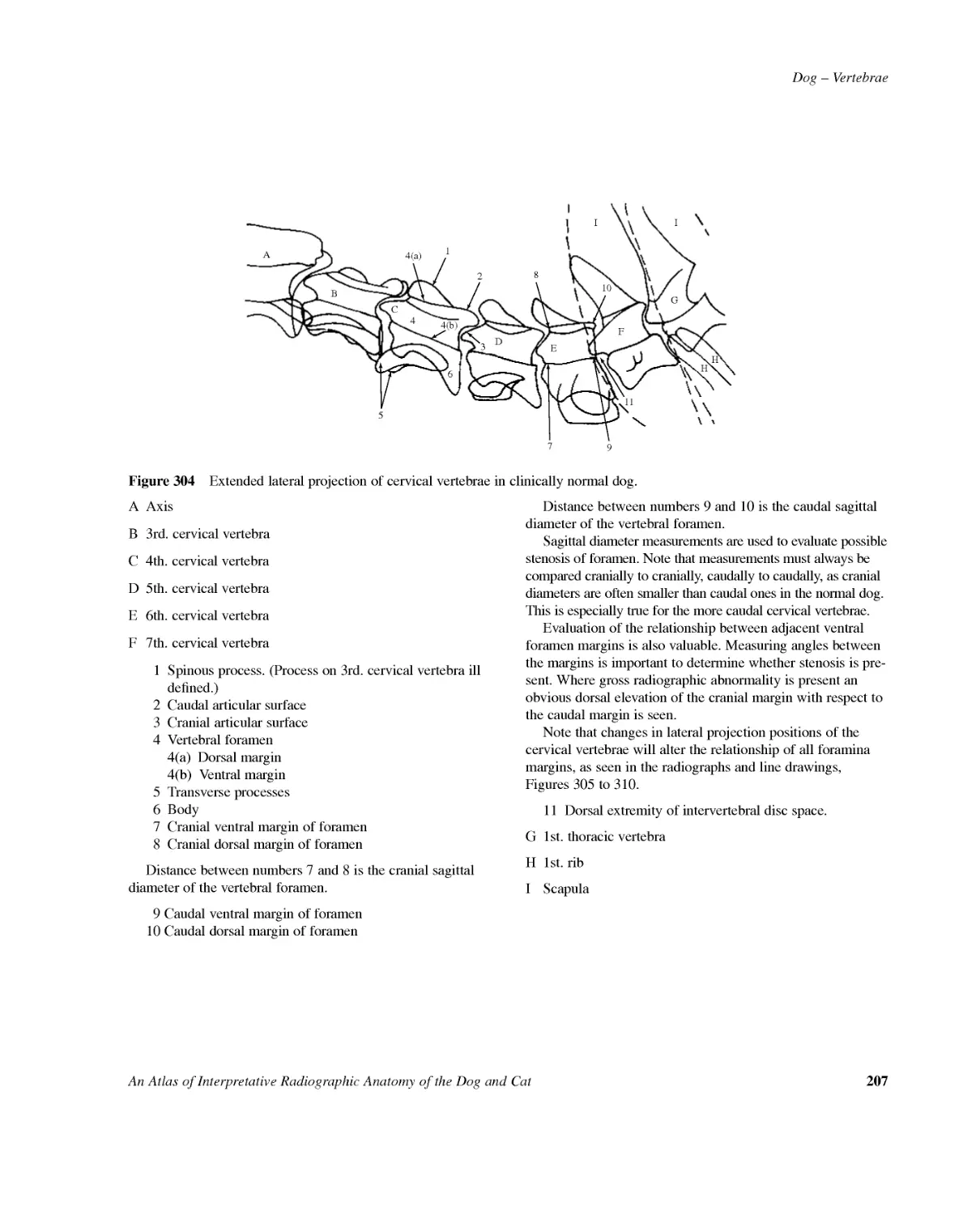
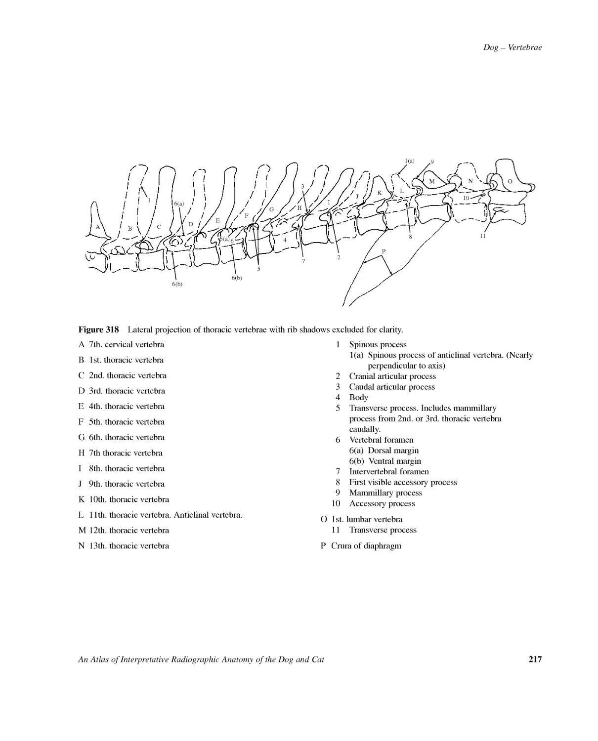
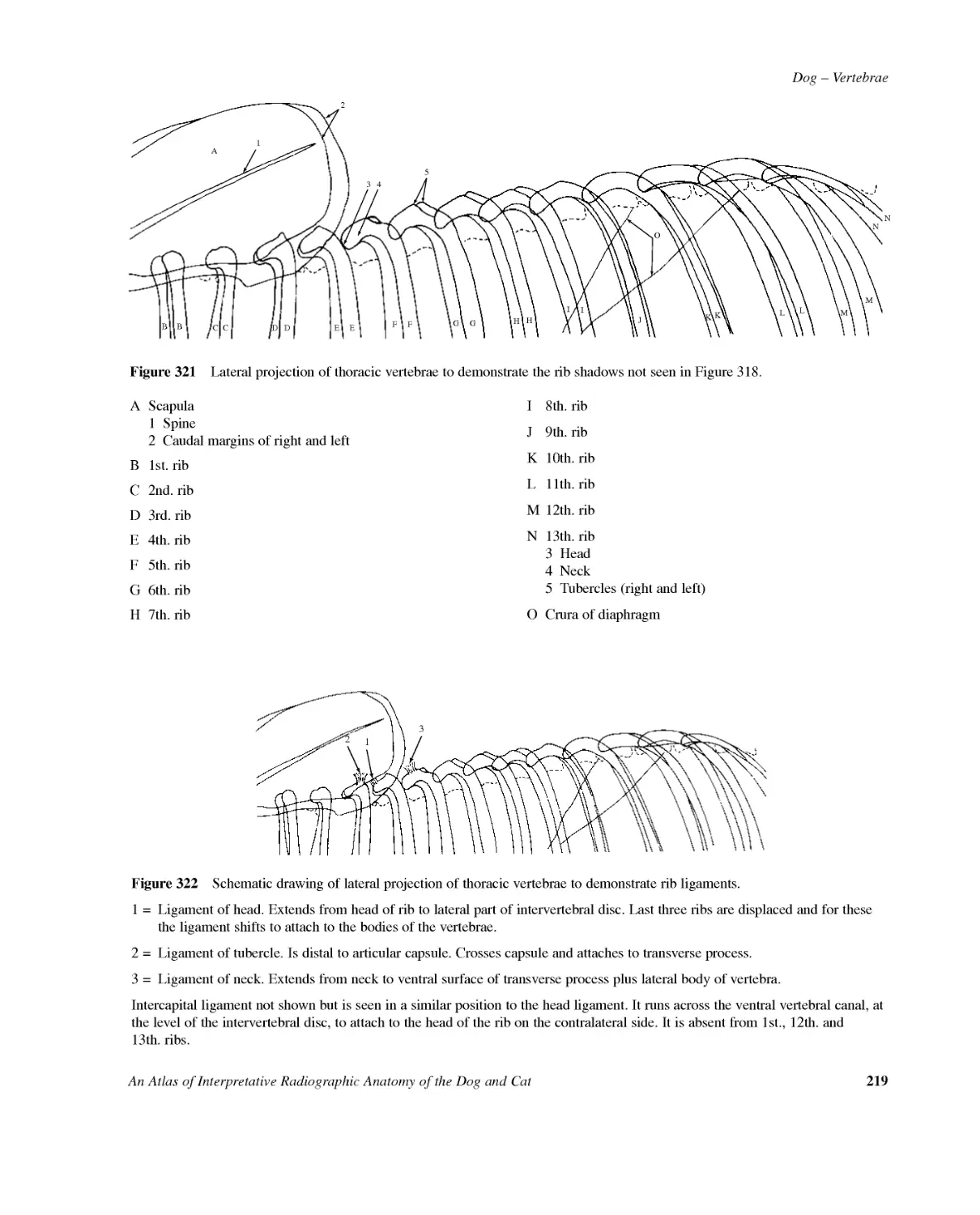
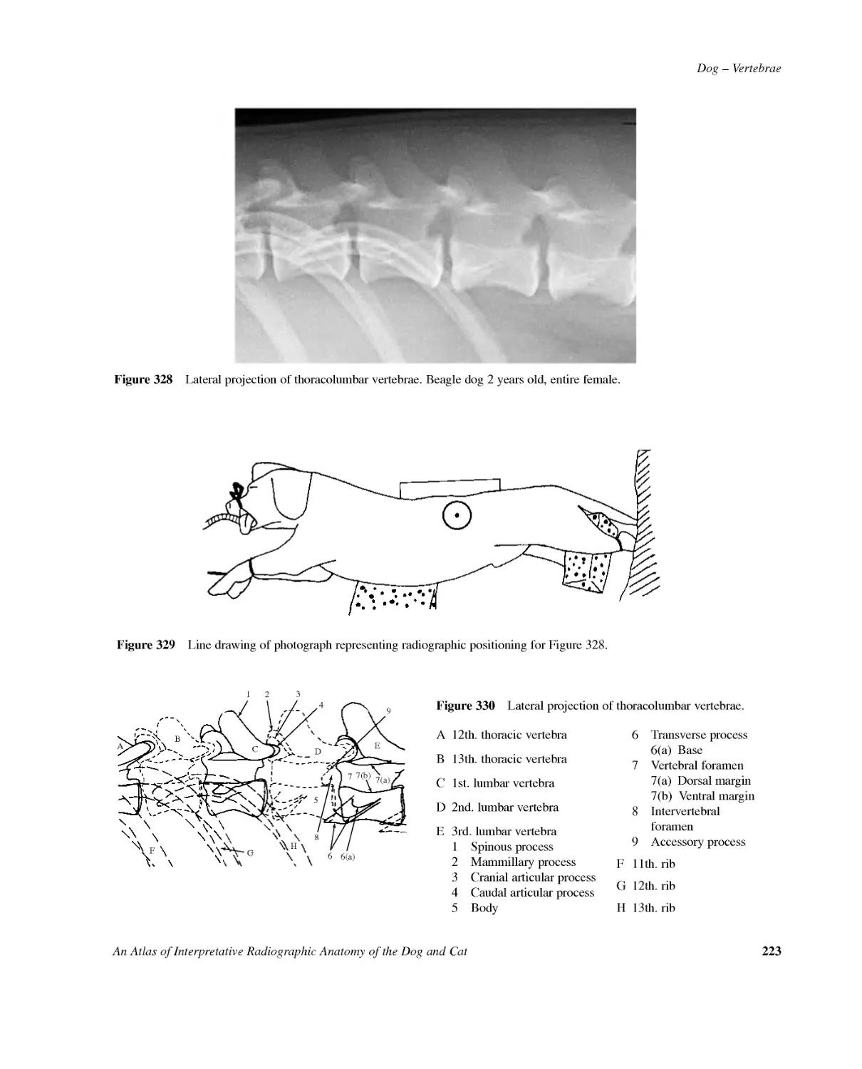
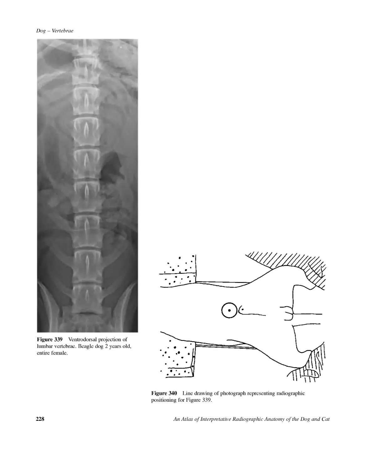
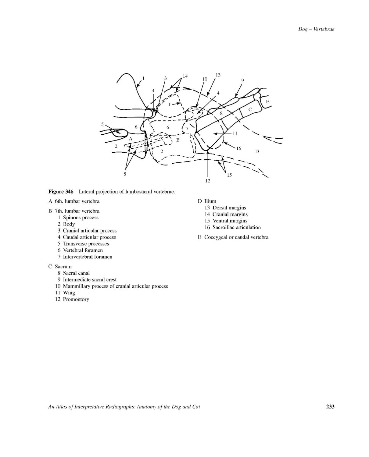
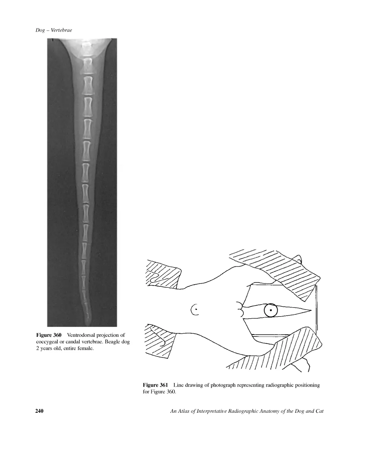
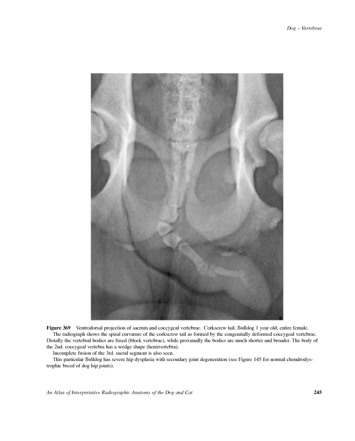
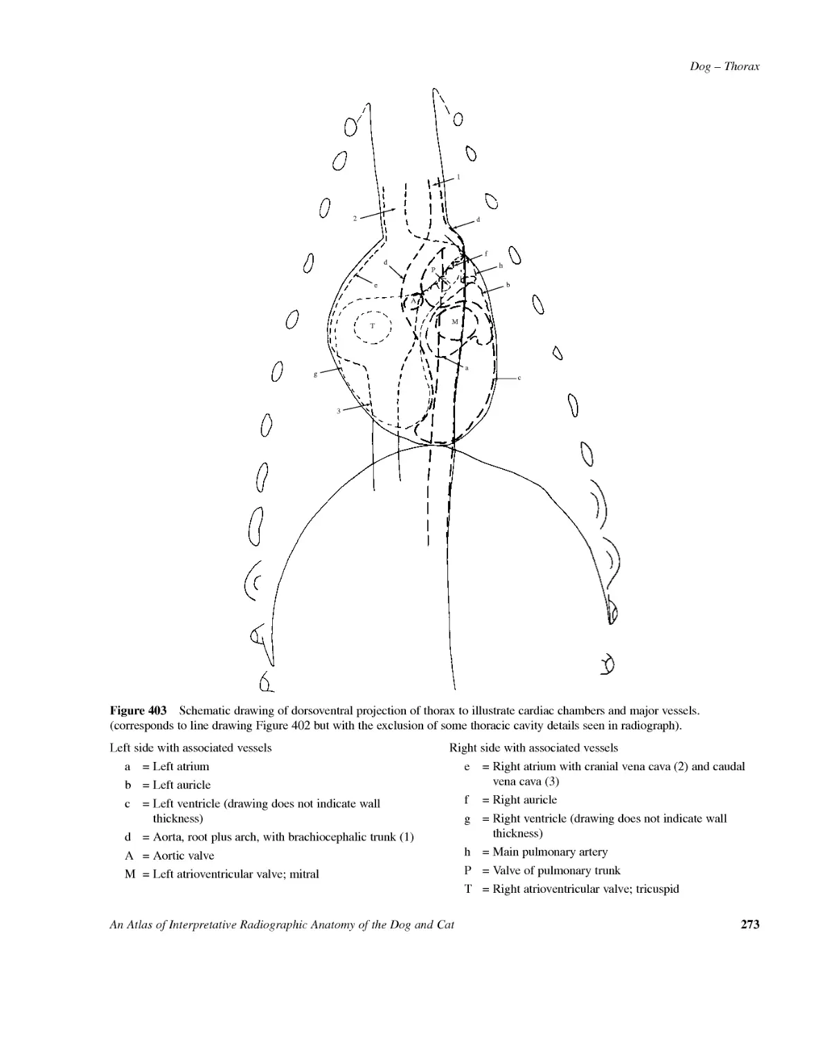
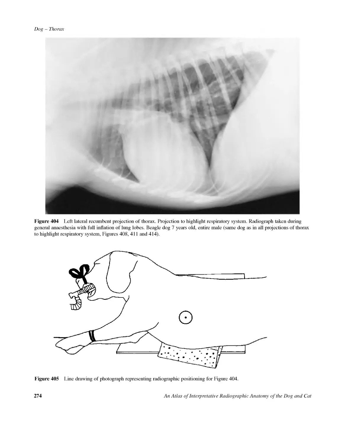
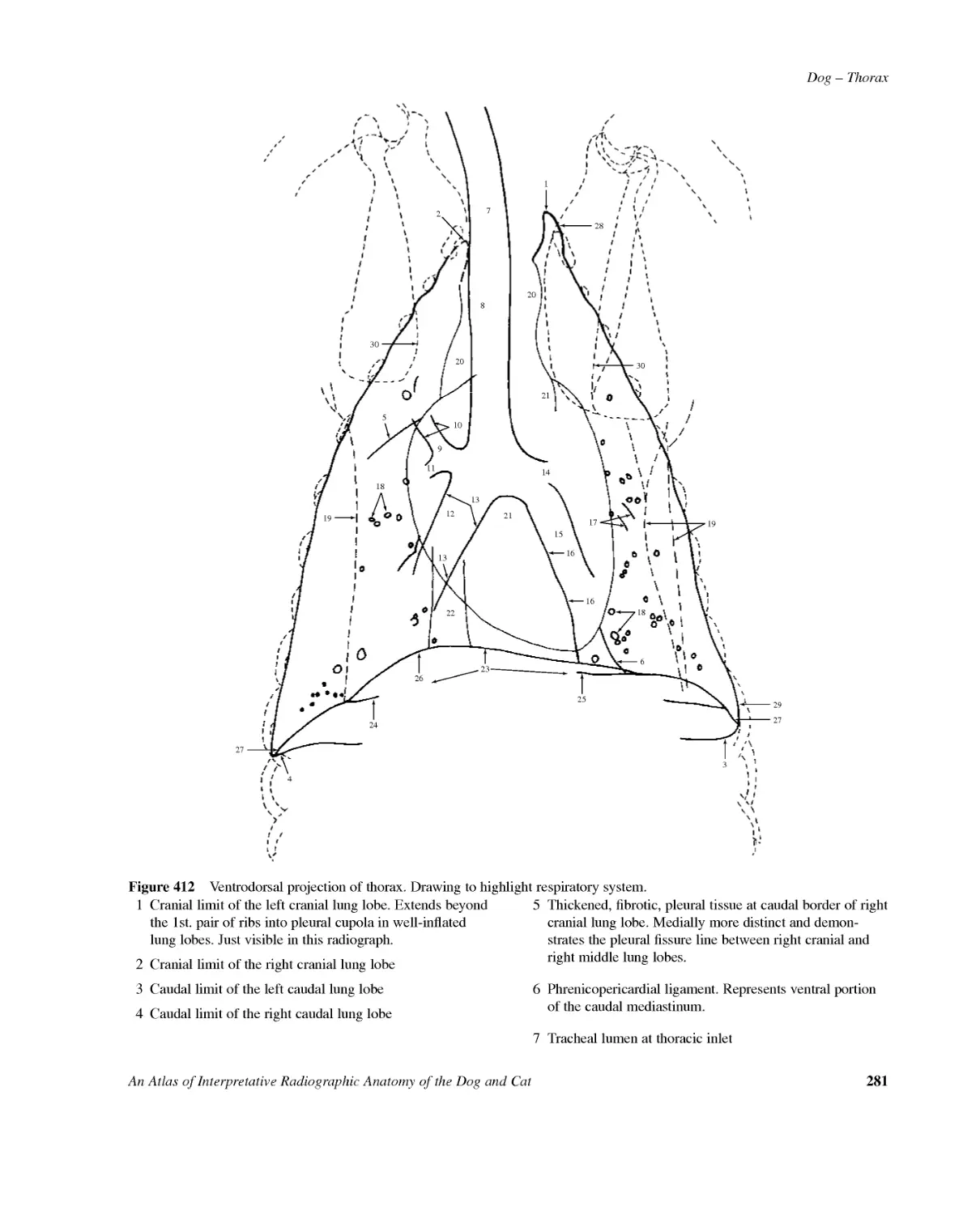
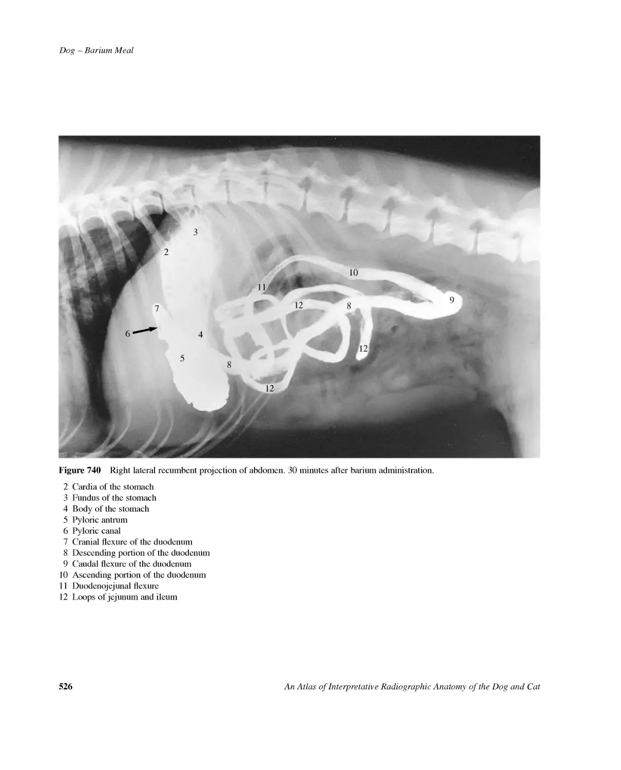
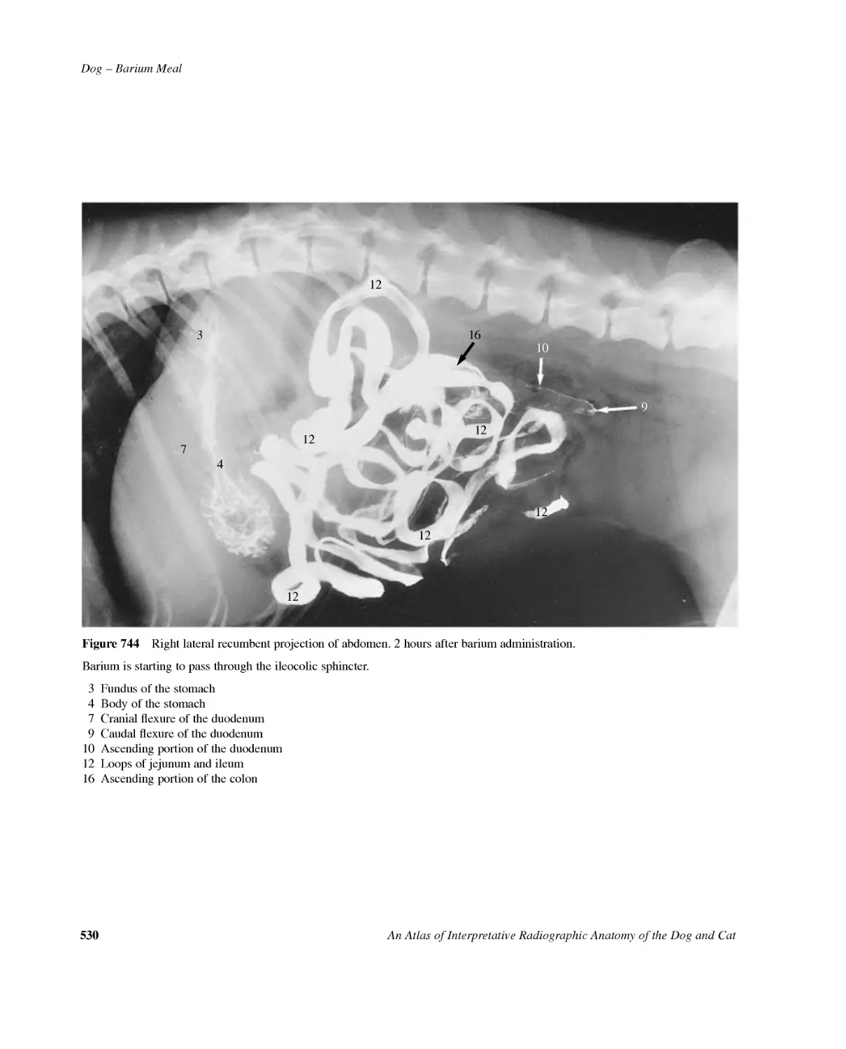
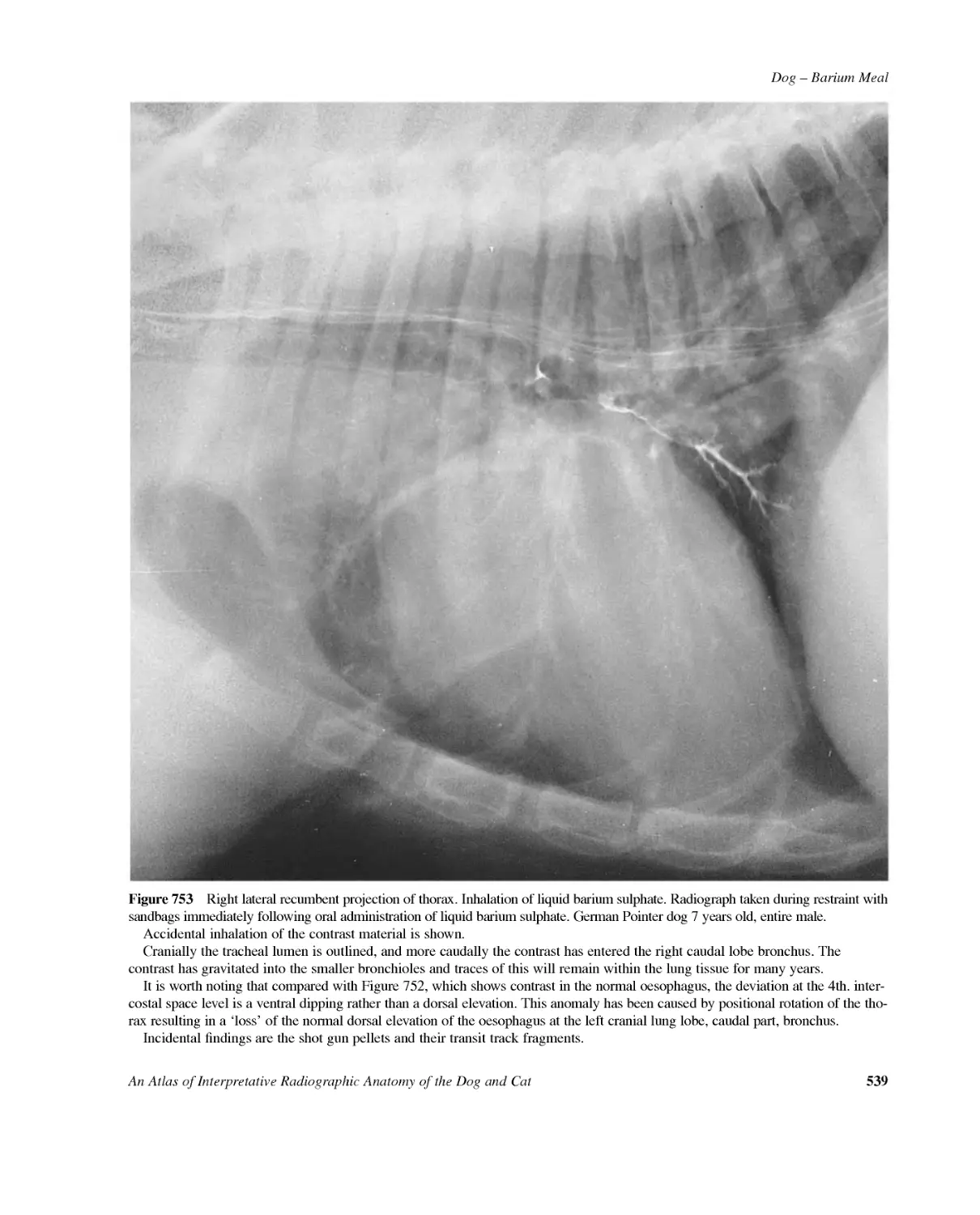
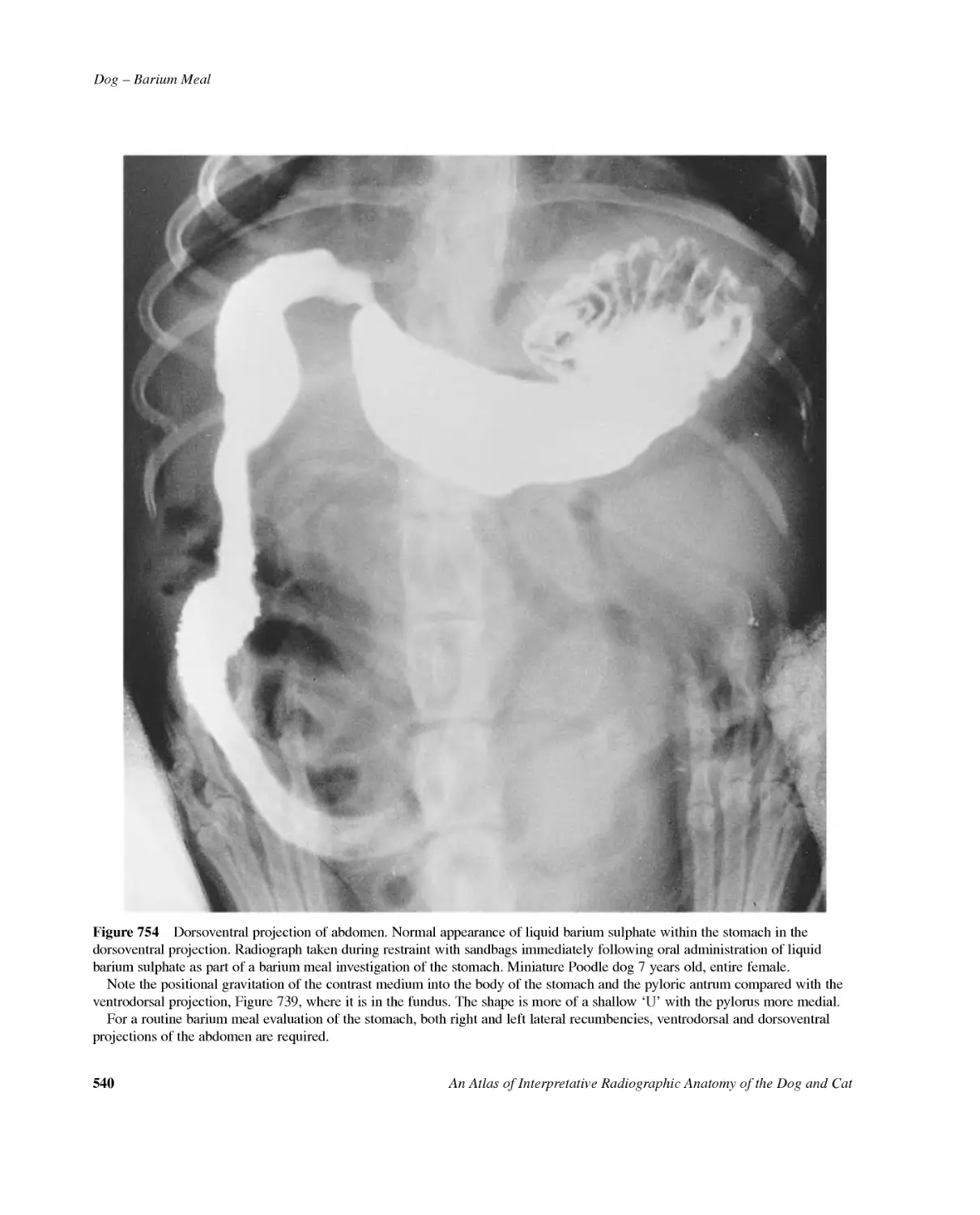
/


Автор: Coulson A. Lewis N.
Теги: biology anatomy animal anatomy radiographic anatomy guide for vet doctor's
Год: 2002


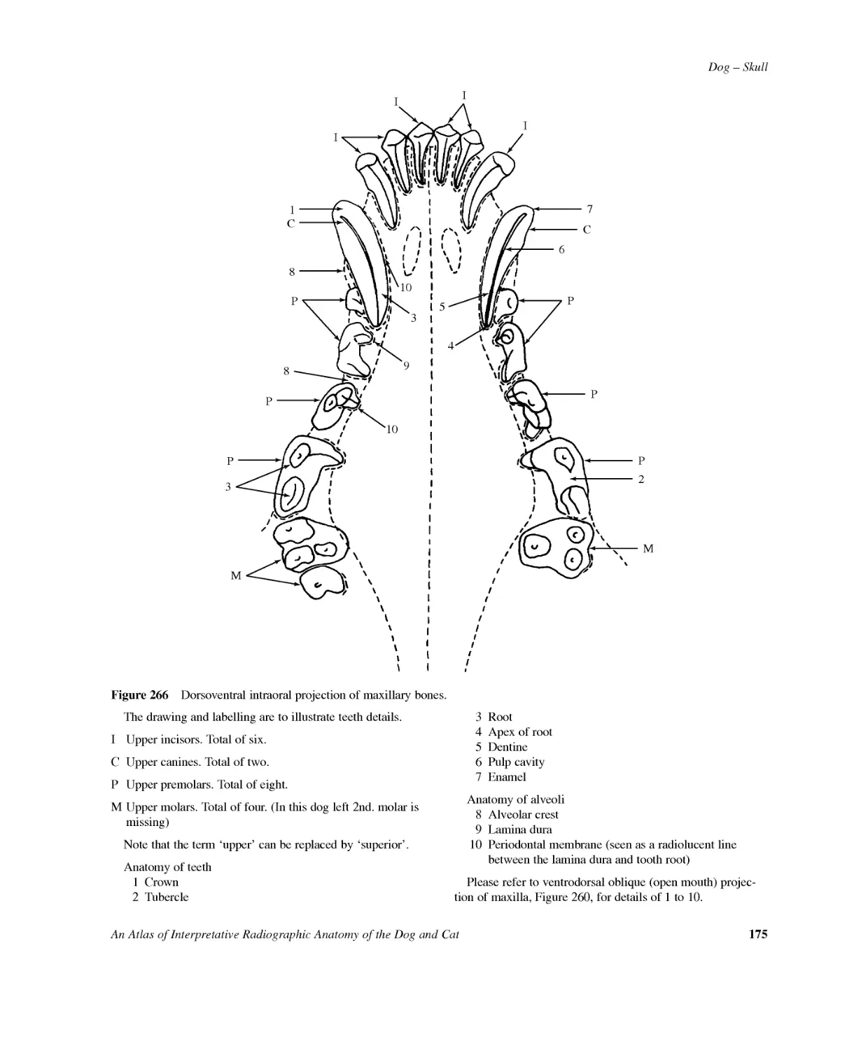
Текст
AnAtlasofInterpretativeRadiographicAnatomyoftheDog&Cat
AnAtlasofInterpretativeRadiographicAnatomyoftheDog&CatARLENECOULSONB.Vet.Med.D.V.R.M.R.C.V.S.withNOREENLEWISB.Sc.D.V.R.M.R.C.V.S.
©2002byBlackwellScienceLtdEditorialOffices:OsneyMead,OxfordOX20EL25JohnStreet,LondonWC1N2BS23AinsliePlace,EdinburghEH36AJ350MainStreet,MaldenMA021485018,USA54UniversityStreet,CarltonVictoria3053,Australia10,rueCasimirDelavigne75006Paris,FranceOtherEditorialOffices:BlackwellWissenschafts-VerlagGmbHKurfürstendamm5710707Berlin,GermanyBlackwellScienceKKMGKodenmachoBuilding7--10KodenmachoNihombashiChuo-ku,Tokyo104,JapanIowaStateUniversityPressABlackwellScienceCompany2121S.StateAvenueAmes,Iowa50014-8300,USATherightoftheAuthortobeidentifiedastheAuthorofthisWorkhasbeenassertedinaccordancewiththeCopyright,DesignsandPatentsAct1988.Allrightsreserved.Nopartofthispublicationmaybereproduced,storedinaretrievalsystem,ortransmitted,inanyformorbyanymeans,electronic,mechanical,photocopying,recordingorotherwise,exceptaspermittedbytheUKCopyright,DesignsandPatentsAct1988,withoutthepriorpermissionofthepublisher.Firstpublished2002SetinTimesbyGrayPublishing,TunbridgeWells,KentPrintedandboundinGreatBritainbyButlerandTannerLtd,FromeandLondonTheBlackwellSciencelogoisatrademarkofBlackwellScienceLtd,registeredattheUnitedKingdomTradeMarksRegistryDISTRIBUTORSMarstonBookServicesLtdPOBox269AbingdonOxonOX144YN(Orders:Tel:01235465500Fax:01235465555)USAandCanadaIowaStateUniversityPressABlackwellScienceCompany2121S.StateAvenueAmes,Iowa50014-8300(Orders:Tel:800-862-6657Fax:515-292-3348Webwww.isupress.comemail:orders@isupress.comAustraliaBlackwellSciencePtyLtd54UniversityStreetCarlton,Victoria3053(Orders:Tel:0393470300Fax:0393475001)AcataloguerecordforthistitleisavailablefromtheBritishLibraryISBN0-632-04078-5LibraryofCongressCataloging-in-PublicationDataCoulson,Arlene.Anatlasofinterpretativeradiographicanatomyofthedogandcat/ArleneCoulson,NoreenD.Lewis.p.cm.Includesbibliographicalreferences(p.).ISBN0-632-04078-51.Dogs--Anatomy--Atlases.2.Cats--Anatomy--Atlases.3.Veterinaryradiography--Atlases.I.Lewis,NoreenD.II.Title.SF767.D6C682001636.7′089607572--dc2100-068009ForfurtherinformationonBlackwellScience,visitourwebsite:www.blackwell-science.com
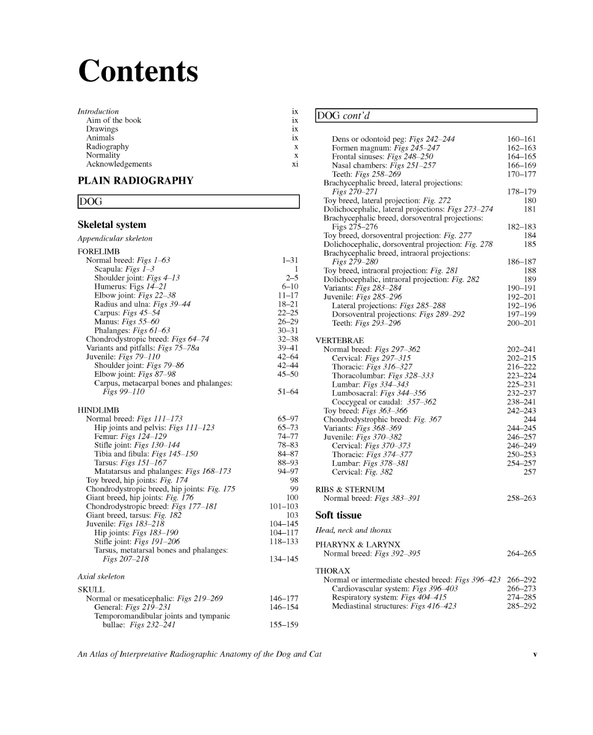
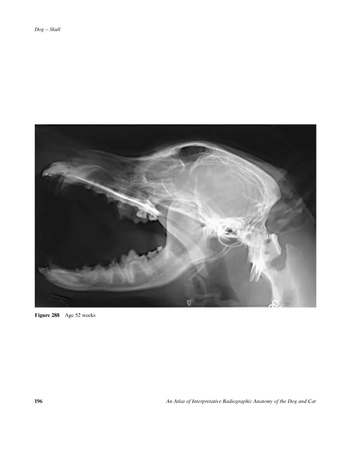
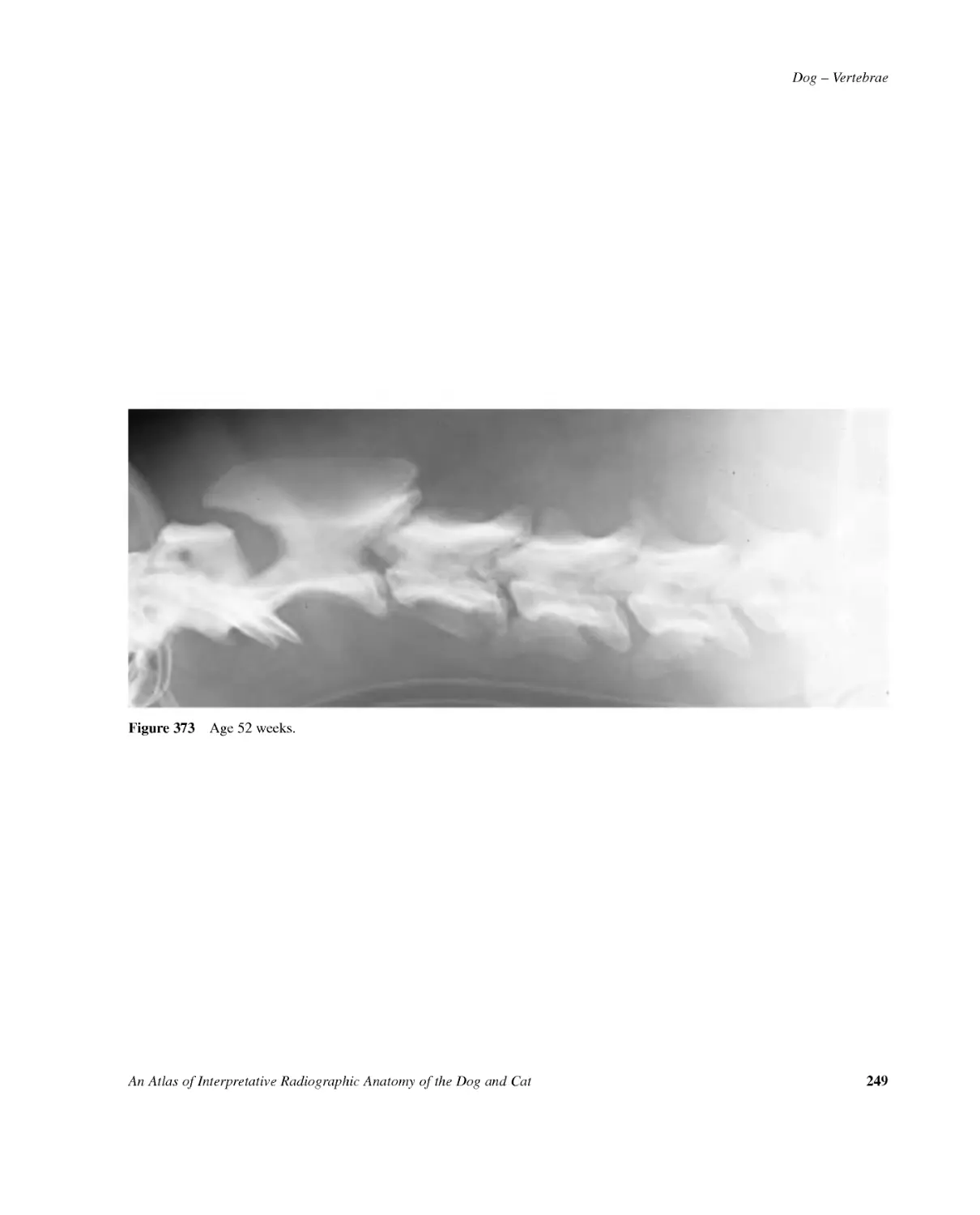
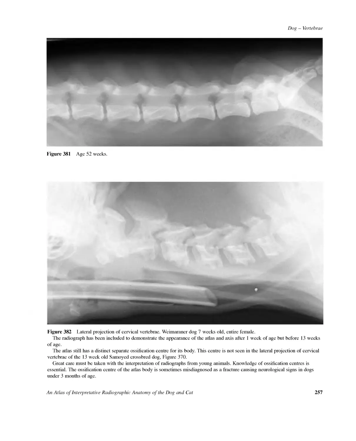
IntroductionixAimofthebookixDrawingsixAnimalsixRadiographyxNormalityxAcknowledgementsxiPLAINRADIOGRAPHYDOGSkeletalsystemAppendicularskeletonFORELIMBNormalbreed:Figs1--631--31Scapula:Figs1--31Shoulderjoint:Figs4--132--5Humerus:Figs14--216--10Elbowjoint:Figs22--3811--17Radiusandulna:Figs39--4418--21Carpus:Figs45--5422--25Manus:Figs55--6026--29Phalanges:Figs61--6330--31Chondrodystropicbreed:Figs64--7432--38Variantsandpitfalls:Figs75--78a39--41Juvenile:Figs79--11042--64Shoulderjoint:Figs79--8642--44Elbowjoint:Figs87--9845--50Carpus,metacarpalbonesandphalanges:Figs99--11051--64HINDLIMBNormalbreed:Figs111--17365--97Hipjointsandpelvis:Figs111--12365--73Femur:Figs124--12974--77Stiflejoint:Figs130--14478--83Tibiaandfibula:Figs145--15084--87Tarsus:Figs151--16788--93Matatarsusandphalanges:Figs168--17394--97Toybreed,hipjoints:Fig.17498Chondrodystropicbreed,hipjoints:Fig.17599Giantbreed,hipjoints:Fig.176100Chondrodystropicbreed:Figs177--181101--103Giantbreed,tarsus:Fig.182103Juvenile:Figs183--218104--145Hipjoints:Figs183--190104--117Stiflejoint:Figs191--206118--133Tarsus,metatarsalbonesandphalanges:Figs207--218134--145AxialskeletonSKULLNormalormesaticephalic:Figs219--269146--177General:Figs219--231146--154Temporomandibularjointsandtympanicbullae:Figs232--241155--159DOGcont'dDensorodontoidpeg:Figs242--244160--161Formenmagnum:Figs245--247162--163Frontalsinuses:Figs248--250164--165Nasalchambers:Figs251--257166--169Teeth:Figs258--269170--177Brachycephalicbreed,lateralprojections:Figs270--271178--179Toybreed,lateralprojection:Fig.272180Dolichocephalic,lateralprojections:Figs273--274181Brachycephalicbreed,dorsoventralprojections:Figs275--276182--183Toybreed,dorsoventralprojection:Fig.277184Dolichocephalic,dorsoventralprojection:Fig.278185Brachycephalicbreed,intraoralprojections:Figs279--280186--187Toybreed,intraoralprojection:Fig.281188Dolichocephalic,intraoralprojection:Fig.282189Variants:Figs283--284190--191Juvenile:Figs285--296192--201Lateralprojections:Figs285--288192--196Dorsoventralprojections:Figs289--292197--199Teeth:Figs293--296200--201VERTEBRAENormalbreed:Figs297--362202--241Cervical:Figs297--315202--215Thoracic:Figs316--327216--222Thoracolumbar:Figs328--333223--224Lumbar:Figs334--343225--231Lumbosacral:Figs344--356232--237Coccygealorcaudal:357--362238--241Toybreed:Figs363--366242--243Chondrodystrophicbreed:Fig.367244Variants:Figs368--369244--245Juvenile:Figs370--382246--257Cervical:Figs370--373246--249Thoracic:Figs374--377250--253Lumbar:Figs378--381254--257Cervical:Fig.382257RIBS&STERNUMNormalbreed:Figs383--391258--263SofttissueHead,neckandthoraxPHARYNX&LARYNXNormalbreed:Figs392--395264--265THORAXNormalorintermediatechestedbreed:Figs396--423266--292Cardiovascularsystem:Figs396--403266--273Respiratorysystem:Figs404--415274--285Mediastinalstructures:Figs416--423285--292ContentsAnAtlasofInterpretativeRadiographicAnatomyoftheDogandCatv
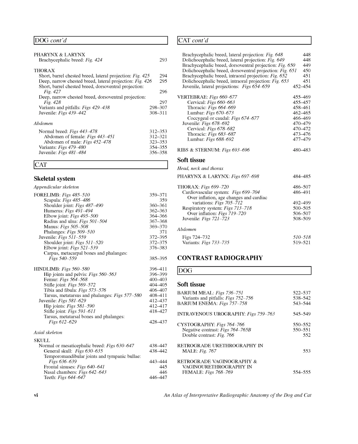
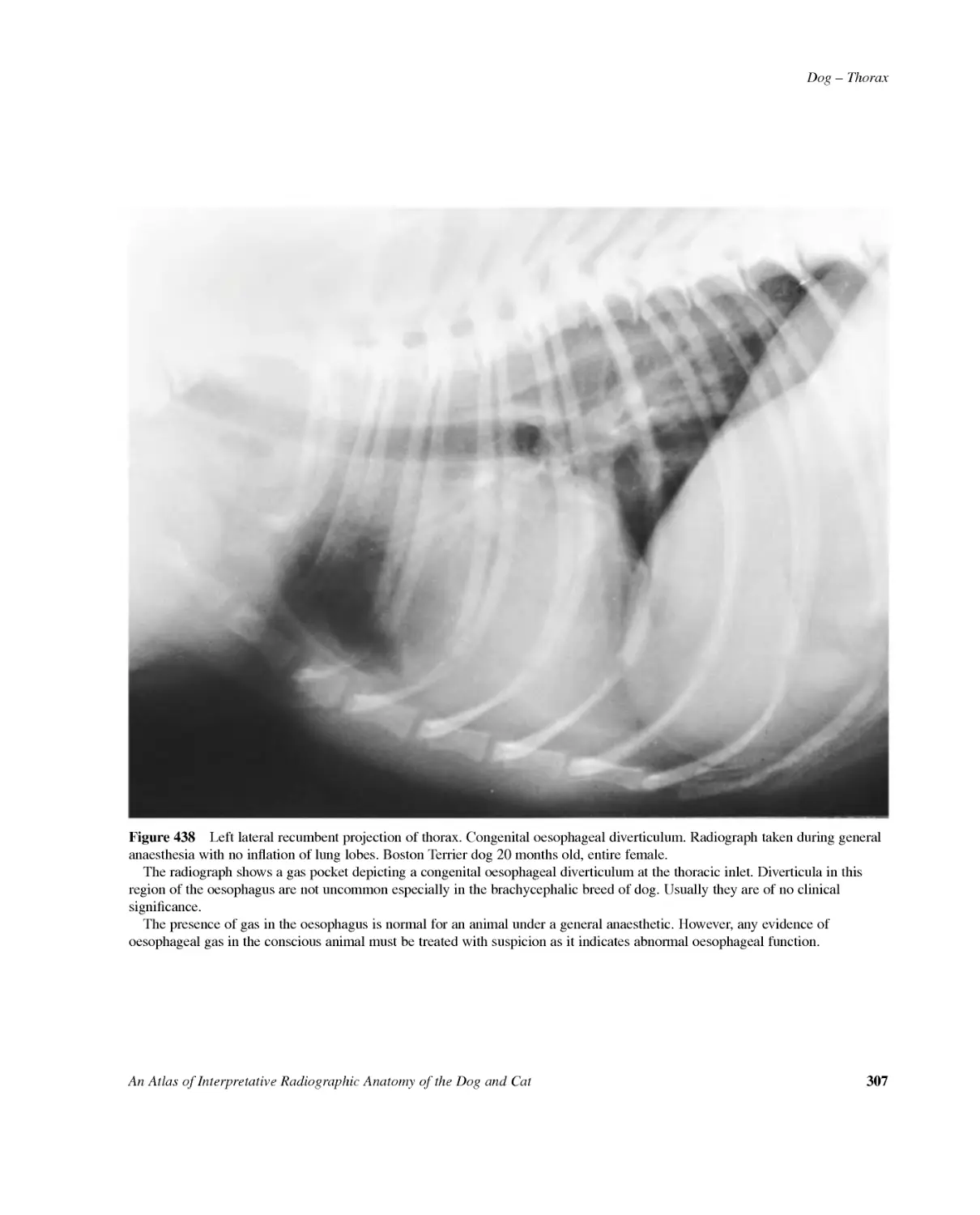
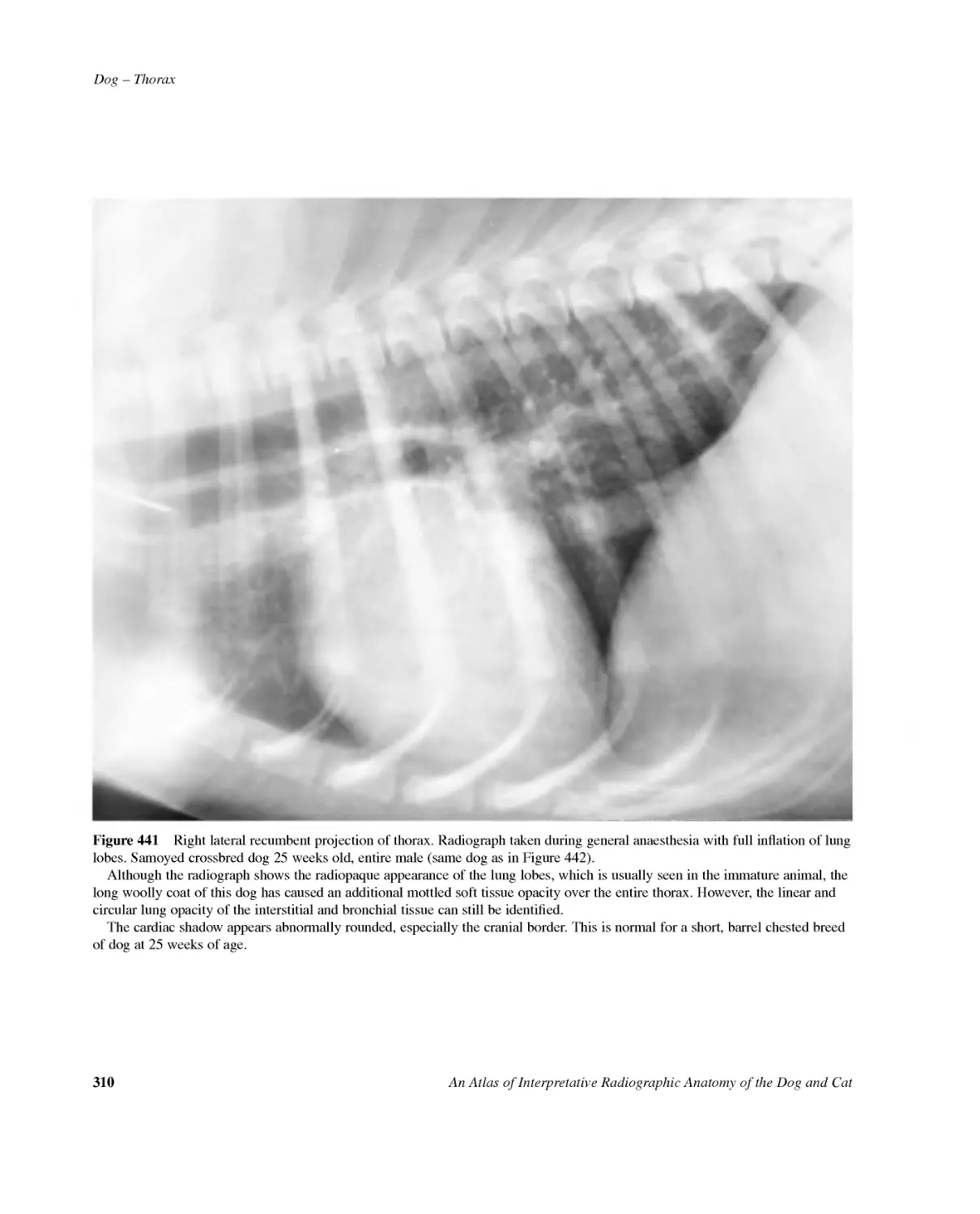
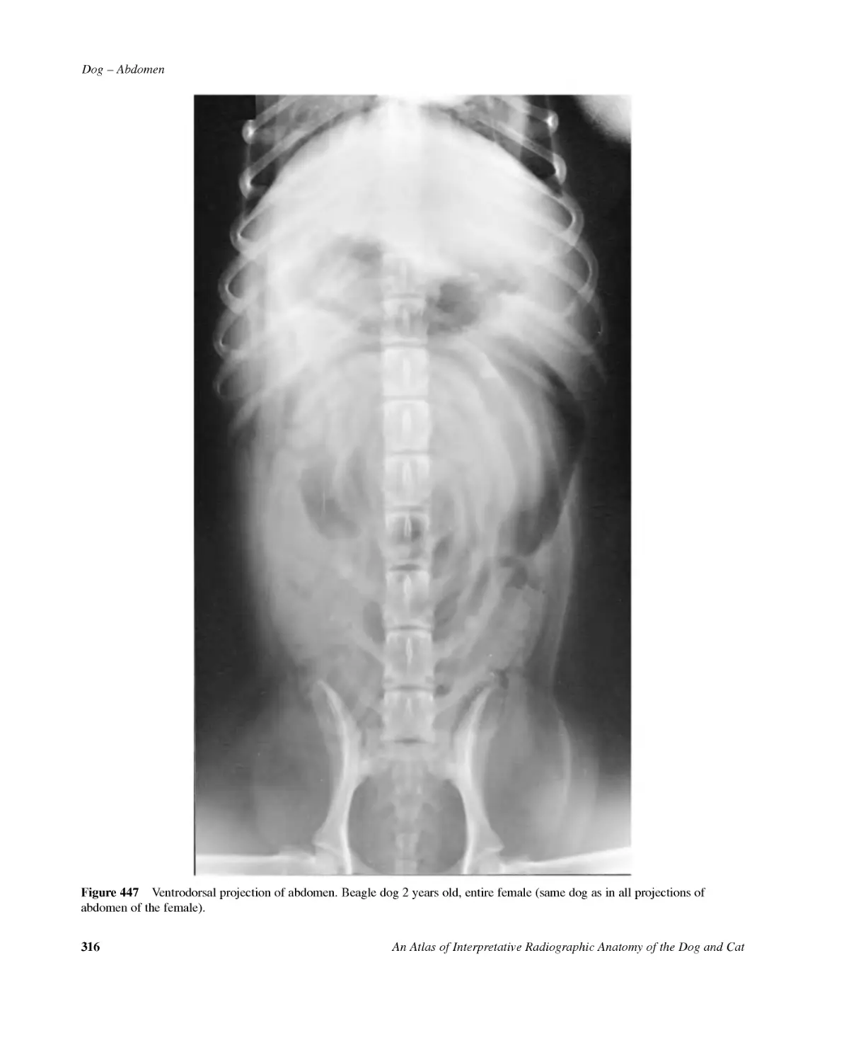
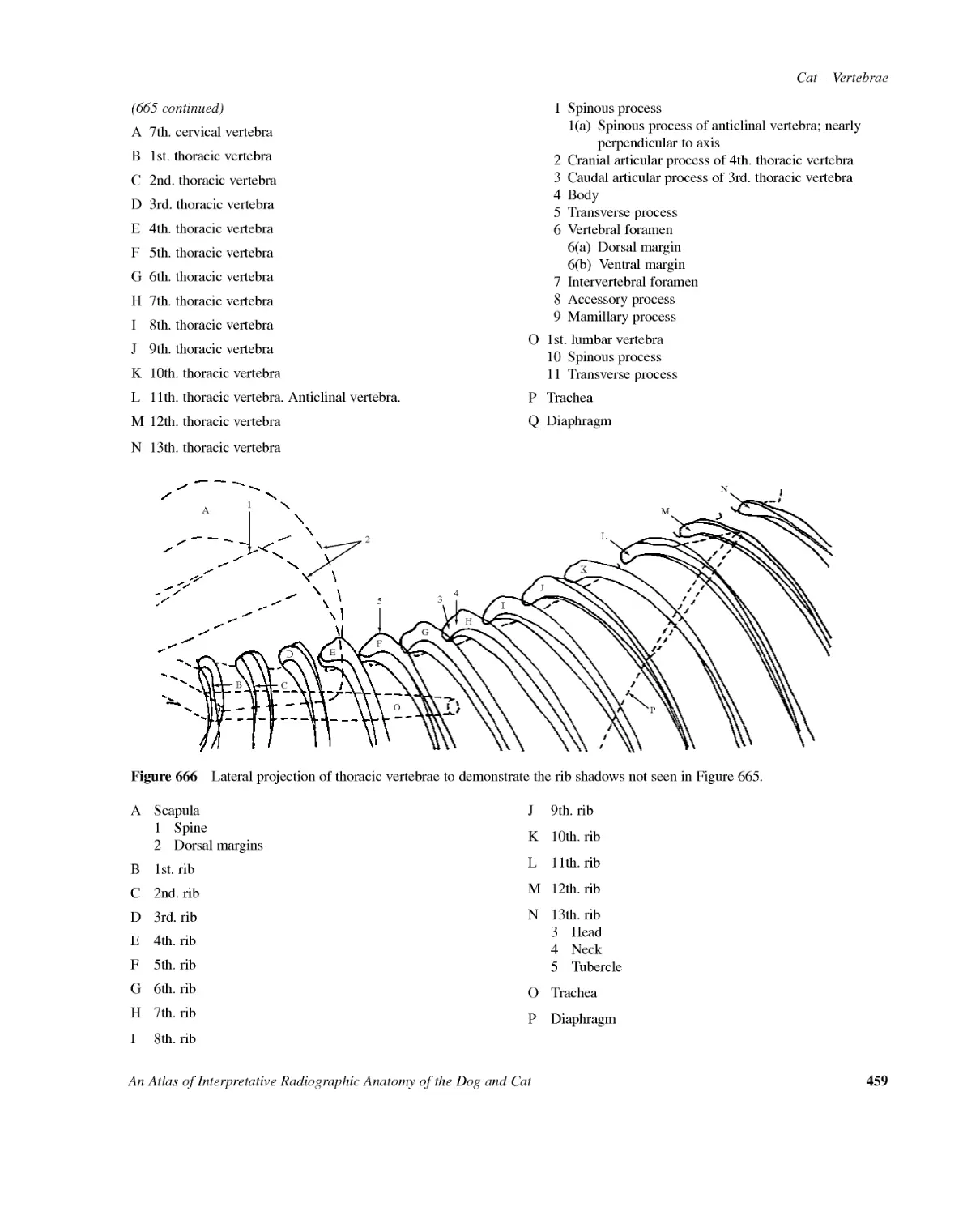
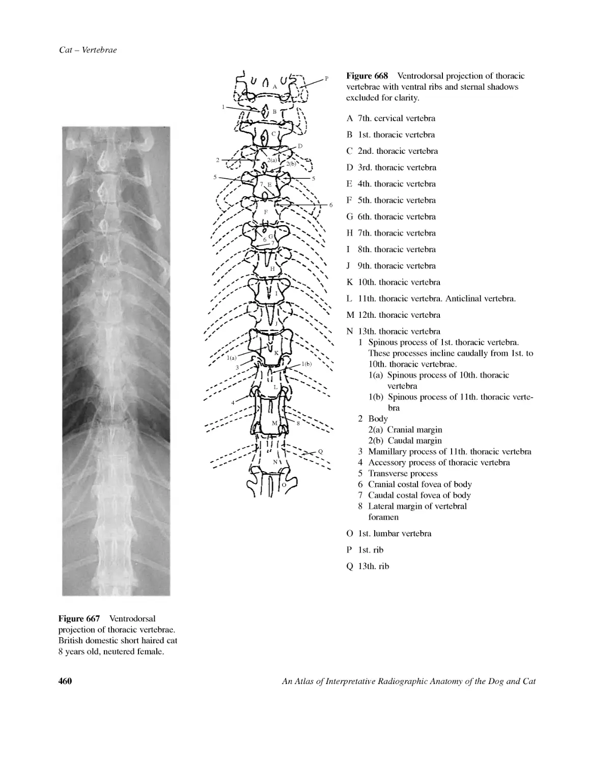
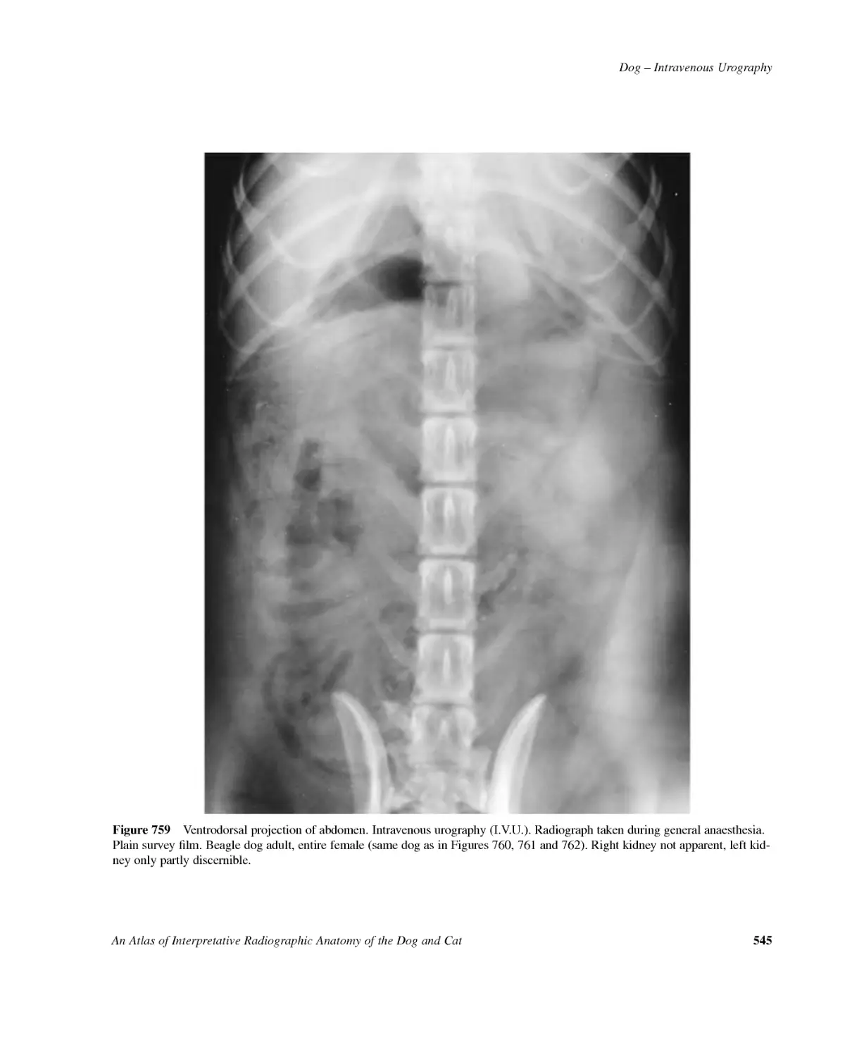
DOGcont'dPHARYNX&LARYNXBrachycephalicbreed:Fig.424293THORAXShort,barrelchestedbreed,lateralprojection:Fig.425294Deep,narrowchestedbreed,lateralprojection:Fig.426295Short,barrelchestedbreed,dorsoventralprojection:Fig.427296Deep,narrowchestedbreed,dorsoventralprojection:Fig.428297Variantsandpitfalls:Figs429--438298--307Juvenile:Figs439--442308--311AbdomenNormalbreed:Figs443--478312--353Abdomenoffemale:Figs443--451312--321Abdomenofmale:Figs452--478323--353Variants:Figs479--480354--355Juvenile:Figs481--484356--358CATSkeletalsystemAppendicularskeletonFORELIMB:Figs485--510359--371Scapula:Figs485--486359Shoulderjoint:Figs487--490360--361Humerus:Figs491--494362--363Elbowjoint:Figs495--500364--366Radiusandulna:Figs501--504367--368Manus:Figs505--508369--370Phalanges:Figs509--510371Juvenile:Figs511--559372--395Shoulderjoint:Figs511--520372--375Elbowjoint:Figs521--539376--383Carpus,metacarpalbonesandphalanges:Figs540--559385--395HINDLIMB:Figs560--580396--411Hipjointsandpelvis:Figs560--563396--399Femur:Figs564--568400--403Stiflejoint:Figs569--572404--405Tibiaandfibula:Figs573--576406--407Tarsus,metatarsusandphalanges:Figs577--580408--411Juvenile:Figs581--629412--437Hipjoints:Figs581--590412--417Stiflejoint:Figs591--611418--427Tarsus,metatarsalbonesandphalanges:Figs612--629428--437AxialskeletonSKULLNormalormesaticephalicbreed:Figs630--647438--447Generalskull:Figs630--635438--442Temporomandibularjointsandtympanicbullae:Figs636--639443--444Frontalsinuses:Figs640--641445Nasalchambers:Figs642--643446Teeth:Figs644--647446--447CATcont'dBrachycephalicbreed,lateralprojection:Fig.648448Dolichocephalicbreed,lateralprojection:Fig.649448Brachycephalicbreed,dorsoventralprojection:Fig.650449Dolichocephalicbreed,dorsoventralprojection:Fig.651450Brachycephalicbreed,intraoralprojection:Fig.652451Dolichocephalicbreed,intraoralprojection:Fig.653451Juvenile,lateralprojections:Figs654--659452--454VERTEBRAE:Figs660--677455--469Cervical:Figs660--663455--457Thoracic:Figs664--669458--461Lumbar:Figs670--673462--465Coccygealorcaudal:Figs674--677466--469Juvenile:Figs678--692470--479Cervical:Figs678--682470--472Thoracic:Figs683--687473--476Lumbar:Figs688--692477--479RIBS&STERNUM:Figs693--696480--483SofttissueHead,neckandthoraxPHARYNX&LARYNX:Figs697--698484--485THORAX:Figs699--720486--507Cardiovascularsystem:Figs699--704486--491Overinflation,agechangesandcardiacvariations:Figs705--712492--499Respiratorysystem:Figs713--718500--505Overinflation:Figs719--720506--507Juvenile:Figs721--723508--509AbdomenFigs724--732510--518Variants:Figs733--735519--521CONTRASTRADIOGRAPHYDOGSofttissueBARIUMMEAL:Figs736--751522--537Variantsandpitfalls:Figs752--756538--542BARIUMENEMA:Figs757--758543--544INTRAVENOUSUROGRAPHY:Figs759--763545--549CYSTOGRAPHY:Figs764--766550--552Negativecontrast:Figs764--765B550--551Doublecontrast:Fig.766552RETROGRADEURETHROGRAPHYINMALE:Fig.767553RETROGRADEVAGINOGRAPHY&VAGINOURETHROGRAPHYINFEMALE:Figs768--769554--555viAnAtlasofInterpretativeRadiographicAnatomyoftheDogandCat
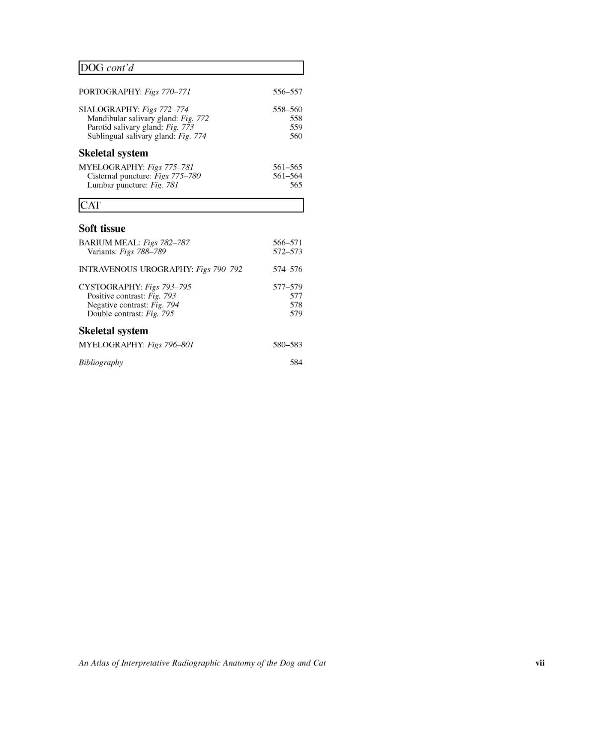
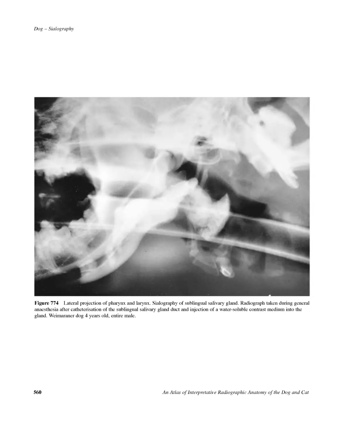
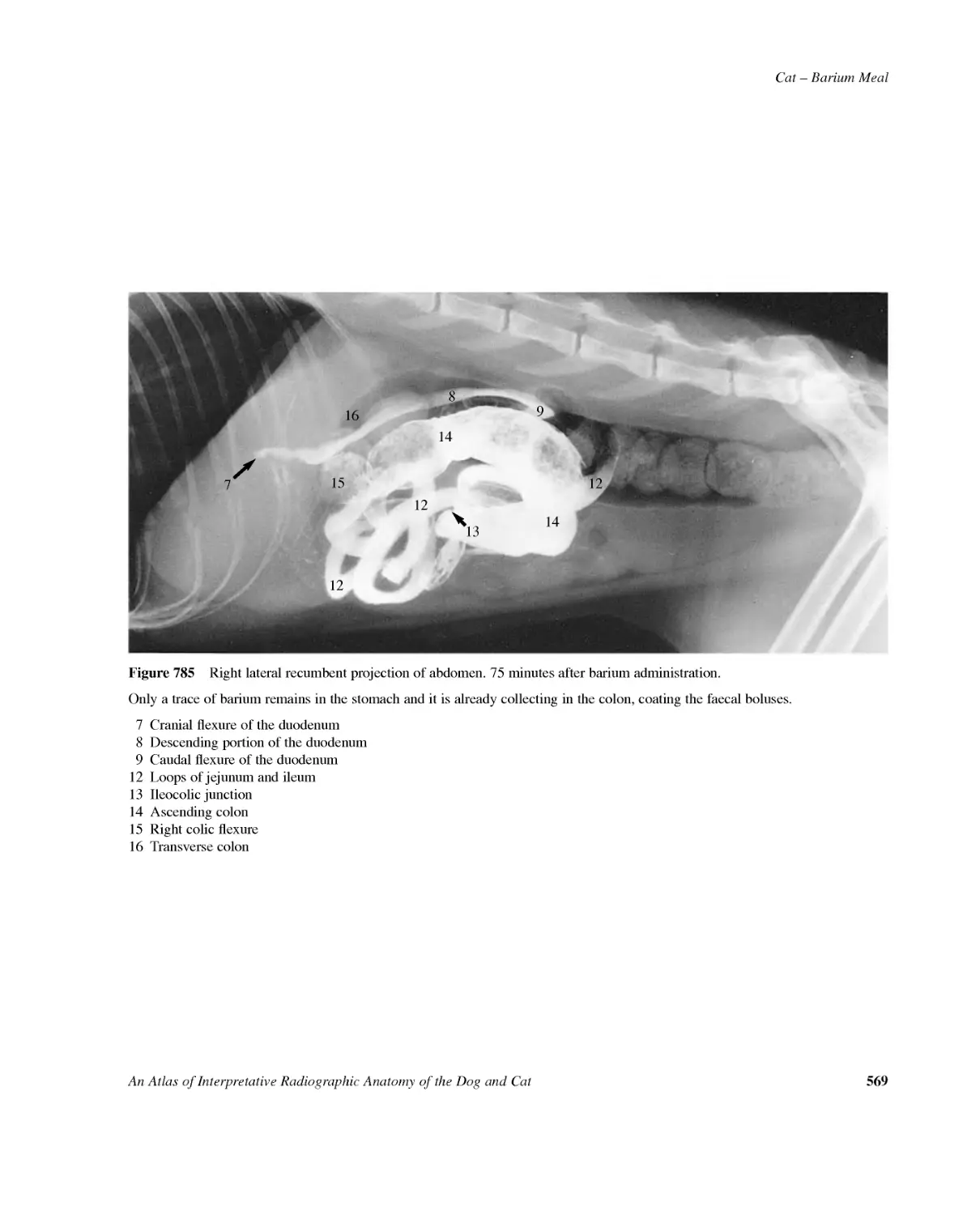
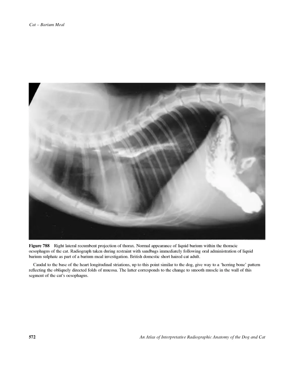
DOGcont'dPORTOGRAPHY:Figs770--771556--557SIALOGRAPHY:Figs772--774558--560Mandibularsalivarygland:Fig.772558Parotidsalivarygland:Fig.773559Sublingualsalivarygland:Fig.774560SkeletalsystemMYELOGRAPHY:Figs775--781561--565Cisternalpuncture:Figs775--780561--564Lumbarpuncture:Fig.781565CATSofttissueBARIUMMEAL:Figs782--787566--571Variants:Figs788--789572--573INTRAVENOUSUROGRAPHY:Figs790--792574--576CYSTOGRAPHY:Figs793--795577--579Positivecontrast:Fig.793577Negativecontrast:Fig.794578Doublecontrast:Fig.795579SkeletalsystemMYELOGRAPHY:Figs796--801580--583Bibliography584AnAtlasofInterpretativeRadiographicAnatomyoftheDogandCatvii
AimofthebookTheprimaryaimofthisbookistoprovideadetailedref-erenceforthebasicradiographicanatomyofthedogandcat.Thisisachievedbytheinclusionofbothradiographsanddrawings.Theimmatureanimaland,whererelevant,aspectrumofbreedshavebeenincluded.Aselectionofanatomicalvariantsandafewofthemorecommonradiographic'pitfalls'arealsotobefoundfollowingthe'normal'radiograph.Followingtheanatomicalsectionsofplainradiographyisaseriesofthemorecommonlyemployedcontraststud-ies.Confusioncanoccurwhentryingtointerpretsuchtech-niques,andmanyanatomicalfeaturescanonlybeseenwiththeaidofcontrastagents.Hencethesehavebeeninclud-ed,hopingtheyaidevaluationofthestudiesperformedmoreregularlyingeneralpractice.Inadditionafewofthelesscommonstudiesarefoundforanatomicalunder-standing.Frompersonalexperienceinteachingandexaminingvet-erinarysurgeonsforpost-graduateradiologycertificationitisclearthatagoodbasicknowledgeofradiographicanatomyisessential.Unfortunately,alltoooften'normal-ity'isnotrecognised,especiallywherebreedvariationhastobeconsidered.Ashortbibliographyisinthelastfewpagesofthisbook.Thelistincludesonlybooksandpublicationsconsulted,andrelevant,forthefiguresandtextofthismanuscript.Noindividualreferencesarecitedinthetext.Noindexhasbeenincludedastheatlasisintendedtobeusedasavisualreferencefornormality.Tofacilitatethisacomprehensivecontentslist,dividedintoanatomicalregionsforplainandcontrastradiography,isprovided.Althoughinitiallyitwouldappearthatthebookismain-lyforthebenefitofveterinarysurgeonswantingtoobtainadditionalradiologyqualifications,basicradiographicanatomywillbeofvaluetobothundergraduatesandvet-erinarysurgeonsingeneralpractice.Itishopedthatthisatlaswillbecomeausefulandwellusedreferencebookforboththespecialisingandnon-specialisingveterinaryaudience.DrawingsThedrawingsfollowtracingsoftheradiographs.Onlyshadowsseenintheradiographhavebeentraced,evenifanatomicallymoredetailshouldhavebeenpresent.Eachdrawinghasadetailedkey.Itishopedthattheradiographicreproductionisofasuf-ficientstandardtoallowrecognitionofalltheradiograph-icshadowsthathavebeentraced.Wheretheshadowsarecomplex,asintheskull,anum-berofdrawingshavebeenmadetoavoidinterpretativecon-fusionofnumerouslineswithinsmallregions.Everyefforthasbeenmadenottooverdraworover-labelthedrawingscorrelatingtotheradiographs.Inthiswayitishopedthatthereaderwillquicklyrecognisetheimportantshadowsandbecomefamiliarwithradiograph-icanatomy.Separatelinedrawingshavealsobeenincludedofsofttissuestructuressurroundingbonyshadows.Thesestruc-turesareoftenoverlookedwhenattentionisfocusedonthemoreobviousopaqueshadows.Muchvaluableinformationcanbegainedfromthesofttissuesurrounding,forexam-ple,thestiflejoint.Inadditiontothelinedrawings,schematicdrawingsofmanyprojectionshavebeenmadetofamiliarisetheread-erwithanatomicalfeaturesnotvisibleontheradiograph.Inthiswaythereaderwillbemoreabletomakelogicaldiagnosis/differentialdiagnosiswhenfacedwithradi-ographsdemonstratingabnormalfeatures.AnimalsMostoftheradiographsinthisbookareoriginalandfortheexclusiveuseoftheauthors.Theremainderhavebeengiventotheauthorsbygenerouscolleagues.Theradiographshavebeenobtainedoveraperiodoffivetosixyearsandabriefsummaryoftheirsourcefollows.The'normal'dogradiographsaremainlyfromagroupofBeagleHoundswhilethe'normal'catradiographsarefromanumberofindividualBritishDomesticShortHairedcats.AnAtlasofInterpretativeRadiographicAnatomyoftheDogandCatixIntroduction
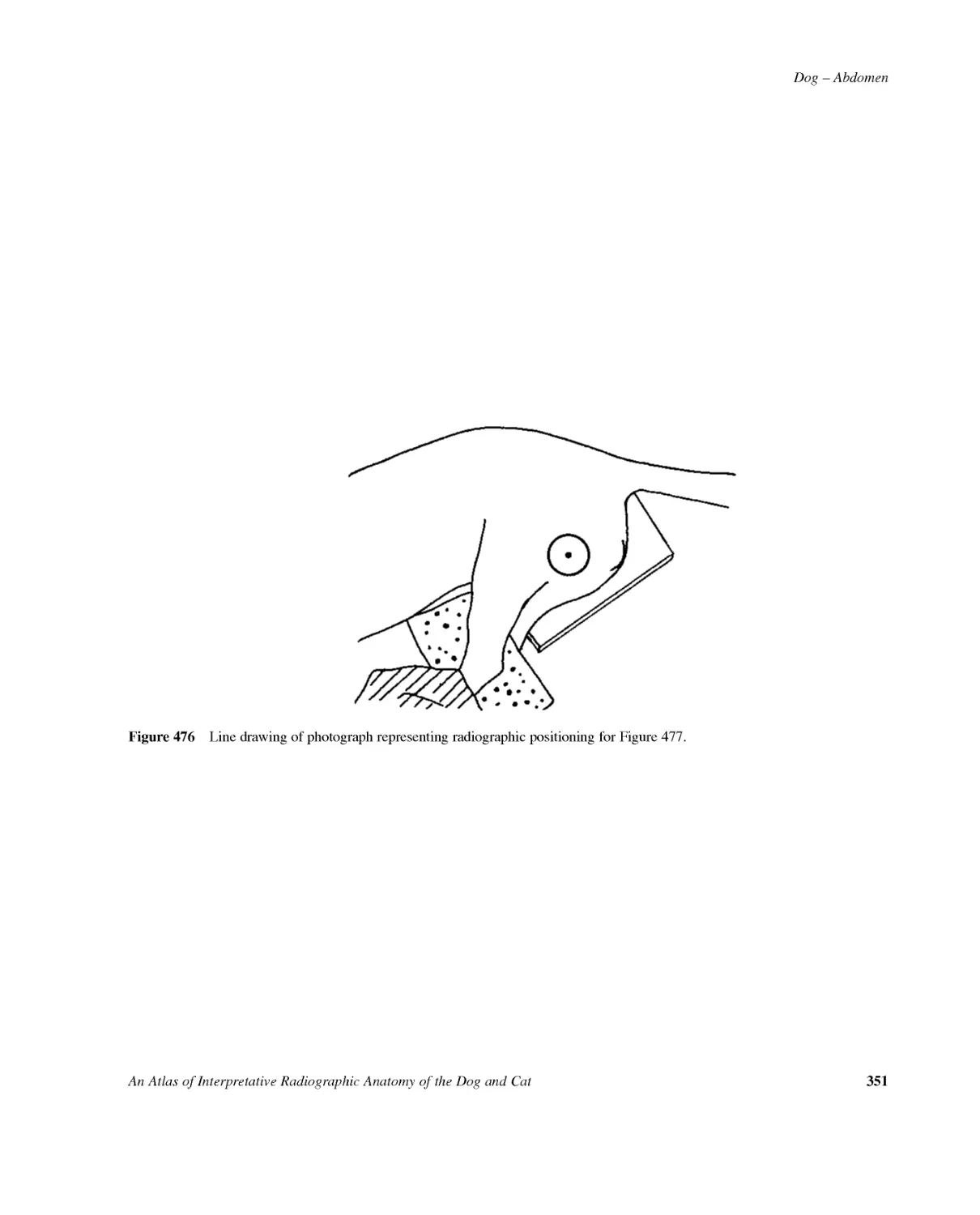
Inbothcasestheradiographswereobtainedspecifical-lyforthebook,radiographytakingplaceinconjunctionwithroutinesurgeryordentistryrequiringgeneralanaesthesia.Thedifferentbreeds,anatomicalvariantsandradi-ographic'pitfall'radiographswereeitherobtainedpri-marilyforthisbookorweretakenfromveterinarycollegefiles.Thiswasprobablyoneofthemostdifficultsectionstocompleteforpublicationasradiographsfallinginto'vari-ant'or'pitfall'arenotusuallyrecorded.Thedogjuvenilesectionwascommissionedforthisbookandradiographywasperformedonthesamedog(SamoyedCrossbredentiremale)from1monthto15monthsofageatintervalsofonemonth.Thisisprobablytheidealsituationforajuvenilestudyasindividual,feed-ingandhousingvariationsareallundercontrol.ThestudywasbasedatUniversityofGuelphinOntarioCanadaunderthewatchfuleyeofProfessorSumner-Smith.Thecatjuvenilesectionusuallyinvolvedadifferentcatateachmonthlyage.Individualsfromabreedinggroupwereradiographedspecificallyforthisbook,duringstud-iesonclinicalanaesthesiabasedinNewcastle,England.Althoughthisisnotidealassomeindividualvariationispresent,variationswithfeedingandhousingwereelimi-nated.Thesignificantadvantageofundertakingtheworkinthismannerhasbeentoensureconsistentanaestheticandradiographictechniquesinproducingthefinalradi-ographs.Radiographywasfromfourweeksto96weeksofageatfour-weeklyintervals.Allcatswereentireanditwasinterestingtoseethedif-ferencesinbonesizebetweenmaleandfemalecats.Thelatterisespeciallyrelevantwiththeskullsection.Thecontraststudysectionradiographswereobtainedfromcollegefilesspanningover20yearsfrom1975to1995.Itwasnotthoughttobeethicaltointroducecontrastmedium,ofanytype,intoanormalanimalforthesolepur-poseofthisbook.RadiographyAllradiographyperformedinEngland,specificallyforthisbook,wasundertheIonisingRadiationRegulationsof1985.Everyefforthasbeenmadetoincludeonlyradiographsofahighradiographicquality.AsavarietyofX-raymachinesandaccessoryequipmenthavebeenused,nospecificdetailsoftheequipment,norexposuredetailsareincludedinthisbook.Acomprehensivedescriptionofradiographicposition-ingoftheanimalhaspurposelybeenexcludedasthereareanumberofexcellentbooksonthissubject.Inadditionitisnotthemainobjectiveofthisatlastoteachpositioning.Insteadalinedrawing,fromaphotographofthelive'normal'dogbeingradiographed,istobefoundbelowtherelevantradiograph.Positioningforthe'normal'catwillbesimilar.Thecentrepointfortheprimarybeamhasbeenindicatedoneachdrawingbyasymbolvaryingwiththephotographicexposureangle.NormalityThequestforradiographsshowingclassicandcomplete-ly'normal'radiographicanatomyprovedtobeverydiffi-cultinanumberofskeletalregions.Somuchsothatitwasdecidedtoincludesomeradiographswhichdemonstratednormalradiographicshadowsoftheboneswhichweretobedetailedinthekeysbuthadevidenceofdegenerativesignselsewhere.Ineverycasethebonydegenerativechangeswerecaus-ingnoclinicalsigns.Thereaderisremindedthatduringradiologicalanalysisofclinicalcases,overinterpretationofobviouschronicbonydegenerationcanresultinfailuretoobserveactivebonychangeselsewhere.Intheirearlystagesacuteskeletallesionsaresofttissuealterationsfol-lowedbysubtlebonychanges.Inthecaseofthestiflejointofthecattheabsenceofabonyshadowforthemedialfabellaofthem.gastrocnemiuswascommonplace.Acraniocaudalshadowofthefemurhasbeenincludedforthesolepurposeofshowingthismedialsesamoidbone.Withregardstothesofttissueradiographsofparticularnoteisthecatthoraxwhichshowedconsiderablecardiacshadowvariation.Asitprovedtobesuchafrequentfind-inganumberofthese'anomalies'havebeenincludedinthethoracicsection.Inadditiontothecardiacshadowabnormallungopac-itieswerecommonlyseen,especiallyaffectingtherightmiddlelunglobe.Radiographsoftheselungopacitieshavenotbeenincludedinthebookasitwasconsideredtobetooclosetodiseasepatterns,butunexpectedradiographicfindingsinseeminglyclinicallynormalanimalsaresomethingofwhichthereadershouldbeaware.Carehasbeentakentoindicatevariationof'normal'radiographicanatomy,plusbonydegenerativechanges.Alsoafullrangeofwhatwouldbeexpectedas'normal'isincludedinthebook.xAnAtlasofInterpretativeRadiographicAnatomyoftheDogandCat
AnAtlasofInterpretativeRadiographicAnatomyoftheDogandCatxiAcknowledgementsThisbookcouldnothavebeenpossiblewithoutthesup-portofavastnumberofpeople.AnenormousthankyoutoDrRayAshdown,EastSus-sex,UK,ouranatomicalandterminologicalconsultant,forhisvastknowledgewhichhasmadesuchavitalandvalu-ablecontributiontothisbookandwhichhasbeenofferedsopatientlyduringthepreparationofthismaterial.MrJonathanClayton-Jones,London,UK,haspreparedthenumerousdrawings,lineandschematic,basedontheoriginaltracingspreparedbytheauthors.Theserepresenttheculminationofmanydraftsandre-draftstoreproducesatisfactorilyforpublication.Withouthisskillandpatiencetheinterpretationofmanyoftheradiographstothesatisfactionoftheauthorswouldnothavebeenpossible.JanetButlerattheAnimalHealthTrust,Newmarket,UKhasprovidedherexpertiseinpreparingphotographsfrommanyoftheoriginalradiographs.MrDavidGunnattheRoyalVeterinaryCollege,Lon-don,UKhaskindlyallowedlinedrawingstobepreparedfromphotographsofradiographicpositioningpreparedattheCollege.Ourspecialthanksareextendedtoanumberofveteri-narysurgeonsingeneralpracticeandacademiawhoatthetimeperseveredwithobtainingnormalradiographstofillthegapsforthebook.Academiccolleaguesfrom:•UniversityofBristolSchoolofVeterinaryScience,DepartmentofClinicalVeterinaryScience,Bristol,UK,inparticularDrChristineGibbs•UniversityofEdinburgh,Royal(Dick)SchoolofVet-erinaryStudies,DepartmentofVeterinaryClinicalStudies,Edinburgh,UK,inparticularMrAndrewBurnie.•UniversityofLondon,TheRoyalVeterinaryCollege,DepartmentofSmallAnimalMedicineandSurgery,London,UK,inparticularDrGaryEnglandandCarolFrance.•TheMedicalSchool,UniversityofNewcastl,Newcas-tle,UK,inparticularDrPaulFlecknell.•UniversityofGuelph,OntarioVeterinaryCollege,DepartmentofClinicalStudies,Guelph,Canada,inpar-ticularProfessorSumner-Smith.Practitionercolleaguesfrom:•TheWellHouseVeterinaryClinic,Crowborough,EastSussex,UK,inparticularMarkandTeresaJohnston.•CastleVeterinaryCentre,Nottingham,UK,inparticu-larBrinandEwanMcNeill.•HighlandsSurgery,Tenterden,Kent,UK,inparticularGaryClayton-Jones.•EtonVeterinaryHospital,Tonbridge,Kent,UK,inpar-ticularRodneyNobleandJulietteWinchurst.•CulverdenVeterinaryGroup,TunbridgeWells,Kent,UK,inparticularHilaryEgan.•GroveLodgeVeterinaryHospital,Worthing,WestSus-sex,UK,inparticularJoArthurandPeterFry.Companiesforprovidingcopiousquantitiesofradio-graphicfilm:3M,UKandFujiUK.
ThisatlasisdedicatedtoOdetteRebeccaCoulson,Arlene'syoungdaughterwhodiedinApril2001.Hergoodhumour,artisticsuggestionsandflexibilityindemandsonhermother'stimewereasinvaluableastheencouragementofherhusbandAndrew.
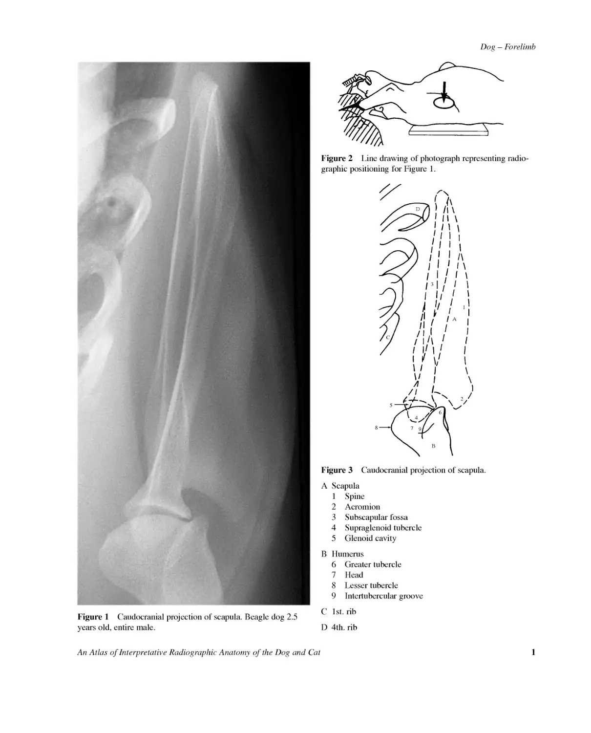
Dog--ForelimbAnAtlasofInterpretativeRadiographicAnatomyoftheDogandCat1Figure1Caudocranialprojectionofscapula.Beagledog2.5yearsold,entiremale.Figure2Linedrawingofphotographrepresentingradio-graphicpositioningforFigure1.CA12497B68D35Figure3Caudocranialprojectionofscapula.AScapula1Spine2Acromion3Subscapularfossa4Supraglenoidtubercle5GlenoidcavityBHumerus6Greatertubercle7Head8Lessertubercle9IntertuberculargrooveC1st.ribD4th.rib
2AnAtlasofInterpretativeRadiographicAnatomyoftheDogandCatDog--ForelimbFigure4Mediolateralprojectionofshoulderjoint.Beagledog2.5yearsold,entiremale.Figure5LinedrawingofphotographrepresentingradiographicpositioningforFigure4.CA1239B835111247151314106Figure6Mediolateralprojectionofshoulderjoint.AScapula1Spine2Supraspinousfossa3Infraspinousfossa4Acromion5Supraglenoidtubercle6Glenoidcavity7InfraglenoidtuberosityBHumerus8Head9Neck10Lessertubercle11Intertuberculargroove12Greatertubercle13Crestofthelessertubercle14Tricipitalline15DeltoidtuberosityCManubriumofsternum
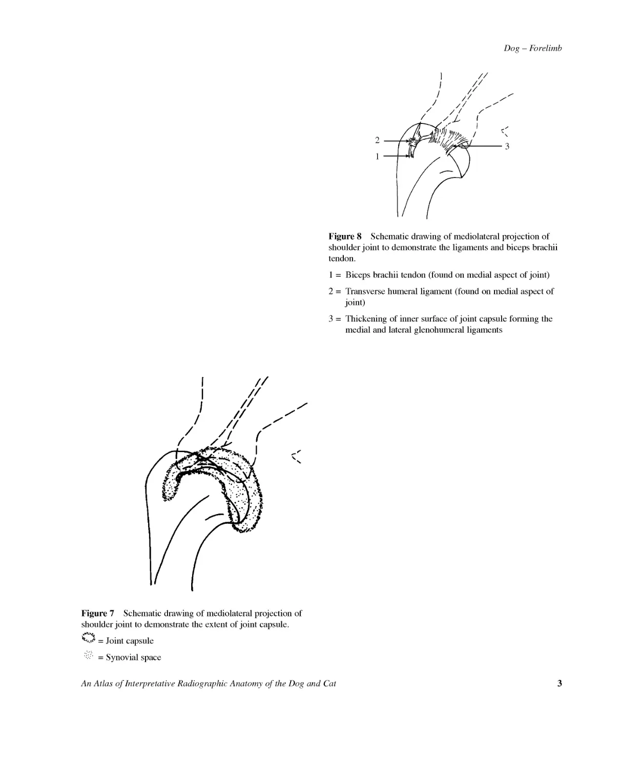
Dog--ForelimbAnAtlasofInterpretativeRadiographicAnatomyoftheDogandCat3Figure7Schematicdrawingofmediolateralprojectionofshoulderjointtodemonstratetheextentofjointcapsule.=Jointcapsule=Synovialspace213Figure8Schematicdrawingofmediolateralprojectionofshoulderjointtodemonstratetheligamentsandbicepsbrachiitendon.1=Bicepsbrachiitendon(foundonmedialaspectofjoint)2=Transversehumeralligament(foundonmedialaspectofjoint)3=Thickeningofinnersurfaceofjointcapsuleformingthemedialandlateralglenohumeralligaments
4AnAtlasofInterpretativeRadiographicAnatomyoftheDogandCatDog--ForelimbFigure9Caudocranialprojectionofshoulderjoint.Beagledog2.5yearsold,entiremale.Figure10LinedrawingofphotographrepresentingradiographicpositioningforFigure9.197BAC638425Figure11Caudocranialprojectionofshoulderjoint.AScapula1Spine2Acromion3Supraglenoidtubercleincludingcoracoidprocessmedially4Glenoidcavity5SubscapularfossaBHumerus6Greatertubercle7Head8Lessertubercle9IntertuberculargrooveCClavicle.Oftenseeninthisprojection.
Dog--ForelimbAnAtlasofInterpretativeRadiographicAnatomyoftheDogandCat5Figure12Schematicdrawingofcaudocranialprojectionofshoulderjointtodemonstrateextentofjointcapsule.=Jointcapsule=Synovialspace4213Figure13Schematicdrawingofcaudocranialprojectionofshoulderjointtodemonstrateligamentsandbicepsbrachiitendon.1=Bicepsbrachiitendon2=Transversehumeralligament3=Medialglenohumeralligament4=Lateralglenohumeralligament
6AnAtlasofInterpretativeRadiographicAnatomyoftheDogandCatDog--ForelimbFigure14Mediolateralprojectionofhumerus.Beagledog2.5yearsold,entiremale.Figure15LinedrawingofphotographrepresentingradiographicpositioningforFigure14.
Dog--ForelimbAnAtlasofInterpretativeRadiographicAnatomyoftheDogandCat71A5764899B32191112C10171510(b)10(a)13141816DFigure16Mediolateralprojectionofhumerus.AScapula1Spine2Acromion3Supraglenoidtubercle4GlenoidcavityBHumerus5Head6Neck7Lessertubercle8Intertuberculargroove9Greatertubercle10Condyle.Anatomicallyonlyonecondyleispresentinthedogbutfrequentlythetermslateralandmedialcondyleareused.10(a)Capitulum(lateralaspect)10(b)Trochlea(medialaspect)11Medialepicondyle12Lateralepicondyle13Supratrochlearforamen.Thisforamenliesbetweentheradialfossaandtheolecranonfossawhichhousestheanconealprocessoftheulna.CRadius14Head15NeckDUlna16Olecranon17Anconealprocess18Lateralcoronoidprocess19Medialcoronoidprocess
8AnAtlasofInterpretativeRadiographicAnatomyoftheDogandCatDog--ForelimbFigure17Craniocaudalprojectionofhumerus.Beagledog2.5yearsold,entiremale.Figure18LinedrawingofphotographrepresentingradiographicpositioningforFigure17.
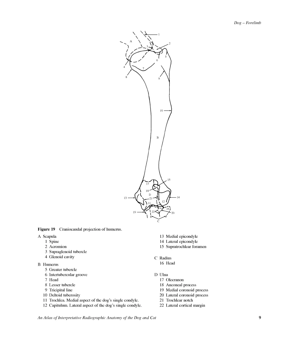
Dog--ForelimbAnAtlasofInterpretativeRadiographicAnatomyoftheDogandCat91A365748910B2131921111222C201715141816DFigure19Craniocaudalprojectionofhumerus.AScapula1Spine2Acromion3Supraglenoidtubercle4GlenoidcavityBHumerus5Greatertubercle6Intertuberculargroove7Head8Lessertubercle9Tricipitalline10Deltoidtuberosity11Trochlea.Medialaspectofthedog'ssinglecondyle.12Capitulum.Lateralaspectofthedog'ssinglecondyle.13Medialepicondyle14Lateralepicondyle15SupratrochlearforamenCRadius16HeadDUlna17Olecranon18Anconealprocess19Medialcoronoidprocess20Lateralcoronoidprocess21Trochlearnotch22Lateralcorticalmargin
10AnAtlasofInterpretativeRadiographicAnatomyoftheDogandCatDog--ForelimbFigure20Caudocranialprojectionofhumerus.Beagledog2.5yearsold,entiremale(samedogasincraniocaudalprojectionofhumerus,Figure17).Figure21LinedrawingofphotographrepresentingradiographicpositioningforFigure20.
Dog--ForelimbAnAtlasofInterpretativeRadiographicAnatomyoftheDogandCat11Figure22Mediolateralprojectionoftheextendedelbowjoint.Beagledog2.5yearsold,entiremale.Figure23LinedrawingofphotographrepresentingradiographicpositioningforFigure22.A321213711414111(b)BC151541(a)1(a)1051(b)689Figure24Mediolateralprojectionofextendedelbowjoint.AHumerus1Condyle.Onlyonecondyleispresent.1(a)Capitulum.Lateralaspect.1(b)Trochlea.Medialaspect.2Medialepicondyle3Lateralepicondyle4Supratrochlearforamen5Radialfossa6OlecranonfossaBRadius7Head8Neck9EminenceforattachmentoflateralcollateralligamentoftheelbowjointCUlna10Medialcoronoidprocess11Lateralcoronoidprocess12Anconealprocess13Olecranon14Trochlearnotch15Proximalarticulationofradiusandulna
12AnAtlasofInterpretativeRadiographicAnatomyoftheDogandCatDog--ForelimbabFigure25Schematicdrawingofmediolateralprojectionoftheextendedelbowjointtodemonstrateextentofjointcapsule.=Jointcapsule=SynovialspaceAdditionalsofttissueshadowsrelatingtointerosseousareaa=Interosseousmembraneb=Interosseousligament.Irregularcorticalradialandulnarmarginsareoftenseeninthisregion,sometimesinvolvingextensiveperiostealnewbonecreatingcorticalthickeningwithsmoothlyundulatingcorticalbonemargins.21Figure26Schematicdrawingofmediolateralprojectionoftheextendedelbowjointtodemonstrateligamentsatjointcapsule.1=Lateralandmedialcollateralligaments.Bothdistallydivideintotwocruratoattachtotheradiusandulnaandonalateralprojectionarealmostsuperimposed.Cranialcrusattachestotheradialtuberositymediallyandradialeminencelaterally.2=Annularligamentoftheradius.Liesundercollateralligaments.Attachedtolateralandmedialaspectsoftheradialnotchoftheulna,itformsa'loop'inwhichtheheadoftheradiuscanrotatearounditslongaxis.
Dog--ForelimbAnAtlasofInterpretativeRadiographicAnatomyoftheDogandCat13Figure27Mediolateralprojectionoftheflexedelbowjoint.Beagledog2.5yearsold,entiremale.Figure28LinedrawingofphotographrepresentingradiographicpositioningforFigure27.2ABC96437581101(b)1(a)1112Figure29Mediolateralprojectionoftheflexedelbowjoint.AHumerus1Condyle.Onlyonecondyleispresent.1(a)Capitulum.Lateralaspect.1(b)Trochlea.Medialaspect.2Medialepicondyle3Lateralepicondyle4Supratrochlearforamen5Radialfossa6OlecranonfossaBRadius7HeadCUlna8Medialcoronoidprocess.Notethatinthisprojectionthelateralcoronoidprocesscannotbeseenasadis-tinctshadow.Theextendedmediolateralprojectionoftheelbowjointdoesshowthelateralcoronoidprocess.9Anconealprocess10Olecranon11Trochlearnotch12Cranialcorticalmargin
14AnAtlasofInterpretativeRadiographicAnatomyoftheDogandCatDog--ForelimbFigure30Schematicdrawingofmediolateralprojectionoftheflexedelbowjointtodemonstratetheextentofjointcapsule.=Jointcapsule=Synovialspace.Thereisavoluminoussacofsynovialcavityinthecranialandcaudalpartsofthisjointbutthesedonotcommunicatethroughthesupratrochlearforamen.Onthelateralandmedialaspectsthejointcapsuleistautwithnosacformation.
Dog--ForelimbAnAtlasofInterpretativeRadiographicAnatomyoftheDogandCat15Figure31Craniocaudalprojectionofelbowjoint.Beagledog2.5yearsold,entiremale.Figure32LinedrawingofphotographrepresentingradiographicpositioningforFigure31.A8C291035B11(a)1(b)116111247Figure33Craniocaudalprojectionofelbowjoint.AHumerus1Condyle.Onlyonecondyleispresent.1(a)Trochlea.Medialaspect.1(b)Capitulum.Lateralaspect.2Medialepicondyle3Lateralepicondyle4SupratrochlearforamenBRadius5Head6Lateraleminence7PositionofradialtuberosityNumbers6and7arelandmarksforcollateralligamentsCUlna8Olecranon9Medialcoronoidprocess10Lateralcoronoidprocess11Lateralcorticalmargin12Medialcorticalmargin
16AnAtlasofInterpretativeRadiographicAnatomyoftheDogandCatDog--ForelimbFigure34Schematicdrawingofcraniocaudalprojectionofelbowjointtodemonstrateextentofjointcapsule.=Jointcapsule=Synovialspace231Figure35Schematicdrawingofcraniocaudalprojectionofelbowjointtodemonstrateligamentsatjointcapsule.1=Lateralcollateralligament2=Medialcollateralligament3=Annularligamentoftheradius
Dog--ForelimbAnAtlasofInterpretativeRadiographicAnatomyoftheDogandCat17Figure36Craniolateral--caudomedialobliqueprojectionofelbowjoint.Beagledog2.5yearsold,entiremale.Figure37LinedrawingofphotographrepresentingradiographicpositioningforFigure36.A7C31(a)11(b)2612131189955104BFigure38Craniolateral--caudomedialobliqueprojectionofelbowjoint.AHumerus1Condyle.Onlyonecondyleispresent.1(a)Trochlea.Medialaspect.1(b)Capitulum.Lateralaspect.2Medialepicondyle3Lateralepicondyle4SupratrochlearforamenBRadius5Head6LateraleminenceforattachmentoflateralcollateralligamentCUlna7Olecranon8Anconealprocess9Trochlearnotch10Medialcoronoidprocess11Lateralcoronoidprocess(seenasaveryopaquelinearshadowonthelateraledgeofthetrochlearnotch)12Lateralcorticalmargin13Medialcorticalmargin
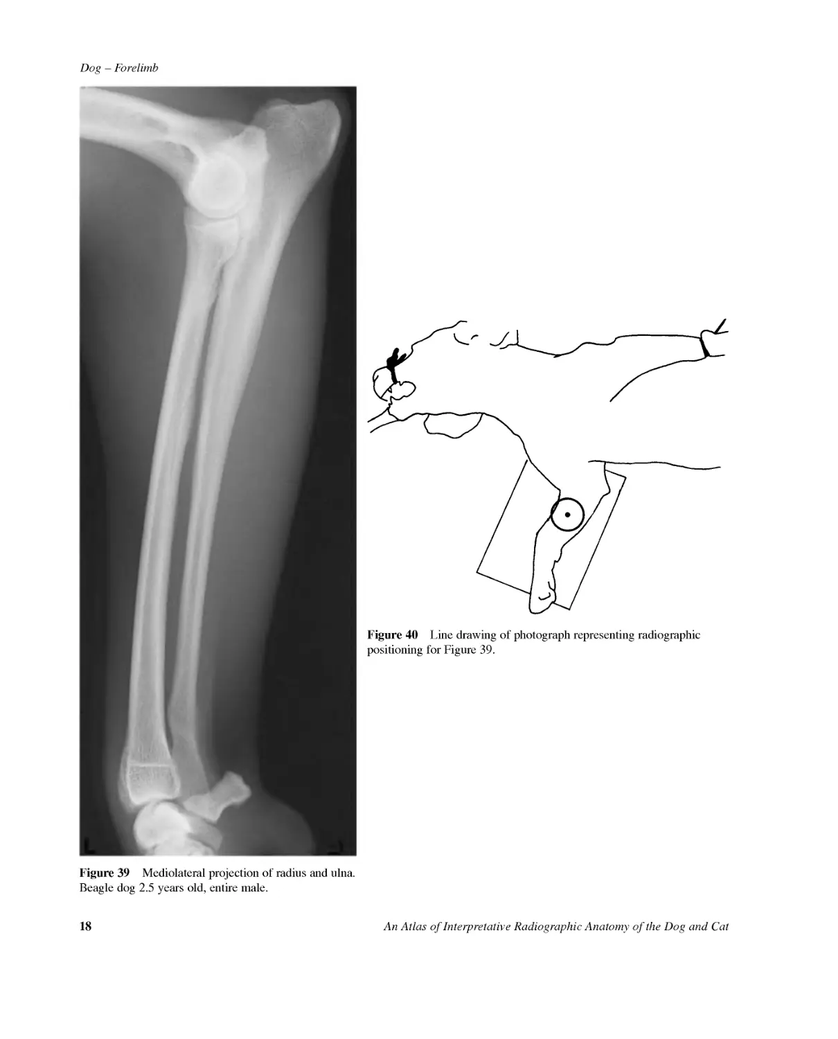
18AnAtlasofInterpretativeRadiographicAnatomyoftheDogandCatDog--ForelimbFigure39Mediolateralprojectionofradiusandulna.Beagledog2.5yearsold,entiremale.Figure40LinedrawingofphotographrepresentingradiographicpositioningforFigure39.
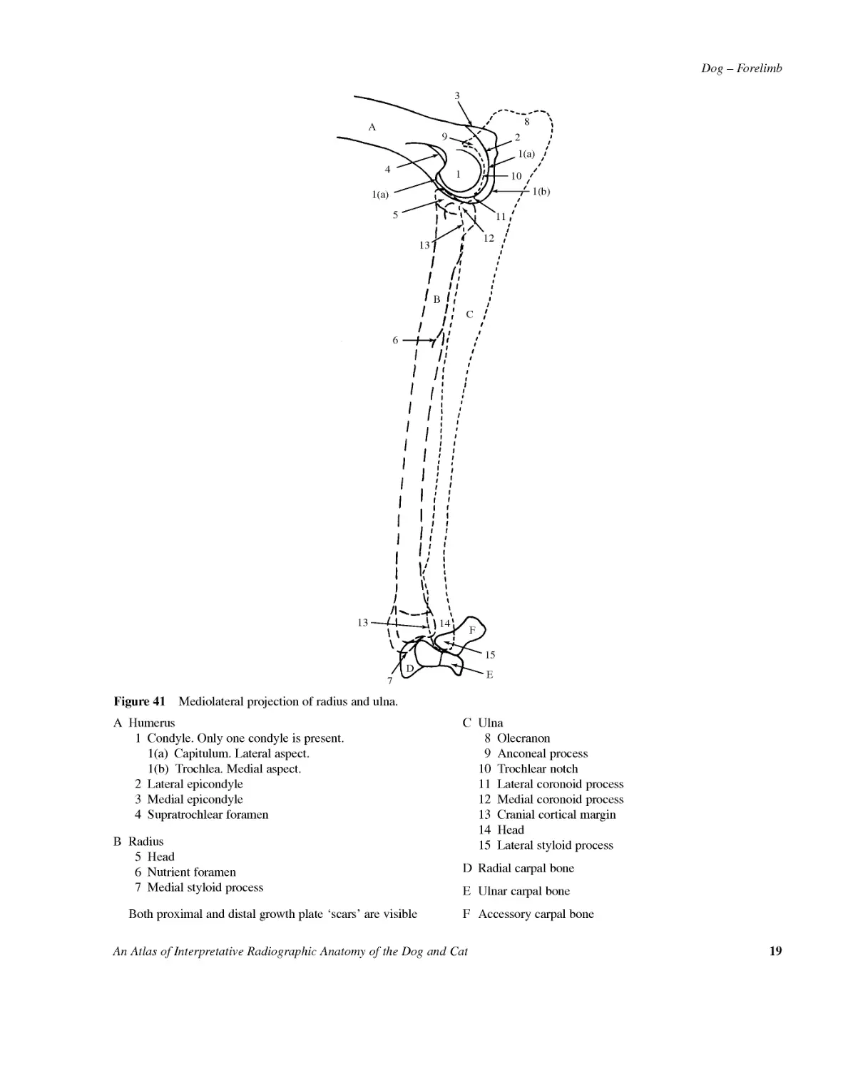
Dog--ForelimbAnAtlasofInterpretativeRadiographicAnatomyoftheDogandCat19Figure41Mediolateralprojectionofradiusandulna.AC161413157FDE1011121(b)28951(a)31341(a)BAHumerus1Condyle.Onlyonecondyleispresent.1(a)Capitulum.Lateralaspect.1(b)Trochlea.Medialaspect.2Lateralepicondyle3Medialepicondyle4SupratrochlearforamenBRadius5Head6Nutrientforamen7MedialstyloidprocessBothproximalanddistalgrowthplate'scars'arevisibleCUlna8Olecranon9Anconealprocess10Trochlearnotch11Lateralcoronoidprocess12Medialcoronoidprocess13Cranialcorticalmargin14Head15LateralstyloidprocessDRadialcarpalboneEUlnarcarpalboneFAccessorycarpalbone
20AnAtlasofInterpretativeRadiographicAnatomyoftheDogandCatDog--ForelimbFigure42Craniocaudalprojectionofradiusandulna.Beagledog2.5yearsold,entiremale.Figure43Linedrawingofphotographrepresentingradio-graphicpositioningforFigure42.
Dog--ForelimbAnAtlasofInterpretativeRadiographicAnatomyoftheDogandCat21AC15612FDE761(b)32841(a)91011BAHumerus1Condyle.Onlyonecondyleispresent.1(a)Trochlea.Medialaspect.1(b)Capitulum.Lateralaspect.2Medialepicondyle3Lateralepicondyle4SupratrochlearforamenBRadius5Head6Growthplatescars7MedialstyloidprocessCUlna8Olecranon9Medialcoronoidprocess10Lateralcoronoidprocess11Lateralcorticalmargin12LateralstyloidprocessDRadialcarpalboneEUlnarcarpalboneFAccessorycarpalboneFigure44Craniocaudalprojectionofradiusandulna.
22AnAtlasofInterpretativeRadiographicAnatomyoftheDogandCatDog--ForelimbFigure45Dorsopalmarprojectionofcarpus.Beagledog2.5yearsold,entiremale.Figure46LinedrawingofphotographrepresentingradiographicpositioningforFigure45.147FDABJIHLNOKPRMCGE23Q65ARadius1Growthplatescar2Ulnarnotch3Carpalarticularsurface4MedialstyloidprocessBUlna5Distalarticularfacetfortheradius6Head7LateralstyloidprocessCRadialcarpalboneDUlnarcarpalboneEAccessorycarpalboneFSesamoidboneinthetendonofm.abductorpollicislongusGCarpalbone1HCarpalbone2ICarpalbone3JCarpalbone4KMetacarpalbone1LMetacarpalbone2MMetacarpalbone3NMetacarpalbone4OMetacarpalbone5PProximalphalanxQDistalphalanxRUngualprocessFigure47Dorsopalmarprojectionofcarpus.
Dog--ForelimbAnAtlasofInterpretativeRadiographicAnatomyoftheDogandCat231324Figure48Schematicdrawingofdorsopalmarprojectionofcarpustodemonstratesomeclinicallyimportantligamentsofthecarpus.1=Shortradialcollateralligament.Onmedialsurface.2=Radioulnarligament.Ondorsalsurface.3=Shortulnarcollateralligament.Onlateralsurface.4=Accessoro-metacarpalligaments.Onpalmarsurface.
24AnAtlasofInterpretativeRadiographicAnatomyoftheDogandCatDog--ForelimbFigure49Mediolateralprojectionofcarpus.Beagledog2.5yearsold,entiremale.Figure50LinedrawingofphotographrepresentingradiographicpositioningforFigure49.14JDABFIHLKMCGE23MON5ARadius1Growthplatescar2Grooveforthem.extensorcarpiradialis3MedialstyloidprocessBUlna4Growthplatescar5LateralstyloidprocessCRadialcarpalboneDUlnarcarpalboneEAccessorycarpalboneFCarpalbone1GCarpalbone2HCarpalbones3and4(superimposedshadows)IMetacarpalbone1JMetacarpalbone2KMetacarpalbones3and4(superimposedshadows)LMetacarpalbone5MProximalsesamoidbonesNProximalphalanxofdigit1ODistalphalanxofdigit1Figure51Mediolateralprojectionofcarpus.
Dog--ForelimbAnAtlasofInterpretativeRadiographicAnatomyoftheDogandCat25Figure52Dorsolateral--palmaromedialobliqueprojectionofcarpus.Samoyeddog6yearsold,entirefemale.Figure53Linedrawingofphotographrepresentingradio-graphicpositioningforFigure52.A1BEDINMC25GKHFJL3467ARadius1Growthplatescar2Grooveforthetendonofm.abductorpollicislongus3Medialstyloidprocess4UlnarnotchBUlna5Distalradialarticularsurface6Articularsurfaceforulnarcarpalbone7LateralstyloidprocessCRadialcarpalboneDUlnarcarpalboneEAccessorycarpalboneFCarpalbone1GCarpalbone2HCarpalbone3ICarpalbone4JMetacarpalbone1KMetacarpalbone2LMetacarpalbone3MMetacarpalbone4NMetacarpalbone5Figure54Dorsolateral--palmaromedialobliqueprojectionofcarpus.
26AnAtlasofInterpretativeRadiographicAnatomyoftheDogandCatDog--ForelimbFigure55Dorsopalmarprojectionofmanus.Beagledog2.5yearsold,entiremale.Figure56LinedrawingofphotographrepresentingradiographicpositioningforFigure55.
Dog--ForelimbAnAtlasofInterpretativeRadiographicAnatomyoftheDogandCat271JDABIHLTRRSSSSRQQRRMCGEFPKUPTTTUUUPPP2UT3321NOFigure57Dorsopalmarprojectionofmanus.ARadiusBUlnaCRadialcarpalboneDUlnarcarpalboneEAccessorycarpalboneFSesamoidboneinthetendonofthem.abductorpollicislongusGCarpalbone1HCarpalbone2ICarpalbone3JCarpalbone4KMetacarpalbone1LMetacarpalbone2MMetacarpalbone3NMetacarpalbone4OMetacarpalbone5PProximalsesamoidbones.Thesearepresentonpalmaraspectofmetacarpophalangealjointsintendonsofmm.interossei(2to5)andm.flexorpollicisbrevis.Onlyoneatdigit1andtwoatdigits2to5.QDorsalsesamoidbones.Thesearepresentondorsalaspectofdistalmetacarpalbones2to5andliewithinthemetacarpophalangealjointcapsules.RProximalphalangesSMiddlephalangesTDistalphalangesUUngualprocessesMetacarpalbones,proximalandmiddlephalangesdividedinto1Base2Body3Head
28AnAtlasofInterpretativeRadiographicAnatomyoftheDogandCatDog--ForelimbFigure58Mediolateralprojectionofmanus.Beagledog2.5yearsold,entiremale.Figure59LinedrawingofphotographrepresentingradiographicpositioningforFigure58.
Dog--ForelimbAnAtlasofInterpretativeRadiographicAnatomyoftheDogandCat29ADBIFCJJLL1M1M2L2O1OON1NJMMNNOEGHL3O2N2LLKFigure60Mediolateralprojectionofmanus.ARadialcarpalboneBUlnarcarpalboneCCarpalbone1DCarpalbone2ECarpalbones2and4(superimposedshadows)FMetacarpalbone1GMetacarpalbone2HMetacarpalbones3and4(superimposedshadows.Thedorsalprotuberanceseenismetacarpalbone3.)IMetacarpalbone5JProximalsesamoidbones.Twoarepresentinthetendonsofmm.interosseiatpalmaraspectofmetacarpophalangealjoints2to5.Onlyoneispresentinmetacarpophalangealjoint1.KDorsalsesamoidbone.Thesearepresentinjointcapsulesatdorsalaspectofdistalmetacarpalbones2to5.LProximalphalangesL1Digit1L2Digits2and5(superimposedshadows)L3Digits3and4(superimposedshadows)MMiddlephalangesM1Digits2and5(superimposedshadows)M2Digits3and4(superimposedshadows)NDistalphalangesN1Digits2and5(superimposedshadows)N2Digits3and4(superimposedshadows)OUngualprocessesO1Digits2and5(superimposedshadows)O2Digits3and4(superimposedshadows)
30AnAtlasofInterpretativeRadiographicAnatomyoftheDogandCatDog--ForelimbFigure61Mediolateralprojectionofphalanges,digitsstressed.Beagledog2.5yearsold,entiremale.Figure62LinedrawingofphotographrepresentingradiographicpositioningforFigure61.
Dog--ForelimbAnAtlasofInterpretativeRadiographicAnatomyoftheDogandCat31S21P4P52(a)3(a)2(b)3(b)2(c)3(c)3(d)3(e)P1P2P3S1S2MMM123Figure63Mediolateralprojectionofphalanges.digitsstressed.MMetacarpalbonesP1Digit1P2Digit2P3Digit3P4Digit4P5Digit5Bonesofdigits1Proximalphalanx2Middlephalanx2(a)Base2(b)Body2(c)Head3Distalphalanx3(a)Flexortubercle3(b)Solarforamen3(c)Ungualcrest3(d)Ungualsulcus3(e)UngualprocessS1Dorsalsesamoidbone.Thesearepresentindorsalaspectofmetacarpophalangealjointcapsules2to5.S2Proximalsesamoidbone.Twoarepresentintendonsofmm.interosseiatpalmaraspectofmetacarpophalangealjoints2to5.Metacarpophalangealjoint1hasonesesamoidboneinthetendonofm.flexorpollicisbrevis.
32AnAtlasofInterpretativeRadiographicAnatomyoftheDogandCatDog--ForelimbFigure64Caudocranialprojectionofscapula.Chondrodystrophicbreedofdog.MiniatureDachshunddog6yearsold,neuteredfemale.Figure65Mediolateralprojectionofshoulderjoint.Chondrodystrophicbreedofdog.MiniatureDachshunddog6yearsold,neuteredfemale.
Dog--ForelimbAnAtlasofInterpretativeRadiographicAnatomyoftheDogandCat33Figure66Mediolateralprojectionofhumerus.Chondrodystrophicbreedofdog.MiniatureDachshunddog6yearsold,neuteredfemale.Figure67Caudocranialprojectionofhumerus.Chondrodystrophicbreedofdog.MiniatureDachshunddog6yearsold,neuteredfemale.
34AnAtlasofInterpretativeRadiographicAnatomyoftheDogandCatDog--ForelimbFigure68Flexedmediolateralprojectionofelbowjoint.Chondrodystrophicbreedofdog.MiniatureDachshunddog6yearsold,neuteredfemale.
Dog--ForelimbAnAtlasofInterpretativeRadiographicAnatomyoftheDogandCat35Caudocranialprojectionofelbowjoint.Chondrodystrophicbreedofdog.MiniatureDachshunddog6yearsold,neuteredfemale.Caudocranialprojectionofelbowjoint.Figure69Caudocranialprojectionofelbowjoint.Thedrawinghasbeenincludedtoindicatethepresenceofalateralsesamoidbone(arrow)intheelbowjoint.Thesesamoidboneismorefrequentlyseenonthelateralaspectandisthoughttobewithinthetendonofthem.supinator.Occasionallyamedialsesamoidboneisobservedinthecollateralligamentandjointcapsule.Althoughboththelateralandmedialsesamoidboneshavebeencitedasacauseoflamenessbysomeauthors,bymostauthoritiestheyarenotclinicallysignificant.Indeedsesamoidcartilageisoftenpresentbutnon-mineralised,andhencecannotbeseeninaradiograph.Inthisparticulardogtherewasnoforelimblameness.
36AnAtlasofInterpretativeRadiographicAnatomyoftheDogandCatDog--ForelimbFigure70Caudolateral-craniomedialobliqueprojectionofelbowjoint.Chondrodystrophicbreedofdog.MiniatureDachshunddog6yearsold,neuteredfemale.Figure71Mediolateralprojectionofradiusandulna.Chondrodystrophicbreedofdog.MiniatureDachshunddog6yearsold,neuteredfemale.
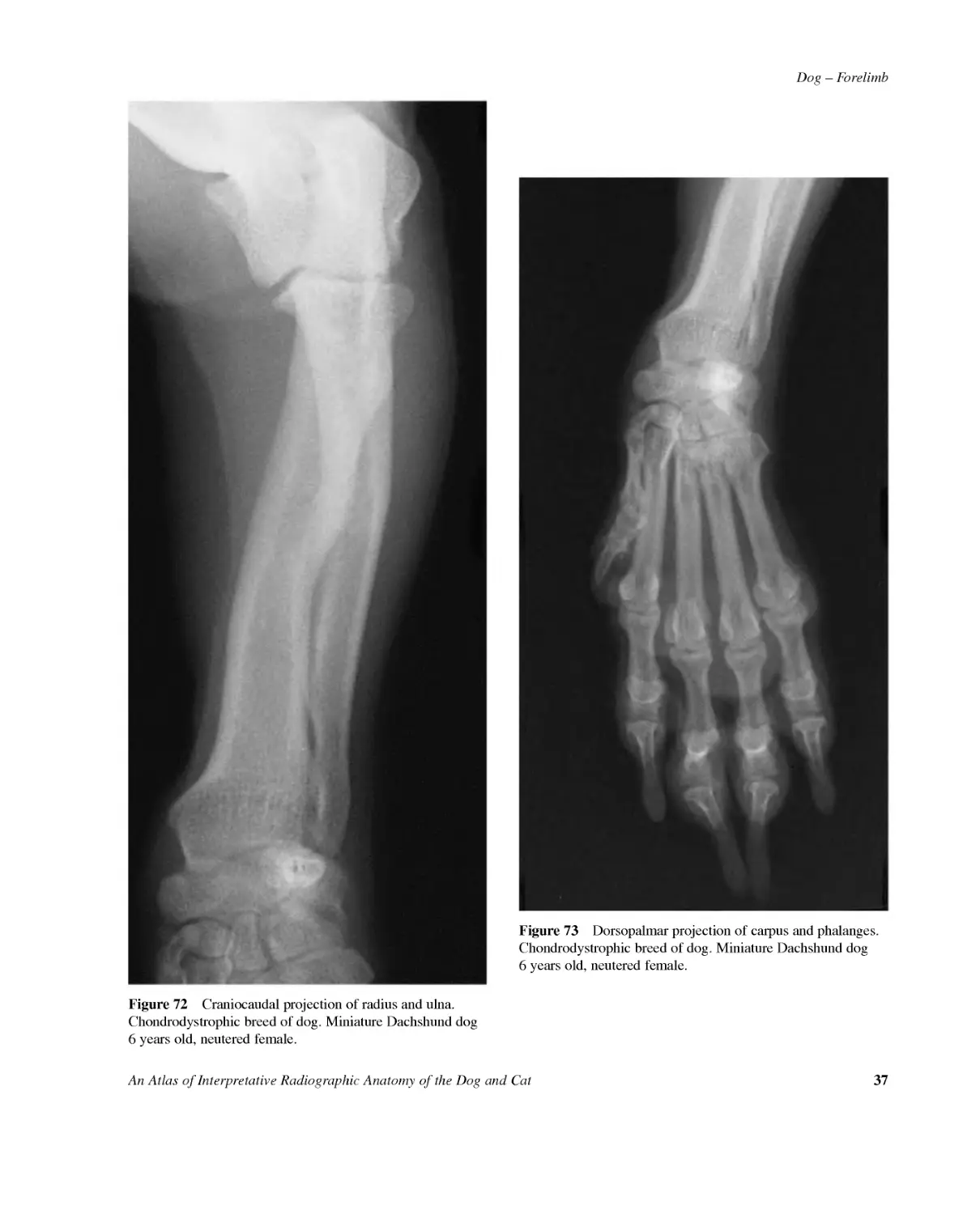
Dog--ForelimbAnAtlasofInterpretativeRadiographicAnatomyoftheDogandCat37Figure72Craniocaudalprojectionofradiusandulna.Chondrodystrophicbreedofdog.MiniatureDachshunddog6yearsold,neuteredfemale.Figure73Dorsopalmarprojectionofcarpusandphalanges.Chondrodystrophicbreedofdog.MiniatureDachshunddog6yearsold,neuteredfemale.
38AnAtlasofInterpretativeRadiographicAnatomyoftheDogandCatDog--ForelimbFigure74Mediolateralprojectionofcarpusandphalanges.Chondrodystrophicbreedofdog.MiniatureDachshunddog6yearsold,neuteredfemale.
Dog--ForelimbAnAtlasofInterpretativeRadiographicAnatomyoftheDogandCat39Figure75Mediolateralprojectionofshoulderjoint.Glenoidcavityvariant.(Correspondstoradiographnotincludedinbook.)GreatDaneGermanShepherdcrossbreddog5monthsold,entiremale.Thedrawingdemonstratesaseparateossificationcentrefortheglenoidcavity(arrow).Asthedogmaturesthecentreoftenformsaseparatebonyshadowparalleltotheglenoidcavity.Thismustnotbemistakenforanosteochondrosisfragment.Thevariantseenhereismostcommonlyfoundinthegiantbreedofdog,inparticulartheIrishWolfhound.Asimilar,separateossificationcentremayoccasionallybeseenattheacetabulum.Herethevariantformsaseparatebonyshadowparalleltothecranialeffectiveacetabularrim.Caremustbetakennottoconfusethisshadowwithafrac-turefragmentorossicle.Thesmoothcorticaloutlineofthebonyvarianttogetherwithanormalacetabularshadowenablesdifferentiationfromabnormality.Figure76Mediolateralprojectionofdistalradiusandulna.Retainedcartilaginouscore.(Correspondstoradiographnotincludedinbook.)GreatDaneGermanShepherdcrossbreddog5monthsold,entiremale.Thedrawingshowsaretainedcartilaginouscore(closedarrows)inthedistalulnametaphysealregion.Thecoreistypi-callyseenatthis5-monthage,especiallyintheGreatDane,althoughotherlargeandgiantbreedscanbeaffected.Althoughatonetimeitwasthoughttoretardgrowthitspresencealoneisnotsignificantandthecorewilldisappearasthedogmatures.Notethenormalgrowthplatesinthisdogwiththecore.Alsopresentonthedrawingisthetypicalirregularcorticaloutlineofthemetaphysealregions(openarrows).Thelatterisinvariablyseeninlargeandgiantbreedsofimmaturedogs.This,togetherwitharelativelyopaqueappearanceofthemeta-physealregions,seeninallimmaturedogs,mustnotbemis-takenforabonymetabolicabnormalitysuchasrickets.Examinationofthebonycorticalopacity,andthickness,isrequiredtoestablishnormalityintheimmatureanimal.
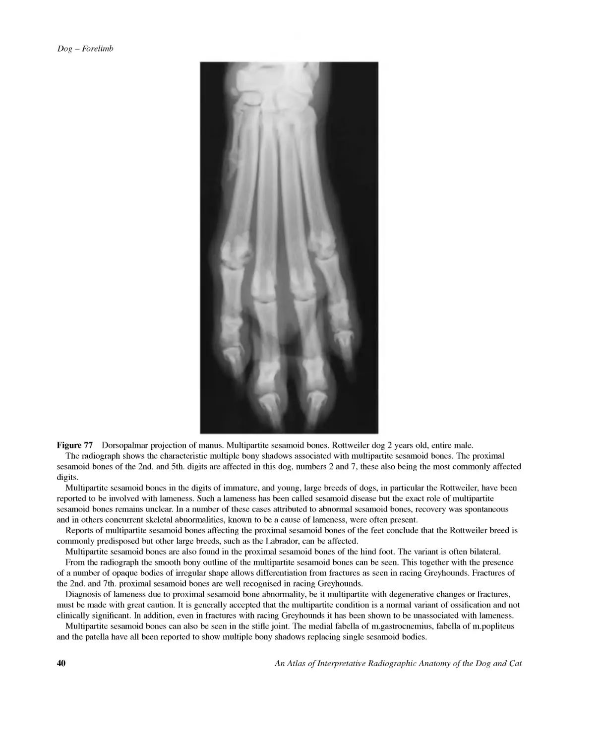
40AnAtlasofInterpretativeRadiographicAnatomyoftheDogandCatDog--ForelimbFigure77Dorsopalmarprojectionofmanus.Multipartitesesamoidbones.Rottweilerdog2yearsold,entiremale.Theradiographshowsthecharacteristicmultiplebonyshadowsassociatedwithmultipartitesesamoidbones.Theproximalsesamoidbonesofthe2nd.and5th.digitsareaffectedinthisdog,numbers2and7,thesealsobeingthemostcommonlyaffecteddigits.Multipartitesesamoidbonesinthedigitsofimmature,andyoung,largebreedsofdogs,inparticulartheRottweiler,havebeenreportedtobeinvolvedwithlameness.Suchalamenesshasbeencalledsesamoiddiseasebuttheexactroleofmultipartitesesamoidbonesremainsunclear.Inanumberofthesecasesattributedtoabnormalsesamoidbones,recoverywasspontaneousandinothersconcurrentskeletalabnormalities,knowntobeacauseoflameness,wereoftenpresent.ReportsofmultipartitesesamoidbonesaffectingtheproximalsesamoidbonesofthefeetconcludethattheRottweilerbreediscommonlypredisposedbutotherlargebreeds,suchastheLabrador,canbeaffected.Multipartitesesamoidbonesarealsofoundintheproximalsesamoidbonesofthehindfoot.Thevariantisoftenbilateral.Fromtheradiographthesmoothbonyoutlineofthemultipartitesesamoidbonescanbeseen.ThistogetherwiththepresenceofanumberofopaquebodiesofirregularshapeallowsdifferentiationfromfracturesasseeninracingGreyhounds.Fracturesofthe2nd.and7th.proximalsesamoidbonesarewellrecognisedinracingGreyhounds.Diagnosisoflamenessduetoproximalsesamoidboneabnormality,beitmultipartitewithdegenerativechangesorfractures,mustbemadewithgreatcaution.Itisgenerallyacceptedthatthemultipartiteconditionisanormalvariantofossificationandnotclinicallysignificant.Inaddition,eveninfractureswithracingGreyhoundsithasbeenshowntobeunassociatedwithlameness.Multipartitesesamoidbonescanalsobeseeninthestiflejoint.Themedialfabellaofm.gastrocnemius,fabellaofm.popliteusandthepatellahaveallbeenreportedtoshowmultiplebonyshadowsreplacingsinglesesamoidbodies.
Dog--ForelimbAnAtlasofInterpretativeRadiographicAnatomyoftheDogandCat41Figure78Dorsopalmarprojectionofmanus.Presenceofforeignmaterialonthepalmarsurface.Colliecrossbreddog10yearsold,neuteredmale(samedogasinFigure78a).Theirregular,well-definedradiopacitiescausedbydirtbetweenthemetacarpalanddigitalpads,plusbetweenindi-vidualdigitalpads,inthisfootshowhowimportantpatientpreparationis.Althoughthelumpsofdirtinthiscasearelargeandunlike-lytobeoverlookedduringradiographyofthefoot,tracesofdirtbetweenthepadsmayeasilybemissedonaroutineinspectionoftheanimalpriortoradiography.Whereverthereareunusualshadowsintheregionofthepads,carefulexaminationoftheskin'ssurfacemustbeunder-taken.Thisalsoappliestoanycontaminationofthehairbysolidorliquidmaterial.Figure78aMediolateralprojectionofmanus.Presenceofforeignmaterialonthepalmarsurface.Colliecrossbreddog10yearsold,neuteredmale(samedogasinFigure78).ThecorrespondingmediolateralprojectiontoFigure78hasbeenincludedtoshowthattheobvious,extremelyradiopaquemetallicfragmentwithinthedirtonthepalmarsurfaceofthefootwasnotclearlyseeninthedorsopalmarprojection.Suchafindingdemonstratesthevalueoftwoprojectionsofthesameregion,eventhoughonemayappeartosufficefordiagnosis.
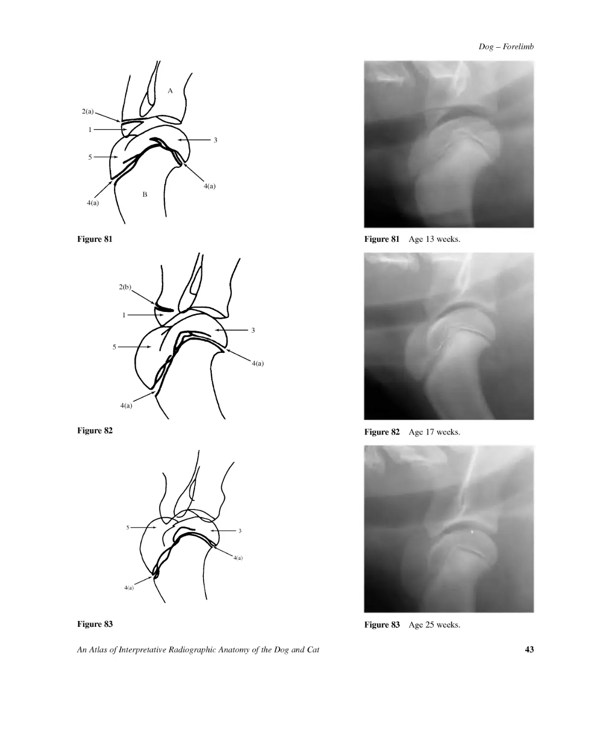
42AnAtlasofInterpretativeRadiographicAnatomyoftheDogandCatDog--ForelimbFigure79Age4weeks.AB34(a)4(a)Figure79AB314(a)4(a)2(a)Figure80Figure80Age8weeks.Figures79,80,81,82,83,84,85,86Mediolateralprojectionofshoulderjoint.Samoyedcrossbreddog-entiremale,at4,8,13,17,25,30,43and56weeksofage.AScapula1Epiphysisofsupraglenoidtubercle2Growthplate2(a)Open2(b)ClosingBHumerus3Proximalepiphysisofhumerus4Proximalgrowthplate4(a)Open4(b)Closing4(c)Remnant5Greatertubercle
Dog--ForelimbAnAtlasofInterpretativeRadiographicAnatomyoftheDogandCat43AB3154(a)4(a)2(a)Figure81Figure81Age13weeks.3154(a)4(a)2(b)Figure82Figure82Age17weeks.354(a)4(a)Figure83Figure83Age25weeks.
44AnAtlasofInterpretativeRadiographicAnatomyoftheDogandCatDog--Forelimb3AB54(a)4(a)Figure84Figure84Age30weeks.AB4(b)4(b)Figure85Figure85Age43weeks.AB4(c)Figure86Figure86Age56weeks.
Dog--ForelimbAnAtlasofInterpretativeRadiographicAnatomyoftheDogandCat45Figures87,88,89,90,91,92Mediolateralprojectionofelbowjoint.Samoyedcrossbreddog,entiremale,at4,8,13,17,25and34weeksofage.AHumerus1Distalepiphysis2Epiphysisofmedialepicondyle3Distalgrowthplateandmedialepicondylegrowthplate3(a)Open3(b)Closing3(c)RemnantBRadius4Proximalepiphysis5Proximalgrowthplate5(a)Open5(c)RemnantCUlna6Proximalepiphysis7Proximalgrowthplate7(a)Open7(b)Closing7(c)RemnantFigure87Age4weeks.ABC14Figure87Figure88Age8weeks.Figure88Figure89Age13weeks.Figure89
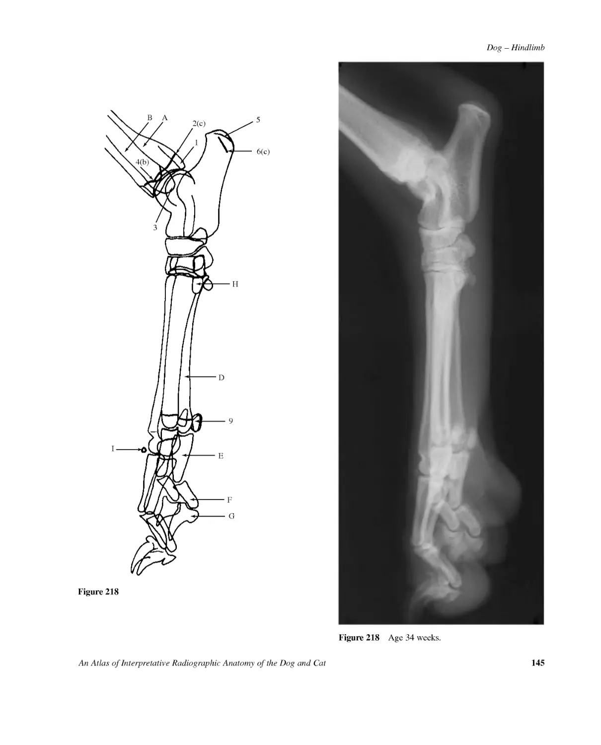
46AnAtlasofInterpretativeRadiographicAnatomyoftheDogandCatDog--ForelimbA45(a)BC7(b)6123(b)Figure903(c)67(c)CB45(a)AFigure91A5(c)AAFigure92Figure90Age17weeks.Figure91Age25weeks.Figure92Age34weeks.
Dog--ForelimbAnAtlasofInterpretativeRadiographicAnatomyoftheDogandCat47Figures93,94,95,96,97,98Craniocaudalprojectionofelbowjoint.Samoyedcrossbreddog,entiremale,at4,8,13,17,25and34weeksofage.AHumerus1Distalepiphysis1(a)Medialcondylarcentre1(b)Lateralcondylarcentre2Epiphysisofmedialepicondyle3Distalgrowthplate3(a)Open3(b)Closing3(c)RemnantBRadius4Proximalepiphysis5Proximalgrowthplate5(a)Open5(c)RemnantCUlna6ProximalepiphysisABC3(a)1(a)41(b)5(a)3(a)Figure93Figure93Age4weeks.
48AnAtlasofInterpretativeRadiographicAnatomyoftheDogandCatDog--ForelimbA3(a)3(a)BC415(a)26Figure94Figure94Age8weeks.A3(a)23(a)BC415(a)6Figure95Figure95Age13weeks.
Dog--ForelimbAnAtlasofInterpretativeRadiographicAnatomyoftheDogandCat49A3(b)3(b)BC415(a)6Figure96Figure96Age17weeks.Figures93,94,95,96,97,98Craniocaudalprojectionofelbowjoint.Samoyedcrossbreddogentiremaleat4,8,13,17,25,and34weeksofage.AHumerus1Distalepiphysis1(a)Medialcondylarcentre1(b)Lateralcondylarcentre2Epiphysisofmedialepicondyle3Distalgrowthplate3(a)Open3(b)Closing3(c)RemnantBRadius4Proximalepiphysis5Proximalgrowthplate5(a)Open5(c)RemnantCUlna6Proximalepiphysis
50AnAtlasofInterpretativeRadiographicAnatomyoftheDogandCatDog--ForelimbA3(c)BC5(c)Figure98Figure98Age34weeks.A3(c)BC45(a)Figure97Figure97Age25weeks.
Dog--ForelimbAnAtlasofInterpretativeRadiographicAnatomyoftheDogandCat51ARadius1Distalepiphysis2Distalgrowthplate2(a)Open2(b)ClosingBUlna3Distalepiphysis4Distalgrowthplate4(a)Open4(c)RemnantCCarpusDMetacarpalbone5(2,3and4similar)5Epiphysis.Notethatthereisonlyadistalepiphysisinthesemetacarpalbones.6Growthplate6(a)Open7Proximalsesamoidbone(lateralidentified)EProximalphalanxofdigit5(2,3and4similar)8Epiphysis.Notethatthereisonlyaproximalepiphysisintheproximalphalanges.9Growthplate9(a)Open9(c)RemnantFMiddlephalanxofdigit5(2,3and4similar)10Epiphysis.Notethatthereisonlyaproximalepiphysisinthemiddlephalanges.11Growthplate11(a)OpenGDistalphalanxofdigit5(2,3and4similar)HMetacarpalbone112Epiphysis.Notethatthereisonlyaproximalepiphysisinthismetacarpalbone.13Growthplate13(a)OpenIProximalphalanxofdigit114Epiphysis.Notethatthereisonlyaproximalepiphysis.15Growthplate15(a)OpenJDistalphalanxofdigit1Figures99,100,101,102,103,104Dorsopalmarprojectionofcarpus,metacarpalbonesandphalanges.Samoyedcrossbreddog,entiremale,at4,8,13,17,25and34weeksofage.
52AnAtlasofInterpretativeRadiographicAnatomyoftheDogandCatDog--ForelimbFigure99Age4weeks.AB4(a)2(a)HDC5FGE6(a)9(a)8IJ1Figure99
Dog--ForelimbAnAtlasofInterpretativeRadiographicAnatomyoftheDogandCat53AB4(a)2(a)IHJDC357FGE6(a)9(a)11(a)81015(a)13(a)12141Figure100Figure100Age8weeks.
54AnAtlasofInterpretativeRadiographicAnatomyoftheDogandCatDog--ForelimbAB4(a)12HDC3587FGE6(a)109(a)11(a)13(a)2(a)15(a)114IJFigure101Figure101Age13weeks.
Dog--ForelimbAnAtlasofInterpretativeRadiographicAnatomyoftheDogandCat55AB4(a)2(a)IHJDC357FGE6(a)9(a)11(a)81013(a)121Figure102Figure102Age17weeks.
56AnAtlasofInterpretativeRadiographicAnatomyoftheDogandCatDog--ForelimbAB4(a)2(a)IHJDC37FGE89(c)1Figure103Figure103Age25weeks.
Dog--ForelimbAnAtlasofInterpretativeRadiographicAnatomyoftheDogandCat57AB4(c)2(b)IHJDC37FGE1Figure104Figure104Age34weeks.
58AnAtlasofInterpretativeRadiographicAnatomyoftheDogandCatDog--ForelimbAB4(a)2(a)DFDC1112(a)FG8(a)HEIE71Figures105,106,107,108,109,110Mediolateralprojectionofcarpus,metacarpalbonesandphalanges.Samoyedcrossbreddog,entiremale,at4,8,13,17,25and34weeksofage.ARadius1Distalepiphysis2Distalgrowthplate2(a)Open2(b)ClosingBUlna3Distalepiphysis4Distalgrowthplate4(a)Open4(c)RemnantCCarpus5Epiphysisofaccessorycarpalbone6Accessorycarpalbonegrowthplate6(a)OpenDMetacarpalbone2or5(3and4similarbutlonger)7Epiphysis.Notethatthereisonlyadistalepiphysisinthesemetacarpalbones.8Growthplate8(a)Open8(c)Remnant9Proximalsesamoidbone10Dorsalsesamoidboneassociatedwithdigits3and4EProximalphalanxdigits2or5(3and4similar)11Epiphysis.Notethatthereisonlyaproximalepiphysisintheproximalphalanges.12Growthplate12(a)Open12(c)RemnantFMiddlephalanxdigits2or5(3and4similar)13Epiphysis.Notethatthereisonlyaproximalepiphysisinthemiddlephalanges.14Growthplate14(a)OpenGDistalphalanxdigit2or5(3and4similar)HMetacarpalbone115Epiphysis.Notethatthereisonlyaproximalepiphysisinthismetacarpalbone.16Growthplate16(a)Open17ProximalsesamoidboneIProximalphalanxdigit118Epiphysis.Notethatthereisonlyaproximalepiphysis.19Growthplate19(a)OpenJDistalphalanxdigit1Figure105
Dog--ForelimbAnAtlasofInterpretativeRadiographicAnatomyoftheDogandCat59Figure105Age4weeks.
60AnAtlasofInterpretativeRadiographicAnatomyoftheDogandCatDog--ForelimbAB4(a)2(a)DDC111312(a)FG916(a)6(a)14(a)H15EJ78(a)I19(a)18531Figure106Figure106Age8weeks.
Dog--ForelimbAnAtlasofInterpretativeRadiographicAnatomyoftheDogandCat61AB4(a)2(a)DDC1113FG916(a)6(a)12(a)14(a)10H15EJ78(a)I19(a)18531Figure107Figure107Age13weeks.
62AnAtlasofInterpretativeRadiographicAnatomyoftheDogandCatDog--ForelimbAB4(a)2(a)DDC1112(a)FG9716(a)14(a)10H15EJ1378(a)I1731Figure108Figure108Age17weeks.
Dog--ForelimbAnAtlasofInterpretativeRadiographicAnatomyoftheDogandCat63AB4(a)2(a)DD7C111012(c)FG9HEJ8(c)I13Figure109Figure109Age25weeks.
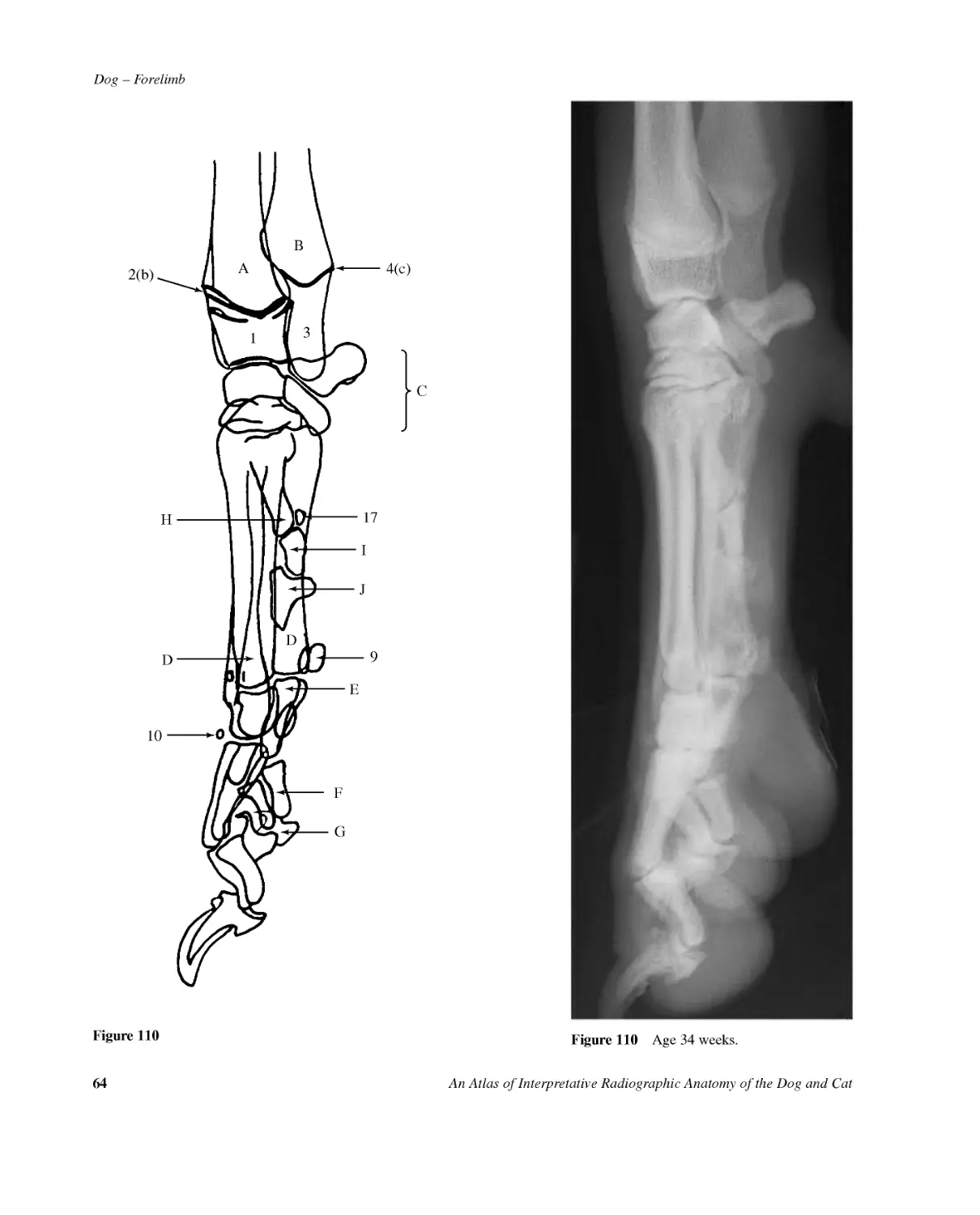
64AnAtlasofInterpretativeRadiographicAnatomyoftheDogandCatDog--ForelimbAB4(c)2(b)DDC10FG9HEJI1713Figure110Figure110Age34weeks.
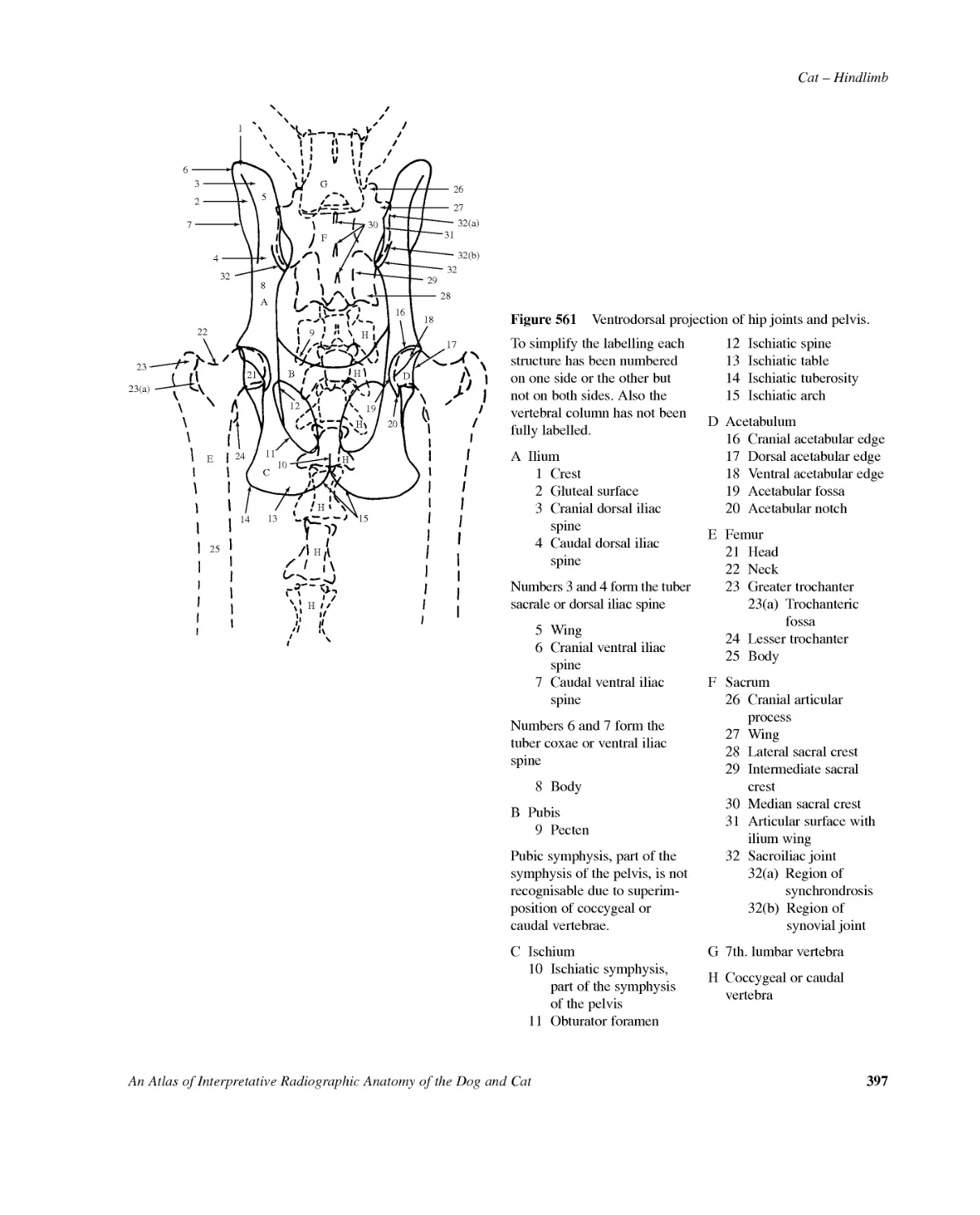
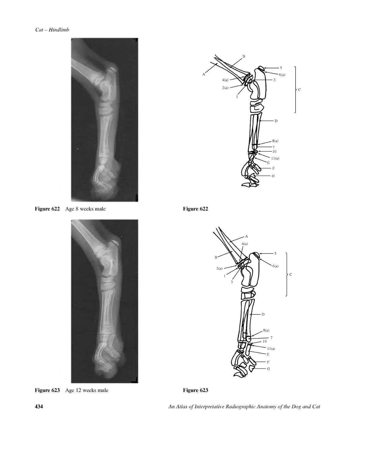
Dog--HindlimbAnAtlasofInterpretativeRadiographicAnatomyoftheDogandCat65Figure111Ventrodorsalprojectionofhipjointsandpelviswithfullextensionoffemurs(stiflejointsincludedforhipdysplasiaevaluation).Beagledog2.5yearsold,entiremale.Figure112LinedrawingofphotographrepresentingradiographicpositioningforFigure111.
66AnAtlasofInterpretativeRadiographicAnatomyoftheDogandCatDog--HindlimbFigure113Ventrodorsalprojectionofhipjointsandpelviswithfullextensionoffemurs.Tosimplifythelabellingeachstructurehasbeennumberedononesideortheotherbutnotonbothsides.Also,thevertebralcolumnhasnotbeenfullylabelled.AIlium1Crest2Glutealsurface3Tubersacraleordorsaliliacspine3(a)Cranialdorsaliliacspine3(b)Caudaldorsaliliacspine4Wing5Tubercoxaeorventraliliacspine5(a)Cranialventraliliacspine5(b)Caudalventraliliacspine6BodyBPubis7Positionofiliopubiceminence.Eminenceisoftenseenasadistinctprocesswherecranialpubicborderjoinsilium.8Pecten9Pubicsymphysis.Partofsymphysisofpelvis.CIschium10Ischiaticsymphysis.Partofsymphysisofpelvis.11Obturatorforamen12Ischiaticspine13Ischiatictable14Ischiatictuberosity15IschiaticarchDAcetabulum16Cranialacetabularedge17Cranialeffectiveacetabularrim18Dorsalacetabularedge19Ventralacetabularedge20Acetabularfossa20(a)Acetabularnotch20(b)AcetabularfissureEFemur21Head22Neck23Greatertrochanter23(a)Trochantericfossa24Lessertrochanter(moredistinctinleftleg)25Body26Lateralcondyle27Medialcondyle28IntercondyloidfossaFSacrum29Wing30Lateralsacralcrest31Mediansacralcrest32Articularsurfacewithiliumwing32(a)Synovialpartofarticularsurface32(b)CartilaginouspartofarticularsurfaceGTibiaHPatellaIFabellaofm.gastrocnemius(lateralandmedialheads)JFabellaofm.popliteusKCoccygealvertebraLLumbarvertebra.(Chronicdegenerativechangesarepresentontheleftsideof6th.and7th.vertebraeatdiscspacelevel.Pleasesee'normality'intheIntroduction.)MOspenis
Dog--HindlimbAnAtlasofInterpretativeRadiographicAnatomyoftheDogandCat67abcdfegFigure114Schematicdrawingofventrodorsalprojectionofhipjointsandpelviswithfullextensionoffemurstodemonstrateextentofjointsandligaments.SacroiliacjointThisisacombinationofasynovialandcartilaginousjoint.Thejointcapsuleisverythinandthetwowingsareunitedbyalayeroffibrocartilage.Bothventrallyanddorsallywidebandsofsacroiliacligamentscoverthejointcapsule.Thedorsalgroupismoresubstantial.a=Dorsalsacroiliacligamentb=Ventralsacroiliacligamentc=SacrotuberousligamentHipjointd=Jointcapsulee=Ligamentoftheheadofthefemur.Formerlycalledtheroundligament.Itextendsfromthefoveacapitisofthefemoralheadtotheacetabularfossa.Thefoveacapitusisnotclearlyseeninthisradiographbutisoftenvisibleasaflatteningonthemedialaspectofthefemoralhead.f=Transverseacetabularligamentg=Softtissueshadowofprepuce.Thisshadowoftencausesconfusionifitisnotidentifiedandtracedalongitsentirelength.Theincreaseinradiopacitycreatedbyitssuperimpositionoverbonystructuresmayleadtomisdiagnosis.
68AnAtlasofInterpretativeRadiographicAnatomyoftheDogandCatDog--HindlimbFigure115Ventrodorsalprojectionofhipjointsandpelviswithabductionoffemurs.Theso-called'froglegged'projection.Beagledog2.5yearsold,entiremale(samedogasinFigure111).Figure116LinedrawingofphotographrepresentingradiographicpositioningforFigure115.
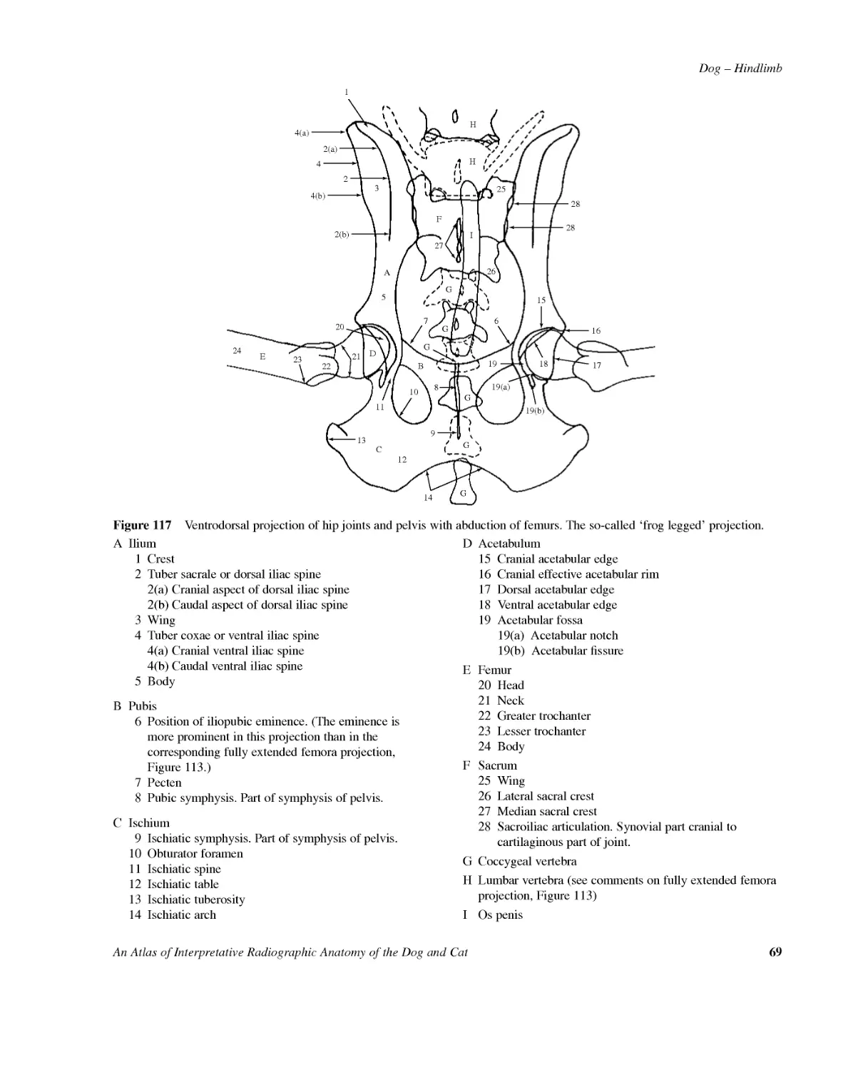
Dog--HindlimbAnAtlasofInterpretativeRadiographicAnatomyoftheDogandCat694(a)143576242220211310111214232(a)2(b)981919(b)19(a)2724(b)HHAEDIGFGGGGGBC1618171528252628Figure117Ventrodorsalprojectionofhipjointsandpelviswithabductionoffemurs.Theso-called'froglegged'projection.AIlium1Crest2Tubersacraleordorsaliliacspine2(a)Cranialaspectofdorsaliliacspine2(b)Caudalaspectofdorsaliliacspine3Wing4Tubercoxaeorventraliliacspine4(a)Cranialventraliliacspine4(b)Caudalventraliliacspine5BodyBPubis6Positionofiliopubiceminence.(Theeminenceismoreprominentinthisprojectionthaninthecorrespondingfullyextendedfemoraprojection,Figure113.)7Pecten8Pubicsymphysis.Partofsymphysisofpelvis.CIschium9Ischiaticsymphysis.Partofsymphysisofpelvis.10Obturatorforamen11Ischiaticspine12Ischiatictable13Ischiatictuberosity14IschiaticarchDAcetabulum15Cranialacetabularedge16Cranialeffectiveacetabularrim17Dorsalacetabularedge18Ventralacetabularedge19Acetabularfossa19(a)Acetabularnotch19(b)AcetabularfissureEFemur20Head21Neck22Greatertrochanter23Lessertrochanter24BodyFSacrum25Wing26Lateralsacralcrest27Mediansacralcrest28Sacroiliacarticulation.Synovialpartcranialtocartilaginouspartofjoint.GCoccygealvertebraHLumbarvertebra(seecommentsonfullyextendedfemoraprojection,Figure113)IOspenis
70AnAtlasofInterpretativeRadiographicAnatomyoftheDogandCatDog--HindlimbFigure118Lateralprojectionofhipjointsandpelvis.Beagledog2.5yearsold,entiremale.Figure119LinedrawingofphotographrepresentingradiographicpositioningforFigure118.
Dog--HindlimbAnAtlasofInterpretativeRadiographicAnatomyoftheDogandCat71Figure120Lateralprojectionofhipjointsandpelvis.19191914HHHFA3GGGGGG2(a)1751722(b)181910915111112161413166713DBCEE8AIlium1Crest2Tubersacraleordorsaliliacspine2(a)Cranialaspectofspine2(b)Caudalaspectofspine3Caudalventraliliacspine.(Cranialventraliliacspineisnotvisibleinthisfilm.)Cranialandcaudalventraliliacspinesformthetubercoxaeorventraliliacspine.4Wing5BodyBPubis6Iliopubiceminence7PectenofpubisCIschium8Pelvicsymphysis9Obturatorforamen10Ischiaticspine11Ischiatictuberosity12IschiatictableDAcetabulumEFemur13Head14Neck15Greatertrochanters(shadowsarenotclearlyvisiblebuttheywillextendalmost,ifnotquite,asfarproximalasdothefemoralheadsonatrulylateralprojection)16LessertrochanterFSacrum17Sacroiliacarticulation18Sacrallamina(dorsalsurfaceisnotclearlydistinguishable)19VertebralcanalGCoccygealvertebraHLumbarvertebra
72AnAtlasofInterpretativeRadiographicAnatomyoftheDogandCatDog--HindlimbFigure121Lateralobliqueprojectionofhipjointsandpelvis.Beagledog7yearsold,entiremale.Figure122LinedrawingofphotographrepresentingradiographicpositioningforFigure121.
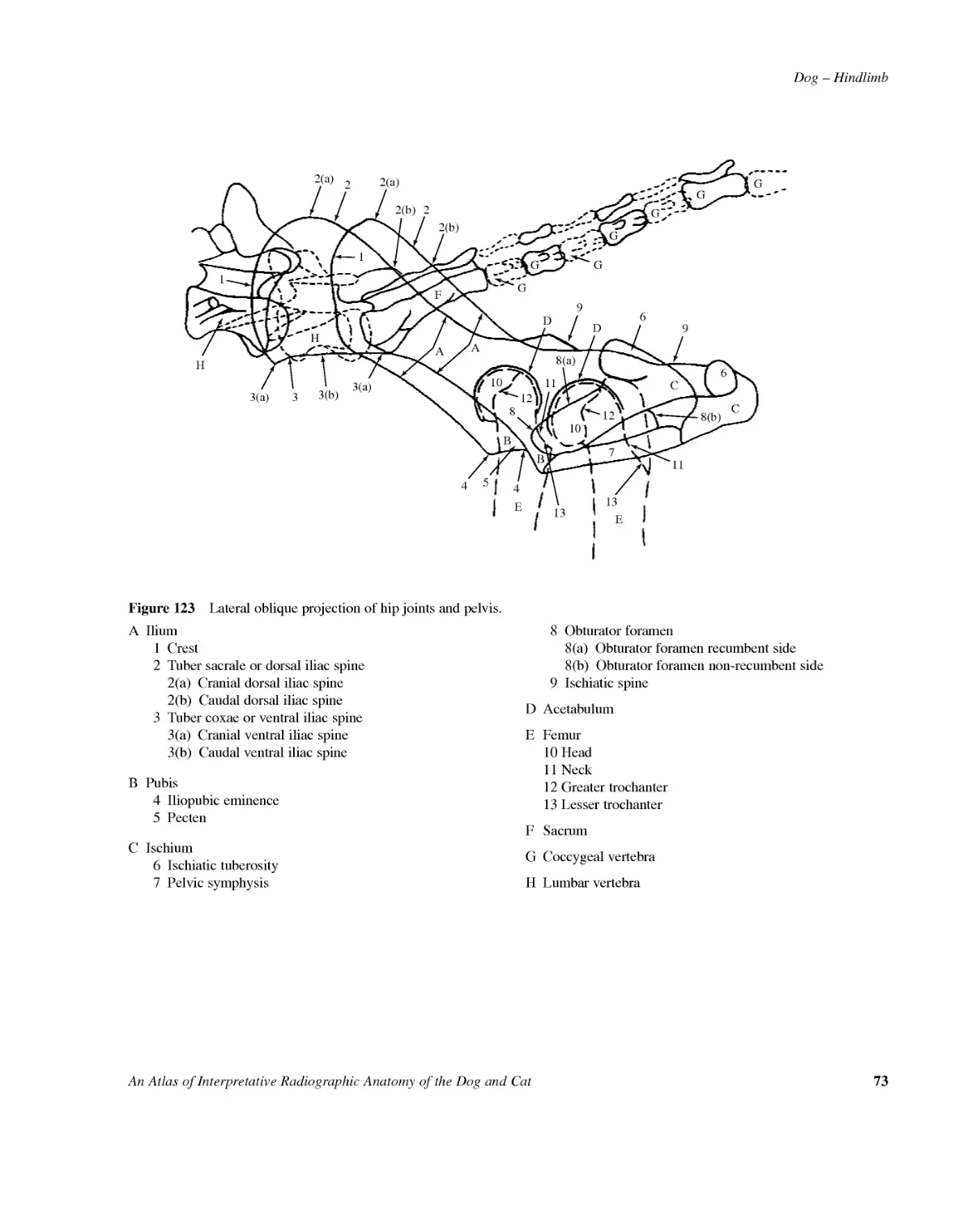
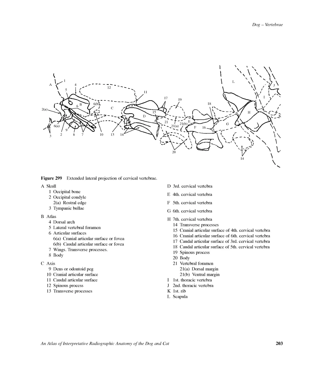
Dog--HindlimbAnAtlasofInterpretativeRadiographicAnatomyoftheDogandCat73Figure123Lateralobliqueprojectionofhipjointsandpelvis.1HHAF3(a)33(a)3(b)GGGGGG2(a)2(a)2(b)1G222(b)998(b)8(a)6A121210111113136710445DDBBCCEE8AIlium1Crest2Tubersacraleordorsaliliacspine2(a)Cranialdorsaliliacspine2(b)Caudaldorsaliliacspine3Tubercoxaeorventraliliacspine3(a)Cranialventraliliacspine3(b)CaudalventraliliacspineBPubis4Iliopubiceminence5PectenCIschium6Ischiatictuberosity7Pelvicsymphysis8Obturatorforamen8(a)Obturatorforamenrecumbentside8(b)Obturatorforamennon-recumbentside9IschiaticspineDAcetabulumEFemur10Head11Neck12Greatertrochanter13LessertrochanterFSacrumGCoccygealvertebraHLumbarvertebra
74AnAtlasofInterpretativeRadiographicAnatomyoftheDogandCatDog--HindlimbFigure124Mediolateralprojectionoffemur.Beagledog7yearsold,entiremale.Figure125Linedrawingofphotographrepresentingradio-graphicpositioningforFigure124.
Dog--HindlimbAnAtlasofInterpretativeRadiographicAnatomyoftheDogandCat75ABCDK7E7(a)L8J1312101114151716I2I1I9HFG4426513Figure126Mediolateralprojectionoffemur.AIliumBAcetabulumCPubisDIschium1ObturatorforamenEFemur2Head3Neck4Greatertrochanter5Lessertrochanter6Trochantericfossa7Body7(a)Nutrientforamen(justvisibleasaradiolucenttrackthroughcortex)8Trochleargroove9Trochlearridge10Lateralcondyle11Medialcondyle12BaseofintercondyloidfossaFTibia13Lateralcondyle14Medialcondyle15Intercondyloideminence.Morecaudalshadowislateral.16Tibialtuberosity17Cranialborderor'tibialcrest'asformerlyknownGFibulaHPatellaIFabellaeofm.gastrocnemiusI1LateralfabellaI2MedialfabellaJFabellaofm.popliteusKOspenisLScrotalshadow
76AnAtlasofInterpretativeRadiographicAnatomyoftheDogandCatDog--HindlimbFigure127Craniocaudalprojectionoffemur.Beagledog2.5yearsold,entiremale.Figure128LinedrawingofphotographrepresentingradiographicpositioningforFigure127.
Dog--HindlimbAnAtlasofInterpretativeRadiographicAnatomyoftheDogandCat77A192121038BD46C5117E1314161915GFHIIJ20211817Figure129Craniocaudalprojectionoffemur.AIliumBPubisCAcetabulumAcetabularfeatures:1Cranialacetabularedge2Dorsalacetabularedge3Ventralacetabularedge4Acetabularnotch5AcetabularfissureDIschium6Obturatorforamen7IschiatictuberosityEFemur8Head9Neck10Greatertrochanter11Lessertrochanter12Trochantericfossa13Body14Medialcondyle15Lateralcondyle16Intercondyloidfossa17Medialtrochlearridge18LateraltrochlearridgeFTibia19Medialcondyle20Lateralcondyle21IntercondyloideminenceGFibulaHPatellaIFabellaofm.gastrocnemiusJFabellaofm.popliteus
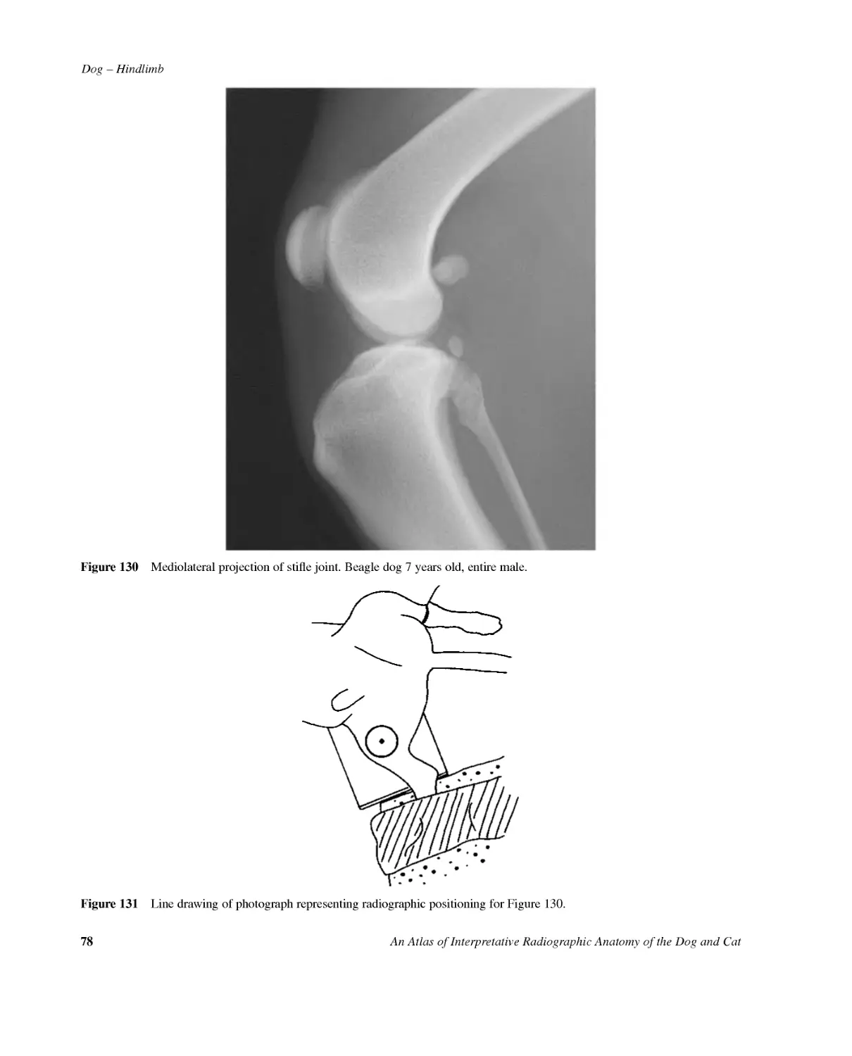
78AnAtlasofInterpretativeRadiographicAnatomyoftheDogandCatDog--HindlimbFigure130Mediolateralprojectionofstiflejoint.Beagledog7yearsold,entiremale.Figure131LinedrawingofphotographrepresentingradiographicpositioningforFigure130.
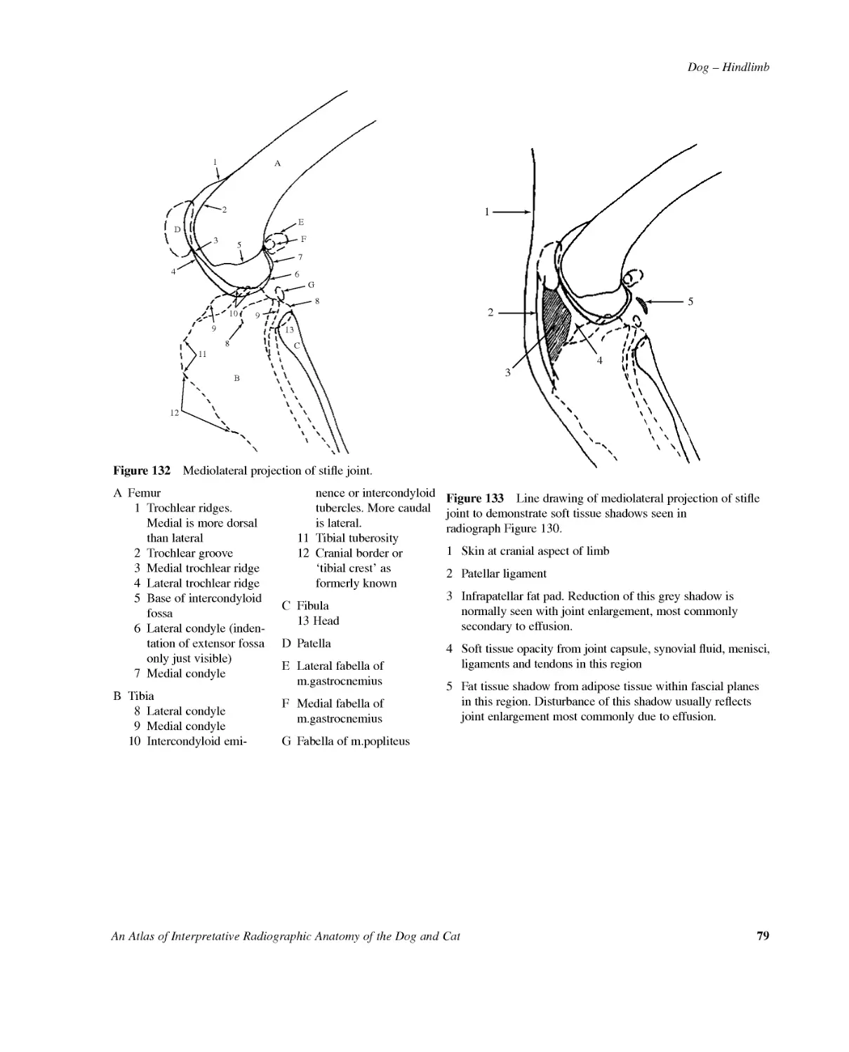
Dog--HindlimbAnAtlasofInterpretativeRadiographicAnatomyoftheDogandCat79A1351311122BD4C9EF76G81098Figure132Mediolateralprojectionofstiflejoint.AFemur1Trochlearridges.Medialismoredorsalthanlateral2Trochleargroove3Medialtrochlearridge4Lateraltrochlearridge5Baseofintercondyloidfossa6Lateralcondyle(inden-tationofextensorfossaonlyjustvisible)7MedialcondyleBTibia8Lateralcondyle9Medialcondyle10Intercondyloidemi-nenceorintercondyloidtubercles.Morecaudalislateral.11Tibialtuberosity12Cranialborderor'tibialcrest'asformerlyknownCFibula13HeadDPatellaELateralfabellaofm.gastrocnemiusFMedialfabellaofm.gastrocnemiusGFabellaofm.popliteus54231Figure133LinedrawingofmediolateralprojectionofstiflejointtodemonstratesofttissueshadowsseeninradiographFigure130.1Skinatcranialaspectoflimb2Patellarligament3Infrapatellarfatpad.Reductionofthisgreyshadowisnormallyseenwithjointenlargement,mostcommonlysecondarytoeffusion.4Softtissueopacityfromjointcapsule,synovialfluid,menisci,ligamentsandtendonsinthisregion5Fattissueshadowfromadiposetissuewithinfascialplanesinthisregion.Disturbanceofthisshadowusuallyreflectsjointenlargementmostcommonlyduetoeffusion.
80AnAtlasofInterpretativeRadiographicAnatomyoftheDogandCatDog--HindlimbaFigure134Schematicdrawingofmediolateralprojec-tionofstiflejointtodemonstrateextentofjointcapsule.=Jointcapsule=Synovialspacea=Distolateralextensionaroundtendonofthem.extensordigitorumlonguswhereittraversestheextensorgrooveofthelateraltibialcondyle.Notethatthestiflejointcavityextendsintothesynovialjointsmadebythepatella,lateralandmedialfabellaeandthefibulaaswellasthefemorotibialjoint.15234Figure136Schematicdrawingofmediolateralprojectionofstiflejoint.Thepositionsofligamentsandtendonsontheaxialandmedialaspectsareindicated.1=Medialfemoropatellarligament2=Tendoninsertionofm.quadricepsfemorisintothepatella3=Patellarligament4=Medialcollateralligament5=RegionofmenisciMeniscalandcruciateligamentsnotshownbutwillbefoundintheregionofmenisci.2315674Figure135Schematicdrawingofmediolateralprojectionofstiflejoint.Thepositionsofligamentsandtendonsontheaxialandlateralaspectsareindicated.1=Lateralfemoropatellarligament2=Tendoninsertionofm.quadricepsfemorisintothepatella3=Patellarligament4=Tendonofm.extensordigitorumlongus5=Lateralcollateralligament6=Tendonofm.popliteusplussesamoidbone7=LigamentoffibularheadMeniscalandcruciateligamentsnotshownbutwillbefoundinthefemorotibialjointbetweenthetendonsofm.extensordigitorumlongusandm.popliteus.
Dog--HindlimbAnAtlasofInterpretativeRadiographicAnatomyoftheDogandCat81Figure137Caudocranialprojectionofstiflejoint.Beagledog2.5yearsold,entiremale.Figure138LinedrawingofphotographrepresentingradiographicpositioningforFigure137.AFE2738BD46C5G191110(a)10Figure139Caudocranialprojectionofstiflejoint.AFemur1Medialtrochlearridge2Lateraltrochlearridge3Medialcondyle4Lateralcondyle5IntercondyloidfossaBTibia6Medialcondyle7Lateralcondyle8Intercondyloideminenceormedialandlateralinter-condyloidtubercles9Tibialtuberosity10Cranialborderor'tibialcrest'asformerlyknown10(a)Outlinefortheextensormuscles,especiallym.cranialistibialisCFibula11HeadDPatellaELateralfabellaofm.gastrocnemiusFMedialfabellaofm.gastrocnemiusGFabellaofm.popliteus
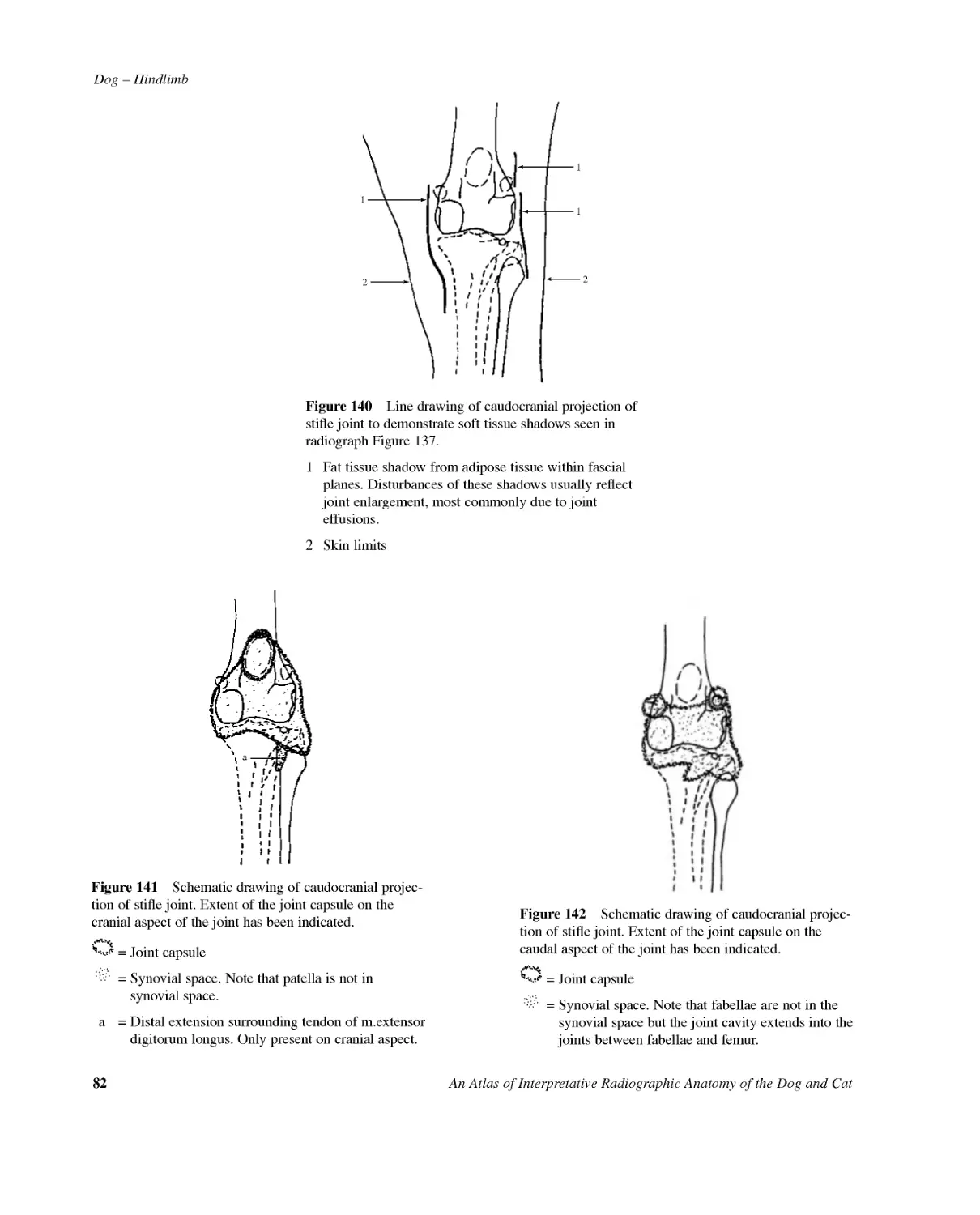
82AnAtlasofInterpretativeRadiographicAnatomyoftheDogandCatDog--Hindlimb11212Figure140LinedrawingofcaudocranialprojectionofstiflejointtodemonstratesofttissueshadowsseeninradiographFigure137.1Fattissueshadowfromadiposetissuewithinfascialplanes.Disturbancesoftheseshadowsusuallyreflectjointenlargement,mostcommonlyduetojointeffusions.2SkinlimitsaFigure141Schematicdrawingofcaudocranialprojec-tionofstiflejoint.Extentofthejointcapsuleonthecranialaspectofthejointhasbeenindicated.=Jointcapsule=Synovialspace.Notethatpatellaisnotinsynovialspace.a=Distalextensionsurroundingtendonofm.extensordigitorumlongus.Onlypresentoncranialaspect.Figure142Schematicdrawingofcaudocranialprojec-tionofstiflejoint.Extentofthejointcapsuleonthecaudalaspectofthejointhasbeenindicated.=Jointcapsule=Synovialspace.Notethatfabellaearenotinthesynovialspacebutthejointcavityextendsintothejointsbetweenfabellaeandfemur.
Dog--HindlimbAnAtlasofInterpretativeRadiographicAnatomyoftheDogandCat831122(a)762832(b)45Figure143Schematicdrawingofcaudocranialprojectionofstiflejoint.Positionsofligamentsofthemedial,lateralandcranialaspectsareindicated.Positionsofthemenisciarealsoshown.1=Femoropatellarligaments2=Collateralligaments2(a)=Medial2(b)=Lateral3=Cranialfibularligament4=Cranialcruciateligament5=Caudalcruciateligament6=Transverseorinter-meniscalligament7=Medialmeniscus8=LateralmeniscusMeniscalligamentsattach-ingmeniscitotibiaandfemurnotshown.Patellarligamentexcludedtoavoidconfusion.1141(a)78321(b)65Figure144Schematicdrawingofcaudocranialprojec-tionofstiflejoint.Positionsofligamentsofthemedial,lateralandcaudalaspectsareindicated.Positionsofthemenisciarealsoshown.1=Collateralligaments1(a)=Medial1(b)=Lateral2=Caudalfibularligament3=Cranialfibularligament4=Caudalcruciateliga-ment.Extendsfromaxialsurfaceofmedialfemoralcondyletotibialpoplitealnotch.Itislongerandheavierthancranial.5=Cranialcruciateliga-ment.Extendsfromcaudalpartofaxialsurfaceoflateralfemoralcondyletocranialintercondyloidareaoftibia.6=Meniscofemoralligament7=Medialmeniscus8=LateralmeniscusMeniscalligamentsattach-ingmeniscitotibiaandintermeniscalligamentnotshown.Patellarligamentexcludedtoavoidconfusion.
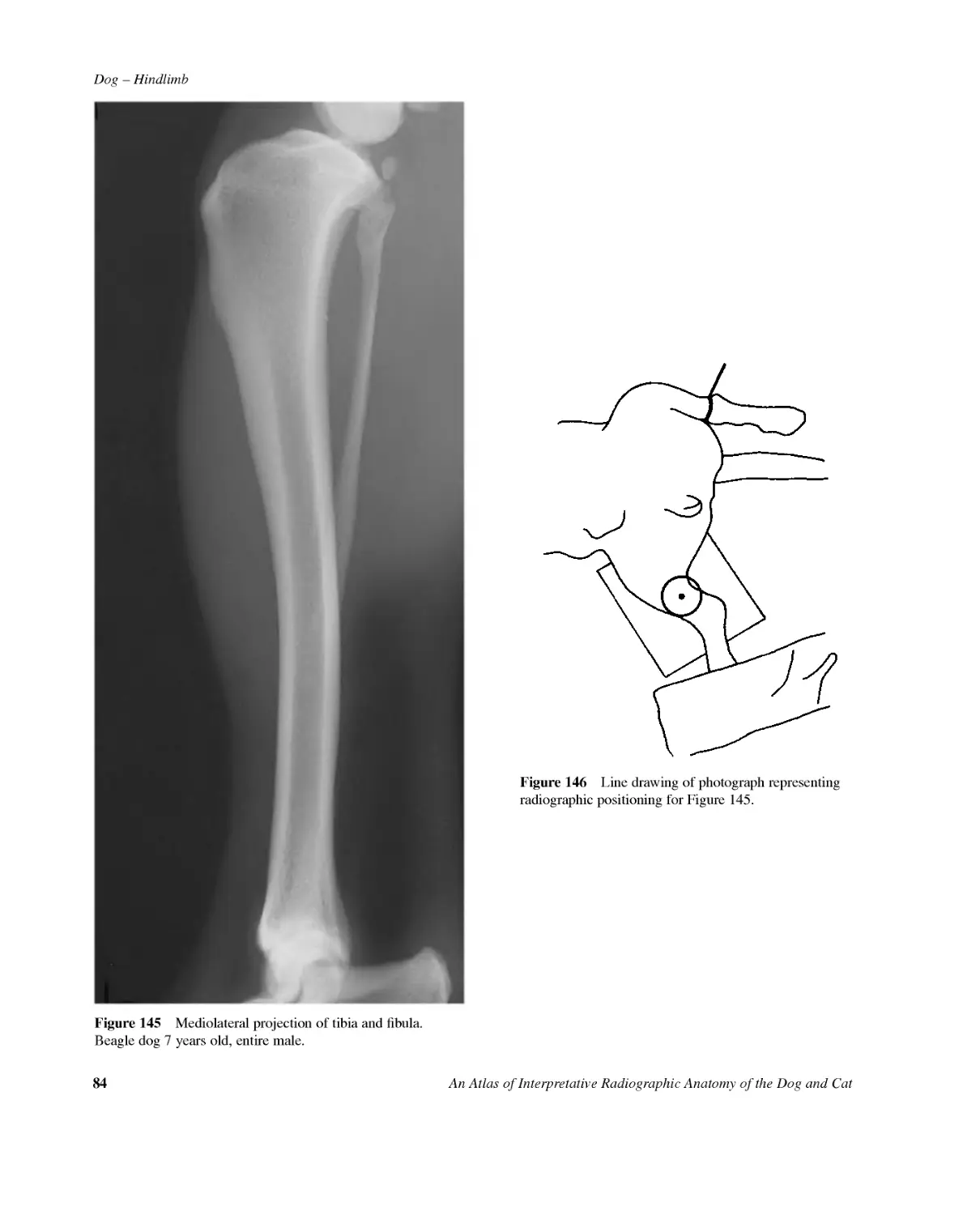
84AnAtlasofInterpretativeRadiographicAnatomyoftheDogandCatDog--HindlimbFigure145Mediolateralprojectionoftibiaandfibula.Beagledog7yearsold,entiremale.Figure146LinedrawingofphotographrepresentingradiographicpositioningforFigure145.
Dog--HindlimbAnAtlasofInterpretativeRadiographicAnatomyoftheDogandCat85A12D12F86B43CE511718HG101713151491616Figure147Mediolateralprojectionoftibiaandfibula.AFemur1Lateralcondyle2Medialcondyle3BaseofintercondyloidfossaBTibia4Lateralcondyle5Medialcondyle6Intercondyloideminenceorintercondyloidtubercles.Themorecaudalshadowisthelateraltubercle7Tibialtuberosity8Cranialborderor'tibialcrest'asformerlyknown9Medialmalleolus10Distalarticularborder11Nutrientforamen(onlyjustvisiblebutcanmimicafractureifthetibiaisslightlyrotatedonexposure)CFibula12Head13LateralmalleolusDLateralfabellaofm.gastrocnemiusEMedialfabellaofm.gastrocnemiusFFabellaofm.popliteusGTibialtarsalboneortalus14Lateraltrochlearridge15Medialtrochlearridge16TrochleargrooveHFibulartarsalboneorcalcaneus17Sustentaculumtali18Calcanealtuber
86AnAtlasofInterpretativeRadiographicAnatomyoftheDogandCatDog--HindlimbFigure148Caudocranialprojectionoftibiaandfibula.Beagledog2.5yearsold,entiremale.Figure149LinedrawingofphotographrepresentingradiographicpositioningforFigure148.
Dog--HindlimbAnAtlasofInterpretativeRadiographicAnatomyoftheDogandCat87A15238(a)BD46C782(a)E111416915GF12131010Figure150Caudocranialprojectionoftibiaandfibula.AFemur1Medialcondyle2Lateralcondyle2(a)Extensorfossa.Originofm.flexordigitorumlongus.3IntercondyloidfossaBTibia4Medialcondyle5Lateralcondyle6Intercondyloideminenceorintercondyloidtubercles7Tibialtuberosity8Cranialborderor'tibialcrest'asformerlyknown8(a)Outlineofconcavityinthetibiawhichhousesextensormuscles9Medialmalleolus10DistalarticularborderCFibula11Head12LateralmalleolusDMedialfabellaofm.gastro-cnemius.(Notetheunusualpositionofthisfabellainrelationshiptothemedialfemoralcondyle.Thisisananatomicalvariantwhichisnottobemisdiagnosedasaruptureofthem.gastro-cnemius.)EFabellaofm.popliteusFTibialtarsalboneortalus13Medialtrochlearridge14LateraltrochlearridgeGFibulartarsalboneorcalcaneus15Sustentaculumtali16Calcanealtuber
88AnAtlasofInterpretativeRadiographicAnatomyoftheDogandCatDog--HindlimbFigure152LinedrawingofphotographrepresentingradiographicpositioningforFigure151.A2387B2(a)6GCDNMLK541011219FEIHJFigure153Plantarodorsalprojectionoftarsus.ATibia1Medialmalleolus2Distalarticularborder.(Medialandlateralgrooves.)2(a)Distalarticularborder.(Cranialaspect.)BFibula3Lateralmalleolus.(Notetherela-tivelyproximalpositionofthelater-almalleoluscomparedtothemedialmalleolus.Inmanydogsthemalleoliareatanequaldistallevel.)CTibialtarsalboneortalus4Medialtrochlearridge5Lateraltrochlearridge6HeadDFibulartarsalboneorcalcaneus7Sustentaculumtali8Calcanealtuber9Tarsalsinus.(Radiolucentshadowwhichisthespacebetweentalusandcalcaneusextendsmoredistallythancanbeseeninthisprojection.)ECentraltarsalbone10PlantarprocessFTarsalbone1GTarsalbone2HTarsalbone3ITarsalbone411ShadowformedbylargetuberosityontheplantarsurfaceJMetatarsalbone1KMetatarsalbone2LMetatarsalbone3MMetatarsalbone4NMetatarsalbone5Figure151Plantarodorsalprojectionoftarsus.Bea-gledog2.5yearsold,entiremale.
Dog--HindlimbAnAtlasofInterpretativeRadiographicAnatomyoftheDogandCat89Figure155LinedrawingofphotographrepresentingradiographicpositioningforFigure154.A71098BD4CIIGEHLFJMLN4632K51Figure156Extendedmediolateralprojectionoftarsus.ATibia1Medialmalleolus2DistalarticularborderBFibula3LateralmalleolusCTibialtarsalboneortalus4Lateraltrochlearridge5Medialtrochlearridge6TrochleargrooveDFibulartarsalboneorcalcaneus7Sustentaculumtali8CalcanealtuberECentraltarsalbone9PlantarprocessFTarsalbone1GTarsalbone2HTarsalbone3ITarsalbone4JMetatarsalbone1KMetatarsalbone3LCombinedshadowsofmetatarsalbones2,4and5MMetatarsalbone2NMetatarsalbone5Figure154Extendedmediolateralprojectionoftarsus.Beagledog2.5yearsold,entiremale.
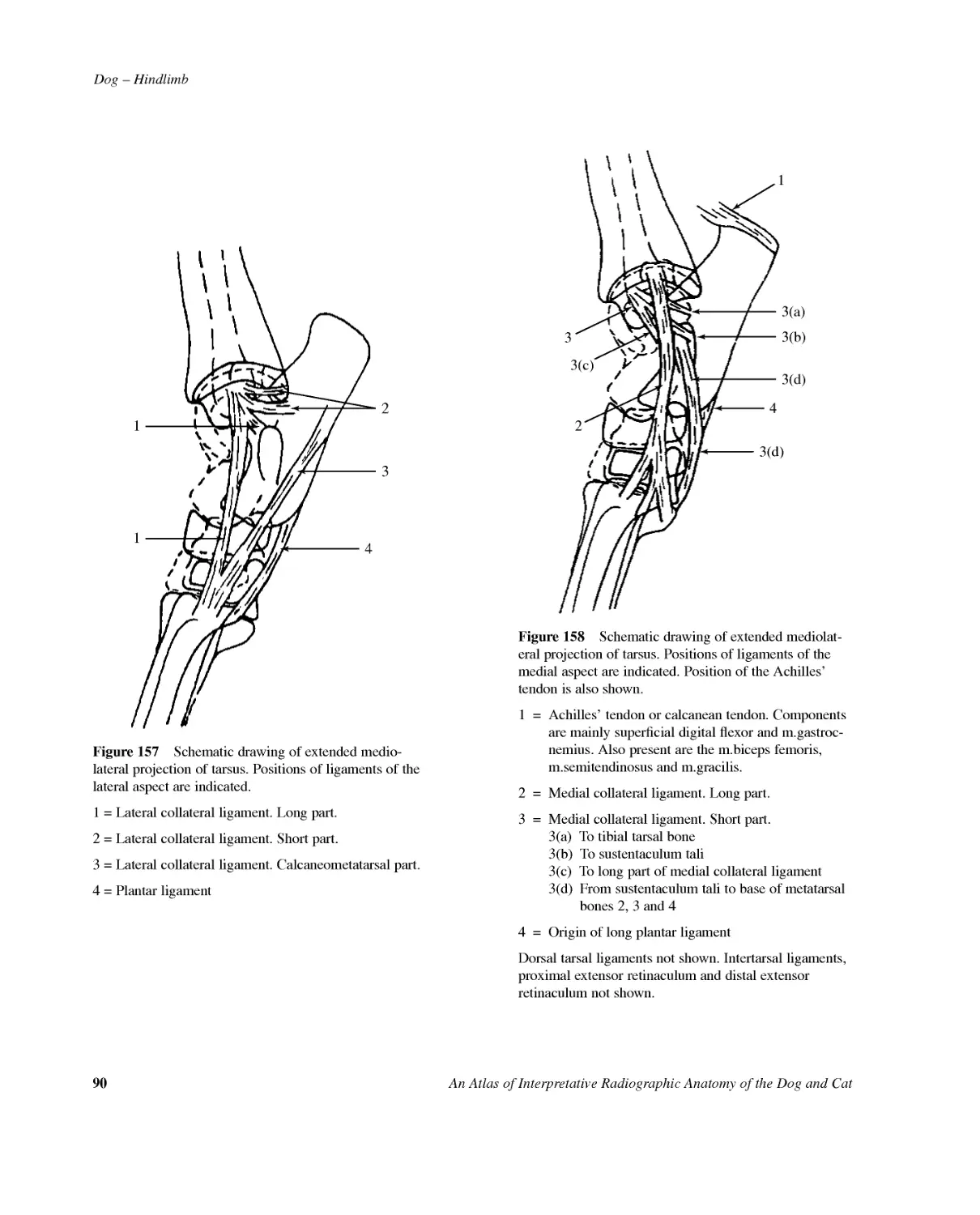
90AnAtlasofInterpretativeRadiographicAnatomyoftheDogandCatDog--Hindlimb23411Figure157Schematicdrawingofextendedmedio-lateralprojectionoftarsus.Positionsofligamentsofthelateralaspectareindicated.1=Lateralcollateralligament.Longpart.2=Lateralcollateralligament.Shortpart.3=Lateralcollateralligament.Calcaneometatarsalpart.4=Plantarligament313(c)3(d)3(a)3(b)3(d)42Figure158Schematicdrawingofextendedmediolat-eralprojectionoftarsus.Positionsofligamentsofthemedialaspectareindicated.PositionoftheAchilles'tendonisalsoshown.1=Achilles'tendonorcalcaneantendon.Componentsaremainlysuperficialdigitalflexorandm.gastroc-nemius.Alsopresentarethem.bicepsfemoris,m.semitendinosusandm.gracilis.2=Medialcollateralligament.Longpart.3=Medialcollateralligament.Shortpart.3(a)Totibialtarsalbone3(b)Tosustentaculumtali3(c)Tolongpartofmedialcollateralligament3(d)Fromsustentaculumtalitobaseofmetatarsalbones2,3and44=OriginoflongplantarligamentDorsaltarsalligamentsnotshown.Intertarsalligaments,proximalextensorretinaculumanddistalextensorretinaculumnotshown.
Dog--HindlimbAnAtlasofInterpretativeRadiographicAnatomyoftheDogandCat91Figure160LinedrawingofphotographrepresentingradiographicpositioningforFigure159.BACGD2345768IIFNE1HKLJMFigure161Flexedmediolateralprojectionoftarsus.ATibia1Medialmalleolus2DistalarticularborderBFibulaLateralmalleoluscannotbeidentifiedasaseparatestructureCTibialtarsalboneortalus3Lateraltrochlearridge4Medialtrochlearridge5TrochleargrooveDFibulartarsalboneorcalcaneus6Sustentaculumtali7CalcanealtuberECentraltarsalbone8PlantarprocessFTarsalbone1GTarsalbone2HTarsalbone3ITarsalbone4JMetatarsalbone1KMetatarsalbone3LCombinedshadowsofmetatarsalbones2,4and5MMetatarsalbone2NMetatarsalbone5Figure159Flexedmediolateralprojectionoftarsus.Beagledog2.5yearsold,entiremale.
92AnAtlasofInterpretativeRadiographicAnatomyoftheDogandCatDog--HindlimbFigure163Linedrawingofphotographrepresentingradio-graphicpositioningforFigure162.BACGD2345768IHNE1FLJMKFigure164Plantaromedial--dorsolateralobliqueprojectionoftarsus.ATibia1Medialmalleolus2DistalarticularborderBFibula3LateralmalleolusCTibialtarsalboneortalus4Lateraltrochlearridge5MedialtrochlearridgeDFibulartarsalboneorcalcaneus6CalcanealtuberECentraltarsalbone7PlantarprocessFTarsalbone1GTarsalbone2HTarsalbone3ITarsalbone48TuberosityonplantaraspectJMetatarsalbone1KMetatarsalbone2LMetatarsalbone3MMetatarsalbone4NMetatarsalbone5Figure162Plantaromedial-dorsolateralobliqueprojectionoftarsus.Beagledog2.5yearsold,entiremale.
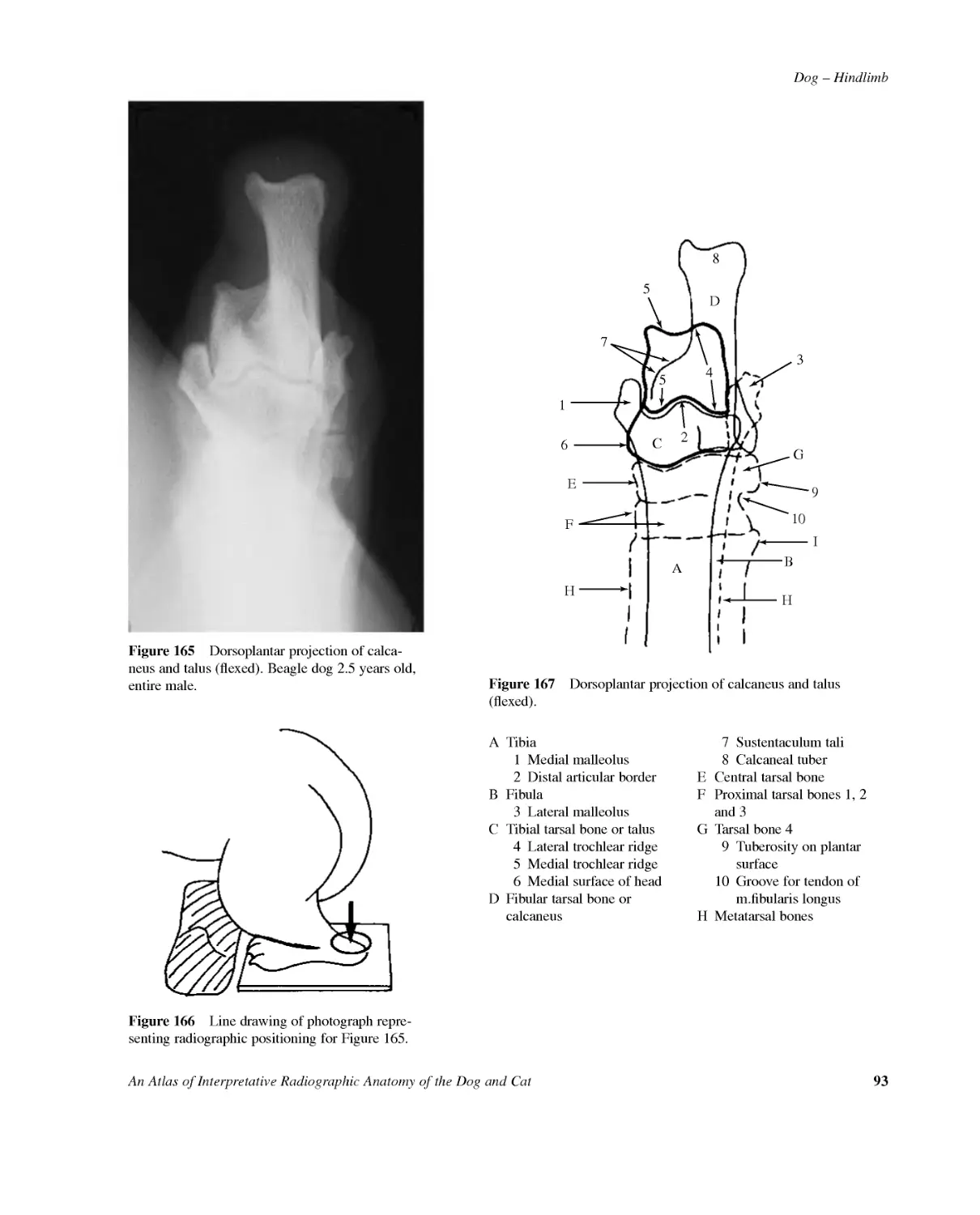
Dog--HindlimbAnAtlasofInterpretativeRadiographicAnatomyoftheDogandCat93Figure166Linedrawingofphotographrepre-sentingradiographicpositioningforFigure165.B10ACGD234557689I1FEHHFigure167Dorsoplantarprojectionofcalcaneusandtalus(flexed).ATibia1Medialmalleolus2DistalarticularborderBFibula3LateralmalleolusCTibialtarsalboneortalus4Lateraltrochlearridge5Medialtrochlearridge6MedialsurfaceofheadDFibulartarsalboneorcalcaneus7Sustentaculumtali8CalcanealtuberECentraltarsalboneFProximaltarsalbones1,2and3GTarsalbone49Tuberosityonplantarsurface10Groovefortendonofm.fibularislongusHMetatarsalbonesFigure165Dorsoplantarprojectionofcalca-neusandtalus(flexed).Beagledog2.5yearsold,entiremale.
94AnAtlasofInterpretativeRadiographicAnatomyoftheDogandCatDog--HindlimbFigure169Linedrawingofphotographrepresentingradio-graphicpositioningforFigure168.Figure168Plantarodorsalprojectionofmetatarsusandphalanges.Beagledog2.5yearsold,entiremale.
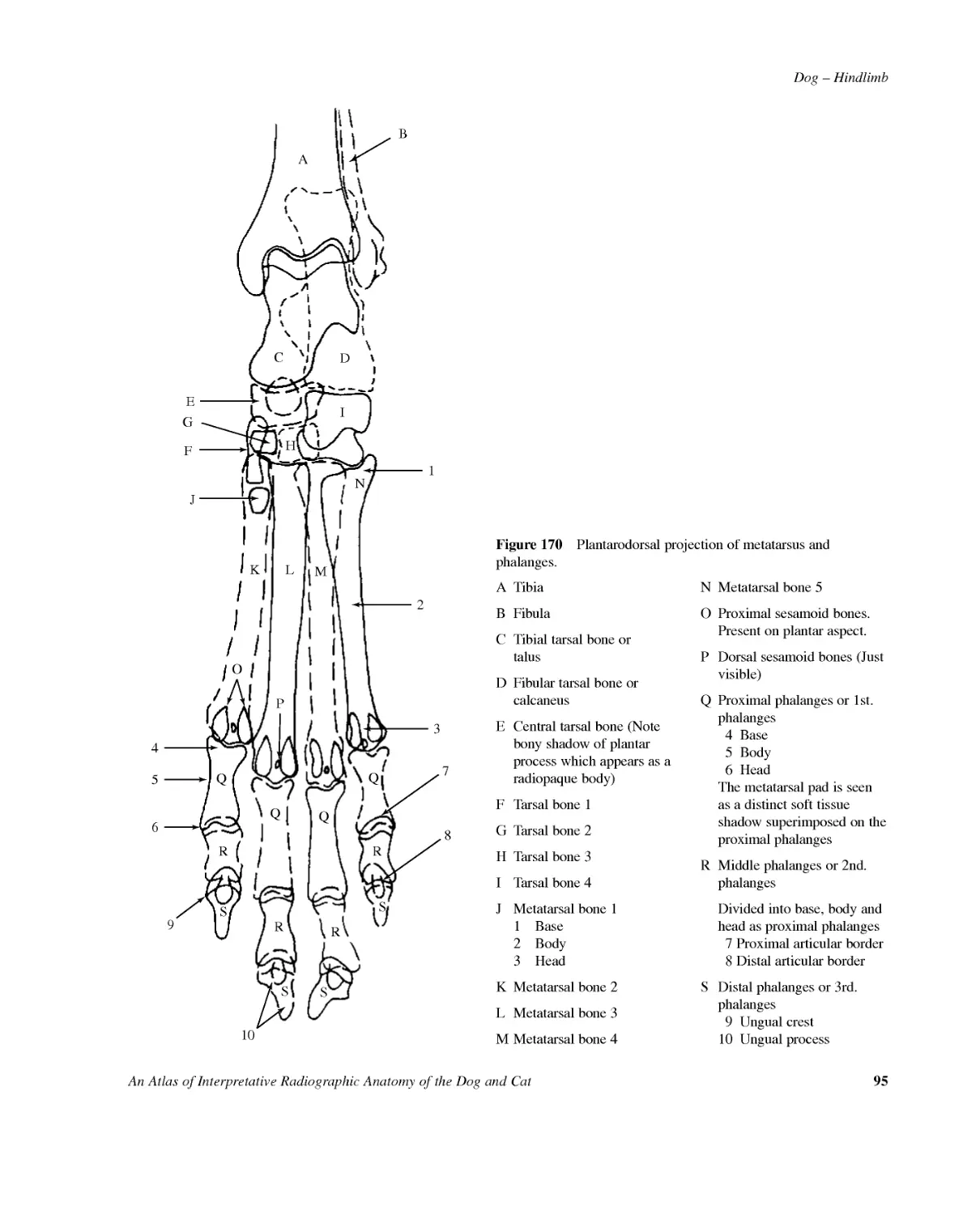
Dog--HindlimbAnAtlasofInterpretativeRadiographicAnatomyoftheDogandCat95BACGD1IHNELKMQQQQOPRRSSSSRRFJ4569102387Figure170Plantarodorsalprojectionofmetatarsusandphalanges.ATibiaBFibulaCTibialtarsalboneortalusDFibulartarsalboneorcalcaneusECentraltarsalbone(Notebonyshadowofplantarprocesswhichappearsasaradiopaquebody)FTarsalbone1GTarsalbone2HTarsalbone3ITarsalbone4JMetatarsalbone11Base2Body3HeadKMetatarsalbone2LMetatarsalbone3MMetatarsalbone4NMetatarsalbone5OProximalsesamoidbones.Presentonplantaraspect.PDorsalsesamoidbones(Justvisible)QProximalphalangesor1st.phalanges4Base5Body6HeadThemetatarsalpadisseenasadistinctsofttissueshadowsuperimposedontheproximalphalangesRMiddlephalangesor2nd.phalangesDividedintobase,bodyandheadasproximalphalanges7Proximalarticularborder8DistalarticularborderSDistalphalangesor3rd.phalanges9Ungualcrest10Ungualprocess
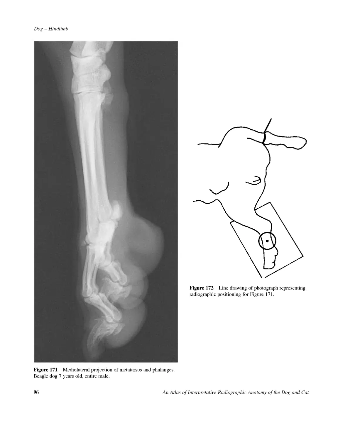
96AnAtlasofInterpretativeRadiographicAnatomyoftheDogandCatDog--HindlimbFigure172LinedrawingofphotographrepresentingradiographicpositioningforFigure171.Figure171Mediolateralprojectionofmetatarsusandphalanges.Beagledog7yearsold,entiremale.
Dog--HindlimbAnAtlasofInterpretativeRadiographicAnatomyoftheDogandCat97BAGGDCFEJKLHI21N4P3O6MMP575QQOFigure173Mediolateralprojectionofmetatarsusandphalanges.ATibialtarsalboneortalusBFibulartarsalboneorcalcaneusCCentraltarsalboneDTarsalbone1ETarsalbone2FTarsalbone3GTarsalbone4HMetatarsalbone1IMetatarsalbone2JMetatarsalbone3KMetatarsalbone4LMetatarsalbone51Superimposedheadsofmetatarsalbones2and52Superimposedheadsofmetatarsalbones3and4MProximalsesamoidbones.Twoinnumberatplantaraspectofeachproximalinterphalangealjoint.NDorsalsesamoidbones.Oneinnumberatdorsalaspectofeachproximalinterphalangealjoint.OProximalphalangesor1st.phalangesPMiddlephalangesor2nd.phalangesQDistalphalangesor3rd.phalanges3Ungualcrest4Ungualprocess5Flexortuberosity6Extensortuberosity7Nutrientcanal
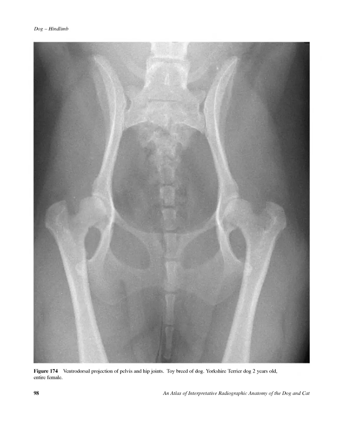
98AnAtlasofInterpretativeRadiographicAnatomyoftheDogandCatDog--HindlimbFigure174Ventrodorsalprojectionofpelvisandhipjoints.Toybreedofdog.YorkshireTerrierdog2yearsold,entirefemale.
Dog--HindlimbAnAtlasofInterpretativeRadiographicAnatomyoftheDogandCat99Figure175Ventrodorsalprojectionofpelvisandhipjoints.Chondrodystrophicbreedofdog.StandardDachshunddog7yearsold,entiremale.
100AnAtlasofInterpretativeRadiographicAnatomyoftheDogandCatDog--HindlimbFigure176Ventrodorsalprojectionofpelvisandhipjoints.Giantbreedofdog.EnglishBullMastiffdog21monthsold,entiremale.
Dog--HindlimbAnAtlasofInterpretativeRadiographicAnatomyoftheDogandCat101Figure177Mediolateralprojectionoffemur.Chondrodys-trophicbreedofdog.MiniatureDachshunddog6yearsold,neuteredfemale.Figure178Mediolateralprojectionofstiflejoint.Chondro-dystrophicbreedofdog.MiniatureDachshunddog6yearsold,neuteredfemale.
102AnAtlasofInterpretativeRadiographicAnatomyoftheDogandCatDog--HindlimbFigure179Caudocranialprojectionofstiflejoint.Chondrodystrophicbreedofdog.MiniatureDachshunddog6yearsold,neuteredfemale.Figure180Mediolateralprojectionoftibiaandfibula.Chon-drodystrophicbreedofdog.MiniatureDachshunddog6yearsold,neuteredfemale.
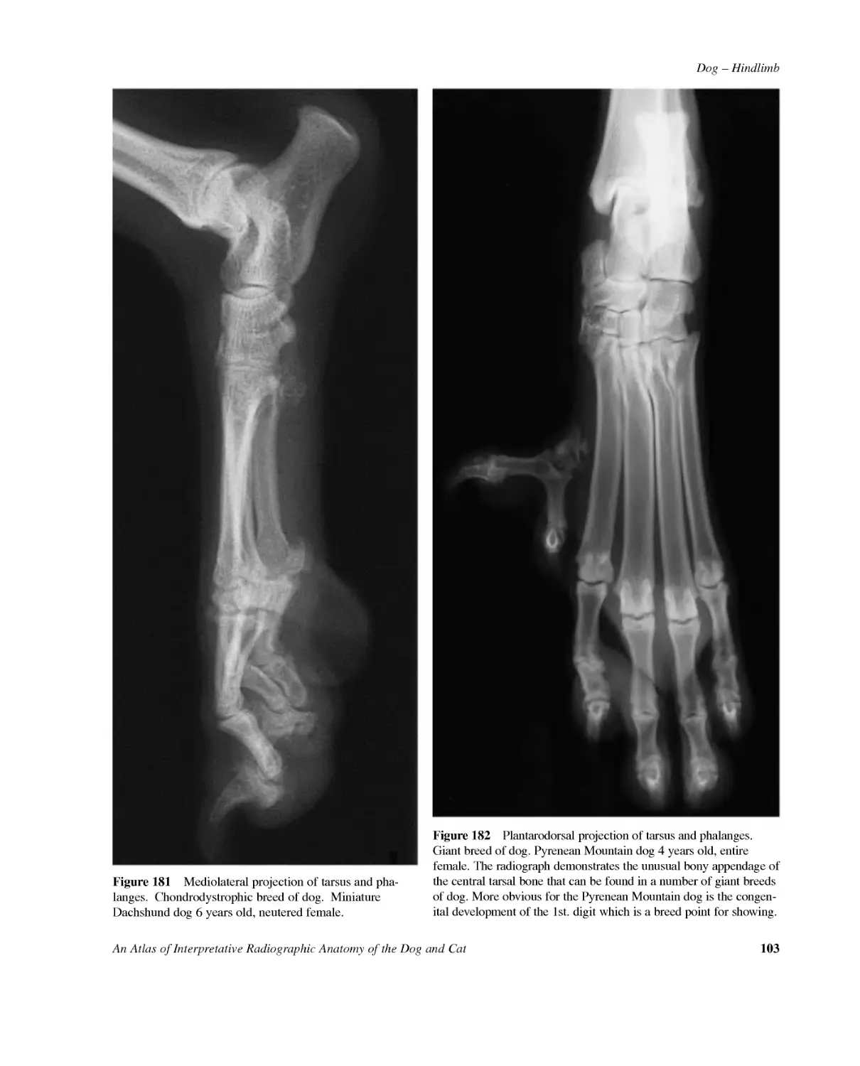
Dog--HindlimbAnAtlasofInterpretativeRadiographicAnatomyoftheDogandCat103Figure181Mediolateralprojectionoftarsusandpha-langes.Chondrodystrophicbreedofdog.MiniatureDachshunddog6yearsold,neuteredfemale.Figure182Plantarodorsalprojectionoftarsusandphalanges.Giantbreedofdog.PyreneanMountaindog4yearsold,entirefemale.Theradiographdemonstratestheunusualbonyappendageofthecentraltarsalbonethatcanbefoundinanumberofgiantbreedsofdog.MoreobviousforthePyreneanMountaindogisthecongen-italdevelopmentofthe1st.digitwhichisabreedpointforshowing.
104AnAtlasofInterpretativeRadiographicAnatomyoftheDogandCatDog--Hindlimb2(a)4(a)11(a)13(a)CBAD125(a)Figures183,184,185,186,187,188,189,190Ventrodorsalprojectionofpelvisandcraniocaudalprojectionofproximalfemur.SamoyedCrossbreddog,entiremale,at4,8,13,17,21,25,34and47weeksofage.Figure183Figure183Age4weeks.AIliumBPubisCIschium1Iliopubicgrowthplate1(a)Open2Ilioischialgrowthplate2(a)Open2(b)Closing3Acetabularbone4Ischiopubicgrowthplate4(a)Open4(c)Remnant5Symphysisofpelvis5(a)Open6Ischiatictuberosity7Ischiatictuberositygrowthplate7(a)Open7(b)Closing8Ischialarchcentre9Ischialarchgrowthplate9(a)Open9(b)Closing10Medianischialarchcentre11Medianischialarchgrowthplate11(a)OpenDFemur12Head13Proximalgrowthplate13(a)Open13(b)Closing14Greatertrochanter15Greatertrochantergrowthplate15(a)Open15(b)Closing16Lessertrochanter
Dog--HindlimbAnAtlasofInterpretativeRadiographicAnatomyoftheDogandCat1052(a)4(a)1(a)13(a)CBAD3121415(a)5(a)Figure184Figure184Age8weeks.
106AnAtlasofInterpretativeRadiographicAnatomyoftheDogandCatDog--Hindlimb2(a)4(c)13(a)CBA3121415(a)5(a)166Figure185
Dog--HindlimbAnAtlasofInterpretativeRadiographicAnatomyoftheDogandCat107Figure185Age13weeks.
108AnAtlasofInterpretativeRadiographicAnatomyoftheDogandCatDog--Hindlimb2(b)1(a)13(a)CB6A1614125(a)15(a)Figure186
Dog--HindlimbAnAtlasofInterpretativeRadiographicAnatomyoftheDogandCat109Figure186Age17weeks.
110AnAtlasofInterpretativeRadiographicAnatomyoftheDogandCatDog--Hindlimb1(a)13(a)CB6A1614125(a)9(a)11(a)1087(a)15(a)Figure187
Dog--HindlimbAnAtlasofInterpretativeRadiographicAnatomyoftheDogandCat111Figure187Age21weeks.
112AnAtlasofInterpretativeRadiographicAnatomyoftheDogandCatDog--Hindlimb13(a)CB6A1614125(a)9(a)11(a)1087(a)15(a)Figure188
Dog--HindlimbAnAtlasofInterpretativeRadiographicAnatomyoftheDogandCat113Figure188Age25weeks.
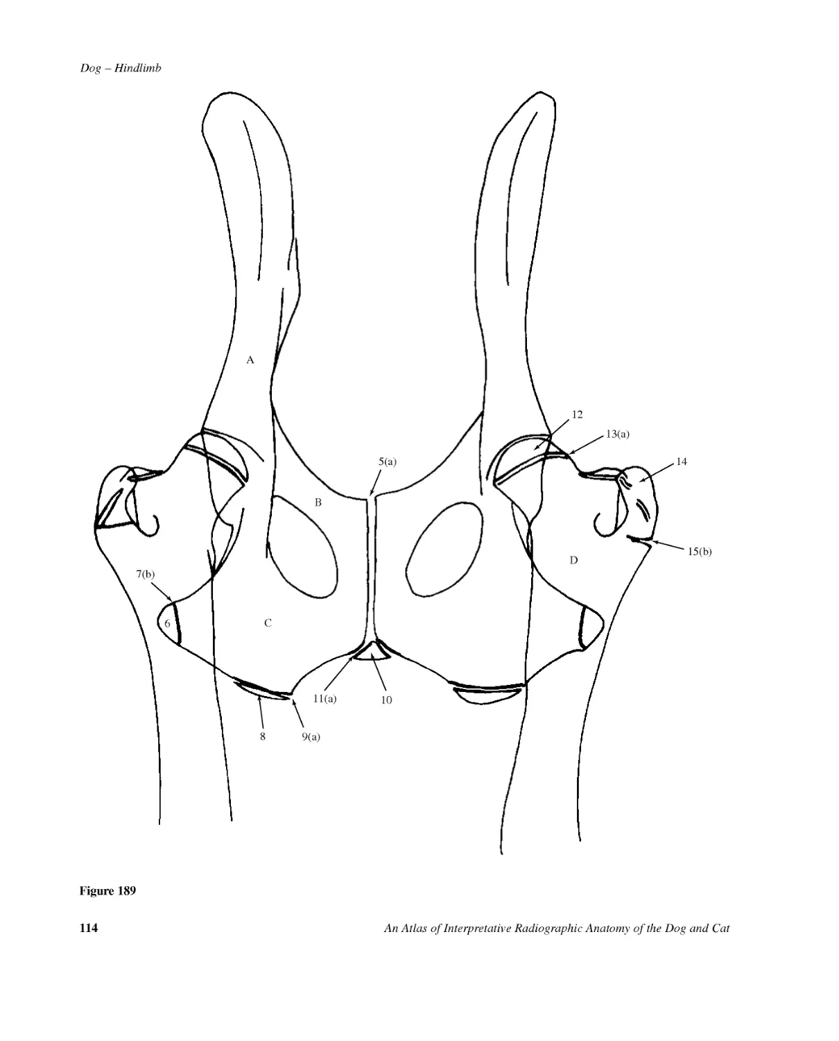
114AnAtlasofInterpretativeRadiographicAnatomyoftheDogandCatDog--Hindlimb13(a)CB6A14125(a)11(a)10D89(a)7(b)15(b)Figure189
Dog--HindlimbAnAtlasofInterpretativeRadiographicAnatomyoftheDogandCat115Figure189Age34weeks.
116AnAtlasofInterpretativeRadiographicAnatomyoftheDogandCatDog--Hindlimb13(b)CBD6A1614125(a)11(a)1089(b)Figure190
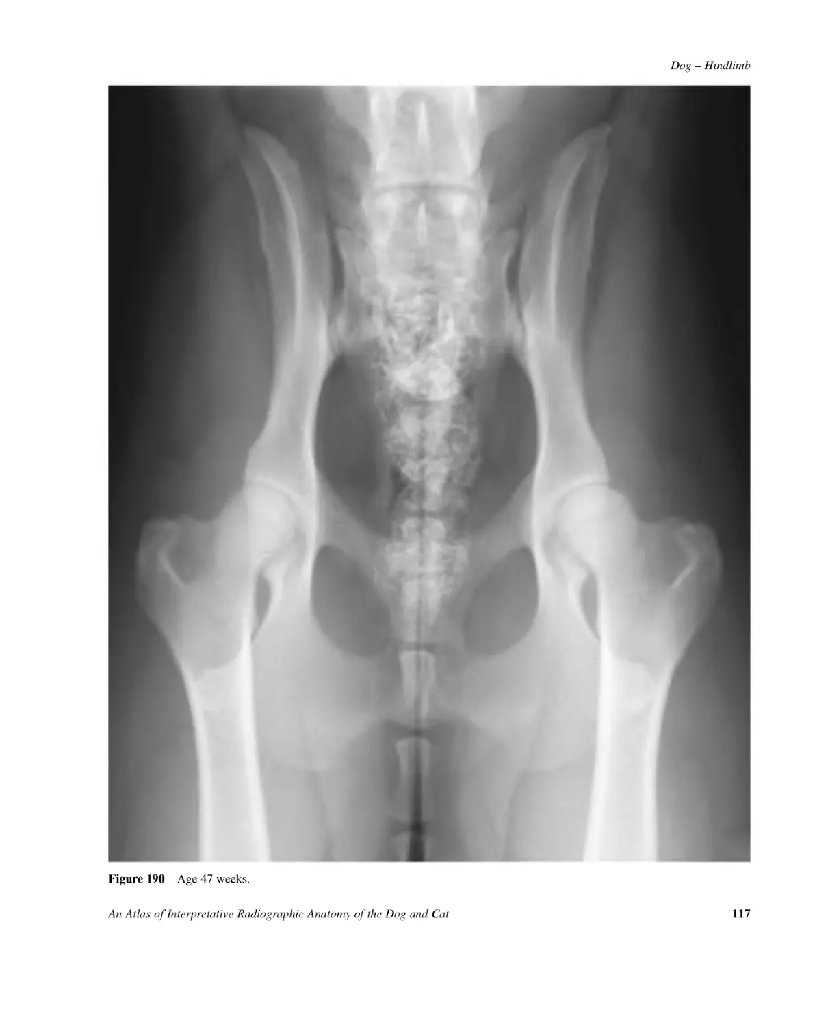
Dog--HindlimbAnAtlasofInterpretativeRadiographicAnatomyoftheDogandCat117Figure190Age47weeks.
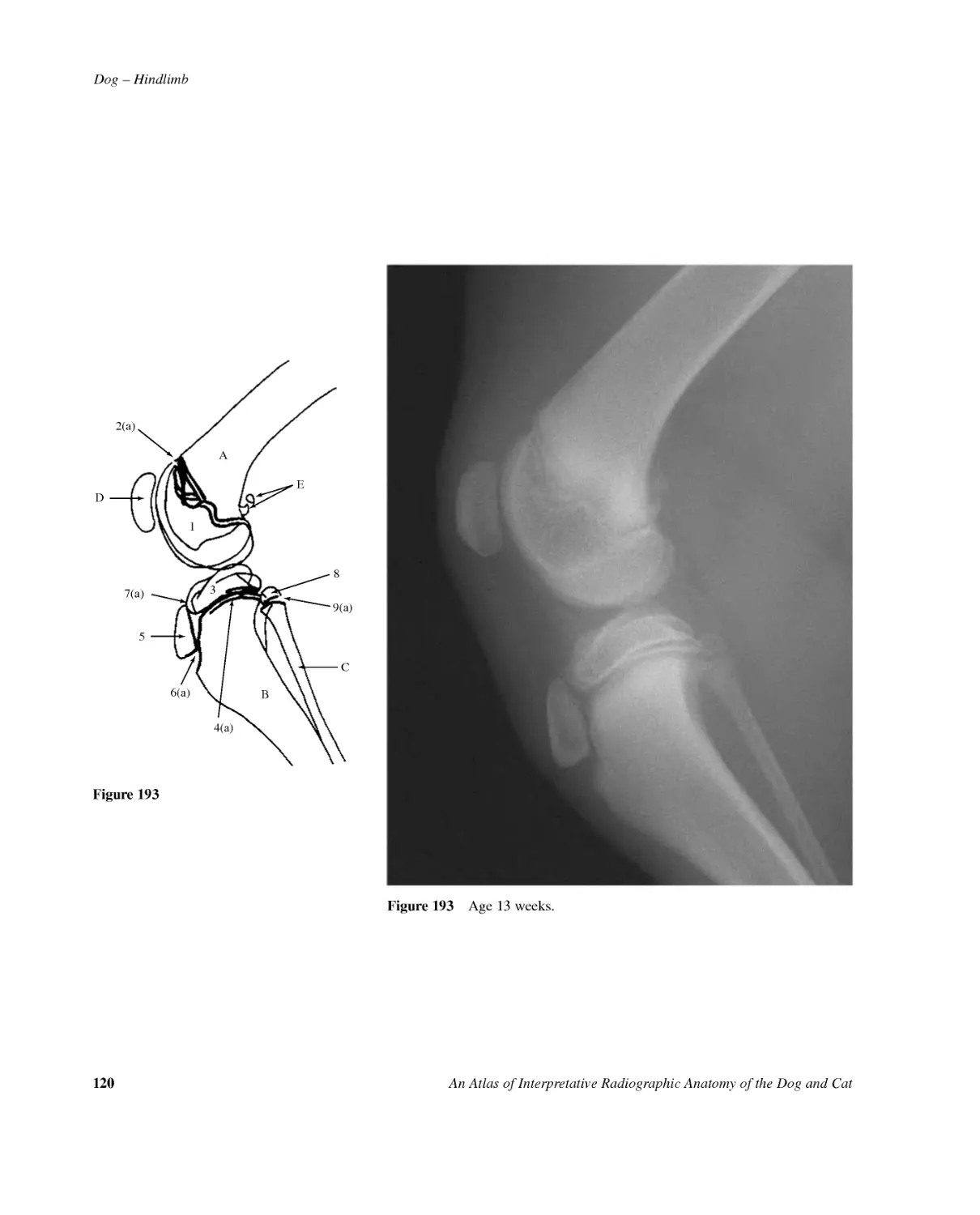
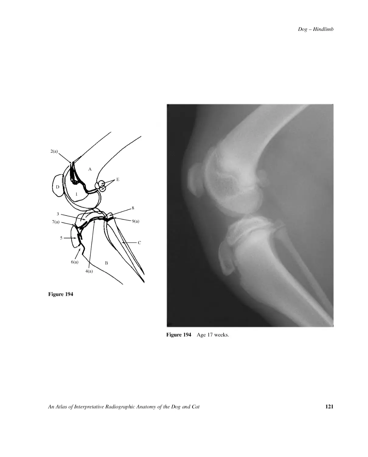
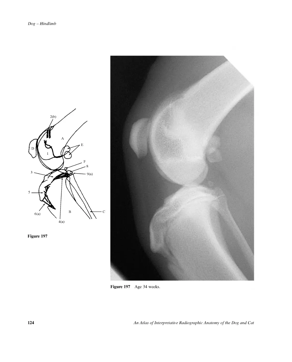
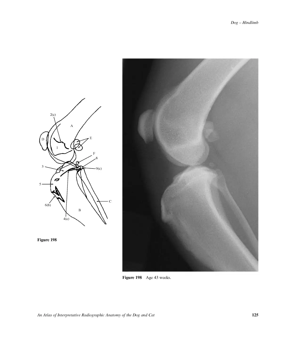
118AnAtlasofInterpretativeRadiographicAnatomyoftheDogandCatDog--Hindlimb2(a)4(a)1389(a)CBAFigures191,192,193,194,195,196,197,198Mediolateralprojectionofstiflejoint.Samoyedcrossbreddog,entiremale,at4,8,13,17,21,25,34and43weeksofage.Figure191Figure191Age4weeks.AFemur1Distalepiphysis2Distalgrowthplate2(a)Open2(b)Closing2(c)RemnantBTibia3Proximalepiphysis4Proximalgrowthplate4(a)Open4(c)Remnant5Tibialtuberosity6Tibialtuberositygrowthplatetodiaphysis6(a)Open6(b)Closing7Tibialtuberositygrowthplatetoproximalepiphysis7(a)Open7(b)Closing7(c)RemnantCFibula8Proximalepiphysis9Proximalgrowthplate9(a)Open9(c)RemnantDPatellaEFabellaeofm.gastrocnemiusFFabellaofm.popliteus
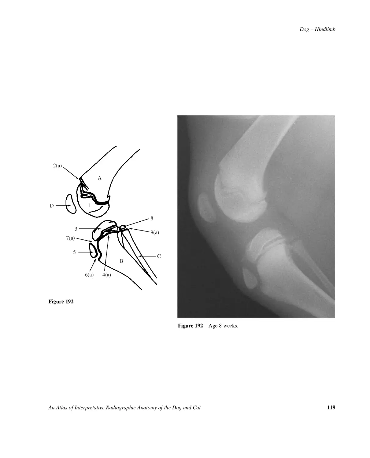
Dog--HindlimbAnAtlasofInterpretativeRadiographicAnatomyoftheDogandCat1192(a)4(a)6(a)1389(a)CBAD57(a)Figure192Figure192Age8weeks.
120AnAtlasofInterpretativeRadiographicAnatomyoftheDogandCatDog--Hindlimb2(a)4(a)6(a)1389(a)CBAD57(a)EFigure193Figure193Age13weeks.
Dog--HindlimbAnAtlasofInterpretativeRadiographicAnatomyoftheDogandCat1212(a)4(a)6(a)1389(a)CBAD57(a)EFigure194Figure194Age17weeks.
122AnAtlasofInterpretativeRadiographicAnatomyoftheDogandCatDog--Hindlimb2(a)4(a)6(a)1389(a)CBAD57(b)EFigure195Figure195Age21weeks.
Dog--HindlimbAnAtlasofInterpretativeRadiographicAnatomyoftheDogandCat1232(a)4(a)6(a)1389(a)CBAD57(c)EFFigure196Figure196Age25weeks.
124AnAtlasofInterpretativeRadiographicAnatomyoftheDogandCatDog--Hindlimb2(b)4(a)6(a)1389(a)CBAD5EFFigure197Figure197Age34weeks.
Dog--HindlimbAnAtlasofInterpretativeRadiographicAnatomyoftheDogandCat1252(c)4(c)6(b)1389(c)CBAD5EFFigure198Figure198Age43weeks.
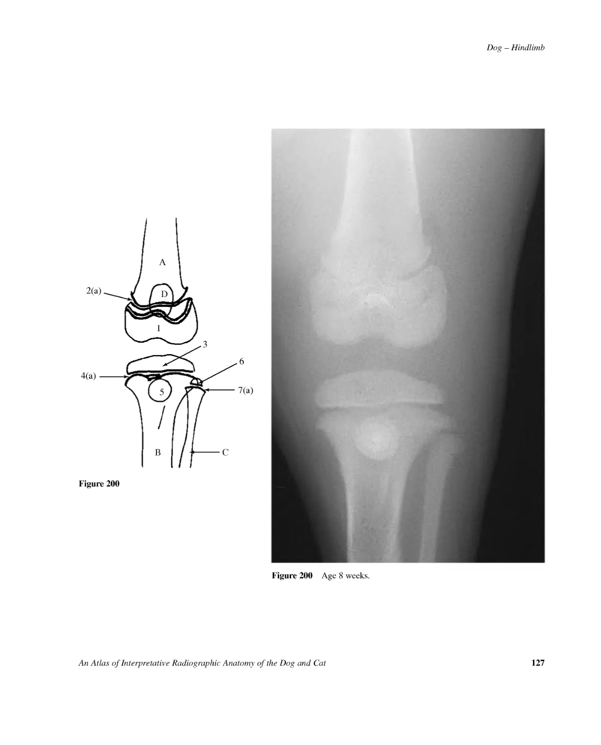
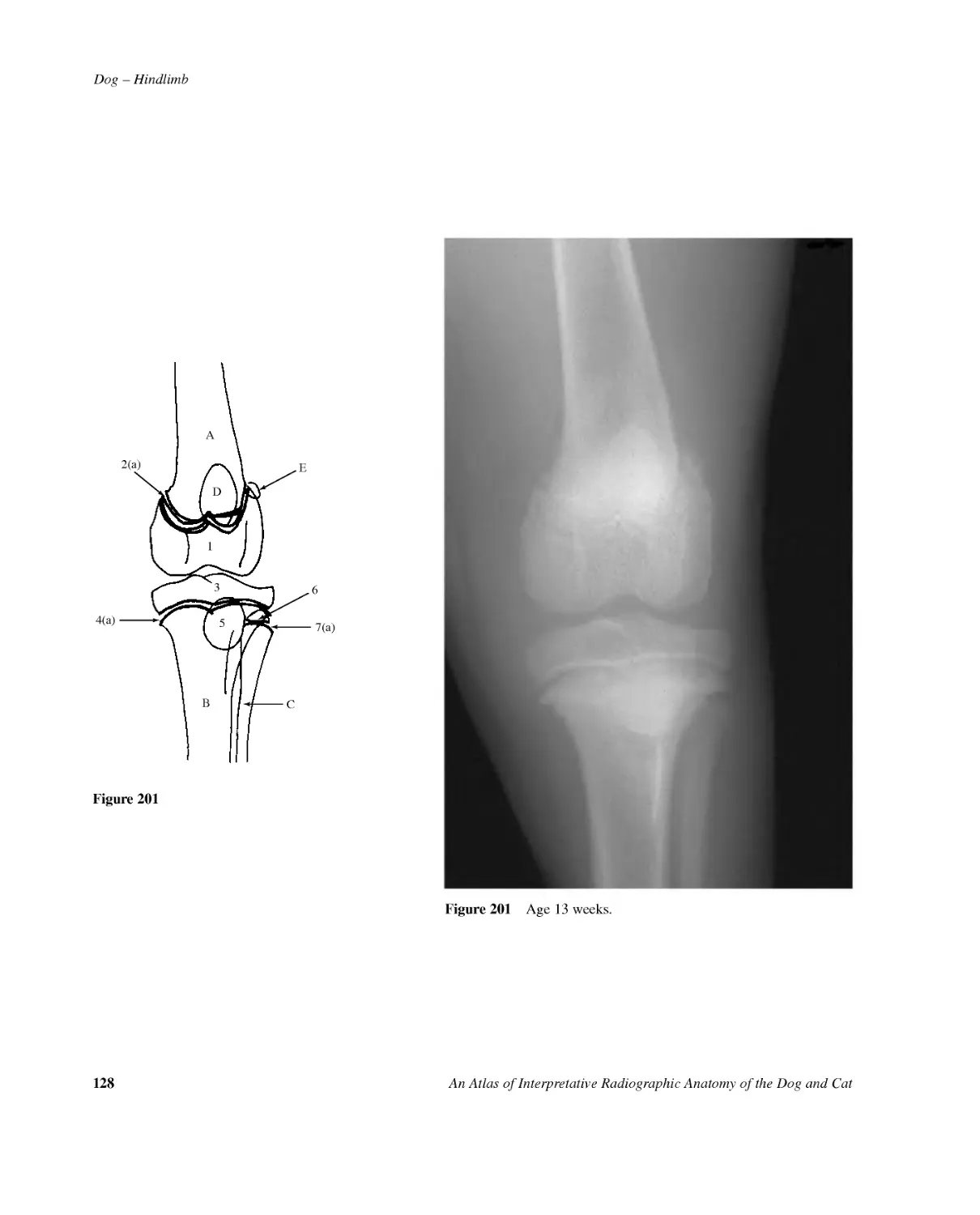
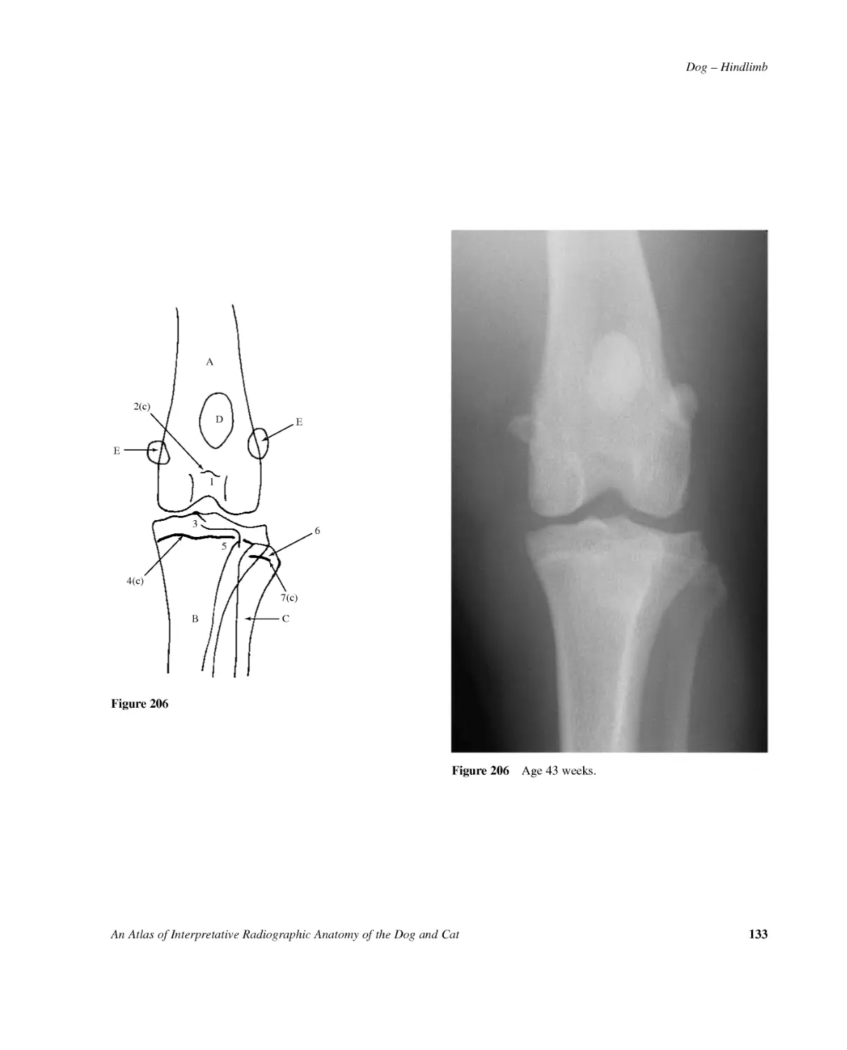
126AnAtlasofInterpretativeRadiographicAnatomyoftheDogandCatDog--Hindlimb2(a)4(a)1367(a)CBAFigures199,200,201,202,203,204,205,206Craniocaudalprojectionofstiflejoint.SamoyedCrossbreddog,entiremale,at4,8,13,17,21,25,34and43weeksofage.Figure199Figure199Age4weeks.AFemur1Distalepiphysis2Distalgrowthplate2(a)Open2(b)Closing2(c)RemnantBTibia3Proximalepiphysis4Proximalgrowthplate4(a)Open4(c)Remnant5TibialtuberosityCFibula6Proximalepiphysis7Proximalgrowthplate7(a)Open7(c)RemnantDPatellaEFabellaofm.gastrocnemiusFabellaofm.poplitealisnotvisibleonanyofthesefilms.
Dog--HindlimbAnAtlasofInterpretativeRadiographicAnatomyoftheDogandCat1272(a)15367(a)CBAD4(a)Figure200Figure200Age8weeks.
128AnAtlasofInterpretativeRadiographicAnatomyoftheDogandCatDog--Hindlimb2(a)4(a)15367(a)CBADEFigure201Figure201Age13weeks.
Dog--HindlimbAnAtlasofInterpretativeRadiographicAnatomyoftheDogandCat1292(a)4(a)15367(a)CBADEEFigure202Figure202Age17weeks.
130AnAtlasofInterpretativeRadiographicAnatomyoftheDogandCatDog--Hindlimb2(a)4(a)5367(a)CBADEE1Figure203Figure203Age21weeks.
Dog--HindlimbAnAtlasofInterpretativeRadiographicAnatomyoftheDogandCat1312(a)4(a)15367(a)CBADEEFigure204Figure204Age25weeks.
132AnAtlasofInterpretativeRadiographicAnatomyoftheDogandCatDog--Hindlimb2(b)4(a)15367(a)CBADEEFigure205Figure205Age34weeks.
Dog--HindlimbAnAtlasofInterpretativeRadiographicAnatomyoftheDogandCat1332(c)4(c)15367(c)CBADEEFigure206Figure206Age43weeks.
134AnAtlasofInterpretativeRadiographicAnatomyoftheDogandCatDog--HindlimbFigure207Age4weeks.AB4(a)2(a)16(a)589(a)3DCEFGFigures207,208,209,210,211,212Dorsoplantarprojectionoftarsus,metatarsalbonesandphalanges.SamoyedCrossbreddog,entiremale,at4,8,13,21,25and34weeksofage.Figure207ATibia1Distalepiphysis.Initiallywithseparateossificationcentreformedialmalleolus.2Distalgrowthplate2(a)Open2(b)Closing2(c)RemnantBFibula3Distalepiphysis4Distalgrowthplate4(a)Open4(b)ClosingCTarsus.Onlytibialtarsalbone,bodyoffibulartarsalbone,centraltarsalboneandtarsalbone4areseenat4weeksofage.DMetatarsalbone5(2,3and4aresimilar)5Epiphysis.Notethatthereisonlyadistalepiphysisinthesemetatarsalbones.6Growthplate6(a)Open6(b)Closing7ProximalsesamoidbonesEProximalphalanxofdigit5(2,3and4aresimilar)8Epiphysis.Notethatthereisonlyaproximalepiphysisintheproximalphalanges.9Growthplate9(a)Open9(b)ClosingFMiddlephalanxofdigit5(2,3and4aresimilar)10Epiphysis.Notethatthereisonlyaproximalepiphysisinthemiddlephalanges.11Growthplate11(a)OpenGDistalphalanxofdigit5(2,3and4aresimilar)HMetatarsalbone1
`Dog--HindlimbAnAtlasofInterpretativeRadiographicAnatomyoftheDogandCat135Figure208Age8weeks.AB4(a)2(a)16(a)589(a)3DCEFGH11(a)10Figure208
136AnAtlasofInterpretativeRadiographicAnatomyoftheDogandCatDog--HindlimbFigure209Age13weeks.AB4(a)2(a)6(a)589(a)3DCEFG1H711(a)10Figure209
Dog--HindlimbAnAtlasofInterpretativeRadiographicAnatomyoftheDogandCat137Figure210Age21weeks.AB4(a)2(a)6(a)589(b)3DCEFGIH7Figure210
138AnAtlasofInterpretativeRadiographicAnatomyoftheDogandCatDog--HindlimbFigure211Age25weeks.AB4(a)2(b)6(b)53DCEFGIH7Figure211
Dog--HindlimbAnAtlasofInterpretativeRadiographicAnatomyoftheDogandCat139Figure212Age34weeks.AB4(b)2(c)3DCEFGIH7Figure212
140AnAtlasofInterpretativeRadiographicAnatomyoftheDogandCatDog--HindlimbB52(a)4a16(a)DCEFGA78(a)3Figures213,214,215,216,217,218Mediolateralprojectionoftarsus,metatarsalbonesandphalanges.SamoyedCrossbreddog,entiremale,at4,8,13,21,25and34weeksofage.Figure213Figure213Age4weeks.ATibia1Distalepiphysis2Distalgrowthplate2(a)Open2(b)Closing2(c)RemnantBFibula3Distalepiphysis4Distalgrowthplate4(a)Open4(b)ClosingCTarsus.Onlytibialtarsalbone,bodyoffibulartarsalbone,centraltarsalboneandtarsalbone4areseenat4weeksofage.5Epiphysisoffibulartarsalbone6Fibulartarsalbonegrowthplate6(a)Open6(b)Closing6(c)RemnantDMetatarsalbone2or5(3and4aresimilar)7Epiphysis.Notethatthereisonlyadistalepiphysisinthesemetatarsalbones.8Growthplate8(a)Open8(b)Closing9ProximalsesamoidsEProximalphalanx2or5(3and4aresimilar)10Epiphysis.Notethatthereisonlyaproximalepiphysisintheproximalphalanges.11Growthplate11(a)Open11(b)ClosingFMiddlephalanx2or512Epiphysis.Notethatthereisonlyaproximalepiphysisinthemiddlephalanges.13Growthplate13(a)Open13(b)ClosingGDistalphalanx2or5HMetatarsalbone1IDorsalsesamoidbone
Dog--HindlimbAnAtlasofInterpretativeRadiographicAnatomyoftheDogandCat141B54(a)2(a)2(a)316(a)DCEFGA71011(a)13(a)128(a)9HFigure214Figure214Age8weeks.
142AnAtlasofInterpretativeRadiographicAnatomyoftheDogandCatDog--HindlimbB54(a)2(a)2(a)316(a)DCEFGA71011(a)13(a)128(a)9HFigure215Figure215Age13weeks.
Dog--HindlimbAnAtlasofInterpretativeRadiographicAnatomyoftheDogandCat14354(a)2(a)2(a)316(b)DEIFGAB71011(b)13(b)128(a)9HFigure216Figure216Age21weeks.
144AnAtlasofInterpretativeRadiographicAnatomyoftheDogandCatDog--Hindlimb54(a)2(b)2(b)316(b)DEIFGAB78(b)9HFigure217Figure217Age25weeks.
Dog--HindlimbAnAtlasofInterpretativeRadiographicAnatomyoftheDogandCat14554(b)2(c)316(c)DEIFAB9HGFigure218Figure218Age34weeks.
146AnAtlasofInterpretativeRadiographicAnatomyoftheDogandCatDog--SkullFigure219Lateralprojectionofskull.Beagledog2.5yearsold,entiremale.Figure220LinedrawingofphotographrepresentingradiographicpositioningforFigure219.
Dog--SkullAnAtlasofInterpretativeRadiographicAnatomyoftheDogandCat147122928272321222220323239(a)312938373936345967825252426262618343040CDE191511171415161213BA4110333523(b)23(a)Figure221Lateralprojectionofskullwithteethshadowsexcludedforclarity.AAtlasBStylohyoidbonesCEpihyoidbonesDCeratohyoidbonesEBasihyoidbone1Incisivebone2Nasalbone3Maxillaormaxillarybone4Frontalbone5Parietalbone6Occipitalbone7Externaloccipitalprotuberance8Externaloccipitalcrest9Interparietalprocessofoccipitalbone10Occipitalbone11Paracondylarprocessofoccipitalboneorjugularprocess12Petroustemporalboneortemporalbone;petrosalpart.13Tympanicbullaoftemporalboneortemporalbone;tympanicpart.14Externalacousticmeatusoftem-poralbone15Zygomaticprocessoftemporalboneortemporalbone;squamouspart.16Retroarticularprocessoftemporalbone17Mandibularfossaoftemporalbone18Massetericborderofzygomaticbone19Frontalprocessofzygomaticbone20Palatinebone21Vomer22Maxillarysinusofmaxilla23Cribriformplateofethmoidbone23(a)Rostrallimit23(b)Caudallimit24Ethmoturbinatesofethmoidbone25Orbitalmargin26Frontalsinuses.Totalofsix;threeoneachside.27Dorsalnasalconchaofethmoidbone28Ventralnasalconchaofmaxilla29Laminadura30Basisphenoidbone31Mandible32Mandibularbody33Mandibularramus34Coronoidprocessofmandible35Condyloidorarticularprocessofmandible36Angularprocessesofmandible37Mandibularforamen38Mentalforamen39Mandibularcanal39(a)Ventralborder40Softpalate41Nasopharynx
148AnAtlasofInterpretativeRadiographicAnatomyoftheDogandCatDog--Skull13425AABB678Figure222LateralprojectionofskulltodemonstratedetailsofteethexcludedinFigure221.1Upperincisors.Totalofsix.2Lowerincisors.Totalofsix.3Uppercanines.Totaloftwo.4Lowercanines.Totaloftwo.5Upperpremolars.Totalofeight.6Lowerpremolars.Totalofeight.7Uppermolars.Totaloffour.8Lowermolars.Totalofsix.AUppercarnassial;4th.premolarBLowercarnassial;1st.molarLinedrawingonlyillustratesgrossdetailsofteethasseeninradiograph,Figure219.Moredetaileddrawingsaregiveninthesectionondentition.
Dog--SkullAnAtlasofInterpretativeRadiographicAnatomyoftheDogandCat14923(a)45111010963871Figure223Linedrawingoflateralprojectionofskulltodemonstratesofttissueshadowsseeninradiograph,Figure219.Externalnose1Nostril2Nasalvestibule3Alarnasalfold3(a)Bulbousterminalenlargement4Dorsolateralnasalcartilage5Straightnasalfold6Dorsalnasalmeatus7Middlenasalmeatus8VentralnasalmeatusExternalear9Pinna10EarcanalsThesuperimposedshadows,9and10,oftheexternalearcanbeconfusedasbonyabnormalities.11Skinfoldlevelwithdorsalaspectoforbits
150AnAtlasofInterpretativeRadiographicAnatomyoftheDogandCatDog--SkullFigure224Ventrodorsalprojectionofskull.Beagledog2.5yearsold,entiremale(samedogasindorsoventralprojectionofskull,Figure230).
Dog--SkullAnAtlasofInterpretativeRadiographicAnatomyoftheDogandCat151CDABB42115106676(b)6(a)161213586(a)13171514181921222026252324FE6(b)9Figure226Ventrodorsalprojectionofskullwithteethshadowsexcludedforclarity.AAtlasBAxisHyoidapparatus.Thebonyshadowsareindis-tinctbutallarevisibleexceptforthebasihyoid.CStylohyoidboneDEpihyoidboneECeratohyoidboneFThyrohyoidbone1Palatinefissure2Vomer3Maxillaryteethobscur-ingmaxillashadow4Laminadura5Ventralnasalconchaeandethmoturbinates6Frontalsinuses6(a)Frontalsinuses(lateral)6(b)Frontalsinuses(medial)7Cribriformplateofethmoidenclosingethmoidalfossa8Medialwalloforbit9Borderofchoanaeformedbypalatineboneandpterygoidbone.(Hamulusseenasthemoreopaqueshadowatcaudalextremity.)10Mandible11Mandibularbody12Mandibularramus13Coronoidprocessofmandible14Angularprocessofmandible15Condyloidorarticularprocessofmandible16Temporalprocessofzygomaticbone17Zygomaticprocessoftemporalbone16and17formthezygomaticarch18Retroarticularprocessoftemporalbone19Tympanicbullaoftemporalbone;tympanicpart20Externalacousticmea-tusoftemporalbone;tympanicpart21Mastoidprocessoftemporalbone;tympanicpart22Jugularforamenoftemporalbone;tympanicpart23Paracondylarprocessofoccipitalboneorjugularprocess24Occipitalcondyle25Externaloccipitalprotuberance26NuchalcrestofoccipitalboneFigure225Linedrawingofphotographrepresentingradio-graphicpositioningforFigure224.
152AnAtlasofInterpretativeRadiographicAnatomyoftheDogandCatDog--SkullFigure227LinedrawingtodemonstrateteethshadowsexcludedinFigure226.Ventrodorsalprojectionofskull.1Lowerincisorteeth.Totalofsix2Upperincisorteeth.Totalofsix.3Lowercanineteeth.Totaloftwo.4Uppercanineteeth.Totaloftwo.5Lowerpremolarteeth.Totalofeight.6Upperpremolarteeth.Totalofeight.7Lowermolarteeth.Totalofsix.8Uppermolarteeth.Totaloffour.(Inthisdog2nd.molaronleftsideismissing.)ALowercarnassial;1st.molarBUppercarnassial;4th.premolarThedrawingonlyillus-tratesgrossdetailsofteethasseeninradi-ograph.Moredetaileddrawingsaregiveninthesectionondentition.421334577AABB8625347223361Figure228Linedrawingofventrodorsalprojectionofskulltodemonstratesofttissueshadowsseeninradiograph,Figure2241Nostril2Caudallimitofpinna3Internalfoldsofpinna4Verticalearcanal5Horizontalearcanal6Externalacousticmeatus7Neckskinfold
Dog--SkullAnAtlasofInterpretativeRadiographicAnatomyoftheDogandCat153BCAFigure229Schematicdrawingofventrodorsalprojectionofskulltodemonstratethetemporomandibularjointsandmandibularsymphysis.A=CapsuleoftemporomandibularjointB=LateralligamentBetweenthecartilagecoveredbonyarticulationsisfoundanarticulardisc.C=Symphysisofmandible.(Fibrocartilageisseenasaradiolucentregionintheradiograph.)
154AnAtlasofInterpretativeRadiographicAnatomyoftheDogandCatDog--SkullFigure231LinedrawingofphotographrepresentingradiographicpositioningforFigure230.Figure230Dorsoventralprojectionofskull.Beagledog2.5yearsold,entiremale(samedogasinventrodorsalprojectionofskull,Figure224).
Dog--SkullAnAtlasofInterpretativeRadiographicAnatomyoftheDogandCat155Figure233LinedrawingofphotographrepresentingradiographicpositioningforFigure232.1214112231311676AB5948101048715Figure234Rostrocaudaloblique(45degreenosetilt)projectionoftemporomandibularjoints.ATemporomandibularjointofrecumbentsideBPositionoftemporo-mandibularjointofnon-recumbentsideobscuredbypetroustemporalbones1Mandibularbody2Angularprocessofmandible3Condyloidorarticularprocessofmandible4Coronoidprocessofmandible5Mandibularfossaoftemporalbone6Retroarticularprocessoftemporalbone7Tympanicbullaoftemporalbone8Externalacousticmeatusoftemporalbone9Basisphenoidbone10Temporalprocessofzygomaticbone11Condyleofoccipitalbone12Atlas13Axis14Stylohoidbone15Epihyoidbone16CeratohyoidboneFigure232Rostrocaudaloblique(45degreenosetilt)projectionoftemporomandibularjoints.Dobermandog7yearsold,entiremale.
156AnAtlasofInterpretativeRadiographicAnatomyoftheDogandCatDog--SkullFigure235Schematicdrawingofrostrocaudaloblique(45degreenosetilt)projectionoftemporomandibularjointstodemonstratejointcapsuleanddisc.=Jointcapsule=Articulardisc.Dividesjointintoventralanddorsalcompartments.Laterallythejointcapsuleisstrengthenedtoformafibrouslateralligament.
Dog--SkullAnAtlasofInterpretativeRadiographicAnatomyoftheDogandCat157Figure237LinedrawingofphotographrepresentingradiographicpostioningforFigure236.19121(a)311221819201413131516262910103754181721Figure238Relaxedlateral(openmouth)projectionoftympanicbullae.1Tympanicbullaoftemporalbone1(a)Recumbentside(shadedindrawing)2Externalacousticmeatusoftemporalbone3Zygomaticprocessoftemporalbone4Temporozygomaticsuture5Externalprotuberanceofoccipitalbone6Externaloccipitalcrest7Occipitalcondyle8Paracondylarprocessofoccipitalboneorjugularprocess9Atlas10Axis11Mandible12Angularprocessofmandible13Coronoidprocessofmandible14Condyloidorarticularprocessofmandible15MandibularfossaoftemporalboneNotethat14and15aresuperimposed,hencethetemporomandibularjointisnotdemonstrated.16RetroarticularprocessoftemporalboneHyoidapparatus17Tympanohyoidcartilage(seenasaradiolucentregionbetweentympanicbullaandstylohyoidbone)18Stylohyoidbone19Epihyoidbone20Ceratohyoidbone21Basihyoidbone22ThyrohyoidboneFigure236Relaxedlateral(openmouth)projectionoftympanicbullae.Beagledog2.5yearsold,entiremale.
158AnAtlasofInterpretativeRadiographicAnatomyoftheDogandCatDog--SkullFigure239Rostroventral-caudodorsaloblique(openmouth)projectionoftympanicbullae.Beagledog2.5yearsold,entiremale.Figure240LinedrawingofphotographrepresentingradiographicpositioningforFigure239.
Dog--SkullAnAtlasofInterpretativeRadiographicAnatomyoftheDogandCat159125781311111214978141815161710(b)43623222110(b)10(a)10192010Figure241Rostroventral--caudodorsaloblique(openmouth)projectionoftympanicbullae.1Temporalprocessofzygomaticbone2Zygomaticprocessoftemporalbone3Mandibularbody4Angularprocessofmandible5Condyloidorarticularprocessofmandible6Verticalramusofmandible7Retroarticularprocessoftemporalbone8Temporomandibulararticulation9Petroustemporalbone(seenasaradiopaqueshadow)10Tympanicbullaoftemporalbone(shadedindrawing)10(a)Externalacousticmeatus(characteristic'lip'atbor-derofmeatuswhichbecomeslargerwithage)10(b)Shadowformedbyhorizontalearcanal11Canalofauditorytube12Foramenmagnum13Caudaledgeofpalatinebonesorhardpalate14Condyleofoccipitalbone15Atlas16Axis17Spinousprocessofaxis18Densorodontoidpegofaxis19Stylohyoidbone20Epihyoidbone21Ceratohyoidbone22Basihyoidbone23Epiglottis
160AnAtlasofInterpretativeRadiographicAnatomyoftheDogandCatDog--SkullFigure243LinedrawingofphotographrepresentingradiographicpositioningforFigure242.Figure242Rostroventral-caudodorsaloblique(openmouth)projectionofdensorodontoidpeg.Beagledog2.5yearsold,entiremale.
Dog--SkullAnAtlasofInterpretativeRadiographicAnatomyoftheDogandCat16137758669(b)5419(b)9(a)18192021917213121614221510(b)10(a)10119(a)Figure244Rostroventral--caudodorsaloblique(openmouth)projectionofdensorodontoidpeg.1Zygomaticprocessoftemporalbone2Mandibularramus3Coronoidprocessofmandible4Angularprocessofmandible5Condyloidorarticularprocessofmandible6Temporomandibulararticulation7Uppermolar8Petroustemporalbone9Tympanicbullaoftemporalbone9(a)Externalacousticmeatus(Characteristic'lip'atborderofmeatus)9(b)Shadowformedbyhorizontalearcanal10Foramenmagnum10(a)Dorsalpart10(b)Ventralpart11Caudalborderofpalatinebonesorhardpalate12Condyleofoccipitalbone13Atlas14Axis15Spinousprocessofaxis16Densorodontoidpegofaxis17Stylohoidbone18Epihyoidbone19Ceratohyoidbone20Thyrohoidbone21Basihyoidbone22Epiglottis
162AnAtlasofInterpretativeRadiographicAnatomyoftheDogandCatDog--Skull45°Figure246LinedrawingofphotographrepresentingradiographicpositioningforFigure245.Figure245Caudodorsal-rostroventralobliqueprojectionofforamenmagnum.Beagledog2.5yearsold,entiremale.
Dog--SkullAnAtlasofInterpretativeRadiographicAnatomyoftheDogandCat1631569213411577148131012Figure247Caudodorsal--rostroventralobliqueprojectionofforamenmagnum.1Parietalbone2Lateralfrontalsinus3Septumoffrontalsinuses4Medialandrostralfrontalsinuses5Foramenmagnum6Zygomaticprocessoffrontalbone7Petroustemporalbone8Tympanicbullaoftemporalbone9Zygomaticprocessoftemporalbone10Osseousnasalseptum11Ethmoturbinates12Coronoidprocessofmandible13Condyloidorarticularprocessofmandible14Marginoforbit15Maxillaormaxillarybone
164AnAtlasofInterpretativeRadiographicAnatomyoftheDogandCatDog--SkullFigure249LinedrawingofphotographrepresentingradiographicpositioningforFigure248.Figure248Rostrocaudalprojectionoffrontalsinuses.Beagledog2.5yearsold,entiremale.
Dog--SkullAnAtlasofInterpretativeRadiographicAnatomyoftheDogandCat1651267125131839101186(a)6(b)1716151419204Figure250Rostrocaudalprojectionoffrontalsinuses.1Frontalbone.Theinnerandoutertablesareseparatedbythelateralfrontalsinus.2Medialsurfaceoffrontalbone.Theseptumbetweenthefrontalsinuses.3Lateralsurfaceoffrontalbone;orbitalpart.4Zygomaticprocessoffrontalbone5Septumbetweenfrontalsinuses.Formedbynasalpartsofthefrontalbonesandseptalprocessesofthenasalbones.6Frontalsinuses(shadedindrawing).Usuallythreeseparatecompartmentsoneachside.Compartmentsaredistinctsinuseswiththeirownopeningsbetweenethmoturbinatesintonasalcavities.6(a)Rostralfrontalsinusanditsassociatedethmo-turbinates.Frontalsinusesextendmorerostrallyatmedialaspectofthisregionthanatlateralaspect.6(b)Frontalsinuscontainingethmoturbinates7Parietalbone8Squamouspartoftemporalbone9Zygomaticprocessoftemporalbone10Frontalprocessofzygomaticbone11Infraorbitalmarginofzygomaticbone12Osseousnasalseptum.Perpendicularplateoftheethmoid.Thecartilaginousnasalseptumisalsopartofethmoidboneandisformedbyarostralprolongationofosseousnasalseptum.Articulationiswiththevomerventrallyandseptalprocessesoffrontalandnasalbonesdorsally.Notethatthecartilaginousnasalseptumisradiolucent.13Ethmoturbinatesandturbinatesofventralnasalconcha14Dorsalnasalmeatus15Middlenasalmeatus16Conchalcrestofmaxilla17Ventralnasalmeatus18Vomer19Nasalbone20Maxillaormaxillarybone
166AnAtlasofInterpretativeRadiographicAnatomyoftheDogandCatDog--SkullFigure252Linedrawingofphotographrepresentingradi-ographicpositioningforFigure251.Figure251Dorsoventralintraoralprojectionofnasalchambers.Beagledog2.5yearsold,entiremale.
Dog--SkullAnAtlasofInterpretativeRadiographicAnatomyoftheDogandCat16715PPPPMMMLRPPPP24311IIIIICC11(a)6109987(a)78Figure253Dorsoventralintraoralprojectionofnasalchambers.1Interincisivesutureshowingincisivecanal2Incisivebonepalatineprocess3Vomer4Suturebetweenincisiveboneandvomer5Palatinefissure6Nasalconchae.(Onlyventralturbinatesseeninthisprojection.)7Ethmoturbinates.Rostralextentat2nd.and3rd.premolarlevel,withbulkofethmoturbinatesendingat3rd.and4th.level.Rostrallimitofethmoidalfossaat1st.molarlevel,alsothepositionofthecribriformplate(7(a)).8Maxillarysinusorrecessofmaxilla9Frontalsinuses10Shadowofexternalsurfaceoffacialbones.11Nasalbone11(a)RostrallimitAradiolucentshadowiscreatedwithinthenasalchambersbythelaterallimitsofthenasalandvomerbones.IUpperincisor.Totalofsix.CUppercanine.Totaloftwo.PUpperpremolar.Totalofeight.MUppermolar.Totaloffour.(Inthisdogleft2nd.molarismissing.)Notethattheterm'upper'canbereplacedby'superior'.11879562243Figure254Linedrawingofdorsoventralintraoralprojectionofnasalchamberstodemonstratesofttissueshadowsseeninradiograph,Figure251.1Vestibuleofmouth2Gum3Cheek4Lip5Nostril(shadedindrawing)6Nasalplane7Ventralnasalmeatus8Middleanddorsalnasalmeatus9Cartilaginousnasalseptum
168AnAtlasofInterpretativeRadiographicAnatomyoftheDogandCatDog--SkullFigure256LinedrawingofphotographrepresentingradiographicpositioningforFigure255.Figure255Ventrodorsaloblique(openmouth)projectionofnasalchambers.Samoyeddog6yearsold,entirefemale.
Dog--SkullAnAtlasofInterpretativeRadiographicAnatomyoftheDogandCat169123IIIIIICPP9C55(a)64PPM136(a)1410812MMMPPP11P7Figure257Ventrodorsaloblique(openmouth)projectionofnasalchambers.1Interincisivesutureshowingincisivecanal2Incisivebonepalatineprocess3Vomer4Palatinefissure5Nasalconchae.Nasalandmaxillaryturbinatebones.Theradiolucentshadowcreatedwithinthenasalchambersbythelaterallimitsofthenasalandvomerbonesisonlyjustvisibleontherightside(5(a)).6Ethmoturbinates6(a)Rostrallimitofethmoidfossa.Thisisalsothepositionofthecribriformplate.7Maxillarysinusorrecessofmaxilla8Frontalsinuses9Medialwalloforbit10Borderofchoanaeformedbypalatineboneandpterygoidbone(Hamulusseenastheopaquershadowatthecaudalextremity)11Temporalprocessofzygomaticbone12ZygomaticprocessoftemporalboneThezygomaticarchisformedby11and12.13Coronoidprocessofmandible14SofttissueshadowofthetongueIUpperincisor.Totalofsix.CUppercanine.Totaloftwo.PUpperpremolar.Totalofeight.MUppermolar.Totaloffour.Notethattheterm'upper'canbereplacedby'superior'.
170AnAtlasofInterpretativeRadiographicAnatomyoftheDogandCatDog--SkullFigure259LinedrawingofphotographrepresentingradiographicpositioningforFigure258.Figure258Ventrodorsaloblique(openmouth)projectionofmaxillarightlateralrecumbency.Beagledog2.5yearsold,entiremale.
Dog--SkullAnAtlasofInterpretativeRadiographicAnatomyoftheDogandCat171CIIPPPPPPPMMFP1M1M1M1M1M1M1IIICA678123GGABED54M1PCAFigure260Ventrodorsaloblique(openmouth)projectionofmaxillarightlateralrecumbency.Thedrawingandlabellinghavebeenkepttoaminimumtodemonstrateteethdetails.IUpperincisor.Totalofsix.CAUppercanine.Totaloftwo.PUpperpremolar.Totalofeight.MUppermolar.Totaloffour.(Inthisdogleft2nd.molarismissing.)P1Lowerpremolar.Totalofeight.M1Lowermolar.Totalofsix.Notethattheterm'upper'canbereplacedby'superior'and'lower'by'inferior'.Teethareanchoredinbonysocketsoralveoli.Paralleltoeachtoothrootisaregionofclearlydefinedbonyradiopacitycalledthelaminadura(A).Withagethealveolarbonehasradiopaquelinearchangesmakingthelaminaduralessobvi-ousinradiographs.Betweenthelaminaduraandthetoothrootisaradiolucentshadowoftheperiodontalmembrane(B).Asharpangleispresentatthejunctionofthelaminaduraandthealveolarcrest(C)adjacenttothedentalcemento-enameljunction.Withage,someverticalboneresorptioncanoccur,reducingthissharpangle.Anatomyofteeth1Neck2Crown3Tubercle4Root5Apexofroot6Dentine.Theperiodontalmembraneisattachedonitsdentalsurfacetoathinlayerofcement,butthisisnotradiographicallydistinguishablefromthedentine.7Pulpcavity.Inyoungdogsthisisverylarge,reducinginsizeuntil2to3yearsofageandthenslowlydecreasing.8EnamelDRostralormesialrootof3rd.premolar(upperright)EDistalrootof3rd.premolar(upperright)FLingualsurfacerootof1st.molar(upperright).Theapexofthisrootappearstohavesomeincreasedlucencywhichwouldindicateresorption.However,idiopathicresorptionofrootsinthisregionhasbeennotedinmaturedogs.GVestibularsurfacerootsof1st.molar(upperright)Inyoungdogs(under1yearofage)rootapicesarenormallyopen.Toavoidconfusionallrootshavenotbeenlabelled.The1st.premolarhasonlyoneroot,the2nd.and3rd.havetworoots(rostralanddistal)whilethe4th.upperpremolar,orcarnassialtooth,hasthreeroots.Ofthelattertworootsarelongandfoundonthevestibularsurface,andtheotherismuchshorteronthelingualsurface.The4th.lowerpremolarissimilartopremolars2and3.Consideringthemolars,theupperhavetwosmallvestibu-larsurfacerootsandonelargerlingualsurfaceroots.Thelowermolarsallhavetworootsbutthe1st.isverylargeandformsthelowercarnassialtooth.
172AnAtlasofInterpretativeRadiographicAnatomyoftheDogandCatDog--SkullFigure262LinedrawingofphotographrepresentingradiographicpositioningforFigure261.Figure261Dorsoventraloblique(openmouth)projectionofmandiblerightlateralrecumbency.Beagledog2.5yearsold,entiremale.
Dog--SkullAnAtlasofInterpretativeRadiographicAnatomyoftheDogandCat173IIICACAII1PPPPPPMMMM1M1M1P1P1P1MMMPP378C456BA2Figure263Dorsoventraloblique(openmouth)projectionofmandiblerightlateralrecumbency.Thedrawingandlabellinghavebeenkepttoaminimumtodemonstrateteethdetails.ILowerincisor.Totalofsix.CALowercanine.Totaloftwo.PLowerpremolar.Totalofeight.MLowermolars.Totalofsix.P1Upperpremolar.Totalofeight.M1Uppermolar.Totaloffour.(Inthisdogleft2nd.molarismissing.)Notethattheterm'lower'canbereplacedby'inferior'and'upper'by'superior'.ALaminaduraBPeriodontalmembraneCCementoenameljunctionPleaserefertoventrodorsaloblique(openmouth)projectionofmaxilla,Figure260,fordetailsofA,BandC.Anatomyofteeth1Neck2Crown3Tubercle4Root5Apexofroot6Dentine7Pulpcavity8EnamelPleaserefertoventrodorsaloblique(openmouth)projec-tionofmaxilla,Figure260,fordetailsof1to8.
174AnAtlasofInterpretativeRadiographicAnatomyoftheDogandCatDog--SkullFigure265Linedrawingofphotographrepresent-ingradiographicpositioningforFigure264.Figure264Dorsoventralintraoralprojectionofmaxillarybones.Beagledog2.5yearsold,entiremale.
Dog--SkullAnAtlasofInterpretativeRadiographicAnatomyoftheDogandCat1757C63910PPPMM38PP8C110P2I54IIIFigure266Dorsoventralintraoralprojectionofmaxillarybones.Thedrawingandlabellingaretoillustrateteethdetails.IUpperincisors.Totalofsix.CUppercanines.Totaloftwo.PUpperpremolars.Totalofeight.MUppermolars.Totaloffour.(Inthisdogleft2nd.molarismissing)Notethattheterm'upper'canbereplacedby'superior'.Anatomyofteeth1Crown2Tubercle3Root4Apexofroot5Dentine6Pulpcavity7EnamelAnatomyofalveoli8Alveolarcrest9Laminadura10Periodontalmembrane(seenasaradiolucentlinebetweenthelaminaduraandtoothroot)Pleaserefertoventrodorsaloblique(openmouth)projec-tionofmaxilla,Figure260,fordetailsof1to10.
176AnAtlasofInterpretativeRadiographicAnatomyoftheDogandCatDog--SkullFigure268LinedrawingofphotographrepresentingradiographicpositioningforFigure267.Figure267Ventrodorsalintraoralprojectionofmandibularbodies.Beagledog2.5yearsold,entiremale.
Dog--SkullAnAtlasofInterpretativeRadiographicAnatomyoftheDogandCat177IIC181011943576PP12P2M10MFigure269Ventrodorsalintraoralprojectionofmandibularbodies.Theteethhavebeenexcludedfromthedrawingoftherightmandiblesothatbonyfeaturesaremoreeasilyidentified.ILowerincisors.Totalofsix.CLowercanine.Totaloftwo.PLowerpremolars.Totalofeight.MLowermolars.Totalofsix.Notethattheterm'lower'canbereplacedby'inferior'.Anatomyofteeth1Crown2Tubercle3Root4Apexofroot5Dentine6Pulpcavity7EnamelAnatomyofalveoli8Alveolarcrest9Bonysocketsoralveoli10Laminadura11Periodontalmembrane(seenasaradiolucentlinebetweenthelaminaduraandtoothroot)Pleaserefertoventrodorsaloblique(openmouth)projec-tionofmaxilla,Figure260,fordetailsof1to11.12Mentalforamen
178AnAtlasofInterpretativeRadiographicAnatomyoftheDogandCatDog--SkullFigure270Lateralprojectionofskull.Brachycephalicbreedofdog.Pugdog9monthsold,entiremale.Theradiographdemonstratestheshortnasalchambersofthebrachycephalicbreedofdog.InadditiontheextremedomeshapeofthecraniuminthisPugbreedhasresultedinareductionofthefrontalsinusesshadow.
Dog--SkullAnAtlasofInterpretativeRadiographicAnatomyoftheDogandCat179Figure271Lateralprojectionofskull.Brachycephalicbreedofdog.Bulldog18monthsold,entirefemale(samedogasindorsoventralprojectionofskull,Figure276).Theradiographdemonstratestheshortnasalchambersofthebrachycephalicbreed.Prognathismofthemandibleisalsopresent,aconditioncommonlyseeninthistypeofbreed.
180AnAtlasofInterpretativeRadiographicAnatomyoftheDogandCatDog--SkullFigure272Lateralprojectionofskull.Toybreedofdog.YorkshireTerrierdog2yearsold,entirefemale.Theradiographhasbeenincludedtoshowdomingofthecraniumintoybreedswiththeconsequentialreductionofthefrontalsinusesshadow.Thesinusesinthisparticulardogarealmostentirelylost.
Dog--SkullAnAtlasofInterpretativeRadiographicAnatomyoftheDogandCat181Figure273Lateralprojectionofskull.Dolichocephalicbreedofdog.Radiographincludesallskullbones.RoughColliedog7yearsold,entiremale.Figure274Lateralprojectionofskull.Dolichocephalicbreedofdog.Theprimarybeamhasbeenconeddownandcentredonthenasalchambers.RoughColliedog6yearsold,neuteredfemale.
182AnAtlasofInterpretativeRadiographicAnatomyoftheDogandCatDog--SkullFigure275Dorsoventralprojectionofskull.Brachycephalicbreedofdog.Pugdog9monthsold,entiremale.
Dog--SkullAnAtlasofInterpretativeRadiographicAnatomyoftheDogandCat183Figure276Dorsoventralprojectionofskull.Brachycephalicbreedofdog.Bulldog18monthsold,entirefemale(samedogasinlateralprojectionofskull,Figure271).Prognathismofthemandiblesispresent,aconditioncommonlyseeninthebrachycephalicbreedofdog.
184AnAtlasofInterpretativeRadiographicAnatomyoftheDogandCatDog--SkullFigure277Dorsoventralprojectionofskull.Toybreedofdog.YorkshireTerrierdog2yearsold,entirefemale.
Dog--SkullAnAtlasofInterpretativeRadiographicAnatomyoftheDogandCat185Figure278Dorsoventralprojectionofskull.Dolichocephalicbreedofdog.RoughColliedog7yearsold,entiremale.
186AnAtlasofInterpretativeRadiographicAnatomyoftheDogandCatDog--SkullFigure279Dorsoventralintraoralprojectionofnasalchambers.Brachycephalicbreedofdog.BostonTerrierdog20monthsold,entirefemale.Theradiographdemonstratestheseverereductioninthesizeofthenasalchambersfoundinthisbreedofdog.Theright4th.premolartoothiscongenitallyabsentinthisparticulardog.Suchanabnormalityisnotuncommoninthebrachycephalicbreedofdogwheretheupperpremolarteethareusuallyaffected.Lowermolarteethcanalsobecongenitallyabsent.
Dog--SkullAnAtlasofInterpretativeRadiographicAnatomyoftheDogandCat187Figure280Dorsoventralintraoralprojectionofnasalchambers.Brachycephalicbreedofdog.Boxerdog9.5yearsold,entiremale.Notethattheright3rd.premolarismissinginthisdog.Thiswasaresultofextractionandnotacongenitalabsence.ThereductionofnasalchambershadowsisnotasgreatasintheBostonTerrier(Figure279).
188AnAtlasofInterpretativeRadiographicAnatomyoftheDogandCatDog--SkullFigure281Dorsoventralintraoralprojectionofnasalchambers.Toybreedofdog.YorkshireTerrierdog2yearsold,entirefemale.
Dog--SkullAnAtlasofInterpretativeRadiographicAnatomyoftheDogandCat189Figure282Dorsoventralintraoralprojectionofnasalchambers.Dolichocephalicbreedofdog.Greyhounddog2yearsold,entiremale.
190AnAtlasofInterpretativeRadiographicAnatomyoftheDogandCatDog--SkullFigure283Caudodorsal--rostroventralobliqueprojectionofskull.Foramenmagnumvariantintoybreedofdog.YorkshireTerrierdog5yearsold,entirefemale.Theradiographdemonstratesadorsalextensionoftheforamenmagnum.Suchanextensionisverycommoninthetoybreedofdog,buthasbeentermed'occipitaldysplasia',andhenceanabnormality,bysomeauthorities.As'occipitaldysplasia'ithasbeenlinkedtocongenitalordevelopmentalneurologicaldysfunction.However,theappearanceoftheforamenmagnum,asseeninthisradiograph,canbefoundextensivelyinclinicallynormaldogsanditspresenceindogsexhibitingabnormalneurologicalsignsisnotconclusiveforadiagnosis.Hencegreatcaremustbetakenwiththeinterpretationofthisradiographicshadowasitoftenreflectsonlyavariantandotherconditionsmustbeeliminated.
Dog--SkullAnAtlasofInterpretativeRadiographicAnatomyoftheDogandCat191Figure284Dorsoventralintraoralprojectionofnasalchambers.Nasalseptumvariant.Samoyeddog6yearsold,entirefemale.Theradiographshowscurvatureofthenasalseptum,totheleftnasalchamber,atthemidchamberlevel.Thebonyshadowsdividingthetwonasalchamberscanbeseenmidlineandareintact.Positionalvariationofthenasalseptum,andthevomer(alsoseeFigure653,Siamesecat),aresometimesseenandanalysisofsurroundingshadowsmustbemadetoensurethatthedeviationisnotpartofadiseaseprocesssuchasneoplasia.Theleftandrightnasalchambersaresymmetricalinthisdog,i.e.arenormal.
192AnAtlasofInterpretativeRadiographicAnatomyoftheDogandCatDog--Skull12(a)2(b)Figure285aD--PM3D--PM4P--PM3P--PM4P--M1Figure285bFigures285,286,287,288Lateralprojectionofskull.Samoyedcrossbreddogentiremaleat13,25,38,and52weeksofage.Correlatinglinedrawingsfor13,25and38weeksofage.Figures285a,286a,287aDrawingstodemonstratefrontalsinusesandethmoidalregion.1Frontalsinuses(regionshaded)2Cribriformplate2(a)Caudallimit2(b)RostrallimitFigures285b,286b,287bDrawingstodemonstratelowerorinferiorteethwithinthecentralmandible.Deciduousteethseenat13weeksofageonly.D--PMPremolars3and4Permanentteethseenonlyasgermsat13weeksofageP--PMPremolars3and4P--MMolars1,2and3Notethenarrowingofpulpcavitiesinpermanentteeth,withage.Overlyingshadowsofcorrespondingteethinopposingmandibularboneareshownbydottedlines.Dentalformulaeforthedog:DeciduousteethI3C1PM3313PermanentteethI3C1PM4M23143
Dog--SkullAnAtlasofInterpretativeRadiographicAnatomyoftheDogandCat193Figure285Age13weeks
194AnAtlasofInterpretativeRadiographicAnatomyoftheDogandCatDog--Skull12(a)2(b)Figure286aP--PM3P--PM4P--M1P--M2P--M3Figure286b12(b)2(a)Figure287aP--PM3P--PM4P--M1P--M2P--M3Figure287b
Dog--SkullAnAtlasofInterpretativeRadiographicAnatomyoftheDogandCat195Figure286Age25weeksFigure287Age38weeks
196AnAtlasofInterpretativeRadiographicAnatomyoftheDogandCatDog--SkullFigure288Age52weeks
Dog--SkullAnAtlasofInterpretativeRadiographicAnatomyoftheDogandCat1973241Figure289Figure289Age13weeksFigures289,290,291,292Ventrodorsalprojectionofskull.Samoyedcrossbreddogentiremaleat13,25,38and52weeksofage.Correlatinglinedrawingsat13,25and38weeksofage.Drawingstodemonstratefrontalsinuses(rightside).1Frontalsinuses(regionshaded)2Cribriformplate3Mandible4Vomer(midline)
198AnAtlasofInterpretativeRadiographicAnatomyoftheDogandCatDog--Skull3124Figure290Figure290Age25weeks
Dog--SkullAnAtlasofInterpretativeRadiographicAnatomyoftheDogandCat1993124Figure291Figure291Age38weeksFigure292Age52weeks
200AnAtlasofInterpretativeRadiographicAnatomyoftheDogandCatDog--SkullFigure294Age38weeksFigure295Age52weeksFigures293,294,295Ventrodorsaloblique(openmouth)projectionofmaxilla.Samoyedcrossbreddogentiremaleat13,38and52weeksofage.Figure293Age13weeksD--ID--CD--PM2D--PM3D--PM4P--IP--CP--PM1P--PM2P--PM3P--PM4P--M1Figure293
Dog--SkullAnAtlasofInterpretativeRadiographicAnatomyoftheDogandCat201D--ID--CD--PM2P--IP--CP--PM1P--PM2P--PM3P--PM4P--M1D--PM3D--PM4Figure296Dorsoventraloblique(openmouth)projectionofmandible.Samoyedcross,dogentiremale,at13weeksofage.Correlatinglinedrawingsat13weeksofage.DeciduousteethD--IIncisorsD--CCanineD--PMPremolars2,3and4.NotenodeciduousPM1.PermanentteethP--IIncisorsP--CCanineP--PMPremolars1,2,3and4P--MMolar1(othermolarteethnoteasilyseenintheseprojections)Notethenarrowingofpulpcavitiesofpermanentteethwithage.
202AnAtlasofInterpretativeRadiographicAnatomyoftheDogandCatDog--VertebraeFigure298LinedrawingofphotographrepresentingradiographicpositioningforFigure297.Figure297Extendedlateralprojectionofcervicalvertebrae.Beagledog2.5yearsold,entiremale.
Dog--VertebraeAnAtlasofInterpretativeRadiographicAnatomyoftheDogandCat203A1541211171918CD152021E21(b)21(a)F1614GHIJLK6(b)B62(a)6(a)32871013149Figure299Extendedlateralprojectionofcervicalvertebrae.ASkull1Occipitalbone2Occipitalcondyle2(a)Rostraledge3TympanicbullaeBAtlas4Dorsalarch5Lateralvertebralforamen6Articularsurfaces6(a)Cranialarticularsurfaceorfovea6(b)Caudalarticularsurfaceorfovea7Wings.Transverseprocesses.8BodyCAxis9Densorodontoidpeg10Cranialarticularsurface11Caudalarticularsurface12Spinousprocess13TransverseprocessesD3rd.cervicalvertebraE4th.cervicalvertebraF5th.cervicalvertebraG6th.cervicalvertebraH7th.cervicalvertebra14Transverseprocesses15Cranialarticularsurfaceof4th.cervicalvertebra16Cranialarticularsurfaceof6th.cervicalvertebra17Caudalarticularsurfaceof3rd.cervicalvertebra18Caudalarticularsurfaceof5th.cervicalvertebra19Spinousprocess20Body21Vertebralforamen21(a)Dorsalmargin21(b)VentralmarginI1st.thoracicvertebraJ2nd.thoracicvertebraK1st.ribLScapula
204AnAtlasofInterpretativeRadiographicAnatomyoftheDogandCatDog--VertebraeFigure300Schematicdrawingofextendedlateralprojectionofcervicalvertebraetodemonstratetheextentofjointcapsulesofvertebrae.=Jointcapsule=Synovialspace.Withtheexceptionofthefirsttwocapsulesthisspaceissmallerthandrawn.Thefirsttwojointcapsulescommunicate;atlanto-occipitalwithatlantoaxial.
Dog--VertebraeAnAtlasofInterpretativeRadiographicAnatomyoftheDogandCat2051325476Figure301Schematicdrawingofextendedlateralprojectionofcervicalvertebraetodemonstratevertebralligaments.1=Lateralatlanto-occipitalligament.Extendstojugularprocessofoccipitalbone.2=Transverseatlantalligament.Connectsbothsidesofatlanticventralarchandsoservestoholddensagainstatlanticventralarch.3=Apicalligamentofdens.Inthreesections.Middlesectiontoventralpartofforamenmagnumwithtwolateralpillars.Lateralsectionsareheavierandattachtooccipitalbonemediallyatthecaudalpartsofoccipitalcondyles.4=Nuchalligament5=Yellowligament6=Dorsallongitudinalligament7=Ventrallongitudinalligament.Thinnerthandorsalligament.Intervertebraldiscsarenotshownbutarefoundbetweenthelongitudinalligamentsintheintervertebralspacesfrom2nd.to3rd.cervicalvertebraecaudally.Dorsalatlanto-occipitalandventralatlanto-occipitalmembranesarenotshownbutacttoreinforcetheatlanto-occipitaljointdorsallyandventrally.Dorsalatlantoaxialmembraneisnotshownbutitisthedorsalfibrouslayeroftheatlantoaxialjointcapsule.
206AnAtlasofInterpretativeRadiographicAnatomyoftheDogandCatDog--VertebraeFigure302Extendedlateralprojectionofcervicalvertebraeinclinicallynormaldog.Beagle2.5yearsold,entiremale(samedogasinFigures305and308).Figure303LinedrawingofphotographrepresentingradiographicpositioningforFigure302.
Dog--VertebraeAnAtlasofInterpretativeRadiographicAnatomyoftheDogandCat207AB56C4(a)1243D7911HHIIGF10E84(b)AAxisB3rd.cervicalvertebraC4th.cervicalvertebraD5th.cervicalvertebraE6th.cervicalvertebraF7th.cervicalvertebra1Spinousprocess.(Processon3rd.cervicalvertebrailldefined.)2Caudalarticularsurface3Cranialarticularsurface4Vertebralforamen4(a)Dorsalmargin4(b)Ventralmargin5Transverseprocesses6Body7Cranialventralmarginofforamen8CranialdorsalmarginofforamenDistancebetweennumbers7and8isthecranialsagittaldiameterofthevertebralforamen.9Caudalventralmarginofforamen10CaudaldorsalmarginofforamenDistancebetweennumbers9and10isthecaudalsagittaldiameterofthevertebralforamen.Sagittaldiametermeasurementsareusedtoevaluatepossiblestenosisofforamen.Notethatmeasurementsmustalwaysbecomparedcraniallytocranially,caudallytocaudally,ascranialdiametersareoftensmallerthancaudalonesinthenormaldog.Thisisespeciallytrueforthemorecaudalcervicalvertebrae.Evaluationoftherelationshipbetweenadjacentventralforamenmarginsisalsovaluable.Measuringanglesbetweenthemarginsisimportanttodeterminewhetherstenosisispre-sent.Wheregrossradiographicabnormalityispresentanobviousdorsalelevationofthecranialmarginwithrespecttothecaudalmarginisseen.Notethatchangesinlateralprojectionpositionsofthecervicalvertebraewillaltertherelationshipofallforaminamargins,asseenintheradiographsandlinedrawings,Figures305to310.11Dorsalextremityofintervertebraldiscspace.G1st.thoracicvertebraH1st.ribIScapulaFigure304Extendedlateralprojectionofcervicalvertebraeinclinicallynormaldog.
208AnAtlasofInterpretativeRadiographicAnatomyoftheDogandCatDog--VertebraeFigure305Hyperextendedlateralprojectionofcervicalvertebraeinclinicallynormaldog.Beagledog2.5yearsold,entiremale(samedogasinFigures302and308).Figure306LinedrawingofphotographrepresentingradiographicpositioningforFigure305.
Dog--VertebraeAnAtlasofInterpretativeRadiographicAnatomyoftheDogandCat209Figure307Hyperextendedlateralprojectionofcervicalvertebraeinclinicallynormaldog.Cranialandcaudalsagittaldiametersaresimilartotheextendedlateralprojectionofcervicalvertebraelinedrawing,Figure304.Cranialandcaudalcervicalventralforamenmarginanglesarethesameasintheextendedlateralprojectionofcervicalvertebraelinedrawing,Figure304.Therearenodorsalelevationsofcranialventralforamenmargins.
210AnAtlasofInterpretativeRadiographicAnatomyoftheDogandCatDog--VertebraeFigure309LinedrawingofphotographrepresentingradiographicpositioningforFigure308.Figure308Hyperflexedlateralprojectionofcervicalvertebraeinclinicallynormaldog.Beagledog2.5yearsold,entiremale(samedogasinFigures302and305).
Dog--VertebraeAnAtlasofInterpretativeRadiographicAnatomyoftheDogandCat211Figure310Hyperflexedlateralprojectionofcervicalvertebraeinclinicallynormaldog.Sagittaldiametersandventralforamenmarginanglesinextendedandhyperextendedlateralprojectionsofcervicalvertebraeweresimilar(seelinedrawingsFigures304and307).Amarkedchangeisnowseeninthehyperflexedlateralprojectionofcervicalvertebrae.Cranialandcaudalcervicalventralforamenmarginangleshaveincreased.Thereareobviousdorsalelevationsofcranialventralforamenmarginsat3rd.,4th.,and5th.cervicalvertebrae.Dorsalelevationsarelessobviousat6th.and7th.cervicalvertebrae.
212AnAtlasofInterpretativeRadiographicAnatomyoftheDogandCatDog--VertebraeFigure311Ventrodorsalprojectionofcervicalvertebrae.Beagledog2.5yearsold,entiremale.Figure312LinedrawingofphotographrepresentingradiographicpositioningforFigure311.
Dog--VertebraeAnAtlasofInterpretativeRadiographicAnatomyoftheDogandCat213A12BCDEFHIJGMLLK98525(b)5(a)4761112101316151214171713141819K3Figure313Ventrodorsalprojectionofcervicalvertebrae.ASkull1Squamouspartofoccipitalbone2OccipitalcondyleBAtlas3Cranialarticularfovea4Caudalarticularfovea5Dorsalarch5(a)Cranialborder5(b)Ventralarchcaudalborder6Wing;transverseprocess7TransverseforamenCAxis8Dens9Cranialarticularsurface10Caudalarticularsurface11Spinousprocess12TransverseprocessD3rd.cervicalvertebraE4th.cervicalvertebraF5th.cervicalvertebraG6th.cervicalvertebraH7th.cervicalvertebra13Cranialarticularsurface14Caudalarticularsurface15Spinousprocess16Transverseforamen17Lateralmarginofvertebralforamen18Dorsalcranialmarginofbody19VentralcaudalmarginofbodyI1st.thoracicvertebraJ1st.ribKClavicleLScapulaMHumerus
214AnAtlasofInterpretativeRadiographicAnatomyoftheDogandCatDog--VertebraeabFigure314Schematicdrawingofventrodorsalprojectionofcervicalvertebraetodemonstratejointcapsules.=Jointcapsuleandsynovialspaceofvertebralandcostalarticulations.Atlanto-occipitaljointcapsulecommunicatesventromediallywithatlantoaxialjointcapsule.a=Jointcapsuleofheadofribb=Jointcapsuleoftubercleofrib
Dog--VertebraeAnAtlasofInterpretativeRadiographicAnatomyoftheDogandCat2153156421781=Lateralatlanto-occipitalligament2=Dorsalatlantoaxialligament3=Apicalligamentofdens.Centrallyattachestoventralfora-menmagnum.Laterallyattachestooccipitalcondyles.4=Transverseatlantalligament.Passesdorsallyoverdens.Abursaseparatesligamentanddens.5=Positionsofdorsalandventrallongitudinalligaments6=Yellowligament.Laterallyextendsasfarasthejointcap-sulessurroundingarticularprocesses.Ventraltoligamentisepiduralspace.7=Nuchalligament.Ligamentisanatomicallyinthemidlinebutisillustratedtotherightofmidlinetodifferentiateitfrom(5).8=Supraspinousligament.Ligamentisanatomicallyinthemidlinebutisillustratedtotherightofthemidlinetodifferentiateitfrom(5).Ribligamentsandintervertebraldiscsnotshown.Figure315Schematicdrawingofventrodorsalprojectionofcervicalvertebraetodemonstratevertebralligaments.
216AnAtlasofInterpretativeRadiographicAnatomyoftheDogandCatDog--VertebraeFigure316Lateralprojectionofthoracicvertebrae.Beagledog2.5yearsold,entiremale.Figure317LinedrawingofphotographrepresentingradiographicpositioningforFigure316.
Dog--VertebraeAnAtlasofInterpretativeRadiographicAnatomyoftheDogandCat217ABCDEFGHIJKLM1(a)9NO16(a)6(a)6(b)572P81110346(b)6Figure318Lateralprojectionofthoracicvertebraewithribshadowsexcludedforclarity.A7th.cervicalvertebraB1st.thoracicvertebraC2nd.thoracicvertebraD3rd.thoracicvertebraE4th.thoracicvertebraF5th.thoracicvertebraG6th.thoracicvertebraH7ththoracicvertebraI8th.thoracicvertebraJ9th.thoracicvertebraK10th.thoracicvertebraL11th.thoracicvertebra.Anticlinalvertebra.M12th.thoracicvertebraN13th.thoracicvertebra1Spinousprocess1(a)Spinousprocessofanticlinalvertebra.(Nearlyperpendiculartoaxis)2Cranialarticularprocess3Caudalarticularprocess4Body5Transverseprocess.Includesmammillaryprocessfrom2nd.or3rd.thoracicvertebracaudally.6Vertebralforamen6(a)Dorsalmargin6(b)Ventralmargin7Intervertebralforamen8Firstvisibleaccessoryprocess9Mammillaryprocess10AccessoryprocessO1st.lumbarvertebra11TransverseprocessPCruraofdiaphragm
218AnAtlasofInterpretativeRadiographicAnatomyoftheDogandCatDog--VertebraeFigure319Schematicdrawingoflateralprojectionofthoracicvertebraetodemonstratetheextentofjointcapsules.=Jointcapsuleandsynovialspaceofvertebralarticulation=Jointcapsuleandsynovialspaceofcostovertebraljoint.Headofrib.=Jointcapsuleandsynovialspaceofcostovertebraljoint.Tubercleofrib.123564Figure320Schematicdrawingoflateralprojectionofthoracicvertebraetodemonstratevertebralligaments.1=Nuchalligament2=Supraspinousligament3=Yellowligament4=Dorsallongitudinalligament5=Ventrallongitudinalligament6=IntertransverseligamentInterspinousligamentsandintervertebraldiscsnotshown.
Dog--VertebraeAnAtlasofInterpretativeRadiographicAnatomyoftheDogandCat219ABBCCDDEEFFGGHHIIJKLLMMNNKO12345Figure321LateralprojectionofthoracicvertebraetodemonstratetheribshadowsnotseeninFigure318.AScapula1Spine2CaudalmarginsofrightandleftB1st.ribC2nd.ribD3rd.ribE4th.ribF5th.ribG6th.ribH7th.ribI8th.ribJ9th.ribK10th.ribL11th.ribM12th.ribN13th.rib3Head4Neck5Tubercles(rightandleft)OCruraofdiaphragm213Figure322Schematicdrawingoflateralprojectionofthoracicvertebraetodemonstrateribligaments.1=Ligamentofhead.Extendsfromheadofribtolateralpartofintervertebraldisc.Lastthreeribsaredisplacedandforthesetheligamentshiftstoattachtothebodiesofthevertebrae.2=Ligamentoftubercle.Isdistaltoarticularcapsule.Crossescapsuleandattachestotransverseprocess.3=Ligamentofneck.Extendsfromnecktoventralsurfaceoftransverseprocesspluslateralbodyofvertebra.Intercapitalligamentnotshownbutisseeninasimilarpositiontotheheadligament.Itrunsacrosstheventralvertebralcanal,attheleveloftheintervertebraldisc,toattachtotheheadoftheribonthecontralateralside.Itisabsentfrom1st.,12th.and13th.ribs.
220AnAtlasofInterpretativeRadiographicAnatomyoftheDogandCatDog--VertebraeFigure323Ventrodorsalprojectionofthoracicvertebrae.Beagledog2.5yearsold,entiremale.Figure324LinedrawingofphotographrepresentingradiographicpositioningforFigure323.
Dog--VertebraeAnAtlasofInterpretativeRadiographicAnatomyoftheDogandCat221ABC1DEFGHIJKLMNO5614169121513231241717111078QPFigure325Ventrodorsalprojectionofthoracicvertebraewithventralshadowsofribsexcludedforclarity.A7th.cervicalvertebraB1st.thoracicvertebraC2nd.thoracicvertebraD3rd.thoracicvertebraE4th.thoracicvertebraF5th.thoracicvertebraG6th.thoracicvertebraH7th.thoracicvertebraI8th.thoracicvertebraJ9th.thoracicvertebraK10th.thoracicvertebraL11th.thoracicvertebra.Anticlinalvertebra.M12th.thoracicvertebraN13th.thoracicvertebraO1st.lumbarvertebra1Spinousprocessof1st.thoracicvertebra.Theseprocessesinclinecaudallyfrom1st.to10th.thoracicvertebrae.2Spinousprocessof9th.thoracicvertebra3Spinousprocessof10th.thoracicvertebra4Spinousprocessof11th.thoracicvertebra5Cranialmarginofbody6Caudalmarginofbody7Cranialarticularmarginof13th.thoracicvertebra8Mammillaryprocess(seenasadistinctstructurejustcaudaltothearticularmargin)9Cranialarticularmarginfoundnearthemedianplanefromthoracicvertebrae2to1010Caudalarticularmarginofthoracicvertebra11Caudalarticularmarginfoundnearthemedianplanefromthoracicvertebrae1to912Transverseprocess13Costalfoveaoftransverseprocess14Body15Cranialcostalfovea16Caudalcostalfovea17LateralmarginofvertebralforamenP1st.ribQ13th.rib
222AnAtlasofInterpretativeRadiographicAnatomyoftheDogandCatDog--VertebraedecabFigure326Sketchdrawingofventrodorsalthoracicvertebraetodemonstrateanatomicallythejointarticulationsofvertebraeandribs.a=Costovertebraljoint.Headofrib.b=Costovertebraljoint.Tubercleofrib.c=Cranialandcaudalarticularfacetsofvertebraljointd=Transverseprocessofthoracicvertebrae=IntervertebraldiscspaceforvertebralbodiesOnly(a)and(e)areconsistentlyvisibleinventrodorsalprojectionofthoracicvertebrae,Figure323,from1st.to13th.thoracicvertebrae.Thetransverseprocesses(d)areveryunclearthrough-outthethoracicvertebraeinFigure323,somaking(b)impossibletosee.Thevertebraljoint(c)isjustseenfrom11th.to13th.thoracicvertebrae.12345Figure327Schematicdrawingofventrodorsalprojectionofthoracicvertebraetodemonstratevertebralligaments.1=Nuchalligament.Positionisanatomicallyinthemidlinebutitisdrawntotherightsidetodifferentiateitfrom(3).2=Supraspinousligament.Positionisanatomicallythemidlinebutitisdrawntotherightsidetodifferentiateitfrom(3).3=Positionofdorsalandventrallongitudinalligaments4=Yellowligament5=IntertransverseligamentRibligamentsandintervertebraldiscsnotshown.
Dog--VertebraeAnAtlasofInterpretativeRadiographicAnatomyoftheDogandCat223Figure328Lateralprojectionofthoracolumbarvertebrae.Beagledog2yearsold,entirefemale.Figure329LinedrawingofphotographrepresentingradiographicpositioningforFigure328.AFGHBC12349DE77(a)7(b)5866(a)Figure330Lateralprojectionofthoracolumbarvertebrae.A12th.thoracicvertebraB13th.thoracicvertebraC1st.lumbarvertebraD2nd.lumbarvertebraE3rd.lumbarvertebra1Spinousprocess2Mammillaryprocess3Cranialarticularprocess4Caudalarticularprocess5Body6Transverseprocess6(a)Base7Vertebralforamen7(a)Dorsalmargin7(b)Ventralmargin8Intervertebralforamen9AccessoryprocessF11th.ribG12th.ribH13th.rib
224AnAtlasofInterpretativeRadiographicAnatomyoftheDogandCatDog--VertebraeFigure332Linedrawingofphotographrepresentingradiographicposi-tioningforFigure331.FFGGAB7CDE468861235Figure333Ventrodorsalprojectionofthoracolumbarvertebrae.A12th.thoracicvertebraB13th.thoracicvertebraC1st.lumbarvertebraD2nd.lumbarvertebraE3rd.lumbarvertebra1Spinousprocess2Cranialmarginofbody3Caudalmarginofbody4Cranialarticularprocessincludingmammillaryprocess5Caudalarticularprocess6Transverseprocess7Costalfoveaofvertebralbodyof13th.thoracicvertebra8LateralmarginofvertebralforamenF12th.ribG13th.ribFigure331Ventrodorsalprojectionofthoracolumbarvertebrae.Beagledog2yearsold,entirefemale.
Dog--VertebraeAnAtlasofInterpretativeRadiographicAnatomyoftheDogandCat225Figure334Lateralprojectionoflumbarvertebrae.Beagledog2yearsold,entirefemale.Figure335LinedrawingofphotographrepresentingradiographicpositioningforFigure334.
226AnAtlasofInterpretativeRadiographicAnatomyoftheDogandCatDog--VertebraeBALLMMNN42(a)CD566(a)EF977(a)7(b)GHIJK23481Figure336Lateralprojectionoflumbarvertebrae.A12th.thoracicvertebraB13th.thoracicvertebraC1st.lumbarvertebraD2nd.lumbarvertebraE3rd.lumbarvertebraF4th.lumbarvertebraG5th.lumbarvertebraH6th.lumbarvertebraI7th.lumbarvertebra1Spinousprocess2Mammillaryprocess2(a)Bothprocessesseen3Cranialarticularprocess4Caudalarticularprocess5Body6Transverseprocess6(a)Base7Vertebralforamen7(a)Dorsalmargin7(b)Ventralmargin8Intervertebralforamen9AccessoryprocessJIliumKSacrumL13th.ribM12th.ribN11th.ribabFigure337Schematicdrawingoflateralprojectionoflumbarvertebraetodemonstratetheextentofjointcapsules.a=Jointcapsuleandsynovialspaceofvertebralarticulationb=Jointcapsuleandsynovialspaceofcostovertebraljoint.Headofrib.
Dog--VertebraeAnAtlasofInterpretativeRadiographicAnatomyoftheDogandCat22732415Figure338Schematicdrawingoflateralprojectionoflumbarvertebraetodemonstratevertebralligaments.1=Supraspinousligament.Extendsto3rd.coccygealvertebra.2=Yellowligament3=Dorsallongitudinalligament4=Ventrallongitudinalligament5=IntertransverseligamentIntervertebraldiscsinterspinousandribligamentsarenotshown.
228AnAtlasofInterpretativeRadiographicAnatomyoftheDogandCatDog--VertebraeFigure339Ventrodorsalprojectionoflumbarvertebrae.Beagledog2yearsold,entirefemale.Figure340LinedrawingofphotographrepresentingradiographicpositioningforFigure339.
Dog--VertebraeAnAtlasofInterpretativeRadiographicAnatomyoftheDogandCat229A12th.thoracicvertebraB13th.thoracicvertebraC1st.lumbarvertebraD2nd.lumbarvertebraE3rd.lumbarvertebraF4th.lumbarvertebraG5th.lumbarvertebraH6th.lumbarvertebraI7th.lumbarvertebra1Spinousprocess2Cranialmarginofbody3Caudalmarginofbody4Cranialarticularprocessincludingmammillaryprocess5Caudalarticularprocess6Transverseprocess(lateralaspectsofsomeareobscuredbysofttissueshadowsmakingtheirappearanceasym-metrical)7Costalfoveaofvertebralbodyof13th.thoracicvertebra8LateralmarginofvertebralforamenJIliumKSacrumL13th.ribM12th.ribABCDEFGHIJKMLML1236845687Figure341Ventrodorsalprojectionoflumbarvertebrae.
230AnAtlasofInterpretativeRadiographicAnatomyoftheDogandCatDog--VertebraebacFigure342Schematicdrawingofventrodorsalprojectionoflumbarvertebraetodemonstrateextentofjointcapsules.a=Jointcapsuleofvertebralarticulationb=Jointcapsuleofcostovertebralarticulation.Headofrib.c=Fibrocartilaginousjointofsacroiliacjoint
Dog--VertebraeAnAtlasofInterpretativeRadiographicAnatomyoftheDogandCat2312413Figure343Schematicdrawingofventrodorsalprojectionoflumbarvertebraetodemonstratevertebralligaments.1=Supraspinousligament.Showndrawntotherightofmidlinetoseparateitfromlongitudinalligaments(2).Trueanatomicalpositionismidline.2=Positionofdorsalandventrallongitudinalligaments.3=Yellowligament4=IntertransverseligamentRibligamentsandintervertebraldiscsnotshown.
232AnAtlasofInterpretativeRadiographicAnatomyoftheDogandCatDog--VertebraeFigure344Lateralprojectionoflumbosacralvertebrae.Beagledog2.5yearsold,entiremale.Figure345LinedrawingofphotographrepresentingradiographicpositioningforFigure344.
Dog--VertebraeAnAtlasofInterpretativeRadiographicAnatomyoftheDogandCat2331431410113551215161194ECB2786A26DA6th.lumbarvertebraB7th.lumbarvertebra1Spinousprocess2Body3Cranialarticularprocess4Caudalarticularprocess5Transverseprocesses6Vertebralforamen7IntervertebralforamenCSacrum8Sacralcanal9Intermediatesacralcrest10Mammillaryprocessofcranialarticularprocess11Wing12PromontoryDIlium13Dorsalmargins14Cranialmargins15Ventralmargins16SacroiliacarticulationECoccygealorcaudalvertebraFigure346Lateralprojectionoflumbosacralvertebrae.
234AnAtlasofInterpretativeRadiographicAnatomyoftheDogandCatDog--VertebraebaFigure347Schematicdrawingoflateralprojectionoflumbosacralvertebraetodemonstratejointcapsules.=Jointcapsuleofvertebralarticulation=JointcapsuleofsacroiliacarticulationaSynovialpartbFibrocartilaginouspart=Auricularsurfaceofsacroiliacjoint------54213Figure348Schematicdrawingoflateralprojectionoflumbosacralvertebraetodemonstratevertebralligaments.1=Dorsallongitudinalligament.Stopsat5th.coccygealvertebra.2=Ventrallongitudinalligament.Stopsatmidsacrallevel.3=Yellowligament4=Ventralsacroiliacligament5=Dorsalsacroiliacligament.Dorsalligamentisthickerthanventral.Figure349Ventrodorsalprojectionoflumbosacralvertebrae.Beagledog2yearsold,entirefemale.
Dog--VertebraeAnAtlasofInterpretativeRadiographicAnatomyoftheDogandCat235Figure350LinedrawingofphotographrepresentingradiographicpositioningforFigure349.1A753762356BD119813C1614121018EE17154Figure351Ventrodorsalprojectionoflumbosacralvertebrae.A6th.lumbarvertebraB7th.lumbarvertebra1Spinousprocess2Cranialmarginofbody3Caudalmarginofbody4Cranialarticularprocessincludingmammillaryprocess5Caudalarticularprocess6Transverseprocess7LateralmarginofvertebralforamenCSacrum8Mediansacralcrest9Intermediatesacralcrest10Lateralsacralcrest11Wing12Positionofsynovialjointbetweenbonyarticularmargins13Base14Cranialarticularprocess15Caudalarticularprocess16SacralforaminaDIliumECoccygealorcaudalvertebra17Cranialarticularprocess18Transverseprocess
236AnAtlasofInterpretativeRadiographicAnatomyoftheDogandCatDog--VertebraebaFigure352Schematicdrawingofventrodorsalprojectionoflumbosacralvertebraetodemonstratetheextentofjoints.=JointcapsuleofvertebralarticulationSacroiliacarticulationa=Synovialpartofsacroiliacjointcapsuleb=Fibrocartilaginouspartofsacroiliacjointcapsule1246(b)36(a)51=Positionofsupraspinousligament.Showndrawntotherightofthemidlinetoseparateitfromlongitudinalliga-ments(2).Trueanatomicalpositionismidline.2=Positionofdorsalandventrallongitudinalligaments3=Continuationofdorsallongitudinalligament4=Intertransverseligament5=Yellowligament6=Positionsofsacroiliacligaments6(a)Dorsalpositionshownonrightside6(b)VentralpositionshownonleftsideFigure353Schematicdrawingofventrodorsalprojectionoflumbosacralvertebraetodemonstratevertebralligaments.
Dog--VertebraeAnAtlasofInterpretativeRadiographicAnatomyoftheDogandCat237Figure354Hyperflexedlateralprojectionoflumbosacralvertebraeincludingproximalcoccygealvertebrae.Beagledog2.5yearsold,entiremale.Figure355LinedrawingofphotographrepresentingradiographicpositioningforFigure354.161110912202122231524D19DDBC8B741174(a)365A21418Figure356Hyperflexedlateralprojectionoflumbosacralvertebraeincludingproximalcoccygealvertebrae.A7th.lumbarvertebra1Spinousprocess2Body3Cranialarticularprocess4Caudalarticularprocess4(a)Processof6th.lumbarvertebra5Transverseprocesses6Vertebralforamen7IntervertebralforamenBSacrum8Sacralcanal9Intermediatesacralcrest10Mediansacralcrest(onlyjustvisible)11Cranialarticularprocess12Caudalarticularprocess13Wing14Promontory15Bodyof3rd.sacralsegmentCIlium16Dorsalmargins17Cranialmargins18Ventralmargins19SacroiliacarticulationDCoccygealorcaudalvertebra20Cranialarticularprocess21Caudalarticularprocess22Transverseprocesses23Body24Vertebralforamen
238AnAtlasofInterpretativeRadiographicAnatomyoftheDogandCatDog--VertebraeFigure357Lateralprojectionofcoccygealorcaudalvertebrae.Beagledog2yearsold,entirefemale.Figure358LinedrawingofphotographrepresentingradiographicpositioningforFigure357.
Dog--VertebraeAnAtlasofInterpretativeRadiographicAnatomyoftheDogandCat239ABCDEFGHIJKLMNOPQR912534810786A2nd.coccygealvertebraB3rd.coccygealvertebraC4th.coccygealvertebraD5th.coccygealvertebraE6th.coccygealvertebraF7th.coccygealvertebraG8th.coccygealvertebraH9th.coccygealvertebraI10th.coccygealvertebraJ11th.coccygealvertebraK12th.coccygealvertebraL13th.coccygealvertebraM14th.coccygealvertebraN15th.coccygealvertebraO16th.coccygealvertebraP17th.coccygealvertebraQ18th.coccygealvertebraR19th.coccygealvertebra1Cranialarticularprocess2CaudalarticularprocessBothprocessesgraduallydisappearsothatbythe12th.vertebratheyarenolongerpresent.3Mammillaryprocess4Transverseprocess5Body6Vertebralforamen.Lumengraduallybecomessmallersothatbythe6th.or7th.vertebraitisonlyagroove.7Haemalarches8Haemalprocess9Intervertebralforamen10PositionofintervertebraldiscoftheintervertebralsymphysisFigure359Lateralprojectionofcoccygealorcaudalvertebrae.
240AnAtlasofInterpretativeRadiographicAnatomyoftheDogandCatDog--VertebraeFigure360Ventrodorsalprojectionofcoccygealorcaudalvertebrae.Beagledog2yearsold,entirefemale.Figure361LinedrawingofphotographrepresentingradiographicpositioningforFigure360.
Dog--VertebraeAnAtlasofInterpretativeRadiographicAnatomyoftheDogandCat241ABCDEFGHIJKLMNOPQR12Figure362Ventrodorsalprojectionofcoccygealorcaudalvertebrae.A3rd.coccygealvertebraB4th.coccygealvertebraC5th.coccygealvertebraD6th.coccygealvertebraE7th.coccygealvertebraF8th.coccygealvertebraG9th.coccygealvertebraH10th.coccygealvertebraI11th.coccygealvertebraJ12th.coccygealvertebraK13th.coccygealvertebraL14th.coccygealvertebraM15th.coccygealvertebraN16th.coccygealvertebraO17th.coccygealvertebraP18th.coccygealvertebraQ19th.coccygealvertebraR20th.coccygealvertebra.Lastvertebra.1Transverseprocess2Positionofintervertebraldiscoftheintervertebralsymphysis
242AnAtlasofInterpretativeRadiographicAnatomyoftheDogandCatDog--VertebraeFigure363Extendedlateralprojectionofcervicalvertebrae.Toybreedofdog.YorkshireTerrierdog5yearsold,entirefemale(samedogasinFigures364and365).Figure364Hyperextendedlateralprojectionofcervicalvertebrae.Toybreedofdog.YorkshireTerrierdog5yearsold,entirefemale(samedogasinFigures363and365).
Dog--VertebraeAnAtlasofInterpretativeRadiographicAnatomyoftheDogandCat243Figure365Hyperflexedlateralprojectionofcervicalvertebrae.Toybreedofdog.YorkshireTerrierdog5yearsold,entirefemale(samedogasinFigures363and364).Thisseriesofthreeradiographshasbeenincludedtodemonstratethenormalrelationshipbetweenthe1st.and2nd.cervicalvertebraeduringextremeflexionandextensionoftheneck.Notethedorsalspacebetweenthe1st.and2nd.cervicalvertebrae,oratlasandaxis,whichshowslittlevariationinthethreeprojections.Alsoobservethedorsalbordersofthefirsttwovertebraetobeparalleltooneanother.Thedensoftheaxisremainsinaventralpositionwithinthevertebralcanalinalltheprojections.Theatlas,axisandoccipitalcondyleoftheskullareallofanormalsizeandconformation.Analysisoftheseanatomicalchar-acteristicsisveryimportantforthisparticularregionasabnormalitiesofthisarea,intoybreeds,arewellrecognised.Theatlasandthedensoftheaxisarenotreducedinsize.Theoccipitalcondylecanbeseentobeanormalsizeandinthecorrectarticularposition.Figure366Lateralprojectionoflumbarvertebrae.Toybreedofdog.YorkshireTerrierdog1yearold,entiremale.Notetheappearanceofthebodiesofthelumbarvertebrae.Theyarecomparativelylongforadogandcouldbemistakenforcatlumbarvertebrae.
244AnAtlasofInterpretativeRadiographicAnatomyoftheDogandCatDog--VertebraeFigure367Lateralprojectionoflumbarvertebrae.Chondrodystrophicbreedofdog.StandardDachshunddog7yearsold,entiremale.Figure368Lateralprojectionofsacrumandcoccygealvertebrae.Corkscrewtail.Bulldog1yearold,entirefemale.Theradiographshowsthespiralcurvatureofthecorkscrewtailformedbythecongenitallyabnormalcoccygealvertebrae.Distallythereisfusionofthebodies(blockvertebrae)asseenbythelonglengthofthesevertebrae.Proximallythe1st.and2nd.coccygealvertebraearecongenitallymalformedhavingwedge-shapedbodies(hemivertebrae).Incompletefusionofthe3rd.sacralsegmentisalsopresent.
Dog--VertebraeAnAtlasofInterpretativeRadiographicAnatomyoftheDogandCat245Figure369Ventrodorsalprojectionofsacrumandcoccygealvertebrae.Corkscrewtail.Bulldog1yearold,entirefemale.Theradiographshowsthespiralcurvatureofthecorkscrewtailasformedbythecongenitallydeformedcoccygealvertebrae.Distallythevertebralbodiesarefused(blockvertebrae),whileproximallythebodiesaremuchshorterandbroader.Thebodyofthe2nd.coccygealvertebrahasawedgeshape(hemivertebra).Incompletefusionofthe3rd.sacralsegmentisalsoseen.ThisparticularBulldoghasseverehipdysplasiawithsecondaryjointdegeneration(seeFigure145fornormalchondrodys-trophicbreedofdoghipjoints).
246AnAtlasofInterpretativeRadiographicAnatomyoftheDogandCatDog--VertebraeFigure370Age13weeks.ABC1112(a)10(a)978(a)6(a)12(a)34(a)5Figure370Figures370,371,372,373Lateralprojectionofcervicalvertebrae.Samoyedcrossbreddogentiremaleat13,25,38,and52weeksofage.Correlatinglinedrawingsforallagesexcept52weeks.AAtlas;1st.cervicalvertebraBAxis;2nd.cervicalvertebra1Proatlas.Centrumforapexofdens2Intercentrum2(a)Open3Centrumfordens4Intercentrum4(a)Open5Centrumforcranialbodyequivalenttocranialepiphysis6Cranialgrowthplate6(a)Open7Caudalepiphysis8Caudalgrowthplate8(a)Open8(b)ClosingC3rd.cervicalvertebra9Cranialepiphysis10Cranialgrowthplate10(a)Open10(b)Closing11Caudalepiphysis12Caudalgrowthplate12(a)Open12(b)ClosingForclaritytransverseprocessshadowshavebeenexcludedfromalldrawings.
Dog--VertebraeAnAtlasofInterpretativeRadiographicAnatomyoftheDogandCat247Figure371Age25weeks.BAC4(a)12(a)1110(a)978(a)Figure371
248AnAtlasofInterpretativeRadiographicAnatomyoftheDogandCatDog--VertebraeFigure372Age38weeks.BA10(b)978(b)C12(b)11Figure372
Dog--VertebraeAnAtlasofInterpretativeRadiographicAnatomyoftheDogandCat249Figure373Age52weeks.
250AnAtlasofInterpretativeRadiographicAnatomyoftheDogandCatDog--VertebraeFigure374Age13weeks.312(a)4(a)ABCFigure374Figures374,375,376,377Lateralprojectionofthoracicvertebrae.Samoyedcrossbreddogentiremaleat13,25,38and52weeksofage.Correlatinglinedrawingsforallagesexcept52weeks.A2nd.thoracicvertebraB3rd.thoracicvertebra1Cranialepiphysis2Cranialgrowthplate2(a)Open3Caudalepiphysis4Caudalgrowthplate4(a)Open4(b)ClosingC4th.thoracicvertebraForclarityribshadowshavebeenexcludedfromalldrawings.
Dog--VertebraeAnAtlasofInterpretativeRadiographicAnatomyoftheDogandCat251Figure375Age25weeks.312(a)4(a)ABCFigure375
252AnAtlasofInterpretativeRadiographicAnatomyoftheDogandCatDog--VertebraeFigure376Age38weeks.Figure376
Dog--VertebraeAnAtlasofInterpretativeRadiographicAnatomyoftheDogandCat253Figure377Age52weeks.
254AnAtlasofInterpretativeRadiographicAnatomyoftheDogandCatDog--VertebraeFigure378Age13weeks.312(a)4(a)ABCFigure378Figures378,379,380,381Lateralprojectionoflumbarvertebrae.Samoyedcrossbreddogentiremaleat13,25,38and52weeksofage.Correlatinglinedrawingsforallagesexcept52weeks.A3rd.lumbarvertebraB4th.lumbarvertebra1Cranialepiphysis2Cranialgrowthplate2(a)Open3Caudalepiphysis4Caudalgrowthplate4(a)Open4(b)ClosingC5th.lumbarvertebraForclaritytransverseprocessshadowshavebeenexcludedfromalldrawings.At25weeksofagethe3rd.sacralsegment,orsacralvertebra,canbeseenasaseparatesegment.By38weeksofagefusionofthe3rd.sacralsegmenthastakenplace.At25,38and52weeksofagetheiliaccrestofthepelviscanbeseenasaseparateossificationcentre.Thisossificationcentrewasnotvisibleontheventrodorsalprojectionsofthepelvis.
Dog--VertebraeAnAtlasofInterpretativeRadiographicAnatomyoftheDogandCat255Figure379Age25weeks.312(a)4(a)ABCFigure379
256AnAtlasofInterpretativeRadiographicAnatomyoftheDogandCatDog--VertebraeFigure380Age38weeks.34(b)ABCFigure380
Dog--VertebraeAnAtlasofInterpretativeRadiographicAnatomyoftheDogandCat257Figure381Age52weeks.Figure382Lateralprojectionofcervicalvertebrae.Weimaranerdog7weeksold,entirefemale.Theradiographhasbeenincludedtodemonstratetheappearanceoftheatlasandaxisafter1weekofagebutbefore13weeksofage.Theatlasstillhasadistinctseparateossificationcentreforitsbody.Thiscentreisnotseeninthelateralprojectionofcervicalvertebraeofthe13weekoldSamoyedcrossbreddog,Figure370.Greatcaremustbetakenwiththeinterpretationofradiographsfromyounganimals.Knowledgeofossificationcentresisessential.Theossificationcentreoftheatlasbodyissometimesmisdiagnosedasafracturecausingneurologicalsignsindogsunder3monthsofage.
258AnAtlasofInterpretativeRadiographicAnatomyoftheDogandCatDog--RibsFigure383Lateralprojectionofthorax(exposureforribs).Beagledog2.5yearsold,entiremale.Figure384LinedrawingofphotographrepresentingradiographicpositioningforFigure383.
Dog--RibsAnAtlasofInterpretativeRadiographicAnatomyoftheDogandCat2591234MMKJIHLLGFDCBNOPQRETSU55AFigure385Lateralprojectionofthorax(exposureforribs)todemonstrateribdetails.A1st.pairofribsB2nd.pairofribsC3rd.pairofribsD4th.pairofribsE5th.pairofribsF6th.pairofribsG7th.pairofribsH8th.pairofribsI9th.pairofribsAtoIaresternalribsJ10th.pairofribsK11th.pairofribsL12th.pairofribsM13th.pairofribsJtoMareasternalribs.13thasternalribsarethefloatingribs.1Headofrib2Neckofrib3Tubercleofrib4Angleofrib5Costalcartilages.Cartilagesarecalcifiedexceptfortheirmostdorsalaspects.1st.ribcartilagesarepoorlycalcified.Cartilages10,11and12formthecostalarch.NManubriumofsternumO2nd.sternebraP3rd.sternebraQ4th.sternebraR5th.sternebraS6th.sternebraT7th.sternebraUXiphoidprocess
260AnAtlasofInterpretativeRadiographicAnatomyoftheDogandCatDog--RibsFigure386Dorsoventralprojectionofthorax(exposureforribs).Beagledog2.5yearsold,entiremale.
Dog--RibsAnAtlasofInterpretativeRadiographicAnatomyoftheDogandCat261Figure387LinedrawingofphotographrepresentingradiographicpositioningforFigure386.NAABCOBPQDEFGHIJKLM2(a)333RSTDEFGHIJKLM1C2A1st.pairofribsB2nd.pairofribsC3rd.pairofribsD4th.pairofribsE5th.pairofribsF6th.pairofribsG7th.pairofribsH8th.pairofribsI9th.pairofribsAtoIaresternalribsJ10th.pairofribsK11th.pairofribsL12th.pairofribsM13th.pairofribsJtoMareasternalribs.13th.asternalribsarethefloatingribs.1Bodyofrib2Costalcartilage(calcified)2(a)Costalcartilageoflast9th.sternalrib3Costalcartilagesofasternalribs10,11and12whichmakeupthecostalarch.Rotationofthethoraxhasallowedsternalshadowstobeseenseparatelyfromvertebralshadows.SeeFigures400,414and420forevaluationofthoracicstructuresinnon-rotatedfilms.NManubriumofsternumO2nd.sternebraP3rd.sternebraQ4th.sternebraR5th.sternebraS6th.sternebraT7th.sternebraXiphoidprocessisnotdiscernible.Figure388Dorsoventralprojectionofthorax(exposureforribs)todemonstrateribdetails.
262AnAtlasofInterpretativeRadiographicAnatomyoftheDogandCatDog--SternumFigure389Lateralprojectionofsternum.Beagledog2.5yearsold,entiremale.Figure390LinedrawingofphotographrepresentingradiographicpositioningforFigure389.
Dog--SternumAnAtlasofInterpretativeRadiographicAnatomyoftheDogandCat263132ABIJKLMNOPCDEF3GH4AManubriumofsternum1Lateralprominence.Siteofarticularfacetforcostalcartilagesof1st.rib.B2nd.sternebraC3rd.sternebraD4th.stenebraE5th.sternebraF6th.sternebraG7th.sternebraHXiphoidprocess.Xiphoidcartilageextendingcaudallyisnotcalcified.2Intersternebralcartilage(non-calcified)3Costalcartilage(poorlycalcifiedat1st.and2nd.ribs)4Costalcartilagesoftheasternal10th.ribsI2nd.pairofribsJ3rd.pairofribsK4th.pairofribsL5th.pairofribsM6th.pairofribsN7th.pairofribsO8th.pairofribsP9th.pairofribsFigure391Lateralprojectionofsternum.
264AnAtlasofInterpretativeRadiographicAnatomyoftheDogandCatDog--PharynxandLarynxNotesforFigure394AMandibles1AngularprocessesBTemporomandibularjointsCTympanicbullaeDOccipitalcondyleEAtlasFAxisG3rd.cervicalvertebraHStylohyoidbonesIEpihyoidbonesJCeratohyoidbonesKBasihyoidboneLThyrohyoidbones2Softpalate3Epiglottis4Arytenoidcartilage4(a)Cuneiformprocess4(b)Corniculateprocess5Thyroidcartilage.(Somecalcificationofcartilageispresentatventralaspect(5(a).)6Cricoidcartilage.(Somecalcificationofcartilageisseenespeciallyatdorsalaspect(6(a).)Calcificationoflaryngealcartilagesisanormalageingprocessinthedogbutcanbepresentasearlyas6to12monthsofage.Thecalcificationpatternandopacityvarybetweenthedifferentcartilagesandindividualdogs.Asinthisdog,thyroidandcricoidcartilagesarethemostcommon-lyaffectedandopacitywillincreasewithage.7Trachealcartilages8Tracheallumen9Lateralventricleoflarynx10Nasopharynx11Oropharynx12Laryngopharynx13Laryngealvestibule14Laryngealglottis15Infraglotticcavity16Thyro-andcricopharyngealmusclesobliteratingthelumenoflaryngopharynxdorsaltothearytenoidandcricoidcartilageregions17OesophageallumenFigure392Lateralprojectionofpharynxandlarynx.Beagledog2.5yearsold,entiremale.(SeeFigure424forbrachycephalicbreedofdog)Figure393Linedrawingofphotographrepresentingradi-ographicpositioningforFigure392.
Dog--PharynxandLarynxAnAtlasofInterpretativeRadiographicAnatomyoftheDogandCat2652113L9KJ55(a)124(b)44(a)136(a)6871517I1H111010BCADEFG1416Figure394Lateralprojectionofpharynxandlarynx.ecdabFigure395Schematicdrawingoflateralprojectionofpharynxandlarynxtoillustratepositionsoflymphaticstructuresandthyroidgland.a=Palatinetonsilb=Mandibularlymphnodesc=Medialretropharyngeallymphnoded=Cranialdeepcervicallymphnodee=Thyroidgland
266AnAtlasofInterpretativeRadiographicAnatomyoftheDogandCatDog--ThoraxFigure396Rightlateralrecumbentprojectionofthorax.Projectiontohighlightcardiovascularsystem.Radiographtakendur-inggeneralanaesthesiawithfullinflationoflunglobes.Beagledog2.5yearsold,entiremale(samedogasindorsoventralpro-jectionofthorax,Figure400).Figure397LinedrawingofphotographrepresentingradiographicpositioningforFigure396.
Dog--ThoraxAnAtlasofInterpretativeRadiographicAnatomyoftheDogandCat267251920221179924(a)2324282462112131415521(a)3261617182182727(a)2927(b)102744PericardiumandheartCranialborder1Rightauricle2RightventricleInthisradiographtheaorticarchisnotvisiblebutitcanoftenbeseenasaseparatestructure.Veryrarelythecranialborderofthepulmonaryarteryisseen.Notethattheaorticarchcanbeseenintherespiratorysystemradiographsanddrawings,Figures404and406.Asmallindentationcansometimesbeseeninthecraniodorsalborderofthecardiacshadowrepresentingthejunctionofthecranialvenacavaandthecranialborder.Thisisknownasthecranialwaistline.Inthisradiographthepulmonaryvesselsareobscuringthewaistlinelevel.Caudalborder3Leftatrium.Dorsallyobscuredbypulmonaryvessels.4LeftventricleThejunctionbetweentheatriumandventricleismarkedbyadefinitesulcusinthecaudalborderasitcurvescranially.Thisisknownasthecaudalwaistlineandisfoundattheleveloftheventralborderofthecaudalvenacava.Unlikethecranialwaistlineitisaconsistentfindinginthenormalcardiacshadow.5Dorsalbase6ApexThepericardium,thefibroseroussacsurroundingtheheart,isnotseenasaseparatestructure.Inveryobesedogsfatcanaccumulatewithinthesacandthemiddlemediastinum.Evenso,thefatdensitymaynotbereadilyinterpretedasadiffer-enttissueandwithoutcarefulevaluationofthecardiacshadowamisdiagnosisof'cardiacenlargement'canbemade.Notethattherightlateralrecumbentprojectionofthoraxispreferredforthecardiacshadowasthephrenicopericardialligamentactstoanchortheapex.Althoughsmalltherearenoticeabledifferencesinthecardiac,caudalvenacavaandaorticsilhouettesinrightandleftlateralrecumbencies(seerespiratorysystemradiographicFigures404and408).Vascular7Aorticarch.Occasionallyanill-definedshadowrepresentinganoutflowvesselfromthisarchisseenintherightlateralrecumbentprojectionofthorax(notpresentinthisradiograph).Figure398Rightlateralrecumbentprojectionofthorax.Drawingtohighlightcardiovascularsystem.
268AnAtlasofInterpretativeRadiographicAnatomyoftheDogandCatDog--Thorax8Thoracicaorta9LevelofcranialvenacavaThecranialvenacavacannotbeseenasaseparatestructurebutwillbefoundattheventrallevelofthecranialmediasti-nalshadow.Itisformedatalevelcranialtothethoracicinlet.10Caudalvenacava.(Inthisprojectionitisseenenteringthecentraltendonofthediaphragmjustrightofthemidline.)Inmostrightlateralrecumbentprojectionsofthoraxtheentryisclearlydefinedasrightsidedandcanbeusedtoconfirmrecumbency.11Rightcraniallobeartery12Rightcraniallobevein13LeftcraniallobeveinTheleftcraniallobearterycannotbeclearlyseeninthisradi-ographbutitwillbepresentjustventraltotherightcraniallobevein.Radiolucentshadowsbetweenthepairedcranialvesselsarethecorrespondinglobebronchiallumen.Theseshouldnotbemistakenfortheabnormalairbronchograms.Oftenthebronchialwallsarealsovisibleandinthisradiographthemostdorsalportionsarejustseenasfineradiopaquelines(14).15Arteryandveinofcaudalsegmentofleftcraniallobecranially16Arteryandveinofrightmiddlelobecaudally17Rightpulmonaryarteryandveins.Rightpulmonaryartery,whenseenasaseparatestructure,passesventraltothetrachealbifurcationandisaround/ovalsofttissueopacityinitsend-onprojection.18Leftpulmonaryarteryandveins.Leftpulmonaryarterycrossesthetrachea,passingcranialtothetrachealbifurcation.Theleftvasculartreesarelocatedjustdorsaltotherightbutidentificationisverydifficult.Also,differentiatingbetweenarteryandveinishard.However,arteriesusuallyaremoreopaque,oftenslightlycurvedandaremorewelldefined.Veinsareusuallyshorterandstubbier.Inadditionarteriesarelocatedfollowingthebronchialtreewhereasveinstraveltotheleftatriumviatheshortestroute.Non-cardiovascularstructures19Tracheallumen20Trachealwalls21Leveloftrachealbifurcation;carina21(a)Leftcranialbronchusatbifurcationintobronchiforcranialandcaudalsegments.Thisshadowisoftenincorrectlynamed'carina'.22Cranialmediastinumoccupiedbylargeveinsandarteriescranialtotheheart,especiallythecranialvenacavaandbrachiocephalictrunk23Pleuralcupola.Areaoflungextendingcranialto1st.rib24Sternum24(a)Manubriumofsternum24(b)Xiphoidprocess251st.thoracicvertebra2611th.thoracicvertebra27Diaphragmaticshadow27(a)Left'crus'27(b)Right'crus'28Calcifiedcostalcartilages29Gas-filledgastricfundusFluid-filledoesophaguscanoftenbeseeninthecaudodorsalthoraciccavityintherightlateralrecumbentprojectionofthorax.(Notpresentinthisradiographbutcanbeseeninthemediastinalstructuressection,Figures417and418.)
Dog--ThoraxAnAtlasofInterpretativeRadiographicAnatomyoftheDogandCat2696234dhL11Ma5ccbegfPTA1eFigure399Schematicdrawingofrightlateralrecumbentprojectionofthoraxtoillustratecardiacchambersandmajorvessels(correspondstolinedrawingFigure398butwiththeexclusionofsomethoraciccavitydetailsseenintheradiograph).Leftsidewithassociatedvesselsa=Leftatriumwithpulmonaryveins(1)b=Leftauriclec=Leftventricle(drawingdoesnotindicatewallthickness)d=Aortawithleftsubclavianartery(2)andbrachiocephalictrunk(3)M=Leftatrioventricularvalve;mitralA=AorticvalveRightsidewithassociatedvesselse=Rightatriumwithcranialvenacava(4),andcaudalvenacava(5),plusazygousvein(6)f=Rightauricleg=Rightventricle(drawingdoesnotindicatewallthickness)h=Pulmonarytrunk.Mainpulmonaryarteryorpulmonaryarterysegment.L=Ligamentumarteriosum.Remnantoffoetalductusarteriosus.P=ValveofpulmonarytrunkT=Rightatrioventricularvalve;tricuspid.
270AnAtlasofInterpretativeRadiographicAnatomyoftheDogandCatDog--ThoraxFigure400Dorsoventralprojectionofthorax.Projectiontohighlightcardiovascularsystem.Radiographtakenduringgeneralanaesthesiawithfullinflationoflunglobes.Beagledog2.5yearsold,entiremale(samedogasinrightlateralrecumbentprojec-tionofthorax,Figure396).
Dog--ThoraxAnAtlasofInterpretativeRadiographicAnatomyoftheDogandCat271Figure401LinedrawingofphotographrepresentingradiographicpositioningforFigure400.2091915186873412131217112104411145242112221142223169111Drawingtohighlightcardiovascularsystem.PericardiumandheartRightside1Rightatrium2Rightventricle.Shadowofventriclejustcrossesthemidline.Leftside3Leftauricle4Leftventricle5Apex.Formedbythewallofleftventricle.Pleaseseerightlateralrecumbentprojectionofthoraxtohighlightcardiovascularsystem,Figure398,formoredetails.Vascular6Aorticarch7Aorta8Pulmonarytrunk.Mainpulmonaryarteryorpulmonaryarterysegment.9Levelofcranialvenacavawithincranialmediastinumsofttissueshadow.10Caudalvenacava11Arteriestocaudallunglobes.Originatecranialtotrachealbifurcationlevel,carina,andtravellateraltoprincipalcaudallobebronchus(12).Thecarinaisnotclearlyseeninthisradiographbutwillbelocatedat(13).Figure402Dorsoventralprojectionofthorax.
272AnAtlasofInterpretativeRadiographicAnatomyoftheDogandCatDog--Thorax(402continued.)14Veinstocaudallunglobes.Locatedcaudaltotrachealbifurcationlevel,carina,andtravelmedialtomaincaudallobebronchi.Inthisradiographtherightveinisobscuredbytheshadowofthecaudalvenacava.Veinsareslightlysmallerthanarteries.Caremustbetakeninidentifyingarteriesandveinsinthisprojectionasthecalcifiedcostalcartilageshadows'mimic'vessels.Todistinguishbetweenthetwosetsofshadowsonemusttracethefullcourseoftheshadows.Shadowsofthecalcifiedcostalcartilageswillbeseentotravelcaudallythencurvecraniallyat,ornear,thecostochondraljunctions.Toavoidconfusionthecalcifiedcostalcartilagesandbonyribshadowshavebeenexcludedfromthedrawingbutthereadershouldnowidentifytheseshadows.Alinedrawingofthecalcifiedcartilagesandribs,Figure388,canbefoundintheaxialskeletonsection.Non-cardiovascularstructures15Tracheallumen16Trachealwall17Rightcranialbronchiallumen18Cranialmediastinum.Therightborderisformedbythecranialvenacavawhiletheleftisformedbytheleftsubclavianartery.Inaddition,boththetracheaandoesophagusliewithinthecranialmediastinum.19Pleuralcupola201st.rib218th.rib22Diaphragmaticshadow23Gas-filledlumenofgastricfundus24Phrenicopericardialligament.Ligamentisafibrousthickeningoftheventralportionofcaudalmediastinum.Althoughthephrenicopericardialligamentisusuallyseenatthecardiacapexamorelateralpositioncansometimesoccur,asinthisradiograph.Theattachmentoftheligamentispericardialnotheart.
Dog--ThoraxAnAtlasofInterpretativeRadiographicAnatomyoftheDogandCat27321dfhbMcaTAPedg3Figure403Schematicdrawingofdorsoventralprojectionofthoraxtoillustratecardiacchambersandmajorvessels.(correspondstolinedrawingFigure402butwiththeexclusionofsomethoraciccavitydetailsseeninradiograph).Leftsidewithassociatedvesselsa=Leftatriumb=Leftauriclec=Leftventricle(drawingdoesnotindicatewallthickness)d=Aorta,rootplusarch,withbrachiocephalictrunk(1)A=AorticvalveM=Leftatrioventricularvalve;mitralRightsidewithassociatedvesselse=Rightatriumwithcranialvenacava(2)andcaudalvenacava(3)f=Rightauricleg=Rightventricle(drawingdoesnotindicatewallthickness)h=MainpulmonaryarteryP=ValveofpulmonarytrunkT=Rightatrioventricularvalve;tricuspid
274AnAtlasofInterpretativeRadiographicAnatomyoftheDogandCatDog--ThoraxFigure405LinedrawingofphotographrepresentingradiographicpositioningforFigure404.Figure404Leftlateralrecumbentprojectionofthorax.Projectiontohighlightrespiratorysystem.Radiographtakenduringgeneralanaesthesiawithfullinflationoflunglobes.Beagledog7yearsold,entiremale(samedogasinallprojectionsofthoraxtohighlightrespiratorysystem,Figures408,411and414).
Dog--ThoraxAnAtlasofInterpretativeRadiographicAnatomyoftheDogandCat275322230291917253621241821715161616417232827(a)17891412111020311362627(c)427(b)275Figure406Leftlateralrecumbentprojectionofthorax.Drawingtohighlightrespiratorysystem.1Craniallimitoftheleftcraniallunglobe.Extendsbeyondthe1st.pairofribsintopleuralcupolainwell-inflatedlunglobes.2Craniallimitoftherightcraniallunglobe3Lucentshadowcreatedbyrightmiddlelunglobe.Thislunglobeislargerthanthecorrespondingcaudalsegmentoftheleftcraniallunglobe.Itspresenceatfullinflationbetweenthecardiacandsternalshadowsshouldnotbemistakenforfreepleuralgas.Pulmonaryopacitiescanbeseeninthelucentregion,soconfirminglungtissue.Notethatthereisnoleftmiddlelunglobeinthedog.4Dorsallimitofcaudallunglobes.(Thelimitismoredor-salthannormal,causedbypositionalrotationofthedogduringradiographicexposure,asseenbyribshadowssuperimposedoververtebralcanal.)Inanon-rotatedlateralprojectionofthethoraxthelimitisatthelevelofthedorsaledgeofvertebralbody.5VentralborderofcaudallunglobeFromplainradiographsofthethoraxitisnotpossibletoidenti-fymanyindividuallunglobesduetoextensivesuperimposition.Theabovelabelstheperipheralextentsofsomeofthelobeswhichcanberecognisedandtheschematicdrawingattemptstodemonstratetheareascoveredbytheindividuallobes.6Pleuralfissurelinebetweenmiddleandcaudallobes7Cervicaltracheallumen8Thoracictracheallumen9Trachealwalls10Leveloftrachealbifurcationintorightandleftprincipalbronchi;carina11Radiolucentcircularshadowdemonstratingend-onprojectionofleftcraniallobebronchusatbifurcationintobronchusforcranialandcaudalsegments
276AnAtlasofInterpretativeRadiographicAnatomyoftheDogandCatDog--Thorax(406continued.)Thisshadowisoftenincorrectlylabelled'carina'.Thecarinacannotbeseenasadistinctshadowonlateralthoraxprojections.12Rightcranialbronchiallumen13Rightcranialbronchialwall14Leftcranialbronchiallumen15Leftcranialbronchialwall16Linearopacitiesrepresentingbronchialwalls17CircularopacitiesrepresentingbronchialwallsseenendonBronchialmarkings,aslabelledabove,areanormalfeatureinolderdogs.Also,theappearanceofthepleuraisnotuncommon.Inadditionnodular,linearandcircularinterstitialopacitieswillbefound,asinthis7-year-olddog.Thelungshadowsinthisfigureshouldbecomparedwiththeonesfoundinthecardiovascularsystemrightlateralrecumbentradiograph,Figure396.Inthelatterthedogis2.5yearsold.Opacitychangeswillstarttoappearfromapproximately4yearsofagebutenvironmentalfactorshavetobeconsiderede.g.townversuscountry(urban/rural).Theopacitychangesrepresentfibroustissueand/orcalcificationofbronchialandinterstitialtissues.Theyaremorepronounced,andoccuratayoungerage,inthechondrodystrophicbreeds.Notethatthisradiographhasbeentakenatfullinflationtoavoidradiographicerrorof'increasedlungopacity'.18Cranialmediastinumoccupiedbylargeveinsandarteriescranialtotheheart,especiallycranialvenacavaandbrachiocephalictrunk19Cardiacshadowincludingaorticarchatcranialborderandatdorsalandventralextremitiestheextensionsofthepericardium20Aorta21Caudalvenacavaseenenteringthecentraltendonofthediaphragmataleveljustrightofthemidline221st.thoracicvertebra2311th.thoracicvertebra24Manubriumofsternum25Xiphoidprocess26Calcifiedcostalcartilages27Diaphragmaticshadow27(a)Left'crus'27(b)Right'crus'27(c)Cupola28Lumbodiaphragmaticrecess29Ventralskinfoldssuperimposedonthoraxshadows30Caudalborderofscapula31Caudalangleofscapula32Spineofscapula
Dog--ThoraxAnAtlasofInterpretativeRadiographicAnatomyoftheDogandCat277AAAAABAABABBBBBFigure407Schematicdrawingofleftlateralrecumbentprojectionofthoraxtoillustratelunglobes(correspondstolinedraw-ingFigure406butwiththeexclusionofthoraciccavitydetailsseenintheradiograph).=Leftlung.Cranial(cranialandcaudalsegments)andcaudallobesA=Medialbordersofthelobesoftheleftlung=Rightlung.Cranial,middleandcaudallobesB=Medialbordersofthelobesoftherightlung=AccessorylobeofrightlungobscuredbycaudallunglobeThetermsapical,cardiac,diaphragmaticandintermediatelunglobesarenolongerincommonusage.
278AnAtlasofInterpretativeRadiographicAnatomyoftheDogandCatDog--ThoraxFigure408Rightlateralrecumbentprojectionofthorax.Projectiontohighlightrespiratorysystem.Radiographtakenduringgeneralanaesthesiawithfullinflationoflunglobes.Beagledog7yearsold,entiremale(samedogasinallprojectionsofthoraxtohighlightrespiratorysystem,Figures404,411and414).Therightlateralrecumbentprojectionhasbeenincludedinthissectiontoillustratethesubtlebutdefinitedifferenceoftherightandleftrecumbencies.Noticetheobviousdiaphragmaticshadowchangesbutalsothemoreobviousoesophagealfluidintherightlateralandthepleuralfissurelineseenintheleftlateral(labelled(6)onlinedrawingFigure406).Bothleftandrightrecumbentlateralsshouldbeperformedforfullevaluationoflungtissue.Radiographicchangesinthemorerecumbentlobescaneasilybeoverlookedorevenbenotevidentononerecumbentprojection.Cardiacshadowalsochangesdependingontherecumbency.Asrotationispresentinbothoftheseprojectionsthecardiacshadowcannotbecriticallyanalysedbutasaroutinetherightlateralrecumbencyissuperiorforcardiacshadowevaluation.Iflungpathologyissuspectedinonehemithorax,radiographymustensurethatinthelateralprojectionthathemithoraxisuppermost.Toperformbothrecumbenciesmayproveimpracticalifrespiratorydistressissevere.
Dog--ThoraxAnAtlasofInterpretativeRadiographicAnatomyoftheDogandCat279Figure409LinedrawingofphotographrepresentingradiographicpositioningforFigure408.Figure410LinedrawingofphotographrepresentingradiographicpositioningforFigure411.
280AnAtlasofInterpretativeRadiographicAnatomyoftheDogandCatDog--ThoraxFigure411Ventrodorsalprojectionofthorax.Projectiontohighlightrespiratorysystem.Radiographtakenduringgeneralanaesthesiawithfullinflationoflunglobes.Beagledog7yearsold,entiremale(samedogasinallprojectionsofthoraxtohigh-lightrespiratorysystem,Figures404,408and414).
Dog--ThoraxAnAtlasofInterpretativeRadiographicAnatomyoftheDogandCat28171282021201415283016212212119131917105181319166325262427429271830231Craniallimitoftheleftcraniallunglobe.Extendsbeyondthe1st.pairofribsintopleuralcupolainwell-inflatedlunglobes.Justvisibleinthisradiograph.2Craniallimitoftherightcraniallunglobe3Caudallimitoftheleftcaudallunglobe4Caudallimitoftherightcaudallunglobe5Thickened,fibrotic,pleuraltissueatcaudalborderofrightcraniallunglobe.Mediallymoredistinctanddemon-stratesthepleuralfissurelinebetweenrightcranialandrightmiddlelunglobes.6Phrenicopericardialligament.Representsventralportionofthecaudalmediastinum.7TracheallumenatthoracicinletFigure412Ventrodorsalprojectionofthorax.Drawingtohighlightrespiratorysystem.
282AnAtlasofInterpretativeRadiographicAnatomyoftheDogandCatDog--Thorax(412continued.)8Tracheallumenwithinthecranialmediastinumjustrightofthemidline9Rightcranialbronchiallumen10Rightcranialbronchialwalls11Rightmiddlebronchiallumen12Rightcaudalbronchiallumen13Rightcaudalbronchialwalls14CaudalsegmentofleftcranialbronchiallumenNotethatthereisnoleftmiddlelunglobe.15Leftcaudalbronchiallumen16Leftcaudalbronchialwalls17Linearopacitiesrepresentingaportionofthebronchialwallstothecaudalsegmentoftheleftcraniallunglobe18Circularopacitiesrepresentingbronchialwallsseenend-onThesamecommentsonbronchialmarkingsapplytothisprojectionasintheleftlateralrecumbentprojectionofthorax,Figure406.Inaddition,withtheventrodorsalprojectionthemorelater-alareasoftenappearmoreopaqueduetothepresenceofskinfolds(19).Skinfoldsaredistinguishedfromthoraciccavitystructuresbyfollowingtheircontinuousshadowsbeyondthecavitylimits.Foldsmustnotbemistakenforabnormallunglobes.20Cranialmediastinum21Cardiacshadowincludingaorticarchatcranialborder.(Shadowisnarrower,withanelongatedshape,inthisventrodorsalprojectionthaninthecorrespondingdorsoventralprojection,Figure414.)22Caudalvenacava23Diaphragmaticshadow.(Tripleshadowinthisventro-dorsalprojectioncomparedtothecorrespondingdorsoventralprojection,Figure414.)24Right'crus'ofdiaphragm25Left'crus'ofdiaphragm26Cupolaofdiaphragm27Costodiaphragmaticrecess281st.rib2910th.rib30Spineofscapula
Dog--ThoraxAnAtlasofInterpretativeRadiographicAnatomyoftheDogandCat2831A45AAA2736AAAFigure413Schematicdrawingofventrodorsalprojectionofthoraxtoillustratelunglobes(correspondstolinedrawingFigure412butwiththeexclusionofthoraciccavitydetailsseenintheradiograph).------1=Rightcraniallunglobe____2=Rightmiddlelunglobe------3=Rightcaudallunglobe------4=Leftcraniallunglobe,cranialsegment------5=Leftcraniallunglobe,caudalsegment------6=Leftcaudallunglobe------7=AccessorylunglobeA=Dorsalborderofcaudallunglobes
284AnAtlasofInterpretativeRadiographicAnatomyoftheDogandCatDog--ThoraxFigure414Dorsoventralprojectionofthorax.Projectiontohighlightrespiratorysystem.Radiographtakenduringgeneralanaesthesiawithfullinflationoflunglobes.BeagleDog7yearsold,entiremale(samedogasinallprojectionsofthoraxtohigh-lightrespiratorysystem,Figures404,408and411).Thedorsoventralprojectionhasbeenincludedinthissectiontoshowthatevenwithfullyinflatedlungs,theareaoflungexposedislessinthisprojectioncomparedwiththeventrodorsal.Thiseffectissmallerinthecatbutwiththisspeciesthethoraciccavityisgreatlyreduced,andappearsmoretriangular,whenthefrontlimbsarenotextendedforwards.Thedorsoventralprojectionshouldalwaysbeusedforcardiovascularevaluationascardiacpositionisnotaltered.Alsothecaudalpulmonaryvesselsaremoreclearlydefined.Theventrodorsalprojectionispreferableforlungtissueevaluation.Italsoshiftspleuralfluiddorsallyandcranialmediastinalshadowsareclearer.However,thispositionmaybeclinicallycontraindicated.Itmustalsoberememberedthattheventrodorsaland/ordorsoventralprojection(s)shouldbeperformedfirst,beforetherecumbentlateralprojections.Hypostaticcongestion/lunglobecollapseoccursrapidlyinrecumbentanimalssufferingfromcardiovascularorrespiratoryproblems.Eveninclinicallynormalanimalshypostaticcongestionwilldevelopduringlateralrecumbency(seeFigure432).
Dog--ThoraxAnAtlasofInterpretativeRadiographicAnatomyoftheDogandCat285Figure415LinedrawingofphotographrepresentingradiographicpositioningforFigure414.Figure416LinedrawingofphotographrepresentingradiographicpositioningforFigure417.
286AnAtlasofInterpretativeRadiographicAnatomyoftheDogandCatDog--ThoraxFigure417Rightlateralrecumbentprojectionofthorax.Projectiontohighlightmediastinalstructures.Radiographtakenduringgeneralanaesthesiawithfullinflationoflunglobes.Beagledog2yearsold,entirefemale(samedogasindorsoventralprojectionofthorax,Figure421).
Dog--ThoraxAnAtlasofInterpretativeRadiographicAnatomyoftheDogandCat28720213618224598(a)8(b)81312211611101915(a)141515(c)23242415(b)177Linedrawingtohighlightmediastinalstructures(pleuraexcludedanddiaphragmincludedinrespiratorysystemdrawings).Cranialmediastinum.Portioncranialtocardiacshadow.1Ventralborderofcranialvenacava.(Seeninthisdogbutoftenitisnotpresentasadistinctshadow.)2Tracheallumen3Trachealwalls4Longuscollimuscleshadow5Gas-filledoesophagusextendingcaudally.Gasfilledoesophagusisalsoseenatthoracicinlet(6).6Gas-filledoesophagusMiddlemediastinum.Portioncontainingcardiacshadow.7Cardiacshadow8Leveloftrachealbifurcation;carina.Notethatthecarinalshadowcannotbeseenonlateralprojections.8(a)Originofrightcraniallobebronchus8(b)OriginofleftcraniallobebronchusSlightrotationofthislateralrecumbentprojectionhasallowedbothcraniallobebronchitobeseen.Oftenonlyoneradiolucentcircularshadowcanbeseenatthislevel,theend-onprojectionofleftcraniallobebronchusatbifurcationintobronchiforcranialandcaudalsegments.Thisshadowisoftenincorrectlynamedthe'carina'.9AorticarchCaudalmediastinum10Caudalvenacava11Fluid-filledlumenofoesophagus12DescendingaortaThemediastinumcanbefurthersubdividedintodorsalandventralportions.Thedorsalportionissimplefrom1st.ribtodiaphragm,buttheventralportioncontainsthepericardiumandtheheart.Inthedogthemediastinumisincompletebutthepleuralcoatingisnon-fenestrated.13Pulmonaryvessels14Caudalvenacavaseenenteringdiaphragmatcavalforamen15Diaphragmaticshadow15(a)Left'crus'15(b)Right'crus'15(c)Cupola16Lumbodiaphragmaticrecess17Ventralskinfoldssuperimposedonthoraciccavityshadows18Craniallimitofleftcraniallunglobe19Craniallimitofrightcraniallunglobe201st.thoracicvertebra2112th.thoracicvertebra22Manubriumofsternum23XiphoidprocessFigure418Rightlateralrecumbentprojectionofthorax.
288AnAtlasofInterpretativeRadiographicAnatomyoftheDogandCatDog--Thorax(418continued.)24CalcifiedcostalcartilagesThepresenceoffatwithinthemiddlemediastinumhasbeennotedinthecardiovasculardrawing,Figure398.Largefatdepositscanalsobefoundinthecranialmediastinuminobeseolddogs.Dogsofthesmallerbreeds,andespeciallythebrachycephalicbreeds,arepronetoaccumulatefatinthecranialmediastinum.Fatmayalsodevelopinthecranio-ventralmediastinumresultinginadorsalelevationofthecardiacshadow.Fataccumulation,asreferencedabove,mustnotbemisdiagnosedasapathologicalcondition.Knowledgeofbreedvariation,plusradiographicopacityevaluation,isrequiredforcorrectinterpretationofthoracicradiographs.Anumberofradiographsfollowthe'normal'dogradio-graphstoillustratetheabovecomments.CABA42133CCDD4Figure419Schematicdrawingofrightlateralrecumbentprojectionofthoraxtoillustratemediastinalstructures.A=Thymusseeninyoungdog.Asdogagesglandulartissuereducesandisreplacedbyfat.B=Sternallymphnodes.Usuallyoneeachsideofthoraciccavity.C=Cranialmediastinallymphnodes.Leftside:onetosixinnumber.Rightside:twotothree,maximumofsix.D=Tracheobronchiallymphnodes.Rightandleftlieintheanglebetweenthelateralsurfaceofeachcranialprincipalbronchusandthetrachea.Middle(Vshaped)isdorsallyattheangleoftrachealbifurcation.1=Leftsubclavianartery2=Brachiocephalictrunk3=Azygousvein4=Thoracicduct
Dog--ThoraxAnAtlasofInterpretativeRadiographicAnatomyoftheDogandCat289Figure420LinedrawingofphotographrepresentingradiographicpositioningforFigure421.
290AnAtlasofInterpretativeRadiographicAnatomyoftheDogandCatDog--ThoraxFigure421Dorsoventralprojectionofthorax.Projectiontohighlightmediastinalstructures.Radiographtakenduringgeneralanaesthesiawithfullinflationoflunglobes.Beagledog2yearsold,entirefemale(samedogasinrightlateralrecumbentprojec-tionofthorax,Figure417).
Dog--ThoraxAnAtlasofInterpretativeRadiographicAnatomyoftheDogandCat29115311413546716810(a)1091211112171713Cranialmediastinum.Portioncranialtocardiacshadow.1Rightborderofcranialvenacava2Leftborderofleftsubclavianartery3Tracheallumen.Thoracicportionliesslightlytotherightuntilitsterminationwhenitismidline.Middlemediastinum.Portioncontainingcardiacshadow.4Cardiacshadow5Aorticarch6DescendingaortaCaudalmediastinum.Portioncaudaltocardiacshadow.7Caudalvenacava8Descendingaorta9Phrenicopericardialligament.Ligamentisafibrousthickeningintheventralportionofthecaudalmediastinum.10Diaphragmaticshadow10(a)Cupola11Costodiaphragmaticrecess12Cardiodiaphragmaticangle13Skinfoldssuperimposedonthoraciccavityshadows14Craniallimitofcraniallunglobes151st.rib1611th.rib17SpineofscapulaFigure422Dorsoventralprojectionofthorax.Drawingtohighlightmediastinalstructures(pleuraexcludedanddiaphragmincludedinrespiratorysystemdrawings).
292AnAtlasofInterpretativeRadiographicAnatomyoftheDogandCatDog--Thorax3ACDEEDBDFA1BF32Figure423Schematicdrawingofdorsoventralprojectionofthoraxtoillustratemediastinalstructures.A=Thymusseeninyoungdogs.Asdogages,lymphoidtissuereducesbutinolderdogsavestigialthymusisoftenseen(B).Thisshadowisknownasthe'sailsign'.C=SternallymphnodesD=Cranialmediastinallymphnodes.Ventralanddorsalpositions.E=TracheobronchiallymphnodesF=Pulmonarylymphnodes.Oftenabsent.1=Leftsubclavianartery2=Brachiocephalictrunk------3=Thoracicduct
Dog--ThoraxAnAtlasofInterpretativeRadiographicAnatomyoftheDogandCat293Figure424Lateralprojectionoflarynxandpharynx.Brachycephalicbreedofdog.Radiographtakenduringgeneralanaesthesiawithendotrachealtuberemovedforclarityofradiographicshadows.Bulldog5yearsold,entirefemale.Theradiographshowstheverticalpositionofthehyoidbonestogetherwithamuchreducedoropharynxandverylargesoftpalateshadow.Thenasopharynxisalsosmallandtheendotrachealintubationhascausedtheepiglottis(closedarrow)tolieventrocranially.Onrecoveryfromthegeneralanaesthetic,whenswallowingreflexesreturn,theepiglottiswillrotatedorsocraniallyandcometorestjustventrocraniallytothesoftpalate.Notethelargeretropharyngealspace(openarrow)whichisnormalinthistypeofbreedbutdoescreateanapparentventraldisplacementofthelaryngealcartilages.
294AnAtlasofInterpretativeRadiographicAnatomyoftheDogandCatDog--ThoraxFigure425Rightlateralrecumbentprojectionofthorax.Short,barrelchestedbreedofdog.Radiographtakenduringgeneralanaesthesiawithfullinflationoflunglobes.Samoyeddog6yearsold,entirefemale(samedogasinFigure427)Theradiographdemonstratestheroundedcardiacshadow,withincreaseinsternalcontact,causedbythehorizontalpositionoftheheartwithinthethoraciccavity.Cardiacmeasurements,whencomparedtoanormalorintermediatechestedbreedofdog,aregreaterinthecraniocaudaldirection.Thecomparativelylargecraniocaudalmeasurementtogetherwithroundingofcranialcardiacborderandincreaseinsternalcontactmustnotbemisdiagnosedasright-sidedcardiacenlargement(atrialandventricular).Thelungopacitiesinthisdogaredueto'age'changeswithinthebronchialwallsandinterstitialtissue(seeFigure433for'age'changes).
Dog--ThoraxAnAtlasofInterpretativeRadiographicAnatomyoftheDogandCat295Figure426Rightlateralrecumbentprojectionofthorax.Deep,narrowchestedbreedofdog.Radiographtakenduringgeneralanaesthesiawithfullinflationoflunglobes.AfghanHounddog2yearsold,entirefemale.Theradiographshowsthe'upright'appearanceofthecardiacshadow.Thisiscausedbythepositionoftheheartwithinthethoraciccavitybeingalmostperpendiculartothethoracicspine.Cardiacmeasurements,comparedtoanormalorintermediatechestedbreedofdog,arelessinthecraniocaudaldirectionandgreaterinthedorsoventraldirection.Caremustbetakenwhenanalysingthistypeofcardiacshadowastheuprightappearance,especiallyofthecaudalborder,maybemisdiagnosedasleft-sidedcardiacenlargement.Inparticular,anenlargedleftatriumcouldbeinterpreted.
296AnAtlasofInterpretativeRadiographicAnatomyoftheDogandCatDog--ThoraxFigure427Dorsoventralprojectionofthorax.Short,barrelchestedbreedofdog.Radiographtakenduringgeneralanaesthesiawithfullinflationoflunglobes.Samoyeddog6yearsold,entirefemale(samedogasinFigure425).Theradiographshowstheroundedleftandrightcardiacbordersseeninthistypeofchestedbreedofdog.Roundedcardiacapexismoreleftofthemidlinecomparedtothenormalorintermediatechestedbreedofdog.Theapicallevelisduetotheobliquepositionoftheheartacrossthethoraciccavity'smidline.Aswiththerightlateralrecumbentprojectionofthorax,theappearanceofthecardiacshadowintheshort,barrelchestedbreedofdogmustnotbeconfusedwithright-sidedcardiacenlargement(atrialandventricular).Alsonotethefatdepositionmimickingawidecranialmediastinumandright-sidedcardiacenlargement(seefatdepositionwithinthoraciccavity,Figure435).
Dog--ThoraxAnAtlasofInterpretativeRadiographicAnatomyoftheDogandCat297Figure428Dorsoventralprojectionofthorax.Deep,narrowchestedbreedofdog.Radiographtakenduringgeneralanaesthesiawithfullinflationoflunglobes.DobermanPinscherdog4yearsold,entirefemale.Theradiographshowstheshort-roundedappearanceofthecardiacshadowtypicalinthistypeofchesteddog.Thecardiacshadowismoremidlinethaninthenormalorintermediatechestedbreedofdog.Alsoofnotearethedeepcostophrenicangles.
298AnAtlasofInterpretativeRadiographicAnatomyoftheDogandCatDog--ThoraxFigures429and430Radiographstoillustratepoortechniqueinthoracicradiography.Figure429Rightlateralrecumbentprojectionofthorax.Radiographtakenwhiledogconscioususingsandbagrestraint.Figure430Rightlateralrecumbentprojectionofthorax.Radiographtakenduringgeneralanaesthesiawithfullinflationoflunglobes.Bothradiographsweretakenusingthesameradiographicequipment,exposurevaluesandfilmfocaldistance.Bulldog18monthsold,entirefemale.SamedogasinFigure431.Intheconsciousdognotetheveryopaqueappearanceofallthelungtissue,especiallycranially;alsotheapparentnarrowingofthetracheaatthethoracicinletandthe'largeglobular'cardiacshadow.Intheanaesthetiseddog,whichisnowmoreproperlypositionedwithfullinflationoflunglobes,thelungtissuehaslostitsextremelyopaqueappearance.Also,thetracheaisnormalinwidth,andthecardiacshadowhasreducedinsizeandislessround-edinoutline.Unfortunately,duetorotationofthechestfullanalysisofthecardiacshadowisnotpossible.However,thebulldogwillhaveacardiacshadowtypicalofashort,barrelchestedbreedofdog.Figure429
Dog--ThoraxAnAtlasofInterpretativeRadiographicAnatomyoftheDogandCat299Figure430
300AnAtlasofInterpretativeRadiographicAnatomyoftheDogandCatDog--ThoraxFigure431Ventrodorsalprojectionofthorax.Radiographtakenduringgeneralanaesthesiawithfullinflationoflunglobes.Bulldog18monthsold,entirefemale(samedogasinFigures429and430).Theradiographconfirmsthatlungdensityisnormalinbothrightandleftlunglobes,asindicatedintherightlateralrecumbentprojectionofthoraxwithfullinflationoflunglobes.Notetherudimentaryclavicleswhicharecommonlyseenintheventrodorsalanddorsoventralprojectionsofthorax.Also,atthe12th.riblevelshadowsofrightandleftnipplescanbeseenasdistinctsofttissueshadowssuperimposedonlungandlivershadows.Caremustbetakentoensurethatnippleshadowsarenotmisdiagnosedasneoplasticmasses.
Dog--ThoraxAnAtlasofInterpretativeRadiographicAnatomyoftheDogandCat301Figure432Ventrodorsalprojectionofthorax.Hypostaticcongestionoflungtissue.Radiographtakenduringgeneralanaesthesiawithfullinflationoflunglobes.GermanShepherddog7yearsold,entirefemale.Theradiographdemonstratestheradiopaqueappearancecausedbyhypostaticcongestionoftheleftcraniallunglobe(cranialandmoreespeciallycaudalparts).Thecongestioniscollapsingofdependentlungtissuewhileinleftlateralrecumbencypriortotheventrodorsalprojectionbeingtaken.Oncecollapsehasoccurredfullreinflationforanormallungappearanceisverydifficulttoachieve.Althoughthisradiographdemonstratesanormalphenomenonthatwilloccurinanydependentlungtissuefollowingprolongedlateralrecumbency,hypostaticcongestionofabnormallungtissuecanoccurwithinminutesoflateralrecumbency.Henceinradiographyofclinicalcasesventrodorsalordorsoventralprojectionsshouldprecedelateralrecumbentprojections(rememberthatventrodorsalprojectionmaybecontraindicatedincasesofseveredyspnoea).
302AnAtlasofInterpretativeRadiographicAnatomyoftheDogandCatDog--ThoraxFigure433Rightlateralrecumbentprojectionofthorax.'Age'changesindog'slungtissue.Radiographtakenduringgeneralanaesthesiawithfullinflationoflunglobes.GoldenLabradordog8yearsold,entirefemale.Theradiographshowschangeswithinthebronchialwallsandinterstitiallungtissuecreatinganopaquemixedbronchial--inter-stitialpattern.Thepatternisbothlinearandcircularopacitiesassociatedwithbronchialwallthickeningand/ormineralisation.Interstitialinvolvementisalsopresentintheformoflinearandmicronodularopacities.Thevascularpatternissomewhatobscuredbytheincreasedinterstitialradiopacity.Suchchangescancommonlybeseenindogsover4yearsofage(so-called'ageing'oflungs).'Ageing'isprogressiveandindogsover10yearsofagetheradiographicappearancecanbeverydramatic.Thelattermaybedifficulttodifferentiatefromseveredisease.Urbandogsaremorepronetothebronchial--interstitialchangesbutcertainbreedsappeartobemorepredisposed.Inparticular,chondrodystrophicbreedshavemineralisationoftheirbronchialwallsastheyage,butchangesareoftenseenbefore4yearsofageandevenat1yearold(seeFigure437,Atonicoesophagus).Althoughthecatdoeshavesome'ageing'oflungtissue,theradiographicappearanceisusuallyafewbronchialwallthicken-ingsscatteredthroughoutthelunglobes.Unlikethedogthecat'age'changesoftenremainlimitedevenintogeriatricyears.
Dog--ThoraxAnAtlasofInterpretativeRadiographicAnatomyoftheDogandCat303Figure434Rightlateralrecumbentprojectionofthorax.Fatdepositionwithinthoraciccavity.Radiographtakenduringgeneralanaesthesiawithinflationoflunglobes.OldEnglishSheepdog8yearsold,entiremale.Theradiographdemonstratesdorsalelevationofthecardiacshadowawayfromthebonysternum.Ventraltothecardiacshadowthegreyopacityoffattissuecanbeseen.Lossofsternalcontactofthecardiacshadowcausedbyfatdepositionmustnotbemistakenforapneumothorax.Inthelattercasetheblackshadowofairwillbeseenventraltothecardiacshadow.Dorsalelevationofthecardiacshadowbyfatdepositionismorefrequentlyseeninthedogthanthecat.
304AnAtlasofInterpretativeRadiographicAnatomyoftheDogandCatDog--ThoraxFigure435Dorsoventralprojectionofthorax.Fatdepositionwithinthoraciccavity.Radiographtakenduringgeneralanaesthe-siawithinflationoflunglobes.Bulldog1yearold,entirefemale.Theradiographshowstheeffectoffataccumulationwithinthecranialmediastinummakingthisareaappearwiderthannormal(closedarrows).Thegreyopacityoffatcanbeseen(1).Thesofttissueopacityofthecranialmediastinumislabelled(2).Alsopresentisfatdepositionintheventralmediastinumandpericardialsac.Thisisseentomimiccardiac'enlargement'.Thegreyfatopacityoutlineislabelledwithopenarrows,whilethecardiacsofttissueoutlineislabelledwithcurvedarrows.Althoughthisfataccumulationisseenmorecommonlyinthechondrodystrophicbreedofdog,anyobesedogorcatcanhavefatdepositsintheregionsdescribedabove.Ofnoteisthecardiacshadowwhichistypicalofashort,barrelchestedbreedofdog.Theleftandrightbordersarebothroundedandtheroundedcardiacapexispositionedtothefarleft.Theapexsiteiscausedbytheobliquepositioningoftheheartacrossthemidlineinthistypeofchest(seeFigure427).2112
Dog--ThoraxAnAtlasofInterpretativeRadiographicAnatomyoftheDogandCat305Figure436Hyperextendedlateralprojectionoftrachea.Falsetrachealcollapse.Radiographtakenduringgeneralanaesthesiaatfullinspiration.YorkshireTerrierdog10yearsold,neuteredfemale.Theradiographshowsanapparentnarrowingofthecervicaltracheallumen(arrow).The'narrowing'isduetoanoverlyingsofttissueshadowoftheoesophagusandventralneckmuscles.Thissofttissueshadowatthedorsalaspectofthetracheacommonlycreatesa'false'trachealcollapse,especiallywithhyper-extensionoftheneck.Differentiationfromabnormalityisaidedbystudyingthepharyngealspacesandthoracictrachea.Pharyngealairspacesandthethoracictracheaareofanormalsize,i.e.arenotenlarged,inthisdog.Ifthereisdoubtininterpretationoftrachealcollapsefurtherradiographymustbeundertaken.Thisincludeslateralprojectionsofthecervicalandthoracictrachea,withtheneckinanormalextendedposition,atbothfullinspirationandfullexpiration.Incasesoftrachealcollapse,collapseofcervicaltracheawillbeseenatinspirationwhilethoracictracheacollapsesatexpiration.Alternatively,oradditionally,acrosssectionofthelowercervicaltrachealshadowcanbeobtainedbyspecialtrachealposition-ing.Whilethedogisinsternalrecumbencytheneckishyperflexed.Theverticalprimarybeamisthendirectedtangentiallytothetrachea.
306AnAtlasofInterpretativeRadiographicAnatomyoftheDogandCatDog--ThoraxFigure437Leftlateralrecumbentprojectionofthorax.Atonicoesophaguscausedbygeneralanaesthesia.Radiographtakenduringgeneralanaesthesiawithfullinflationoflunglobes.BullTerrierdog18monthsold,entiremale.Theradiographhighlightstheatoniceffectofgeneralanaesthesiaontheoesophagus.Thistypeofatonymustnotbemisdiag-nosedasanabnormalityofthemuscle,orevenasanenlargement,i.e.megaoesophagus.Theoesophaguswillreturntoitsnormalsize,andfunction,followingrecoveryfromthegeneralanaesthetic.Closedarrows=DorsalwallofoesophagusOpenarrows=VentralwallofoesophagusNotethelinearradiolucentareabetweenthetwotypesofarrowCurvedarrows=Tracheoesophagealstripesign.Thisoccursnormallywhentheoesophagusisdistendedwithgas.Itisnotathickeningofthetrachealoroesophagealwall.Theappearanceisduetooesophageallumenalgascontrastallowingthesofttissueofthedorsaltrachealwallandthecontactingoesophagealwalltobeseenasasingleshadow.Alsoofnoteistheobviousbronchialpatterneventhoughthedogisonly18monthsold.Thisisduetocalcificationofthebronchialwallsinachondrodystrophicbreedofdog.Micronodularcalcifiedplaquesarealsopresentthroughouttheinterstitialtissue.Althoughcardiacshadowanalysisshouldbemadefromtherightlateralrecumbentprojection,thecardiacshadowhereistypicalofashort,barrelchestedbreedofdog(seeFigure425).Cardiacshadowappearsmorehorizontalinthoracicpositionwithincreaseinsternalcontact.Thiseffectgivesroundingofcardiacbordersandthecraniocaudalmeasurementoftheheartislargerthancomparedwiththenormalorintermediatechestedbreedsofdog.Mostterrierbreedsofdoghavethistypeofcardiacshadow.
Dog--ThoraxAnAtlasofInterpretativeRadiographicAnatomyoftheDogandCat307Figure438Leftlateralrecumbentprojectionofthorax.Congenitaloesophagealdiverticulum.Radiographtakenduringgeneralanaesthesiawithnoinflationoflunglobes.BostonTerrierdog20monthsold,entirefemale.Theradiographshowsagaspocketdepictingacongenitaloesophagealdiverticulumatthethoracicinlet.Diverticulainthisregionoftheoesophagusarenotuncommonespeciallyinthebrachycephalicbreedofdog.Usuallytheyareofnoclinicalsignificance.Thepresenceofgasintheoesophagusisnormalforananimalunderageneralanaesthetic.However,anyevidenceofoesophagealgasintheconsciousanimalmustbetreatedwithsuspicionasitindicatesabnormaloesophagealfunction.
308AnAtlasofInterpretativeRadiographicAnatomyoftheDogandCatDog--ThoraxFigure439Rightlateralrecumbentprojectionofthorax.Radiographtakenduringgeneralanaesthesiawithfullinflationoflunglobes.Weimaranerdog7weeksold,entirefemale.Theradiographshowstheopaqueappearanceoflungtissueintheveryyounganimal.Thisopacityisthoughttobeduetofluidwithintheinterstitialtissuenotpresentintheadult.Itmustnotbemisdiagnosedasdisease.Alsoofnoteisthelargecardiacshadow.Youngdogsofeverybreed,thatisalltypechestedbreeds,havecomparativelylargecardiacshadowswithroundedbordersandincreaseinsternalcontact.Againtheshadowmustnotbeconfusedwithanabnormality.Whenadult,theWeimaranercardiacshadowwouldbethenormalorintermediatechestedbreedofdog(seeFigure396).
Dog--ThoraxAnAtlasofInterpretativeRadiographicAnatomyoftheDogandCat309Figure440Rightlateralrecumbentprojectionofthorax.Radiographtakenduringgeneralanaesthesiawithfullinflationoflunglobes.GoldenLabradordog20weeksold,entirefemale.Theradiographdemonstratesalinearradiopaqueinterstitialpatternwhichisnormalforadogofthisage.Somebronchialmarkingsarealsopresentwheretheinterstitialfluidhasinvolvedtheperibronchialtissue.Thecardiacshadowappearstohaveenlargement,especiallyofthecranialborderorrightventricle.Aventralindentationcanbeseenatthejunctionoftherightandleftventricle.Thesesignsmustnotbemistakenforcardiacabnormalityasadogof20weeksofagenormallyhasalargecardiacshadowcomparedtotheadult.TheLabradordogisanormalorintermediatechestedbreedofdogandwhenmaturethisdogwillloseitsapparentright-sidedcardiacenlargement.
310AnAtlasofInterpretativeRadiographicAnatomyoftheDogandCatDog--ThoraxFigure441Rightlateralrecumbentprojectionofthorax.Radiographtakenduringgeneralanaesthesiawithfullinflationoflunglobes.Samoyedcrossbreddog25weeksold,entiremale(samedogasinFigure442).Althoughtheradiographshowstheradiopaqueappearanceofthelunglobes,whichisusuallyseenintheimmatureanimal,thelongwoollycoatofthisdoghascausedanadditionalmottledsofttissueopacityovertheentirethorax.However,thelinearandcircularlungopacityoftheinterstitialandbronchialtissuecanstillbeidentified.Thecardiacshadowappearsabnormallyrounded,especiallythecranialborder.Thisisnormalforashort,barrelchestedbreedofdogat25weeksofage.
Dog--ThoraxAnAtlasofInterpretativeRadiographicAnatomyoftheDogandCat311Figure442Rightlateralrecumbentprojectionofthorax.Radiographtakenduringgeneralanaesthesiawithfullinflationoflunglobes.Samoyedcrossbreddog38weeksold,entiremale(samedogasinFigure441).Theradiographnowshowsthatthecardiacshadowhasbecometypicalforashort,barrelchestedbreedofdog.Theextremeroundingoftheborders,especiallythecranialborderorrightventricle,hasdisappeared.Thelungtissuestillappearsopaqueatthisage,butrepeatradiographyofthesamedogat52weeksold,showedoverallreduc-tionofbothinterstitialandbronchialpatterns.Cardiacshadowat52weeksofagewassimilarinsizeandshapetothis38weekfilm.
312AnAtlasofInterpretativeRadiographicAnatomyoftheDogandCatDog--AbdomenFigure443Leftlateralrecumbentprojectionofabdomen.Beagledog2yearsold,entirefemale(samedogasinallprojectionsoftheabdomenofthefemale).
Dog--AbdomenAnAtlasofInterpretativeRadiographicAnatomyoftheDogandCat313Figure444LinedrawingofphotographrepresentingradiographicpositioningforFigure443.21(a)111221131710(b)261(b)16158141833331171(c)3399(a)10765410(a)323130181832252928232227202422191Diaphragmaticshadow1(a)Left'crus'1(b)Right'crus'1(c)Cupola211th.thoracicvertebra3Softtissueshadowsofnipples4Skinmargin5Subcutaneousfat6M.rectusabdominis7Intraperitonealfat8CalcifiedcostalcartilagesFigure445Leftlateralrecumbentprojectionofabdomen.
314AnAtlasofInterpretativeRadiographicAnatomyoftheDogandCatDog--Abdomen9Softtissueshadowofliver9(a)Caudoventralmarginofliver.Normallyseenjustbeyondcostalarchforminganacuteangleof30degreesorless.Notethatinagedandobeseanimalsthisshadowoftenpro-jectsbeyondthecostalarchduetostretchingoftheligamentssupportingtheliver.Arightlateralrecumbentprojectionshowsthedorsalextentofthelivertobelessinthesecasesthaninyounger,non-obeseddogs.Thelivershadowisslightlymorecaudallypositioneddur-inginspirationbutmostadbominalradiographyisperformedduringexpiration.10Spleen10(a)Ventralextremity.Normallyseenatthispositionbutvariesdependingongastricdistention.10(b)Dorsalextremity11Gasingastricfundus.(Inthisradiographthereisanabnormallylargevolumeofgasseenforleftlateralrecumbency.)Thefundusisdependentinthisprojectionandisusuallyfluidfilled.Normallymostofthegastricgaswillbefoundinthepyloricpart,asthisisuppermostinleftlateralrecumbency.12Gastricmucosa13Gastricbody14Pyloricpart,antrumandcanal,ofgastricshadow.(Somegasispresentduetoleftlateralrecumbency,seenumber11.)15Positionofpylorus(pylorusnotseeninthisfilm)Thetermpylorusisoftenincorrectlyusedtomeanpyloricantrumandcanal.16Positionofgastriccardia(cardianotseeninthisfilm)Contrastmedianormallyrequiredtodemonstratenumbers15and16.17Duodenalshadow(seenduetothepresenceoflumenalgascontrast)18Jejunumandileum(seenduetothepresenceoflumenalgascontrast)Loopsofbowelarenormallyvisible,theirlumensfilledwithgasorfluid,oramixtureofboth.Themixtureoflumi-nalgasandfluidcanmimicwall'thickening'whenlateralrecumbentprojectionsaretaken.Thisiscausedbythefluidliningthelumenbeingincontactwiththewallwhilegasisinthecentreofthelumen.Fluidandsofttissuehavethesameradiographicopacityandhencecannotbedifferentiatedinplainradiographs.Abowelfluidline,i.e.agasandfluidinterface,willonlybeseeninlateralstandingprojectionsoftheabdomen.Inthedogradiographedforthisfilm,verylittleabdominalintraperitonealfatwaspresent.Thisresultedinpoorsofttissuecontrastandhasmadethegas-filledgastrointestinalshadowsmostobvious.Thelongjejunumisindistinguishablefromtheshortileumandgenerallythediameterofallthebowelisequal;theruleofthumbis,maximumwidthnormallyequaltotheheightofthelumbarvertebra'sbody.Ithasbeennotedthattheterminalileumcanbegreaterindiameterasitapproachesthecaecum.Theterminalileumisusuallylocatedinthecentralabdomen,justtotherightofthemidline.19Caecalshadow(elongatedcommashapeisnotclearlyseeninthisfilm)20Ascendingcolon21Transversecolon22Descendingcolon23Rectum24LeftkidneyTheleftkidneyismoremobilethantheright.Inobesedogsthecraniocaudalaxisoftenbecomesmoreverticalthanhorizontalandfatopacityispresentdorsally.Kidneyshapevariesfrombeantoellipticalbuttheoutlineisalwayssmooth.Sizeis2.5to3.5timesthelengthofthe2nd.lumbarvertebralbody.Kidneysandureters,notvisibleinthenormaldoginplainfilms,areretroperitoneal.25RegionofurinarybladderAlthoughnotseeninthisfilm,theurine-filledbladderisusuallyclearlyidentifiable.Itextendscraniallyinaventralposition.Thegenitalsystemofthenon-pregnantnormalfemaledogisnotusuallyseenwithoutcontrastmaterial.26Sublumbarmuscles;m.psoasminor,m.iliopsoasandm.quadratuslumborumAlsoincludedinthisshadowareureters,lymphnodesplusaorta,caudalvenacavaandothervessels.Theshadowgraduallydecreasesinopacitycaudaltothe4th.lumbarvertebraasthem.iliopsoasdivergetoinsertontheproximalfemora.Them.quadratuslumboruminsertsonthewingoftheilium.Thecaudalvenacavaformsfromtheiliacveinsandtheaortadividestoformtheleftandrightinternalandexternalarteries.272nd.lumbarvertebra287th.lumbarvertebra29Bodyofilium30Iliopubicoriliopectinealeminence31Femoralbodies32Skinfolds
Dog--AbdomenAnAtlasofInterpretativeRadiographicAnatomyoftheDogandCat315Figure446LinedrawingofphotographrepresentingradiographicpositioningforFigure447.
316AnAtlasofInterpretativeRadiographicAnatomyoftheDogandCatDog--AbdomenFigure447Ventrodorsalprojectionofabdomen.Beagledog2yearsold,entirefemale(samedogasinallprojectionsofabdomenofthefemale).
Dog--AbdomenAnAtlasofInterpretativeRadiographicAnatomyoftheDogandCat3171104181417152228202020202931302715161012131124356782719192111(a)14(a)2724262424323214(b)28(a)43929(a)254Figure448Ventrodorsalprojectionofabdomen.
318AnAtlasofInterpretativeRadiographicAnatomyoftheDogandCatDog--Abdomen(448continued.)1Diaphragmaticshadow1(a)Cupola28th.thoracicvertebra38th.rib4Skinfolds5Skinmargin6Subcutaneousfat.(Onlyjustvisibleinthisdogasathingreylinebetweentheouterskinandthefirstsuperficialmusclelayerofthelateralabdominalwall.)7M.obliquusexternusabdominis8Verythinlayeroffatoverlyingthesuperficialsurfaceofthecaudalribs.Thislayerusuallyservestoseparatethem.obliquusexter-nusabdominisfromthem.obliquusinternusabdominisandm.transversusabdominis.Unfortunatelythefatlayeristoothintodistinguishclearlybetweenthetwomusclelayersinthisfemaledog.9Intraperitonealfat10Softtissueshadowofliver11Dorsalextremityofspleen12Gastricfundus13Gastricmucosa14Gastricbody.(Containsmostofgaswithinthegastricstructuresduetotheventrodorsalpositioningallowinggastocollectintheuppermostpartofgastricshadow.)14(a)Lessercurvature14(b)Greatercurvature15Pyloricpartofgastricshadow;Pyloricantrumandcanal.16Pylorus;pyloricsphinctermuscle.Thetermpylorusisoftenmisusedtomeanpyloricantrumandcanal.17Ingestawithingastriclumen18Positionofgastriccardia(cardianotvisibleinthisfilm)19Duodenalshadow(seenduetothepresenceofgasinthelumen)20Jejunumandileum(seenduetothepresenceofgasinthelumen)See(18)inleftlateralrecumbentprojectionofabdomenoffemale,Figure445,foradditionalinformation.21Caecalshadow.(Commashapemoreobviousinthisfilmthaninthecorrespondingdorsoventralprojectionlinedrawing,Figure451.)22Ascendingcolon23Transversecolon(superimposedinpartbygastricshadow)24Descendingcolon25Rectum26Leftkidney(onlyjustvisible)See(24)inleftlateralrecumbentprojectionofabdomenoffemale,Figure445,formoreinformation.Therightkidneyisnotvisibleinthisfilmduetothesmallamountofperirenalfatcontrast.27Softtissueshadowsrepresentingnipples282nd.lumbarvertebra28(a)Transverseprocess297th.lumbarvertebra29(a)Transverseprocess30Bodyofilium31Sacrum32Femoralbodies
Dog--AbdomenAnAtlasofInterpretativeRadiographicAnatomyoftheDogandCat319Figure449LinedrawingofphotographrepresentingradiographicpositioningforFigure450
320AnAtlasofInterpretativeRadiographicAnatomyoftheDogandCatDog--AbdomenFigure450Dorsoventralprojectionofabdomen.Beagledog2yearsold,entirefemale(samedogasinallprojectionsofabdomenofthefemale).
Dog--AbdomenAnAtlasofInterpretativeRadiographicAnatomyoftheDogandCat3211(a)2113143101211262328202221211913139567820(a)29(a)202727293132323025171814(b)14(c)28(a)16151914(a)1441091924Althoughthedorsoventralprojectionisnotroutinelyusedasastandardabdominalprojection,comparisonoftheventrodorsalanddorsoventralprojections,inthesamefemaledog,highlightshowfreegaswithinthegastrointestinaltractchangesposition.Inaddi-tion,structuresfurtherawayfromthefilmedcassette,e.g.kidneys(inthedorsoventralprojection),arelesswelldefined.Alsonotetheshapeofthepelvisinthedorsoventralprojection,asopposedtotheusualappearanceintheventrodorsalprojection.1Diaphragmaticshadow1(a)Cupola29th.thoracicvertebra39th.rib4Skinfolds5Skinmargin6SubcutaneousfatFigure451Dorsoventralprojectionofabdomen.
322AnAtlasofInterpretativeRadiographicAnatomyoftheDogandCatDog--Abdomen(451continued.)7M.obliquusexternusabdominis8VerythinfatlayerSee(8)inventrodorsalprojectionofabdomenoffemale,Figure448,foradditionaldetails.9Softtissueshadowofliver10Lucentgasshadowsofcaudallunglobessuperimposedoversofttissueshadowofliver11Dorsalextremityofspleen(mostofthetriangularshadowlinesareonlyjustvisible)12Gastricfundus.(Containsmostofthegasinthestomachduetopositioningcausinggastorisetotheuppermostpartofthestomach,i.e.fundusindorsoventralprojection.)13Gastricmucosa14Gastricbody14(a)Lessercurvature14(b)Angularnotch14(c)Greatercurvature15Pyloricpartofgastricshadow;Pyloricantrumandcanal.16Pylorus;pyloricpartofthestomachcontainingthepyloricsphinctermuscle.Thetermpylorusisoftenmisusedtomeanpyloricantrumandcanal.17Positionofgastriccardia(cardianotvisibleinthisfilm)18Ingestawithingastriclumen19Duodenum.(Shadowseenduetothepresenceofgasinlumenandcanbeseenmoreextensivelythaninthecorrespondingventrodorsalprojection,Figure448.)20Jejunumandileum.See(18)inleftlateralrecumbentabdominalprojectionoffemale,Figure445,foradditionalinformation.20(a)Terminalportionofileum.Apartfromthisbowelshadowjejunumandileumcannotbedifferentiat-edbetweeninplainradiographs.21Caecalshadow22Ascendingcolon23Transversecolon.(SuperimpositionlessinthisfilmthanthecorrespondingventrodorsalprojectioninFigure448.)24Descendingcolon25Rectum26Leftkidney(onlythecraniolateralpoleisvisiblewiththeaidofcolonicgascontrast)See(24)inleftlateralrecumbentprojectionofabdomenoffemale,Figure445,foradditionalinformation.Therightkidneyisnotvisibleinthisfilmduetoinsufficientperirenalfatcontrastandradiographicprojection.27Softtissueshadowsrepresentingnipples282nd.lumbarvertebra28(a)Ttransverseprocess297th.lumbarvertebra29(a)Transverseprocess30Bodyofilium31Sacrum32Femoralbodies
Dog--AbdomenAnAtlasofInterpretativeRadiographicAnatomyoftheDogandCat323Figure452LinedrawingofphotographrepresentingradiographicpositioningforFigure453.
324AnAtlasofInterpretativeRadiographicAnatomyoftheDogandCatDog--AbdomenFigure453Leftlateralrecumbentprojectionofabdomen.Craniallycentred.Beagledog7yearsold,entiremale(samedogasinallprojectionsofabdomenofthemale).
Dog--AbdomenAnAtlasofInterpretativeRadiographicAnatomyoftheDogandCat3251(a)1012109131414118329(a)9(a)9(a)10(a)762618181520232422211919181617172554811(b)1(c)14Figure454Leftlateralrecumbentprojectionofabdomen.Craniallycentred.1Diaphragmaticshadow1(a)Left'crus'1(b)Right'crus'1(c)Cupola2Caudalvenacavaenteringthecentraltendonjustrightofthemidline311th.thoracicvertebra4Skinmargin5Subcutaneousfat6M.rectusabdominis7Fatinfalciformligamentoftheliver8Skinfolds9Calcifiedcostalcartilages9(a)Costalarch.(Formedbycostalcartilages10,11and12.)
326AnAtlasofInterpretativeRadiographicAnatomyoftheDogandCatDog--Abdomen(454continued.)10Softtissueshadowofliver.See(9)inleftlateralrecumbentprojectionabdomenoffemale,Figure445,formoredetails.10(a)Caudoventralmargin.(Acuteanglenotclearlyseenduetosuperimpositionoffluid-filledintestinalshadow.)11Ventralextremityofspleen12Pyloricpart,antrumandcanal,ofgastricshadow.(Largevolumeofgaspresentduetoleftlateralrecumbencyresultingingasenteringtheuppermostpartofthegastricshadow.Nootherpartofthegastricshadowisseeninthisfilm.)13Positionofpylorus;sphincteratentranceofduodenum.Thetermpylorusisoftenincorrectlyusedtomeanthepyloricantrumandcanal.(Notseenasaseparatestructureinthisfilm.)14Jejunumandileum.(Seenasfluidfilledviscerawithafewgaspocketsinlumens.)See(18)inleftlateralrecumbentprojectionabdomenoffemale,Figure445,formoredetails.15Caecalshadow.(Contrastofgasinthelumenmakesthecommashapeofthecaecumdistinguishablefromthesurroundingintestinalshadows.)16Ascendingcolon17Transversecolon18Descendingcolon19Rightkidney.See(24)inleftlateralrecumbentprojec-tionofabdomenoffemale,Figure445,foradditionalkidneyinformation.20Positionofleftkidney.(Shadowcannotbeseenasitisobscuredbycaecalandcolonicshadows.)Seeventrodorsalanddorsoventralprojectionslinedrawing,Figures464--469and471--475,forleftkidneyinthisdog.21Sublumbarmuscles.Formoredetailssee(26)inleftlateralrecumbentprojectionofabdomenoffemale,Figure445.222nd.lumbarvertebra237th.lumbarvertebra.(Adegenerativebonychange,spondylosis,ispresentontheventralaspectofcranialendplate.See'normality'intheIntroduction.)24Ilia25Ospenis26Preputialshadow
Dog--AbdomenAnAtlasofInterpretativeRadiographicAnatomyoftheDogandCat327Figure455LinedrawingofphotographrepresentingradiographicpositioningforFigure456.
328AnAtlasofInterpretativeRadiographicAnatomyoftheDogandCatDog--AbdomenFigure456Leftlateralrecumbentprojectionofabdomen.Caudallycentred.Beagledog7yearsold,entiremale(samedogasinallprojectionsofabdomenofthemale).
Dog--AbdomenAnAtlasofInterpretativeRadiographicAnatomyoftheDogandCat329111344446666171919202021(a)18142315175102122798131321216Figure457Leftlateralrecumbentprojectionofabdomen.Caudallycentred.12nd.lumbarvertebra2Caecalshadow3Ascendingcolon4Descendingcolon5Rectum6Jejunumandileum.See(18)inleftlateralrecumbentpro-jectionofabdomenoffemale,Figure445,formoredetails.7Ventralextremityofspleen8Softtissueshadowofnipple9Skinfold10M.rectusabdominis11Rightkidney.See(24)inleftlateralrecumbentprojectionofabdomenoffemale,Figure445,formorekidneydetails.12Positionofleftkidney.(Shadowcannotbeseenasitisobscuredbycaecalandcolonicshadows.)Seeventrodorsalanddorsoventralprojectionslinedrawings,Figures464--469and471--475,forleftkidneyinthisdog.13Sublumbarmuscles.Formoredetailssee(26)inleftlateralrecumbentprojectionofabdomenoffemale,Figure445.14Regionofurinarybladder.Whendistendedwithurineadistinctsofttissueshadowcanbeseenbutwhenemptytheurinarybladderliesalmostentirelywithinthepelvis.15Softtissueshadowofprostaticgland167th.lumbarvertebra.(Adegenerativebonychange,spondylosis,ispresentontheventralaspectofthecranialendplate.See'normality'intheIntroduction.)17Bodyofilium18Iliopubicoriliopectinealeminence19Ischiatictuberosity20Femoralbodies21Ospenis21(a)Roofofurethralsulcus22Preputialshadow23Softtissueshadowsofhindlimbmuscles
330AnAtlasofInterpretativeRadiographicAnatomyoftheDogandCatDog--AbdomenFigure458Rightlateralrecumbentprojectionofabdomen.Craniallycentred.Beagledog7yearsold,entiremale(samedogasinallprojectionsofabdomenofthemale).Figure459LinedrawingofphotographrepresentingradiographicpositioningforFigure458.
Dog--AbdomenAnAtlasofInterpretativeRadiographicAnatomyoftheDogandCat33131(b)1(c)187101(a)21223991418181720212224251319171919191515151542726561117161616Figure460Rightlateralrecumbentprojectionofabdomen.Craniallycentred.Foraroutinesurveyradiographoftheabdomen,arightlateralrecumbencyisoftenpreferredbymanyradiographers/radio-logists.Themostimportantfactorisbeingconsistentwithone'sradiographicapproachandhenceradiographicappraisalandinterpretation.1Diaphragmaticshadow1(a)Left'crus'1(b)Right'crus'1(c)Cupola2Caudalvenacavaenteringthecentraltendonjustrightofthemidline311th.thoracicvertebra4Skinmargin5Subcutaneousfat6M.rectusabdominis7Fatwithinthefalciformligamentoftheliver8Calcifiedcostalcartilages9Softtissueshadowofliver10Caudoventralmarginofliver.(Acuteanglejustvisible.)See(9)inleftlateralrecumbentprojectionofabdomenoffemale,Figure445,foraditionalinformation.11Ventralextremityofspleen12Gasingastricfundus.(Largevolumeispresentduetorightlateralrecumbencyresultingingasrisingintouppermostpartofgastricstructure.)13Gasingastricbody14Pyloricpart,antrumandcanal,ofgastricshadow.(Seenasaroundedsofttissueshadowduetothepresenceoffluidgravitatingtothispartinrightlateralrecumbency.Themostventralaspectoftheshadowistheantrum.)Thepyloricpartshadowinrightlateralrecumbencycanbemistakenforaballwithinthegastriclumen.However,the'ball'appearancewilldisappearinleftlateralrecumbencyasfluidshiftstothedependentgastricfundus.Radiographyofbothlateralrecumbenciesisveryimportantforallgastricregionanalysis.15Jejunumandileum.(Seenasfluid-filledviscerawithafewlumenalgasshadows.)
332AnAtlasofInterpretativeRadiographicAnatomyoftheDogandCatDog--Abdomen(460continued.)See(18)inleftlateralrecumbentprojectionofabdomenoffemale,Figure445,formoredetails.16Caecalshadow.(Lumenalgascontrastshowingitsshape.)17Ascendingcolon18Transversecolon19DescendingcolonNumbers17to19shouldbecomparedwiththesameshad-owsseeninleftlateralrecumbencylinedrawing,Figures454and457.Thedifferentappearancesareduetotheshiftinglumenalgascontrast.Identificationofintestinalshadowsmayrequirebothlateralrecumbencies.20Rightkidney.See(24)inleftlateralrecumbentprojectionofabdomenoffemale,Figure445,formorekidneydetails.21Positionofleftkidneyinthisdog.(Shadowcannotbeseenasitisobscuredbycaecalandcolonicshadows.)Seeventrodorsalanddorsoventralprojectionslinedraw-ing,Figures464--469and471--475,fortheleftkidneyshadowinthisdog.22Sublumbarmuscles.See(26),leftlateralrecumbentpro-jectionofabdomenoffemale,Figure445,formoredetails.232nd.lumbarvertebra247th.lumbarvertebra.(Adegenerativebonychange,spondylosis,ispresentontheventralaspectofthecranialendplate.See'normality'intheIntroduction.)25Ilia26Ospenis27Preputialshadow
Dog--AbdomenAnAtlasofInterpretativeRadiographicAnatomyoftheDogandCat333Figure461LinedrawingofphotographrepresentingradiographicpositioningforFigure462.
Figure462Rightlateralrecumbentprojectionofabdomen.Caudallycentred.Beagledog7yearsold,entiremale(samedogasinallprojectionsofabdomenofthemale).334AnAtlasofInterpretativeRadiographicAnatomyoftheDogandCatDog--Abdomen
Dog--AbdomenAnAtlasofInterpretativeRadiographicAnatomyoftheDogandCat335112313352522565479228777242020151716191918142121(a)2310115Figure463Rightlateralrecumbentprojectionofabdomen.Caudallycentred.Foraroutinesurveyradiographoftheabdomen,arightlateralrecumbencyisoftenpreferredbymanyradiographers/radio-logists.Themostimportantfactorisbeingconsistentwithone'sradiographicapproachandhenceradiographicappraisalandinterpretation.12nd.lumbarvertebra2Caecalshadow3Ascendingcolon4Transversecolon5Descendingcolon6Rectum7Jejunumandileum.See(18)inleftlateralrecumbentpro-jectionofabdomenoffemale,Figure445,formoredetails.8Ventralextremityofspleen9Softtissueshadowsofnipples10Skinfold11Rightkidney.See(24)inleftlateralrecumbentprojectionofabdomenoffemale,Figure445,foradditionalkidneyinformation.12Positionofleftkidney.(Shadowcannotbeseenasitisobscuredbycaecalandcolonicshadows.)Seeventrodorsalanddorsoventralprojectionslinedrawing,Figures464--469and471--475,fortheleftkidneyshadowinthisdog.13Sublumbarmuscles.Formoredetailssee(26)inleftlateralrecumbentabdomenprojectionoffemale,Figure445.14Regionofdistendedurinarybladder.(Distinctsofttissueshadownotseeninthisfilm.)15Softtissueshadowoftheprostaticgland167th.lumbarvertebra.(Adegenerativebonychange,spondylosis,ispresentontheventralaspectofthecranialendplate.See'normality'intheIntroduction.)17Bodyofilium18Iliopubicoriliopectinealeminence19Ischiatictuberosity20Femoralbody21Ospenis21(a)Roofofurethralsulcus22Preputialshadow23Softtissueshadowsofhindlimbmuscles24Scrotalshadow
336AnAtlasofInterpretativeRadiographicAnatomyoftheDogandCatDog--AbdomenFigure464Ventrodorsalprojectionofabdomen.Craniallycentred.Beagledog7yearsold,entiremale(samedogasinallpro-jectionsofabdomenofthemale).
Dog--AbdomenAnAtlasofInterpretativeRadiographicAnatomyoftheDogandCat337211111131414121918171842322221014152020(a)13(a)13(b)2121(a)993876541517151416131624Figure466Ventrodorsalprojectionofabdomen.Craniallycentred.Figure465LinedrawingofphotographrepresentingradiographicpositioningforFigure464.
338AnAtlasofInterpretativeRadiographicAnatomyoftheDogandCatDog--Abdomen(466continued.)1Diaphragmaticshadow28th.thoracicvertebra38th.rib4Skinfolds5Skinmargin6Subcutaneousfat.(Onlyjustvisibleinthisdogasagreylinebetweentheouterskinandfirstmusclelayerofthelateralwallofthemiddleabdomen.)7M.obliquusexternusabdominis8Verythinlayeroffatoriginatingfromsuperficialsurfaceofcaudalribs.Thislayerusuallyservestoseparatethem.obliquusexternusabdominisfromthem.obliquusinter-nusabdominisandm.transversusabdominis.(Thefatlayerwhichdifferentiatesthesemusclelayersofthelateralwallisonlyjustvisibleinthisdog.)9M.obliquusinternusabdominisandm.transversusabdo-minis.Seenasasinglesofttissuelinearshadowextendingfromthecaudalmarginoftheribcage.Thesemusclesattachtothelasttworibsandthemedialaspectofthelastfourorfiveribsandcostalcartilage,respectively.10Fatlayerofperitonealcavity11Softtissueshadowofliver12Dorsalextremityofspleen13Pyloricantrum.(Containsmostofthegaswithinthegastriclumenduetoventrodorsalprojectionresultingingasrisingtotheuppermostpart.Noothergastricpartisclearlyvisible.)13(a)Lessercurvature13(b)Greatercurvature14Jejunumandileum.(Seenasfluid-filledviscerawithafewsmallgasshadowswithinthelumens.)Formoredetailsonthesestructuressee(18)LeftlateralrecumbentprojectionofabdomenoffemaleFigure445.15Caecalshadow16Ascendingcolon17Transversecolon18Descendingcolon19Leftkidney.(Medialsurfacecannotbeseenandinbothlateralrecumbencies,linedrawingsFigures454/457and460/463,thiskidneywasnotvisible.Notethatinthisdogitspositionisrelativelycaudal,extendingfrommid2nd.lumbarvertebratocranial5th.lumbarvertebrallevels.)See(24)inleftlateralrecumbentprojectionofabdomenoffemale,Figure445,formorekidneyinformation.Therightkidneyisnotvisibleinthisfilmduetoinsuffi-cientperirenalfatcontrast.Thecaudalpoleofthiskidneywasvisibleinbothlateralrecumbencies,linedrawingsFig-ures454/457and460/463,extendingtocranial3rd.lumbarvertebrallevel.202nd.lumbarvertebra20(a)Transverseprocess217th.lumbarvertebra(Adegenerativebonychange,spondylosis,ispresentleftlateralaspectofcranialend-plate.See'normality'intheIntroduction.)21(a)Transverseprocess22Ilium23Sacrum24Preputialshadow.(Thisshadowisonlyjustvisiblebutitcanbeseenclearlyinthecaudallycentredprojection,(13)inFigure469.Inthislatterfilmtheshadowmaybemistakenasanabnormalvertebralbonyopacityratherthanasuperimposedsofttissueopacity.)
Dog--AbdomenAnAtlasofInterpretativeRadiographicAnatomyoftheDogandCat339Figure467LinedrawingofphotographrepresentingradiographicpositioningforFigure468.
340AnAtlasofInterpretativeRadiographicAnatomyoftheDogandCatDog--AbdomenFigure468Ventrodorsalprojectionofabdomencaudallycentred.Beagledog7yearsold,entiremale(samedogasinallpro-jectionsofabdomenofthemale).
Dog--AbdomenAnAtlasofInterpretativeRadiographicAnatomyoftheDogandCat341Figure469Ventrodorsalprojectionofabdomen.Caudallycentred.29925411(a)679310742214(a)1314131515121211188181717161612nd.lumbarvertebra1(a)Transverseprocess2Skinfolds3Dorsalextremityofspleen4Caecalshadow5Ascendingcolon6Transversecolon7Descendingcolon8Rectum9Jejunumandileum.(Seenasfluid-filledviscerawithafewsmallgasshadowswithinthelumens.)See(18)inleftlateralrecumbentprojectionofabdomenoffemale,Figure445,formoredetails.10Leftkidney.(Medialsurfacecannotbeseeninthisfilmandleftkidneywasnotvisibleineitherlateralrecumben-cylinedrawingFigures454/457and460/463.Notetherelativelycaudalpositionofthekidneyinthisdogextendingfrommid2nd.lumbarvertebratocranial5th.lumbarvertebrallevels.)See(24)inleftlateralrecumbentprojectionofabdomenoffemale,Figure445,formorekidneyinformation.Therightkidneyisnotvisibleinthisfilmduetoinsuffi-cientperirenalfatcontrast.Thecaudalpoleofthiskidneyisseeninbothlateralrecumbencies,linedrawingsFigures454/457and460/463,extendingtocranial3rd.lumbarvertebrallevel.
342AnAtlasofInterpretativeRadiographicAnatomyoftheDogandCatDog--Abdomen(469continued.)11Prostaticshadow12Ospenis(shadedindrawing)13Preputialshadow147th.lumbarvertebra.(Adegenerativebonychange,spondylosis,ispresentintheleftlateralaspectofthecranialendplate.See'normality'intheIntroduction.)14(a)Transverseprocess15Bodyofilium16Ischialtuberosity17Femoralbody18SofttissueshadowsofhindlimbmusclesTheindividuallateralabdominalwalllayersarenotclearlyseenwiththiscaudallycentredfilm.Pleaserefertothecraniallycentredfilmlinedrawing,Figure466.
Dog--AbdomenAnAtlasofInterpretativeRadiographicAnatomyoftheDogandCat343Figure470LinedrawingofphotographrepresentingradiographicpositioningforFigure471.
344AnAtlasofInterpretativeRadiographicAnatomyoftheDogandCatDog--AbdomenFigure471Dorsoventralprojectionofabdomen.Craniallycentred.Beagledog7yearsold,entiremale(samedogasinallprojectionsofabdomenofthemale).
Dog--AbdomenAnAtlasofInterpretativeRadiographicAnatomyoftheDogandCat345Figure472Dorsoventralprojectionofabdomen.Craniallycentred.1Diaphragmaticshadow28th.thoracicvertebra38th.rib4Skinfolds5Skinmargin6Subcutaneousfat7M.obliquusexternusabdominis8Verythinlayeroffatoriginatingfromsuperficialsurfaceofcaudalribs9M.obliquusinternusabdominisandm.transversusabdominisNotethatnumbers(6)to(9)areseenmoreclearlyintheventrodorsalprojectionofabdomen,Figure466.Pleaserefertothelatterifnumbers(6)to(9)inthisfilmareconfusing.10Fatlayeroftheperitonealcavity11Softtissueshadowofliver12Dorsalextremityofspleen13Gastricfundus.(Containsmostofthegaswithingastriclumenduetodorsoventralprojectionresultingingasrisingtotheuppermostpart.Noothergastricpartisclearlyseen.)14Jejunumandileum(seenasfluid-filledviscera).See(18)inleftlateralrecumbentprojectionofabdomenoffemale,Figure445,formoredetails.
346AnAtlasofInterpretativeRadiographicAnatomyoftheDogandCatDog--Abdomen(472continued.)15Caecalshadow16Ascendingcolon17Transversecolon18Descendingcolon19Leftkidney.(Medialsurfacejustvisibleinfilmbutcaudalpoleobscuredbycolonicshadow.Thiskidneyisnotseenineitherlateralrecumbencylinedrawing,Figures454/457and460/463,ofthisdoganddoesappeartobeslightlymorecaudalinpositionthannormal.)See(24)inleftlateralrecumbentprojectionofabdomenoffemale,Figure445,foradditionalkidneyinformation.Therightkidneyisnotvisibleduetoinsufficientperirenalfatcontrast.ThecaudalpoleisseeninbothlateralrecumbencieslinedrawingsFigures454/457and460/463,extendingtocranial3rd.lumbarvertebrallevel.202nd.lumbarvertebra20(a)Transverseprocess217th.lumbarvertebra.(Adegenerativebonychange,spondylosis,ispresentintheleftlateralaspectofthecranialendplate.See'normality'intheIntroduction.)21(a)Transverseprocess22Ilium23Ospenis(shadedindrawing)24Preputialshadow
Dog--AbdomenAnAtlasofInterpretativeRadiographicAnatomyoftheDogandCat347Figure473LinedrawingofphotographrepresentingradiographicpositioningforFigure474.
348AnAtlasofInterpretativeRadiographicAnatomyoftheDogandCatDog--AbdomenFigure474Dorsoventralprojectionofabdomen.Caudallycentred.Beagledog7yearsold,entiremale(samedogasinallprojectionsofabdomenofthemale).
Dog--AbdomenAnAtlasofInterpretativeRadiographicAnatomyoftheDogandCat349Figure475Dorsoventralprojectionofabdomen.Caudallycentred.11st.lumbarvertebra2Skinfolds3Skinmargin4Subcutaneousfat5M.obliquusexternusabdominis6Verythinlayeroffat7M.obliquusinternusabdominisandm.transversusabdominis8Fatlayerofperitonealcavity9Dorsalextremityofspleen10Jejunumandileum.(Seenasfluid-filledviscerawithafewgasshadowswithinthelumens.)See(18)inleftlateralrecumbentprojectionofabdomenoffemale,Figure445,formoredetails.11Caecalshadow12Ascendingcolon
350AnAtlasofInterpretativeRadiographicAnatomyoftheDogandCatDog--Abdomen13Transversecolon14Descendingcolon15Rectum16Leftkidney.(Caudalpolejustvisiblesuperimposedoncolonicshadow.Thiskidneyisnotseenineitherlateralrecumbencylinedrawing,Figures454/457and460/463,inthisdog.Inadditionitdoesappeartobeslightlymorecaudalthannormal.)Pleasesee(24)LeftlateralrecumbentprojectionofabdomenoffemaleFigure445formorekidneyinformation.Therightkidneyisnotvisibleduetoinsufficientperirenalfatcontrast.Thecaudalpoleisseeninbothlateralrecumben-cies,linedrawingsFigures454/457and460/463,extendingtocranial3rd.lumbarvertebrallevel.17Ospenis(shadedindrawing)18Preputialshadow197th.lumbarvertebra.(Adegenerativebonychange,spondylosis,ispresentintheleftlateralaspectofthecranialendplate.See'normality'intheIntroduction.)19(a)Transverseprocess20Bodyofilium21Ischialtuberosity22Femoralbody23Softtissueshadowsofhindlimbmuscles
Dog--AbdomenAnAtlasofInterpretativeRadiographicAnatomyoftheDogandCat351Figure476LinedrawingofphotographrepresentingradiographicpositioningforFigure477.
352AnAtlasofInterpretativeRadiographicAnatomyoftheDogandCatDog--AbdomenFigure477Lateralrecumbentprojectionofurethrawithhyperflexionofhindlegs.Beagledog7yearsold,entiremale.
Dog--AbdomenAnAtlasofInterpretativeRadiographicAnatomyoftheDogandCat3531091(a)1(b)11(c)1(d)31(d)1(d)11854671(d)2910Figure478Lateralrecumbentprojectionofurethrawithhyperflexionofhindlegs.1Ospenis(shadedindrawing).Ossificationofthedistalextremityofthecorporacavernosapenis.Itissurroundedbythebulbusglandisandparslongaglandis.1(a)Apex.Endsincartilaginoustipattachedbyaligamenttothecoronaoftheglanspenis.1(b)Body.Ventrallycontainstheurethralgroove.Theurethra,surroundedbycorpusspongiosumpenis,runsinthisgroove.1(c)Base.Attachestothefibroustissuesofthecorporacavernosapenis.1(d)Roofofurethralsulcus2Prepuce3Scrotum4Extentoftheexternalanalsphinctermuscle5Rectum6Foldsofskinencirclingtheanus7Ischiatictuberosity8Pelvicsymphysis9Poplitealfabella10Gastrocneumiusfabella11Femoralbodies
354AnAtlasofInterpretativeRadiographicAnatomyoftheDogandCatDog--AbdomenFigure479Rightlateralrecumbentprojectionofabdomen.Breedofdogwithdeep,narrowchestandlong,shallowabdomen.AfghanHounddog2yearsold,entirefemale.Theradiographshowstheeconomicaluseofspaceforthegutviscerainthistypeofabdomen.Thisdogwasalsoverylean,emphasisingthenarrowappearanceoftheabdomen.Thelackoffatcontrastmakesdifferentiationofsofttissuestructuresmoredifficultbutinthistypeofabdomenleftkidneyshadowcansometimesbeseentocause'ventraldisplacement'ofthedescendingcolon,e.g.inGreatDanes.Whenseeninthistypeofbreed,thecolonicpositionisduetothelong,shallowabdomenandnottodisease.
Dog--AbdomenAnAtlasofInterpretativeRadiographicAnatomyoftheDogandCat355Figure480Rightlateralrecumbentprojectionofabdomen.Colonvariant.Samoyeddog6yearsold,entirefemale.Theradiographshowsextremeventralpositioningofthedescendingcolon.Suchaventraldeviationmaybelinkedtodisplace-mentbyanabnormalorgan,especiallyaneoplasticleftkidney.Inthiscasetheshadowsdorsaltothecolonarewithinnormalradiographiclimitsandbothkidneysarevisible.Thelargeamountofabdominalfatinthisdoghasenhancedcontrastmakingidentificationofsofttissueshadowseasier.Theventrodorsalprojectionofthisdoghasnotbeenincludedbutthedescendingcolonwasinthenormalleftabdominalposition.Thedescendingcoloncommonlyhasanotherpositionalvariation.Thisistobefoundontheventrodorsalordorsoventralpro-jectionswherethedescendingcoloniscurvedtowardsthemidlineandnotrunningparalleltotheabdominalwall.Again,carefulevaluationofradiographicshadowsisnecessarytoensurethatadiseaseprocessisnotthecauseoftheunusualcolonicposition.
356AnAtlasofInterpretativeRadiographicAnatomyoftheDogandCatDog--AbdomenFigure481Rightlateralrecumbentprojectionofwholebodyofpuppy.ShihTzudog4weeksold,entiremale.Figure482Rightlateralrecumbentprojectionofwholebodyofpuppy.Bulldog12daysold,entiremale(samedogasinFig-ure483).InthejuvenilesectionoftheSamoyedcrossbreddogradiographyoftheaxialskeletonwasnotperformeduntil13weeksofage.Notetheappearanceoftheatlasandaxisinthisdog,especiallytheseparateossificationcentresfortheatlasbodyandtheaxisdens.Thesemustnotbeconfusedasfractures.Comparethisfigurewiththe4-weekwholebodyofpuppy(Figure481).
Dog--AbdomenAnAtlasofInterpretativeRadiographicAnatomyoftheDogandCat357Figure483Dorsoventralprojectionofwholebodyofpuppy.Bulldog12daysold,entiremale(samedogasinFigure482).
358AnAtlasofInterpretativeRadiographicAnatomyoftheDogandCatDog--AbdomenFigure484Rightlateralrecumbentprojectionofabdomen.Absenceofabdominalfattissueintheyounganimal.Weimaranerdog7weeksold,entirefemale.Theradiographshowstheeffectoflackofabdominalfatwhichresultsinamarkedreductionofcontrastwithintheabdominalcavity.Mostofthedistinctinnersurfaceoftheabdominalwallandserosalsurfaceofbowelarelost.Differentiationofvisceraisonlypossibleinthisradiographbythepresenceofgas,ingestaandfaecalmaterial.Averysmallamountofgreyopacity,fat,isseenoutliningthecaudoventrallobeoftheliverseparatingitfromthesofttissueopacityoftheventralabdominalwall.Thisabdominalradiographistypicaloftheyounganimalandmustnotbeconfusedwithdisease.Inthiscasethesomewhatmottledappearanceoftheopaqueabdomenmaybemisdiagnosedasperitonitis(alsoseeFigure735ofthematureanimalwithextremelossofabdominalfatcontrast).
Cat--ForelimbAnAtlasofInterpretativeRadiographicAnatomyoftheDogandCat359Figure485Caudocranialprojectionofscapula.Britishdomesticshorthairedcat2yearsold,neuteredfemale.Figure486Caudocranialprojectionofscapula.HKGJFED2123B895711C41A610AScapula1Spine2Metacromion3Acromion4Subscapularfossa5Supraglenoidtubercle6Coracoidprocess7GlenoidcavityBHumerus8Head9Greatertubercle10Lessertubercle11Intertuberculargroove12Fossaform.infraspinatusinsertionC1st.ribD2nd.ribE3rd.ribF4th.ribG5th.ribH6th.ribI3rd.thoracicvertebraJ4th.thoracicvertebraK5th.thoracicvertebra
360AnAtlasofInterpretativeRadiographicAnatomyoftheDogandCatCat--ForelimbFigure487Mediolateralprojectionofshoulderjoint.Siamesecat3yearsold,neuteredmale.Figure488Mediolateralprojectionofshoulderjoint.D1E34H98B2AFC6111012C75GAScapula1Spine2Supraspinousfossa3Infraspinousfossa4Metacromion5Acromion6Supraglenoidtubercle7Coracoidprocess8GlenoidcavityBHumerus9Head10Lessertubercle11Greatertubercle12IntertuberculargrooveCClavicleD6th.cervicalvertebraE7th.cervicalvertebraF1st.thoracicvertebraG1st.ribHManubriumofsternum
Cat--ForelimbAnAtlasofInterpretativeRadiographicAnatomyoftheDogandCat361Figure490CaudocranialprojectionofshoulderjointHGFED13B4A892126571011CAScapula1Spine2Metacromion3Acromion4Subscapularfossa5Supraglenoidtubercle6Coracoidprocess7GlenoidcavityBHumerus8Head9Greatertubercle10Lessertubercle11Intertuberculargroove12Fossaform.infra-spinatusinsertionCClavicleD4th.cervicalvertebraE5th.cervicalvertebraF6th.cervicalvertebraG7th.cervicalvertebraH1st.ribFigure489Caudocranialprojectionofshoulderjoint.Britishdomesticshorthairedcat2yearsold,neuteredfemale.
362AnAtlasofInterpretativeRadiographicAnatomyoftheDogandCatCat--ForelimbFigure492MediolateralprojectionofhumerusA34109687BFC125B121516131417DE11212011(a)221918AScapula1Spine2Metacromion3Acromion4Supraglenoidtubercle5Coracoidprocess6GlenoidcavityBClavicleCHumerus7Head8Lessertubercle9Greatertubercle10Intertuberculargroove11Condyle11(a)Radiodensecircularshadowformedbycondylargroove12Supracondyloidforamen13Trochleaofcondyle.Trochleaisonthemedialaspectofthecondylewhilethecapitulumislateral.Thecapitulumisnotclearlydefinedinthisradiograph.14Medialepicondyle15Lateralepicondyle16OlecranonfossaDUlna17Olecranon18Anconealprocess19Positionoflateralcoronoidprocess20MedialcoronoidprocessERadius21Head22TuberosityFManubriumofsternumFigure491Mediolateralprojectionofhumerus.Britishdomesticshorthairedcat2yearsold,neuteredfemale.
Cat--ForelimbAnAtlasofInterpretativeRadiographicAnatomyoftheDogandCat363Figure494Caudocranialprojectionofhumerus.11181317161912CD2014152156AB9410786(a)E123AScapula1Spine2Metacromion3Acromion4Supraglenoidtubercle5Coracoidprocess6Glenoidcavity6(a)MedialaspectofglenoidcavityBHumerus7Head8Greatertubercle9Lessertubercle10Intertuberculargroove11Supracondyloidforamen12Olecranonfossa13Medialepicondyle14Lateralepicondyle15Capitulumofcondyle16TrochleaofcondyleCUlna17Olecranon18Anconealprocess19Medialcoronoidprocess20LateralcoronoidprocessDRadius21HeadEClavicleFigure493Caudocranialprojectionofhumerus.Britishdomesticshorthairedcat2yearsold,neuteredfemale.
364AnAtlasofInterpretativeRadiographicAnatomyoftheDogandCatCat--ForelimbFigure496Extendedmediolateralprojectionofelbowjoint.562ACB1310947811AHumerus1Condyle2Grooveofcondyle3Trochleaofcondyle.Trochleaisonthemedialaspectofthecondylewhilethecapitulumislateral.Thecapitulumcannotbeseeninthisradiograph.4Medialepicondyle5LateralepicondyleBUlna6Olecranon7Anconealprocess8Lateralcoronoidprocess9MedialcoronoidprocessCRadius10Head11TuberosityFigure495Extendedmediolateralprojectionofelbowjoint.Britishdomesticshorthairedcat6yearsold,neuteredfemale.
Cat--ForelimbAnAtlasofInterpretativeRadiographicAnatomyoftheDogandCat365Figure498Flexedmediolateralprojectionofelbowjoint.63AC8B171252943101113AHumerus1Condyle2Grooveofcondyle3Trochleaofcondyle.Thetrochleaisonthemedialaspectofthecondylewhilethecapitu-lumislateral.Thecapitulumcannotbeseeninthisradio-graph.Thereisonlyonehumer-alcondylealthoughthetermsmedialandlateralcondyleareoftenincorrectlyused.4Medialepicondyle5Lateralepicondyle6Supracondyloidforamen7OlecranonfossaBUlna8Olecranon9Anconealprocess10Positionoflateralcoronoidprocess11MedialcoronoidprocessCRadius12Head13TuberosityFigure497Flexedmediolateralprojectionofelbowjoint.Britishdomesticshorthairedcat2yearsold,neuteredfemale.
366AnAtlasofInterpretativeRadiographicAnatomyoftheDogandCatCat--ForelimbFigure500Caudocranialprojectionofelbowjoint.17A2365BC9BC141048111213AHumerus1Supracondyloidforamen2Olecranonfossa3Medialepicondyle4Lateralepicondyle5Capitulumofcondyle6TrochleaofcondyleBUlna7Olecranon8Positionofanconealprocess9Medialcoronoidprocess10LateralcoronoidprocessCRadius11Head12Tuberosity13Neck14InterosseousspaceFigure499Caudocranialprojectionofelbowjoint.Britishdomesticshorthairedcat2yearsold,neuteredfemale.
Cat--ForelimbAnAtlasofInterpretativeRadiographicAnatomyoftheDogandCat367Figure502Mediolateralprojectionofradiusandulna.ABC31215398410111613114DEF172657AHumerus1Condyle2Trochleaofcondyle.Medialaspectofcondyle.3Capitulumofcondyle.Lateralaspectofcondyle.4Medialepicondyle5Lateralepicondyle6Supracondyloidforamen7OlecranonfossaBUlna8Olecranon9Anconealprocess10Trochlearnotch11Lateralcoronoidprocess12Medialcoronoidprocess13Cranialcorticalmargin14LateralstyloidprocessCRadius15Head16Tuberosity17MedialstyloidprocessDAccessorycarpalboneERadialcarpalboneFUlnarcarpalboneFigure501Mediolateralprojectionofradiusandulna.Britishdomesticshorthairedcat2yearsold,neuteredfemale.
368AnAtlasofInterpretativeRadiographicAnatomyoftheDogandCatCat--ForelimbFigure504Craniocaudalprojectionofradiusandulna.7AB1489412C15131211D16GFE10365AHumerus1Supracondyloidforamen2Olecranonfossa3Medialepicondyle4Lateralepicondyle5Capitulumofcondyle6TrochleaofcondyleBUlna7Olecranon8Anconealprocess9Medialcoronoidprocess10Medialcorticalmargin11Lateralstyloidprocess12Circularlucencyoftenpresentinregionofgrowthplatescar13GrowthplatescarCRadius14Head15Growthplatescar16MedialstyloidprocessDAccessorycarpalboneEUlnarcarpalboneFRadialcarpalboneGSesamoidboneofthetendonofthem.abductorpollicislongusFigure503Craniocaudalprojectionofradiusandulna.Britishdomesticshorthairedcat2yearsold,neuteredfemale.
Cat--ForelimbAnAtlasofInterpretativeRadiographicAnatomyoftheDogandCat369Figure506Dorsopalmarprojectionofmanus.ABCIKLMQQQQNOF243678PSRR5EDJGH1PPPPQSTPPPP678SRSR11910SARadius1Medialstyloidprocess2RadiocarpalarticulationBUlna3Lateralstyloidprocess4Radioulnararticulation5UlnarcarpalarticulationCRadialcarpalboneDUlnarcarpalboneEAccessorycarpalboneFSesamoidboneinthetendonofthem.abductorpollicislongusGCarpalbone1HCarpalbone2ICarpalbone3JCarpalbone4KMetacarpalbone1LMetacarpalbone2MMetacarpalbone3NMetacarpalbone4OMetacarpalbone5PProximalsesamoidbonesQProximalphalangesRMiddlephalanges6Base7Body8HeadSDistalphalanges9Ungualcrest10Ungualprocess11FlexortubercleFigure505Dorsopalmarprojectionofmanus.Britishdomesticshorthairedcat6yearsold,neuteredfemale.
370AnAtlasofInterpretativeRadiographicAnatomyoftheDogandCatCat--ForelimbFigure508Mediolateralprojectionofmanus.ADC1E2BMNMMOQQONNPLJHFGIKARadius1MedialstyloidprocessBUlna2LateralstyloidprocessCRadialcarpalboneDUlnarcarpalboneEAccessorycarpalboneFCarpalbone2GCarpalbone3HCarpalbone4(Carpalbone1issuperimposedbutcannotbeseenasaseparateshadow)IMetacarpalbone1JMetacarpalbone2KMetacarpalbones3and4(superimposedshadows)LMetacarpalbone5MProximalsesamoidbonesNProximalphalangesOMiddlephalangesPDistalphalangesQUngualprocessFigure507Mediolateralprojectionofmanus.Britishdomesticshorthairedcat2yearsold,neuteredfemale.
Cat--ForelimbAnAtlasofInterpretativeRadiographicAnatomyoftheDogandCat371Figure510Mediolateralprojectionofphalanges.Digitsstressed.MS13M1M22(a)3(b)3(d)3(c)3(a)2(b)2(c)P1P2P3P5P4MMetacarpalbonesP11st.digitP22nd.digitP33rd.digitP44th.digitP55th.digit1Proximalphalanx2Middlephalanx2(a)Base2(b)Body2(c)Head3Distalphalanx3(a)Ungualcrest3(b)Ungualprocess3(c)Flexortuberosity3(d)SolarforamenSProximalsesamoidbonesFigure509Mediolateralprojectionofphalanges.Digitsstressed.Britishdomesticshorthairedcat6yearsold,neuteredmale.
372AnAtlasofInterpretativeRadiographicAnatomyoftheDogandCatCat--ForelimbABC45(a)Figures511,512,513,514,515,516,517,518,519,520Mediolateralprojectionofshoulderjoint.Britishdomesticshorthairedcatsat4weeksentiremale,8weeksentiremale,12weeksentiremale,16weeksentirefemale,20weeksentiremale,24weeksentiremale,46weeksentirefemale,68weeksentiremale,80weeksentirefemale,and96weeksentirefemale.Correlatinglinedrawingsforallagesexcept96weeks.AScapula1Epiphysisofsupraglenoidtubercle2Growthplate2(a)Open3CoracoidprocessBHumerus4Proximalepiphysis5Proximalgrowthplate5(a)Open5(b)ClosingCClavicleFigure511Age4weeksmaleFigure511
Cat--ForelimbAnAtlasofInterpretativeRadiographicAnatomyoftheDogandCat373ABC2(a)145(a)Figure512Age8weeksmaleFigure512ABC145(a)2(a)Figure513Age12weeksmaleFigure513AC1345(a)2(a)BFigure514Age16weeksfemaleFigure514
374AnAtlasofInterpretativeRadiographicAnatomyoftheDogandCatCat--ForelimbABC1345(a)2(a)Figure515Age20weeksmaleFigure515ABC345(a)Figure516Age24weeksmaleFigure516ABC45(a)Figure517Age46weeksmaleFigure517
Cat--ForelimbAnAtlasofInterpretativeRadiographicAnatomyoftheDogandCat375AC345(b)BFigure518Age68weeksmaleFigure518AC345(b)BFigure519Age80weeksfemaleFigure519Figure520Age96weeksfemale
376AnAtlasofInterpretativeRadiographicAnatomyoftheDogandCatCat--ForelimbFigures521,522,523,524,525,526,527,528,529,530Mediolateralprojectionofelbowjoint.Britishdomesticshorthairedcatsat4weeksentiremale,8weeksentiremale,12weeksentiremale,16weeksentirefemale,20weeksentiremale,24weeksentiremale,28weeksentirefemale,36weeksentirefemale,40weeksentirefemale,and46weeksentirefemale.Correlatinglinedrawingsforallagesexcept46weeks.AHumerus1Distalepiphysis.Initiallytwoseparateossificationcentresformedialandlateralcondylarcentres.2Medialepicondyle3Distalgrowthplates3(a)Open3(b)ClosingBRadius4Proximalepiphysis5Proximalgrowthplate5(a)Open5(b)ClosingCUlna6Proximalepiphysis7Proximalgrowthplate7(a)Open7(b)Closing7(c)RemnantFigure521Age4weeksmaleAB41C3(a)5(a)Figure521
Cat--ForelimbAnAtlasofInterpretativeRadiographicAnatomyoftheDogandCat377Figure522Age8weeksmaleAB13(a)26C7(a)45(a)Figure522Figure523Age12weeksmaleAB13(a)26C7(a)45(a)Figure523Figure524Age16weeksfemaleAB13(a)26C7(a)45(a)Figure524
378AnAtlasofInterpretativeRadiographicAnatomyoftheDogandCatCat--ForelimbFigure525Age20weeksmaleAB13(a)26C7(a)45(a)Figure525Figure526Age24weeksmaleAB13(b)26C7(a)45(b)Figure526Figure527Age28weeksfemaleABC67(b)Figure527
Cat--ForelimbAnAtlasofInterpretativeRadiographicAnatomyoftheDogandCat379Figure528Age36weeksfemaleAB6C7(b)Figure528Figure529Age40weeksfemaleAB6C7(c)Figure529Figure530Age46weeksfemale
380AnAtlasofInterpretativeRadiographicAnatomyoftheDogandCatCat--ForelimbFigures531,532,533,534,535,536,537,538,539Craniocaudalprojectionofelbowjoint.Britishdomesticshorthairedcatsat4weeksentiremale,8weeksentiremale,12weeksentiremale,16weeksentirefemale,20weeksentiremale,24weeksentiremale,28weeksentirefemale,36weeksentirefemale,and46weeksentirefemale.Correlatinglinedrawingsforallagesexcept46weeks.AHumerus1Distalepiphysis.Initiallytwoseparateossificationcentresformedialandlateralcondylarcentres.2Medialepicondyle3Distalgrowthplate3(a)Open3(b)ClosingBRadius4Proximalepiphysis5Proximalgrowthplate5(a)Open5(b)ClosingCUlna6Proximalepiphysis7Proximalgrowthplate7(a)Open7(b)ClosingFigure531Age4weeksmale1ABC43(a)5(a)Figure531
Cat--ForelimbAnAtlasofInterpretativeRadiographicAnatomyoftheDogandCat381Figure532Age8weeksmale1AB3(a)45(a)C2Figure532Figure533Age12weeksmale1A64BC7(a)5(a)3(a)2Figure533Figure534Age16weeksfemaleA64C1B5(a)3(a)27(a)Figure534
382AnAtlasofInterpretativeRadiographicAnatomyoftheDogandCatCat--ForelimbFigure535Age20weeksmaleA64C1B5(a)3(a)27(a)Figure535Figure536Age24weeksmaleA7(a)64C1B5(b)3(b)2Figure536Figure537Age28weeksmaleA6BC7(b)Figure537
Cat--ForelimbAnAtlasofInterpretativeRadiographicAnatomyoftheDogandCat383Figure538Age36weeksfemaleABCFigure538Figure539Age46weeksfemale
384AnAtlasofInterpretativeRadiographicAnatomyoftheDogandCatCat--ForelimbFigures540,541,542,543,544,545,546,547,548,549Dorsopalmarprojectionofcarpus,metacarpalbonesandphalanges.Britishdomesticshorthairedcatsat4weeksentiremale,8weeksentiremale,12weeksentiremale,16weeksentirefemale,24weeksentiremale,32weeksentiremale,46weeksentirefemale,54weeksentirefemale,68weeksentiremale,and96weeksentirefemale.Correlatinglinedrawingsforallagesexcept96weeks.ARadius1Distalepiphysis2Distalgrowthplate2(a)Open2(b)Closing2(c)RemnantBUlna3Distalepiphysis4Distalgrowthplate4(a)Open4(b)Closing4(c)RemnantCCarpusDMetacarpalbone5(2,3and4similar)5Epiphysis.Notethatthereisonlyadistalepiphysisinthesemetacarpalbones.6Growthplate6(a)Open6(b)Closing7Proximalsesamoidbone(lateral)EProximalphalanxofdigit2(3,4and5similar)8Epiphysis.Notethatthereisonlyaproximalepiphysisintheproximalphalanx.9Growthplate9(a)Open9(b)Closing9(c)RemnantFMiddlephalanxofdigit2(3,4and5similar)EpiphysisandgrowthplatesimilartoproximalphalanxGDistalphalanxofdigit2(3,4and5similar)HMetacarpal110Epiphysis.Notethatthereisonlyaproximalepiphysisinthisbone.11Growthplate11(a)Open11(b)ClosingIProximalphalanxofdigit1Epiphysisandgrowthplatesimilartoproximalphalanxofdigit2JDistalphalanxofdigit1
Cat--ForelimbAnAtlasofInterpretativeRadiographicAnatomyoftheDogandCat385Figure540Age4weeksmaleG5D4(a)6(a)3BA2(a)1HI8J9(a)EFCFigure540Figure541Age8weeksmaleG5D4(a)6(a)3BA2(a)1H11(a)10I8J9(a)EFCFigure541
386AnAtlasofInterpretativeRadiographicAnatomyoftheDogandCatCat--ForelimbFigure542Age12weeksmaleG5D4(a)6(a)3DA2(a)110H11(a)I8J9(a)EFCFigure542Figure543Age16weeksfemaleG5D4(a)76(a)3BA2(a)1H11(a)I810J9(a)EFCFigure543
Cat--ForelimbAnAtlasofInterpretativeRadiographicAnatomyoftheDogandCat387Figure544Age24weeksmaleG57D4(a)6(a)3BA11(a)10HI8J9(a)EFC2(a)1Figure544Figure545Age32weeksmaleG5D4(a)76(b)3BA2(a)1HI8J9(c)EFCFigure545
388AnAtlasofInterpretativeRadiographicAnatomyoftheDogandCatCat--ForelimbFigure546Age46weeksfemaleGD4(a)73BA2(a)1HIJEFCFigure546Figure547Age54weeksfemale4(b)3BA2(b)1CFigure547
Cat--ForelimbAnAtlasofInterpretativeRadiographicAnatomyoftheDogandCat389Figure548Age68weeksmale4(c)3BA2(c)1CFigure548Figure549Age96weeksfemale
390AnAtlasofInterpretativeRadiographicAnatomyoftheDogandCatCat--ForelimbFigures550,551,552,553,554,555,556,557,558,559Mediolateralprojectionofcarpus,metacarpalbonesandphalanges.Britishdomesticshorthairedcatsat4weeksentiremale,8weeksentiremale,12weeksentiremale,16weeksentirefemale,24weeksentiremale,32weeksentiremale,46weeksentirefemale,54weeksentirefemale,68weeksentirefemale,and96weeksentirefemale.Correlatinglinedrawingsforallagesexcept96weeks.ARadius1Distalepiphysis2Distalgrowthplate2(a)Open2(b)Closing2(c)RemnantBUlna3Distalepiphysis4Distalgrowthplate4(a)Open4(b)Closing4(c)RemnantCCarpus5Epiphysisofaccessorycarpalbone6Accessorycarpalbonegrowthplate6(a)OpenDMetacarpalbone3or4(2and5similarbutshorter)7Epiphysis.Notethatthereisonlyadistalepiphysisinthesemetacarpalbones.8Growthplate8(a)Open8(b)Closing9ProximalsesamoidsEProximalphalanxofdigits3or4(2and5similar)10Epiphysis.Notethatthereisonlyaproximalepiphysisintheproximalphalanges.11Growthplate11(a)Open11(b)ClosingFMiddlephalanxofdigits3or4(2and5similar)Epiphysisandgrowthplatearesimilartoproximalphalanx.GDistalphalanxofdigits3or4(2and5similar)HMetacarpalbone1EpiphysisandgrowthplateareproximalIProximalphalanxofdigit1JDistalphalanxofdigit1
Cat--ForelimbAnAtlasofInterpretativeRadiographicAnatomyoftheDogandCat391Figure550Age4weeksmaleAB4(a)2(a)13CG8(a)710H11(a)IDDJEFFigure550Figure551Age8weeksmaleAB4(a)36(a)CG5710H11(a)IJ8(a)DEF2(a)1Figure551
392AnAtlasofInterpretativeRadiographicAnatomyoftheDogandCatCat--ForelimbFigure552Age12weeksmale1B4(a)3G58(a)H11(a)6(a)10JID7EF2(a)Figure552Figure553Age16weeksfemaleAB4(a)2(a)16(b)CG35710H11(a)JI9D8(a)EFFigure553
Cat--ForelimbAnAtlasofInterpretativeRadiographicAnatomyoftheDogandCat393Figure554Age24weeksmaleAB4(a)2(a)137G11(b)H9IJ910D8(a)EFCFigure554Figure555Age32weeksmaleAB4(a)2(a)13CGHI9J8(b)7EFDFigure555
394AnAtlasofInterpretativeRadiographicAnatomyoftheDogandCatCat--ForelimbFigure556Age46weeksfemaleAB4(a)2(a)13CGHIJ9DEFFigure556Figure557Age54weeksfemaleAB4(b)2(b)13CFigure557
Cat--ForelimbAnAtlasofInterpretativeRadiographicAnatomyoftheDogandCat395Figure558Age68weeksfemaleAB4(c)2(c)13CFigure558Figure559Age96weeksfemale
396AnAtlasofInterpretativeRadiographicAnatomyoftheDogandCatCat--HindlimbFigure560Ventrodorsalprojectionofhipjointsandpelvis.Britishdomesticshorthairedcat6yearsold,neuteredmale.
Cat--HindlimbAnAtlasofInterpretativeRadiographicAnatomyoftheDogandCat3972627281618981711151220191424131023(a)23222132(a)32(b)32302931GFAHDHBHHHHC25EH453272361Figure561Ventrodorsalprojectionofhipjointsandpelvis.Tosimplifythelabellingeachstructurehasbeennumberedononesideortheotherbutnotonbothsides.Alsothevertebralcolumnhasnotbeenfullylabelled.AIlium1Crest2Glutealsurface3Cranialdorsaliliacspine4CaudaldorsaliliacspineNumbers3and4formthetubersacraleordorsaliliacspine5Wing6Cranialventraliliacspine7CaudalventraliliacspineNumbers6and7formthetubercoxaeorventraliliacspine8BodyBPubis9PectenPubicsymphysis,partofthesymphysisofthepelvis,isnotrecognisableduetosuperim-positionofcoccygealorcaudalvertebrae.CIschium10Ischiaticsymphysis,partofthesymphysisofthepelvis11Obturatorforamen12Ischiaticspine13Ischiatictable14Ischiatictuberosity15IschiaticarchDAcetabulum16Cranialacetabularedge17Dorsalacetabularedge18Ventralacetabularedge19Acetabularfossa20AcetabularnotchEFemur21Head22Neck23Greatertrochanter23(a)Trochantericfossa24Lessertrochanter25BodyFSacrum26Cranialarticularprocess27Wing28Lateralsacralcrest29Intermediatesacralcrest30Mediansacralcrest31Articularsurfacewithiliumwing32Sacroiliacjoint32(a)Regionofsynchrondrosis32(b)RegionofsynovialjointG7th.lumbarvertebraHCoccygealorcaudalvertebra
398AnAtlasofInterpretativeRadiographicAnatomyoftheDogandCatCat--HindlimbFigure562Lateralprojectionofhipjointsandpelvis.Britishdomesticshorthairedcat4yearsold,neuteredfemale.
Cat--HindlimbAnAtlasofInterpretativeRadiographicAnatomyoftheDogandCat399122036AGFBEC111572141314121817D519168HHHHHH910AIlium1Crest2Cranialdorsaliliacspine3CaudaldorsaliliacspineNumbers2and3formthetubersacraleordorsaliliacspine4Cranialventraliliacspine5CaudalventraliliacspineNumbers4and5formthetubercoxaeorventraliliacspine6Wing7BodyBPubis8Iliopubiceminence9Pecten10Pubictubercle.Craniallimitofthepubicpartofthepelvicsymphysis.CIschium11Pelvicsymphysis12Obturatorforamen13Ischiaticspine14Ischiatictuberosity15IschiatictableDAcetabulumEFemur16Head17Greatertrochanter18LessertrochanterFSacrum19Sacroiliacarticulation20Spinalprocesses21VertebralcanalG7th.lumbarvertebraHCoccygealorcaudalvertebraFigure563Lateralprojectionofhipjointsandpelvis.
400AnAtlasofInterpretativeRadiographicAnatomyoftheDogandCatCat--HindlimbFigure564Mediolateralprojectionoffemur.Britishdomesticshorthairedcat6yearsold,neuteredmale.
Cat--HindlimbAnAtlasofInterpretativeRadiographicAnatomyoftheDogandCat401Figure565Mediolateralprojectionoffemur.I101213G168H11F9151414JDDE7ACB2434615AIliumBAcetabulumCPubisDIschium1ObturatorforamenEFemur2Head3Neck4Greatertrochanter5Lessertrochanter6Trochantericfossa7Body8Trochleargroove9Trochlearridge10Superimposedlateralandmedialcondyles11BaseofintercondyloidfossaFTibia12Lateralcondyle13Medialcondyle14Intercondyloideminence(morecaudalshadowisthatofthelateralintercondyloidtubercle)15Tibialtuberosity16Cranialborderor'tibialcrest'asformerlyknownGFibulaHPatellaIFabellaoflateralheadofm.gastrocnemius.(Noossifiedfabellavisibleonthemedialsideinthiscat.Thisisacommonfindinginthecat.Figures564and566ofthefemurareofthesamecat.)JFabellaofm.popliteus
402AnAtlasofInterpretativeRadiographicAnatomyoftheDogandCatCat--HindlimbFigure567Craniocaudalprojectionoffemur.AECDF11B4(a)4EE1069(a)9172835IH1517J1613G1412AIliumBPubisCAcetabulum1Cranialacetabularedge2Dorsalacetabularedge3Ventralacetabularedge4Acetabularfossa4(a)AcetabularfissureDIschium5Obturatorforamen6IschiatictuberosityECoccygealorcaudalvertebraeFFemur7Head8Neck9Greatertrochanter9(a)Trochantericfossa10Lessertrochanter11Body12Medialcondyle13Lateralcondyle14IntercondyloidfossaGTibia15Medialcondyle16Lateralcondyle17Intercondyloideminence(medialandlateralintercondyloidtuber-clesareclearlyseen)HPatellaILateralfabellaofm.gastrocnemius.(Noossifiedmedialfabellaisvisibleinthiscat.Thisisacommonfindinginthecat.Figures564and566ofthefemurareofthesamecat.)JFabellaofm.popliteusFigure566Craniocaudalprojectionoffemur.Britishdomesticshorthairedcat6yearsold,neuteredmale
Cat--HindlimbAnAtlasofInterpretativeRadiographicAnatomyoftheDogandCat403Figure568Craniocaudalprojectionoffemur.Britishdomesticshorthairedcat2yearsold,neuteredfemale.Theradiographhasbeenincludedtodemonstratetheossifiedmedialfabellainthetendonofthem.gastrocnemius.Themedialfabellaiscommonlynotseeninradiographs.Thisisusuallyduetonon-mineralisationofthesesamoidfibrocartilageratherthanagenesis.
404AnAtlasofInterpretativeRadiographicAnatomyoftheDogandCatCat--HindlimbFigure569Mediolateralprojectionofstiflejoint.Britishdomesticshorthairedcat2yearsold,neuteredfemale.Figure570Mediolateralprojectionofstiflejoint.AB37410112C691D12FE58AFemur1Trochleargroove2Medialtrochlearridge3Lateraltrochlearridge4Medialcondyle5Lateralcondyle6IntercondyloidfossaBPatellaCLateralfabellaofm.gastrocnemius(Noossifiedmedialfabellaisvisibleinthiscat.Alsonotseeninradiographandlinedrawing,Figures571and572,ofthe6-year-oldcat.)DFabellaofm.popliteusETibia7Medialcondyle8Lateralcondyle9Intercondyloideminence10Tibialtuberosity11Cranialborderor'tibialcrest'asformerlyknown.FFibula12Head
Cat--HindlimbAnAtlasofInterpretativeRadiographicAnatomyoftheDogandCat405Figure572Caudocranialprojectionofstiflejoint.AB321587C946DF10EAFemur1Medialcondyle2Lateralcondyle3Intercondyloidfossa4PoplitealfossaBPatellaCLateralfabellaofm.gastrocnemius(Noossifiedmedialfabellaisvisibleinthiscat.Alsonotseeninradiographandlinedrawing,Figures569and570,ofthe2-year-oldcat.)DFabellaofm.popliteusETibia5Medialcondyle6Lateralcondyle7Intercondyloideminence(Medialandlateralinter-condyloidtubercles)8Tibialtuberosity9Cranialborderor'tibialcrest'asformerlyknown.FFibula10HeadFigure571Caudocranialprojectionofstiflejoint.Britishdomesticshorthairedcat6yearsold,neuteredmale.
406AnAtlasofInterpretativeRadiographicAnatomyoftheDogandCatCat--HindlimbFigure574Mediolateralprojectionoftibiaandfibula.1D5A104C26B378121113E14159FAFemur1Lateralcondyle2Medialcondyle3BaseofintercondyloidfossaBTibia4Lateralcondyle5Medialcondyle6Intercondyloideminence7Tibialtuberosity8Cranialborderor'tibialcrest'asformerlyknown9MedialmalleolusCFibula10Head11LateralmalleolusDFabellaofm.popliteusETibialtarsalboneortalus12Medialtrochlearridge13LateraltrochlearridgeFFibulartarsalboneorcalcaneus14Calcanealtuber15SustentaculumtaliFigure573Mediolateralprojectionoftibiaandfibula.Britishdomesticshorthairedcat2yearsold,neuteredfemale.
Cat--HindlimbAnAtlasofInterpretativeRadiographicAnatomyoftheDogandCat407Figure576Caudocranialprojectionoftibiaandfibula.A14326CBD58G97HFEAFemur1Medialcondyle2Lateralcondyle3IntercondyloidfossaBPatellaCLateralfabellaofm.gastrocnemius.(Noossifiedmedialfabellaisvisibleinthiscat.)DFabellaofm.popliteusETibia4Medialcondyle5Lateralcondyle6Intercondyloideminence(lateralandmedialintercondyloidtubercles)7MedialmalleolusFFibula8Head9LateralmalleolusGFibulartarsalboneorcalcaneusHTibialtarsalboneortalusFigure575Caudocranialprojectionoftibiaandfibula.Britishdomesticshorthairedcat2yearsold,neuteredfemale.
408AnAtlasofInterpretativeRadiographicAnatomyoftheDogandCatCat--HindlimbFigure577Plantarodorsalprojectionoftarsus,metatarsusandphalanges.Britishdomesticshorthairedcat3yearsold,neuteredmale.
Cat--HindlimbAnAtlasofInterpretativeRadiographicAnatomyoftheDogandCat409Figure578Plantarodorsalprojectionoftarsus,metatarsusandphalanges.A78DE254CB311(a)I910113(a)3(b)FGHJ121213131414RR1516RPKLOMNPPP17RQQQ6ATibia1Medialmalleolus1(a)Groovefortendonofm.flexordigitorumlongus2DistalarticularbordersBFibula3Lateralmalleolus3(a)Groovefortendonofm.fibularislongus3(b)Groovefortendonofm.fibularisbrevisandm.extensordigitorumlater-alisCTibialtarsalboneortalus4Medialtrochlearridge5Lateraltrochlearridge6HeadDFibulartarsalboneorcalcaneus7Calcanealtuber8SustentaculumtaliECentraltarsalboneFTarsalbone1GTarsalbone2HTarsalbone3ITarsalbone4JMetatarsalbone1KMetatarsalbone2LMetatarsalbone3MMetatarsalbone4NMetatarsalbone59Base10Body11HeadOProximalsesamoids.Presentontheplantaraspect.PProximalphalangesQMiddlephalanges12Base13Body14HeadRDistalphalanges15Ungualcrest16Ungualprocess17Flexortuberosity
410AnAtlasofInterpretativeRadiographicAnatomyoftheDogandCatCat--HindlimbFigure579Mediolateralprojectionoftarsus,metatarsusandphalanges.Britishdomesticshorthairedcat3yearsold,neuteredmale.
Cat--HindlimbAnAtlasofInterpretativeRadiographicAnatomyoftheDogandCat411Figure580Mediolateralprojectionoftarsus,metatarsusandphalanges.ATibia1Medialmalleolus2Distalarticularborders(lateralborderisproximaltothemedial)BFibula3LateralmalleolusCTibialtarsalboneortalus4Trochlearridges(lateralridgeisproximaltothemedial)5HeadDFibulartarsalboneorcalcaneus6Calcanealtuber7Sustentaculumtali8DepressionforattachmentoftheshortpartofthelateralcollateralligamentECentraltarsalboneFTarsalbone1GTarsalbone2HTarsalbone3ITarsalbone4JMetatarsalbone1KMetatarsalbone2LMetatarsalbone3MMetatarsalbone4NMetatarsalbone59Superimposedheadsofmetatarsalbones2and510Superimposedheadsofmetatarsalbones3and4OProximalsesamoidsPProximalphalangesQMiddlephalangesRDistalphalanges11Ungualcrest12Ungualprocess13Flexortuberosity14Solarforamen
412AnAtlasofInterpretativeRadiographicAnatomyoftheDogandCatCat--HindlimbAB3(a)2(a)4(a)8(a)D1(a)7CFigure581Age4weeksmaleFigure581Figures581,582,583,584,585,586,587,588,589,590Ventrodorsalprojectionofpelvisandcraniocaudalproximalfemur.Britishdomesticshorthairedcatsat4weeksentiremale,8weeksentiremale,12weeksentiremale,16weeksentirefemale,20weeksentiremale,24weeksentiremale,32weeksentiremale,36weeksentirefemale,40weeksentirefemale,and54weeksentirefemale.Correlatinglinedrawingsforallagesexcept54weeks.AIlium1Iliopubicgrowthplate1(a)Open1(b)Closing1(c)Remnant2Ilioischialgrowthplate2(a)Open2(b)ClosingBPubisCIschium3Symphysisofpelvis3(a)Open4Ischiopubicgrowthplate4(a)Open4(b)Closing5Ischiatictuberosity6Ischiatictuberositygrowthplate6(a)OpenDFemur7Femoralhead8Proximalgrowthplate8(a)Open8(b)Closing8(c)Remnant9Greatertrochanter10Greatertrochantergrowthplate10(a)Open10(b)Closing11Lessertrochanter
Cat--HindlimbAnAtlasofInterpretativeRadiographicAnatomyoftheDogandCat413AB3(a)2(a)4(b)118(a)910(a)D1(a)7CFigure582Age8weeksmaleFigure582AB3(a)2(a)118(a)910(a)D1(a)7CFigure583Age12weeksmaleFigure583
414AnAtlasofInterpretativeRadiographicAnatomyoftheDogandCatCat--HindlimbAB3(a)2(a)6(a)5118(a)910(a)D1(a)7CFigure584Age16weeksfemaleFigure584AB3(a)2(b)6(a)58(a)910(a)D1(b)7CFigure585Age20weeksmaleFigure585
Cat--HindlimbAnAtlasofInterpretativeRadiographicAnatomyoftheDogandCat415AB3(a)6(a)5118(a)910(a)D1(c)7CFigure586Age24weeksmaleFigure586AB3(a)6(a)5118(b)910(b)D7CFigure587Age32weeksmaleFigure587
416AnAtlasofInterpretativeRadiographicAnatomyoftheDogandCatCat--HindlimbAB3(a)6(a)5118(c)9D7CFigure588Age36weeksfemaleFigure588AB3(a)6(a)5118(c)9D7CFigure589Age40weeksfemaleFigure589
Cat--HindlimbAnAtlasofInterpretativeRadiographicAnatomyoftheDogandCat417Figure590Age54weeksfemale
418AnAtlasofInterpretativeRadiographicAnatomyoftheDogandCatCat--HindlimbCorrelatinglinedrawingsforallagesexcept80weeksAFemur1Distalepiphysis2Distalgrowthplate2(a)Open2(b)Closing2(c)RemnantBTibia3Proximalepiphysis4Proximalgrowthplate4(a)Open4(b)Closing4(c)Remnant5Tibialtuberosity6Tibialtuberositygrowthplatetodiaphysis6(a)Open6(b)Closing6(c)Remnant7Tibialtuberositygrowthplatetoproximalepiphysis7(a)Open7(b)ClosingCFibula8Proximalepiphysis9Proximalgrowthplate9(a)Open9(b)Closing9(c)RemnantDPatellaELateralfabellaofm.gastrocnemiusNotetheapparentabsenceofthemedialfabella.Thefibro-cartilageofthissesamoidboneoftenremainsinanon-mineralisedstate(seeadultsection).FFabellaofm.poplitealFigure591Age4weeksmale4(a)3A2(a)1CBFigure591Figures591,592,593,594,595,596,597,598,599,600,601Mediolateralprojectionofstiflejoint.Britishdomesticshorthairedcatsat4weeksentiremale,8weeksentiremale,12weeksentiremale,16weeksentirefemale,20weeksentiremale,24weeksentiremale,32weeksentiremale,36weeksentirefemale,46weeksentirefemale,54weeksentirefemale,and80weeksentirefemale.
Cat--HindlimbAnAtlasofInterpretativeRadiographicAnatomyoftheDogandCat419Figure592Age8weeksmale4(a)83A2(a)7(a)6(a)15C9(a)BFigure592Figure593Age12weeksmale4(a)83A2(a)7(a)6(a)1D5C9(a)BFigure593Figure594Age16weeksfemale4(a)83EA2(a)7(a)2(a)6(a)D1C9(a)BFigure594
420AnAtlasofInterpretativeRadiographicAnatomyoftheDogandCatCat--HindlimbFigure595Age20weeksmale4(a)83EA2(a)7(a)2(a)6(a)D51C9(a)BFigure595Figure596Age24weeksmale4(a)8F3EA2(a)7(a)2(a)6(a)D51C9(a)BFigure596Figure597Age32weeksmaleF83EA2(a)7(b)2(a)6(a)4(a)D51C9(a)BFigure597
Cat--HindlimbAnAtlasofInterpretativeRadiographicAnatomyoftheDogandCat421Figure598Age36weeksfemaleF83EA2(a)52(a)6(a)4(a)D19(a)CBFigure598Figure599Age46weeksfemale4(b)83EFA2(b)52(b)6(b)D1C9(b)BFigure599Figure600Age54weeksfemaleEA56(c)DCFBFigure600Figure601Age80weeksfemale
422AnAtlasofInterpretativeRadiographicAnatomyoftheDogandCatCat--HindlimbFigure602Age4weeksmaleA4(a)2(a)B17(a)C3Figure602Correlatinglinedrawingsforallagesexcept80weeks.AFemur1Distalepiphysis2Distalgrowthplate2(a)Open2(b)Closing2(c)RemnantBTibia3Proximalepiphysis4Proximalgrowthplate4(a)Open4(b)Closing4(c)Remnant5TibialtuberosityCFibula6Proximalepiphysis7Proximalgrowthplate7(a)Open7(b)Closing7(c)RemnantDPatellaELateralfabellaofm.gastrocnemiusNotetheapparentabsenceofthemedialfabella.Thefibro-cartilageofthissesamoidboneoftenremainsinanon-mineralisedstate(seeadultsection).FFabellaofm.poplitealFigures602,603,604,605,606,607,608,609,610,611Craniocaudalprojectionofstiflejoint.Britishdomesticshorthairedcatsat4weeksentiremale,8weeksentiremale,12weeksentiremale,16weeksentiremale,20weeksentiremale,28weeksentirefemale,36weeksentirefemale,46weeksentirefemale,54weeksentirefemale,and80weeksentirefemale.
Cat--HindlimbAnAtlasofInterpretativeRadiographicAnatomyoftheDogandCat423Figure603Age8weeksmaleA2(a)4(a)513B7(a)6CFigure603Figure604Age12weeksmaleA4(a)12(a)3B57(a)6CFigure604
424AnAtlasofInterpretativeRadiographicAnatomyoftheDogandCatCat--HindlimbFigure605Age16weeksmaleA4(a)12(a)3B57(a)DE6CFigure605Figure606Age20weeksmaleA4(a)12(a)3B57(a)DE6CFigure606
Cat--HindlimbAnAtlasofInterpretativeRadiographicAnatomyoftheDogandCat425Figure607Age28weeksfemaleA4(a)12(a)3B57(a)DE6CFigure607Figure608Age36weeksfemaleA4(b)12(b)3B57(b)DE6CFigure608
426AnAtlasofInterpretativeRadiographicAnatomyoftheDogandCatCat--HindlimbFigure609Age46weeksfemaleA4(b)12(b)3B57(b)DE6CFigure609Figure610Age54weeksfemaleA4(c)12(c)3B7(c)DE6CFFigure610
Cat--HindlimbAnAtlasofInterpretativeRadiographicAnatomyoftheDogandCat427Figure611Age80weeksfemale
428AnAtlasofInterpretativeRadiographicAnatomyoftheDogandCatCat--HindlimbFigures612,613,614,615,616,617,618,619,620Dorsoplantarprojectionoftarsus,metatarsalbones,andphalanges.Britishdomesticshorthairedcatsat4weeksentiremale,8weeksentiremale,12weeksentiremale,16weeksentirefemale,20weeksentiremale,28weeksentirefemale,36weeksentirefemale,40weeksentirefemale,and54weeksentirefemale.Correlatinglinedrawingsforallagesexcept54weeks.ATibia1Distalepiphysis2Distalgrowthplate2(a)Open2(b)ClosingBFibula3Distalepiphysis4Distalgrowthplate4(a)Open4(b)ClosingCTarsusDMetatarsalbones2and5(3and4similar)5Epiphysis.Notethatthereisonlyadistalepiphysisinthesemetatarsalbones.6Growthplate6(a)Open6(b)Closing6(c)Remnant7ProximalsesamoidboneEProximalphalanxofdigits2and5(3and4similar)8Epiphysis.Notethatthereisonlyaproximalepiphysisintheproximalphalanx.9Growthplate9(a)OpenFMiddlephalanxofdigits2and5(3and4similar)Epiphysisandgrowthplatesimilartoproximalphalanx.GDistalphalanxofdigits2and5(3and4similar)HMetatarsalbone1Figure612Age4weeksmaleAB4(a)2(a)13C56(a)8EG9(a)FDFigure612
Cat--HindlimbAnAtlasofInterpretativeRadiographicAnatomyoftheDogandCat429Figure613Age8weeksmaleAB4(a)2(a)13C56(a)8EG9(a)FDFigure613Figure614Age12weeksmaleAB4(a)2(a)13C56(a)8EG9(a)FDFigure614
430AnAtlasofInterpretativeRadiographicAnatomyoftheDogandCatCat--HindlimbFigure615Age16weeksfemaleAB4(a)2(a)13C576(a)8EG9(a)FDHFigure615Figure616Age20weeksmaleAB4(a)2(a)13C56(a)78EG9(a)FDHFigure616
Cat--HindlimbAnAtlasofInterpretativeRadiographicAnatomyoftheDogandCat431Figure617Age28weeksfemaleAB4(a)2(a)13C576(a)8EG9(b)FDHFigure617Figure618Age36weeksfemaleAB4(a)2(a)13C576(a)8EG9(c)FDHFigure618
432AnAtlasofInterpretativeRadiographicAnatomyoftheDogandCatCat--HindlimbFigure619Age40weeksfemaleAB4(b)2(b)13C576(c)EGFDHFigure619Figure620Age54weeksfemale
Cat--HindlimbAnAtlasofInterpretativeRadiographicAnatomyoftheDogandCat433Figure621Age4weeksmaleAB4(a)2(a)18(a)11(a)103DCEFFigure621Figures621,622,623,624,625,626,627,628,629Mediolateralprojectionoftarsus,metatarsalbonesandphalanges.Britishdomesticshorthairedcatsat4weeksentiremale,8weeksentiremale,12weeksentiremale,16weeksentirefemale,20weeksentiremale,28weeksentirefemale,36weeksentirefemale,40weeksentirefemale,and54weeksentirefemale.Correlatinglinedrawingsforallagesexcept54weeks.ATibia1Distalepiphysis2Distalgrowthplate2(a)Open2(b)ClosingBFibula3Distalepiphysis4Distalgrowthplate4(a)Open4(b)ClosingCTarsus5Epiphysisoffibulartarsalbone6Fibulartarsalbonegrowthplate6(a)Open6(b)ClosingDMetatarsalbones2or5(3and4similar)7Epiphysis.Notethatthereisonlyadistalepiphysisinthesemetatarsalbones.8Growthplate8(a)Open8(b)Closing8(c)Remnant9ProximalsesamoidboneEProximalphalanxofdigit2or5(3and4similar)10Epiphysis.Notethatthereisonlyaproximalepiphysisintheproximalphalanx.11Growthplate11(a)OpenFMiddlephalanxofdigit2or5(3and4similar)Epiphysisandgrowthplatesimilartoproximalphalanx.GDistalphalanxofdigit2or5(3and4similar)HMetatarsalbone1
434AnAtlasofInterpretativeRadiographicAnatomyoftheDogandCatCat--HindlimbFigure622Age8weeksmaleAB4(a)6(a)2(a)15CDGFE1078(a)11(a)3Figure622Figure623Age12weeksmaleBA4(a)2(a)18(a)6(a)11(a)7103DCEFG5Figure623
Cat--HindlimbAnAtlasofInterpretativeRadiographicAnatomyoftheDogandCat435Figure624Age16weeksfemaleAB4(a)2(a)18(a)11(a)103HCEG97FD56(a)Figure624Figure625Age20weeksmaleAB4(a)2(a)13C56(a)8(a)11(a)10EG97FDFigure625
436AnAtlasofInterpretativeRadiographicAnatomyoftheDogandCatCat--HindlimbFigure626Age28weeksfemaleBA4(a)2(a)13C56(b)8(a)EG97FDHFigure626Figure627Age36weeksfemaleBA4(a)2(a)13C8(b)EG97FDHFigure627
Cat--HindlimbAnAtlasofInterpretativeRadiographicAnatomyoftheDogandCat437Figure628Age40weeksfemaleAB4(b)32(b)CHEGFD1Figure628Figure629Age54weeksfemale
438AnAtlasofInterpretativeRadiographicAnatomyoftheDogandCatCat--Skull12212727201930137(a)978B37383912511CAD1416173234E37FG242324(a)23(a)3628352633314615102918322(a)252722Figure631Lateralprojectionofskullwithteethshadowsexcludedforclarity.Figure630Lateralprojectionofskull.Britishdomesticshorthairedcat4yearsold,neuteredfemale.
Cat--SkullAnAtlasofInterpretativeRadiographicAnatomyoftheDogandCat439(631continued.)AAtlasBAxisCStylohyoidboneDEpihyoidboneECeratohyoidboneFBasihyoidboneGThyrohyoidbone1Incisivebone2Nasalbone3Frontalbone4Parietalbone5Osseoustentoriumofthecerebellum6Interparietalbone7Occipitalbone7(a)Externaloccipitalprotuberanceor'Nuchalcrest'8Occipitalcondyle9Jugularprocessofoccipitalbone10Petroustemporalboneortemporalbone;petrosalpart11Tympanicbullaoftemporalboneortemporalbone;tympanicpart12Externalacousticmeatusoftemporalbone13Zygomaticprocessoftemporalboneortemporalbone;squamouspart14Retroarticularprocessoftemporalbone15Mandibularfossaoftemporalbone16Palatineprocessofmaxilla.Thevomerliesdorsally.17Palatinebone.Rostrallythepalatineprocessofmaxil-laisincluded.18Sphenoidsinusofpresphenoid19Cribriformplateofethmoidbone.Rostrallimitonlyisvisible.20Ethmoidalfossa21Ethmoidalconchae22Dorsalnasalconchae22(a)Dorsalnasalmeatus23Middlenasalconchae23(a)Massofconchaeattachedtotheethmoidbone,fromthedorsalandmiddleconchae.Seenasaradiopaqueregionandmustnotbemistakenforaforeignbody.24Ventralnasalconchae24(a)Ventralnasalmeatus25Dorsalorbitalmargin26Basisphenoidbone27Frontalsinuses28Mandibularbody29Mandibularramus30Coronoidprocessofmandible31Condyloidorarticularprocessofmandible32Angularprocessofmandible33Mandibularforamina34Mandibularcanal35Mentalforamen36Laminadura37Softpalate38Nasopharynx39Epiglottis861243571Upperincisors.Totalofsix.2Lowerincisors.Totalofsix.3Uppercanines.Totaloftwo.4Lowercanines.Totaloftwo.5Upperpremolars.Totalofsix.6Lowerpremolars.Totaloffour.7Uppermolars.Totaloftwo.8Lowermolars.Totaloftwo.Notethatthetermuppercanbereplacedby'superior'andlowerby'inferior'.Figure632LateralprojectionofskulltodemonstratedetailsofteethexcludedinFigure631.
440AnAtlasofInterpretativeRadiographicAnatomyoftheDogandCatCat--SkullFigure633Dorsoventralprojectionofskull.Britishdomesticshorthairedcat1yearold,neuteredmale.
Cat--SkullAnAtlasofInterpretativeRadiographicAnatomyoftheDogandCat441Figure634Dorsoventralprojectionofskullwithteethshadowsexcludedforclarity.AAtlasBAxisCCartilaginousearcanalDPinna1Incisivebone2Maxillaormaxillarybone2(a)Maxillaryteethobscuringbonyshadowsofmaxil-larybone(laminaduraseenasradiopaquelines)3Vomerandnasalseptum(theosseouspartofthenasalseptumcontributestotheradiopaqueline)4Temporalprocessofzygomaticbone5Frontalprocessofzygomaticbone6Zygomaticprocessoffrontalbone7Zygomaticprocessoftemporalbone.Formszygomat-icarchwith(4).8Frontalsinuses9Sphenoidalsinuses10Cribriformplateofethmoidenclosingethmoidalfossa11Ethmoturbinatesanddorsalnasalconcha(dorsalnasalconchaisnotseenclearlyonthisprojection)12Borderofchoanaeformedbypalatinebone13Medialwalloforbit14Ventralmarginoforbit15Laminadura15(a)Mandibularlaminadura15(b)Maxillarylaminadura16Mandibularbody17Coronoidprocessofmandibularramus18Angularprocessofmandibularramus19Condyloidorarticularprocessofmandibularramus20Retroarticularprocessoftemporalbone21Tympanicbullaoftemporalbone22Externalacousticmeatusoftemporalbone23Internalacousticmeatusoftemporalbone24Jugularforamen25Petroustemporalbone26Mastoidprocessofpetroustemporalbone27Occipitalcondyle28Nuchalcrest29Hamulusofpterygoidbone30Osseoustentoriumofcerebellum31Foramenmagnum145161110388915(b)15(a)2(a)2(a)46718262527121530311312121920212123252921242822CDBA17
442AnAtlasofInterpretativeRadiographicAnatomyoftheDogandCatCat--Skull13446682557873Figure635DorsoventralprojectionofskulltodemonstratedetailsofteethshadowsexcludedinFigure634.1Lowerincisors.Totalofsix.2Upperincisors.Totalofsix.3Lowercanines.Totaloftwo.4Uppercanines.Totaloftwo.5Lowerpremolars.Totaloffour.6Upperpremolars.(The1st.premolarinthiscatappearstobepresent,makingatotalofeightinsteadofsix.(Thisisunusual.)7Lowermolars.Totaloftwo.8Uppermolars.Totaloftwo.Notethatthetermlowercanbereplacedby'inferior'andupperby'superior'.
Cat--SkullAnAtlasofInterpretativeRadiographicAnatomyoftheDogandCat443Figure636Relaxedlateralskullcentredontemporomandibularjoint.Britishdomesticshorthairedcat2yearsold,neuteredfemale.Figure637Relaxedlateralskullcentredontemporomandibularjoint.ATemporomandibularjointofrecumbentsideBTemporomandibularjointofnon-recumbentside1Bodyofmandible2Angularprocessofmandible3Condyloidorarticularprocessofmandible4Coronoidprocessofmandible5Mandibularfossaoftemporalbone6Retroarticularprocessoftemporalbone7Tympanicbullaoftemporalbone8Externalacousticmeatusoftemporalbone9Cribriformplate10Petroustemporalbone11Cranialbase12Occipitalcondyle13Atlas14Stylohyoidbone15Epihyoidbone16Ceratohyoidbone17Basihyoidbone18Thyrohyoidbone19Softpalate20Epiglottis21Rostrallimitofarytenoidcartilage22Nasopharynx23Oropharynx4BA51012311122813151916171827614202322219
444AnAtlasofInterpretativeRadiographicAnatomyoftheDogandCatCat--SkullFigure638Rostroventral--caudodorsaloblique(openmouth)projectionofskullcentredontympanicbullae.Britishdomesticshorthairedcat2yearsold,neuteredfemale.57116882999910161371041314151217Figure639Rostroventral-caudodorsaloblique(openmouth)projectionofskullcentredontympanicbullae.1Zygomaticbone2Frontalprocessofzygomaticbone3Zygomaticprocessoftemporalbone4Mandibularbody5Coronoidprocessofmandible6Condyloidorarticularprocessofmandible7Temporomandibulararticulation8Petroustemporalbone9Tympanicbullaoftemporalbone10Externalacousticmeatusoftemporalbone11Canalofauditorytube12Foramenmagnum13Condyleofoccipitalbone14Atlas15Axis16Densofaxis17Spinousprocessofaxis
Cat--SkullAnAtlasofInterpretativeRadiographicAnatomyoftheDogandCat445Figure640Rostrocaudalprojectionofskullcentredonfrontalsinuses.SiameseCat15yearsold,neuteredmale.Figure641Rostrocaudalprojectionofskullcentredonfrontalsinuses.1Frontalbone1(a)Squamouspartoffrontalbone2Medialsurfaceoffrontalbone3Lateralsurfaceoffrontalbone4Zygomaticorsupraorbitalprocessoffrontalbone.Thesizeoftheprocessvariesastheorbitalligamentisossi-fied.Itisonly0.3cmlongin'immatures'but1.3cmlonginveryoldcatswhenitfuseswiththezygomaticarch(RayAshdown,unpublished).5Septumbetweenrightandleftfrontalsinuses6Frontalsinus.Thecathasonlyonesinusoneachside.6(a)Medialpartoffrontalsinuscontainingeth-moidalectoturbinates1and26(b)Lateralpartoffrontalsinusextendingintothebaseofthezygo-maticorsupraorbitalprocess6(c)DorsalpartoffrontalsinusTheventrallimitofthefrontalsinusisinthemedialwalloftheorbit.Itdoesnotreachasfarasthesphenoidbone.7Parietalbone1526(c)6(a)6(b)36471(a)
446AnAtlasofInterpretativeRadiographicAnatomyoftheDogandCatCat--SkullFigure642Dorsoventralintraoralprojectionofnasalchambers.Britishdomesticshorthairedcat7monthsold,entirefemale.Figure643Dorsoventralintraoralprojectionofnasalchamberswithteethshadowsexcludedforclarity.1Incisivebone2Incisivebonepalatineprocess3Vomerandosseouspartofnasalseptum.Thenasalseptumextendsrostrallyfromtheethmoidboneasaperpendicularplateofosseousandcartilaginoustissues.4Palatinefissure5Dorsalmeatusofthenasalcavity6Nasalconchae(dorsal,middleandventralturbinates)7Ethmoturbinatesanddorsalnasalconchae8Cribriformplateofethmoidenclosingethmoidalfossa9Maxillaryrecessofmaxilla10Frontalsinus11MedialwalloforbitAnatomyofalveoli12Laminadura13Periodontalmembrane.Appearsasaradiolucentlinebetweenlaminaduraandtoothroot.14Alveolarcrest15Bonysocketsoralveolioflingualandvestibularrootsofpremolar315971011141532612131248Figure644DorsoventralintraoralprojectionofnasalchamberstodemonstratedetailsofteethshadowsexcludedfromFigure643.TeethIUpperincisors.Totalofsix.CUppercanine.Totaloftwo.PUpperpremolar.Totalofsix.Uppermolars,totaloftwo,arenotseeninthisradiographbutcanbeseeninthesameprojectionofa2.5-year-oldcat,Figure645.Notethatthetermuppercanbereplacedby'superior'.Anatomyofteeth1Crown2Tubercles3Root4Apexofroot(stillopenatthisage)5Dentine6Pulpcavity(Largeinthis7-month-oldcatbutreducesconsiderablyascatages.Comparewiththesameprojec-tionofa2.5-year-oldcatFigure645.)7EnamelIP63IIC15742P
Cat--SkullAnAtlasofInterpretativeRadiographicAnatomyoftheDogandCat447Figure645Dorsoventralintraoralprojectionofnasalchambers.Britishdomesticshorthairedcat2.5yearsold,neuteredfemale.Thisradiographhasbeenincludedtoillustratethenormalagechangesthatoccurwithinthenasalchambersandteeth.Thedefinitionofthenasalconchaeisreducedasthecatagesbutmoredramaticallyteethpulpcavitiesbecomemuchsmallerwithadvancingyears.Figure646Ventrodorsalintraoralprojectionofmandibularbodies.Britishdomesticshorthairedcat2yearsold,neuteredfemale.Figure647Ventrodorsalintraoralprojectionofmandibularbodieswithteethshadowsexcludedfromtherightmandibularbodysothatbonyfeaturesaremoreeasilyidentified.TeethILowerincisors.Totalofsix.CLowercanine.Totaloftwo.PLowerpremolar.TotaloffourMLowermolar.Totaloftwo.Notethatthetermlowercanbereplacedby'inferior'.Anatomyofteeth1Crown2Tubercle3Root4Apexofroot5Dentine6Pulpcavity7EnamelAnatomyofalveoli8Alveolarcrest9Bonysocketsoralveoli10Laminadura11Periodontalmembrane.appearsasaradiolucentlinebetweenlaminaduraandtoothroot.12Mandibularsymphysis.Notethatthisisseenasaradiolucentshadowduetothefibrocartilaginousunionpresent.IC752MP163481011912
448AnAtlasofInterpretativeRadiographicAnatomyoftheDogandCatCat--SkullFigure648Lateralprojectionofskull.Persiancat9monthsold,neuteredmale.Figure649Lateralprojectionofskull.Siamesecat9yearsold,neuteredmale.
Cat--SkullAnAtlasofInterpretativeRadiographicAnatomyoftheDogandCat449Figure650Dorsoventralprojectionofskull.Persiancat9monthsold,neuteredmale.
450AnAtlasofInterpretativeRadiographicAnatomyoftheDogandCatCat--SkullFigure651Dorsoventralprojectionofskull.RussianBluecat.
Cat--SkullAnAtlasofInterpretativeRadiographicAnatomyoftheDogandCat451Figure652Dorsoventralintraoralprojectionofnasalcham-bers.Persiancat6monthsold,neuteredmale.Figure653Dorsoventralintraoralprojectionofnasalcham-bers.Siamesecat9yearsold,neuteredmale.Theradiographshowsthevomertohaveawavycurvaturethroughoutitsentirelength.Nasalseptumandvomervariationsarenotuncommon,especiallyinthecat,butinthiscasetheextentofthecurvatureisunusual(seealsoFigure284,Samoyeddog).
452AnAtlasofInterpretativeRadiographicAnatomyoftheDogandCatCat--SkullFigures654,655,656,657,658,659Lateralprojectionofskull.Britishdomesticshorthairedcatsat12weeksentiremale,24weeksentiremale,36weeksentirefemale,46weeksentirefemale,54weeksentirefemale,and68weeksentiremale.Correlatinglinedrawingsforallmalesat12,24and68weeksofageDrawingtodemonstratefrontalsinusesandethmoidalregion1Frontalsinuses(Shadedareas)2Cribriformplate2(a)Caudallimit2(b)RostrallimitDrawingtodemonstratelowerorinferiorteethwithinrostralportionofmandibleDDeciduousteethD.I.IncisorsD.C.CanineD.PM.PremolarsPPermanentteethseenasgermsonlyat12weeksofageP.I.IncisorsP.CCanineP.PM.PremolarsP.M.MolarDentalformulaeforthecatDeciduousteethI3C1PM3312PermanentteethI3C1PM3M13121Thepremolarteethinthecataretheequivalentofpremolars2,3,and4intheupperjawandpremolars3and4inthelowerjawofthedog.Figure654Age12weeksmale1D.PM.D.C.D.I.2(a)P.I.P.C.P.PM.2(b)Figure654Figure655Age24weeksmale12(a)P.I.P.C.P.PM.2(b)2(b)Figure655
Cat--SkullAnAtlasofInterpretativeRadiographicAnatomyoftheDogandCat453Figure656Age36weeksfemaleFigure657Age46weeksfemale
454AnAtlasofInterpretativeRadiographicAnatomyoftheDogandCatCat--SkullFigure658Age54weeksfemaleFigure659Age68weeksmale1P.I.2(a)P.C.P.PM.2(b)Figure659
Cat--VertebraeAnAtlasofInterpretativeRadiographicAnatomyoftheDogandCat455Figure660Lateralprojectionofcervicalvertebrae.Britishdomesticshorthairedcat18monthsold,neuteredfemale.
456AnAtlasofInterpretativeRadiographicAnatomyoftheDogandCatCat--VertebraeFigure661Lateralprojectionofcervicalvertebrae.ASkull1Occipitalbone2Occipitalcondyle3TympanicbullaeBAtlas4Dorsalarch5Lateralvertebralforamen6Vascularforamen7Articularfoveae7(a)Cranialarticularfovearostraledge7(b)Caudalarticularfoveacaudaledge8Wings;transverseprocesses9Ventralarch;bodyCAxis10Dens11Cranialarticularsurface12Caudalarticularsurface13Spinousprocess14Transverseforamen15TransverseprocessD3rd.cervicalvertebraE4th.cervicalvertebraF5th.cervicalvertebraG6th.cervicalvertebraH7th.cervicalvertebra16Transverseprocesses17Cranialarticularsurfaceof4th.cervicalvertebra18Cranialarticularsurfaceof6th.cervicalvertebra19Caudalarticularsurfaceof3rd.cervicalvertebra19(a)Intervertebralsynovialjointof3rd.and4th.cervicalvertebrae20Caudalarticularsurfaceof5th.cervicalvertebra20(a)Intervertebralsynovialjointof5th.and6th.cervicalvertebrae21Spinousprocess22Body23Vertebralforamen23(a)Dorsalmargin23(b)Ventralmargin24IntervertebralforamenI1st.thoracicvertebraJ2nd.thoracicvertebraK1st.ribLScapulae7(a)23910862414111523(b)23(a)19(a)2320LIHGEDCKJ161820(a)22A177(b)131219172154BF
Cat--VertebraeAnAtlasofInterpretativeRadiographicAnatomyoftheDogandCat457Figure662Ventrodorsalprojectionofcervicalvertebrae.Britishdomesticshorthairedcat3yearsold,neuteredfemale.Figure663Ventrodorsalprojectionofcervicalvertebrae.ASkull1Occipitalbone2OccipitalcondyleBAtlas3Cranialarticularfovea4Caudalarticularfovea5Ventralarch,body,cranialborder6Ventralarch,body,caudalborder7Wing;transverseprocess8AlarnotchCAxis9Dens10Cranialarticularsurface11Caudalarticularsurface12Spinousprocess13TransverseprocessD3rd.cervicalvertebraE4th.cervicalvertebraF5th.cervicalvertebraG6th.cervicalvertebraH7th.cervicalvertebra14Cranialarticularsurfaceof4th.cervicalvertebra15Caudalarticularsurfaceof4th.cervicalvertebra16Spinousprocess17Transverseprocess18Lateralmarginofvertebralforamen19Dorsalcranialmarginofbodyof3rd.cervicalvertebra20Ventralcaudalmarginofbodyof3rd.cervicalvertebraI1st.thoracicvertebraJ1st.ribKClavicleLScapulaMManubriumofsternumN2nd.sternebraofsternum8M1813NIHGFEDC2B5A16410KL1718151411129162019J372
458AnAtlasofInterpretativeRadiographicAnatomyoftheDogandCatCat--VertebraeFigure665Lateralprojectionofthoracicvertebraewithribshadowsexcludedforclarity.3ABCP425D1EFGH66(b)IJKLMNO10Q78891(a)6(a)11Figure664Lateralprojectionofthoracicvertebrae.Britishdomesticshorthairedcat8yearsold,neuteredfemale.
Cat--VertebraeAnAtlasofInterpretativeRadiographicAnatomyoftheDogandCat459(665continued)A7th.cervicalvertebraB1st.thoracicvertebraC2nd.thoracicvertebraD3rd.thoracicvertebraE4th.thoracicvertebraF5th.thoracicvertebraG6th.thoracicvertebraH7th.thoracicvertebraI8th.thoracicvertebraJ9th.thoracicvertebraK10th.thoracicvertebraL11th.thoracicvertebra.Anticlinalvertebra.M12th.thoracicvertebraN13th.thoracicvertebra1Spinousprocess1(a)Spinousprocessofanticlinalvertebra;nearlyperpendiculartoaxis2Cranialarticularprocessof4th.thoracicvertebra3Caudalarticularprocessof3rd.thoracicvertebra4Body5Transverseprocess6Vertebralforamen6(a)Dorsalmargin6(b)Ventralmargin7Intervertebralforamen8Accessoryprocess9MamillaryprocessO1st.lumbarvertebra10Spinousprocess11TransverseprocessPTracheaQDiaphragmFigure666LateralprojectionofthoracicvertebraetodemonstratetheribshadowsnotseeninFigure665.AScapula1Spine2DorsalmarginsB1st.ribC2nd.ribD3rd.ribE4th.ribF5th.ribG6th.ribH7th.ribI8th.ribJ9th.ribK10th.ribL11th.ribM12th.ribN13th.rib3Head4Neck5TubercleOTracheaPDiaphragmAB1CDEFGHIJPKLO5243MN
460AnAtlasofInterpretativeRadiographicAnatomyoftheDogandCatCat--VertebraeFigure668Ventrodorsalprojectionofthoracicvertebraewithventralribsandsternalshadowsexcludedforclarity.A7th.cervicalvertebraB1st.thoracicvertebraC2nd.thoracicvertebraD3rd.thoracicvertebraE4th.thoracicvertebraF5th.thoracicvertebraG6th.thoracicvertebraH7th.thoracicvertebraI8th.thoracicvertebraJ9th.thoracicvertebraK10th.thoracicvertebraL11th.thoracicvertebra.Anticlinalvertebra.M12th.thoracicvertebraN13th.thoracicvertebra1Spinousprocessof1st.thoracicvertebra.Theseprocessesinclinecaudallyfrom1st.to10th.thoracicvertebrae.1(a)Spinousprocessof10th.thoracicvertebra1(b)Spinousprocessof11th.thoracicverte-bra2Body2(a)Cranialmargin2(b)Caudalmargin3Mamillaryprocessof11th.thoracicvertebra4Accessoryprocessofthoracicvertebra5Transverseprocess6Cranialcostalfoveaofbody7Caudalcostalfoveaofbody8LateralmarginofvertebralforamenO1st.lumbarvertebraP1st.ribQ13th.rib125572(a)2(b)ABP76DCEFGHIJKLMNOQ81(b)431(a)6Figure667Ventrodorsalprojectionofthoracicvertebrae.Britishdomesticshorthairedcat8yearsold,neuteredfemale.
Cat--VertebraeAnAtlasofInterpretativeRadiographicAnatomyoftheDogandCat461IABCDEFGHJFigure669VentrodorsalprojectionofthoracicvertebraetodemonstratesternalshadowsnotseeninFigure668.AManubriumofsternumB2nd.sternebraC3rd.sternebraD4th.sternebraE5th.sternebraF6th.sternebraG7th.sternebraH8th.sternebraI1st.ribJ13th.rib
462AnAtlasofInterpretativeRadiographicAnatomyoftheDogandCatCat--VertebraeFigure670Lateralprojectionoflumbarvertebrae.Britishdomesticshorthairedcat6yearsold,neuteredmale.
Cat--VertebraeAnAtlasofInterpretativeRadiographicAnatomyoftheDogandCat463Figure671Lateralprojectionoflumbarvertebrae.A13th.thoracicvertebraB1st.lumbarvertebraC2nd.lumbarvertebraD3rd.lumbarvertebraE4th.lumbarvertebraF5th.lumbarvertebraG6th.lumbarvertebraH7th.lumbarvertebra1Spinousprocess2Mamillaryprocess3Cranialarticularprocessof4th.lumbarvertebra4Caudalarticularprocessof4th.lumbarvertebra5Body6Transverseprocess6(a)Baseoftransverseprocess7Vertebralforamen7(a)Dorsalmargin7(b)Ventralmargin8Intervertebralforamen9AccessoryprocessISacrum10Sacralcanal11Mediansacralcrest;spinousprocesses12Intermediatesacralcrest;articularprocesses13Lateralsacralcrest;cranialtransverseprocesses,1st.and2nd.sacralsegments.Includesthewingwhichisthoughttoincludetworibelementsoneachside14Lateralsacralcrest;caudaltransverseprocesses,3rd.sacralsegment15Promontory16Cranialarticularprocessofsacrumformingthelumbosacralsynovialjoint17CaudalarticularprocessofsacrumformingthesacrococcygealsynovialjointJ1st.coccygealorcaudalvertebraKIliumLFemurM13th.ribsN12th.ribs
464AnAtlasofInterpretativeRadiographicAnatomyoftheDogandCatCat--VertebraeFigure672Ventrodorsalprojectionoflumbarvertebrae.Britishdomesticshorthairedcat6yearsold,neuteredmale.
Cat--VertebraeAnAtlasofInterpretativeRadiographicAnatomyoftheDogandCat465Figure673Ventrodorsalprojectionoflumbarvertebrae.A13th.thoracicvertebraB1st.lumbarvertebraC2nd.lumbarvertebraD3rd.lumbarvertebraE4th.lumbarvertebraF5th.lumbarvertebraG6th.lumbarvertebraH7th.lumbarvertebra1Spinousprocess2Cranialmarginofbodyof4th.lumbarvertebra3Caudalmarginofbodyof4th.lumbarvertebra4Cranialarticularprocessof2nd.lumbarvertebra5Caudalarticularprocessof2nd.lumbarvertebra6Accessoryprocessof2nd.lumbarvertebra7Transverseprocess8LateralmarginsofvertebralforamenISacrum9Mediansacralcrest;spinousprocesses10Intermediatesacralcrest;articularprocessesof1st.,2nd.and3rd.sacralsegments11Lateralsacralcrest;transverseprocessof3rd.sacralsegment.Thoseof1st.and2nd.formthesacralwing12Lateralmarginofsacralcanal13Wing14Sacroiliacjoint14(a)Ventrallimitofsacroili-acjoint15Cranialarticularprocess16CaudalarticularprocessJIlium17Tubersacrale;dorsaliliacspineK1st.coccygealorcaudalvertebraL13th.ribMCostalcartilageof10th.rib(calcified)NCostalcartilageof11th.rib(calcified)OCostalcartilageof12th.rib(calcified)
466AnAtlasofInterpretativeRadiographicAnatomyoftheDogandCatCat--VertebraeFigure674Lateralprojectionofcoccygealorcaudalvertebrae.Britishdomesticshorthairedcat6yearsold,neuteredmale.
Cat--VertebraeAnAtlasofInterpretativeRadiographicAnatomyoftheDogandCat467A332414EFB2176213(b)3(a)76CD5Figure675Lateralprojectionofcoccygealorcaudalvertebrae.ASacrumB1st.coccygealvertebraC13th.coccygealvertebraD24th.coccygealvertebra1Cranialarticularprocess2Caudalarticularprocess3Transverseprocess3(a)Cranialtransverseprocess3(b)Caudaltransverseprocess4Spinousprocess5Vertebralforamen6Haemalarch7HaemalprocessEIliumFIschiumAlthoughthisprojectionwouldappearsimpletoachievenotethecranialpositionofthe1st.coccygealvertebra.Thiscranialextentofthe1st.coccygealvertebracreatesanexposureproblemwhenradiographyofthewholecoccygealregionisrequired.Oftenthecaudalsacral/proximalcoccygealareaisunderexposedinordernottooverexposethethinnerdistalcoccygealvertebrae.Themostcommonerroristounderestimatethecranialpositionofthe1st.coccygealvertebraandhencenottoincludethecaudalsacrum/proximalcoccygealareaintheprimarybeam.
468AnAtlasofInterpretativeRadiographicAnatomyoftheDogandCatCat--VertebraeFigure676Ventrodorsalprojectionofcoccygealorcaudalvertebrae.Britishdomesticshorthairedcat6yearsold,neuteredmale.
Cat--VertebraeAnAtlasofInterpretativeRadiographicAnatomyoftheDogandCat469A3(b)3(a)321FIHGBE44CDFigure677Ventrodorsalprojectionofcoccygealorcaudalvertebrae.ASacrumB1st.coccygealvertebraC13th.coccygealvertebraD24th.coccygealvertebra1Cranialarticularprocess2Caudalarticularprocess3Transverseprocess3(a)Cranialtransverseprocess3(b)Caudaltransverseprocess4HaemalprocessEIliumFIschiumGPubisHAcetabulumIFemurPleaseseenotesaboutradiographyofthelateralprojectionofcoccygealorcaudalvertebrae,Figure675,asthesameappliestothisventrodorsalprojection.
470AnAtlasofInterpretativeRadiographicAnatomyoftheDogandCatCat--VertebraeFigures678,679,680,681,682Lateralprojectionofcervicalvertebrae.Britishdomesticshorthairedcatsat12weeksentiremale,24weeksentiremale,36weeksentirefemale,46weeksentirefemale,and54weeksentirefemale.Correlatinglinedrawingsforallagesexcept46and54weeks.AAtlas.1st.cervicalvertebra.BAxis.2nd.cervicalvertebra.1Centrumfordens2Growthplate2(a)Open3Centrumforcranialepiphysisofbody4Cranialgrowthplate4(a)Closing5Caudalepiphysisofbody6Caudalgrowthplate6(a)Open6(b)ClosingC3rd.cervicalvertebra7Cranialepiphysis8Cranialgrowthplate8(a)Open8(b)Closing9Caudalepiphysis10Caudalgrowthplate10(a)Open10(b)ClosingForclaritytransverseprocessshadowshavebeenexcludedfromalldrawings.12(a)4(b)6(a)10(a)8(a)AB3C7955Figure678Figure678Age12weeksmale16(a)10(a)8(a)957ABCFigure679Figure679Age24weeksmale
Cat--VertebraeAnAtlasofInterpretativeRadiographicAnatomyoftheDogandCat471Figure680Age36weeksmaleFigure681Age46weeksmale6(b)10(b)8(b)957ABCFigure680
472AnAtlasofInterpretativeRadiographicAnatomyoftheDogandCatCat--VertebraeFigure682Age54weeksfemale
Cat--VertebraeAnAtlasofInterpretativeRadiographicAnatomyoftheDogandCat473Figures683,684,685,686,687Lateralprojectionofthoracicvertebrae.Britishdomesticshorthairedcatsat12weeksentiremale,24weeksentiremale,36weeksentirefemale,46weeksentirefemale,and54weeksentirefemale.Correlatinglinedrawingsforallagesexcept46and54weeks.A3rd.thoracicvertebraB4th.thoracicvertebra1Cranialepiphysisofbody2Cranialgrowthplate2(a)Open2(b)Closing3Caudalepiphysisofbody4Caudalgrowthplate4(a)Open4(b)ClosingC5th.thoracicvertebraForclaritytheribshadowshavebeenexcludedfromalldrawings.12(a)AB3C4(a)Figure683Figure683Age12weeksmale
474AnAtlasofInterpretativeRadiographicAnatomyoftheDogandCatCat--Vertebrae12(a)AB3C4(a)Figure684Figure684Age24weeksmale
Cat--VertebraeAnAtlasofInterpretativeRadiographicAnatomyoftheDogandCat47512(b)AB3C4(b)Figure685Figure685Age36weeksfemale
476AnAtlasofInterpretativeRadiographicAnatomyoftheDogandCatCat--VertebraeFigure68646weeksfemaleFigure687Age54weeksfemale
Cat--VertebraeAnAtlasofInterpretativeRadiographicAnatomyoftheDogandCat477Figures688,689,690,691,692Lateralprojectionoflumbarvertebrae.Britishdomesticshorthairedcatsat12weeksentiremale,24weeksentiremale,36weeksentirefemale,46weeksentirefemale,54weeksentirefemale.Correlatinglinedrawingsforallagesexcept46and54weeks.A4th.lumbarvertebraB5th.lumbarvertebra1Cranialepiphysisofbody2Cranialgrowthplate2(a)Open2(b)Closing3Caudalepiphysisofbody4Caudalgrowthplate4(a)Open4(b)ClosingC6th.lumbarvertebraForclaritythetransverseprocessshadowshavebeenexcludedfromalldrawings.At24weeksofagethe3rd.sacralsegment,orsacralvertebra,canbeseenasaseparatesegment.By36weeksofagefusionofthe3rd.sacralsegmenthastakenplace.At36weeksofagetheiliaccrestofthepelviscanbeseenasaseparateossificationcentre.Thisossificationcentrewasnotseenontheventrodorsalprojectionsofthepelvis.12(a)AB3C4(a)Figure688Figure688Age12weeksmale
478AnAtlasofInterpretativeRadiographicAnatomyoftheDogandCatCat--Vertebrae12(a)AB3C4(a)Figure689Figure689Age24weeksmale12(b)AB3C4(b)Figure690Figure690Age36weeksfemale
Cat--VertebraeAnAtlasofInterpretativeRadiographicAnatomyoftheDogandCat479Figure691Age46weeksfemaleFigure692Age54weeksfemale
480AnAtlasofInterpretativeRadiographicAnatomyoftheDogandCatCat--RibsFigure693Lateralprojectionofthorax.Britishdomesticshorthairedcat1yearold,neuteredmale.
Cat--RibsAnAtlasofInterpretativeRadiographicAnatomyoftheDogandCat481Figure694Lateralprojectionofthoraxtodemonstrateribshadows.A1st.pairofribsB2nd.pairofribsC3rd.pairofribsD4th.pairofribsE5th.pairofribsF6th.pairofribsG7th.pairofribsH8th.pairofribsI9th.pairofribsJ10th.pairofribsK11th.pairofribsL12th.pairofribsM13th.pairofribs1st.to9th.pairsofribsaresternalwhile10th.to13th.areasternal.The13th.pairofribsisfloating.1Headofrib2Neckofrib3Tubercleofrib4Angleofrib5Costalcartilages.Cartilagesof10,11and12formthecostalarch.Thecartilagesarecalcifiedexceptforthemostdorsalaspects.1st.and13th.ribcartilagesarepoorlycalcifiedinthiscat.NManubriumofsternumO2nd.sternebraP3rd.sternebraQ4th.sternebraR5th.sternebraS6th.sternebraT7th.sternebraUXiphoidprocessVDiaphragmMLKJHVGFEDQR5STUANOPBC4321I
482AnAtlasofInterpretativeRadiographicAnatomyoftheDogandCatCat--RibsFigure695Ventrodorsalprojectionofthorax.Britishdomesticshorthairedcat10monthsold,entirefemale.
Cat--RibsAnAtlasofInterpretativeRadiographicAnatomyoftheDogandCat483Figure696Ventrodorsalprojectionofthoraxtodemonstrateribshadows.A1st.pairofribsB2nd.pairofribsC3rd.pairofribsD4th.pairofribsE5th.pairofribsF6th.pairofribsG7th.pairofribsH8th.pairofribsI9th.pairofribsJ10th.pairofribsK11th.pairofribsL12th.pairofribsM13th.pairofribs1Headofrib2Bodyofrib3Costalcartilage(calcified)3(a)Costalcartilageof9th.rib,laststernalrib4Costalcartilagesofasternalribs10,11and12,whichmakeupthecostalarchNManubriumofsternumO2nd.sternebraP3rd.sternebraQ4th.sternebraR5th.sternebra6th.,7th.andxiphoidprocess,8th.sternebra,arenotdiscernibleSDiaphragmNOPQR1ABCDE2FGHIJKLABCDEF3HIJKMMG43(a)SL4
484AnAtlasofInterpretativeRadiographicAnatomyoftheDogandCatCat--PharynxandLarynxFigure697Lateralprojectionofpharynxandlarynx.Britishdomesticshorthairedcat6yearsold,neuteredmale.
Cat--PharynxandLarynxAnAtlasofInterpretativeRadiographicAnatomyoftheDogandCat485Figure698Lateralprojectionofpharynxandlarynx.1Mandibularbodies2Temporomandibularjoints3Tympanicbullaeoftemporalbones4Externalacousticmeatusoftemporalbone5Petroustemporalbone6OccipitalcondyleAAtlasBAxisC3rd.cervicalvertebraDStylohyoidbonesEEpihyoidbonesFCeratohyoidbonesGBasihyoidboneHThyrohyoidbones7Softpalate8Epiglottis9Craniallimitoflaryngealpartofpharynx10Caudallimitoflaryngealpartofpharynx.Caudallimitisthecaudalborderofthecricoidcartilage.11Caudallimitoflarynx12Positionoftherostralcornuofthyroidcartilage13Rostrallimitofthearytenoidcartilage.Thecorniculateprocessofthearytenoidcartilageismissinginthecat.14Vestibularfold15Glotticcleft.Thecatlackslateralventriclesandhasinsteadshallowdepressions.16Infraglotticcavity17Trachea18Oesophagus19Nasopharynx20Oropharynx21Intrapharyngealostium22Laryngealvestibule23Thyropharyngealmuscle24Cricopharyngealmuscle1EFAB38C1919457H11151416GD29122221201318101723246
486AnAtlasofInterpretativeRadiographicAnatomyoftheDogandCatCat--ThoraxFigure699Rightlateralrecumbentprojectionofthorax.Radiographtakenduringgeneralanaesthesiawithinflationoflunglobes.Britishdomesticshorthairedcat6yearsold,neuteredmale(samecatasinleftlateralrecumbentprojectionofthorax,Figure713).
Cat--ThoraxAnAtlasofInterpretativeRadiographicAnatomyoftheDogandCat487Figure700Rightlateralrecumbentprojectionofthoraxtohighlightcardiovascularsystem.PericardiumandheartCranialborder1Rightauricle2RightventricleInthisprojectiontheaorticarchisnotvisiblebutalargeaorticarchismorecommonlyseeninthecatthanthedog.Itappearsasadistinctbulgeatthecranialborder,atorbelowtherightauriclelevel,extendingintothecranialmediastinum.Wheretheaorticarchcoversthecranialwaistthecardiacshadowappearscraniallytiltedwithanincreaseinsternalcontact.Caudalborder3Leftatrium4LeftventricleAlthoughthecathasacranialandcaudalwaistline,asinthedog,usingtheseasaguidelineforcardiacenlargementisnotasstraightforward,particularlyforthecaudalwaistline.Separationofleftatriumandleftventricleismoredifficultandenlargementsoftendonotaffectthewaistline.5Dorsalbase6ApexFataccumulationwithinthepericardialsacisonlyoccasion-allyseeninthecatbutcaremustbetakentodifferentiatebetweensofttissueandfatopacities.Inthisradiographfatopacityisvisibleoutsidethepericardiumontheventralthoraciccavitywall(7).Therightlateralrecumbencyispreferredforthecardiacshadow,asdiscussedinthedogsection.Inaddition,theprojectionshouldbeatfullinflationofthelunglobes.Thisfilmisnotasfullyinflatedastheleftlateralrecum-bentprojectionofthoraxofthesamecat,Figure713.Assuch,thevascularshadowsaremoreprominentandthecar-diacshadowisslightlycraniallytilted.However,craniocaudalanddorsoventralcardiacmeasurementsforbothprojectionsarethesame.Rightandleftlateralrecumbentprojectionsofthoraciccavityinforcedinflation,orover-inflation,oflunglobes,Figures705and706,havebeenincludedtoshowtheeffectof'loss'ofpulmonaryradiographicopacityand'upright'car-diacshadowscausedbyhyperinflation.2026232117161415111113121210182418252218(c)18(a)18(b)44467714(a)19912853229
488AnAtlasofInterpretativeRadiographicAnatomyoftheDogandCatCat--Thorax(700continued.)Vascular8Thoracicaorta.(Aorticarchisnotclearlyseenasaseparatestructureinthisradiograph.)9Ventrallimitofcranialvenacava10Caudalvenacava11Craniallobeartery12CraniallobeveinTheradiolucentshadowbetweenthepairedcranialvesselsisthelumenofthecraniallobebronchus.Itshouldnotbemistakenforanairbronchogram.Anairbronchogramisacharacteristicradiographicfeatureofanalveolarpattern.Thelatteroccursindiseaseswhichcauseaninfiltrationintothealveoli.Softtissueopacityreplacesthealveolarairlucencyresultinginadjacentair-filledlumensofthebronchitobecomevisible.Bronchialwallsarenotgenerallyvisibleinthecatunlesstheyarediseased.13PulmonaryarteryandveinsThearrangementofpulmonaryveinsisdifferentinthecatascomparedtothedog.Inthedogtheveinsaresymmetricallyarrangedasleftandrightsetsoflobarveins.Inthecattherearethreegroupsdraining:thetwopartsoftheleftcraniallobe;themiddleandcraniallobesoftherightside;andcaudallobesoftherightandleftlungs.Eachofthesegroupsisoftwoorthreeveinsandtheyarenotsym-metricallyarranged.Henceradiographicdifferentiationofveinsinthecatisverydifficult.Noncardiovascularstructures14Tracheallumen14(a)Endotrachealtube15Trachealwalls16Cranialmediastinum17Ventralmediastinum.(Seejuvenilesection,Figure721,forappearanceinkittenswhenthethymusoccupiestheentirecranioventralmediastinumatalevelventraltothecranialvenacava.Italsoextends1to2cmcranialtothe1st.ribsintotheneck.)18Diaphragmaticshadow18(a)Right'crus'18(b)Left'crus'18(c)CupolaInthisradiographtheappearanceoftherightandleft'crura'wouldsuggestleftratherthanrightrecumbency.Identifica-tionoflateralrecumbencyismoredifficultinthecatthanthedog.Often'crura'aresuperimposedand,asinthisradiograph,aremisleading.Entryofthecaudalvenacavaisusuallyunhelpfulleavingonlythegastricgas,caudaltotheleft'crura',asaguideforrecumbencyanalysis.Inthisradiographinsufficientgaswaspresenttoshowthepresenceofthegastricfunduscaudaltotheleft'crura'.19Caudalborderofscapula20Humerus21Manubriumofsternum22Xiphoidprocess231st.thoracicvertebra2411th.thoracicvertebra25Calcifiedcostalcartilages26Skinandmusclemassesofforelimbs'Variations'Itiswidelyreportedthatthecathasafairlystandardshapeandsizeforitscardiacshadow,comparedtothebreedvaria-tionseenindogs.Withthisassumptionradiographicallynormalthoracicprojections,fromclinicallynormalcatswithnoevidenceofcardiacabnormalities,shouldhavebeenrelativelyeasytoobtain(see'normality'intheIntroduction).Thiswasnotthecaseasmanycatswerefoundtohavecardiac'enlargements'fromtheirradiographicshadows.Aseriesofradiographs,Figures709--712,hasbeenincludedtoalertthereadertopossiblevariationincardiacshadowsor,moreseriously,latentcardiacdisease.
Cat--ThoraxAnAtlasofInterpretativeRadiographicAnatomyoftheDogandCat4893426hgildjkae5cbfg111Figure701Schematicdrawingofrightlateralrecumbentprojectionofthoraxtoillustratecardiacchambersandmajorvessels(correspondstolinedrawingFigure700,butwiththeexclusionofsomethoraciccavitydetailsseenintheradiograph).Leftsidewithassociatedvesselsa=Leftatriumwithpulmonaryveins(1)b=Leftauriclec=Leftventricle(drawingdoesnotindicatewallthickness)d=Aortawithleftsubclavianartery(2)andbrachiocephalictrunk(3).Aorticarchformsthemostcranialstructureofthe'heartandmajorvessels'.e=Leftatrioventricularvalve;mitral.Lengthisusuallyabout25%ofthecraniocaudalwidthoftheheart.f=AorticvalveRightsidewithassociatedvesselsg=Rightatriumwithcranialvenacava(4)andcaudalvenacava(5)andazygousvein(6)h=Rightauricle.Rightauricleisincontactwithaorticarchandalmostformsthecranialborderofthe'heart'massatthecardiacbase.i=Rightventricle(drawingdoesnotindicatewallthickness)j=Pulmonarytrunk;mainpulmonaryarteryorpulmonaryarterysegmentk=Rightatrioventricularvalve;tricuspid.Lengthisusuallyabout50%ofthecraniocaudalwidthoftheheart.Itoverlapsbothaortaandpulmonaryartery.l=Pulmonaryvalve
490AnAtlasofInterpretativeRadiographicAnatomyoftheDogandCatCat--Thorax17687341341525111(c)199419181110212141116Figure703Dorsoventralprojectionofthoraxtohighlightcar-diovascularsystem.Figure702Dorsoventralprojectionofthorax.Radiographtakenduringgeneralanaesthesiawithinflationoflunglobes.Britishdomesticshorthairedcat10monthsold,neuteredfemale(samecatasinventrodorsalprojectionofthorax,Figure716).PericardiumandheartRightside1Rightatrium2RightventricleLeftside3Leftauricle4Leftventricle5Levelofapex;formedbyleftventricle.(Unfortunatelytheapexisnotclearlyvisibleinthisfilmduetosuperimpositionofbonyshadowsofvertebralbodiesandribs.)Vascular6Levelofaorticarch.(Theaorticarchandthedescendingaortacannotbeseeninthisfilm.)Inyoungcatstheaorticarchisoftenobscuredbythymictissuewhileinagedcatsthisstructuremaybecomeenlargedanddistorted(seeFigure708).7Pulmonarytrunk;mainpulmonaryartery8Levelofcranialvenacavawithincranialmediastinumsofttis-sueopacity.(Thesofttissueshadowshavebeenobscuredbybonyshadowsofthethoracicvertebrae,ribsandsternebrae.)
Cat--ThoraxAnAtlasofInterpretativeRadiographicAnatomyoftheDogandCat4919Levelofcaudalvenacava.(Theveinisnotseenasasepa-rateshadowinthisfilm.)Itcanbedistinguishedasalinearsofttissueshadowintheventrodorsalprojectionofthoraxradiograph,Figure719.10Arteriestocaudallunglobe11Veinstocaudallunglobe12Rightcraniallobeartery.Occasionallythecorrespondinglobarveinisseenmedialtotheartery.Betweenthesepairedvesselsalinearradiolucentshadowiscreated.Asinthelateralrecumbentprojectionsthelucentshadowmustnotbemistakenforanairbronchogram.(Airbron-chogramsappearindiseaseasacharacteristicradiographicfeatureofanalveolarpattern.See(12)inrightlateralrecumbentprojectionofthorax,Figure700.)Non-cardiovascularstructures13Tracheallumen14Trachealwall15Caudallobebronchus16Pleuralcupola171st.rib188th.rib19Diaphragmaticshadowggi3ciakefd21jbbclFigure704Schematicdrawingofdorsoventralprojectionofthoraxtoillustratecardiacchambersandmajorvessels(correspondstolinedrawingFigure703,butwiththeexclusionofsomethoraciccavitydetailsseenintheradiograph).Leftsidewithassociatedvesselsa=Leftatriumb=Leftauriclec=Leftventricle(diagramdoesnotindicatewallthickness)d=Aortawithaorticarch(1)e=Leftatrioventricularvalve;mitralf=AorticvalveRightsidewithassociatedvesselsg=rightatriumwithcranialvenacava(2)andcaudalvenacava(3)h=Rightauriclei=Rightventricle(diagramdoesnotindicatewallthickness)j=Pulmonarytrunk;mainpulmonaryarteryk=Rightatrioventricularvalve;tricuspidl=Pulmonaryvalve
492AnAtlasofInterpretativeRadiographicAnatomyoftheDogandCatCat--ThoraxFigure705Forcedorover-inflationoflungs.Rightlateralrecumbentprojectionofthorax.Radiographtakenduringgeneralanaesthesiawithforcedorover-inflationoflunglobes.Britishdomesticshorthairedcat10monthsold,entirefemale(samecatasinFigures706and719).
Cat--ThoraxAnAtlasofInterpretativeRadiographicAnatomyoftheDogandCat493Figure706Forcedorover-inflationoflungs.Leftlateralrecumbentprojectionofthorax.Radiographtakenduringgeneralanaesthesiawithforcedoroverinflationoflunglobes.Britishdomesticshorthairedcat10monthsold,entirefemale(samecatasinFigures705and719).Theselateralrecumbentprojectionshavebeenincludedtodemonstratetheeffectofforcedinflationonradiographicopacityandcardiacshadow.Althoughwhenanalysingpulmonaryfeaturesgoodradiolucencyispreferabletopoorradiolucencyforthelunglobes,thecontrastwithforcedinflationissohighthatsofttissueshadowsofbloodvessels,heartandbronchialwallsarelessdistinctwhencomparedtofilmsinfullinflation.Thehyperlucencyofthelunglobesrequirebrightlightilluminationforfullevaluationofallthelungfieldsandcardiacoutline.Theeffectoncardiacshadowisalsoworthyofnote.Thecardiacshadowappearsoverallsmallerinareacomparedtotheshadowinfullinflation.Also,acaudalrotationoccurschangingthecardiacoutlinetobecome'upright'inappearance.Thisrotationresultsinanincreaseddorsoventralmeasurement,atthemaximumcardiacdepth,whiledecreasingthecraniocaudalmeasurementatthemaximumcardiacwidth.Inaddition,thediaphragmaticshadowisflattened,especiallyventrally,makingthecupolaindistinguishableasadistinctpartofthediaphragmaticshadow.RadiographicshadowsinthiscatarenormalandthecorrespondingventrodorsalprojectionofthoraxcanbefoundinFigure719.
494AnAtlasofInterpretativeRadiographicAnatomyoftheDogandCatCat--Thorax312Figure707Linedrawingtodemonstratecardiovascularchangesassociatedwithageinthecat.Leftlateralrecumbentprojec-tionofthorax(Correspondstooriginalradiographnotincludedinthebook.Theradiographwastakenduringgeneralanaesthesiaandatfullinflationoflunglobes.).Britishdomesticshorthairedcat14yearsold,neuteredfemale.Thelinedrawingcorrespondstoaradiographofthe14-year-oldcatshowingnoclinicalsignsofcardiacdiseasewhichwasradiographedasaroutineinastudyoffelinerhinitis.Notethecranialrotationofthecardiacshadowdespitefullinflationoflunglobesandcorrectradiographicpositioningforthethoraciccavity.Therotationhascreatedanabnormallylargecraniocaudalmeasurementforthecardiacshadow,withincreaseinsternalcontact(1).Inaddition,thecranialrotationhascausedapex(2)tobedorsallyelevated.Thelatterisnotacommonfeatureofadiseasedheartresultinginleft-sidedenlargement.Left-sidedcardiacenlargementmoreoftenshowstheapextobeclosertothesternumwithanoverallincreaseincardiacsternalcontact.Thehorizontalpositionofthecardiacshadowisfurtherexaggeratedinthisfilmbytheenlargedaorticarch(3).Thecardiacandaorticshadowsseenanddescribedinthisagedcatarethoughttobenormalvariationsinthegeriatriccat.However,caremustbetakentoensurethatcardiacdiseaseisnotpresent.Thisagegroupoftenshowscardiacabnormality,mostcommonlyhypertrophiccardiomyopathy.Thedorsoventralprojectionofthoraxofthiscatdidnotshowanyevidenceofleft-sidedenlargement.Alarge,well-definedaorticarchshadowwasseenbuttherewasnounusualcurvatureofthearchordescendingaorta.
Cat--ThoraxAnAtlasofInterpretativeRadiographicAnatomyoftheDogandCat49521Figure708Linedrawingtodemonstratecardiovascularchangesassociatedwithageinthecat.Dorsoventralprojectionofthorax(Correspondstooriginalradiographnotincludedinthebook.Theradiographwastakenduringgeneralanaesthesiaandatfullinflationoflunglobes.)Britishdomesticshorthairedcat11yearsold,neuteredmale.Thelinedrawingcorrespondstoaradi-ographofthe11-year-oldcatshowingnoclinicalsignsofcardiacdiseasewhichwasradiographedasaroutineinastudyoffelinerhinitis.Notetheaorticarch(1)whichappearslarge.Aknoborknuckleshape(2)isseenatthejunctionofthearchanddescendingaorta;aorticisthmus.Theappearanceoftheaorta,asabove,isthoughttobeanormalvariationoftheagedcat.Thelateralrecumbentprojectionofthoraxofthiscatshowedsimilarchangestothe14-year-oldcat(Figure707)withrespecttothecardiacandaorticshadows.Inaddition,aradiopaqueappearanceoftheelongatedaorticarchattheleveloftheaorticisthmuswaspresent.
496AnAtlasofInterpretativeRadiographicAnatomyoftheDogandCatCat--ThoraxFigure709Cardiac'variations'.Rightlateralrecumbentprojectionofthorax.Radiographtakenduringgeneralanaesthesiawithfullinflationoflunglobes.Britishdomesticshorthairedcat7yearsold,neuteredmale(samecatasinleftlateralrecumbentpro-jectionofthorax,Figure710).
Cat--ThoraxAnAtlasofInterpretativeRadiographicAnatomyoftheDogandCat497Figure710Cardiac'variations'.Leftlateralrecumbentprojectionofthorax.Radiographtakenduringgeneralanaesthesiawithfullinflationoflunglobes.Britishdomesticshorthairedcat7yearsold,neuteredmale(samecatasinrightlateralrecumbentprojectionofthorax,Figure709).Theseprojectionshavebeenincludedtoillustratethecommentsmadeaboutcardiacshadow'variations'(seetextofrightlater-alrecumbentprojectionofthorax,Figure700)withreferencetolatentclinicaldisease.Theyweretakenasascreeningprocedureforpossiblepulmonarymetastasispriortodigitalamputationofanosteosarcoma.Itwasnotedthatthecardiacshadowappearedlargerthanexpected.Nocardiacabnormalitieswerepresentfromclinicalsignsandexamination.Echocardiographyandelectrocardiographywerenotperformedatthistime.Followingdigitalamputationthecatmadeanuneventfulrecoverybutwithin18monthswasinvolvedinaroadtrafficaccident.Thecatwashospitalisedafterbeingpresentedtotheemergencydutyveterinarysurgeon.Hindlimbbonyfractureswereanobviousinjurybutclinicalexaminationofthethoraxrevealedasystoliccardiacmurmurwhichhadnotpreviouslybeenpresent.Therespirationwasalsoabnormalandthoracicradiographyrevealedpleuraleffusion.Thepleuraleffusionobscuredthecardiacandmediastinalshadowsventrally.Diaphragmaticshadowappearedintact,althoughtheshadowwaslostmoreventrallybythepleuraleffusion.Nodisturbanceofabdominalshadowstosuggestaruptureofthediaphragmwaspresent.Theribshadowswereintact.Thoracocentesisshowedtheeffusiontobeserousandobstructiveinnature.Cardiacfailurewassuspectedasthecauseoftheeffusionbutneoplasiawasalsoconsidered.Echocardiographyusing2DandMmodeimagingconfirmedthathypertrophiccardiomyopathywaspresent.Noabnormalmediastinalechoeswereseentosuggestneoplasticmetastasisasthecauseoftheobstructiveeffusion.Diaphragmaticlinewasintact.
498AnAtlasofInterpretativeRadiographicAnatomyoftheDogandCatCat--ThoraxFigure711Cardiac'variations'.Leftlateralrecumbentprojectionofthorax.Radiographtakenduringgeneralanaesthesiawithfullinflationoflunglobes.Burmesecat18monthsold,neuteredmale(samecatasinFigure712).
Cat--ThoraxAnAtlasofInterpretativeRadiographicAnatomyoftheDogandCat499Figure712Cardiac'variations'.Dorsoventralprojectionofthorax.Radiographtakenduringgeneralanaesthesiawithfullinfla-tionoflunglobes.Burmesecat18monthsold,neuteredmale(samecatasinFigure711).Furthertothecommentsmadeoncar-diacshadow'variation'(seetextofrightlateralrecumbentprojectionofthorax,Figure700),thethoracicradiographyperformedonthisyoungBurmesecatshowsthatclassicnormalparametersforcatcardiacshadowsdonotalwaysapply.Inparticular,thelateralprojectionhasaroundedcardiacshadow,asdemonstratedbythecranialborderandapex.Adegreeofcardiacenlargementcanalsobearguedalthoughthereisasmalldegreeofpositionalthoracicrotation.ThisBurmesecatwasshowingnoclinicalsignsofcardiacabnormality,radiographybeingundertakenaspartofaninvestiga-tionintopossiblepulmonarychangesfollowingrhinitis.Unfortunatelynofurtherthoracicradiographyorclinicalhistoryisavail-ableforthisparticularcatandonecanonlyspeculateonwhetherthecardiacshadowwasnormaloraprecursortoovertcardiacdisease.
500AnAtlasofInterpretativeRadiographicAnatomyoftheDogandCatCat--ThoraxFigure713Leftlateralrecumbentprojectionofthorax.Radiographtakenduringgeneralanaesthesiawithfullinflationoflunglobes.Britishdomesticshorthairedcat6yearsold,neuteredmale(samecatasinrightlateralrecumbentprojectionofthorax,Figure699).
Cat--ThoraxAnAtlasofInterpretativeRadiographicAnatomyoftheDogandCat501118231(a)11687512145(a)16151324241021211920422217339Figure714Leftlateralrecumbentprojectionofthoraxtohighlightrespiratorysystem.1Craniallimitoftherightandleftcraniallunglobes.Extensionisbeyondthe1st.pairofribsintothepleuralcupolainwell-inflatedlunglobes.Unlikeinthedog,therightandleftcraniallunglobesinthecatoftenextendtothesamecranioventrallevel,asinbothlateralrecumbentprojectionsofthiscat,Figures699and713.1(a)Craniodorsallimitofrightcraniallunglobe2Lucentshadowcreatedbymiddlelobeofrightlung.Thismustnotbemistakenforfreepleuralgasorair.Pulmonaryopacitiescanbeseeninthelucentregionconfirmingtheidentityoflungtissue.3Mostdorsalpartsofcaudallunglobes4Ventrallimitsofcaudallobe,rightandleftlung.Therightcaudallobeextendsfurtherventrallythantheleft.5Tracheallumen5(a)Endotrachealtube6Trachealwalls7Leveloftrachealbifurcationintorightandleftprincipalbronchi8Radiolucentcircularsilhouettedemonstratingend-onprojectionofleftcraniallunglobebronchusatbifurca-tionintobronchusforcranialandcaudalparts.Thecaudalpartisoftenincorrectlytermedthemiddlelobeoftheleftlung.Onlytherightlunghasamiddlelobe.9Linearopacitiesindicatingbronchialwalls10Circularopacitieswithradiolucentcentresindicatingbronchialwalls;end-onbronchialsilhouettesAlthoughbronchialandinterstitialmarkingsarecommonindogsfromtheageof4years,andoftenaredramaticatagesover10years,thesamedoesnotapplytothecat.Arecog-nisablebronchialandinterstitialpatterninthecatindicatespresentorpreviousclinicalabnormality.11Cranialmediastinum12Cardiacshadow13Aorta14Caudalvenacava15M.longuscollishadow161st.thoracicvertebra1711th.thoracicvertebra18Manubriumofsternum19Xiphoidprocess20Calcifiedcostalcartilages21Diaphragm22Lumbodiaphragmaticrecess.Thepositionoftherecessisverydifferenttothedogandmustnotbemistakenforabnormality.Inthisradiographthedorsalborderofthecaudallunglobeisseparatedfromthebonyvertebraebyasofttissueshadowextendingfromcaudal11th.thoracicvertebratocaudal13th.thoracicvertebralevels.Suchanappearanceisnormalinthecatandmustnotbeconfusedwithlungretractionduetothepresenceofpleuraleffusion.23Musclemassesofforelimbs24Caudalborderofscapula
502AnAtlasofInterpretativeRadiographicAnatomyoftheDogandCatCat--ThoraxFigure715Schematicdrawingofleftlateralprojectionofthoraxtoillustratelunglobes(correspondstolinedrawingFigure714,butwiththeexclusionofthoraciccavitydetailsseenintheradiograph).=Leftlung.Cranial(cranialandcaudalparts)andcaudallobes.=Rightlung.Cranial,middleandcaudallobes.=AccessorylobeThetermsapical,cardiac,diaphragmaticandintermediatelunglobesarenolongerincommonusage.
Cat--ThoraxAnAtlasofInterpretativeRadiographicAnatomyoftheDogandCat503Figure716Ventrodorsalprojectionofthorax.Radiographtakenduringgeneralanaesthesiawithfullinflationoflunglobes.Britishdomesticshorthairedcat10monthsold,neuteredfemale(samecatasindorsoventralprojectionofthorax,Figure702).1213561010(a)151578911149121243Figure717Ventrodorsalprojectionofthoraxtohighlightres-piratorysystem.
504AnAtlasofInterpretativeRadiographicAnatomyoftheDogandCatCat--Thorax(717continued.)1Craniallimitoftheleftcraniallunglobe.Extendsbeyondthe1st.pairofribsintopleuralcupolainwell-inflatedlunglobes.2Craniallimitofrightcraniallunglobe3Caudalvisiblelimitofleftcaudallunglobe4Caudalvisiblelimitofrightcaudallunglobe5Trachealwall6Tracheallumenwithinthecranialmediastinumjustrightofthemidline7Leftcaudalbronchiallumen8Rightcaudalbronchiallumen9Circularopacitywithradiolucentcentreindicatingwallwithcentrallumen;end-onbronchialshadow.Thecommentsonbronchialmarkingsandinterstitialopacitiesmadefortheleftlateralrecumbentprojectionofthorax,Figure714,alsoapplytothisprojection.Althoughskinfoldsdonotusuallycreatethesamefalseeffectoflungopacity,comparedwiththedog,itisworthnotingthattheextremecranial,andcorrect,positionoftheforelimbsinthisradiographhascausedsuperimpositionofthebonyscapulaeoverthecranialthoraciccavity.Allbonyshadowsseeninathoraciccavityradiographmustbetracedtotheirorigintoavoidmisdiagnosis.Thelatterisextremelyimportantforribshadowswiththeirassociatedcalcifiedcostalcartilages.10Cardiacshadow.Notethecurvatureoftherightatrium(10a)createdbytheventrodorsalpositioning.11Costodiaphragmaticrecess12Diaphragmaticshadow131st.rib1410th.rib15ScapulaThecranialpositionoftheforelimbsmakesthethoraxappearlongerandlesstriangularthanifthelegsarepositionedtothesidesofthethorax.Theventrodorsalpositionisthepreferredprojectionforlungshadowsbutevensodorsoventralprojectioncanshowimprovementbypullingtheforelegscranially.Ashort,triangularthoraciccavity,andhenceareductionoflungshadow,isgreatestinthedorsoventralprojectionwiththeforelegsatthesidesofthethorax.
Cat--ThoraxAnAtlasofInterpretativeRadiographicAnatomyoftheDogandCat505B14A72563BAAACCFigure718Schematicdrawingofventrodorsalprojectionofthoraxtoillustratelunglobes(correspondstolinedrawingFig-ure717,butwiththeexclusionofsomethoraciccavitydetailsseenintheradiograph).1=Rightcraniallunglobe2=Rightmiddlelunglobe3=Rightcaudallunglobe4=Leftcraniallunglobe,cranialpart5=Leftcraniallunglobe,caudalpart6=LeftcaudallunglobeA=MostventralpartsofrightandleftlunglobesB=MostlateralpartsofrightandleftcaudallunglobesC=Mostdorsalpartsofrightandleftcaudallunglobes7=AccessorylunglobeThetermsapical,cardiac,diaphragmaticandintermedi-atelunglobesarenolongerincommonusage.
506AnAtlasofInterpretativeRadiographicAnatomyoftheDogandCatCat--ThoraxFigure719Ventrodorsalprojectionofthorax.Radiographtakenduringgeneralanaesthesiawithforcedorover-inflationoflunglobes.Britishdomesticshorthairedcat10monthsold,entirefemale(samecatasinrightandleftlateralrecumbentprojectionsofthorax,Figures705and706).
Cat--ThoraxAnAtlasofInterpretativeRadiographicAnatomyoftheDogandCat50727183451091112(a)126Figure720Ventrodorsalprojectionofthoraxtohighlighttheeffectsofforcedorover-inflationoncardiacanddiaphragmaticshadows.Theeffectofforcedoroverinflationcomparedtofullinflationwasdiscussedintherightandleftlateralrecumbentprojectionofthorax,Figures705and706.Consideringtheventrodorsalprojectionnotetheincreasedradiolucencyofthelunglobesandflatteningofthecentralandperipheralpartsofthediaphragmaticshadow.Althoughthecardiacshadowisreducedinareawithforcedinflation,evaluationofthisshadowcannotbeaccuratelymadeonanyventrodorsalprojection.Thisisbecausetheheartrotatescraniodorsallycreatingaspacebetweenthecardiacanddiaphragmaticshadows.Therotationoftendistortsthepositionofthecardiacshadowwithinthethorax.Also,thecurvedcranialcardiacbordersmaybecomemoreprominent.ComparewithFigures702and716ofthesamecatindorsoventralandventrodorsalprojections.1Pleuralcupola2Trachealwall3Cranialmediastinalshadow4Aorticarch5Aorta6Leftauricle7RightatriumNumbers6and7appear'enlarged'duetopositionaleffect8Cardiacapex;leftventriclewall9Caudalvenacava10Caudalbronchiallumen11Caudallimitoflunglobe12Diaphragmaticshadow.Thecentralpartofthediaphragm(12(a))ismarkedlyflattenedduetoforcedorover-infla-tionofthelunglobes.
508AnAtlasofInterpretativeRadiographicAnatomyoftheDogandCatCat--ThoraxFigure721Rightlateralrecumbentprojectionofthorax.Radiographtakenduringgeneralanaesthesiawithfullinflationoflunglobes.Britishdomesticshorthairedcat12weeksold,entiremale.Theradiographshowstheopaquelinearpatternseenintheyounganimalthoughttobeduetointerstitialfluidnotpresentinadults.Theopacityinthiscathasconsiderablyreducedtheclarityofthevascularpatternwiththeaortabeingbarelyvisible.Thecardiacshadowistypicallylargeandroundingoftheborders,especiallythecranial,withincreaseinsternalcontactisevident.Adiffusesofttissueshadowwithawell-definedventralmarginisseeninthecranialthorax,itscaudalbordermergingwiththecranialcardiacborder.Thisopacityisthethymuswithintheventralmediastinum.Radiographicallythethymicpresenceisclearlydefinedinthelateralprojectionofthethoraciccavityofveryyoungcats.
Cat--ThoraxAnAtlasofInterpretativeRadiographicAnatomyoftheDogandCat509Figure722Rightlateralrecumbentprojectionofthorax.Radiographtakenduringgeneralanaesthesiawithfullinflationoflunglobes.Britishdomesticshorthairedcat24weeksold,entiremale.Theradiographshowsareductioninlungopacitycomparedto12weeksofage(seeFigure721)butstillaveryfinelinearinterstitialpatternispresent.Thecardiacshadowhaschangedlittleinappearance,withtheroundedborderstillbeingvisible.Thecranialthymicopacityseenat12weeksofagehasalmostdisappeared.Onlyanindistinctsofttissueshadowisevidentatthe1st.and2nd.riblevels.Figure723Rightlateralrecumbentprojectionofthorax.Radiographtakenduringgeneralanaesthesiawithfullinflationoflunglobes.Britishdomesticshorthairedcat36weeksold,entirefemale.Theradiograph,comparedto24weeksofage,Figure722,nowshowspulmonary,ventralmediastinalandcardiacshadowssimilartothoseofanadultcat.Thelungopacityseenpreviouslyattheyoungerageshasbeenlostandlucentshadowsoflungtissuereplacethethymicsofttissueopacityatthe1st.and2nd.riblevels.Reductionintheroundingofthecardiacshadowhasgiventhecranialborderanormalcontour.
510AnAtlasofInterpretativeRadiographicAnatomyoftheDogandCatCat--AbdomenFigure724Leftlateralrecumbentprojectionofabdomen.Britishdomesticshorthairedcat8yearsold,neuteredfemale(samecatasinrightlateralrecumbentprojectionofabdomen,Figure726).
Cat--AbdomenAnAtlasofInterpretativeRadiographicAnatomyoftheDogandCat5111(c)91011121515131389(c)19191429(a)7(a)9(b)4(a)2022162123131645672417182521(b)21(a)4(a)2627292828311(b)1(a)20Figure725Leftlateralrecumbentprojectionofabdomen.1Diaphragmaticshadow1(a)Left'crus'1(b)Right'crus'1(c)Cupola212th.thoracicvertebra313th.rib4Skinmargin4(a)Softtissueshadowsofnipples(onlyjustvisible)5Subcutaneousfat6M.rectusabdominis7Intraperitonealfat7(a)Falciformligamentofliver,mainlyfat8Calcifiedcostalcartilages9Softtissueshadowofliver.Caudallythisshadowcreatesapositivesilhouettesignwiththesofttissueshadowofthepyloricpartofthegastricshadow9(a).9(b)Caudoventralmarginofliver;leftlaterallobe9(c)Caudodorsalmarginofliver;caudateprocessofcaudallobe10Gastricfundus11Gastricbody12PyloricpartofgastricshadowThelackofgastricgasiscausingpoordefinitionofthegastricshadowpartsnumbers10to12.Shadowsofcardiaandpylorusarenotseen.13Jejunumandileum(seenastubularsofttissueshadows)Jejunumandileumcannotbedifferentiatedonnon-contrastfilms,orevenclearlyoncontraststudies.Generallythediameterofallsmallintestinalbowelisequal;'ruleofthumb'maximumdiameterisnormallyequaltoheightoflumbarvertebralbody.14Ascendingcolon(caecumcannotbeseenasaseparatestructureonthisfilm)15Transversecolon.Inthecatitisaverysmallportionofbowelandisonlyrarelyseeninabdominalprojections.16Descendingcolon17Rectum18Anus19Rightkidney20LeftkidneyCats'kidneysaremorevariableintheirlocationthandogs'kidneys.Theytendtobemorecaudoventralandoverlaponthelateralprojections.Inaddition,themostcaudallylocatedkidneyisnotalwaysright,hencerequiringaventrodorsalordorsoventralprojectionforanaccuratekidneyanalysis.Kidneyshapeissimilartothedogbuttheyaresmallerinsize,beingtwicethelengthofthe2nd.lumbarvertebralbody.Kidneysandureters,notvisibleonplainradiographs,areretroperitoneal.21Urinarybladder.21(a)Bladderneck.Thecatbladderhasalongnarrownecksothatthebladder'sradiographicpositionis2to3cmcranialtothepubis.Thisisincontrasttothedogwherethepositionisimmediatelycranialtothepubisinthefemaleandcranialtotheprostateinthemale.21(b)Urethralocatedinthefatcaudaltothebladderneck.Theurethrainthecatisverynarrowandusuallyrequirescontrastmaterialtodemonstrateitsposition.22Sublumbarmuscles.Althoughthemusclesinthecataresimilartothoseinthedogtheirvolumeisgreater,socreatingaverydistinctsofttissueshadow.Innon-domesticpredatorycatsthemusclemassisverylargeatthe4th.lumbarvertebrallevel.237th.lumbarvertebra24Sacrum.Onlytwosacralsegmentsarepresentinthiscat.25Bodyofilium26Pubictubercle27Femoralbodies28Skinmarginofhindleg29Musclesofhindleg
512AnAtlasofInterpretativeRadiographicAnatomyoftheDogandCatCat--AbdomenFigure726Rightlateralrecumbentprojectionofabdomen.Britishdomesticshorthairedcat8yearsold,neuteredfemale(samecatasinleftlateralrecumbentprojectionofabdomen,Figure724).
Cat--AbdomenAnAtlasofInterpretativeRadiographicAnatomyoftheDogandCat5131(c)9(a)7(a)4(a)891(b)11(a)76454(a)9(b)13121110239(c)2223242019151614131321252627292828161718Figure727Rightlateralrecumbentprojectionofabdomen.Foraroutinesurveyradiographoftheabdomen,arightlateralrecumbencyisoftenpreferredbymanyradiographersandradiologists.Themostimportantfactorisbeingconsis-tentwithone'sradiographicapproachandhenceradiographicappraisalandinterpretation.1Diaphragmaticshadow1(a)Left'crus'1(b)Right'crus'1(c)Cupola212th.thoracicvertebra313th.rib4Skinmargin4(a)Softtissueshadowsofnipples(justvisible)5Subcutaneousfat6M.rectusabdominis7Intraperitonealfat7(a)Falciformligamentoftheliver,mainlyfat8Calcifiedcostalcartilages9Softtissueshadowofliver.Caudallycreatingapositivesilhouettesignwiththesofttissueshadowofthepyloricpartofgastricshadow9(a).9(b)Caudoventralmarginofliver;leftlaterallobe9(c)Caudodorsalmarginofliver;caudateprocessofcaudallobe10Gastricfundus11Gastricbody12PyloricpartofgastricshadowRightlateralrecumbencyhascausedgravitationalfillingoftheseventrallypositionedgastricareas(numbers11and12).Noticethelargeshadowcomparedtotheleftlateralrecum-bentprojectionlinedrawing,Figure725.13Jejunumandileum(Seenastubularsofttissueshadows.Seetextinleftlateralrecumbentprojection,Figure725.)14Ascendingcolon.(Notethemore'normal'ventralpositioncomparedtoleftlateralrecumbentprojectionlinedrawing,Figure725.)15Transversecolon16Descendingcolon17Rectum18Anus19Rightkidney.Intherightlateralrecumbencytherightkidneyismorecaudallypositionedthanintheleftlateralrecumbency.20Leftkidney21Urinarybladder22SublumbarmusclesFornumbers19to22seecorrespondingnumbersintheleftlateralrecumbentprojectionofabdomen,Figure725.237th.lumbarvertebra24Sacrum.Onlytwosacralsegmentsarepresentinthiscat.25Bodyofilium26Pubictubercle27Femoralbodies28Skinmarginofhindleg29Musclesofhindlegs
514AnAtlasofInterpretativeRadiographicAnatomyoftheDogandCatCat--AbdomenFigure728Ventrodorsalprojectionofabdomen.Britishdomesticshorthairedcat8yearsold,neuteredfemale(samecatasinleftandrightlateralrecumbentprojectionsofabdomen,Figures724and726).
Cat--AbdomenAnAtlasofInterpretativeRadiographicAnatomyoftheDogandCat515110112151215(a)19212020202728292313132418181716242616162226(a)2511439(a)45899(b)2525246731(a)Figure729Ventrodorsalprojectionofabdomen.1Diaphragmaticshadow1(a)Cupola213th.thoracicvertebra311th.rib4Skinmargin5Subcutaneousfat6M.obliquusexternusabdominis7Fatlayeroverlyingthesuperficialsurfaceofthecaudalribs.Thislayerusuallyservestoseparatethem.obliquusexternusabdominisfromthem.obliquusinternusabdominisandm.trans-versusabdominis.8M.obliquusinternusabdominisandm.trans-versusabdominis.Seenasasinglesofttissuelinefromthecaudalmarginoftheribcage.9Retroperitonealfatsur-roundingkidneyshadow9(a)Fatingastrosplenicpartofgreateromen-tumattachinggreatercurvaturetospleen9(b)Peritonealcavityfat10Softtissueshadowofliver11Rightlaterallobeofliver12Dorsalextremityofspleen13Ventralextremityofspleen14Gastricfundus15Gastricbody15(a)GreatercurvatureThelackofgastricgasiscausingpoordefinitionofthegastricshadowpartsnumbers10to12.Gastricfundusispoorlydefinedandpositionsofcardiaandpyloricpartsarenotseen.16Jejunumandileum(seenastubularsofttissueshadows)17Caecum.Inthecatthisstructureisrarelyseenwithouttheaidofcon-trastmediabutinthiscatadistinctcaecalshadowispresent.Unlikethedogthecathasnoseparatecaecocolicjunctionorcompartments.18Ascendingcolon19Transversecolon20Descendingcolon.Usu-allyfoundontheleftsidenotmidline.Inthiscatitisontherightsideoftheabdomen.Thedistalthirdofthedescendingcolonvariesconsiderablyinposition.21Rightkidney22Leftkidney23Urinarybladder24Sublumbarmuscles.Thesublumbarmusclemassextendslaterallytoagreaterdegreethaninthedog.25Skinfolds263rd.lumbarvertebra26(a)Transverseprocess27Sacrum.Onlytwosacralsegmentsarepresentinthiscat.28Bodyofilium29Femoralhead
516AnAtlasofInterpretativeRadiographicAnatomyoftheDogandCatCat--AbdomenFigure730Dorsoventralprojectionofabdomen.Britishdomesticshorthairedcat4yearsold,neuteredfemale(samecatasinventrodorsalprojectionofabdomen,Figure732).
Cat--AbdomenAnAtlasofInterpretativeRadiographicAnatomyoftheDogandCat517121115172281616(a)16(b)28(a)242118232529312711919873131427223092626618452010Figure731Dorsoventralprojectionofabdomen.1Diaphragmaticshadow213th.thoracicvertebra311th.rib4Skinmargin5Subcutaneousfat6M.obliquusexternusabdominis7Fatlayeroverlyingthesuperficialsurfaceofthecaudalribs8M.obliquusinternusabdominisandm.transversusabdominis9Intraperitonealfat10Softtissueshadowofliver11Rightlaterallobeofliver12Leftlaterallobeofliver13Dorsalextremityofspleen14Ventralextremityofspleen15Gastricfundus16Gastricbody16(a)Lessercurvature16(b)Greatercurvature17Pyloricpart18Jejunumandileum(mainlyseenassofttissueshadowsbutafewgas-filledloopsarepresentcaudally)19Ascendingcolon20Transversecolon21Descendingcolon22Rectum23Rightkidney24Leftkidney25Urinarybladder26Sublumbarmuscles27Hindlimbmuscles281st.lumbarvertebra28(a)Transverseprocess29Sacrum30Bodyofilium31Femoralhead
518AnAtlasofInterpretativeRadiographicAnatomyoftheDogandCatCat--AbdomenFigure732Ventrodorsalprojectionofabdomen.Britishdomesticshorthairedcat4yearsold,neuteredfemale(samecatasindorsoventralprojectionofabdomen,Figure730).Theradiographicshadowsareverysimilartothecorrespondingdorsoventralprojectionofabdomeninthiscat.Kidneyshadowsareusuallymorewelldefinedintheventrodorsalprojectionwhilethedorsalextremityofthespleenislessclear.Gastricandintestinalshadowsarevirtuallyunchangedintheseventrodorsalanddorsoventralprojectionsoftheabdomen.Thisisduetotheabsenceofrecognisablelumenalgasshiftingwithalterationsofabdominalpositioning.Pleaseseethedifferencesinthedogventrodorsalanddorsoventralprojectionsofabdomen,Figures464--469and471--475.
Cat--AbdomenAnAtlasofInterpretativeRadiographicAnatomyoftheDogandCat519Figure733Rightlateralrecumbentprojectionofabdomen.Britishdomesticshorthairedcat6yearsold,entiremale(samecatasindorsoventralprojectionofabdomen,Figure734).Thegastricshadowshowsgrossdistensionofthegastriclumenwithrecentingesta.Noticehowlargethecat'sstomachcanstretchwithoutclinicalsignsofdilation.Thedegreeofdistensionhascauseddisplacementoftheventralextremityofthespleen,makingitappearmoreelongatedandreadilyvisible.
520AnAtlasofInterpretativeRadiographicAnatomyoftheDogandCatCat--AbdomenFigure734Dorsoventralprojectionofabdomen.Britishdomesticshorthairedcat6yearsold,entiremale(samecatasinrightlateralrecumbentprojectionofabdomen,Figure733).Thegastricshadowisgrosslydistendedwithrecentingesta.Thedegreeoflumenaldistensionhasdisplacedthecaudalbodyandpyloricpartsofthestomachtotherightsideoftheabdomen.Whenthestomachisemptyormoderatelyfilledthebodyusuallyliestotheleftandthepyloricpartslieinthemidlineoftheabdomen.Thisradiographdemonstratesthechangesbroughtaboutbynormalgastricdistensioninthecat.Gastricenlargementhasresultedinthesplenicshadowbeingobscuredattheventralextremitybutthedorsalextremityishighlighted.Thedescendingcoloncanbeseenontheleftsideoftheabdomen,whichisthemorefrequentanatomicalpositioninboththedogandcat.
Cat--AbdomenAnAtlasofInterpretativeRadiographicAnatomyoftheDogandCat521Figure735Leftlateralrecumbentprojectionofabdomen.Extremelossofabdominalfatcontrast.Britishdomesticshorthairedcat13yearsold,neuteredfemale.Theradiographshowsgrosslossofawell-definedinnersurfaceofabdominalwall,serosalsurfaceofbowelandorgansofttissueshadowssuchasliver,kidneysandbladder.Abdominalshadowsarelimitedtogas-filledstructuressurroundedbyahomogeneoussofttissueopacity.Theappearanceisduetoexcessivelossofthegreyopacityfattissue,inthiscasesecondarytohyperthyroidism.Identificationofdifferentsofttissueshadowswithintheabdomenisonlypossibleinthepresenceoffattissueandhencefatisvitalforabdominalcontrast.Thereductioninabdominalcontrastseeninthisradiographmustnotbemistakenforfreeabdominalfluid.Thelatteralsocausesalossofcontrastasthefluidhasthesameradiographicopacityassofttissue.Differentiationfromfreeabdominalfluidorascitesisaidedbythelong,shallowappearanceoftheabdomen,togetherwiththeabsenceofsubcutaneousfat.
522AnAtlasofInterpretativeRadiographicAnatomyoftheDogandCatDog--BariumMealFigure736Rightlateralrecumbentprojectionofabdomen.Plainsurveyfilm.Figures736--751Bariummeal;bariuminthegastrointestinaltract.Comprises,routinely,seriallateralrecumbentandventrodorsalradiographsoftheabdomenandshowstheappearanceofliquidbariumsulphatepassingthroughthegastrointestinaltractoveraperiodofupto24hours.Restraintisbypositioningaids,e.g.sandbags,andtranquillisationwherenecessary.Notethattheappearanceofthestomachcanvarywithspeciesanddogbreedconformation,thedegreeofdistensionandtheratiooffluidtogas.Thedistributionoffluidandgasdependsontheradiographicprojectionbeingused.Also,thegastricemptyingandgeneraltransittimesthroughthebowelcanvaryconsiderablyaccordingtothetemperamentoftheanimal,thetypeofanychemicalrestraintusedandwhetherthereisresidualingestainthetract.Rightlateralrecumbentandventrodorsalprojectionsofabdomen.Bariummealinthedog.Radiographstakenduringrestraintwithsandbagsbeforeandfollowingoraladministrationofliquidbariumsulphate.Beagledogadult,entiremale(samedoginallfiguresforbariummeal).
Dog--BariumMealAnAtlasofInterpretativeRadiographicAnatomyoftheDogandCat523Figure737Ventrodorsalprojectionofabdomen.Plainsurveyfilm.
524AnAtlasofInterpretativeRadiographicAnatomyoftheDogandCatDog--BariumMealFigure738Rightlateralrecumbentprojectionofabdomen.Immediatelyafterbariumadministration.Streaksofbariumshowwithinthemucosalfoldsoftheoesophaguswhilstbariumisalreadypassingalongthedescendingportionoftheduodenum.Openarrowsindicate'pseudoulcers'intheduodenum(seealsoFigure739).1Oesophagus2Cardiaofthestomach3Fundusofthestomach4Bodyofthestomach5Pyloricantrum6Pyloriccanal7Cranialflexureoftheduodenum8Descendingportionoftheduodenum9Caudalflexureoftheduodenum1234588976
Dog--BariumMealAnAtlasofInterpretativeRadiographicAnatomyoftheDogandCat525Figure739Ventrodorsalprojectionofabdomen.Immediatelyafterbariumadministration.Streaksofbariumwithintheoesophagusaremoredifficulttoseeinthisprojectionbecauseofthesuperimpositionofthespine.Bariumispooledinthefundusofthestomach(comparedwithadorsoventralprojectionseeFigure754).Openarrowsindicate'pseudoulcers'intheduodenum(seealsoFigure738).1Oesophagus2Cardiaofthestomach3Fundusofthestomach4Bodyofthestomach5Pyloricantrum6Pyloriccanal7Cranialflexureoftheduodenum8Descendingportionoftheduodenum9Caudalflexureoftheduodenum123465789
526AnAtlasofInterpretativeRadiographicAnatomyoftheDogandCatDog--BariumMealFigure740Rightlateralrecumbentprojectionofabdomen.30minutesafterbariumadministration.2Cardiaofthestomach3Fundusofthestomach4Bodyofthestomach5Pyloricantrum6Pyloriccanal7Cranialflexureoftheduodenum8Descendingportionoftheduodenum9Caudalflexureoftheduodenum10Ascendingportionoftheduodenum11Duodenojejunalflexure12Loopsofjejunumandileum2475811128101291263
Dog--BariumMealAnAtlasofInterpretativeRadiographicAnatomyoftheDogandCat527Figure741Ventrodorsalprojectionofabdomen.30minutesafterbariumadministration.Rugalfoldsofthestomachwallarewellseenhere,incross-section(arrows).Bubblesofgasarehighlightedbythebariumwithinjejunalloops.3Fundusofthestomach4Bodyofthestomach5Pyloricantrum7Cranialflexureoftheduodenum8Descendingportionoftheduodenum9Caudalflexureoftheduodenum10Ascendingportionoftheduodenum11Duodenojejunalflexure12Loopsofjejunumandileum7541210912128113
528AnAtlasofInterpretativeRadiographicAnatomyoftheDogandCatDog--BariumMealFigure742Rightlateralrecumbentprojectionofabdomen.1hourafterbariumadministration.Thecolumnofbariumhasreachedtheileocolicjunction.3Fundusofthestomach4Bodyofthestomach5Pyloricantrum10Ascendingportionoftheduodenum12Loopsofjejunumandileum13Terminalportionoftheileum14Ileocolicjunction3341212131410121212512
Dog--BariumMealAnAtlasofInterpretativeRadiographicAnatomyoftheDogandCat529Figure743Ventrodorsalprojectionofabdomen.1hourafterbariumadministration.Thecolumnofbariumhasreachedtheileocolicjunction.3Fundusofthestomach4Bodyofthestomach7Cranialflexureoftheduodenum8Descendingportionoftheduodenum10Ascendingportionoftheduodenum12Loopsofjejunumandileum13Terminalportionoftheileum14Ileocolicjunction7431314101212121212128
530AnAtlasofInterpretativeRadiographicAnatomyoftheDogandCatDog--BariumMealFigure744Rightlateralrecumbentprojectionofabdomen.2hoursafterbariumadministration.Bariumisstartingtopassthroughtheileocolicsphincter.3Fundusofthestomach4Bodyofthestomach7Cranialflexureoftheduodenum9Caudalflexureoftheduodenum10Ascendingportionoftheduodenum12Loopsofjejunumandileum16Ascendingportionofthecolon34121212121291016712
Dog--BariumMealAnAtlasofInterpretativeRadiographicAnatomyoftheDogandCat531Figure745Ventrodorsalprojectionofabdomen.2hoursafterbariumadministration.Bariumisstartingtopassintotheascendingportionofthecolon.3Fundusofthestomach5Pyloricantrum7Cranialflexureoftheduodenum8Descendingportionoftheduodenum9Caudalflexureoftheduodenum10Ascendingportionoftheduodenum12Loopsofjejunumandileum13Terminalportionoftheileum14Ileocolicjunction16Ascendingportionofthecolon7125131216812161091214123
532AnAtlasofInterpretativeRadiographicAnatomyoftheDogandCatDog--BariumMealFigure746Rightlateralrecumbentprojectionofabdomen.4hoursafterbariumadministration.Thecaecumhasfilledaswellastheascendingportionofthecolon.3Fundusofthestomach12Loopsofjejunumandileum15Caecum16Ascendingportionofthecolon17Rightcolicflexureorhepaticflexureofthecolon317161615151212121212
Dog--BariumMealAnAtlasofInterpretativeRadiographicAnatomyoftheDogandCat533Figure747Ventrodorsalprojectionofabdomen.4hoursafterbariumadministration.Muchofthebariumhascollectedinthecaecum,welldemonstratedinthisprojection.3Fundusofthestomach7Cranialflexureoftheduodenum8Descendingportionoftheduodenum9Caudalflexureoftheduodenum10Ascendingportionoftheduodenum12Loopsofjejunumandileum13Terminalportionoftheileum14Ileocolicjunction15Caecum16Ascendingportionofthecolon17Rightcolicflexureorhepaticflexureofthecolon18Transverseportionofthecolon71718121415161315312121298810
534AnAtlasofInterpretativeRadiographicAnatomyoftheDogandCatDog--BariumMealFigure748Rightlateralrecumbentprojectionofabdomen.6hoursafterbariumadministration.Mostofthebariumisnowwithinthecolon.12Loopsofjejunumandileum14Ileocolicjunction15Caecum16Ascendingportionofthecolon17Rightcolicflexureorhepaticflexureofthecolon19Leftcolicflexureorsplenicflexureofthecolon19161415171215121216
Dog--BariumMealAnAtlasofInterpretativeRadiographicAnatomyoftheDogandCat535Figure749Ventrodorsalprojectionofabdomen.6hoursafterbariumadministration.Thecaecumisemptyingandbariumhasjustreachedtheleftcolicflexure.7Cranialflexureoftheduodenum8Descendingportionoftheduodenum9Caudalflexureoftheduodenum10Ascendingportionoftheduodenum12Loopsofjejunumandileum13Terminalportionoftheileum14Ileocolicjunction15Caecum16Ascendingportionofthecolon17Rightcolicflexureorhepaticflexureofthecolon18Transverseportionofthecolon19Leftcolicflexureorsplenicflexureofthecolon71716151315151912121810981214
536AnAtlasofInterpretativeRadiographicAnatomyoftheDogandCatDog--BariumMealFigure750Rightlateralrecumbentprojectionofabdomen.24hoursafterbariumadministration.Somebariumstillremainsinthedescendingportionofthecolonandtherectum.20Descendingportionofthecolon21Rectum2021
Dog--BariumMealAnAtlasofInterpretativeRadiographicAnatomyoftheDogandCat537Figure751Ventrodorsalprojectionofabdomen.24hoursafterbariumadministration.Somebariumstillremainsinthedescendingportionofthecolonandtherectum.20Descendingportionofthecolon21Rectum202021
538AnAtlasofInterpretativeRadiographicAnatomyoftheDogandCatDog--BariumMealFigure752Rightlateralrecumbentprojectionofthorax.Normalappearanceofliquidbariumsulphatewithinthethoracicoesophagusofthedog.Radiographtakenduringrestraintwithsandbagsimmediatelyfollowingoraladministrationofliquidbariumsulphateaspartofabariummealinvestigation.GermanShepherddog9yearsold,entiremale.Thenormalstripedappearanceofbariuminthelongitudinalmucosalfoldsoftheoesophagealwallisseen,togetherwiththedorsalelevationoftheoesophagealshadowatthe4th.intercostalspaceleveloverthebaseoftheheart(dorsaltothecircularradiolucentshadowoftheleftcraniallobe,caudalpart,bronchus).Arepeatradiograph,approximately10minutespost-administrationoftheliquidbarium,shouldrevealnotracesofbariuminthenormaloesophagus.Thecardiacshadowappearsanabnormalsizeandshape.
Dog--BariumMealAnAtlasofInterpretativeRadiographicAnatomyoftheDogandCat539Figure753Rightlateralrecumbentprojectionofthorax.Inhalationofliquidbariumsulphate.Radiographtakenduringrestraintwithsandbagsimmediatelyfollowingoraladministrationofliquidbariumsulphate.GermanPointerdog7yearsold,entiremale.Accidentalinhalationofthecontrastmaterialisshown.Craniallythetracheallumenisoutlined,andmorecaudallythecontrasthasenteredtherightcaudallobebronchus.Thecontrasthasgravitatedintothesmallerbronchiolesandtracesofthiswillremainwithinthelungtissueformanyyears.ItisworthnotingthatcomparedwithFigure752,whichshowscontrastinthenormaloesophagus,thedeviationatthe4th.inter-costalspacelevelisaventraldippingratherthanadorsalelevation.Thisanomalyhasbeencausedbypositionalrotationofthetho-raxresultingina'loss'ofthenormaldorsalelevationoftheoesophagusattheleftcraniallunglobe,caudalpart,bronchus.Incidentalfindingsaretheshotgunpelletsandtheirtransittrackfragments.
540AnAtlasofInterpretativeRadiographicAnatomyoftheDogandCatDog--BariumMealFigure754Dorsoventralprojectionofabdomen.Normalappearanceofliquidbariumsulphatewithinthestomachinthedorsoventralprojection.Radiographtakenduringrestraintwithsandbagsimmediatelyfollowingoraladministrationofliquidbariumsulphateaspartofabariummealinvestigationofthestomach.MiniaturePoodledog7yearsold,entirefemale.Notethepositionalgravitationofthecontrastmediumintothebodyofthestomachandthepyloricantrumcomparedwiththeventrodorsalprojection,Figure739,whereitisinthefundus.Theshapeismoreofashallow'U'withthepylorusmoremedial.Foraroutinebariummealevaluationofthestomach,bothrightandleftlateralrecumbencies,ventrodorsalanddorsoventralprojectionsoftheabdomenarerequired.
Dog--BariumMealAnAtlasofInterpretativeRadiographicAnatomyoftheDogandCat541Figure755Ventrodorsalprojectionofabdomen.Duodenalpseudoulcersandsmallintestinalfimbriationinthedog.Radiographtakenduringrestraintwithsandbags5minutesafteroraladministrationofliquidbariumsulphateaspartofabariummealinvestigation.Crossbreddogadult,entirefemale.Smallout-pouchings(arrows)ofthebariumcolumninthedescendingduodenummaysometimesbeseen.Theyaretermedpseudoulcersandoccurwherethemucosathinsoutoversubmucosallymphfollicles.Theyshouldnotbeconfusedwithpathologicalulcers.Afinelyvillousappearanceofthemucosalsurface,varyinginextentalongthesmallintestine,mayalsobeevident.Thisiscalledfimbriationandisgenerallyconsideredtobeduetobariumseparatingindividualmucosalvilli.Otherfactorsarealsoinvolved.Fimbriationisreducedwithincreasedboweldistensionbutmucus,clumpingofvilliandregionaldifferencesinmucosalmovementallplayapart.Differentiationofnormalfimbriationfromdiseasedmucosamustbemadebystudyingotherradiographicsignsandclinicalhistory.Herethewholeoftheduodenalmucosalsurfaceisinvolved.
542AnAtlasofInterpretativeRadiographicAnatomyoftheDogandCatDog--BariumMealFigure756Ventrodorsalprojectionofabdomen.Smallintestinalstringing.Radiographtakenduringrestraintwithsandbags1hourafteroraladministrationofliquidbariumsulphateaspartofabariummealinvestigation.Crossbreddogadult,entirefemale.Finelynarrowedcolumnsofcontrastmaybeseen,causedbylengthy,segmentalcontractionsofthesmallintestine.Serialradiographswillconfirmtheirtransientnature.
Dog--BariumEnemaAnAtlasofInterpretativeRadiographicAnatomyoftheDogandCat543Figure757Rightlateralrecumbentprojectionofabdomen.Bariumenema.Radiographtakenduringgeneralanaesthesiafol-lowingrectaladministrationofliquidbariumsulphate.JackRussellTerrierdog2yearsold,entirefemale(samedogasinFigure758).1Caecum2Ascendingportionofthecolon3Rightcolicflexure,orhepaticflexureofthecolon4Transverseportionofthecolon5Leftcolicflexure,orsplenicflexureofthecolon6Descendingportionofthecolon7RectumNotethatrepeatradiographswoulddemonstratethetemporary,peristalticnatureoftheflexureapparentlypresenthereinthedistalthirdofthedescendingportionofthecolon.43216675
544AnAtlasofInterpretativeRadiographicAnatomyoftheDogandCatDog--BariumEnemaFigure758Ventrodorsalprojectionofabdomen.Bariumenema.Radiographtakenduringgeneralanaesthesiafollowingrectaladministrationofliquidbariumsulphate.JackRussellTerrierdog2yearsold,entirefemale(samedogasinFigure757).1Caecum2Ascendingportionofthecolon3Rightcolicflexure,orhepaticflexureofthecolon4Transverseportionofthecolon5Leftcolicflexure,orsplenicflexureofthecolon6Descendingportionofthecolon7RectumNotethatrepeatradiographswoulddemonstratethetemporary,peristalticnatureoftheflexureapparentlypresenthereinthedistalthirdofthedescendingportionofthecolon.12345667
Dog--IntravenousUrographyAnAtlasofInterpretativeRadiographicAnatomyoftheDogandCat545Figure759Ventrodorsalprojectionofabdomen.Intravenousurography(I.V.U.).Radiographtakenduringgeneralanaesthesia.Plainsurveyfilm.Beagledogadult,entirefemale(samedogasinFigures760,761and762).Rightkidneynotapparent,leftkid-neyonlypartlydiscernible.
546AnAtlasofInterpretativeRadiographicAnatomyoftheDogandCatDog--IntravenousUrographyFigure760Ventrodorsalprojectionofabdomen.Intravenousurography(I.V.U.).Radiographtakenduringgeneralanaesthesia1minuteafterinjectionofaniodine-based,water-solublecontrastmedium.Beagledogadult,entirefemale(samedogasinFigures759,761and762).NephrogramphaseofI.V.U.Sufficientcontrasthasreachedthekidneystoshowtheirsize,shapeandposition.
Dog--IntravenousUrographyAnAtlasofInterpretativeRadiographicAnatomyoftheDogandCat547Figure761Ventrodorsalprojectionofabdomen.Intravenousurography(I.V.U.).Radiographtakenduringgeneralanaesthesia3minutesafterinjectionofaniodine-based,water-solublecontrastmedium.Beagledogadult,entirefemale(samedogasinFig-ures759,760and762).PyelogramphaseofI.V.U.Therenalpelves,thoughinthiscasenotthepelvicrecesses(seeFigure763),andthecranialuretersarevisible(arrows).
548AnAtlasofInterpretativeRadiographicAnatomyoftheDogandCatDog--IntravenousUrographyFigure762Ventrodorsalprojectionofabdomen.Intravenousurography(I.V.U.).Radiographtakenduringgeneralanaesthesia8minutesafterinjectionofaniodine-based,water-solublecontrastmedium.Beagledogadult,entirefemale(samedogasinFig-ures759,760and761).PyelogramphaseofI.V.U.Normalperistalticcontractionscausesegmentalgaps(openarrows)inthecontrastcolumns(closedarrows)oftheureters.Contrastisalreadyappearingintheurinarybladder.
Dog--IntravenousUrographyAnAtlasofInterpretativeRadiographicAnatomyoftheDogandCat549Figure763Lateralrecumbentprojectionofabdomen.Intravenousurography(I.V.U.)withpneumocystography.Radiographtakenduringgeneralanaesthesia10minutesafterinjectionofaniodine-based,water-solublecontrastmediumwithpriorinsertionofairintourinarybladder.IrishSetteradult,entirefemale.PyelogramphaseofI.V.U.Therenalpelvesandthepelvicrecessesareoutlined.Tracing,andidentifyingseparately,theindividualuretersalongtheirlengthcanbeparticularlydifficultinthelateralprojectionbecauseoftheeffectsofperistalsisandsuperimposition.Therightureterismarkedwiththinarrowswhiletheleftureterislabelledwiththickarrows.Theureterovesicaljunctionsaremorelikelytobeclearlyseenthanintheventrodorsalprojectionwherethepelvisoverlies.Claritymaybeenhancedbytheuseofpneumocystography.
550AnAtlasofInterpretativeRadiographicAnatomyoftheDogandCatDog--CystographyFigure764Lateralrecumbentprojectionofcaudalabdomen.Pneumocystography.Radiographtakenduringgeneralanaesthe-siaafterinsertionofairintourinarybladderuntilfulldistensionwasreached.Crossbreddogadult,entirefemale.Theurinarybladderisdistendedwithair;theurinarycatheterisstillinplaceandprojectingfromtheurethraanacceptabledistanceintothebladderlumen.Thecathetershaftislabelledwithablackarrowwhilethecathetertiphasawhitearrow.
Dog--CystographyAnAtlasofInterpretativeRadiographicAnatomyoftheDogandCat551Figures765Aand765BLateralrecumbentprojectionsofcaudalabdomen.Pneumocystography.Differentvolumesofairinthesameurinarybladder.Labradordogadult,entirefemale.(A)Radiographtakenduringgeneralanaesthesiaafterinsertionof100mlofairintotheurinarybladder.Theinadequatedistensionofthebladderresultsinaquestionableshapecranially.(B)Radiographtakenduringgeneralanaesthesiaafterinsertionof200mlofairintotheurinarybladderTheurinarybladderismoreadequatelydistended,demonstratingamorenormalshapethaninradiograph765A.However,theurinarycatheterhasnowbeeninsertedfartoocraniallysothatitcurvesbackonitselfwithinthebladderlumen.Also,itstipisdistortingtheshapeofthebladderneckventrally.Easeofextractioncouldbeaproblem.Thecathetershaftislabelledwitharrows.Thecathetertipismarkedwithanopen/whitearrow.(A)(B)
552AnAtlasofInterpretativeRadiographicAnatomyoftheDogandCatDog--CystographyFigure766Lateralrecumbentprojectionofcaudalabdomen.Doublecontrastcystography.Radiographtakenduringgeneralanaesthesiaafterinsertionofaniodine-based,water-solublecontrastmediumfollowedbyairintotheurinarybladderuntilfulldistensionwasreached.MiniatureDachshundadult,entirefemale.Thisbladderliesmorecraniallythaninthepreviouscasesillustrated.Notethefaecalbolusespresentinthedescendingcolonandtherectum.Thelinearopacityjustvisibleparallelanddorsaltotheurethrainthecaudalabdomenisthecranialvaginacontainingasmallamountofthepositivecontrastmedium.
Dog--UrethrographyAnAtlasofInterpretativeRadiographicAnatomyoftheDogandCat553Figure767Rightlateralrecumbentprojectionofcaudalabdomenwithhindlegscraniallyextended.Retrogradeurethrographyinthemaledog.Radiographtakenduringgeneralanaesthesiaafterinsertionofaniodine-based,water-solublecontrastmediumintotheurethra.GermanShepherddog1.5yearsold,entiremale.Radiographyisperformedimmediatelyafterfillingtheurethrawiththecontrastmediaviaacatheterwhosetipislodgedinthedistalurethrallumen.Distensionofthelumen,particularlythatoftheprostaticportionoftheurethra,isaidedbyafullbladder.Anapparentlynarrowprostaticportionisnotnecessarilyabnormalandcontrastmayshowaslongitudinalstreakswithinthefoldsoftheliningmucousmembrane.Inthisradiographsomeofthecontrastmediumhaspassedintothebladder.Theprostaticportion(1)ofthepelvicurethraisnotasdistendedasthecaudalportion(2).Globularlucencieswithinthelatterareairbubbles.12
554AnAtlasofInterpretativeRadiographicAnatomyoftheDogandCatDog--VaginographyFigures768and769Rightlateralrecumbentprojectionsofcaudalabdomen.Retrogradevaginographyandvaginourethrographyinthefemaledog.Radiographsaretakenafterfillingthevagina,orvestibule,vaginaandurethra,withwater-solublecontrastmediumviaacatheterwhosetipislodgedatthevaginovestibularjunction,orjustwithinthevestibule.Thelengthandthediameterofthevaginavarywiththebodysizeandbreedofthesubjectaswellasthehormonalstatus.Thecaudal,orplicate,intrapelvicsectionhaslongitudinalfoldsandiseasilydistended,thefoldssmoothingout.Thecranial,orparacervical,intra-abdominalsectionismuchmorerestrictedwitha'U'-shapedlumenandonefoldalwayspresent,thedorsomedianfold,lyingcaudaltothevaginalcervix.Withonlyasmallamountofcontrastthecranialvaginamayshowsimplyasafinehorizontalline,whereasmorecompletefillingwilldefine,caudally,thecrescent-shapedlowerborderofthedorsomedianfoldand,cranially,theprotrusionofthecervixintothevagina.Radiographyisperformedduringgeneralanaesthesia.Figure768(PleaseseetextreferringtoFigures768and769.)Rightlateralrecumbentprojectionofcaudalabdomen.Retrogradevaginographywithpneumocystography.SpringerSpanielcrossbreddog2yearsold,entirefemale.Thecaudalvaginaisonlymoderatelydistendedsothatsomeofthelongitudinalfoldsappear,dorsally,asfillingdefects.Furtherdistensionwithcontrastwouldeliminatethem.Thecranialvaginaisjustapparent,withasuggestionoftheshapesofthedorsomedianfoldandthevaginalcervix(arrows).
Dog--VaginourethrographyAnAtlasofInterpretativeRadiographicAnatomyoftheDogandCat555Figure769(PleaseseetextreferringtoFigures768and769.)Rightlateralrecumbentprojectionofcaudalabdomen.Retrogradevaginourethrography(urineleftinbladder).GoldenLabradorRetrieverdog4yearsold,entirefemale.Thedorsomedianfoldandthevaginalcervixareoutlinedinthecranialvagina(arrows).Notethatthediameteroftheurethrawidensconsiderablytowardstheexternalorifice.Somecontrasthaspassedintothebladdertomixwiththeurine.
556AnAtlasofInterpretativeRadiographicAnatomyoftheDogandCatDog--PortographyFigure770Leftlateralrecumbentprojectionofcranialabdomen.Operativemesentericveinportographyinthedog.Radiographtakenduringventralmidlinelaparotomyasthefinalmillilitreofwater-solublecontrastmediumisinjectedintotheportalveinviaacatheterinsertedintoacontributoryjejunalvein.Munsterlanderdog6monthsold,entiremale(samedogasinFigure771).Directionofflowofcontrastintheportalveinisindicatedbythearrow.
Dog--PortographyAnAtlasofInterpretativeRadiographicAnatomyoftheDogandCat557Figure771Ventrodorsalobliqueprojectionofcranialabdomen.Operativemesentericveinportographyinthedog.Radiographtakenduringventralmidlinelaparotomyasthefinalmillilitreofwater-solublecontrastmediumisinjectedintotheportalveinviaacatheterinsertedintoacontributoryjejunalvein.Munsterlanderdog6monthsold,entiremale(samedogasinFigure770).Thedirectionofflowofthecontrastintheportalveinisindicatedbythearrow.Therotationoftheabdomenenhancesthepicturebyavoidingsuperimpositionofthespine.
558AnAtlasofInterpretativeRadiographicAnatomyoftheDogandCatDog--SialographyFigure772Lateralprojection(openmouth)ofpharynxandlarynx.Sialographyofmandibularsalivarygland.Radiographtakenduringgeneralanaesthesiaaftercatheterisationofthemandibularsalivaryglandductandinjectionofawater-solublecontrastmediumintothegland.BullTerrierdog1.5yearsold,entiremale.
Dog--SialographyAnAtlasofInterpretativeRadiographicAnatomyoftheDogandCat559Figure773Lateralprojectionofpharynxandlarynx.Sialographyofparotidsalivarygland.Radiographtakenduringgeneralanaesthesiaaftercatheterisationoftheparotidsalivaryglandductandinjectionofawater-solublecontrastmediumintothegland.Labradordog1yearold,entiremale.
560AnAtlasofInterpretativeRadiographicAnatomyoftheDogandCatDog--SialographyFigure774Lateralprojectionofpharynxandlarynx.Sialographyofsublingualsalivarygland.Radiographtakenduringgeneralanaesthesiaaftercatheterisationofthesublingualsalivaryglandductandinjectionofawater-solublecontrastmediumintothegland.Weimaranerdog4yearsold,entiremale.
Dog--MyelographyAnAtlasofInterpretativeRadiographicAnatomyoftheDogandCat561Figure775Rightlateralrecumbentprojectionofcervicalandupperthoracicvertebrae(1st.cervicalvertebrato3rd.thoracicvertebra).Myelographyinthedog.Radiographtakenduringgeneralanaesthesiaimmediatelyfollowingtheinjectionofwater-sol-ublecontrastmediumintothecisternamagna.Labradordogadult(samedogasinFigure777).Thesubarachnoidspaceisatitswidestthroughthe1st.and2nd.cervicalvertebrae.Theventralcolumnofcontrastnotinfre-quentlybendsslightlydorsally,aswellasnarrowing,atthe2nd.to3rd.cervicalintervertebraldiscspace.Slightnarrowingoftheventralcolumnwithminorindentationsmayalsooccurovereachofthesubsequentintervertebraldiscspaces.Thespinalcordisatitswidestinthelowercervicalregionwhereitgivesrisetothenervesofthebrachialplexus.Throughthe5th.and6th.cervicalvertebraetheventralcolumnoftenappearsraisedfromthefloorofthecanal.Theuseofatiltingtableforthiscontrasttechniqueenablespositioningoftheheadandneckatanangleof5to10degreesfromthehorizontalinadorsocranial--ventrocaudaldirection,whichencouragesthecaudalflowofthecontrastmediumalongthesubarachnoidspace.Intheabsenceofsuchatabletemporarilyplacingtheanimalinsternalrecumbencywiththeheadmoderatelyelevatedhasasimilareffect.
562AnAtlasofInterpretativeRadiographicAnatomyoftheDogandCatDog--MyelographyFigure776Ventrodorsalprojectionofcervicalver-tebrae(2nd.to7th.cervicalvertebrae).Myelographyinthedog.Radiographtakenduringgeneralanaesthesiafollowingtheinjectionofwater-solublecontrastmediumintothecisternamagna.Dobermandog.Linearopacitiesrepresentingthewallsoftheendotrachealtubecanbeseensuperimposedonthe2nd.and3rd.cervicalvertebraeandonthecranialpartofthe4th.Insomecasessuchshadowscreateconfusionintheinterpretationofthemyelogram.Figure777Rightlateralrecumbentprojectionofthoracicvertebrae(1st.to13th.thoracicvertebrae).Myelographyinthedog.Radiographtakenduringgeneralanaesthesia3minutesafterinjectionofwater-solublecontrastmediumintothecisternamagna.Labradordogadult(samedogasinFigure775).Thereisagradualre-distributionofthecontrastinthecaudalthoracicsubarachnoidspace,resultinginawell-filleddorsalcolumnandathinnerventralcolumn.Considerabletiltingofthelongaxisofthespineinadorsocranial--ventrocaudaldirectionwasnecessaryinthiscasetofurthertheflowofcontrastmediumbeyondthecaudalcervicalregion.
Dog--MyelographyAnAtlasofInterpretativeRadiographicAnatomyoftheDogandCat563Figure778Rightlateralrecumbentprojectionofthoracolumbarvertebrae(11th.thoracicvertebrato5th.lumbarvertebra).Myelographyinthedog.Radiographtakenduringgeneralanaesthesia10minutesafterinjectionofawater-solublecontrastmediumintothecisternamagna.GreatDanedog2.5yearsold,entiremale(samedogasinFigures779and780).Inthecaudalthoracicregionthedorsalcolumnofcontrastismuchwiderthantheventral.Throughthecraniallumbarvertebraetheventralcolumnincreasesagaininwidth.Fromthe4th.lumbarvertebrathecolumnsbegintoconvergeasthespinalcordtapersintothecaudaequina.Radiolucentlinearfillingdefectsobliquelycrossingthecordatthe4th.lumbarvertebraarecausedbytheemergingspinalnerverootsandconfirmthesubarachnoiddistributionofthecontrastmedium(asopposedtoepiduraldeposition;seelumbarpuncturemyelogram,Figure781).Shallowundulationsoftheventralcolumn,liftingovertheintervertebralspaces,arefrequentlyseeninthelumbarregion,occasionallywithaslightbreakinthecontrast.Somewideningofthecordatthe4th.and5th.lumbarvertebrae,theoriginofthenervesofthesacralplexus,maybeapparent.Figure779Rightlateralrecumbentprojectionoflumbosacralanduppercoccygealvertebrae(6th.lumbarvertebrato3rd.coc-cygealvertebra).Myelographyinthedog.Radiographtakenduringgeneralanaesthesia15minutesaftertheinjectionofwater-solublecontrastmediumintothecisternamagna.GreatDanedog2.5yearsold,entiremale(samedogasinFigures778and780).Thecontrasttaperscaudallythroughthe6th.and7th.lumbarvertebraearoundthecaudaequina,toterminatewithinthespinalcanalofthe1st.sacralsegment.Theshape,lengthandpositionofthecontrastimageatthelumbosacraljunctionshouldnotalterwitheitherflexionorextensionofthevertebrae.
564AnAtlasofInterpretativeRadiographicAnatomyoftheDogandCatDog--MyelographyFigure780Ventrodorsalprojectionoflumbosacralanduppercoccygealvertebrae(6th.lumbarvertebrato5th.coccygealvertebra).Myelographyinthedog.Radiographtakenduringgeneralanaesthesia20minutesaftertheinjectionofwater-solublecontrastmediumintothecisternamagna(correspondingprojectiontorightlateralrecumbentprojectionoflumbosacralanduppercoccygealvertebrae,Figure779).GreatDanedog2.5yearsold,entiremale(samedogasinFigures778and779).Thesubarachnoidcontrastcolumntapersaroundthecaudaequinatoterminatewithinthesacrum.
Dog--MyelographyAnAtlasofInterpretativeRadiographicAnatomyoftheDogandCat565Figure781Rightlateralrecumbentprojectionoflumbarvertebrae(13th.thoracicvertebrato6th.lumbarvertebra).Myelographyinthedog.Radiographtakenduringgeneralanaesthesiafollowinglumbarpuncturebetweenthe5th.and6th.lumbarvertebraewiththeinjectionofwater-solublecontrastmedium.Labradordog8.5yearsold,entiremale.Somecontrasthasenteredthesubarachnoidspacetobeseenasthinstraightlinearopacitiesalongthespinalcanalwhiletheresthasbeendepositedepidurally,resultinginsecondary,undulatingandthickerlinearopacitiesfollowingtheupperandlowerbonylimitsofthespinalcanalandlifting,ventrally,overtheemergingspinalnervesattheintervertebralforamina.Theresultingpatternisbestseenonthisfilmatthe3rdand4th.lumbarvertebrae.Suchanepiduralpattern,notuncommonwithlumbarmyelography,shouldnotbemistakenforabnormality.
566AnAtlasofInterpretativeRadiographicAnatomyoftheDogandCatCat--BariumMealFigure782Rightlateralrecumbentprojectionofabdomen.Plainsurveyfilm.Faecalbolusesarepresentinthecolon.Figures782--787Rightlateralrecumbentandventrodorsalprojectionsofabdomen.Bariummealinthecat.Radiographstakenduringrestraintwithsandbagsbeforeandfollowingoraladministrationofliquidbariumsulphate.Britishdomesticshorthairedcatadult(samecatinallfiguresforbariummeal).Thisbariummealserieswasnotfollowedbeyondthetransversecolon.Notethatthecaecumisnotidentifiableinanyofthefilms;inthecatitisaverysmall,simplesaccomparedwiththatofthedog.
Cat--BariumMealAnAtlasofInterpretativeRadiographicAnatomyoftheDogandCat567Figure783Rightlateralrecumbentprojectionofabdomen.Immediatelyafterbariumadministration.Verylittlebariumisleftintheoesophagusanditisjustbeginningtopassdownthedescendingportionoftheduodenum.1Oesophagus2Cardiaofthestomach3Fundusofthestomach4Bodyofthefundus5Pyloricantrum6Pyloriccanal7Cranialflexureoftheduodenum54672431
568AnAtlasofInterpretativeRadiographicAnatomyoftheDogandCatCat--BariumMealRugalfoldsofthestomachwallareseenincross-section(arrows)aroundthefundicpoolofbarium,withstreakingofbariumalongtheirlength.Notethatthecatstomachismore'J'shapedinthisprojectionthanisthedog,withthepylorusclosertothemidline.Bubblesofgasarepresentwithinthejejunalloopsandgasappearstobecollectingwithinthecolon,highlightingthefaecalboluses.3Fundusofthestomach4Bodyofthestomach5Pyloricantrum6Pyloriccanal7Cranialflexureoftheduodenum8Descendingportionoftheduodenum9Caudalflexureoftheduodenum10Ascendingportionoftheduodenum11Duodenojejunalflexure12LoopsofjejunumandileumFigure784Ventrodorsalprojectionofabdomen.15minutesafterbariumadministration.76538121110912124
Cat--BariumMealAnAtlasofInterpretativeRadiographicAnatomyoftheDogandCat569Figure785Rightlateralrecumbentprojectionofabdomen.75minutesafterbariumadministration.Onlyatraceofbariumremainsinthestomachanditisalreadycollectinginthecolon,coatingthefaecalboluses.7Cranialflexureoftheduodenum8Descendingportionoftheduodenum9Caudalflexureoftheduodenum12Loopsofjejunumandileum13Ileocolicjunction14Ascendingcolon15Rightcolicflexure16Transversecolon7151212131412981614
570AnAtlasofInterpretativeRadiographicAnatomyoftheDogandCatCat--BariumMealFigure786Ventrodorsalprojectionofabdomen.75minutesafterbariumadministration.3Fundusofthestomach8Descendingportionoftheduodenum12Loopsofjejunumandileum13Ileocolicjunction14AscendingcolonSeerightlateralrecumbentprojectionofabdomen,Figure785,alsoat75minutes.814121231213121414
Cat--BariumMealAnAtlasofInterpretativeRadiographicAnatomyoftheDogandCat571Figure787Rightlateralrecumbentprojectionofabdomen.3hoursafterbariumadministration.Thebariumisalmosttotallyinthecolonalthoughtransithasstoppedatasimilarlevelasinthe75minutefilm.Itisprobablybeingheldupbythefaecalbolusesandtheaccumulationofgaswhichhasoccurred.13Ileocolicjunction14Ascendingcolon15Rightcolicflexure16Transversecolon16151413
572AnAtlasofInterpretativeRadiographicAnatomyoftheDogandCatCat--BariumMealFigure788Rightlateralrecumbentprojectionofthorax.Normalappearanceofliquidbariumwithinthethoracicoesophagusofthecat.Radiographtakenduringrestraintwithsandbagsimmediatelyfollowingoraladministrationofliquidbariumsulphateaspartofabariummealinvestigation.Britishdomesticshorthairedcatadult.Caudaltothebaseoftheheartlongitudinalstriations,uptothispointsimilartothedog,givewaytoa'herringbone'patternreflectingtheobliquelydirectedfoldsofmucosa.Thelattercorrespondstothechangetosmoothmuscleinthewallofthissegmentofthecat'soesophagus.
Cat--BariumMealAnAtlasofInterpretativeRadiographicAnatomyoftheDogandCat573Figure789Ventrodorsalprojectionofabdomen.Duodenalbeadingandpseudostringinginthecat.Radiographtakenduringrestraintwithsandbags5minutesafteroraladministrationofliquidbariumsulphateaspartofabariummealinvestigation.Britishdomesticshorthairedcatadult.Shortbutstrongsegmentalperistalsismayoccuralongtheproximalduodenum,resultinginregularlyspaced,ratherglobularexpansions,'beads',ofthebariumcolumn.Linearfillingdefectswithintheinterveningcontractedsegmentsrepresentmucosalfoldsindentingthenarrowedbowellumenandaredescribedas'pseudostringing'.Notethatthecontractedportionsofthebowelaresymmetricallycentraltotheexpandedportions,incomparisonwiththeasymmetricpatternseeninthepresenceofalinearforeignbody.Aroundwormishighlightedbythecontrastinthedescendingportionoftheduodenum.
574AnAtlasofInterpretativeRadiographicAnatomyoftheDogandCatCat--IntravenousUrographyFigure790Ventrodorsalprojectionofabdomen.Intravenousurography(I.V.U.)withpneumocystography.Radiographtakenduringgeneralanaesthesiaimmediatelyafterinjectionofaniodine-based,water-solublecontrastmediumwithpriorinsertionofairintourinarybladder.Britishdomesticshorthairedcatyoungadult,neuteredmale(samecatasinFigures791and792).NephrogramphaseofI.V.U.Sufficientcontrasthasreachedthekidneystoshowtheirsize,shape,andposition.
Cat--IntravenousUrographyAnAtlasofInterpretativeRadiographicAnatomyoftheDogandCat575Figure791Ventrodorsalprojectionofabdomen.Intravenousurography(I.V.U.)withpneumocystography.Radiographtakenduringgeneralanaesthesia5minutesafterinjectionofaniodine-based,water-solublecontrastmediumwithpriorinsertionofairintotheurinarybladder.Britishdomesticshorthairedcatyoungadult,neuteredmale(samecatasinFigures790and792).PyelogramphaseofI.V.U.Therenalpelvesandtheirrecessesaredelineatedandcontrastisalreadybeginningtocollectintheurinarybladder,althoughthisisnotclearlyseenbecauseofsuperimpositionoftherightsacralwing.
576AnAtlasofInterpretativeRadiographicAnatomyoftheDogandCatCat--IntravenousUrographyFigure792Lateralrecumbentprojectionofabdomen.Intravenousurography(I.V.U.)withpneumocystography.Radiographtakenduringgeneralanaesthesia10minutesafterinjectionofaniodine-based,water-solublecontrastmediumwithpriorinsertionofairintotheurinarybladder.Britishdomesticshorthairedcatyoungadult,neuteredmale(samecatasinFigures790and791).PyelogramphaseofI.V.U.Thebladderandtheureterovesicaljunctionsaremoreeasilyseeninthisprojection.Clarityisenhancedbypneumocystography.
Cat--CystographyAnAtlasofInterpretativeRadiographicAnatomyoftheDogandCat577Figure793Lateralrecumbentprojectionofcaudalabdomen.Positivecontrastcystography.Radiographtakenduringgeneralanaesthesiaafterinsertionofaniodine-based,water-solublecontrastmediumintotheurinarybladderuntilfulldistensionwasreached,catheterstillinsitu.Britishdomesticshorthairedcatyoungadult,neuteredfemale(samecatasinFigure794).NotethepositionofthebladderandthepresenceoffaecalbolusesinthecolonandrectumcomparedtoFigure794ofapneumocystograminthesamecat,taken48hourslater.
578AnAtlasofInterpretativeRadiographicAnatomyoftheDogandCatCat--CystographyFigure794Lateralrecumbentprojectionofcaudalabdomen.Negativecontrastcystography;pneumocystography.Radiographtakenduringgeneralanaesthesiaafterinsertionofairintotheurinarybladderuntilfulldistensionwasreached,catheterstillinsitu.Britishdomesticshorthairedcatyoungadult,neuteredfemale(samecatasinFigure793).
Cat--CystographyAnAtlasofInterpretativeRadiographicAnatomyoftheDogandCat579Figure795Lateralrecumbentprojectionofcaudalabdomen.Doublecontrastcystography.Radiographtakenduringgeneralanaesthesiaafterinsertionofaniodine-based,water-solublecontrastmediumfollowedbyairintotheurinarybladderuntildistensionwasreached.Burmesecat7.5yearsold,neuteredfemale.Thebladderismoderatelydistendedandthecatheterhasbeenremoved.
580AnAtlasofInterpretativeRadiographicAnatomyoftheDogandCatCat--MyelographyFigure796Rightlateralrecumbentprojectionofcervicalandupperthoracicvertebrae(1st.cervicalvertebrato3rd.thoracicvertebra).Myelographyinthecat.Radiographtakenduringgeneralanaesthesiaimmediatelyfollowingtheinjectionofwater-sol-ublecontrastmediumintothecisternamagna.Britishdomesticshorthairedcat10monthsold(samecatinallfiguresformyel-ography).Thesubarachnoidspacefilledwithcontrastisatitswidestatthe1st.and2nd.cervicalvertebrae.Thinningandslightindenta-tionoftheventralcontrastcolumnoccurovereachcervicalintervertebraldiscspace.
Cat--MyelographyAnAtlasofInterpretativeRadiographicAnatomyoftheDogandCat581Figure797Ventrodorsalprojectionofcervicalandupperthoracicvertebrae(1st.cervicalvertebrato2nd.thoracicvertebra).Myelographyinthecat.Radiographtakenduringgeneralanaesthesiaimmediatelyfollowingtheinjectionofwater-solublecontrastmediumintothecisternamagna.Britishdomesticshorthairedcat10monthsold(samecatinallfiguresformyelography).
582AnAtlasofInterpretativeRadiographicAnatomyoftheDogandCatCat--MyelographyFigure798Rightlateralrecumbentprojectionoftho-racicvertebrae(7th.cervicalvertebrato1st.lumbarvertebra).Myelographyinthecat.Radiographytakenduringgeneralanaesthesiafollowingtheinjectionofwater-solublecontrastmediumintothecisternamagna.Britishdomesticshorthairedcat10monthsold(samecatinallfiguresformyelography).Thinningandslightindentationoftheventralcolumnovertheintervertebraldiscspacescanoccur,asinthecervicalvertebrae.Inthecaudalthoracicregiontheventralcontrastcolumnbecomesnarrowerandthedorsalcolumnbroader,asinthedog.Figure799Ventrodorsalprojectionofthoracicvertebrae(1st.thoracicver-tebrato1st.lumbarvertebra).Myelographyinthecat.Radiographtakendur-inggeneralanaesthesiafollowingtheinjectionofwater-solublecontrastmediumintothecisternamagna.Britishdomesticshorthairedcat10monthsold(samecatinallfiguresformyelography).
Cat--MyelographyAnAtlasofInterpretativeRadiographicAnatomyoftheDogandCat583Figure800Rightlateralrecumbentprojectionoflumbar,sacralandcranialcoccygealvertebrae(13th.thoracicvertebrato5th.coccygealvertebra).Myelographyinthecat.Radiographtakenduringgeneralanaesthesiafollowingtheinjectionofwater-solu-blecontrastmediumintothecisternamagna.Britishdomesticshorthairedcat10monthsold(samecatinallfiguresformyelog-raphy).Thedorsalandventralcontrastcolumnsstarttoconvergethroughthe6th.lumbarvertebra,caudaltothelumbarenlargementofthecord,totaperaroundthecaudaequinaandfinishinafinepointbeyondthesacrum,muchfurthercaudalthaninthedog.Dorsocranial--ventrocaudalobliqueradiolucentstriationscrossingthecordfromthe4th.lumbarvertebracaudallyindicatetherootsofthespinalnerves.Figure801Ventrodorsalprojectionoflumbar,sacralandcoccygealvertebrae(13ththoracicvertebrato6th.coccygealvertebra).Myelographyinthecat.Radiographtakenduringgeneralanaesthesiafollowingtheinjectionofwater-solublecontrastmediumintothecisternamagna.Britishdomesticshorthairedcat10monthsold(samecatinallfiguresformyelography).
584AnAtlasofInterpretativeRadiographicAnatomyoftheDogandCatAckerman,N.(1983)Radiologyofurogenitaldiseasesindogsandcats.IowaStateUniversityPress,Ames,IA.Allen,W.E.andFrance,C.(1985)Acontrastradiographicstudyofthevaginaanduretersinthedog.J.SmallAnim.Pract.26,153--166.Arnbjerg,J.andHeje,N.I.(1993)Fabellaeandpoplitealsesamoidbonesincats.J.SmallAnim.Pract.34,95--98.Barrett,R.E.,Delahunta,A.,Roenigk,W.J.,Hoffer,R.E.andCoons,F.H.(1976)Fourcasesofcongenitalportocavalshuntinthedog.J.SmallAnim.Pract.17,71--85.Bennett,D.andKelly,D.F.(1985)Sesamoiddiseaseasacauseoflamenessinyoungdogs.J.SmallAnim.Pract.26,567--579.Dennis,R.(1992)Bariummealtechniquesindogsandcats.InPractice14,237--248.Done,S.H.,Goody,P.C.,Evans,S.A.andStickland,N.C.(1996)Coloratlasofveterinaryanatomy.Volume3.Thedogandcat.Mosby.London.Evans,H.E.andChristensen,G.C.(1993)Miller'sanatomyofthedog,3rd.edn.W.B.Saunders,Philadelphia,PA.Field,E.J.andHarrison,R.J.(1968)Anatomicalterms,3rdedn.Heffers.Cambridge.Gaskell,C.J.(1974)Theradiographicanatomyofthepharynxandlarynxofthedog.J.SmallAnim.Pract.15,89--100.Geary,J.C.,Oliver,J.E.andHoelein,B.F.(1967)Atlanto-axialsub-luxationinthecanine.J.SmallAnim.Pract.8,577--582.Getty,R.(1975)SissonandGrossman'sTheanatomyofthedomesticanimals,5thedn.W.B.Saunders,Philadelphia,PA.Gibbs,C.(1986)Radiographicexaminationofthepharynx,lar-ynxandsoft-tissuestructuresoftheneckindogsandcats.The26thveterinaryannual.Scientechnia.Bristol.Gilbert,S.G.(1968)Pictorialanatomyofthecat.UniversityofWashingtonPress,Seattle,WA.Grandage,J.(1974)Theradiologyofthedog'sdiaphragm.J.SmallAnim.Pract.15,1--17.Grandage,J.(1974)Theappearanceofstomachgasinthedog.Aust.Vet.J.50,529--532.Grandage,J.(1975)Someeffectsofpostureontheradiographicappearanceofthekidneysofthedog.J.Am.Vet.Med.Ass.166,165--166.Hare,W.C.D.(1959)Radiographicanatomyofthecanine.Pec-torallimb.PartII.Developinglimb.J.Am.Vet.Med.Ass.135,305--310.Hare,W.C.D.(1960)Radiographicanatomyofthecanine.Pelviclimb.PartII.Developinglimb.J.Am.Vet.Med.Ass.136,603--611.Hare,W.C.D.(1961)Radiographicanatomyofthecervicalregionofthecaninevertebralcolumn.PartII.Developingver-tebrae.J.Am.Vet.Med.Ass.139,217--220.Holt,P.E.,Gibbs,C.andLatham,J.(1984)Anevaluationofpos-itivecontrastvagino-urethrographyasadiagnosticaidinthebitch.J.SmallAnim.Pract.25,531--549.Kealy,J.K.(1979)Diagnosticradiologyofthedogandcat.W.B.Saunders,Philadelphia,PA.McClure,R.C.,Dallman,M.J.andGarret,P.G.(1973)Catanatomy.Lea&Febiger.Philadelphia,PA.Moon,M.L.,Keene,B.W.,Lessard,P.andLee,J.(1993)Agerelatedchangesinthefelinecardiacsilhouette.Vet.Radiol.Ultrasound34,315--320.Morgan,J.P.(1981)Radiologyofskeletaldisease--principlesofdiagnosisinthedog.IowaStateUniversityPress,Ames,IA.Morgan,J.P.andMiyabayshi,T.(1991)Dentalradiology:age-ingchangesinpermanentteethofbeagledogs.J.SmallAnim.Pract.32,11--18.NominaAnatomicaVeterinaria--NominaHistologica(1983)PublishedbytheInternationalCommitteeonVeterinaryGrossAnatomy.NomenclatureoftheWorldAssociationofVeterinaryAnatomy,3rdedn.Ithaca,NY.NominaAnatomicaVeterinaria--NominaHistologica(1992)PublishedbytheInternationalCommitteeonVeterinaryGrossAnatomy.NomenclatureoftheWorldAssociationofVeterinaryAnatomy,4thedn.Zurich.O'Brien,T.R.(1978)Radiographicdiagnosisofabdominaldis-ordersinthedogandcat.W.B.Saunders,Philadelphia,PA.Olsson,S.E.andAudell,L.(1978)Developmentandpatholo-gyofthecanineacetabularrim.Symposiumonosteoarthri-tisandcaninehipdysplasia.Proceedings.AurasenKirjapaino,Forssa.Poogird,W.andWood,A.K.W.(1986)Radiologicstudyofthecanineurethra.Am.J.Vet.Res.47,2491--2497Read,R.A.,Black,A.P.,Armstrong,S.J.etal.(1992)IncidenceandclinicalsignificanceofsesamoiddiseaseinRottweilers.Vet.Rec.130,533--535.Root,C.R.(1974)Interpretationofabdominalsurveyradi-ographs.Vet.Clin.NorthAmerica4,768--786.Rutgers,C.(1993)Diagnosisandmanagementofportosystemicshunts.InPractice.15,175--181.Bibliography
BibliographyAnAtlasofInterpretativeRadiographicAnatomyoftheDogandCat585Schebitz,H.andWilkens,H.(1989)Atlasofradiographicanatomyofthedogandcat.5thedn.Parey,Berlin.Smallwood,J.E.,Shively,M.J.,Rendano,V.T.andHabel,R.E.(1985)Astandardizednomenclatureforradiographicpro-jectionsusedinveterinarymedicine.Vet.Radiol.26,2--9.Smith,R.N.(1960)Radiologicalobservationsonthelimbsofyounggreyhounds.J.SmallAnim.Pract.184--90.Smith,R.N.(1962)Thenormalandradiologicalanatomyofthehipjointofthedog.J.SmallAnim.Pract.4,1--9.Smith,R.N.(1968)Appearanceofossificationcentresinthekit-ten.J.SmallAnim.Pract.9,497--511.Smith,R.N.(1969)Fusionofossificationcentresinthecat.J.SmallAnim.Pract.10,523--530.Suter,P.F.(1982)Radiographicdiagnosisofliverdiseaseindogsandcats.Vet.Clin.NorthAmericaSmallAnim.Pract.12,153--173.Suter,P.F.(1984)Thoracicradiography.Thoracicdiseasesofthedogandcat.Suter.Switzerland.Thrall,D.E.(1994)Textbookofveterinarydiagnosticimaging.2ndedn.W.B.Saunders,Philadelphia,PA.Vaananen,M.andSkutnabb,K.(1978)Elbowlamenessintheyoungdogcausedbyasesamoidalfragment.J.SmallAnim.Pract.19,363--371.Vaughan,L.C.andFrance,C.(1986)AbnormalitiesofthevolarandplantarsesamoidbonesinRottweilers.J.SmallAnim.Pract.27,551--558.Webbon,P.M.andClaytonJones,D.G.(1976)Radiologicalrefresher--6.Theelbow.J.SmallAnim.Pract.17,395--401.Weber,W.J.andBerry,C.R.(1994)Determiningthelocationofcontrastmediuminthecaninelumbarmyelogram.Vet.Radiol.andUltrasound.35,430--432.Wheeler,S.J.,ClaytonJones,D.G.andWright,J.A.(1985)Myelographyinthecat.J.SmallAnim.Pract.26,143--152.Wright,J.A.(1977)Astudyoftheradiographicanatomyofthecervicalspineinthedog.J.SmallAnim.Pract.18,333--357.Wright,J.A.(1979)Theuseofsagittaldiametermeasurementinthediagnosisofcervicalspinalstenosis.J.SmallAnim.Pract.20,331--334.Wright,J.A.(1979)Astudyoftheradiographicanatomyoftheforamenmagnumindogs.J.SmallAnim.Pract.20,501--508.Wright,J.A.(1984)Myelographyinthedog.InPractice.6,25--27.