/


Текст
MILLER
CHRISTENSEN
EVANS
ANATOMY
OF
THE DOG
ANATOMY
OF
THE DOG
MALCOLM E. MILLER, D.V.M., M.S., Ph.D.
Late Professor and Head of the Department of Anatomy,
New York State Veterinary College, Cornell University
with the assistance of
GEORGE C. CHRISTENSEN, D.V.M., M.S., Ph.D.
Dean of the College of Veterinary Medicine, Iowa State University;
Formerly Head of the Department of Veterinary Anatomy,
Purdue University
HOWARD E. EVANS, Ph.D.
Professor of Veterinary Anatomy, New York State Veterinary College,
Cornell University
Illustrated by Marion E. Newson and Pat Barrow
W. B. SAUNDERS COMPANY • Philadelphia . London
W. В. Saunders Company: West Washington Square
Philadelphia, Pa. 19105
12 Dyott Street
London WC1A 1DB
833 Oxford Street
Toronto 18, Ontario
Listed here is the latest translated edition of this
book together with the language of the translation
and the publisher.
Japanese (1st Edition)—Gakutosha, Ltd.,
Tokyo, Japan
Anatomy of the Dog
SBN 0-7216-6360-5
The majority of the illustrations used in this volume were prepared at the Department of
Anatomy at New York State Veterinary College at Cornell University and are used by permission
of and remain the property of Cornell University. © Assigned to Cornell University 1964.
© 1964 by W. B. Saunders Company. Copyright under the International Copyright Union. All
rights reserved. Made in the United States of America. Press of W. B. Saunders Company.
Library of Congress catalog card number 63—7038.
Print No.: 15 14 13 12
Foreword
This text is a monument to the memory of the author, Malcolm E. Miller, who labored faith-
fully and long to produce it. During many of the years while it was being written the author
worked under the handicap of ill health. The struggle for health was a losing one that culmi-
nated in his death in 1960 while he was in his fiftieth year.
From the beginning it was the author’s intent to produce a wholly original work. He was
not content to accept and use descriptions of others. His original goal, to which I think he ad-
hered to the end, was to base each of his anatomical descriptions and illustrations on not less
than five original dissections. This work was meticulously done. It was slow, and it was inter-
rupted by several hospitalizations and other periods when he was incapable of working. Alto-
gether more than 15 years elapsed between the time the work was begun and when the task
had to be relinquished. By the time some of the later sections had been finished it was neces-
sary to revise parts that had been finished years earlier.
Unfortunately time ran out on him before he was able to complete the manuscript and the
work had to be finished by two of his friends, former students and colleagues, who have done
it as a labor of love.
Since I am not an anatomist, I cannot adequately judge of the excellence of the work.
Since I saw the manuscript in the making, know of the devotion of the author to his specialty,
and know of the large amount of conscientious labor that went into it, I am led to believe that
the volume will be a fitting memory to “Mac” Miller, an excellent and well-loved teacher, a
devoted veterinary anatomist, and a long-time colleague and friend.
The generous spirit of George C. Christensen and Howard E. Evans, the two friends who
assumed the task of completing the manuscript and preparing it for publication, deserves men-
tion here. Without their efforts the volume could not have been published.
WILLIAM A. HAGAN
Late Professor Emeritus and former Dean,
New York State Veterinary College,
Cornell University, Ithaca, New York
Late Director, National Animal Disease Laboratory,
U. S. Department of Agriculture, Ames, Iowa
Page v
Malcolm E. Miller
B.S., D.V.M., M.S., PH.D.
1909 - 1960
Dr. Malc oi m E. Miller was born on a farm in Durrell, Pennsylvania, studied for two years
at Pennsylvania State University, and then earned his B.S. and D.V.M. (1934), M.S. (1936),
and Ph.D. (19401 degrees from Cornell University. He was appointed Instructor in 1935, and
at the time of his death was Professor and Head of the Department of Anatomy and Secre-
tary of the New York State Veterinary College at Cornell University. His zest for life, devo-
tion to his family, and enjoyment of teaching and research sustained his spirit through several
operations which provided only temporary relief.
This volume was envisioned by Dr. Miller in 1944 as a comprehensive treatise docu-
menting the morphology of the dog. His efforts were aided considerably by the encourage-
ment of Dean W. A. Hagan, whose initiative resulted in the appointment of a Medical
Illustrator in 1946 to prepare plates from his numerous dissections. Preliminary work resulted
Page vii
Dedication and Preface
in the preparation, in 1947, of a “Guide to the Dissection of the Dog,” which is now in its
third edition.
The major portions of the manuscript and plates for this volume were near completion
at the time of Dr. Miller’s death. We have endeavored to accept and edit the chapters that
had been previously solicited, and to make the necessary alterations and additions to
the manuscript while preserving the original style.
The terminology employed is based on the Nomina Anatomica (second edition, 1961
Excerpta Medica Foundation), with modifications suggested by the Nomenclatorial Com-
missions of the World Association of Veterinary Anatomists and the American Association
of Veterinary Anatomists. Structures are generally designated by the anglicized forms in
common use. Each term, when introduced for the first time in the main discussion of the part,
is followed by its Latin equivalent. Eponyms and synonyms have been used occasionally for
reference to antecedent systems of nomenclature.
We wish to thank Mrs. Mary Wells Miller for entrusting to us the completion and editing
of the unfinished manuscript. We welcome this opportunity to show our gratitude to our for-
mer colleague and good friend.
George C. Christensen
Howard E. Evans
Page viii
Acknowledgments
The completion of this volume at the present time would not have been possible without
the cooperation of the following contributing authors.
Ralph Kitchell, D.V.M., Ph.D., Dean of the College of Veterinary Medicine,
Kansas State University; formerly Head of the Department of Veterinary
Anatomy, College of Veterinary Medicine, University of Minnesota: “In-
troduction to the Nervous System.”
Hermann Meyer, Dr.med.vet., Ph.D., Associate Professor of Anatomy, De-
partment of Anatomy, College of Veterinary Medicine, Colorado State
University: “The Brain.”
Robert McClure, D.V.M., Ph.D., Professor and Chairman, Department of
Veterinary Anatomy, University of Missouri: “The Spinal Cord and
Meninges” and “The Cranial Nerves.”
Melvin Stromberg, D.V.M., Ph.D., Professor and Head, Department of
Veterinary Anatomy, School of Veterinary Science and Medicine, Purdue
University: “The Autonomic Nervous System.”
J. F. Smithcors, D.V.M., Ph.D., Technical Editor, American Veterinary Pub-
lications, Inc., Santa Barbara, California; formerly Associate Professor of
Anatomy, College of Veterinary Medicine, Michigan State University:
“The Endocrine System.”
Robert Getty, D.V.M., M.S., Ph.D., Professor and Head, Department of
Veterinary Anatomy, Iowa State University, with the assistance of James
Lovell, D.V.M., M.S., Ph.D., Robert Hadek, Dr.med.vet., Ph.D., and
John Bowne, D.V.M., M.S., Ph.D.: “The Sense Organs.”
Most of the illustrations have been prepared from repeated dissections by Dr. Miller and
his students or colleagues at Cornell University and have not appeared previously. The great-
est number of illustrations have been prepared by Miss Marion Newson, Medical Illustrator in
the Department of Anatomy at the New York State Veterinary College since 1951. Her skill as
an illustrator and her knowledge of anatomy have proved invaluable. Several illustrations for
the Skeletal and Muscular systems were drawn by Miss Pat Barrow at Cornell University; those
for the Introduction to the Nervous System and the Autonomic Nervous System were pre-
pared by Algernon R. Allen and Neil Harris of Purdue University; and illustrations for the
Sense Organs and Skin were drawn by Robert R. Billiar, Dan J. Hillmann, and Santiago B.
Plurad of Iowa State University. Permission to use the illustrations which appeared in “Das
Lymphgefasssystem des Hundes” by Baum (1918) was kindly granted by Springer-Verlag.
The editors of the American Journal of Veterinary Research and American Journal of Anatomy
have also granted permission to use illustrations.
Page ix
Acknowledgments
The Smith Kline & French Foundation of Philadelphia made a generous grant which
enabled us to double the number of colored illustrations originally intended to appear in this
volume.
Aid in securing pertinent literature and photocopied materials was cheerfully given by
Miss Mia Reinap and the staff of the New York State Veterinary College Library. Some trans-
lations from the foreign literature were provided by Drs. Lisabeth Kraft, Karl Reinhard, and
Hermann Meyer. By generous permission of author and publisher parts of the text on the Mus-
cular System has been freely translated and adapted from the Baum-Zietzschmann Anatomie
des Hundes (Paul Parey Verlag, Berlin).
The cooperation of many students and colleagues in innumerable ways is appreciated.
Dr. George C. Poppensiek, Dean, and Dr. Robert E. Habel, Head of the Department of Anat-
omy at the New York State Veterinary College, and Dean Erskine V. Morse of the School
of Veterinary Science and Medicine at Purdue University made facilities and illustrative serv-
ices available, provided secretarial assistance, and encouraged completion of the work.
Our relations with the publishers and their efficient staff have been most cordial. We
wish to acknowledge the helpful corrections and suggestions made by Miss Jean Husted while
working on the manuscript. Mr. John Dusseau, Vice President and Editor of the W. B.
Saunders Company, has continuously given his personal attention to all major and minor
problems that arose. His gracious advice and expeditious assistance are deeply appreciated.
George C. Christensen
Howard E. Evans
Page x
Contents
Chapter 1
THE SKELETAL SYSTEM ................................................ 1
Chapter 2
ARTHROLOGY ........................................................ 95
Chapter 3
MYOLOGY .......................................................... 131
Chapter 4
THE HEART AND ARTERIES............................................ 267
Chapter 5
THE VENOUS SYSTEM ................................................ 389
Chapter 6
THE LYMPHATIC SYSTEM ............................................. 430
Chapter 7
INTRODUCTION TO THE NERVOUS SYSTEM ............................... 464
By Ralph L. Kitchell
Chapter 8
THE RRAIN ........................................................ 480
By Hermann Meyer
Chapter 9
THE SPINAL CORD AND MENINGES...................................... 533
By Robert C. McClure
Chapter 10
THE CRANIAL NERVES................................................ 544
By Robert C. McClure
xi
Contents
Chapter 11
THE SPINAL NERVES.................................................. 572
Chapter 12
THE AUTONOMIC NERVOUS SYSTEM....................................... 626
By M. W. Stromberg
Chapter 13
THE DIGESTIVE SYSTEM AND ABDOMEN................................... 645
Chapter 14
THE RESPIRATORY SYSTEM ............................................ 713
Chapter 15
THE UROGENITAL SYSTEM AND MAMMARY GLANDS........................... 741
By George C. Christensen
Chapter 16
THE ENDOCRINE SYSTEM .............................................. 807
By J. F. Smithcors
Chapter 17
THE SENSE ORGANS AND INTEGUMENT.................................... 837
The Eye, Orbit, and Adnexa.................................... 837
By Robert Getty
The Ear ...................................................... 847
By Robert Getty
The Nasal Cavity.............................................. 863
By Robert Getty and Robert Hadek
The Organ of Taste............................................ 868
By John G. Bowne and Robert Getty
The Integument................................................ 875
By James E. Lovell and Robert Getty
INDEX ..................................................................889
xii
CHAPTER 1
THE SKELETAL SYSTEM
GENERAL
The vertebrate skeleton serves for support
and protection while providing levers for mus-
cular action. It functions as a storehouse for
minerals, and as a site for fat storage and blood
cell formation. In the living body the skeleton is
composed of a changing, actively metabolizing
tissue which may be altered in shape, size, and
position by mechanical or biochemical demands.
The process of bone repair and the incorpora-
tion of heavy metals and rare earths (including
radioisotopes) in the adult skeleton attest to its
dynamic nature. Bone responds in a variety of
ways to vitamin, mineral, and hormone defi-
ciency or excess. Inherent in these responses are
changes in the physiognomy, construction, and
mechanical function of the body.
For a general discussion of the skeleton and
the bones which comprise it, reference may be
made to Reynolds (1913), Murray (1936), Wein-
mann and Sicher (1947), Lacroix (1951), and
Bourne (1956). More specific information on the
skeleton of the dog is included in the veterinary
anatomical texts of Chauveau (1891), Baum and
Zietzschmann (1936), Ellenberger and Baum
(1943), Sisson and Grossman (1953), Bourdelle
and Bressou (1953), Nickel, Schummer, and Sei-
ferle (1954), Miller (1958), and Bradley and
Grahame (1959). Various phases of skeletal
morphology in the dog have been considered by
Lumer (1940)—evolutionary allometry; Stock-
ard (1941)—genetic and endocrine effects; Haag
(1948)—osteometric analysis of aboriginal dogs;
Hildebrand (1954)—comparative skeletal mor-
phology in canids; and Leonard (1960)—ortho-
pedic surgery.
Classification of Skeletal Elements
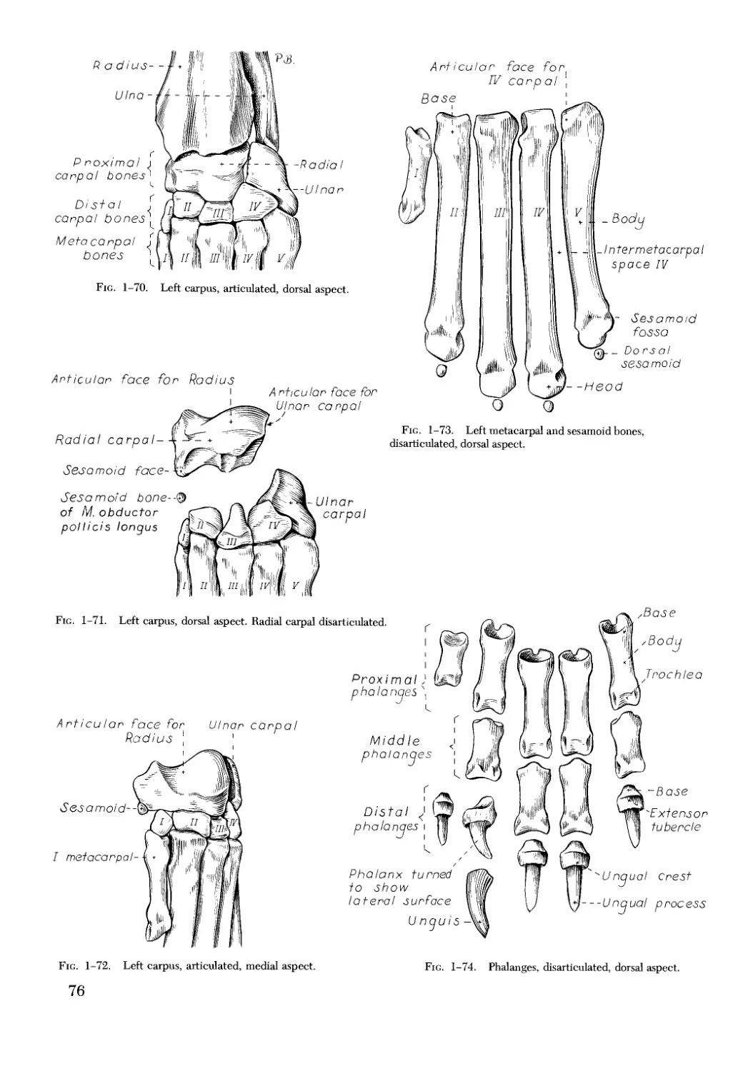
Bones may be grouped according to shape,
structure, function, origin, or position. The total
average number of bones in each division of the
skeletal system, as found in an adult dog (Fig.
1-1), is given in Table 1. In this enumeration,
the bones of the dewclaw (the first digit of the
hindpaw) are not included, because this digit is
absent in many breeds of dogs, and in other
breeds a single or double first digit is required
for showing purposes (American Kennel Club
1956).
Table 1. Bones of Skeletal System
DIVISION TOTAL AVERAGE NUMBER
Axial Skeleton
Vertebral column 50
Skull and hyoid 50
Ribs and sternum 34
Appendicular Skeleton
Pectoral limbs 92
Pelvic limbs 92
Heterotopic Skeleton
Os penis 1
Total 319
Classification of Bone According to Shape
Bone and cartilage may be classified in various
ways. Anatomists have long grouped bones ac-
cording to shape. Although borderline forms ex-
ist, for descriptive purposes five general divi-
sions on this basis are recognized: long bones,
short bones, sesamoid bones, flat bones, and ir-
regular bones. Long, short, and sesamoid bones
are found in the limbs, whereas the flat and ir-
regular bones are characteristic of the axial
skeleton. The terms are readily understandable,
except possibly sesamoid, which is derived from
the Greek word for a seed that is small, flat, and
obovate. Sesamoid bones vary from tiny spheres
to the slightly bent, ovoid patella (kneecap),
which is 2 or more centimeters long in a large
dog. Some sesamoid elements never ossify but
remain as cartilages throughout life.
1
General
3
Long bones (ossa longa) occur only in the ex-
tremities, or limbs. The bones of the thigh and
arm, that is, the femur and humerus, are good
examples. Typically a long bone, during its
growth, possesses a long middle part, the shaft
or diaphysis, and two ends, the epiphyses. Dur-
ing development each end is separated from the
shaft by a plate of growing cartilage, the epi-
physeal cartilage (cartilago epiphysialis), or
plate. At maturity the epiphyseal cartilage
ceases to grow, and the epiphysis fuses with the
shaft as both share in the bony replacement of
the epiphyseal cartilage. Fractures sometimes
occur at the epiphyseal plate. Usually after ma-
turity no distinguishable division exists between
epiphysis and diaphysis. The ends of most long
bones enter into the formation of freely movable
joints. Long bones form levers and possess great
tensile strength. They are capable of resisting
many times the stress to which they are nor-
mally subjected. The stress on long bones is both
through their long axes, as in standing, and at
angles to these axes, as exemplified by the pull
of muscles which attach to them. Although
bones appear to be rigid and not easily influ-
enced by the soft tissues which surround them,
soft tissues actually do contour the bones. In-
dentations in the form of grooves are produced
by blood vessels, nerves, tendons, and ligaments
that lie adjacent to them, whereas roughened
elevations or depressions are produced by the
attachments of tendons and ligaments. The ends
of all long bones are enlarged and smooth. In
life, these smooth surfaces are covered by a layer
of hyaline cartilage, as they enter into the for-
mation of joints. The enlargement of each ex-
tremity of a long bone serves a dual purpose. It
diminishes the risk of dislocation and provides a
large bearing surface for the articulation. The
distal end of the terminal phalanx of each digit
is an exception to the stated rule. Since it is cov-
ered by horn and is not articular, it is neither en-
larged nor smooth.
Short bones (ossa brevis) are confined to the
carpal (wrist) and tarsal (ankle) regions, which
contain seven bones each. They vary in shape
from the typical cuboidal shape with six surfaces
to irregularly compressed rods with only one flat,
articular surface. In those bones having many
surfaces, at least one surface is nonarticular.
This surface provides an area where ligaments
may attach and blood vessels may enter and
leave the bone.
Sesamoid bones (ossa sesamoidea) are pres-
ent near freely moving joints. They are usually
formed in tendons, but they may be developed
in the ligamentous tissue over which tendons
pass. They usually possess only one articular sur-
face, which glides on a flat or convex surface of
one or more of the long bones of the extremities.
Their chief function seems to be to alter the
course of tendons and to protect tendons at the
places where greatest friction is developed.
Flat bones (ossa plana) are found in the proxi-
mal portions of the limbs, and in the head and
thorax. The most obvious function of these
bones is protection. The ribs and the bones of
the cranium are primarily for this purpose. The
bones of the face are also flat, providing maxi-
mum shielding without undue weight, and
streamlining the head. Furthermore, the heads
of all quadrupeds overhang their centers of grav-
ity; a heavy head would be a handicap in loco-
motion. The flat bones of the cranium consist of
outer and inner tables of compact bone and an
intermediate uniting spongy bone, called diploe.
In certain bones of the head the diploe is pro-
gressively invaded, during growth, by extensions
from the nasal cavity which displace the diploe
and cause a greater separation of the tables than
would otherwise occur. The intraosseous air
spaces of the skull formed in this way are known
as the paranasal sinuses. Bones which contain air
cavities are called pneumatic bones (ossa pneu-
matica).
Irregular bones (ossa irregulata) are those of
the vertebral column, but the term also includes
all bones of the skull not of the flat type, and the
three parts of the hip bone (os coxae). Jutting
processes are the characteristic features of ir-
regular bones. Most of these processes are for
muscular and ligamentous attachments; some
are for articulation. The vertebrae of quad-
rupeds protect the spinal cord and furnish a
relatively incompressible bony column through
which the propelling force generated by the
pelvic limbs is transmitted to the trunk. The
vertebrae also partly support and protect the
abdominal and thoracic viscera, and give rigid-
ity and shape to the body in general. The
amount of movement between any two verte-
brae is small, but the combined movement per-
mitted in all the intervertebral articulations is
sufficient to allow considerable mobility of the
whole body in any direction.
Development of Bone
Bone develops in both cartilage and mem-
branous connective tissue. By far the greater
number of bones develop in cartilage, or, to
speak more accurately, replace it. These bones
4
Chapter 1. The Skeletal System
are known as endochondral, replacement, or
cartilage bones. Some bones, such as those
which form the roof of the cranium, develop in
connective tissue sheets or membranes. Bones
developed in such a way are called membrane,
or dermal, bones.
Bone is about one-third organic material,
wh । h is both intracellular and extracellular in
location. Within or around the bone cells,
known as osteoblasts, the bone matrix is laid
down. The osteoblasts later become the osteo-
cytes of mature bone. The cells which seem to
direct the deposition of cartilage and bone are
derived from mesenchyme, which forms the
greater part of the middle germ layer, or meso-
derm, of the embryo. Since most bones are pre-
formed in cartilage, the cartilage appears early
in development, followed by perichondral and
endochondral ossification. The first evidence of
ossification in an embryonic long bone is seen as
a collar around the middle of the shaft. Forma-
tion of the inorganic material is preceded by dis-
solution of the cartilage at the site of its deposi-
tion. One stage follows another so rapidly that all
calcified tissue soon takes the form of true bone
of the spongy type. Secondary centers of ossifi-
cation appear in the epiphyses. From this stage
bone formation, much like the writing of a book,
consists of altering or destroying the first-formed
material and the building of a more perfect
structure. Osteoclasts are thought to be the cells
of bone destruction. Bones grow in length by
endochondral ossification, but their increase in
circumference, and the entire formation of cer-
tain bones of the skull, occurs through a differ-
ent process, described in the following para-
graph.
The bones of the face and dorsum of the cra-
nium develop in sheets of connective tissue, not
in cartilage. This type of bone formation is
known as intramembranous ossification. The os-
teoblasts and the osteoclasts continue to be the
laborers in this activity. The compact bone
formed by the periosteum is identical with mem-
brane bone in its elaboration. Bony tissue of
either type is capable of growing in any direc-
tion.
The larger sesamoid bones are preformed in
cartilage, whereas the smaller ones may develop
in membrane.
Structure of Bone
The gross structure of a dried, macerated
bone is best revealed if the bone is sectioned in
various planes. Two types of bone structure will
be seen. One is compact, or dense, bone, which
forms the outer shell of all skeletal parts. The
other is spongy, or cancellous, bone, which oc-
cupies the interior of the extremities of all long
bones and the entire interior of most other bones,
except certain of the skull bones and the bones of
the pectoral and pelvic girdles. Spongy bone is
not found in the girdles, where the two compact
plates are fused.
Compact bone (substantia compacta, or sub-
stantia corticalis) is developed in direct ratio to
the stress to which the bone is subjected. It is
thicker in the shafts of long bones than in their
extremities. It attains its greatest uniform thick-
ness where the circumference of the bone is
least. The maximum thickness of the compact
bone found in the femur and humerus of an
adult Great Dane was 3 mm. Local areas of in-
creased thickness are present at places where
there is increased tension from muscles or liga-
ments.
Spongy bone (substantia spongiosa) is elabo-
rated in the extremities of long bones, forms the
internal substance of short and irregular bones,
and is interposed between the two compact lay-
ers of most flat bones. Spongy bone consists of a
complicated maze of crossing and connecting
osseous leaves and spicules which vary in shape
and direction. The spongy bone of the skull is
known as diploe.
The shafts of long bones in the adult are
largely filled with yellow bone marrow (medulla
ossium flava). This substance is chiefly fat. In the
fetus and the newborn, red bone marrow (me-
dulla ossium rubra) occupies this cavity and
functions in forming red blood cells. No spongy
bone is present in the middle of the shafts of
long bones, and the marrow-filled spaces thus
formed are known as medullary cavities (cava
medullaria).
Spongy bone is developed where greatest
stress occurs. The leaves or lamellae and bars are
arranged in planes where pressure and tension
are greatest, this structural development for
functional purposes being best seen in the proxi-
mal end of the femur. The interstices between
the leaves and bars of spongy bone are occupied
by red marrow. The spongy bone of ribs and
vertebrae and of many other short and flat bones
is filled with red marrow throughout life. In the
emaciated or the extremely aged, red marrow
gives way to fatty infiltration.
The periosteum is an investing layer of con-
nective tissue which covers the nonarticular sur-
General
5
faces of all bones in the fresh state. The connec-
tive tissue covering of cartilage, known as
perichondrium, does not differ histologically
from periosteum. Perichondrium covers only the
articular margins of articular cartilages, but in-
vests cartilages in all other locations. Periosteum
blends imperceptibly with tendons and liga-
ments at their attachments. Muscles do not ac-
tually have the fleshy attachment to bone which
they are said to have, since a certain amount of
connective tissue, periosteum, intervenes be-
tween the two. At places where there are no
tendinous or ligamentous attachments it is not
difficult, when bone is in the fresh state, to
scrape away the periosteum from it.
The endosteum is similar in structure to peri-
osteum, but is thinner. It lines the larger medul-
lary cavities, being the condensed peripheral
layer of the bone marrow. Both periosteum and
endosteum, under emergency conditions, such
as occur in fracture of bone, provide cells (osteo-
blasts) which aid in repair of the injury. Some-
times the broken part is over-repaired with bone
of poor quality. Such osseous bulges at the site of
injury are known as exostoses.
Mucoperiosteum is the name given to the
covering of bones which participate in forming
boundaries of the respiratory or digestive sys-
tem. It lines all of the paranasal sinuses and con-
tains mucous cells.
Physical Properties of Bone
Bone is about one-third organic and two-
thirds inorganic material. The inorganic matrix
of bone has a microcrystalline structure com-
posed principally of calcium phosphate. The
exact constitution of the crystal lattice is still
under study, but it is generally agreed that bone
mineral is largely a hydroxyapatite 3 Ca3(PO4)2
• Ca(OH), with adsorbed carbonate. Some con-
sider that it may exist as tricalcium phosphate
hydrate 3 Ca3(PO4)2 • 2 H2O with adsorbed cal-
cium carbonate (Dixon and Perkins 1956). The
organic framework of bone can be preserved
while the inorganic part is dissolved. A 20 per
cent aqueous solution of hydrochloric acid will
decalcify any of the long bones of a dog in about
one day. Such bones retain their shape but are
pliable. A slender bone, such as the fibula, can be
tied into a knot after decalcification. The organic
material is essentially connective tissue, which
upon boiling yields gelatin.
Surface Contour of Bone
Much can be learned about the role in life of
a specific bone by studying its eminences and
depressions. There is a functional, embryologi-
cal, or pathological reason for the existence of
every irregularity.
Most eminences serve for muscular and liga-
mentous attachments. Grooves and fossae in
some instances serve a similar function. Facets
are small articular surfaces which may be flat,
concave, or convex. Trochleas and condyles are
usually large articular features of bone. The
roughened enlarged parts which lie proximal to
the condyles on the humerus and femur are
known as epicondyles.
Vessels and Nerves of Bone
Bone, unlike cartilage, has both a nerve and
a blood supply. Long bones and many flat and
irregular bones have a conspicuous nutrient
(medullary) artery and vein passing through the
compact substance to serve the marrow within.
Such arteries pass through a nutrient foramen
(foramen nutricium) and canal (canalis nutric-
ius) of a bone and, upon reaching the marrow
cavity, divide into proximal and distal branches
which repeatedly subdivide and supply the bone
marrow and the adjacent cortical bone. In the
long and short bones terminal branches reach
the epiphyseal plate of cartilage where, in
young animals, they end in capillaries. In adults
it is likely that many twigs nearest the epiphyses
anastomose with twigs arising from vessels in
the periosteum. Nutrient veins pursue the re-
verse course. Not all of the blood supplied by the
nutrient artery is returned by the nutrient vein
or veins; much of it, after traversing the capillary
bed, returns through veins which perforate the
compact bone adjacent to the articular surfaces
at the extremities of these bones. The periosteal
arteries and veins are numerous but small; these
arteries supply the extremities of long bones and
much of the compact bone also. They enter
minute canals which lead in from the surface,
and ramify proximally and distally in the micro-
scopic tubes which tunnel the compact and
spongy bone. The arterioles of the nutrient
artery anastomose with those of the periosteal
arteries deep within the compact bone. It is
chiefly through enlargement of the periosteal
arteries and veins that an increased blood supply
and increased drainage are obtained at the site
of a fracture. Veins within bone are devoid of
valves, the capillaries are large, and the endo-
thelium from the arterial to the venous side is
continuous. Lymph vessels are present in the
periosteum as perivascular sheaths and probably
6
Chapter 1. The Skeletal System
also as unaccompanied vessels within the bone
marrow. The nerves of bone are principally sen-
sory. They serve as an inner defense against in-
jury. The sensory nerves of the skin form the
outer defense. Both carry impulses which result
in pain. Kuntz and Richins (1945) state that both
the afferent and sympathetic fibers probably
play a role in reflex vasomotor responses in the
bone marrow.
Function of Bone
The skeleton of the vertebrate body serves
four functions.
1. Bone forms the supporting and in many in-
stances the protecting framework of the body.
The supportive function does not need explana-
tion. The essential organs of vertebrates receive
protection from the skeleton. These are the
brain and spinal cord, heart, and liver. To these
may be added certain pelvic organs which, al-
though not essential for life, are protected by the
pelvis. (The urinary bladder is largely an abdom-
inal organ in the dog.) The lungs further protect
the heart, and are in turn protected by the ribs.
2. Many bones serve as levers by which the
muscles move the body. Of the three types of
levers, only two are represented by bones.
Many bones may serve as either a first or a
third class lever, owing to the action of differ-
ent muscles at different times and to changes
in the positions of force and fulcrum. No lever
of the second class is represented in the living
AXIAL S
THE SKULL
The skull (cranium) is the most important,
complex, and specialized part of the skeleton. It
lodges the brain, and houses the sense organs for
hearing, equilibrium, sight, smell, and taste. In
addition to providing the attachment for the
teeth, tongue, larynx, and a host of muscles, it
contains the master endocrine gland, or hy-
pophysis. It is basically divided into a facial plus
palatal region, and a neural, or braincase, por-
tion (Fig. 1-2).
The facial and palatal region, consisting of 36
bones, is specialized to provide a large surface
area subserving the sense of smell, and a long
surface for the implantation of the teeth. This
elongation results in a pointed anterior end, or
apex, and a wide, deep base which impercepti-
bly blends with the braincase.
The braincase (Fig. 1-3), formed by 14
bones, encloses the brain in the large cranial
body. In all lever movements of bones by mus-
cles the force or the fulcrum is always at one
end and the weight at the other. The weight is
never between the force and the fulcrum, which
is necessary for a second class lever. Nearly all
muscles act at a mechanical disadvantage. The
speed at which the weight travels is in direct
proportion to the shortness of the force arm, and
this is determined by the distance of the inser-
tion of the muscle from the joint, or fulcrum.
3. Bone serves as a storehouse for calcium
and phosphorus and for many other elements in
small amounts. These substances are withdrawn
from the bone as complicated compounds. The
greatest drain occurs during pregnancy; con-
versely, the greatest deposition takes place dur-
ing growth. In the large breeds, such as the
Great Dane and St. Bernard, the skeleton is the
system most likely to show the effects of a nutri-
tional deficiency. Undermineralization of the
skeleton is a common manifestation of under-
feeding, improper feeding, or inability of the in-
dividual to assimilate food adequately.
4. Bone serves as a factory for red blood cells
and for several kinds of white blood cells. In the
normal adult it also stores fat. Red marrow,
where the red and many white blood cells de-
velop, occurs most richly in the bones of the
axial skeleton and in the proximal epiphyses of
the humerus and femur; yellow or fatty marrow
is most abundant in the long bones of the ex-
tremities.
cavity (cavum cranii), and houses the organs of
hearing and equilibrium in the petrous part of
the temporal bone. The cranial cavity is sepa-
rated from the cavity of the nose (cavum nasi)
by a curved perforated plate of bone, and is
open caudally by way of the foramen magnum
for the passage of the spinal cord and attendant
structures. The ventral part of the cranium has
a number of foramina and canals for the passage
of nerves and blood vessels. At the junction of
the facial and cranial parts, on each side, are the
orbital cavities, in which are located the globes
of the eyes and accessory structures.
The bones of the ventral part (Fig. 1-4) of the
cranium, or basicranial axis, are preformed in
cartilage, whereas those of the dorsum, or cal-
varium, are formed in membrane. A classical
treatment of the development of the vertebrate
skull by de Beer (1937) considers the homologies
of skull components, compares chondro crania,
and discusses modes of ossification. Romer
7
F nontal.
Jph enoid
Palat in el
Ptenij c]Oid
Fig. 1-2. Bones of the skull, lateral aspect. (Zygomatic arch and mandible removed.)
Fig. 1-3. Bones of the skull, dorsal aspect.
8
Chapter 1. The Skeletal System
Table 2. Average Measurement of Three Skull Types
MEASUREMENT BRACHY- CEPHALIC MESATI- CEPHALIC DOLICHO- CEPHALIC
Facial length Nasion to prosthion 48 mm. 89 mm. 114 mm.
Facial width Widest interzygomatic distance 103 mm. 99 mm. 92 mm.
Cranial length Inion to nasion 99 mm. 100 mm. 124 mm.
Cranial width Widest interparietal distance 56 mm. 56 mm. 59 mm.
Cranial height Middle of external acoustic meatus to bregma 54 mm. 60 mm. 61 mm.
Mandibular length Caudal border of condyle to pogonion 85 mm. 134 mm. 163 mm.
Skull length Inion to prosthion 127 mm. 189 mm. 238 mm.
Skull width Widest interzygomatic distance 1U3 mm. 99 mm. 92 mm.
Skull base length Basion to prosthion 107 mm. 170 mm. 216 mm.
/ width X : too
Indices [
у length
Skull index 81 52 39
Cranial index 57 56 48
Facial index 215 111 81
(1962) briefly reviews the phylogenetic history of
the vertebrate skull. Skulls differ more in size
and shape among domestic dogs than in any
other mammalian species. For this reason, cra-
niometry in dogs takes on added significance.
Certain points and landmarks on the skull are
recognized in making linear measurements and
have been used by Stockard (1941) and others.
The more important of these are:
Inion: Central surface point on the external
occipital protuberance.
Bregma: Junction on the median plane of the
right and left frontoparietal sutures, or the point
of crossing of the coronal and sagittal sutures.
Nasion: Junction on the median plane of the
right and left nasofrontal sutures.
Prosthion: Anterior end of the intermaxillary
suture, located between the roots of the upper
central incisor teeth.
Pogonion: Most anterior part of the mandi-
ble, at the symphysis, located between the roots
of the lower central incisor teeth.
Basion: Middle of the ventral margin of the
foramen magnum.
The center of the external acoustic meatus:
Although unnamed, this spot also serves as a ref-
erence point.
Three terms are frequently used to designate
head shapes:
Dolichocephalic, meaning long, narrow-
headed. Breed examples: collie, Russian wolf-
hound.
Mesaticephalic, meaning a head of medium
proportions. Breed examples: German shepherd,
beagle, setter.
Brachycephalic, meaning short, wide-headed.
Breed examples: Boston terrier, Pekingese.
The face of the dog varies more in shape and
size than does any other part of the skeleton. In
brachycephalic breeds the facial skeleton is
shortened and broadened. In some brachyceph-
alic breeds, the English bulldog, for example, the
lower jaw protrudes anterior to the upper jaw,
producing the undershot condition known as
prognathism of the mandible. Most other breed
types have brachygnathic mandibles, that is, re-
ceding lower jaws. Although brachygnathism of
the mandibles is relative, both the collie and the
dachshund frequently exemplify this condition
to a marked extent.
Table 2 shows average measurements in milli-
meters taken from randomly selected adult-
skulls of the three basic types. From these data
it can be seen that the greatest variation in skull
shape occurs in the facial part. In making com-
parisons of skull measurements it is essential that
the over-all size of the individuals measured is
taken into consideration. As a rule the dolicho-
cephalic breeds are larger than the brachyce-
phalic, whereas the working breeds fall in the
mesaticephalic group, and these as a division
have the greatest body size. The only measure-
ment in which the brachycephalic type exceeds
the others, in the small sampling shown, is facial
width. To obviate the size factor among the
breed types, indices are computed. These indi-
cate relative size and are expressed by a single
term representing a two-dimensional relation-
ship. The cranial index is computed by multiply-
ing the cranial width by 100 and dividing the
product by the cranial length. Skull and facial
indices are computed in the same manner.
Differences among the breeds in facial skele-
tal development are the most salient features re-
vealed by craniometry. The face is not only
short in the brachycephalic breeds but it is also
The Skull
9
10
Chapter 1. The Skeletal System
actually wider than it is in the heavier, longer-
headed breeds. These data do not show that ap-
preciable asymmetry exists, especially in the
round-headed types. Even though the neurocra-
nium varies least in size, it frequently develops
asymmetrically. The caudal part of the skull is
particularly prone to show uneven development.
The further a breed digresses from the ancestral
German shepherd type, the more likely are dis-
tortions to be found. This is particularly true of
the round-headed breeds, as these types have
been developed to please man’s fancy without
regard to the health of the strain or even to their
expected survival without special attention.
Their is little rationale in developing a breed of
dog like the Boston terrier, with large, round
heads and small pelves. In this instance the
transgression against nature is twofold. The
large crania of the young frequently exceed the
dimensions of the dam’s pelvis, and normal par-
turition is impossible. The breed would soon be
extinct if cesarean sections were not performed.
The ugly appearance of the English bulldog is
partly produced by the prognathic condition of
the lower jaw, as well as the brachygnathic con-
dition of the upper jaw. This structural dishar-
mony results in poor occlusion of the teeth.
Stockard (1941) found, by crossing purebred
breeds of extreme jaw sizes, that the lengths of
the upper and of the lower jaw are inherited in-
dependently. Dogs with prognathic upper jaws
and brachygnathic lower jaws are unable to eat
from a flat surface. Disproportionate growth of
the length of the face occurs after the early
weeks of life, so that suckling the dam is not im-
paired. Dental malocclusion is treated under the
description of the teeth in Chapter 12 on the
Digestive System.
Cranial capacity varies but little among the
different breeds and skull types. The terms mi-
crocephalic, mesocephalic, and megacephalic
indicate skulls with small, medium, and large
cranial capacity, respectively. The following
data were computed by filling the crania with
mustard seed after the foramina had been closed
with modeling clay, and then determining the
volume of seed used. Average Boston terrier
skulls held 82 cc. A sampling of skulls of medium
size and medium length showed an average ca-
pacity of 92 cc.; the average skull capacity of the
crania of the Russian wolfhound and of the collie
was 104 cc.
The names of the individual bones making up
the 50 which compose the skull are listed in
Table 3.
Bones of the Braincase
Occipital Bone
The occipital bone (os occipitale) (Figs. 1-5,
1-6) forms the posterior portion of the skull. It
develops from four centers: a squamous part
Table 3. Individual Bones of the Skull
Bones of the braincase:
Paired: 1. Exoccipital
2. Parietal
Unpaired: 1. Supraoccipital
2. Interparietal
3. Basioccipital
Bones of the face and palate:
Paired: 1. Premaxilla
2. Nasal
3. Maxilla
4. Nasoturbinate
5. Maxilloturbinate
3. Frontal
4. Temporal
4. Basisphenoid
5. Presphenoid
6. Ethmoid
6. Zygomatic
7. Palatine
8. Lacrimal
9. Pterygoid
10. Mandible
Unpaired: 1. Vomer
Bones of the hyoid apparatus and middle ear:
Paired: 1. Stylohyoid 2. Epihyoid 5. Malleus 6. Incus
3. Keratohyoid 7. Stapes
4. Thyrohyoid
Unpaired: 1. Basihyoid
The Skull
11
Occipital condyle - - -
Ventral condyloid fossa --
Foramen maynum
Dorsal condyloid fossa......
-Supraoccipital
H’N-
Dorsal nuchal lire -^
Ventral nuchal line--..^
-Interparietal process
Ext. occipital protuberance
- Ext. occi pi tai crest
_ _ -ExoccipitaI
- - Condyloid canal,posterior opening
~ -Jugular process
I ntercondgl о I d notch
Hypog lossal foramen'' "-Basioccipital
Fig. 1-5. Occipital bone, posterior lateral aspect.
Int. occipital protuberance ... -Foramen impar
Transverse sulcus- 5°^Па1 CreSt
- Vermiform impression
Location of supramastoid foramen-
Sulcus of ventral petrosal sinus
Int. occipital crest—
— Nucha! tubercle
Anterior v posterior openings of
- - condyloid canal
Ventral opening of condyloid canal
'Int. opening of hypoglossal canal
Fig. 1-6. Occipital bone, anterior lateral aspect.
12
Chapter 1. The Skeletal System
dorsally, two lateral condylar parts, and a basilar
part ventrally.
The squamous part (pars squamosa), or supra-
occipital bone, is the largest division. Dorsoan-
teriorly it is wedged between the parietal bones
to form the interparietal process (processus in-
terparietalis). This process represents the un-
paired interparietal bone which fuses prenatally
with the supraoccipital. From the interparietal
process arises the mid-dorsal external sagittal
crest (crista sagittalis externa), which, in some
specimens, is confined to this bone. The anterior
end of the interparietal process is narrower and
thinner than the caudal part, which turns ven-
trally to form a part of the posterior surface of
the skull. The dorsal nuchal line (linea nuchalis
dorsalis) marks the division between the dorsal
and posterior surfaces of the skull. It is an un-
paired sharp-edged crest of bone which reaches
its most dorsal point at the external occipital
protuberance. On each side it arches ventrally
before ending on a small eminence located dor-
soposterior to the external acoustic meatus. The
ventral nuchal line (linea nuchalis ventralis) is a
line located in a frontal plane, ventral to the
middle part of the dorsal nuchal line, and forms
the base of an uneven triangular area. It extends
transversely between the dorsolateral parts of
the dorsal nuchal line. It is not distinct. The ex-
ternal occipital protuberance (protuberantia oc-
cipitalis externa) is the median, triangular pro-
jection forming the most dorsoposterior portion
of the skull. The external occipital crest (crista
occipitalis externa) is a smooth median ridge ex-
tending from the external occipital protuber-
ance to the foramen magnum. It is poorly devel-
oped in some specimens.
Within the dorsal part of the occipital bone
and opening bilaterally on the cerebral surface
is the transverse canal (canalis transversa),
which, in life, contains the venous transverse
sinus. The transverse canal is continued later-
ally, on each side, by the sulcus for the trans-
verse sinus (sulcus sinus transversi). Mid-dor-
sally or to one side, the sagittal sinus enters the
transverse sinus via the foramen impar. Between
the laterally located sulci the skull protrudes an-
teroventrally to form the internal occipital pro-
tuberance (protuberantia occipitalis internus).
Extending anteriorly from the internal occipital
protuberance is the variably developed, usually
paramedian and always small internal sagittal
crest (crista sagittalis interna). The vermiform
impression (impressio vermis), forming the thin-
nest part of the caudal wall of the skull, is an ir-
regular excavation of the median portion on the
cerebellar surface of the squamous part of the
occipital bone which houses a part of the vermis
of the cerebellum. The vermiform impression is
bounded laterally by the paired internal occipi-
tal crest (crista occipitalis interna), which is usu-
ally asymmetrical and convex laterally. Lateral
to the internal occipital crest, as well as on the
ventral surface of the interparietal process, there
are elevations, juga cerebralia et cerebellaria,
and depressions, impressiones digitatae. Ven-
trally the squamous part is notched to form the
dorsal part of the foramen magnum. On either
side the supraoccipital is fused with the paired
exoccipital. This union represents the former ar-
ticulation (synchondrosis intraoccipitalis squa-
molateralis) which extended from the foramen
magnum to the temporal bone.
The lateral parts (partes laterales), or exoccip-
ital bones, bear the occipital condyles (condyli
occipitales), which are convex and, with the at-
las, form the atlanto-occipital joints. The jugular
process (processus jugularis) is located, one on
either side, lateral to the condyle, and ends in a
rounded knob ventrally, usually on a level with
the bottom of the anteriorly located tympanic
bulla. Between the jugular process and the oc-
cipital condyle is the ventral condyloid fossa
(fossa condylaris ventralis). On a ridge of bone
anterior to this fossa is the hypoglossal foramen
(foramen hypoglossi), which is the external
opening of the hypoglossal canal (canalis hypo-
glossi), a direct passage through the ventral part
of the occipital bone. The dorsal condyloid fossa
(fossa condylaris dorsalis) is located dorsal to the
occipital condyle. The rather large condyloid
canal (canalis condylaris) runs through the me-
dial part of the lateral occipital bone. There is an
intra-osseous passage between the condyloid
canal and the hypoglossal canal. Usually there is
also a small passage between the condyloid canal
and the petrobasilar fissure.
The basilar part (pars basilaris), or basioccipi-
tal bone, is unpaired, and forms the posterior
third of the cranial base. It is roughly rectangu-
lar, although caudally it tapers to a narrow, con-
cave end which forms the central portion of the
intercondyloid notch (incisura intercondyloidea).
The adjacent occipital condyles on each side
deepen the incisure as they contribute to its for-
mation. The incisure bounds the ventral part of
the foramen magnum. The foramen magnum is
a large, transversely oval opening in the postero-
ventral portion of the skull, through which pass
the spinal cord and its associated structures, the
meninges, vertebral venous sinuses, the spinal
portion of the accessory nerve, and the various
The Skull
13
arteries associated with the spinal cord. In
brachycephalic breeds it is more circular than
oval, and it is frequently asymmetrical. The dor-
sal boundary of the foramen magnum is featured
by the caudally flared ventral part of the supra-
occipital bone. The caudal extension is increased
by the paired nuchal tubercles (tubercula nu-
chalia). These projections are sufficiently promi-
nent to make spinal punctures at this site diffi-
cult. The dorsal surface of the basioccipital bone
is concave to form the sulcus medulla oblongata.
The lateral surfaces of the caudal half of the
basioccipital bone fuse with the exoccipital
bones along the former ventral intraoccipital
synchondrosis (synchondrosis intraoccipitalis
basilateralis). The ventral surface of the basioc-
cipital bone adjacent to the petrotympanic syn-
chondrosis possesses muscular tubercles (tuber-
cula muscularia). These are rough, sagittally
elongated areas, located medial to the smooth,
rounded tympanic bullae. The pharyngeal tu-
bercle (tuberculum pharyngeum) is a single tri-
angular rough area anterior to the intercondy-
loid incisure. Laterally the basioccipital bone is
grooved to form the ventral petrosal sulcus,
which concurs with the pyramid of the temporal
bone to form the petrobasilar canal (canalis
petrobasilaris).
Ventrally the anterior end of the basioccipital
bone articulates with the body of the basisphe-
noid bone at the cartilaginous spheno-occipital
joint (synchondrosis spheno-occipitalis). Ventro-
laterally the occipital bone articulates with the
tympanic part of the temporal bone to form the
cartilaginous tympano-occipital joint (synchon-
drosis tympano-occipitalis). Deep to this joint is
the important petro-occipital suture (sutura
petro-occipitalis), in which the foramen lacerum
caudalis, or jugular foramen, opens. The joint
between the petrosal and the occipital bones
which forms the petro-occipital suture is the
synchondrosis petro-occipitalis. Laterally, and
proceeding dorsally, the occipital bone first ar-
ticulates with the squamous temporal bone su-
perficially, the occipitosquamous suture (sutura
occipitosquamosa), and with the mastoid part
of the petrous temporal bone deeply, the occip-
itomastoid suture (sutura occipitomastoidea);
further dorsally it articulates with the parietal
bone, the lambdoid suture (sutura lambdoidea).
Where the squamous and lateral parts of the oc-
cipital bone articulate with each other and with
the mastoid part of the temporal bone, the su-
pramastoid foramen (foramen supramastoi-
deum) is formed.
Variations in the occipital bone are numer-
ous. The foramen magnum varies in shape and
is not always bilaterally symmetrical. The con-
dyloid canal may be absent on one or both sides.
Even when both canals are present, connections
between the hypoglossal and condyloid canals
may fail to develop. The jugular processes may
extend several millimeters ventral to the tym-
panic bullae so that they will support a skull
without the mandibles when it is placed on a
horizontal surface; conversely, they may be
short, retaining the embryonic condition. The
vermiform impression may be deep, causing a
posteromedian rounded, thin protuberance on
the posterior face of the skull. The foramen im-
par may be double. It is rarely median in posi-
tion. A sutural bone may be present at the an-
terior end of the interparietal process.
Parietal Bone
The parietal bone (os parietalis) (Fig. 1-7) is
paired and forms most of the dorsolateral part
Vascular groove for med.meningeal a.
Tentorium osseum -
Transverse sulcus —
- -1nterparietal suture
/^.Nfv^soN
Fig. 1-7. Parietal bones, ventral lateral aspect.
14
Chapter 1. The Skeletal System
of the cranial wall. It articulates dorsally with
its fellow and with the interparietal process of
the occipital bone. Each parietal bone lies di-
rectly anterior to the squamous occipital and
dorsal to the squamous temporal. In the new-
born no elevation is present at the sagittal in-
terparietal suture or on the interparietal proc-
ess, but soon thereafter in the heavily muscled
breeds, particularly in the male, the mid-dorsal
external sagittal crest is developed. This crest,
which increases in size with age, forms the me-
dial boundary of the temporal fossa (fossa tem-
poralis), a large area on the external surface
(facies externa) of the cranium from which the
temporal muscle originates. In dolichocephalic
breeds with heavy temporal muscles, the exter-
nal sagittal crest may reach a height of more
than 1 cm. and extend from the external occipi-
tal protuberance to the parietofrontal suture.
Anteriorly, it continues as the diverging frontal
crests. In most brachycephalic skulls the exter-
nal sagittal crest is confined to the interparietal
part of the occipital bone and is continued an-
teriorly as the diverging temporal lines (lineae
temporales). The temporal lines at first are con-
vex laterally, then become concave as they cross
the parietofrontal, or coronal, suture and are
continued as the external frontal crests to the
zygomatic processes. The temporal lines replace
the external sagittal crest in forming the medial
boundaries of the temporal fossae in most
brachycephalic skulls.
The internal surface (facies interna) of the
parietal bone presents digital impressions and
intermediate ridges corresponding, respectively,
with the cerebral gyri and sulci. A well-defined
vascular groove, the sulcus for the middle me-
ningeal artery (sulcus arteriae meningeae me-
diae), starts at the ventrocaudal angle of the
bone and arborizes over its internal surface. The
groove runs toward the opposite angle of the
bone, giving off smaller branched grooves along
its course. A leaf of bone projects anteromedially
from the dorsal part of the posterior border. This
leaf concurs with its fellow and with the internal
occipital protuberance to form the curved ten-
torium ossium. On the internal surface of the
parietal bone near its caudal border is a portion
of the transverse sulcus, which leads dorsally
into the transverse canal of the occipital bone
and ventrally into the temporal meatus.
The borders of the parietal bone are anterior,
posterior, dorsal, and ventral in position, since
the bone is essentially a curved, square plate.
The anterior or frontal border (margo frontalis)
overlaps the frontal bone, forming the fronto-
parietal or coronal suture (sutura frontoparie-
talis). The posterior or occipital border (margo
occipitalis) meets the occipital bone to form the
occipitoparietal suture (sutura occipitoparie-
talis). The anterior half of the dorsal or sagittal
border (margo sagittalis) articulates with its
fellow on the midline to form the sagittal suture
(sutura sagittalis). The posterior half of the dorsal
border articulates with the interparietal process
of the occipital bone to form the parietointer-
parietal suture (sutura parietointerparietalis).
The ventral or squamous border (margo squamo-
sus) is overlaid by the squamous temporal bone in
forming the squamous suture (sutura squamosa).
A small area of the squamous border at its an-
terior end articulates with the temporal wing of
the sphenoid bone to form the parietosphenoi-
dal suture (sutura parietosphenoidalis). Over-
lapping of the bones at the squamous and coro-
nal sutures allows for cranial compression of the
fetal skull during its passage through the pelvic
canal.
Frontal Bone
The frontal bone (os frontale) (Figs. 1-8,1-9)
is irregular in shape, being broad posteriorly and
somewhat narrower anteriorly. Laterally, the
anterior part is concave and forms the medial
wall of the orbit. Posterior to this concavity, it
flares laterally to form part of the temporal fossa.
The frontal sinus (sinus frontalis) is an air cavity
located between the inner and outer tables of
the anterior end of the frontal bone and is di-
vided into two or three compartments. It is dis-
cussed in greater detail under the heading Para-
nasal Sinuses.
For descriptive purposes the frontal bone is
divided into orbital, temporal, frontal, and nasal
parts.
The orbital part (pars orbitalis) is a segment of
a cone with the apex located at the optic canal
and the base forming the medial border of the
orbital margin (margo orbitalis). Lateral to the
most dorsal part of the frontomaxillary suture
(sutura frontomaxillaris) the orbital margin is
slightly flattened for the passage of the vena an-
gularis oculi. Ventrally, a long, distinct, dorsally
arched muscular line marks the approximate
ventral boundary of the bone. The ethmoidal
foramina (foramina ethmoidalia) are two small
openings about 1 cm. anterior to the optic canal.
The smaller opening is in the frontosphenoidal
suture; the larger foramen, located dorsopos-
terior to the smaller, passes obliquely through
the orbital part of the frontal bone. Sometimes
the two ethmoidal foramina are confluent. At
The Skull
15
Vascular groove- Cerebral juga - -h »J / Ethmoidal incisure-' Digital impressions'*' To frontal sinus' Septum of frontal sinus -Nasal incisure ----Nasal process Г z ' Maxillcir4 incisure \ xMaxiHarg process ' ''Articular surface for ethmoid и.ч. \ \Ethmoidal foramina
Fig. 1-8. Left frontal bone, medial aspect.
Groove for angularis oculi v.x PARS NASALIS Fossa for lacrimal gland-' Frontal foramen7 ' / я Ethmoidal foramina7 ^^~ ~^^~PARS FR0NTALIS ^~Jr~--PARS TEMPORALIS < r. £ frOntCll CI~eS^ ' Zgg°mafic proce$5 «1.Н. 4 PARS ORBITALIS
Fig. 1-9. Left frontal bone, lateral aspect.
16
Chapter 1. The Skeletal System
the orbital margin, the frontal and orbital sur-
faces meet, forming an acute angle. The supra-
orbital or zygomatic process (processus zygoma-
ticus) is formed where the orbital margin meets
the external frontal crest (crista frontalis ex-
terna), which curves anterolaterally from the
temporal line or sagittal crest. On the orbital
surface of the zygomatic process is a small fora-
men which is only large enough to admit a horse
hair. Ventroanterior to this foramen in some
adult skulls the fossa for the small lacrimal
gland (fossa glandulae lacrimalis) can be seen.
The temporal part (pars temporalis) forms
that part of the frontal bone posterior to the or-
bital part. Dorsally the two tables of the frontal
bone are separated to form the frontal sinus,
whereas ventrally and posteriorly the two tables
are fused or united by a small amount of diploe
to form the braincase.
The frontal part (pars frontalis), or frontal
squama (squama frontalis), is roughly triangular,
with its base facing medially, and articulating
with that of the opposite bone. It is gently
rounded externally and is largely subcutaneous
in life. Its posterior boundary is the external
frontal crest and the lateral part of its anterior
boundary is the orbital margin.
The nasal part (pars nasalis) is the anterior ex-
tension of the frontal bone. Its sharp, pointed
nasal process (processus nasalis) lies partly under
and partly between the posterior parts of the
nasal and maxillary bones. The septum of the
frontal sinus (septum sinuum frontalium) is a
vertical median partition which closely articu-
lates with its fellow in separating right and left
frontal sinuses. It is widest near its middle,
which is opposite the cribriform plate. Anteri-
orly it is continuous with the septal process of
the nasal bone. The ventral part of the septum
of the frontal sinus is the internal frontal crest
(crista frontalis interna). The conjoined right
and left crests articulate with the perpendicular
plate of the ethmoid bone ventrally and with the
conjoined right and left septal processes of the
nasal bones anteriorly. The sagittally located
notch between the pointed anterior end of the
septum and the nasal process is the maxillary
incisure (incisura maxillaris). The ethmoid in-
cisure (incisura ethmoidalis), which lies dorsal
and lateral to the cribriform plate of the eth-
moid bone, is formed by the smooth concave
edge of the internal table of the nasal part of
the frontal bone.
The internal surface (facies interna) of the
frontal bone forms a part of the brain case pos-
teriorly and a small portion of the nasal cavity
anteriorly. The salient ethmoidal notch sepa-
rates the two parts. The posterior part is deeply
concave and divided into many fossae by the
digital impressions and the cerebral juga. Fine,
dorsocaudally running vascular grooves indi-
cate the position occupied in life by the an-
terior meningeal vessels. The large aperture for
the frontal sinus is located dorsal to the eth-
moidal notch. The nasal part of the internal
surface of the frontal bone is marked by many
longitudinal lines of attachment for the eth-
moturbinates.
The mid-dorsal articulation of the frontal
bones forms the frontal suture (sutura inter-
frontalis). This suture is a forward continuation
of the sagittal suture between the parietal bones.
Posteriorly the frontal bone is overlapped by the
parietal bone, forming the frontoparietal suture
(sutura frontoparietalis). Ventrally the rather firm
sphenofrontal suture (sutura sphenofrontalis) is
formed. Anteriorly the frontal bone articulates
with the nasal, maxillary, and lacrimal bones to
form the frontonasal suture (sutura fronto-
nasalis), the frontomaxillary suture (sutura
frontomaxillaris), and the frontolacrimal suture
(sutura frontolacrimalis). Deep in the orbit, the
frontal bone articulates with the palatine bone to
form the frontopalatine suture (sutura fronto-
palatina). Medially, hidden from external view,
the frontal bone articulates with the ethmoid
bone in forming the frontoethmoidal suture
(sutura frontoethmoidalis).
Sphenoid Bone
The sphenoid bone (os sphenoidale) (Figs.
1-10, 1-11, 1-12) forms the anterior two-thirds
of the base of the neurocranium, between the
basioccipital posteriorly and the ethmoid an-
teriorly. It consists of two parts, each possess-
ing a pair of wings and a median body. The
anterior part is the presphenoid (os presphe-
noidale); the posterior part, with the larger
wings, is the basisphenoid (os basisphenoidale),
or postsphenoid.
The dorsal part of the body of the presphe-
noid is roofed over by the fusion of right and
left orbital wings (alae orbitales) to form the
yoke (jugum sphenoidale). The yoke forms the
base of the anterior cranial fossa. A small,
median tubercle, the rostrum (rostrum sphenoi-
dale), divided in the newborn, projects from
the anterior border of the yoke. Posteriorly, the
yoke forms a shelf, the orbitosphenoidal crest
The Skull
17
Sphenoidal sinus_
Orbital wing - __
Optic conal —
Anterior clinoid process- -
Fig. 1-10. Presphenoid, dorsal aspect.
~ Sulcus chiasmatic
„Jugum sphenoidale
-Orbitosphenoidal crest
Tuberculum sellae
Groove for-
med- meningeal a-
- Notch for orbital fissure
-Foramen rotundum
" - Foramen ovale
Temporal wing-„
Posterior clinoid process- __
„'Anterior end of pterygoid process
Lingula sphenoidal is'' / ' '''Hypophyseal fossa
Dorsum sellae' ''Carotid notch
Fig. 1-11. Basisphenoid, dorsal aspect.
Orbital wi
Orbital fissure
Foramen for zygomatic n.
Anterior opening of
pterygoid canal-
'Optic canal
Sphenoidal sinus
'-Body of presphenoid
Pterygoid processes
Fig. 1-12. Basisphenoid and presphenoid, anterior lateral aspect.
Posterior alar foramen
Anterior alar foramen1
18
Chapter 1. The Skeletal System
(crista orbitosphenoidalis), under which lie the
diverging optic canals (canales opticae). On
either side the anterior clinoid process (pro-
cessus clinoideus anterior) projects posteriorly
from the orbitosphenoidal crest and overhangs
the orbital fissure. On the dorsum of the body,
posterior to the optic canals, is the unpaired
sulcus chiasmatis, in which lies the optic
chiasma. The body of the basisphenoid forms
the base of the middle cranial fossa. The mid-
dle of its dorsal surface is slightly dished to
fo.rm the oval hypophyseal fossa (fossa hypo-
physeos). The fossa is limited anteriorly by the
tuberculum sellae, an upward sloping ridge of
bone formed at the junction of the presphenoid
and basisphenoid. The hypophyseal fossa is
limited posteriorly by a bony process, the
dorsum sellae, which, in adult skulls, is flat-
tened and expanded at its free end. Projecting
anteriorly on either side of the dorsum sellae is
a posterior clinoid process (processus clinoideus
posterior). This complex of bony structures,
consisting of the tuberculum sellae and anterior
clinoid processes, the hypophyseal fossa, and the
dorsum sellae with its two posterior clinoid proc-
esses, is called the sella turcica, or Turkish sad-
dle. In life it contains the hypophysis. Occasion-
ally the small craniopharyngeal canal (canalis
craniopharyngeus) persists in the adult. This
canal is a remnant of the pharyngeal diverticu-
lum to the hypophyseal fossa from which the
pars glandularis of the hypophysis develops.
The orbital or lesser wings (alae orbitales s.
minores), or orbitosphenoids, leave each side of
the presphenoid and roof across its body. An-
teriorly, at the junction of the wings and the
body, the presphenoid is hollow and divided by
a longitudinal septum to form the sphenoidal
fossae (fossae sphenoidales) into which extend
the ventrocaudal parts of the ethmoturbinates.
The orbital wings articulate ventrally with the
palatine and dorsally with the frontal bones. In
the frontosphenoidal suture is located the eth-
moidal foramen (foramen ethmoidale); a sec-
ond larger ethmoidal foramen is usually present
in the frontal bone dorsoposterior to the one in
the suture. These foramina may be confluent.
The posterior parts of the orbital wings slope
upward and outward and are thicker, but
smaller, than the anterior parts. Their bases
are perforated by the optic canals. Medially, in
young specimens, the two elliptical optic canals
are confluent across the midline. The orbital
fissures (fissurae orbitales) are located lateral to
the body of the sphenoid in the sutures be-
tween the orbital wings and the temporal wings.
These large openings are at a lower level and
are located slightly posterolateral to the optic
canals.
The temporal or great wings (alae temporales
s. majores), or alisphenoids, of the basisphe-
noid are larger than the orbital wings, and
curve outward and upward. Anteriorly they
extend to the lateral margin of each frontal
bone to form the sphenofrontal suture. The
posterior two-thirds of the temporal wings are
covered laterally by the squamous temporal
bone in forming the sphenotemporal suture. At
the base of each wing, near its junction with
the body, are a series of foramina. The oval
foramen (foramen ovale) is a large opening
which leads directly through the cranial wall.
It is located about 0.5 cm. medial to the tem-
poromandibular joint. A small notch or even a
foramen, foramen spinosum, may be present in
its posterolateral border for the transmission of
the middle meningeal artery. The alar canal
(canalis alaris) runs through the anterior part of
the base of the temporal wing. Its smaller pos-
terior opening is the posterior alar foramen
(foramen alare posterius), and its larger an-
terior one is the anterior alar foramen (foramen
alare anterius). Entering the canal from the
cranium is the round foramen (foramen ro-
tundum). It can be seen by viewing the medial
wall of the alar canal through the anterior alar
foramen. Dorsoanterior to the alar canal is the
orbital fissure. A small foramen alare parvum
may be present as the dorsal opening of a small
canal which leaves the alar canal. It is located
on the ridge of bone separating the orbital fis-
sure from the anterior alar foramen. When
present it conducts the zygomatic nerve from
the maxillary trunk. Two pairs of grooves are
present on the basisphenoid bone. The ex-
tremely small pterygoid groove (sulcus nervi
pterygoidei) leads into the minute pterygoid
canal (canalis pterygoideus). It begins anterior
to the small, pointed, muscular process of the
temporal bone where it is located in the suture
between the pterygoid and basisphenoid bones.
It ends in the posterior part of the pterygo-
palatine fossa. Probing with a horse hair will
reveal that it runs medial to the pterygoid proc-
ess of the sphenoid in the suture between this
process and the pterygoid bone. The second
groove of the basisphenoid is the sulcus for the
middle meningeal artery (sulcus arteriae men-
ingeae mediae). This groove runs obliquely dor-
solaterally from the oval foramen on the cerebral
surface of the temporal wing and continues
mainly on the temporal and parietal bones. Two
The Skull
19
notches indent the posterior border of the tem-
poral wing. The medial notch (incisura carotica)
concurs with the temporal bone to form the ex-
ternal carotidfbramen (foramen caroticum ex-
ternum). The lateral notch, with its counterpart
on the temporal bone, forms the short osseous
auditory tube (tuba auditiva ossea). A low ridge
of bone, the lingula (lingula sphenoidalis), end-
ing in a process, separates the two openings.
The pterygoid processes (processus ptery-
goidei) are the only ventral projections of the
basisphenoid. They are thin, sagittal plates about
1 cm. wide, 1 cm. long, and a little over 1 cm.
apart. Attached to their medial surfaces are the
posteriorly hooked, approximately square ptery-
goid bones. The processes and pterygoid bones
separate the posterior parts of the pterygopal-
atine fossae from the nasal pharynx.
The body of the basisphenoid articulates pos-
teriorly with the basioccipital, forming the
spheno-occipital synchondrosis (synchondrosis
spheno-occipitalis); and anteriorly with the pre-
sphenoid, forming the intersphenoidal syn-
chondrosis (synchondrosis intersphenoidalis).
Anteriorly, the presphenoid contacts the vomer,
forming the vomerosphenoidal suture (sutura
vomerosphenoidalis). The ethmoid also contacts
the body of the presphenoid, forming the spheno-
ethmoidal suture (sutura sphenoethmoidalis).
As the orbital wing of the presphenoid bone ex-
tends dorsoanteriorly, the sphenopalatine suture
(sutura sphenopalatina) is formed ventrally, and
the sphenofrontal suture (sutura sphenofron-
talis), dorsally. Posterodorsally, the temporal
wing is overlapped by the squamous temporal
bone, forming the sphenosquamous suture
(sutura sphenosquamosa). The dorsal end of the
temporal wing overlaps the parietal bone, form-
ing the sphenoparietal suture (sutura spheno-
parietalis). The medial surface of the pterygoid
process, with the pterygoid bone, forms the
pterygosphenoid suture (sutura pterygosphe-
noidalis).
Temporal Bone
The temporal bone (os temporale) (Figs. 1-13
to 1-16) forms a large part of the ventrolateral
wall of the cranium. Its structure is intricate,
owing to the presence of the cochlea and the
semicircular canals, and an extension of the
nasal pharynx into the middle ear. In a young
skull the temporal bone can be separated into
petrosal, tympanic, and squamous parts. The
petrosal part can further be divided into the
pyramid and mastoid part (pars mastoidea). The
pyramid houses the cochlea and the semicircular
canals, and is the last to fuse with the other parts
in development. It is located completely within
the skull. The pars mastoidea is the only part of
the temporal bone to form a part of the posterior
surface of the skull. It is located between the
ventral end of the dorsal nuchal line laterally
and the jugular process ventromedially. The
tympanic part, or bulla tympanica, is a sac-
shaped protuberance, roughly as large as the
end of one’s finger, which lies ventral to the
mastoid part and is in opposition to the jugular
process posteriorly. The squamous part consists
of two basic divisions, an expanded plate which
lies above the bulla, and the anteriorly project-
ing zygomatic process which forms the posterior
half of the zygomatic arch.
The petrosal part (pars petrosa), pyramid, or
petrosum (Fig. 1-14), is fused around its pe-
riphery laterally to the medial surfaces of the
tympanic and squamous parts; posteriorly, it may
on rare occasions be united with the mastoid
part. It is roughly pyramidal in shape, and is
called the pyramid for this reason. The part im-
mediately surrounding the membranous laby-
rinth ossifies first and is composed of very dense
bone. The cartilage which surrounds the inner
ear, known as the otic capsule, is a conspicuous
feature of early embryos. Its sharp petrosal crest
(crista petrosa) extends downward and forward;
its axis forms an angle of about 45 degrees poste-
riorly with a longitudinal axis through the skull.
It nearly meets the tentorium ossium dorsally to
form a partial partition between the cerebral
and cerebellar parts of the brain; anteriorly it
ends in a sharp point, the apex pyramidalis. Its
cerebral and cerebellar surface (facies encepha-
lica) is divided by the petrosal crest into antero-
dorsal and posteromedial parts. The third
surface, facing ventrolaterally, is the tympanic
surface (facies tympanica).
The posteromedial surface presents several
features. The most dorsal of these is the cerebel-
lar fossa (fossa cerebellaris), which attains its
greatest relative size in puppies and houses the
paraflocculus of the cerebellum. Ventral to the
cerebellar fossa is a very important recess, the in-
ternal acoustic meatus (meatus acusticus inter-
nus). The opening into this recess is the porus
acusticus internus. The meatus is an irregularly
elliptical depression which is divided deeply by
the transverse crest (crista transversa). Dorsal to
the crest is the opening of the facial canal, which
contains the facial nerve as well as the cribri-
form dorsal vestibular area (area vestibularis
20
Chapter 1. The Skeletal System
utriculo-ampullaris) for the passage of nerve
bundles from the semicircular canals. Ventral to
the crest is the ventral vestibular area (area ves-
tibularis saccularis), through which pass addi-
tional vestibular nerve bundles that come from a
deep, minute depression, the foramen singulare.
The spiral cribriform tract (tractus spiralis fo-
raminosus) is formed by the wall of the hollow
modiolus of the cochlea. The perforations are
formed by the fascicles of the cochlear nerve
which arise from the spiral ganglion on the out-
side of the modiolus. The cochlea and semicircu-
lar canals can be seen by removing a portion of
the pyramid. These parts are described in Chap-
ter 17, on the Special Sense Organs. Ventroan-
terior to the internal acoustic meatus is the short
canal through the pyramid for the passage of
the trigeminal nerve (canalis trigemini). The
posteroventral part of the pyramid articulates
with the occipital bone. On the cerebral surface,
or on the border between the cerebral surface
and the suture for the occipital bone, is the ex-
ternal opening of the cochlear canaliculus (aper-
ture externa canaliculi cochleae). This opening
is in the anterior edge of the jugular foramen
and is large enough to be probed with a horse
hair. The jugular foramen (foramen jugulare) is
located between the pyramid and the occipital
bone. A smaller opening for the vestibular aque-
duct, the apertura externa aqueductus vestibuli,
is located posterodorsal to the opening of the
cochlear canaliculus in a small but deep cleft in
the bone.
The anterodorsal part of the cerebral sur-
face of the pyramid is gently undulating, its only
features being the digital impressions and eleva-
tions corresponding to the gyri and sulci of the
cerebrum. Its lateral border is usually grooved
by the small middle meningeal artery.
The tympanic or ventral surface of the pyra-
mid forms much of the dorsal wall of the tym-
panic cavity (cavum tympani). At its periphery
it articulates with the squamosum dorsally and
the tympanicum ventrally. It can be seen from
the outside through the external acoustic meatus.
An eminence, two openings (windows), and
three fossae are the prominent features of this
surface. The barrel-shaped eminence, or pro-
montory (promontorium), has at its larger poster-
olateral end the round window (fenestra coch-
lea), which, in life, is closed by the secondary
tympanic membrane. Just anterior and slightly
dorsolateral to the round window is the oval or
vestibular window (fenestra vestibuli), which is
occluded by the foot plate of the stapes. The
fossae lie at the angles of a triangle located an-
terolateral to the windows. The smallest fossa is
a curved groove with its concavity facing the
oval window; it is the open part of the canal for
the facial nerve peripheral to the genu and ante-
rior to the stylomastoid foramen. The largest is
the fossa for the tensor tympani muscle (fossa
m. tensoris tympani); it is a spherical depression
which lies anterior to the oval window. A thin
scale of bone with a point extending caudally
forms part of its ventral wall. The epitympanic
recess (recessus epitympanicus), the third fossa,
lies posterolateral to the fossa m. tensoris tym-
pani and at a higher level. The incus and the
head of the malleus lie in this recess.
The petrosum contains the osseous labyrinth
(capsula ossei labyrinthi), which is divided into
three parts: the cochlea, semicircular canals,
and vestibule. The basal turn of the cochlea is
SQUAMA TEMPORALIS
Petrosal crest
Zygomatic process
Canal for maj- superf. petrosal n.
Canal for trigeminal n.-"~
Carotid canal
Muscular process
Tympanic bulla
-Mandi bular fossa
Petrotympanic fissure
for chorda tympani
Retroglenoid process
Ext. acoustic meatus
4 Audifory tube
Fig. 1-13. Left temporal bone, anterior aspect.
The Skull
21
Canal for acoustic n.
Petrosal crest
Transverse sulcus
PARS MASTOI DEA- '
PARS PETROSA or pyramid
CerebeJ lar fossa
-Canal for trigeminal n.
Canal for facial n-
----Zygomatic process
Transverse crest of
int. acoustic meatus
SQUAMA TEMPORALIS
Opening for vestibular aqueduct
Opening for cochlear canaliculus
Jugular incisure
PARS TYMPAN I CA 'Posterior carotid foramen
Fig. 1-14. Left temporal bone, medial aspect.
"'Lat. wall of petrobasi I ar canal
Ret год I enoid foramen
Zygomatic process
Mandibu/ar fossa-^’^p
Retrog Ienoid process - -
Apex of pyramid------------
Carotid notch--''
Manubrium of malleus7
-.--SQUAMA TEMPORALIS
----Dorsal nuchal line
--Mastoid process
''Area of attachment of tympanohyoid
'•-Stylomastoid foramen
Round window
''Ext. acoustic meatus
Tympanic bulla
Fig. 1-15. Left temporal bone, lateral aspect.
Epi tympan i c recess-'
Fossa for tensor tympani m-
Mandibular fossa
Retroglenoid process -
SQUAMA TEMPORAL! S--
Round window
-PARS MASTOIDEA
Retroglenoid foramen
/Dorsal boundary of ext. acoustic meatus
i /Canal for facial n.
Promontory of pyramid7
''Opening for cochlear canaliculus
'•Oval window
Fig. 1-16. Left temporal bone, ventral aspect. (Tympanic bulla removed.)
22
Chapter 1. The Skeletal System
located lateral to the ventral part of the internal
acoustic meatus, its initial turn producing the
bulk of the promontory. The semicircular canals
(canales semicirculares ossei) are three in num-
ber, each located in a different plane posterior
to the cochlea. The bony vestibule (vestibulum)
is the osseous common chamber where the three
semicircular canals and the cochlea join. The
oval and round windows of the vestibule com-
municate with the tympanic cavity in well
cleaned skulls. For details of the labyrinth, see
Chapter 17, on the Special Sense Organs.
The facial canal (canalis facialis) carries the
seventh cranial, or facial nerve. It enters the
petrosum in the dorsal part of the internal acous-
tic meatus and, after pursuing a sigmoid course
through the temporal bone, emerges at the sty-
lomastoid foramen. The initial 3 mm. of the
canal, starting at the internal acoustic meatus, is
straight. The canal makes its first turn on arriv-
ing at the thin medial wall of the fossa m. ten-
soris tympani. At this turn, or genu of the facial
canal (geniculum canalis facialis), there is an in-
distinct enlargement for the sensory geniculate
ganglion of the facial nerve. In the concavity of
this bend is the anterior half of the vestibule. As
the canal straightens after the first turn, and be-
fore the second turn begins, it opens into the
cavity of the middle ear lateral to the oval
window. The direction of the second bend of the
canal is the reverse of that of the first, so that the
whole passage is S-shaped, but does not lie in
one plane. The fossa for the stapedius muscle
(fossa m. stapedius) is located on the dorsal wall
of the facial canal just before the canal opens into
the middle ear cavity from the cranium. After
completing its second arc the facial canal opens
to the outside by the deeply placed stylomastoid
foramen (foramen stylomastoideum). The small
petrosal canal (canalis petrosus) leaves the
facial canal at the genu by extending anteriorly,
dorsal to the fossa m. tensoris tympani. It runs
anteroventrally just within the wall of the fossa
to a small opening near the distal end of the
petrosquamous suture and lateral to the canal
for the trigeminal nerve. If a dark bristle is in-
serted in the canal its path can be seen through
the wall of the fossa. The greater superficial
petrosal nerve passes through the petrosal canal.
The canaliculus chordae tympani carries the
chorda tympani nerve from the facial canal to
the cavity of the middle ear. It arises from the
peripheral turn of the facial canal. After the
nerve has crossed the medial surface of the han-
dle of the malleus it passes under a fine bridge of
bone of the tympanic ring to continue in the di-
rection of the auditory tube. The chorda tym-
pani usually passes through a small canal in the
anterodorsal wall of the bulla tympanica and
emerges through the petrotympanic fissure (fis-
sura petrotympanica) by a small opening medial
to the retroglenoid process. When the canal fails
to develop, the opening is through the antero-
lateral wall of the tympanic bulla. Both the
petrosal canal and the canaliculus chordae tym-
pani leave the facial canal at an acute angle and
course toward the brain.
Two minute canals run from the labyrinth to
the cerebral surface of the pyramid. The coch-
lear aqueduct (aqueductus cochleae) runs ven-
trad from a point on the ventral wall of the scala
tympani near the round window to the border
of the jugular foramen. It carries the perilym-
phatic duct to the subarachnoid space. The ves-
tibular aqueduct (aqueductus vestibuli) from
the vestibule passes posteroventrally to the pos-
terior part of the cerebral surface of the pyramid
about 3 mm. dorsoanterior to the cochlear open-
ing. This duct is too small to be probed easily. It
carries the endolymphatic duct to the epidural
space.
The mastoid part (pars mastoidea) of the pe-
trosum is the posterior portion and is the only
part to have an external surface. This surface lies
between the supramastoid foramen (foramen
supramastoideum) dorsally and the stylomastoid
foramen (foramen stylomastoideum) ventrally,
both of which it helps to form. It articulates with
the exoccipital medially and the squamosum
laterally. The ventral part is slightly enlarged,
forming the mastoid process (processus mas-
toideus), which serves for muscle attachment
and articulates with the tympanohyoid cartilage.
The facial canal, as it leaves the stylomastoid
foramen, grooves the ventral surface of the pars
mastoidea. The stylomastoid foramen is dorsal
to the posterior part of the tympanic bulla.
The tympanic part (pars tympanica) of the
temporal bone, or tympanicum, is the ventral
portion and is easily identified by its largest
component, the smooth bulbous enlargement,
or bulla tympanica, which lies between the
retroglenoid and jugular processes. In puppies
its walls are not thicker than the shell of a hen’s
egg. The cavity it encloses is the fundic part of
the tympanic cavity, which is delimited from
the dorsal part of the tympanic cavity proper
by a thin ledge of bone. In old animals this
bony ledge has fine, knobbed spicules protrud-
ing from its free border. The osseous external
acoustic meatus (meatus acusticus extemus)
is the canal from the external ear to the
The Skull
23
tympanic membrane. Its length increases with
age but rarely exceeds 1 cm. even in old, large
skulls. It is piriform, with its greatest dimension
dorsoventrally and its smallest dimension trans-
versely. In carefully prepared skulls the malleus
can be seen through the meatus, somewhat dis-
placed but articulated with the incus. All but the
dorsal part of the external acoustic meatus is
formed by the tympanicum. The tympanic
membrane (membrana tympani), or ear drum, is
a membranous diaphragm attached to the tym-
panic ring (annulus tympanicus). If planes are
drawn through the ear drums they meet at the
anterior end of the brain case. At the anterior
margin of the bulla, lateral to the occipitosphe-
noidal suture, there are paired notches and two
large openings. The more medial of the two
openings is the external carotid foramen (fo-
ramen caroticum externum). It is flanked on the
medial and lateral sides by sharp, pointed proc-
esses of bone from the bulla wall. The medial
process meets the lingula of the sphenoid bone
in separating the foramen from the lateral open-
ing, the osseous auditory tube (tuba auditiva
ossea). By means of the osseous auditory (eusta-
chian) tube, the tympanic cavity communicates
with the nasal pharynx. The carotid canal (ca-
nalis caroticus) runs longitudinally through the
medial wall of the osseous bulla where it articu-
lates with the basioccipital bone. It begins at the
posterior carotid foramen (foramen caroticum
posterius), which is hidden in the depths of the
petrobasilar fissure. It runs anteriorly, makes a
ventral turn at a little more than a right angle,
and opens to the outside at the external carotid
foramen. At its sharp turn ventrad it concurs
with the posterior part of the sphenoid bone
which here forms not only the anterior boundary
of the vertical parts of the carotid canal, but also
the anterior boundary of an opening in the brain
case, the internal carotid foramen (foramen
caroticum internum). The carotid canal trans-
mits the internal carotid artery. The lateral
boundary of the petrobasilar canal (canalis pet-
robasilaris) is formed by the tympanic bulla and
petrosum. Medially the basioccipital bone
bounds it. The petrobasilar canal contains the
ventral petrosal sinus which parallels the hori-
zontal part of the carotid canal and lies medial
to it.
The tympanic cavity (cavum tympani) is the
cavity of the middle ear. It can be divided into
three parts: the largest, most ventral part is lo-
cated entirely within the tympanic bulla and is
the fundic part. The smaller, middle compart-
ment, which is located opposite the tympanic
membrane, is the tympanic cavity proper, and
its most dorsal extension, for the incus, part of
the stapes, and head of the malleus, is the epi-
tympanic recess (recessus epitympanicus).
The squamous part (pars squamosa) of the
temporal bone, or squamosum, possesses a long,
curved, zygomatic process (processus zygomati-
cus) (Figs. 1-15, 1-16), which extends antero-
laterally and overlies the posterior half of the
zygomatic bone in forming the zygomatic arch
(arcus zygomaticus). The ventral part of the
base of the zygomatic process expands to form a
transversely elongated, smooth area, the man-
dibular fossa (fossa mandibularis), which re-
ceives the condyle of the mandible to form the
temporomandibular joint (articulatio temporo-
mandibularis). The retro glenoid process (proces-
sus retroglenoideus) is a ventral extension of the
squamosum. Its anterior surface forms part of
the mandibular fossa and its posterior surface is
grooved by an extension of the retroglenoid
foramen (foramen retroglenoideum). The dorsal
part of the squamosum is a laterally arched, con-
vex plate of bone which articulates with the
parietal bone dorsally, the temporal wing of the
basisphenoid bone anteriorly, the tympanicum
ventrally, and the pars mastoidea and the supra-
occipital posteriorly. Near the posterolateral
border of the bone is the ventral part of the dor-
sal nuchal line. This line is continued anteriorly
dorsal to the external acoustic meatus as the
temporal crest (crista temporalis) of the zygoma-
tic process. The smooth, rounded outer surface
above the root of the zygomatic process is the
facies temporalis. The temporal meatus (meatus
temporalis), or canal, seen on the inner postero-
ventral surface of the squamosum, forms a pas-
sage for the retroglenoid vein which exits by
means of the retroglenoid foramen.
The squamous part of the temporal bone
overlaps the parietal bone, forming a squamous
suture (sutura squamosa). It also extends over
the caudal margin of the temporal wing of the
sphenoid bone, forming the sphenosquamosal
suture (sutura sphenosquamosa). Anteriorly, the
zygomatic process of the squamosum meets the
zygomatic bone at the temporozygomatic suture
(sutura temporozygomatica). Ventrally, the
tympanic part of the temporal bone meets the
basioccipital to form the anterior part of the
tympano-occipital joint (synchondrosis tym-
pano-occipitalis). Posteriorly, the tympanicum
articulates with the jugular process of the ex-
occipital to form the posterior part of this joint.
The petrobasilar fissure (fissura petrobasilaris) is
formed between these articulations. At the
24
Chapter 1. The Skeletal System
depth of this fissure the petrous temporal bone
articulates with the occipital bone in forming
the petro-occipital synchondrosis (synchondro-
sis petro-occipitalis).
Ethmoid Bone
The ethmoid bone (os ethmoidale) (Figs. 1-
17 to 1-21) is located between the cranial and
facial parts of the skull, both of which it helps
to form. It is completely hidden from view in
the intact skull. Its complicated structure is best
studied from sections and disarticulated speci-
mens. Although unpaired, it develops from
paired anlagen. It is situated between the walls
of the orbits, and is bounded dorsally by the
frontal, laterally by the maxillary, and ventrally
by the vomer and palatine bones. It consists of
four parts: a median perpendicular plate, or
lamina; two lateral masses covered by the ex-
ternal lamina; and a cribriform plate, to which
the ethmoturbinates of the lateral masses attach.
The perpendicular plate (lamina perpendicu-
laris), or mesethmoid, is a median vertical sheet
of bone which, by articulating with the vomer
below and the septal processes of the frontal and
nasal bones above, forms the osseous nasal sep-
tum (septum nasi osseum). This bony septum is
prolonged anteriorly by the cartilaginous nasal
septum. Posteriorly, it fuses with the cribriform
plate, but usually does not extend through it to
form a crista galli. It forms only the ventral half
of the nasal septum as the septal plates of the
frontal and nasal bones extend down halfway
and fuse with it. The perpendicular plate is
roughly rectangular in outline, with a rounded
anterior border and an inclined posterior one,
so that it is longer ventrally than it is dorsally.
The turbinates of the lateral masses fill the na-
sal cavities so completely that an inappreciable
Fig. 1-17. Vomer and left ethmoid, lateral aspect.
Roman numerals indicate endoturbinates.
Arabic numerals indicate ectoturbinates.
POSTERIOR
/VJX.
Fig. 1-18. Vomer and medial aspect of left ethmoid. (Perpendicular plate removed.)
Roman numerals indicate endoturbinates.
Arabic numerals indicate ectoturbinates.
ANTERIOR
The Skull
25
common nasal meatus (meatus nasi communis)
remains between each lateral mass and the lat-
eral surface of the septum. The dorsal border
does not follow the contour of the face, but par-
allels the hard palate. The roof of the thin exter-
nal lamina arises from the dorsal part of the
perpendicular plate.
The external lamina (lamina externa), or
papyraceous lamina, is developmentally the
osseous lining of the nasal fundus. It is extremely
thin and in places it is deficient as it coats the
inner surfaces of the heavier bones that form
this part of the face. This lamina is divided into
dorsal, lateral, and ventral parts, commonly
called the roof, side, and floor plates, respec-
tively, of the lateral masses. From its origin on
the perpendicular plate the external lamina runs
dorsally in contact with frontal and nasal parts
of the septum, swings laterally over the top of
the ethmoidal labyrinth, forming the roof plates
or dorsal lamina (lamina dorsalis), and laterally
becomes the lateral lamina (lamina lateralis) as
it partly covers the side of the ethmoturbinates.
This portion of the lamina is exceedingly thin,
incomplete in places, and porous throughout.
Its anterodorsolateral part is channeled to form
the uncinate process (processus uncinatus),
which is a part of the first endoturbinate as well
as of the lateral lamina. The uncinate notch (in-
cisura uncinata), in the meatus between the first
two endoturbinates, is located dorsoposterior to
the uncinate process. The depressed area of the
lateral lamina, the maxillary fossa (fossa maxil-
laris), forms the medial wall of the maxillary
sinus (sinus maxillaris). The external lamina is
deficient posteriorly, occurring only as paper-
thin, irregular plaques which remain attached
to the basal laminae of the scrolls of bone. The
individual turbinates arise from the roof and
lateral portions of this delicate covering. The
ventral or transverse lamina (lamina transversa),
which forms the floor plate, can be isolated as a
thin, smooth leaf fused to the medial surfaces of
the maxillae. It continues from the ventral part
of the lateral plate medially to the vomer in a
transverse, dorsally convex arch. It is closely ap-
plied to the horizontal part of the vomer, the
two conjoined sheets in this manner forming a
partition which separates the ethmoturbinates
in the nasal fundus from the nasopharyngeal
duct.
The cribriform plate (lamina cribrosa) (Figs.
1-18, 1-19) is a deeply concave partition, pro-
truding anteriorly, which articulates with the
ethmoidal notches of the frontal bones dorsally
and with the presphenoid ventrally and later-
ally. It is the sievelike partition between the
nasal and cranial cavities. Approximately 300
foramina, some as large as 1.5 mm. in diameter,
perforate the plate and serve for the transmis-
sion of olfactory nerve bundles. These cribri-
form foramina (foramina laminae cribrosae) are
grouped into tracts which surround the attach-
ments of the turbinates, the larger foramina be-
ing adjacent to these attachments as well as
around the periphery of the bone. Extending
anteromedially from the middle of the lateral
border is a slightly raised, foramen-free ridge of
bone which is surrounded by large foramina.
Caudal to this low ridge the ethmoid concurs
with the presphenoid to form one of the double
ethmoidal foramina (foramina ethmoidalia) on
each side. These two foramina carry the ethmoi-
dal vessel and nerve. A crista galli, dividing the
cranial surface of the cribriform plate into right
and left fossae for the olfactory bulbs of the
brain, is present only in old specimens. The
most anterior limit of the single ethmoidal fossa
(fossa ethmoidalis) touches a transverse plane
passing through the middle of the orbital open-
ings. The cribriform plate is not transverse in
position, the right and left halves lying in nearly
sagittal planes, and meeting in front at an angle
of about 45 degrees. Its cerebral surface forms
the inside of a laterally compressed cone which
is curved in all directions.
The ethmoidal labyrinth (labyrinthus ethmoi-
dalis) forms the bulk of the lateral mass. It is
largely composed of delicate bony scrolls, or
ethmoturbinates (ethmoturbinalia), which at-
tach to the external lamina by basal laminae and
attach posteriorly to the cribriform plate. Since
the cribriform plate does not extend to the body
of the presphenoid, but only to its inner table, a
posteriorly extending space on each side of the
body is formed. Likewise, the cribriform plate
attaches dorsally to the inner table of the frontal
bone, which in old, long skulls is separated from
the outer table by over 2 cm. Into these spaces
extend the ethmoturbinates. So completely is the
cavity of the presphenoid filled by the ethmo-
turbinates that the dog is usually regarded as
not possessing a sphenoidal sinus (sinus sphe-
noidalis), although in every other respect a
sinus does exist. Dorsally, the uppermost turbi-
nates grow upward and backward from the
cribriform plate into the cavity of the frontal
sinus. Usually all compartments of the medial
part of the frontal sinus have secondary linings
formed by ethmoturbinates. The anterior end
of the large lateral compartment contains the
end of an ethmoturbinal scroll that is always
26
Chapter 1. The Skeletal System
Fig. 1-19. Cross section of the skull posterior to the cribriform plate.
Fig. 1-20. Scheme of the ethmoturbinates in cross section immediately anterior to cribriform plate.
Roman numerals indicate endoturbinates.
Arabic numerals indicate ectoturbinates.
Dorsal nasal meatus
Middle nasal meatus-~
__ --Nasoturbinote lendoturbinate'l")
Nasal bone
Common nasal meatus-
Ventral nasal meatus--
- Maxi I ioturbi nate crest
Moxilla
~~ Cartilaginous nasal septum
Vomer
Endoturbinate"H
" '-2r,d premolar
Hard palate
Fig. 1-21. Scheme of the maxilloturbinates in cross section.
The Skull
27
open, allowing free interchange of air between
the nasal fossa and the sinus. The ethmoturbi-
nates are surprisingly alike in different speci-
mens. They may be divided into four long,
deeply lying endoturbinates (endoturbinalia I to
IV) and six smaller, more superficially lying ecto-
turbinates (ectoturbinalia 1 to 6). The difference
between these two groups of turbinates is in
their location and not in their form. Each
ethmoidal element (turbinate) possesses a basal
leaf which attaches to the external lamina.
Most of these scrolls come from the lateral part
of this lamina, but some arise from the roof plate
proper and others from the septal part. Most
turbinates also attach to the cribriform plate
posteriorly. Each ethmoturbinate is rolled into
one or more delicate scrolls of ll/2 to 2'Л turns.
Those turbinates with a single scroll turn ven-
trally, with the exception of the first endoturbi-
nate, which turns dorsally. The elements with
two scrolls usually turn toward each other, and
thus toward their attachments. Variations are
common, as the illustrations show. The endo-
turbinates nearly reach the nasal septum medi-
ally. The first endoturbinate is the longest and
arises from the dorsal part of the cribriform
plate posteriorly as well as from the medial part
of the roof plate. In the region dorsal to the in-
fraorbital foramen it passes from the roof plate
to the medial surface of the maxilla. Further for-
ward it attaches to the medial wall of the nasal
bone as the dorsal nasal concha, or nasal turbi-
nate (nasoturbinale). The uncinate process is
formed at the attachment of the basal lamina to
the nasal bone. This process is coextensive with
the lateral lamina and extends posteroventrally
into the maxillary sinus. The posterior part of
the first endoturbinate is represented by a dorso-
medially rolled plate. The small, ventrally in-
folded first ectoturbinate is located dorsally.
The second endoturbinate arises from its basal
lamina near the middle of the lateral lamina. It
divides into two or more scrolls, which become
widened and flattened in a sagittal plane an-
teriorly and rest against the posterodorsal part
of the ventral nasal concha, or maxilloturbinate
(maxilloturbinale). Viewed from the medial
side the third and fourth endoturbinates have
the same general form as the second. They are
progressively shorter than the second, so that
the wide anterior free end of the second over-
laps the third as do shingles on a roof. The
fourth element is the smallest, and lies dorsal to
the wing of the vomer. Posteriorly it invades the
sphenoidal fossa.
The ectoturbinates are squeezed in between
the basal laminae of the endoturbinates and do
not approach the nasal septum as closely as do
the endoturbinates. The first two protrude
through the floor of the frontal sinus. According
to Maier (1928), the second ectoturbinate
pushes up into the medial compartment of the
frontal sinus, whereas the third ectoturbinate
pushes up into the lateral compartment. Since
the form of any one turbinate changes so dras-
tically from level to level, these delicate bones
can best be studied from sagittally sectioned
heads which have been decalcified.
The cribriform plate of the ethmoid articu-
lates ventrally with the presphenoid to form the
sphenoethmoid suture (sutura sphenoethmoi-
dalis) and with the vomer to form the vomero-
ethmoid suture (sutura vomeroethmoidalis).
Laterally and dorsally the frontoethmoidal
suture (sutura frontoethmoidalis) is formed by
the union of the cribriform plate with the
medial surface of the frontal bone. The trans-
verse lamina and the anterior part of the lateral
lamina attach to the maxilla, forming the eth-
moideomaxillary suture (sutura ethmoideomax-
illaris). The caudal part of the lateral lamina as it
meets the transverse lamina attaches to the pala-
tine bone, forming the palatoethmoid suture
(sutura palatoethmoidalis). The lateral lamina
attaches to the small lacrimal bone to form the
lacrimoethmoidal suture (sutura lacrimoethmoi-
dalis). Dorsally the dorsal lamina of the eth-
moid articulates with the nasal bones to form
the nasoethmoidal suture (sutura nasoethmoi-
dalis). The external lamina intimately fuses
with the bones against which it lies so that in
a young, disarticulated skull lines and crests are
present on the inner surfaces of the bones
against which the lateral mass articulates. The
more salient lines are for the attachment of the
endoturbinates, and the smaller ones for the at-
tachment of the ectoturbinates, since the exter-
nal lamina has largely fused to the bones against
which it lies.
Bones of the Face and Palate
Incisive Bone
Each incisive bone (os incisivum), or pre-
maxilla (Fig. 1-22), carries three upper incisor
teeth. These teeth are anchored in the body
(corpus ossis incisivi) by deep, conical sockets
(alveoli dentales), which increase in size from
the medial to the lateral position. The bony
partitions between the alveoli are the inter-
28
Chapter 1. The Skeletal System
alveolar septa (septa interalveolaria). A later-
ally facing concavity on the posterior alveolar
surface forms the anteromedial wall of the alve-
olus for the canine tooth. The dorsoposterior
part of the incisive bone is the curved, tapering
nasal process (processus nasalis), the free an-
terior border of which bounds the piriform aper-
ture. A minute groove on the medial surface of
each incisive bone concurs with its fellow to
form the incisive canal (canalis incisivus) in the
interincisive suture (sutura interincisiva). This
canal varies in size and position and occasion-
ally is absent. Extending posteriorly from the
body is the laterally compressed, pointed pala-
tine process (processus palatinus). This process,
with that of the opposite bone, forms a dorsal
sulcus (sulcus septi nasi), in which the anterior
part of the cartilaginous nasal septum fits. The
oval space medial to the palatine process is the
palatine fissure (fissura palatina), which is the
only large opening in each half of the bony
palate. The incisive bone articulates posteriorly
with the maxilla to form the incisivomaxillary
suture (sutura incisivomaxillaris). The postero-
dorsal parts of the right and left palatine proc-
esses form the vomeroincisive suture (sutura
vomeroincisiva) as they articulate with the
vomer. The medial surface of each nasal process
articulates with the nasal bone to form the naso-
incisive suture (sutura nasoincisiva).
Nasal Bone
The nasal bone (os nasale) (Fig. 1-23) is long,
slender, and narrow posteriorly, but in large
dogs is almost 1 cm. wide anteriorly. The dorsal
or external surface (facies externa) of the nasal
bone varies in size and shape, depending on the
breed. In brachycephalic types the nasal bone is
very short, whereas in dolichocephalic breeds of
the same weight its length may exceed its width
by 15 times. The external surface usually pre-
sents a small foramen at its midlength for the
transmission of a vein.
The ventral or internal surface (facies in-
terna), in life, is covered by mucous membrane.
It is deeply channeled throughout its anterior
half, where it forms the dorsal nasal meatus
(meatus nasi dorsalis) and bears the nasal turbi-
nate. The posterior half of the nasal surface is
widened to form the shallow ethmoidal fossa,
which bounds the dorsal part of the lateral mass
of the ethmoid. The nasoturbinate crest (crista
nasoturbinalis) is a thin shelf of bone which gives
rise to the nasal turbinate throughout its an-
terior half and to the first endoturbinate in its
posterior half. The division between the two
parts of this turbinate is arbitrary. The nasal
bone ends anteriorly in a concave border which,
with that of its fellow, forms the dorsal bound-
ary of the piriform aperture. The lateral pointed
part is more prominent than the medial and is
called the nasal process (processus nasalis). The
posterior extremity of the bone is usually
pointed near the midsagittal plane and is known
as the frontal process (processus frontalis). The
nasal bone articulates extensively with its fellow
on the median plane, forming the intemasal su-
ture (sutura internasalis) externally and the
nasoethmoidal suture (sutura nasoethmoidalis)
internally. Posteriorly it articulates with the
frontal bone, forming the frontonasal suture
(sutura frontonasalis). Laterally the nasal bone
articulates with the maxilla and the incisive bone,
forming the nasomaxillary suture (sutura naso-
maxillaris) and the nasoincisive suture (sutura
nasoincisiva), respectively.
Maxilla
The maxilla (Fig. 1-24) and the incisive bone
of each side form the upper jaw. The maxilla is
divided grossly into a body, three processes, and
four surfaces. It is the largest bone of the face,
and bears all of the upper cheek teeth. It is
roughly pyramidal in form, with its apex an-
teriorly and its wide base posteriorly. Like the
other facial bones, it shows great variation in
size and form, depending on the skull type.
The smooth external surface (facies facialis)
of the maxilla has as its most prominent feature
an elliptical infraorbital foramen (foramen infra-
orbitale), for the passage of the infraorbital
nerve and artery. The ventrolateral surface of
the bone which bears the teeth is the alveolar
process (processus alveolaris). The partitions
between adjacent teeth are the interalveolar
septa (septa interalveolaria), and the septa be-
tween the roots of an individual tooth are the
interradicular septa (septa interradicularia).
The smooth elevations on the lower facial surface
of the maxilla caused by the roots of the teeth
are the alveolar juga (juga alveolaria), thejuga
for the canine and the lateral roots of the shear-
ing tooth (fourth upper premolar) being the
most prominent. The alveolar process contains
fifteen sockets (alveoli dentales) for the roots of
the seven teeth that it contains. Where the teeth
are far apart the spaces between them are
known as interdental spaces, and the margin of
the jaw at such places is called the interalveolar
margin. Interdental spaces are found between
each of the four premolar teeth and posterior to
The Skull
29
Alveolus for canine tooth
medial side
Nasal process
Fig. 1-22. Left incisive bone (preinaxilla), ventral lateral aspect.
~-Locat ion of palatine fissure
--Palatine process
Lot. Surface
Nasal process
Septal process ~“
Nasoturbinate crest
- Frontal process
--Ethmoidal fossa
Fig. 1-23. Left nasal bone, ventral lateral aspect.
Cavity for zygomatic articulation
Entrance to infraorbital canal^
Alveolar foramina
Zygomatic process
Pterygopalatine fossa
Maxillary tuberosity -
-Permanent canine tooth
Cavity for molar II'
Ventral ethmoidal crest1 1
i
Maxillary fossa1
Frontal process
Dorsal ethmo i dal crest
Crest for uncinate process
Maxil loturbinote crest
_ - Incisi vo maxiI lary suture
-Caudal edge of palatine fissure
'I/' ^Palatine process
x Bristle through nasolacrimal canal
''Alveolar process
Fig. 1-24. Left maxilla of a young dog, medial aspect.
30
Chapter 1. The Skeletal System
the canine tooth. The lateral border of the alveo-
lar process (margo alveolaris) is scalloped as a re-
sult of the presence of the tooth sockets, with
their interalveolar and interradicular septa.
There are three alveoli for each of the last three
cheek teeth, two each for the next two an-
teriorly, and one for the first cheek tooth. In ad-
dition to these alveoli the large posteriorly
curved alveolus for the canine tooth lies dorsal
to those for the first two cheek teeth, or pre-
molars I and II. Lying dorsal to the three alveoli
for the shearing tooth is the short infraorbital
canal (canalis infraorbitalis). This canal begins
posteriorly at the maxillary foramen (foramen
maxillare). Leading from the infraorbital canal
to the individual roots of the premolar teeth
(first four cheek teeth) are the alveolar canals
(canales alveolares), which open by numerous
alveolar foramina (foramina alveolaria) at the
apex of each alveolus. The special incisivomax-
illary canal (canalis maxilloincisivus) carries the
nerves and blood vessels to the first three pre-
molar and the canine and incisor teeth. It leaves
the medial wall of the infraorbital canal within
the infraorbital foramen, passes dorsal to the
apex of the canine alveolus with which it com-
municates, and enters the incisive bone. It con-
tinues anteriorly and medially in the incisive
bone, giving off branches to the incisor alveoli.
The frontal process (processus frontalis)
arches dorsally between the nasal bone and or-
bit to overlap the frontal bone in a squamous
suture. The zygomatic process (processus zygo-
maticus) is largely hidden, in an articulated
skull, by the laterally lying zygomatic bone
which is mitered into the maxilla both above
and below the bulk of the process. This type of
articulation prevents dislocation at a place
where injury frequently occurs. The palatine
process (processus palatinus) is a transverse
shelf of bone which, with its fellow, forms most
of the hard palate (palatum osseum) and sepa-
rates the respiratory from the digestive passage-
way. The dorsal surface of the palatine process
forms part of the floor of the ventral nasal
meatus. Its ventral surface (facies palatina) is
grooved on each side by the palatine sulcus
(sulcus palatinus) and forms part of the roof of
the oral cavity. Each sulcus extends anteriorly
from the major palatine foramen (foramen pal-
atinum majus), which is an oval, oblique open-
ing in the suture between the palatine process
of the maxilla and the palatine bone. In some
specimens the palatine sulcus may reach the
palatine fissure (fissura palatina), which is a
large, sagittally directed oval opening formed
posteriorly by the anterior border of the pala-
tine process of the maxilla. The most posterior
process of the maxilla is a small pointed spur,
the pterygoid process of the maxilla (processus
pterygoideus), located posteromedial to the al-
veolus for the last cheek tooth. This process
and the palatine bone form a notch, rarely a
foramen, through which the minor palatine ves-
sels pass.
The nasal surface (facies nasalis) of the max-
illa is its medial surface, and bears several crests.
The maxilloturbinate crest (crista maxilloturbi-
nalis) begins at or near the incisivomaxillary
suture, runs posteriorly, inclines ventrally, and
terminates anterior to the opening of the maxil-
lary sinus. The small ventral ethmoidal crest
(crista ethmoidalis ventralis) is a sagittofrontal
crest for the attachment of the floor plate or
transverse lamina of the ethmoid bone. The
dorsal ethmoidal crest (crista ethmoidalis dor-
salis) limits the maxillary sinus dorsally and
marks the line of attachment of the lateral
lamina of the ethmoid to the maxilla. An ob-
lique line passes from the nasoturbinate crest
posteroventrally and laterally to the mouth of
the maxillary sinus to which the uncinate proc-
ess of the ethmoid is attached. The lacrimal
canal (canalis lacrimalis) continues from the
lacrimal bone into the maxilla, where it opens
ventral to the nasoturbinate crest. The medial
wall of the canal is thin and may be incomplete.
The maxillary sinus or recess (recessus maxil-
laris) lies medial to the infraorbital and lacrimal
canals, both of which protrude slightly into it.
The lateral wall of the maxillary sinus is formed
largely by the maxilla with the addition of the
palatine bone posteriorly. The floor of the ptery-
gopalatine fossa (fossa pterygopalatina) lies pos-
terior to the maxillary foramen. The shelf of
bone which forms it is thicker anteriorly and
contains many foramina which lead to the al-
veoli for the last two cheek teeth. The thin pos-
terior part, barely thick enough to cover the
roots of the last molar tooth, is the maxillary
tuberosity (tuber maxillae).
The maxilla articulates with the incisive bone
anteriorly, forming the incisivomaxillary suture
(sutura incisivomaxillaris). Dorsomedially, the
nasal bone meets the maxilla at the nasomaxil-
lary suture (sutura nasomaxillaris). Dorsopos-
teriorly, the maxilla articulates with the frontal
bone, forming the frontomaxillary suture (sutura
frontomaxillaris) at its dorsocaudal angle. Ven-
tral to this suture the lacrimal bone and maxilla
form the short lacrimomaxillary suture (sutura
lacrimomaxillaris). The ventrolateral part of the
The Skull
31
maxilla forms the unusually stable zygomatico-
maxillary suture (sutura zygomati com axillaris),
as it articulates with the zygomatic bone. Ven-
troposteriorly, the maxilla forms the extensive
palatomaxillary suture (sutura palatomaxillaris)
with the palatine bone. The median palatine
suture (sutura palatina mediana) is formed by
the two palatine processes. The transitory joint
between the ethmoid and maxilla is the ethmoi-
deomaxillary suture (sutura ethmoideomaxil-
laris). The vomeromaxillary suture (sutura
vomeromaxillaris) is formed in the median
plane, within the nasal cavity.
Nasal Turbinate
The nasal turbinate (os nasoturbinale) is the
continuation of endoturbinate I of the ethmoid,
which attaches by means of the nasoturbinate
crest (Fig. 1-23) to the nasal bone. Baum
and Zietzschmann (1936) regarded the first
endoturbinate and the nasal turbinate as one
structure. In this description, however, the nasal
turbinate is regarded as that turbinate which
begins at the uncinate process and continues an-
teriorly, attached to the nasal bone. The unci-
nate process and the posteriorly extending scroll
constitute endoturbinate I of the ethmoid. The
nasal turbinate, unlike the ethmoturbinates and
maxilloturbinate, is a simple, curved shelf of
bone which is separated from the ventrally lying
maxilloturbinate by a small cleft, the middle
nasal meatus (meatus nasi medius). In life, the
scroll is continued anterior to the nasoturbinate
crest by a plica of mucosa which diminishes and
disappears in the vestibule of the nose.
Maxilloturbinate Bone
The maxilloturbinate bone (os maxilloturbi-
nale) (see Fig. 1-21) is irregularly oval in shape,
with its extremities anterior and posteroventral
in position. It is attached to the medial wall of
the maxilla by a single basal lamina, the maxillo-
turbinate crest. The common nasal meatus
(meatus nasi communis) is a small sagittal space
between the maxilloturbinate and the nasal sep-
tum. The space dorsal to the maxilloturbinate is
the middle nasal meatus (meatus nasi medius),
and the space ventral to it is the ventral nasal
meatus (meatus nasi ventralis). The osseous
plates are continued as soft tissue folds which
converge anteriorly to form a single medially
protruding ridge that ends in a clublike emi-
nence in the vestibule. The direction of the
bony scrolls is posteroventral. Usually five pri-
mary scrolls can be identified, and they are
numbered, dorsoventrally, from 1 to 5. The first
primary unit leaves the dorsal surface of the
basal lamina and runs toward the nasoturbinate.
It is displaced laterally in its posterior part by
endoturbinates I and II. The second primary
unit arises several millimeters peripheral to the
first, and some of its subsequent leaves reach
nearly to the nasal septum. The third unit and
its secondary and tertiary scrolls largely fill the
space formed by the union of the nasal septum
with the hard palate. The fourth unit at first runs
ventrally nearly to the palate and then inclines
medially, ventral to the third unit. The fifth, or
terminal, unit curves dorsally as a simple pos-
teriorly closed scroll which runs under the
maxilloturbinate crest. It has fewer secondary
scrolls than do the others. The secondary scrolls
divide further, so that the whole nasal fossa is
nearly filled with a labyrinthine mass of deli-
cately porous, bony plates. The larger the nasal
cavity, the more numerous the bony scrolls.
Zygomatic Bone
The zygomatic (os zygomaticum), jugal, or
malar bone (Fig. 1-25) forms the anterior half of
the zygomatic arch (arcus zygomaticus). It is di-
vided into two surfaces, four borders, and four
processes. The lateral surface (facies lateralis) is
convex longitudinally and transversely, although
it is slightly dished ventral to the orbit. Usually
a nutrient foramen is present near its middle.
The medial or orbital surface (facies orbitalis) is
concave in all directions. The maxillary border
articulates broadly with the maxilla and is re-
cessed to form an unusually stable foliate type of
sutural joint. At the middle of this articular bor-
der the zygomatic bone receives the zygomatic
process of the maxilla, which it partly overlays.
The temporal border forms a long harmonial
suture with the zygomatic process of the tem-
poral bone. This suture is one of the last to close.
The orbital border (margo orbitalis) forms the
ventral margin of the eye socket. It is thick and
beveled medially. The masseteric border
(margo massetericus) is also thick, but is beveled
laterally. Both the thickness of the border and
the degree to which it is beveled decrease pos-
teriorly. This border provides the origin for the
strong masseter muscle. The lacrimal process
(processus lacrimalis) of the zygomatic bone is
turned up and is of uniform width; it articulates
with the maxilla and lacrimal bones. The pos-
teroventral margin of the zygomatic bone is
32
Chapter 1. The Skeletal System
Orbital margin-.
Lacrimal process'
Articulation with maxilla
Maxillary process -%
Fig. 1-25. Left zygomatic bone, lateral aspect.
/Frontal process
-Temporal process
Masseteric margin
Ethmoidal crest
PerpendIcular part
Sphenoethmoid lamina^
(articulation with vomer)
Sphen oidal process —
Posterior border of hard palate^'
Nasal Spine
Maxillary fossa
z Sphenopalatine foramen
^'Groove for sphenopalatine a.
--Ethmoidal crest
-Articulation with vomer
\ Horizontal part
1 Palatine crest
Fig. 1-26. Left palatine bone, dorsal medial aspect.
turned down and pointed; it is the temporal
process (processus temporalis). The frontal
process (processus frontalis), smaller than the
others, is located between the orbital and tem-
poral borders. It is joined to the zygomatic
process of the frontal bone by the orbital liga-
ment. The anteroventral border of the zygo-
matic bone is the maxillary process (processus
maxillaris), which is partly buried in the lateral
part of the maxilla.
The zygomatic bone articulates with the
maxilla in forming the mitered zygomatico-
maxillary suture (sutura zygomaticomaxillaris).
At the anterior edge of the orbit the lacrimo-
zygomatic suture (sutura lacrimozygomatica)
is formed by the zygomatic joining the lacri-
mal bone. The temporozygomatic suture (sutura
temporozygomatica) is an oblique, late-closing
suture between the zygomatic process of the
temporal bone and the temporal process of the
zygomatic bone.
Palatine Bone
The palatine bone (os palatinum) (Fig. 1-26)
is located posteromedial to the maxilla, where it
forms the posterior part of the hard palate, the
anteromedial wall of the pterygopalatine fossa,
and the lateral wall of the nasopharyngeal duct.
It is divided into horizontal and perpendicular
laminae. The horizontal lamina (lamina horizon-
talis) forms, with its fellow, the posterior third of
the hard palate (palatum osseum). Each horizon-
tal lamina has a palatine surface (facies palatina),
a nasal surface (facies nasalis), a convex anterior
border, and a free concave posterior border.
The nasal surface of the bone adjacent to the
median palatine suture is raised to form the
palatine crest (crista palatina). The anterior part
of this crest articulates with the vomer. The
palatine crest ends posteriorly in the unpaired,
but occasionally bifid, nasal spine (spina nasalis).
Sometimes a notch in the lateral, sutural margin
The Skull
33
of the horizontal part concurs with a similar, but
always deeper, notch in the maxilla to form the
major palatine foramen (foramen palatinum
majus), which opens on the hard palate. Pos-
terior to this foramen there is usually one or, oc-
casionally, two or more minor palatine foramina
(foramina palatina minora). All of these open-
ings lead into the palatine canal (canalis pala-
tinus), which runs through the palatine bone
from the pterygopalatine fossa. This canal trans-
mits the major palatine artery, vein, and nerve.
The perpendicular lamina (lamina perpen-
dicularis) of the palatine bone leaves the pos-
terolateral border of the horizontal lamina at
nearly a right angle. Medially it forms the
lateral wall (facies nasalis) of the nasopharyn-
geal meatus, and laterally it forms the medial
wall (facies maxillaris) of the pterygopalatine
fossa. The nasal surface is partly divided by a
frontally protruding shelf, the lamina spheno-
ethmoidalis. This shelf parallels the horizontal
part of the bone as it lies dorsal and extends
about half its length posterior to it. Dorsal to
the anterior end of the sphenoethmoid lamina
is the sphenopalatine foramen (foramen sphe-
nopalatinum), which lies dorsal to the posterior
palatine foramen and extends from the ptery-
gopalatine fossa to the nasal cavity. The nasal
vessels and nerve of the sphenopalatine foramen
groove the anterior end of the lamina spheno-
ethmoidalis. The area dorsal to this lamina is
articular for the orbital wing of the sphenoid
and the lateral lamina of the ethmoid bones.
The anterior part of the nasal surface is exca-
vated to form the posterolateral part of the
maxillary fossa (fossa maxillaris), which bounds
part of the maxillary sinus laterally. The small
ventral ethmoidal crest, located at the postero-
ventral margin of the maxillary fossa, marks the
line along which the lateral lamina of the eth-
moid articulates with the palatine bone to form
the medial wall of the maxillary sinus. The part
of the palatine bone ventral to the sphenoeth-
moid crest is smooth, slightly concave, and
faces medially to form the anterior part of the
lateral wall of the nasopharyngeal meatus. The
posterior border of the hard palate provides
attachment for the soft palate. The perpendic-
ular lamina of the palatine bone has three proc-
esses. The posterior part between the pterygoid
bone medially and the sphenoid bone laterally
is the sphenoidal process (processus sphenoid-
alis).
The maxillary process (processus maxillaris)
of the palatine bone articulates with the maxilla
at the anteroventrolateral extremity of the per-
pendicular part. The thin, irregularly convex
border of the most dorsal part of the palatine
bone is the ethmoidal crest (crista ethmoidalis),
which conceals the medially lying ethmoidal
bone. The medial wall of the pterygopalatine
fossa is formed by the palatine bone. The
round dorsal opening is the sphenopalatine fo-
ramen, and the oblong ventral one, about 1
mm. distant, is the posterior palatine foramen
(foramen palatinum posterius). The palatine
bone articulates posteriorly with the sphenoid
and pterygoid bones, anteriorly with the max-
illa and ethmoid, dorsolaterally with the lacri-
mal and frontal bones, dorsomedially with the
vomer, and ventromedially with its fellow at
the median palatine suture (sutura palatina
mediana). Anteriorly the palatine bones articu-
late with the maxillae by a suture which crosses
the mid line, the transverse palatine suture
(sutura palatina transversa). Dorsally, at the
anterior end of the median palatine suture, the
vomer articulates with the palatine bones, form-
ing the vomeropalatine suture (sutura vomero-
palatina). In the medial part of the pterygo-
palatine fossa the palatine bone articulates
with the maxilla, forming the palatomaxillary
suture (sutura palatomaxillaris), which is a con-
tinuation of the transverse palatine suture.
Where the palatine bone articulates with the
pterygoid process of the sphenoid bone, as well
as with its orbital wing, the sphenopalatine
suture (sutura sphenopalatina) is formed. The
pterygopalatine suture (sutura pterygopalatina)
is formed by the small pterygoid bone articu-
lating with the medial surfaces of the posterior
part of the palatine bone as it unites with the
pterygoid process of the sphenoid. On the
medial side of the orbit the frontop alatine
suture (sutura frontopalatina) runs dorsoanteri-
orly. The deep surface of the palatine bone joins
anteriorly with the ethmoid bone to form the
palatoethmoidal suture (sutura palatoethmoid-
alis).
Lacrimal Bone
The lacrimal bone (os lacrimale) (Fig. 1-27),
located in the anterior margin of the orbit, is
roughly triangular in outline and pyramidal in
shape. Its orbital face (facies orbitalis) is con-
cave and free. Located in its center is the fossa
for the lacrimal sac (fossa sacci lacrimalis),
which is about 6 mm. in diameter. The two
lacrimal ducts, one from each eyelid, unite in
a slight dilatation to form the lacrimal sac. From
34
Chapter 1. The Skeletal System
the lacrimal sac the soft nasolacrimal duct
courses to the vestibule of the nose. The osse-
ous lacrimal canal (canalis lacrimalis), contain-
ing the nasolacrimal duct, begins in the lacrimal
bone at the fossa for the lacrimal sac, runs
ventroanteriorly through the lacrimal bone, and
leaves at the apex of the bone. It continues in
a dorsally concave groove in the maxilla and
opens ventral to the posterior end of the max-
illoturbinate crest. The orbital crest (crista
orbitalis) is that part of the lacrimal bone which
forms the margin of the orbit. The frontal
process (processus frontalis) is a narrow strip
of the orbital margin which projects dorsally.
The crest is formed by the facial surface (facies
facialis) meeting the orbital surface at an acute
angle. Only a small part of the facial surface is
free; most of it is covered by the maxilla and
zygomatic bones. In some specimens a free
facial surface is lacking. The nasal surface
(facies nasalis) forms a small portion of the
nasal cavity. It contains a small ethmoid crest
(crista ethmoidalis) for articulation with the
ethmoid bone.
The lacrimal bone articulates dorsoposteriorly
with the frontal bone, forming the frontolacri-
mal suture (sutura frontolacrimalis); anteriorly
with the maxilla, forming the lacrimomaxillary
suture (sutura lacrimomaxillaris); and antero-
ventrally with the zygomatic bone, forming the
zygomaticolacrimal suture (sutura zygomat-
icolacrimalis). Posteroventrally the palatola-
crimal suture (sutura palatolacrimalis) is formed
by the articulation between the palatine and
lacrimal bones. Medially, the ethmoid bone
articulates with the lacrimal, forming the lacri-
moethmoidal suture (sutura lacrimoethmoidalis).
Pterygoid Bone
The pterygoid bone (os pterygoideum) (Fig.
1-28) is a small, thin, slightly curved, nearly
four-sided plate of bone which articulates with
the bodies of both the presphenoid and the
basisphenoid bone, but particularly with the
medial surface of the pterygoid process of the
basisphenoid. It extends ventrally beyond this
process, to form the posterior part of the osse-
ous lateral wall of the nasal pharynx, or the
basipharyngeal canal. The posteroventral angle,
hamulus pterygoideus, is in the form of a pos-
teriorly protruding hook around which, in life,
the tendon of the m. tensor veli palatini plays.
The smooth concave medial surface is the facies
nasopharyngea. Running in the suture between
the pterygoid bone and the pterygoid process
of the sphenoid is the minute pterygoid canal
(canalis pterygoideus), which carries the auto-
nomic nerve of the pterygoid canal. The ptery-
goid bone forms an extensive squamous suture
with the pterygoid process of the sphenoid
bone posteriorly, the pterygosphenoid suture
(sutura pterygosphenoidalis), and with the pala-
tine bone anteriorly, the pterygopalatine suture
(sutura pterygopalatina).
Vomer
The vomer (Fig. 1-29) is an unpaired bone
which forms the posteroventral part of the
nasal septum and therefore forms the medial
boundaries of the choanae, or posterior nares,
and most of the medial wall of the nasopharyn-
geal meatus. Since this bone runs obliquely
from the base of the neurocranium to the nasal
surface of the hard palate, the choanae are
located in oblique planes in such a way that
the ventral parts of the choanae are anterior to
a transverse plane through the posterior border
of the hard palate. The choanae are the open-
ings whereby the right and left nasopharyngeal
meatuses are continued as the single basipha-
ryngeal canal, the skeletal base of the nasal
pharynx. The vomer is divided into sagittal and
horizontal parts.
The sagittal part is formed of two thin, bony
leaves, laminae laterales, which unite ventrally
to form a sulcus, sulcus septi nasi, which in
turn receives the cartilaginous nasal septum
anteriorly and the bony nasal septum, or per-
pendicular plate of the ethmoid, posteriorly. It
articulates ventrally with the palatine processes
of the maxillae, with the posterior parts of the
palatine processes of the incisive bones, and
with the anterior parts of the horizontal por-
tions of the palatine bones. This posterior artic-
ulation is at the palatine suture and the ventro-
anterior half of the base of the vomer (basis
vomeris). The sagittal part of the vomer is
sharply forked at each end. The anterior notch
is the incisive incisure (incisura incisiva); the
posterior is the sphenoidal incisure (incisura
sphenoidalis).
The horizontal part of the vomer, constitut-
ing the wings (alae vomeris), is at right angles
to the sagittal part, flaring laterally and articu-
lating with the sphenoid, ethmoid, and palatine
bones. The wings, with the transverse lamina
of the ethmoid, form a thin septum that sepa-
rates the dorsally lying nasal fundus, in which
The Skull
35
Bristle through lacrimal canal —
Orbital crest—
Facial surface —
--Frontal process
-Fossa for lacrimal sac
Fig. 1-27. Left lacrimal bone, lateral aspect.
— Orbital surface
Posterior open!ng of pterygoid canal-->&
-- Facies nasopharyngeo
Hamulus pterygoideus-
Fig. 1-28. Left pterygoid bone, medial aspect.
-----Incisive incisure
Articulation with lamina-
latera/is of ethmoid
--Sagittal part
--Horizontal part
M.
Articulation with palatine-
----Sphenoidal incisure
Fig. 1-29. Vomer, ventral aspect.
36
Chapter 1. The Skeletal System
lie the ethmoturbinates, from the ventrally
lying nasopharyngeal meatuses and the nasal
pharynx.
The vomer articulates dorsally with the sphe-
noid bone, forming the vomerosphenoid suture
(sutura vomerosphenoidalis). Anterior to this
suture and hidden from external view is the
vomeroethmoid suture (sutura vomeroefhmoi-
dalis), for articulation with the ethmoid bone.
Laterally the wings of the vomer articulate with
the palatine bones, forming the dorsal vomero-
palatine suture (sutura vomeropalatina dorsalis).
The vomer articulates with the conjoined pala-
tine crests to form the ventral vomeropalatine
suture (sutura vomeropalatina ventralis). An-
terior to this suture the vomer articulates with
the palatine processes of the maxillae and in-
cisive bones to form the vomeromaxillary su-
ture (sutura vomeromaxillaris) and the vomero-
incisive suture (sutura vomeroincisiva), respec-
tively.
Mandible
The mandible (mandibula) (Fig. 1-30) of the
dog consists of right and left halves firmly united
in life at the mandibular symphysis (symphysis
mandibulae), which is a strong, rough-surfaced,
fibrous joint. Each half is divided into a hori-
zontal part, or body, and a vertical part, or
ramus.
The body of the mandible (corpus mandib-
ulae) can be further divided into the part that
bears the incisor teeth (pars incisiva), and the
part that contains the molar teeth (pars molaris).
The sockets, or alveoli (alveoli dentales), which
are conical cavities for the roots of the teeth,
indent the alveolar border (arcus alveolaris) of
the body of the mandible. There are single
alveoli for the roots of the three incisor teeth,
the canine, and the first and last cheek teeth.
The five middle cheek teeth have two alveoli
each, with those for the first molar, or fifth
cheek tooth, being the largest, as this is the
shearing tooth of the mandible. The free dorsal
border of the mandible between the canine and
first cheek tooth (first premolar) is larger than
the others and is known as the interalveolar
margin (margo interalveolaris). Similar but
smaller spaces are usually present between ad-
jacent premolar teeth, where the interalveolar
septa (septa interalveolaria) end in narrow
borders. From the symphysis, the bodies of
each half of the mandible diverge from each
other, forming the mandibular space (spatium
mandibulae), in which lies the tongue. On each
side the body of the mandible presents a lateral
surface which faces to the cheek (facies buc-
calis) and lips (facies lingual is), and a lingual sur-
face (facies lingualis), which faces the tongue.
The lingual surface may present a wide, smooth,
longitudinal ridge, the mylohyoid line (linea
mylohyoidea), for the attachment of the mylo-
hyoid muscle. Anteriorly, the lingual surface
gives way to the symphyseal surface, which
articulates extensively with its fellow. The
lateral surface is long, smooth, and of a uniform
width posterior to the symphysis. It ends in the
thick, convex ventral border, with which the
lateral and lingual surfaces are confluent. An-
teriorly it turns medially and presents the
anterior mental foramen (foramen mentale
anterius near the symphysis, ventral to the
alveolus of the central incisor tooth. The larg-
est of the mental foramina, the middle mental
foramen (foramen mentale medium), is located
ventral to the septum between the first two
cheek teeth. The small posterior mental foramen
(foramen mentale posterius) is located about
1 cm. posterior to the middle opening.
The ramus of the mandible (ramus mandib-
ulae) is the posterior non-tooth-bearing, verti-
cal part of the bone. It contains three salient
processes. The coronoid process (processus cor-
onoideus), which forms the most dorsal part of
the mandible, extends upward and outward. It
is a large thin plate of bone with a thickened
anterior border. The condyloid or articular
process (processus condylaris s. articularis) is a
transversely elongated, sagittally convex articu-
lar process which forms the temporomandibular
joint by articulating with the temporal bone.
The mandibular notch (incisura mandibulae) is
located between the condyloid and coronoid
processes. The angle of the mandible (angulus
mandibulae) is the posteroventral part of the
bone. It contains a salient hooked process in the
dog, the angular process (processus angularis).
The lateral surface of the ramus contains a
prominent, three-sided depression, the mas-
seteric fossa (fossa masseterica), for the inser-
tion of the strong masseter muscle. This muscle
attaches to the coronoid and condyloid proc-
esses and is limited by the coronoid crest (crista
coronoidea) anteriorly and by the condyloid
crest (crista condyloidea) posteriorly. The me-
dial surface of the ramus is slightly dished for
the insertion of the temporal muscle. Directly
ventral to this insertion is the mandibular fo-
ramen (foramen mandibulae). This foramen is
approximately 8 mm. dorsal to the ventral
The Skull
37
Anterior mental foramen^'
Mandibular foramen
Mylohyoid crest-
Condyloid process^
Ramus of mandible
- Condyloid ores t
Anyular process
Masseteric line
Middle mental foramen
Coronoid process
Coronoid crest
Mand i bular notch
Masseteri c fossa
‘'Body of mandible
Posterior mentaI foramen
Fig. 1-30. Left and right mandibles, dorsal lateral aspect.
Stylohyoid- - p
Кeratohyoid-
Basihyoid —
Epihyoid--
Tympanohyoid ca rtilaye- -
—Thyrohyoid
Fig. 1-31. Hyoid bones, anterior lateral aspect.
38
Chapter 1. The Skeletal System
border and 16 mm. anterior to the border which
extends between the angular and condyloid
processes of the mandible. It is the posterior
opening of the mandibular canal (canalis man-
dibulae), which opens anteriorly by means of
the three mental foramina. The mandibular
canal contains the mandibular artery and vein,
and the mandibular alveolar nerve, which supply
the lower teeth and jaw. The mandible articu-
lates with the temporal bones.
Bones of the Hyoid Apparatus
The hyoid apparatus (apparatus hyoideus)
(Figs. 1-31, 1-32) acts as a suspensory mechan-
ism for the tongue and larynx. It attaches to the
skull dorsally, and to the larynx and base of the
tongue ventrally, suspending these structures in
the posterior part of the mandibular space. The
component parts, united by synchondroses,
consist of the single basihyoid and the paired
thyrohyoid, keratohyoid, epihyoid, and stylo-
hyoid bones, and the tympanohyoid cartilages.
Basihyoid Bone
The basihyoid bone or body (os basihyoideum)
is a transverse unpaired element lying in the
musculature of the base of the tongue as a ven-
trally bowed, dorsoventrally compressed rod.
Its extremities articulate with both the thyro-
hyoid and the keratohyoid bones.
Thyrohyoid Bone
The thyrohyoid bone (os thyrohyoideum), or
cornu majus of man, is a laterally bowed, sagit-
tally compressed, slender element which ex-
tends dorsocaudally from the basihyoid to ar-
ticulate with the cranial cornu of the thyroid
cartilage of the larynx.
Keratohyoid Bone
The keratohyoid bone (os keratohyoideum) is
a small, short, tapered rod having a distal extrem-
ity which is about twice as large as its proximal
extremity. It articulates with the basihyoid and
the thyrohyoid. The proximal extremity, which
points nearly cranially in life, articulates with
the epihyoid bone at a right angle.
Epihyoid Bone
The epihyoid bone (os epihyoideum s. pars
epihyoidea) is approximately parallel to the thy-
rohyoid bone. It articulates with the keratohyoid
at nearly a right angle distally and with the sty-
lohyoid proximally.
Stylohyoid Bone
The stylohyoid bone (os stylohyoideum s. pars
stylohyoidea) is slightly longer than the epihy-
oid, with which it articulates. It is flattened
slightly craniocaudally and is distinctly bowed
toward the median plane. It gradually increases
in size from its proximal to its distal end. Both
ends are slightly enlarged.
Tympanohyoid Cartilage
The tympanohyoid cartilage (cartilago tym-
panohyoideum s. pars tympanohyoidea) is a small
cartilaginous bar which continues the proximal
end of the stylohyoid bone to the inconspicuous
mastoid process of the skull.
The Skull as a Whole
Dorsal Surface of Skull (Fig. 1-3)
Cranial part. The dorsal surface of the cranial
or neural part of the skull (neurocranium) is
nearly hemispherical in the newborn and is de-
void of prominent markings. On the other hand,
a skull from a heavily muscled adult possesses a
prominent external sagittal crest, a median lon-
gitudinal projection which is the most prominent
feature of the dorsal surface of the skull. Poste-
riorly, the dorsal surface is limited by the dorsal
nuchal line, a transverse, variably developed
crest which marks the transition between the
dorsal and the posterior surface of the skull. An-
teriorly, in brachycephalic skulls, the external
sagittal crest gives way to the right and left tem-
poral lines, which diverge from the sagittal crest
at the anterior end of the interparietal process of
the occipital bone. In dolichocephalic skulls the
external sagittal crest ends about 1 cm. anterior
to the coronal suture, where it gives rise to the
external frontal crests, which continue anteriorly
to the zygomatic processes of the frontal bones.
The convex surface on each side of the dorsum
of the skull is the temporal fossa, from which the
temporal muscle arises. Each is bounded by the
external frontal crest anteriorly, by the temporal
The Skull
39
line and the external sagittal crest medially in
brachycephalic breeds, and by the dorsal nuchal
line posteriorly in all breeds. This surface of the
skull is the parietal plane (planum parietale).
The frontal plane (planum frontale) begins
anteriorly at a transverse plane through the pos-
terior ends of the nasal bones. This plane passes
through the cribriform plate. The frontal plane
is a curved pentagon in shape, and of variable
size, depending on the development of the fron-
tal sinuses. Its anterior boundary is imaginary,
but the two lateral limits are the concave dorsal
margins of the orbits and the convex to nearly
straight external frontal crests.
Facial part. The dorsal surface of the facial
part of the skull is extremely variable, depend-
ing on the breed, and is greatly foreshortened in
brachycephalic dogs. It is formed by the dorsal
surfaces of the nasal, incisive, and maxillary
bones, and the nasal processes of the frontal
bones. Its most prominent feature is the un-
paired external nasal opening or piriform aper-
ture (apertura nasi ossea s. piriformis). In
brachycephalic skulls this opening is not piri-
form, since its transverse dimension is greater
than its dorsoventral one.
The stop, or glabella, present only in brachy-
cephalic skulls, is a wide, smooth, transverse
ridge which lies directly dorsal to the dish of the
face or in a transverse plane through the postero-
dorsal parts of the frontomaxillary sutures. An
unpaired midsagittal fossa, the frontal fossa, ex-
tends forward on the nasal bones from the fron-
tal bones.
Lateral Surface of the Skull
(Figs. 1-32, 1-33)
Cranial part. The salient features of the lat-
eral surface of the cranial part of the skull are
the prominent zygomatic arch and the orbit.
The zygomatic arch is a heavy, laterodorsally
convex bridge of bone located between the facial
and neural parts of the skull; it is laterally com-
pressed anteriorly and laterally, and dorsoven-
trally compressed posteriorly. It is composed of
the zygomatic bone and the zygomatic proc-
esses of the temporal and maxillary bones. It
serves three important functions: to protect the
eye, to give origin to the masseter and a part of
the temporal muscle, and to provide an articu-
lation for the mandible. The external acoustic
meatus is the opening of the external acoustic
process to which the external ear is attached.
Ventral and medial to the external acoustic proc-
ess is the bulla tympanica, which can be seen
best from the ventral aspect. The •jugular process
is a sturdy ventral projection posterior to the
bulla tympanica, and lateral to the occipital
condyle.
The orbital region is formed by the orbit and
the ventrally lying pterygopalatine fossa. The
orbital opening faces anterolaterally, and is
nearly circular in the brachycephalic breeds and
irregularly oval in the dolichocephalic. Approx-
imately the posterior fourth of the orbital mar-
gin is formed by the orbital ligament. A line
from the center of the optic canal to the cen-
ter of the orbital opening is the axis of the orbit.
The eyeball and its associated muscles, nerves,
vessels, glands, and fascia are the structures of
the orbit. Only the medial wall of the orbit is en-
tirely osseous. Its posterior part is marked by
three large openings which are named, from an-
terodorsal to posteroventral, the optic canal, or-
bitalfissure, and anterior alar foramen. In addi-
tion to these there are usually two ethmoidal
foramina, which are located anterodorsal to the
optic canal. Within the anterior orbital margin
is the fossa for the lacrimal sac. The lacrimal
canaZleaves the fossa and extends anteroven-
trally. Ventral to the medial surface of the orbit,
and separated from it by the dorsally arched
ventral orbital crest (crista orbitalis ventralis), is
the pterygopalatine fossa. The anterior end of
this fossa funnels down to the maxillary foramen
which is located dorsal to the posterior end of
the shearing tooth. A small part of the medial
wall of the fossa just posterior to the maxillary
foramen frequently presents a defect. Still
farther posteriorly are the more ventrally lo-
cated sphenopalatine foramen and the posterior
palatine foramen. The more dorsally located
sphenopalatine foramen is separated from the
posterior palatine foramen by a narrow septum
of bone. The ventral orbital crest marks the dor-
sal boundary of the origin of the medial ptery-
goid muscle. The crest ends posteriorly in the
septum between the orbital fissure and the an-
terior alar foramen. The posterior border of the
pterygoid bone also forms the posterior border
of the pterygopalatine fossa.
Facial part. The lateral surface of the facial
part of the skull is formed primarily by the max-
illa. It is gently convex dorsoventrally, and has
as its most prominent feature the vertically oval
infraorbital foramen, which lies dorsal to the
septum between the third and fourth cheek
teeth. The alveolar juga of the shearing and ca-
nine teeth are features of this surface.
40
Chapter 1. The Skeletal System
Temporal wing of
Zygomati c । sphenoid
Thyrohyoid? i I ^Trachea
Thyroid cartilage1
1 Cricoid cartilage
Fig. 1-32. Bones of the skull, hyoid apparatus and laryngeal cartilages, lateral aspect.
The Skull
41
Ext. frontal crest^
Zygomat ic process of frontal bone '
Fthmoida I foramina ~ . \
Groove forangularis oculi u
Orbital margin
Ventral orbital crest-.
Fossa for lacrimal sac
Infraorbital foramen
Posterior alar foramen
I-
Anterior alar foramen
Alveolarjuga
Sphenopalatine foramt
Posterior palatine foramen
Pterygoid process'
-Optic co
Orbital fissure1.
i 'Pterygoid bone
interior opening of pterygoid canal
Fig. 1-33. Skull, lateral aspect. (Zygomatic arch removed.)
^Temporal fossa
i tExt. sagittal crest
! i tarsal nuchal line
‘Interparietal process
Ext occipital protuberance
Supramasfoid foramen
7 Mastoid process
Dorsal condyloid fossa
^Foramen maynum
^Ventral condyloid fossa
-Occi pit al condyle
- Jugular process
Stylomastoid foramen
Ext, acoustic meatus
Tympanic bulla
x \tetroalenoid foramen
' \ \ Retroglenoid process
42
Chapter 1, The Skeletal System
Ventral Surface of the Skull (Fig. 1-34)
Cranial part. The ventral surface of the cra-
nial part of the skull extends from the foramen
magnum to the hard palate. Posteriorly, it pre-
sents the rounded occipital condyles with the
intercondyloid notch and the median basioccipi-
tal which extends forward between the hemi-
spherical tympanic bullae. The muscular tuber-
cles are low, rough, sagittally elongated ridges
of the basioccipital bone which articulate with
the medial surfaces of the bullae. Between the
bullae and the occipital condyle is the ventral
condyloid fossa, in which opens the small circu-
lar hypoglossal foramen. Between this small
round opening and the tympanic bulla (in the
petro-occipital suture) is the obliquely placed,
oblong petrobasilar fissure, or foramen lacerum
caudale, into which open the jugular and poste-
rior carotid foramina. Fused to the posterior sur-
face of the bulla is the jugular process. Immedi-
ately anterior to the bulla and guarded ventrally
by the sharp-pointed muscular process of the
temporal bone is the osseous auditory tube. The
external carotid foramen lies medial to the osse-
ous auditory tube and lateral to the anterior part
of the basioccipital, where it is flanked by small
bony processes from the tympanic bulla. The
largest foramen of this region is the oval fora-
men, which lies medial to the mandibular fossa.
The mandibular fossa is the smooth articular
area on the transverse posterior part of the zygo-
matic arch. The minute opening medial to the
retroglenoid process is the petrotympanic fis-
sure, through which passes the chorda tympani
nerve. Posterior dislocation of the mandible,
which articulates in the mandibular fossa, is pre-
vented by the curved, spadelike retroglenoid
process. The posterior surface of this process
contains a groove which helps to form the retro-
glenoid foramen.
The basipharyngeal canal of Jayne (1898), or
the choanal region, is the osseous part of the
nasal pharynx which extends from the choanae to
the posterior borders of the pterygoid bones. It
is twice as long as it is wide, and its width ap-
proximates its depth. The palatine and ptery-
goid bones form its lateral walls and part of the
roof. The median portion of its roof is formed by
the vomer, presphenoid, and basisphenoid. In
young skulls a small space exists between the
presphenoid and vomer, which is later closed
by a posterior growth of the vomer. In the liv-
ing animal the soft palate completes the basi-
pharyngeal canal and forms a tube, the nasal
pharynx, which starts anteriorly at the choanae
and ends posteriorly at the pharyngeal isthmus.
At the junction of the temporal wing with the
body of the basisphenoid is the short alar canal.
Running in the suture between the pterygoid
process of the sphenoid bone and the pterygoid
bone is the pterygoid canal. The minute ptery-
goid groove leading to the posterior opening of
the canal will be seen in large skulls lying dorsal
to and in the same direction as the muscular
process of the temporal bone. The anterior open-
ing of the canal is in the posterior part of the
pterygopalatine fossa in the vicinity of the sep-
tum between the orbital fissure and the optic
canal. It conducts the nerve of the pterygoid
canal.
Facial part. The ventral surface of the facial
part of the skull is largely formed by the hori-
zontal parts of the palatine, maxillary, and in-
cisive bones, which form the hard palate. Lat-
eral to the hard palate on each side lie the teeth
in their alveoli. There are three alveoli for each
of the last three cheek teeth, two for each of the
next two, anteriorly, and one for the first cheek
tooth. The largest alveolus is at the anterior end
of the maxillai, for the canine tooth. At the ante-
rior end of the hard palate, in the incisive bones,
are the six incisor teeth in individual alveoli. In
the puppy skull only nine alveoli are present in
each maxilla. There is one for the canine tooth,
two for the first cheek tooth, and three for each
of the last two deciduous molar teeth. The first
permanent premolar has no deciduous prede-
cessor. The medial alveoli for the last three
cheek teeth diverge from the lateral ones, and
the lateral alveoli of the shearing tooth diverge
from each other.
The features of the hard palate vary with age.
The palatine sulcus extends to the palatine fis-
sure only in adult skulls. In old skulls, transverse
ridges and depressions may be present on the
hard palate. The major palatine foramina me-
dial to the carnassial teeth lie anterior to the
minor palatine foramina. The minor palatine
foramina are usually two in number, located
close together ventral to the palatine canal. The
major palatine vessels and a nerve leave the pal-
atine foramina, run forward in the palatine sul-
cus, and supply the hard palate and adjacent
soft structures. The posterior border of the hard
palate forms the choanal border, and the me-
dian eminence, or posterior nasal spine, may be
inconspicuous. The lateral posterior part of the
hard palate presents a distinct notch, which fol-
lows the palatomaxillary suture and is located
The Skull
43
/'Palatine fissure
Palatine process of maxilla
— Petrotympanic fissure
- Ext. acoustic meatus
~~ -Tympanic bulla
^''Stylomastoid foramen
Muscular tubercle
Palatine sulcus^
Major palatine foramen
Minor palatine foramen
.Frontal foramen^
Optic сапа I
Orbital fissure\
Anterior alar foramen^
.Posterior alar foramens
JOval foroment^_
Mandibular fosscm
Retroy Ienoid process
Int. carotid foramen—>-
Retroglenoid foramen
Fossa for tensor tympani--
Facial canal-''
Round window/
Promon to ry
Ventral condylaid fossa/
Condyloid process1
Nuchal tuberclel
Intercondyloid^ incisure
Alveoli of fourth premolar tooth
Posterior nasal spine of palatine
Alveoli of first molar tooth
x Pterygoid process of maxilla
Zygomatic process of frontal
, Hamulus of pterygoid
''Post, foramen, pterygoid canal
^.Spinous foramen
^Muscular process
Osseous auditory tube
~~Ext. carotid foramen
\Jucjiilnr foramen
Jugular process
Hypoglossal foramen
I 'Pharyngeal tubercle
^Foramen magnum
Fig. 1-34. Skull, ventral aspect. (Right tympanic bulla removed. Left fourth premolar and left first molar removed.)
44
Chapter 1. The Skeletal System
between the palatine bone and the pterygoid
process of the maxilla. The minor palatine ves-
sels pass through it. The sagittal parts of the pal-
atine bones and the pterygoid bones project
ventrally to a frontal plane through the hard pal-
ate. The oval palatine fissures between the ca-
nine teeth are separated by the palatine proc-
esses of the premaxillary bones. Through them
the palatine vessels anastomose with the infra-
orbital and nasal vessels. On the midline the two
halves of the hard palate join to form the pala-
tine suture. On the premaxillary part of this su-
ture is located the small ventral opening of the
incisive canal.
Posterior Surface of the Skull
The posterior surface of the skull (planum
nuchale) is three-sided and irregular. It is
formed laterally by the exoccipitals, with their
condyles and jugular processes, dorsally by the
supraoccipital, and midventrally by the basioc-
cipital, with its intracondyloid incisure. The lat-
eral sides of the posterior surface are separated
from the temporal fossae by the crestlike dorsal
nuchal line. About 1 cm. ventral to the mid-dor-
sal point of the dorsal nuchal line and running
from side to side is the ventral nuchal line. The
external occipital protuberance is the mid-dorsal
posterior end of the external sagittal crest. Lat-
eral to the external occipital protuberance is a
rough area for the attachment of the m. semi-
spinalis capitis. Between the external occipital
protuberance and the foramen magnum is the
external occipital crest, which is frequently
bulged in its middle by the vermiform fossa.
The foramen magnum is the large, frequently
asymmetrical, ventral, median opening for the
spinal cord and associated structures. Lateral
to the foramen magnum are the smooth, con-
vex occipital condyles. Each is separated from
the jugular process by the ventral condyloid
fossa, in the anterior part of which is the hypo-
glossal foramen. In the young skull the occipi-
tomastoid suture is present lateral to the jugu-
lar process. This suture fails to close dorsally,
forming the supramastoid foramen. The mas-
toid process is no more than the mastoid part
of the temporal bone dorsal to the stylomastoid
foramen, which is anterior to the jugular proc-
ess and dorsal to the tympanic bulla.
Apex of the Skull
The apex of the skull is formed by the ante-
rior ends of the upper and the lower jaw, each
of which bears six incisor teeth. Its most promi-
nent feature is the nearly circular nasal or piri-
form aperture.
Cavities of the Skull
Cranial Cavity
The cranial cavity (cavum cranii) (Figs. 1-35,
1-36) contains the brain, with its coverings and
vessels. Its capacity varies more with body size
than with head shape. The smallest crania have
capacities of about 40 cc., and are known as mi-
crocephalic; the largest have capacities of ap-
proximately 140 cc., and are known as mega-
cephalic. The boundaries of the cranial cavity
may be considered as the roof, base, posterior
wall, anterior wall, and the side walls. The roof
of the skull (cranial vault or skull cap) is the cal-
varium. It is formed by the parietal and frontal
bones, although posteriorly the interparietal
process of the occipital bone contributes to its
formation. The anterior two-thirds of the base
of the cranium is formed by the sphenoid bones
and the posterior third by the basioccipital. The
posterior wall is formed by the occipitals, and
the anterior wall by the cribriform plate of the
ethmoid. The lateral wall on each side is formed
by the temporal, parietal, and frontal bones, al-
though ventrally the sphenoid and posteriorly
the occipital bones contribute to its formation.
The base of the cranial cavity is divided into an-
terior, middle, and posterior cranial fossae. The
interior of the cranial cavity contains smooth
digital impressions bounded by irregular eleva-
tions, the cerebral and cerebellar juga. These
markings are formed by the gyri and sulci, re-
spectively, of the brain.
The anterior cranial fossa (fossa cranii an-
terius) supports the olfactory bulbs and tracts
and the remaining parts of the frontal lobes of
the brain. It lies at a higher level and is much
narrower than the part of the cranial floor pos-
terior to it. It is continued anteriorly by the
deep ethmoidal fossa. Only in old dogs is a
crista galli present, and this vertical median
crest is confined usually to the ventral half of
the ethmoidal fossa. In most specimens a line
indicates the posterior edge of the perpendicular
plate of the ethmoid, which takes the place of
the crest. The cribriform plate is so deeply in-
dented that its lateral walls are located more
nearly in sagittal planes than in a transverse
The Skull
45
plane. It is perforated by the numerous cribri-
form foramina. At the junction of the ethmoid
with the frontal and sphenoid bones are located
the double ethmoidal foramina. The transversely
concave sphenoidal yoke of the presphenoid
bone forms most of the floor of this fossa. The
right and left optic canals diverge as they run
rostrally through the cranial floor. The sulcus
chiasmatis lies posterior to the optic canals,
and in young specimens its middle part forms
a transverse groove connecting the internal
portions of the two canals. The shelf of bone
located above the anterior part of the sulcus
chiasmatis is the orbitosphenoidal crest which
bears the anterior clinoid processes.
The middle cranial fossa (fossa cranii media)
is situated at a lower level than the anterior
fossa. The body of the basisphenoid forms its
floor. Posteriorly, it is limited by the antero-
dorsal surfaces of the pyramids, which end
medially in the sharp petrosal crests. The or-
bital fissures are large, diverging openings
on the lateral sides of the tuberculum sellae.
Posterior and slightly lateral to the orbital fis-
sures are the round foramina, which open into
the alar canals. Posterolateral to the round foram-
ina are the larger oval foramina. The complex
of structures which surround the hypophysis is
called the sella turcica. It consists of an anterior
pommel, or tuberculum sellae, and a posterior
elevation, or dorsum sellae. The posterior clinoid
processes, which are irregular in outline, form
the sides of the flat but irregular top of the dor-
sum sellae. The hypophyseal fossa, in which the
pituitary body lies, is a shallow oval excavation
of the basisphenoid bone, located between the
tuberculum sellae and the dorsum sellae. The
temporal lobes of the brain largely fill the lateral
parts of the middle cranial fossa.
The caudal cranial fossa (fossa cranii caudalis)
is formed by the dorsal surface of the basioccipi-
tal bone and is located posterior to the middle
cranial fossa. It is bounded in front by the
dorsum sellae; posteriorly, it ends at the foramen
magnum. Its dorsal surface is concave where the
pons, medulla oblongata, and vessels rest upon
it. Laterally, a considerable cleft exists between
the apical part of the petrous temporal and the
basioccipital bones. At the posteromedial part of
this cleft is located the petro-occipital foramen
(foramen petro-occipitalis), by means of which
the petrobasilar canal opens anteriorly. This
foramen is continued toward the dorsum sellae
by a groove. The foramen and canal conduct the
ventral petrosal venous sinus. The internal
carotid foramen is located anterolateral to the
petrobasilar foramen, which it resembles in size
and shape. It lies directly under the apex of the
petrous temporal bone, where it is located ven-
tral to the canal for the trigeminal nerve and
dorsal to the external carotid foramen. It is the
internal anterior opening of the carotid canal,
which conducts the internal carotid artery and a
vein. The canal for the trigeminal nerve runs al-
most directly anteriorly and is nearly horizontal
in direction as it perforates the petrous temporal
bone.
Posterolateral to the canal for the trigeminal
nerve is located the internal acoustic pore, which
leads into the short internal acoustic meatus.
Dorsolateral to the pore is the variably developed
cerebellar fossa. In the petro-occipital suture, at
the posteroventral angle of the pyramid, is the
jugular foramen. Posteromedial to this opening
is the small internal opening of the hypoglossal
canal. Located within the medial part of the
lateral occipital bone is the large condyloid
canal, for the transmission of the condyloid vein.
The anterior part of the canal frequently bends
dorsally, so that its opening faces anterodorsally.
The cranial fossae form the floor of the
cranial cavity. The remaining portion of the
cranium is marked internally by smooth de-
pressions and elevations which are formed by
the gyri and sulci of the brain. The impressions
are called digital impressions. The elevations are
formed by the sulci of the cerebellum as well as
those of the cerebrum, but unless specifically
indicated by the name cerebellar juga, they are
all known by the more comprehensive term,
cerebral juga. The vascular groove (sulcus vascu-
losus arteriae meningeae mediae), for the middle
meningeal artery and vein, begins at the oval
foramen and ramifies dorsally. Its branches vary
greatly in their course and tortuosity, and in old
specimens parts of the groove may be bridged
by bone.
The edges of the petrosal crests and the ten-
torium ossium serve for the attachment of the
tentorium cerebelli, which separates the cere-
brum from the cerebellum. Extending from the
tentorium ossium to the suture, between the
petrosum and squamosum, is the groove of the
transverse sinus. This transverse groove connects
the transverse canal in the internal occipital
protuberance with the temporal canal, which
leads to the outside by the retroglenoid foramen.
The foramen impar is usually a single opening,
46
Chapter 1. The Skeletal System
Tentoriurn osseum
Groove for mid meningeal a.
Transverse groove^
i
Foramen impar^ 1
Transverse cana\
Cerebedlar fossa
Supramastaid
foramen
Condyloid canal,
Anterioropening
Posterior opening
Jugular foramen
Hypoglossal foramen'
Int acoustic meatus
CanaJ for trigeminal n.
Petrobasilar foramen'
^Lat. part of frontal sinus
1 /Internal table of frontal bone
Medial part of
frontal sinus
Cribri form
plate
Moxi I loturbinates
Dorsum sellae1
Foramen ovale
^Foramen rotundurrc
Fig. 1-35. Sagittal section of skull. The position of the vomer is indicated by a dotted line.
Roman numerals indicate endoturbinates.
Arabic numerals indicate ectoturbinates.
.Inf, carotid foramen
Entrance to maxi 11 ary recess
^Sphenopalatine foramen
^-Ethfnoidal foramina
''Optip canal
Orbital fissure
The Skull
47
Pi ri form aperture
Palatine fissure
Sulcus chiasmatis-
TubercuIum sellae ..
Dorsum sellae-"
Crista petrosa-"
Transverse groove
Cerebzl I ar fossa
Anterior clinoid process _
Hypophyseal fossa-----
Posterior cl i no id process —
'Optic canal
_ -Orbital fissure
'Foramen rotundum
--Foramen ovale
--Canal for trigeminal n
~^lnt. acoustic meatus
Maxillary foramen
^-Alveolar foramina
Infraorbital foramens
Fossa for lacrimal sac
Lat part of frontal sinus
Cribriform plate-.
Int. opening of hypoglossal canal
Jugular foramen
Condyloid canal,anterior opening
Fig. 1-36. Skull with calvarium removed, dorsal aspect.
48
Chapter 1. The Skeletal System
not necessarily median in position, which is lo-
cated on the anterior surface of the internal
occipital protuberance dorsal to the tentorium
ossium. The small internal sagittal crest is a
median, low, smooth ridge which runs a short
distance forward from the internal occipital
protuberance and provides attachment for the
falx cerebri. No constant sulcus for the dorsal
sagittal sinus exists. Ventral to the internal oc-
cipital protuberance is the vermiform impression
for the vermis of the cerebellum. The divided
internal occipital crest flanks it.
Nasal Cavity
The nasal cavity (cavum nasi) is the facial part
of the respiratory tract. It is composed of two
symmetrical halves, the nasal fossae (fossae
nasales), which are separated from each other by
the nasal septum (septum nasi). This median
partition is formed anteriorly by the septal carti-
lage, and posteriorly by the septal processes of
the frontal and nasal bones, the perpendicular
plate of the ethmoid, and the sagittal portion of
the vomer. The single osseous anterior nasal
opening is the piriform aperture. Each nasal
fossa is largely filled by the maxilloturbinates an-
teriorly and the ethmoturbinates posteriorly.
The longest ethmoturbinate is endoturbinate I.
The nasoturbinate (see Fig. 1-21) is a curved
shelf of bone which protrudes medially from the
nasoturbinate crest into the dorsal part of the
nasal fossa. It separates the relatively large un-
obstructed dorsal nasal meatus from the middle
nasal meatus which is located between the naso-
turbinate and the maxilloturbinate bones.
The maxilloturbinates (see Fig. 1-21) pro-
trude into the anterior part of the nasal fossa
from a single leaf of attachment, the maxillotur-
binate crest. The basal lamina of the maxillotur-
binates curves inward and downward from this
crest. From the convex surface of the lamina
arise five or six accessory leaves which divide
several times, forming a complicated but rela-
tively constant pattern of delicate bony scrolls.
The greatest number of subdivisions leave the
first accessory leaf. Subsequent accessory leaves
have fewer subdivisions. The free ends of the
bony plates are flattened near the floor of the
nasal septum and nasoturbinate.
In each nasal fossa the shelflike nasal turbi-
nate and the elaborate oblique scrolls of the
maxilloturbinates divide the nasal cavity into
four primary passages, known as meatuses.
The dorsal nasal meatus (meatus nasi dorsalis)
is located between the dorsal turbinate and the
nasal bone. The middle nasal meatus (meatus
nasi medius) is located between the nasal and
the maxilloturbinate bones. The ventral nasal
meatus (meatus nasi ventralis) is located be-
tween the maxilloturbinates and the dorsum of
the hard palate. The common nasal meatus
(meatus nasi communis) is the median lon-
gitudinal space located between the turbinate
bones and the nasal septum.
The nasopharyngeal meatus (meatus naso-
pharyngeus) is the air passage extending from
the posterior ends of the middle, ventral, and
common nasal meatuses to the choana. In the
fresh state, it is continued by the nasal pharynx
or by the basipharyngeal canal in the skull. It is
bounded by the sagittal part of the vomer me-
dially and by the maxillary and palatine bones
laterally and ventrally. The dorsal part is
bounded by the floor plate of the ethmoid bone.
The entire mass of bony scrolls of the maxillotur-
binates are so formed that numerous ventropos-
teriorly directed air passages exist. The posterior
portion of the maxilloturbinates is overlapped
medially by endoturbinates II and III. Incoming
air is directed by the maxilloturbinate scrolls to-
ward the maxillary sinus and the nasopharyngeal
meatus.
The ethmoidal labyrinth (see Figs. 1-17, 1-
18) forms the scrolls which lie largely in the nasal
fundus. Each ethmoidal labyrinth is composed
of four medially lying endoturbinates and six
smaller, laterally lying ectoturbinates. The ecto-
turbinates are interdigitated between the basal
laminae of the endoturbinates. The endoturbi-
nates attach posteriorly to the cribriform plate.
By means of basal laminae both the endoturbi-
nates and ectoturbinates attach to the external
lamina of the ethmoid bone. The external lam-
ina is a thin, imperfect, papyraceous osseous
coating of the ethmoidal labyrinth. It is fused
largely to adjacent bones around its periphery.
The most ventroposterior extension of the eth-
moturbinates is endoturbinate IV, which fills the
sphenoidal fossa so that what would otherwise
be a sphenoidal sinus is largely obliterated. The
most dorsoposterior extensions of the ethmotur-
binates are the first two ectoturbinates, which
invade the frontal sinus, completely lining the
medial part and also, to some extent, the anterior
portion of the lateral part. A posteroventrally
running canal exists between the maxilloturbi-
nates and ethmoturbinates. This canal lies
The Vertebral Column
49
against the maxilla and directs incoming air past
the opening of the maxillary sinus into the naso-
pharyngeal meatus. The area occupied by the
ethmoturbinates is the fundus of the nasal fossa
(fundus nasi). It is separated from the nasopha-
ryngeal meatus by the floor plate of the ethmoid
bone and the wings of the vomer. Anteriorly, the
floor of each nasal fossa contains the oblong pal-
atine fissure. The nasolacrimal canal arises from
the anterior part of the orbit and courses to the
concavity of the maxilloturbinate crest, where it
opens. Its medial wall may be deficient in part.
The sphenopalatine foramen is an opening into
the nasopharyngeal meatus from the anterior
part of the pterygopalatine fossa.
Paranasal Sinuses
The maxillary sinus or recess (Fig. 1-37) is a
large, lateral diverticulum of the nasal fossa. The
opening into the sinus, or aditus nasomaxillaris,
usually lies in a transverse plane through the an-
terior roots of the upper shearing tooth; the sinus
runs posteriorly to a similar plane through the
last cheek tooth. The posterior part of the sinus
forms a rounded fundus by a convergence of its
walls. The medial wall of the maxillary sinus is
formed by the lateral lamina of the ethmoid
bone, and the lateral wall is formed by the max-
illary, palatine, and lacrimal bones. The medial
and lateral walls meet dorsally and ventrally at
acute angles. The osseous lateral wall of the
sinus may be deficient, as a result of which there
may be a communication with the pterygopala-
tine fossa. Although this excavation in the max-
illa is known as the maxillary sinus, it would be
more appropriate, in the dog, to call it the maxil-
lary recess, because the circumference of its
opening into the nasal fossa is as great as that of
the main portion of the cavity.
The frontal sinus (Fig. 1-37) is located chiefly
between the outer and inner tables of the frontal
bone. It varies more in size than any other cavity
of the skull. It is divided into lateral and medial
parts. The lateral part occupies the whole trun-
cated enlargement of the frontal bone which
forms the supraorbital process. It may be partly
divided by osseous septa which extend into the
cavity from its periphery. Anteriorly an uneven
transverse partition unites the two tables of the
frontal bone. This partition is deficient medially,
resulting in formation of the nasofrontal opening
(apertura sinus frontalis) into the nasal fossa.
Through the opening extends the delicate scroll
of ectoturbinate 3, the posterior extremity of
which flares peripherally and ends as a delicate
free end closely applied to the heavier frontal
bone. Not only is the ectoturbinate covered by
mucosa, but the whole sinus is also lined with
mucosa, because it is an open cavity in free
communication with the nasal fossa in and
around ectoturbinate 3. The medial part of the
frontal sinus is more irregular and subject to
greater variations in size than is the lateral. The
inner table of the frontal bone here is largely de-
ficient, so that the ethmoturbinates completely
invade this compartment. Ectoturbinates 1 and
2 are the scrolls which are located in this com-
partment. They are usually separated by a lat-
eral shelf of bone, to which ectoturbinate 2 is at-
tached in such a way that ectoturbinate 1 lies
anterior to 2, although many variations occur.
The size and form of the frontal sinus are de-
pendent on skull form and age. In heavily mus-
cled, dolichocephalic breeds, the lateral com-
partment is particularly large. In brachycephalic
breeds, the medial compartment is much re-
duced in size or absent, and the lateral part is
small. All paranasal sinuses enlarge with age,
and only the largest definitive diverticula are
present at birth.
THE VERTEBRAL COLUMN
General
The vertebral column (columna vertebralis),
or spine (see Fig. 1-1), consists of approximately
50 irregular bones, the vertebrae. (The three sep-
arate hemal arches to be described later are not
included in this number.) The vertebrae are ar-
ranged in five groups: cervical, thoracic, lumbar,
sacral, and coccygeal. The first letter (or abbre-
viation) of the word designating each group, fol-
lowed by a digit designating the number of ver-
tebrae in the specific group, constitutes the verte-
bral formula. That of the dog is C7T13L7S3Cy20.
The number 20 is arbitrary for the coccygeal
vertebrae; many dogs have less, and a few have
more. All vertebrae except the sacral vertebrae
remain separate and articulate with contiguous
vertebrae in forming movable joints. The three
sacral vertebrae are fused to form a single bone,
the sacrum (os sacrum). The vertebrae protect
the spinal cord and roots of the spinal nerves, aid
in the support of the head and the internal or-
gans, and furnish attachment for the muscles
governing body movements. Although the
50
Chapter 1. The Skeletal System
Fig. 1-37. Paranasal sinuses in three types of skull.
The Vertebral Column
51
amount of movement between any two verte-
brae is limited, the vertebral column as a whole
possesses considerable flexibility (Slijper 1946).
A typical vertebra consists of a body, or cen-
trum; a vertebral arch, or neural arch, consist-
ing of right and left pedicles and laminae; and
processes for muscular or articular connections,
which include transverse, spinous, and articular
processes.
The body (corpus vertebrae) of a typical ver-
tebra is constricted centrally. It has a slightly
convex cranial articular surface and a centrally
depressed caudal articular surface. In life, the
intervertebral fibrocartilage or disc (discus in-
tervertebralis) is located between adjacent ver-
tebrae. Its center is composed of a pulpy nu-
cleus (nucleus pulposus), which bulges freely
when the confining pressure of the outer por-
tion, or fibrous ring (annulus fibrosus), is re-
leased. The tough outer or laminar portion of
the disc attaches firmly to adjacent vertebrae,
forming a formidable retaining wall for the
amorphous, gelatinous center. A pathologic-
anatomical study of disc degeneration in the dog
has been made by Hansen (1952).
The vertebral arch (arcus vertebralis), or neu-
ral arch, consists of two pedicles (pediculi arcus
vertebrae) and two laminae (laminae arcus ver-
tebrae). Together with the body, the arch forms
a short tube, the vertebral foramen (foramen
vertebrale). All the vertebral foramina concur to
form the vertebral canal (canalis vertebralis). On
each side the root of the vertebra extends dor-
sally from the dorsolateral surface of the body,
presenting smooth-surfaced notches. The cranial
vertebral notch (incisura vertebralis cranialis) is
shallow; the caudal vertebral notch (incisura
vertebralis caudalis) is deep. When the vertebral
column is articulated in the natural state, the
notches on either side of adjacent vertebrae,
with the intervening fibrocartilage, form the
right and left intervertebral foramina (foramina
intervertebralia). Through these pass the spinal
nerves, arteries, and veins. The dorsal part of the
vertebral arch is composed of right and left lam-
inae which unite at the mid-dorsal line to form a
single spine, or spinous process (processus spi-
nosus), without leaving any trace of its paired
origin. Most processes arise from the vertebral
arch. Each typical vertebra has, in addition to
the single, unpaired, dorsally located spinous
process, on either side an irregularly shaped
transverse process (processus transversus) which
projects laterally from the region where the
pedicle joins the vertebral body. At the root
of each transverse process, in the cervical region,
is the transverse foramen (foramen transver-
sarium), which divides the process into dorsal
and ventral parts. The dorsal part is an intrinsic
part of the transverse process. It is comparable
to the whole transverse process found in a tho-
racic vertebra. The part ventral to the transverse
foramen is serially homologous with a rib, a cos-
tal element which has become incorporated into
the transverse process. It is not unusual in the
dog for this costal element to be free from the
seventh cervical vertebra on one or both sides.
In such instances the separate bone is known as
a cervical rib. Paired articular processes are
present at both the cranial and the caudal sur-
face of a vertebra, at the junction of the root
and lamina. The cranial process (processus ar-
ticularis cranialis), or prezygapophysis, faces
craniodorsally, whereas the caudal process
(processus articularis caudalis), or postzyga-
pophysis, faces caudoventrally.
Cervical Vertebrae
The cervical vertebrae (vertebrae cervicales)
(Figs. 1-38 to 1-42) are seven in number in most
mammals. The first two, differing greatly from
each other and also from all the other vertebrae,
can be readily recognized. The third, fourth, and
fifth differ only slightly, and are difficult to dif-
ferentiate. The sixth and seventh cervical verte-
brae present differences distinct enough to make
their identification possible.
The atlas (Fig. 1-38), or first cervical verte-
bra, is atypical in both structure and function. It
articulates with the skull cranially, and with the
second cervical vertebra caudally. Its chief pecu-
liarities are the modified articular processes, lack
of a spinous process, and reduction of its body.
As if to compensate for these deficiencies, the
lateral parts are thick and strong, forming the
lateral masses (massae laterales), which are
united by the dorsal and the ventral arch (arcus
dorsalis et ventralis). The elliptical space be-
tween the dorsal arch of the atlas and the occip-
ital bone is the spatium interarcuale atlanto-oc-
cipitale. The shelflike transverse processes, or
wings (alae atlantis), project from the lateral
masses. Other eminences of the atlas are the dor-
sal and the ventral tubercle (tuberculum dorsale
et ventrale), located on the respective arches,
median in position and near their caudal bor-
ders. Frequently the dorsal tubercle is bifid,
and the ventral tubercle may take the form of a
52
Chapter 1. The Skeletal System
conical process. The cranial articular surface
(fovea articularis cranialis) consists of two coty-
loid cavities which sometimes meet ventrally.
They articulate with the occipital condyles of
the skull, forming a joint of which the main
movements are flexion and extension. Since the
atlanto-occipital joint allows rather free up-and-
down movement of the head, it may be remem-
bered as the “yes joint.” The caudal articular
surface (fovea articularis caudalis) consists of
two shallow glenoid cavities which form a freely
movable articulation with the second cervical
vertebra. This is sometimes spoken of as the “no
joint,” since rotary movement of the head oc-
curs at this articulation. The caudal part of the
dorsal surface of the ventral arch contains the
odontoid fovea (fovea dentis), which is concave
from side to side and articulates with the dens
of the second cervical vertebra. This articular
area blends with the articular areas on the cau-
dal surface of the lateral masses. Besides the
large vertebral foramen, through which the
spinal cord passes, there are two pairs of foram-
ina in the atlas. The transverse foramina are
short canals passing obliquely through the trans-
verse processes, or wings, of the atlas, whereas
the intervertebral foramina perforate the cranio-
dorsal part of the dorsal arch. Cranial and caudal
notches are located at the origin of the trans-
verse processes. The atlantal fossae (fossae at-
lantis) are depressions ventral to the wings. In
some specimens there is an intraosseous canal
running from the atlantal fossa into the lateral
mass. The vertebral vein and artery traverse
the atlantal fossa. The vein extends through the
transverse foramen caudally and anastomoses
with the internal jugular vein in the ventral con-
dyloid fossa rostrally. A venous branch runs dor-
sally through the cranial notch in the wing and
aids in forming the external vertebral venous
plexus. The vertebral artery enters the vertebral
canal through the first intervertebral foramen,
after first having run through the transverse fora-
men of the atlas.
The axis (Figs. 1-39, 1-40), or second cervi-
cal vertebra, presents an elongated, dorsal spi-
nous process, which is bladelike cranially and ex-
panded caudally. The spinous process overhangs
the cranial and caudal articular surfaces of the
vertebral body. The axis is further characterized
by a cranioventral peglike eminence, the dens,
or odontoid process. This process is morphologi-
cally the caudal part of the body of the atlas, al-
though definitively it lies on the ventral floor
within the vertebral foramen of the atlas, held
down by the transverse ligament. The cranial
articular surfaces of the axis are located laterally
on the expanded cranial end of the vertebral
body. The caudal articular processes are ventro-
lateral extensions of the vertebral arch which
face ventrally. Through the pedicles of the ver-
tebra extend the short transverse canals. Two
deep fossae, separated by a median crest, mark
the ventral surface of the body. The cranial ver-
tebral notches concur on either side with those
of the atlas to form the large intervertebral foram-
ina for the transmission of the second pair of
cervical nerves and the intervertebral vessels.
The caudal notches concur with those of the
third cervical vertebra to form the third pair of
intervertebral foramina, through which pass the
third pair of cervical nerves and the interverte-
bral vessels.
The third, fourth, and fifth cervical vertebrae
(Fig. 1-41) differ slightly from each other. The
spinous processes increase in length from the
third to the fifth vertebra. The laminae are par-
ticularly strong on the third cervical vertebra,
but gradually become shorter and narrower on
the remaining vertebrae of the series. Tuber-
cles are present on the caudal articular proc-
esses, decreasing in prominence from the third
to seventh cervical segment. The transverse
processes are two-pronged and slightly twisted
in such a manner that the caudal prong lies at a
more dorsal level than the cranial. The trans-
verse processes of the fifth cervical vertebra are
the shortest. On each vertebra there is a pair of
transverse foramina, which extend through the
pedicles of the vertebral arches.
The sixth cervical vertebra possesses a higher
spine than the third, fourth, or fifth, but its main
peculiarity is the expanded platelike transverse
processes. These plates, which extend down-
ward and outward, represent only the caudal
portion of the transverse processes. The remain-
ing cranial portion is in the form of a conical pro-
jection ventrolateral to the transverse foramen.
In contrast to all other vertebrae, the first six
cervical vertebrae are characterized by trans-
verse foramina.
The seventh, or last, cervical vertebra (Fig.
1-42) lacks transverse foramina. Cervical ribs,
when these are present, articulate with the ends
of the single-pronged transverse processes of this
vertebra. The spine of this vertebra is the high-
est of all those on the cervical vertebrae. Some-
times rib foveae appear caudoventral to the cau-
dal vertebral notches. In these instances the
heads of the first pair of true ribs articulate here.
The Vertebral Column
53
Cauda/ articular surface
Tran с/ease fora men
Fig. 1-38. Atlas, caudal lateral aspect.
Fig. 1-39. Axis, cranial aspect.
Fig. 1-40. Atlas and axis articulated, cranial lateral aspect.
Spinous process
— Transverse foramen
Bodij
Tra ns verse
~process
Caudal articular
.surface
Cranial articular
surface
Fig. 1-41. Fifth cervical vertebra, cranial lateral aspect.
Fig. 1-42. Seventh cervical vertebra, caudal aspect.
54
Chapter 1. The Skeletal System
Thoracic Vertebrae
There are thirteen thoracic vertebrae (verte-
brae thoracicae) (Figs. 1-43 to 1-45). The first
nine are similar; the last four present minor dif-
ferences from each other and from the preced-
ing nine. The bodies of the thoracic vertebrae
are shorter than those of the cervical or lumbar
region. Although there are about twice as many
thoracic as there are lumbar vertebrae, the tho-
racic region is slightly less than a third longer
than the lumbar region. The body of each tho-
racic vertebra possesses a cranial and a caudal
costal fovea or demifacet (fovea costalis crani-
alis et caudalis) on each side as far caudally as
the eleventh. The body of the eleventh fre-
quently lacks the caudal demifacets, and the
twelfth and thirteenth thoracic vertebrae always
have one complete fovea on each side. The
foveae on the bodies of the thoracic vertebrae
are for articulation with the heads of the ribs.
The bodies of most of the thoracic vertebrae
have a pair of nutrient foramina entering the
middle of the ventral surface. All show paired
vascular foramina on the flattened dorsal sur-
face of the body. The pedicles of the vertebral
arches are short. The caudal vertebral notches
are deep, but the cranial notches are frequently
absent. The laminae give rise to a spinous proc-
ess, which is the most conspicuous feature of the
first nine thoracic vertebrae. The spine of the
first thoracic vertebra is more massive than
the others, but is of about the same length. The
massiveness gradually decreases with successive
vertebrae, but there is little change in the length
and direction of the spines until the seventh or
eighth thoracic is reached. The spines then be-
come progressively shorter and are inclined in-
creasingly caudad through the ninth and tenth
segments. The spine of the eleventh thoracic
vertebra is nearly perpendicular to the long axis
of that bone. This vertebra, the anticlinal verte-
bra (vertebra anticlinalis), is the transitional seg-
ment of the thoracolumbar region. All spines
caudal to those of the twelfth and thirteenth tho-
racic vertebrae are directed cranially, whereas
the spines of all vertebrae cranial to the eleventh
thoracic are directed caudally. In an articulated
vertebral column the palpable tips of the spines
of the sixth and seventh thoracic vertebrae lie
dorsal to the cranial parts of the bodies of the
eighth and ninth; the tips of the spines of the
eighth to tenth thoracic vertebrae lie dorsal to
the bodies of the vertebrae behind them.
The heads of the first pair of ribs articulate
with the first thoracic and sometimes with the
last cervical vertebra. The first ribs therefore
articulate usually with the cranial part of the
body of the first thoracic vertebra and with the
fibrocartilage which forms the joint between
the last cervical and the first thoracic segment.
The tubercles of the ribs articulate with the trans-
verse processes of the thoracic vertebrae of the
same number in all instances. The last three tho-
racic vertebrae usually possess only one pair of
costal fovea on their bodies, owing to a gradual
caudal shifting of the heads of each successive
pair of ribs.
The transverse processes are short, blunt, and
irregular. All contain foveae (foveae costales
transversales) for articulation with the tubercles
of the ribs. These foveae decrease in size and con-
vexity from the first to the last thoracic vertebra.
The mammillary processes (processus mam-
millares), or metapophyses, start at the second
or third thoracic vertebra and continue as paired
projections through the remaining part of the
thoracic and through the lumbar, sacral, and
coccygeal regions. They are small knoblike emi-
nences which project dorsally from the trans-
verse processes. At the eleventh thoracic verte-
bra they become associated with the cranial
articular processes and continue as laterally
compressed tubercles throughout the remaining
vertebrae of the thoracic and those of the lum-
bar region.
The accessory processes (processus accessorii),
or anapophyses, appear first in the mid-thoracic
region and are located on succeeding segments
as far caudally as the fifth or sixth lumbar verte-
bra. They leave the caudal borders of the pedi-
cles and, when well developed, form a notch
lateral to the caudal articular process which re-
ceives the cranial articular process of the verte-
bra behind.
The articular processes are located at the
junctions of the pedicles and the laminae. The
cranial pair of facets are widely separated on
the first and second thoracic vertebrae, and
nearly confluent at the median plane on thoracic
vertebrae 3 to 10. On thoracic vertebrae 11,12,
and 13, the right and left facets face each other
across the median plane and are located at the
base of the mammillary processes. The cranial
articular facets, with the exception of those on
the last three thoracic vertebrae, face forward
and upward. The caudal facets articulate with
the cranial ones of the vertebra behind, are simi-
lar in shape, and face downward and backward
on thoracic vertebrae 1 to 9. The joints between
thoracic vertebrae 10 to 13 are conspicuously
The Vertebral Column
55
Sp i
Cranial costal -
fovea
Bachy'
Fig. 1-43. First thoracic vertebra, left lateral aspect.
M amm i 1I ary
Costal fovea of ' '
trans, process '
g--Caudal costal
fovea
Fig. 1-44. Sixth thoracic vertebra, cranial lateral aspect.
Anticl inal vertebra
Fig. 1-45. The last four thoracic vertebrae, lateral aspect.
56
Chapter 1. The Skeletal System
modified, since the caudal articular facets are lo-
cated on the lateral surfaces of dorsocaudally
projecting processes. This type of interlocking
articulation allows flexion and extension of the
caudal thoracic and the lumbar region, while
limiting sagittal movement. Foveae on the trans-
verse processes and demifacets on the centra for
articulation with the ribs characterize the thora-
cic vertebrae.
Lumbar Vertebrae
The lumbar vertebrae (vertebrae lumbales)
(Figs. 1-46 to 1-48), seven in number, have
longer bodies than do the thoracic vertebrae.
They gradually increase in width throughout the
series, and in length through the first five or six
segments. The body of the seventh lumbar ver-
tebra is approximately the same length as the
first. The ventral foramina of each body are not
always paired or present. The dorsal foramina
are paired and resemble those of the thoracic
vertebrae. Although longer and more massive,
the pedicles and laminae of the lumbar vertebrae
resemble those of typical vertebrae of the other
regions.
The spinous processes are highest and most
massive in the mid-lumbar region. The spines
are about half as long, and the dorsal borders
are approximately twice as wide as those of the
vertebrae at the cranial end of the thoracic re-
gion.
The transverse processes are directed crani-
ally and slightly ventrally. They are longest in
the mid-lumbar region. In emaciated animals
the broad extremities of the transverse proc-
esses can be palpated.
The accessory processes are well developed
on the first three or four lumbar vertebrae, and
absent on the fifth or sixth. They overlie the
caudal vertebral notches and extend caudad
lateral to the articular processes of the succeed-
ing vertebrae.
The articular processes lie mainly in sagittal
planes. The caudal processes lie between the
cranial processes of succeeding vertebrae and
restrict lateral flexion. All cranial articular proc-
esses bear mammillary processes.
There are 20 vertebrae in the thoracolumbar
region. This number is quite constant. Iwanoff
(1935) found only one specimen out of 300 with
21 thoracolumbar vertebrae; all of the remain-
ing had 20. Among the specimens he studied the
last lumbar segment was sacralized (fused to the
sacrum) in three, and the first sacral vertebra
was free in two.
Sacral Vertebrae
The bodies and processes of the three sacral
vertebrae (vertebrae sacrales) fuse in the adult
to form the sacrum (os sacrum) (Figs. 1-49 to 1-
52). The bulk of this four-sided, wedge-shaped
complex lies between the ilia and articulates
with them. The body of the first segment is
larger than the bodies of the other two segments
combined. The three are united to form an
arched, bony mass with a concave ventral, or
pelvic surface, a feature of obstetrical impor-
tance.
The dorsal surface (facies dorsalis) (Fig. 1-
50) presents the median sacral crest (crista
sacralis mediana), which represents the fusion of
the three spinous processes. Two indentations on
the crest indicate the areas of fusion. The dorsal
surface also bears two pairs of dorsal sacral fo-
ramina (foramina sacralia dorsalia), which trans-
mit the dorsal divisions of the sacral nerves and
vessels. Medial to these processes are low projec-
tions representing the fused mammillo-articular
processes of adjacent segments. In some speci-
mens the three mammillo-articular processes on
each side are united by intervening ridges. The
aggregate of the processes and the connecting
ridges then forms the intermediate sacral crest
(crista sacralis intermedia). The caudal articular
processes are small and articulate with the first
coccygeal vertebra. The cranial articular proc-
esses are large, face dorsomedially, and form
joints with the seventh lumbar vertebra.
The pelvic surface (facies pelvina) (Fig. 1-51)
of the sacrum is variable in its degree of con-
cavity. During the first six postnatal months two
intervertebral fibrocartilages mark the separa-
tion of the vertebral bodies. These persist in the
adult as two transverse lines (lineae transversae).
Two pairs of pelvic sacral foramina (foramina
sacralia pelvina), situated just lateral to the fused
sacral bodies, are larger than the corresponding
dorsal foramina. In addition to blood vessels,
they transmit the ventral branches of the first
two sacral nerves. Lateral to the pelvic sacral
foramina are the fused transverse processes.
Those of the first and part of the second segment
are greatly enlarged and modified for articula-
tion with the ilium. The transverse processes of
the third segment and part of the second form
the narrow, thin lateral sacral crest (crista sac-
ralis lateralis), which terminates caudally in a
flattened, pointed process, the caudolateral
angle. This angle frequently articulates with the
adjacent transverse process of the first coccygeal
vertebra.
Cranial articular-
surface
Cranial articular'
process
Mammillary process
inous process
- -Access a rg
pro cess
'-Vertebral
foramen
Caudal articular
--process
Transverse process
Fig.
Body
1-46. First lumbar vertebra, cranial lateral aspect.
Fig. 1-48. Seventh lumbar vertebra, caudal aspect.
Spinous process
Cranial articular surface
Fig. 1-47. Fifth lumbar vertebra, caudal lateral aspect.
A rticulor -
surface
Transverse
process
Crania^ articular surface
V/irg-^-
Dorsal sacral
foramina -
Mammilla-articular
-^processes
(Intermediate
sacral crest)
Spi nous
process
Bodg
'Caudal articular process
Fig. 1-49. Sacrum, caudal lateral aspect.
(Medion sacral crest)
Fig. 1-50. Sacrum, dorsal aspect.
Caudal art icular surf ace
Fig. 1-51. Sacrum, ventral aspect.
57
58
Chapter 1. The Skeletal System
The so-called wing of the sacrum (ala ossis
sacri) is the enlarged lateral part (pars lateralis),
which has a large, rough semilunar facet, the
auricular surface (facies auricularis), which ar-
ticulates with the ilium.
The base of the sacrum (basis ossis sacri) faces
cranially. Above its slightly convex articular sur-
face is the beginning of the wide sacral canal
(canalis sacralis), which traverses the bone and is
formed by the coalescence of the three vertebral
foramina. The dorsal and ventral parts of the
base are clinically important. The cranioventral
part of the base has a transverse ridge, the prom-
ontory (promontorium). This slight ventral pro-
jection, along with the ilia, forms the dorsal
boundary of the smallest part of the bony ring,
or pelvic inlet (inlet pelvina), through which the
fetuses pass during birth. The angle formed at
the sacrolumbar junction is known as the sacro-
vertebral angle (angulus sacrovertebralis). The
laminae above the entrance to the sacral canal
do not extend to the median plane, but leave a
concave caudal recession in the osseous dorsal
wall of the sacral canal which is covered only by
soft tissue. Lumbar punctures are made through
this osseous opening. The caudal extremity of
the sacrum, although broad transversely, is
known as the apex (apex ossis sacri) and articu-
lates with the first coccygeal vertebra. Its base,
in a similar manner, articulates with the last
lumbar vertebra. Occasionally the first coccyg-
eal vertebra is sacralized, that is, fused to the
sacrum.
Coccygeal Vertebrae
The average number of coccygeal vertebrae
(vertebrae coccygeae) (Figs. 1-53 to 1-58) is
usually 20, although the number may vary from
6 to 23. The coccygeal vertebrae are subject to
greater variation than are the vertebrae of any
other region. The cranial members of the series
conform most typically to the representative
type, whereas the caudal segments are gradually
reduced to simple rods.
The body of the first coccygeal vertebra is as
wide as it is long. Succeeding segments gradually
lengthen, as far as the middle of the series, after
which they become progressively shorter. The
segments decrease in width from the sacrum
caudally. The last segment is minute, and ends
as a tapering process.
The vertebral arch is best developed in the
first coccygeal segment. The lumen, which the
consecutive arches enclose, becomes progres-
sively smaller until in the sixth or seventh coc-
cygeal vertebra only a groove remains to
continue the vertebral canal. The coccygeal part
of the vertebral canal contains the coccygeal
nerves which supply the structures of the tail
(the spinal cord usually ends at the articulation
between the last two lumbar vertebrae). The
cranial articular processes exist, although they
have lost their articular function. Each vertebra
bears a mammillary process, which persists
caudally in the series until all trace of the articu-
lar process has vanished. The caudal articular
Mammilla-articular
Median sacral crest
processes
Dorsa I surface
Wing
,Cranial articular
process
-Caudal artic.
process
Auricular' surface
'Transverse
paccess
^Cranial articular process
-Mammillary process
'Caud. articular process
Fig. 1-53. First coccygeal vertebra, dorsal aspect.
'Transve rse proc.
Lot. sacral crest
''Pelvic surface
Fig. 1-52. Sacrum and first coccygeal vertebra, lateral aspect.
The Vertebral Column
59
'Caud. artic. proc.
^-Mammillary process
--Cran. articular process
Fig. 1-54. Second and third coccygeal vertebrae,
dorsal lateral aspect.
-Transv process
- -Caud, ortlc. proc.
Caud. articular process
,Cran. a rticu to r process
-Neural arch
Fig. 1-55. Fourth coccygeal vertebra, cranial aspect.
--Transve rse
process
'Body
-hi ema I a rch
Caud, artic process
/Mammillary process-
-Cran. articular proc-
/Тrans verse..
,, ,i
Hemal process
-~Mammillaru process
Fig. 1-56. Fifth coccygeal vertebra, cranial and dorsal aspects.
Cran. tnansv'
process
Caudal
articular process' |
Caudal transverse process'
Cran. articular process
Transverse process'
Fig. 1-57. Sixth coccygeal vertebra, dorsal and lateral aspects.
Dorsal Lateral
Fig. 1-58. Representative coccygeal vertebrae.
60
Chapter 1. The Skeletal System
processes project from the caudal border of the
arch and are frequently asymmetrical. They
gradually disappear in a craniocaudal sequence,
so that at the twelfth coccygeal vertebra both
caudal and cranial articular processes are no
longer present. The spinous processes are small
and disappear early in the series—at about the
seventh coccygeal vertebra. The first four or five
pairs of transverse processes are well developed
and typical. Caudal to the fifth coccygeal verte-
bra they are reduced in size, and they disappear
at about the fifteenth segment.
Hemal arches (arcus hemales) (Fig. 1-55) are
present as separate bones which articulate with
the ventral surfaces of the caudal ends of the
bodies of the fourth, fifth, and sixth coccygeal
vertebrae. They slope caudally and are shaped
like a V or Y. In life, they protect the me-
dian coccygeal artery, which passes through
them. Caudal to the hemal arches, and in corre-
sponding positions on succeeding vertebrae, are
the paired hemal processes (processus hemales).
Hemal processes are the last processes to disap-
pear, and remnants of them can still be identified
as far caudally as the seventeenth or eighteenth
coccygeal vertebra.
The Vertebral Column as a Whole
The vertebral column protects, supports, and
acts as a flexible, slightly compressible rod
through which the propelling force generated by
the pelvic limbs is transmitted to the rest of the
body. It is also utilized by the axial and abdomi-
nal muscles in locomotion. The basic movements
of the vertebral column are: flexion or dorsal
arching of the spine; extension, straightening, or
ventral arching of the spine; lateral flexion and
extension; and rotation.
In the support of the viscera of the trunk,
Slijper (1946) compares the vertebral column to
a bow, and the abdominal muscles and linea alba
to a string. As the string, the abdominal muscles,
particularly the recti, do not attach to the ends
of the bow, but at some distance from them.
Cranially, the attachment is to the rib cage;
caudally it is to the ventral cranial edge of the
pelvis. This variance does not alter the aptness of
the comparison since the abdominal muscles and
the vertebral column form a functional unit
which is transported by the four legs. The intrin-
sic architecture of the vertebral column would
not support the abdominal viscera without the
powerful abdominal muscles, which act for this
purpose as a complete elastic apron.
In the fetus the vertebral column is uniformly
dorsally arched from the head to the tip of the
tail. In the adult standing position the head is
elevated, resulting in a secondary cervical curva-
ture, which extends the joints between the
caudal cervical vertebrae. It is interesting to
note that the greatest movement of the vertebral
column takes place near one or both ends of the
several regions into which it is divided: at both
ends of the cervical region, near the caudal end
of the thoracic region, at the lumbosacral junc-
tion, and in the cranial part of the coccygeal
region.
The total length of a freshly isolated vertebral
column of a shepherd-type, medium-propor-
tioned mongrel dog weighing 45 pounds was
found to be 109 cm. The lengths of the various
regions as measured along the ventral surface of
the articulated vertebral column are shown in
Table 4.
Table 4. Length of Various Regions of a Freshly
Isolated Vertebral Column
REGION WITH INTERVERTEBRAL FIBROCARTILAGES WITHOUT INTERVERTEBRAL FIBROCARTILAGES
Cervical 19 cm. 16.5 cm.
Thoracic 25.5 cm. 23.0 cm.
Lumbar 20.0 cm. 17.5 cm.
Sacrum 4.5 cm. 4.0 cm.
Coccygeal 40.0 cm. 36.0 cm.
The size of the vertebral canal reflects quite
accurately the size and shape of the contained
spinal cord, since there is only a small amount of
epidural fat in the dog. The spinal cord is largest
in the atlas, where its diameter is about 1 cm. It
tapers to about half this size in the caudal end of
the axis. The canal in the first three cervical
vertebrae is nearly circular. In the fourth cervi-
cal vertebra the canal enlarges and becomes
slightly oval transversely. This shape and en-
largement continues through the second thoracic
vertebra. The increased size of the spinal cord
in this region is caused by the issuance of the
brachial plexus of nerves and accounts for the
larger size and oval shape of the vertebral canal.
From the second thoracic to the anticlinal seg-
ment, or eleventh thoracic vertebra, the verte-
bral canal is nearly circular in cross section and
is of a uniform diameter. From the eleventh
thoracic vertebra through the lumbar region the
height of the canal remains about the same but
The Ribs
61
the width increases, so that the canal becomes
transversely oval. The shape of the canal does
not grossly change in the last two lumbar verte-
brae, where it is larger than in any other verte-
bra caudal to the first thoracic. The lumbar en-
largement of the vertebral canal accommodates
the lumbosacral enlargement of the spinal cord.
Possibly the small lumbar subarachnoid cistern
and epidural fat contribute to this enlargement,
as the spinal cord usually ends opposite the fibro-
cartilage between the last two lumbar vertebrae.
THE RIBS
The ribs (costae) (Figs. 1-59, 1-60) form all of
the thoracic skeleton, except for narrow mid-
dorsal and mid-ventral strips formed by the
vertebral column and the sternum, respectively.
There are usually 13 pairs of ribs in the dog.
Each rib is divided into a laterally and caudally
convex dorsal bony part, the os costale, and a
ventral cartilaginous part, the costal cartilage
(cartilago costalis). The first nine ribs articulate
with the sternum and are called the true ribs
(costae verae); the last four are called the false
ribs (costae spuriae). The costal cartilages of the
tenth, eleventh, and twelfth ribs unite with the
cartilage of the rib above to form the costal arch
(arcus costalis) on each side. Since the cartilages
of the last (thirteenth) pair of ribs end freely in
the musculature, these ribs are known as floating
ribs (costae fluctuantes). The ninth ribs are the
longest, with the longest costal cartilages. Pass-
ing both caudally and cranially from the ninth
rib, both the bony and the cartilaginous parts of
the other ribs become progressively shorter. The
costochondral junctions, or symphyses, of the
third through eighth ribs Be nearly in the same
horizontal plane. Since the sternum and thoracic
spine diverge from the thoracic inlet and the
successive ribs become progressively more lat-
erally arched, the caudal part of the rib cage is
much more capacious than the cranial part. The
space between adjacent ribs is known as the in-
tercostal space (spatium intercostale). These
spaces are two or three times as wide as the ad-
jacent ribs.
A typical rib (os costale) as exemplified by the
seventh, presents a vertebral extremity, a ster-
nal extremity, and an intermediate shaft, or
body. The vertebral extremity consists of a head
(caput costae), a neck (collum costae), and a tu-
bercle (tuberculum costae). The head of the rib
has a wedge-shaped articular surface which artic-
ulates with adjacent costal foveae of contiguous
vertebrae and the intervening fibrocartilage.
The articular areas, corresponding to those of
the vertebrae with which they articulate, are of
about equal size and convex, and face cranially
(facies articularis capitis costae cranialis) and
caudally (facies articularis capitis costae cau-
dalis). At the eleventh or twelfth thoracic ver-
tebra the caudal pair of demifacets or articular
areas on the head of the rib disappear, as the
last two or three ribs articulate only with their
corresponding vertebrae. The heads of these ribs
are modified accordingly, and each lacks the
crest (crista capitis costae), or transverse ridge,
which separates the two articular areas when
they are present. The tubercle of the rib bears an
articular facet (facies articularis tuberculi cos-
tae) for articulation with the transverse process
of the vertebra of the same number. The space
between the neck and tubercle of the rib and
the body of the vertebra is known as the costo-
transverse foramen (foramen costotransversa-
rium), which is homologous to the transverse
foramen of a cervical vertebra. In the last two
or three ribs the articular areas of the head and
that of the tubercle become confluent, but the
tubercle remains for muscular attachment.
The body of the rib (corpus costae), in general,
is cylindrical and slightly enlarged at the costo-
chondral junction. The third, fourth, and fifth
ribs show some lateral compression of the distal
halves of the bony parts. In the large breeds the
ribs are flatter than they are in the small breeds.
In all breeds the vertebral portions of the ribs
are slightly thicker from side to side than they
are from front to back. The angle (angulus cos-
tae) is an indistinct lateral eminence about 2 cm.
distal to the tubercle. The costal groove (sulcus
costae) on the inner surface, for the intercostal
vessels and nerve, is not distinct on any of the
ribs.
The costal cartilage is the cartilaginous cylin-
drical distad continuation of the bony rib. It is
smaller in diameter than the bony rib and, in ma-
ture dogs, may be calcified. Near the costochon-
dral junctions the cartilages incline cranially.
This is most marked in the first and twelfth ribs.
The first rib articulates with the first sternebra, or
manubrium sterni. Succeeding true rib cartilages
articulate with successive intersternebral carti-
lages. However, the eighth and ninth costal car-
tilages articulate with the cartilage between the
seventh sternebra and the last sternebra, or
xiphoid process. The costal cartilages of the
tenth, eleventh, and twelfth ribs are long, slen-
der rods each joined to the one above by connec-
62
Chapter 1. The Skeletal System
True ribs
False ribs (
_ Manubrium of sternum
__First rib
Xiphoid process
-Eighth intercostal space
' I nfrasternal angle
Costal arch
^Xiphoid cartilage
Thirteenth rib
-Costochondral junction
---Body of rib
Fig. 1-59. Ribs and sternum, ventral aspect.
Intersternebral cartilage
Second sfernebra
r--Costal cartilage
- - Seventh sfernebra
63
64
Chapter 1. The Skeletal System
tive tissue to form the costal arch. The costal
cartilage of the thirteenth rib, shorter and more
rudimentary than those of the adjacent ribs, en-
ters the musculature of the flank, in which it ter-
minates.
THE STERNUM
The sternum (see Figs. 1-59, 1-60) is an im-
paired segmental series of eight bones (sterne-
brae) which forms the floor of the thorax. It is
slightly turned up in front and turned down be-
hind. The consecutive sternebrae are joined by
short blocks of cartilage, the interstemebral
cartilages (cartilago intersternebralis). The ster-
nal ends of the ribs articulate with the inter-
sternebral cartilages, with the exception of the
first pair, which articulate with the first sterne-
bra. The sternum of the dog is laterally com-
pressed, so that its width is in a vertical plane
and its thickness is in a horizontal one. The first
and last sternebrae are specialized. The cranial
half of the first sternebra is expanded and bears
lateral projections for the attachment of the first
costal cartilages. The first sternebra is longer
than the others and is known as the manubrium
(manubrium sterni).
The last sternebra, called the xiphoid process
(processus xiphoideus), is wide horizontally and
thin vertically. Its length is about three times its
width. It is roughly rectangular, and may have
an elliptical foramen in its caudal half. A thin
cartilaginous plate (cartilago xiphoidea) prolongs
the xiphoid process caudally.
The firm cartilaginous joints between the
sternebrae (synchondroses sternales) may ossify
in old individuals.
APPENDICULAR SKELETON
The development of the limbs and epiphyseal
fusion in the dog has been studied by Schaeffer
(1934), Pomriaskinsky-Kobozieff and Kobozieff
(1954), Bresson et al. (1957), Hare (1961), Smith
(I960), and Smith and Allcock (1960).
BONES OF THE THORACIC LIMB
Each pectoral or thoracic limb (membrum
thoracicum) consists of its half of the pectoral
girdle (cingulum membri thoracici), composed
of the clavicle and scapula; the arm or brachium,
represented by the humerus; the forearm
or antebrachium, consisting of the radius and
ulna; and the forepaw, or manus. The manus
includes the wrist or carpus, with its digits,
consisting of metacarpals, phalanges, and dor-
sal as well as palmar sesamoid bones.
Clavicle
The clavicle (clavicula) (Fig. 1-61) is a vestig-
ial bone not articulated with the skeleton. It is
located near the medial end of the clavicular
tendon of the brachiocephalicus muscle, and
rarely it may be absent. A large clavicle may be
over 1 cm. long, and a third as wide. It is thin
and slightly concave both longitudinally and
transversely. Its medial half may be twice as
wide as its lateral half. The clavicle is more
closely united to the clavicular tendon between
the m. cleidomastoideus and the m. cleido-
brachialis than to the underlying axillary fascia
to which it is related. The clavicle of the dog
does not usually appear on radiographs.
Scapula
The scapula (Figs. 1-62 to 1-64) is the large,
flat bone of the shoulder. Its highest part lies
just below the level of the free end of the spine
of the first or second thoracic vertebra. Longi-
tudinally, it extends from a transverse plane
cranial to the manubrium sterni to one through
the body of the fourth or fifth thoracic verte-
bra. Since the pectoral limb has no articulation
with the axial skeleton and supports the trunk
by muscles only, the normal position of the
scapula may vary by the length of one vertebra.
In outline it forms an imperfect triangle having
two surfaces, three borders, and three angles.
The lateral surface (facies lateralis) (Fig. 1-
62) is divided into two nearly equal fossae by a
shelf of bone, the spine of the scapula (spina
scapulae). The spine is the most prominent fea-
ture of the lateral surface of the bone. It begins
at the junction of the cranial and middle thirds
of the vertebral border as a thick, low ridge,
which gradually becomes wider but thinner as it
is traced distally, so that it presents definite cra-
nial and caudal surfaces throughout most of its
length, and near its distal end there is a definite
caudal protrusion. The free border or crest of the
spine is slightly thickened and rolled caudally in
heavily muscled specimens. The widened trun-
cate distal end of the spine of the scapula is
called the acromion. Its broadened superficial
Bones of the Thoracic Limb
65
Cranial angle
Vertebra! border
-Infraspinous fossa
Cranial border
Spine -
Acromion-
Scapular notch
i.ocm.
Supraspinous
^Muscular lines
Infra-anticular tuberosity
Spine
Glenoid
cavity ~
Coracoid process
Scapular
/- - tu berosity
-Subscapular
fossa
Scapular -
tuberositu------------,
-J Articular angle
Figure 1-62
Figure 1-61
Glenoid cavity - -
Caudal
border
serrata
Scapular tuberosity
Coracoid process -
Acrom i on -
Supraspinous fossa -
] 'infraspinous fossa
'Caudal border
।
Infra-articuiar tuberosity
Figure 1-64
Figure 1-63
Pa/ darroa/
Fig. 1-61. Left clavicle, cranial aspect.
Fig. 1-62. Left scapula, lateral aspect.
Fig. 1-63. Left scapula, medial aspect.
Fig. 1-64. Left scapula, ventral aspect.
66
Chapter 1. The Skeletal System
portion is subcutaneous and easily palpated in
the living animal. A nutrient foramen is fre-
quently present at the junction of the ventral
border of the spine and the scapula proper. The
acromial part of the m. deltoideus arises from the
acromion and extends distally. The m. omotrans-
versarius arises from the distal end of the spine
adjacent to the acromion and extends cranially.
The m. trapezius inserts on, and the spinous part
of the m. deltoideus arises from, the whole crest
of the spine dorsal to the origin of the m. omo-
transversarius. (Fig. 3-43).
The supraspinous fossa (fossa supraspinata) is
bounded by the cranial surface of the scapular
spine and the adjacent lateral surface of the scap-
ula. It is widest in the middle because the cranial
border of the scapula extends in an arc from the
scapular notch to the cranial angle. The whole
thin plate of bone which forms the supraspinous
fossa is sinuous, possessing at its greatest undu-
lation a lateral projection involving the middle
of the fossa. The m. supraspinatus arises from all
but the distal part of the supraspinous fossa.
The infraspinous fossa (fossa infraspinata) is
in general triangular. Since the caudal and ver-
tebral borders are thick and the spine leaves the
lateral surface at nearly a right angle, this fossa
is well defined. The m. infraspinatus arises from
the infraspinous fossa.
The medial or costal surface (facies costalis)
(Fig. 1-63) of the scapula lies opposite the first
five ribs and the adjacent four or five thoracic
vertebrae. Two areas are recognized: a small
dorsocranial rectangular area, facies serrata,
from which arises the powerful m. serratus ven-
tralis, and the large remaining part of the costal
surface, or the subscapular fossa (fossa subscap-
ularis). It is nearly flat and usually presents three
relatively straight muscular lines which converge
toward the distal end of the bone. Between the
lines the bone is smooth, and in some places it is
concave. The largest concavity lies opposite the
spine. From the whole subscapular fossa, and
particularly from the muscular lines, arises the
m. subscapularis.
The cranial border (margo cranialis) is thin ex-
cept at its extremities. Distally, it forms a con-
cavity, the scapular notch (incisura scapulae)
which marks the position of the constricted part
of the bone. The border undulates as it reflects
the warped nature of the supraspinous fossa. In
the working breeds, the cranial border forms an
arc, whereas in dogs with slender extremities,
the border is nearly straight. Distally, the bor-
der becomes smoother and thicker; proximally,
it becomes rougher and thicker, as it runs into
the vertebral border at the cranial angle.
The vertebral border (margo vertebralis),
sometimes called the base, extends between the
cranial and caudal angles. In life it is capped by
a narrow band of scapular cartilage (cartilago
scapulae), which represents the unossified part
of the bone. The m. rhomboideus attaches to the
vertebral border of the scapula.
The caudal border (margo caudalis) (Fig. 1-
64) is the thickest of the three borders and bears,
just dorsal to the articular angle, the infraglenoid
tuberosity (tuberculum infraglenoidale). This
tuberosity is much thicker than the border, and
is located largely on the costal surface of the
bone. Parts of the mm. triceps-caput longum and
teres minor arise from the infraglenoid tuberos-
ity. The ventral third of the thick caudal border
contains two muscular lines which diverge dis-
tally; the more cranially located line extends
nearly to the lip of the glenoid cavity, and the
more caudal one ends in the infraglenoid tuber-
osity. The more cranial line and adjacent cau-
dal border of the scapula give origin to the m.
teres minor; the more caudal line and adjacent
caudal border give origin to the m. triceps-
caput longum. The middle third of the caudal
border of the scapula is broad and smooth; the
m. subscapularis curves laterally from the me-
dial side and arises from it here. Approximately
the dorsal fourth of the caudal border is sur-
rounded by a lip in the heavily muscled breeds.
From this part arises the m. teres major.
The caudal angle (angulus caudalis) is obtuse
as it unites the adjacent thick caudal border with
the thinner, rougher, gently convex vertebral
border. The m. teres major arises from the cau-
dal angle and the adjacent caudal border of the
scapula.
The cranial angle (angulus cranialis) imper-
ceptibly unites the thin, convex cranial border
to the rough, convex, thick vertebral border. No
muscles attach directly to the cranial angle.
The articular angle (angularis articularis)
forms the expanded distal end of the scapula.
Clinically, the articular angle is the most impor-
tant part of the bone, since it contains the gle-
noid cavity (cavitas glenoidalis), which receives
the head of the humerus in forming the shoulder
joint. The glenoid cavity is very shallow; its lat-
eral border is flattened, and cranially it extends
out on the tuber scapulae. The medial border
forms a larger arc than does the caudal border.
The scapular or supraglenoid tuberosity (tu-
ber scapulae s. tuberculum supraglenoidale) is
Bones of the Thoracic Limb
67
the largest tuberosity of the scapula. For the
most part it projects cranially, with a medial in-
clination. From it arises the single tendon of the
m. biceps brachii. The small beaklike process
which leaves the medial side of the scapular tu-
berosity is the coracoid process (processus cora-
coideus), from which the m. coracobrachialis
arises. The coracoid process is a remnant of the
coracoid bone, which is still distinct in mono-
tremes. Although in dogs this osseous element is
no longer a separate bone, it still retains its own
center of ossification.
Humerus
The humerus (Figs. 1-65 to 1-67) is the bone
of the true arm, or brachium, and is the largest
bone of the thoracic limb. Proximally it articu-
lates with the scapula in forming the shoulder
joint; distally it articulates with the radius and
ulna in forming the elbow joint. Developmen-
tally it is divided into a shaft and two extremities;
definitively it is divided into a head, neck, and
body.
The head (caput humeri) is oval, being elon-
gated in a sagittal plane. The articular area it
presents is about twice the size of that of the gle-
noid cavity of the scapula with which it articu-
lates. Although it is rounded in all planes, it does
not form a perfect arc in any plane, as the cra-
nial part is much flatter than the caudal part.
The articular area of the head is continued dis-
tally by the intertubercular groove (sulcus inter-
tubercularis), which ridges the craniomedial
part of the proximal extremity of the bone. The
extension of the shoulder joint capsule into the
groove lubricates the bicipital tendon which
lies in it (Fig. 2-9).
The greater tubercle (tuberculum majus), or
large craniolateral projection of the proximal ex-
tremity of the humerus, has a smooth convex
summit which in most breeds extends higher
than the head. It serves for the total insertion of
the m. supraspinatus and the partial insertion of
the m. pectoralis profundus. Between the head
and greater tubercle are several small foramina
for the transmission of veins. The relatively
smooth facet distal to the summit of the greater
tubercle serves for the insertion of the m. infra-
spinatus. The lesser tubercle (tuberculum minus)
is a medially flattened enlargement of the proxi-
mal medial part of the humerus, the convex bor-
der of which does not extend as high as the head.
To this convex border attaches the m. subscapu-
laris. Planes through the lesser and the greater
tubercle meet at about a right angle cranially.
The two tubercles are separated craniomedially
by the intertuberal groove, and caudolaterally
by the head of the humerus.
The neck of the humerus (collum humeri) is
distinct only caudally and laterally. It indicates
the line along which the head and parts of the
tubercles have fused with the shaft.
The body of the humerus (corpus humeri),
or shaft, is the long, slightly sigmoid-shaped
part of the humerus which unites the two ex-
tremities. It varies greatly in shape and size, de-
pending on the breed. Usually it is laterally com-
pressed, possessing medial and lateral surfaces,
and cranial and caudal borders which are not
distinct throughout their length.
The lateral surface (facies lateralis) (Fig. 1-66)
is marked proximally by the anconeal line (linea
anconea), and a tuberosity which divides it into
a narrow, slightly convex area, which faces
craniolaterally, and a wider, smoother surface,
which is slightly concave and faces caudolater-
ally. The anconeal line begins at the head of the
humerus caudal to the greater tubercle, and in
an uneven, cranially protruding arc extends dis-
tally to the elongated deltoid tuberosity (tuberos-
itas deltoides). On the crest, below the head, is
a small enlargement for the insertion of the m.
teres minor. The remaining distal part of the
crest serves for the origin of the m. triceps-caput
laterale. The deltoid tuberosity is the most prom-
inent feature of the lateral surface of the hu-
merus and serves for the insertion of the m.
deltoideus. The musculospiral groove (sulcus
spiralis) (Hughes and Dransfield 1953) forms the
smooth, flat to convex, lateral surface of most of
the humerus. It begins at the neck caudally and
extends laterally and finally cranially as it twists
to the distal extremity of the bone. Although the
m. brachialis lies in the whole groove, it arises
from the proximal part only. Both the proximal
and the distal part of the lateral surface incline
cranially. The proximal part lies between the
crest of the major tubercle medially and the
anconeal crest laterally.
The medial surface (facies medialis) is
rounded transversely, except for a nearly flat tri-
angular area in its proximal fourth. Caudally, this
area is bounded by the crest of the lesser tuber-
cle (crista tuberculi minoris), which ends distally
in an inconspicuous eminence, and the teres tu-
berosity (tuberositas teres), which lies in the
same transverse plane as the laterally located
deltoid tuberosity. The m. coracobrachialis in-
serts on the crest of the lesser tubercle adjacent
Lesser tubercle^
fineater -tubercle
Greater
Teres tuberosity-
In tertu bercular_
yroove
Crest of-
Greater tubercle
- - Head
--Anconeal crest
- - Del told
tu berosi ty
Radial fossa
Supratrochlear
foramen
Lat. epi condyloid
crest J '
Fig. 1-65. Left humerus, cranial lateral aspect.
- -M usculospiral
yroove
Greater tubercle
\ Head
Lesser
Olecranon fossa -
Lateral epicondyle- -
Lateral epicondyloid ~
crest
t epicondyle
Trochlea' Capitulum
-Medial epicondyle
Teres
tuberosity
Fig. 1-67. Left humerus, caudal aspect.
-Nutrient
foramen
tu be rcle-
tubercle
--Head
--Neck
--Shaft
Left humerus, lateral aspect.
Fig. 1-66.
। Humeral
^condyle
-Deltoid tuberosity
-Musculospi rat
yroove
Anconeal crest
68
Bones of the Thoracic Limb
69
to the teres tuberosity. Cranial to this insertion
the m. triceps-caput mediale arises from the
crest by a small aponeurosis. The mm. teres ma-
jor and the latissimus dorsi insert on the teres
tuberosity. The medial surface of the humerus is
loosely covered by the m. biceps brachii.
The cranial surface (facies cranialis) of the hu-
merus begins proximally at the crest of the
greater tubercle. This crest swings laterally just
medial to the deltoid tuberosity, where it
reaches the cranial edge of the spiral groove.
The entire m. pectoralis superficialis attaches to
the crest of the greater tubercle, and a portion of
the m. pectoralis profundus attaches to its prox-
imal part (Figs. 3-47 and 3-48).
The caudal surface (facies caudalis) (Fig. 1-
67) begins at the neck of the humerus where the
m. triceps-caput accessorium arises. As a trans-
versely rounded margin, it extends to the distal
fourth of the bone, where it is continued by the
lateral epicondyloid crest. The caudal border is
perforated below its middle by the distally di-
rected nutrient foramen.
The sagittally rounded distal end of the hu-
merus may be divided into a small, lateral articu-
lar area, the capitulum humeri, for articulation
with the head of the radius, and the trochlea hu-
meri, a much larger, medially located pulley-
shaped part which extends proximally into the
adjacent fossae (Fig. 1-65). The trochlea articu-
lates extensively with the trochlear notch of the
ulna in forming one of the most stable hinge
joints in the body. The distal end of the hu-
merus, including its articular areas and the adja-
cent fossae, may be regarded as the humeral
condyle (condylus humeri) (Fig. 1-66).
The lateral epicondyle (epicondylus lateralis)
(Figs. 1-65,1-67) is the enlarged distolateral end
of the humerus. It lies caudoproximal to the lat-
eral articular margin of the capitulum. It gives
origin to the mm. extensor digitorum communis,
extensor digitorum lateralis, and extensor carpi
ulnaris. Functionally, it is known as the extensor
epicondyle of the humerus. The proximal end of
the lateral ligament of the elbow joint attaches
to the articular margin and adjacent surface of
the lateral epicondyle. The lateral epicondyloid
crest (crista epicondylica lateralis) extends prox-
imally from the lateral epicondyle. It is a thick
rounded crest which ends by blending with the
caudal border at the beginning of the distal
fourth of the humerus. The m. brachioradialis
arises from the proximal part of the crest, and
the m. extensor carpi radialis arises from the re-
maining part.
The medial epicondyle (epicondylus medialis)
(Fig. 1-67) is also known as the flexor epicon-
dyle. Larger than the lateral epicondyle, it gives
origin to the m. flexor digitorum superficialis
and the humeral heads of the mm. flexor digi-
torum profundus, flexor carpi ulnaris, and flexor
carpi radialis. The proximal end of the medial
ligament of the elbow joint attaches to the artic-
ular margin and adjacent surface of the medial
epicondyle.
The olecranon fossa (fossa olecrani) is a deep
excavation of the caudal part of the distal ex-
tremity of the humerus. It receives the anconeal
process (processus anconeus) of the ulna when
the elbow joint is extended. The olecranon fossa,
in life, is covered by the m. anconeus, which
arises from its margin. Opposite the olecranon
fossa is the radial fossa (fossa radialis), which is
also called the coronoid fossa (fossa coronoidea)
by many veterinary anatomists (Sisson and
Grossman 1953, Baum and Zietzschmann 1936,
and Hughes and Dransfield 1953). The dog has
no coronoid fossa, since only the head of the ra-
dius enters this depression when the elbow joint
is flexed, and not the coronoid process of the
ulna. The radial and olecranon fossae commu-
nicate with each other by means of the supra-
trochlear foramen (foramen supratrochleare).
The foramen may be absent when the humerus
is small.
Radius
The radius (Figs. 1-68, 1-69) is the main
weight-supporting bone of the forearm; it is
shorter than the ulna, which parallels it and
serves primarily for muscle attachment. The ra-
dius articulates with the humerus proximally in
forming the elbow joint and with the carpal
bones distally in forming the main joint of the
carpus. It also articulates with the ulna proxi-
mally by its caudal surface and distally by its
lateral border. The radius, like the humerus, is
divided into proximal and distal extremities,
with an intervening shaft or body.
The head (caput radii) is irregularly oval in
outline as it extends transversely across the prox-
imal end of the bone. It is concave, articulates
with the capitulum of the humerus, and bears
practically all the weight transmitted from the
arm to the forearm. The articular circumference
(circumferentia articularis s. articularis ulnaris
radii proximalis) is a caudal, smooth, osseous
band on the head for articulation with the radial
notch of the ulna. The articular circumference
is longer than the corresponding notch in the
ulna, so that a limited amount of rotation of the
70
Chapter 1. The Skeletal System
,-O lecra non
Cap! tutor depression Wi|l
Articular । j ill
circumference''^ । /
Head - -
Neck - - v Лш 11
-A neo ne a I pnoce-ss
-Trochlear notch
-Coronoid process
Д adial - -
tuberosity
Radial notch
-Ulnar tuberosity
-Nutrient foramen
Nutrient —
foramen
Medial —
bonder
Body - -
Lateral--
border
- Interosseous
crest
Interosseous -
crest
RA DI US -
--ULNA
Ulnar
notch।
Carpal rH
articular wC /
surface- aysCMi
Articular
- circumference
-Styloid process
Styloid process
Fig. 1-68. Left radius, ulnar surface. Left ulna, radial surface.
Groove for
Ext digitalis communis^
Groove for
Ext. carpi radial is к
Groove for
Abd. pollicis tongas--
--Lateral border
- Medial border
-Cranial surface
---Ulnar notch-
----Styloid process--
~ Styloid process- -
- Interosseousspace - -
A rticu tar .
circumference^ /
01 ecranon
Interosseous crest-
Caudal surface-
„'Anconeal process
--------ULNA--------
-- -Trochlear notch —
,-Capitular depression
Coronoid process
or tu benositg - -
RA DIUC-------
Fig. 1-69. Left radius and ulna articulated. A. Cranial
aspect. B. Caudal aspect.
Bones of the Thoracic Limb
71
forearm is possible. The bulbous eminence on
the lateral surface of the head does not serve for
muscular attachment; the m. supinator plays
over it in its course to a more distal attachment.
The neck (collum radii) is the constricted seg-
ment of the radius which joins the head to the
body. The constriction is more distinct laterally
and cranially than it is elsewhere.
The body (corpus radii), or shaft, is com-
pressed so that it presents two surfaces and two
borders. Its width is two or three times its thick-
ness. The radial tuberosity (tuberositas radii) is
a small projection which lies distal to the neck
on the medial border and adjacent caudal sur-
face of the bone. It is particularly variable in de-
velopment, depending on the breed. This tuber-
osity serves for the lesser insertions of the mm.
biceps brachii and brachialis. A large eminence
lies proximal to the radial tuberosity on the lat-
eral border of the radius just distal to the neck
and serves for the distal attachment of the cra-
nial crus of the lateral ligament of the elbow
joint (Fig. 2-13).
The cranial surface (facies cranialis) is convex
both transversely and vertically. At the junction
of the proximal and middle thirds, on the medial
border, there frequently is an obliquely placed
rough line or ridge to which the m. pronator
teres attaches. The m. supinator attaches to most
of the cranial surface of the radius proximal to
the insertion of the m. pronator teres, In large
specimens, starting at the middle of the lateral
border, and continuing distally from this border,
there are alternating smooth ridges and grooves
which run across the cranial surface of the ra-
dius; these markings converge toward a short,
but distinct, oblique groove on the medial part
of the distal extremity of the bone. The m. ab-
ductor pollicis longus, which arises on the ulna,
as it courses distally, crosses the cranial surface
of the radius obliquely and accounts for these
markings.
The caudal surface (facies caudalis) is di-
vided into two flat to concave areas by the ver-
tical interosseous crest (crista interossea). The
crest, which does not extend to either extrem-
ity of the radius, divides the surface into a me-
dial two-thirds and a lateral one-third. The in-
terosseous membrane attaches to it. The larger,
flat, rough area medial to the crest gives attach-
ment to the m. pronator quadratus. A prominent
rough area extends from the proximal part of the
interosseous crest distally to the lateral border.
The heavy, short, interosseous ligament which
unites the radius and ulna attaches to this raised,
roughened area. Slightly above the middle of the
caudal surface of the radius is the proximally di-
rected nutrient foramen. Distally, the caudal
surface becomes smoother, wider, and more con-
vex, as it blends with the caudal surface of the
distal extremity of the bone.
The medial and lateral borders of the radius
present no special features. They are smooth
and acutely rounded as they form the margins of
the two surfaces of the bone. In large specimens,
the rough area just above the middle of the bone
on the caudal surface, for the attachment of the
interosseous ligament, encroaches on the lateral
border.
The distal extremity of the radius is the most
massive part of the bone. It is irregularly quad-
rilateral in shape. Its distal surface (facies articu-
laris carpea) articulates primarily with the radial
carpal bone and to a lesser extent with the ulnar
carpal. This surface is concave, both transversely
and longitudinally, except for a caudomedial
projection which lies in the groove of the radial
carpal bone. The lateral surface of the distal ex-
tremity is slightly concave and lipped, forming
the ulnar notch (incisura ulnaris), which articu-
lates with a facet near the distal end of the ulna.
Medially, the styloid process (processus styloi-
deus) extends distal to the main carpal articular
surface in the form of a sharp, wedge-shaped
projection. The lateral surface of the styloid
process enters into the formation of the carpal
articular surface; the medial portion is some-
what flattened for the proximal attachment of
the medial ligament of the carpal joint. The dor-
sal surface of the distal extremity of the radius
presents three distinct grooves. The most medial
groove, which is short, distinct, and obliquely
placed, lodges the tendon of the m. abductor
pollicis longus. The middle groove, which is the
largest, contains the tendon of the m. extensor
carpi radialis. The most lateral groove, which is
wider but occasionally less distinct than the
others, contains the tendon of the m. extensor
digitorum communis. The dorsal carpal ligament
blends with the periosteum on the Up of the car-
pal articular surface. The caudal surface of the
distal extremity is rough-ended and tuberculate.
It contains many foramina for the passage of
veins from the bone. The palmar carpal ligament
blends with the periosteum on this surface.
Ulna
The ulna (see Figs. 1-68, 1-69), for descrip-
tive purposes, is divided into a body, or shaft,
and two extremities. Located largely in the post-
axial part of the forearm, it exceeds the radius
72
Chapter 1. The Skeletal System
in length, and is, in fact, the longest bone in the
body. Proximally it articulates with the humerus
by the trochlear notch (incisura trochlearis) and
with the articular circumference of the radius by
the radial notch (incisura radialis) of the ulna.
Distally it articulates with the ulnar notch of the
radius, and with the ulnar carpal and accessory
carpal bones by means of two confluent facets
on the knoblike distal end.
The proximal extremity of the ulna is the ole-
cranon, which serves as a lever arm, or tension
process, for the powerful extensor muscles of the
elbow joint. It is four-sided, laterally com-
pressed, and medially inclined; its proximal end
is grooved cranially and enlarged and rounded
caudally. The mm. triceps brachii, anconeus,
and tensor fasciae antebrachii attach to the cau-
dal part of the olecranon; the mm. flexor carpi
ulnaris-caput ulnare and flexor digitorum pro-
fundus-caput ulnare arise from the medial sur-
face of the olecranon (Figs. 3-53 and 3-54).
The trochlear notch, is known, in some texts,
as the semilunar notch. It is a smooth, vertical,
half-moon-shaped concavity which faces crani-
ally. A transverse plane through the middle of
the trochlear notch separates the proximal ex-
tremity from the shaft of the ulna. The semilu-
nar outline of this salient notch is formed by a
sagittally placed ridge which divides its articular
area into two nearly equal parts. The whole
trochlear notch articulates with the trochlea of
the humerus so that the sharp-edged, slightly
hooked anconeal process (processus anconeus),
at its proximal end, fits in the olecranon fossa
of the humerus when the elbow is extended.
The coronoid process (processus coronoideus),
distal to the trochlear notch, is divided into a
prominent, medial projection, and a less prom-
inent, lateral one. Both of these eminences are
articular, facing cranially and proximally, where
they articulate with the radius and humerus, re-
spectively. They increase the surface area of the
elbow joint without contributing materially to
its weight-bearing function.
The body (corpus ulnae), or shaft, in the
larger working breeds is typically compressed
laterally in its proximal third, three-sided
throughout its middle third, and cylindrical in
its distal third. Great variation exists, however,
and in long-limbed breeds the body is some-
what flattened throughout its length. The cranial
surface (facies cranialis) is rough and convex,
both longitudinally and transversely. Its most
prominent feature is a slightly raised, oval, rough
area on the middle third of the bone. It serves
for the ulnar attachment of the short, but strong,
interosseous ligament that attaches to the radius.
The interosseous crest extends proximally from
the notch which separates the distal extremity
from the body of the ulna. The interosseous
membrane attaches to the interosseous crest.
Medial to the crest a faint vascular groove indi-
cates the position, in life, of the palmar interos-
seous artery. The largest nutrient foramen is
directed proximally and is usually located proxi-
mal to the rough area for the attachment of the
interosseous ligament, near the interosseous
crest. Other smaller nutrient foramina are located
along the course of the vascular groove in the
middle third of the body. The m. pronator
quadratus attaches to the cranial surface of the
ulna medial and adjacent to the interosseous
crest. The mm. abductor pollicis longus and
extensor pollicis longus et indicus proprius arise
in that order from the cranial surface of the body
of the ulna, progressing from the interosseous
crest to the lateral border. The caudal surface
(facies caudalis) of the ulna, unlike the cranial
surface, is smooth and concave throughout. It
gradually tapers toward the distal end. The m.
flexor digitorum profundus-caput ulnare arises
largely from this surface lateral to the radius. The
mm. biceps brachii and brachialis insert mainly
on the roughened ulnar tuberosity (tuberositas
ulnae), which is located near the proximal end of
the caudal surface just distal to the trochlear
notch. The medial border (margo medialis) is
sharper and straighter than the lateral one. The
lateral border (margo lateralis) continues the
wide, rounded, caudal border of the olecranon
distally and laterally to the distal extremity of
the bone. The foregoing description of the body
of the ulna does not apply to some specimens, in
which the middle third is more prismatic than
flat. When the middle third is definitely three-
sided, this feature continues distally, transform-
ing the usually rodlike distal third to one which
is three-sided.
The distal extremity of the ulna is separated
from the body of the bone by a notch in its
cranial border. An oval, slightly raised facet
(circumferentia articularis s. facies articularis
radialis ulnae distalis) is located in the distal part
of the notch for articulation with the ulnar notch
of the radius. The ulna of the dog possesses no
head. The pointed, enlarged distal extremity of
the ulna is the styloid process (processus styloi-
deus); on its distomedial part there are two con-
fluent facets. The one which faces cranially is
concave and articulates with the ulnar carpal
bone; the smaller, convex, medial facet articu-
lates with the accessory carpal bone. The styloid
Bones of the Thoracic Limb
73
process of the ulna projects slightly farther dis-
tally than the styloid process of the radius.
The Forepaw
The skeleton of the forepaw (manus) in-
cludes the carpus, metacarpus, phalanges, and
certain sesamoid bones associated with them.
The carpus, or wrist, is composed of seven bones
arranged in two transverse rows, plus a small
medial sesamoid bone. Articulating with the
distal row of carpal bones are the five metacarpal
bones which lie alongside one another and are
enclosed in a common integument. Each of the
lateral four metacarpal bones bears three phalan-
ges which, with their associated sesamoid bones,
form the skeleton of the four main digits. The
small, medially located, first metacarpal bone
bears only two, which form the skeleton of the
rudimentary first digit. The bones of a typical
tetrapod manus are serially homologous with
those of the pes. In the lower vertebrate forms
three groupings of the carpal and tarsal bones
are made. The proximal grouping includes the
radial, intermediate, and ulnar carpal bones for
the manus, and the tibial, intermediate, and
fibular tarsal bones for the pes. The middle
grouping includes the central elements, of which
there are three or four in each extremity. The
distal grouping comprises a row of five small
bones which articulate distally with the five
metacarpal or metatarsal bones. There has been
considerable modification of this primitive ar-
rangement in mammals, with the fusion or loss
of various elements.
Carpus
The carpus (Figs. 1-70 to 1-72, 75, and 76), or
wrist, includes the carpal bones (ossa carpi)
and the associated sesamoid bones. The term
carpus also designates the compound joint
formed by these bones, as well as the region be-
tween the forearm and metacarpus. The carpal
bones of the dog are arranged in a proximal and
a distal row so that they form a transversely con-
vex cranial outline and a concave caudal one.
The bones of the proximal row are the radial,
ulnar, and accessory carpal bones. Those of the
distal row are the first, second, third, and fourth
carpal bones.
The radial carpal bone (os carpi radiale s. os
scaphoideum), located on the medial side of the
proximal row, is the largest of the carpal ele-
ments. It represents a fusion of the primitive
radial carpal bone with the central and inter-
mediate carpal bones. The proximal surface of
the bone is largely articular for the distal end of
the radius. The distal surface of the radial carpal
bone articulates with all four distal carpal
bones. Laterally it articulates extensively with
the ulnar carpal. Its transverse dimension is
about twice its width.
The ulnar carpal bone (os carpi ulnare s. os
triquetrum) is the lateral bone of the proximal
row. It is shaped somewhat like the radial carpal,
but is smaller. It articulates proximally with the
ulna and radius, distally with the fourth carpal
and the fifth metacarpal, medially with the radial
carpal, and on the palmar side with the acces-
sory carpal. It possesses a small lateral process
and a larger palmar one for articulation with the
accessory carpal and metacarpal V. This latter
process is separated from the main part of the
bone on the lateral side by a concave articular
area for articulation with the styloid process of
the ulna.
The accessory carpal bone (os carpi acces-
sorium s. os pisiforme) is a truncated rod of bone
located on the caudal or palmar side of the ulnar
carpal. Both ends of this bone are enlarged. The
basal enlargement bears a slightly saddle-shaped
articular surface for the ulnar carpal which is
separated by an acute angle from a smaller,
transversely concave, proximally directed artic-
ular area for the styloid process of the ulna. The
free end is thickened and overhangs slightly.
The accessory carpal bone is not a true carpal
bone phylogenetically, but is rather a relatively
new acquisition found in reptiles and mammals
(Romer 1962). The m. flexor carpi ulnaris inserts
on it.
The first carpal bone (os carpale primum s. os
trapezium) is the smallest carpal bone. It is
somewhat flattened as it articulates with the
palmaromedial surfaces of the second carpal and
the base of metacarpal II. It articulates proxi-
mally with the radial carpal and distally with
metacarpal I.
The second carpal bone (os carpale secundum
s. os trapezoideum) is a small, wedge-shaped,
proximodistally compressed bone which articu-
lates proximally with the radial carpal, distally
with metacarpal II, laterally with the third
carpal, and medially with the first carpal.
The third carpal bone (os carpale tertium s.
os capitatum) is larger than the second carpal.
It has a large palmar projection, which articulates
with the three middle metacarpal bones. It ar-
ticulates medially with the second carpal, later-
74
Chapter 1. The Skeletal System
ally with the fourth carpal, proximally with the
radial carpal, and distally with metacarpal III.
The fourth carpal bone (os carpale quartum
s. os hamatum) is the largest bone of the distal
row. It presents a caudal enlargement and is
wedge-shaped in both cranial and proximal
views. It articulates distally with metacarpals
IV and V, medially with the third carpal, and
proximomedially with the radial carpal.
According to Baum and Zietzschmann (1936),
each true carpal element arises from a single
center of ossification. The intermediate carpal
element fuses with the radial carpal, and then
the two in turn fuse with the separate root of
origin of the central carpal. The accessory carpal
bone has an epiphyseal center of ossification
which elaborates the cap of the enlarged caudal
end of the bone.
The smallest bone of the carpus is a spherical
sesamoid bone, about the size of a radish seed,
which is located in the tendon of insertion of
the m. abductor pollicis longus on the medial
side of the proximal end of the first metacarpal.
According to Baum and Zietzschmann (1936),
on the palmar side of the carpus between the
two rows of bones there are two flat bones
similar in size to this small sesamoid bone.
Metacarpus
The term metacarpus refers to the region of
the manus, or forepaw, located between the
carpus and the digits. The metacarpal bones
(ossa metacarpalia I-V) (Figs. 1-73, 1-77)
are typically five in number in primitive mam-
mals, although supernumerary metacarpal bones
and digits may appear. In many mammals
some of the abaxial metacarpal bones and their
accompanying digits have been lost. Like the
distal row of carpal bones, the metacarpal
bones are numbered from the medial to the
lateral side. The five metacarpal bones are
each cylindrically shaped and enlarged at each
end, proximally to form the base, and distally
to form the head. The middle portion, or shaft,
of each metacarpal bone is known as the
body. Unlike the first metatarsal bone of the
hindpaw, the first metacarpal bone of the fore-
paw is usually present, although it is by far the
shortest and most slender of the metacarpal
bones. It bears the first digit, which does not
quite reach the level of the second metacarpo-
phalangeal joint. Metacarpal I articulates prox-
imally with the first carpal, and laterally with
the second metacarpal. Distally, its laterally en-
larged head articulates with the proximal
phalanx of the first digit and a single palmar
sesamoid bone.
Metacarpal bones II to V are the main meta-
carpal bones. They are irregular rods with a
uniform diameter. Metacarpals II and V are
shorter than III and IV and are four-sided, par-
ticularly at their proximal ends, whereas meta-
carpals III and IV are more triangular proxi-
mally. Distally, the bones diverge, forming the
intermetacarpal spaces. The heads of the main
metacarpal bones possess roller-like cranial parts
which are undivided and are separated from the
bodies dorsally by sesamoid fossae (fossae sesa-
moidales). Between the heads and bodies of the
metacarpal bones on the palmar side are the
sesamoid impressions (impressiones sesamoi-
dales). The caudal parts of the heads possess
prominent, sharp-edged sagittal crests (cristae
sagittales), which effectively prevent lateral lux-
ation of the two crescent-shaped sesamoid bones
which articulate with these heads. The base of
metacarpal II extends farther proximally than do
the other metacarpal bones. It articulates with
the first, second, and third carpals, as well as
with metacarpals I and III. Besides articulating
with adjacent metacarpals, the base of meta-
carpal III articulates with the third and fourth
carpals; the base of metacarpal IV articulates
with the fourth carpal; the base of metacarpal V
articulates with the fourth carpal and the disto-
caudal extension of the ulnar carpal. The interos-
seous muscles arise from the palmar surfaces of
the bases of all of the main metacarpal bones.
The proximal palmar surfaces of the bodies of
metacarpals II and III provide insertion for the
m. flexor carpi radialis, and the dorsal surfaces of
the bases provide insertion for the m. extensor
carpi radialis. The small m. adductor digiti
quinti inserts on the medial surfaces of the distal
parts of metacarpals IV and V, and on the lateral
surface of metacarpal V near the base of the
bone. The accessory carpal bone provides inser-
tion for the m. extensor carpi ulnaris, the m.
abductor pollicis longus inserts on the proximal
medial part of metacarpal I, and the m. exten-
sor pollicis longus inserts on the distal medial
part of metacarpal I.
The middle parts of the bodies of the meta-
carpal bones have particularly dense walls.
These walls become thinner toward the extrem-
ities, so that the articular cartilages lie on thin
cortical bases. During development, the main
metacarpal bones have only distal epiphyses.
According to Schaeffer (1934), metacarpal I has
only a proximal epiphysis. On the proximal third
of the caudal surface of each of the four main
metacarpal bones there is a nutrient foramen.
Bones of the Thoracic Limb
75
Phalanges
The digital skeleton (ossa digitorum manus)
(Figs. 1-74, 1-78) of the forepaw consists of
five units, of which four are fully developed and
one is rudimentary. Each main digit consists of
a proximal phalanx, middle phalanx, and distal
phalanx, and two large palmar sesamoid bones
at the metacarpophalangeal joint. A small osse-
ous nodule is also located in the dorsal part of the
joint capsule of each of the four main metacar-
pophalangeal joints, and a small cartilaginous
nodule is located in a like place on each of the
distal interphalangeal joints.
The proximal or first phalanx (phalanx prox-
imalis s. prima) of each of the main digits, II to
V, is a medium-length rod with enlarged ex-
tremities. Proximally, at its base, it bears a
transversely concave articular surface with a
sharp cranial border and a bituberculate palmar
border. The palmar tubercles are separated by
a deep groove which receives the sagittal crest
of the head of a metacarpal bone when the
joint is flexed. The palmar tubercles articulate
with the distal end of the palmar sesamoid
bones. The joint surface of the distal trochlea is
saddle-shaped, sagittally convex, and trans-
versely concave. It extends more proximally on
the palmar surface than on the dorsal one. As
if to prevent undue spreading of the main
abaxial digits, the m. adductor digiti quinti in-
serts on the medial surface of the proximal
phalanx of digit V and the m. adductor digiti
secundi inserts on the lateral surface of the
proximal phalanx of digit II. The proximal
phalanx of digit I receives the insertions of mm.
abductor pollicis brevis et opponens pollicis and
adductor pollicis.
The middle or second phalanx (phalanx media
s. secunda) is present only in each of the main
digits, there being none in digit I. Each middle
phalanx is a rod about one-third shorter than
the corresponding proximal phalanx with which
it articulates. A palmar angle of about 135 de-
grees is formed by the proximal interphalangeal
joint, whereas distally an obtuse palmar angle
is formed as the distal phalanx butts against the
middle phalanx, forming nearly a right angle
dorsally. Each middle phalanx, like the proxi-
mal ones, is divided into a proximal base, a
middle body, and a distal head. The base of
each middle phalanx possesses an intermediate
sagittal ridge, with palmar tubercles which are
smaller and a palmar groove between these
tubercles which is shallower than are those of
the proximal phalanges. The m. flexor digitorum
superficialis attaches to the palmar, proximal
surfaces of the four middle phalanges by means
of its four tendons of insertion.
The distal or third phalanx (phalanx distalis
s. tertia) is approximately the same size in all
four main digits. The distal phalanx of digit I is
similar to the others in form, but is smaller. The
proximal part of the distal phalanx is enlarged.
It has a shallow, sagittally concave articular
area for contact with the middle phalanx (prox-
imal phalanx of digit I), to form the distal inter-
phalangeal joint. A rounded, broad, low tubercle
on the palmar side serves for the insertion of
one of the five parts into which the tendon of
the m. flexor digitorum profundus divides. Each
side of this tubercle is perforated by a foramen,
the opening of a vascular canal which trans-
versely perforates the bone. The dorsal part of
the bone is also perforated by a vascular canal.
The dorsal parts of the four main distal phalan-
ges serve for the insertions of the four branches
into which the tendon of the m. extensor digi-
torum communis divides. Joining the branches
of the tendon of the m. extensor digitorum com-
munis over the proximal phalanges is the ten-
don of the mm. extensor pollicis longus et indicis
proprius and the tendons of the m. extensor
digitorum lateralis to digits III, IV, and V. The
distal part of the distal phalanx is a laterally
compressed cone which is shielded by the homy
claw. It is porous and has ridges on its proximal
dorsal part which fade distally. The wall of the
claw attaches to this surface. The sole of the
claw attaches to the flattened palmar surface.
The lateral and dorsal parts of the base of the
cone are overhung by a crescent-shaped shelf
of bone, the ungual crest (crista unguicularis),
under which the root of the claw is located.
Sesamoid Bones
On the palmar surface of each metacarpopha-
langeal joint of the main digits are two elon-
gated, slightly curved sesamoid bones (ossa
sesamoidea) (Fig. 1-77), which articulate pri-
marily with the head of each metacarpal bone
and secondarily with the palmar tubercles
of each proximal phalanx. The sesamoid bones
are more than twice as long as they are wide,
and have short articular surfaces. Their trun-
cated distal ends articulate by small facets with
the palmar tubercles of the corresponding proxi-
mal phalanges. Only a single osseous bead is
located on the palmar side of the metacarpo-
phalangeal joint of digit I.
Small bony nodules are located in the dorsal
DistaI
carpal bones
Metacarpal 11
bones J \
Radius-
UI na -
P r oxi ma!
carpal bones
-Radio I
--UI nar
nn
Fig. 1-70. Left carpus, articulated, dorsal aspect.
Articular face for^
IV carpal ,
Base
V
_ Body
I ntermetacarpal
space IV
*'UA- Sesamoid
/ fossa
Do rsal
sesamoid
Articular face for Radius
i
A rticular face for
О
W--Head
<3
Fig. 1-73. Left metacarpal and sesamoid bones,
disarticulated, dorsal aspect.
Fig. 1-74. Phalanges, disarticulated, dorsal aspect.
Fig. 1-72. Left carpus, articulated, medial aspect.
76
Bones of the Thoracic Limb
77
R a d ia I
Accessory
Ulnar
Fig. 1-75. Left carpus, articulated, palmar aspect.
Fig. 1-77. Left metacarpal and sesamoid bones, disarticu-
lated, palmar aspect.
Sulcus for tendon of
Accessory carpal flexor carpi radialis
Fig. 1-76. Left carpal and metacarpal bones, palmar
aspect. Radial and accessory carpals disarticulated.
Distal
phal a nc/es
Unc/ual process
Fig. 1-78. Phalanges, disarticulated, palmar aspect.
78
Chapter 1. The Skeletal System
parts of the tendons of the four main digits at the
metacarpophalangeal joints, (Fig. 1-73), whereas
cartilaginous nodules are found at both the dor-
sal and palmar sides of the distal interphalangeal
joints.
BONES OF THE PELVIC LIMB
Each pelvic limb (membrum pelvinum) con-
sists of its half of the pelvic girdle (cingulum
membri pelvini), composed of the ilium, ischi-
um, pubis, and acetabular bone fused as the
hip bone (os coxae); the thigh, represented by
the femur; the stifle or knee joint, with its me-
nisci, fabellae, and patella; the crus or leg, con-
sisting of the tibia and fibula; and the hindpaw,
or pes. The pes includes the ankle or tarsus,
with its digits, consisting of metatarsals, pha-
langes, and the sesamoid bones associated with
the phalanges.
The bony pelvis (Fig. 1-79) is formed by the
ossa coxarum, the sacrum, and the first coccyg-
eal vertebra.
Os Coxae
The os coxae, or hip bone (Figs. 1-80, 1-81),
is composed of three distinct bones develop-
mentally. These are the ilium, ischium, and
pubis. They fuse during the twelfth postnatal
week, forming the socket which receives the
head of the femur in creation of the hip joint.
This socket is a deep, cotyloid cavity, called the
acetabulum. The acetabulum in a medium-sized
dog is 1 cm. deep and 2 cm. in diameter. The
lunate surface (facies lunata) is the smooth artic-
ular circumference which is deficient over the
medial portion of the acetabulum. The cranial
part of the lunate surface is widest as it extends
Obturotor foramen-
articular surface of sacrum
Sp inous processes of sacrum (median sacral crest)
(Cranial
C rest of ilium - -
Gluteal surface of ilium-
Gneater ischia11c notch-
Ischiatic spine -
Lesser ischiatic notch-
Ischiatic tuberosity--
Body of sacrum -
Caudal articular process
of sacrum -
- Winy
Fig. 1-79. Pelvis, caudal dorsal aspect.
Cranial dorsal iliac spine
of i I iu m
of sacrum
---Dorsal
- - Caudal
- Promontory of
foramina of sacrum
dorsal
- Body of ilium
iliac spine
sacrum
em inence
- IIiopectineaI
-Pecten of pubic bone
- Symphysis pubis )
7 (Symphysis
( pelvis
< Symphysis ischiiJ
- Ischiatic table
Bones.of the Pelvic Limb
79
- Ventral iliac spine
-Iliac fossa
A nficulan surface
--Iliac crest
- Nutrient foramen
--Body of ilium
-lliopectineal line
~ - Pecten of pubic bone
'-Pubic tubercle
any le of ischiatic tuberosity
Fig. 1-80. Fused ossa coxae, ventral aspect.
- -//iac tuberosity
-II io pectineal eminence
-Lunate surface of acetabulum
-Acetabular fossa
Fig. 1-81. Left os coxae, lateral aspect.
80
Chapter 1. The Skeletal System
from the acetabular margin three-fourths of the
distance to the depth of the acetabulum. The
lunate surface is narrowest mid-laterally, being
about one-half its maximum width. The cranial
portion ends medially in a rounded border.
Medially the acetabulum is indented by a notch,
the incisura acetabuli. The caudal part of the
acetabular margin or lip which forms the caudal
boundary of the notch is indented by a fissure 2
to 4 mm. deep. The quadrangular, non-articular,
thin, depressed area which extends laterally
from the acetabular notch is the acetabular fossa
(fossa acetabuli). During the seventh postnatal
week, a small osseous element, the acetabular
bone (os acetabuli), located in the floor of the
acetabulum between the ilium and ischium, be-
comes incorporated with these larger bones
(Fig. 1-82). The pelvic cavity is of considerable
obstetrical importance since, for survival of the
species in nature, it must be large enough to
allow for the passage of the young during partu-
rition. The cranial pelvic aperture (apertura
pelvis cranialis), or pelvic inlet, is formed by the
terminal line (linea terminalis) of the sacrum
dorsally, the cranial border of the pubis (pecten
ossis pubis) ventrally, and the iliopectineal crest
bilaterally. The iliopectineal crest (crista iliopec-
tinea) extends from the iliopectineal eminence
(eminentia iliopectinea), which is located on the
terminal line cranial to the acetabulum, to the
promontory of the sacrum.
The following conventional measurements of
the pelvis are useful in obstetrics: the transverse
diameter (diameter transversa) is the greatest
transverse measurement of the bony pelvic cav-
ity. According to Roberts (1956), only in the
achondroplastic types of dogs like the Sealyham
and Pekingese are the transverse diameters
greater than the conjugate or sacropubic di-
ameters. The conjugate (conjugata) measure-
ment is the distance from the sacrovertebral
angle or the sacral promontory to the cranial
border of the symphysis pubis. The oblique di-
ameter (diameter obliqua) is measured from the
sacroiliac articulation of one side to the iliopec-
tineal eminence of the other. The pelvic axis
(axis pelvis) is an imaginary, slightly curved line
drawn through the middle of the pelvic cavity
from the pelvic inlet to the pelvic outlet. The
caudal pelvic aperture (apertura pelvis caudalis),
or pelvic outlet, is bounded dorsally by the first
coccygeal vertebra, bilaterally by the sacrotu-
berous ligament, and ventrally by the caudo-
lateral border of the tuber ischiadicum on each
side and the ischiatic arch located between them.
The sacral part of the roof of the pelvic canal is
about as long as its floor, but is offset to the ex-
tent that a transverse plane touching the caudal
part of the sacrum also touches the cranial border
of the pubis. The lateral osseous wall of the
pelvic canal is formed largely by the body of
the ilium and caudally, to a small extent, by the
bodies of the ischium and pubis as these fuse to
form the acetabulum. The floor of the bony
pelvis is formed by the sacropelvic surfaces of
the rami of the pubes and ischii. Between these
rami and the body of the ischium is the large,
oval to triangular obturator foramen (foramen
obturatum). The symphysis pelvis is the median
synostosis formed by the right and left pubic and
ischial bones. It is, therefore, composed of the
symphysis pubis cranially and the symphysis
ischii caudally. Occasionally, in young speci-
mens, there is in the caudal part of the symphy-
sis a separate triangular bone which is widest
and thickest caudally.
The ilium (os ilium) is the largest and most
cranial of the bones which compose the os
coxae. It is basically divided into a cranial, nearly
sagittal, laterally concave part, the wing (ala
ossis ilii) and a narrow, more irregular caudal
part, the body (corpus ossis ilii). The body, at its
expanded caudal end, forms the cranial two-
fifths of the acetabulum. In this cavity it fuses
with the ischium caudally and the pubis medi-
ally. The ilium is divided into two surfaces, three
borders, and three angles. The cranial border is
more commonly known as the iliac crest (crista
iliaca). It forms a cranially protruding arc which
is thin in its ventral half; the dorsal half gradu-
ally increases in thickness until it reaches a width
of nearly 1 cm. dorsally in the large working
breeds. The iliac crest, in heavily muscled
breeds, presents a slight lateral eversion. The
dorsal border is thicker in its cranial half than in
its caudal half. The caudal half of the dorsal
border is gently concave, forming the greater
ischiatic notch (incisura ischiadica major). The
dorsal border of the ilium is continuous with
the dorsal border of the ischium as a slight con-
vexity dorsal to the acetabulum. This is the
ischiatic spine (spina ischiadica). The eminence
located dorsal to the iliosacral joint between the
thin and thick parts of this border is the caudal
dorsal iliac spine (spina iliaca dorsalis caudalis).
The obtuse angle located between the cranial
and dorsal borders is the cranial dorsal iliac spine
(spina iliaca dorsalis cranialis). These two spines
and the intermediate border constitute what is
known as the tuber sacrale in the dog and in
the large herbivores, in which it is more sali-
ent than it is in the dog. The ventral border
Bones of the Pelvic Limb
81
Fig. 1-82. Left os coxae of young dog, lateral aspect.
(margo ventralis) begins at the cranioventral
angle of the ilium or the cranial ventral iliac
spine (spina iliaca ventralis cranialis). About
1 cm. caudal to this spine is a small eminence on
the thin ventral border which is known in man
as the caudal ventral iliac spine (spina iliaca
ventralis caudalis). These two spines and the
connecting border constitute what is called the
tuber coxae. Grooving the ventral border just
caudal to the tuber coxae and extending on the
lateral surface of the ilium in old specimens is
the vascular groove for the iliolumbar artery and
vein. After running caudad parallel to a plane
through the lateral surface of the ilium, the
prominent caudal half of the ventral border ends
in the low, but prominent, iliopubic eminence
(eminentia iliopubica).
The gluteal surface (facies glutea) of the ilium
faces laterally and slightly upward. It embodies
the whole external surface of the bone. An inter-
mediate fossa which parallels the axis of the
bone divides the surface into a strong ridge dor-
sally and a triangular, moderately rough area
ventrally. The medial or sacropelvic surface
(facies sacropelvina) articulates with the wing of
the sacrum by a synchondrosis which forms the
auricular surface (facies auricularis). The iliac
tuberosity (tuberositas iliaca) is the rough,
slightly protruding eminence of the sacropelvic
surface located dorsal to the auricular surface.
The iliac surface (facies iliaca) is a nearly
square, flat area cranial to the auricular surface.
Between the ventral and dorsal margins of the
body of the ilium lies the lateral part of the ter-
minal line, or the iliopectineal line, which ex-
tends from the auricular surface to the iliopectin-
eal eminence. It divides the sacropelvic surface
of the body of the ilium into a medial two-thirds
and a ventromedial one-third. The caudally di-
rected nutrient foramen is located near the mid-
dle of this surface adjacent to the ventral border.
The mm. sartorius and tensor fasciae latae arise
from the tuber coxae. The iliac portion of the m.
iliopsoas, the m. sacrospinalis, and portions of
the mm. coccygeus and levator ani attach to the
sacropelvic surface (Figs. 3-40 and 3-71). The
mm. gluteus medius, gluteus profundus, and
capsularis coxae arise from the gluteal surface of
the ilium. The mm. psoas minor, rectus ab-
dominis, rectus femoris, and pectineus attach
to the iliopectineal eminence and the terminal
line adjacent to the eminence.
The ischium (os ischii) consists of a body,
ramus, and tuberosity. It forms the caudal third
of the os coxae and enters into the formation of
the acetabulum, obturator foramen, and sym-
physis pelvis. The ischiatic tuberosity (tuber
ischiadicum) is the caudolateral part of the bone.
It gradually thickens, from the medial to the lat-
eral side, where it ends in a pronounced rough
hemispherical eminence. The caudal end of the
sacrotuberous ligament attaches to the dorsal
surface of this eminence.
The body of the ischium (corpus ossis ischii)
is that part of the bone which lies lateral to the
obturator foramen and at its cranial end forms
about two-fifths of the acetabulum. Its thick
dorsal border continues with the dorsal border
of the ilium in a slight convexity, forming the
ischiatic spine (spina ischiadica). Caudal to the
82
Chapter 1. The Skeletal System
spine the dorsal border is flattened and creased
by about five shallow grooves, in which lie the
multiple tendons of the m. obturatorius internus.
In life the lesser ischiatic notch (incisura ischi-
adica minor) is converted into a large open-
ing, the lesser ischiatic foramen, by the sacrotu-
berous ligament. The ramus of the ischium
(ramus ossis ischii) joins the body at a right an-
gle. The ramus and the body are twisted at their
junction in such a way that the ramus faces dor-
sally, forming the ischiatic table (tabula ossis
ischii), and the body faces medially, forming the
caudal part of the lateral boundary of the pelvic
cavity. The m. obturatorius internus arises from
the shallow fossa of the ischiatic table which lies
cranial to the ischiatic tuberosity, as well as from
the medial and cranial edges of the obturator
foramen and the adjacent pelvic surface of the
os coxae. The cranial border of the ramus forms
the caudal boundary of the obturator foramen
and the caudal border meets its fellow in form-
ing the deep ischial arch (arcus ischiadicus),
which is formed by the joining of the caudome-
dial parts of the adjacent bones. The ventral sur-
face of the ischiatic tuberosity gives rise to the
most powerful muscles of the thigh, the ham-
string muscles: mm. biceps femoris, semitendi-
nosus, and semimembranosus. The adjacent
ventral Surface of the ramus gives rise to the m.
quadratus femoris, and a zone next to the caudal
and medial borders of the obturator foramen
gives rise to the m. obturatorius externus. The
m. adductor arises from the symphysis and the
ventral surface of the ischium adjacent to it.
The mm. gemelli arise from the lateral surface
of the ischium ventral to the lesser ischiatic
notch. The root of the penis, with its m. ischio-
cavernosus, attaches to the medial angle of the
ischiatic tuberosity.
The pubis (os pubis) is a dorsoventrally com-
pressed, curved bar of bone which extends from
the ilium and ischium laterally to the symphysis
pubis medially. Its caudal border bounds the
cranial part of the obturator foramen, which is
particularly smooth and partly grooved by the
n. obturatorius and the ascending obturator ves-
sels. It is divided into a body and a ramus. The
body (corpus ossis pubis) is thick and triangular
in cross section, and enters into the formation of
the acetabulum. The iliopectineal eminence
(eminentia iliopectinea), its largest process, is
located on the cranial border of the bone as it
joins the ilium. At its narrowest part the body
gives way to the ramus (ramus ossis pubis), the
flattened, triangular part of the pubis. Medially
it fuses with its fellow, forming the pubic part
of the symphysis pelvis. Caudally it fuses with
the ischium without demarcation at a plane
through the acetabular fossa. The ventral sur-
face of the pubis gives origin to the mm. gracilis,
adductor, and obturatorius externus. The dorsal
or pelvic surface gives rise to the m. levator ani
and a part of the m. obturatorius internus. The
pubic tubercle (tuberculum pubicum) is located
on the ventral surface of the pubic symphysis.
The cranial border of the pubis, stretching from
the iliopectineal eminence to the symphysis pu-
bis, is also called the pecten ossis pubis, or the
medial part of the terminal line. It serves for
the attachment of the prepubic tendon,
whereby all of the abdominal muscles, except
for the m. transversus abdominis, attach wholly
or in part. The m. pectineus also arises here.
Femur
The femur (Figs. 1-83 to 1-85) is the heaviest
bone in the skeleton. In well proportioned
breeds it is slightly shorter than the tibia and
ulna, but is about one-fifth longer than the
humerus. It articulates with the os coxae prox-
imally, forming a flexor angle of 110 degrees
cranially. Distally, it articulates with the tibia,
forming a flexor angle of 110 degrees caudally.
Right and left femurs lie in parallel sagittal
planes when the animal is standing. In fact, all
the main bones of the pelvic limb are in about
the same sagittal plane as those of the ipsilateral
pectoral limb, but the flexor angles of the first
two joints of each limb face in opposite direc-
tions.
The proximal end of the femur presents a
smooth, nearly hemispherical head (caput fem-
oris), supported by a neck on its proximolateral
side and three processes, or trochanters. The
head caps the dorsocaudal and medial parts of
the neck. The fovea capitis femoris is a small,
rather indistinct, circular pit on the medial part
of the head. Occasionally a depressed, moder-
ately rough, nonarticular strip extends from the
fovea to the nearest caudoventral nonarticular
margin. The fovea serves for the attachment of
the round ligament of the hip joint. The neck
(collum femoris) unites the head with the rest
of the proximal extremity. It is about as long as
the diameter of the head, slightly compressed
craniocaudally, and it is reinforced by a ridge
of bone which extends from the head to the
large, laterally located greater trochanter.
The greater trochanter (trochanter major),
the largest tuber of the proximal extremity of
the bone, is located directly lateral to the head
Neck-
Head in -
acetabu I a m
Th i rd
frochanter-
Greater
_ trochanter
Obtura tar
foramen
Body
Lat epicondyle
Extensor
-Medial
condyle
'Lat. condyle
fossa
Fig. 1-83. Left femur and os coxae articulated,
lateral aspect.
Fobel!ae
Lateral condyle-
Greater trochanter
। froch anteric fossa
--Lesser trochanter
-Medial lip
-Lateral lip
Left femur with fabellae, caudal aspect.
--Popliteal surface
Nutrient
- foramen
~ Trochanteric
surface
I ntert rochanteric
- crest
Intercondyloid fossa
- Medial condyle
n i r Fig. 1-84.
-Rouqh face
Ulf -Fo i/ea
Fig. 1-85.
Head ~t~
Neck-
Lesser
trochanter
G r eater
trochanter
-Transverse
line
Line of
'- Vastus lat
-Line of
Vastus med.
Medial
ep icon dyle
Lateral
-epicondyle
Patellar
surface
Base} n , ..
I Patella
Fir-Apex j
Left femur with patella, cranial aspect.
83
84
Chapter 1. The Skeletal System
and neck. Its free, pyramid-shaped apex usu-
ally extends nearly to a frontal plane lying on
the head. Between the femoral neck and the
greater trochanter, caudal to the ridge of bone
connecting the two, is the deep trochanteric
fossa (fossa trochanterica). The mm. gluteus
medius, gluteus profundus, and piriformis insert
on the greater trochanter. The mm. gemelli,
obturatorius internus, and obturatorius ex-
ternus insert in the trochanteric fossa. The
lesser trochanter (trochanter minor) is a dis-
tinct, pyramid-shaped eminence which pro-
jects from the caudomedial surface of the
proximal extremity near its junction with the
shaft. It is connected with the greater trochanter
by a low but wide arciform crest, the intertro-
chanteric crest (crista intertrochanterica). The
m. quadratus femoris inserts distal to the inter-
trochanteric crest adjacent to the lesser tro-
chanter. The most craniolateral eminence of the
greater trochanter is called the cervical tubercle.
On the line which arches distocaudally from this
tubercle is the third trochanter (trochanter ter-
tius), which is about as large as the cervical tu-
bercle. The m. gluteus superficialis inserts on the
third trochanter. This lateral eminence is about
2 cm. distal to the apex of the greater trochanter.
The transverse line (linea transversa) is dorsally
arched and runs from the femoral head across
the cranial surface of the intertrochanteric crest
to the greater trochanter.
The shaft, or body (corpus femoris), is nearly
cylindrical, and is straight proximally and crani-
ally arched distally. Its cranial, lateral, and
medial surfaces are not demarcated from each
other, but the caudal surface is flatter than the
others. A small proximal nutrient foramen
pierces the cranial surface of the cortex in a
distal direction. Covering all but the caudal sur-
face of the femur is the large m. quadriceps
femoris. All except the rectus femoris division of
this muscle arise from the proximal part of the
body of the femur, where occasionally indistinct
lines indicate the most proximal attachments for
the m. vastus lateralis and the m. vastus medialis.
The caudal surface is marked by a finely rough-
ened surface, the facies aspera, which is narrow
in the middle and wider at both ends. This
slightly roughened face is bounded by the
medial and lateral lips (labium mediale et
laterale), which diverge proximally, running into
the lesser and greater trochanters, and, distally,
becoming obscured in the medial and lateral
epicondyles, respectively. The sagittally con-
cave, transversely flat area enclosed distally by
these lips is the popliteal surface (facies pop-
litea). The relatively flat surface proximally,
which is flanked by the diverging femoral lips, is
the trochanteric surface (facies trochanterica) of
Nickel, Schummer, and Seiferle (1954). The
largest nutrient foramen to enter the femur is
found on the caudal surface at approximately
the junction of the proximal and middle thirds
of the bone. The m. adductor longus inserts on
the lateral lip distal to the third trochanter,
whereas the m. adductor magnus et brevis in-
serts on the whole lateral lip from the third tro-
chanter to the popliteal surface. The m. pectin-
eus inserts on the popliteal surface and the m.
semimembranosus inserts on the adjacent distal
end of the medial lip.
The distal end of the femur is quadrangular
and protrudes caudally. It contains three main
articular areas. Two of these are on the medial
and lateral condyles, and the third is an articular
groove on the cranial surface. The lateral con-
dyle (condylus lateralis) is convex in both the
sagittal and the transverse plane. The medial
condyle (condylus medialis) is smaller and less
convex in both the transverse and the sagittal
plane. Each condyle articulates directly with the
tibia, but most extensively with the menisci of
the tibia. They are separated by the intercondy-
lar fossa (fossa intercondylaris), which is
slightly oblique in direction as the caudal part of
the intercondyloid fossa is located farther later-
ally than is the cranial part. The articular sur-
faces of the condyles are continuous caudally
with small facets on the adjacent epicondyles.
The facet on the lateral epicondyle is larger than
that on the medial one. These articulate with
sesamoid bones in the tendons of origin of the m.
gastrocnemius. The femoral trochlea or patellar
surface (trochlea femoris s. facies patellaris) is
the smooth, wide articular groove on the cranial
surface of the distal extremity which is continu-
ous with the articular surfaces of the condyles.
Proximally, the limiting ridges diverge slightly.
The medial ridge is somewhat thicker than the
lateral one. The patella, or knee cap, articulates
with the patellar surface of the femur.
Proximal and cranial to the medial and lateral
condyles are the medial and lateral epicondyles
(epicondylus medialis et lateralis). These serve
for the proximal attachments of the medial and
lateral collateral ligaments of the stifle joint. The
extensor fossa (fossa extensoria) is a small pit
located at the junction of the lateral ridge of the
patellar surface and the lateral epicondyle. From
it arises the m. extensor digitorum longus. The
m. popliteus arises under the lateral collateral
ligament from the lateral condyle of the femur.
Bones of the Pelvic Limb
85
On the caudal proximal surfaces of the medial
and lateral condyles are facets for the articula-
tion of the medial and lateral fabellae, the sesa-
moid bones located in the tendons of origin of
the heads of the m. gastrocnemius. Proximal to
these facets, at the proximal edge of the pop-
liteal surface, are located tubercles which are
known as the medial and lateral supracondylar
tuberosities (tuberositas supracondylaris medi-
alis et lateralis). The m. gastrocnemius arises
from both tuberosities. The m. flexor digitorum
superficialis also arises from the lateral supra-
condylar tuberosity.
Sesamoid Bones of the Stifle Joint
The patella (Fig. 1-84), or knee cap, is the
largest sesamoid bone in the body. It is ovate in
shape and curved so as to articulate with the
patellar surface of the femur. The base (basis
patellae) is blunt and faces proximally. It may
extend beyond the adjacent articular surface.
The distally located apex (apex patellae) is
slightly more pointed than the base and does not
extend beyond the articular surface. The articu-
lar surface (facies articularis) is smooth, convex
in all directions, and in some specimens shows
longitudinal striations. Several nutrient foramina
enter the bone from the medial side. The patella
is an ossification in the tendon of insertion of the
great extensor of the stifle, the m. quadriceps
femoris. That part of the tendon between its in-
sertion on the tibial tuberosity and the patella is
also known as the straight patellar ligament. The
patella alters the direction of pull of the tendon
of the quadriceps; it protects the tendon and it
provides a greater bearing surface for the tendon
to play on the trochlea of the femur than would
be possible without it.
The cranial articular area of the stifle is
greatly increased by the presence of two or
three parapatellar fibrocartilages (cartilagines
parapatellares). These are grooved cartilages,
one on each side of the patella, which articulate
with the ridges of the patellar surface of the
femur. Proximally, the two cartilages may ex-
tend far enough above the patella to curve
toward each other and meet, or a third cartilage
may be located at this site. For a more complete
description of these cartilages refer to Chapter
2, on Arthrology.
The fabellae are three sesamoid bones, each
less than 1 cm. long. Two of these are located in
the heads of the m. gastrocnemius caudal to the
stifle joint on the medial and lateral condyles
(Fig. 1-85), and the third, intercalated in the
tendon of the m. popliteus (Fig. 1-88), articu-
lates with the lateral tibial condyle. The fabella
located in the lateral head of origin of the m.
gastrocnemius is the largest fabella and re-
sembles a small accessory carpal bone. It is
globular in shape, except for a truncated end,
which faces distally and has a nearly flat articu-
lar surface for the facet on the caudal part of the
lateral femoral condyle. The fabella in the
medial head of origin of the m. gastrocnemius is
smaller than the lateral one, and is angular in
form. It may not leave a distinct facet on the
medial condyle separate from the articular area
of the condyle. The smallest sesamoid bone of
the stifle region is the fabella located in the
tendon of origin of the m. popliteus, adjacent to
its muscle fibers. It has many sides, one of which
articulates with the lateral condyle of the femur
proximal to the lateral margins of the lateral
meniscus.
Tibia
The tibia (Figs. 1-86 to 1—88) is a long, strong
bone which lies in the medial part of the crus, or
true leg. It articulates proximally with the femur,
distally with the tarsus, and on its lateral side
both proximally and distally with the companion
bone of the crus, the fibula. Its proximal half is
triangular in cross section and more massive
than its distal half, which is nearly cylindrical.
The proximal end of the tibia is relatively flat
and triangular, with its apex cranial. Extending
from the margin of the base on each side of a
central elevation are articular areas which form
the proximal articular surface (facies articularis
proximalis). The divided proximal articular sur-
face lies on the lateral and medial condyles
(condylus lateralis et medialis). The articular
areas of the condyles are separated by a sagittal,
non-articular strip, and two eminences. Although
the surface area of the two is approximately the
same, the medial condyle is oval and the lateral
condyle is nearly circular. Both are convex in the
sagittal plane, and concave transversely. In the
fresh state they are covered by articular cartilage
and have only a small area of contact with the
articular cartilage of the femoral condyles.
Functionally the medial and lateral tibial con-
dyles are separated from the medial and lateral
femoral condyles by the medial and lateral
menisci (meniscus medialis et lateralis). These
fibrocartilages are biconcave, incomplete discs,
which are open toward the axis of the bone. The
central edges of these C-shaped cartilages are
thin and concave, and their peripheral margins
Crania! intercondyla
area.
Medio! condyle^
In Aereo ndс/ lar eminence
Gran, intercondylar , dntercondylar
area', I eminence
Tibial crest -
TIBIA -
Medial
malleolus
Ti bi al ~\\‘
tuberosity
body
'Muscular c/noore
Muscular yroore^
-Lateral condyle
-He ad
-FIBULA
Interosseous
space
Caudal intercondylar area
Lat. condole [ Med. condu/e
Fabella in 1 ' - 1
tendon of
M.popl i teus-
Fig. 1-86. Left tibia and fibula articulated, cranial aspect.
Lateral malleolus-
Latera I
malleolus
-TIBIA
.FIBULA
Fig. 1-87.
Distal articular
-Medial malleolus
-Nutrient
Foramen
- Pop! i teal
notch
- Fi bular
surface
11 bi a I crest
Ti bial-
tu berosi ty
Lateral condyle
-Nutrient foramen
-FIBULA
-Head
-TIBIA
'Al- Interosseous
1 border
Lateral
malleolus
of the
malleolus
Left tibia and fibula articulated, lateral aspec'
iP_ .Groove
Lateral
Groove of Lot malleolus -
Fig. 1-88. Left tibia and fibula disarticulated, caudal aspect.
86
Bones of the Pelvic Limb
87
are thick and convex. The intercondylar emi-
nence (eminentia intercondylaris) is a low but
stout divided eminence between the medial and
lateral tibial condyles. The two spurs which are
articular on their abaxial sides are known as the
medial and lateral intercondylar tubercles
(tuberculum intercondylare mediate et laterale).
The oval, depressed area cranial to the inter-
condyloid eminence is the area intercondylaris
cranialis; the smaller, depressed area caudal to
it is the area intercondylaris caudalis. The menis-
cial ligaments attach to these areas. The con-
dyles are more expansive than the articular areas
located on their proximal surfaces. Between the
condyles caudally is the large popliteal notch
(incisura poplitea). The muscular groove (sulcus
muscularis) is a smaller notch which cuts into
the lateral condyle as far as the articular area.
On the caudolateral surface of the lateral con-
dyle is the facies articularis fibularis proximalis,
an obliquely placed facet for articulation with
the head of the fibula. The m. semimembranosus
inserts on the caudal part of the medial condyle,
and the proximal part of the origin of the m.
tibialis cranialis arises from the lateral condyle.
The tibial tuberosity (tuberositas tibiae) is the
large, quadrangular, proximocranial process
which provides insertion for the powerful m.
quadriceps femoris and parts of the mm. biceps
femoris and sartorius. Extending distally from
the tibial tuberosity is the tibial crest (crista
tibiae), which has a slight medial inclination. To
it insert the mm. gracilis, semitendinosus, and
parts of the mm. sartorius and biceps femoris.
The body (corpus tibiae) is three-sided
throughout its proximal half, whereas the distal
half is essentially quadrilateral or cylindrical.
Three surfaces and three borders are recognized
in the proximal half of the tibia. These are the
caudal, medial, and lateral surfaces, and the
medial, interosseous, and cranial borders. The
interosseous border (margo interosseus) is re-
placed in the distal half of the tibia by a narrow,
flat surface apposed to the adjacent, closely lying
fibula. This is the facies fibularis of the tibia.
The caudal surface (facies caudalis) presents
an oblique line which courses from the proximal
part of the interosseous border to the middle of
the medial border. At the junction of the proxi-
mal and middle thirds of the interosseous border
is the distally directed nutrient foramen of the
bone. The m. popliteus inserts on the proximal
medial part of the caudal surface, the proximal
part of the medial border, and the adjacent
medial surface of the tibia proximal to the ob-
lique line. The mm. flexor hallucis longus, tibi-
alis posterior, and flexor digitorum longus arise
from the proximal half of the caudal surface in
lateral to medial sequence. Running obliquely
distolaterally across the lower part of the caudal
surface may be a vascular groove which extends
to the distal end of the bone adjacent to the
lateral malleolus.
The medial surface (facies medialis) of the
tibia is wide and nearly flat proximally, as it is
partly formed by the tibial crest. Near the tibial
crest in large specimens is a low, but wide, mus-
cular line for the insertions of the mm. semi-
tendinosus, gracilis, and sartorius. The medial
surface of the tibia is relatively smooth through-
out, as it is largely subcutaneous in life.
The lateral surface (facies lateralis) of the
tibia is smooth, wide, and concave proximally,
flat in the middle, and narrow and convex dis-
tally. Part of the m. biceps femoris inserts on the
medial surface of the tibial crest, and just caudal
to this attachment the m. tibialis cranialis arises.
This muscle intimately covers the lateral surface
of the tibia. The m. flexor hallucis longus arises
from the proximal three-fourths of the lateral
border of the tibia. The m. fibularis brevis arises
from the lateral surfaces of the distal two-thirds
of the fibula and tibia.
The distal end of the tibia is quadrilateral and
slightly more massive than the adjacent part of
the body. The distal articular surface is in the
form of two nearly sagittal, arciform grooves,
the cochlea tibiae, which receive the ridges of
the proximal trochlea of the tibial tarsal bone.
The grooves are separated by an intermediate
ridge. A transversely located synovial fossa ex-
tends from one groove to the other across the
intermediate ridge. The whole medial part of
the distal extremity of the tibia is the medial
malleolus (malleolus medialis). Its cranial partis
formed by a stout pyramid-shaped process.
Caudal to this is a semilunar notch. The small,
but distinct, sulcus for the tendon of the m. flexor
digitorum longus grooves the lip of the medial
malleolus at the center of the semilunar notch.
On the caudal side of the distal extremity is a
much wider sulcus for the tendon of the m.
flexor hallucis longus. The lateral surface of the
distal extremity of the tibia is in an oblique plane
as it slopes caudolaterally. It is slightly flattened
by the fibula. At the distal end of the fibular sur-
face is a small facet, the facies articularis malle-
oli, for articulation with the distal end of the
fibula. No muscles attach to the distal half of the
tibia.
88
Chapter 1. The Skeletal System
Fibula
The fibula (see Figs. 1-86 to 1-88) is along,
thin, laterally compressed bone located in the
lateral part of the crus. It articulates with the
caudolateral part of the lateral condyle of the
tibia proximally and with the tibia and tibial tar-
sal bone distally. It serves mainly for muscle
attachment, as it supports little weight. It is di-
vided into a head, or proximal extremity; body,
or shaft; and distal extremity.
The head (caput fibulae) is flattened trans-
versely, being expanded beyond the planes
through the borders of the body both cranially
and caudally. A small tubercle, which is articu-
lar, projects from its medial surface, facing
proximomedially. This small facet, the facies ar-
ticularis capitis fibulae, articulates with a simi-
lar one on the caudolateral part of the lateral
condyle of the tibia.
The body of the fibula (corpus fibulae) is slen-
der and irregular. Its distal half is flattened trans-
versely; its proximal half is also thin transversely,
but is slightly concave facing medially. Near its
middle it is roughly triangular in cross section.
The proximal half of the body of the fibula is
separated from the tibia by a considerable inter-
osseous space. The cranial margin of the fibula is
the interosseous border (margo interosseus). It
runs straight distally and disappears at about the
middle of the fibula, where the bone widens as it
contacts the tibia. The interosseous membrane
which in life stretches across the interosseous
space attaches to this border or to the rounded
ridge of bone which lies adjacent to it, facing the
tibia. The proximal half of the fibula may be
twisted; the distal half is wider, thinner, and
more regular than the proximal half. The medial
surface (facies medialis) is rough, as it lies closely
applied to the tibia. A fine, proximally directed
nutrient foramen pierces the middle of its me-
dial surface. The lateral surface (facies lateralis)
is smooth, as it lies embedded in the muscles of
the crus. The distal end of the fibula is known as
the lateral malleolus. Medially, it contains the
articular surface, facies articularis malleoli,
which slides on the lateral surface of the trochlea
of the tibial tarsal bone. The distal border of the
lateral malleolus is thin and flat. Its caudal angle
contains a distinct groove, the sulcus malleolaris
lateralis, through which run the tendons of the
mm. extensor digitorum lateralis and fibularis
brevis. The muscles which attach to various
parts of the fibula include: head of fibula—m.
flexor digitorum longus; the head and adjacent
shaft—mm. extensor digitorum lateralis and fibu-
laris longus; the medial part of the proximal end
—m. tibialis caudalis; caudal surface of proximal
three-fifths—m. flexor hallucis longus; cranial
border between proximal and middle thirds—m.
extensor hallucis longus; distal two-thirds—m.
fibularis brevis.
The Hindpaw
The skeleton of the hindpaw (pes) (Figs. 1-89
to 1-96) is composed of the tarsus, metatarsus,
phalanges, and the sesamoid bones associated
with the phalanges. The tarsus is composed
of bones basically arranged in two transverse
rows. Articulating with the distal surfaces of the
most distally located tarsal bones are the four
(sometimes five) metatarsal bones. Each of the
four main metatarsal bones bears three pha-
langes which, with their associated sesamoid
bones, form the skeleton of each of the four
digits. The first digit, or hallux, is usually absent
in the dog. When it is fully developed, as it is in
some breeds, it contains only two phalanges.
The first digit of the hindpaw is known as the
dewclaw, regardless of its degree of develop-
ment. Except for the first digit, the skeleton of
the hindpaw distal to the tarsus closely resem-
bles the comparable part of the forepaw.
Tarsus
The tarsus, or hock, consists of seven tarsal
bones (ossa tarsi). The term also applies collec-
tively to the several joints between the tarsal
bones, as well as the region between the crus
and the metatarsus. The tarsal bones of the dog
are arranged in such a way that the tibia and fib-
ula articulate with only the tibial tarsal bone.
The tarsus is more than three times as long as
the carpus, and the distance between its most
proximal and its most distal articulation may be
9 cm. The long, laterally located fibular tarsal
bone and the shorter, medially located tibial tar-
sal bone make up the proximal row. The distal
row consists of four bones. Three small bones,
the first, second, and third tarsal bones, are lo-
cated side by side and are separated from
the proximal row by the central tarsal bone. The
large fourth tarsal bone, which completes the
distal row laterally, is as long as the combined
lengths of the third and central tarsal bones
against which it lies.
The tibial tarsal bone (os tarsi tibiale), or
talus, is the second largest of the tarsal bones.
It articulates proximally with the tibia and fib-
ula, distally with the central tarsal, and on the
plantar side with the fibular tarsal. The tibial tar-
sal may be divided for descriptive purposes into
Bones of the Pelvic Limb
89
a head, neck, and body. The body (corpus tab)
forms the proximal half of the bone. The most
prominent feature of the body is the proximal
trochlea (trochlea tali proximalis), the surface
which articulates with the sagittal grooves and
the intermediate ridge of the distal articular
surface of the tibia. The sides of the trochlea ar-
ticulate with the medial and lateral malleoli and
are known as the facies malleolaris medialis and
facies malleolaris lateralis, respectively. The tib-
ial tarsal articulates with the fibular tarsal by
three distinct and separate facets. The large,
concave proximal articular surface of the tibial
tarsal bone (facies articularis calcanea proxi-
malis) is plantarolaterally located. The lateral
part of this facet is located on a large right-
angled process which is articular on three sides.
It is the lateral process of the talus (processus
lateralis tali). The oval middle articular surface
(facies articularis calcanea media) is separated
from the distal part of the dorsal articular surface
by the deep but narrow sulcus tali. The smallest
articular surface for the fibular tarsal bone is
located on the extreme distolateral part of the
tibial tarsal. It is the distal articular surface
(facies articularis calcanea distalis). The head
(caput tali) of the tibial tarsal bone is the trans-
versely elongated distal extremity. The distal
surface is rounded, and irregularly oval trans-
versely, and contacts only the central tarsal to
form the articular surface for the central tarsal
(facies articularis centralis). The neck (collum
tab) unites the large, proximally located body
with the head. It is smooth and convex medially,
and lies directly under the skin.
The fibular tarsal bone (os tarsi bbulare s.
calcaneus) is the largest and longest bone of the
tarsus. The distal half of the bone is wide trans-
versely and possesses three facets and two proc-
esses whereby it is mortised with the tibial tarsal
bone, to form a very stable joint. The tuber cal-
canei, or proximal half of the bone, is a sturdy
traction process which serves for the insertion
of the calcanean tendon. Its slightly bulbous
free end contains the medial and lateral proc-
esses (processus medialis et lateralis), which are
separated by a wide groove. A jutting shelf, the
sustentaculum tali, leaves the medial side of the
bone. On the plantar side of this process is a
wide shallow groove over which the tendon of
the m. flexor hallucis longus glides. On the dorso-
medial side is a concave, oval facet, the facies
articularis talaris media, for articulation with
the middle articular surface of the tibial tarsal.
The dorsal articular surface, facies articularis
talaris dorsalis, is convex as it articulates with
the comparable surface of the tibial tarsal. The
most distal and the smallest articular surface on
the dorsal part of the bone is the facies articu-
laris talaris distalis. This surface is confluent
with a small articular facet for the central tarsal
on the distal surface. Between the middle and
distal articular surfaces is the calcanean sulcus
(sulcus calcanei). This sulcus concurs with a sim-
ilar one of the tibial tarsal to form the tarsal
sinus (sinus tarsi). On the distal end of the fibu-
lar tarsal is a large flat facies articularis cuboidea,
for articulation mainly with the central tarsal
and by a small facet with the tibial tarsal.
The central tarsal bone (os tarsi centrale s. os
naviculare) lies in the medial part of the tarsus
between the proximal and distal rows. It articu-
lates with all of the other tarsal bones. Proxi-
mally it articulates with the tibial tarsal by a
large, concave, roughly oval area. On the proxi-
mal surface of the plantar process of the bone,
tuberositas plantaris, is a small facet for articu-
lation with the fibular tarsal. The central tarsal
articulates distally with the first, second, and
third tarsals, and laterally with the proximal half
of the fourth tarsal.
The first tarsal bone (os tarsale primum s. os
cuneiforme mediale) varies greatly in develop-
ment. When it does not exist as a separate bone
it is fused with the distally lying first metatarsal
bone. It is always compressed transversely.
When it is fused with the first metatarsal it
forms a rough, bent plate. The first tarsal bone
normally articulates with the central tarsal, the
second tarsal, and the first metatarsal. Occasion-
ally the first tarsal bone articulates with the sec-
ond metatarsal. Other possible variations are
described in the discussion of the first digit of
the hindpaw, under Phalanges.
The second tarsal bone (os tarsale secundum
s. os cuneiforme intermedium) is the smallest of
the tarsal bones. It is a wedge of bone which ex-
tends toward the plantar side only a short dis-
tance. It articulates with the central tarsal prox-
imally, the third tarsal laterally, the first tarsal
medially, and the second metatarsal distally.
The joint with the second metatarsal is at a
higher level than the similar joints lateral to it.
The third tarsal bone (os tarsale tertium s. os
cuneiforme laterale) is nearly three times larger
and two times longer than the second tarsal
bone. It articulates proximally with the central
tarsal, laterally with the fourth tarsal, distally
with the third metatarsal, and medially with the
second tarsal and metatarsal. On the plantar side
it ends in a rounded plantar tuberosity which is
embedded in the joint capsule.
90
Chapter 1. The Skeletal System
Fibula - -
Fibular
tarsal
bone
Sulcus --S
for flexors \
Tibial tarsal'
bon e
Plantar process
Central tarsal bone
IV tarsal bone
I tarsal t
I meta tarsal
II meta tars al bone
Fig. 1-89. Left tarsus, articulated, medial aspect.
Lateral medial processes of calcaneal tuber
Sustentaculam tali
Calcaneal tuber- -
Lat. process
IV tarsal bone
Sulcus for tendon of
M. fibularis longus
Fibular tarsal -
bone
--Neck
'- -Head
-Plantar process
-g^r-Plantar process
III-CentraI tarsal bone
--Body
-Tibial tarsal bone
-Plantar process
Plantar process-
IIГ
Fig. 1-90. Tarsal bones disarticulated, plantar aspect.
Sulcus for
tendon of Ih
M. fibularis
longus j
-I metatarsal
Fig. 1-91. Left tarsus, articulated, plantar aspect.
- ^-Calcaneal process
- Fibular tarsal
И Sustentaculum tali
- _T i b i a I tarsal
..Plantar process
'Central tarsal
'Plantar process
of Ш tarsal
-II
-I
Base
-Plantar tubercles
-Bode/
Plantar sesamoids
-Sesamoid impression
--Head
Sulcus for perforating
metatarsal a.
Fig. 1-92. Left metatarsal and sesamoid bones, disarticulated,
plantar aspect.
'Saqittal crest
Bones of the Pelvic Limb
91
Calcaneal tuber
Tibia
III-
Tibial tarsal.
Ja]usbone
Central tarsal
bone
Fig. 1-93. Left tarsus, articulated, lateral aspect.
Fibula A\ - -
Fibulae W
tarsal -
bone
Trochlea-4
Fig. 1-94. Left tarsus, disarticulated, dorsal aspect.
Calcaneal tuber.
Tibial tarsal
bone
1
Fig. 1-95. Left tarsus, articulated, dorsal aspect.
Fig. 1-96. Left metatarsal and sesamoid bones, disarticulated, dorsal aspect.
92
Chapter 1. The Skeletal System
The fourth tarsal bone (os tarsale quartum s.
os cuboideum) is as long as the combined dimen-
sions of the central and third tarsals, with which
it articulates medially. The joint between the
fourth and central tarsals slopes upward and out-
ward, whereas the joint with the third tarsal
slopes downward and inward. Proximally, the
fourth tarsal articulates mainly with the fibular
tarsal and slightly with the tibial tarsal on its
dorsomedial edge. Medially, the fourth tarsal
articulates with the central and third tarsals and
distally with metatarsals IV and V. The distal
half of the lateral surface is widely grooved for
the tendon of the m. fibularis longus, forming
the sulcus tendinis m. fibularis longi. Proximal
to the sulcus is the salient tuberosity of the
fourth tarsal bone (tuberositas ossis tarsalis
quarti). Distally there are two indistinct rectan-
gular areas, sometimes partly separated by a syn-
ovial fossa, for articulation with metatarsals IV
and V. All tarsal bones of the distal row possess
prominent plantar processes for the attachment
of the heavy plantar portion of the joint capsule.
Metatarsus
The term metatarsus refers to the region of
the pes, or hindpaw, located between the tarsus
and the phalanges. The metatarsal bones (ossa
metatarsalia I-V) resemble the corresponding
metacarpal bones in general form. They are,
however, longer. The shortest main metatarsal
bone, metatarsal II, is about as long as the long-
est metacarpal bone. The metatarsus is com-
pressed transversely, so that the dimensions of
the bases of the individual bones are consider-
ably greater sagittally than they are transversely.
Furthermore, as a result of this lateral crowding
the areas of contact between adjacent bones is
greater and the intermetatarsal spaces are
smaller. The whole skeleton of the hindpaw is
longer and narrower than that of the forepaw.
The first metatarsal bone (os metatarsale I) is
usually atypical and will be described with the
phalanges of the first digit.
Metatarsal bones II, III, IV, and V (ossa met-
atarsalia II-V) are similar. A typical metatar-
sal bone consists of a proximal base (basis), which
is transversely compressed and irregular and
a shaft or body (corpus), which in general is tri-
angular proximally, quadrangular at mid-shaft,
and oval distally. Each body possesses one large
and several small nutrient foramina which enter
the proximal halves of the bones from either the
contact or the plantar surface. Oblique grooves
on the opposed surfaces of the proximal fourths
of metatarsals II and III form a space through
which passes the perforating metatarsal artery
from the dorsal to the plantar side of the paw.
The distal end of each main metatarsal bone,
like each corresponding metacarpal bone, has a
ball-shaped head (caput), which is separated
from the body dorsally by a deep transverse
sesamoid fossa (fossa sesamoidalis). On the
plantar part of the head the articular surface is
divided in such a way that the area nearer the
axis of the paw is slightly narrower and less ob-
lique transversely than the one on the abaxial
side. The four mm. interossei arise from the
plantar side of the bases of the main metatarsal
bones and intimately cover most of their plantar
surfaces. The m. tibialis cranialis inserts on the
medial side of the base of metatarsal II and the
m. fibularis brevis inserts on the lateral side of
the base of metatarsal V.
Phalanges
The phalanges and sesamoid bones of the
hindpaw are so similar to those of the forepaw
that no separate description is necessary, except
for the bones of digit 1.
The term “dewclaw” is applied to the vari-
ably developed first digit of the hindpaw of the
dog. It should not be applied to the first digit of
the forepaw because that appendage, although
rudimentary, is always present. Some breeds are
recognized by the American Kennel Club (1956)
as normally possessing fully developed first digits
on their hindpaws. Many individuals of the
larger breeds of dogs possess “dewclaws” in
various degrees of development (Kadletz 1932).
In the most rudimentary condition an osseous
element bearing a claw is attached only by skin
to the medial surface of the tarsus. The proximal
phalanx may be absent, and metatarsal I, much
reduced in size, may or may not be fused with
the first tarsal. Occasionally two claws of equal
size are present on the medial side of the hind-
paw. These supernumerary digits probably have
no phylogenetic significance. Complete duplica-
tion of the phalanges and metatarsal I is some-
times encountered. The first metatarsal may also
be divided into a proximal and a distal portion.
The distal metatarsal element is never fused to
the proximal phalanx. It may be united to its
proximal part by fibrous tissue, or a true joint
may exist. Although the dewclaw may be lack-
ing, a rudiment of the first metatarsal is occa-
sionally seen as a small flattened osseous plate
which lies in the fibrous tissue on the medial side
of the tarsus.
Os Penis
93
HETEROTOPIC SKELETON
OS PENIS
The os penis (Fig. 1-97), or baculum, is the
only constant heterotopic bone in the dog. In
large dogs it is about 10 cm. long, 1.3 cm. wide,
and 1 cm. thick. The bone forms the skeleton of
the penis as it lies in the region of the glans and
the adjacent part of the body. The caudal part,
or base, of the os penis, is truncate, whereas the
cranial part, or apex, tapers gradually and ends
in a cartilaginous tip. The body is long and trun-
cate and has as its most distinctive feature a
urethral groove (sulcus urethrae), which runs
ventrally along the base and body of the bone.
The urethral groove, which is of clinical impor-
tance, begins caudally, where its width equals its
depth. From the middle of the bone, where the
depth of the groove is about 7 mm., it gradually
becomes shallower, until it becomes the flattened
cranial ventral surface of the bone. The lips of
the groove are about 4 mm. apart over the base;
they then converge, leaving a space about 2 mm.
wide between them in the middle of the bone.
They gradually diverge as the groove becomes
shallower on the ventrally flattened cranial por-
tion. The urethral groove contains the urethra
and the corpus cavemosum urethrae which sur-
rounds it. Throughout the length of the bone
there are many foramina pitting its surface.
These foramina are particularly large and numer-
ous in the caudal half. The external surface of the
os penis is rough and tuberculate on the base but
smooth over the body. Some specimens show, on
their lateral surfaces, dorsally arched vascular
grooves which are formed by the dorsal veins of
the penis.
Many female carnivores have a homologous
bone, known as the os clitoridis. The dog, how-
ever, lacks this element.
The morphology of the canid baculum has
been described by Pohl (1911), Chaine (1926),
and Hildebrand (1954). Its structure is extremely
variable, being more massive in the large breeds.
94
Chapter 1. The Skeletal System
BIBLIOGRAPHY
American Kennel Club. 1956. The Complete Dog Book. New
revised edition. Garden City, N.Y., Garden City Books.
Baum, H., and O. Zietzschmann. 1936. Handbuch der An-
atomie des Hundes. Band I: Skelett- und Muskelsystem.
Berlin, Paul Parey.
de Beer, G. R. 1937. The Development of the Vertebrate
Skull. London, Oxford University Press.
Bourdelle, E., and C. Bressou. 1953. Anatomie regionale des
Animaux Domestiques. IV. Carnivores: Chien etChat.
Paris, J.-B. Bailliere.
Bourne, G. H. 1956. The Biochemistry and Physiology of
Bone. New York, Academic Press.
Bradley, О. C., and T. Grahame. 1959. Topographical An-
atomy of the Dog. 6th Ed. New York, Macmillan Co.
Bressou, C., N. Pomriaskinsky-Kobozieff, and N. Kobozieff.
1957. Etude radiologique de 1’ossification du squelette
du pied du chien aux divers Stade de son evolution, de la
naissance a Page adulte. Rec. Med. Vet. Alfort /33:449-
464.
Chaine, J. 1926. L’os penien; etude descriptive et compara-
tive. Actes Soc. Linne. Bordeaux 78.T-195.
Chauveau, A. 1891. The Comparative Anatomy of the Do-
mesticated Animals. Rev. by S. Arloing. 2nd English Ed.
translated and edited by G. Fleming. New York, Apple-
ton Co.
Dixon, T. F., and H. R. Perkins. 1956. The Chemistry of Cal-
cification. Chap. X, pp. 287-317, in The Biochemistry
and Physiology of Bone, edited by G. H. Bourne. New
York, Academic Press.
Ellenberger, W., and H. Baum. 1943. Handbuch der verglei-
chenden Anatomie der Haustiere. 18th Ed. Berlin,
Springer.
Haag, W. G. 1948. An osteometric analysis of some aboriginal
dogs. Univ, of Kentucky Reports in Anthropology VII(3):
107-264.
Hansen, H. 1952. A pathologic-anatomical study on disc de-
generation in the dog. Acta Orth. Scandinav. Suppl. 11:
1-117.
Hare, W. C. D. 1961. The ages at which the centers of ossifica-
tion appear roentgenographically in the limb bones of the
dog. Am. J. Vet. Res. 22:825-835.
Hildebrand, M. 1954. Comparative morphology of the body
skeleton in recent canidae. Univ, of Calif. Pub. Zool. 52:
399-470.
Hughes, H. V., and J. W. Dransfield. 1953. McFadyean’s
Osteology and Arthrology of the Domesticated Animals.
London, Bailliere, Tindall & Cox.
Iwanoff, S. 1935. Variations in the ribs and vertebrae of the
dog. Jb. Vet. Med. Fat. Sofia (Bulg.) 10:461-497.
Jayne, H. 1898. Mammalian Anatomy; Part I. The Skeleton of
the Cat. Philadelphia, J. B. Lippincott Co.
Kadletz, M. 1932. Anatomischer Atlas der Extremitatenge-
lenke von Pferd und Hund. Berlin, Wien, Urban &
Schwarzenberg.
Kuntz, A., and C. A. Richins. 1945. Innervation of the bone
marrow. J. Comp. Neurol. 83:213-222.
Lacroix, H. 1951. The Organization of Bones. Philadelphia,
Blakiston.
Leonard, E. P. 1960. Orthopedic Surgery of the Dog and Cat.
Philadelphia, W. B. Saunders Co.
Lumer, H. 1940. Evolutionary allometry in the skeleton of the
domesticated dog. Am. Nat. 74:439-467.
Maier, V. 1928. Untersuchungen liber die Pneumatizitat des
Hunde-schadels mit Beriicksichtigung der Rassenunter-
schiede. Ztschr. Anat. u. Entw. 85:251-286.
Miller, M. E. 1958. Guide to the Dissection of the Dog. 3rd Ed.,
Ithaca, N.Y., Pub. by Author.
Murray, P. D. F. 1936. Bones; A Study of the Development
and Structure of the Vertebrate Skeleton. Cambridge,
University Press.
Nickel, R., A. Schummer, and E. Seiferle. 1954. Lehrbuch der
Anatomie der Haustiere. Band 1: Bewegungsapparat.
Berlin, Paul Parey.
Pohl, L. 1911. Das Os penis der Carnivoren einschliesslich der
Pinnipedia. Jen. Ztschr. Naturw. 47:115-160.
Pomriaskinsky-Kobozieff, N., andN. Kobozieff. 1954. Etude
radiologique de 1’aspect du squelette normal de la main
du chien aux divers stades de son Evolution, de la nais-
sance a Page adulte. Rec. Med. Vet. Alfort 130:617-646.
Reynolds, S. H. 1913. The Vertebrate Skeleton. 2nd Ed.
Cambridge, University Press.
Roberts, S. 1956. Veterinary Obstetrics and Genital Diseases.
Ithaca, N.Y., Pub. by Author.
Romer, A. S. 1962. The Vertebrate Body. 2nd Ed. Philadel-
phia, W. B. Saunders Co.
Schaeffer, H. 1934. Die Ossifikationsvorgange im Gliedmas-
senskelett des Hundes. Morph. Jahrb. 74:472-512.
Sisson, S., and J. D. Grossman. 1953. Anatomy of the Domestic
Animals. 4th Ed. Philadelphia, W. B. Saunders Co.
Slijper, E. J. 1946. Comparative biologic-anatomical investiga-
tions on the vertebral column and spinal musculature of
mammals. Kon. Ned. Akad. Wet., Verh. (Tweede Sectie)
42(5): 1-128.
Smith, R. N. 1960. Radiological observations of the limbs of
young greyhounds. J. Small Animal Practice l(2):84-90.
Smith, R. N., and J. Allcock. 1960. Epiphysial fusion in the
greyhound. Vet. Rec. 72(5):75-79.
Stockard, C. R. 1941. The Genetic and Endocrinic Basis for
Differences in Form and Behavior. Amer. Anat. Memoir
19. Philadelphia, Wistar Institute of Anatomy and
Biology.
Weinmann, J. P., and H. Sicher. 1947. Bone and Bones; Fun-
damentals of Bone Biology. St. Louis, The Mosby Co.
CHAPTER 2
ARTHROLOGY*
GENERAL
Articulations (articulationes), or joints (junc-
turae ossium), are formed when two or more
bones are united by fibrous, elastic, or cartilagi-
nous tissue, or by a combination of these tissues.
Three main groups are recognized and named
according to their most characteristic structural
features. Where little movement is required the
union is short, direct, and often transitory. A
fibrous joint (junctura fibrosa), formerly known
as a synarthrosis, is one of this nature; such joints
include syndesmoses, sutures, and gomphoses. A
cartilaginous joint (junctura cartilaginea), for-
merly known as an amphiarthrosis, permits only
limited movement, such as compression or
stretching. A synovial joint (junctura synovialis),
or true joint, formerly known as a diarthrosis,
facilitates mobility. The studies of Kadletz (1932)
provide detailed information on the arthrology
of the dog, and the well documented work of
Barnett, Davies, and MacConaill (1961) dis-
cusses the structure and mechanics of synovial
joints in considerable detail.
Fibrous Joints
A syndesmosis is a fibrous joint with a consid-
erable amount of intervening connective tissue.
The attachment of the hyoid apparatus to the
petrous temporal bone is an example of a syn-
desmosis.
* The term “Syndesmologia” was used in the Basel Nomina
Anatomica (B.N.A.) of 1895 for the joints and ligaments of
the body. This was changed to “Arthrology” in the Birming-
ham Revision (B.R.) of 1933. However, the original B.N.A.
term was reintroduced at Paris (N.A.P.) in 1955. It should
be noted that the similar-sounding term “Syndesmosis” is
used to denote one type of fibrous joint.
A suture (sutura) is a fibrous joint of the type
which is confined largely to the flat bones of the
skull. Depending on the shape of the apposed
edges, sutures are further divided into: (1) ser-
rate suture (sutura serrata), one which articulates
by means of reciprocally alternating processes
and depressions; (2) squamous suture (sutura
squamosa), one which articulates by overlapping
of reciprocally beveled edges; (3) plane suture
(sutura plana), one in which the bones meet at
an essentially right-angled edge or surface; and
(4) foliate suture (sutura foliata), one in which the
edge of one bone fits into a fissure or recess of an
adjacent bone. Serrate sutures are found where
stable noncompressible joints are needed, such
as the parieto-occipital and the interparietal
union. Where a slight degree of compressibility
is advantageous, such as is required in the fetal
cranium at birth, squamous sutures are found.
Similarly, the frontonasal and frontomaxillary
squamous sutures allow enough movement to
absorb the shock of a blow which might other-
wise fracture the bones of the face. Examples of
plane sutures are those of the ethmoid and those
between most of the bones of the face. Where
extreme stability is desirable, foliate sutures are
formed. The best example of this type is the
zygomaticomaxillary suture. The various fibrous
sutures of the skull also permit growth to take
place at the periphery of the bones.
The implantation of a tooth in its socket is by
means of a fibrous union known as a gomphosis.
This specialized type of fibrous joint is formed
by the periodontal ligament, which attaches the
cementum of the tooth to the alveolar bone of
the socket and permits slight movement while at
the same time it provides firm attachment.
Cartilaginous Joints
Many bones are united by cartilaginous joints,
which are sometimes referred to as synchon-
95
96
Chapter 2. Arthrology
droses. Unions of this type may be formed by
hyaline cartilage, by fibrocartilage, or by a com-
bination of the two, and they are subject to
change with increasing age.
Hyaline cartilage joints, or primary joints, are
usually temporary and represent persistent parts
of the fetal skeleton or secondary cartilage of
growing bones. The epiphysis of an immature
long bone is united with the diaphysis by a
cartilaginous epiphyseal plate. When adult stat-
ure is reached, osseous fusion occurs and a joint
no longer exists, although a slight epiphyseal line
may mark the union. This osseous union, in some
anatomical works, is called a synostosis. Similar
transitory hyaline cartilage joints are typical of
the union of the shafts with the femoral tro-
chanters or the humeral tubercles, and of the
spheno-occipital synchondrosis. Some hyaline
cartilage joints, such as the costochondral junc-
tions, remain throughout life.
Fibrocartilaginous joints, or secondary joints,
are sometimes referred to as amphiarthroses.
The best examples of such joints are those of the
pelvic symphysis, mandibular symphysis, sterne-
brae, and vertebral bodies. The fibrocartilage
uniting these bones may have an intervening
plate of hyaline cartilage at each end. Occasion-
ally these joints may ossify, as do hyaline carti-
lage joints.
Synovial Joints
The true joints of the extremities permit the
greatest degree of movement and are most com-
monly involved in dislocations. All synovial joints
are characterized by a joint cavity (cavum ar-
ticulare), joint capsule (capsula articularis),
synovial fluid (synovia), and articular cartilage
(cartilago articularis). A few of the synovial joints
have modifications peculiar to the functions they
perform and may possess intra-articular liga-
ments, menisci, fat pads, or synovial projections.
The blood supply of synovial joints is provided
by an arterial and venous network from parent
trunks in the vicinity of the joint. The vessels
supply the capsule and also the epiphyses
bordering the joint. Around the articular mar-
gins the blood vessels of the synovial membrane
form anastomosing loops, referred to collectively
as the circulus articuli vasculosus, according to
Barnett, Davies, and MacConaill (1961), who
quote William Hunter’s description of 1743.
Lymphatic vessels are also present in synovial
membrane and account for the rapid removal of
some substances from the joint cavity. Bauer,
Short, and Bennett (1933), working with anes-
thetized dogs, concluded that “the lymphatic
system is the essential apparatus for the removal
of protein from joints.”
The nerve supply of synovial joints is derived
from peripheral or muscular branches in the
vicinity of the joint. Included in these articular
nerves are proprioceptive fibers, pain receptor
fibers, and sympathetic fibers related to vaso-
motor or vasosensory functions. Some areas of
the joint capsule are more richly innervated than
others. Gardner (1950) reviewed the mor-
phology and physiology of joints, including their
innervation, and cites over 500 references. An-
sulayotin (1960) studied the nerves which supply
the appendicular joints in the dog.
Structure of Synovial Joints
The joint capsule is composed of an inner
synovial membrane and an outer fibrous mem-
brane. The synovial membrane (membrana syn-
ovialis) is a vascular connective tissue which
lines the inner surface of the capsule and is re-
sponsible for the production of synovial fluid.
The synovial membrane does not cover the ar-
ticular cartilage but blends with the periosteum
as it reflects onto the bone. Joint capsules may
arise postnatally if the need exists, and thus false
joints often form following unreduced fractures.
Synovial membrane covers all structures within
a synovial joint except the articular cartilage and
the contact surfaces of fibrocartilaginous plates.
Synovial membrane also forms sleeves around
intra-articular ligaments and covers muscles,
tendons, nerves, and vessels if these cross the
joint closely. Adipose tissue often fills the
irregularities between articulating bones, and
in some instances it is aspirated into or squeezed
out of the joint as the surfaces of the artic-
ulating bones part or come together during
movement. Fat in such locations is covered by
synovial membrane. A synovial fold (plica syn-
ovialis) is an extension of the synovial membrane;
such folds usually contain fat. Around the pe-
riphery of some synovial joints the synovial mem-
brane is in the form of numerous processes, or
synovial villi (villi synoviales). These are soft and
velvety. The synovial membrane may extend be-
yond the fibrous layer and act as a bursa under a
tendon or ligament, or even be in communica-
tion with a synovial sheath.
The fibrous membrane (membrana fibrosa) of
a joint capsule is composed mainly of white fi-
brous tissue containing yellow elastic fibers. It is
also known as the capsular ligament. In some
synovial joints the true ligaments are quite sep-
General
97
arate from the fibrous capsular ligament such as
the patellar ligaments of the stifle joint, but in
most joints the ligaments are thickenings of the
fibrous portion of the joint capsule. In those
joints where great movement occurs in a single
plane the fibrous membrane is usually thin and
loose on the flexor and extensor surfaces, and
thick on the sides of the bone which move the
least. Such thickenings of the fibrous layer are
known as collateral ligaments (ligg. collateralia)
and are present to a greater or lesser degree in
all hinge joints. The fibrous membrane attaches
at the margin of the articular cartilage, or at
most 3 cm. from it, where it blends with the
periosteum.
The synovial fluid (synovia) serves chiefly to
lubricate the contact surfaces of synovial joints.
In all cases these surfaces are hyaline cartilage or
fibrocartilage. Fibrocartilage contains few blood
vessels and nerves, and hyaline cartilage has
neither. Therefore, the synovial fluid serves the
additional function of transporting nutrient ma-
terial to the hyaline cartilage and removing the
waste metabolites from it. Synovia also enables
the wandering leukocytes to circulate in the
joint cavity and phagocytize the products of the
wear and tear of the articular cartilage. In many
joints of man there is little, if any, free synovia.
In 29 normal human knee (stifle) joints, Cogges-
hall et al. (1940) collected on an average only
0.45 ml. of synovia. Davies (1944), on the other
hand, collected 10 ml. of synovia from both the
hip and the stifle joints of normal adult cattle.
The amount and composition vary from joint
to joint. The average volume in the stifle joint of
adult dogs of various sizes varied from 0.2 ml. to
2 ml. The general health and condition of the
dog has a marked influence on the amount of
synovia present in the joints. No glands are
found in the synovial membrane. Synovia is
thought to be a dialysate, although mucin is
probably produced by the fibroblasts of the syn-
ovial membrane (Davies 1944). The chemical
composition of synovia closely resembles that of
tissue fluid. In addition to mucin, it contains
salts, albumin, fat droplets, and cellular debris.
The quantitative composition of synovia is
largely dependent on the type of tissue underly-
ing the surface fibroblasts and the degree of
vascularity of this tissue.
The articular cartilage (cartilago articularis)
is usually hyaline cartilage. It covers the artic-
ular surfaces of bones where its deepest part may
be calcified. It contains no nerves or blood ves-
sels, although it is capable of some regeneration
after injury or partial removal (Bennett, Bauer,
and Maddock 1932). It receives its nutrition
from the synovia. Because of its mucin content,
the synovia forms a viscous capillary film on the
articular cartilage. The articular cartilage varies
in thickness in different joints and in different
parts of the same joint. It is thickest in young
healthy joints and in joints which bear consider-
able weight. Its thickness in any particular joint
is in direct proportion to the weight borne by
the joint, and it may atrophy from disuse.
Healthy articular cartilage is translucent, with a
bluish sheen. Elasticity and compressibility are
necessary physical properties which it possesses.
This resiliency guards against fracture of bone
by absorbing shock.
A meniscus (meniscus articularis), or disc (dis-
cus articularis), is a complete or partial fibro-
cartilaginous plate which divides a joint cavity
into two parts. The temporomandibular joint
contains a thin, but complete, meniscus, and,
because the capsular ligament attaches to the
entire periphery of the meniscus, the joint cav-
ity is completely divided into two parts. Two
menisci are found in the stifle joint, and neither
is complete, thus allowing all parts of the joint
cavity to intercommunicate. Menisci have a
small blood and nerve supply, and are capable of
regeneration (Dieterich 1931). Their principal
function, according to MacConaill (1932), is “...
to bring about the formation of wedge-shaped
films of synovia in relation to the weight-trans-
mitting parts of joints in movement.” An obvi-
ous function is the prevention of concussion.
The stifle and temporomandibular joints are the
only synovial joints in the dog which possess
discs, or menisci.
A ligament (ligamentum) is a band or a cord
of nearly pure collagenous tissue which unites
two or more bones. The term also designates
remnants of fetal structures and relatively avas-
cular narrow serous membrane connections.
Ligaments, as the term is used in this Chapter,
unite bone with bone. Tendons unite muscle
with bone. Ligaments may be intracapsular (stifle
and hip joints) or extracapsular where they are
developed within or in relation to the capsular
ligament. They are heaviest on the sides of joints
where the margins of the bones do not separate
but glide on each other. Hinge joints with the
greatest radii of movement have the longest
ligaments. The ligaments often widen at their
attached ends, where they blend with the peri-
osteum. Histologically, ligaments are largely
composed of long parallel or spiral collagenous
fibers, but all possess some yellow elastic fibers
also. The integrity of most joints is ensured by
98 Chapter 2.
the ligaments, but in some (shoulder and hip) the
heavy muscles which traverse the joints play a
more important part than do the ligaments. Such
muscles and their tendons are sometimes spoken
of as active ligaments. In hinge joints ligaments
limit lateral mobility, and some (cruciate liga-
ments of the stifle joint) limit folding and open-
ing of the joint as well. In certain ball-and-
socket synovial joints the sockets are deepened
by ridges of dense flbrocartilage, known as gle-
noid lips (labia glenoidalia).
Pathology
Articular separations are spoken of as luxa-
tions or dislocations. Although most luxations
are due to injury or degenerative changes, there
are also predisposing genetic factors (often
breed-specific) which play an important role.
Leonard (1960) has described the etiology, diag-
nosis, and treatment of luxations in the dog.
Classification of Synovial Joints
Synovial joints may be classified according to
(1) the number of articulating surfaces involved,
(2) the shape or form of the articular surfaces, or
(3) the function of the joint (Barnett, Davies,
and MacConaill 1961).
According to the number of articulating sur-
faces a joint is either simple (articulatio simplex)
or compound (articulatio composita). A simple
joint is formed by two articular surfaces within
an articular capsule. When more than two artic-
ular surfaces are enclosed within the same cap-
sule, the joint is compound.
The classification of synovial joints, approved
by the VI International Congress of Anatomists
in 1955 and revised in 1961 (Nomina Anatomica,
2nd Edition. Excerpta Medica Foundation), is
based on the shape or form of the articular sur-
faces. There are seven basic types:
A plane joint (articulatio plana) is one in
which the articular surfaces are essentially flat.
It permits a slight gliding movement. An ex-
ample would be the costotransverse joint.
A ball-and-socket, or spheroidal joint (artic-
ulatio spheroidea), is formed by a convex hemi-
spherical head which fits into a shallow glenoid
cavity (shoulder joint) or into a deep cotyloid
cavity (hip joint).
An ellipsoidal joint (articulatio ellipsoidea) is
similar to a spheroidal joint. It is characterized
by an elongation of one surface at a right angle
to the other, forming an ellipse. The reciprocal
convex (male) and concave (female) elongated
Arthrology
surfaces of the radiocarpal articulation form an
ellipsoidal joint.
A hinge joint (articulatio angularis s. gingly-
mus) permits flexion and extension with a limited
degree of rotation. The most movable surface of
a hinge joint is usually concave. An example
would be the elbow joint.
A condylar joint (articulatio condylaris) re-
sembles a hinge joint in its movement but differs
in structure. The surfaces of such a joint include
rounded prominences or condyles which fit into
reciprocal depressions or condyles on the adja-
cent bone, resulting in two articular surfaces
usually included in one articular capsule. Ex-
amples of condylar joints include the temporo-
mandibular joint and the knee joint. The knee,
or stifle, joint is best classified as a complex-
condylar joint since it possesses an intra-articu-
lar fibrocartilage which partially subdivides the
intra-articular cavity.
A trochoid (articulatio trochoidea), or pivot
joint, is one in which the chief movement is
around a longitudinal axis through the bones
forming the joint. The median atlanto-axial joint
and the proximal radio-ulnar joint are examples
of trochoid joints.
A saddle joint (articulatio sellaris) is character-
ized by opposed surfaces each of which is con-
vex in one direction and concave in the other,
usually at right angles. When opposing joint sur-
faces are concavoconvex, the main movements
are also in planes which meet at right angles.
The interphalangeal joints are examples of this
type of articulation.
Movements of Synovial Joints
Joint movements that are brought about by
the contraction of muscles which cross the joints
are known as active movements; those joint
movements caused by gravity or secondarily by
the movement of some other joint or by an ex-
ternal force are known as passive movements.
Synovial joints are capable of diverse move-
ments. Flexion, or folding, denotes moving two
or more bones so that the angle between them
becomes less than 180 degrees. Extension, or
straightening, denotes movement by which the
angle is increased to 180 degrees. It is readily
seen that some joints such as the metacarpopha-
langeal and metatarsophalangeal joints are in
a state of overextension. This is called dorsal
flexion, since the angle of a ginglymus which can
be reduced the most from 180 degrees is al-
ways regarded as the flexor side. When an animal
Ligaments and Joints of the Skull
99
“humps up” it flexes its vertebral column. Some
parts of the vertebral column are normally in a
state of flexion (the joints between the first few
coccygeal vertebrae), whereas others are in a
state of overextension, or dorsal flexion (the
joints between the last few cervical vertebrae).
Flexion and extension occur in the sagittal plane
unless the movement is specifically stated to be
otherwise (right or left lateral flexion of the
vertebral column). Adduction is the term applied
to moving an extremity toward the median plane
or a digit toward the axis of the limb. Abduction,
or taking away, is the opposite movement. Cir-
cumduction occurs when an extremity follows in
the curved plane of the surface of a cone. Rota-
tion is the movement of a part around its long
axis.
LIGAMENTS AND JOINTS OF THE SKULL
Temporomandibular Joint
The temporomandibular joint (articulatio
temporomandibularis) (Fig. 2-1) is a condylar
joint which allows considerable sliding move-
ment. The transversely elongated condyle of
the mandible does not correspond entirely to the
articular surface of the mandibular fossa of the
temporal bone. A thin articular meniscus (discus
articularis) lies between the cartilage-covered
articular surface of the condyloid process of the
mandible and the similarly covered mandibular
fossa of the temporal bone. The loose joint cap-
sule extends from the articular cartilage of one
bone to that of the other. On the temporal bone
the capsular ligament also attaches to the retro-
glenoid process. It attaches to the entire edge of
the meniscus as it passes between the two bones.
The joint cavity is thus completely divided into
a dorsal meniscotemporal compartment, between
the meniscus and temporal bone, and a ventral
meniscomandibular compartment, between the
meniscus and mandible. Laterally the fibrous
part of the joint capsule is strengthened by fi-
brous strands to form the mandibular ligament
(lig. mandibulae).
The symphysis of the mandible (symphysis
mandibulae) is the median synchondrosis unit-
ing right and left mandibular bodies. The op-
posed articular surfaces are interdigitated and
the fibrocartilage of the symphysis persists
throughout life.
Joints of Auditory Ossicles
The joints of the auditory ossicles (articula-
tiones ossiculorum auditus) function to facilitate
the movements of the malleus, incus, and stapes.
The head of the malleus articulates with the
body of the incus via a synovial incudomallear
joint (articulatio incudomallearis). The lenticular
process of the long crus of the incus, with the
head of the stapes, likewise forms a synovial
joint, which is called the incudostapedial joint
(articulatio incudostapedia). The footplate, or
base, of the stapes articulates with the oval win-
dow (fenestra vestibuli) by means of a fibrous
union (syndesmosis tympanostapedia).
The ligaments of the auditory ossicles (ligg.
ossiculorum auditus) function to limit their
movement. The attachments of these ligaments
have been described by Getty et al. (1956). Asso-
ciated with the malleus is a short lateral ligament
between the lateral process of the malleus and
the tympanic notch, a dorsal ligament joining
Fig. 2-1. Mandibular joint, lateral aspect and sagittal section.
100
Chapter 2. Arthrology
the head of the malleus to the roof of the epi-
tympanic recess, and a short rostral ligament
connecting the rostral process of the malleus to
the osseous tympanic ring. The body of the incus
is attached to the roof of the epitympanic recess
by a dorsal ligament, and the short crus of the
incus is attached to the fossa incudis by a caudal
ligament. The base of the stapes is attached to
the margin of the oval window by an annular
ligament.
Hyoid Apparatus
The tympanohyoid cartilage articulates with
the mastoid process of the petrous temporal
bone forming the synchondrosis temporohy-
oidea. This articulation is adjacent to the stylo-
mastoid foramen. Except for the temporohyoid
joint there are tightly fitting synovial cavities be-
tween all parts of the hyoid complex, as well as a
small synovial cavity between the thyrohyoid
bone and the cranial cornu of the thyroid carti-
lage.
Synchondroses of the Skull
The synchondroses of the skull (synchondro-
ses cranii) include the following:
Synchondrosis intraoccipitalis squamolateralis
Synchondrosis intraoccipitalis basilateralis
Synchondrosis spheno-occipitalis
Synchondrosis tympano-occipitalis
Synchondrosis petro-occipitalis
Synchondrosis intersphenoidalis
Synchondrosis sphenopetrosa
Synchondrosis mandibularis
Synchondrosis temporohyoidea
Sutures of the Skull
The sutures of the skull (suturae cranii) are
described in the discussion of the individual
bones of the skull in Chapter 1. The name of
each bone in the following list is followed by the
names of the sutures in which it participates.
Occipital Bone
Sutura occipitosquamosa
Sutura occipitomastoidea
Sutura occipitoparietalis
Parietal Bone
Sutura parietointerparietalis
Sutura occipitoparietalis
Sutura frontoparietalis
Sutura squamosa
Sutura sagittalis
Sutura sphenoparietalis
Frontal Bone
Sutura interfrontalis
Sutura frontoparietalis
Sutura sphenofrontalis
Sutura frontonasalis
Sutura frontomaxillaris
Sutura frontolacrimalis
Sutura frontopalatina
Sutura frontoethmoidalis
Sphenoid Bone
Sutura vomerosphenoidalis
Sutura sphenoethmoidalis
Sutura sphenopalatina
Sutura sphenofrontalis
Sutura sphenosquamosa
Sutura sphenoparietalis
Sutura pterygosphenoidalis
Temporal Bone
Sutura squamosa
Sutura sphenosquamosa
Sutura temporozygomatica
Ethmoid Bone
Sutura sphenoethmoidalis
Sutura vomeroethmoidalis
Sutura frontoethmoidalis
Sutura ethmoideomaxillaris
Sutura ethmolacrimalis
Sutura nasoethmoidalis
Sutura palatoethmoidalis
Incisive Bone
Sutura incisivomaxillaris
Sutura vomeroincisiva
Sutura nasoincisiva
Sutura interincisiva
Nasal Bone
Sutura internasalis
Sutura frontonasalis
Sutura nasomaxillaris
Sutura nasoincisiva
Sutura nasoethmoidalis
Maxilla
Sutura incisivomaxillaris
Sutura nasomaxillaris
Sutura frontomaxillaris
Sutura lacrimomaxillaris
Ligaments and Joints of the Vertebral Column
101
Sutura zygomaticomaxillaris
Sutura palatomaxillaris
Sutura palatina mediana (s. intermaxillaris)
Sutura ethmoideomaxillaris
Sutura vomeromaxillaris
Zygomatic Bone
Sutura zygomaticomaxillaris
Sutura zygomaticolacrimalis
Sutura temporozygomatica
Palatine Bone
Sutura palatina mediana
Sutura palatina transversa
Sutura vomeropalatina dorsalis
Sutura vomeropalatina ventralis
Sutura palatomaxillaris
Sutura sphenopalatina
Sutura pterygopalatina
Sutura frontopalatina
Sutura palatoethmoidalis
Sutura palatolacrimalis
Lacrimal Bone
Sutura frontolacrimalis
Sutura lacrimomaxillaris
Sutura zygomaticolacrimalis
Sutura palatolacrimalis
Sutura ethmolacrimalis
Pterygoid Bone
Sutura pterygosphenoidalis
Sutura pterygopalatina
Vomer
Sutura vomerosphenoidalis
Sutura vomeroethmoidalis
Sutura vomeropalatina dorsalis
Sutura vomeropalatina ventralis
Sutura vomeromaxillaris
Sutura vomeroincisiva
LIGAMENTS AND JOINTS OF THE
VERTEBRAL COLUMN
Atlanto-occipital Articulation
The atlanto-occipital joint (articulatio atlanto-
occipitalis) is formed by the dorsolaterally ex-
tending occipital condyles and the correspond-
ing concavities of the atlas. The spacious joint
capsule (capsula articularis) on each side attaches
to the margins of the opposed articular surfaces.
Ventromedially the two sides are joined so that
an undivided U-shaped joint cavity is formed.
The atlanto-occipital joint cavity communicates
with the atlanto-axial joint cavity. The dorsal and
ventral atlanto-occipital membranes reinforce
the joint capsule at their respective locations.
The dorsal atlanto-occipital membrane (mem-
brana atlanto-occipitalis dorsalis) extends be-
tween the dorsal edge of the foramen magnum
and the cranial border of the dorsal arch of the
atlas. Two oblique straplike thickenings, about
8 mm. wide, arise on each side of the notch of
the supraoccipital bone, diverge as they run
caudally, and attach to the dorsolateral parts of
the atlas. In the triangular space formed by these
bands, punctures are made for the removal of
cerebrospinal fluid from the cisterna magna.
The ventral atlanto-occipital membrane
(membrana atlanto-occipitalis ventralis) and its
synovial layer form the uniformly thin joint cap-
sule located between the ventral edge of the
foramen magnum and the ventral arch of the
atlas.
The lateral atlanto-occipital ligament (lig. at-
lanto-occipitalis lateralis) (Fig. 2-2) runs from
the lateral part of the dorsal arch of the atlas to
the jugular process of the occipital bone. Its
course is cranioventrolateral, and its caudal at-
tachment is narrower than its cranial one. An-
other small ligament runs from each side of the
inner surface of the lateral part of the ventral
arch of the atlas to the lateral part of the fora-
men magnum. Ventral and medial to these liga-
ments the unpaired joint cavities between the
skull and the atlas and between the atlas and the
axis freely communicate.
Atlanto-axial Articulation
The atlanto-axial joint (articulatio atlanto-axi-
alis) (Figs. 2-2, 2-3) is a pivot joint which
permits the head and atlas to rotate around a
longitudinal axis. The joint capsule (capsula
articularis) is loose and uniformly thin as it ex-
tends from the dorsal part of the cranial articular
surface of one side of the axis to a like place on
the opposite side. Cranially it attaches to the
caudal margins of the sides and ventral arch of
the atlas. The fibrous layer of the joint capsule ex-
tends from right to left dorsally between the
dorsal arch of the atlas and the neural arch of the
axis. This is the dorsal atlanto-axial membrane
(membrana atlanto-axialis dorsalis). The apical
ligament of the dens (fig. apicis dentis) leaves the
apex of the dens by three pillars. The middle one
goes straight forward to the ventral part of the
foramen magnum. The lateral pillars (ligg. alaria)
are wider and heavier than the middle one; they
102
Chapter 2. Arthrology
Fig. 2-2. Ligaments of occiput, atlas, and axis.
Fig. 2-3. Atlanto-occipital space, head flexed, caudal aspect.
Ligaments and Joints of the Vertebral Column
103
diverge from each other and attach to the occip-
ital bone medial to the caudal parts of the occipi-
tal condyles. The transverse atlantal ligament
(lig. transversum atlantis) is a strong ligament
which connects one side of the ventral arch of
the atlas to the other. It crosses dorsal to the
dens and functions to hold this process against
the ventral arch of the atlas. A spacious bursa
exists between the ventral surface of the liga-
ment and the dens.
Other Synovial Joints of the
Vertebral Column
The synovial joints of the vertebral column
caudal to the axis are those which appear in pairs
between the articular processes of contiguous
vertebrae and the joints between the ribs and
the vertebrae. The articular capsules are most
voluminous in the cervical region and at the base
of the tail, where the greatest degrees of move-
ment occur. In the lumbar region there is essen-
tially a sagittal interlocking of the cranial and
caudal articular processes. At the tenth thoracic
vertebra the direction of the articular processes
changes. The caudal articular processes of this
segment face laterally, and the cranial articular
processes face dorsally. The articular processes
of all vertebrae cranial to the tenth thoracic are
in nearly a frontal plane so that the cranial artic-
ular processes face dorsally and the caudal ar-
ticular processes face ventrally.
Long Ligaments of the Vertebral
Column
The nuchal ligament (lig. nuchae) (Fig. 2-4)
is composed of longitudinal yellow elastic fibers
which attach cranially to the caudal part of the
heavy spinous process of the axis. It extends cau-
dally to the tip of the spinous process of the first
thoracic vertebra. It is a laterally compressed,
paired band which lies between the medial sur-
faces of the mm. semispinales capiti. The yellow
nature of the nuchal ligament can be traced in
the supraspinous ligament to the tenth thoracic
spinous process (Baum and Zietzschmann 1936).
The supraspinous ligament (lig. supraspinale)
(see Fig. 2-7) extends from the spinous process
of the first thoracic vertebra to the third coccyg-
eal vertebra. It is a heavy band especially in the
thoracic region, where it attaches to the apices
of the spines as it passes from one to another.
Bilaterally the dense collagenous lumbodorsal
fascia imperceptibly blends with it throughout
the thoracic and lumbar regions. The feeble in-
terspinous ligaments send some strands to its
ventral surface, but the supraspinous ligament
more than the interspinous ligaments prevents
abnormal separation of the spines during flexion
of the vertebral column.
The ventral longitudinal ligament (lig. longi-
tudinale ventrale) (Fig. 2-5) lies on the ventral
surfaces of the bodies of the vertebrae. It can be
traced from the axis to the sacrum, but it is best
developed caudal to the middle of the thorax.
The dorsal longitudinal ligament (lig. longitudi-
nale dorsale) (Fig. 2-6) lies on the dorsal surfaces
of the bodies of the vertebrae. It therefore forms
a part of the floor of the vertebral canal. It is nar-
rowest at the middle of the vertebral bodies and
widest over the intervertebral fibrocartilages.
The dorsal longitudinal ligament attaches to the
rough ridges on the dorsum of the vertebral bod-
ies and to the intervertebral fibrocartilages. It
extends from the dens of the axis to the end of
the vertebral canal in the coccygeal region. The
dorsal longitudinal ligament is heavier than the
ventral longitudinal ligament.
Short Ligaments of the Vertebral
Column
The intervertebral discs (disci interverte-
brales) are interposed in every intervertebral
space, uniting the bodies of the adjacent verte-
brae, except the first two. In the sacrum of
young specimens, transverse lines indicate the
planes of fusion of the discs with the adjoining
vertebral bodies. The thickness of the discs is
greatest in the cervical and lumbar regions, the
thickest ones being between the last few cervical
vertebrae. The thinnest discs are in the coccyg-
eal region, those between the last few segments
being smaller in every way than any of the
others. Each intervertebral disc consists of an
outer laminated fibrous ring, and a central,
amorphous, gelatinous center, the pulpy nucleus.
The fibrous ring (annulus fibrosus) consists of
bands of parallel fibers which run obliquely from
one vertebral body to the next. They provide a
means for the transmission of stresses and strains
which are required by all lateral and upward
movements. These bands of fibers cross each
other in a lattice-like pattern and are over eight
layers thick ventrally. Near the nucleus pulpo-
sus the annulus fibrosus loses its distinctive struc-
ture and form and becomes more cartilaginous
and less fibrous.
The pulpy nucleus (nucleus pulposus) is a ge-
latinous remnant of the notochord. Its position
and shape are indicated on each end of the verte-
104
Chapter 2. Arthrology
Fig. 2-4. Nuchal ligament.
Ligaments and Joints of the Vertebral Column
105
Fig. 2-5. Ligaments of vertebral column and ribs, ventral aspect.
----Conjugal ligament
I ntervertebral disc
Fig. 2-6. Ligaments of vertebral column and ribs, dorsal aspect.
-Dorsal costotransverse I ig.
- Liqarnenf of neck
----Dorsal longitudinal lig.
106 Chapter 2.
bral body as a depressed area surrounded by a
line. Since its consistency is semifluid, it bulges
when the retaining fibrous ring ruptures or de-
generates. Resultant pressure upon the spinal
cord may cause posterior paralysis.
The interspinous ligaments (ligg. interspi-
nalia) (Fig. 2-7) connect adjacent vertebral
spines. They consist of laterally compressed
bands of tissue interspersed with muscle bundles
of the mm. interspinalis. The bands run from the
bases and borders of adjacent spines and decus-
sate as they insert on the opposed caudal and
cranial borders of adjacent spines near their dor-
sal ends. The stronger fibers of the interspinous
ligaments lie almost vertically. Some of their
fibers blend dorsally with the supraspinous liga-
ment. Great variation exists, and there seems to
be no correlation with body type.
The intertransverse ligaments (ligg. inter-
transversaria) consist of bundles of fibers which
unite the craniolaterally directed transverse
processes of the lumbar vertebrae. They are not
distinct in any of the other regions of the spine.
The yellow ligaments (ligg. flava) (see Fig. 2-
4), or interarcuate ligaments, are loose, thin elas-
tic sheets between the arches of adjacent verte-
brae. Laterally they blend with the articular
capsules surrounding the articular processes.
Ventral to this ligament is the epidural space
which separates the ligaments and the arches of
the vertebrae from the dura covering the spinal
cord.
LIGAMENTS AND JOINTS OF THE RIBS
Each typical rib articulates with the vertebral
column by two synovial joints and with the ster-
num by one. There is usually a slightly enlarged
synchondrosis between the rib and its costal car-
tilage.
The costovertebral joints (articulationes cos-
tovertebrales) are formed by the articulation of
the capitulum of each rib (articulatio capitis cos-
tae) with the costal facets of the appropriate ver-
tebrae, and the articulation of each tuberculum
(articulatio costotransversaria) with the trans-
verse process of the corresponding vertebra. The
articular capsules of these joints are thin-walled
synovial sacs which completely surround each
joint and are associated with the four ligaments
of the costovertebral articulation. These are the
ligament of the head and the conjugal ligament
both of the capitular joint, and the ligaments of
the tubercle and neck of the tubercular joint.
The ligament of the head (lig. capituli costae)
(see Fig. 2-5) is a small ligamentous band which
Arthrology
passes from the head of the rib to the lateral part
of the disc. The last three or four ribs are dis-
placed caudally at their vertebral articulations;
the ligament also shifts caudally and attaches to
the body of the vertebra adjacent to the disc.
The conjugal ligament (lig. conjugale cos-
tarum) (see Figs. 2-5, 2-6) might well be classi-
fied as a ligament of the head, probably being
homologous with the intra-articular ligament of
man. Because of its unique position and func-
tion it merits a separate description. It is a col-
lagenous cord which runs from the head of one
rib over the dorsal part of the disc, but under
the dorsal longitudinal ligament, to the head of
the opposite rib. It grooves the dorsal part of the
disc. A synovial membrane between the lig-
ament and the disc joins the joint capsules of the
opposite rib heads. The conjugal ligament is at-
tached both cranially and caudally to the disc by
a delicate membrane, and is attached dorsally to
the dorsal longitudinal ligament and dura by
areolar tissue. The ligament functions to hold
the heads of opposite ribs tightly against their
articular sockets and to prevent excessive cranial
and caudal movements of the ribs. There is no
conjugal ligament uniting the first pair or the last
two pairs of ribs, and that connecting the heads
of the eleventh pair of ribs is smaller than the
others.
The dorsal costotransverse ligament (see Fig.
2-6), or the ligament of the tubercle (lig. tuber-
culi costae), is the strongest single ligament unit-
ing the rib to the vertebra. It attaches just distal
to the articular capsule of the tubercle, crosses
the capsule, and blends with the periosteum of
the transverse process of the vertebra corre-
sponding to the rib. The ligaments of the tuber-
cles of the first five ribs lie cranial to the joints
and run obliquely craniomedially from the tu-
bercles to the transverse processes, the ligaments
of the next three run almost directly medially to
the transverse processes from the dorsal surfaces
of the tubercles, and those of the last four incline
increasingly caudally as they run from the rib
tubercles to the transverse processes of the ver-
tebrae. Great variation in size and position of
these ligaments exists in different dogs. The lig-
aments of the tubercles are usually strongest on
the last four ribs.
The ligament of the neck (lig. colli costae) (see
Figs. 2-5, 2-6) consists of collagenous bundles
which extend between the neck of the rib to the
ventral surface of the transverse process and the
adjacent lateral surface of the body of the verte-
bra. Running from the caudal surface of the
neck of the rib to the body of the vertebra of the
Ligaments and TOints
J TS OF THE BIBS
107
Fic. 2-7.
Liga®entS of thoracic
C;W, lateral
aspect.
108
Chapter 2. Arthrology
same number is a ligament; passing caudally,
these ligaments progressively decrease in size. A
ligament also runs from the cranial surface of the
neck of each rib to the transverse process of the
vertebra of the same number.
The sternocostal joints (articulationes sterno-
costales) (Figs. 2-7, 2-8) are synovial joints
formed by the first eight costal cartilages articu-
lating with the sternum. The second to seventh
pairs of joints are typical, but the first and last
pairs present special features. The first sternebra
is widened cranially by the formation of lateral
shelves of bone which articulate with the trans-
versely compressed costal cartilages of the first
ribs. These costal cartilages approach their ster-
nal articulations at a more acute angle than do
any of the other costal cartilages. The last sterno-
costal joints, typically, are formed by the ninth
pair of cartilages joining each other and together
articulating with the ventral surface of the fibro-
cartilage between the last two sternebrae, or
with the sternebra cranial to the xiphoid process.
The ends of the right and left ninth costal carti-
lages are united by an indistinct collagenous lig-
ament. No synovial joint is found here, as the
ninth costal cartilages lie closely applied to the
eighth costal cartilages. A typical sternocostal
joint is vertically elongated so that its length is
double its width. Being in a vertical plane, it al-
lows only forward and backward movements.
The joint capsule is usually thin, except dorsally
and ventrally, where the heavy perichondrium
leaving the costal cartilages thickens and spreads
out as it goes to the intersternebral fibrocarti-
lages. These are the dorsal and ventral sterno-
costal radiate ligaments (ligg. sternocostalia
radiata dorsalia et ventralia). The dorsal and
ventral surfaces of the sternum are covered by
white membranous sheets and bands of thick-
ened periosteum, the sternal membrane (mem-
brana sterni). The dorsal part is divided into two
or more strands, whereas the ventral part con-
sists of a single median band. The costoxiphoid
ligaments (ligg. costoxiphoidea) are two flat
cords which originate on the eighth costal carti-
lages. They cross ventral to the ninth costal car-
tilages, and converge and blend as they join the
periosteum on the ventral surface of the caudal
half of the xiphoid process.
The costochondral joints (articulationes costo-
chondrales) are the joints between the ribs and
the costal cartilages. Apparently, no synovial
cavities ever develop here. In puppies these
joints are slightly enlarged and appear as a lon-
gitudinal line of beads on the ventrolateral sur-
face of the thorax.
LIGAMENTS AND JOINTS OF THE
THORACIC LIMB
Shoulder Joint
The shoulder joint (articulatio humeri) (Figs.
2-9, 2-10) is the ball-and-socket joint between
the glenoid cavity of the scapula and the head
of the humerus. It is capable of movement in
any direction, but its chief movements are flex-
ion and extension. The shallow, small glenoid
cavity of the scapula is increased in size and
deepened by the glenoid lip (labrum glenoidale),
which extends 1 or 2 mm. beyond the edge of
the cavity caudolaterally. The articular capsule
(capsula articularis) forms a loose sleeve which
attaches just peripheral to the glenoid lip prox-
imally. In places the capsule attaches several
millimeters distal to the articular part of the hu-
Fig. 2-8. Ligaments of xiphoid region.
Ligaments and Joints of the Thoracic Limb
109
Fig. 2-9. Left shoulder joint.
Fig. 2-10. Capsule of left shoulder joint.
110 Chapter 2.
meral head, where it blends with the periosteum
on the neck of the humerus. A part of the joint
capsule surrounds the tendon of origin of the m.
biceps brachii and extends distally about 2 cm.
in the intertubercular groove. The tendon with
its synovial sheath is held in the groove by the
transverse humeral ligament (lig. transversum
humerale). The capsule blends with this liga-
ment craniomedially and with the tendon of the
m. subscapularis medially. Laterally the joint
capsule blends with the tendons of the mm. su-
praspinatus and infraspinatus. Elsewhere, espe-
cially caudally, the articular capsule is thin and
possesses a number of irregular pouches when it
is distended. Medially and laterally the fibrosa
of the capsule is irregularly thickened to form
the medial and lateral glenohumeral ligaments
(ligg. glenohumeralia medialis et lateralis). These
reinforcing bands protrude appreciably into the
joint cavity.The heavy tendons which cross the
joint may be called active ligaments. They en-
sure its integrity, and do this so well that shoul-
der dislocation is practically unknown in domes-
tic animals.
Elbow Joint
The elbow joint (articulatio cubiti) (Figs. 2-
11 to 2-14) is a composite joint formed by the
humeral condyle with the head of the radius,
the humeroradial joint (articulatio humeroradi-
alis), and with the semilunar notch of the ulna,
the humeroulnar joint (articulatio humeroul-
naris). The proximal radioulnar joint (articula-
tio radioulnaris proximalis) freely communicates
with the main part of the elbow joint, and is re-
garded as a part of it. The humeroradial part of
the elbow joint transmits most of the weight sup-
ported by the limb. The humeroulnar part stabi-
lizes and restricts the movement of the joint to
a sagittal plane, and the proximal radioulnar
joint allows rotation of the antebrachium. Lat-
eral movements of the elbow joint are minimal
because of the strong collateral ligaments and
the forward protrusion of the anconeal process
of the ulna into the deep olecranon fossa of the
humerus. Enough rotational movement occurs
at the radioulnar and carpal joints so that the
forepaws can be supinated about 90 degrees.
The joint capsule is common to all three artic-
ular parts. It is taut on the sides but expansive
in front and behind. On the cranial or flexor sur-
face, it attaches proximal to the supratrochlear
foramen and encompasses most of the radial
fossa. Caudally, or on the extensor surface, the
joint capsule forms a loose fat-covered synovial
Arthrology
pouch which attaches distal to the supratroch-
lear foramen, so that there is no intercommuni-
cation between the extensor and flexor pouches
through the supratrochlear foramen. The joint
capsule dips down between the radial notch of
the ulna and the articular circumference of the
radius. Everywhere but cranially the synovial
membrane attaches closely to the articular carti-
lage. Medially it sends a distal pouch under the
m. biceps brachii, and similar extensions occur
laterally under the mm. extensor carpi radialis
and extensor digitorum communis. On the cau-
domedial side, extensions of the capsule occur
under the mm. flexor carpi radialis and flexor
digitorum profundus, caput humerale.
The ulnar collateral ligament (lig. collateral
ulnare) attaches proximally to the lateral epicon-
dyle of the humerus. Distally it divides into two
crura. The slightly larger cranial crus attaches to
a small lateral eminence distal to the neck of the
radius. The flatter caudal crus passes to the ulna.
At the level of the articular circumference the
ligament blends with the annular ligament and,
according to Baum and Zietzschmann (1936),
often contains a sesamoid bone.
The radial collateral ligament (lig. collateral
radiale) is weaker than the ulnar collateral liga-
ment, which it resembles. It attaches proximally
to the medial epicondyle of the humerus, crosses
the annular ligament distally, and divides into
two crura. The weaker cranial crus attaches
proximal to the radial tuberosity. The stronger
caudal crus passes deeply into the interosseous
space, where it attaches mainly on the ulna but
also partly on the radius.
The annular ligament of the radius (lig. annu-
lare radii) is a thin band which runs transversely
around the radius. It attaches to the lateral and
medial extremities of the radial incisure of the
ulna. It lies under the collateral ligaments and is
slightly blended with the ulnar collateral liga-
ment. In conjunction with the ulna, it forms a
ring in which the articular circumference of the
radius turns when the forearm is rotated.
The oblique ligament (lig. obliquum s. chorda
obliqua) attaches to the dorsal edge of the supra-
trochlear foramen. As a small but distinct band,
it crosses the flexor surface of the elbow joint
distomedially to the tendons of the mm. biceps
brachii and brachialis. At the level of these ten-
dons, directly peripheral to the annular liga-
ment, it divides into two parts. The shorter part
blends with the cranial crus of the radial collat-
eral ligament. The longer branch ends on the
medial border of the radius after looping around
the tendons of the mm. biceps brachii and
brachialis.
Ligaments and Joints of the Thoracic Limb
111
-Humerus
—Brae h I a I is
Ulna-
R a di us
Fig. 2-11. Left elbow joint, medial aspect.
Radial col lateral
I igament
Caudal v cranial
ecu ra--
B iceps
~-Obliepue hep
---Biceps /
brochialis tendon
JOINT CAPSULE
Fig. 2-12. Left elbow joint, cranial aspect.
JOINT CAPSULE
112
Chapter 2. Arthrology
JOINT CAPSULE
Fig.
2-13. Left elbow joint, lateral aspect.
Fig. 2-14. Left elbow joint, caudal aspect.
JOINT CAPSULE
Ligaments and Joints
of the Thoracic Limb
Radioulnar Joints
The radius and ulna are united by the proxi-
mal and distal radioulnar synovial joints and by
the surprisingly heavy interosseous ligament and
the narrow weak interosseous membrane which
extends both proximally and distally from the in-
terosseous ligament.
The proximal radioulnar joint (articulatio
radioulnaris proximalis), already mentioned as a
part of the main elbow joint, extends distally be-
tween the articular circumference of the radius
and the radial notch of the ulna to a depth of
about 5 mm. The joint allows rotation of the ra-
dius in the radial notch of the ulna.
The interosseous ligament of the antebra-
chium (lig. interossei antebrachii) (Fig. 2-13) is a
heavy but short collagenous ligament which ex-
tends across the interosseous space from the ap-
posed rough areas on the radius and ulna. It is
about 2 cm. long, 0.5 cm. wide, and 0.2 cm.
thick. It extends distally slightly beyond the mid-
dle of the ulna but not quite to the middle of the
radius, since this bone does not extend as far
proximally as does the ulna. The long axis of the
ligament is slightly oblique so that the distal part
is more lateral than the proximal. It is wider dis-
tally and is separated from the interosseous
membrane by a small fossa, which extends under
it for about half its length. In the fornix of the
fossa the interosseous membrane and ligament
fuse. There appears to be no precedent for the
name interosseous ligament of the antebra-
chium. Yet it is so much heavier than the interos-
seous membrane located both proximal and dis-
tal to it that it warrants separate treatment. The
interosseous membrane of the antebrachium
(membranainterossea antebrachii) (Fig. 2-13)
is a narrow, thin septum which connects the ra-
dius and ulna both above and below the interos-
seous ligament. It attaches to the apposed inter-
osseous crests of the radius and ulna. The
membrane extends from the proximal to the dis-
tal radioulnar synovial joints but is perforated
proximally for the passage of the common inter-
osseous artery and vein and the interosseous
nerve. Distally a smaller perforation in the mem-
brane allows for the passage of the distal dorsal
interosseous artery and vein from the palmar to
the dorsal side. There are also, throughout the
length of the interosseous membrane, small
openings for the anastomotic vessels which
course between the palmar interosseous and the
dorsal interosseous vessels.
The distal radioulnar joint (articulatio radio-
ulnaris distalis), which extends between the ra-
dius and ulna distally, is a proximal extension of
113
the antebrachiocarpal joint capsule. The distal
end of the ulna bears a slight articular convexity
and the adjacent surface of the radius bears a
shallow articular cavity. The fibrosa of the joint
capsule is essentially a part of the interosseous
membrane and is short and tight. It is the distal
pivotal joint for the small amount of rotational
movement permitted between the bones of the
forearm.
Carpal, Metacarpal, and Phalangeal
Joints (Articulationes Manus)
Carpal Joints
The carpal joints (articulationes carpi) are
composite articulations which include proximal,
middle, distal, and intercarpal joint surfaces.
The antebrachiocarpal joint (articulatio ante-
brachiocarpea) is located between the distal
parts of the radius and ulna and the proximal
row of carpal bones. The middle carpal joint
(articulatio mediocarpea) is located between the
two rows of carpal bones. The carpometacarpal
joints (articulationes carpometacarpeae) are lo-
cated between the carpus and metacarpus.
Joints between the individual carpal bones of
each row constitute the intercarpal joints (artic-
ulationes intercarpeae s. intraordinarii carpi).
The carpal joint as a whole acts as a ginglymus,
permitting flexion and extension with some lat-
eral movement. Greatest movement occurs in
the antebrachiocarpal and middle carpal joints.
Considerably less movement takes place in the
intercarpal and carpometacarpal joints.
There are no continuous collateral ligaments
for the three main joirits of the carpus. The dor-
sal and palmar parts of the joint capsule are
much thicker than is usually the case on the ex-
tensor and flexor surfaces of hinge joints. Long
collateral ligaments are lacking. Two superim-
posed sleeves of collagenous tissue, with tendons
located between them, ensure the integrity of
the carpus. The superficial sleeve is a modifica-
tion of the deep carpal fascia, and the deep
sleeve is the fibrous layer of the joint capsule.
Laterally and medially, the two sleeves fuse and
become specialized in part to form the short col-
lateral ligaments.
The transverse palmar carpal ligament (lig.
carpi palmare transversum) (Fig. 2-15) is well
developed in the dog; it is a modification of the
caudal part of the carpal fascia. It attaches lat-
erally to the medial part of the enlarged base of
the accessory carpal bone, and widens as it
passes medially to attach to the styloid process
114
Chapter 2. Arthrology
of the radius and on the palmar projections of
the radial and first carpals. The transverse pal-
mar carpal ligament is divided into two parts.
One lies superficial and the other lies between
the tendons of the superficial and deep digital
flexors. The carpal canal (canalis carpi) on the
palmar side of the carpus is formed superficially
by the superficial part of the transverse palmar
carpal ligament and deeply by the palmar part
of the joint capsule. It is bounded laterally by
the accessory carpal bone. It contains the ten-
dons and synovial sheaths of the mm. flexor digi-
torum superficialis and flexor digitorum profun-
dus, as well as the radial, ulnar, and palmar
interosseous arteries and veins and the ulnar and
median nerves.
The palmar carpal fibrocartilage (Sbroca.rti-
lago carpometacarpeum palmare) (Fig. 2-15) is
quite thick and sharply defined proximally. As it
crosses the palmar surfaces of the carpal bones,
it attaches to all except the accessory carpal bone.
The thickest attachment on the accessory carpal
bone is to its dorsomedial border, just caudal to
the articulation of the radial carpal with the
ulnar carpal. The palmar carpal fibrocartilage is
very heavy distally. This attaches to the palmar
surfaces of the distal row of carpal bones and the
adjacent surfaces of the proximal parts of met-
acarpals III, IV, and V. The palmar carpal
fibrocartilage serves as the origin for most of the
special muscles of digits 2 and 5, as well as
furnishing part of the origin for the interosseous
muscles. It flattens the palmar irregularities at
the carpometacarpal joints and furnishes a
smooth deep surface for the carpal canal.
The special ligaments of the carpus will be
treated briefly. Some of the smaller ones will not
be described, but all are illustrated in Figures 2-
17 to 2-21.
The short radial collateral ligament (lig. col-
lateral radiale breve) consists of a straight and
an oblique part. The straight part runs from a
tubercle above the styloid process to the most
medial part of the radial carpal. The oblique
part, after leaving the styloid process, runs ob-
liquely to the palmaromedial surface of the
radial carpal. The tendon of the m. abductor
pollicis longus lies between the two parts as it
crosses the medial surface of the carpus.
The short ulnar collateral ligament (lig. collat-
eral ulnare breve) extends from the styloid
process of the ulna to the ulnar carpal. In addi-
tion to the short collateral ligaments the cranial
distal lip of the radius is attached to the cranial
surface of the ulnar carpal by a strong ligament.
These ligaments diverge as they run distally,
thus allowing a free opening on the cranial sur-
face of the antebrachiocarpal joint during flex-
ion. The ulna is securely anchored to the palmar
side of the radial carpal by an obliquely running
ligament located just proximal to the accessory
carpal bone. From the palmar surface of the
radius, near its distal articular cartilage, a liga-
ment runs to the palmar surface of the radial
carpal. A short leaf of this ligament runs from
the midpalmar surface of the radius to the radial
carpal. A flat band nearly 1 cm. wide runs from
the palmarolateral surface of the radius from
within the distal part of the interosseous space to
the lateral surface of the radial carpal adjacent
to the ulnar carpal. The accessory carpal bone is
secured distally by two ligaments which origi-
nate near its enlarged, rounded, free end. Dis-
tally one attaches to metacarpal V and the other
to metacarpal IV. Many short ligaments unite
the carpal bones transversely, holding them as
units in the two rows.
Metacarpal Joints
The intermetacarpal joints (articulationes
intermetacarpeae) are close fitting joints be-
tween the proximal ends of adjacent metacarpal
bones. The synovial membrane from the adja-
cent carpometacarpal joint extends a few milli-
meters between the metacarpal bones. Distal to
the synovial part, the bones are united for vari-
able distances by fibrous tissue, the interosseous
metacarpal ligaments (ligg. metacarpea interos-
sea). Distal to these ligaments are the interosse-
ous spaces of the metacarpus (spatia interossea
metacarpi).
The metacarpophalangeal joints (articula-
tiones metacarpophalangeae) are the five joints
formed by the distal ends of the metacarpal
bones and the proximal ends of the proximal
phalanges. To these are added in each of the
four main joints the two palmar sesamoid bones.
Each joint has a joint capsule that runs between
the four bones which form the joint and the two
collateral ligaments (ligg. collateralia) which
unite the osseous parts. Each pair of palmar
sesamoid bones of the four main joints are joined
together by the intersesamoidean ligament (lig.
intersesamoideum). This short, cartilaginous lig-
ament consists of transverse fibers which unite
the paired sesamoid bones and cover their pal-
mar surfaces. The lateral and medial sesa-
moidean ligaments (ligg. sesamoideum laterale
(Text continued on page 118.)
Ligaments and Joints of the Thoracic Limb
115
Fig. 2-15. Superficial ligaments of left carpus, palmar aspect.
— Superf dipital flexor
---Deep di pita I flexor
---Palmar trans. carpal lip.
— Palmar carpal fi brocarti lape
— Cross section — Pip. 2-16
mi-ip- from rad. carp, to тс. II vIII
M. i nterosseus
- -~M. interosseus
---Metacarpal I
-Special mm. to
M.adductor digiti secundi
M. flexor digitorum prof, to olipit
Median nerve
M. ext. dig i forum communis
M. ext. d i g i to rum lat-
Palmar carpal fibrocartilage-^
Distal Upp- of actp carpal
M. inte rosseus V
M. adductor dipiti puinti-----
Palmar interosseous a.- --
Ulna r nerve -
M. fl exon digitorum brevis-
м. flexor digitorum prof.-'
M. flexor d ig i to rum superf.'
Fig. 2-16. Cross section through proximal end of left metacarpus.
--- M. ext pollicis loop, 1 nd. pnoprius
II
III
dipit I
116
Short radial collateral ligg.-----
Dorsal elastic ligg.--
- Collateral ligg. of proximal
interphalangeal joint
--Collateral ligg. of distal
interphalangeal joint
Fig. 2-17. Ligaments of left forepaw, dorsal aspect.
CR = radial carpal. CU = ulnar carpal.
Cl to C4 = first, second, third, fourth carpals.
I to V = metacarpals.
__ -Radioulnar lig.
-Dorsal radiocarpal lig.
- -Collateral ligg. of proximal
metacarpophalangeal joint
Short radial collateral ligg.- -
-Dorsal radiocarpal lig.
-Radioulnar lig.
--Ulna
f—Short ulnar collateral lig.
Fig. 2-18. Ligaments of flexed carpus, dorsal aspect.
117
Radius
Fig, 2-19.
-Collateral ligg. of distal
interphalangeal joint
--Collateral Ugg. of proximal
interphalangeal joint
|i--Short radial collateral ligg.
-Palmar radiocarpal lig.
' Polmar ulnocarpal lig.
Short radial
collateral Iig- ''
Tendon of~
Abductor
pollicis longus
- Intersesamoidean lig.
Medial sesamoidean ligg.
- Cruciate ligg. of sesamoid bones
Uim
Radius
- Epi physes
-Articular disc
—Tendon of ext.
f/'jjfj digitorum I at.
Fig.
Ligaments of forepaw, lateral aspect.
2-20.
CA = accessory carpal. V = metacarpal V.
Deep ligaments of left forepaw, palmar aspect.
CA = accessory carpal. I to V = metacarpals.
Collateral lig. of
metacarpophalangeal joint -
Col lateral lig. of proximal
interphalangeal joint -
Dorsal elastic ligg. -
Collateral lig. of distal- -jp
interphalangeal joint
Radioulnar I ig —
Dorsal radiocarpal lig."
Short ulnar collateral lig."
Lateral
sesamoidean ligg-
Fig. 2-21. Schematic section of left carpus, showing articular cavities.
CR = radial carpal. CU = ulnar carpal.
C2, СЗ, C4 = second, third, fourth carpals. II to V = metacarpals.
118 Chapter 2.
et mediate) are short, flat bands on each side of
the metacarpophalangeal joint. The first part
attaches the corresponding lateral and medial
surfaces of the sesamoid bones to the distal sur-
faces of the metacarpal bone caudal to the proxi-
mal attachments of the collateral ligaments. The
second part goes to the medial and lateral tuber-
cles of the proximal phalanx. From the distal
ends of each pair of sesamoid bones there is a
thin, flat band which attaches to the palmar side
of the proximal phalanx. It is called the distal
sesamoidean ligament (lig. sesamoideum distale).
The cruciate ligaments of the sesamoid bones
(ligg. sesamoidea cruciata) extend from the
bases of the sesamoid bones to the diagonally
opposite tubercles on the proximal end of the
proximal phalanges. In the first digit there is
usually only one sesamoid bone, and therefore
only one ligament.
The dorsal sesamoid bones of the metacarpo-
phalangeal joints are secured by delicate fibers
from the tendons of the m. extensor digitorum
communis and the mm. interossei proximally,
and by a ligament to the dorsal surface of the
middle phalanx distally.
Phalangeal Joints
The proximal interphalangeal joints (articula-
tiones interphalangeae proximales) are formed
by the heads of the proximal phalanges articulat-
ing with the fossae of the middle phalanges in
each of the main digits, II to V. These are
saddle-type joints. The joint capsules have dor-
sal walls which are thickened by a bead of carti-
lage. Here the capsules are intimately united
with the extensor tendons so that the sesamoid
cartilages appear to be intercalated in them. On
the palmar side the joint capsules are intimately
fused with the flexor tendons. The collateral
ligaments are stout collagenous bands which do
not parallel the axis through the digit but extend
in vertical planes as the dog stands. They attach
proximally to the fossae on the sides of the distal
ends of the first phalanges and distally to the col-
lateral tubercles on the proximal ends of the
middle phalanges. In the first digit, which has
only two phalanges, the collateral ligaments
attach distally to the proximal end of the distal
phalanx.
The distal interphalangeal joints (articula-
tiones interphalangeae distales), in the second to
fifth digits, are formed by the heads of the mid-
dle phalanges articulating with the saddle-
shaped fossae on the proximal ends of the distal
phalanges. A single, small, spheroidal, sesamoid
Arthrology
cartilage is located on the palmar side of the
joint capsule. The joint capsule is thickened to
form the collateral ligaments, which attach
proximally to the shallow fossae on each side of
the head of the middle phalanx and extend ob-
liquely caudodistally to attach to the sides of the
ungual crest of the third, or distal, phalanx. The
dorsal ligaments (ligg. dorsalia) are two elastic
cords which extend across the dorsal part of the
distal interphalangeal joint some distance from
its surface. They attach proximally to the dorsal
surface of the proximal part of the middle pha-
lanx, where they are about 2 mm. apart. Distally
they attach close together on the dorsal part of
the ungual crest. They passively keep the claws
retracted, so that the claws do not touch the sub-
stratum except when their tension is overcome
by the m. flexor digitorum profundus.
Interdigital Ligaments
The interdigital ligaments (ligg. interdigitalia)
form a continuous superficial, V-shaped liga-
mentous structure which not only holds the dig-
its together but also acts as a fastening mecha-
nism for the large heart-shaped metacarpal pad.
They originate bilaterally as small fibrous strands
from the abaxial borders of the second and fifth
tendons of the m. flexor digitorum superficialis.
From their origin proximal to the metacarpo-
phalangeal joints they extend distally to the
proximal digital annular ligaments which cross
the flexor tendons at these joints of digits II and
V. The interdigital ligaments attach to the proxi-
mal digital annular ligaments of the second and
fifth digits and, augmented in size, run disto-
axially to the proximal digital annular ligaments
of the third and fourth digits. They attach to
these annular ligaments and again increase in
size, reaching a maximum width of 4 mm. in
large dogs. Continuing distally they unite in a
single broad band located dorsal to the meta-
carpal pad. The conjoined interdigital ligaments
continue to the integument of the pad and cover
the flexor tendons opposite the proximal inter-
phalangeal joints. This is the main supportive
structure of the pad, but there are in addition
several fibro-elastic strands which pass radially
into the substance of the pad from the interdig-
ital ligaments as they cross and are fused to the
annular ligaments. Proximal to the interdigital
ligaments is a feeble collagenous strand which
runs from the palmar surface of metacarpal II to
a like place on metacarpal V. It is not present in
the hindpaw, according to Baum and Zietzsch-
mann (1936).
Ligaments and Joints of the Pelvic Limb
119
LIGAMENTS AND JOINTS OF THE
PELVIC LIMB
Joints of Pelvic Girdle (Juncturae
Cinguli Membri Pelvinae)
The right and the left os coxae, in young dogs,
are united midventrally by cartilage, to form the
pelvic symphysis (symphysis pelvis). The cranial
half is formed by the pubic symphysis (symphy-
sis pubica) and the caudal half by the ischial
symphysis (symphysis ischiadica). In the adult,
the joint ossifies.
Sacroiliac Joint
The sacroiliac joint (articulatio sacroiliaca) is a
combined synovial and cartilaginous joint. The
apposed auricular surfaces on the wings of the
sacrum and ilium are covered by cartilage, and
their margins are united by a thin joint capsule.
The fibrosa of the caudoventral part is so thin
that the capsular wall is translucent. Dorsal to
the crescent-shaped auricular surfaces, the wing
of the sacrum and the wing of the ilium are
rough and possess irregular projections and de-
pressions which tend to interlock. In life this
space is occupied by a plate of fibrocartilage
(homologous to the ligamenta sacroiliaca inter-
ossea of man), which unites the two wings. When
this joint is disarticulated by injury, or by force
as in an autopsy procedure, the fibrocartilage
usually remains attached to the sacrum. Through
the medium of this fibrocartilage, the ilium and
sacrum are firmly united, to form the sacroiliac
synchondrosis (synchondrosis sacroiliaca). The
sacroiliac synchondrosis is located craniodorsal
to the synovial portion of the joint.
The ventral sacroiliac ligament (lig. sacroili-
acum ventrale) (Fig. 2-22) consists of many
short, fibrous fascicles, which are arranged in two
groups. Those of the cranial group run inward
and backward; those of the shorter caudal group
run inward and forward. The thin joint capsule
appears between them.
The dorsal sacroiliac ligaments (lig. sacroili-
acum dorsale breve et longum) (Fig. 2-23) are
more formidable than the ventral ones. They can
be divided into a short and a long part. The short
part consists of collagenous bands which extend
obliquely caudomedially from the caudal dorsal
iliac spine to the cranial two-thirds of the lateral
border of the sacrum. The long part is dorso-
caudal to the short part, and is fused to it crani-
ally. It is questionable whether or not a long
dorsal sacroiliac ligament should be recognized,
since it represents largely the attachment of the
fasciae of the rump and tail. The long part of the
ligament extends further caudally on the sacrum
and may even reach the transverse process of the
first coccygeal vertebra.
The sacrotuberous ligament (lig. sacrotuber-
ale) (Fig. 2-23) is a fibrous cord which is flat-
tened at both ends. It extends from the caudo-
lateral part of the apex of the sacrum and the
transverse process of the first coccygeal vertebra
to the lateral angle of the ischiatic tuberosity. In
large dogs the middle part of the ligament may
be 3 mm. thick and its flattened ends may be 1
cm. wide. The sacrotuberous ligament lies
buried mainly in the m. gluteus superficialis. It
forms the caudodorsal boundary of the lesser
ischiadic foramen (foramen ischiadicum minus).
The following muscles arise wholly or in part
from it: mm. biceps femoris, gluteus superficialis,
piriformis, and abductor cruris caudalis.
Hip Joint
The hip joint (articulatio coxae) (see Figs. 2-
22, 2-23) is formed by the head of the femur
articulating with the acetabulum, the cotyloid
cavity of the os coxae. Axes through the femur
and os coxae meet at the hip joint in a cranially
open angle of about 95 degrees. Although flexion
and extension are the chief movements of the
joint, its ball-and-socket construction allows a
great range of movement. The deep acetabulum
is further deepened in life by a band of fibro-
cartilage, the acetabular lip (labrum acetabu-
lare), which is applied to the rim of the acetabu-
lum. It extends across the acetabular notch as a
free ligament, the transverse acetabular liga-
ment (lig. transversum acetabuli). The joint cap-
sule is very capacious. It attaches, medially, a
few millimeters from the edge of the acetabular
Up, and, laterally, on the neck of the femur, 1 or
2 cm. from the cartilage-covered head. The
fibrous coat has various thickenings, but no
definite ligaments. The most distinct thickening
is in the dorsal part of the fibrosa. This causes a
nearly horizontal bulging of the synovial mem-
brane, known as the orbicular zone (zona orbicu-
laris). As it arches from the cranial to the caudal
border across the dorsal surface of the neck, it
parallels both the dorsal part of the acetabular
rim and the dorsal part of the head-neck junc-
tion. It presents no definite fiber pattern and ap-
pears as a white thickening in the joint capsule,
measuring less than 1 mm. thick by 2 or 3 mm.
wide.
The ligament of the head of the femur (lig.
120
Chapter 2. Arthrology
Fig. 2-22. Ligaments of pelvis, ventral aspect.
Ligaments and Joints of the Pelvic Limb
121
122
Chapter 2. Arthrology
capitis femoris), formerly called the round liga-
ment, is a rather heavy flattened cord which
extends from the fovea in the head of the fe-
mur to the acetabular fossa. Since it is largely
intracapsular and not weight-bearing it is
covered by synovial membrane. In large dogs
it is about 1.5 cm. long, and 5 mm. wide at its
femoral attachment. Its acetabular attachment is
wide as it blends with the periosteum of the
acetabular fossa, and the transverse acetabular
ligament. The pelvic attachment of the ligament
of the femoral head is over 1 cm. wide in large
dogs. In and peripheral to the rectangular ace-
tabular fossa there is usually a small quantity of
fat.
Stifle Joint
The stifle joint, or knee (articulatio genus)
(Figs. 2-24 to 2-30), is a complex condylar
synovial joint. The main spheroidal part is
formed by the thick roller-like condyles of the
femur articulating with the flattened condyles of
the tibia to form the femorotibial or condyloid
part of the joint (articulatio femorotibialis).
Freely connected with this is the femoropatellar
joint (articulatio femoropatellaris), located be-
tween the patella and the trochlea of the femur.
The two joints are interdependent in that the
patella is held to the tibia firmly by ligamentous
tissue so that any movement between the femur
and tibia also occurs between the patella and
femur. The incongruence which exists between
the tibia and femur is occupied, in life, by two
fibrocartilages, or menisci, one located between
the adjacent medial condyles (meniscus medialis)
and the other (meniscus lateralis) between the
adjacent lateral condyles of the femur and tibia.
The proximal tibiofibular joint is also a com-
ponent of the stifle joint.
The joint capsule of the stifle joint is the larg-
est in the body. It forms three sacs, all of which
freely intercommunicate. Two of these are be-
tween the femoral and tibial condyles (saccus
medialis et lateralis), and the third is beneath the
patella. The patellar part of the joint capsule is
very capacious. It attaches to the edges of the
parapatellar fibrocartilages and extends beyond
these in all directions. Proximally a sac protrudes
1.5 cm. under the tendon of the m. quadriceps
femoris. Laterally and medially the patellar part
of the joint capsule extends about 2 cm. from the
crests of the trochlear ridges toward the femoral
epicondyles. Distally, the patellar and femoro-
tibial parts join without sharp demarcations.
Distal to the patella the synovial and fibrous
layers of the joint capsule are separated by a
quantity of fat, the infrapatellar fat body (corpus
adiposum infrapatellare), which increases in
thickness distally. The femorotibial sacs are less
roomy than the femoropatellar. Both femoro-
tibial sacs are partly divided by the menisci into
femoromeniscal and tibiomeniscal parts. The
fibrosa of the capsule is firmly attached to the
discs so that the two parts communicate only
around their concave, sharp-edged axial borders,
where the tibial and femoral condyles contact
each other. A free transverse communication
also exists between the lateral and medial condy-
loid parts of the joint. Both the lateral and medial
condyloid parts extend between the caudal,
proximal parts of the femoral condyles and the
fabellae that articulate with them. The lateral
femorotibial joint capsule has three other sub-
pouches, in addition to the extension between
the lateral fabella and femur. One of these ex-
tends laterally between the head of the fibula
and the lateral condyle of the tibia to form the
proximal tibiofibular joint capsule. This sub-
pouch is tight-walled since little movement is
necessary here. Cranial to this articulation in the
sulcus muscularis of the tibia another pouch ex-
tends distally about 2 cm. The tendon of the m.
extensor digitorum longus at its origin from the
extensor fossa of the femur (in the proximal part
of the sulcus muscularis) is ensheathed by syno-
vial membrane. The tendon of origin of the m.
popliteus on the lateral epicondyle of the femur
is never completely surrounded by synovial
membrane, but it does possess, on its deep sur-
face, a well defined synovial pouch which acts as
a bursa.
The lateral and medial menisci (meniscus
lateralis et medialis) are semilunar fibrocartilag-
inous discs with sharp, deeply concave axial, and
thick convex abaxial borders. The lateral menis-
cus is slightly thicker and forms a slightly greater
arc than the medial one. In large dogs the periph-
eral border of the lateral meniscus measures
about 8 mm. The lateral meniscus does not reach
the border of the tibia caudolaterally, but allows
the tendon of origin of the m. popliteus to pass
over the tibial condyle.
Ligaments of Stifle Joint
The meniscal ligaments attach the menisci to
the tibia and femur. Four of these, two from
each meniscus, go to the tibia, and one from the
lateral meniscus goes to the femur. The individ-
ual meniscal ligaments are as follows:
The cranial tibial ligament of the medial
Ligaments and Joints of the Pelvic Limb
123
meniscus (lig. tibiale craniale menisci medialis)
goes from the cranial, axial angle of the medial
meniscus to the cranial intercondyloid area of
the tibia. This attachment is immediately cranial
to the intermeniscal ligament, the cranial tibial
attachment of the lateral meniscus, and the tibial
attachment of the cranial cruciate ligament.
The caudal tibial ligament of the medial me-
niscus (lig. tibiale caudale menisci medialis)
goes from the caudal axial angle of the medial
meniscus to the caudal intercondyloid area of
the tibia. This attachment is just cranial to the
tibial attachment of the caudal cruciate liga-
ment.
The cranial tibial ligament of the lateral me-
niscus (lig. tibiale craniale menisci lateralis)
goes to the cranial intercondyloid area of the
tibia where it attaches caudal to the cranial
tibial attachment of the medial meniscus.
The caudal tibial ligament of the lateral me-
niscus (lig. tibiale caudalis menisci lateralis) goes
from the caudal axial angle of the lateral menis-
cus to the popliteal notch of the tibia just caudal
to the caudal intercondyloid area of the tibia.
The femoral ligament of the lateral meniscus
(lig. femorale menisci lateralis) is the only fem-
oral attachment of the menisci. It passes from
the caudal axial angle of the lateral meniscus
dorsally to that part of the medial femoral con-
dyle which faces the intercondyloid fossa.
The transverse or intermeniscal ligament (lig.
transversum genus) is a small transverse fibrous
band which leaves the caudal side of the cranial
tibial ligament of the medial meniscus and goes
to the cranial part of the cranial tibial ligament of
the lateral meniscus.
The femorotibial ligaments are the collateral
and the cruciate ligaments. The cruciate liga-
ments of the stifle (ligg. cruciata genus) are lo-
cated within the joint cavity.
The tibial (medial) collateral ligament (lig.
collateral tibiale) is a strong ligament which ex-
tends between the medial epicondyle of the fe-
mur and the medial border of the tibia about 2
cm. distal to the medial tibial condyle. As it
passes over this condyle a bursa is interposed be-
tween the ligament and the bone. The total
length of the ligament is over 4 cm. in medium-
sized dogs. It fuses with the joint capsule and the
medial meniscus.
The fibular (lateral) collateral ligament (lig.
collateral fibulare) is similar to its fellow in size
and length. As it crosses the joint cavity it passes
over the tendon of origin of the m. popliteus. It
ends distally on the head of the fibula, with a few
fibers going to the adjacent lateral condyle of the
tibia.
The cranial or lateral cruciate ligament (lig.
cruciatum craniale) runs between the caudo-
medial part of the lateral condyle of the femur
somewhat diagonally across the intercondyloid
fossa to the cranial intercondyloid area of the
tibia.
The caudal or medial cruciate ligament (lig.
cruciatum caudale) runs from the lateral surface
of the medial femoral condyle caudodistally to
the lateral edge of the popliteal notch of the tibia.
The caudal cruciate ligament is slightly heavier
and definitely longer than the cranial one. As
their name implies, the cruciate ligaments de-
cussate, or cross each other. The caudal cruciate
ligament lies medial to the cranial one. Being
intra-articular, they are covered by synovial
membrane, which, in fact, forms an imperfect
sagittal septum in the joint. However, this is in-
complete, allowing right and left parts to com-
municate.
Although the patella is a large sesamoid bone
intercalated in the tendon of insertion of the m.
quadriceps femoris, it is acceptable to regard the
tendon from the patella to the tibial tuberosity
as the patella ligament (lig. patellae). The
patellar ligament is separated from the joint
capsule by a large quantity of fat, which is par-
ticularly thick distally. Between the distal part
of the patellar ligament and the tibial tuberosity,
just proximal to its attachment, there is fre-
quently located a small synovial bursa. The
patella is held in the trochlea of the femur
mainly by the heavy lateral femoral fascia, or
fascia lata, and the lighter medial femoral fascia.
Aiding in this function are the delicate medial
and lateral femoropatellar ligaments (lig. fem-
oropatellare mediale et laterale). They are nar-
row bands of loose fibers which partially blend
with the overlying femoral fasciae. The lateral
band can usually be traced from the lateral side
of the patella to the fabella in the lateral head of
the m. gastrocnemius. The medial ligament,
weaker than the lateral, usually blends with the
periosteum of the medial epicondyle of the fe-
mur. The sides of the patella are continued into
the femoral fascia by the medial and lateral
parapatellar fibrocartilages (cartilago parapatel-
laris medialis et lateralis). These usually meet
dorsally. Baum and Zietzschmann (1936) men-
tion a suprapatellar fibrocartilage being present
in older dogs in the tendon of the m. rectus fem-
oris. The lateral and medial cartilages ride on
the crests of the femoral trochlea and tend to
prevent dislocation of the patella.
Paatsama (1952) has shown that a ruptured
cruciate ligament may be reconstituted by fascia
lata, which will provide a new functional liga-
ment. Free forward movement of the tibia, with
(Text continued on page 127.)
124
Chapter 2. Arthrology
CAUDAL
Lateral cruciate ligament 4
/Femoral I iof lateral meniscus
/Caudal Libia/ lie/- of lat. meniscus
.— Medial cruciate 11 ament
Intermeniscal Ihj. —
------Medial
Cranial tibial
Cauda/ tibial
meniscus_____________'
I icj. of med. meniscus —
Ис/. of med. meniscus
meniscus-------------
- Lateral
Caudal fibular Iicj.
Patellar H<j. -
CRANIAL
Fig. 2-24. Ligaments of left stifle joint.
CRANIAL
Fig. 2-25. Capsule of left stifle joint.
Ligaments and Joints of the Pelvic Limb
125
Tendon of quadri ceps
-----Patel I a------
Fab&l I a e
Fibulae
Tibial
----T endon
----Cran i a I
col I ateral Up.
collateral lip.
of popliteus
fibulae lip.
--Tendon of lonp dipital ext.
---Patellar lipament
LATERAL
Fig. 2-26. Ligaments of left stifle joint.
Fig. 2-27. Capsule of left stifle joint.
126
Chapter 2. Arthrology
- Lateral cruciate lig.
Medial cruciate lig.
---Medial meniscus (cut)
Fig. 2-28. Cruciate and meniscal ligaments of left stifle joint, medial aspect.
Femoral lig. of lat. meniscus —
Lateral meniscus —
Caudal tib. lig. of lat. meniscus -~l
----Epiphyseal line
on cut surface
Intermeniscal lig.~^
Lateral meniscus —
Cranial tib. lig. of lat. meniscus
Fi bul a r c ol latera I lig.—
Femoral lig. of lat. meniscus---'
Patel I ar li
racial tibial lig. of med. meniscus
'Medial meniscus
----Lateral cruciate lig.
----Ti bi a I col lateral lig.
----Caudal tib. lig. of med. meniscus
'---Medial cruciate lig.
Fig. 2-29. Menisci and ligaments of left stifle joint, dorsal aspect.
Patellar lig.---
M. rectus femoris
Fat—
Mm.vastus lateralis F
intermedius (fused') —
- ~M.vastus medialis
-- Patella
M. Sartori us
----Para patel lar fibrocartilages
Fig. 2-30. Parapatellar fibrocartilages, caudal aspect.
Ligaments and Joints of the Pelvic Limb
127
the joint in extension, is positive evidence of a
rupture of the cranial cruciate ligament (Schroe-
der and Schnelle 1941). The cranial cruciate lig-
ament is the one most often torn or severed, as a
result, usually, of trauma. Hyperflexion is more
likely to cause it than other movements. In ex-
treme instances the tibial collateral ligaments
also may be ruptured. Schreiber (1947) has writ-
ten an anatomical treatise on the canine stifle
joint.
It has long been known that the removal of all
or part of a meniscus results in a regenesis of the
excised part. According to Smillie (1943) and
Nilsson (1949), the regenerated portion consists
entirely of connective tissue. By close attention
to the radii of patellar movement the femoral
trochlea, the femoropatellar ligaments can be
successfully reinforced to prevent chronic luxa-
tion of the patella.
Tibiofibular Joints
The fibula articulates with the tibia at each
end by small synovial cavities and, in addition,
possesses an extensive tibiofibular syndesmosis.
Barnett and Napier (1953) studied the rotatory
mobility of the fibula in eutherian mammals and
concluded that in the dog no rotation could be
demonstrated on passive movements of the foot.
The proximal tibiofibular joint (articulatio
tibiofibularis proximalis) is small and tightly fit-
ting; its synovial lining is a distal extension of the
lining for the lateral femorotibial part of the
stifle joint capsule. The fibrous layer is not well
developed, although a recognizable spray of
short fibers goes from the head of the fibula prox-
imocaudally under the fibular collateral ligament
to the adjacent lateral condyle of the tibia as the
ligament of the fibular head (lig. capitis fibulare).
The interosseous membrane of the crus (mem-
brana interossea cruris) extends from the proxi-
mal to the distal tibiofibular articular capsule.
The fibula has many muscles attaching to it, and
many of these extend beyond their fibular at-
tachment to the interosseous membrane, which,
more than anything else, fastens the fibula to the
tibia. The fibers which compose this fibrous
sheet decussate, forming a lattice-like flat liga-
ment. Proximally an opening exists in the liga-
ment for the passage of the large cranial tibial
artery and its small satellite vein.
The distal tibiofibular joint (articulatio tibio-
fibularis distalis) receives an extension of the
synovial membrane from the lateral side of the
talocrural joint. Like the proximal tibiofibular
joint, the distal joint is hardly more than a syno-
vial pocket between the lateral malleolus and
the distal lateral surface of the tibia. Besides the
strong connection to the fibular tarsal, fourth
tarsal, and metatarsal V, by means of the fibular
collateral ligament, the lateral malleolus of the
fibula has two ligaments which are proper to the
distal tibiofibular joint. The cranial tibiofibular
ligament (lig. tibiofibulare craniale) runs a short
distance transversely from the cranial edge of
the lateral malleolus to the adjacent lateral sur-
face of the tibia. The caudal tibiofibular liga-
ment (lig. tibiofibulare caudale) runs caudally
from the distal lateral surface of the lateral mal-
leolus to the lateral surface of the fibular tarsal
bone.
Tarsal, Metatarsal, and Phalangeal
Joints (Articulationes Pedis)
Tarsal Joints
The tarsal joints (articulationes tarsi) (Figs. 2-
31 to 2-33), like the carpal joints, are composite
articulations. The talocrural joint (articulatio
talocruralis), or ankle joint, permits the greatest
degree of movement. The trochlea of the tibial
tarsal formed largely of two articular ridges fits
into reciprocal grooves which form the cochlea
of the tibia. The grooves and ridges are not quite
in sagittal planes but deviate laterally about 25
degrees so that the open angle faces cranially.
This allows the hindpaws to be thrust past the
forepaws on the outside when the dog gallops.
Due to the presence of the central tarsal bone
in the medial half of the tarsus, the intertarsal
joint (articulatio intertarsea) is divided into
the proximal intertarsal joint (articulatio in-
tertarsea proximalis) and the distal intertarsal
joint (articulatio intertar sea distalis). The middle
tarsal joint of the lateral side and the proximal
middle tarsal joint of the medial side are coex-
tensive; they are formed between the tibial tar-
sal and the fibular tarsal proximally, and be-
tween the central and the fourth tarsal distally.
Some side movement as well as flexion and ex-
tension are possible here as the slightly convex
distal ends of the tibial and fibular tarsals fit into
glenoid cavities of the central and fourth tarsals.
The four distal tarsal bones, the first to fourth,
articulate with metatarsals I to V, forming the
tarsometatarsal joints (articulationes tarsometa-
tarseae). The vertical joints between the indi-
vidual bones of the tarsus are the intratarsal
joints (articulationes intratarseae), all of which
are exceedingly rigid.
128
Chapter 2. Arthrology
- - Distal transverse lig.
- Proximal transverse lig.
- - Tibiofi bular Uy-
---Fibular collateral lig---
Sustentaculurn tali —
Tibia
F ibu I a
Tarsal fibrocartilage-
Tl -
Tendon of
Tibialis cran.--
Tendon of Flexor--^
digitorum superf.
Calcanean tendon
Tendon of Flexor
hallucis longus-
Tendon of Flexor /[{
digitorum longus
Tendon of
Fibularis longus
DORSAL
VENTRAL
Fig. 2-31. Ligaments of left tarsus.
TF = fibular tarsal.
TT = tibial tarsal. Tl, ТЗ, T4 = first, third, fourth tarsals.
TC = central tarsal. I to V = metatarsals.
TF
TT
Fig. 2-32. Schematic section of left tarsus
showing articular cavities, dorsal
aspect.
— fibular tarsal
= tibial tarsal.
T2, ТЗ, T4 = second, third, fourth
tarsals.
= central tarsal.
II to V = metatarsals.
TC
Fig. 2-33. Ligaments of left tarsus.
TF = fibular tarsal.
TT = tibial tarsal. T2, ТЗ, T4 = second, third, fourth tarsals.
TC = central tarsal. I to V = metatarsals.
Ligaments and Joints of the Pelvic Limb
129
The fibrous part of the tarsal joint capsule
extends from the periosteum proximal to the dis-
tal articular cartilage of the tibia and fibula to
the proximal ends of the metatarsal bones. As
the fibrous layer covers the individual tarsal
bones and their ligaments it fuses to the free sur-
faces of the bones and ligaments. Like the com-
parable carpal joint capsule, thickenings are
found on both the dorsal and plantar surfaces.
On the plantar surface the fibrous part is deeply
concave transversely and thickened distally to
form the deep wall of the tarsal canal (canalis
tarsea). The tarsal canal contains the tendon
and sheath of the m. flexor hallucis longus, the
plantar branches of the saphenous artery and
vein, and the medial and lateral plantar nerves.
The synovial layer of the joint capsule extends
to the edges of the articular cartilages. There
are three lateral and four medial joint sacs. Prox-
imally the largest sac lines the most freely mov-
able joint of the tarsus, the talocrural joint. Dis-
tal to this, in the medial part of the tarsus, are
the proximal and distal middle tarsal sacs. Lat-
erally, only a single middle tarsal sac exists be-
tween the fibular and fourth tarsals. Between
the tarsus and metatarsus is the tarsometatarsal
sac enclosing the joint which extends between
the distal row of tarsal bones, the first to fourth,
and the bases of metatarsals I to V. According to
Baum and Zietzschmann (1936), the talocrural
and the proximal middle intertarsal sacs commu-
nicate with each other and these communicate
with the synovial sheath surrounding the tendon
of the m. flexor hallucis longus. These authors
further state that the distal middle tarsal sac
communicates with the tarsometatarsal sac, but
that the two intercommunicating proximal sacs
do not communicate with the two intercommu-
nicating distal sacs.
The tibial collateral ligament (lig. collateral
tibiale) (Fig. 2-33) is divided into a long and a
short part. The long, more superficial part is a
rather strong band which runs from the medial
(tibial) malleolus to attach firmly to the first tar-
sal and feebly to metatarsals I and II. As it
crosses the tarsus it attaches weakly to the free
surface of the tibial tarsal and strongly to the
free surface of the central tarsal. The short part,
attaching craniodistal to the long part, divides as
it passes under it. One part extends caudally to
attach on the tibial tarsal bone. The other part
is longer and parallels the long part of the tibial
collateral ligament, caudal to which it lies, after
passing under it. A few fibers may end distally
on the sustentaculum tali of the fibular tarsal
bone by a fascial connection. However, it at-
taches primarily to the first tarsal and metatarsal
bones.
The fibular collateral ligament (lig. collateral
fibulare), like the tibial collateral ligament, is di-
vided into a long and a short part. The long part
passes from the lateral (fibular) malleolus to the
base of metatarsal V, attaching along its course
to the fibular tarsal and fourth tarsal. The short
part lies under the long part proximally. From
the lateral malleolus one band extends to the tu-
ber calcanei of the fibular tarsal; a secondhand
goes to the more dorsally located tibial tarsal
bone. Both bands run at nearly right angles to
the long part of the fibular collateral ligament.
On the dorsal surface of the tarsus there are
various short dorsal ligaments. One prominent
ligament unites the tibial tarsal with the third
and fourth tarsals. It blends proximally with the
proximal transverse ligament of the tarsus (lig.
tarsi transversi proximalis), which holds the ten-
dons of the mm. extensor digitorum longus, ex-
tensor hallucis longus, and tibialis cranialis to the
tibia. A small band connects the second and
third tarsals. Oblique bands exist between the
central and second tarsals, as well as between
the central and third tarsals. The distal row of
tarsal bones are joined to the proximal ends of
the metatarsal bones by small vertical ligaments
on the dorsal surface. A ligamentous loop which
attaches to the fibular tarsal surrounds the ten-
don of the m. extensor digitorum longus. This is
the so-called distal transverse ligament of the
tarsus (lig. tarsi transversi distalis). On the plan-
tar surface of the tarsus the special plantar liga-
ments are heavier than those on the dorsal side.
Most of these fuse distally with the thickened
part of the joint capsule at the tarsometatarsal
junction. Several of these ligaments are distinct.
A ligament from the body of the fibular tarsal
passes over and is attached to the fourth tarsal
on its way to the bases of metatarsals IV and V.
Another ligament leaves the plantar surface of
the sustentaculum tali, and attaches to and
passes over the central tarsal to end in the thick-
ened tarsometatarsal joint capsule. Laterally, a
conspicuous band leaves the caudolateral sur-
face of the fibular tarsal and blends with the long
fibular collateral ligament attached to the base of
metatarsal V.
Metatarsal and Phalangeal Joints
The joints and ligaments of the metatarsus
and digits are similar to the comparable joints
and ligaments of the forepaw.
130
Chapter 2. Arthrology
BIBLIOGRAPHY
Ansulayotin, C. 1960. Nerve Supply to the Shoulder, Elbow,
Carpal, Hip, Stifle and Tarsal Joints of the Dog as Deter-
mined by Gross Dissection. Thesis, Cornell University,
Ithaca, New York.
Barnett, С. H., D. V. Davies, and M. A. MacConaill. 1961.
Synovial Joints; Their Structure and Mechanics. Spring-
field, Ill., Charles C Thomas.
Barnett, С. H., and J. R. Napier. 1953. The rotatory mobility
of the fibula in eutherian mammals. J. Anat. 87:11-21.
Bauer, W„ C. L. Short, and G. A. Bennett. 1933. The manner
of removal of proteins from normal joints. J. Exp. Med.
57:419-433.
Baum, H., and O. Zietzschmann. 1936. Handbuch der Anato-
mie des Hundes. 2nd Ed. Berlin, Paul Parey.
Bennett, G. A., W. Bauer, and S. J. Maddock. 1932. A study of
the repair of articular cartilage and the reaction of nor-
mal joints of adult dogs to surgically created defects of ar-
ticular cartilage, ‘joint mice’ and patellar displacements.
Am. J. Path. &499-523.
Coggeshall, H. С., C. F. Warren, and W. Bauer. 1940. The cy-
tology of normal human synovial fluid. Anat. Rec. 77:
129-144.
Davies, D. V. 1944. Observations on the volume, viscosity,
and nitrogen content of synovial fluid, with a note of the
histological appearance of the synovial membrane. J.
Anat. 78:68-78.
Dieterich, H. 1931. Die Regeneration des Meniscus. Dtsch.
Ztschr. Chir. 230:251-260.
Gardner, E. 1950. Physiology of movable joints. Physiol. Rev.
30:127-176.
Getty, R., H. L. Foust, E. T. Presley, and M. E. Miller. 1956.
Macroscopic anatomy of the ear of the dog. Am. J. Vet.
Res. 17:364-375.
International Anatomical Nomenclature Committee. 1961.
Nomina Anatomica. 2nd Ed. Amsterdam, Excerpta
Medica Foundation.
Kadletz, M. 1932. Anatomischer Atlas der Extremitatenge-
lenke von Pferd und Hund. Berlin, Wien, Urban &
Schwarzenberg.
Leonard, E. P. 1960. Orthopedic Surgery of the Dog and Cat.
Philadelphia, W. B. Saunders Co.
MacConaill, M. A. 1932. The function of intra-articular fibro-
cartilages, with special reference to the knee and inferior
radio-ulnar joints. J. Anat. 66:210-227.
Nilsson, F. 1949. Meniscal injuries in dogs. North Am. Vet. 30:
509-516.
Paatsama, S. 1952. Ligament Injuries in the Canine Stifle
Joint, a Clinical and Experimental Study. Thesis, Hel-
sinki.
Schreiber, J. 1947. Beitrage zur vergleichenden Anatomie und
zur Mechanik des Kniegelenkes. Wiener Tierarztl. Mo-
natsschr. 34:725-744.
Schroeder, E. F., and G. B. Schnelle. 1941. Veterinary radiog-
raphy; the stifle joint. North Am. Vet. 22:353-360.
Smillie, I. S. 1943. Observations on the regeneration of the
semilunar cartilages in man. Brit. J. Surg. 31:398-401.
CHAPTER 3
MYOLOGY
The muscular system, composed of contractile
units of varied morphology, energized by volun-
tary or involuntary nerve impulses, and nour-
ished by the blood, constitutes the motive power
for circulation, respiration, digestion, secretion,
excretion, and locomotion, as well as performing
a host of minor functions. The functional cell
unit is known as a muscle fiber, and it is cus-
tomary to classify muscle fibers as smooth,
cardiac, or skeletal.
Smooth muscle fibers are spindle-shaped in
form, with a central nucleus. Like other muscle
cells they possess myofibrils, but they are ho-
mogeneous and not striated. They are found in
the walls of hollow organs, and in blood vessels,
as well as in association with glands, and with
the spleen, the eyeball, and hair follicles of the
skin. The shortest fibers are about 20 microns
long, whereas the longest (in the wall of the
gravid uterus) may be 500 microns in length.
Smooth muscle is innervated by the autonomic
nervous system, and in many cases is also under
hormonal influence. Other names that have been
used for smooth muscle are: unstriated, plain,
involuntary, white, or visceral muscle.
Cardiac muscle fibers form the bulk of the
heart. The fibers are arranged in a network of
individual cellular units with intercalated discs
between the cell extremities. They exhibit cross
striations, as do skeletal muscle fibers, and have
centrally placed nuclei like smooth muscle fibers.
Cardiac muscle is capable of rhythmic contrac-
tions and is under autonomic control. Specialized
cardiac muscle fibers (Purkinje fibers) serve as a
conducting system for impulses within the heart.
Skeletal muscle fibers are long, cylindrical,
multinucleated cells organized into distinct
bundles within connective tissue envelopes.
Other names applied to skeletal muscle include
striated, voluntary, or somatic muscle. The cells
appear striated because the light and dark bands
of adjacent myofibrils are in register with each
other. Each muscle fiber is composed of several
hundred or several thousand parallel myofibrils,
which also exhibit cross striations. The myofibril
is in turn composed of several hundred thick and
thin myofilaments, which consist of the proteins
myosin (thick) and actin (thin). These myo-
filaments alternate and interdigitate along the
length of the myofibril and thus produce the
characteristic alternation of the isotropic (I), or
light, bands and the anisotropic (A), or dark,
bands. The use of the electron microscope has
added greatly to knowledge of the significance
of the cross bands in contraction and relaxation.
An analysis of myofibril structure is in the realm
of protein chemistry. The control of skeletal
muscles is largely voluntary, although they are
capable of involuntary contraction and reflex
movements.
For a consideration of structural detail the
reader is referred to the texts of Ham and Lee-
son (1961), or Bloom and Fawcett (1962).
Bourne (1960) has brought together in three
volumes a considerable wealth of information on
the structure, biochemistry, physiology, pharma-
cology, and disease of muscle. The research re-
ferred to in these volumes is evidence of the
wide interest in muscles and the variety of the
unanswered problems.
SKELETAL MUSCLES
The present chapter is concerned primarily
with the named axial and appendicular muscles
of the body. (Parts of this chapter have been
adapted from the Baum-Zietzschmann Anato-
mie des Hundes, by permission of the Publisher,
Paul Parey, of Berlin and Hamburg.) In mam-
malian species the skeletal muscles comprise
approximately one-third to one-half of the total
body weight. They range in size from the minute
131
132 Chapter 3.
stapedius muscle of the middle ear to the large
gluteus medius muscle of the rump.
Each muscle fiber is surrounded by a thin
sarcolemma and a delicate connective tissue
sheath known as the endomysium. When several
fibers are grouped into a fasciculus they are en-
closed by perimysium. The definitive muscle is
composed of several fasciculi wrapped by an
epimysium, which delimits one muscle from an-
other or occasionally fuses with the intervening
fascia. The size of an individual muscle fiber de-
pends on the species as well as on the physical
condition of the animal, since individual muscle
fibers are capable of hypertrophy as well as
atrophy.
Conflicting statements have appeared in re-
gard to the length of a muscle fiber within a
muscle. Lockhart and Brandt (1938) found mus-
cle fibers running the entire length of the sar-
torius muscle (5 cm.) in a human fetus, and were
able to isolate fibers 34 cm. long in a 52 cm. sar-
torius muscle of an adult. Huber (1916) and Van
Harreveld (1947), working with rabbit thigh
muscles, found that many fibers do not extend
from end to end. They concluded that, although
the longer fasciculi have longer fibers, many
fibers end intrafascicularly.
Muscles take diverse shapes and are usually
named according to some structural or func-
tional feature, although other criteria have also
been used. The variations encountered in the
muscular system within a species are numerous
and may constitute a breed-specific feature.
Huntington (1903) considered problems of gross
myological research and the significance and
classification of muscular variations. The most
complete account of the muscles in the dog is by
Baum and Zietzschmann (1936). Other sources
include Miller (1958), Ellenberger and Baum
(1943), Bradley and Grahame (1959), Nickel,
Schummer, and Seiferle (1954), and Sisson and
Grossman (1953). The muscles of the cat were
described in detail by Straus-Durckheim (1845)
and less completely by Reighard and Jennings
(1935).
Origin and Insertion
Most skeletal muscles are attached by con-
nective tissue to a bone or cartilage. Some are
attached to an organ (eye, tongue), to another
muscle, or to the skin; others lie free beneath
the skin and act as sphincters. The connective
tissue attachment may be in the form of a cord-
like tendon or a flat sheetlike aponeurosis. Some
muscles have no demonstrable tendons or apo-
Myology
neuroses but attach directly to the periosteum of
bones. Such origins or insertions are spoken of as
fleshy attachments. The more fixed point of mus-
cle attachment is spoken of as the origin; the
more movable point of attachment is called the
insertion. In the limb the insertion of a muscle is
always considered to be distal to its origin, al-
though functionally it may be the most fixed
point at some phase of the stride. Certain mus-
cles have equally fixed or mobile attachments,
and the naming of an origin and an insertion is
rather arbitrary.
The expanded fleshy portion of a muscle is
its belly, the origin is a head, and minor inser-
tions are called slips. A muscle may have more
than one belly (digastric) or more than one head
(triceps), and frequently has several insertions.
Function
Muscles which attach to long bones (the
levers) and span one or more joints usually work
at a mechanical disadvantage. When a muscle
fiber contracts it does so at its maximum power
and it is capable of contracting to about half of
its stretched length. The contraction is initiated
by a nerve impulse traveling over a motor nerve
fiber (axon) to the muscle fibers, or cells. Each
axon supplies several muscle fibers. These neuro-
muscular units are known as motor units, and
the number of motor units functioning at any
one time determines the activity of the muscle.
If a muscle has many motor units, each of which
includes only a few muscle fibers, then the pre-
cision of movement is great (as in the extrinsic
muscles of the eyeball).
It is important to study and experiment with
muscles in the living body to appreciate the full
significance of precise muscular movement and
the value of such movement in a neurological
examination for the determination of intact or
defective nerve supply. Electromyography, as
shown by Basmajian (1962), is an excellent tech-
nique for studying living muscles.
Of two muscles of equal size and shape, the
muscle which contains the greater number of
fibers is the stronger. Although a muscle fiber
can shorten to about half of its length, never do
whole muscles contract to this degree. Straplike
and sheetlike muscles contract to a greater de-
gree than do those of the extremities. Muscles
possessing tendons throughout all or part of their
length are known as pennate muscles. A muscle
with a tendon running along one side is called
unipennate; if there is a tendon on each side of
Skeletal
the muscle, it is bipennate; when a muscle is in-
vaded by tendons in several places it is multipen-
nate. Pennate muscles are stronger (exert more
force) than straplike or sheetlike types because
they are composed of many short oblique fibers
which have an additive effect upon the insertion.
Because of their elasticity, tendons protect mus-
cles from sudden strains. Fleshy, rectangular
muscles usually attach farther from the fulcrum
than do pennate types; they also move their in-
sertions farther, but with less speed.
Muscles which straighten bone alignment, or
open a joint, are called extensors; those which
angulate the bones, or bend the joint, are known
as flexors. Flexion and extension are the primary
movements necessary for locomotion. Accom-
panying movements include adduction, or the
movement of an extremity toward the median
plane; abduction, or movement away from the
median plane (in the case of the digits the refer-
ence point is the axis of the limb); circumduc-
tion, or moving an extremity in a plane describ-
ing the surface of a cone; and rotation, or mov-
ing a part around its long axis. The pattern of
movement resulting from muscle contractions,
even for apparently simple movements, is
brought about by the complex interactions of
many muscles. The characteristic movement of
a joint is produced by a muscle or muscles,
called prime movers, or agonists. The muscles
responsible for the opposite action are known as
antagonists, although they actually aid the
prime mover by relaxing in a controlled manner
so that the movement will be smooth and pre-
cise. For the elbow joint, the prime mover in
flexion is the biceps brachii; the antagonist is the
triceps. Conversely, in bringing about extension,
the prime mover is the triceps. Fixation and ar-
ticular muscles are those which stabilize joints
while the prime movers are acting. Synergists
are fixation muscles which stabilize intermediate
or proximal joints and enable the force of the
prime mover to be exerted on a more distal joint.
Accessory Structures
Associated with muscles are accessory struc-
tures of great physiological and clinical impor-
tance, such as sesamoid bones, bursae, synovial
tendon sheaths, and fascia.
Sesamoid bones are located in certain tendons
or joint capsules as small rounded nodules. Oc-
casionally they develop in response to friction,
but usually they form prenatally. The patella, or
knee cap, is an example of a large sesamoid bone
in the tendon of insertion of the quadriceps fem-
Muscles 133
oris muscle. Sesamoid bones serve three im-
portant functions: (1) they protect tendons
which pass over bony prominences; (2) they in-
crease the surface area for attachment of ten-
dons over certain joints; and (3) they serve to
redirect the pull of tendons so that greater effec-
tive force can be applied to the part being
moved.
Bursae are simple connective tissue sacs con-
taining a viscous fluid and serving to reduce fric-
tion. They are usually located between a tendon,
ligament, or muscle, and a bony prominence.
Occasionally they are located between tendons
or between a bony prominence and the skin. In-
constant bursae may develop at various sites in
response to undue friction, and, conversely, cel-
lular proliferation due to infection or trauma
may eliminate them.
Synovial tendon sheaths are double-layered,
elongated sacs containing synovia which wrap
tendons as they pass through osseous or fibrous
grooves. The inner layer of the sheath, which is
fused to the tendon, attaches to the wall of the
passageway and is known as the mesotendon.
Blood vessels and nerves enter the tendon via
the mesotendon. The tendon sheath with its con-
tained synovia serves for reducing friction dur-
ing movement.
Fascia is connective tissue which remains
after the recognizable mesodermal structures
have been differentiated in the fetus. It serves
many important functions, and has considerable
clinical significance. For descriptive purposes it
is convenient to distinguish many fascial entities
which envelop, separate, or connect muscles,
vessels, and nerves. Fascial sheets provide routes
for the passage of blood vessels, lymphatics, and
nerves, as well as serving for the storage of fat.
The superficial fascia beneath the skin is closely
associated with the dermis, and often includes
cutaneous muscle fibers. The deep fascia which
covers and passes between the muscles is partic-
ularly tough and distinct in the limbs. It func-
tions as a sleeve within which the muscles can
operate, and often serves as an aponeurosis of
origin or insertion. In certain locations fascia
blends with the periosteum of bone, forming
interosseous membranes or annular bands which
confine tendons or redirect their force. Most
commonly, distinct fascial septa separate groups
of muscles from one another and result in fascial
planes along which infection may spread or
fluids drain.
Connective Tissue
The amount of connective tissue present is
134
Chapter 3. Myology
much greater in some muscles than in others.
When the connective tissue content is high, the
muscle has a high tensile strength and tends to
be capable of more finely graded movements.
Connective tissue elements include collagen
fibers, elastic fibers, reticular fibers, fibroblasts,
and histiocytes.
Blood and Nerve Supply
Muscles have a high metabolic rate and are
well supplied with blood by twigs from neigh-
boring blood vessels. The arteries supplying a
muscle enter at rather definite places and often
anastomose within the muscle. There is much
constancy in arterial supply, although variations
do occur. Lymphatics accompany the arteries
and, like them, form capillary plexuses around
the muscle fibers. Veins also accompany the ar-
teries, and during muscular contraction blood
is forced into the larger veins which, as a rule,
are more superficial than the arteries.
Nerves accompany the blood vessels and ram-
ify within the muscle. About half of the fibers are
motor and the other half sensory. Efferent nerve
fibers form motor end-plates which are neuro-
muscular junctions on muscle fibers. Sensory
receptors of a muscle include muscle spindles
and tendon endings which discharge proprio-
ceptive impulses in response to relaxation or
contraction of the muscle, and modify the ac-
tivities of motor neurons.
Regeneration
Mammalian skeletal muscle fibers are capable
of some regeneration, although the success of
the reparative process is dependent on the vital-
ity of the injured fibers. Le Gros Clark (1946)
and Hess (1954) demonstrated that muscle fibers
can regenerate in both the adult and the fetus as
continuous outgrowths from severed stumps if
endomysial pathways are available to the grow-
ing fibers. The regenerative process can be
aborted by conditions which stimulate connec-
tive tissue formation, such as circulatory insuffi-
ciency, widening of the gap, infection, or the
presence of foreign bodies. The ability of cardiac
muscle to regenerate is denied by some authors
and supported by others. Field (1960), after re-
viewing the literature, concluded that, although
cardiac muscle has less regenerative capacity
than skeletal muscle even under optimal condi-
tions, it does at times exhibit appreciable regen-
eration.
MUSCLES OF THE HEAD
The muscles of the head are composed of six
groups on the basis of their embryonic origin
and their innervation: (1) the facial musculature,
innervated by branches of the facial nerve; (2)
the masticatory musculature, innervated by the
mandibular branch of the trigeminal nerve; (3)
the tongue musculature, supplied by the hypo-
glossal nerve; (4) the pharyngeal musculature,
under the control of the glossopharyngeal and
vagus nerves; (5) the laryngeal musculature, sup-
plied for the most part by the vagus nerve; and
(6) the eye musculature, innervated by the ocu-
lomotor, trochlear, and abducens nerves. The
cranial muscles of many vertebrates have been
described by Edgeworth (1935). The facial mus-
culature of the dog has been well described and
illustrated by Huber (1922, 1923).
Superficial Muscles of the Face
Muscles of the Cheek and Lips
The superficial muscles of the face are derived
from three primary layers of the primitive
sphincter colli. They include the m. sphincter
colli superficialis, platysma, and m. sphincter
colli profundus.
The m. sphincter colli superficialis (Fig. 3-1)
is best developed in the laryngeal region beneath
the skin. Its delicate transverse fibers span the
ventral borders of the platysma muscles at
the junction of the head and neck. Caudally the
fibers of the loose sphincter colli superficialis
blend with isolated bundles of fibers from the
sphincter colli primitivus. Occasionally fibers of
the sphincter colli superficialis reach the thorax,
radiate over the shoulder joint, or blend with the
cervical part of the platysma.
The platysma is a well developed muscle sheet
which takes origin from the mid-dorsal tendinous
raphe of the neck and the skin. The two separate
layers of origin fuse near the mid line. In its lon-
gitudinal course it extends over the parotid and
masseter regions to the cheek and commissure of
the lips, where it radiates into the m. orbicularis
oris. In the lips the platysma is designated as the
m. cutaneus labiorum. At the ventral mid line
these bilateral cutaneous muscles, when they are
well developed, approach each other and meet
behind a transverse plane through the commis-
sures of the lips. The platysma covers large por-
tions of the m. sphincter colli profundus; its dor-
sal border, extending from the neck to the
commissural portion of the upper lip, is united
Muscles of the Head
135
by many fiber bundles with the underlying
sphincter colli profundus. The ventral border
has a distinct boundary. Only rarely does the
platysma have defects.
Action: To draw the commissure of the lips
posteriorly.
Innervation: Rami buccalis dorsalis et ven-
tralis, and ramus auricularis posterior, n.
facialis.
The m. sphincter colli profundus includes
muscles of the cheek, lip, and external ear. It is
divided into a pars oralis, pars palpebralis, pars
intermedia, and pars auricularis.
Pars oralis. The pars oralis of the sphincter
colli profundus includes the mm. orbicularis oris,
incisivus dorsalis et ventralis, maxillonasolabialis,
buccinatorius, and mentalis.
The m. orbicularis oris (Figs. 3-1, 3-2), the
principal component of the lips, extends from
the commissural region into the lips near their
free borders. In the median segment of both lips,
the muscle is interrupted. It lies between the skin
and mucosa. The other muscles of the lips and
the muscles of the cheeks (platysma, and mm.
buccinatorius, zygomaticus, and levator nasola-
bialis) enter the m. orbicularis oris so that here
these muscles blend with each other; the m.
incisivus is also attached to it. The portion of
the orbicularis oris lying in the upper lip is the
stronger; separate fibers extend from it to the ex-
ternal naris.
Action: The muscle closes the mouth opening
and is a pressor of the labial glands. Of the
bundles extending to the lateral nasal car-
tilage on either side, the medial ones act to
pull the entire nose downward (in sniffing),
and the lateral bundles act to increase the
diameter of the external nares. In strong
contractions both the medial and lateral fi-
ber bundles function to dilate the external
nares.
Innervation: Rami buccalis dorsalis et ven-
tralis, n. facialis.
The mm. incisivus dorsalis et ventralis fie
deep to the orbicularis oris. These are two thin
muscles not clearly defined from the orbicularis
and buccinatorius; they arise on the alveolar bor-
ders of the incisive bone and mandible as far as
the corner incisor teeth, and are situated imme-
diately beneath the mucosa of the lips. They ex-
tend to the orbicularis oris. According to Huber
(1922), the lower one cannot be isolated as a sep-
arate muscle.
Action: The m. incisivus dorsalis raises the up-
per lip. The m. incisivus ventralis pulls the
lower lip downward.
Innervation: Rami buccalis dorsalis et ven-
tralis, n. facialis.
The m. maxillonasolabialis (Figs. 3-2, 3-3, 3-
4) has developed from the upper lip portion of
the orbicularis oris. It corresponds to the m. leva-
tor labii superioris alaeque nasi of the primates.
In the dog it is distinctly separated into nasal
and labial portions, which are both covered by
the levator nasolabialis.
The stronger pars nasalis is a flat muscle, only
2 mm. thick in large dogs, which lies beneath the
apical end of the levator nasolabialis on the max-
illa and incisive bone. The nasal part arises cau-
doventral to the infraorbital foramen deep to the
palpebral part of the sphincter colli profundus.
The fibers of insertion spread out as they enter
the nasal ala and the upper lip.
The weaker pars labialis is immediately ven-
tral to the nasal portion and separates from it, in
that it extends over the labial end of the levator
nasolabialis into the upper lip.
Action: To increase the diameter of the exter-
nal naris, and lift the apical portion of the
upper lip.
Innervation: Ramus buccalis dorsalis, n. faci-
alis.
The m. buccinatorius (Figs. 3-2, 3-3) has de-
veloped from the deep part of the orbicularis
oris. It is a strong, flat, wide muscle which forms
the foundation of the true cheek. It is composed
of two portions, which extend caudally from the
labial commissure. Only a superficial portion
proceeds in an arch from one lip into the other,
like the orbicularis. The upper portion has incor-
rectly been called the m. buccalis, the lower the
m. malaris.
The pars dorsalis, or the m. buccalis of de-
scriptive nomenclature, is the somewhat stronger
portion. It arises from the upper lip and second-
arily from the region caudal to the infraorbital
foramen on the alveolar border of the maxilla.
It is superficial as far as the raphe. It proceeds
posteroventrally and, except for a narrow pos-
terior portion, crosses through the ventral por-
tion. After running deeply, it ends in three or
four distinct portions in such a way that the oral
portion remains superficial and proceeds sphinc-
ter-like into the lower lip. This portion runs be-
neath and parallel to the orbicularis oris, and can
be isolated in carefully prepared specimens. Re-
tween the two, the anterior end of the m. zygo-
maticus enters. The other portions of the pars
dorsalis of the buccinatorius pierce the ventral
part and cross beneath the latter in a postero-
ventral direction to end more or less independ-
ently on the mandible. The posterior branch
(Text continued on page 139.)
136
Chapter 3. Myology
Trac/oh el icinus,
i
H e I i c i s i
Spine of heli
Scuti form cartilage^
Scutu loa и ri с и I a r i s supe rfi ci a I i s dorsalis^ '
Orbicularis oris
Lateral crus of helix
Scu tu loau r i с и I a r i s superficial is medius^
Fronfa I is
Retractor angul i ocuh
Superci liaris-
Orbicularis oculi-.
Leva tor nasolabiaI is
Sphi note r col I i prof.-pars palpebralis1 i
PI a t Ljsma 1
Zc/cfoma t i cu s1 \ '
Sphincter colli p rof - pa rs intermed ia"1
Zу cj о ma t icoa и r i cu la ri s'
Dep ressor auriculae''
Sph i n ct e r col I i superficial is1'
Fig. 3-1. Superficial muscles of the head, lateral aspect.
Muscles of the Head
137
Fronta I is ।
t Aponeurosis
i
1 tZygomafi coau ri си I a r i s
oris 1
Mental is'
Orbicularis
Bucci natar - ora I portion
Sphincter colli prof. - pa rs pa I pebral is1
Pa rotid
Sph i ncter col I i prof.—pars i ntermed ia
Retrac to г ang и 11 acul i
Zygomaticus x \
Superc i I i a r is*
Orbi cularis ocul
Levator nasala bial is
Maxi llonasolabialis^
-pars nasa I is 4
-pars labialis^
tObligui rtransversi auriculae
i
:Cervi caauricularis superf.
I
t tCervicoauricularis
prof, major
fCervicaaun cularis
prof, minor
duct' I 1 । \
i । . \
Mand i bu I a r lymph nodes' i । \
Pa rotid gland1 ' i
Ma nd I bu I a r g I a nd1 '
Depressor auriculae^
Ext. j иди I a r v?
Fig. 3-2. Superficial muscles of the head, lateral aspect. (Platysma and sphincter colli superficialis removed.)
138
Chapter 3. Myology
/Interior part of ce rv i coau ri си I a ris superfi ci a 11 s
Interscutularis
Scufu loau ri си I a ri s superf. accessorius^ ।
Scutuloauricula ris superf. medius^ J j
I I i I
Scutuloauriculari s superf. dorsal is i । I ।
Frontalis ' 'i ' I 1
Zygomaticus y 1 i । 1
Superci I ia ris \ i 1 !
' ' ' \ \ 111
Orbicularis oculi. ' \ \ \ ' i 1 1
Levator nasolabialis^
Ma x i I lonasolabia I is '
- pars nasali'
-pa rs labial is \ t
M e n ta I i sz / I i
' I i
Orbicularis arts' I i
i i
Buccinator-oral portion1 '
Buccinator —dorsal portion^
Mandibular buccal gland
Buccinator — ventra I portion1
Masseter
jOcc i pi fa I i s
j t Cervi с о s cut и I a ris
1 J 11nterparietoscutularis
। i Interparietoauricularis
I i 1
। i { iCervicoauricularis
I 1 i I prof, major
I / I
1 I I I [Cervtcoauriculari s
' ' ' prof, minor
Cervicoauric.
superficial is
7Tem poral i s
^Ext. Jugular v.
i I l ,
[ । Depressor auriculae
I I
i Mandibular gland
Sphincter colli prof, - pars infermedia
Fig. 3-3. Deep muscles of the head and ear, lateral aspect.
139
Muscles of
runs almost horizontally and passes beneath the
masseter. Furthermore, as already mentioned,
a little muscle bundle separates from the un-
crossed part of the posterior border; this arches
posteriorly and in some animals extends super-
ficially over the anterior border of the masseter.
The pars ventralis is the weaker portion of the
buccinatorius. It arises from the lower lip and,
secondarily, in the region of the three lower mo-
lars, from the alveolar border of the mandible. It
proceeds beyond the caudal edge of the dorsal
portion. The anterior portions end on the maxilla
although most of the fibers pass beneath the mas-
seter.
The fibers of the dorsal and ventral portions
of the buccinatorius, disappearing beneath the
edge of the masseter, fuse into a loose delicate
muscle lying directly upon the mucous mem-
brane of the cheek. Closely attached to the buc-
cal mucosa, they also attach on the maxilla and
mandible peripheral to the edge of the mucosal
covering. Functionally, this is the origin of the
muscle. This attachment may be circumscribed
by the following lines: the alveolar border of the
mandible in the region of the last two cheek
teeth, the anterior edge of the coronoid process
to half its length, and the alveolar border of the
maxilla in the region of the last two cheek teeth.
The “free” portions of the buccinatorius lying
anterior to the masseter also surround the ven-
tral buccal glands with fibrous masses medially.
At the level of the third maxillary cheek tooth,
the parotid duct perforates the buccinatorius
near its dorsal border.
Action: To return food from the vestibule to
the masticatory surface of the teeth.
Innervation: Rami buccalis dorsalis et ven-
tralis, n. facialis.
The m. mentalis is an incomplete division of
the ventral portion of the m. buccinatorius. It
arises from the alveolar border and body of the
mandible near the corner teeth. The fibers unite
with those of the opposite side and radiate into
the lower lip, forming a strong, fat-infiltrated
muscle.
Action: To stiffen the lower lip in the apical re-
gion.
Innervation: Ramus buccalis ventralis, n. faci-
alis.
Pars palpebralis. The pars palpebralis of the
sphincter colli profundus has incorrectly been
identified with the m. malaris of man. It is found
adjacent to the caudal portions of the pars oralis
and is a thin muscle consisting of separated, deli-
cate muscular strands extending in front of the
masseter (beneath the platysma) from the ven-
the Head
tral mid line to the lower lid. At the level of the
dorsal border of the platysma, it is divided into
two portions by the pressure of the platysma: (1)
a ventral portion, covered by the platysma, and
(2) the free dorsal segment. The muscle is found
between the pars oralis and pars intermedia of
the sphincter colli profundus, and covers the an-
terior end of the m. zygomaticus and the m. buc-
cinatorius. In the lower eyelid it extends upon
the orbicularis oculi.
Action: To depress the lower lid.
Innervation: Ramus buccalis ventralis, n. faci-
alis.
Pars intermedia. The pars intermedia of the
sphincter colli profundus (Fig. 3-2) is the contin-
uation of the palpebral portion and consists of
loose muscular strands which arise in the inter-
mandibular region behind those of the palpebral
portion. There the two portions extend over to
the other side and cross each other. Toward the
anteroventral border of the scutulum on which
they end, the strands are closer together. The
superficial strands occasionally arise from the
superficial surface of the platysma at its dorsal
border. From the anterior margin of the pars
intermedia the long, broad muscular band which
proceeds toward the angle of the mouth is the m.
zygomaticus (auriculolabialis).
The m. zygomaticus (Figs. 3-1 to 3-4), a de-
rivative of the pars intermedia of the sphincter
colli profundus, is usually intimately fused with
the intermediate part at the scutulum; only ex-
ceptionally can both be completely isolated at
this point. The straplike, long muscle extends
from the anterior angle of the scutulum to the
edge of the mouth, where it sinks into the or-
bicularis oris after crossing beneath the palpe-
bral portion of the sphincter colli profundus. Its
apical portion is deep and bears no relationship
to the platysma. Proximally it is distinctly sepa-
rated from the m. frontalis, which arises from a
fascia at the deep surface of the scutulum; this
portion of the m. zygomaticus is covered by the
skin.
Action: To fix the angle of the mouth and draw
it back, or to fix and draw the scutulum for-
ward.
Innervation: Ramus auriculopalpebralis, n.
facialis.
Muscles of the Forehead and the Dorsum of
the Nose
Dorsal to the pars intermedia of the sphincter
colli profundus is a large homogeneous muscle
mass, the orbitofrontoauricular muscle leaf. This
140
Chapter 3. Myology
extends from the concha over the scutulum to
the forehead and the eyelids; it sends further de-
rivatives between the orbits to the nose and up-
per lip.
The preauriculur muscles, the m. frontalis, the
muscles of the lids, and the m. levator nasolabi-
alis belong to the orbitofrontoauricular muscle
complex (Figs. 3-1 to 3-4).
The m. frontalis is a thin muscle which lies on
the temporalis. It arises in front of the anterior
border of the scutulum, beneath the pars inter-
media of the sphincter colli profundus, by means
of a fascial leaf, and extends to the forehead and
toward the upper eyelid. The frontal portion,
unseparated posteriorly from the m. interscutu-
laris, unites with that of the opposite side and
anteriorly joins the nasofrontal fascia by which
it attaches to the zygomatic process. The palpe-
bral portion passes beneath the orbicularis oculi
and attaches to the orbital ligament. From the
concha a considerable number of muscle strands
of the m. scutuloauricularis superficialis dorsalis
extend over the scutulum into the frontalis. It is
seen from this relationship that these two muscle
groups belong together. The m. frontalis has also
been called the m. frontoscutularis. In the dog
it has become attached to the frontal bone and
the orbital ligament. It has been completely
separated from the mm. retractor anguli oculi,
superciliaris, and levator nasolabialis.
Action: To fix and pull the scutulum forward.
Innervation: Rami temporalis and auriculopal-
pebralis, n. facialis.
The m. orbicularis oculi surrounds the palpe-
bral fissure. Portions of the muscle adjacent to
the borders of the lids extend from the medial
palpebral ligament over the upper lid, around
the temporal angle of the lids, and along the
lower lid back to the ligament. Thus in the dog,
this muscle, which originally was divided into
dorsal and ventral portions, has become one.
Huber (1922) states that the ventral portion
comes from the m. zygomaticus, and the dorsal
portion comes from the m. frontalis.
Action: To close the palpebral fissure.
Innervation: Ramus auriculopalpebralis, n.
facialis.
The m. retractor anguli oculi, as a division of
the m. frontalis, arises beside the latter from the
temporal fascia. It extends horizontally to the
lateral palpebral angle, and, in so doing, it
crosses the orbicularis oculi before it sinks into
the fibers of the latter.
Action: To draw the lateral palpebral angle
posteriorly.
Innervation: Ramus auriculopalpebralis, n.
facialis.
The m. superciliaris is a small, strong muscle
strand which arises from the median line on the
frontal bone from the nasofrontal fascia. It ex-
tends over the orbicularis oculi into the medial
half of the upper lid. It passes through portions
of the upper lid which bear the hairs (pili supra-
orbitales) designated as the eyebrow.
Action: To lift the upper lid, especially its
nasal portion, and erect the hairs of the eye-
brow.
Innervation: Ramus auriculopalpebralis, n.
facialis.
The m. levator nasolabialis is the most ante-
rior extension of the original muscle sheet (pars
intermedia of the sphincter colli profundus) that
passes from the ear past the eye to the muzzle.
It is a very flat, thin, and broad muscle (even in
large dogs), lying immediately beneath the skin
on the lateral surface of the nasal and maxillary
bones. It arises in the frontal region between the
orbits from the nasofrontal fascia, the nasal pal-
pebral ligament, and the maxillary bone; occa-
sionally a few additional fibers come from the
lacrimal bone. Spreading out, it proceeds to the
nose and upper lip to push beneath the orbicu-
laris oris. The posterior portion inserts on the
buccinatorius; the apical, larger portion passes
beneath the orbicularis partly between both por-
tions of the m. maxillonasolabialis, to end near
the edge of the lip. The most dorsal and anterior
fibers, however, force their way between the fi-
bers of the nasal portion of the maxillonasolabi-
alis and so break it up into separate leaflike
layers in gaining attachment to the external
naris. These fibers cannot be considered a spe-
cial m. nasalis (Boas and Paulli 1908). The spe-
cific mm. nasales of other mammals (ungulates,
proboscidae, primates) are divisions of the pars
oralis of the sphincter colli profundus. Further
divisions of the m. levator nasolabialis, which
would serve for special nasal movements, do not
exist in the dog. At most, one finds a flat, thin di-
lator of the naris which arises on the anterodor-
sal portions of the nasal cartilage and extends
into the lateral nasal ala. It is noticeable only
in large dogs.
Action: To increase the diameter of the naris,
and lift the apical portion of the upper lip.
Innervation: Ramus auriculopalpebralis, n.
facialis.
Muscles of the External Ear
The muscles of the ear, from comparative and
ontogenetic viewpoints, fall into three groups.
The preauricular group is a derivative of the pars
intermedia of the sphincter colli profundus; the
Muscles of the Head
141
Осс/pi ta I is
Cervicoscufularis
Cervicoauncularis prof, major
I nterpa rietoscutu la ris\
Infer pa ri etoauri cu laris
~ Sea fifarm cartilage
Refractor anguli oculi
I nterscutul arts
Orbicularis oculi
Superc i I i a ri s
Levator nasolabialis
Fic. 3-4. Deep muscles of the head and ear, dorsal aspect.
Scutuloauricularis
superf. medius
Zggomati coauricula ris
Zygomaticus v
Sphincter colli prof.
—pars Intermedia
~~S cut и I oau ri cula ri s
superf. dorsalis
Max i 11 onasola bi a I is
-pars labia I is
— pars nasali
Cervi coauricularis superf.
Anterior part of
' cervi coauri c. superf.
Scutuloauri cula ri s
superf. accessorius
Frontal is
142 Chapter 3.
ventroauricular group is represented by the pars
auricularis of the sphincter colli profundus; and
the postauricular group is a derivative of the deep
portion of the platysma of the neck.
Preauricular Muscles
Because of the position of the scutulum in the
dog, the m. scutuloauricularis superficialis dor-
salis, the zygomaticoauricularis, and the intrinsic
muscles—the mm. tragohelicinus, tragotubo-
helicinus, and conchohelicinus—have all become
completely separated from the orbitofronto-
auricular muscle complex. The m. interscutularis
and the m. scutuloauricularis profundus major
are also distinct. All of these preauricular mus-
cles are innervated by the ramus temporalis, n.
facialis.
The m. scutuloauricularis superficialis dor-
salis (m. auricularis anterior superior of Huber)
(Figs. 3-1, 3-3, 3-4) is the dorsal inward rotator
of the ear. It separates dorsoposteriorly from the
m. frontalis, with which it is always partly
united. It is a broad, strong muscle lying free
over the anterior portion of the scutulum and
coursing in a fold of skin to the medial border of
the concha, where it attaches in the region of the
spine of the helix (crus heli cis distale).
Action: To turn the conchal fissure forward.
The m. zygomaticoauricularis (Figs. 3-1, 3-2,
3-4) is the external inward rotator that arises as
a rather broad muscle from the tendinous leaf
lying in front of the scutulum. It is continuous
anteriorly with the lower division of the frontalis.
Posteriorly, it extends ventrally to the basal por-
tion of the tragus.
Action: To turn the conchal fissure forward.
The mm. tragohelicinus, tragotubohelicinus,
and conchohelicinus lie together in one muscle
complex, which bridges the space between the
superimposed conchal edges of the conchal
canal. This muscle aggregate passes from the
deep surface of the lateral crus of the helix to the
tragus. In certain cases all of these muscles are
independent. See Leahy (1949) for further pic-
torial and descriptive treatment. The m. trago-
helicinus arises from the external surface of the
tragus, the m. tragotubohelicinus from the tragus
and the conchal canal, and the m. concho-
helicinus from the external surface of the con-
cha.
Action: To narrow the entrance to the conchal
canal and thus make the concha rigid.
The m. interscutularis (Fig. 3-3) is a thin
muscle extending from one scutulum to the
other, without attaching to the cranial bones. It
Myology
has arisen from the fusion of bilateral portions.
The origin is from the entire dorsomedial border
of the scutulum. The posterior portion of the
muscle has a distinct border and covers the m.
occipitalis and the m. cervicoscutularis, both of
which blend with the interscutularis. Anteriorly
it has no distinct border and encroaches upon
the frontal portion of the m. frontalis.
Action: Fixation of the scutulum.
The m. scutuloauricularis profundus major,
or large rotator of the concha (Fig. 3-5), is com-
pletely separated from the m. frontalis to which
it belongs. As a strong, well defined muscle, it
lies beneath the scutulum and arises on its deep
surface to extend to the concha over the m.
temporalis. The muscle has an almost sagittal
course as it crosses the scutuloauricularis pro-
fundus minor on its deep surface.
Action: To turn the conchal fissure backward.
Ventroauricular Muscle
The m. depressor auriculae (parotideoauricu-
laris of Huber) (Fig. 3-2) arises posterior to the
laryngeal region, on or near the midline, where
it blends with the cervical fascia. As a strong,
well defined band, it runs obliquely toward the
concha, crossing the mandibular and parotid
glands in its course. The muscle is almost com-
pletely covered by the platysma and inserts on
the antitragus.
Action: To depress the ear.
Innervation: Ramus colli, n. facialis.
Postauricular Muscles
The postauricular muscles are derivatives of
the platysma and are innervated by rami post-
auriculares of the facial nerve. The muscles
forming this complex consist of the cervicoauric-
ular musculature, the posterior conchal muscles,
and the m. mandibuloauricularis.
The cervicoauricular musculature is a con-
tinuation of the deep layer of the platysma in the
region of the neck. It is divided into three layers
which, in the dog, are not completely separated.
The superficial layer, m. cervicoauriculo-
occipitalis, forms a completely homogeneous
muscle plate which comes from the dorsum of
the neck and the occipital bone. It inserts on the
caudal border of the scutulum as well as on the
m. interscutularis and nasofrontal fascia. In this
muscle complex are contained the m. cervico-
auricularis superficialis, the m. occipitalis, and,
as a connecting link between the two, the m.
cervicoscutularis.
Muscles of the Head
143
The m. cervicoauricularis superficialis, or
long levator (Fig. 3-4), arises from the cervical
mid line and the external occipital protuberance.
As a broad muscle mass it passes to the concha
and ends by two branches on the dorsum of the
ear. The anterior branch is made wider by fibers
from the lateral border of the scutulum; these
correspond to the m. scutuloauricularis super-
ficialis accessorius, or short levator, of other ani-
mals. The posterior branch covers the auricular
end of the interparietoauricularis.
Action: To raise the concha.
The m. cervicoscutularis (cervicointerscutu-
laris of Huber) (Figs. 3-3, 3-4) is a narrow,
intermediate portion of the muscle complex
which is not clearly defined; it goes to the pos-
terior border and the posteromedial angle of the
scutulum, and is united with the deep surface of
the interscutularis by means of a few fibers.
Action: To draw the scutulum downward, or
fix it when the scutulum is drawn forward
at the same time.
The m. occipitalis (Figs. 3-3, 3-4) is the third
portion of the cervicoauriculo-occipital complex.
From the external sagittal crest, its fibers turn
anteriorly in bilaterally symmetrical arches in
such a way that they form an unpaired, oval,
thin membranous muscle which can be followed
a short distance forward beneath the posterior
portion of the m. interscutularis; there, on the
frontal bone, they spread out into the naso-
frontal fascia.
Action: To tense the nasofrontal fascia.
The middle and deep layers of the cervico-
auricular musculature in the dog are divided into
a number of individual muscles. Of these the m.
cervicoauricularis profundus major, the m. inter -
parietoscutularis, and the three parts of the m.
scutuloauricularis belong to the middle layer,
while the m. interparietoauricularis and m. cer-
vicoauricularis profundus minor belong to the
deep layer.
The m. cervicoauricularis profundus major,
or long outward rotator (cervicoauricularis
medius of Huber ) (Figs. 3-4, 3-5), is a strong,
relatively wide muscle which, covered partly by
the cervicoauricularis superficialis, arises on the
external sagittal crest, the external occipital
protuberance, and the neighboring attachment
of the nuchal ligament. It extends to the base of
the concha and finally ends on the root of the
lateral conchal border (antitragus), where it lies
next to the insertion of the depressor auriculae.
This muscle covers a portion of the origin of the
interparietoauricularis, the greater part of the
cervicoauricularis profundus minor, and the m.
temporalis of that region. Primitively, the auric-
ular end of this muscle is simple. There are cases,
however, in which this end is double.
Action: To turn the conchal fissure outward
and backward.
The m. interparietoscutularis (Figs. 3-3, 3-
4) is only exceptionally an independent muscle.
It is described by Huber (1923) as the m. cervi-
coscutularis medius belonging to the middle
layer. It arises from the interparietal portion of
the external sagittal crest and inserts on the
caudal border of the scutulum, which is com-
pletely covered by the superficial layer of this
muscle complex. Ordinarily this muscle is united
with the m. interparietoauricularis almost as far
as the scutulum.
Action: With other scutular muscles, it aids in
fixation of the scutulum.
The m. scutuloauricularis profundus minor,
or short rotator (Fig. 3-5), is considered by
Huber (1922) to be a special branch of the
scutuloauricularis medius. Owing to the presence
of the scutulum, the short rotator is interrupted
as a continuation of the m. interparietoscutularis.
From the deep surface of the scutulum near the
lateral angle it descends in a somewhat vertical
direction to the concha. At the same time, this
small short rotator crosses the large rotator which
belongs to the preauricular muscle group. It
inserts on the external surface of the lateral crus
of the helix opposite the attachment of the m.
mandibuloauricularis.
Action: To turn the conchal fissure laterally.
The m. scutuloauricularis superficialis acces-
sorius, or short levator (Figs. 3-3, 3-4), is the
other part of Huber’s m. scutuloauricularis
medius, as the second indirect continuation of
the m. interparietoscutularis. It arises from the
posterior portion of the lateral scutular border
with the anterior segment of the m. cervico-
auricularis superficialis. It inserts basal to the
long levator or cervicoauricularis superficialis.
Action: With the other levators, erection of
the concha.
The m. scutuloauricularis superficialis medi-
us, or middle inward rotator (Figs. 3-3, 3-4, 3-
5), is, according to Huber (1922), a branch of
the m. scutuloauricular medius and comes
from the platysma. It extends from the deep sur-
face of the caudal half of the lateral edge of the
scutulum to the lateral crus of the helix. Anterior
to the short levator, it lies deeply between the
scutulum and the base of the concha.
Action: To turn the conchal fissure forward.
The m. interparietoauricularis, or middle
levator (cervicoauricularis profundus anterior of
144
Chapter 3. Myology
Huber) (Fig. 3-4), is only seldom completely
isolated. Indeed, it belongs to the deep layer,
but usually fuses with the m. interparietoscu-
tularis, which becomes separate only near the
scutulum. In its entire course it is covered by the
superficial layer of the postauricular muscula-
ture. It arises from the interparietal segment of
the external sagittal crest and goes directly over
to the dorsum of the concha, where it attaches
under the posterior terminal branch of the m.
cervicoauricularis superficialis basal to its inser-
tion.
Action: To raise the concha.
The m. cervicoauricularis profundus minor,
or short outward rotator (Figs. 3-3, 3-5), is a
division of the deep layer of the postauricu-
lar musculature. At its origin, it is rather variable
in that it can be divided into two to five clearly
defined muscle bundles. Of these, the posterior
one usually comes from the external occipital
protuberance, whereas the other portions are
more or less shortened and arise aponeurotically
on the m. temporalis. Covered by the long out-
ward rotator, the muscle runs toward the concha
and beneath it to the extended lateral conchal
border; in extreme cases it can descend to the
conchal canal itself.
Action: To turn the conchal fissure outward
and backward.
The mm. obliqui et transversi auriculae (Fig.
3-2) are unevenly distributed muscle strands on
the convex surface of the concha which, for the
most part, proceed in a longitudinal direction.
This layer probably belongs to the deep portion
of the cervicoauricular musculature. A few of its
divisions appear to be distinct: thus there are
short longitudinal fibers in the transverse groove
between the conchal fossa and the dorsum of
the remaining distal part of the pinna. There
is a pars sulci transversi, of which a partic-
ularly long segment extends over the end of the
long levator. Directly lateral to this is the pars
marginalis located distal to the end of the long
outward rotator in the region of the antitragus.
It extends distally. There is also the m. helicis,
or m. helicis retroauricularis of Huber, which
proceeds quite independently from the deep
surface of the lateral crus of the helix. It runs
distally between the medial and lateral crura to
the insertion of the m. scutuloauricularis super-
ficialis dorsalis.
Action: Erection of the free portion of the
pinna.
Innervation: The innervation by a branch of
the ramus auricularis posterior, n. facialis,
which can be traced from behind the con-
cha to the medial edge thereof, indicates
that the m. helicis belongs to the postauric-
ular muscle group. The same branch sup-
plies the mm. obliqui et transversi, the short
levator, and the m. mandibuloauricularis.
The m. mandibuloauricularis (Figs. 3-5, 3-
11) is a muscle of the auditory canal. It is a long,
narrow muscle, which also bears the name m.
tragicus lateralis in descriptive nomenclature. It
arises tendinously in the niche between the
angular and condyloid processes of the mandible
and extends dorsally to the lateral crus of the
helix, covered by the parotid salivary gland. In
its course it passes over the root of the zygomatic
process of the temporal bone, extends along the
anterior side of the cartilaginous auditory canal,
and ends opposite the short rotator. This muscle
may undergo great reduction, and in extreme
cases may be represented only by tendinous re-
mains. Often it is connected directly with the m.
helicis, whose innervation it shares.
Deep Muscles of the Face
The deep facial muscles are completely sep-
arated from the superficial facial musculature
and are innervated by deep branches of the n.
facialis (Huber 1923).
The m. stapedius (see Fig. 17-13) was origi-
nally associated with the primitive mandibular
joint. During evolution the stapes and its associ-
ated muscle have been incorporated into the
middle ear. The muscle is described along with
the m. tensor tympani as a muscle of the middle
ear.
The m. digastricus (biventer mandibulae)
(Fig. 3-6) runs from the processus jugularis of
the occiput to the ventral border of the mandi-
ble. Although it appears as a single-bellied mus-
cle in the dog, a tendinous intersection and an
innervation by both the n. trigeminus and the n.
facialis are evidence of its dual nature. Much has
been written concerning this muscle in mam-
mals (Rouviere 1906, Bijvoet 1908, and Chaine
1914). Only the caudal belly of the digastricus
belongs to the deep facial muscle group.
The digastricus lies medial to the parotid and
mandibular glands. After crossing the ventro-
posterior edge of the insertion of the masseter, it
has a fleshy ending on the ventromedial border
of the mandible over a distance of about 2.5 cm.,
to the level of the canine tooth. Small muscle
bundles extend far forward toward the chin.
Action: To open the jaws.
Innervation: N. facialis to posterior belly and
n. trigeminus to anterior belly.
Muscles of the Head
145
Scutuloauricularis prof, minor
Interparietoauricularis
Interscutularis
Cervicoscutularis
- Interparietoscutularis
" " — Occi pi tai is
N|.U.
Ma nd ibuloauri cularis1
Fig. 3-5. Muscles of the external ear, dorsal aspect.
\ zCervicoauricularis superf
Interpa rietoscutularis
§ ^-Interparietoauricularis
Deep surface of
scuti form cortilacje '
Scutuloauricularis prof, major
Scutuloauricularis superf. medius
Lateral crus of helix.1
Cervi coauri cularis prof- major
t Cervicoauricularis prof, minor
Fig. 3-6. Superficial hyoid muscles and the digastricus, lateral aspect.
146 Chapter 3.
The m. stylohyoideus (Figs. 3-6, 3-11) is a
narrow muscle bundle which proceeds from the
tympanohyoid and proximal end of the stylo-
hyoid obliquely across the lateral surface of the
m. digastricus to the lateral end of the basihyoid.
It inserts immediately posterior to the m. mylo-
hyoideus, between the m. hyoglossus and m.
sternohyoideus. This weak muscle is very much
reduced, and may only be observed as a narrow
tendinous strand. Often its origin is tendinous as
far as the dorsal border of the m. digastricus.
During its course it is covered by the mandibular
gland. Evans (1959) has described anomalies of
the stylohyoideus in mongrels and beagles which
reflect the phylogenetic history of the muscle.
Action: To raise the basihyoid bone.
Innervation: N. facialis.
The m. jugulohyoideus (jugulostyloideus of
some authors) (Fig. 3-6) is a small rectangular
muscle which extends from the jugular process
of the occiput to the cartilaginous tympanohyoid
and proximal end of the stylohyoid. The muscle
is partly covered by the end of the m. sterno-
mastoideus. The m. jugulohyoideus arises on the
laterally projecting caudal border, whereas
the m. digastricus attaches to the knobby end
of the jugular process.
Action: To move the stylohyoid bone caudally.
Innervation: N. facialis.
Extrinsic Muscles of the Eyeball
There are seven extrinsic muscles of the eye-
ball: two oblique muscles, four recti muscles,
and the retractor bulbi. Closely associated with
these, but inserting in the upper eyelid, is the m.
levator palpebrae. All of the extrinsic ocular
muscles insert in the fibrous coat of the eyeball
near its equator. The level of insertion of the
recti muscles is nearer the corneoscleral junction
than is that of the four parts of the retractor. In
general, the oblique muscles insert in an inter-
mediate zone between the insertions of the recti
and retractor groups. All arise from the margin
of the optic canal and orbital fissure, except the
ventral oblique, which comes from the anterior
part of the pterygopalatine fossa. Gilbert (1947)
has investigated the origin and development of
the extrinsic ocular muscles in the domestic cat.
The m. obliquus ventralis (Fig. 3-7) arises
from the anterolateral margin of a variably sized
opening in the palatine bone adjacent to the
suture between the palatine, lacrimal, and max-
illary bones. Frequently a groove harbors the
origin of the muscle and extends posteriorly. As
Myology
the ventral oblique muscle passes beneath the
eyeball, it gradually widens, and crosses below
the tendon of insertion of the ventral rectus. The
ventral oblique divides as it reaches the ventral
border of the lateral rectus. Part of its tendon
crosses that of the lateral rectus superficially; the
deep part goes underneath the lateral rectus, and
ends in the sclera. The superficial part ends in the
sclera lateral to the insertion of the dorsal rectus.
Action: To rotate the eyeball around an axis
through its poles so that the ventral part is
moved medially and dorsally.
Innervation: N. oculomotorius.
The m. obliquus dorsalis, or trochlearis (Figs.
3-7, 3-8), arises from the medial border of the
optic canal. It ascends on the dorsomedial face
of the periorbita to a cartilaginous pulley0 lo-
cated on the medial wall of the orbit near the
medial canthus of the eye. The small, long,
spherical tendon passes through a groove on the
medial surface of the ventroanterior end of the
trochlear cartilage, where it is held in place by a
collagenous ligament. As it passes through this
pulley, or trochlea, it bends at an angle of about
45 degrees. It passes dorsolaterally and under
the tendon of the dorsal rectus, at the lateral
edge of which it inserts in the sclera. It is the
longest and slenderest muscle of the eyeball.
Action: To rotate the eyeball around an axis
through its poles so that the dorsal part is
pulled medially and ventrally.
Innervation: N. trochlearis.
The mm. recti, or straight muscles of the eye-
ball, include the mm. rectus lateralis, rectus
medialis, rectus dorsalis, and rectus ventralis
(Figs. 3-7, 3-8). They all arise from a poorly de-
fined fibrous ring which is attached around the
optic canal and is continuous with the dural
sheath of the optic nerve. The dorsal and medial
recti arise farther peripherally from the optic
canal than do the others. As the four muscles
° The pulley, or trochlea (Figs. 3-7, A, and 3-8, A) is a disc
of hyaline cartilage located dorsoposteriorly to the medial
canthus of the lids on the medial wall of the orbit, less than 1
cm. from the orbital margin. It is spherical to oval in outline,
with its long axis parallel to that of the head. It is about 1 cm.
long by 1.5 mm. thick. It is closely related to the periorbita, in
which its ligaments run. Three of these may be recognized. A
long distinct ligament runs from the anterior end of the
trochlea, where the trochlear tendon bends around the carti-
lage, to the periosteum at the medial canthus. A short but wide
thickening of the periorbita anchors the trochlea to the dorsal
wall near its margin. The third ligament is a thickening in the
periorbita which runs from the posterior pole of the trochlea
to the periosteum on the ventral surface of the supraorbital
process.
M. levator palpebrae
M. rectus ventralis
M. rectus lateralis
M. obiiquus dorsalis- _
M. rectus medialis-
Optic n.~
Optic foramen
Abducens n.
_ ^Ophthalmic br. of V
~ ~-Orbital fissure
M. rectus dorsal is
/Ophthalmic a. v v.
Oculomotor n.
-Trochlear n.
Fig. 3-8.
M. retractor bul bi
Orbital v.
Anastomotic a.
Muscles of the eye.
A. Dorsolateral aspect.
B. Scheme of origin of extraocular muscles. (The m. obliquus ventralis is not shown since it originates anteriorly in
the region of the palatine-lacrimal suture.)
147
148 Chapter 3.
course anteriorly from this small area of origin,
they diverge and insert laterally, medially, dor-
sally, and ventrally on an imaginary line circling
the eyeball, about 5 mm. from the margin of the
cornea. The muscles are fusiform, with widened
peripheral ends which give rise to delicate apo-
neuroses. The muscles diverge from each other
so that wedge-like spaces are formed between
them. In the depths of these spaces the four
segments of the m. retractor bulbi lie under the
fascia and fat. The recti are longer and larger
than the parts of the retractor with which they
alternate. They therefore insert a greater dis-
tance from the caudal pole than do the parts of
the retractor. The medial rectus is slightly larger
than the others.
Action: The medial and lateral recti rotate the
eyeball about a vertical axis through the
equator; the dorsal and ventral recti rotate
the eyeball about a horizontal axis through
the equator.
Innervation: N. oculomotorius to the ventral,
medial, and dorsal recti; n. abducens to the
lateral rectus.
The m. retractor bulbi (Figs. 3-7, 3-8) arises
deep to the mm. recti at the apex of the orbit,
where they attach to the ventral end of the
pterygoid crest and the adjacent orbital fissure.
This places the initial part of the muscle lateral
to the optic nerve. The four fasciculi of the m.
retractor bulbi diverge as they run to the equa-
tor of the eye. The muscle fasciculi can be di-
vided into dorsal and ventral pairs. The optic
nerve, as it emerges from the optic canal, passes
between the dorsal and ventral portions. The in-
sertion of the several parts of the retractor on the
globe of the eye is about 5 mm. caudal and deep
to the recti. This muscle is sometimes spoken of
as the choanid.
Action: To retract the eyeball. In addition, be-
cause of its essentially alternate attach-
ments with the recti, it aids in bringing
about oblique eye movements.
Innervation: N. abducens.
Muscles of the Eyelids
The mm. orbicularis oculi, retractor anguli
oculi, and superciliaris have been described on
page 140 with the superficial muscles of the face.
The m. levator palpebrae superioris (Figs. 3-
7, 3-8) is the main retractor of the upper eyelid.
It arises dorsal to the optic canal between the
dorsal rectus and the dorsal oblique muscles. It
courses deep to the periorbita and superficial to
Myology
the ocular muscles in reaching the upper eyelid.
The levator inserts in the upper eyelid by means
of a wide, flat tendon which passes between the
fascicles of the m. orbicularis oculi.
Action: To lift the upper eyelid.
Innervation: N. oculomotorius.
There are also smooth muscles associated
with the eyeball, orbit, and lids. Several of these,
including the ventral and dorsal palpebral mus-
cles, have been referred to in the past as muscles
of Muller. Acheson (1938) described and illus-
trated the inferior and medial smooth muscles of
the kitten’s eye and showed their relationship to
the eyelids and nictitating membrane. In the dog
a delicate fan of muscle fibers arises from the
trochlear cartilage and inserts in the upper lid.
These fibers are nearly continuous at their in-
sertion with the edge of the m. levator palpe-
brae superioris. Walls (1942) illustrates a muscle
of Muller in man as an unstriped slip of the leva-
tor which inserts in the upper lid. Chauveau
(1891) described a similar arrangement in the
horse.
Muscles of the Hyoid Apparatus
The m. sternohyoideus (Figs. 3-9, 3-46) is a
straplike muscle that arises from the deep sur-
face of the manubrium sterni and the anterior
edge of the first costal cartilage. It lies in con-
tact with its fellow, and together they extend
up the neck, covering the ventral surface of the
trachea, to be inserted on the basihyoid bone.
At its origin, and throughout its caudal third,
the deep surface of the m. sternohyoideus is
fused to the m. sternothyroideus. The caudal
third of the m. sternohyoideus is covered by the
m. sternocephalicus, and, in specimens in which
there is a decussation of fibers between the ster-
nocephalic muscles, the caudal two-thirds of the
muscle will be covered by these crossed fascic-
uli. The anterior portion of the muscle which is
not covered by the m. sternocephalicus is the
most ventral muscle of that portion of the neck,
except for the platysma. A transverse fibrous in-
tersection separates the caudal third from the
cranial two-thirds of the muscle.
Variations: Occasionally muscle slips will
arise from the transverse fibrous intersection of
the m. sternohyoideus and pass up the neck to
be inserted half in the stylohyoideus muscle, just
lateral to the basihyoid bone, and half in the di-
gastricus muscle at the angle of the jaw. The m.
digastricus may be considerably smaller than
normal and separated by a short intermediate
tendon, as is the homologous muscle of man and
Muscles of the Head
149
Sty lopharyngeus
herotopharunqeus
J \ HuopharyngeuS
i .Chondnopharynjeus]
Pterygoid med. cut,
-Cleidocen vicaHs
-S-tenno-ocGpi tolls
-Sternomast oideus
- Thy rophorLjn^eus
Mytohyoideud 1 ।
Styloglossus ; J
ttyoglossus' [
Gen iohyoideus
ThyrOhyoideuS
-Sternothynoideus
~S tennohyoideus
St eonocephalicus
Fig. 3-9. The hyoid muscles and muscles of the neck, lateral aspect. (Stylohyoideus and digastricus removed.)
150
Chapter 3. Myology
horse. Leahy (1949) and Evans (1959) have de-
scribed unilateral and bilateral anomalous slips
in the dog.
Action: To pull the basihyoid bone and tongue
posteriorly.
Innervation: Nn. cervicales et n. accessorius.
The m. thyrohyoideus (Figs. 3-9, 3-15) origi-
nates on the lamina of the thyroid cartilage. At
the thyroid attachment it is bordered dorsally by
the insertion of the m. thyropharyngeus and cau-
dally by the mm. cricothyroideus and sternothy-
roideus. Its fibers pass obliquely forward and
downward, over the surface of the thyroid lam-
ina, to be inserted along most of the caudal bor-
der of the thyrohyoid bone.
Action: To draw the hyoid apparatus poste-
riorly and dorsally.
Innervation: Nn. cervicales et n. accessorius.
The m. mylohyoideus (Figs. 3-9, 3-10) lies
most ventrally in the intermandibular space. To-
gether with the muscle of the opposite side, it
forms a sling for the tongue. It has a long ori-
gin from the medial side of the mandible. In
most specimens the most anteriorly arising fibers
are opposite the first lower premolar tooth and
the most posteriorly arising fibers are slightly
posterior to the last lower molar tooth. From its
origin the muscle fibers extend medially, form-
ing a thin plate which is largely inserted on a
median fibrous raphe with its fellow of the oppo-
site side. The most anterior fibers curve and in-
sert on the mid line farther forward than their
point of origin. A few of the most posterior fi-
bers curve and pass posteriorly, to be inserted
on the basihyoid bone. The deep surface of the
muscle is related to the m. geniohyoideus, the
tongue, and the oral mucosa.
Action: To raise the floor of the mouth and
draw the hyoid apparatus anteriorly.
Innervation: Ramus mandibularis, n. trigemi-
nus.
The m. keratohyoideus (Figs. 3-16, 3-20) is a
small triangular plate of muscle, one side of
which attaches to the anterior border of the thy-
rohyoid bone. The fibers run anteroventrally
from the thyrohyoid bone to the keratohyoid
bone, to be attached along the dorsal border of
the bone. In some specimens a few fibers attach
to the ventral end of the epihyoid bone. The
deep surface of the muscle is related to the root
of the tongue and the oral mucosa, and the outer
surface is related to the m. keratopharyngeus.
Action: To decrease the angle formed by the
thyrohyoid and keratohyoid bones.
Innervation: N. glossopharyngeus.
The m. geniohyoideus (Fig. 3-10) is a fusi-
form muscle which extends from the chin, paral-
lel to the mid-ventral line, to the basihyoid bone.
It arises by a short tendon from the mandibular
symphysis and, muscularly, from the inner sur-
face of the mandible adjacent to the symphysis.
It passes directly caudad, at first bordered on the
lateral side by the m. genioglossus and in its fur-
ther course by the m. mylohyoideus, which also
covers much of its ventral surface. Throughout
its length the muscle is in close contact with its
fellow of the opposite side. It is inserted on the
anterior border of the basihyoid bone.
Action: To draw the hyoid apparatus ante-
riorly.
Innervation: N. hypoglossus.
The mm. jugulohyoideus and stylohyoideus
are described under the deep muscles of the
face.
Muscles of Mastication
The m. masseter (Fig. 3-13) lies on the lateral
surface of the ramus of the mandible ventral to
the zygomatic arch. It projects somewhat be-
yond the ventral and posterior borders of the
mandible. The muscle is covered by a strong,
glistening aponeurosis, and tendinous intermus-
cular strands are interspersed throughout its
depth. One can divide the muscle into three
layers (superficial, middle, and deep), using the
change of fiber direction as a guide to the sep-
aration between the layers.
The superficial layer, the strongest part, arises
from the ventral border of the anterior half of
the zygomatic arch. Its fibers pass posteroven-
trally and insert, partly, on the ventrolateral
surface of the mandible. Some fibers project
around the ventral and posterior borders of the
mandible and insert on its ventromedial surface,
as well as on a tendinous raphe which passes be-
tween the masseter and the m. pterygoideus me-
dius. The tendinous raphe continues caudally
from the angle of the jaw and attaches on the
bone adjacent to the tympanic bulla. In speci-
mens with well developed masseters, this layer,
at its ventral border, projects somewhat over the
m. digastricus.
The middle layer, the weakest part, arises
from the zygomatic arch, medial to the origin of
the superficial layer and in part posterior to it.
Most of its fibers pass ventrally to be inserted
on the ventral margin of the masseteric fossa and
the narrow area just ventral to the fossa. In some
specimens a small bundle of fibers, which belong
to this layer, run in a more anterior direction to
be inserted on the anteroventral margin of the
fossa.
Muscles of the Head
151
/ I 1 I
H yoglossus* ! । Thyrohyoi deus
Sty lohyoideus ’
eus
Fig. 11. Muscles of mastication, lateral aspect.
152
Chapter 3. Myology
The deep layer is impossible to isolate at its
origin because many of its fibers intermingle
with those of the temporalis. Some fibers, how-
ever, arise from the medial surface of the zygo-
matic arch. The majority of its fibers are di-
rected posteroventrally and are inserted in the
posterior part of the masseteric fossa and on the
ridge adjacent to it. A few fibers pass down
along the anterior margin of the temporal mus-
cle to be inserted on the anterior ridge of the
masseteric fossa.
Action: To raise the mandible in closing the
mouth.
Innervation: N. massetericus of the ramus
mandibularis, n. trigeminus.
The m. temporalis (Figs. 3-11, 3-12, 3-13) is
the largest and strongest muscle of the head. It
occupies the temporal fossa, from which it ex-
tends downward around the coronoid process of
the mandible. During the course of its down-
ward extension, it is related anteriorly to the or-
bit and orbital fat, medially to the mm.
pterygoidei and laterally to the m. masseter.
Dorsolaterally it is covered by the postauricular
muscles, the scutulum, and the ear. It arises
largely from the parietal bone and to a lesser ex-
tent from the temporal, frontal, and occipital
bones. The margins of the muscle at its origin
are the orbital ligament and external frontal
crest anteriorly, the zygomatic arch laterally,
the dorsal nuchal line posteriorly, and the ex-
ternal sagittal crest medially. Closely applied to
the muscle, within these margins, is a strong,
glistening fascia. In dolichocephalic dogs the
temporal muscle meets its fellow of the oppo-
site side and forms a mid-dorsal sulcus. In dogs
with brachycephalic heads the temporal mus-
cles usually do not meet on the mid line, and
the area is devoid of muscle, except for the post-
Fig. 3-12. The m. temporalis, lateral aspect. (Zygomatic
arch removed.)
auricular muscles. From its large origin the mus-
cle fibers curve forward and downward beneath
the zygomatic arch to invest and insert on the
coronoid process of the mandible, as far down
as the ventral margin of the masseteric fossa. On
the lateral side of the coronoid process the fibers
are intermingled with fibers of the deep layer of
the m. masseter; on the medial side the fibers lie
in contact with the mm. pterygoidei. A bundle
of muscle fibers arises from the dorsal nuchal line,
near the base of the zygomatic process of the
temporal bone, and sweeps forward dorsal and
parallel to the zygomatic arch. It blends grad-
ually into the main mass of the muscle.
Action: To raise the mandible in closing the
mouth.
Innervation: N. temporalis of the ramus man-
dibularis, n. trigeminus.
The m. pterygoideus lateralis (Fig. 3-13) is a
much smaller and shorter muscle than the m.
pterygoideus medialis. It arises from the sphe-
noid bone in a small fossa, which lies ventral to
the alar canal, round foramen, and orbital fis-
sure. The ventral boundary of its origin is a bony
ridge also on the sphenoid bone. This short mus-
cle passes ventrolaterally and slightly posteriorly,
to be inserted on the medial surface of the con-
dyle of the mandible just ventral to its articular
surface.
Action: To raise the mandible.
Innervation: Nn. pterygoidei of the ramus
mandibularis, n. trigeminus.
The m. pterygoideus medialis (Fig. 3-13)
arises from the lateral surface of the pterygoid,
palatine, and sphenoid bones. It passes postero-
laterally to be inserted on the inner surface of
the mandible, adjacent to the angle and ventral
to the insertion of the mm. temporalis and ptery-
goideus lateralis. Many fibers insert on a fibrous
raphe that passes between the insertion of this
muscle and the superficial layer of the masseter
muscle. When viewed from the pharyngeal side,
the medial pterygoid completely covers the lat-
eral one.
The mandibular alveolar nerve passes across
the lateral face of the m. pterygoideus medialis
and the medial surface of the m. pterygoideus
lateralis, thus separating the two muscles. The
m. pterygoideus medialis extends to the poste-
rior margin of the mandible and is inserted on
the posterior margin and slightly on the pos-
teromedial surface.
Action: To raise the mandible.
Innervation: Nn. pterygoidei of the ramus
mandibularis, n. trigeminus.
The m. digastricus is described with the deep
muscles of the face.
Muscles of the Head
153
^Pterygoideus medialis
।Pterygoideus lateralis
,Areo of insertion
^of Pterygoid. lat.
-Area of insertion of
Pterygoideus med
of mandible, cut
Fig. 3-13. Muscles of mastication.
A. Mm. pterygoideus medialis and pterygoideus lateralis.
B. Mm. masseter and pterygoideus medialis.
C. Areas of origin of mm. temporalis, pterygoideus medialis, and pterygoideus lateralis.
D. M masseter, cut to show the deep portion.
154
Chapter 3. Myology
Muscles of the Tongue
The m. styloglossus (Figs. 3-15, 3-16) ex-
tends from the stylohyoid bone to the tongue. It
is composed of three muscle heads which insert
in the tongue at different levels along its long
axis.
The short head arises from the distal half of
the posterior surface of the stylohyoid bone. It
curves downward and forward across the lateral
surface of the epihyoid bone. Immediately after
crossing the epihyoid bone the fibers diverge
and insert on the base of the tongue among the
inserting fibers of the hyoglossal muscle.
The anterior head arises from the anterior sur-
face of the proximal half of the stylohyoid bone.
These fibers curve downward and forward, pass
over part of the inserting fibers of the short head,
intermingle with fibers of the m. hyoglossus, and
insert in the tongue, along its ventrolateral sur-
face.
The long head arises just above and lateral to
the origin of the fibers of the short head. These
fibers immediately cross the stylohyoid bone,
then curve downward and forward along the
ventral border of the anterior head. They con-
tinue forward along the ventral mid line of the
tongue and across the lateral side of the genio-
glossus muscle, to their insertion on the ventral
surface of the anterior half of the tongue, near
the median plane.
Action: To draw the tongue backward when
all three heads act together. Each muscle
head depresses the tongue sector to which
it is attached.
Innervation: N. hypoglossus.
The m. hyoglossus (Figs. 3-14, 3-15, 3-16) is
located in the root of the tongue. It arises from
the ventrolateral surface of the basihyoid and
the adjoining end of the thyrohyoid bone. It runs
forward dorsal to the m. mylohyoideus and lat-
eral to the mm. geniohyoideus and genioglossus.
At the base of the tongue it crosses the medial
side of the m. styloglossus to be inserted in the
root and posterior two-thirds of the tongue.
Action: To retract and depress the tongue.
Innervation: N. hypoglossus.
The m. genioglossus (Figs. 3-14, 3-15) is a
thin, triangular muscle which lies in the inter-
mandibular space, in and beneath the tongue.
The apex of this triangular muscle corresponds
to its origin on the medial surface of the man-
dible, just posterior to the origin of the genio-
hyoideus. The fibers run backward and upward
in a sagittal plane. In their course the muscle
fibers lie lateral to the m. geniohyoideus and dor-
sal to the m. mylohyoideus. The most anterior fi-
bers run upward and forward, to be inserted on
the mid-ventral surface of the tip of the tongue.
These fibers form the substance of the frenulum.
The remaining fibers sweep upward and back-
ward in a fanlike arrangement, to be inserted
along the mid-ventral surface of the tongue in
close contact with the fibers of the correspond-
ing muscles of the opposite side. A distinct bun-
dle of fibers runs directly posteriorly, to be
inserted on the basihyoid and keratohyoid bones.
Action: To depress the tongue. The posterior
fibers draw the tongue forward; the anterior
fibers curl the tip of the tongue downward.
Innervation: N. hypoglossus.
The m. propria linguae consists of many mus-
cular bundles which are located among the fas-
cicles of insertion of the extrinsic muscles of the
tongue. They are arranged bilaterally in four
poorly delineated groups: (1) longitudinalis dor-
salis, (2) longitudinalis ventralis, (3) transversus
linguae, and (4) verticalis linguae. The dorsal
longitudinal fibers lie directly under the dorsal
mucosa of the organ and are well developed.
The transverse and oblique fibers form a rather
wide zone under the dorsal longitudinal bundles.
A few long muscle strands lie ventral to the
above-mentioned zone and compose the ventral
longitudinal muscle. Thus the muscle bundles
run in diverse directions.
Action: To protrude the tongue and bring
about complicated intrinsic, local move-
ments; to prevent the tongue from being
bitten. The tongue functions in mastication
and deglutition, as well as serving as the pri-
mary organ of taste. Bennett and Hutchin-
son (1946) have discussed the action of the
tongue in the dog.
Innervation: N. hypoglossus.
Muscles of the Pharynx
The m. hyopharyngeus (Figs. 3-9, 3-15, 3-
17) is often divided into two parts: the m. chon-
dropharyngeus and the m. keratopharyngeus.
The m. chondropharyngeus, the larger part,
arises from the lateral surface of the thyrohyoid
bone under cover of the hyoglossal muscle. The
m. keratopharyngeus arises from the keratohyoid
bone. The muscle fibers of both parts form a
muscle plate, the fibers of which pass upward
over the larynx and pharynx to be inserted on
the medial dorsal raphe of the pharynx, opposite
the insertions of the muscles of the opposite side.
Muscles of the Head
155
'Root of tongue, left side
(Hyoglossus)
Root of tongue, night side' I
Hgoepiglott icus
Fig. 3-14. The larynx, hyoid apparatus, and left half of the tongue.
Jtylaglossus, short
head
Styloglossus, ant. head.
,-ThynopharyngeuS
rnothynoideus
Gem og loss us1
i
Hyoglossus'
/Juyulohy oideus
/Ke ra topharyngeus
Hyo-
Chondnophanyng eus pharyngeus
—Cricot hyro ideus
----Sternohyoid eus
Thyrohyoideus
Fic. 3-15. Museles of the tongue and pharynx, lateral aspect.
Styloglossus, ant. head
•Ke ra tohyoideus
Thyroph aryngeus
-Esophagus
Sternothyroideus
- Trachea
ttyogloss us'
Styloglossus, longhead
'Cricothyro ideas
'T hyrohyoideus
Fic. 3-16. Muscles of the tongue and pharynx, deep dissection, lateral aspeet.
156
Chapter 3. Myology
Near their insertions the caudal fibers are over-
laid by inserting fibers of the m. thyropharyn-
geus. The m. hyopharyngeus is the most anterior
pharyngeal constrictor.
Action: To constrict the anterior part of the
pharynx.
Innervation: Nn. glossopharyngeus et vagus.
The m. thyropharyngeus (Figs. 3-15, 3-17)
lies on the larynx and pharynx just caudal to the
hyopharyngeus muscle. It arises from the
oblique line on the lamina of the thyroid carti-
lage, and goes upward and forward over the dor-
sal border of the thyroid lamina. The fibers
spread out over the dorsal surface of the pharynx
and insert on the median dorsal raphe of the
pharynx, just caudal to the m. hyopharyngeus.
Some of the most anterior fibers of insertion
overlie fibers of the m. hyopharyngeus.
Action: To constrict the middle part of the
pharynx.
Innervation: Nn. glossopharyngeus et vagus.
The m. cricopharyngeus (Figs. 3-16, 3-17)
lies on the larynx and pharynx immediately cau-
dal to the m. thyropharyngeus. It arises from the
lateral surface of the cricoid cartilage and passes
upward to be inserted on the median dorsal
raphe. As the muscle fibers pass over the dorsal
wall of the pharynx they blend, at their caudal
margin, with muscle fibers of the esophagus.
Action: To constrict the caudal part of the
pharynx.
Innervation: Nn. glossopharyngeus et vagus.
The m. stylopharyngeus (Figs. 3-18, 3-20) is
a weak muscle that extends (from the stylohyoid
bone to the anterodorsal wall of the pharynx. In
most specimens the fibers arise from the caudal
border of the proximal end of the stylohyoid
bone; on some specimens, however, a few fibers
arise on the epihyoid bone. From their origin the
fibers run backward and inward beneath the
constrictor muscles on the dorsolateral wall of
the pharynx, where they are loosely arranged
and intermingle with fibers of the m. palato-
pharyngeus.
Action: To dilate, elevate, and draw the phar-
ynx forward.
Innervation: Nn. glossopharyngeus et vagus.
The m. palatopharyngeus (Figs. 3-18,3-19)
is a poorly developed muscle, medial to the m.
tensor veli palatini, whose fibers are loosely as-
sociated as they encircle the pharynx. Dyce
(1957) divides the muscle into a dorsal and ven-
tral portion. Most of the fibers arise from the soft
palate and sweep obliquely upward and back-
ward over the pharynx to the mid-dorsal line.
Some fibers of the mm. pterygopharyngeus and
stylopharyngeus blend with the m. palatopha-
ryngeus on the dorsal wall of the pharynx. A few
fibers run forward from their palatine origin and
are dispersed in the soft palate, nearly as far for-
ward as the hamulus of the pterygoid bone.
Action: To constrict the pharynx and draw it
forward and upward.
Innervation: Nn. glossopharyngeus et vagus.
The m. pterygopharyngeus (Figs. 3-18,3-19)
arises from the hamulus of the pterygoid bone,
passes backward lateral to the m. levator veli
palatini, and continues upward over the pharynx
to be inserted on the mid-dorsal raphe. Its fibers
are intermixed with fibers of the m. palatophar-
yngeus and the m. stylopharyngeus as they radi-
ate toward their insertions.
Action: To constrict the pharynx and draw it
forward.
Innervation: Nn. glossopharyngeus et vagus.
Muscles of the Soft Palate
The m. tensor veli palatini (Fig. 3-19) is a
very small muscle that arises from the muscular
process at the anterior margin of the tympanic
bulla. From its origin it passes downward over
the wall of the pharynx to the hamulus of the
pterygoid bone. At the hamulus the muscular
fibers become tendinous and pass over a troch-
lear ridge on the hamulus. In their distal course
these tendinous fibers radiate forward and are
dispersed in the palate.
Action: To stretch the palate between the
pterygoid bones.
Innervation: Ramus mandibularis, n. trigemi-
nus.
The m. levator veli palatini (Figs. 3-19,3-20)
is slightly larger than the m. tensor veli palatini.
It arises from the muscular process adjacent to
the tympanic bulla and passes downward and
backward on the wall of the pharynx. In its distal
course it passes between the m. palatopharyn-
geus and the m. pterygopharyngeus and radiates
to its insertion on the caudal half of the soft pal-
ate lateral to the m. palatinus.
Action: To raise the caudal part of the soft pal-
ate.
The m. palatinus (m. uvulae) (Fig. 3-19) is a
small, straight muscle that runs longitudinally
through the soft palate. It arises from the pala-
tine process of the palatine bone and passes with
its fellow to the caudal free border of the soft
palate.
Action: To shorten the palate and curl the pos-
terior border downward.
Muscles of the Head
157
Tonsil-
l-lyophanyngeus-----
Median naphe---
Cme aphonyngeus
-Oro/ phony.
--Nasal phanynx
E sop hag us
Fig. 3-17. Muscles of the pharynx, dorsal aspect.
— P tenygop honyng eus
~~S ty Io phonyngeus
Soft palate, cut edge----
Styloglossus----
Arhculohon of thyrohyoid
---and thyroid cant Hay e
----Thyrophonуngeus
S tyiophangngeus---
-Paia finus
---Palatophanyngeus
Epl hg Old
Pteny go phonyngeus
Fig. 3-18. Muscles of the pharynx, deep dissection, dorsal aspect.
Sty Io gios sus- •
S tylohyoid *
158
Chapter 3. Myology
Pa la tinus
Palatine process
,-Tenson veli palatini
--Levator* veil palatini
Ptenygophanyngeus
Paia tophanyngeus
"Nasal ohanynx
Fig. 3-19. Muscles of the pharvnx and palate, deep dissection, ventrolateral aspect.
d’tyiohuoid, pulled ronward
Soft palate
Oral pharynx । ।
J t ylopharynyeus' Kera tohyoideus
Fic. 3-20. Muscles of the pharynx and palate, deep dissection, lateral aspect.
_ -Nasal pnanynx
—Leva ton veli palatini
-----Ptenygophanyng eus
--Palatophanynq eus
Muscles of
Muscles of the Larynx
The larynx, which has evolved from primitive
gill arch supports, serves as a protective sphinc-
ter mechanism, in addition to subserving the
function of sound production. The intrinsic mus-
cles of the larynx are innervated by branches of
the vagus nerve. The m. cricothyroideus is in-
nervated by the ramus extemus of the n. laryn-
geus cranialis. All other intrinsic muscles receive
their motor supply via the n. laryngeus caudalis,
the terminal portion of the n. recurrens. Vogel
(1952) reinvestigated the innervation of the
larynx in man and dog. Pressman and Kelemen
(1955) have reviewed the anatomy and physi-
ology of the larynx in a variety of animals. Duck-
worth (1912) considered the plica vocalis and the
tendency for subdivision of the thyroarytenoi-
deus muscle mass in the dog and other animals.
The m. cricothyroideus (Figs. 3-16, 3-21) is
a thick muscle on the lateral surface of the lar-
ynx between the thyroid lamina and the cricoid
cartilage. From its attachment on the lateral sur-
face of the cricoid cartilage (beneath the crico-
thyroid articulation), it runs upward and forward
to attach to the caudal margin and medial sur-
face of the thyroid cartilage. Some anterior fi-
bers may attach ventrally close to the origin of
the m. vocalis.
Action: To pivot the cricoid cartilage on its
thyroid articulation, thus tensing the vocal
cords.
The m. cricoarytenoideus dorsalis (Figs. 3-
21, 3-23) arises from the entire length of the
dorsolateral surface of the cricoid cartilage. The
fibers run craniolaterally and converge at their
insertion on the muscular process of the aryte-
noid cartilage. A few of the most lateral fiber
bundles blend with the m. thyroarytenoideus.
Action: Tо open the glottis.
The m. cricoarytenoideus lateralis (Fig. 3-22)
arises from the lateral and anterior surface of the
cricoid cartilage. Its fibers pass upward and
slightly forward to insert on the muscular proc-
ess of the arytenoid cartilage between the m.
cricoarytenoideus dorsalis above and the m. vo-
calis below.
Action: To pivot the arytenoid cartilage in-
ward and close the rima glottis.
The m. thyroarytenoideus (Fig. 3-21) is the
parent muscle mass which has given rise to the
m. ventricularis and the m. vocalis. It originates
along the internal mid line of the thyroid carti-
lage and passes caudodorsally to insert on the
arytenoid cartilage at the raphe which repre-
the Head 159
sents the origin of the m. arytenoideus transver-
sus. Dorsally the m. thyroarytenoideus (m. thy-
roarytenoideus externus of some authors) sends
a few fibers to the m. ventricularis anteriorly and
to the m. cricoarytenoideus dorsalis posteriorly.
The major middle portion of the m. thyroaryte-
noideus blends with the aponeurosis of the m.
arytenoideus transversus superficially and at-
taches to the muscular process of the arytenoid
cartilage deeply.
Action: To relax the vocal cord and constrict
the glottis.
The m. vocalis (Fig. 3-22) is a medial division
of the original thyroarytenoid muscle mass. It is
also known as the m. thyroarytenoideus aboralis
(Nickel, Schummer, and Seiferle 1954) or the
thyroarytenoideus intemus of some other anat-
omists. The vocalis originates on the internal
mid line of the thyroid cartilage medial and
partly caudal to the m. thyroarytenoideus. It in-
serts on the vocal process of the arytenoid carti-
lage, its greatest bulk being on the lateral side.
Attached along the anterior border of the m. vo-
calis is the vocal ligament which can be distin-
guished grossly by its lighter color and finer tex-
ture.
Action: To draw the arytenoid cartilage down-
ward, thus relaxing the vocal cord.
The m. ventricularis (Figs. 3-21, 3-22,3-23)
is an anterior division of the thyroarytenoid mus-
cle mass which has shifted its origin in the dog
from the thyroid cartilage to the cuneiform proc-
ess of the arytenoid cartilage. It is also known as
the thyroarytenoideus oralis (Nickel, Schummer,
and Seiferle 1954). The ventricularis lies medial
to the laryngeal saccule and possibly aids in di-
lating the saccule. From its ventral origin on the
cuneiform process, the ventricularis passes dor-
sally and slightly caudally to insert on the dorsal
surface of the interarytenoid cartilage, where it
meets its fellow of the opposite side. Occasion-
ally an unpaired cartilage is present on the dorsal
mid line, above the interarytenoid cartilage,
onto which the bulk of the fibers may insert. The
m. ventricularis receives some connecting fibers
from the anterior dorsal surface of the thyroary-
tenoideus.
Action: To constrict the glottis and dilate the
laryngeal saccule.
The m. arytenoideus transversus (Figs. 3-21,
3-23) originates broadly on the muscular proc-
ess of the arytenoid cartilage at the line of inser-
tion of the thyroarytenoideus. It inserts on the
lateral expanded ends and dorsal surface of the
interarytenoid cartilage, meets its fellow fibers
M. ventricularis
Cuneiform process of arytenoid carti lage^
M. thyroarytenoideus—
Laryngeal saccule-
Epiglottis
Thyroid carti lage -
(reflected)
Fig. 3-21. Laryngeal muscles, lateral aspect. (The thyroid cartilage is cut left of mid line and reflected.)
^Corniculate process of arytenoid cartilage
j 'M. arytenoideus transversus
i M. cricoarytenoideus dorsalis
i Cricoid cartilage
Articulation with
thyroid cartilage
- ~M. cricoarytenoideus lot.
- -M. vocal is
~ ~Cricothyroid lig
- -M. cricothyroideus
Epig I attis-
Cuneiform process of_ _
arytenoid cartilage
Ventricular I ig--
M. thy roarytenoideus (cut) ' J
Vocal lig-'
M. ventricularis
। Corniculate process of arytenoid cartilage
-Interarytenoid cartilage
--Cricoid cartilage
- Articulation with thyroid cart.
--M. cricoarytenoi deus lat.
--Trachea
'Cricothyroid lig.
'M. vocal is
Fig. 3-22. Laryngeal muscles, lateral aspect. (The thyroid cartilage is cut left of mid line and removed; the mm. thyroarytenoi-
deus, arytenoideus transversus, and cricoarytenoideus dorsalis have also been removed.)
Cranial cornu of thyroid cartilage--
Lat. ventricle ~
Laryngeal saccule
Trachea /
Corniculate cartilage'
Cricoid cartilage
-Epiglottis
Laryngeal saccule
14 - -M. ventricularis
~ - M. thyroarytenoideus
" M. a rytenoideus transversus
~M. cricoarytenoideus dorsalis
'-Caudal cornu of thyroid cartilage
Fig. 3-23. Laryngeal muscles, dorsal aspect. (The right corniculate cartilage has been cut and the right laryngeal saccule is re-
flected.)
160
Muscles of the Head
161
from the opposite side, and blends with the more
dorsally located m. ventricularis which spans the
mid line.
Action: To constrict the glottis and adduct the
vocal folds.
The m. hyoepiglotticus (Fig. 3-14), a small,
spindle-shaped muscle, arises from the medial
surface of the keratohyoid bone. It passes medi-
ally to the mid line, then turns dorsally and
passes to the ventral mid line of the epiglottis to
be inserted. The fibers of fellow muscles blend
into a common tendon of insertion, which fades
into the ventral surface of the epiglottis.
Action: To draw the epiglottis downward.
Fasciae of the Head
The superficial fascia of the head lies directly
beneath the skin; for the most part it is easily
displaceable, but in the muzzle it fuses with the
skin. It contains the cutaneous muscles of the
head, portions of the platysma and the m.
sphincter colli profundus. It covers the entire
head like a mask and continues on the neck like
a cylinder. It is divided into the pars temporalis,
pars nasofrontalis, pars buccalis, pars parotideo-
masseterica, and pars submandibularis. In many
places the special nerves and vessels for the skin
pass through the superficial fascia of the head.
The superficial temporal fascia (fascia tem-
poralis superficialis) conceals the muscles of the
scutular group as well as the scutulum itself.
Medially it goes into the superficial temporal
fascia of the other side without attaching to the
median system of cranial ridges. Anteriorly it
continues as the superficial nasofrontal fascia;
more laterally and ventrally, however, and also
displaceable with respect to the underlying tis-
sue, it goes over the orbital ligament to the lids.
Posteriorly it extends over the long levator of the
pinna and becomes the superficial fascia which
contains the platysma. Laterally it passes over to
the pinna and, anterior to this, spreads over the
zygomatic arch into the parotideomasseteric
fascia.
The superficial nasofrontal fascia (fascia naso-
frontalis superficialis) comes from the superficial
temporal fascia and, containing the mm. fronta-
lis and levator nasolabialis and their divisions,
covers the nasofrontal region. It spreads out into
the upper eyelid, the nose, and the upper lip;
posteriorly, between the palpebral and labial
commissures, it goes into the fascia of the cheek.
The superficial buccal fascia (fascia buccalis
superficialis) comes from the superficial naso-
frontal fascia. In addition to buccal portions of
the platysma, it contains the pars palpebralis,
and pars intermedia, (parts of the m. sphincter
colli profundus) and covers the buccinator and
the large facial vessels and nerves. The parotid
duct is loosely surrounded as it lies behind the
labial commissure. The fascia spreads out into
the lips. Posteriorly it turns over the external
surface of the m. masseter.
The parotideomasseteric fascia (fascia paroti-
deomasseterica) is the continuation of the above-
described portion of the superficial fascia cover-
ing the m. masseter and going to the external
surface of the parotid gland and mandible; the
branches of the facial nerve and the parotid duct
are therefore surrounded by it. Dorsally this
fascia goes over the zygomatic arch into the
superficial temporal fascia and spreads out on
the pinna of the ear. It contains the pars inter-
media and pars auricularis of the m. sphincter
colli profundus, and the platysma with which it
extends into the superficial cervical fascia pos-
teriorly. Ventrally it goes into the submandibular
fascia.
The superficial submandibular fascia (fascia
submandibularis superficialis) is the intermandib-
ular portion of the fascia of the head; it courses
between the bodies of the mandible on either
side as a continuation of the superficial buccal
and masseteric fasciae, and covers the m. mylo-
hyoideus and the body of the hyoid bone, with
its musculature. Posteriorly it runs into the
region of the larynx and into the superficial
cervical fascia.
The deep fascia of the head is found on all
parts of the head. As the deep temporal fascia
(fascia temporalis profunda) it is thick as it
covers the temporal muscle and spreads out, en-
closed by and attached to the external frontal
crest, external sagittal crest, dorsal nuchal line,
and the zygomatic process. If a part of the pari-
etal bone is not covered by the temporal muscle,
as frequently occurs in brachycephalic breeds,
then this fascia fuses with the periosteum of the
bone. Anteriorly, the deep temporal fascia be-
comes the deep nasofrontal fascia (fascia naso-
frontalis profunda), called the galea aponeurotica
in man, and attaches to the orbital ligament.
Ventrally the deep temporal fascia passes over
the zygomatic arch and the masseter as the deep
masseteric fascia (fascia masseterica profunda).
It then spreads over the m. buccinator, extends
into both lips, and passes over the mandible and
larynx, as the buccopharyngeal fascia (fascia
buccopharyngea). From the m. masseter, pos-
teriorly, the buccopharyngeal fascia passes
around the parotid gland, forming the fascia
162
Chapter 3. Myology
parotidea, crosses the digastricus, and goes be-
neath the mandibular gland and thence into the
deep cervical fascia. Everywhere, on the head,
the deep fascia lies beneath the large superficial
vessels.
MUSCLES OF THE TRUNK
The trunk muscles are divided topographically
into the muscles of the cervical, thoracic, and
lumbar vertebrae; muscles of the lateral and
ventral thoracic wall, including the diaphragm;
muscles of the abdomen; and muscles of the tail.
The special muscles of the trunk are partly
covered by those passing from the trunk to the
limbs. This applies especially to those of the
neck and the thoracic wall. In the lumbosacral
region, the axial muscles continue into the dorsal
coccygeal muscles, and the muscles of the pelvic
limb overlap those of the trunk.
Muscles of the Cervical, Thoracic,
and Lumbar Vertebrae
The muscles of the vertebrae, as far caudally
as the sacrum, represent the trunk muscles in
the narrow sense. They are grouped, aside from
the cutaneous musculature, into five layers,
which lie beside and one above another. Of these
the two superficial layers and part of the third
layer are discussed with the muscles of the
thoracic limb.
The muscles of the first layer are: mm. trape-
zius and cleidocephalicus; the second layer: mm.
latissimus dorsi and rhomboideus; the third
layer: mm. serratus ventralis, serratus dorsalis,
and splenius; the fourth layer: mm. iliocostalis,
longissimus thoracis, longissimus cervicis, longis-
simus capitis, longissimus atlantis, spinalis et
semispinalis dorsi et cervicis, and semispinalis
capitis; and the fifth layer: mm. multifidus, inter-
spinales, intertransversarii, and the dorsal mus-
cles on the atlanto-occipital and axio-atlantal
joints—the mm. obliquus capitis caudalis, ob-
liquus capitis cranialis, and rectus capitis dorsalis,
consisting of three parts.
The m. serratus dorsalis (Figs. 3-24, 3-25, 3-
31) is a wide flat muscle partially covering the
m. longissimus and the m. iliocostalis. Lying
under the mm. rhomboideus, serratus ventralis,
and latissimus dorsi, it arises by a broad apo-
neurosis from the tendinous raphe of the neck
and from the thoracic spines, and inserts on the
proximal portions of the ribs. The muscle is com-
pletely divided into two portions (see the special
investigations of Maximenko 1929, 1930).
The m. serratus dorsalis cranialis, also known
as the inspiratory part, lies on the dorsal surface
of the thorax, where its aponeurosis covers the
m. splenius and its fleshy part covers the mm.
longissimus dorsi and iliocostalis from ribs 2 to
10. The muscle arises by a broad aponeurosis
from the superficial leaf of the fascia spino-
transversalis and, by means of this, from the
tendinous raphe of the neck as well as from the
spines of the first six to eight thoracic vertebrae.
This aponeurosis fuses caudally with that of the
mm. latissimus dorsi and serratus dorsalis cauda-
lis. The fleshy portion of the muscle begins at
about the dorsal border of the m. latissimus dorsi;
it ends immediately lateral to the m. iliocostalis,
with distinct serrations on the cranial borders
and the lateral surfaces of ribs 2 to 10. The
fibers of the muscle, as well as those of its apo-
neurosis, are directed caudoventrally.
Action: To lift the ribs for inspiration.
Innervation: Nn. intercostales (branches from
the branch to the m. intercostalis externus).
The narrower m. serratus dorsalis caudalis, or
expiratory part, consists of three rather distinctly
isolated portions. These arise by a broad apo-
neurosis from the lumbodorsal fascia from which
the m. obliquus externus abdominis and m. ob-
liquus internus abdominis also arise. After ex-
tending cranioventrally, they end on the caudal
border of the eleventh, twelfth, thirteenth, and,
occasionally, also the tenth rib.
Action: To draw the last three or four ribs
caudally for expiration.
Innervation: Branches from the nn. inter-
costales (from the trunk or the ramus me-
dialis of the thoracic nerves).
The m. splenius (Fig. 3-25) is a flat, fleshy,
triangular muscle with the caudal end as the
apex, and the cranial end as the base of the tri-
angle. It lies on the dorsolateral portion of the
neck, extending from the third thoracic vertebra
to the skull. Its fibers run in a cranioventrad di-
rection and cover the mm. semispinalis capitis,
longissimus capitis, and the terminal part of the
m. spinalis et semispinalis dorsi. It arises by
fleshy fibers from the end of the first and some-
times of the second thoracic spine, and from
about 1 cm. of the ligamentum nuchae immedi-
ately in front of the first thoracic spine. A third
point of origin is from the median dorsal raphe
of the neck as far cranial as the first cervical
vertebra. This tendinous raphe runs from the
first thoracic spine, where it fuses with the liga-
mentum nuchae, anteriorly to the occiput. The
final origin of the m. splenius is by an aponeuro-
sis from the deep leaf of the fascia spinotrans-
versalis, which extends caudally to the fifth or
Muscles of the Trunk
163
External intercostal m.III
> V Rib
Fig. 3-24. Muscles of neck and thorax, lateral aspect.
164
Chapter 3. Myology
Fic. 3-25. Topography of the mm. splenitis and serratns dorsalis cranialis.
sixth thoracic spine. At the cranial border of the
atlas the m. splenitis is enclosed in a coarse apo-
neurosis which inserts on the dorsal nuchal line
of the occipital bone and the mastoid part of the
temporal bone. The in. splenitis may occasion-
ally send a strong serration to the transverse
process of the axis. At the lateral border of the
atlas the dorsal surface of the m. longissimus
capitis attaches firmly to the in. splenitis and, by
means of a strong tendon so formed, inserts along
with the m. splenitis on the mastoid part of the
temporal bone.
Action: To extend and raise the head and neck.
In unilateral action to draw the head and
neck laterally. It also functions in fixation of
the first thoracic vertebra.
Innervation: Nn. cervicales.
Epaxial Spinal Musculature
The dorsal trunk musculature, associated with
the vertebral column and ribs, may be divided
into three longitudinal muscle masses, each com-
prising many overlapping fascicles. The muscles
act as extensors of the vertebral column and also
produce lateral movements of the trunk when
acting only on one side.
On the lateral aspect is the m. iliocostalis sys-
tem; intermediately, the m. longissimus system;
and deep medially, the m. transversospinalis sys-
tem. Various fusions of these three primary seg-
mental muscle masses result in patterns which
differ according to the species. The specializa-
tions seen in the epaxial musculature, associated
with the leaping-gallop type of locomotion, in-
volve the development of long fascicles instead
of short inetaineric ones, the shifting of muscle
insertions onto the neural spines for better lever-
age, and the fusion of the caudal portions of the
m. iliocostalis with the m. longissimus to form
the m. sacrospinalis or erector spinae. Slijper
(1946) described the functional anatomy of the
epaxial spinal musculature in a wide variety of
mammals.
Iliocostalis System
The m. iliocostalis (Fig. 3-26) consists of a
series of fascicles lateral to the other epaxial
muscles. The caudal members of the series arise
on the ilium and constitute a lumbar portion
whereas the cranial fascicles extend to the
seventh cervical vertebra and constitute the
thoracic portion.
The m. iliocostalis lumborum is a strong mus-
cle mass which arises from the pelvic surface of
the wing of the ilium, the iliac crest, and from an
intermuscular septum located between the m.
iliocostalis and tn. longissimus. This septum is
attached to the ilium and the deep surface of the
lumbodorsal fascia. As the fibers of the muscle
run cranioventrad. strong lateral fascicles from
the ends of all the lumbar transverse processes
join them. The cranial end of the lumbar portion
runs toward the ribs and is distinctly separated
from the in. longissimus. With increasingly
weaker fleshy serrations, the m. iliocostalis luin-
boruin attaches to the thirteenth, twelfth,
eleventh, and tenth ribs, and occasionally, by a
long delicate tendon, to the ninth rib also.
Fibers from longissimus thoracis et Longissimus thorocis et lumborum
Spinalis thoracis* lumborum', [ Lumbodorsol fascia covering
transversospinal is muscu loture
Spinalis
cervicis
Spinalis et semispinalis thorocis
OS
СЛ
Fic. 3-26. The superficial epaxial muscles.
Muscles of the Trunk
166 Chapter 3.
The m. iliocostalis thoracis is a long, narrow
muscle mass extending from the ribs, except the
first and last, to the transverse process of the
seventh cervical vertebra. Its origin lies medially
under the cranial segments of the m. iliocostalis
lumborum. It lies lateral to the m. longissimus
and reaches its greatest size between the fifth
and third ribs. It is composed of individual por-
tions which originate on the cranial borders of
the vertebral ends of the ribs; they extend crani-
olaterally and, after passing over one rib, form a
common muscle belly. From this belly, terminal
serrations arise which, by means of long tendons
(stronger cranially), end on the costal angles of
the ribs and most cranially on the transverse
process of the seventh cervical vertebra.
Action: Fixation of the vertebral column or
lateral movement when only one side con-
tracts; aids in expiration by pulling the ribs
caudally.
Innervation: Dorsal branches of the nn. tho-
racales and lumbales.
Longissimus System
The m. longissimus (Figs. 3-26, 3-27,3-38, A)
is the medial portion of the m. erector spinae.
Lying medial to the m. iliocostalis, its overlap-
ping fascicles extend from the ilium to the head.
The m. longissimus consists of thoracolumbar,
cervical, and capital regional divisions. The m.
sacrococcygeus lateralis can be regarded as the
caudal continuation of the m. longissimus on the
tail; this muscle is discussed with the tail mus-
cles.
The m. longissimus thoracis et lumborum is
the strongest muscle of the trunk in the lumbar
and thoracic regions. Lateral to the spinous
processes of the lumbar and thoracic vertebrae
(which are covered by deeper muscles), and dor-
sal to the lumbar transverse processes and the
ribs, it runs from the iliac crest to the last cer-
vical vertebra. In the lumbar region, it is inti-
mately fused with the m. iliocostalis lumborum
to form the m. erector spinae (m. sacrospinalis).
In the thoracic region the m. spinalis et semi-
spinalis dorsi et cervicis arises from its strong
aponeurotic covering. The thoracolumbar divi-
sion reaches its greatest development in the cra-
nial part of the lumbar region; in the thoracic
region it gradually narrows, whereas the m. ilio-
costalis gets larger.
The m. longissimus lumborum (Eisler 1912) is
covered by an exceptionally dense aponeurosis
which is separated from the thoracolumbar fas-
cia by fat. It arises caudally from the iliac crest
Myology
and ventral surface of the ilium, and medially
from the spinous processes and supraspinous lig-
ament. Its fibers run craniolaterally. The apo-
neurosis is divided into many strong tendinous
strands between which narrower intermediate
portions extend. Cranially, it is dissipated at the
fifth rib. From the eleventh to the seventh rib, it
serves as an origin superficially for the m. spi-
nalis et semispinalis thoracis et cervicis. In the
lumbar region the m. longissimus sends off seven
medially directed fascicles from the ilium and
the intermuscular septum. These fascicles cover
the roots of the lumbar transverse processes, and
end on the accessory processes of the sixth to
first lumbar vertebra. The weak, most caudal
portion runs to a fleshy insertion on the arch of
the seventh lumbar vertebrae and to the inter-
vertebral disc of the lumbosacral joint. There
are also independent, more dorsally placed, me-
dial tendons going to the cranial articular proc-
esses of the seventh, sixth, and fifth lumbar ver-
tebrae.
The m. longissimus thoracis has serrations
which run to the caudal borders of the ribs by
means of broad tendinous leaves. Each tendi-
nous leaf separates into a medial and a lateral
terminal tendon, the edges thicken, and between
them pass dorsal branches of the thoracic nerves.
The medial tendons of these ventral serrations
end on the accessory processes of the thirteenth
to sixth thoracic vertebrae. Since accessory proc-
esses are lacking from the fifth to first thoracic
vertebrae, the medial tendons insert on the cau-
dal ends of the transverse processes. The lateral
tendons of the m. longissimus thoracis insert on
the thirteenth to sixth ribs, where they attach
medial to the attachment of the m. iliocostalis on
the edge of a flat groove adjacent to the costal
tubercle. Cranial to the sixth rib the muscle be-
comes so narrow that its tendons appear undi-
vided. The terminal tendons end on the costal
tubercles of the fifth to first ribs immediately
lateral to the costal tubercular joint. Occasion-
ally, further divisions of the terminal tendon in-
sert on the transverse processes of the sixth and
fifth cervical vertebrae, where they fuse with
serrations of the m. longissimus cervicis.
Action: Extension of the vertebral column.
Raising of the cranial portion of the body
from the pelvis, sacrum, and loin; in con-
junction with other muscles, fixation of the
vertebral column; deflection of the back by
fixation of the cervicothoracic junction;
sudden raising of the caudal portion of the
body, which is initiated by means of the
rear extremities.
Muscles of the Trunk
167
Multifidus cervicis
Median fibrous raphe
R. semispinalis cap. (biventer}
Rectus cap. dorsalis maj.t
Semispin. cap. :
S iventer - -
Complexu s-
Oblicpuus cap.
caudalis
От a t cans versa r. - -
I liocostalis
Obliques cap,
cranialis
Ligament urn nuchoe
Spinal is et semi spinal i s thoracis et cervicis
i , , . .
. . Lonq issimus
dcolenus
Longus capitis'
Longissimus cervicis
Serrahis ventralis'
Tnansversus cast arum
Fig. 3-27. Muscles of neck and head, deep dissection, lateral aspect.
Interiransv. cervicis dorsalis'
I ntentransvensarius i ntermechus
Semispinalis capitl.
Long issimus capitis
Intertransversarius centralis
168 Chapter 3.
Innervation: Dorsal branches of the thoracic
and lumbar nerves.
The m. longissimus in the lumbar and thoracic
regions gives rise to serrations from its deep me-
dial part. These follow the fiber direction of the
m. longissimus, but, in contrast to it, they pass
over only a small number of vertebrae. These
are described under the system of the mm. inter-
transversarii.
The m. longissimus cervicis (Figs. 3-25, 3-26,
3-27) is a continuation of the m. longissimus
thoracis, lying in the angle between the cervical
and thoracic vertebrae. It is triangular in form,
and in large dogs is 1 to 1.5 cm. thick. The mus-
cle complex is composed of four serrations which
are incompletely separable; each consists of a
long, lateral bundle and several short medial
bundles; they are so arranged that a caudal ser-
ration partly covers its cranial neighbor.
Action: To extend the neck; in unilateral ac-
tion to raise the neck obliquely and turn it
to one side.
Innervation: Dorsal branches of cervical and
thoracic nerves.
The m. longissimus capitis (Fig. 3-26) is a
strong muscle 3.5 to 4.5 cm. wide and 5 to 7 mm.
thick in large dogs; it lies medial to the mm.
longissimus cervicis and splenius. It covers the
m. semispinalis capitis along its ventral border
and extends from the first three thoracic verte-
brae to the temporal bone. It arises by separate
bundles from the transverse processes of the
third to first thoracic vertebrae in combination
with corresponding serrations of the m. semispi-
nalis capitis and on the caudal articular processes
of the seventh to third or fourth cervical verte-
brae. The muscle narrows gradually and is di-
vided by one or two tendinous intersections. It
runs over the dorsal surface of the atlas and, by
means of a strong tendon, inserts on the mastoid
part of the temporal bone. At the level of the at-
las, it unites firmly with the m. splenius.
According to Bogorodsky (1930), in 20 per
cent of specimens there is a deep portion, the m.
longissimus atlantis, whose fibers come from the
articular processes of the seventh to fourth cer-
vical vertebrae and end on the edge of the wing
of the atlas.
Action: Extension of the atlanto-occipital joint.
The atlantal portion in unilateral action ro-
tates the atlanto-axial joint, whereas in bi-
lateral action it fixes the atlanto-axial joint.
Innervation: Dorsal branches of the cervical
nerves.
Myology
Transversospinalis System
The most medial and deep epaxial muscle
mass consists of a number of different systems of
fascicles which join one vertebra with another or
span one or more vertebrae. The nomenclature
employed by various authors varies consider-
ably (Plattner 1922, Winckler 1939, and Slijper
1946).
This portion of the trunk musculature is di-
vided, according to Stimpel (1934), into: (1) the
independent m. spinalis thoracis et cervicis, (2)
the m. transversospinalis, composed of the m.
semispinalis (thoracis et capitis), the m. multifi-
dus (thoracis et cervicis), the m. sacrococcygeus
dorsalis medialis, and the mm. rotatores (longi et
breves), and (3) the mm. interspinales (thoracis
et cervicis). The m. sacrococcygeus dorsalis me-
dialis is considered to be the direct caudal con-
tinuation of the m. multifidus; it will be de-
scribed with the other coccygeal muscles. The
mm. spinalis thoracis et cervicis and semispinalis
thoracis have intimate relationships with each
other, so that they are considered together un-
der the name m. spinalis et semispinalis thoracis
et cervicis.
The m. spinalis et semispinalis thoracis et
cervicis (Figs. 3-26, 3-27) of the dog cannot be
considered as a pure m. spinalis, even though its
segments run mainly between spinous processes,
since it receives strands from the mammillary
processes of some vertebrae. For this reason it is
designated by a compound name, despite the
fact that it is predominantly spinous in nature.
As a strong, partly unsegmented, longitudinal
muscle which consists largely of incompletely
isolated segments, it lies lateral to the spinous
processes of the thoracic vertebrae and dorso-
medial to the m. longissimus thoracis. It runs on
the cervical vertebrae to the spinous process of
the axis ventral to the ligamentum nuchae. It
arises from the tendinous leaf on the dorsal sur-
face of the m. longissimus thoracis and, by
means of this, directly from the spinous proc-
esses of the first few lumbar vertebrae; it also
arises from the spinous processes of the sixth to
first thoracic and last cervical vertebrae and,
finally, from the mammillary processes of the
first two lumbar and last thoracic vertebrae. The
large tendinous leaf on the outer surface of the
m. longissimus, however, is attached to the iliac
crest and the spinous processes of the second
sacral to the last thoracic vertebrae. Considered
superficially as a muscle mass, the spinalis et
Multi f i d и s thoracis
Intertrorsuensarii dorsalis
ventralis cervicis
Muscles of the Trunk
Fig. 3-28. Deep axial muscles.
170
Chapter 3. Myology
semispinalis thoracis et cervicis in the dog ex-
tends from the spinous process of the eleventh
thoracic vertebra to the spinous process of the
axis. This combined muscle is clearly separated
into lateral and medial parts. The lateral part
is the m. spinalis et semispinalis thoracis; the
medial part is the m. spinalis cervicis.
The m. spinalis et semispinalis thoracis (Fig.
3-26) is the lateral part of the compound muscle
which arises from the fascia of the m. longissi-
mus. From the spinous processes of the eleventh
to the seventh thoracic vertebra, this muscle
with its nearly horizontal fibers is unsegmented;
from the ninth thoracic vertebra forward it
sends off gradually ascending bundles to the spi-
nous processes of the thoracic vertebrae. The
muscle divides into eight separate bundles,
which insert on the spinous processes of the
sixth thoracic to the sixth cervical vertebra by
means of superficial tendons which become
progressively more distinct cranially. The seg-
ment to the sixth cervical vertebra almost com-
pletely covers the segment to the seventh cervi-
cal vertebra. Each ends by well-developed
tendinous leaves on the spinous processes. Each
also gives off a wide, leaflike portion to the
neighboring caudal, tendinous leaves of the m.
spinalis cervicis. In the cranial thoracic region
variations may occur, in that intermediate
bundles with their own tendons may appear
(Stimpel 1934).
The m. spinalis cervicis (Fig. 3-26) is the me-
dial, flat muscular strand bearing four tendinous
inscriptions. It arises from the tendon of the
most cranial thoracic segment of the m. semispi-
nalis thoracis and from the cranial border of the
first thoracic spine, but it also receives a few
bundles from the spinous process of the seventh
cervical vertebra. Separated from the muscle of
the opposite side only by the median ligamen-
tous septum, it runs cranially ventral to the lig-
amentum nuchae. It inserts on the spinous proc-
esses of the fifth to second cervical vertebrae; it
is covered in part by portions of the m. multifi-
dus.
Action: To fix the thoracic vertebral column
and to raise the neck.
Innervation: Medial branch of the dorsal
branches of the cervical and thoracic
nerves.
The m. semispinalis capitis (Figs. 3-26,3-27)
is the strong continuation to the head of the m.
spinalis et semispinalis thoracis et cervicis. The
capital portion of the semispinalis strand covers
the cranial end of the m. spinalis et semispinalis
thoracis et cervicis laterally; its broad origin is
covered by the mm. longissimus and splenius.
The muscle lies rather deep as it extends from
the first five thoracic and the last cervical verte-
bra to the occiput. It surrounds each half of the
ligamentum nuchae laterally and dorsally, meet-
ing its fellow of the opposite side. The two mus-
cles are separated only by the nuchal ligament
and the median fibrous raphe. It is divided into
the dorsally located m. biventer cervicis and the
ventrally placed m. complexus, which can be
separated as far as their insertions, despite the
intimate connections between them.
The m. biventer cervicis (Fig. 3-26) arises, by
three strong serrations medial to the m. longis-
simus cervicis and capitis, from the transverse
processes of the fourth, third, and second thora-
cic vertebrae. Fascial strands also come from the
lateral surfaces of the spinous processes under-
neath the m. semispinalis dorsi; other fibers are
added to the dorsal border from the fascia spino-
transversalis at the level of the shoulder. The m.
biventer cervicis is firmly connected with the
median fibrous raphe of the neck. It appears to
be divided, by four (rarely five) very oblique
tendinous inscriptions, into separate portions
having longitudinal fibers. It inserts on a dis-
tinct, oval, rough area ventrolateral to the ex-
ternal occipital protuberance on the caudal sur-
face of the skull.
The m. complexus (Fig. 3-26) arises from the
caudal articular processes of the first thoracic to
the third cervical vertebra in common with the
m. longissimus capitis (laterally) and the m. mul-
tifidus cervicis (medially). The caudal segments
are more fleshy; the one arising on the first
thoracic vertebra has a tendinous covering me-
dially which is also related to one of the por-
tions of the m. multifidus. Fibers also arise in
the fascia of the m. obliquus capitis caudalis
somewhat cranial to the caudal border of the
atlas. The fibers run craniomedially to end lat-
erally on the dorsal nuchal line by means of a
tendon coming from a strong superficial fibrous
covering.
Action: To raise the head and neck; in unilat-
eral action to flex the head and neck later-
ally.
Innervation: Dorsal branches of the nn. cervi-
cales.
The m. multifidus (Figs. 3-28, 3-38, A) is
a muscle composed of numerous individual por-
tions which extend from the sacrum to the sec-
ond cervical vertebra; it is augumented at both
joints of the head, as well as in the tail, by more
or less modified muscles—at the head by the
mm. obliquus capitis caudalis and cranialis, in
Muscles of
the tail by the m. sacrococcygeus dorsalis medi-
alis. The m. multifidus as a segmental muscle ex-
tends from mammillary, transverse, or articular
processes of caudally lying vertebrae to spinous
processes of cranially lying ones. As a rule, two
vertebrae are passed over by each bundle. The
m. multifidus, aside from the oblique capital
muscles, is divided into four portions: the pars
lumborum, pars thoracis, pars cervicis, and the
pars coccygeae, which is described with the coc-
cygeal muscles as the m. sacrococcygeus dorsalis
medialis.
The m. multifidus lumborum is a strong,
seemingly homogeneous muscle which runs
from the sacrum to the spinous process of the
eighth or ninth thoracic vertebra. It is divided
into eleven individual, flat portions which are
united with each other. They originate from the
three articular processes of the sacrum (includ-
ing the mammillary process of the first coccygeal
vertebra) and from the mammillary processes of
the seventh lumbar to the twelfth thoracic ver-
tebra. After the several parts pass over two seg-
ments, they end laterally on the ends of the spi-
nous processes of the sixth lumbar to the ninth
(occasionally eighth) thoracic vertebra immedi-
ately beneath the supraspinous ligament.
The m. multifidus thoracis lies more ventrally
on the vertebral column, and its segments are
more vertical than those of the lumbar part. It
arises by nine distinctly isolated portions on the
mammillary and transverse processes of the
eleventh to the third thoracic vertebra and in-
serts on the spinous processes of the eighth tho-
racic to the seventh cervical vertebra.
The m. multifidus cewicis is covered by the
m. semispinalis capitis. It appears under the ven-
trolateral border of the m. spinalis et semispinalis
thoracis et cervicis, where it extends from the
articular process of the second thoracic vertebra
to the spinous process of the axis. It consists of
six incompletely separable individual portions
which themselves are again partially divided
into lateral principal, medial accessory, and deep
accessory parts, according to Stimpel (1934);
collectively they arise essentially from the artic-
ular processes.
Action: As a whole, the m. multifidus, along
with the other dorsal back muscles, fixes the
vertebral column, especially in bilateral ac-
tion.
Innervation: Medial branches of the rami dor-
sales in the lumbar, thoracic, and cervical
regions.
From the medial surface of the m. multifidus
certain deep muscles have become extensively
the Trunk 171
differentiated; these are the mm. rotatores longi
and breves. In addition, there are the mm. inter-
spinales lumborum, thoracis, and cervicis be-
tween the spinous processes, and the mm. inter -
transversarii coccygeus, lumborum, thoracis,
and cervicis, which in general run between the
transverse processes. The intertransverse mus-
cles of the tail are described with the coccygeal
muscles.
The mm. rotatores (Fig. 3-28) are developed
as eight long and nine short rotators; in the dog
they are confined strictly to the cranial thoracic
region, where the pairs of articular processes are
tangentially placed, thus allowing rotatory
movements.
The mm. rotatores longi extend between the
transverse and spinous processes of two alternate
vertebrae; the most caudal extends from the
transverse process of the tenth to the spinous
process (basal to the insertion of the correspond-
ing segment of the multifidus) of the eighth tho-
racic vertebra, and the most cranial extends be-
tween corresponding points of the third and the
first thoracic vertebra. These segments are more
vertical than those of the m. multifidus, along
the caudal border of which they appear.
The mm. rotatores breves pass between verte-
brae. They are situated more deeply than are
the long rotators. The most caudal belly runs be-
tween the transverse process of the tenth and
the spinous process of the ninth thoracic verte-
bra, the most cranial belly between similar
points on the second and the first thoracic verte-
bra. Often this portion is surrounded extensively
by tendinous tissue (Kruger 1929).
Action: Rotation of the greater cranial portion
of the thoracic vertebral column about the
longitudinal axis in unilateral action; other-
wise, fixation.
Innervation: Medial branches of the rami dor-
sales of the thoracic nerves.
The mm. interspinales (Fig. 3-28) are dis-
tinctly separable into lumbar, thoracic, and cer-
vical portions; the lumbar portion is covered by
the m. multifidus. In the thoracic region, after
removal of the mm. semispinalis and longissi-
mus, the mm. interspinales are visible at the
ends of the spinous processes. They run be-
tween contiguous edges of spinous processes
and overlap these edges somewhat. They also
extend between the spinous processes of the first
thoracic to the fifth cervical vertebra.
Action: Fixation of the vertebral column.
Innervation: Medial branches of the rami dor-
sales of the spinal nerves.
The mm. intertransversarii (Fig. 3-28) are
172 Chapter 3.
deep segments split off from the longissimus sys-
tem. They are separable into coccygeal, lumbo-
thoracic, and cervical parts, and, as delicate
muscle bundles, they pass over one or two, or, at
most, three vertebrae.
The mm. intertransversarii coccygei are dis-
cussed with the muscles of the tail.
Only weak mm. intertransversarii lumborum
et thoracis are formed on the trunk. These sep-
arate parts run between the mammillary proc-
esses of the seventh lumbar to the thirteenth or
twelfth thoracic and the accessory processes of
the fifth lumbar to the ninth thoracic vertebra,
and between the transverse processes of the
twelfth to the eighth and those of the eighth to
the fourth thoracic vertebra.
The mm. intertransversarii cervicis are larger.
They are divided into three separate muscle
strands: a dorsal one, running between articular
and transverse processes of the cervical verte-
brae, and, corresponding to the intertransverse
muscle of the lumbar and thoracic regions, an
intermediate strand, and a ventral strand.
The mm. intertransversarii dorsalis cervicis lie
between the lines of insertion of the mm. longis-
simus cervicis, longissimus capitis, and semi-
spinalis capitis. As a segmental muscle strand
it extends from the eminence on the cranial
articular process of the first thoracic vertebra to
the wing of the atlas. Its individual bundles run
craniolaterally in the form of five indistinctly
separated bellies from the first thoracic and the
seventh to fourth cervical vertebrae to the trans-
verse processes of the sixth to second cervical
vertebrae. The cranial portion of the muscle ex-
tends from the eminence of the third and second
cervical vertebrae to the caudal border of the
wing of the atlas.
The mm. intertransversarii intermedii cervicis
form a strand which is composed of five or six
distinctly separable, delicate parts which extend
only between transverse processes; they lie ven-
tral to the insertion of the m. serratus ventralis
cervicis and dorsal to the m. scalenus, and are
partly covered by these two muscles. The seg-
ments course between the terminal tubercles of
the ends of the transverse processes from the
first thoracic to the second cervical vertebra. On
the sixth cervical vertebra it is on the transverse
process proper, and, from the fifth cervical ver-
tebra forward, it is on the caudal branch of the
transverse process and the border of the wing of
the atlas; the most cranial portion runs under
the dorsal m. intertransversarius of the axis. The
deep fibers pass from segment to segment; the
superficial ones pass over one segment.
The mm. intertransversarii ventralis cervicis
Myology
run cranially from the m. scalenus and form a
homogeneous longitudinal strand. This is found
ventral to the m. scalenus and dorsal to the m.
longus colli; it extends from the ventral border
of the winglike transverse process of the sixth
cervical vertebra to insert by three separate ter-
minal segments on the caudal branch of the
transverse process of the fourth, third, and sec-
ond cervical vertebrae. This strand is covered by
the m. scalenus caudally and by the intermedi-
ate portion of the mm. intertransversarii crani-
ally.
At its cranial end the cervical vertebral col-
umn serves special functions. There is a corre-
sponding special development of the first two
cervical vertebrae, as well as of their joints. The
specialized musculature dorsal and ventral to
the atlas and axis is adapted to these special
functions. There are three portions of the m.
rectus capitis which run between regions on the
spine of the axis, the atlas, and the occiput, and
which can be compared to the m. interspinalis;
these are the mm. rectus capitis dorsalis major,
intermedins, and minor. There are also two
oblique muscles, the mm. obliquus capitis cau-
dalis and cranialis, which can be considered
modifications of the m. multifidus or derivatives
of the m. intertransversarius.
The m. rectus capitis dorsalis major (Fig. 3-
27) is covered by the m. semispinalis capitis as it
runs between the spine of the axis and the
squama of the occiput. It arises cranial to the at-
tachment of the ligamentum nuchae on the cau-
dal end of the spine of the axis, and it ends me-
dially on the occiput. The dorsal portion of the
m. obliquus capitis cranialis, which lies on the
border of the wing of the atlas, also inserts on
the ventrolateral part of the occiput.
The m. rectus capitis dorsalis intermedius is
a strong, almost triangular muscle. Covered by
the major part, it arises cranially on the axis and
with diverging fibers runs over the atlas to the
occiput where it inserts on the ventral nuchal
line. It is obviously only the deeper part of the
m. rectus capitis dorsalis major, although it fuses
in part with the small extensor of the head.
The m. rectus capitis dorsalis minor is a short,
flat muscle, lying between the atlas and the oc-
ciput on the capsule of the atlanto-occipital joint
immediately next to its fellow of the opposite
side. It arises on the cranial edge of the dorsal
arch of the atlas and inserts above the foramen
magnum near the ventral nuchal line, where it
fuses with the intermediate extensor of the head.
Action: All three portions extend the atlanto-
occipital joint.
Innervation: Ramus dorsalis of n. cervicalis 1.
Muscles of the Trunk
173
The m. obliquus capitis caudalis (Fig. 3-27) is
a strong, flat muscle lying under the mm. semi-
spinalis capitis and splenius; it covers the atlas
and axis dorsally. It arises along the entire spi-
nous process and the caudal articular process of
the axis and runs obliquely craniolaterally over
the capsule of the atlanto-axial joint to insert on
the border of the wing of the atlas near the alar
notch.
Action: Unilateral: rotation of the atlas and
thus the head on the axis; bilateral: fixation
of the atlanto-axial joint.
Innervation: Rami dorsales of the nn. cervi-
cales 1 and 2.
The m. obliquus capitis cranialis (Fig. 3-27)
extends obliquely craniolaterally over the at-
lanto-occipital joint; it lies under the m. splenius
and is divided into two portions. The principal
part arises on the lateroventral surface and lat-
eral border of the wing of the atlas. Inclined dor-
somedially, it runs over the jugular process and
inserts on the mastoid part of the temporal bone
and from there upward on the dorsal nuchal
line. The accessory portion is a superficial flat
belly extending to the atlantal end of the m.
obliquus capitis caudalis; it takes its origin on the
tip of the wing of the atlas and, provided with
tendinous leaves, inserts between the principal
portion and the m. rectus capitis dorsalis major
on the dorsal nuchal line.
Action: Extension of the atlanto-occipital joint.
Innervation: Ramus dorsalis or n. cervicalis 1.
Muscles of the Ventral Neck Region
The muscles of the ventral neck region are di-
vided into two groups. The first group is closely
related to the trachea and esophagus and in-
cludes the large superficially located mm. bra-
chiocephalicus and sternocephalicus and the
small, deeply located mm. sternohyoideus, ster-
nothyroideus, and scalenus. The second group in-
cludes muscles that lie on the ventral surfaces of
the cervical vertebrae: the mm. longus capitis,
longus colli, rectus capitis ventralis, and rectus
capitis lateralis.
The m. brachiocephalicus and m. stemoceph-
alicus are described with the muscles of the tho-
racic limb.
The m. sternohyoideus is closely allied with
the m. sternothyroideus throughout its course.
It is described with the muscles of the hyoid ap-
paratus.
The m. sternothyroideus (Figs. 3-9, 3-24, 3-
46) lies deep to the m. sternohyoideus and has a
similar tendinous intersection which divides the
muscle into cranial and caudal portions. The m.
sternothyroideus arises from the first costal car-
tilage, and passes up the neck covered by the m.
sternocephalicus. Although weaker than the m.
sternohyoideus, it covers more of the lateral sur-
face of the trachea. It inserts on the lateral sur-
face of the thyroid lamina.
Action: To draw the hyoid apparatus, larynx,
and tongue caudally.
Innervation: Ramus ventralis of n. cervicalis 1.
The m. scalenus (Figs. 3-24, 3-30) bridges
the space between the first three ribs and the
cervical vertebrae. The muscle is divided into a
superficial and a deep portion.
The m. scalenus primae costae arises as three
portions on the cranial border of the first rib.
The two deep segments, which are not clearly
separated, run to the transverse process of the
seventh cervical vertebra and to the wing-like
process of the sixth cervical vertebra. In its
course it is covered proximally by the supracos-
tal part of the m. scalenus, and distally by a
superficial portion (also arising on the first rib).
The superficial segment, on the other hand, ex-
tends from the first rib to the transverse proc-
esses of the fifth, fourth, and third cervical ver-
tebrae.
The m. scalenus supracostalis forms the prin-
cipal part of the superficial layer. In the form of
two or three flat, distinctly separated portions it
arises from the outer surfaces of the ribs and in-
serts on the cervical vertebrae. In so doing it
covers parts of the m. scalenus primae costae,
the caudal portions of the m. intertransversarius
intermedins, and the first three or four serrations
of the m. serratus ventralis. This part of the m.
scalenus attaches to the transverse processes
(caudal branches) of the fifth and fourth (and
also, occasionally the third) cervical vertebrae.
The dorsal portion arises underneath the corre-
sponding segment of the serratus on the third
rib and the middle portion on the fourth; both
may arise in common on the fourth rib. The ven-
tral portion is the longest; its origin from the
eighth or ninth rib, by means of a long tendinous
leaf, is covered by the m. obliquus abdominis ex-
ternus.
Action: To draw the neck downward. In uni-
lateral action, to bend the neck sideward.
When the neck is fixed, the supracostal part
can act in inspiration.
Innervation: Rami ventrales of the nn. cervi-
cales and thoracales.
The m. longus capitis (Figs. 3-27, 3-29) is a
long, flat muscle which lies on the lateral and
ventral sides of the cervical vertebrae lateral to
the m. longus colli. It arises from the caudal
branches of the transverse processes of the sixth
174
Chapter 3. Myology
Scalenus
pnimae costae
Fic. 3-29. Ventral muscles of the vertebral column.
Rectus capitis
-ventralis
Longus capitis
pulled loteroii.
-Longus calli
Fic. 3-30. The scalenus muscles.
Rectus capitis
I at eno! is
Transverse process
of 6thcervical
vertebra
Muscles of the Trunk
175
to the second cervical vertebra, and extends
cranially to the axis, where it receives a strong,
tendinous leaf laterally. After crossing the
atlanto-occipital joint, it inserts (tendinous later-
ally, muscular medially) on the muscular
tubercle of the basioccipital, between the tym-
panic bullae.
Action: To flex the atlanto-occipital joint and
to draw the neck downward.
Innervation: Rami ventrales of the nn. cervi-
cales.
The m. longus colli (Fig. 3-29) is a long mus-
cle composed of separate bundles; it lies adja-
cent to its fellow on the bodies of the first six
thoracic and all of the cervical vertebrae, and
thus is divided into thoracic and cervical por-
tions. On the neck the bilateral muscle is en-
closed by the right and the left m. longus capitis.
The thoracic portion consists of three incom-
pletely separated parts which arise on the
convex ventral surfaces of the first six thoracic
vertebrae. These portions, complicated in their
make-up, are provided with tendinous coverings.
Diverging cranially from those of the opposite
side, the fibers of these three portions become
partly tendinous laterally; the medial fibers in-
sert immediately beside this tendon on the
ventral border of the wing of the sixth cervical
vertebra, as well as on the transverse process of
the seventh cervical vertebra. The continuation
of the cervical portion consists of four separate
bundles. These bundles arise on the ventral
border of the transverse process of the sixth to
the third cervical vertebra and end on the ven-
tral spine of the next preceding vertebra. The
caudal V-shaped segment formed by the mus-
cles of both sides has lying in its angle the
cranial part of the thoracic portion. The cranial
segment ends on the ventral tubercle of the
atlas.
Action: To draw the neck downward.
Innervation: Rami ventrales of the nn. cervi-
cales.
The m. rectus capitis ventralis (Fig. 3-29) is a
short, strong muscle which lies dorsal to the end
of the m. longus capitis. It extends from the
ventral arch of the atlas to the basioccipital
bone. As it crosses the atlanto-occipital joint, it
converges somewhat with its fellow of the op-
posite side.
Action: Flexion of the atlanto-occipital joint.
Innervation: Ramus ventralis of the n. cervi-
calis 1.
The m. rectus capitis lateralis (Fig. 3-29) is a
small muscle which lies lateral to the m. rectus
capitis ventralis (separated from it by the ventral
branch of the first cervical nerve). It originates
on the ventral surface of the caudal half of the
wing of the atlas (lateral to the n. rectus capitis
ventralis); it passes sagittally toward the cranium
over the atlanto-occipital joint, and inserts on
the base of the jugular process of the occiput.
This muscle can be considered a special portion
of the m. intertransversarius ventralis.
Action: Flexion of the atlanto-occipital joint.
Innervation: Ramus ventralis of n. cervicalis 1.
The Fasciae of the Neck
The superficial and deep cervical fasciae are
the direct continuations of the superficial and
deep fasciae of the head.
The superficial fascia of the neck (fascia colli
superficialis) is cylindrical in form as it clothes
the whole neck. It is delicate, lies directly under
the skin, and is easily displaced. It originates from
the superficial temporal, parotideomasseteric
and intermandibular fasciae, and it continues
caudally into the superficial omobrachial fascia
and ventrally into the superficial trunk fascia of
the sternal region. It contains the m. sphincter
colli superficialis and the platysma; with these it
covers the mm. trapezius, omotransversarius,
cleidocephalicus, and sternocephalicus, and it
bridges the external jugular vein which lies in
the jugular groove. The bilateral portions of the
fascia meet dorsally and ventrally. At the dorsal
mid fine there is no special attachment to the
underlying portions (median raphe) so that on
the neck, just as on the back and loins, this
fascia can be lifted in a big fold with the skin. In
many places the smaller cutaneous vessels and
nerves pass through the superficial fascia.
The deep fascia of the neck (fascia colli pro-
funda) is a strong binder which extends under
the mm. sternocephalicus, cleidomastoideus,
omotransversarius, and cleidocervicalis; it covers
the mm. sternohyoideus and sternothyroideus
superficially and surrounds the trachea, thyroid
gland, larynx, and esophagus, but passes over
the large cervical vessels and nerves (nn. vago-
sympatheticus and recurrens, a. carotis com-
munis, and v. jugularis internus) to the superfi-
cial surface of the mm. scalenus and longus colli.
From these it goes to the superficial surface of
the mm. serratus ventralis and rhomboideus, to
end in the median raphe of the neck. Cranially,
it continues as the external pharyngeal fascia
and passes under the mandibular and parotid
glands and finally ends on the hyoid bone and
the base of the head. Under the salivary glands
the deep cervical fascia is united with the deep
176 Chapter 3.
fascia of the m. masseter. The dorsal part of the
deep cervical fascia continues over the m.
rhomboideus to the superficial surface of the
scapula with its muscles, and thus runs into the
deep fascia of the shoulder. On the surfaces of
the cervical parts of the mm. serratus ventralis
and scalenus, the deep cervical fascia continues
to the thoracic parts of these muscles medial to
the shoulder to become the deep thoracic fascia.
Ventral to the m. scalenus it attaches to the first
rib and the manubrium sterni. The deep fascia
sends various divisions between the layers of
muscles of the neck. One of these is a strong
leaf which passes beneath the cervical parts of
the mm. serratus ventralis, trapezius, and rhom-
boideus but external to the m. splenius. This, the
spinotransverse fascia (fascia spinotransversalis),
is an important fascial leaf in muscle dynamics.
One part of the deep cervical fascia ventral to
the vertebral column is the prevertebral fascia
(fascia prevertebralis), on which the superficial
surface of the mm. longi colli lies. Cranially, it is
attached to the base of the head; caudally, it
continues with the mm. longi colli over the first
six thoracic vertebrae into the thorax to unite
with the endothoracic fascia (fascia endotho-
racica).
Finally, from the deep leaf of the cervical
fascia, there detaches a delicate proper fascia of
the trachea (fascia tracheae propria), which sur-
rounds the trachea. Much loose connective tis-
sue is accumulated around both the trachea and
esophagus, to provide them with a high degree
of displaceability. The carotid sheath is a special
loose condensation of fascia in which the com-
mon carotid artery, internal jugular vein, lym-
phatics (tracheal duct), and the vagosympa-
thetic trunk are located. It is located dorsolateral
to the trachea and is the fascial union of the
lateral and prevertebral parts of the deep cervi-
cal fascia.
Muscles of the Lateral and
Ventral Thoracic Wall
The spaces between the ribs are filled by the
mm. intercostales, which appear in a double
layer, internal and external, and cross each
other. Where these muscles occur between the
costal cartilages, they are specifically called mm.
intercartilaginei. Each m. intercostalis externus
gives rise to a m. levator costarum proximally.
The fibers which make up its almost spindle-
shaped belly do not come from the following rib,
but come rather from the transverse process of
the corresponding thoracic vertebra. Cranially,
Myology
on the thorax, the m. transversus costarum
covers the superficial (ventral) ends of the first
ribs; the m. transversus thoracis crosses the carti-
lages of the sternal ribs and the sternum deeply.
The mm. retractor costae and subcostalis are
special muscles of the last rib.
The mm. intercostales extern! (Fig. 3-32)
form the stronger outer layer of the intercostal
spaces; they are 4 or 5 mm. thick in large dogs,
but become weaker in the region of the floating
ribs. They extend from the mm. levatores cos-
tarum, which are indistinctly set apart, to the
costochondral junctions; they may also extend
into the spaces between the costal cartilages as
the mm. intercartilaginei. The fibers of the ex-
ternal intercostal muscle arise on the caudal
border of each rib, and run caudoventrally to
the cranial border of the next rib.
Action: Inspiration. For both external and in-
ternal intercostal muscles, the more proxi-
mal attachment is to be looked upon as the
fixed point, since, for each arched rib, the
points farther from the tubercle are rela-
tively easier to move than those closer to
the tubercle. Thus the m. intercostalis ex-
temus acts to increase the transverse di-
ameter of the thorax.
Innervation: Muscular branches of the nn.
intercostales 1 to 12.
The mm. intercartilaginei externi are unsepa-
rated continuations of the mm. intercostales ex-
terni into the interchondral spaces. They are
lacking in the first two or three spaces because
the external intercostals end proximal to or at
the costochondral junctions. Distal to the ends
of the external intercostals the internal inter-
costals make their appearance. With each suc-
cessive segment, the external interchondral mus-
cles extend farther distally, so that the ninth and
tenth interchondral spaces are completely filled,
although occasional defects in the muscle are
found. Although they are rather strongly de-
veloped in the false or asternal interchondral
spaces, the muscle is completely absent in the
twelfth interchondral space.
Action and Innervation: Same as for the mm.
intercostales externi.
The mm. levatores costarum (Fig. 3-32) are
present as twelve special formations of the ex-
ternal intercostal muscles. They are flat, spindle-
shaped muscles covered by the mm. longissimus
thoracis and iliocostalis; they are fleshy at their
origins on the transverse processes of the first to
twelfth thoracic vertebrae. After running caudo-
ventrally to the angle of the rib next caudad,
they end on the cranial borders of the second to
Muscles of the Trunk
177
II locostolis
l
Longissimus
'intercostalis externus
Fic. 3-31. Superficial muscles of thoracic cage, lateral aspect.
Levator costae
।
tlntercosta I is internus
- -Intercostalis
externus
Fic. 3-32. Deep muscles of thoracic cage, lateral aspect.
178
Chapter 3. Myology
thirteenth ribs. They then pass over into the
mm. intercostales externi.
Action: Inspiration; the fixed point is the
transverse process of the vertebra.
Innervation: Delicate little trunks of the nn.
intercostales 1 to 12, given off shortly be-
fore their division into lateral and medial
branches.
The mm. intercostales interni (Figs. 3-32, 3-
34) form the weaker internal layer of the inter-
costal musculature; this layer is 2 or 3 mm. thick
in large dogs. The internal intercostals extend
from the vertebral column, where they leave
free only a small triangular space adjacent to the
vertebrae, to the distal ends of the ribs; they are
only very slightly separated from the interchon-
dral muscles. The fibers course from the cranial
border of one rib to the caudal border of the rib
next cranial to it. In this cranioventral course the
fibers attain angles of inclination which, at the
vertebral column, decrease from 78 to 71 de-
grees, and, at the sternum, from 68 to 54 degrees.
Thus, they are steeper than the mm. inter-
costales externa, which they cross.
Action: Expiration.
Innervation: Nn. intercostales.
The mm. intercartilaginei interni are contin-
uations of the mm. intercostales interni. They
fill the interchondral spaces and are 4 or 5 mm.
thick. After removal of the m. rectus abdominis,
they appear at the sites where the external inter-
chondral muscles are lacking.
Action and Innervation: Same as for the mm.
intercostales interni.
The m. transversus costarum (Figs. 3-24,3-
31) is a flat, almost rectangular muscle which
runs caudoventrally. It covers the cranial part of
the lateral surface of the thorax from the origin
of the ventral portion of the m. scalenus supra-
costalis on the first rib to rib 3 or 4. Here it joins
the deep fascia of the trunk and radiates into the
m. obliquus externus abdominis. It covers the
tendon of origin of the m. rectus abdominis
superficially.
This muscle may correspond with the rarely
occurring m. stemalis of man. Artemenko (1929)
regards it as a division of the m. obliquus ex-
ternus abdominis. The belief of other authors
who relate it with the m. rectus abdominis is.
fallacious (Zimmermann 1927); the muscle thus
does not warrant the name m. rectus thoracis.
The m. transversus costarum has no genetic re-
lationship to the m. scalenus.
Action: Inspiration.
Innervation: Lateral branch of the nn. inter-
costales.
The m. retractor costae (Fig. 3-33) is a thin
muscle lying under the tendon of origin of the
m. transversus abdominis. It bridges the space
between the transverse processes of the first 3 or
4 lumbar vertebrae and the last rib (Iwakin
1928). Seen from the interior, this thin muscle,
the fibers of which cross those of the m. trans-
versus abdominis, lies directly under the peri-
toneum. Over its cranial border lies the arcus
lumbocostalis of the diaphragm. Farther distally
the caudal fiber bundles extend upon the last rib
and partly encroach upon the peritoneal surface
of the pars costalis of the diaphragm. The m.
retractor costae belongs to the system of the m.
intercostalis internus and is innervated by the
last thoracic nerve (Kolesnikow 1928).
The m. transversus thoracis (Fig. 3-34) is a
flat, fleshy muscle, lying on the inner surfaces of
the sternum and sternal costal cartilages. It
forms a continuous triangular leaf which covers
the second to eighth costal cartilages. Only ex-
ceptionally do slips from its lateral border reach
the sternum. A delicate, special bundle maybe
given off to the first costal cartilage. Its fibers
arise by a narrow aponeurosis, on the lateral in-
ternal surface of the sternum, from the second
sternebra to the caudal end of the xiphoid proc-
ess. They end with indistinct segmentations on
the second to seventh costal cartilages, some-
what ventral to the costal symphyses.
Diaphragm
The diaphragm (diaphragma) (Figs. 3-33, 3-
34) is a musculotendinous plate between the
thoracic and abdominal cavities; it projects for-
ward into the thoracic cavity like a dome. On
the thoracic side, it is separated from the pleura
by the endothoracic fascia; on the abdominal
side, it is separated from the peritoneum by the
transversalis fascia. Peripherally, this wall which
separates the body cavities attaches to the ven-
tral surfaces of the lumbar vertebrae, the ribs,
and the sternum. The fibers of the diaphragm
arise on these skeletal parts and radiate toward
the tendinous center.
The central tendon of the diaphragm, in the
dog, is relatively small. It consists of a triangular
body, the blunt point of which is directed ven-
trally, and two, steeply rising, slender, pointed
columns, which are rather large. From the
cranial aspect this tendinous area appears to be
displaced somewhat ventrally. The two-layered
disposition of the tendon fibers is easily followed.
To the right, at the base of the body of the
tendon, there is a concentric arrangement of
Muscles of the Trunk.
179
Costal arch
Central tendon
Postcaval foramen
Esophoaeal hiatus
Aortic hiatus
Tronsversus thorocis
Intercostalis
lintemus
-Retractor costae
-Left crus
-Right crus
-Tronsversus
abdom inis
Psoas minor' [ 'Quadratus lumborum
4th lumbar vertebra
Fic 3-33. Diaphragm, abdominal surface.
a. = medial crus.
b. = intermediate crus.
c. = lateral crus.
I \
Fig. 3-34. Diaphragm, thoracic surface.
Postcovo - -
Esophogus-Г
Aorta -J
180
Chapter 3. Myology
strong fibers about the foramen venae cavae
which courses slightly cranioventrally. On the
columns of the tendon, fibers run in an arch
from the crural musculature directly to those of
the costal parts. Special strong reinforcements
extend lengthwise along the borders. Fibers
from the muscle surrounding the esophagus
radiate on the body of the tendon to the sternal
and ventral parts of the costal diaphragmatic
musculature. Transverse fibers course from one
side to the other as a reinforcing apparatus.
Peculiar whorls are formed near the bases of
both columns. Into the dorsal border of the
foramen venae cavae, muscle fibers from the
costal portion often radiate (Pancrazi 1928).
The muscular part of the diaphragm sur-
rounds the central tendon on all sides, and its
fibers stream into the latter in a radial direction.
It is divided into the pars lumbalis, a pars costalis
on each side, and the pars sternalis, all of which,
with the exception of the lumbar portion, have a
uniform thickness of 3 or 4 mm. in large dogs.
The pars lumbalis of the diaphragmatic mus-
culature is formed by the right and left dia-
phragmatic crura. At the aortic hiatus (hiatus
aorticus) they enclose the aorta, the azygos and
hemiazygos veins, and the lumbar cistern of the
thoracic duct. Although at first glance they ap-
pear to be symmetrical, they are not symmetrical
in their construction or in the strength of their
fibers. The right crus is considerably larger than
the left. Each crus arises sagittally by a long
bifurcate tendon, one part of which is longer
and stronger and comes from the cranial edge of
the body of the fourth lumbar vertebra. The
shorter and somewhat weaker part of the tendon
comes from the body of the third lumbar verte-
bra. Both portions of the tendon of each side
unite (the right is considerably stronger) to form
an almost sagittal tendon which appears medial
to the m. psoas minor. The bilateral tendons
press closely against the aorta, and, from their
lateral surfaces in particular, they give rise to
more and more muscle fibers. This results in a
flat, fan-shaped muscle which bears a medial
tendon of origin. The muscle parallels the dorsal
thoracic wall. A tendinous strand descends on
each side of the aorta, immediately anterior to
the celiac artery, to form the aortic ring. Seen
from the abdominal cavity, each crus of the dia-
phragm is a triangular muscle plate whose
borders give rise to the tendinous portions; as a
whole, this plate of muscle radiates forward
toward the concavity of the diaphragmatic ten-
don. The muscle fiber arrangement is somewhat
different in the two crura, although each is
further differentiated into a lateral, intermediate,
and medial portion.
The crus laterale of the right diaphragmatic
crus originates mainly from the tendon of origin
coming from the third lumbar vertebra. It ex-
tends ventral to the psoas muscles in an almost
transverse arch—the arcus lumbocostalis. The
pleura and peritoneum encroach directly upon
one another dorsal to the arch. After crossing the
lumbar musculature, the fiber bundles of the
lateral crus run toward those of the pars costalis
with which they coalesce into a narrow tendi-
nous band, the extension of the end of the column
of the tendinous center. In the wedge between
these portions is a triangular area which is free
of muscle—the trigonum lumbocostale; only
fascial coverings of the diaphragmatic muscula-
ture radiate into it. This portion of the peripheral
diaphragmatic attachment crosses the m. retrac-
tor costae and the last rib ventrally. On each
side the splanchnic nerves and the sympathetic
trunk cross dorsal to the lumbocostal arch.
The crus laterale of the left diaphragmatic
crus arises in a similar way from its correspond-
ing tendon; however, it has another special
lateral division which radiates into the lumbo-
costal arch from the ventral border of the psoas.
Thus on the left side the trigonum lumbocostale
is muscular; therefore the relationships of the
left crus laterale correspond entirely to those in
man. The course of the fibers into the tendinous
center is the same as on the right side.
The crus intermedium of the right diaphrag-
matic crus derives its fibers from the principal
part of the tendon of origin and from the right
column of the tendinous aortic ring. On the left
side, the fibers of this part come from the left
column of the ring along its entire length. These
fiber masses, only indistinctly separable from
those of the crus mediale, radiate into the medial
borders of the bilateral columns of the central
tendon.
The musculature of the crus mediale is the
thickest (5 or 6 mm.) and originates asymmetri-
cally on the two sides. On the right side its fibers
originate from the terminal portion of the right
column of the aortic ring; on the left side the
fibers originate from the apical portion of the ring
underneath the aorta itself. With blunt edges
facing each other, the two parts extend ventrally
until they reach the dorsal border of the body of
the central tendon. The thick borders of the
medial crura are fused by means of fibrous
tissue. Distally they separate for the trans-
mission of the esophagus with its vessels
and the two vagal trunks, thus forming the
Muscles of the Trunk
181
hiatus esophageus. Ventral to this, they fuse
by partial crossing of the fiber bundles. There is
an evident asymmetry in the development of the
right and left diaphragmatic crura: from the
tendon of origin of the right crus come all three
crura dextra and, in addition, the crus mediale
sinistrum. From the tendon of the left diaphrag-
matic crus come the crus intermedium sinistrum
and a portion of the crus laterale sinistrum.
The generally homogeneous pars costalis on
each side consists of fibers radiating from the
costal wall to the tendinous center. This muscle
arises by indistinct serrations from the medial
proximal part of the thirteenth rib, distal part of
the twelfth rib, costochondral junction or sym-
physis of the eleventh rib, as well as the whole
length of the tenth and ninth, and at the bend on
the eighth costal cartilage. In the caudal part of
the line of origin the serrations encroach distally
on those of the m. transversus abdominis. In the
region of the tenth, ninth, and eighth costal carti-
lages (often only the eighth alone) openings may
be found which allow the passage of the first
three cranial serrations of the m. transversus ab-
dominis. The serrations of the diaphragm reach
beyond those of the m. transversus abdominis
and insert cranial and caudal to them on the cor-
responding costal cartilages. Interspersed with
many radial, fatty strands, the bundles of the
costal part run centrally into the lateral borders
of the columns and body of the central tendon.
The pars sternalis of the diaphragm is an un-
paired medial part unseparated from the bi-
lateral costal portions. Its fibers arise on the base
of the xiphoid cartilage, the adjacent transver -
salis fascia, and the eighth costal cartilages. They
extend dorsally to the apex of the body of the
central tendon.
The diaphragm projects far into the thoracic
cavity, and its costal part lies on the internal sur-
face of the last few ribs. A capillary space be-
tween the diaphragm and the ribs, the recessus
phrenicocostalis, is thus formed. This decreases
on inspiration but increases in size on expiration.
During active flattening of the summit of the
diaphragm, the inflated lung pushes into the
opened space, and upon cessation of the dia-
phragmatic action it is again pushed out of the
space. Even during the most extreme inspira-
tion the space is not entirely filled by the lung.
Similar relationships exist in the region dorsal to
the diaphragmatic crura over which (along the
vertebrae covered by the psoas muscles) a bi-
lateral recessus phrenicolumbalis extends back-
ward to the middle of the lumbar vertebrae. In
the mid-plane the diaphragm forms an arch
bulging into the thoracic cavity; this arch extends
freely downward from the first few lumbar
vertebrae, passing cranioventrally over more
than half of the height of the thoracic cavity;
near the sternum it turns in a caudoventral di-
rection. The summit of the diaphragm comes to
lie at the junction of the middle and ventral
thirds of the muscle. On expiration the dia-
phragm protrudes farthest into the thoracic
cavity and recedes on inspiration. The dia-
phragm undergoes an excursion of at least 1J
thoracic segments at each respiration.
The muscle of the diaphragm is covered on
the convex thoracic side by the fascia endo-
thoracica and the pleura, on the concave ab-
dominal side by a continuation of the fascia
transversalis and the peritoneum. Both the fascia
and serosa are so thin in the dog that over the
tendinous portion they can only be seen micro-
scopically.
The convex thoracic side of the diaphragm
lies against the surfaces of the lungs, from which
it is separated by a capillary space. At about the
mid-plane of the thorax the mediastinum de-
scends from the thoracic vertebrae; the two
pleural leaves on either side of the mediastinum
separate on the diaphragm to become its pleural
covering. The attachment of the mediastinum is
median only from the dorsal portion of the dia-
phragm to the esophagus. Ventral to the esopha-
gus the mediastinum makes a strong deflection
to the left, to return to the mid-plane just above
the sternum. Here the pleura connecting to the
postcava branches off in a convex arch.
In the dorsal part of the mediastinum the
aorta, the azygos and hemiazygos veins, and the
thoracic duct extend to the hiatus aorticus. The
esophagus passes to the hiatus esophageus with
the dorsal and ventral vagal trunks. On the right
side the esophagus is covered by pleura, which
comes from the mediastinum. In the ventral part
of the mediastinum the left phrenic nerve lies in
its own mediastinal fold, and the phrenicoperi-
cardial ligament runs to the diaphragm near the
mid line. The post cava and the right phrenic
nerve reach the diaphragm in the plica vena
cava. The stomach and liver attach by ligaments
to the concave peritoneal surface of the dia-
phragm.
Action: Retraction of the diaphragmatic sum-
mit and thus inspiration.
Innervation: Nn. phrenici (from the ventral
branches of the fifth, sixth, and seventh
cervical nerves).
182 Chapter 3.
Muscles of the Abdominal Wall
From without inward the abdominal muscles
are: the obliquus externus abdominis, the ob-
liquus internus abdominis, the rectus abdominis,
and the transversus abdominis. The m. rectus
abdominis extends in the ventral abdominal wall
on each side of the linea alba from the external
surface of the thorax to the pecten ossis pubis.
The mm. obliqui and the transversus are in the
lateral abdominal wall. In general these muscles
arise from the outer surface of the ribs, the
lumbar region, or the tuber coxae to pass in the
lateral wall to the ventral abdominal wall or to
the pelvis. In the ventral wall the tendons of the
two oblique muscles cross the rectus muscle
superficially, while the tendon of the transverse
muscle crosses deeply. In this way the so-called
“sheath of the rectus” is formed. The abdominal
muscles are covered superficially by the large
cutaneous muscle of the trunk (m. cutaneus
trunci).
The oblique muscles, the fibers of which cross
each other at about right angles, form the ob-
lique girdle of the abdomen. The straight and
transverse muscles, which also cross each other
at right angles, form the straight girdle of the
abdomen.
The m. obliquus externus abdominis (Figs. 3-
31, 3-35, 3-37) is an expansive sheet covering
the ventral half of the lateral thoracic wall and
the lateral part of the abdominal wall; in large
dogs the thoracic portion is 2 to 3 mm. and the
abdominal portion 4 to 7 cm. thick. According
to its origin, the muscle is divided into two parts.
The pars costalis arises by indistinct serrations
in a caudally rising line from the middle parts of
the fourth or fifth to the twelfth rib, and the ad-
jacent deep trunk fascia which covers the exter-
nal intercostal muscles. It is partly covered by
the ventral edge of the m. latissimus dorsi at its
origin. The unserrated pars lumbalis arises from
the last rib and, in common with the pars cos-
talis of the obliquus internus abdominis, from
the principal lamina of the thoracolumbar fascia.
The cranial serrations of the muscle extend
between the terminal serrations of the m. ser-
ratus ventralis and cover the terminal tendon of
the longest part of the m. scalenus. The caudal
serrations are higher on the costal wall than the
cranial ones; thus the line of origin of the lum-
bar portion meets the lateral border of the m.
iliocostalis.
The fibers of the external oblique muscle run
caudoventrally, the caudal part being more hori-
zontal than the cranial. In the ventral abdominal
Myology
wall, 6 to 8 cm. from the mid line in large dogs,
it forms a wide aponeurosis. This can be differ-
entiated into an abdominal and a pelvic tendon,
separated by means of the superficial (subcutane-
ous or external) inguinal ring.
The abdominal aponeurosis, or tendon, is by
far the largest part of the aponeurosis of the m.
obliquus externus abdominis; it is the part which
arises from the pars costalis of the muscle. This
flat tendon extends over the m. rectus abdominis
to the linea alba, where it unites with that of the
opposite side. It extends caudally also to attach
to the pecten ossis pubis. The deep trunk fascia
closely adheres to the aponeurosis, obscuring the
direction of its fibers. The aponeurosis fuses
deeply near the mid line with the aponeurosis of
the m. obliquus internus abdominis and with it
forms the external leaf of the sheath of the rec-
tus abdominis. It lies closely upon the superficial
surface of the m. rectus abdominis, where it is
intimately connected with the tendinous in-
scriptions of the rectus. Cranial to the pecten os-
sis pubis the abdominal tendon is separated from
the pelvic tendon by the superficial inguinal
ring, the medial crus of which it forms. The deli-
cate fibers cover the caudal 2 to 3.5 cm. of the
m. rectus abdominis. At the level of the superfi-
cial inguinal ring, the external leaf of the rectus
sheath is rather sharply defined by a fibrous
band. Strong fibers come from the opposite side
and, after crossing the mid line, stream upward
in the direction of the tendon of origin of the m.
pectineus. This tough strand becomes fixed, and
with it the outer leaf of the rectus sheath, m.
pectineus, and prepubic tendon pass to the ilio-
pectineal eminence. This, in man, is called the
reflected ligament (ligamentum reflexum). At
the same place, the tendinous fibers of the lat-
eral crus of the superficial inguinal ring attach to
form the strong caudal commissure.
The pelvic aponeurosis, or tendon, arises es-
sentially from the lumbar part of the muscle;
however, the serration arising from the twelfth
rib also takes part. Like the abdominal tendon,
it is intimately fused with the deep trunk fascia
and ventrally is not separated from the abdomi-
nal tendon. It extends down into the niche be-
tween the abdominal wall and the femur. It
crosses the contents of the inguinal canal and
forms its lateral wall. The free dorsal border of
the pelvic tendon courses along the femoral ves-
sels which come through the femoral ring to en-
ter the femoral triangle; ventrally the pelvic
tendon is separated from the abdominal tendon
by the sharp-edged sagittal slit, the superficial in-
guinal ring. The lateral crus of the pelvic tendon
Muscles of
meets the medial crus of the abdominal tendon
to form the cranial and caudal commissures of
the superficial inguinal ring. Rarely, a small
heterotopic bone may be present in the aponeu-
rosis caudal to the medial crus where the m. pec-
tineus arises (Baumeier 1908).
The inguinal ligament (ligamentum ingui-
nale) comes from the ilium and runs over the lat-
eral surface of the m. iliopsoas to blend with the
lateral part of the prepubic tendon. It serves as
the origin for the principal part of the pars in-
guinalis of the m. obliquus internus abdominis.
The prepubic tendon is a tough tendinous
mass which extends from the iliopectineal emi-
nence and the tendon of origin of the m. pectin-
eus to the same structures of the opposite side.
It is firmly attached to the median pubic tuber-
cle situated on the external surface of the sym-
physis caudal to the free edge. The paired por-
tions of the tendon have a slightly caudomedial
course. The m. rectus abdominis radiates into
the prepubic tendon. On either side, the m. ad-
ductor longus arises close by.
A m. obliquus abdominis externus profundus
jis not rare. It is a plate of muscle consisting of
two, exceptionally three, deep serrations be-
neath the principal muscle which gives rise to
the pelvic tendon (Baum and Zietzschmann
1936).
Action: Along with other abdominal muscles,
compression of the abdominal viscera. This
action, known as abdominal press, aids in
such vital functions as expiration, urination,
defecation, and parturition. Flexion of the
vertebral column when fellow muscles con-
tract. Lateral bending of the vertebral col-
umn.
Innervation: Lateral ventral branches of the
last 8 or 9 nn. thoracales and the lateral
branches of the nn. iliohypogastricus and
ilioinguinalis.
The m. obliquus internus abdominis (Figs. 3-
31,3-35, 3-37) is a flat muscle lying medial to
m. obliquus externus abdominis in the lateral ab-
dominal wall, where it is almost completely cov-
ered by the external oblique. In large dogs it is
4 to 6 mm. thick. Its fibers arise from the princi-
pal lamina of the thoracolumbar fascia caudal to
the last rib, in common with the lumbar portion
of the m. obliquus externus abdominis. It origi-
nates mainly from the tuber coxae. Some fibers
arise also from the fascia covering the m. ilio-
psoas and the inguinal ligament. Its fibers in
general run cranioventrally and thereby cross
those of the external oblique muscle at approxi-
mately a right angle. The portion of the muscle
the Trunk 183
arising from the lumbar region is divided accord-
ing to its terminal insertion into a costal and an
abdominal part; the portion coming from the in-
guinal ligament represents the inguinal part.
The strong cranial part, pars costalis, proxi-
mally, is often separated from the middle part
by a distinct fissure (containing vessels of the
abdominal wall); its fleshy ending is on the thir-
teenth rib and on the cartilage of the twelfth rib.
The middle portion, pars abdominalis, gives
rise to a broad aponeurosis at the lateral border
of the m. rectus abdominis. This line of transition
is often irregular; it extends from the bend of the
twelfth costal cartilage to the iliopectineal emi-
nence. This, together with the abdominal ten-
don of the m. obliquus externus abdominis, ex-
tends over the outer surface of the rectus as part
of the superficial leaf of the rectus sheath. It
ends on the linea alba. A narrow cranial lamina
of the aponeurosis is split off from the princi-
pal portion and runs over the inner surface of
the m. rectus abdominis to aid in forming the
deep leaf of the rectus sheath.
According to Kassianenko (1928), this part of
the muscle becomes amplified at its cranial bor-
der (in 30 per cent of dogs) by one to three
slender muscle bundles (pars costoabdominalis),
which arise from the medial surface of the thir-
teenth, twelfth, and eleventh costal angles; their
tendons are related to that portion of the tendon
of the internal abdominal oblique muscle which
helps make up the deep leaf of the rectus sheath.
The caudal portion of the m. obliquus inter-
nus abdominis, pars inguinalis, is rather dis-
tinctly separated from the middle portion by a
fissure (containing vessels of the abdominal
wall). This is the part of the muscle which comes
from the tuber coxae, by means of a short apo-
neurosis, and from the inguinal ligament. The
m. obliquus internus abdominis extends beyond
the caudal border of the m. obliquus externus
abdominis. It sends its outermost fibers in the re-
gion of the inguinal canal caudoventrally toward
the linea alba.
Action: Compression and support of the ab-
dominal viscera.
Innervation: Medial branches of the last few
nn. thoracales and the nn. iliohypogastricus
and ilioinguinalis.
The m. transversus abdominis (Figs. 3-33,3-
36, 3-37) is the deepest abdominal muscle and,
like the oblique muscles, it is developed into an
extensive leaf which reaches a thickness of 2 to
4 mm. in large dogs. It lies in the lateral and
ventral abdominal wall on the internal surface
of the m. obliquus internus abdominis and adja-
184
Chapter 3. Myology
Peet
abd
Fic. 3-35. Superficial muscles of trunk, ventral aspect. (M. pectoralis profundus removed.)
Muscles of the Trunk
185
-Rectus abdominis
Intercostalis externus -
Intercostalis internus-
- -Tendinous inscription
-Transversus abdominis
Cut edge of- 1
Rectus abdominis
Cut edge of-
Obliquus internus abd. i
•Sartorius-
-Inquiral Hq ament
Pec ti neus-
Spermatic cord
' ' Inguinal сапа I
Adductor - -
'-Prepubic tendon
Gracilis- -
Fic. 3-36. Muscles of trunk, deep dissection, ventral aspect.
186 Chapter 3.
cent costal cartilages; it arises from the eighth
costal cartilage, last lumbar transverse process,
and the tuber coxae. It is divided into a lumbar
and a costal part.
The pars lumbalis arises by broad, short ten-
dons from the transverse processes of all the
lumbar vertebrae and the deepest division of the
thoracolumbar fascia. This fascia completely
surrounds the m. iliocostalis. Out of the dorsal
parts of this fascial leaf arises the m. obliquus in-
ternus abdominis which radiates into the lateral
abdominal wall.
The pars costalis, not divided from the lumbar
part, arises muscularly on the medial sides of the
thirteenth and twelfth ribs and the eleventh
to eighth costal cartilages in such a way that its
line of origin crosses that of the diaphragm.
From one to three serrations have pleural
coverings. The entire muscle extends ventrally
and slightly caudally from the internal surface
of the thorax, 3 or 4 cm. cranial to the origin
of the m. obliquus internus abdominis. The
medial branches of the ventral divisions of the
last few thoracic and the first few lumbar nerves
run over the superficial surface of the m. trans-
versus abdominis. The muscle is marked by these
into several (usually six) “segments” which oc-
cur in the part behind the last rib, the remainder
appearing medial to the costal arch. The muscle
extends on the inner surface of the m. rectus ab-
dominis and beyond its dorsal border before giv-
ing rise to its end aponeurosis. This forms a lat-
erally convex line, the summit of which lies at
the region of the umbilicus, 5 cm. from the mid
line; toward the xiphoid cartilage it lies only 1.5
cm. from the mid line. The end aponeurosis
forms most of the inner leaf of the sheath of the
rectus abdominis. It unites inseparably at the
linea alba with the external leaf. In the costal re-
gion, it unites only with part of the end aponeu-
rosis of the m. obliquus internus abdominis.
The cranial part of the muscle, by the devel-
opment of incomplete fissures, encroaches di-
rectly upon the m. transversus thoracis, and the
end aponeurosis covers the outer surface of the
free end of the xiphoid cartilage. The caudal part
becomes aponeurotic in the region of the last
two to four “segments” near the lateral edge of
the m. rectus abdominis. This does not cross the
deep surface of the rectus abdominis muscle; in-
stead it traverses the outer surface to take part
in the formation of the external leaf of the rectus
sheath. It fuses toward the pelvis with a tendi-
nous strand of the rectus. On the deep surface of
the pelvic end of the rectus there is no aponeu-
rotic covering; there is only a thin continuation
of the fascia transversalis and peritoneum.
Myology
Action and Innervation: Same as for the inter-
nal abdominal oblique.
The fascia transversalis covers the inner sur-
faces of the mm. transversi abdominis. It runs
between the iliac fascia on the ventral, lateral
border of the m. iliopsoas and the ventral mid
line of the abdomen; during its course it covers
the pelvic part of the m. rectus abdominis, which
is free of the end aponeurosis of the transversus
abdominis. Farther cranially, it fuses with the
deep leaf of the rectus sheath. In the lateral ab-
dominal wall it runs cranially to the diaphragm
and continues on the abdominal surface of the
latter, which it completely covers. The fascia
transversalis may contain much fat. The fascia
contains strong reinforcements of coarse elastic
fibers which run in anastomosing strands from
behind forward, thus crossing the course of the
fibers of the m. transversus abdominis. These in-
filtrations come from the entire length of the m.
iliopsoas, and are especially strong ventrally. Be-
neath the point of separation of the m. cremaster
externus from the caudal border of the m. ob-
liquus internus abdominis, the elastic masses
are the thickest; toward the ribs they become
correspondingly thinner. The peritoneum and
the transversalis fascia evert to form the
processus vaginalis just caudal to the caudal
border of the m. rectus abdominis. The m. cre-
master externus is located on its lateral and
caudal sides.
The inguinal ligament (lig. inguinale) (Fig. 3-
36) is closely related to the fascia transversalis
and, like it, contains much elastic tissue. In com-
parison with that in other domestic animals it is,
in the dog, a relatively incomplete structure and
independent of the external oblique. It is a
strong band extending in the iliac fascia from
the tuber coxae obliquely over the m. iliopsoas,
marking the caudal border of origin of the fascia
transversalis. Together with this fascia it extends
ventrolaterally along the m. iliopsoas to radiate
into the transversalis fascia which covers the vag-
inal process. The main part of the inguinal lig-
ament continues distally between the internal
inguinal and femoral rings to attach to the lateral
border of the prepubic tendon. By taking this
course it forms the caudal border of the internal
inguinal ring. At its ilial end, it gives origin to
part of the m. obliquus internus abdominis. By
fusing with the deep trunk, iliac, pelvic, and
transversalis fasciae, it acts as a binder in clos-
ing the potential space which might exist be-
tween the pelvic and abdominal walls.
The m. rectus abdominis (Figs. 3 -35, 3-36,
3-37) is a long, rather wide, flat muscle which
extends, one on each side of the linea alba on
Muscles of the Trunk
187
--Fct
Ext abd. oblique.
— р^рЦ-опси m
__ R&ctus abdominis
abd. obi.
---Tea ns. abd.
.--Inf. abd. obl-
____^Frans. abd-
____Ext. abd. obi.
C—
Fie. 3-37- Dorsal wall of sheatn
----Mid. umbilical Iicj.
---Ext abd. obi.
---1 nf, abd. obl.
. abd-
188
Chapter 3. Myology
the thoracic and abdominal walls between the
external and internal leaves of the rectus sheath,
and runs from the first costal cartilage to the
pecten ossis pubis. Cranially, in large dogs, it is
7 to 8 cm. broad; caudally, it gradually narrows
to 3.5 to 4 cm. Its thickness is 5 to 7 mm., de-
creasing toward the lateral border. The fibers
of the muscle course longitudinally. It arises by
a broad, flat tendon from the sternum and the
first costal cartilage and rib, where it is covered
by the terminal tendon of the m. transversus
costarum. It also has a fleshy origin by means
of a special serration from the sternal portion
of the ninth costal cartilage. As it passes over
the ventral abdominal wall, it lies in a nearly
horizontal position, with the medial border fac-
ing the linea alba. Occasionally the terminal por-
tion of the muscle is wide enough to help in the
formation of the medial wall of the inguinal
canal and to appear at the level of the superficial
inguinal ring. United by the linea alba and cov-
ered externally by a strong tendinous covering,
the two recti end on the pecten ossis pubis, from
one iliopectineal eminence to the other. At its
insertion each muscle unites with the tendon
of origin of the m. pectineus and the prepubic
tendon. A conical, paired segment of superficial
fibers, however, continues farther and ends on
the tubercle on the ventral surface of the pelvic
symphysis. This crosses the thickened border of
the external leaf of the rectus sheath. This long
muscle is divided into segments by three to six
(usually five) transverse, zigzag tendinous inter-
sections. Their distinctness varies. Their number
does not correspond with the number of enter-
ing nerves. Intimately attached to the tendinous
intersections are fibers of the external leaf of the
rectus sheath. The fibers of the internal leaf of
the sheath are not as firmly attached. The first
intersection is at the level of the seventh costal
cartilage; the last segment is usually the longest;
all other relations vary (Strauss 1927).
Action: All functions which are dependent
upon abdominal press, such as expiration,
urination, defecation, and parturition; sup-
port of the abdominal viscera; to bring the
pelvis forward; flexion of the back.
Innervation: Medial branches of the ventral
branches of the nn. thoracales and medial
branches of the nn. iliohypogastricus and
ilioinguinalis.
The sheath of the rectus abdominis (Figs. 3-
35, 3-37) covers both surfaces of the rectus ab-
dominis muscle. It is formed primarily by the
aponeuroses of the other abdominal muscles.
The external leaf of the rectus sheath consists
of the wide and long aponeuroses of the m. ob-
liquus externus abdominis, most of the aponeu-
rosis of the m. obliquus internus abdominis, and,
near its caudal end, a portion of the aponeurosis
of the m. transversus abdominis. The internal
leaf of the rectus sheath is formed by the end
aponeurosis of the m. transversus abdominis, the
fascia transversalis, and, cranially, by an internal
leaf of the aponeurosis of the m. obliquus inter-
nus abdominis. At its pelvic end, the m. rectus
abdominis lacks an internal aponeurotic cover-
ing, being covered here by only a thin continua-
tion of the transversalis fascia and peritoneum.
The inguinal canal (canalis inguinalis) (Fig. 3-
36), in both sexes, is a connective tissue-filled
fissure between the abdominal muscles and their
aponeuroses. In the male the inguinal canal
serves as the passageway for the processus vagi-
nalis with the m. cremaster externus and the
spermatic cord; in the female it may contain the
vaginal process with the round ligament and
much fat. It is relatively short. It begins at the
deep inguinal ring, which is formed by (1) the
ventral end of the inguinal ligament, (2) the cau-
dal border of the m. obliquus internus abdomi-
nis, and (3) the lateral border of the m. rectus
abdominis. In large dogs the lower angle of this
ring is 3 cm. lateral to the mid line, 2 cm. cra-
nial to the origin of the m. pectineus, and 2 to
3 cm. caudal to the caudal border of the m.
transversus abdominis. The deep inguinal ring is
covered externally by the aponeurosis of the m.
obliquus externus abdominis. The path of the
canal is determined by the processus vaginalis.
Since the latter pushes over the caudal border of
the m. obliquus internus abdominis for a short
distance, the medial wall of the inguinal canal is
formed by the superficial surface of this muscle.
The superficial surfaces of the aponeuroses of
the mm. transversus abdominis and rectus ab-
dominis also aid in forming the medial wall. The
lateral wall is formed solely by the aponeurosis
of the external oblique. The canal is open to the
outside because the abdominal and pelvic parts
of the aponeurosis of the m. obliquus externus
abdominis separate and then come together in
the slitlike, superficial inguinal ring (annulus in-
guinalis superficialis). In this way the pelvic ten-
don forms the lateral border, crus lateral#, and
the abdominal tendon forms the medial border,
crus mediale, of the more or less sagittal slit.
Where the borders meet, the cranial and caudal
angles, or commissures, are formed. The caudal
commissure is strong, as it is backed by the ten-
don of origin of the m. pectineus. The cranial
commissure is much weaker, as the parallel
Muscles of the Tail
189
strands of collagenous tissue which form the ab-
dominal and pelvic parts of the aponeurosis of
the external oblique are held together mainly
by the deep trunk fascia. Unlike in man, there
is a minimum of cross-over of fibers of the apo-
neurosis at the cranial angle.
The linea alba (Fig. 3-35) is a mid-ventral
strip of collagenous tissue which extends from
the xiphoid process to the symphysis pelvis. It
serves for the main insertion of the abdominal
transverse and external and internal oblique
muscles. The medial borders of the right and
left rectus muscles lie closely against its lateral
borders. At the level of a transverse plane
through the last ribs, the linea alba contains a
scar, the umbilicus, a remnant of the umbilical
ring and cord. The linea alba, just caudal to the
xiphoid process, is a little over 1 cm. wide and
less than 1 mm. thick. It gradually narrows and
thickens caudally. Caudal to the umbilicus it ap-
pears as a line, being less than 1 mm. wide but
considerably thicker. It blends with the prepu-
bic tendon and attaches to the cranial edge of
the pelvic symphysis.
The m. cutaneus trunci (Figs. 3-45,3-52),
according to Langworthy (1924), is a derivative
of the m. pectoralis profundus. As a thin leaf it
covers almost the entire dorsal, lateral, and ven-
tral walls of the thorax and abdomen. It begins
caudally in the gluteal region and, running for-
ward and downward, covers the dorsal and lat-
eral surfaces of the abdomen and thorax. It ends
in the axilla and on the caudal border of the deep
pectoral. It lies in the superficial trunk fascia and
is not attached to the vertebral spines. It is prin-
cipally a longitudinal muscle; its origin is in the
superficial gluteal fascia. The dorsal borders of
the muscle on each side run parallel along the
spines of the lumbar and thoracic vertebrae.
Only in the region behind the scapula, where
the muscle begins to extend downward on the
thorax, do the fibers arise from the mid line and
meet those of the opposite side. Since this part
of the muscle is also not attached to the spines of
the vertebrae, it is free over the vertebral col-
umn to be included in raised folds of the skin. Its
ventral border crosses, in the fold of the flank, to
the lateral and ventral abdominal wall. The
course of the fibers is slightly ventrocranial. Its
craniodorsal border covers the m. trapezius, a
portion of the m. infraspinatus, and the m. latis-
simus dorsi, and ends by means of the muscular
axillary arch in the medial brachial fascia. The
principal part of the muscle, however, with its
loose fiber bundles, passes to the superficial sur-
face of the m. pectoralis profundus adjacent to
its free edge, where it ends in the superficial tho-
racic fascia. The fibers of the ventral border
coming from the flank reach each other in the
mid-ventral line caudal to the sternum. The gap
thus existing between the muscles of the oppo-
site sides is filled in by a division of the abdomi-
nal cutaneous muscle, right and left muscles
spread out to the prepuce as the m. preputialis
in the male, and to the mammary glands as the
m. supramammaricus in the female.
The m. preputialis, filling the space between
the opposite abdominal cutaneous muscles in
the region of the xiphoid cartilage in the male, is
an unpaired longitudinal strand. Toward the
umbilicus a pair of muscular strands arise from
the m. preputialis. They radiate into the prepuce
in such a way that they come together archlike in
the prepuce ventral to the glans. In so doing they
are firmly united with each other and with the
external preputial leaf.
The m. supramammaricus of the bitch is ho-
mologous with the m. preputialis of the male. In
contrast to the muscle in the male, this muscle is
more delicate and narrower, and is paired from
its beginning. From the region caudal to the xiph-
oid cartilage they extend caudally in loose
bundles, over the mammary gland complex, to
the pubic region. Cranial to the paired inguinal
mammary glands, each blends with the ipsilat-
eral m. cutaneus trunci.
Action: The m. cutaneus trunci shakes the skin
to help rid the animal of external foreign
bodies. It pulls the prepuce cranially over
the unsheathed glans penis.
Innervation: Cutaneous branches of the ven-
tral branches of nn. cervicalis 8 and tho-
racalis 1 (Langworthy 1924).
MUSCLES OF THE TAIL
The coccygeal vertebrae are largely enclosed
in muscles. The mm. sacrococcygeus dorsalis lat-
eralis and medialis, dorsal in location, are exten-
sors or levators of the tail. The mm. sacrococcyg-
eus ventralis lateralis and medialis, ventral in
location, are flexors or depressors of the tail. The
mm. coccygeus, levator ani, and the inter-
transversarius caudae, lateral in location, are
the lateral flexors of the tail. The dorsal muscles
are direct continuations of the epaxial muscula-
ture of the trunk. The coccygeal muscles lie on
the lumbar vertebrae, sacrum, and coccygeal
vertebrae, and insert on the coccygeal vertebrae,
exclusively. They have fleshy endings as well as
190 Chapter 3.
tendinous ones of variable length. The most cau-
dal tendons go to the last coccygeal vertebrae.
Proximally the muscles, as well as the vertebral
bodies, are larger. The coccygeal muscles of the
dog resemble those of the cat (Schumacher
1910).
The m. sacrococcygeus dorsalis lateralis, or
long levator of the tail (Fig. 3-38), is a flat, seg-
mental muscle strand becoming stronger toward
its dorsal border, 2.5 to 3 cm. high in the lumbo-
sacral region in large dogs and 1 to 1.5 cm. thick
at its free edge. It may be regarded as a continu-
ation of the m. longissimus on the tail. In the
caudal part of the lumbar region it lies between
the m. longissimus, laterally, and the mm. mul-
tifidus lumborum and sacrococcygeus dorsalis
medialis, medially. It is covered by the thick
coccygeal fascia. Out on the tail, it extends be-
tween the mm. intertransversarius dorsalis and
sacrococcygeus dorsalis medialis. It has a fleshy
origin from the aponeurosis of the m. longissimus
and a tendinous origin from the mammillary
processes of the first to sixth lumbar vertebrae,
the articular processes of the sacrum, and the
mammillary processes of at least the first eight
coccygeal vertebrae. It is indistinctly divided into
long individual parts which partly cover one an-
other. From this muscular belly which extends
from the second sacral to the fourteenth coccyg-
eal vertebra (when 20 coccygeal segments are
present), there appear 16 thin, long tendons.
These are arranged into a flat bundle by the ac-
cumulation of successive tendons. They lie em-
bedded in the thick, deep coccygeal fascia. The
first tendon ends on the mammillary process of
the fifth coccygeal vertebra, the next ends on the
sixth, and so on, to the last one. Cranial to their
terminations a few take on a little tendon of the
underlying segment of the m. sacrococcygeus
dorsalis medialis.
Action: Extension or lifting of the tail, possibly
also to move it to one side.
Innervation: Branches of the truncus coccyg-
eus dorsalis.
The m. sacrococcygeus dorsalis medialis, or
short levator of the tail (Fig. 3-38), is the direct
continuation on the tail of the m. multifidus and,
like the latter, it is composed of relatively short,
individual segments. It lies next to the median
plane on the sacrum and coccygeal vertebrae
and extends from the seventh lumbar to the last
coccygeal vertebra. The individual segments can
be isolated at the root of the tail. They are com-
posed of deep, short muscle masses and a strong,
superficial, long part which possesses a little ten-
don that spans four or five vertebrae. These in-
Myology
dividual muscles run between the spines of cra-
nial vertebrae and the dorsolaterally located tu-
bercles, as well as on the mammillary processes
on the cranial ends of more caudal coccygeal
vertebrae. Toward the tip of the tail the muscle
segments become shorter, smaller, and more ho-
mogeneous. They arise from the small processes
which are dorsolateral to the caudal edge of the
rodlike coccygeal vertebrae. They pass over only
one segment and end on dorsolateral humps
which correspond to the mammillary processes
of the lumbar vertebrae. The superficial tendons
end in common with the long tendons of the m.
sacrococcygeus dorsalis lateralis. Muscle fibers
also accompany the tendons.
Action: Extension of the tail, possibly also lat-
eral flexion.
Innervation: Branches of the truncus coccyg-
eus dorsalis.
The m. sacrococcygeus ventralis lateralis, or
long depressor of the tail (Fig. 3-39), is strong;
in large dogs, at the sacrum, it is 2.25 cm. high
and 0.75 cm. thick. It consists of numerous long,
individual parts which are arranged like those of
the long levator and which end by means of long
tendons from the sixth to the last segment. The
first segment comes from the ventral surface of
the body of the last lumbar vertebra and from
the sacrum; the remainder arise from the ven-
tral surfaces and the roots of the transverse proc-
esses of the coccygeal vertebrae. From the seg-
mented bellies of the third and successive seg-
ments caudally, the individual long tendons
arise and are embedded in the thick, deep coc-
cygeal fascia. The first of these is attached to the
ventrolateral tubercle (processus hemalis) of
the proximal end of the sixth coccygeal verte-
bra, the second on the corresponding elevation
of the seventh, and so on to the last coccygeal
vertebra. Before inserting, each of these tendons
acquires the little tendon of the segment of the
short depressor which has been crossed by the
segment of the long depressor.
Action: Flexion of the tail and, occasionally,
lateral movement.
Innervation: Branches of the truncus coccyg-
eus ventralis.
The m. sacrococcygeus ventralis medialis, or
short depressor of the tail (Fig. 3-39), consists of
segmental, short individual parts extending from
the last sacral vertebra throughout the length of
the tail. It lies against the ventral surface of the
vertebrae and, with the muscle of the opposite
side, forms a deep furrow (for the a. coccygea).
At the pelvic outlet the bundles are very strong
and the segmentation is indistinct. Soon, how-
Muscles of the Tail
191
ever, independent segments are separated out.
The fibers of each of these segments arise essen-
tially from the ventral surface of one vertebra.
Superficially, a small flat tendon is then formed.
This unites with the tendon of the long depres-
sor which lies immediately lateral to it, and this
common tendon then passes over the following
segment to end on the hemal process of the next
following vertebra.
Action and Innervation: Same as for the m.
sacrococcygeus ventralis lateralis (supra).
The m. intertransversarius dorsalis coccyg-
eus (Figs. 3-38, A; 3-40, B) lies between the sa-
crum and the middle of the tail. In general, it
consists of short, individual parts, of which only
the first is well developed. This portion arises on
the long, dorsal sacroiliac ligament, on the lateral
part of the third sacral vertebra, and forms a
large, round muscle belly which ends on the
transverse process of the fifth or sixth coccygeal
vertebra by means of a long tendon. In its course
it receives supplementary fibers from the trans-
verse processes of the first few coccygeal verte-
brae. These deep elements gradually become in-
dependent muscles which extend from one
transverse process to that of the following verte-
bra. They lie on the dorsal surfaces of the trans-
verse processes or their rudiments, where they
are partly covered by the long tendons of the
levators. These muscle segments become so
small in the caudal half of the tail that they can
hardly be isolated. Superficial parts of the first
large segment give rise to two or three long, flat
tendons which extend to the thick coccygeal
fascia and to the rudiment of the transverse
process of the sixth or seventh or even the
eighth coccygeal vertebra. This is the m. ab-
ductor caudae dorsalis, or m. coccygeus acces-
sorius.
Action: With the m. intertransversarius
ventralis coccygeus, lateral flexion of the
tail.
Innervation: Branches of the truncus coccyg-
eus ventralis.
The m. intertransversarius ventralis coccyg-
eus (Fig. 3-40, B), situated ventral to the trans-
verse processes, begins at the third coccygeal
vertebra. It forms a round belly, composed of
segments, and, at the base of the tail, is smaller
than the dorsal muscle; however, it has a more
constant size and is well segmented, and thus is
easily traced to the end of the tail. Ventrally the
muscle is covered by the long tendons of the
long depressor of the tail. From the third to the
fifth coccygeal vertebra the ventral and dorsal
mm. intertransversarii are separated by the m.
coccygeus lateralis; otherwise they are separated
by a strong intermuscular septum of the coccyg-
eal fascia.
Action and Innervation: Same as for the m.
intertransversarius dorsalis coccygeus
(supra).
The pelvic diaphragm (diaphragma pelvis) in
quadrupedal mammals is the vertical closure of
the pelvic cavity through which the rectum
passes. The two muscles of the pelvic diaphragm
are the m. coccygeus (m. coccygeus lateralis)
and the m. levator ani (m. coccygeus medialis).
The m. coccygeus (m. coccygeus lateralis)
(Figs. 3-40, 3-71, 3-86) is a strong muscle aris-
ing by means of a narrow tendon on the ischiatic
spine cranial to the internal obturator muscle. It
crosses the sacrotuberous ligament medially and,
spreading like a fan, extends to the lateral sur-
face of the tail. There it ends, between the mm.
intertransversarii, on the transverse processes of
the second to fifth coccygeal vertebrae. It is par-
tially covered by the caudal branch of the m.
gluteus superficialis. In large dogs it is 2.5 to 3.5
cm. wide and 5 to 6 mm. thick.
Action: Bilateral: to press the tail against the
anus and genital parts and, in conjunction
with the depressors, to draw the tail be-
tween the rear legs. Unilateral: lateral flex-
ion.
Innervation: Ventral branches of the third
sacral nerve.
The m. levator ani, also known as the m. coc-
cygeus medialis or the m. ilioischiopubococcyg-
eus (Figs. 3-40, 3-71), lies largely cranial to the
coccygeus. It is a broad, flat, triangular muscle
originating on the medial edge of the shaft of the
ilium, on the inner surface of the ramus of the
pubis, and on the entire pelvic symphysis. Bi-
laterally, the muscles spread out and radiate up-
ward toward the root of the tail. In so doing,
they surround a large median, fatty mass, as well
as the genitalia and the rectum. Caudally, each
encroaches upon the inner surface of the m.
obturator internus. After decreasing in size, the
muscle then appears at the caudal edge of the m.
coccygeus, passes into the coccygeal fascia, and
ends on the hemal process of the seventh coc-
cygeal vertebra by means of a strong tendon
immediately next to the tendon of its fellow of
the opposite side. This muscle can be divided
into an ischial part, m. ischiococcygeus, and an
iliopubic part, m. iliopubococcygeus, between
which the n. obturatorius passes. The fibers of
both parts enter the tendon at an angle. The
deep surface of the muscle is firmly covered by
the pelvic fascia, which is also connected with
the m. sphincter ani externus. Pettit (1962) has
summarized many cases of perineal hernia in the
192
Chapter 3. Myology
- 2na lumbar vertebra
-Nul tifidus
lumborum
- Longissimus
dorsi
-Sa crococcygeus
dorsalis lateralis
- - 7 th Iumbar vertebra,
spinous process
- - Sacrococcygeus
dorsalis mediahs
-I -1 ntert ransversarius
La dorsalis coccygeus
---- origins
----i nserti о ns
Sacrococcygeus
-dorsalis laterolis
Sacrococcygeus
dorsolis medial
Fic. 3-38. Muscles of himbococcygeal region.
A. Epaxial muscles, dorsal aspect
B. Diagram of sacrococcygeal muscles, dorsal aspect.
Figure 3-40B
Fic. 3-39. Sacrococcygeal muscles, ventral aspect.
Fig. 3-40. Muscles of the pelvis.
A. Mm levator ani and coccvgeus, ventral aspect.
B. Coccygeal and gluteal muscles, lateral aspect.
194
Chapter 3. Myology
dog and described their surgical repair in regard
to the muscles of the pelvic diaphragm.
Action: Bilateral: to press the tail against the
anus and genital parts; unilateral: to bring
the tail cranially and laterally. The mm.
levatores ani, in combination with the
levators of the tail, cause the sharp angula-
tion between the sixth and seventh coccyg-
eal vertebrae which is characteristic for def-
ecation; compression of the rectum.
Innervation: Ventral branches of the third
(last) sacral and the first coccygeal nerve.
The m. rectococcygeus (Fig. 3-41) is a paired
smooth muscle composed of fibers from the ex-
ternal longitudinal musculature of the rectum.
The fibers sweep caudodorsally from the sides of
the rectum and pass through the fascial arch
formed by the attachment of the external anal
sphincter to the fascia of the tail. Right and left
portions of the muscle fuse beneath the third
coccygeal vertebra. The median muscle thus
formed lies between the ventral sacrococcygeal
muscles and passes caudally to insert on the fifth
and sixth coccygeal vertebrae. The attachment
of the rectococcygeus muscle on the tail serves
to anchor the rectum and provide for caudal
traction in defecation. Extension of the tail dur-
ing defecation aids in evacuating the rectum be-
cause of the attachments of the mm. rectococ-
cygeus, coccygeus, and levator ani. The mm.
coccygeus and levator ani cross the rectum
laterally and tend to compress it; the m. recto-
coccygeus, by shortening the rectum, aids in
evacuation of the fecal column.
Action: To aid in defecation.
Innervation: Autonomic fibers from pelvic
plexus.
The m. sphincter ani internus is the caudal
thickened portion of the circular coat of the anal
canal. It is composed of smooth muscle fibers
and is smaller than the striated external anal
sphincter. Between the two sphincter muscles,
on either side, lies the anal sac. The duct from
the anal sac crosses the caudal border of the
internal sphincter muscle.
The m. sphincter ani externus (Fig. 3-41),
composed of striated muscle fibers, surrounds
the anus, covers the internal sphincter except
caudally, and is largely subcutaneous. The
cranial border of the external sphincter is united
by fascia to the caudal border of the levator ani.
Dorsally the external sphincter attaches mainly
to the coccygeal fascia at the level of the third
coccygeal vertebra. This attachment is such that
a cranially directed concave fascial arch is
formed, through which the rectococcygeus mus-
cle passes. About half of the fibers of the external
sphincter encircle the anus ventrally. The re-
maining superficial ventral fibers end on the
urethral muscle and the bulbocavernosus muscle
of the male. In the female comparable fibers
blend with the constrictor vulvae.
The m. coccygeoanalis (Fig. 3-41) is homol-
ogous with the caudo-anal and caudo-caverno-
sus muscles described in the cat by Straus-
Durckheim (1845), and in the dog by Langley
and Anderson (1895). It arises as a band of
smooth muscle fibers about 3 mm. wide and less
than 1 mm. thick on each side of the sacrum or
the first coccygeal vertebra. At their origins
there is a considerable decussation of fibers ven-
tral to the rectococcygeus muscle. Each band
passes ventrocaudally across the lateral surface
of the rectum, to which it contributes some
fibers. It becomes wider distally as it passes be-
hind the anal sac and into the sphincters. The
bulk of its fibers appear to end near the duct of
the anal sac, although some fibers insert in the
external sphincter. Occasionally, a rudiment of a
ventral anal loop may be present. A ventral por-
tion of the muscle band, in combination with
some fibers from the external sphincter, con-
tinues distally as the retractor penis muscle.
Superficially the m. coccygeoanalis is covered by
the levator ani, with which there may be some
fiber interchange.
FASCIAE OF THE TRUNK AND TAIL
On the trunk, as on other parts of the body,
there is a superficial and a deep fascia, known
collectively as the external fascia of the trunk. It
covers the muscles and bones of the thorax and
abdomen. In addition, there is an internal fascia
of the trunk, which serves a special function in
the formation of the body cavities.
The internal fascia of the trunk lies on the
deep surfaces of the muscles of the body wall
and on the superficial surfaces of the serous
coverings of the cavities. In the thoracic cavity,
it is the fascia endothoracica; in the abdominal
cavity, the fascia transversalis. The latter covers
the m. transversus abdominis on its deep surface
and fuses ventrally with its aponeurosis. Crani-
ally the fascia transversalis covers the diaphragm
as a thin membrane. The internal trunk fascia is
reinforced by yellow elastic tissue wherever it
covers a movable or expansible structure, such
as the diaphragm. The fascia iliaca covers the
deep lumbar muscles and is connected with
the last few lumbar vertebral bodies and with
the ilium. The fascia pelvina clothes the pelvic
cavity; it lies deeply on the bones and gluteal
muscles concerned and it continues on the pelvic
Fasciae of the Trunk and Tail
195
M. coccygeus
M. levator an!
M. rectococcygeus
_ - M. sphincter ani internus
--- Anal sac
Rectum -
— M. retractor penis
~ - M. bulbocavernosus
Fig. 3-41. Muscles of the anal region, lateral aspect.
M. levator ani
(cutv reflected)
coccygeoanaiis
M. sphincter ani externus
_ (reflected)
Epaxial musculature^
II Lumbar vertebra
Inf. abd. obligue
Left
External abd.
oblique^
tLumbodorsal fascia, deep layer
iLumbodorsal fascia,
superficial
layer
capsule
Right
Transversus
abd.'
Peritoneum
1 'Adipose
'R. Kidney
Sublumbar musculature
of Kidney
1Posfcava
Aorta
Fig. 3-42. Schematic plan of cross section through lumbar region, showing areas of fat deposition.
196 Chapter 3.
surface of the muscles of the pelvic diaphragm.
In obese dogs it contains much fat.
The superficial external fascia of the trunk
(fascia trunci superficialis) is relatively thick; it
covers the thorax and abdomen in a manner
similar to that on other parts of the body. It ex-
tends cranially, dorsally, and laterally to the
shoulder and neighboring parts of the brachium.
The ventral part uses the sternal region to gain
the neck and also sends connections to the
superficial fascia of the medial surface of the
thoracic limb. Caudally, direct continuations are
found in the superficial gluteal fascia and, by
means of the flank, on the cranial crural portions
to the lateral and medial crural fasciae, and fi-
nally, in the pubic region, to correspondingly
superficial fascial parts. There are no attach-
ments with the dorsal ends of the thoracic and
lumbar vertebrae. Thus, as on the neck, the
fascia can be picked up with folds in the skin.
There are ventral fascial leaves to the prepuce
and to the mammary glands. The superficial
trunk fascia covers the mm. trapezius and latis-
simus dorsi, as well as parts of the pectoral mus-
cles, omotransversarius, deltoideus, and triceps.
In relation to the underlying structures, all parts
of the superficial fascia are extremely displace-
able; only on the shoulder is this mobility
limited. Wherever there is great mobility, in
well-nourished animals large quantities of sub-
fascial fat are deposited.
The deep external fascia of the trunk (fascia
trunci profunda) is a strong, shining, tendinous
membrane; it begins on the ends of the spinous
processes of the thoracic and lumbar vertebrae,
from the supraspinous ligament. It passes over
the epaxial musculature to the lateral thoracic
and abdominal wall, to fuse with the fascia of
the opposite side at the linea alba. In the sternal
region it passes under the pectoral musculature
on the sternum and costal cartilages. Caudally it
is attached to the ilium.
The principal leaf of the thoracolumbar fascia
is again divided into two superimposed leaves,
and in well-nourished dogs is covered with large
amounts of fat (Fig. 3-42). It passes under the
mm. latissimus thoracis, trapezius, and rhom-
boideus to the medial surface of the base of the
shoulder; here, following the mm. iliocostalis
and longissimus, it becomes the strong, eventu-
ally bilaminate fascia spinotransversalis. Espe-
cially in the lumbar region, a strong deeper leaf
is separated from the thoracolumbar fascia; this
is the aponeurosis of the mm. longissimus and
iliocostalis. From its deep surface arise numerous
fibers for both muscles. Moreover, it sends an
intermuscular septum between the two muscles;
Myology
after completely surrounding the lumbar part of
the laterally projecting m. iliocostalis, it termi-
nates on the free ends of the lumbar transverse
processes where the pars lumbalis of the m.
transversus abdominis originates. Cranially this
fascia gradually disappears; on the thorax it
attaches to the ribs lateral to the m. iliocostalis.
The principal lamina of the thoracolumbar fascia
gives rise laterally to the m. serratus dorsalis
cranialis. It becomes extremely strong as the
superficial leaf of the fascia spinotransversalis.
It passes over the m. splenius, where it en-
croaches upon the fascia colli profunda by means
of a distinct border. In the lumbar region, the
deeper layer of the principal lamina of the
thoracolumbar fascia is the stronger leaf. At
about the level of the lateral border of the
epaxial musculature, it gives rise to portions of
the mm. obliquus externus abdominis and ob-
liquus internus abdominis. As the relatively deli-
cate fascia trunci profunda passes to the lateral
wall of the abdomen and thorax, it continues on
the superficial surface of the m. obliquus ex-
ternus abdominis and on the thoracic serrations
of the m. serratus ventralis, with which it inti-
mately fuses. Over the m. serratus ventralis
thoracis and under the shoulder, the deep tho-
racic fascia is connected with the deep cervical
fascia, these two fasciae meeting on the super-
ficial surface of the mm. serratus ventralis cervi-
cis and scalenus. In the abdominal region and in
the caudal thoracic region the deep fascia of the
trunk descends over the external surface of its
pelvic and abdominal tendons and is more or less
firmly united with them. Insofar as it has rela-
tionships with the abdominal tendon of the rec-
tus muscle, the deep abdominal fascia also takes
part in the formation of the superficial leaf of
the rectus sheath. Caudal to the lumbar region,
the deep fascia of the trunk becomes the deep
gluteal fascia, and from the lateral abdominal
wall it becomes the crural fascia. In the region of
the superficial inguinal ring, the deep trunk
fascia has special significance. At the commis-
sures of the crura it unites the diverging collage-
nous strands (cranial commissure) and tends to
prevent enlargement of the ring during hernia-
tion. At the caudal commissure it blends with the
tendon of origin of the m. pectineus. From the
crura, especially the medial one, the deep fascia
extends on the processus vaginalis and its con-
tents, there being known as the external sper-
matic fascia.
Deep beneath the shoulder, the deep fascia of
the trunk is represented by the fascia spino-
transversalis, which is somewhat independent
from the fascia thoracolumbalis; like its principal
Muscles of the Thoracic Limb
197
leaf, it consists of a superficial and a deep part,
the superficial leaf being by far the stronger.
Here under the shoulder it lies medial to the
mm. rhomboideus, serratus ventralis, and latis-
simus thoracis, and lateral to the mm. semi-
spinalis, longissimus, and iliocostalis. The fascia
spinotransversalis is that portion of the deep
trunk fascia which arises from the supraspinous
ligament of the first eight to ten thoracic verte-
brae. The two leaves are fused for 0.5 to 1 cm.
from their origin.
The stronger superficial leaf of the fascia
spinotransversalis is the aponeurosis of origin of
the m. serratus dorsalis cranialis. Cranially, it has
a distinct border; caudally, it becomes weaker
and blends with the principal leaf of the thora-
columbar fascia. From this the m. serratus dor-
salis caudalis arises. The cranial segment of the
superficial fascial leaf seems to lie transversely
over the origin of the m. splenius; however, it
also sends a narrow tendinous strand ventrally
over the mm. longissimus and iliocostalis to end
on the transverse processes of the first thoracic
and last cervical vertebrae.
The more delicate, deep leaf of the fascia
spinotransversalis extends from a transverse
plane through the third intercostal space to the
last few thoracic vertebrae, where it goes into
the deep layer of the thoracolumbar fascia. Its
fibers extend transversely over the mm. semi-
spinalis and longissimus to end with the lateral
tendons of the m. longissimus on the ribs. Crani-
ally the m. splenius arises from this leaf.
The superficial and deep fasciae of the tail
(fasciae coccygeae) arise from the correspond-
ing leaves of the gluteal fascia. The superficial is
very insignificant; the thick, deep leaf provides
thick connective tissue masses for special en-
sheathment of the long tendons of the mm.
sacrococcygeus dorsalis lateralis and sacrococ-
cygeus ventralis lateralis.
MUSCLES OF THE THORACIC LIMB
Extrinsic Muscles
The extrinsic muscles of the thoracic limb
originate on the neck and thorax and extend to
the shoulder or brachium as far distally as the el-
bow joint. They include a superficial layer of
muscles lying directly upon the fascia of the
shoulder and brachium, and a second, deeper
layer, being in part medial and in part lateral to
the shoulder and brachium. According to the
points of attachment, the extrinsic muscles can
be divided into those from the trunk to the
shoulder and those from the trunk to the bra-
chium.
The m. trapezius is a broad, thin, triangular
muscle (Figs. 3-43, 3-45). It lies under the skin
and the cervical cutaneous muscle in the neck,
and crosses the interscapular region of the
shoulder. It arises from the median fibrous raphe
of the neck and the supraspinous ligament of the
thorax. Its origin extends from the 3rd cervical
vertebra to the 9th thoracic vertebra. The in-
sertion is on the spine of the scapula. It is di-
vided into a cervical and a thoracic portion by a
tendinous band extending dorsally from the
spine of the scapula.
The fibers of the comparatively narrow m.
trapezius cervicis arise on the mid-dorsal raphe
of the neck. They run obliquely cau do ventrally
to the spine of the scapula, and end on the free
edge of the spine. Only a small distal portion of
the spine remains free for the attachment of the
m. omotransversarius. This muscle cannot be
separated from the ventral border of the trape-
zius near the spine.
The m. trapezius thoracis arises from the
supraspinous ligament and the dorsal spines of
the 3rd to the 8th or 9th thoracic vertebra, and
by an aponeurosis which blends with the lumbo-
dorsal fascia. Its fibers are directed cranioven-
trally and end on the proximal third of the spine
of the scapula.
The fibrous band which divides the m. trape-
zius varies considerably. Sometimes it is lacking;
sometimes it is broad and includes the dorsal
border of the middle part of the entire muscle;
sometimes it is interrupted. When it is present,
it serves as a common attachment for the two
parts of the m. trapezius.
Action: To elevate the limb and draw it for-
ward.
Innervation: Dorsal branch of the n. acces-
sorius.
The m. omotransversarius (Figs. 3-43,3-45)
lies at the side of the cervical vertebrae as a flat,
narrow muscle. It arises on the distal portion of
the scapular spine, as far as the acromion, and
from that part of the omobrachial fascia which
covers the acromial part of the m. deltoideus. It
soon separates from the m. trapezius cervicis,
dips under the m. cleidocervicalis, and proceeds
over the mm. scalenus and intertransversarius
cervicalis, which cover the transverse processes
of the cervical vertebrae dorsally, to the caudal
end of the wing of the atlas. In large dogs it is at
first as much as 4 cm. wide and 2 to 4 mm. thick;
cranially it becomes narrower and thicker. Its
198
Chapter 3. Myology
Fig. 3-44. Left scapula, showing areas of muscle attachment, medial aspect.
Muscles of the Thoracic Limb
199
200 Chapter 3.
ventral border is limited by the transverse proc-
esses of the cervical vertebrae.
Action: To draw the limb forward.
Innervation: N. accessorius.
The m. rhomboideus (Figs. 3-24, 3-43, 3-44),
covered by the trapezius, fans out on the neck
and on the scapular region of the back between
the median line of the neck and the base of the
scapula. It is in part flat and in part thick, and is
divided into two portions.
The stronger cervical part, m. rhomboideus
cervicis, lies dorsolateral on the neck from the
2nd or 3rd cervical vertebra to the 3rd thoracic
vertebra. It arises on the tendinous raphe of the
neck and the ends of the spinous processes of
the first three thoracic vertebrae, and ends on
the rough medial surface and on the edge of the
base of the scapula, including the scapular carti-
lage close to the cervical angle. Near the scapula,
in large dogs, it becomes as much as 1.5 cm.
thick. From the cervical part, cranial to the 4th
cervical vertebra, a lateral portion, the m. rhom-
boideus capitis, is given off as a straplike muscle
to the occiput. In large dogs it is 2 cm. wide and
1 to 2 mm. thick. The thoracic portion, m. rhom-
boideus thoracis, in large dogs 4 to 8 mm. thick,
arises on the spinous processes of the 4th to the
6th or 7th thoracic vertebra and inserts on the
medial and partly on the lateral edge of the base
of the scapula. This portion of the m. rhomboid-
eus is covered by the m. latissimus dorsi. The
two portions are never clearly separated from
each other and are often intimately bound to-
gether.
Action: To elevate the limb, pull the limb and
shoulder forward or backward; to draw the
scapula against the trunk (in common with
all the extrinsic muscles).
Innervation: Rami ventrales of nn. cervicales
et thoracales.
The m. serratus ventralis (Figs. 3-24, 3-44)
covers the caudal half of the lateral surface of
the neck and the cranial half of the lateral tho-
racic wall; it is a very strong, fan-shaped muscle.
It arises on the facies serrata of the scapula, its
fibers diverging to form an angle of about 150
degrees. It ends on the transverse processes of
the last five cervical vertebrae as the m. serratus
ventralis cervicis, and on the first seven or eight
ribs, somewhat ventral to their middle, as the m.
serratus ventralis thoracalis. In large dogs the
muscle is 1.5 to 2 cm. thick near the scapula.
The terminal serrated edge of the cervical por-
tion is not sharply defined; the individual slips
insert between the m. longissimus cervicis and
the m. intertransversarius. The thoracic portion
Myology
has well-defined serrations which are covered in
part by the m. scalenus. Its three or four caudal
serrations interdigitate with those of the m.
obliquus externus abdominis.
Action: Support of the trunk, to carry the
trunk forward and backward; inspiration;
to carry shoulder forward and backward
with respect to the limb.
Innervation: Cervical portion: rami ventrales
of nn. cervicales; thoracic portion: n. tho-
racalis longus.
The m. sternocephalicus (Figs. 3-24,3-45) in
the dog can be more or less clearly separated
into mastoid and occipital parts. In large dogs
this flat muscle is 2.5 to 3.5 cm. wide at the ster-
num and 10 to 14 mm. thick. It arises as a unit
on the manubrium sterni and, covered only by
skin and the m. cutaneus colli, runs to the mas-
toid part of the temporal bone and to the dorsal
nuchal line of the occipital bone. At their origin
the muscles of the two sides are intimately
joined, but they separate at or before the middle
of the neck, and each crosses under the external
jugular vein of its own side, and encroaches
closely upon the ventral edge of the ipsilateral
m. cleidocervicalis.
The ventral portion, the m. sternomastoideus,
separates as a strong, elliptical bundle and,
united with the m. cleidomastoideus in a strong
tendon, goes to the mastoid part of the temporal
bone; the broader, thinner, dorsal segment, the
m. sterno-occipitalis, attaches to the dorsal nu-
chal line as far as the mid line of the neck by
means of a thin aponeurosis. Because of the di-
vergence of the two sternocephalic muscles,
there is a space ventral to the trachea in which
the bilateral mm. sternohyoideus and sternothy-
roideus appear. Here in the deep cervical fascia,
additional fibers for the m. sternomastoideus
may arise.
The m. brachiocephalicus (Figs. 3-47,3-48,
3-52), lying on the neck under the m. sphincter
colli superficialis and platysma as a long, flat
muscle, extends between the brachium and the
head and neck. Cranial to the shoulder the mus-
cle is traversed by a clavicular remnant, a trans-
verse, often arched, fibrous intersection or plate,
the clavicular tendon (tendo clavicularis). The
vestigial clavicle is connected with the medial
end of the clavicular tendon and lies under the
muscle.
In man, in whom the clavicle is completely
developed, the m. cleidomastoideus extends
from the medial end of the clavicle to the head;
from the lateral end of the clavicle the pars cla-
vicularis of the m. deltoideus, which is closely
Muscles of the Thoracic Limb
201
joined with the two other portions of the deltoid
muscle, extends to the arm. In the phylogenetic
scheme, when the clavicle is reduced and short-
ened, the origins of these muscles come closer
together until they fuse. The muscle now ex-
tends from the arm to the head—the m. brachio-
cephalicus. The m. cleidocephalicus, which ex-
tends up the neck from the clavicular tendon, is
further divided into two portions. In the dog it
divides into a superficial m. cleidocervicalis
(pars cervicalis), which broadens and attaches to
the dorsal part of the neck, and a deep m. cleido-
mastoideus (pars mastoidea), which extends to
the mastoid part of the temporal bone. However,
the m. cleidobrachialis (pars brachialis of the
brachiocephalicus), which runs from the clavicu-
lar tendon to the humerus, corresponds to the
pars clavicularis of the m. deltoideus of man.
The m. cleidobrachialis, 5 to 6 cm. broad and
5 to 8 mm. thick in large dogs, arises from a nar-
row part of the distal end of the humeral crest.
It appears between the m. brachialis and m. bi-
ceps and, covering the shoulder joint cranially
and somewhat laterally, ends on the clavicular
tendon. With the m. pectoralis superficialis it
forms a flat border which corresponds to the col-
lateral sternal groove of the horse.
The m. cleidocervicalis is in an equally super-
ficial position, and appears as a cranial extension
of the m. cleidobrachialis from the clavicular
tendon to the back of the neck. Nevertheless,
there is no connection between fibers proximal
and distal to the tendinous plate. Moreover, in
the fascia of the triangle bounded by the mm.
cleidocephalicus, pectoralis superficialis, and
sternocephalicus there are scattered bundles
coming from the medial edge of the m. cleido-
cephalicus. The m. cleidocervicalis gradually be-
comes broader and thinner as it goes to its
aponeurosis of insertion on the fibrous raphe of
the cranial half of the neck.
The m. cleidomastoideus is the deep cranial
continuation of the m. cleidocephalicus anterior
to the clavicular tendon. It is covered by the
m. cleidocervicalis and m. sterno-occipitalis. It
reaches a width of 2.5 to 3 cm. and a thickness
of 7 to 10 mm., and is often split into two round
bundles throughout its length. By means of a
strong tendon it ends on the mastoid part of the
temporal bone with the m. sternomastoideus,
dorsal to which it lies.
Action: To draw the limb forward, draw the
trunk backward, and, acting unilaterally, to
fix the neck.
Innervation: M. cleidocephalicus: n. accesso-
rius, rami ventrales of the nn. cervicales; m.
cleidobrachialis: n. axillaris.
The m. latissimus dorsi (Figs. 3-48, 3-49,3-
52) is a flat, almost triangular muscle which lies
caudal to the muscles of the shoulder and bra-
chium on the dorsal half of the lateral thoracic
wall. It begins as a wide, tendinous leaf from the
superficial leaf of the lumbodorsal fascia and
thus from the spinous processes of the lumbar
vertebrae and the last seven or eight thoracic
vertebrae; and it arises muscularly from the last
two or three ribs. Its fibers converge toward the
shoulder. The cranial border of the muscle lies
under the m. trapezius thoracis, where it covers
the caudal angle of the scapula. The apical end
of the muscle encroaches upon the dorsal edge
of the deep pectoral and with it goes under the
shoulder and arm musculature, ending in an
aponeurosis medially on the m. triceps; this
aponeurosis partly blends with the tendon of the
m. teres major to end on the teres tubercle and
partly goes with the deep pectoral muscle to the
medial fascia of the brachium. Laterally a tip of
the m. cutaneus trunci joins it; this is near the
origin of the m. tensor fasciae antebrachii. Since
the ventral border of the m. latissimus dorsi
gives off a bundle over the biceps to the m.
pectoralis profundus and with it inserts apo-
neurotically on the crest of the major tubercle,
the dog, like the cat, has a “muscular axillary
arch” (Heiderich 1906 and Langworthy 1924).
Action: To draw the trunk forward and pos-
sibly laterally; depress the vertebral col-
umn; support the limb, draw the limb
against the trunk, draw the free limb back-
ward during flexion of the shoulder joint.
Innervation: Nn. pectorales caudales.
The m. pectoralis superficialis (Figs. 3-46, 3-
47, 3-48, 3-52) is the smaller of the two pectoral
muscles and lies under the skin on the cranio-
ventral part of the thorax between the cranial
end of the sternum and the humerus. It arises
paramedially on the cranial end of the sternum
as far as the third costal cartilage. It runs later-
ally and distally, and covers the m. biceps
brachii; then, with the m. cleidobrachialis, it
passes between the mm. biceps brachii and
brachialis and ends, except for a small distal
part, on the entire crest of the major tubercle of
the humerus. Cranially (in large dogs) the mus-
cle is 1.5 to 2 cm. thick; caudally it is thinner.
Three divisions of the muscle are discernible.
Action: To support the limb, draw the limb in-
ward, draw the limb forward or backward
according to its position, and draw the
trunk sideward.
Innervation: Nn. pectorales craniales and also
branches from nn. cervicales 7 and 8 (Lang-
worthy 1924).
202
Chapter 3. Myology
Parotid duct..
Stylohyoideus..
Parotid gland -
Mandibular salivary gland.
Medial retropharyngeal In.
Sternomastoideus
Mylohyoideus
Mandibular lymph nodes
Serratus ventralis
Cleidocerv i ca11 s
Clavicular tendon-
Cleidobrachia11s —
Del to ideus- "
Tri ceps-t at. head'
Brachialis''
Biceps
Digastricus
Masseter
Sterno-occIpi tails
Sternohyoideus-
Ent. jugular и-
C leidomasto ideas
" Latissimus dorsi
Fig. 3-46. Superficial muscles of neck and thorax, ventral aspect.
- Vaq о sympathetic trunk
-Omotransversarius
/Geniohyoideus
/Genioglossus
Styloglossus
Digastri cus
z Hyoglossus
.Masseter
Thyrohyoideus
Cricothyroideus
Sternothyroi deus
Trachea
Longus capitis
Trapezius
Esophagus
Common carotid a.
— Supraspinatus
s
Muscles of the Thoracic Limb
203
Supnaspinatusi
iBrachialis
Infraspinatus-
Triceps, lateral head-
Deltoideus-
Superficial p ectonal-
Teres minor-
-Triceps, accessory head-
Conacobrachialis-
Extensor carpi radialis--
Brachiocephalicus-%
Supinator—
--Extensors of
carpus t dibits
H- -Brachiaradialis
-/Inconeus
Subscapularis-
Tniceps, medial head-
--duperficial pectoral
- -В r achiocep halicus
Pronator teres
I -Teres major and
Latissimus dor si
-Flexors of
carpus t digits
J ^—Supraspinatus
--Deep pectoral
I
Bnachialis—
Fig. 3-47. Left humerus, showing areas of muscle attach- Fig. 3-48. Left humerus, showing areas of muscle attach-
ment, lateral aspect. ment, medial aspect.
204
Chapter 3. Myology
The m. pectoralis profundus (Figs. 3-46,3-
48, 3-52) is a broad muscle lying ventrally on the
thorax; it can be divided into a major portion
and a minor superficial, lateral portion. It ex-
tends between the sternum and the humerus and
corresponds to the pars humeralis of the same
muscle of some other animals. It arises from the
first to the last sternebra and, with a superficial
marginal portion as the pars abdominalis, from
the deep fascia of the trunk in the region of the
xiphoid cartilage. Its fibers run cranially and lat-
erally toward the brachium. It covers the ster-
num and the cartilages of the sternal ribs from
which it is separated by the aponeurosis of the
mm. rectus abdominis and transversus costarum.
After going underneath the superficial pectoral,
the major part of the muscle largely inserts,
partly muscularly and partly tendinously, on the
minor tubercle of the humerus. An aponeurosis
goes over the m. biceps brachii to the major tu-
bercle. The superficial part, which originates
from the abdominal fascia, and which is crossed
laterally by the terminal fibers of the m. cuta-
neus trunci, goes to the middle of the humerus.
There the m. latissimus dorsi and the m. cuta-
neus trunci attach to it. It then radiates into the
medial fascia of the brachium.
The muscle in large dogs is 2 to 2.5 cm. thick
in its cranial part; caudally it is thinner. The m.
pectoralis superficialis covers it cranially.
Action: To pull the trunk up on the advanced
limb; extend the shoulder joint; draw the
limb backward. According to Slijper (1946),
the m. pectoralis profundus, along with the
m. serratus ventralis, plays an important
role in supporting the trunk, since its hu-
meral insertion is considerably dorsal to its
sternal origin.
Innervation: Nn. pectorales caudales, and also
branches from nn. cervicalis 8 and thoraci-
cus 1.
The lateral shoulder muscles. The lateral
shoulder muscles, mm. supraspinatus and infra-
spinatus, occupy the scapular fossae. Superfi-
cially, the m. deltoideus and the m. teres minor
traverse the flexor angle of the shoulder joint lat-
erally.
The m. supraspinatus (Figs. 3-43 and 3-47 to
3-50) is covered by the mm. trapezius cervicis
and omotransversarius. It fills the supraspinous
fossa and curves over the lateral edge of the neck
of the scapula. It arises from the entire surface
of the supraspinous fossa, including the spine of
the scapula, and from the edge of the neck of the
scapula by numerous tendons from which the
subscapularis also partly originates. Distally the
strong muscular belly curves far around the neck
of the scapula so that it also appears on the me-
dial surface of the shoulder. The entire muscle
ends with a short, extremely strong tendon on
the free edge of the major tubercle of the hu-
merus. In the distal third of the muscle, a strong
tendinous fold develops which extends into the
terminal tendon. The end of the muscle appears
to be pennate. The caudal half of the muscle is
covered by a glistening tendinous sheet from the
spine of the scapula.
Action: Extension of the shoulder joint and
forward advancement of the limb.
Innervation: N. suprascapularis.
The m. infraspinatus (Figs. 3-43, 3-47,3-50)
is covered largely by the m. deltoideus. It lies in
the infraspinous fossa and extends caudally
somewhat beyond the fossa. It arises from the
fossa, the scapular spine, and the caudal bor-
der of the scapula, and finally from the tendi-
nous sheet which covers it (shoulder aponeu-
rosis and tendon of origin of the m. deltoideus).
At the shoulder joint the fleshy muscle becomes
a strong tendon which crosses the caudal part of
the major tubercle. The infraspinous bursa is
found here. The muscle ends distal to the tuber-
cle. This tendon originates from the middle of
the muscle so that it is circumpennate in form.
Proximal to the infraspinous bursa, which is
about 1 cm. in diameter in large dogs, there is
constantly found a second, smaller one.
Action: The muscle is the outward rotator and
abductor of the humerus and a flexor or ex-
tensor of the shoulder joint, depending on
the position of the joint when the muscle
contracts. Its tendon functions as a lateral
collateral ligament of the shoulder joint.
Innervation: N. suprascapularis.
The m. teres minor (Figs. 3-43, 3-44,3-47,
3-51) lies distocaudally on the scapula on the
flexor side of the shoulder joint, where it is cov-
ered by the m. deltoideus and the m. infraspi-
natus. It arises by an aponeurosis which lies on
the long head of the m. triceps, from the distal
third of the caudal edge of the scapula, and pri-
marily from the infraglenoid tuberosity. It in-
serts by a short, strong tendon on a special
eminence of the humeral crest above the deltoid
tuberosity. It is covered on both sides by a ten-
dinous sheet.
Action: Flexion of the shoulder joint.
Innervation: N. axillaris.
The m. deltoideus (Figs. 3-43, 3-45,3-47,3-
50) is composed of two portions lying side by
side. It lies superficially directly under the shoul-
der fascia between the scapular spine and the
Muscles of the Thoracic Limb
205
Fic. 3-49. Muscles of left shoulder and arm, medial aspect.
Triceps, lateral head-------
Brachialis—
Extensor carpi radialis-
Supraspinatus—
Humerus, greater tubercle-
Scopula, acramian-
Del tot deus -
- Scopula, spine
- Infnaspina tus
— -Scapula, caudal
---Teres maJon
Triceps, lana head
----Triceps, Iona head
border
Fic. 3-50. Muscles of left shoulder and arm, lateral aspect.
206
Chapter 3. Myology
proximal half of the humerus and is covered to
a great extent by an opalescent aponeurosis
from which it arises. This aponeurosis blends
with the m. infraspinatus and comes from the
scapular spine. Distal to the shoulder joint it be-
comes a tendinous sheet which slips under the
acromial part, medially. This arises at the acro-
mion; its oval, flat belly, which in large dogs is
1.25 to 1.5 cm. thick, crosses the lateral side of
the shoulder joint, unites with the tendinous
sheet of the scapular part, and ends partly in
tendon and partly in muscle on the deltoid tu-
berosity. Over half of the acromial part is cov-
ered by an aponeurotic sheet composed of ra-
diating fibers from which two distinct tendinous
processes penetrate into the body of the muscle.
The medial surface of both portions has an apo-
neurosis, which is weak distally as it attaches to
the deltoid tuberosity. Between the acromial
part and the tendon of the m. infraspinatus there
is occasionally found a synovial bursa.
Action: Flexion of the shoulder joint, lifting of
the humerus.
Innervation: N. axillaris.
The medial shoulder muscles. The medial
shoulder muscles fill the subscapular fossa—m.
subscapularis, or cross the flexor angle of the
shoulder joint medially—the m. teres major.
The broad, flat m. subscapularis (Figs. 3-43,
3-44, 3-48, 3-49) lies in the subscapular fossa
and overhangs the caudal edge of the scapula. It
is covered by a shiny, tendinous sheet which
sends four to six tendinous bands that divide the
muscle into broad pennate portions. Three or
four of these portions have separate, tendinous
coverings on their free medial side. In the in-
terior of the muscle there are tendinous bands
which parallel the surface of the muscle. Cor-
respondingly, the muscle has an exceedingly
complicated system of fasciculi which run in
many different directions. The m. subscapularis
arises in the subscapular fossa, especially from
the muscular lines on the caudal edge of the
scapula and on the curved boundary line be-
tween the facies serrata and subscapular fossa.
The muscle becomes narrower and is partly ten-
dinous as it passes over the shoulder joint medi-
ally. It inserts by means of a short, very strong
tendon on the minor tubercle of the humerus.
The tendon unites intimately with the joint cap-
sule.
Action: Primarily to adduct and extend the
shoulder joint and to draw the humerus for-
ward; during flexion of the joint, it aids in
maintaining flexion. Its tendon functions as
a medial collateral ligament.
Innervation: N. subscapularis.
The m. teres major (Figs. 3-43, 3-44,3-48,
3-49) is a fleshy, slender muscle lying caudal to
the m. subscapularis. It, as well as the m. sub-
scapularis, arises at the caudal angle and the ad-
jacent caudal edge of the scapula. Distally it
crosses the mm. triceps and coracobrachialis as
it diverges from the m. subscapularis. It inserts
on the teres tubercle by a short, flat tendon,
which blends with that of the m. latissimus dorsi.
The lateral surface of the muscle bears a tendi-
nous sheet which is strong distally; into this
blends a similar tendmous sheet from the m.
latissimus dorsi.
Action: Flexion of the shoulder joint, to draw
the humerus backward.
Innervation: Branch of the n. axillaris.
The Brachial Muscles
The muscles of the brachium completely sur-
round the humerus except for a small portion,
mediodistally, which is left bare. Cranially are
the extensors of the shoulder or flexors of the el-
bow joint—the mm. biceps brachii, coraco-
brachialis, brachialis; caudally, the extensors of
the elbow—the mm. triceps, anconeus, and ten-
sor fasciae antebrachii. The shoulder is so at-
tached to the lateral thoracic wall that the wall is
covered as far as the third intercostal space. Ac-
cessibility of the heart for clinical examination
would be diminished if the limb could not be
drawn forward.
The cranial brachial muscles. The cranial
brachial muscles include the m. biceps brachii,
m. brachialis, and m. coracobrachialis.
The m. biceps brachii (Figs. 3-43, 3-49,3-51,
3-52, 3-53, 3-62) is a very homogeneous mus-
cle. It begins on the tuber scapulae by means of
a long tendon of origin which crosses the shoul-
der joint in a sharp curve to gain the cranial sur-
face of the humerus through the intertuberal
groove. Cranially, it invaginates the joint cap-
sule deeply, and is held in place by a transverse
band between the tubercles. The joint capsule
reflects around the tendon as its synovial
sheath. Distal to the trochlea the tendon be-
comes a strong, spindle-shaped muscle, which in
large dogs is 3 to 4 cm. thick in the middle, and
which extends from the medial to the cranial
surface of the humerus. In the region of the el-
bow joint the tendon of insertion splits into two
parts. The stronger of the two inserts on the ul-
nar tuberosity and the weaker one inserts on the
radial tuberosity. The terminal tendon of the m.
brachialis inserts between the two parts of the
Muscles of the Thoracic Limb
207
Biceps-
Infraspina fus-
Teres minor-
Trice psi
accessary
tony
la feral
head'
head —
head —
Brachialis —
Fig. 3-51.
Deep muscles of the brachium.
A. Lateral aspect.
B. Lateral aspect. (Lateral head of triceps removed.)
C. Medial aspect.
D. Caudolateral aspect.
208
Chapter 3. Myology
iCephalic v.
Fig. 3-52. Schematic plan of cross section through the middle of the arm.
a.
Muscles of the Thoracic Limb
209
tendon of insertion of the m. biceps brachii. Be-
ginning at the tendon of origin, the muscle is
covered by two extensive fibrous sheets which
cover three-fourths to four-fifths of its length.
The narrower one is applied to the side of the
muscle next to the bone; the other is broader
and covers the cranial and medial surfaces.
Pushed into the interior of the muscle is a strong
tendinous fold which, externally, is manifested
by a groove. The fold does not reach the proxi-
mal tendon of origin; it makes the m. biceps
brachii in the dog double pennate. The fibers of
the m. biceps brachii run obliquely from both fi-
brous coverings to the interior fibrous fold, so
that their length is less than one-fifth that of the
entire muscle. The m. biceps brachii in the dog
is not composed of long fibers as is the case in
man; rather, it shows the first step toward the
acquisition of a passive tendinous apparatus
(Kruger 1929), which in quadrupeds is neces-
sary for the fixation of the shoulder joint when
standing. Distally from the interior fold, there
extends a tendinous strand in the groove be-
tween the m. extensor carpi radialis and m. pro-
nator teres; it crosses these muscles and spreads
out in the antebrachial fascia. It corresponds to
the lacertus fibrosus of man.
Action: Flexion of the elbow joint.
Innervation: N. musculocutaneus.
The m. brachialis (Figs. 3-47, 3-51, 3-52)
arises muscularly from the proximal part of the
caudal surface of the humerus or proximal part
of the musculospiral groove. It extends laterally
as far as the humeral crest, and medially as far
as the medial surface. It winds from the caudo-
lateral to the cranial surface of the humerus in its
course distally. At the distal third of the hu-
merus it becomes narrower, goes over the flexor
surface of the elbow joint, lateral to the m. bi-
ceps brachii, and ends partly fleshy on that part
of the tendon of the m. biceps brachii which
goes to the radial tuberosity. The remainder be-
comes the tendon of insertion which goes to the
ulnar tuberosity between the two tendons of the
m. biceps brachii. The muscle is mostly covered
by the m. triceps. Medially it is covered by a
closely adherent fascial leaf which extends dis-
tally to the m. extensor carpi radialis.
Action: Flexion of the elbow joint.
Innervation: N. musculocutaneus, without the
participation of the n. axillaris (Reimers
1925).
The m. coracobrachialis (Figs. 3-44, 3-48,3-
49), short and rather thick, arises on the coracoid
process of the scapula by a long, narrow tendon
which is surrounded by a synovial sheath. The
tendon extends obliquely caudodistally over the
medial side of the shoulder joint and thus lies in
a groove close to the tendon of the m. subscapu-
laris. The muscle runs between the medial and
accessory heads of the m. triceps brachii, ending
on the crest of the minor tubercle, as well as
caudal to the crest between the medial head of
the m. triceps brachii and the m. brachialis.
From its insertion a delicate tendinous leaf ex-
tends proximally over almost the entire muscle
belly.
Action: Extension and adduction of the shoul-
der joint.
Innervation: N. musculocutaneus.
The caudal brachial muscles. The muscles
which fill in the triangular space between the
scapula, humerus, and olecranon form a mighty
muscular mass. They are the extensors of the el-
bow joint. The principal part of this musculature
is formed by the m. triceps brachii. The other
extensors of the elbow joint in the dog are the
m. anconeus and the m. tensor fasciae antebra-
chii.
The m. triceps brachii (Figs. 3-43 to 3-54)
consists of four heads: caput longum, laterale,
mediale, and accessorium, with a common ten-
don to the olecranon. Where this tendon crosses
the grooves and prominences of the proximal
end of the ulna, a synovial bursa is interposed.
The caput longum of the m. triceps forms
a triangular muscle belly whose base lies on the
caudal edge of the scapula and the apex on the
olecranon. The muscle arises partly fleshy and
partly tendinously on the distolateral two-thirds
of the caudal edge of the scapula and chiefly by
tendon on the infraglenoid tuberosity. Its fibers,
which are covered laterally by a rather weak and
somewhat extensive fascia, converge toward the
olecranon and end in a short, thick, round ten-
don. This tendon is attached to the caudal part
of the olecranon, but under the lateral head, it is
supplemented by a fascial sheet which is strong
distally and which radiates between the long
head and lateral head in a proximal direction.
This fascia also embraces the cranial edge of the
muscle. The interior of the muscle reveals a
weak, tendinous strand which is parallel to the
surface. Between the terminal tendon and the
cranial, grooved portion of the olecranon, there
is a synovial bursa (bursa subtendinea olecrani),
which may be over 1 cm. wide; here there is
abundant fat. The muscle is interspersed with
several tendinous bands and manifests distinct
subdivisions. Near the scapula, the mm. deltoi-
deus and teres minor are found laterally and the
m. teres major lies medially.
210
Chapter 3. Myology
The caput laterale of the m. triceps is a strong,
almost rectangular muscle lying between the
long head and the humerus. This muscle, which
blends with the accessory head and which lies
on the m. brachialis, arises on the humeral crest
by an aponeurosis, which in small dogs is about
1 cm. wide. After emerging from the caudal bor-
der of the m. teres major, its fibers run toward
the olecranon and terminate in a broad, short
tendon which blends partially with the tendon
of the long head and partially with the deep leaf
of the antebrachial fascia.
The caput mediale of the m. triceps is a spin-
dle-shaped muscle which arises tendinously on
the crest of the minor tubercle between the
point of insertion of the teres major and that
of the m. coracobrachialis. A strong, tendinous
fascia extends over the proximal two-thirds of
the muscle. It attaches medially and independ-
ently on the olecranon. In addition, the tendon
blends with that of the long head and continues
into the antebrachial fascia. The bursa subten-
dinea olecrani is underneath the tendon.
The caput accessorium of the m. triceps, ir-
regularly rectangular in cross section, lies on the
caudal side of the humerus between the other
heads of the m. triceps brachii and the m. bra-
chialis. It arises from the proximal caudal part
of the neck of the humerus, and becomes ten-
dinous at the distal third of the humerus. The
tendon is elliptical in cross section and blends
with that of the long and lateral heads and thus
inserts on the olecranon. The common tendon
lies over the subtendinous bursa.
Action: Extend the elbow joint.
Innervation: N. radialis.
The short, strong m. anconeus (Figs. 3-45, 3-
47, 3-48, 3-51, 3-54) lies on the caudal side of
the distal half of the humerus between the epi-
condyles. It arises on the lateral condyloid crest,
the lateral epicondyle, and, since it almost com-
pletely fills the olecranon fossa, part of the me-
dial epicondyle also. It ends on the lateral sur-
face of the proximal end of the ulna and is mostly
covered by the m. triceps brachii. It covers the
proximal surface of the elbow joint capsule and
one of its outpocketings.
Action: The m. anconeus, with the m. triceps
brachii, extends the elbow joint, and helps
tense the antebrachial fascia.
Innervation: N. radialis.
The m. tensor fasciae antebrachii (Figs. 3-49,
3-52, 3-53) is a flat, broad, straplike muscle
which, in large dogs, is only 2 mm. thick; it lies
on the caudal half of the medial surface and on
the caudal edge of the long head of the m. tri-
ceps brachii. It arises above the “axillary arch”
from the thickened perimysium of the lateral
surface of the m. latissimus dorsi. It ends, in
common with the m. triceps brachii, in a ten-
don on the olecranon, and independently in the
antebrachial fascia. Occasionally one finds a
synovial bursa between the muscle and the me-
dial surface of the olecranon.
Action: It supports the action of the m. triceps
brachii and is the chief tensor of the ante-
brachial fascia.
Innervation: N. radialis.
The Antebrachial Muscles
The muscles of the forearm embrace the
bones in such a way that the distal two-thirds of
the medial side of the antebrachial skeleton (es-
pecially the radius) is uncovered. The extensors
of the carpus and digits lie dorsally and laterally.
The carpal and digital joints of the thoracic limb
have equivalent angles; that is, their extensor
surfaces are directed dorsally or cranially. On
the palmar side are the flexors of the joints. The
mm. pronator teres and supinator serve to turn
the forepaw about the long axis; these are found
in the flexor angle of the elbow joint. Because
most of the muscles appear on the palmar side,
the antebrachium of the dog appears to be com-
pressed laterally. Since the muscle bellies are lo-
cated proximally and the slender tendons dis-
tally, the extremity tapers toward the paw.
The dorsolateral antebrachial muscles. The
dorsolateral group of antebrachial muscles are
represented chiefly by the extensors of the car-
pal and digital joints. These are the mm. exten-
sor carpi radialis, extensor digitorum communis,
extensor digitorum lateralis, extensor carpi ul-
naris, extensor pollicis longus et indicis proprius,
and abductor pollicis longus. To these are added
the mm. brachioradialis and the supinator in the
flexor angle of the elbow joint. The majority of
these muscles arise directly or indirectly from
the lateral (extensor) epicondyle of the humerus.
The m. brachioradialis (Figs. 3-47, 3-55, 3-
57), much reduced and occasionally lacking, is a
long, narrow muscle in the flexor angle of the el-
bow joint; in large dogs it is 0.5 to 0.75 cm. wide
and about 1 mm. thick. It is cranial in position
between the superficial and the deep antebra-
chial fascia, and is intimately bound to the
superficial leaf of the latter fascia. It arises on the
proximal end of the lateral condyloid crest of the
humerus directly above the m. extensor carpi
radialis. It extends cranially at first beside the m.
Muscles of the Thoracic Limb
211
Biceps and Brachialis—
Flexor carpi ulnaris—
Pronator puadratus--
Deep digital flexor--
В r achioradialis —
Biceps —
Supinator—
Triceps —
Tensor fasciae antebrachii—
Flexor carpi radialis —
Pronator teres—
Fig. 3-53. Left radius and ulna, showing areas of muscle
attachment, medial aspect.
-Triceps
- -Ancont us
--Deep digital flexor
--Supinator
--Pronator teres
---Abductor pollicis longus
Fig. 3-54. Left radius and ulna, showing areas of muscle
attachment, lateral aspect.
--Ext. pollicis longus and
indicis proprius
212
Chapter 3. Myology
Fig. 3-56. Superficial antebrachial muscles, craniolateral aspect.
213
Muscles of the
extensor carpi radialis, then turns more medially
and extends distally in the groove between the
m. extensor carpi radialis and the radius. Be-
tween the third and the distal fourth of the bone
it ends on the periosteum by a thin aponeurosis.
Action: Rotation of the radius dorsolaterally.
Innervation: N. radialis.
The m. extensor carpi radialis (Figs. 3-47,3-
56 to 3-59, 3-66) is a long, strong, fleshy muscle
lying on the cranial surface of the radius medial
to the m. extensor digitorum communis. It is the
first muscle encountered after the free surface
of the radius, when one palpates from the medial
to the dorsal surface. The m. extensor carpi radi-
alis arises on the lateral condyloid crest of the
humerus, united with the m. extensor digitorum
communis for a short distance by an intermus-
cular septum. It forms a muscle belly which
fades distally and splits into two flat tendons at
the distal third of the radius. This muscle in the
dog reminds one of the relations prevailing in
man: an incomplete division into a weaker, more
superficial, medial m. extensor carpi radialis
longus, and a stronger, deeper, more lateral m.
extensor carpi radialis brevis. The deep muscle
is limited on its deep surface by a fascial leaf
which extends from the lateral epicondyle to its
terminal tendon. Both tendons are closely ap-
proximated as they extend distally along the ra-
dius. By way of the middle sulcus of the radius,
they gain the extensor surface of the carpus,
where they lie in a groove formed by the dorsal
transverse carpal ligament. They are often sur-
rounded by a synovial sheath. The tendons sep-
arate; one inserts on a small tuberosity on meta-
carpal II (m. extensor carpi radialis longus) and
the other on metacarpal III (m. extensor carpi
radialis brevis). From the aponeurosis covering
the medial surface of the m. brachialis arises a
fascial leaf which extends over the proximal me-
dial surface of the belly of the m. extensor carpi
radialis, as does a fascial leaf from the m. biceps
brachii. The most proximal part of the muscle
lies on the joint capsule, which forms a bursa-
like pocket at this point. In about half of all
specimens the tendons are completely or almost
completely surrounded by a common tendon
sheath which extends from the beginning of the
tendon to the proximal end of the metacarpus. A
synovial bursa may exist at the proximal row of
carpal bones under both tendons or only under
the lateral tendon. A second bursa is occasion-
ally found under the lateral tendon at the distal
row of carpal bones. In other specimens, in
place of the synovial sheath, one finds loosely
meshed tissue.
Thoracic Limb
Action: Extension of the carpal joint and flex-
ion of the elbow joint.
Innervation: N. radialis.
The m. extensor digitorum communis (Figs.
3-47, 3-56 to 3-58, 3-64 to 3-66) lies on the
craniolateral surface of the radius between the
m. extensor carpi radialis and the m. extensor
digitorum lateralis. It arises on the lateral epi-
condyle somewhat in front of and above the at-
tachment of the ulnar collateral ligament of the
elbow joint, and with a smaller portion from the
antebrachial fascia. At its origin it is fused
deeply with the m. extensor carpi radialis by a
common aponeurosis which separates into two
parts distally, one for each muscle. After the ap-
pearance of a corresponding number of superfi-
cial tendinous bands, the slender belly divides
into four bellies and tendons distally; at first
these lie so close together that the whole tendon
appears to be undivided. At the same time the
deep muscle fibers extend over to the medial
tendon. The compound tendon, enclosed in a
common synovial sheath, extends distally on the
m. abductor pollicis longus and passes through
the lateral distal sulcus of the radius, where it is
covered by a strong indistinct transverse liga-
ment. After the tendon crosses the extensor sur-
face of the carpal joint, the individual tendons
separate from each other and pass on the exten-
sor surface of the corresponding metacarpal
bones and phalanges to the distal phalanges of
digits II to V, inclusive. Here each tendon
broadens into a caplike structure and ends on
the dorsal portion of the edge of the horn of the
distal phalanx, covered by the crura of the dor-
sal elastic ligaments. The m. extensor digitorum
communis is composed of digital extensors II,
III, IV, and V. Each tendon, at the distal end of
the proximal phalanx, receives bilaterally thin
check ligaments which cross obliquely from the
palmar mm. interossei. The tendons of the lat-
eral digital extensor unite with the tendons of
the common digital extensor on digits III, IV,
and V. Thus, all extensor tendons are deeply em-
bedded in the dorsal fibrous tissue of the digits.
Under the origin of the m. extensor digitorum
communis there extends an outpouching of the
elbow joint capsule. The separation of the ter-
minal portion of the muscle is usually described
as distinct, although an undivided muscle is sim-
ulated. On the other hand, the tendons may fuse
in part with one another; this is especially true
for the tendons of digits IV and V. The muscle
branch for digit II is the longest and becomes
tendinous at the middle of the antebrachium.
The three remaining muscle branches reach only
214
Chapter 3. Myology
to the middle third of the antebrachium. The
synovial sheath which surrounds the tendon
bundle of this muscle and that of the m. extensor
pollicis longus et indicis proprius begins shortly
after the muscle has become tendinous (in large
dogs 3 to 4 cm. above the carpus). It reaches at
least to the middle of the carpus, often to the
proximal end of the metacarpus. Its fibrosa fuses
with the periosteum of the radius and with the
joint capsule of the carpus, that is, with the dor-
sal carpal ligament. Its mesotendon, which ap-
pears at its medial border, first covers the tendon
of the m. extensor pollicis longus et indicis pro-
prius and then the four tendons of the m. exten-
sor digitorum communis. At the metacarpopha-
langeal joint the tendon glides on the sesamoid
element which is embedded in the joint capsule;
this sesamoid has an ossified nucleus, whereas
those at the proximal interphalangeal joints re-
main cartilaginous.
Action: Extension of the joints of the four
principal digits.
Innervation: N. radialis.
The m. extensor digitorum lateralis (Figs. 3-
47, 3-56 to 3-58, 3-65, 3-66) is somewhat simi-
lar to the common extensor in strength; in the
antebrachium it lies laterally on the radius be-
tween the m. extensor digitorum communis and
the m. extensor carpi ulnaris. It covers the m.
abductor pollicis longus. The muscle has two
bellies. It arises on the cranial edge of the ulnar
collateral ligament of the elbow joint, and on
the head and lateral tuberosity of the radius. A
band from the ulnar collateral ligament runs un-
der the muscle, then separates into two branches
in its distal half, each half going into a tendon.
The tendon adjacent to the common digital ex-
tensor is the weaker one and comes from a slen-
der, distal fascial sheet; the other tendon arises
from a considerably stronger, distal fascial leaf
which lies next to the m. extensor carpi ulnaris.
The tendons lie close together and usually are
enclosed in a common synovial sheath. They
pass through the groove between the distal ends
of the radius and ulna, over the dorsolateral bor-
der of the carpus to the metacarpus, and then
diverge from each other. The tendon of the
stronger caudal belly extends from metacarpal V
to the proximal phalanx of digit V, unites with
the corresponding tendon of the m. extensor dig-
itorum communis and ends with it on the distal
phalanx as well as on the dorsal surface of the
proximal ends of the proximal and middle pha-
langes. The tendon of the weaker belly divides
at the carpus into two branches which extend
obliquely under the tendons of the m. extensor
digitorum communis medially to the third and
fourth metacarpophalangeal joints; on the proxi-
mal phalanx of digits III and IV they unite with
the corresponding tendons of the common digit-
al extensor; often they also unite with one or
both of the check ligaments which come from
the m. interossei. They end principally on the
distal phalanges of digits III and IV. The ten-
dons of the lateral digital extensor are only about
one-third the width of those of the common dig-
ital extensor.
In about one half of all specimens, both ten-
dons are enclosed in a common synovial sheath,
which in large dogs begins 2.5 to 3 cm. above
the carpus and often reaches the metacarpus.
In others there is no distinct synovial sheath, but
rather there is a space under both tendons
bounded by the fascia. In exceptional specimens
the tendon for digit III is independent and arises
from the fascia distal to the carpus (Ziegler
1929).
Action: Extension of the joints of digits III, IV,
and V.
Innervation: N. radialis.
The strong m. extensor carpi ulnaris (Figs. 3-
56, 3-57, 3-61, 3-65, 3-66) lies on the caudolat-
eral side of the ulna, and is directly under the
fascia. It arises on the lateral or extensor epi-
condyle of the humerus behind the ulnar col-
lateral ligament of the elbow joint by a long,
relatively strong tendon. At the middle of the
antebrachium the strong distal tendinous band
of the lateral surface, which eventually goes into
the strong terminal tendon, takes on the delicate
distal tendinous leaf of the medial surface. On
the medial surface of the terminal tendon, fibers
of the deeper muscle mass radiate into the broad
tendon as far as the carpus; the tendon passes
laterally over the carpus, being held in place by
connective tissue without a sulcus in which to
glide. It ends laterally on the proximal end of
metacarpal V. From the accessory carpal bone,
two fiber bundles arise from the antebrachial
fascia and cross each other to blend with the
tendon of the m. extensor carpi ulnaris at the
carpus. A tendinous fold, which parallels the sur-
face, is concealed in the interior of the muscle.
Under the tendon of origin of the muscle in
older dogs, there is constantly a synovial bursa,
1 to 2 cm. in diameter; a second bursa is found
occasionally, between the tendon and the distal
end of the ulna.
Action: Extension of the carpal joint with
weak lateral rotation.
Innervation: N. radialis.
The m. supinator (Figs. 3-47, 3-53, 3-59) is
Muscles of the Thoracic Limb
215
Proximal coll.
radial a., medial br^
Super fi cial
Ex tensor
carpi
radio
Pronator
here: -
t n.- -
cjuadratus-
Volar antebrachial a. f v-
Median a.,v.
Pronator
Deep dig. flexor radial head-
F lexer carpi radial is -
Deep dip. flexor humeral head- -
Superficial digital flexor-
Cephalic
radial n., me
- - Lateral digital extensor
---Volar i nterosseous a. t v.
--Deep digital flexor
ulnar head
Abductor pollicis longus
--Common digital extensor
--Accessory volar
interosseous a. v.
-Extensor carpi ulnaris
'^Ulnar n.
Proximal collateral radial a., lateral bn
_ - - -Brachionadia Us
-Superficial radial n„ lateral bn
'Flexor carpi ulnanis
caudal
Fig. 3-57. Schematic plan of cross section of the forearm between the proximal and middle thirds.
216
Chapter 3. Myology
Extensor canpi—
rad lai is
Interosseous tendons
of 3ra diqit
Abductor pot I ids —
longus
- -E xtensor
communis
- -Extensor'
digitorum lateralis - -
digi to num
Extensor canpi nadialis--
Ext. pollicis longus--
et India's pnopniu s
Tendon slip from
Ext. carpi radialis to
Ext. digi torum lateralis
•Sc -Extensor digitorum
" lateralis
--Flexor digit! guinti
-Lateral sesamoid
bone
-Lateral interosseous
tendon of gthdigit
Fig. 3-58. Tendons on the dorsum of the left forepaw.
A. Dorsal aspect.
B. Lateral aspect, with tendons of extensor digitorum communis removed.
C. Two common variations.
—Abductor digit!
guln ti
- -Extensor pollicis longus et
indicis propnius
~E x tensor dig i torum I a ter a I is
-Tendon to Jra digit
Muscles of the Thoracic Limb
217
broad and flat and is almost completely covered
by a delicate fascia. It lies laterally in the flexor
surface of the elbow, covered by the m. extensor
carpi radialis and the digital extensors. It lies di-
rectly on the joint capsule and radius. It arises
by a short, strong tendon on the ulnar collateral
ligament of the elbow joint, and on the lateral
epicondyle. It extends obliquely distomedially
and, covering the proximal fourth of the radius,
ends on its dorsal surface as far as the medial
border. The muscle pushes a short distance
under the border of the m. pronator teres.
Action: Rotation of the paw so that the palmar
surface faces medially.
Innervation: N. radialis.
The m. extensor pollicis longus et indicis
proprius (Figs. 3-54, 3-59, 3-66) is an exceed-
ingly small, slender, flat muscle on the lateral
side of the antebrachium, where it is covered by
the m. extensor carpi ulnaris, and the lateral and
common digital extensors. After arising from
about the middle third of the dorsolateral border
of the ulna, adjacent to the m. abductor pollicis
longus, it runs distally, parallel to the ulna. Its
fibers run obliquely distomedially. The muscle
gradually crosses under the extensors, so that its
extremely delicate tendon appears medial to the
common extensor tendons at the carpal joint,
where the two tendons are surrounded by a
common synovial sheath. On the dorsal surface
of metacarpal III the tendon divides into two
parts. The medial portion expands over meta-
carpal II to the distal end of metacarpal I, where
it is buried in the fascia. The lateral portion, after
passing under the tendon of the m. extensor dig-
itorum communis to digit II, unites with it at the
metacarpophalangeal joint. Rarely another weak
tendon goes to digit III, and occasionally the
muscle splits into two bellies.
Action: Extension of digits I and II and ad-
duction of digit I.
Innervation: N. radialis.
The m. abductor pollicis longus (Figs. 3-54,
3-57, 3-59, 3-60, 3-67), almost completely
covered by the digital extensors, lies in the
lateral groove between the radius and the ulna.
It arises on the lateral surface of the radius and
ulna, and the interosseous membrane. Its fibers,
which are directed obliquely medially and dis-
tally, blend into a narrow but strong tendinous
band which proceeds along the dorsomedial
border of the muscle and which becomes the
terminal tendon toward the carpus, after it has
bridged the gap between the tendons of the m.
extensor digitorum communis and the m. ex-
tensor carpi radialis. Its tendon crosses the ten-
don of the m. extensor carpi radialis, passes into
the medial sulcus of the radius and crosses the
medial border of the carpus under the short
collateral ligament. Finally, the tendon in-
serts medially on the proximal end of meta-
carpal I, where a sesamoid bone is embedded
in it.
Where the tendon goes over that of the m. ex-
tensor carpi radialis, there is usually a bursa or a
short synovial sheath.
Action: Abduction and extension of the first
digit; medial deviation of the forepaw.
Innervation: N. radialis.
The caudal antebrachial muscles. The cau-
dal group of antebrachial muscles consists of the
flexors of the carpus and digits: mm. flexor carpi
radialis, flexor carpi ulnaris, flexor digitorum
superficialis, and flexor digitorum profundus. To
these are added the small mm. pronator teres
and pronator quadratus, which do not extend
beyond the antebrachium. Most of these muscles
come from the medial or flexor epicondyle. They
form the caudal part of the antebrachium.
When viewed medially, the muscles appear
in the following order, beginning cranially: mm.
pronator teres (only the proximal third of the
forearm), flexor carpi radialis, flexor digitorum
profundus (only the distal half of the forearm),
flexor digitorum superficialis, and caput ulnare
of the m. flexor carpi ulnaris (only in a very in-
significant andjoroximal segment of the ante-
brachium). Seen from the palmar aspect, the
caput ulnare of the m. flexor carpi ulnaris is
medial to the m. flexor digitorum superficialis.
However, at the distal half of the antebrachium,
the caput humerale of the m. flexor carpi ulnaris
pushes between these two. In the dog, the caput
humerale is deep; in other animals, when viewed
from the medial aspect, it is covered by the m.
flexor digitorum superficialis. The m. extensor
carpi ulnaris is next to the m. flexor carpi ulnaris
on the lateral surface of the forearm.
The m. pronator teres (Figs. 3-48, 3-53, 3-
57, 3-59, 3-60), round in cross section, crosses
the medial surface of the elbow joint. It lies
under the skin and fascia largely on the proximal
third of the radius. It arises from the medial
epicondyle in front of the m. flexor carpi radialis.
The body of the muscle extends obliquely crani-
odistally and, upon forming a strong tendinous
band, ends distal to the m. supinator on the
medial border of the radius as far as its middle.
Its internal surface is provided with a strong,
proximal tendinous band.
Action: It rotates the forearm so that the dorsal
surface tends to become medial. It may
218
Chapter 3. Myology
Fig. 3-59. Antebrachial muscles.
A. Deep antebrachial muscles, craniolateral aspect.
B. Origins of supinator and extensor digitorum lateralis.
Muscles of the Thoracic Limb
219
Triceps, medial head- -
--Flexon canpi nadiahs
-Flexor dgi forum prof., radial head
Flexon canpi ulnaris,-
ulnar head
- Abducton pollicis longus
exon digitorum profundus, humeral head
Pnonoton tenes
।—Sesamoid bone
— Humerus, medial epicondgle
Л. _ Radius
head--
Canpal fascio, cut edge-
- - Interosseous m.
Annular ligaments 1
Flexor digitorum profundus - -
M ddle
Dis tai
2nd metacarpal bone
Flexon aigi tonum superf.
Proximal - -
Fic. 3-60. Muscles on the medial surface of the left forearm and forepaw.
Flexor digitorum
Flexor digitorum pnof., humeral
Flexor carpi ulnaris, ulnar head
Abductor ppilicis brevis etOpponens pollicis -
Flexon digitorum superf., tendon to 2nd aigi
-Extensor pollicis longus
et indicis proprius
Flexor digitorum profundus, tendon to 1st digi
^-Extensor digitorum
com munis
220 Chapter 3.
function only as a flexor of the elbow joint
(Zimmermann 1928).
Innervation: N. medianus.
The m. flexor carpi radialis (Figs. 3-57,3-60,
3-65, 3-67) lies in the medial part of the ante-
brachium directly under the skin and ante-
brachial fascia, where it covers the m. flexor dig-
itorum profundus. It arises on the medial epi-
condyle behind the radial collateral ligament of
the elbow joint between the m. pronator teres
and the m. flexor digitorum profundus. It ex-
tends distally between the m. pronator teres and
the m. flexor digitorum superficialis and, form-
ing a short, thick fusiform belly, merges into a
flat tendon near the middle of the radius. It re-
ceives a delicate supporting fascia from the
radius throughout its entire length. At the flexor
surface of the carpus it runs through the palmar
carpal ligament, where it is enclosed in a syno-
vial sheath. At the metacarpus, it splits into two
strong tendons which end on the palmar side of
metacarpals II and III, very close to the proxi-
mal articular surface. The end of the muscle
bears a lateral tendinous band and a delicate
medial one. A projection from the joint capsule
extends under the muscle at its origin.
Action: Flexion of the carpal joint.
Innervation: N. medianus.
The strong, flat m. flexor digitorum superfi-
cialis (Figs. 3-57, 3-60, 3-61, 3-64, 3-65,3-67)
in dogs, in contrast to that in other animals, lies
directly beneath the skin and antebrachial fascia
in the caudomedial part of the antebrachium. It
covers the m. flexor digitorum profundus and
the humeral head of the m. flexor carpi ulnaris.
It arises by a short but strong tendon on the
medial or flexor epicondyle cranial to the
humeral head of the m. flexor carpi ulnaris and
somewhat proximal to the flexor digitorum pro-
fundus. The fleshy muscle belly reaches far dis-
tally and becomes tendinous only a short dis-
tance above the carpus. Its tendon is strong,
elliptical in cross section, about 1 cm. wide and
0.5 cm. thick. This tendon runs over the flexor
surface of the carpus medial to the accessory
carpal bone, but is not enclosed in the carpal
synovial sheath, as it crosses the palmar carpal
transverse ligament superficially. By means of
this thick ligament it is separated from the deep
flexor tendon. In the proximal third of the meta-
carpus the tendon splits into four parts, which
diverge to the second to fifth metacarpophalan-
geal joints. Lying in a palmar relationship to the
corresponding terminal tendons of the deep
flexor tendon, each extends over the respective
proximal phalanx and ends on the proximal
Myology
border of the palmar surface of the middle
phalanx after being “perforated” by one of the
deep flexor tendons passing to a distal phalanx.
Each branch of the superficial tendon, at the
metacarpophalangeal joint, forms a tubelike en-
closure around the deep flexor tendon. The
proximal edge of this sleeve projects a short dis-
tance proximally beyond the articular surfaces
of the sesamoid bones. Distal to the sesamoids,
the tube is so split for the passage of the deep
tendon that the superficial tendon appears to be
in two branches when it is viewed from the pal-
mar side. The deep part of the sleeve, however,
accompanies the enclosed tendon farther and
attaches to the palmar, proximal edge of the
middle phalanx throughout its breadth. The
four terminal tendons of the muscle are as a rule
of equal strength (in large dogs, about 5 mm.
wide and 0.5 mm. thick). The branch to digit V
is much weaker. At the metacarpophalangeal
joint and at the proximal and middle digital
joints, the branches of the superficial and deep
digital flexor tendons are bridged by the three
well-defined proximal, middle, and distal trans-
verse ligaments.
Beneath the origin of the superficial digital
flexor there is a synovial bursa 2 to 2.5 cm. long
in large dogs. This communicates with a second
bursa beneath the origin of the caput humerale
of the m. flexor carpi ulnaris. Each of the four
terminal tendons has a long digital synovial
sheath. This sheath is described more fully
below, in the discussion of the m. flexor digito-
rum profundus.
Action: Flexion of the proximal and middle
digital joints of the four principal digits and
thereby of the whole forepaw.
Innervation: N. medianus.
The m. flexor carpi ulnaris (Figs. 3-53,3-57,
3-61, 3-63, 3-67) consists of two bellies, which
converge into a tendon ending on the accessory
carpal bone. The muscle lies caudolaterally on
the antebrachium, with its weaker ulnar head
most superficial and lateral to (and, in part,
upon) the m. flexor digitorum superficialis. The
much stronger humeral head is in the second
layer of the palmar musculature beneath the m.
flexor digitorum superficialis and upon the m.
flexor digitorum profundus.
The rather flat ulnar head (caput ulnare),
which is straplike in small dogs, arises medially
on the palmar border of the proximal end of the
ulna and is covered at its origin by the terminal
tendon of the medial head of the m. triceps bra-
chii. Above the middle of the antebrachium the
ulnar head becomes a flat tendon which extends
221
Muscles of the
distally, lateral and palmar to the m. flexor digi-
torum superficialis covering the humeral head.
Toward the accessory carpal bone the tendon
gradually dips beneath the terminal tendon of
the humeral head and ends independently on the
accessory carpal bone. The strong antebrachial
fascia fuses with it throughout its length.
The much stronger humeral head (caput hu-
merale) arises on the medial or flexor epicondyle
of the humerus by a short, strong tendon which
is a close neighbor of that of the m. flexor digi-
torum superficialis. It ends by an equally short,
strong tendon on the accessory carpal bone. This
strong, flat muscle, as much as 3 cm. wide and 1
cm. thick, in the dog, in contrast to that in other
mammals, is almost completely covered by the
m. flexor digitorum superficialis. Only the lateral
edge of its distal half and its terminal tendon en-
croach upon the antebrachial fascia. Both sur-
faces of its body are covered by a tendinous
sheet. The palmar sheet is almost entirely a dis-
tal one, the dorsal tendinous sheet is equally ex-
tensive proximally and distally, and is provided
with a narrow but strong tendinous sulcus
which, near the middle, appears to be displaced
somewhat medially. Thus the muscle has a com-
plicated fiber structure.
Beneath the origin of this muscle is found a
synovial bursa which communicates with the
one beneath the origin of the m. flexor digitorum
superficialis. A second bursa, beneath the termi-
nal tendon, in large dogs extends proximally 1 to
1.5 cm. from the accessory carpal bone.
Action: Flexion of the forepaw with abduction.
Innervation: N. ulnaris.
The m. flexor digitorum profundus (Figs. 3-
53, 3-57, 3-60, 3-63, 3-64, 3-67) consists of
three heads and, generally speaking, forms the
deepest layer of the caudal musculature of the
forearm. It is covered by the mm. flexor carpi ra-
dialis, flexor digitorum superficialis, and flexor
carpi ulnaris. Its bellies, along with the m. pro-
nator quadratus, lie directly on the caudal sur-
face of the radius and ulna. It consists of
humeral, radial, and ulnar heads, which repre-
sent completely separate muscles whose tendons
fuse to form the strong, deep digital flexor ten-
don. The homologization of these three muscles
is still in dispute (Kajava 1922).
The humeral head (caput humerale) of the m.
flexor digitorum profundus, as the strongest di-
vision of the entire muscle, consists of three
bellies which are difficult to isolate. It is pro-
vided with tendinous sheets and bands and thus
has a very complex fiber arrangement. The three
bellies arise by a common short, strong tendon
Thoracic Limb
on the medial epicondyle of the humerus, imme-
diately caudal to the tendon of origin of the m.
flexor carpi radialis and covered by that of the
m. flexor digitorum superficialis, and the hu-
meral head of the m. flexor carpi ulnaris. The
strongly constructed body of the muscle lies on
the caudomedial side of the antebrachium in
such a way that it appears partly enclosed be-
tween the radial head medially and deeply, and
the ulnar head laterally. Near the carpus, or at
the border between the carpus and antebra-
chium, the tendons of the humeral head fuse into
a flat, but strong, main tendon which is grooved
on its palmar surface. Just distal to the groove
the weaker tendons of the radial and ulnar heads
converge to form the deep flexor tendon.
The radial head (caput radiale) of the m.
flexor digitorum profundus lies on the caudome-
dial surface of the radius, among the mm. prona-
tor quadratus, pronator teres, flexor carpi radi-
alis, and the humeral head of the flexor digitorum
profundus. It arises, as the weakest division of
the entire muscle, from the medial border and
for a small distance also on the caudal surface of
the proximal three-fifths of the radius. Near the
carpus it forms a thin, flat tendon, which runs
from a slender tendinous sheet on the proximal,
caudal border of the muscle. This joins the
strong tendon of the humeral head at the proxi-
mal border of the carpus. After being united for
a short distance with the principal tendon, it
again splits away to insert on digit I.
According to Kajava (1922), the muscle does
not correspond to the m. flexor pollicis longus of
man but is only a division of the m. flexor digi-
torum profundus. Exceptionally, the branch of
the deep tendon going to the first digit is inde-
pendent in the dog; in such a case the tendon
proceeds from the palmar carpal fascia (Zieg-
ler 1931).
The ulnar head (caput ulnare) of the m. flexor
digitorum profundus is stronger than the radial
head. It is a flat muscle at the caudal side of the
ulna, located among the m. extensor carpi ul-
naris, flexor carpi ulnaris, and the humeral head
of the m. flexor digitorum profundus. It is
covered superficially by both the m. flexor
and the m. extensor carpi ulnaris. It arises on the
caudal border of the ulna from the distal portion
of the medial ridge of the olecranon to the distal
fourth of the ulna. Its fibers run obliquely distally
and caudally to a strong, broad tendinous sheet
which accompanies the palmar edge of the mus-
cle almost from the level of the elbow joint. At
the distal fourth of the antebrachium the muscle
ends in a tendon which is soon united with the
222 Chapter 3.
common tendon of the m. flexor digitorum pro-
fundus.
The deep flexor tendon crosses the flexor sur-
face of the carpus in a groove which is converted
into the carpal canal by the transverse palmar
carpal ligament. The tendon is very wide and,
because of great thickening of its edges, forms a
palmar groove. In the proximal portion of the
metacarpus, from its medial border, the deep
flexor tendon gives off the round, weak tendon
to digit I. Shortly thereafter the principal tendon
divides into four branches, for digits II to V,
which run distally, covered by the correspond-
ing grooved branches of the m. flexor digitorum
superficialis. At the level of the sesamoids of the
metacarpophalangeal joints of digits II to V they
pass through the tubular sheaths formed by the
branches of the superficial digital flexor tendons.
They emerge from the palmar sheaths, extend
over the flexor surface of the distal digital joints,
and end on the tuberosities of the distal phalan-
ges of digits II to V.
The two digital flexor tendons on each of the
four main digits are held in place by three trans-
verse (annular) ligaments. The proximal one lies
at the metacarpophalangeal joint and runs be-
tween the collateral borders of the sesamoid
bones. The middle one lies on the proximal pha-
lanx, at about its middle, and the distal one lies
immediately distal to the proximal interphalan-
geal joint. The distal one is lacking in digit I.
Synovial apparatus. Beneath the origin of
the m. flexor digitorum profundus is a synovial
bursa. The terminal tendon of the muscle, as it
passes through the carpal canal, is partly or
wholly surrounded by a synovial sheath. This ex-
tends from the distal end of the radius to the
metacarpus. It may be replaced by a sac with
rough, weak walls, without synovia. In the digits
the digital synovial sheaths are formed around
the individual branches of the deep flexor ten-
don. The branch going to the first digit has its
own sheath, which extends from the middle of
metacarpal I to the tuberosity of the distal pha-
lanx. The principal tendons going to digits II to
V, however, bear synovial sheaths which are also
common for the superficial flexor tendons. These
extend from the ends of the metacarpal bones to
the tuberosities of the distal phalanges. These
common synovial sheaths begin, in large dogs, 1
to 1.5 cm. proximal to the metacarpophalangeal
articulations immediately proximal to the sesa-
moid bones in the region of the proximal ends of
the rings formed by the superficial flexor ten-
dons. At their origin the sheaths enclose only the
deep flexor tendons. Somewhat farther distally,
Myology
they also enclose the superficial flexor tendons.
Here their fibrosa fuses with the proximal trans-
verse ligaments. To each collateral border of the
deeply situated and flattened superficial flexor
tendons, there extends a mesotendon. Distally
the synovial sheaths also enclose the middle
transverse ligaments of the flexors so that they
receive mesotendons extending to their proximal
edges. Farther distally the deep branches alone
are surrounded by synovial sheaths. The distal
transverse ligaments, however, do not push into
the synovial spaces, but fuse with the palmar and
lateral walls of the synovial sheaths.
Action: The m. flexor digitorum profundus is
the flexor of the forepaw (carpus and digital
joints).
Innervation: The radial head as well as the
deep and medial portions of the humeral
head, n. medianus; the lateral portion of the
humeral head and the ulnar head, n. medi-
anus and n. ulnaris (Agduhr 1915).
The m. pronator quadratus (Figs. 3-53,3-57,
3-62) fills in the space between the radius and
the ulna medially. It is rhomboidal in outline,
and covers the interosseous membrane, a por-
tion of the medial surface of the ulna, and the
caudal surface of the radius, except for the proxi-
mal and distal ends. It is covered by the m.
flexor digitorum profundus. Its fibers run from
the ulna obliquely, distally and medially, to in-
sert on the radius.
Action: Turn the forepaw inward.
Innervation: N. medianus.
The Muscles of the Forepaw
In the forepaw are the tendons of the antebra-
chial muscles which insert on the metacarpal
bones and phalanges, as well as the special mus-
cles which are confined to the palmar surface of
the forepaw. Digits I to V are thus covered by a
large number of muscles. A portion of these spe-
cial muscles lies between the large flexor ten-
dons; another portion lies between these and
the skeleton, either lying directly upon the four
large metacarpal bones or occurring as special
muscles of digits I, II, and V.
The muscles lying between the flexor ten-
dons. These muscles include the m. interflex-
orus, m. flexor digitorum brevis, and the mm.
lumbricales.
The weak m. interflexorius (Fig. 3-63) is the
longest of the group. It arises at the level of the
distal fourth of the antebrachium from the pal-
mar tendinous sheet of the lateral superficial
belly of the humeral head of the m. flexor digi-
Muscles of the Thoracic Limb
223
Olecranon--
Extensor canpi --
ulnaris
—Flexor carpi ulnaris,
ulnar head
Flexon carpi ulnaris - -
humeral head
-Flexon digitorum
superficialis
Flexon carpi ulnaris- -
humeral head
--Plexon carpi radialis
Abductor digi ti- -
QU int I
Flexor digi ti quinti ~ ~
-Carpal fascia,
cut edge
interosseous m. -
Annular ligaments'- [
Proximal---------------".
.- -Abductor polhcis
\ breeis et Opponens polI-
iV --Flexor digi forum
L\ prof to 1st digit
-Lumbnicoles mm.
Accessory carpal
Abductor pollicis-
I a ng us
Middle- -
Distal- -
Radius—
Flexor digitorum—
prof, humeral head
Tendon of--
Flexor canpi radio I is
Pronator teres—
Pronator quadrates—
Flexon digitorum---
prof, ulnon head
Flexor digi torum—
prof, radial head
- -Flexor digitorum Fic. 3-62. Deep antebrachial muscles, caudomedial aspect.
Fig. 3-61. Antebrachial muscles, palmar aspect.
224 Chapter 3.
torum profundus. As a slender, rounded muscle
belly, it runs to the carpal joint lying between
the digital flexors. It crosses under the palmar
carpal transverse ligament with the deep flexor
tendon, and its thin tendon splits into two (or
three) branches at the middle of the metacar-
pus. These accompany the branches of the m.
flexor digitorum superficialis for digits III and
IV, and occasionally digit II, and fuse with them.
Gurlt (1859) homologizes this muscle with the
m. palmaris longus of man, whereas Ellenberger
and Baum (1943) designate it as the m. palmaris
longus accessorius. The name m. interflexorius is
derived from Agduhr (1915) and Pitzomo
(1905). According to Kajava (1923), the muscle
could be designated m. interflexorius profundo-
sublimis.
Action: Flexion of the forepaw.
Innervation: N. medianus.
The m. flexor digitorum brevis (Fig. 3-63),
the weakest of this group, is a delicate, only
slightly fleshy muscle, which arises distal to the
carpus from the palmarolateral surface of the
superficial flexor tendon branch for digit V. It
goes into a flat tendon, which occasionally takes
the place of the whole muscle. It ends on the
transverse ligament of the metacarpophalangeal
joint.
Gurlt (1859) compares this with the m. pal-
maris brevis of man; according to Kajava (1923),
however, one would have to regard this as a
muscle mass in the carpal pad. Ellenberger and
Baum (1943) describe this muscle as the m. pal-
maris brevis accessorius.
The mm. lumbricales (Figs. 3-61, 3-64) are
three small muscles which are associated with
the tendons of the flexor digitorum profundus.
The first muscle arises from the contiguous sides
of the flexor digitorum profundus tendons to the
second and third digits; the second from the ten-
dons to the third and fourth digits; and the third
from the tendons to the fourth and fifth digits.
They pass obliquely distally and laterally and
end in thin tendons which are inserted on the
proximal medial surfaces of the first phalanges
of the third, fourth, and fifth digits. The tendon
of the first muscle inserts on the third digit, the
second on the fourth, and the third on the fifth
(Leahy 1949).
Action: Flexion of the metacarpophalangeal
joints.
Innervation: Deep branch of the n. ulnaris.
The muscles lying on the palmar side of the
metacarpal bones. These include the interos-
seous muscles.
The fleshy mm. interossei (Figs. 3-63, 3-64,
Myology
3-67) are four in number. They lie on the palmar
side of the four large metacarpal bones at the
depth of the tendon branches of the flexor digi-
torum profundus. They are relatively strong
and border on one another. They arise from
the proximal ends of metacarpals II, III, IV, and
V, and from the joint capsule, and cover the en-
tire palmar surfaces of these metacarpal bones.
After coursing a short distance, each muscle di-
vides into two branches, which attach by tendons
to the sesamoids of the respective metacarpo-
phalangeal joints. A portion of each tendon ex-
tends over the collateral borders of the joint and
runs distally on the dorsal surface of the proxi-
mal phalanx to unite with the tendon branch of
the common extensor tendon.
Morphologically each of these muscles results
from the fusion of two muscles. According to
Kajava (1923), they represent the mm. flexores
breves profundi, which are placed dorsal to the
ramus palmaris profundus of the n. ulnaris and
are innervated by it. Each of the four muscles is
collaterally covered by a considerable tendinous
fascia which extends far distally. According to
Forster (1916), each muscle is invaginated by a
tendinous sheet which comes from the diverging
portions and which extends proximally, causing
the duplicity of each muscle to be much more
marked.
Action: Flexion of the metacarpophalangeal
joints.
Innervation: Deep branch of the n. ulnaris.
The special muscles of digit I. The rudimen-
tary first, or medial, digit has three special mus-
cles: an outward rotator, a flexor, and an inward
rotator.
The m. abductor pollicis brevis et opponens
pollicis (Figs. 3-60, 3-63, 3-67) arises from a
tendinous band which comes from the synovial
sheath of the superficial flexor tendon and goes
to the sesamoid of the tendon of the m. abductor
pollicis longus located on the medial side of the
carpus. It ends in the ligamentous tissue at the
metacarpophalangeal joint of digit I. According
to Kajava (1923), the muscle represents the ra-
dial head of the m. flexor pollicis brevis profun-
dus.
Action: Flexion of digit I.
Innervation: Deep branch of the n. ulnaris.
The m. flexor pollicis brevis (Figs. 3-63, 3-
67) is larger than the special abductor just de-
scribed and lies between it and the m. adductor
pollicis. It arises on the palmar carpal ligament,
runs obliquely to digit I, and ends on the sesa-
moid bone or on the proximal phalanx.
(Text continued on page 230.)
Muscles of the Thoracic Limb
225
Flexor digitorum
humeral head
Flexor digitonum p
ulnar head
Flexon canpi uln
cut
Flexor digitonum prof-
ited distal
Abducton
accessory lig. -
digiti guinti -
Fl exon
digiti guinti -
Lumbmcales—
Flexor digitorum brevis-
Flexon di q i to rum-
superf.
-In ten fiexonius
- -Abducton pollicis
longus
I —Abducton pollicis
“ 'Flexor polhcis brevis
brevis et Opponens pollicis
---Flexor digitorum profundus
tendon to 1st dig'!
---Interflexorius tendon to
3 rd dig if
Interosseous m.-
Flexor digiti guinti -
Adductor digiti guinti -
Accessory -
carpal bone
Abductor digiti guinti-
----Flexor carpi radialis
- -Adductor pollicis
-Ulna
—Interflexorius
—Flexon digitorum
profundus (cut)
Abductor pollicis brevis
, et Opponens pollicis
Flexon polhcis brevis
Fig. 3-63. Muscles of left forepaw.
A. Superficial muscles, palmar aspect.
B. Deep muscles, palmar aspect.
r--Adductor digiti
secundi
In terosseous m.
226
Chapter 3. Myology
Flexor digitorum profundus tendon--
Flexor digitorum profundus-й
tendon to 3rd digit, cut
L umbricalis —
Flexon digitorum profundus -
metacarpal bone
Flexor digi torum superf.- -
Medial palmar sesamoid bone -
Distal annular ligament, cut —
Flexor digi torum pnofundus —
tendon to ^.,h digit
---Middle annular lig., cut
r-1st phalanx
-3rd phalanx
Fig. 3-64. The fourth digit, medial aspect. (Proximal annular ligament removed.)
-3nd interosseous m
---Extensor digitorum communis
-Proximal dorsal sesamoid bone
If--Dors, elastic ligament
--2 nd phalanx
Muscles of the Thoracic Limb
227
cranial
Proximal collateral radial a., med. branch
Superficial radial n., med branchy
•Access о ng cephalic v.
। /Proximal collateral radial a.,
[ / ^'Superficial radial n., lat.
lat. branch
branch
Extensor carpi
Superficial
radialis
carpal
Deep
Abductor
carpal
pollicis
fascia - -
Palmar carpa1 lipament
Flexon canpi radialis--
Radial a. t v.,palmar branch- -
Transverse carpal lipament- -
Cephalic ..
Superficial digital flex
longus- Ji
fascia--
H a ch al 4
Una
-Deep
Ulnar
Palmar
Median n!
carp
---Lateral digital
---Dorsal carpal
Ulnar m
U n ar
carp
ntermed.
carp.
Rad
Fused
Ext. pollicis longus t
-Common digital extensor
indie is- prop.
xtensor carpi
extensor
ligament
ulnaris
digital flexor
a.
interosseous a. v.
—Subfascial bursa
caudal
Fig. 3-65. Schematic plan of cross section of forepaw through accessory carpal bone.
228
Chapter 3. Myology
Fig. 3-66. Left forepaw with muscle attachments, dorsal aspect.
Muscles of the Thoracic Limb
229
Flexon canpi ulnanis--------
Flexor carpi radialis —
Lum bnica! is IV-
Deep digital flexor IV---------
Adductor digiti V- -
Extensor carpi ulnaris - -
Interossei -
Flexor t Abductor digiti V- -
v Interosseus V
Volar sesamoid bones with
insertions of Inferossei
Adductor digiti II
- -Abductor pollicis longus
Fig. 3-67. Left forepaw with muscle attachments, palmar aspect.
Abducton digiti V- - f *
' Interossei
^Flexor pollicis brevis
---Abd. pollicis brev. t Opponens pollicis
'Adductor pollicis
-Superficial digital flexor II
230
Chapter 3. Myology
Action: Flexion of digit I.
Innervation: Deep branch of the n. ulnaris.
The m. adductor pollicis (Figs. 3-63, 3-67) is
the strongest muscle of digit I. It arises as a
small, fleshy muscle body between the special
flexor and the m. interosseus of digit II on the
palmar carpal ligament and ends on the lateral
surface of the proximal phalanx of digit I.
The special muscles of digit V. The fifth
digit, like the first, also has three special mus-
cles: an adductor, a flexor, and an abductor.
The m. adductor digiti quinti (Figs. 3-63, 3-
67) arises from the palmar carpal ligament and
extends as a slender belly obliquely in a lateral
direction to end on the medial surface of meta-
carpal V and the proximal phalanx of digit V.
Its proximal portion lies on the mm. interossei
3 and 4 and its distal portion lies between mm.
interossei 4 and 5.
Action: Adduction of digit V.
Innervation: Deep branch of the n. ulnaris.
The m. flexor digiti quinti (Figs. 3-63,3-67)
arises on the ligament from the accessory carpal
bone to metacarpal IV, runs obliquely over the
m. interosseus of the fifth digit laterally, and by
a thin tendon joins that of the m. abductor digiti
quinti.
Action: Flexion of the fifth toe.
Innervation: Deep branch of the n. ulnaris.
The strong m. abductor digiti quinti (Figs. 3-
63, 3-67) arises on the accessory carpal bone. It
ends by means of a tendon which unites with
the m. flexor digiti quinti on the lateral sesamoid
and frequently by a thin tendon on the proximal
phalanx of the fifth digit. It lies directly under
the skin on the palmar carpal ligament. On its
deep surface the fusiform body bears a delicate,
proximal tendinous leaf.
Action: Abduction of digit V.
Innervation: Deep branch of the n. ulnaris.
The special muscle of digit II. The second
digit has only one special muscle, an adductor.
The m. adductor digiti secundi (Figs. 3-63,3-
67) arises on the palmar carpal ligament between
the m. interosseus 2 and the m. adductor digiti
quinti, runs distal (between the mm. interossei 2
and 3), and ends by means of a tendon on the
proximal end of the proximal phalanx of digit II.
Action: Adduction of digit II.
Innervation: Deep branch of the n. ulnaris.
Forster (1916), includes the adductors of the
first, second, and fifth digits under the name,
mm. contrahentes digitorum, which in the dog
are represented separately but which, when bet-
ter developed, constitute a “contrahentes leaf.”
They lie palmarly on the ramus profundus of the
n. ulnaris and are innervated by it.
The Fasciae of the Thoracic Limb
The superficial and deep fasciae of the neck
and thorax extend laterally over the shoulder
and there form the superficial and deep fasciae
of the shoulder and brachium. The whole limb
is covered by this double system of connective
tissue, the parts of which take their name from
the portion of the limb that they cover.
The superficial fascia on the shoulder and
brachium covers these portions of the extremity
as a bridge laterally from the superficial neck
and superficial trunk fasciae. In the distal bra-
chial region, however, it is completely closed
into a cylinder, since that portion of the super-
ficial fascia in the region of the sternum goes
over to the medial brachial surface. Cranially
and laterally this covers the mm. brachiocephali-
cus, deltoideus, and triceps brachii, on which it
unites strongly with the deep leaf of the brachial
fascia. It covers the m. pectoralis superficialis
medially. The v. cephalica is covered laterally by
the superficial fascia and, as it crosses the flexor
surface of the elbow joint, it lifts the fascia into
a fold. Beyond the elbow joint the closely ap-
plied superficial fascia is extremely delicate and
less easily movable with respect to the deep tis-
sue. On practical grounds one also differentiates
the special superficial fascia of the forearm, car-
pus, metacarpus, and digits, under which the cu-
taneous vessels and cutaneous nerves extend
over long distances before they actually enter
the skin.
The deep fascia of the lateral surface of the
shoulder and brachium is called the fascia omo-
brachialis lateralis. From the deep fascia of the
neck it runs along the m. rhomboideus; the su-
perficial leaf of the deep fascia of the back runs
over the mm. latissimus dorsi and trapezius to
the lateral surface of the shoulder. Here it firmly
attaches to the spine of the scapula after it has
covered the mm. infraspinatus, supraspinatus,
deltoideus, and triceps brachii, or as much of
these muscles as make their appearance later-
ally. It extends distally as far as the elbow joint,
attaching, between the mm. deltoideus and bra-
chialis on one side and the m. cleidobrachialis on
the other, to the crest of the major tubercle of
the humerus. Distally, where the m. brachialis is
free in the triangle between the mm. triceps
brachii and brachiocephalicus, the fascia is espe-
cially strong; here it extends under the cephalic
Muscles of the Pelvic Limb
231
vein and surrounds the muscle loosely with in-
termuscular septa under the m. brachiocephali-
cus. Farther medially it covers the m. biceps
brachii. The deep fascia is thinner and more
firmly attached to the long and lateral heads of
the m. triceps brachii than it is elsewhere. The
lateral omobrachial fascia, including the origin
of the m. tensor fasciae antibrachii, passes over
the m. cleidobrachialis into the fascia omobra-
chialis medialis, which arises from the inner sur-
face of the mm. subscapularis and teres major,
and passes distally over the m. triceps brachii
and the medial surface of the humerus to merge
into the deep antebrachial fascia at the elbow
joint. The fascia antebrachii covers the muscles
of the forearm as a closely applied tube which is
thickest medially. Between the extensor and
flexor muscles it sends septa to the periosteum
of the radius and ulna; these septa enclose the
individual muscles, as well as small groups of
muscles. The fascia is intimately united with the
extensors, more loosely covers the flexors, and
is firmly fused to all free portions of the bones of
the antebrachium. In the distal antebrachial re-
gion it is intimately joined with the connective
tissue found between the tendons. In the groove
between the tendons of the mm. extensor and
flexor carpi ulnaris proximal to the accessory
carpal bone, the fascia invaginates more deeply.
At the carpus it becomes the fascia of the fore-
paw (fascia manus), which, as the dorsal and pal-
mar deep fascia, ensheathes all tendons and
superficial muscles of the forepaw distally and
attaches to all projecting parts of bones. Even
the cushions of all of the pads are closely united
with the deep fascia. From the cushion of the
carpal pad an especially strong band of the fas-
cia extends directly laterally and a little proxi-
mally toward the distal end of the ulna; on the
medial side another such band, the palmar car-
pal transverse ligament, goes to the medial bor-
der of the carpus, bridging over the flexor ten-
dons. On the dorsal surface of the carpus are
found transverse supporting fibers. Finally, on
the dorsal surface of the metacarpus, there is
formed a triangular, fibrous leaf which extends
from metacarpal I obliquely laterally and dis-
tally to the branches of the common digital ex-
tensor tendon for digits II, III, and IV. Palmarly,
on the metacarpophalangeal joints, thickened
portions of the deep fascia (transverse ligaments)
attach primarily to the sesamoid bones.
MUSCLES OF THE PELVIC LIMB
The Muscles of the Loin, Hip, and Thigh
The pelvis and thigh are covered on all sides
by muscles which are for the most part common
to both of these body regions, so that the two
groups cannot be sharply differentiated. The
so-called hip muscles act mostly on the hip joint,
but a few act also on the sacroiliac joint. The
muscles of the thigh act primarily on the knee
joint. The loin and hip muscles are divided into
three groups. The sublumbar or inner loin mus-
cles lie on the ventral surfaces of the lumbar ver-
tebrae and ilium: mm. psoas minor, iliopsoas,
and quadratus lumborum. The rump muscles lie
on the lateral side of the pelvis: mm. gluteus su-
perficialis, medius, and profundus, piriformis,
and tensor fasciae latae. The inner pelvic mus-
cles are in part inside the pelvis: mm. obturator
internus, gemelli, and quadratus femoris.
The muscles of the thigh are designated as to
their cranial, caudal, and medial positions. To
the caudal group belong the mm. biceps femoris,
semitendinosus, and semimembranosus (some-
times called the hamstring muscles). They form
a strong fleshy mass on the caudal side of the fe-
mur. The m. biceps femoris is lateral, the m.
semitendinosus caudal, and the m. semimem-
branosus medial. To the cranial group belongs
the m. quadriceps femoris, which forms the
fleshy foundation of the cranial half of the thigh.
A small m. capsularis and the m. sartorius crani-
alis are associated with it. The medial group in-
cludes the mm. gracilis, pectineus, adductores,
obturator externus, and sartorius caudalis.
The sublumbar muscles. The sublumbar
muscles arise on the ventral surfaces of the cau-
dal thoracic and lumbar vertebrae and insert on
the os coxae and femur; they lie on one another
in several layers.
The m. psoas minor (Figs. 3-68 to 3-71) runs
toward the pelvis ventromedially as it lies be-
tween the iliac fascia and peritoneum ventrally
and the mm. iliopsoas and quadratus lumborum
dorsally. Its muscular belly is weaker than that
of the m. iliopsoas. It arises from the tendinous
fascia of the m. quadratus lumborum at the level
of the last thoracic vertebrae and on the ventral
surface of the last thoracic vertebra and the first
four or five lumbar vertebrae; it is separated from
its fellow of the opposite side by an interval
232
Chapter 3. Myology
gradually increasing caudally so that the bodies
of the lumbar vertebrae become visible. The
strong, flat tendon fused with the iliac fascia
comes out of a shining, tendinous leaf at the fifth
lumbar vertebra; it runs to the iliopectineal line
and inserts on this line as far as the iliopectineal
eminence.
Action: To steepen the pelvis, or to flex the
lumbar part of the vertebral column.
Innervation: Lateral branches of the rami ven-
trales of lumbar nerves 1 to 4 or 5.
The m. iliopsoas (Figs. 3-69, 3-73) repre-
sents a fusion of the m. psoas major and the
m. iliacus. It lies ventral to the m. quadratus
lumborum and dorsal to the m. psoas minor,
which it covers laterally and also medially cau-
dal to its point of transition into tendon. It is
narrow and tendinous at its origin on the trans-
verse processes of lumbar vertebrae 2 and 3,
where it lies lateral to the m. quadratus lumbo-
rum. It also attaches by means of the ventral
aponeurosis of this muscle on lumbar vertebrae
3 and 4, and, finally, on the ventral and lateral
surfaces of lumbar vertebrae 4 to 7. After this
portion of the m. iliopsoas passes over the ilium
as the m. psoas major it receives the m. iliacus
from the smooth ventral surface of the ilium be-
tween the iliopectineal line and the lateral bor-
der of the ilium. The two muscle masses (m.
psoas major and m. iliacus) composing the m.
iliopsoas can be easily isolated. The m. iliopsoas
attaches to the trochanter minor of the femur
between the m. rectus femoris, laterally, and the
m. pectineus, medially.
Action: To draw the pelvic limb forward by
flexion of the hip joint; when the femur is
fixed in position, flexion and fixation of the
vertebral column; when the leg is extended
backward, it draws the trunk backward.
Innervation: Branches of the rami ventrales of
the lumbar nerves.
The m. quadratus lumborum (Figs. 3-68, 3-
69, 3-71) is the most dorsal of the sublumbar
muscles. It lies directly on the bodies of the last
three thoracic and all the lumbar vertebrae, as
well as under the proximal portions of the last
two ribs and the transverse processes of the
lumbar vertebrae. It is covered ventrally from
the first lumbar vertebra by the m. psoas minor
and also, from the fourth lumbar vertebra, by
the m. psoas major. It has a thoracic and alum-
bar portion. The thoracic portion of this muscle,
which is rather strong in the dog, consists of in-
completely isolated bundles which become ten-
dinous; these bundles extend more or less dis-
tinctly caudolaterally from the bodies of the
last three thoracic vertebrae to the transverse
processes of the lumbar vertebrae as far as the
seventh; it is covered by tendinous leaves dor-
sally and ventrally. It ends on the medial sur-
face of the wing of the ilium between the articu-
lar surface and the caudal ventral iliac spine.
The lateral portion of this muscle overhangs the
transverse processes of the lumbar vertebrae, so
that it also comes to lie on the ventral surface of
the tendon of origin of the m. transversus ab-
dominis. ’
Action: Fixation of the lumbar vertebral col-
umn.
Innervation: Rami of the ventral branches of
the lumbar nerves.
The rump muscles. The rump muscles ex-
tend between the ilium and the thigh; they are
arranged in several layers.
The m. tensor fasciae latae (Figs. 3-70,3-72)
is a triangular muscle which attaches proximally
to the tuber coxae. With three more or less dis-
tinct slips, it radiates distally and caudally over
the m. quadriceps femoris. It arises (partly super-
ficially and partly deeply) from the aponeurosis
of the m. gluteus medius and deeply from the
tuber coxae. Laterally it covers the origin of the
caudal belly of the m. sartorius. Distally it con-
tinues into the deep leaf of the fascia lata on a
horizontal line running cranially from the tro-
chanter major. Over this deep aponeurotic leaf
runs the m. biceps femoris and a superficial leaf
of this same fascia. Since this fascia runs over the
m. quadriceps femoris to the patella, this muscle
extends the stifle.
Action: Tension of the fascia lata, and thus
flexion of the hip joint; extension of the
stifle joint.
Innervation: N. gluteus cranialis.
The m. gluteus superficialis (Figs. 3-72, 3-73,
3-75), the most superficial gluteal muscle, is a
rather small, flat, almost rectangular muscle. It
extends between the sacrum and first coccygeal
vertebra proximally and the trochanter major
distally. The gluteal fascia covers this muscle
loosely; it fuses with the muscle more intimately
only at the proximal two-thirds of its cranial
portion. The muscle arises from the gluteal
fascia and thereby from the tuber sacrale of the
ilium; neighboring portions come from the coc-
cygeal fascia. The thick caudal portions come
from the lateral part of the sacrum, from the first
coccygeal vertebra, and from more than half of
the proximal part of the sacrotuberous ligament.
Its fibers converge distally and laterally and be-
come tendinous; the tendon runs over the tro-
chanter major and inserts on the weak trochanter
Muscles of the Pelvic Limb
233
Aortic hiatus--
Retractor costae--
Transversus abdominis —
Quadratus lumborum --
Psoas minor
Psoas major- -
Rectus abd., cut
Pubic bone
External obturator
Intervertebral fibro-
cartilage between IV
and V lumbar
Transversus abd-
cut edge
--Internal intercostal
m
--XII mb
---Quadratus I umborum
Psoas major
Iliocostalis lumborum
Psoas minor, cut
---Middle gluteal
— Rectus femoris
Deep gluteal
- -Quadratus femonis
Vastus lateralis
---Lesser trochanter of
fem ur
Fig. 3-68. Sublumbar muscles, ventral aspect.
234
Chapter 3. Myology
Muscles of the Pelvic Limb
235
tertius. This tendon fuses with the aponeurosis
of the m. tensor fasciae latae. The m. gluteus
superficialis covers portions of the mm. gluteus
medius and piriformis, and also the sacrotuber-
ous ligament. In large dogs it is 5 to 7 cm. wide
and more than 1 cm. thick caudally. A thin, deep
portion was seen by Ziegler (1934), lying under
the caudal border of the muscle and having a
special origin on the sacrotuberous ligament.
Beneath its terminal tendon on the trochanter
major, in about one-third of the specimens,
there is a synovial bursa approximately 1 cm.
wide.
Action: Extension of the hip joint.
Innervation: N. gluteus caudalis.
The strong m. gluteus medius (Figs. 3-70,3-
72, 3-73, 3-76) lies on the gluteal surface of the
ilium, from which it takes its principal origin. It
also arises from the iliac crest and from both
angles (tuberosities) of the ilium. Some fibers
also come from the dorsal sacroiliac ligament
and from the deep surface of the gluteal fascia,
which is fused with the muscle caudal to the
iliac crest. A large part of the muscle lies under
the gluteal fascia and skin, and only caudally is
it covered by the superficial gluteal muscle. In a
craniodistal direction it extends over the m.
gluteus profundus and ends by a short, strong
tendon on the free end of the trochanter major.
The muscle is 2.5 to 3.5 cm. thick and 7 to 9 cm.
wide. From the caudal border of the m. gluteus
medius there is split off a narrow but strong,
deep belly; this does not, however, correspond
to the m. gluteus accessorius of other animals. It
arises on the transverse processes of the last
sacral and first coccygeal vertebrae and on the
sacrotuberous ligament, and ends on the tro-
chanter major by a narrow, strong tendon which
sinks into the principal tendon. Only a small,
proximal portion of the caudal border of the
muscle can be seen under the principal muscle.
Action: Extension of the hip joint.
Innervation: N. gluteus cranialis.
The flat m. piriformis (Figs. 3-73, 3-76, 3-
86), which, in large dogs, is 2 to 2.5 cm. wide
and 1 to 1.25 mm. thick, joins the m. gluteus
medius caudally and is completely covered by
the m. gluteus superficialis. It arises with this
muscle on the transverse process of the last
sacral vertebra and on the sacrotuberous liga-
ment. It runs to the outer surface of the tro-
chanter major where its narrow, flat tendon ends
on the weak trochanter tertius, with the tendon
of the m. gluteus superficialis, which covers it.
Action: Extension of the hip joint.
Innervation: N. gluteus caudalis.
The strong, fan-shaped m. gluteus profundus
(Figs. 3-70, 3-74, 3-78, 3-86) is the deepest of
the gluteal muscles. It is completely covered by
the mm. gluteus medius and piriformis; at the
same time it extends, for a considerable distance,
caudal to the deep portion of the gluteus medius.
It takes its origin laterally from the shaft of the
ilium near the ischiatic spine. Its fibers converge
over the hip joint distolaterally and form a short,
strong tendon which ends cranially on the tro-
chanter major distal to the insertion of the m.
gluteus medius. Underneath the tendon of inser-
tion, there is often a small synovial bursa.
Action: Extension of the hip joint, with some
abduction.
Innervation: N. gluteus cranialis.
The inner pelvic muscles. The so-called
“small pelvic association” includes a group of
short muscles which lie caudal to the m. gluteus
profundus and the hip joint. They extend from
the inner and outer surfaces of the ischium to
the femur. These are the mm. obturator internus,
the gemelli, and the quadratus femoris.
The m. obturator internus (Figs. 3-71, 3-73,
3-78) is the strongest of this group. As a fan-
shaped muscle it covers the obturator foramen
internally. It arises medial to the foramen on the
pelvic surfaces of the rami of the pubis and
ischium, and from the ischiatic arch. Its fibers
converge laterally and, extending under the
sacrotuberous ligament, are drawn over the
lesser sciatic notch in a distinct, gliding surface
directly behind the ischiatic spine. At the same
time the muscle turns almost at right angles, dis-
tolaterally, and forms a strong, flat tendon which
is embedded deeply between the edges of the
broader mm. gemelli which lie beneath. The
overhanging portions of the mm. gemelli, bear-
ing a shining distal tendinous sheet, run from the
edges of the lesser sciatic notch into the obtura-
tor tendon, so that the triple tendinous appara-
tus ends undivided in the trochanteric fossa.
Caudally the tendon of the m. obturator externus
accompanies it. Where the muscle glides over
the ramus of the ischium, there is a very thin-
walled synovial bursa, 1.5 to 2 cm. wide, which
surrounds the edges of the tendon and may ex-
tend out on the underlying mm. gemelli. A sec-
ond bursa, 2 to 3 mm. wide, lies under the mm.
obturator and gemelli tendons in the trochan-
teric fossa between the trochanter major and the
joint capsule.
Action: Outward rotation of the hip joint.
Innervation: N. ischiadicus.
The mm. gemelli (Figs. 3-70, 3-73, 3-78)
represent a muscle which has been formed by
236
Chapter 3. Myology
the fusion of two parts and which lies between
the terminal portions of the mm. obturator
internus and obturator externus caudal to the m.
gluteus profundus behind the hip joint. It over-
hangs the tendon of the m. obturator internus
cranially and caudally. The mm. gemelli arise
together on the outer surface of the ramus of the
ischium in the arch under the gliding surface for
the m. obturator internus and are covered super-
ficially by a shining fascia which forms the floor
of the groove for the obturator tendon. Alto-
gether this tendon apparatus ends undivided in
the trochanteric fossa.
Action: Outward rotation of the hip joint.
Innervation: N. ischiadicus.
The short, fleshy m. quadratus femoris (Figs.
3-70, 3-73, 3-76) arises on the ventral surface of
the ischium medial to the lateral angle of the
ischial tuberosity. It is surrounded by the origins
of the mm. adductor magnus et brevis, semi-
membranosus, semitendinosus, biceps femoris,
and obturator externus. Medial to the m. biceps
femoris, it extends in an almost sagittal direction
cranially; it bends slightly laterally and runs
distally to reach the distal portion of the tro-
chanteric fossa between the mm. obturator ex-
ternus and adductor magnus et brevis. It ends
immediately above the trochanter tertius.
Action: Extension and outward rotation of the
hip joint.
Innervation: N. ischiadicus.
The caudal muscles of the thigh. The cau-
dal thigh muscles are grouped about the ischial
tuberosity and some of them run to the lateral
side of the thigh—the mm. biceps femoris and
abductor cruris caudalis; others run to the medial
side of the thigh—the mm. semitendinosus and
semimembranosus.
The m. biceps femoris (Figs. 3-70, 3-72, 3-
82, 3-83, 3-87) is a large, long muscle, lying in
the lateral part of the buttock and thigh, and ex-
tending from the region of the ischial tuberosity
to the middle of the tibia. It arises by two un-
equal heads, a cranial, superficial one, and a cau-
dal, much smaller, deep one. The principal
superficial head arises from the ventrocaudal
end of the sacrotuberous ligament and partly
from the lateral angle of the ischial tuberosity,
where it is firmly united with the m. semitendi-
nosus in an intermuscular leaf. The fibers of this
muscle run nearly parallel. New fiber bundles,
which arise from a strong, tendinous leaf on the
deep surface of the muscle, are added distally. A
portion of these laterally located fibers can be
differentiated from the main mass of the cranial
head as the middle branch of the m. biceps
femoris. The deep head, covered by the super-
ficial portion, arises under the medial tendinous
origin of the latter from the ventral side of the
lateral angle of the ischiatic tuberosity by a long,
strong tendon. The slender muscle, at the mid-
dle third of the femur, passes over the lateral
surface of the thigh at the caudal edge of the
principal portion; here it broadens to unite with
the middle head as the caudal branch of the m.
biceps femoris, at the level of the popliteal space.
The m. abductor cruris caudalis runs along its
free edge. In the region of the femorotibial joint
the entire m. biceps femoris, with slightly di-
verging fibers, runs on the lateral surface of the
vastus lateralis and the gastrocnemius group,
and, as an aponeurosis, radiates into the fascia
lata and the fascia cruris. By means of this apo-
neurosis the cranial head, by fusing with the
covering of the m. quadriceps femoris, attaches
to the patella and the straight patellar ligament
and by this to the tibial tuberosity. The middle
and caudal branches radiate into the fascia lata
and crural fascia as these enclose all of the
cranial and lateral muscles of the thigh and crus
to insert on the tibial crest. The deep surface of
the muscle is covered by a distinct perimysium.
The covering appears strengthened distally as a
distinct tendon. Where the muscle passes over
the deep surface of the m. abductor cruris cau-
dalis (which accompanies the caudal border of
the m. biceps femoris) the tendon reaches a
width of 5 mm. It lies under the abductor and,
by means of the crural fascia, runs distally along
the m. gastrocnemius. It curves in front of the
main part of the calcanean tendon to end on the
medial tuberosity of the tuber calcanei after it
has united with a thick, tendinous strand com-
ing from the mm. semitendinosus and gracilis.
The calcanean tendon (tendo calcaneus), also
known as Achilles’ tendon, consists of all those
structures attaching to the point of the heel, or
the tuber calcanei of the fibular tarsal bone. The
tendons of the mm. flexor digitorum superficialis
and gastrocnemius are its main components,
although the mm. biceps femoris, semitendino-
sus, and gracilis also contribute to its formation.
From the proximal end of the strand which aids
in forming the calcanean tendon, fibers pass to
the medial lip of the femur. Heavy, connective
tissue fibers of the interfascicular septa of the m.
biceps femoris enter the tendon and, without
the aid of muscle fiber attachment, fasten it
intimately so that a single functional unit results.
Action: Extension of the hip, stifle, and tarsal
joints. The caudal part of the muscle raises
the crus when the extremity is not bearing
Muscles of the Pelvic Limb
237
Rectus abdominis' 'Gracilis
Fig. 3-71. Left os coxae, showing areas of muscle attachment, medial aspect.
238 Chapter 3.
weight; this part, therefore, flexes the stifle
at these times.
Innervation: Cranial part: ramus distalis of the
n. gluteus caudalis. Middle and caudal
parts: ramus muscularis proximalis of the n.
tibialis (Skoda 1908, Ziegler 1934, Nickel,
Schummer, and Sieferle 1954). This double
innervation supports the theory that the m.
biceps femoris of the dog is a m. gluteobi-
ceps; its cranial belly is to be regarded as
split off from the m. gluteus superficialis.
The straplike m. abductor cruris caudalis
(Figs. 3-72, 3-86, 3-87), 10 mm. wide and 1
mm. thick, lies under the caudal edge of the m.
biceps femoris. It arises by a long, flat tendon,
which lies on the aponeurosis of the deep sur-
face of the m. biceps femoris on the ventrocaudal
edge of the sacrotuberous ligament near the
ischial tuberosity. It extends under both heads of
the m. biceps femoris on the lateral surface of
the mm. quadratus femoris, adductor, and semi-
membranosus. At the level of the popliteal space,
it appears superficially between the mm. biceps
femoris and semitendinosus and, keeping close
to the edge of the m. biceps femoris, crosses the
shank in an arc and goes into the crural fascia,
where it often extends beyond the end of the m.
biceps femoris to the digital extensors. Because
of its different innervation, this muscle cannot be
looked upon as a division of the m. biceps fem-
oris.
Action: With the caudal branch of the m. bi-
ceps femoris, it abducts the limb.
Innervation: N. ischiadicus.
The m. semitendinosus (Figs. 3-70,3-72,3-
82, 3-87) is 2.5 to 3.5 cm. thick, and has four
edges in cross section. It lies in the caudal part
of the thigh between the cranial and lateral parts
of the m. biceps femoris and the medial and
cranial parts of the m. semimembranosus. It ex-
tends in an arc between the ischial tuberosity
and the proximal segment of the shank, where it
forms a large part of the caudal contour of the
thigh. Its distal end diverges from the m. biceps
femoris to the medial side of the m. gastroc-
nemius. The m. semitendinosus arises on the
caudal and ventrolateral parts of the lateral
angle of the ischiatic tuberosity between the
mm. biceps femoris and semimembranosus. It
extends distally at the caudal edge of the m. bi-
ceps femoris and diverges from it at the popliteal
space to the medial side of the shank, where it
follows the m. semimembranosus. By means of a
strong, flat tendon it passes under the aponeuro-
sis of the m. gracilis. Both of these aponeuroses
represent portions of the crural fascia and, as
Myology
such, attach to the medial surface of the tibia in
front of the flexor muscles. Separate strands of
both tendons, however, extend in the fascia in
an arc cranially and upward toward the rough
surface on the distal end of the tibial crest. From
the caudal edge of the tendon of the m. semi-
tendinosus (close to its origin) a tendon is sepa-
rated which unites with a distinct, strong strand
from the tendon of the m. gracilis. The conjoined
tendons extend on the medial surface of the m.
gastrocnemius medialis to the calcanean tendon
and thus to the tuber calcanei. It is related to the
corresponding tendon of the m. biceps femoris.
The strands are attached to each other caudal to
the main part of the calcanean tendon and the
digital flexor tendons by the fascial bridge, which
distally becomes progressively thicker.
Between the proximal and the middle third of
the semitendinosus, and visible on the free sur-
face, there is a delicate, but complete, tendinous
inscription. This intercalation is probably the re-
mains of the tendon of the m. caudofemoralis,
which in lower mammals divides the semi-
tendinosus transversely. This muscle has dis-
appeared but its tendon remains as the inscrip-
tion; the proximal portion of the muscle is to be
regarded as a division of the m. gluteus super-
ficialis. The two portions of the m. semitendino-
sus are provided with individual nerves.
Action: Extension of the hip, stifle, and tarsal
joints; flexion of the stifle joint in the free
non-weight-bearing limb.
Innervation: Ramus muscularis proximalis of
the n. tibialis (special branches for the part
found proximal to and the part found distal
to the tendinous inscription).
The m. semimembranosus (Figs. 3-70, 3-73,
3-80, 3-85, 3-87) is entirely fleshy, has parallel
fibers, and is oval to triangular in cross section.
It crosses the m. semitendinosus medially and
lies between the mm. biceps femoris and semi-
tendinosus laterally, and the mm. adductor and
gracilis medially. It arises caudal and medial to
the m. semitendinosus on the lower surface of
the rough portion of the tuber ischiadicum. It
soon splits into two equally strong bellies which
extend in a slight arc to the medial side of the
stifle joint. The cranial belly, which is 3 to 3.5
cm. thick, joins the m. adductor magnus et
brevis and ends in a short, flat tendon on the
aponeurosis of origin of the m. gastrocnemius. It
attaches also to the distal end of the medial lip of
the femur which runs distally on the shaft as a
rough line to the medial condyle. The caudal
belly partly covers the cranial belly from the
lateral side. It goes into a somewhat longer, nar-
caudalis
Abductor
- Sartori us
--В iceps femoris
— Sem i me m bran os us
Semi tendi n asus
- Tensor fasciae lotae
Gluteus superficialis
Gluteus medius
--Insertion of Biceps
-Gluteus medius
- Gluteus superficialis
-Adductor majnus et brevis
-Semi mem bronosus
-Sem i tendinosus
Removed:
Biceps
Sartorius
Quadratus femoris
Gem elli
--Vastus lateralis under
Fascia lata
-Tensor fosciae latae
Fig. 3-72. Muscles of thigh.
A. Superficial muscles, lateral aspect.
B. Superficial muscles, lateral aspect. (Biceps femoris removed.)
C. Deep muscles, lateral aspect.
239
240 Chapter 3.
rower tendon, which extends more distally than
the m. semitendinosus over the m. gastrocnemius
medialis to end under the tibial collateral liga-
ment of the femorotibial joint on the margin of
the medial condyle of the tibia.
Action: Extension of the hip and stifle joints
with the stifle adducted.
Innervation: Ramus muscularis proximalis of
the n. tibialis.
The cranial muscles of the thigh. The cra-
nial thigh muscles extend between the pelvis
and the femur proximally and the patella and
tibial tuberosity distally. The four subdivisions
of the m. quadriceps femoris form the bulk of
the group. To this is added the insignificant mus-
cle of the joint capsule of the hip, the m. capsu-
laris, and the m. articularis genus proximal to the
stifle joint.
The strong m. quadriceps femoris (Figs. 3-72
to 3-75, 3-79, 3-80, 3-82, 3-87) covers the fe-
mur cranially, laterally, and medially. Distally it
forms a tendon which includes the patella within
it and ends on the tibial tuberosity as the straight
ligament of the patella; it fuses with the fascia
lata and thereby with the aponeurosis of the
mm. biceps femoris and sartorius. The quadri-
ceps muscle consists of the rectus femoris (cra-
nial), vastus lateralis (lateral), vastus medialis
(medial), and sometimes a fourth belly, the vas-
tus intermedins, directly underneath the rectus
femoris and covering the cranial surface of the
femur.
The m. rectus femoris, 2 to 3 cm. thick in large
dogs, is round in cross section proximally and is
laterally compressed distally; it is enclosed be-
tween the vasti in such a way that it overhangs
them somewhat cranially. It arises by a short,
strong tendon from the iliopubic eminence of
the ilium, and appears between the m. sartorius
and the m. tensor fasciae latae. Covered crani-
ally and medially by the cranial belly of the m.
tensor fasciae latae, it extends between the vas-
tus lateralis and vastus medialis to the patella,
which is included in its strong tendon as a sesa-
moid bone. This tendon continues distally as the
straight ligament of the patella, over the stifle
joint, to insert on the tibial tuberosity. Proximal
to and at the sides of the patella, islands of carti-
lage are found buried in the tendon. (For details,
see the description of the stifle joint in Chapter
2.) The tendons of the vastus lateralis, vastus
medialis, and the m. tensor fasciae latae fuse in-
timately with the patellar portion of the rectus
tendon. Each collateral surface of the rectus
bears a strong, tendinous leaf; the lateral surface
takes on tendinous strands from the vastus later-
alis.
Myology
The m. vastus medialis, 4 to 5 cm. wide and
up to 2.5 cm. thick, arises in the proximal fifth
of the femur on the intertrochanteric crest cra-
nially. It arises medially on the line for the vas-
tus medialis and, somewhat farther distally, on
the proximal portion of the medial lip. It is lat-
erally compressed and, with portions of the vas-
tus intermedins, covers the distal portion of the
rectus medially. Laterally and cranially it bears
strong distal tendinous leaves; medially it bears
a strong proximal one. Between these two tendi-
nous systems the muscle fibers extend rather ob-
liquely. Only above the patella do its tendinous
elements fuse with those of the rectus femoris.
The muscle ends on the patella, covered by the
cranial belly of the m. tensor fasciae latae, and
its principal portions encroach upon the mm.
sartorius and pectineus. The vastus intermedius
sends many fibers into the tendinous leaf.
The stronger m. vastus lateralis arises on the
craniolateral part of the proximal fifth of the fe-
mur on the transverse line and on the line of the
vastus lateralis to the lateral lip. It covers the
rectus femoris laterally to some extent. Later-
ally, at the origin, it bears a strong tendinous leaf
from which the majority of the muscle fibers ex-
tend obliquely medially and forward. The upper
fibers go to the rectus femoris and radiate into
its lateral aponeurotic covering as far as the ter-
minal tendon. The vastus lateralis is inseparably
united with the rectus femoris and its terminal
tendon except proximally. The caudal fibers of
the muscle are more longitudinal in direction;
they extend from the lateral lip rather directly
to the stifle joint where their tendinous portion
unites intimately with the joint capsule. There
are firm connections with the fascia lata and
with a strong aponeurotic leaf which pushes be-
tween the vastus lateralis and the m. biceps fem-
oris to attach to the bone.
The m. vastus intermedius is the weakest por-
tion of the quadriceps. It arises with the vastus
lateralis, which covers it, and also from the lat-
eral part of the proximal fourth of the femur. Ly-
ing under the rectus, directly on the femur, its
terminal tendinous leaf radiates into the m. vas-
tus medialis.
Bursae. Under the tendon of origin of the m.
rectus femoris, a small, synovial bursa is occa-
sionally found. There is almost constantly a
bursa (0.5 to 1.5 cm. in diameter) between the
distal third of the muscle and the femur. A small
(0.5 cm.) bursa is usually present under the ter-
minal tendon of the vastus medialis and the vas-
tus lateralis. In addition, the patellar joint cap-
sule has a considerable proximal out-pocketing
under the tendon of the quadriceps.
Quadratus fe mor is- -\-
Vastus lateralis- -
Geirelli, Inf, obturator
t Ext. obturator
Superficial gluteal-
Middle gluteol-
Pi niformis-
- -II/opsoas
-Adductor longus
-Vastus medialis
Adductor - -
magnus et brevis
Pectineus
Pop! iteus-
Gasfrocn emi us, -
lateral head
•Superf digifal fl.
Semimembranosus
-Gastrocnemius,
medial head
Fig. 3-73. Left femur, showing areas of muscle attachment, caudal aspect.
Vastus met-
-Capsul anis coxae
-Vastus lateralis
t intermedius
^-Middle gluteal
• ^-Deep gluteal
Fig. 3-74. Left femur, showing areas of muscle attach-
ment, cranial aspect.
— Vastus lateralis
Fabellae
^Articularis genus
--Gastrocnemius
-Middle gluteal
-Deep gluteal
--Superf. gluteal
--Capsulcris coxae
Pop/iteus
— Long. dig. ext.
Fig. 3-75. Left femur, showing areas of muscle attach-
ment, lateral aspect.
241
242
Chapter 3. Myology
Action of the m. quadriceps: Extension of the
stifle joint, tension of the fascia cruris.
Innervation: N. femoralis.
The m. capsularis coxae (Figs. 3-70, 3-74,3-
77, 3-81) is a small, spindle-shaped muscle 2 to
4 cm. long in the region of the hip joint. It is
placed laterally and caudally to the rectus fem-
oris, and is covered laterally by the cranial bor-
der of the m. gluteus profundus. It arises with
the rectus femoris on the iliopubic eminence
and extends cranially and laterally over the cap-
sule of the hip joint to the neck of the femur,
where it attaches to the common ridge between
the lateral and the medial vastus muscles.
Action: Flexion of the hip joint.
Innervation: N. femoralis.
The m. articularis genus (Fig. 3-74) is a small,
short muscle which arises from the cranial sur-
face of the femur just proximal to the trochlea
for the patella. It inserts on the tibial tuberosity.
It is separated from the main portion of the m.
quadriceps femoris by a delicate intermuscular
septum. It is closely related to the proximal
pouch of the stifle joint capsule as it courses dis-
tally.
Action: Extension of the stifle joint and pos-
sibly tension of the proximal pouch of the
stifle joint capsule.
Innervation: N. femoralis.
The medial muscles of the thigh. The ad-
ductors are a strong group of muscles on the me-
dial side of the thigh. Proximally, the long belly
of the m. tensor fasciae latae lies on the medial
side; distally, the m. semitendinosus takes part
in the caudal border of the medial surface of the
thigh. The adductors are arranged in superficial
and deep layers. The superficial group includes
the mm. sartorius and gracilis; the deep group is
represented by the mm. pectineus, adductor,
and obturator externus. Between the m. sarto-
rius and the m. gracilis a broad gap exists proxi-
mally. In its depth the mm. adductor and pectin-
eus can be seen caudally; the rectus femoris
and vastus medialis cranially. Superficially, this
gap is closed by the medial femoral fascia.
The m. sartorius (Figs. 3-70, 3-79, 3-82,3-
86, 3-87) is a long, flat muscle which extends in
two, peculiar, straplike strands, each 3 to 4 cm.
wide on the cranial contour of the thigh from
the region of the tuber coxae to the medial sur-
face of the stifle joint.
The cranial belly arises on the iliac crest and
the cranial ventral iliac spine, as well as from the
lumbodorsal fascia. Along with the caudal belly
it bounds the m. quadriceps femoris cranially
and medially, and with the m. tensor fasciae la-
tae it also bounds the muscle laterally. The cra-
nial belly of the m. sartorius is visible proximally
on the lateral aspect of the thigh in front of the
m. tensor fasciae latae. From the cranial border
it slowly turns to the medial surface of the thigh,
to pass into the medial femoral fascia immedi-
ately above the patella. There is a firm union
with the tendon of the rectus femoris and the
vastus medialis.
The caudal belly lies close beside the cranial
belly, entirely on the medial surface of the thigh.
It arises on the bony ridge between the two ven-
tral spines of the ilium, between the m. iliopsoas
and the lumbar portion of the m. iliocostalis on
the one side, and the mm. tensor fasciae latae
and gluteus medius on the other. It runs over the
medial surfaces of the vastus medialis and stifle
joint, and forms an aponeurosis which blends
with that of the m. gracilis. Radiating into the
crural fascia, it ends on the tibial crest.
Action: To flex the hip; to advance and adduct
the free thigh.
Innervation: N. femoralis (cranial branch) for
both bellies.
The m. gracilis (Figs. 3-70, 3-79, 3-82,3-87),
in the dog, forms an extensive, broad muscular
sheet which is found in the superficial layer of
the caudal portion of the inner surface of the
thigh. Caudally the muscle becomes rather
thick, and here it can be seen to a small extent
from the lateral aspect. By means of its broad
aponeurosis, which covers the medial surface
of the m. adductor magnus et brevis and also
the median subpubic tendon, the muscle arises
from the pelvic symphysis. The subpubic ten-
don is a thick, flat tendon below the symphysis
pelvis in the region of origin of the bilateral ad-
ductors. Its line of origin presents a distally con-
vex arc which passes from the pecten ossis pubis
to the ischial arch. This is about 4 cm. in its
symphyseal length and 2 cm. in its maximal
width. Tendinous fibers extend from one side of
this median, unpaired tendinous plate to the
other. The aponeurosis of origin of the m. gracilis
from the subpubic tendon is wider cranially than
it is caudally. The m. gracilis passes over the m.
adductor magnus et brevis as well as both bellies
of the m. semimembranosus. At approximately
the edge of this latter muscle it goes into a strong,
flat tendon which pushes under the m. sartorius,
passes over the popliteal space with the m. gas-
trocnemius medialis and the end of the m. semi-
tendinosus, to end along the entire length of the
tibial crest. This end aponeurosis also spreads
out into the crural fascia, and from its caudal
(Text continued on page 247.)
Muscles of the Pelvic Limb
243
Gluteus
medius
Glu teas '
profundus
rcrotuberous
ligament
Obturator int,
tendon
Gem ell us
, 'Quadratus femoris
'Obturator externus
'Gluteus profundus
- Gemellus
Fig. 3-76. Muscles of the gluteal region.
A. Superficial muscles.
B. Deep dissection.
244
Chapter 3. Myology
-Gluteus medius
- Gluteus profurdus
Capsularis coxae
dead of femur
-Adductor lonqus
- * --Quadratus femoris
Ц externus
—Obturator externus
Fig. 3-77. Muscles of the hip joint.
A. Ventral aspect.
B. Obturator externus, lateral aspect.
Fig. 3-78. Muscles of the hip joint, dorsal aspect.
Muscles of the Pelvic Limb
245
Gastrocnemius
Popliteus
-Tensor fasciae
latae
Semimembranosus
Fig. 3-79. Muscles of thieh.
Л. Superficial muscles, medial aspect.
B. Deep muscles, medial aspect.
Rectus femoris
Head of femur
Pe ctineus
-fascia la to
- Vastus medialis
-Adductor
maqnus et brevis
246
Chapter 3. Myology
Fig. 3-80. Muscles of thigh.
A. Deep muscles, lateral aspect.
B. Deep muscles, cranial aspect.
Muscles of the Pelvic Limb
247
border it sends a well-developed reinforcing
band to the calcanean tendon of the semitendi-
nosus. According to Frandson and Davis (1955),
the caudal part of the gracilis, the part which at-
taches to the tuber cal cis after joining the cal-
canean tendon of the semitendinosus, may rup-
ture in racing dogs. Their findings suggest that
the caudal part of the gracilis is an important ex-
tensor of the tarsus in the racing greyhound.
Action: Adduction of the thigh, extension of
the hip joint.
Innervation: N. obturatorius.
The m. pectineus (Figs. 3-70, 3-73, 3-79,3-
81, 3-87) belongs to the deeper group of adduc-
tors, although in front of the m. gracilis it en-
croaches in part upon the medial fascia of the
thigh. It is closely applied to the m. adductor
magnus et brevis cranially, but is separated from
the m. sartorius by the femoral fascia and ves-
sels. Its spindle-shaped body is circular in cross
section and may reach a thickness of 2 or 3 cm.
It arises tendinously on the prepubic tendon and
from the abdominal muscles which sink into this
tendon; it also has a fleshy origin on the iliopec-
tineal eminence. Distally the muscle becomes
flatter after it has passed under the m. sartorius.
It fills the narrow space between the vastus me-
dialis and the adductor in such a way that its
wide and long tendon coming from tendinous
sheets on two surfaces of the muscle passes over
to the caudal surface of the femur after turning
obliquely. The medial border of the tendon is
thickened and ends on the raised ridge of the
medial femoral condyle in common with the
cranial belly of the m. semimembranosus. The
thinner, principal portion of the tendon goes
into the periosteum on the popliteal surface of
the femur medial to the insertion of the m. ad-
ductor magnus et brevis.
Action: Adduction of the thigh, outward rota-
tion of the stifle joint.
Innervation: N. obturatorius.
The mm. adductores (Figs. 3-70,3-73, 3-79,
3-81, 3-87) form the caudal part of the deep
group of muscles next to the m. pectineus. In
the dog they are represented by two separable
muscles: the small fusiform m. adductor longus,
and the much stronger m. adductor magnus et
brevis. Both extend distally and laterally from
the pelvic symphysis to the femur. It is uncer-
tain if there actually is a homology between this
muscle and the similarly named muscle of man.
The m. adductor longus encroaches upon the
proximal part of the m. pectineus. It is com-
pletely covered by the other part of the adduc-
tor. From its origin on the pubic tubercle, it
extends cranial to the m. obturator externus and
inserts on the lateral lip of the femur near the
third trochanter. Its tendon covers the end of
the m. quadratus femoris.
The m. adductor magnus et brevis arises along
the entire pelvic symphysis and on the neigh-
boring parts of the ischiatic arch, but primarily
from the subpubic tendon and pelvic symphysis.
The strong muscle belly of the large portion of
the adductor extends obliquely under the m.
gracilis and accompanies the distal portion of
the m. pectineus. Under the m. sartorius it
presses so closely against the vastus medialis that
the m. pectineus with its tendon is compressed
to a thin leaf. Here the m. adductor magnus et
brevis receives a glistening tendinous leaf which
unites firmly with the tendon of the m. pectin-
eus. The muscle ends over the whole length of
the lateral lip, from the trochanter tertius to a
place near the origin of the m. gastrocnemius
laterale. Its tendon, with that of the m. pectin-
eus, blends with the periosteum of the popliteal
surface of the femur. Laterally the muscle is cov-
ered by the m. biceps femoris. It extends be-
tween the m. quadratus femoris and the m.
semimembranosus to the caudal surface of the
femur. From the broad muscle body of the ad-
ductor magnus et brevis a less distinct portion
can regularly be separated.
Action: Adduction and flexion of the hip joint.
Innervation: N. obturatorius.
The m. obturator externus (Figs. 3-70, 3-73,
3-77) is a fan-shaped muscle which covers the
obturator foramen externally. It arises on the
ventral surface of the pelvis adjacent to the
pubic symphysis. It is separated from the ischial
symphysis by the mm. adductores. On some of
its pennate parts, the muscle body bears deli-
cate, tendinous strands from the origin of the
muscle. The fibers converging toward the tro-
chanteric fossa appear under the ramus of the
ischium and form a strong tendon. The m. quad-
ratus femoris, which takes a more sagittal course,
is crossed on its deep surface in such a way that
a small part of the obturator externus appears
between the mm. quadratus femoris and gemelli
laterally. It ends between these in the trochan-
teric fossa.
Action: To rotate the limb outward.
Innervation: N. obturatorius.
Femoral Triangle and Associated Structures
The opening caudal to the abdominal wall for
the passage of the iliopsoas muscle and its con-
248
Chapter 3. Myology
Fig. 3-81. Muscles of thigh.
A. Min. adductor inaguus et brevis, adductor longus, pectineus, and iliopsoas, cranial aspect.
B. Min. adductor magnus et brevis and adductor longus, cranial aspect.
C. Mm. adductor inaguus et brevis, quadratus femoris, and capsularis coxae, lateral aspect.
Muscles of the Pelvic Limb
249
tained femoral nerve is known as the muscular
lacuna (lacuna muscularis). It is bounded later-
ally and caudally by the os coxae, medially by
the rectus abdominis, and cranially by the ilio-
pectineal arch (arcus iliopectineus). This arch,
composed of blended iliac and transversalis fas-
cia, separates the muscular lacuna from the vas-
cular lacuna.
The vascular lacuna (lacuna vasorum) lies
craniomedial to the muscular lacuna, separated
from it by the iliopectineal arch. It is bounded
cranially by the caudal muscular border of the
internal abdominal oblique and the inguinal lig-
ament and medially by the rectus abdominis. It
is separated from the superficial inguinal ring,
which lies only a few millimeters craniolateral
to it, by the inguinal ligament. It contains the
femoral artery and vein and the saphenous
nerve. The transversalis fascia which surrounds
the femoral vessels as they pass through the vas-
cular lacuna forms the femoral sheath. The fun-
nel-shaped femoral sheath of man is divided into
three compartments: a lateral one for the artery,
an intermediate one for the vein, and a medial
one partly occupied by lymph vessels and fat. It
is into this medial compartment, known as the
femoral canal in man, that a hernia may occur.
The base of the femoral canal, in the vascular la-
cuna, is the femoral ring. It is closed by a con-
densation of extraperitoneal connective tissue
and covered internally by parietal peritoneum.
The femoral canal in the dog is considered by
some authors to include the sheath and its con-
tents (lymph vessels, femoral artery and vein,
and saphenous nerve).
The femoral triangle (trigonum femorale) is
the shallow triangular space through which the
femoral vessels run to and from the leg. It is
bounded cranially by the thin caudal belly of the
m. sartorius and caudally by the m. pectineus.
Its floor or lateral boundary is formed proximally
for a short distance by the m. iliopsoas. The m.
pectineus and m. vastus medialis complete this
boundary. Superficially, the femoral artery and
vein and the lymphatics are covered by the me-
dial femoral fascia and skin. Because of the su-
perficial position of the artery in the femoral
triangle it is a favorable site for taking the pulse
of the dog.
The Muscles of the Crus (True Leg, Shank,
or Gaskin)
On the crus the muscles lie on the cranial, lat-
eral, and caudal surfaces of the crural skeleton,
whereas the medial surface is essentially left
free. Flexor and extensor groups are not sep-
arated on the crus as they are on the antebra-
chium. Cranially and laterally are found exten-
sors of the digital joints and flexors of the tarsus;
caudally lie flexors of the digital joints and exten-
sors of the tarsus. These functional muscle
groups are mixed on the crus because the tarsal
joint is set at an angle opposite to that of the dig-
ital joints. The tarsal joint has its flexor surface
dorsally, whereas each of the digital joints has its
extensor surface dorsally; therefore, the muscles
lying over the dorsal surface must be flexors of
the tarsus and extensors of the digital joints.
The craniolateral muscles of the crus. The
flexors of the tarsal joint which lie on the cranio-
lateral side of the crus are the mm. tibialis crani-
alis, peroneus (fibularis) longus, extensor digito-
rum longus, extensor digitorum lateralis, and
extensor hallucis longus. They are separated
topographically into two groups according to the
predominating muscles, not according to their
function. One is the long digital extensor group;
the other is the peroneal group. The former in-
cludes the mm. tibialis cranialis, extensor digi-
torum longus, and extensor hallucis longus,
which lie for the most part on the cranial aspect
of the tibia. The fibular group includes the mm.
peroneus longus, peroneus brevis, and extensor
digitorum lateralis, which lie on the lateral part
of the crural skeleton.
The m. tibialis cranialis (Figs. 3-83, 3-86, 3-
88, 3-89, 3-94) is a superficial, strong, some-
what flattened muscle lying on the cranial sur-
face of the crural skeleton. It arises medial to the
sulcus muscularis on the cranial portion of the
articular margin of the tibia and on the laterally
arched edge of the tibial crest. From its origin
the muscle, which is about 3 cm. wide, passes
over the craniomedial surface of the crus, and
near its distal third becomes a thin, flat tendon.
This tendon extends obliquely over the tarsus to
the rudiment of metatarsal I, which is very often
fused with the first tarsal bone and to the proxi-
mal end of metatarsal II. The threadlike tendon
of the m. extensor hallucis longus encroaches
closely upon the lateral edge of this tendon
throughout its extent as far as the metatarsus,
both turning toward the medial edge of the tar-
sus. It becomes increasingly removed from the
long digital extensor, as this passes to the meta-
tarsus lateral to the axis of the foot. On the distal
part of the tibia all three tendons are bridged
over by the broad proximal transverse ligament
which extends obliquely upward in a medial di-
rection. The long digital extensor tendon passes
250
Chapter 3. Myology
Quadricep
-Biceps femoris
Fig. 3-83, Left tibia and fibula, showing areas of muscle
attachment, lateral aspect.
Fig, 3-82. Left tibia and fibula, showing areas of muscle
attachment, cranial aspect.
Muscles of the Pelvic Limb
251
under the distal transverse ligament as it crosses
the tarsus. This is a collagenous loop coming
from the fibular tarsal bone. The end of the ten-
don often shows variations which frequently are
associated with variations in the m. extensor
hallucis longus (Grau 1932). The fascia is thick-
ened into a strand on the medial side of the mus-
cle which reaches from the tibial crest to the
proximal transverse ligament and farther, a
strand continues to the proximal end of meta-
tarsal III. In its entirety, this strand may, per-
haps, be comparable to the m. peroneus tertius
of the horse. The terminal tendons of the mm.
tibialis cranialis and extensor hallucis longus are,
according to Walter (1908), surrounded by a
synovial sheath between the proximal transverse
ligament and the middle of the tarsus.
Action: Rotation of the hindpaw in a lateral di-
rection, extension of digit II.
Innervation: N. peroneus.
The m. extensor digitorum longus (Figs. 3-
75, 3-88, 3-89, 3-92), spindle-shaped, and at
most 2 to 2.5 cm. thick, lies in the group of digital
extensors on the tibia between the m. tibialis
cranialis and the m. peroneus longus. Proximally
it is covered by the m. peroneus longus; distally
it lies free medial and caudal to the m. tibialis
cranialis. It arises in the extensor fossa on the
lateral epicondyle of the femur and passes
through the sulcus muscularis of the tibia. The
muscle belly bears a strong distal, cranial, tendi-
nous leaf toward which all fibers converge; near
the tarsus it goes into its terminal tendon. This
tendon runs along the tendon of the m. tibialis
cranialis and that of the m. extensor hallucis
longus, laterally. At the distal fourth of the crus
it is held in place by the proximal transverse lig-
ament. Toward the tarsus it diverges from the
other tendons and at the bend of the tarsus it is
fastened by a second weaker, transverse liga-
ment. At the same time the extensor tendon,
from the very beginning, is split into four
branches; these extend distally along the dorsal
surfaces of the metatarsal bones and digits II,
III, IV, and V. Each ends on a distal phalanx
after it has received the collateral branches of
the m. interosseus just proximal to the proximal
digital joint. As in the pectoral limb, the dorsal
elastic ligaments attach with them on the distal
phalanx. A broadened portion of the tendon of
the m. extensor digitorum lateralis unites with
the branch for the fifth digit on the proximal
phalanx. At the level of the proximal digital joint
there is embedded in each branch a small, dorsal
sesamoid element; the corresponding sesamoid
of the metatarsophalangeal joint is located in the
joint capsule. The tendon of origin of the long
digital extensor is underlaid on its deep surface
by a pouch (3 to 4 cm. long) of the meniscotibial
portion of the stifle joint capsule. This is the so-
called capsular synovial bursa. Since this bursa
extends slightly around the caudal border of the
tendon, it is also spoken of as a synovial sheath.
The bundle of the four tendons of the long digital
extensor is surrounded by a synovial sheath
which extends from the beginning of the tendon
at the proximal transverse ligament to the distal
border of the tarsus, where the branches diverge.
Accordingly, the sheath passes under both trans-
verse ligaments; the mesotendon is sent out from
the lateral surface.
Action: Extension of the digits; flexion of the
tarsus.
Innervation: N. peroneus.
The m. extensor hallucis longus (Figs. 3-82,
3-88, 3-89, 3-92) is a delicate muscle band,
elliptical in cross section, and covered by the
mm. extensor digitorum longus and peroneus
longus. It lies directly on the tibia. It arises on
the cranial border of the fibula between the
proximal and the middle third, and on the inter-
osseous membrane cranial to the m. peroneus
brevis. The muscle extends obliquely distomedi-
ally under the long digital extensor, on the m.
peroneus brevis and then on the tibia. It is ac-
companied by the a. and v. tibialis cranialis. Near
the distal fourth of the crus it goes into a fine
tendinous strand which continues between the
long extensor tendon, laterally, and the tendon
of the m. tibialis cranialis, medially. The tendi-
nous strand of the m. extensor hallucis longus
passes over the dorsal surfaces of the tarsus and
metatarsal II to the metatarsophalangeal joint of
the second digit, where it is lost in the fascia
beside the corresponding branch of the long ex-
tensor tendon. It occasionally broadens into the
aponeurosis of the metatarsus, and, exception-
ally, it ends on the rudiment of the hallux (first
digit). If the first digit is present, the muscle may
give off a tendinous branch to it. In about one
third of all specimens the muscle shows varia-
tions (see Grau 1932), which are often associated
with variations in the m. tibialis cranialis. Often
this muscle is stronger in smaller breeds than it
is in larger ones. Occasionally the muscle is
fused completely with the long digital extensor.
Action: Extension of digit II; extension of digit
I also if it attaches to it.
Innervation: N. peroneus.
The short-bellied m. peroneus [fibularis]
longus (Figs. 3-83, 3-88 to 3-91, 3-94) is the
principal representative and the most superficial
252 Chapter 3.
muscle of the fibular group. It lies in the proxi-
mal half on the lateral surface of the crus, be-
tween the m. tibialis cranialis and the m. flexor
hallucis longus. It covers the proximal portions
of the m. extensor digitorum lateralis and the m.
peroneus brevis. In large dogs it may be 2 cm.
thick. It arises on the lateral condyle of the tibia,
the fibular collateral ligament of the femorotibial
joint, and the proximal end of the fibula. Near
the middle of the tibia it becomes an elliptical
tendon which is enclosed in a tough fascial mass
with the tendons of the mm. extensor digitorum
lateralis and peroneus brevis. From the lateral
surface of the tarsus it runs over the sulcus of the
lateral malleolus. Running distally, cranial to the
fibular collateral ligament, it crosses the tendon
of the m. extensor digitorum lateralis and the m.
peroneus brevis superficially; then, making use
of the groove in the fourth tarsal, it passes in a
sharp curve underneath the tendinous m. abduc-
tor digiti quinti to the plantar surface of the me-
tatarsus, which it crosses transversely. It ends on
the proximal end of metatarsals I, II, and V, and
on the rudiment of the first digit opposite the in-
sertion of the m. tibialis cranialis. The synovial
sheath of the m. peroneus longus begins 3 to 4
cm. above the lateral malleolus and is provided
with a mesotendon on its deep surface. It
reaches approximately the end of the tarsus and
may be divided. Beneath the plantar end of the
tendon there is a synovial bursa which com-
municates with the joint capsule between the
third and fourth tarsals.
Action: Rotation of the hindpaw so that the
plantar surface faces laterally.
Innervation: N. peroneus.
The m. extensor digitorum lateralis (Figs. 3-
82, 3-88 to 3-92) is the weakest muscle (1 cm.
wide; 2 to 3 cm. thick) in the fibular group. It
lies between the m. peroneus longus and the m.
flexor hallucis longus on the m. peroneus brevis
and fibula. It arises on the proximal third of the
fibula. Superficially it has a strong distal, tendi-
nous covering which, at the middle of the crus,
becomes a thin tendon. This tendon lies between
the cranially located tendon of the m. peroneus
longus and the caudally located tendon of the
m. peroneus brevis, as these cross the proximal
lateral surface of the tarsus. As a rule the tendon
of the lateral digital extensor lies in front of that
of the m. peroneus brevis. Thus, the caudal
groove of the lateral malleolus is crossed in
common by these two tendons. Both the long
fibular collateral ligament of the tarsal joint and
the tendon of the m. peroneus longus are crossed
Myology
obliquely on their deep sides by the tendon of
the lateral digital extensor. It unites with the
long digital extensor and the m. interosseus of
the proximal phalanx of the fifth digit. The ten-
don of the lateral digital extensor is enclosed in
a synovial sheath, having a medial mesotendon
in common with that of the m. peroneus brevis.
In large dogs this begins 1.5 to 2.5 cm. above the
lateral malleolus, reaches almost to the middle of
the tarsus, and almost always communicates
with the capsule of the talocrural joint.
Action: Extension and abduction of digit V.
Innervation: N. peroneus.
The m. peroneus [fibularis] brevis (Figs. 3-
83, 3-89, 3-91, 3-94) is the deepest muscle of
the fibular group. It first appears distally under
the lateral digital extensor between the m. pero-
neus longus and the m. flexor hallucis longus. It
arises from the lateral surface of the distal two-
thirds of the fibula and tibia, almost as far as the
lateral malleolus. Here the muscle becomes
completely tendinous, although the relatively
strong tendon begins far proximad as a narrow
strong leaf. After crossing under the long fibular
collateral ligament of the tarsus and the tendon
of the m. peroneus longus, it runs further be-
tween the m. peroneus longus and the tendon of
the lateral digital extensor, to attach to the proxi-
mal end of metatarsal V. The tendon of the
peroneus brevis is included in a common syno-
vial sheath with the tendon of the m. extensor
digitorum lateralis. An insignificant bursa lies
under the tendon at its insertion.
Action: Flexion of the tarsal joint.
Innervation: N. peroneus.
The caudal muscles of the crus. On the cau-
dal side of the crus lie the extensor of the tarsal
joint—the m. gastrocnemius; the flexors of the
digital joints—the mm. flexor digitorum super-
ficialis and profundus; and the flexor of the knee
joint—the m. popliteus. The m. gastrocnemius is
superficial and largely encloses the m. flexor
digitorum superficialis. It covers the deep, digit-
al flexor group consisting of the mm. flexor digi-
torum profundus, tibialis caudalis, and popliteus.
The m. gastrocnemius (Figs. 3-73, 3-86, 3-
88 to 3-91, 3-94) is divided into a lateral and a
medial head covered by strong tendinous leaves
and infiltrated by tendinous strands. The lateral
head (caput laterale) arises by a large tendon on
the lateral plantar tuberosity of the femur. The
medial head (caput mediale) arises on the me-
dial plantar tuberosity.
Each tendon of origin has a prominent sesa-
moid bone, the lateral and the medial fabella,
Muscles of the Pelvic Limb
253
respectively; these are cartilage covered on the
side toward the femur, and there articulate with
the corresponding condyle on a small, flat area.
The two heads of the m. gastrocnemius almost
completely enclose the m. flexor digitorum
superficialis; they fuse with each other distally,
forming a flat (in large dogs 5 to 6 cm. wide)
muscle, the duplicity of which is accentuated
distally by a middle tendinous plate. After cross-
ing the superficial flexor tendon laterally, the
tendon of the m. gastrocnemius attains its deep
surface and ends on the tuber calcanei. Proxi-
mally the muscle is covered laterally by the m.
biceps femoris and medially by the mm. semi-
tendinosus, semimembranosus, and gracilis.
Distally it lies under the fascia and skin. Beneath
the m. gastrocnemius, directly on the tibia and
fibula, are located the m. popliteus and the heads
of the m. flexor digitorum profundus.
The calcanean tendon (tendo calcaneus
[Achillis]) is the aggregate of those structures
which attach to the tuber calcanei. The tendon
of the m. gastrocnemius, the main component, is
crossed medially by that of the superficial digital
flexor, which first lies cranial to the m. gastroc-
nemius but attains its caudal surface at the
tuber calcanei. Joining these two tendons are
those of the mm. biceps femoris and semitendi-
nosus.
Action: Extension of the tarsal joint, flexion of
the stifle joint.
Innervation: N. tibialis.
The m. flexor digitorum superficialis (Figs.
3-73, 3-88 to 3-91), infiltrated by tendinous
strands, lies on the mm. flexor digitorum pro-
fundus and popliteus. It is enclosed to a great
extent by the heads of the m. gastrocnemius and
is compressed to an angular body which flattens
distally and becomes broader. A small portion of
the proximal segment and a narrow portion of
the distal border appear beyond the m. gastroc-
nemius and thus encroach upon the fascia. The
muscle is multipennate, because of the numer-
ous tendinous folds which course through it.
Proximally, it is firmly united with the lateral
head of the m. gastrocnemius. It arises with the
gastrocnemius on the popliteal surface of the
femur and on the lateral fabella. At the middle
of the tibia the tendon of the m. flexor digitorum
superficialis winds medially around the tendon
of the m. gastrocnemius to gain its caudal sur-
face. On the tuber calcanei it broadens like a cap
and inserts collaterally on the tuber calcanei,
united with the crural fascia and the calcanean
tendon of the mm. biceps femoris and semiten-
dinosus. The tendon divides twice, at the distal
row of tarsal bones, forming four branches, each
of which, in large dogs, is 5 mm. wide. These ex-
tend distally over metatarsals II, III, IV, and V.
At the metatarsophalangeal joints, the branches
are enclosed, in common with the corresponding
branches of the deep flexor tendon, in a trans-
verse ligament. Here they form the cylinders, as
on the thoracic limb, for the passage of the
tendons of the deep digital flexor. The synovial
apparatus and the transverse ligaments corre-
spond with those of the thoracic limb. Under-
neath the tendon on the tuber calcanei, and
extending proximally and distally from the tuber,
there is an extensive synovial bursa, the bursa
calcanei. Its proximal portion lies between the
superficial digital flexor tendon and the tendon
of the m. gastrocnemius; its distal portion lies
between the superficial flexor tendon and the
plantar ligament.
The portion of the muscle on the crus, as far
as the tuber calcanei, corresponds to the m.
plantaris of man, and its pedal portion corre-
sponds to the m. flexor digitorum brevis of man.
Action: Flexion of the digits; extension and fix-
ation of the tarsus; flexion of the stifle joint.
Innervation: N. tibialis.
The strong m. flexor digitorum profundus lies
on the caudal surface of the tibia, covered by the
m. gastrocnemius and the superficial digital
flexor. It consists of the large, laterally located
m. flexor hallucis longus, located between the
fibular group and the gastrocnemius group, and
the weaker, medially located m. flexor digitorum
longus. The latter muscle, short and spindle-
shaped, lies on the medial side, behind the tibia,
between the m. popliteus and the m. flexor hal-
lucis longus. In hoofed animals the m. tibialis
caudalis, whose tendon joins that of the m. flexor
hallucis longus, constitutes a third head of the
deep digital flexor muscle. The caudal tibial mus-
cle is independent of the two heads of the deep
digital flexor in carnivores.
The m. flexor hallucis longus (Figs. 3-84, 3-
88 to 3-91, 3-93), laterally located and large,
arises from the caudal surface of the proximal
three-fifths of the fibula, the proximal caudo-
lateral border of the tibia, and along the whole
length of the popliteal line. It also arises from
the interosseous membrane. It has many tendi-
nous strands, resulting in formation of a multi-
pennate muscle. In large dogs it is 2.5 to 3 cm.
wide and 1 to 1.5 cm, thick. Caudally it is
covered by a strong tendinous leaf from which
the principal tendon arises and runs distally.
At the level of the middle row of tarsal bones
it fuses with the weaker tendon of the m.
(Text continued on page 262.)
254
Chapter 3. Myology
Fig. 3-84. Left tibia and fibula, showing areas of muscle Fig. 3-85. Left tibia and fibula, showing areas of muscle
attachment, caudal aspect. attachment, medial aspect.
Muscles of the Pelvic Limb
255
Coccggeus- _
P i rifonmis-^
Levator an I-
^'Ventral lat j a c ro coccygeal
Rectus femoris-
'G em e 111
Gr trochanter'
''Quadratus femoris
Sartorius - '
—Adductor
—Semitendinosus
--Semimembranosus
--Gastrocnemius
Deep gluteal-
M id. gluteal
'-Dorsal lat. sacrococcygea I
- - Inter transvers a ri us
Flexor hallucis
longus
Tendon of -
Int. obturator
--Caudal crural abductor
Vastus loteralis-
-Sacrotuberous ligament
-External anal sphincter
-Sciatic tuberosity
Long digital extensor-----
Tendons of Biceps femoris v Semi tendinoses-
Fibu laris brevis
Fig. 3-86. Deep muscles of pelvic limb, lateral aspect.
Tendon of Fi bularis l°rg
Tendon of Superf. d ig i fa I flexor - - -
TendonofLaf.digital extensor- - -
Fibula
256
Chapter 3. Myology
Fig. 3-87. Schematic plan of cross section through left thigh.
-Tibialis cranialis
- PopH teus
- Tibia
Exten s о r digi to rum !ongus_
Extensor hallucis lorgus--
femoris- -
Biceps
Fibularis
Extensor digitorum
longus - -
lateralis - -
Fibula -
Flexor hallucis longus-
Abductor cruris caudalis
Gast rocriemi us, lateral head-
--Tibialis caudalis
--Flexor d ig i tar urn longus
-Flexor digitorum superf
-Gastrocnemius, medial head
Fig. 3-88. Schematic plan of cross section through left crus.
Muscles of the Pelvic Limb
257
-Tendon of Ext. digit, longus
-Gastrocnemius
'Tendon
Txt. diq.
-Tibialis
cranial i s
- La teral colla teral ligament
cranialis
Transverse f igg. <
distal
iqit Io terol.
к - - Tibia
digit, superf.
[Flexor hallucis
longus
--Extensor hallucis
longus
Ext. digitorum
lateralis
- Fibularis longus
--Flexor hallucis
Fic. 3-89. Muscles of left crus.
A.
B.
C.
D.
Extensor
longus
proximal- v
-Flexor hallucis longus
--Tendon of Fib. long
-Ext. digit, lateral.
U-- Fi but ar is
brevis
Fibularis longus
Fibularis brevis
--Extensor digit, lateralis
Fibula
-Fibularis brevis
Superficial muscles, cranial aspect.
Superficial muscles, lateral aspect
Deep muscles, cranial lateral aspect.
Deep muscles, lateral aspect.
258
Chapter 3. Myology
- Popliteus
-Flexon hallucis
longus
-Flexor digitorum
longus
Fibularis longus
Flex, hallucis long.
-Ext dig. lonq.
Extensor digi torum
lateralis tendon
Pi bu I ar is bre vis
Tibialis caudalis
„Location of
sesamoid bone
- Pop I it eus
- Tibialis
caudalis
- Poplileus
-Proximal transverse lig.
— Tibialis cranialis
Medial collateral
I iq ament
---Gastrocnemius
Fic. 3-90. Muscles of left crus.
A. Deep muscles, caudal aspect.
B. Tibialis caudalis, caudal aspect.
C. Muscles of crus at stifle joint, lateral aspect.
D. Muscles of crus, medial aspect.
_-Flexor dip' torum
longus
__Flexor d ig i torum
superficialis
-- Flexor hallucis longus
___Tibialis caudalis tendon
----В iceps fem о ris v Sem ifendinosus
tendons
Flexor digitorum superficiolis
Flexor digi torum profundus
D
Muscles of the Pelvic Limb
259
-Gostrocn emi us,
lateral head
-Gas trocn em / us,
medial head
-Flexor digitorum
profundus
Annular ligg.
- -Proximal
fexor digitorum
longu-s
-Flexor digitorum
superficialis
-Tendo calcaneus
- Middle
-Distal
Fig. 3-91.
Tendon of
gastrocnemius
-Flexor digitorum- -
superficial is
interosseous m.
- Flexor hallucis longus
-Fibularis brevis
Ext digit, lat.
--Fibularis lonqus
*-Ext dig. lat.
Flexor digit superf.
t- -Abductor digiti gumti
- Quadratus plantae
- -Flexon digit. prof.
-Proximal annular lig. removed
--Flexor digitorum superf.
--Middle annular lig.
—Distal annular lig.
—Flexor digitorum prof
Muscles of left crus and hindpaw.
A. Superficial muscles, plantar aspect.
B. Deep muscles of crus, plantar aspect.
C. Deep muscles, lateral aspect.
260
Chapter 3. Myology
- -Extensor
lateral.
-Ext. digit, brevis
lateral.
--Ext. dig. brevis
lateral.
it
Ext. digit. brevis -
A
-Dorsal elastic
lig aments
--In terosseous
tendons of
3fh di jit
-Extensor digitorum longus--
sA--Extensor hallucis longus-
Ext. dig. brevis-
med i al.
Fig. 3-92. Extensor muscles of left hindpaw.
A. Schematic plan of superficial extensor muscles, dorsal aspect
B. Schematic plan of deep extensor muscles, dorsal aspect.
C. Two variations of the extensor digitorum brevis.
Muscles of the Pelvic Limb
261
Lumbricales-
•Suspensory lig.
of plantar pad-
Quadratus plantae
- Abductor dyiti Cjuinti
interosseous m
Flexor dyit. prof
Flexor hallucis longus
Flexor dy superf. I cut)
-Flexor dig longus
-Flexor dig. prof,
to 3 rd d ig i t
-In terfiexorii
Flexor dig superf
to 3 rd digit
Fig. 3-93. Muscles of left hindpaw.
A Superficial muscles, plantar aspect.
В Deep muscles, plantar aspect.
C. Deep muscles, plantar aspect. (Flexor digitorum profundus and lumbricales removed.)
262
Chapter 3. Myology
flexor digitorum longus to form the deep flexor
tendon. Becoming wider and flatter, it divides
into four branches at the middle of the metatar-
sus; these behave as do the tendons of the m.
flexor digitorum profundus of the thoracic limb.
In the region of the extensor surface of the tar-
sus, and proximal to it, a synovial sheath is
formed, which communicates with the capsule
of the talocrural joint; its mesotendon passes to
the caudal surface of the tendon.
The weak m. flexor digitorum longus (Figs, fl-
84, 3-88, 3-90, 3-93) is a short, flat muscle lying
medial to the m. flexor hallucis longus and lateral
to the m. popliteus. At its origin it is narrow; it
arises on the head of the fibula, the popliteal
line, and the fascial leaf separating it from the m.
flexor hallucis longus. Above the middle of the
tibia it becomes a fine tendon which runs along
the caudomedial border of the tibia with the
even finer tendon of the m. tibialis caudalis. Both
tendons pass through the groove of the medial
malleolus, with the tendon of the tibialis caudalis
lying cranial to that of the m. flexor digitorum
longus. In this position they accompany the
tibial collateral ligament of the tarsus to the level
of the tibial carpal, where the tendon of the m.
flexor digitorum longus unites with the tendon
of the m. flexor hallucis longus to form the deep
flexor tendon. The synovial sheath of the muscle
begins 1 to 1.5 cm. above the medial malleolus
and extends on the tendon to its union with the
main tendon. A variable mesotendon passes to
the tendon, laterally.
Action: Flexion of the digits and stifle joint;
extension of the tarsus.
Innervation: N. tibialis.
The m. tibialis caudalis (Figs. 3-84,3-88, fl-
90), deep and medially placed, is completely
separated in the dog from the heads of the flexor
digitorum profundus, in contrast to the condi-
tion in the hoofed animals. As an insignificant
spindle-shaped muscle, it lies between the m.
popliteus and the m. flexor hallucis longus. It is
covered by the m. flexor digitorum longus and
lies directly on the caudal surface of the tibia. It
arises on the medial part of the proximal end of
the fibula and soon forms a very delicate tendon
which extends distally in front of the somewhat
stronger tendon of the m. flexor digitorum
longus. It ends, as in man, on the medial liga-
mentous masses of the tarsus.
Exceptionally, the muscle may be lacking.
Action: Extension of the tarsus; outward rota-
tion of the foot.
Innervation: N. tibialis.
The m. popliteus (Figs. 3-75, 3-84, 3-85, fl-
88, 3-90) is a strong, triangular muscle lying in
the popliteal space. It covers the joint capsule
and the medial half of the proximal third of the
tibia. It is covered caudally by the mm. gastroc-
nemius and flexor digitorum superficialis; it en-
croaches medially upon the m. semitendinosus
and fascia. The popliteus arises on the plantar
surface of the lateral condyle of the femur by a
long tendon which contains a sesamoid bone.
This tendon invaginates the joint capsule and
crosses under the fibular collateral ligament of the
femorotibial joint. The triangular muscle extends
obliquely medially over the popliteal space to
the medial border of the tibia and ends on its
proximal third, as well as on the popliteal line.
Action: Flexion of the stifle joint; inward rota-
tion of the leg.
Innervation: N. tibialis.
Muscles of the Hindpaw
The muscles of the dorsal surface of the hind-
paw. Only one muscle, the m. extensor digito-
rum brevis, is found on the dorsal surface of the
hindpaw.
The weak m. extensor digitorum brevis
(Figs. 3-92, 3-94) is a flat muscle, 2.5 to
3 cm. wide, on the dorsum of the foot; it lies on
the distal row of tarsal bones and on the meta-
tarsal bones. It is covered by the tendons of the
m. extensor digitorum longus, the fascia, and
skin. It consists of three heads, of which the
middle is the longest. The heads arise from the
distal part of the fibular tarsal bone and on the
ligamentous masses on the flexor surface of the
tarsus. Of the three terminal tendons, the lateral
one goes to digit IV, the intermediate one to
digits III and IV, and the medial one to digits II
and III. Only the branch going to digit II radi-
ates directly into the corresponding branch of
the long digital extensor. All others unite on the
proximal phalanx with the tendinous branches of
the mm. interossei. The latter go to the dorsum of
the paw and thereby unite secondarily with the
digital extensor tendons.
Action: Extension of the digits.
Innervation. N. peroneus.
The muscles of the plantar surface of the
hindpaw. The muscles of the plantar surface of
the hindpaw, the mm. interossei, adductor digiti
II, adductor digiti V, and lumbricales, behave as
do the corresponding ones in the thoracic limb.
The mm; abductor digiti V and interflexorii,
which are united with the suspensory ligament
of the metatarsal pad, are modified. When the
first digit (dewclaw) is lacking, its muscles are
Muscles of the Pelvic Limb
263
also lacking, and there is no special flexor of the
fifth digit as there is in the forepaw of the dog
and in man.
These muscles belong to the region of inner-
vation of the n. tibialis.
The mm. lumbricales (Fig. 3-93) lie between
the four branches of the deep flexor tendon and
are covered on their plantar surfaces by the mm.
interflexorii and the suspensory ligament of the
metatarsal pad. They are similar to those of the
pectoral limb.
The mm. interflexorii (Fig. 3-93) are two flat,
relatively strong muscles which are shorter than
the corresponding unpaired muscle of the pec-
toral limb. They are located between the deep
and superficial flexor tendons. They arise, with
the suspensory ligament of the metatarsal pad,
from the plantar surface of the tendon of the m.
flexor digitorum profundus proximal to the mid-
dle of the tarsus; they end about 2 cm. proximal
to the metatarsophalangeal joints on the tendons
of the m. flexor digitorum profundus of digits
III and IV, which partly cover them. The flat
ligament for the metatarsal pad appears between
the mm. interflexorii.
The mm. adductores digiti II and V (Fig. 3-
93) lie on the plantar surfaces of the mm. inter -
ossei.
The m. quadratus plantae (Figs. 3-93,3-94)
is insignificant; it is the tarsal head of the m.
• flexor digitorum profundus. It arises on the
lateral tuberosity of the fibular tarsal bone and
the fibular collateral ligament of the tarsus. Di-
rected mediodistally as a delicate muscle, it
passes dorsal to the superficial flexor tendon to
unite with the deep one. This is near the point
where the tendon of the m. flexor digitorum
longus joins the main tendon.
The m. abductor digiti V (Figs. 3-91,3-93) is
a very small, partially tendinous muscle. It arises
from the tuber calcanei, under the tendon of the
m. flexor digitorum superficialis. It courses dis-
tally superficial to the m. peroneus longus to
insert with the m. peroneus brevis on the head
of the fifth metatarsal bone.
The special muscles of digit I. The first digit
of the dog is usually absent. When it is de-
veloped, it is known as the “dewclaw.”
If the first digit is completely developed on
the hindpaw of the dog, it receives special
branches from an extensor and a flexor. Its own
muscle, the fleshy m. flexor hallucis brevis, ex-
tends from the first tarsal bone and from the
proximal end of metatarsal I by a short tendon
to the head of proximal phalanx or to the sesa-
moid apparatus found there.
The Fasciae of the Pelvic Limb
The fasciae of the pelvic limb are classified as
superficial and deep. These can be further di-
vided in certain places, but the layers cannot al-
ways be separated from each other. In general,
the deep one is the stronger.
The superficial fascia of the trunk continues
dorsally on the lumbosacral region as the super-
ficial gluteal fascia and passes over to the tail as
the superficial coccygeal fascia. In obese speci-
mens it is mainly separated from the deep fascia
by a thick layer of fat. This is found primarily
between the base of the tail and the ischial
tuberosity. Blood vessels and nerves pass through
it on their way to the skin. From the lateral
abdominal wall the superficial trunk fascia con-
tinues on the lateral surface of the thigh; here,
as the superficial lateral fascia of the thigh, as
well as in the gluteal region, it conceals the
origins of the m. cutaneus trunci. The superficial
fascia passes over to the thigh almost as far as
the patella; in the entire region of the m. biceps
femoris there is an intimate union with the deep
fascia. Otherwise the superficial fascia encloses
the distal portion of the femur like a cylinder; a
leaf of the superficial fascia pushes out over the
m. sartorius and the femoral canal, as well as
over the m. gracilis, to unite firmly with the deep
leaf. The superficial fascia also envelops the
crus, tarsus, metatarsus, and digits, as it does on
the thoracic limb. Cutaneous vessels and nerves
can be seen through the fascia over long dis-
tances before they themselves pass to the skin.
The v. saphena medialis (magna) on the medial
side, and the v. saphena lateralis (parva) on the
lateral side, are located in the superficial fascia.
The deep fascia of the pelvic limb will be dis-
cussed more thoroughly. This fascia surrounds
all portions of the extremity, with its bones,
muscles, and tendons, like a tube. In the gluteal
region is the gluteal fascia. This comes from the
lumbodorsal fascia over the crest of the ilium; it
continues as the deep coccygeal fascia on the
tail. It is rather thick and covers the m. gluteus
medius, which partly takes its origin from it; it is
less firmly attached to the m. gluteus superfi-
cialis. Over these muscles and the m. tensor
fasciae latae it radiates into the strong lateral
femoral fascia, or fascia lata, on the lateral sur-
face of the thigh. Because the end aponeurosis
of the m. tensor fasciae latae dips into its deep
surface, it is two-leaved over a considerable dis-
tance. The m. biceps femoris is covered firmly,
but parts of the mm. semitendinosus and semi-
membranosus are covered more loosely; the
264
Chapter 3. Myology
Fig. 3-94. Left tarsal and metatarsal bones, showing areas of muscle attachment.
A. Dorsal aspect.
B. Plantar aspect. ’
fascia passes over both the caudal and cranial
contours of the thigh, to its medial surface. The
lateral femoral fascia and the medial femoral
fascia thus join to form a cylinder on the thigh.
The fascia lata passes from the m. biceps femoris
cranially and covers the individual portions of
the m. quadriceps femoris toward the medial
surface as far as the vastus medialis. The medial
fascia of the thigh reaches the medial surfaces of
the mm. adductor and semimembranosus after
going beneath the branches of the m. sartorius
and after bridging the femoral canal. Into this
fascia the m. gracilis sinks proximally; however,
this muscle, like the caudal belly of the m. sar-
torius in particular, is also covered superficially
by a thin leaf of the deep medial femoral fascia,
which unites with the deeper lamella over its
caudal edge. Distally, on the medial surface of
Muscles of the Pelvic Limb
265
the thigh, the a. and v. saphena and the n.
saphenus are included between the two leaves.
The fascia lata is attached to the lateral lip of the
femur by an intermuscular septum between the
m. biceps femoris and the vastus lateralis. The
medial fascia is attached to the medial lip of the
femur by a septum behind the vastus medialis.
Toward the stifle both portions of the fascia
(lateral and medial) attach to the patella and to
the corresponding condyles of the femur. The
fascia of the stifle joint (fascia genus) is de-
marcated by its thickness and appears to be
intimately united with the straight patellar liga-
ment. Distally, it becomes the crural fascia
(fascia cruris). The fascia of the shank is two-
leaved. The superficial leaf is essentially the con-
tinuation of the lateral and medial femoral fas-
ciae; it partially fuses with the superficial and
deep fascia of the shank. It is completely lost on
the metatarsus alter passing over the tarsus. The
deep leaf covers the muscles of the shank and
the free-lying surfaces of the crural skeleton;
laterally the fibers of the caudal branch of the m.
biceps femoris and the m. abductor cruris cau-
dalis radiate into it. Where the two leaves are
united with each other, rigid fibrous strands are
present, especially in the region of the calcanean
tendon of the mm. biceps femoris and semiten-
dinosus. Two other distinct strands exist on the
medial side of the crus; one extends from the
distal terminal tendon of the m. semimembra-
nosus over the m. popliteus to the tibial crest,
and the other extends along the caudal border
of the m. tibialis cranialis to its terminal tendon.
This tendinous strand is not homologous with the
m. peroneus tertius of other animals. Moreover,
the deep leaf forms special sheaths about the
mm. tibialis cranialis, extensor digitorum longus,
peroneus longus, extensor digitorum lateralis,
flexor digitorum profundus, and popliteus, and,
with the superficial flexor tendon, about the
gastrocnemius group. On the individual portions
of the foot, the relationships of the fascia of the
foot (fascia pedis) are essentially the same as
those of the fascia of the forepaw. There are,
however, special relationships to the calcaneal
tuber, the tendons, the malleoli, and the col-
lateral ligaments of the tarsal joints.
BIBLIOGRAPHY
Acheson, G. H. 1938. The topographical anatomy of the
smooth muscle of the cat’s nictitating membrane. Anat.
Rec. 71:297-311.
Agduhr, E. 1915. Anatomische, statische und experimentelle
Untersuchungen iiber N. medianus and N. ulnaris, bes.
deren motorisches Innervationsgebiet im Vorderarm von
Haustieren, nebst einigen Bemerkungen fiber die Musku-
latur desselben Gebietes und iiber N. musculocutaneus.
Anat. Hefte 52.-497-647.
Artemenko, B. A. 1929. Uber die morphologische Bedeutung
des M. transversus costarum. Anat. Anz. 68:248-255.
Basmajian, J. V. 1962. Muscles Alive; Their Functions Re-
vealed by Electromyography. Baltimore, Williams &
Wilkins.
Baum, H., and O. Zietzschmann. 1936. Handbuch der Ana-
tomie des Hundes. Band I: Skelett- und Muskelsystem.
Berlin, Paul Parey.
Baumeier, M. 1908. Zur vergleichenden Anatomie und Mor-
phologic des Musculus obliquus abdominis ext. und der
Fascia flava. Dissertation, Bern.
Bennett, G. A., and R. C. Hutchinson. 1946. Experimental
studies on the movements of the mammalian tongue; pro-
trusion mechanism of the tongue (dog). Anat. Rec. 94:57-
83.
Bijvoet, W. F. 1908. Zur vergleichenden Morphologic des
Musculus digastricus mandibulae bei den Saugtieren.
Ztschr. Morph, u. Anthropol. 11..-249-316.
Bloom, W„ and D. W. Fawcett. 1962. Textbook of Histology.
8th Ed. Philadelphia, W. B. Saunders Co.
Boas, J. E. V., and S. Paulli. 1908. Uber den allgemeinen Plan
der Gesichtsmuskulatur der Saugetiere. Anat. Anz. 33:
497-512.
Bogorodsky, B. W. 1930. Der laterale Strang der Dorsalmusk-
ulatur bei den fleischfressenden Tieren. Anat. Anz. 69:
82-121.
Bourne, G. H. 1960. The Structure and Function of Muscle:
Vol. I, Structure; Vol. II, Biochemistry and Physiology;
Vol. HI, Pharmacology and Disease. New York, Aca-
demic Press.
Bradley, О. C., and T. Grahame. 1959. Topographical Anat-
omy of the Dog. 6th Ed. New York, Macmillan Company.
Chaine, J. 1914. Le digastrique abaisseur de la mandibule des
mammiferes. J. Anat. 50;248-319, 393-417, 529-703.
Chauveau, A. 1891. The Comparative Anatomy of the Do-
mesticated Animals. Rev. by S. Arloing. 2nd English Ed.
translated and edited by G. Fleming. New York, Apple-
ton Co.
Duckworth, W. L. H. 1912. On some points in the anatomy of
the plica vocalis. J. Anat. 47:80-115.
Dyce, К. M. 1957. The muscles of the pharynx and palate of
the dog. Anat. Rec. 127:497-508.
Edgeworth, F. H. 1935. The cranial muscles of vertebrates.
Cambridge, University Press.
Eisler, P. 1912. Die Muskeln des Stammes. Jena, G. Fischer.
Ellenberger, W., and H. Baum. 1943. Handbuch der ver-
gleichenden Anatomie der Haustiere. 18th Ed. Berlin,
Springer.
Evans, H. E. 1959. Hyoid muscle anomalies in the dog (Canis
familiaris). Anat. Rec. /33:145-162,
Field, E. J. 1960. Muscle regeneration and repair, in Structure
and Function of Muscle, edited by G. H. Bourne. New
York, Academic Press. Vol. Ill, pp. 139-170.
Forster, A. 1916. Die Mm. contrahentes und interossei manus
in der Saugetierreihe und beim Menschen. Arch. Anat.
Phys. (1916): 101-378.
Frandson, R. D., and R. W. Davis. 1955. “Dropped muscle”
in the racing greyhound. J. Am. Vet. Med. Assoc. 126:
468-469.
Gilbert, P. W. 1947. The origin and development of the ex-
trinsic ocular muscles in the domestic cat. J. Morph. 81:
151-194.
266
Chapter 3. Myology
Grau, H. 1932. Uber einige Muskelvarietaten bei Haustieren
besonders uber Varietaten des M. extensor hallucis longus
und des M. tibialis anterior beim Hunde. Anat. Anz. 74:
218-227.
Gurlt, E. F. 1859. Handbuch der vergleichenden Anatomie
der Haussaugetiere. 4th Ed. Stuttgart, Ebner and Seukert.
Ham, A. W., and T. S. Leeson. 1961. Histology. 4th Ed. Phila-
delphia, J. B. Lippincott.
Heiderich, F. 1906. Die Faszien und Aponeurosen der Achsel-
hohle. Anat. Hefte 30:517-557.
Hess, A. 1954. Reactions of mammahan fetal tissues to injury:
III. Skeletal muscle. Anat. Rec. 720:583-598.
Huber, E. 1922,1923. Uber das Muskelgebiet des N. facialis
beim Hund, nebst allgemeinen Betrachtungen uber die
Fascialismuskulatur. Morph. Jahrb. 52:1-110, 354-414.
Huber, G. C. 1916. On the form and arrangement in fasciculi
of striated voluntary muscle fibers. Anat. Rec. 77:149-
168.
Huntington, G. S. 1903. Present problems of myological re-
search and the significance and classification of muscular
variations. Am. J. Anat. 2:157-175.
Iwakin, A. A. 1928. Zur Frage uber die Homologie der ven-
tralen Lumbalmuskulatur: I. Uber den M. retractor cos-
tae ultimae. Morph. Jahrb. 59:179-195.
Kajava, Y. 1922. Uber Homologisierung einiger Muskeln der
Hand unserer Haussaugetiere. Verhdlg. Anat. Ges. 55:
136-153.
Kajava, Y. 1923. Die volare Handmuskulatur. Act. Soc. Med.
Fennie. Duodecim 4:1-184.
Kassianenko, W. 1928. Zur vergleichenden Anatomie der Mm.
intercartilaginei bei den Saugetieren. Ztschr. Anat. u.
Entw. 85:166-177.
Kolesnikow, W. 1928. Zur Morphologie des M. iliocostalis,
Ztschr. Anat. u. Entw. 88:397-404.
Kruger, W. 1929. Uber den Bau des M. biceps brachii in
seinen Beziehungen zur Funktion beim Menschen und
bei einigen Haussaugetieren. Baum Festschrift, Hanno-
ver, pp. 139-147.
Langley, J. N., and H. K. Anderson. 1895. The innervation of
the pelvic and adjoining viscera: III. The external gener-
ative organs. J. Physiol. 79:85-121.
Langworthy, O. R. 1924. The panniculus carnosus in cat and
dog and its genetic relation to the pectoral musculature.
J. Mammal. 5:49-63.
Leahy, J. R. 1949. Muscles of the Head, Neck, Shoulder and
Forelimb of the Dog. Thesis, Cornell University, Ithaca,
New York.
Le Gros, Clark, W. E. 1946. An experimental study of the re-
generation of mammalian striped muscle. J. Anat. 80:24-
36.
Lockhart, R. D., and W. Brandt. 1938. Length of striated mus-
cle fibers. J. Anat. 72:470.
Maximenko, A. 1929, 1930. Material zum Studium der Mm.
serrati dorsales der Saugetiere. Ztschr. Anat. u. Entw. 89:
156-170, 92:151-177.
Miller, M. E. 1958. Guide to the Dissection of the Dog. 3rd
Ed. Ithaca, N.Y. Pub. by author.
Nickel, R., A. Schummer, and E. Seiferle. 1954. Lehrbuch der
Anatomie der Haustiere. Band 1: Bewegungsapparat,
Berlin, Paul Parey.
Pancrazi, G. 1928. Intorno al “foramen venae cavae” del dia-
framma dei mammiferi. Atti Soc. Nat. Modena 7:191-
192.
Pettit, G. D. 1962. Perineal hernia in the dog. Cornell Veteri-
narian 52:261-279.
Pitzorno, M. 1905. Musculi accessorii ad flexorum perforatum.
Studi Sassarei Anno 4. Schwalbes Jahresbericht III: 207.
Plattner, F. 1922. Uber die ventral innervierte und die genu-
ine Rfickenmuskulatur bei drei Anthropomorphen.
Morph. Jahrb. 52:241.
Pressman, J. J., and G. Kelemen. 1955. Physiology of the lar-
ynx. Physiol. Rev. 35:506-554.
Reighard, J., and H. S. Jennings. 1935. Anatomy of the Cat.
3rd Ed., by R. Elliott. New York, Henry Holt and Com-
pany.
Reimers, H. 1925. Die Innervation des M. brachialis der Haus-
saugetiere. Anat. Anz. 59: 289-301.
Rouviere, H. 1906. Etude sur le development phylogenetique
de certain muscles sushyoidiens. J. Anat. 42:487-540.
Schumacher, S. von. 1910. Die Segmentale Innervation des
Saugetierschwanzes als Beispiel fiir das Vorkommen einer
“kollateralen” Innervation. Anat. Hefte 720 (Bd. 40): 47-
94.
Sisson, S., and J. D. Grossman. 1953. Anatomy of the Domes-
tic Animals. 4th Ed. Philadelphia, W. B. Saunders Co.
Skoda, C. 1908. Eine beim Pferde vorkommende scheinbare
Homologie des M. abductor cruris posterior der Carnivo-
ren. Anat. Anz. 32:216-221.
Slijper, E. J. 1946. Comparative biologic-anatomical investiga-
tions on the vertebral column and spinal musculature of
mammals. Kon. Ned. Akad. Wet., Verh. (Tweede Sectie)
42(5):1-128.
Stimpel, J. 1934. Die Morphologie des medialen Muskelstran-
ges der Stammzone bei den Haustieren. Morph. Jahrb.
74:337-363.
Straus-Durckheim, H. 1845. Anatomie descriptive et com-
parative du Chat, type des mammiferes en general et des
Carnivores en particulier. Vol. II. Syndesmologie et la
Myologie. Paris.
Strauss, L. H. 1927. Der Rectus abdominis. Bruns Beitrage z.
klin. Chir. 747:684-698.
Van Harreveld, A. 1947. On the force and size of motor units
in the rabbit’s sartorius muscle. Am. J. Physiol. 757:96-
106.
Vogel, P. 1952. The innervation of the larynx of man and the
dog. Am. J. Anat. 90:427-440.
Walls, G. L. 1942. The Vertebrate Eye and Its Adaptive Radia-
tion. Cranbrook Inst, of Science Bull. 19. Bloomfield
Hills, Mich., The Cranbrook Press. *
Walter, C. 1908. Die Sehnenscheiden und Schleimbeutel der
Gliedmassen des Hundes. Dissertation, Dresden-Leipzig.
Winckler, G. 1939. Contribution a 1’etude de la morphogenese
du muscle spinalis dorsi. Arch. Anat. Hist. Embr. 27:99.
Ziegler, H. 1929. Muskelvarietaten bei Haustieren. Ztschr.
Anat. u. Entw. 97:442-451.
Ziegler, H. 1931. Die Innervationsverhaltnisse der Becken-
muskeln bei Haustieren im Vergleich mit denjenigen
beim Menschen. Morph. Jahrb. 68.T-45.
Ziegler, H. 1934. Weitere Untersuchungen fiber den M.
glutaeobiceps von Hund (Canis familiaris) und Katze
(Felis catus dom.) Morph. Jahrb. 73:385-391.
Zimmermann, A. 1927. Uber die Quermuskeln der Rippen.
Allattani Kozlemenyek 24:53-60.
Zimmermann, A. 1928. Zur vergleichenden Anatomie des M.
pronator teres. Verhdlg. Anat. Ges. 66:281-282.
CHAPTER 4
THE HEART AND ARTERIES
PERICARDIUM AND HEART
PERICARDIUM
The pericardium, or heart sac, is the fibro-
serous envelope of the heart. It is divided into an
outer fibrous and an inner serous part. The serous
part consists of a parietal and a visceral layer.
The fibrous pericardium (pericardium fibro-
sum) (Fig. 4-1) is a thin, strong, cone-shaped fi-
brous sac which contains the heart, serous peri-
cardium, and a small amount of fluid. The
greater part of its outer surface is covered by the
pericardial mediastinal pleura. In young dogs
the thymus is in contact with a variable portion
of its cranial surface. The inner surface of the fi-
brous pericardium is intimately lined by the
parietal layer of the serous pericardium. The
base of the fibrous pericardium is continued on
the great arteries and veins which leave and en-
ter the heart. It blends with the adventitia of
these vessels. Its apex is continued to the ventral
part of the muscular periphery of the diaphragm
in the form of a dorsoventrally flattened band of
yellow elastic fascicles, the sternopericardiac lig-
ament (lig. sternopericardiaca). This is nearly
1 cm. wide, less than 1 mm. thick, and about 5
mm. long.
The serous pericardium (pericardium sero-
sum) (Fig. 4-1) forms a closed cavity into which
about one-half of its wall is invaginated by the
heart to form its visceral layer, the smooth, outer
covering of the heart. The uninvaginated part
forms the parietal layer as it covers the inner sur-
face of the fibrous pericardium. The pericardial
cavity (cavum pericardii) is located between the
two layers of the serous pericardium. It is the
smallest of the three great body cavities and, un-
like the other two, contains 0.3 to 1 ml. of
a clear, light yellow fluid, the liquor pericardii.
The parietal layer (lamina parietalis) of the
serous pericardium is so firmly fused to the fi-
brous pericardium that no separation is possible.
It is composed of interlacing collagenous fibers
which are paved on the inside by mesothelium
but on the outside are indistinguishable from the
tissue of the fibrous pericardium.
The visceral layer (lamina visceralis) of the
serous pericardium, or epicardium, is attached
firmly to the heart muscle, except primarily
along the grooves where fat and the coronary
vessels or their branches intervene. Its smooth
mesothelial surface is underlaid by a stroma
which contains elastic fibers. The division be-
tween the parietal and visceral layers of the
serous pericardium is marked by an undulating
line which follows the highest part of the peri-
cardial cavity around and across the base of the
heart. The aorta and pulmonary trunk, the two
great arteries leaving the heart, are united by a
tube of epicardium and areolar tissue at their
origins so that, caudal to them, a part of the
pericardial cavity curves transversely across the
base of the heart. This is the transverse sinus of
the pericardium (sinus transversus pericardii)
(Fig. 4-2), a U-shaped passage between the right
and left sides of the pericardial cavity.
HEART
The heart (cor) (Figs. 4-3, 4-4, 4-5) is the
muscular pump of the cardiovascular system. It
is cone-shaped and obliquely placed in the tho-
rax so that its base (basis cordis) faces dorsocra-
nially and its apex (apex cordis) is directed ven-
trocaudally. A small, roughly triangular area of
its dorsocaudal surface adjacent to the apex is
related to the diaphragm. The large remaining
part of its circumference is largely covered by
the lungs and faces the sternum and ribs. The
heart is divided internally by a transversely
curved, longitudinal septum, lying in an oblique
(Text continued on page 273.)
267
268
Chapter 4. The Heart and Arteries
V nib - -
Parietal pleura
R. lung, cardiac lobe-
Postcava —
I nfermediate lobe-
Diaphragmatic lobe —
R. pulmonary ligament—
Pulmonary pleura—
Fold of postcava- -
Esophagus- -
Azygos v.- -
---Heart
-Aorta
--L. pulmonary ligament
--Diaphragmatic lobe
- -Pulmonary pleura
—L, tuny, cardiac lobe
--Pericard, mediast. pleura
Fibrous pericardium
-Pericard, mediastinal pleura
— VI thoracic vertebra
Sternum, V segment
Fig. 4-1. Cross section of body through the sixth thoracic vertebra.
— Visceral serous pericardium
-Parietal serous pericardium
Heart
269
Esophagus
Trachea
Brachiocephal ic trunk
Apical lobe, right lung
L. subclavian a.
Precava
Aorta -
Azygos v. -
Cardiac labe~
R. pulmonary vv.
Diaphragmatic lobe
Postcava
Pericardium, cut edge - _
'Apical lobe, left lung
Pulmonary trunk
--Transverse sinus
-Cardiac lobe
-Diaphragmatic lobe
~ -L.pulmonary vv.
Azygos lobe7 ''Esophagus
Fig. 4-2. Dorsal wall of the pericardial sac, with adjacent structures, ventral aspect.
270
Chapter 4. The Heart ai\d Arteries
Right auricle^
Obl ique v. of I. atrium
Corona ry sinus
Dorsal interventricular br of
I. coronary a.
Fic. 4-3. The heart, dorsal aspect.
Heart
271
L.proximal atrial a.t
L. pu I топа ry a.-
L. subclavian a.-
Aortic arch--
Brachiocephalic- -
trunk
Left corona ry a.
Right auri cle-
Conus arteriosus- -
VentraI
interventricular br.---
Great coronary v.—
L. atrium
,'Left distal atrial
Great coronary
L. pulmonary vv.
,, Obi ique v. of I eft atri um
L. auricle ,
Ci rcumflex
--L. ventricle
ventri cut a r
a a. and vv.
v.
branch
Right vent ri cl e ''
Ventral interventricular Sulcus
'Apex
Fig. 4-4. The heart, left lateral aspect.
272
Chapter 4. The Heart and Arteries
Right pul monory uv., ,R. pulmonary a.
Vent rot interventricular bn1
of left coronary a.
Right distal a. of atrium
Fic. 4-5. The heart, right lateral aspevt
Heart
273
plane, into a cranioventral (right) and a caudo-
dorsal (left) part. These in turn are partly divided
transversely into the blood-receiving chambers,
the atria, and the pumping chambers, the ven-
tricles. The internal partitions are indicated on
the surface of the organ by grooves. The caudo-
dorsal part of the heart, consisting of the left
atrium (atrium sinistrum) and the left ventricle
(ventriculus sinister), receives blood from the
lungs and pumps it to all parts of the body (sys-
temic circulation). The cranioventral part of the
heart, consisting of the right atrium (atrium dex-
trum) and right ventricle (ventriculus dexter), re-
ceives blood from all parts of the body and
pumps it to the lungs (pulmonary circulation).
Although the terms left and right are not de-
scriptively accurate as to the portions of the
heart concerned in the systemic and pulmonary
circulations in the dog, they will, in deference to
custom, be used to designate these parts.
Orientation
The heart forms a part of the mediastinum,
the partition which separates the two pleural
cavities. It is the largest organ in this tissue-filled
septum located between the walls of the medi-
astinal pleurae. Covered by its pericardium, the
heart extends from the third rib to the caudal
border of the sixth rib. Roentgenograms show
that variations in position occur among the
breeds, strains, and individuals, and in the same
animal according to age, condition, and the pres-
ence of pathological processes. A longitudinal
axis through the heart tips forward about 45 de-
grees from a vertical plane. The base, therefore,
faces dorsocranially and the bulk of it lies
above a frontal plane dividing the thorax into
dorsal and ventral halves. The apex points cau-
doventrally where it lies slightly to the left and
caudal to a transverse plane through the most
cranial part of the diaphragm. It touches the
cranial surface of a transverse plane through the
thorax which bisects the caudal part of the
seventh sternebra. Normally the lungs cover
most of the surface of the heart. On the right
side the cardiac notch of the lung allows a vari-
able area of the heart, covered by its fibrous peri-
cardium and three layers of serosa, to contact
the lateral thoracic wall. The cardiac notch of
the right lung is V-shaped, with the apex di-
rected dorsally. This allows a greater exposure of
the heart on the right side than on the left, where
the ventral border of the lung is usually not
notched. The lung is thin adjacent to the lateral
surfaces of the heart so that, in spite of the lung
covering it, the beat of the organ can be easily
heard and felt through the thoracic wall. Accord-
ing to Detweiler (1955), the heart can be felt
beating more forcibly against the left chest wall
than against the right. The conformation of the
dog has most to do with the ease with which the
heart beat can be felt and heard. The heart beat
is most pronounced in thin, narrow-chested,
athletic type animals.
Size and Weight
The dog heart is frequently used in studies of
cardiac hypertrophy, and several surveys have
been made of the normal values. Herrmann
(1925) includes data on 200 dogs and cites heart
weight to body weight ratios averaging 8.10
grams per kilogram of body weight for males
and 7.92 for females. Northrup, Van Liere, and
Stickney (1957) analyzed Herrmann’s data and
found that the sex differences were not signifi-
cant. However, when they studied the heart
weight to body weight ratios of an additional
346 adult dogs and 135 pups, they found that
females have significantly smaller ratios than
males. Small adults were found to have higher
ratios than large adults, although pups had sig-
nificantly smaller ratios than those in any adult
category. The average ratio for 169 adult males
was 7.74, and for 177 females it was 7.56. The
range for 346 adult dogs was 4.53 to 11.13 gm./
kg. Latimer (1961) has presented data on the
ratios between the weights of the walls of the
right and left ventricles in 46 dogs. The right
ventricular wall accounted for 22 per cent of the
total heart weight, the left ventricular wall for
39 per cent.
In the average young dog the heart weight is
approximately 1 per cent of the body weight.
Surface Topography
The coronary groove (sulcus coronarius)
marks, on the surface of the heart, the separation
of the atria and ventricles. It contains much fat,
which surrounds the coronary vessels. The coro-
nary groove encircles the heart except cranio-
ventrally, where the dorsal part of the right ven-
tricle (pulmonary conus) intervenes.
The interventricular grooves are indistinct sur-
face markings of the separation of the right and
left ventricles. Since they neither indent the
substance of the heart nor contain as much fat as
does the coronary groove, vessels are visible in
them; however, most vessels stream toward the
apex on the surface of the ventricles, rather than
274
Chapter 4. The Heart and Arteries
following the interventricular grooves. Obliquely
traversing the cranioventral surface of the heart
is the ventral (cranial) interventricular groove
(sulcus interventricularis ventralis). It begins on
the caudodorsal side of the pulmonary conus,
where it is covered by the auricular portion of
the left atrium; it ends before reaching the apex
of the heart. The dorsal (caudal) interventricular
groove (sulcus interventricularis dorsalis) is a
short, straight, shallow furrow which marks, on
the dorsocaudal surface of the heart, the approxi-
mate position of the interventricular septum.
Two surfaces of the heart are recognized. The
one which faces the diaphragm is the diaphrag-
matic surface (facies diaphragmatica). This lies
in an oblique plane, and is flattened. The other
surface, the sternocostal surface (facies sterno-
costalis), is rounded and, as the name implies,
faces toward the sternum and ribs. This consti-
tutes about two-thirds of the surface area of the
cone-shaped organ. A line drawn on the left side
from the root of the pulmonary trunk to the apex
would follow an ill-defined border, known as the
left border (margo sinister), located at the junc-
tion of the sternocostal and diaphragmatic sur-
faces on the left side. A similar poorly defined
border on the right side is known as the right
margin (margo dexter). The base of the heart
(basis cordis) is the hilus of the organ. It is the
craniodorsal portion and receives the great veins
and emits the great arteries. The atria also enter
into its formation. The apex of the heart (apex
cordis) is formed by the looping of a swirl of
muscle fibers at the apex of the left ventricle. It
forms the most ventrocaudal part of the organ.
Atria
The right atrium (atrium dextrum) (Fig. 4-6)
receives the blood from the systemic veins and
most of the blood from the heart itself. It lies
dorsocranial to the right ventricle. It is divided
into a main part, the sinus venarum cavarum,
and a blind part which projects forward and
downward, the right auricle (auricula dextra).
The main openings of the right atrium are four
in number. The coronary sinus (sinus corona-
rius) is the smallest of these, and enters the
atrium from the left. Dorsal to it is the large
postcava (vena cava inferior), which enters the
heart from behind. The postcava returns blood
from the abdominal viscera, part of the abdomi-
nal wall, and the pelvic limbs. The precava (vena
cava superior) is about the same size as the post-
cava. It enters the heart from above and in front.
In the dog the azygos vein usually enters the
precava, although occasionally it enters the
atrium directly. The azygos vein drains blood
back to the heart from part of the lumbar region
and the caudal three-fourths of the thoracic wall.
The precava returns blood to the heart from the
head, neck, thoracic limbs, the ventral thoracic
wall, and the adjacent part of the abdominal
wall. The right atrioventricular orifice (ostium
atrioventriculare dextrum) is the large opening
from the right atrium into the right ventricle.
This opening and the valve which guards it are
described with the right ventricle.
Other features of the right atrium are the tu-
berculum intervenosum, crista terminalis, fossa
ovalis, limbus fossae ovalis, and the mm. pecti-
nati. On the dorsal wall of the right atrium is a
transverse ridge of tissue placed between the two
caval openings. This is the intervenous tubercle
(tuberculum intervenosum). It diverts the con-
verging inflowing blood from the two caval veins
into the right ventricle. Just caudal to the interve-
nous tubercle on the medial wall of the atrium is a
slitlike depression, the fossa ovalis, which varies
from one to several millimeters in depth. The
crescent-shaped ridge of muscle which projects
from the caudal side of the intervenous tubercle
and deepens the fossa is the limbus fossae ovalis.
In the fetus there is an opening at the site of the
fossa, the foramen ovale, which allows blood to
pass from the right to the left atrium. The fora-
men usually closes during the first few postnatal
weeks. Even though a small anatomical opening
frequently persists, because the obliquity of the
foramen enables the greater pressure in the left
atrium to close the passage, it is closed physio-
logically.
The right auricle (auricula dextra) is the blind,
ear-shaped pouch of the right atrium which ex-
tends cranioventrally. The internal surface of the
wall of the right auricle is strengthened by freely
branching, interlacing muscular bands, the mm.
pectinati. These are also found on the lateral
wall of the atrium proper. Most of the pectinate
muscles radiate from a semilunar crest, a thick
ridge of tissue, which is placed between the en-
trance of the precava and the atrioventricular
opening. This is the crista terminalis. It is also
the dorsal separation of the sinus venarum cava-
rum and the auricle. On the external surface of
a dilated heart a poorly defined groove, the sul-
cus terminalis, lies opposite the crista terminalis
at the ventral part of the junction of the precava
with the atrium. Small veins empty into the right
atrium through openings in the pits between the
mm. pectinati. Everywhere, the internal surface
of the heart is lined with a thin glistening mem-
Heart
275
I ntervenous tubercle
Right pulmonary a ] pu/monory vv.
Left vent rid e'
Ventral interventricular suicus
Fig. 4-6. A dissection showing the interior of the right atrium, right lateral aspect.
Z1Q
Chapter 4. The Heart and Arteries
brane, the endocardium, which is continuous
with the tunica intima of the blood vessels enter-
ing and leaving the organ.
The left atrium (atrium sinistrum) forms the
left, dorsocaudal part of the base of the heart.
The mm. pectinati are confined to its left auricle
(auricula sinistra), which is similar to the right
auricle in shape and structure. It lies caudal to
the pulmonary conus and pulmonary trunk. Its
apex covers the proximal end of the left longi-
tudinal groove. The left auricle lies along the
right auricle, caudal to it, for nearly its entire
length, the two being separated by the pulmo-
nary trunk. Elsewhere on the surface of the
heart there is no distinct indication of the sepa-
ration of the two atria. Internally, the atria are
separated by the interatrial septum (septum
interatriale). Five or six openings (ostia venarum
pulmonalium) mark the entrance of the pulmo-
nary veins into the atrium. The two or three
veins from the right lung cross over the right
atrium and open into the craniodorsal part of the
chamber; the three veins from the left lung usu-
ally empty into its caudodorsal part. The veins
from the diaphragmatic lobes are larger than the
others and are most caudal in position. Fre-
quently a thin concave flap of tissue is present
on the cranial part of the septal wall. This is the
valve of the foramen ovale (valvula foraminis
ovalis).
Fibrous Base
The fibrous base of the heart, or “cardiac
skeleton” (Fig. 4-7, B), is the fibrous tissue, con-
taining some cartilage, which separates the thin
atrial musculature from the much thicker muscle
of the ventricles. Only the special neuromuscular
tissue, the atrioventricular bundle, extends con-
tinuously through the fibrous base from the atria
to the ventricles. Baird and Bobb (1950) have
reconstructed the principal parts of the conduc-
tion system of a puppy’s heart. Their dissections
and reconstruction show the close spatial rela-
tionship which exists between the primary con-
duction and supporting tissues of the dog’s
heart. The cardiac skeleton is in the form of two
narrow fibrous rings (annuli fibrosi), one sur-
rounding each atrioventricular orifice, and two
scalloped cuffs, or rings, one surrounding each
arterial orifice.
Of the two arterial rings, the aortic fibrous
ring (annulus fibrosus aorticus) is the better de-
veloped. Peripherally the collagenous fibers
which form it give way to the yellow elastic
fibers composing the tunica media of the wall of
the ascending aorta. There are three points on
its aortic margin, each of which conforms to the
attachments of two adjacent semilunar valvulae
of the aortic valve. The projections between the
attachments of the dorsal and right and the
dorsal and left semilunar valvulae are best de-
veloped and are composed of hyaline cartilage.
Between the points, the margin of the aortic
fibrous ring is semilunar so that its scalloped cir-
cumference is in the form of three arcs. To the
right of the aortic fibrous ring the fibrous base of
the heart is best developed. Here, in the interval
between the right and left atrioventricular ostia,
it consists of the thickened parts of the aortic
and left atrioventricular rings as they lie adja-
cent to each other. Because of its triangular
shape, it is called the right fibrous trigone (trigo-
num fibrosum dextrum). The left fibrous trigone
(trigonum fibrosum sinistrum) is smaller than the
right and lies in the triangle between the aortic
and left atrioventricular ostia to the left of the
right fibrous trigone. Both trigones contain carti-
lage and are united through the medium of the
opposed aortic and left atrioventricular fibrous
rings. In man they are fibrous, hence the name
fibrous trigone. In the ox and horse, bone largely
replaces the soft tissue in these areas.
The pulmonary fibrous ring (annulus fibrosus
pulmonalis) is similar to the aortic ring. It serves
for the attachment of the pulmonary semilunar
valvulae and distally it blends with the media of
the pulmonary trunk. It is much weaker than the
aortic ring. The muscle fibers of the conus arteri-
osus attach to its distal surface. Between the ap-
posed surfaces of the pulmonary and aortic
annuli there is a short mass of collagenous tissue,
the ligament of the conus, which unites these
two structures.
The atrioventricular fibrous rings (annuli fi-
brosi atrioventriculares) (Fig. 4-7, B) are thin
rings of collagenous tissue to which the muscle
fibers of the atrial and ventricular walls attach.
From their endocardial edges emanate the atrio-
ventricular valves. Although the proximal parts
of the ventricular musculature attach to their
distal surfaces, the bulk of the fibers of this mus-
cle at its origin lie peripheral to the rings as the
muscle tissue bulges proximally after arising
from them. These fibrous rings are so delicate
they are best seen in longitudinal sections of the
heart cut through the atrioventricular junctions.
Ventricles
The ventricles form the bulk of the heart. To-
gether they form a conical mass, the apex of
Heart
277
Dorsal interventricular br.
Right coronary a.
A
DORSAL
Right fibrous trigone
Ratrioventricular valve—
Right ventride —
Dorsal semilunar
valvula of aortic valve
Left coronary a.
Septal br
--Ventral interventricular br
SON
Left fibrous trigone
Circumflex branch
Left ventricle
Left atrioventricular valve
' Left sem i lunar valvulae
'Vent, semilunar valvula of pulmonary valve
Right semi lunar valvulae
Ring for
left atrioventricular valve
R. fibrous trigone-
Ring for r atrio-
ventricular valve ---
Root of aorta
Left fibrous frigane
Root of pulmonary trunk
Fig. 4-7. The base of the heart.
A. Atrioventricular, aortic, and pulmonary valves, craniodorsal aspect.
B. The fibrous base of the heart, craniodorsal aspect.
В
278
Chapter 4. The Heart and Arteries
which is also the apex of the left ventricle. The
right ventricle is ventral and cranial to the left
and arciform in shape (Fig. 4-8).
The right ventricle (ventriculus dexter) (Fig.
4-9) receives the systemic blood from the right
atrium and pumps it to the lungs. Its outline is
in the form of a curved triangle, as it is molded
on the surface of the caudodorsally lying conical-
shaped left ventricle. It is crescentic in cross
section, and its long axis extends from the dorsal
interventricular groove to the pulmonary trunk,
which leaves the most craniodorsal part of the
ventricular mass.
The potentially large opening through which
the blood enters the right ventricle is the right
atrioventricular ostium (ostium atrioventriculare
dextrum). Blood leaves the chamber through the
pulmonary ostium (ostium trunci pulmonalis)
and is received by the pulmonary trunk. The
conus arteriosus, or infundibulum, is the funnel-
shaped part of the right ventricle which lies be-
tween planes going through the two openings of
the ventricle. It is embraced by the right auricle
externally. The supraventricular crest (crista
supraventricularis) is a blunt, obliquely placed
ridge of muscle between the origin of the conus
arteriosus and the atrioventricular opening.
Both ventricles contain muscular projections
and small, usually deep and oblong depressions.
The conical-shaped muscular projections which
give rise to the chordae tendineae are called
papillary muscles (musculi papillares). They are
the most conspicuous features of the ventricular
walls. There are usually three main papillary
muscles in the right ventricle, although great
variation exists. Typically they arise from the
apical third of the septal wall, 1 to 3 cm. from its
junction with the outer wall. Their branched
chordae tendineae go to the free border and ad-
jacent ventricular surface of the ventral cusp of
the right atrioventricular valve. Commonly the
papillary muscle which lies farthest to the right
is larger than the others and, when it is, its apex
is usually bifid. The papillary muscle which lies
farthest to the left may be bifid also. In such
specimens the middle muscle is absent. A single
compound papillary muscle may replace three
main ones. Other variations are common. Near
the angle formed by the septal and the outer
wall, and caudodorsal to the most sinistral large
papillary muscle, is a small, blunt to conical pap-
illary muscle which gives rise to chordae tendi-
neae going to the most caudodorsal part of the
ventral cusp. Truex and Warshaw (1942), in the
12 dogs they studied, found only three large
papillary muscles from the septum and a small
constant papillary muscle of the conus. The
numerous chordae tendineae which go to the
septal or dorsal cusp arise from the septal wall
directly or from muscular ridges or papillae
located peripheral to the septal or dorsal cusp,
when this valvula lies against the septum.
The trabeculae cameae are myocardial ridges
which project mainly from the outer wall of the
ventricle. Since they are endocardial-covered
portions of the deepest muscular layer of the
right ventricle, their long axes parallel the direc-
tions of these fibers. They run from the base and
converge toward the apex. Those on the septal
wall are largely adjacent to the ventral inter-
ventricular sulcus and they largely parallel the
axis of the heart. Some of the fossae coalesce
near the ventral interventricular groove so that
muscular columns or pillars are formed.
The chordae tendineae are fibromuscular
cords which arise from the apices of the papillary
muscles or directly from smaller, blunter eleva-
tions on the wall. The cords branch, at their
valvular extremity, as they approach the free
border of the cusps. The smaller cords blend
with the tunica media of these cusps at the points
of their free borders, and the larger strands fan
out on the ventricular surfaces of the cusps.
The trabecula septomarginalis, formerly called
moderator band, is a thin, delicate, branched,
muscular strand which extends across the lumen
of the right ventricle. It usually leaves the septal
wall of the right ventricle near or from the base
of the largest papillary muscle and runs to the
peripheral wall. The extremity of the trabecula
usually branches repeatedly as it blends with the
muscular ridges of the outer right ventricular
wall. It serves the morphological role of con-
ducting a fascicle of Purkinje fibers from the
right branch of the atrioventricular bundle across
the lumen of the cavity. Instead of the usual
single band, there may be two or more anasto-
mosing strands forming a loose plexus.
The left ventricle (ventriculus sinister) (Fig.
4-10) is conical in shape, with its apex forming
the apex of the heart. It receives the oxygenated
blood from the lungs by way of the left atrium
and pumps it to most parts of the body through
the aorta. The left atrioventricular orifice (ostium
atrioventriculare sinistrum) is the large opening
between the left atrium and the left ventricle.
The aortic orifice (ostium aortae), located near
the center of the base of the heart, is the open-
ing from the left ventricle into the ascending
aorta. The left ventricle is characterized by its
thick wall, conical shape, and its two large pap-
illary muscles. Its wall is three or four times
Heart
279
, Left ventricle
I
। Right ventricle
Fig. 4-8. Cross section through the ventricles, craniodorsal aspect.
Aortic arch
'Pulmonary trunk
Precava,
i
|
Right auricle
Coronary sulcus-
Left auricle
Septal cusp of right- -
atrioventricular valve
Lateral cusp af right--
atrioventricular valve
\Left ventricle
Papi I lory muscles1'
i
'Trabeculae carneae
Fig. 4-9. A dissection showing the interior of the right ventricle, ventral aspect.
—Ventral semi lunar valvula
of pulmonary valve
— Ventral inter-
ventricular sulcus
280
Chapter 4. The Heart and Arteries
Dorsal semiIunar
volvuI a of aortic valve^
Aortic arch
Ventral cusp of
I. atrioventricular
val ve
Pulmonary trunk
R. semi I uno r-
valvula of
aortic valve
'L. auricle
Right auricle
Brachiocepholic trunks
hL-
pulmonary vv.
,'L. coronary o.
Trabeculoe carneae'
Pa pi 11 ory mm.
L.semilunar valvula
of aortic valve
Fic. 4-10. A dissection showing the interior of the left ventricle, left lateral aspect
Nodule of
semilunar valvula^
--Dorsal cusp of I.
atrioventricular
valve
Heart
281
thicker than that of the right ventricle. Truex
and Warshaw (1942), in 12 specimens, found the
mean thickness of the left and the right ventricle
to be 13 and 4.2 mm., respectively. The in-
creased thickness of the left ventricle is due pri-
marily to the approximately threefold increase
in the number of fibers it contains, compared
with the right ventricle. Part of it is due also to
the fact that the individual fibers of the left
ventricle are slightly larger than those of the
right. Ashley (1945) points out that, in histolog-
ical material, the capillary to fiber ratio in the
dog’s heart is essentially 1 to 1.
The two large papillary muscles (musculi
papillares) of the left ventricle come from its
outer wall. They are heavy, smooth rolls of myo-
cardium which have compound apices and give
rise to the stout chordae tendineae of this cham-
ber. The dorsal papillary muscle (musculus pa-
pillaris dorsalis) lies near the dorsal interventricu-
lar groove; the ventral papillary muscle (muscu-
lus papillaris ventralis) is closer to the ventral
interventricular groove. Adjacent to the ventral
papillary muscle, near its attachment, is a fine
network of muscular strands which come from
the septal wall. From findings in similar strands
from other species (Truex and Warshaw 1942),
it is probable that these contain fascicles of
Purkinje fibers derived from the left branch of
the atrioventricular bundle en route to the ven-
tral papillary muscle and general ventricular
musculature. The strand nearest the atrioven-
tricular opening is larger than the others and ex-
tends obliquely to the ventral papillary muscle
from the septal wall. It may be the only one
present. Near the apex of the ventricle some of
the fine threads which compose this network ex-
tend under the endocardium which covers the
crests of the muscular ridges. Other threads
bridge the sulci between the larger trabeculae
carneae in their courses to the ventral papillary
muscle. The chordae tendineae from the ventral
papillary muscle go to both the ventral and the
dorsal part of the left atrioventricular valve.
Those which go to the ventral or septal part are
shorter and arise closer to the ventricular wall
than those which go to the dorsal or outer part
of the valve. The dorsal papillary muscle is
separated from the ventral muscle by a deep
cleft. The origin, course, and termination of the
chordae tendineae from this muscle are similar
to those of the chordae tendineae from the ven-
tral muscle. Both papillary muscles extend to
within a few millimeters of the apex of the ven-
tricle.
The interventricular septum (septum interven-
triculare) consists of an inconspicuous, small,
proximally located membranous part and a large,
thick muscular part. The membranous part (pars
membranacea) of the interventricular septum is
the thinnest part and is the last part of the sep-
tum to form embryonically. This is brought
about in man (Odgers 1938) by the fusion of the
atrioventricular cushions and not by a down-
ward growth of the aortic (pulmonoaortic) sep-
tum to meet the interventricular septum. In
man, this closure is completed by the end of the
second month of gestation. In the dog the mem-
branous part can be seen by transmitted light
under the septal cusp of the right atrioventricu-
lar valve adjacent to the origin of the aorta.
When the foramen fails to close, a subaortic de-
fect or interventricular foramen is left. The
muscular part (pars muscularis), which consti-
tutes the bulk of the interventricular septum, is
formed by the myocardium of the combined
walls of the two ventricles as they lie adjacent to
each other. It is usually 1.5 to 2 cm. thick.
Myocardium
The myocardium, or heart muscle of the atria,
is not divided into distinct layers, except at the
interatrial septum. Here the deep fibers of the
two chambers fold in and lie adjacent to each
other, to form this partition, whereas the super-
ficial fibers are common to both atria. Muscular
fibers encircle the ostia of the systemic and the
pulmonary veins as they empty into the atria.
Between the pectinate muscles the musculature
is so thin that the heart wall is translucent at
these places. The fixed point of the atrial mus-
culature, like that of the ventricles, except for
the atrioventricular bundle, is the fibrous base of
the heart.
The musculature of the ventricles in the dog
was described by Thomas (1957). There are
superficial, middle, and deep layers. All mus-
cle fasciculi of the superficial layer arise from the
fibrous base and return to it. The superficial
layer is common to both ventricles. When the
heart is viewed from the base, these bundles run
toward the apex, showing a clockwise twist. At
the apex of the heart they turn in and run toward
the base in such a manner that they cross, at
right angles, the superficial fibers running down.
They form the papillary muscles of the left ven-
tricle. The superficial fibers penetrate the middle
layer of the right ventricle to become its deep
layer, after which they are disposed as on the
left side. The middle layer forms the bulk of the
ventricular walls. These are spiral or circular
282
Chapter 4. The Heart and Arteries
muscle masses which interdigitate between the
two chambers. They primarily decrease the size
of the lumen of the ventricles, and the superficial
and deep layers shorten and twist the organ. The
apex of the heart is quite thin, being formed by
muscle fasciculi of the superficial layer of the left
ventricle as they swirl in figure-of-eight fashion
to form the apex of the chamber.
Atrioventricular Valves
The atrioventricular valves (valvae atrioven-
triculares) (Figs. 4-7, 4-9, 4-10) are irregular,
serrated cusps which are located in the atrioven-
tricular ostia. They are the intake valves to the
ventricles. They prevent blood from returning
to the atria during the systolic phase of the heart
beat. Peripherally, they attach to the fibrous
rings which separate the musculature of the atria
from that of the ventricles. When the ventricles
contract, the valves are kept from being pushed
into the atria by the chordae tendineae. Accord-
ing to Trautmann and Fiebiger (1952), the stra-
tum proprium of the atrioventricular valves con-
sists of connective tissue rich in elastic fibers.
This is covered by a thin endocardium on each
surface. The chordae tendineae attach to the
ventricular surfaces of the valvulae. The strong-
est cords can be followed as ridges under the en-
docardium to their attached borders, whereas
the weakest cords go to the points of the serra-
tions and disappear. The medium-sized cords go
as far as the middle part of the ventricular sur-
faces of the valvulae before blending with the
stratum proprium. Although some blood vessels
have been described in the valves adjacent to
their attached borders, the bulk of the nutrition
of the valves is derived from the free blood in
the heart.
The right atrioventricular valve (valva atrio-
ventricularis dextra) in man is also known as the
tricuspid valve. The valve in the dog consists
basically of two cusps. The cusp of the right
atrioventricular valve which attaches to the
fibrous ring adjacent to the septum is called the
septal or dorsal cusp (cuspis dorsalis). The cusp
which attaches to the fibrous ring adjacent to the
ventral wall is the ventral cusp (cuspis ventralis).
The extremities of the dorsal and ventral cusps
become narrower and merge or, in some speci-
mens, small secondary cusps are formed at these
sites.
According to Detweiler (1955), tricuspid
valve lesions cause murmurs which are usually
heard most distinctly at the fourth right intercos-
tal space at the level of the costochondral junc-
tion, the so-called “tricuspid area.”
The left atrioventricular valve (valva atrio-
ventricularis sinistra), or mitral valve (valva mi-
tralis), is basically similar to the right atrioven-
tricular valve in form and structure, but it is
made on a heavier scale. The chordae tendineae
as well as the papillary muscles from which they
arise are several times larger in the left ventricle
than they are in the right. Likewise their stratum
proprium contains considerably more collage-
nous tissue. This stronger construction of the left
intake valve, as compared with the right, is nec-
essary as the blood leaving the left ventricle
through the aorta is under about four times more
pressure than that which leaves the right ventri-
cle through the pulmonary trunk (Dukes 1955).
The division of the left atrioventricular valve
into cusps is indistinct. The part which arises
from the fibrous ring adjacent to the septum is
wider than that coming from the remainder of
the ring so that when the valve is open and
viewed from the atrial side it is tricuspid in ap-
pearance.
According to Detweiler (1955), murmurs as-
sociated with disease of the mitral valve are gen-
erally most intense at the fifth left intercostal
space above the middle of the lower third of the
thorax, the so-called “mitral area.”
Aortic and Pulmonary Valves
The aortic valve (valva aortae) (Figs. 4-7, 4-
10) consists of right, left, and dorsal semilunar
cusps, or valvulae (valvulae semilunares dextra
et sinistra et dorsalis). These consist of a fibrous
tissue stroma covered on each surface by endo-
thelium. Opposite their free borders they are at-
tached to the aortic fibrous ring. In the middle
of the free borders of the semilunar cusps are
nodules (noduli valvularum aortae). Extending
from each nodule toward the periphery of the
valvulae are the lunulae (lunulae valvularum
semilunarium). These represent the areas of con-
tact with the adjacent cusps when the valve is
closed. The nodules close the space that would
otherwise be left open by the coming together of
the three contiguous arcs. On the vessel side, or
peripheral to each of the semilunar cusps, the
wall of the aorta is dilated to form the three aor-
tic sinuses (sinus aortae). These, like the cusps,
are right, left, and dorsal in position, with the
right and left coronary arteries leaving the right
and left sinuses. The sinuses function in holding
a reserve of blood which prevents the cusps from
being closely applied to the aortic wall. Thus fol-
Heart
283
lowing the end of systole, or heart contraction,
the pressure in the aorta is greater than that in
the left ventricle, and a back pressure is devel-
oped. This immediately forces the cusps to-
gether, closing the valve, thus retaining the
blood in the aorta and allowing the left ventricle
to fill from the atrium.
The valve of the pulmonary trunk (valva
trunci pulmonalis) (Figs. 4-7, 4-9) lies cranial to
the aortic valve, and is similar to it in construc-
tion. Since the blood pressure developed in the
pulmonary trunk which it guards is not as great
as that in the aorta, the development of the
cusps and related structures is not as great. All
parts present in the aortic valve are represented
in the pulmonary. The valve of the pulmonary
trunk consists of right, left, and ventral semi-
lunar cusps, or valvulae (valvulae semilunares
dextra et sinistra et ventralis).
According to Detweiler (1955), the “aortic
area,” the area in which sounds associated with
lesions of the aortic valve may be heard, is lo-
cated at the fourth left intercostal space slightly
below a line drawn through the point of the
shoulder. Sounds associated with functioning of
the pulmonary trunk valve can also best be
heard at this place. Furthermore, such sounds
are audible on each side and both farther for-
ward and more ventrally than are the sounds of
the aortic valve.
Conduction System
No part of the conducting system of a pre-
served dog’s heart can be adequately identified
in gross dissection. It consists of three parts,
which are closely integrated physiologically: (1)
the sinoatrial node, (2) the atrioventricular node,
and (3) the atrioventricular bundle.
The sinoatrial node (nodus sinuatrialis) ap-
pears to be the center which initiates the heart
beat and also regulates the interval between
beats. It is located in the terminal crest at the
confluence of the precava, sinus venarum cava-
rum, and auricular orifice. When it is destroyed
experimentally the heart beat slows or stops
(Dukes 1955). The sinoatrial node is composed
of Purkinje fibers little modified from those
which compose the atrioventricular conduction
system. Nonidez (1943) has shown that both the
sinoatrial and the atrioventricular node are sup-
plied by postganglionic parasympathetic nerve
terminals.
The atrioventricular bundle (fasciculus atrio-
ventricularis) begins as a mass of Purkinje fibers
known as the atrioventricular node (nodus atrio-
ventricularis). This is about 1.5 mm. in diameter
in the dog, according to Baird and Robb (1950),
and shows little histological differentiation from
the bundle. Nonidez (1943) demonstrated that
the atrioventricular node received a richer para-
sympathetic supply than did the sinoatrial node.
The atrioventricular node begins in the septal
wall of the atrium about 5 mm. cranioventral to
the opening of the coronary sinus and craniodor-
sal to the septal cusp of the right atrioventricu-
lar valve. From this apparently blind beginning
the atrioventricular bundle runs forward and
downward through the fibrous base of the heart.
As it does so it divides into right and left
branches. These lie closely under the endocar-
dium of the septal wall of the right and left ven-
tricles. The right septal branch crosses the cavity
of the right ventricle in the septomarginal tra-
becula of this chamber. It arborizes in the outer
ventricular wall of the right ventricle, where it
ends. The left septal branch is more diffuse than
the right one, and partly traverses the cavity of
the left ventricle to the outer wall of this cham-
ber in bundles which are smaller and more
branched than those on the right side. The rami-
fication of the conduction system of the heart is
more complicated than the previous description
indicates. Baird and Robb (1950) found that
there were transitions from the atrioventricular
node to the atrial muscle and that various parts
of the atrioventricular bundle blended with the
general ventricular musculature. Abramson and
Margolin (1936) say that in the dog, as in other
species, the myocardial branches as they leave
the subendothelial plexus tend to pass perpen-
dicularly to the endocardium in the left ventricu-
lar wall and obliquely in the right ventricular
wall. They further state that the interventricular
septum is traversed by myocardial Purkinje fi-
bers which arise from the adjacent subendothe-
lial Purkinje networks. No one has demonstrated
a structural link between the sinoatrial and
atrioventricular node or bundle.
Blood Vessels of the Heart
Coronary Arteries
Four arteries supply the heart. These are the
right coronary artery and the three branches of
the left coronary artery: the septal, the circum-
flex, and its continuation in or near the dorsal in-
terventricular groove, the dorsal interventricu-
lar.
The right coronary artery (a. coronaria dex-
tra) (Figs. 4-5, 4-7, A) is smaller than the left
284
Chapter 4. The Heart and Arteries
coronary artery and measures about 1.5 mm. in
diameter and 5 cm. in length. It arises from the
right sinus of the aorta and makes a sweeping
curve to the right and ventrocranially, lying in
the fat of the coronary groove. Its initial part
is bounded by the pulmonary trunk and the
conus arteriosus craniolaterally, and dorsally it
is covered by the right auricle. The right coro-
nary artery supplies the bulk of the outer wall
of the right ventricle. As a result of ligation stud-
ies, Donald and Essex (1954) found that an aver-
age of 66 per cent of the free right ventricular
wall normally receives its blood through the
right coronary artery. According to Kazzaz and
Shanklin (1950), the right coronary artery rarely
extends as far as the borders of the right ventri-
cle and in no case goes beyond them. It also
sends branches to the right atrium, and small
twigs go to the initial parts of the aorta and pul-
monary trunk. In 20 per jent of specimens, ac-
cording to Moore (1930), an accessory right coro-
nary artery (a. coronaria dextra accessoria) arises
closely adjacent to the main right coronary ar-
tery from the right sinus of the aorta and runs
about 2 cm. out on the conus arteriosus, where it
largely becomes dissipated.
According to Pianetto (1939), there are 4 to 9
ventricular branches of the main right coronary
artery. Most of these are small vessels which ar-
borize on and in the right ventricular wall. They
run at right angles to the long axis of the cavity,
and none extend as far as the ventral interven-
tricular groove. One of these, the right marginal
branch (ramus marginalis dextra), is larger,
longer, and more branched than the others. It
supplies the middle part of the right lateral ven-
tricular wall.
The atrial branches from the right coronary
artery are variable in development. Kazzaz and
Shanklin (1950), Meek et al. (1929), Moore
(1930), and Pianetto (1939) all describe an atrial
branch, larger than the others, which leaves the
distal half of the artery, traverses the sulcus ter-
minalis and, as it does so, supplies the sinoatrial
node. This vessel anastomoses with one or
more atrial branches which arise from the cir-
cumflex vessel. Usually one or two small atrial
rami leave the right coronary artery both proxi-
mal and distal to the branch destined for the
sinoatrial node. The distal branches supply the
caudal part of the right atrium adjacent to
the coronary sulcus. A terminal twig ends on the
postcava. The first branch or two which leave
the dorsal surface of the right coronary artery
supply the right auricle.
The normal anastomoses between the right
coronary artery and adjacent vessels are small.
Donald and Essex (1954) found that, in those
dogs which survived the ligation of the right
coronary artery, four demonstrable anastomoses
could be found after 30 days. These were as fol-
lows:
(1) By means of the right (anterior) and left at-
rial (auricular) arteries.
(2) Through the large, often tortuous, connec-
tion of the (left) circumflex and the right coro-
nary artery.
(3) Through small connections between the
ventral interventricular (anterior descending)
and the ventricular (lateral) branches of the right
coronary artery.
(4) By means of several branches of the dorsal
interventricular (descending branch of the left
circumflex) and the last large ventricular (lateral)
branch of the right coronary artery.
The left coronary artery (a. coronaria sinistra)
(Figs. 4-4, 4-7, 4-11) is a short trunk about 5
mm. long and nearly as wide. It always termi-
nates in the circumflex and ventral interventric-
ular branches and, in many specimens, in a sep-
tal branch also.
The circumflex branch (ramus circumflexus),
about 1.5 cm. in diameter, lies in the coronary
groove as it extends to the left, then winds dorso-
caudally and to the right across the caudodorsal
surface of the heart. On reaching or approach-
ing the dorsal interventricular groove it turns to-
ward the apex of the heart and is known as the
dorsal interventricular branch. The combined
lengths of the circumflex and dorsal interven-
tricular branches is approximately 8 cm. The
circumflex branch has ventricular and atrial
branches.
According to Kazzas and Shanklin (1950),
there are 4 to 11 ventricular branches. Pianetto
(1939) found 2 to 6 principal ventricular
branches. Most of the hearts examined in the
present study had five main ventricular branches
leaving the circumflex branch, but great varia-
tion was found. When the left ventricular
branches of the ventral interventricular branch
are well developed they are the major source of
supply to the outer wall of this chamber, and the
ventricular branches of the circumflex are con-
fined to a triangular area adjacent to the coro-
nary and dorsal interventricular grooves. When
the first or first few branches of the circumflex
branch cross the superficial muscle fibers of the
left ventricle obliquely, they are short; they are
much longer when they parallel the superficial
muscle fibers as well as the left ventricular
branches of the ventral interventricular vessel.
Heart
285
Usually the longest branch leaving the circum-
flex lies adjacent and parallel to the dorsal inter-
ventricular branch. This is appropriately known
as the left marginal branch (ramus marginalis
sinister), as it follows the left border down the
surface of the left ventricle.
The atrial branches of the circumflex branch
of the left coronary artery, which supply the left
atrium and auricle, are small and variable. The
first branch arises deep to the great coronary
vein and, extending dorsally and toward the cen-
ter of the base of the heart, supplies the deep
surface of the left atrium and the ventral part
of the interatrial septum. It is the largest of the
right atrial branches and, according to Meek et
al. (1929), its terminal branches may partly en-
circle the termination of the precava, where
they anastomose with the right atrial vessel to
the sinoauricular node. It may be the main
source of supply to this node. At least two other
small branches cross the lateral sjirface of the
great coronary vein or the corori'ary sinus and
supply the right atrium.
The dorsal interventricular branch or caudal
descending coronary artery (ramus interven-
tricularis dorsalis) is a continuation of the cir-
cumflex branch in or near the' poorly defined
dorsal interventricular groove. It is over 1 mm.
wide and about 3.5 cm. long, and has left, right,
and septal branches. It is shorter than the ven-
tral interventricular branch, as its terminal
branches usually end at about the junction of the
middle and distal thirds of the ventricular mass.
They may reach to the apex of the heart, or even
extend beyond it. Usually three or four branches
supply the adjacent musculature of the left ven-
tricle, and usually five or six larger and longer
branches arborize in the adjacent part of the
right ventricle. The septal branches supply a
narrow zone of the interventricular septum ad-
jacent to the dorsal interventricular groove.
The ventral interventricular branch or cranial
descending coronary artery (ramus interventric-
ularis ventralis) is about the same diameter as the
circumflex branch, and averages 1.5 mm. in
width. It is about 7 cm. long as it winds obliquely
and distally from left to right across the sterno-
costal surface of the heart in the ventral inter-
ventricular groove. It usually extends beyond
the apex of the heart (Christensen 1962). It has
left ventricular, right ventricular, and septal
branches.
The left ventricular branches are long and
large. Usually there are seven, which in general
decrease in size toward the apex. As they largely
parallel the superficial muscle fibers of the left
ventricle they lie in grooves on its surface. Usu-
ally the branch which arises near the end of the
proximal third of the parent vessel is longer than
the others and supplies the apex of the heart.
Most of the left ventricular branches are quite
free of superficial collateral branches.
The right ventricular branches (rami ventric-
ulares dextri), usually five in number, supply a
strip, about 2 cm. wide, of the right outer ven-
tricular wall adjacent to the ventral interventric-
ular groove. The first branch is prominent as it
partly encircles the conus arteriosus adjacent to
the origin of the pulmonary trunk. This is the
conal branch. It anastomoses with the conal
branch of the right coronary artery. Most of the
remaining branches are short and form small
anastomoses with the branches of the right coro-
nary artery and the dorsal interventricular
branch.
The septal branch (ramus septalis) (Fig. 4-11),
as found by Donald and Essex (1954) in the 125
specimens they studied, arose as follows:
(1) Ventral interventricular branch, 48 per
cent.
(2) As one of the three terminal branches of
the left coronary artery, 27 per cent;
(3) Left coronary artery, 19 per cent;
(4) Aorta, 5 per cent;
(5) Circumflex branch, 1 per cent.
Immediately after entering the interventric-
ular septum the septal branch usually runs ob-
liquely toward the apex of the heart, giving off
major and minor branches along its course. Ini-
tially it runs obliquely to the right ventricular
side of the septum. In this course it may parallel
the atrioventricular fibrous ring as it lies under
the endocardium adjacent to the dorsal half of
the septal cusp of the right atrioventricular
valve. The first half of the artery lies closely un-
der the endocardium of the right ventricle; the
second half penetrates deeply into the septum.
It supplies all the main papillary muscles of the
right ventricle. According to Donald and Essex
(1954), it supplies 70 to 75 per cent of the inter-
ventricular septum. These authors and many
others have found that the anastomoses between
the septal artery and the adjacent arteries of the
canine heart are not sufficiently large or numer-
ous to permit retrograde filling of the septal ar-
tery with injected dye. The dorsal and ventral
interventricular branches supply the periphery
of the interventricular septum. The ventral ves-
sel contributes much more blood than does the
dorsal vessel (Christensen and Campeti 1959).
286
Chapter 4. The Heart and Arteries
Fic. 4-11. Branches of the left coronary artery, ventral aspect. The right ventricle has been removed.
Pulmonary Trunk
287
Cardiac Veins
The cardiac veins (venae cordis), although in
many instances satellites of the arteries to the
heart, do not take the names of the comparable
vessels. Most of the blood to the heart is re-
turned to the right atrium by a short, wide
trunk, the coronary sinus. Some of the ventral
cardiac veins and the smallest cardiac veins open
into the cavities of the heart directly.
The coronary sinus (sinus coronarius) (Figs.
4-3, 4-6) is the dilated terminal end of the great
coronary vein. It is about 2 cm. long and 5 to 8
mm. in diameter. It lies in the fat of the dorso-
dextral part of the coronary groove below the
postcava and above the terminal part of the cir-
cumflex branch of the left coronary artery. It
opens into the right atrium ventral to the termi-
nation of the postcava. It may be partly covered
by a few muscle fibers derived from the left
atrium. Frequently a small, inefficient semilunar
valve is located at its termination.
The great coronary vein (v. cordis magna)
(Figs. 4-3, 4-4) lies in the dorsal part of the cor-
onary groove as it circles the diaphragmatic sur-
face of the heart from the left. It arises near the
apex of the heart and ascends toward the base in
the ventral interventricular groove, where it is
usually paired. Along its course it collects nu-
merous veins from the ventricles and small twigs
from the left atrium. Most of those from the ven-
tricles are paired, in that a vein lies on each side
of the comparable artery. At its termination in
the coronary sinus the diameter of the passage
increases threefold, according to Meek et al.
(1929). At this place two veins usually enter the
great coronary vein or the coronary sinus. The
larger branch, which may not be paired, ascends
from near the apex of the heart and is known as
the dorsal vein of the left ventricle (v. dorsalis
ventriculi sinistri). It is the largest vein draining
this chamber. Frequently a smaller vein, the
oblique vein of the left atrium (v. obliqua atrii
sinistri) can be seen entering the coronary sinus
after emerging from under the pulmonary veins.
Its importance lies in the fact that, like the coro-
nary sinus, it is a vestige of the embryonic left
common cardinal vein. It may persist as a sec-
ond (left) precava, or it may be non-patent.
The middle cardiac vein (v. cordis media) as-
cends in the dorsal interventricular groove in
company with or near the dorsal interventricular
artery. It is a large paired vessel which collects
tributaries from both ventricles and empties into
the coronary sinus near its termination. Near the
apex of the heart, where the dorsal and ventral
interventricular grooves merge, the middle and
great coronary veins anastomose.
The small cardiac vein (v. cordis parva) be-
gins at the pulmonary ring and collects small
radicles from the right ventricle and atrium as it
traverses the sternocostal surface of the heart by
running to the right in the coronary groove. It
empties into the terminal part of the coronary
sinus near the proximal end of the dorsal inter-
ventricular groove.
The ventral cardiac veins (vv. cordis ven-
trales) consist of several rather long, narrow ves-
sels which may not be paired as they ascend to
the ventral part of the coronary groove from the
right ventricle. They either enter the small car-
diac vein or open into the right atrium directly.
The small cardiac veins (vv. cordis minimae)
were formerly known as the thebesian veins.
These are microscopic channels of venule size
which open into every chamber of the heart and,
within the myocardium, anastomose with both
the coronary arteries and other coronary veins.
It is possible that when gradually occluding le-
sions develop the blood may flow in a retrograde
direction in these valveless veins, which hyper-
trophy, to provide nourishment for the affected
part.
PULMONARY ARTERIES AND VEINS
PULMONARY TRUNK
The pulmonary trunk (truncus pulmonalis)
and its branches are the only arteries in the body
which carry unaerated (venous) blood. The
trunk arises from the pulmonary fibrous ring at
the conus arteriosus and, after a course of about
4 cm., it divides into the right and left pulmo-
nary arteries. At the conus it is flanked by the
right and left auricles. Farther out on the trunk,
but still within the fibrous pericardium, fat usu-
ally masks the true length and position of the
288
Chapter 4. The Heart and Arteries
intrapericardial part of the vessel. The ventral,
lateral, and caudal surfaces of the proximal three-
fourths of the vessel are covered by serous peri-
cardium and fat. The distal fourth of the vessel
serves for the attachment of the fibrous pericar-
dium and can be examined without opening the
pericardial cavity. The pulmonary trunk, along
its entire medial surface, contacts the aorta. The
two vessels form a slight spiral as they obliquely
cross each other. The ligamentum arteriosum
is a connective tissue remnant of the fetal ductus
arteriosus which arises near the bifurcation of
the pulmonary trunk and passes to the aorta.
The right pulmonary artery (a. pulmonalis
dextra) is about 2 cm. long and 1 cm. in diam-
eter. It leaves the pulmonary trunk at nearly a
right angle and runs to the right, where at first it
is in contact with the concavity of the arch of
the aorta and later with the right bronchus. It
lies obliquely across the base of the heart be-
tween the precava and postcava. Its first branch
enters the apical lobe of the lung cranial to the
apical bronchus (prebronchial). About 1 cm. dis-
tal to the origin of this branch the vessel divides
into numerous vessels which supply the apical,
cardiac, diaphragmatic, and intermediate lobes
of the right lung.
The left pulmonary artery (a. pulmonalis sinis-
tra) is shorter and slightly smaller in diameter
than the right artery. It is partly covered dor-
sally at its origin by the left bronchus. The artery
then passes obliquely across the pulmonary vein
coming from the apical lobe and divides un-
evenly into two or more branches. The smaller
branch or branches enter the apical lobe; the
large one enters the bulk of the lung, where it
subdivides and supplies the cardiac and dia-
phragmatic lobes. The relations of the pulmo-
nary arteries within the lungs are described in
Chapter 14, Respiratory System.
PULMONARY VEINS
The pulmonary veins (vv. pulmonales) are
valveless and return aerated (arterial) blood
from the lungs to the left atrium. Unlike the pul-
monary arteries, the pulmonary veins from each
of the lobes of the lungs as a rule retain their sep-
arate identity to the heart. An exception to this
is frequently found in the veins from the right
diaphragmatic and intermediate lobes, which
fuse just before entering the atrium. Not uncom-
monly the vein from the left diaphragmatic lobe
joins the venous trunk from the right side, a sin-
gle opening in the left atrium thus serving for
the return of the blood from the right and left
diaphragmatic and the intermediate lobes. Other
variations are common; not infrequently two
veins may enter the heart directly from one of
the lobes. Usually, however, several veins con-
verge and empty into the heart by a single vessel
which is from a few millimeters to about 1.5 cm.
in length. In medium-sized dogs all pulmonary
veins are over 5 mm. in diameter as they enter
the heart. The pulmonary veins may be divided
into the right pulmonary veins (vv. pulmonales
dextrae) from the right lung, and the left pulmo-
nary veins (vv. pulmonales sinistrae) from the
left lung. The pulmonary veins within the lungs
are described with the intrinsic structure of the
lungs, in Chapter 14.
SYSTEMIC ARTERIES
AORTA
The aorta leaves the left ventricle near the
center of the base of the heart. It is a heavily
walled vessel through which all the systemic
blood of the body passes. All of the large sys-
temic arteries arise directly from it. For descrip-
tive purposes, it may be divided into an
ascending and a descending portion, separated
by the aortic arch. The initial part attaches to
the fibrous base of the heart, is largely located
within the pericardium, and is known as the
ascending aorta (aorta ascendens). It is about
2 cm. long before it makes a U-turn dorsocau-
dally and to the left, the aortic arch (arcus aor-
tae). The remainder of the aorta, from the arch
to its terminal iliac branches, is the descending
aorta (aorta descendens). The descending aorta
may be divided further into a thoracic part
(aorta thoracica) and an abdominal part (aorta
abdominalis).
The ascending aorta, at its origin, is slightly
expanded to form the bulb of the aorta (bulbus
aortae). Peripheral to each of the three semi-
lunar cusps which form the aortic valve are the
sinuses of the aorta (sinus aortae) (see discussion
under aortic valve, p. 282). The aggregate of the
sinuses form the bulb of the aorta. The coronary
Aortic Arch
289
arteries arise from the aortic bulb (see discussion
on pp. 283 to 285).
The normal development of the heart and aor-
tic arches in several domestic animals is dis-
cussed by Krediet (1962), in a thesis on anoma-
lies of the arterial trunks in the thorax.
AORTIC ARCH
Arteries of the Head, Neck, and Thorax
The blood supply of the head and neck and
the thoracic limbs leaves the aorta through two
great vessels arising from the aortic arch, the
brachiocephalic trunk, and the left subclavian
artery.
Brachiocephalic Trunk
The brachiocephalic artery or trunk (truncus
brachiocephalicus) (Fig. 4-12), the first large
artery from the aortic arch, passes obliquely to
the right and cranially across the ventral surface
of the trachea. It is about 4 cm. long and 8 mm.
in diameter. The left common carotid artery is
the first branch to leave the brachiocephalic
trunk. Frequently a small branch leaves the
brachiocephalic artery close to the heart to aid
in the supply of the thymus and pericardium.
The brachiocephalic artery terminates in the
right common carotid and the right subclavian
arteries. This termination is medial to the first
rib or first intercostal space of the right side.
The common carotid arteries (aa. carotides
communes) arise from the brachiocephalic artery
about 1 cm. apart. Of 123 dogs examined, the
right and left common carotids arose from the
brachiocephalic artery by a common trunk in
four specimens. The interval between the origins
of the common carotids varies from 15 to less
than 1 mm. When a bicarotid trunk (truncus bi-
caroticus) is formed, it usually arises from the
brachiocephalic artery about 2 cm. distal to its
origin from the aorta opposite the first intercostal
space or second rib.
The left common carotid (a. carotis communis
sinistra) usually arises opposite the vertebral end
of the second rib and ventral to the trachea. Its
relations are similar to those of the right vessel as
it traverses the neck, except that it is on the left
side and is loosely bound to the esophagus dorso-
medially by the deep cervical fascia. Its branches
and termination are similar to those of the right
vessel, which is described here;
The right common carotid (a. carotis com-
munis dextra) diverges from the left and ob-
liquely crosses the ventrolateral surface of the
trachea as it runs toward the head. Throughout
the neck it lies in the angle formed by the longus
colli or longus capitis dorsally, the trachea ven-
tromedially, and the brachiocephalicus and
sternocephalicus laterally. At the thoracic inlet
the vagosympathetic nerve trunk becomes asso-
ciated with the dorsal surface of the artery and
remains bound to it during its course through the
neck. The internal jugular vein is also associated
with the common carotid artery in the middle
half of the neck. The fascia which binds these
structures together and attaches them rather
loosely to adjoining parts is the carotid sheath. It
is a part of the poorly developed deep cervical
fascia. The common carotid artery terminates at
or near a transverse plane through the body of
the hyoid bone by dividing into internal and ex-
ternal carotid arteries. The internal carotid,
much smaller than the external one, leaves the
medial side of the parent vessel and immediately
runs through the deep structures of the head to
the brain. The external carotid is the main sup-
ply to either half of the head. The branches of
the common carotid arteries are the caudal thy-
roid occasionally, the cranial thyroid, and the
terminal external and internal carotid vessels.
The caudal thyroid artery (a. thyroidea cau-
dalis) (Fig. 4-13) is a small vessel which usually
arises from the brachiocephalic between the
origin of the common carotids. It is not constant
in its origin; it may arise from the brachioce-
phalic, the left subclavian, or the ascending cer-
vical, a branch of the omocervical artery. It
occasionally comes from the costocervical trunk
on the right side. The most common origin is in
the form of a short trunk from the brachioce-
phalic, giving rise to the right and left caudal
thyroid arteries, which run cranially toward the
respective lobes of the thyroid gland. They lie
on the trachea and in contact with the respec-
tive borders of the esophagus. Branches from the
caudal thyroid arteries are freely supplied to the
esophagus, trachea, caudal cervical ganglia, and
nerves in the region of the thoracic inlet. They
anastomose with the larger cranial thyroid
arteries.
The cranial thyroid artery (a. thyroidea cra-
nialis) (Fig. 4-13) is a short vessel which arises
from the common carotid opposite the caudal
part of the larynx. It is the largest and the only
constantly present branch of the common ca-
rotid. It has the following branches: thyroid,
pharyngeal, cricothyroid, and muscular. The
290
Chapter 4. The Heart and Arteries
Brachiocephahcus m
Superf. cervical a.--
Cost ocervical a.
Com. carotid ao.
Descending br,
Pectoral is
^Vertebra! a. jBrachiocephalic a.
' /?. subclavian a/ J., subclavian a
Ext.
thoracic a.
Omacervico! a'
Superf. m.'
Brachial a. -
ВI c eps m. -
- Longus colli m
-Descending aorta
~ Esophagus
--Pulmonary a.
-Aortic arch
-R ventricle
-Internal thoracic
-Pectoralis prof m.
c eps m.
Tensor fasciae
lot SL
antebrachii m.
•Supraspinous o'
Suprascapular a'
Scalenus m'
Supraspinotus m'
Ото cervical a:
a.
P ver triele-
Postcavo -
В
ao
AxiI la ry a.-
Int thoracic a -
R subclovian a.
Preca
Brachiocephalic a,
Common carotid
Ve rt e bral
Costocervical
Omocervical a
P. ouride -
mocervical
Internal thoracic
-Esophagus
-Aortic arch
- Pulmon ary o.
L. subclavian a.
L ventricle
-Descend i ng
_Vertebral a.
-Costocervical a.
~L. auricle
aorta
Fic. 4-12. The aortic arch and great vessels.
A. Branches of the right subclavian artery, media) aspect.
B. The heart and great vessels, in situ, ventral aspect.
Aortic Arch
291
Muscular branch- -
Cran. thyroid a.—
Pharyngeal br -
Thyroid gland'
Med. retropharyngeal
lymph node --..
Cricoid cartilage-^
Rt common carotid a.-
Rtcaudal thyroid a.-
Rt. vertebral a.~
Rt. cos tocervi cal a —
Rt. int. thoracic a. - -
Brachiocephalic a.—
—Trachea
---Esophagus
---L.common carotid a.
Esophageal br.
-L.caudal thyroid a.
~L. Subclavian a.
„ -Thyroid cartilage
_ -M. cricothyroideus
_ „ 'Cricothyroid branch
---Thyroid branches
-Parathyroid gland
-Thyroid gland
-Cranial thyroid a.
Fic. 4-13 The relation of the common carotid arteries to the larynx, trachea, and related structures, ventral aspect
292
Chapter 4. The Heart and Arteries
branches vary in distribution and constancy.
Frequently the thyroid branches and the pha-
ryngeal and cricothyroid branches come directly
from the common carotid as two separate trunks.
The thyroid branches (rr. thyroidei) are those
which run in a caudal direction to the thyroid
lobe. Their number and location vary; usually,
however, several branches enter the dorsal and
ventral borders of the lobe from its middle to the
cranial pole and diverge as they ramify on its
lateral and medial surfaces. Thus the blood sup-
ply to the thyroid lobe may be divided into
dorsal and ventral groups of vessels. One ramus,
usually from the dorsal group, and more often
larger than the others, extends from the cranial
pole of the thyroid lobe caudally past the dorsal
border of the gland. This branch continues cau-
dally in association with the recurrent laryngeal
nerve and anastomoses with the caudal thyroid
artery, giving off esophageal and tracheal
branches along its course. When the caudal thy-
roid artery is well developed the cranial vessel is
reduced in a reciprocal ratio. In specimens with
well-developed caudal thyroid arteries most of
the cervical parts of the trachea and esophagus
are supplied by them.
The pharyngeal branch (r. pharyngeus) leaves
the cranial side of the cranial thyroid artery, or it
may leave in common with one of the thyroid
branches. It is the smallest of the branches of the
cranial thyroid artery. It runs obliquely dorso-
cranially and supplies twigs to the beginning of
the esophagus; ventrally, a twig enters the larynx
in company with the recurrent nerve; continu-
ing forward, the pharyngeal branch supplies the
constrictor muscles of the pharynx.
The cricothyroid branch (r. cricothyroideus) is
a freely branching vessel which leaves the cranial
thyroid artery and runs cranioventrally over the
cricothyroid muscle. Twigs go to the sterno-
hyoideus, sternothyroideus, thyrohyoideus, and
cricothyroideus. End-twigs go through the crico-
thyroid membrane to the mucosa of the caudal
compartment of the larynx, where they anasto-
mose with the laryngeal artery. Right and left
vessels anastomose on the cricothyroid mem-
brane.
The muscular branches (rr. musculares) go
dorsolaterally to both parts of the sternocephal-
icus and the mastoid part of the brachiocephali-
cus. Only small parts of these muscles are sup-
plied by the muscular branches. The cranial
thyroid artery also sends twigs to the capsule of
the submandibular salivary gland and subman-
dibular and medial retropharyngeal lymph
nodes. A large muscular branch may go to the
longus capitis and longus colli. This usually
comes from the external carotid vessel near its
origin as described under the muscular branches
of that vessel.
The external carotid artery (a. carotis ex-
terna) (Figs. 4-14, 4-15) is the main continua-
tion of the common carotid to the head. It is
about 4 cm. long and forms a sigmoid flexure as
it winds its way under the cranial end of the hy-
poglossal nerve, submandibular salivary gland,
and digastric muscle. It is bounded deeply by
the muscles of the larynx and pharynx. The fol-
lowing branches leave the external carotid
artery: occipital, laryngeal, ascending pharyn-
geal, lingual, facial, great auricular, parotid,
terminal superficial temporal, and maxillary
arteries.
The occipital artery (a. occipitalis) is most fre-
quently the first branch of the external carotid.
It may, however, arise in the angle formed by
the splitting of the common carotid into the ex-
ternal and the internal carotid. In some dogs it
arises an appreciable distance out on the exter-
nal carotid and therefore it will be regarded as a
branch of this vessel. It is slightly smaller than
the internal carotid, measuring about 1.5 mm. in
diameter. Occasionally a slight bulbous enlarge-
ment can be detected in the vessel at its origin.
In general, the vessel takes a tortuous course
dorsally, its terminal branches anastomosing
with those of its fellow. This occurs caudal to the
external occipital protuberance in the dorsal
straight muscles of the head. The vessel initially
runs dorsocranially, being crossed laterally by
the hypoglossal nerve and medially by the inter-
nal carotid artery. A larger structure, which
bears important relations to the initial part of the
vessel, is the medial retropharyngeal lymph
node, lying caudal and partly over the vessel.
The boundary, craniolaterally, is the digastric
muscle, and medially the last four cranial nerves,
although the accessory nerve may lie lateral to
it. Usually the first 15 mm. of the occipital artery
is free of branches. When it reaches the condy-
loid fossa, several branches arise. The vessel then
forms an arc around the caudal surface of the
jugular process and, lying between the two
nuchal lines, runs to the median plane dorsally.
In this location it is covered by the brachioce-
phalicus, splenius, obliquus capitis cranialis and
the semispinalis capitis muscles. The branches of
the occipital artery are: occasionally a ramus to
the cranial pole of the medial retropharyngeal
lymph node, a condyloid artery, a cervical ramus,
a descending ramus, a caudal meningeal artery,
and occipital branches.
Arteries of the Head
293
The condyloid artery (a. condylica) may arise
from the cervical branch of the occipital or di-
rectly from the occipital. It enters the petro-
basilar fissure and, in association with the acces-
sory nerve, passes through the petrobasilar canal
and finally becomes dissipated in the dura at the
ventral end of the pyramid. It supplies twigs to
the middle and inner ear. A branch goes to the
digastricus before the vessel enters the fissure.
Davis and Story (1943) describe this vessel as
the inferior (caudal) tympanic in the canids on
which they worked.
The cervical branch (r. cervicalis) is the sec-
ond branch of the occipital. It usually is a short
trunk which arises medial to the jugular process
and dorsolateral to the last four cranial nerves.
One branch descends under the wing of the
atlas, where it lies close to the bone and supplies
the atlanto-occipital joint capsule. It also sup-
plies parts of the rectus capitis ventralis, obliquus
capitis cranialis, longus capitis, and rectus capitis
lateralis muscles. Some branches end by forming
a feeble ventral anastomosis with the vertebral
artery. Others enter minute foramina on the
ventral surface of the atlas in the region of the
transverse foramen. The ascending branch runs
medial to the last four cranial nerves as they
leave the skull. It sends minute twigs to these
nerves, as well as to the cranial cervical ganglion
and the two ganglia on the glossopharyngeal
and the vagus nerve. The main muscular branch
continues cranially past these ganglia and ends
primarily in the longus capitis and rectus capitis
ventralis. Some twigs go to the mucosa of the
roof of the pharynx, where they anastomose
with the ascending pharyngeal artery.
The descending branch (r. descendens) is the
largest branch of the occipital. It is nearly as
large as the continuation of the parent vessel. A
branch enters the origin of the digastricus from
the proximal end of the descending branch or
from the occipital artery directly. The descend-
ing vessel takes origin about 3 mm. from the
cervical branch and goes directly under the ob-
liquus capitis cranialis to the alar notch of the
atlas. Here it becomes associated with the ven-
tral division of the first cervical nerve and the
vertebral artery and vein. The vertebral artery is
several times larger than the descending branch
of the occipital. An anastomosis between the
two vessels occurs. The descending branch con-
tinues dorsomedially in the obliquus capitis cau-
dalis, the cranial part of which it supplies. It also
supplies the cranial part of the dorsal straight
muscles of the head and the semispinalis capitis.
The caudal meningeal artery (a. meningea
caudalis) leaves the occipital as it lies between
the nuchal lines of the occipital bone. This is
about 1 cm. from the base of the jugular process.
The vessel is about 0.7 mm. in diameter, and the
extracranial part is 5 mm. long. It goes through
the supramastoid foramen and ramifies in the
dura of the occipital cranial fossa. Some branches
supply the tentorium cerebri just dorsal to the
pyramid.
From the digastricus to the mid-dorsal line,
where the occipital artery anastomoses with its
fellow, numerous branches arise. Most of these
are dissipated in the muscles which attach to the
caudal surface of the skull. Some, however,
ramify in the temporal muscle and the post-
auricular musculature, where they anastomose
with branches of the great auricular artery.
The laryngeal artery (a. laryngea) (Fig. 4-14)
is usually the second branch of the external
carotid artery, arising ventrally nearly opposite
the occipital artery. Sometimes this vessel, which
is less than 1 mm. in diameter, arises from the
common carotid at its termination. Near its
origin it supplies one or two small twigs to the
sternomastoid muscle. Another branch runs
dorsally and supplies the dorsal portions of the
cricopharyngeal, thyropharyngeal, and hyopha-
ryngeal muscles. The main continuation of the
vessel runs ventrally with the cranial laryngeal
nerve over the surface of the larynx and dis-
appears in the triangle formed by the mm. thyro-
hyoideus, thyropharyngeus, and hyopharyngeus.
It perforates the thyrohyoid membrane and sup-
plies most of the mucosa and intrinsic muscles of
the larynx. Cranial branches have been found
which supply the m. hyoglossus and in which
they may anastomose with the lingual artery.
Caudally and ventrally, twigs ramify in the thy-
rohyoideus where an anastomosis occurs with
the cricothyroid branch of the cranial thyroid
artery.
A muscular branch (r. muscularis), slightly less
than 1 mm. in diameter, frequently leaves the
dorsal surface of the external carotid at its origin.
Usually this origin is in the same transverse plane
as the origin of the occipital artery. It runs di-
rectly to the ventral surface of the longus capitis,
on which it arborizes. Its most caudal branches
also supply the longus colli. In many specimens
the branch just described arises from the cranial
thyroid artery or from the external carotid di-
rectly or by a short trunk with the ascending
pharyngeal.
The ascending pharyngeal artery (a. pharyn-
gea ascendens) (Fig. 4-16) is a small, freely
branching vessel that arises from the external
294
Chapter 4. The Heart and Arteries
Lot. auricular
Intermed auricular^
Muscular br.x
Med. auricular
Deep auricular.
Occipital br.
Superficial
temporal-
Maxillary.
Gr. auricular-
Ant. auricular
Masse t e r i c
Parot id
Trans, facial
Dorsal palpebral
,Ventral palpebral
'Malar
labiaj_
oris
Muscular br.
Stylomastai d
.Occipital
Ascending
.pharyngeal
Int. carotid
Cran. thyroid
Com, cgrotid
Thyroid gland
Sternothyroid m:
Sternohyoideus m. ।
Thyrahyoideus rri.
и labial
] ] ] ^Facial .
i । Diqastricus m., cut
Sublingual
Styloylassus m.
tiyoglossus m.
------.... .Facial
f) Laryngeal LinguaL
Ant.
, i mental
Middle mental
Post, men tai
?of
Fig. 4-14. Branches of the common carotid artery lateral aspect.
Arteries of the Head
295
Middle meningeal\
Transverse facial^
Pre-auncular
Medial
auricular
Deep auricular-..
Temporal branches^
।
tMasseteric
Occipital br
Lat. auricular—
Muscular br.~
Gr. auricular
Ext. carotid
Intermed/ate
auricular
Buccinator
To orbital gl.
Malar
Parot i d'
I
Mandibular ramus
Тут pa n i c'
Anterior deep
temporal
Superficial temporal1
Maxi Ila ry
Masseteric ramus
tExt ophthalmic
Ext. ethmoidal
Dorsal palpebral
Ventral palpebral
Infraorbital
_ Sphenopalati ne
— Major palatine
Minor palatine
Pterygoid branch
A. at pterygoid canal
Orbital
''Anastomotic
Pterygoid branch
Posteriordeep temporal
' Mandi bular alveolar
Fig. 4-15. Arteries of the head in relation to lateral aspect of the skull.
296
Chapter 4. The Heart and Arteries
carotid in common with or close to the occipital
artery. When the relatively large muscular
branch to the longus capitis and longus colli
arises here, instead of from the cranial thyroid,
it also may be closely related to the ascending
pharyngeal or arise in common with it. Thus
from a medial origin the ascending pharyngeal
runs dorsomedially on the pharyngeal mucosa
medial to the tympanic bulla. The pharyngeal
branch of the vagus is the only nerve to cross its
medial surface. The artery extends as far for-
ward as the pharyngeal opening of the auditory
tube, where its terminal branch anastomoses
with the loop of the internal carotid artery. Its
branches are the palatine and pharyngeal.
The palatine branches (rr. palatini) are a few
small branches which leave the initial part of the
ascending pharyngeal. They run ventrally in the
lateral wall of the pharynx to the soft palate,
where they supply the extensive palatine glands,
and the palatine mucosa and muscles. These
branches anastomose with their fellows, as well
as with the tonsillar branch of the lingual.
The pharyngeal branches (rr. pharyngei) are
distributed to the musculature and mucosa of
the cranial part of the pharynx as well as to the
ventral axial muscles, mainly the longus capitis.
The main ascending pharyngeal artery lies ex-
ternal to the pharyngeal musculature directly
against the bulla. It sends many branches to the
cranial part of the roof and sides of the pharynx.
After giving origin to these the artery continues
to the external carotid foramen. Here an unusual
vascular occurrence takes place. The internal
carotid, after having traversed the carotid canal,
forms a loop which fills the external carotid fo-
ramen. The ascending pharyngeal artery ends
by anastomosing with the internal carotid loop.
In the adult domestic cat, where the internal
carotid is not patent, the ascending pharyngeal
artery extends through the external carotid fo-
ramen and contributes directly to the formation
of the arterial circle at the base of the brain. This
distal part of the ascending pharyngeal in the
domestic cat is morphologically the internal
carotid (Davis and Story 1943).
The lingual artery (a. lingualis) (Figs. 4-14, 4-
16, 4-18) is usually the largest collateral branch
of the external carotid. It leaves the parent trunk
just medial to the digastric muscle, runs cranio-
ventrally in company with the hypoglossal nerve,
and enters the tongue medial to the hyoglossal
muscle, and lateral to the genioglossus. (The
hypoglossal nerve does not accompany the vessel
during its initial intramuscular course but runs
lateral to the hyoglossal muscle. After a course
of about 4 cm. in this location the nerve usually
crosses the medial surface of the lingual artery
and then, in contact with its dorsal surface, ex-
tends to the tip of the tongue.) At the root of the
tongue two or more branches are given off which
can be traced to the hyoid and pharyngeal mus-
cles and the palatine tonsil. These are (I) the
hyoid branches (rr. hyoidei), and (2) the tonsillar
artery (a. tonsillaris). The tonsillar artery leaves
the dorsal surface of the lingual opposite the
lateral surface of the keratohyoid bone, and runs
dorsally rostral to it. In its course dorsocaudally
it perforates the styloglossal muscle to become
related medially to the pharyngeal mucosa. The
vessel then enters the tonsil near its middle by
three or more minute branches. Twigs from the
hyoid branches enter the caudal end of the ton-
sil and anastomose with the tonsillar artery from
the lingual. The twigs from the caudally lying
hyoid branches may be the major supply of the
palatine tonsil. All other branches of the lingual
are destined for the supply of the muscles of the
tongue. The artery lies near the mid line, near
the ventral part of the organ. The lingual vein
accompanies the artery only in its anterior third.
The vein lies in a more ventral and superficial
position than the artery in the remainder of the
organ. According to Prichard and Daniel (1953),
arteriovenous anastomoses occur in the tongue
of the dog. The lingual and sublingual arteries
anastomose in the tongue.
The facial artery (a. facialis), or external max-
illary (Figs. 4-14, 4-16), is about 3 cm. long and
1.5 mm. in diameter. It arises near the angle of
the jaw, 1 cm. from the lingual artery, and for
the first centimeter is bounded medially by the
styloglossal muscle; it then runs anteriorly super-
ficial to the stylohyoid muscle. The masseter
muscle is related to it dorsally and laterally, and
the digastricus lies ventral to it. The artery runs
forward, but its deviation from the horizontal
depends on the degree of closure of the jaws.
The facial artery gives rise to a glandular branch
and to muscular branches, before its first large
collateral branch, the sublingual artery, arises.
The glandular branch (r. glandularis) is the
largest but not necessarily the first branch to
leave the initial part of the facial artery. It is the
main supply to the mandibular and sublingual
salivary glands.
The muscular branches (rr. musculares) are
usually two small vessels which supply the adja-
cent parts of the digastricus, pterygoideus medi-
alis and, occasionally, the styloglossus. The larg-
est of these branches are smaller than the glan-
dular branch and arise anterior to it. A twig may
Arteries of the Head
297
supply a small patch of mucosa in the region of
the caudal pole of the palatine tonsil. The branch
to the mucosa, when present, usually anasto-
moses with the ascending pharyngeal.
The sublingual artery (a. sublingualis) arises
from the facial medial to the ventral part of the
body of the mandible, in the depths of a deep
cleft which is bounded laterally by the masseter,
medially by the caudal part of the mylohyoideus,
and ventrally by the digastricus. The sublingual
artery parallels the medial surface of the mandi-
ble near its ventral border and is distributed
largely to the mylohyoid and the anterior belly
of the digastric muscle, although some branches
perforate the mylohyoideus and supply the
genioglossus and geniohyoideus. It is accompa-
nied by a satellite vein and the mylohyoid nerve.
It anastomoses with the lingual and ventral labial
arteries. Near the middle of the body of the
mandible the submental artery (a. submentalis)
is given off. This runs to the ventral surface of
the symphysis of the mandible, supplying this
region and the incisor teeth. Other branches are
distributed to the muscles and mucosa in the
region of the frenulum of the tongue.
The ventral labial artery (a. labialis ventralis)
arises about 1 cm. from the ventral border of the
mandible anterior to the masseter muscle. As it
runs anteriorly it lies along the ventral border of
the orbicularis oris. Some fibers of this muscle
may actually cover the artery during its course
forward. At the posterior mental foramen it
anastomoses with the posterior mental artery.
The ventral labial artery sends twigs across the
ventral border of the mandible which anasto-
mose with the sublingual artery.
The angular artery of the mouth (a. angularis
oris) arises from one to several centimeters pe-
ripheral to the origin of the ventral labial. It
takes a rather tortuous course to the commissure
of the lips, where usually one branch extends to
the dorsal and the other to the ventral margin.
The angularis oris supplies in part the buccina-
tor, orbicularis oris, and the skin and mucosa of
this region. It anastomoses with the dorsal and
ventral labial arteries, and may anastomose with
the posterior mental artery.
The dorsal labial artery (a. labialis dorsalis) is
the termination of the facial artery and ramifies
on the cheek and nose. Small branches may ex-
tend dorsally to the orbit and anastomose with
the terminal branches of the ventral palpebral
and malar arteries; others run forward in the or-
bicularis oris and anastomose with the lateral
nasal artery. Fine twigs, following the buccal
nerves caudally, anastomose with those of the
masseter artery, which runs forward. It supplies
mainly the orbicularis oris and levator nasolabi-
alis.
The great auricular artery (a. auricularis
magna) (Figs. 4-14, 4-15, 4-16) arises at the
base of the annular cartilage from the dorsocau-
dal surface of the external carotid. It circles
around the caudal half of the base of the ear. It
is a medium-sized vessel which at first lies under
the parotid salivary gland, then is located more
dorsally under the postauricular group of mus-
cles. For convenient exposure of the origin of
the great auricular it is necessary to remove the
digastric muscle which lies caudoventral to it.
Its branches may vary considerably in origin and
disposition. They are: (1) stylomastoid, (2) glan-
dular, (3) muscular, (4) lateral auricular, (5) inter-
mediate auricular, (6) deep auricular, (7) medial
auricular, and (8) occipital.
The stylomastoid artery (a. stylomastoidea) is
the smallest branch of the great auricular. It
leaves the posteroventral surface of the vessel
and runs directly to the stylomastoid foramen in
company with the facial nerve, which it sup-
plies. Sometimes the vessel is double. It is lo-
cated directly caudal to the external acoustic
process.
The glandular branches (rr. glandulares) go to
the parotid and mandibular salivary glands.
These may arise not from the great auricular di-
rectly but from its muscular or lateral auricular
branches. The mandibular branches enter the
dorsal surface of the gland; the parotid branches
enter the deep surface of the parotid salivary
gland.
The muscular branches (rr. musculares) are
one or two large vessels which supply the cranial
parts of the brachiocephalicus and sternocephal-
icus. They also supply the skin, platysma, and
subcutaneous fat, and finally anastomose with
the omocervical artery. The muscular branch lo-
cated at a deeper level runs under the sterno-
cephalicus and brachiocephalicus and supplies
the cranial end of the splenius muscles. Occa-
sionally branches supply the medial retropha-
ryngeal lymph node. The dorsal deep part of the
muscular branch anastomoses with the occipital
artery.
The lateral auricular artery (a. auricularis lat-
eralis) arises from the muscular branch to the
splenius or from the intermediate auricular. It is
a large artery which branches as it passes
through or in contact with the caudal border of
the parotid salivary gland. It extends distally on
the caudal surface of the auricular cartilage,
near its lateral border. It usually extends beyond
298
Chapter 4. The Heart and Arteries
Middle mem ngeal \
Pterygoid branch
Post, deep temporal
Mandi bular a I ve'olar
Parotid - _
Muscular br.— -
Ascending pharyngeal' '
Ext. carotid
Lat. auricular-
Palatine branches-
Maxi I lari
Grauricular-
Tympan ic
Mand i bu la r-\
Sublingual
'Faci a I
Mandibular alveolar
^Post deep temporal
Masseteric a.
Masseteric ramus
Superf. temporal
- -Transverse facial
'Preauricu lar
Glandular br.
Facial
Int. carotid
Common carotid
Pharyngeal br.
Lingual
Occipitai
Laryngeal
Fic. 4-16. Branches of the common carotid artery in relation to the ventral aspect of the skull.
Fig. 4-17. 1 enninai branches of the maxillary artery.
Artebies of the Head
299
R. stylohyoid bone
Gemoglossus m, cut R. lingual a.
-Styloylossusm
-Tonsillar a.
Symphysis
Genioglossus m, cut
Sublingual
Root of tongue,
ht side
Keratohyoid bone
Styloglossus
m.
-Basihyoid bone
j 1 Hyoid branch
'tiyoglossus m.
Fig. 4- IK Lingual and sublingual arteries, medial aspect.
Cricothunoid a.
Styloglossus m.
LGeniohyoideus m.
i 1 Genioqlossus m.
Fig. 4-19. The mandibular alveolar artery and intermandibular structures, ventrolateral aspect
Mandibular alveolara.
Max i11 ary a x
Glandular br 4 \
Facial a. s \
Tons111 a r a. ' -
R. lingual a.s'
Hyophanyngeus m.
Laryngeal ax
Thyrohyoideus m.s
Cran. thyroid a.^
Com. carotid a. •
i
i
i
Ant.mental a.
। Middle mentala.
‘Post, mental a.
4 tiyoylossus m.
ч ''/? t L. hyoid branch
'L. keratohyoid bone
'Basi hy oid bone
'R. L. sternothuroideus
300
Chapter 4. The Heart and Arteries
the cutaneous pouch 1 cm. or more and termi-
nates by anastomosing with a branch of the in-
termediate auricular artery.
The intermediate auricular artery (a. auricu-
laris intermedia) is the largest artery to the ear.
It arises about 1 cm. peripheral to the lateral
auricular deeply under the postauricular mus-
cles. During its initial course toward the apex of
the ear it sends branches to the postauricular
muscles. After emerging it sends many anasto-
mosing branches both laterally and medially
over the auricular cartilage. Many twigs pass
through the foramina in the auricular cartilage
to its concave surface.
The deep auricular artery (a. auricularis pro-
funda) usually arises independently from the
great auricular peripheral to the origin of the in-
termediate auricular. Occasionally it arises from
the intermediate or medial auricular. It is a small
vessel which runs distally about 2 cm. and passes
through the space between the tragus and the
anthelix to supply part of the dermis of the car-
tilaginous external acoustic meatus.
The medial auricular artery (a. auricularis me-
dialis), about the same size as the lateral auricu-
lar, arises about 1 cm. from the considerably
larger intermediate auricular. It crosses the cau-
dal part of the temporal muscle under cover of
the auricular cartilage. At the scutiform cartilage
it becomes subcutaneous and continues along
the anterior border of the cartilage to within 2
cm. of the apex of the ear. It anastomoses freely
with the intermediate auricular artery and the
anterior auricular branch of the superficial tem-
poral. Branches also perforate the cartilage and
extend around its margin to supply the fascia
and dermis of the concave surface.
The occipital branch (r. occipitalis) is the main
peripheral continuation of the great auricular
after the auricular branches are given off. It en-
ters the caudal part of the temporal muscle and
at first nearly parallels the dorsal nuchal line. It
supplies a large caudal part of the temporal mus-
cle and finally anastomoses with the posterior
deep temporal artery. Branches from this or a
separate vessel from the great auricular also sup-
ply parts of the postauricular muscles and anas-
tomose with the ascending cervical branch of
the omocervical artery.
The parotid artery (a. parotis) is a small vessel
which arises, 5 to 15 mm. distal to the origin of
the great auricular, from the dorsal surface of
the external carotid artery as this artery crosses
the ventral end of the annular cartilage; it may
arise from the great auricular artery. Thus, the
vessel arises under the parotid gland and imme-
diately enters it. It freely branches in the gland.
Although the periphery of the parotid gland is
supplied by parotid rami from adjacent vessels,
such as the great auricular and superficial tem-
poral, its main supply in the dog is the parotid
artery. This vessel sends twigs to the facial nerve
and the most dorsal mandibular lymph node,
and may supply the skin.
The superficial temporal artery (a. temporalis
superficialis) (Figs. 4-14, 4-15) is the smaller of
the terminal branches of the external carotid. Its
diameter is approximately 1.5 mm., compared
with 4 mm. for the maxillary, the other terminal
branch. It arises in front of the base of the auric
ular cartilage and at first extends dorsally. As i
crosses the zygomatic arch it makes a sweepin
curve anteriorly and, about 1 cm. above it, dip
under the heavy, deep temporal fascia. Durin
part of its subsequent course toward the eye
actually lies in the temporal muscle. Opposil
the orbital ligament the superficial temporal pe
forates the deep temporal fascia and divides ini
its two terminal branches, which lie in the supe
ficial fascia. The branches of the superficial ten
poral artery are the (1) masseteric, (2) transvers
facial, (3) anterior auricular, (4) temporal, (E
dorsal and (6) ventral palpebral arteries.
The masseteric artery (a. masseterica) is a rel
atively large branch, usually over 1 mm. in diam
eter, which arises from the anterior side of th<
superficial temporal near its origin or from th<
maxillary artery directly. Hidden by the parotic
salivary gland, it runs forward and enters the
deep surface of the masseteric muscle where it
passes anteroventrally between the muscle anc
the masseteric fossa. Usually several other fine
branches arise from the vessel and supply other
structures. In about half of the specimens they
come off separately from the superficial tempo-
ral close to the masseteric artery. Some twigs
enter the parotid salivary gland and parotid
lymph node. Others run forward on the face
with the dorsal and ventral buccal branches of
the facial nerve and anastomose with arterial
branches of the dorsal labial; still other branches
supply the skin and occasionally the temporo-
mandibular joint capsule.
The transverse facial artery (a. transversa
faciei) is no larger than the nutrient branches
which accompany the buccal nerves. It usually
arises distal to the masseteric from the anterior
border of the superficial temporal. It emerges
from under the parotid salivary gland, usually
after the artery has divided. One branch follows
the zygomatic branch of the facial nerve, and
the other runs parallel and ventral to the zygo-
Arteries of the Head
301
matic arch in company with the auriculotempo-
ral branch of the trigeminal nerve.
The anterior auricular branch (r. auricularis
anterior) arises distal to the transverse facial on
the opposite or caudal side of the superficial
temporal. It is larger than the transverse facial,
but less than 1 mm. in diameter. It runs between
the upper anterior part of the parotid gland and
the temporal muscle. It supplies both of these
and finally ends in the preauricular muscles near
the tragus.
The temporal branches (rr. temporales) arise
from the distal half of the superficial temporal.
These are variable in number, size, and origin.
Usually two to five branches leave the dorsal sur-
face of the vessel and are distributed to the sub-
stance of the temporal muscle. From the ventral
surface of the vessel an average of two dissect-
able rami are present. These also supply the tem-
poral muscle. Some branches run medial to the
zygomatic arch and then ventral to it, to supply
the masseter. The larger temporal branches
anastomose with the deep temporal arteries of
the maxillary.
The dorsal palpebral artery (a. palpebrae dor-
salis), about 1 mm. in diameter, is about twice as
large as the ventral palpebral artery. It arises op-
posite the orbital ligament and, by a tortuous
course at the junction of the upper eyelid and
frontal bone, extends toward the medial canthus
of the eye. It freely branches along its course,
sending twigs to the various structures which
form the upper eyelid. Twigs also supply the
muscles, fascia, and the skin covering the tem-
poral muscle and the subcutaneous part of the
frontal bone. It forms an anastomosis with the
small arteries that leave the dorsal part of the
orbit and with those that perforate the skull
through small foramina in the region of the
frontonasomaxillary suture.
The ventral palpebral artery (a. palpebrae
ventralis) sends branches to the caudal half of
the lower eyelid. Several branches pass ven-
trally across the zygomatic arch and masseter.
Here in the superficial fascia these branches
anastomose with the transverse facial and malar
arteries. The palpebral arteries are the terminal
branches of the superficial temporal artery.
The (internal) maxillary artery (a. maxillaris)
(Figs. 4-15, 4-16, 4-17, 4-20, 4-22) gives off
many branches which supply the deep struc-
tures of the head lying outside of the braincase.
It is the larger of the two terminal branches of
the external carotid and, in a medium-sized dog,
measures about 4 mm. in diameter. It is the
main continuation of the external carotid. For
convenience in describing its branches, it may
be divided into three parts: the mandibular por-
tion, the pterygoid portion, and the pterygopala-
tine portion. The mandibular portion extends to
the alar canal. The pterygoid portion lies in the
canal, and the pterygopalatine portion extends
from the alar canal across the pterygopalatine
fossa. No branches arise from the vessel as it
passes through the alar canal.
The first part, or mandibular portion, of the
maxillary artery includes that part of the artery
from the point where the superficial temporal
leaves the external carotid to the alar canal. It
begins at the base of the ear, where the vessel
reaches its most dorsal level and is covered by
the parotid salivary gland. It continues the arch
formed by the external carotid forward and
downward to the caudal border of the mandible,
where it is bounded laterally by the masseter
muscle. On reaching the mandible the artery
changes its course and runs medially, lying
against the caudal part of the temporomandibu-
lar joint capsule as it does so. It actually follows
the ventral border of the retroglenoid process
closely, and since this border is convex the ar-
tery also makes a ventral arch, lying, as it makes
the arch, on the pterygoid muscles. Before en-
tering the alar canal the vessel is embraced by
the mandibular division of the trigeminal nerve
dorsally, and the chorda tympani ventrally. The
first part ends by making a bend forward and
entering the alar canal. The following vessels
leave the first part of the maxillary artery: man-
dibular branch, mandibular alveolar, posterior
deep temporal, tympanic, pterygoid branch, and
middle meningeal.
The mandibular branch (r. mandibularis) is
the main supply to the caudal part of the tempo-
romandibular joint capsule. Sometimes two or
three branches are present, instead of one. The
branch or branches leave the dorsal surface of
the maxillary 5 to 15 mm. distal to the origin of
the superficial temporal. When more than a sin-
gle vessel is present, they are small and thread-
like.
The mandibular alveolar artery (a. alveolaris
mandibularis) (Fig. 4-19) measures slightly over
1 mm. in diameter and enters the mandibular
canal after a course of about 1 cm. It arises from
the ventral surface of the first part of the maxil-
lary artery. Sometimes a trunk is formed from
which the mandibular alveolar and posterior
deep temporal arise in common. After entering
the mandibular canal the alveolar artery of the
mandible closely follows the ventral border of
the bone. It runs from the mandibular foramen to
302
Chapter 4. The Heart and Arteries
To nasolacri mal duct
Maxillary —
Minor palatine
Anastomosis with
ventral pa I pebral a.
Anastomosi s with
trans, foci a I a.
Ma lar
Г. lateral nasal aa.
Dorsal nasal
Ant. septal branches
To maxillary sinus v ethmoturbinate
Middle dorsal alveolar
To orbital gland
Infraorbi tai
Ta soft palate
Sphenopalatine /
Major palatine
Post dorsal alveolar
Accessory pa I a
Middle septal
max 111 ary sinus
To maxilloturbinote
Fig. -1-20. Scheme of the terminal branches of the maxillary artery, lateral aspect.
Dors, labial _
Angularis aris -
Facial -
Vent, labial
Dorsal nasal
Lat. nasal _
Infraorbital —
- 7o maxilloturbinate
xAnt mental
Middle mental
xPost. mental
Fit.. 4-21. Terminal branches of the infraorbital and facial arteries.
Arteries of the Head
303
the middle mental foramen. During its course in
the mandible it sends many small twigs through
the apical foramina to the roots of the teeth
(Boling 1942) and others to the bone itself. The
mandibular nerve is dorsolateral in the mandib-
ular canal. The artery is in the middle, and the
vein is ventromedial to the artery. Usually a con-
siderable amount of fat surrounds these struc-
tures. Three vessels continue anteriorly from the
mandibular alveolar to supply the anterior part
of the lower jaw. These are the posterior, mid-
dle, and anterior mental arteries.
The posterior mental artery (a. mentalis pos-
terior), with its satellite nerve and vein, leaves
the posterior mental foramen and runs to the
lower lip. It is much smaller than the middle
mental artery, with which it anastomoses. It also
anastomoses with the ventral labial artery.
The middle mental artery (a. mentalis media)
is the largest of the three mental vessels and is
the main blood supply to the anterior part of
the lower jaw. It leaves the middle mental fo-
ramen, which is located in the ventral half of the
mandible, ventral to the first two cheek teeth.
With its accompanying vein and nerve it sup-
plies the skin, tactile hair follicles, and other soft
structures. It forms an anastomosis with the an-
terior and posterior mental arteries. It is the
main continuation of the alveolar artery of the
mandible.
The anterior mental artery (a. mentalis ante-
rior) is the smallest of the three mental arteries.
It leaves the mandibular alveolar less than 1 cm.
caudal to the middle mental foramen and, with
its satellite vein and nerve, runs in the delicate
incisivomandibular canal, which closely follows
the ventral border of the body of the mandible
to the anterior mental foramen. It anastomoses
with its fellow of the opposite side, as well as
with the middle mental artery.
The posterior deep temporal artery (a. tempo-
ralis profunda posterior) arises from the ventral
surface of the maxillary just distal to or in com-
mon with the mandibular alveolar. It immedi-
ately crosses the lingual, mylohyoid, and alveo-
lar branches of the trigeminal nerve, as well as
the lateral pterygoid muscle. It enters the tem-
poral muscle and extensively arborizes in it. It
also sends rami which accompany the mylohy-
oid and lingual nerves. Most of the branches,
however, are confined to that part of the tem-
poral muscle lying medial to the coronoid proc-
ess. It forms anastomoses with the anterior deep
temporal, the occipital branches of the great
auricular, and the temporal branches of the su-
perficial temporal. One branch passes with the
masseteric nerve through the mandibular notch
to the masseter muscle. This is the masseteric
branch (r. massetericus). It anastomoses with the
masseteric artery of the superficial temporal,
which is the main supply to the masseter, as it
runs on its deep surface. The posterior -deep
temporal artery is accompanied by a satellite
nerve and vein or veins.
The tympanic artery (a. tympanica) is a small
inconstant branch of the maxillary artery. It may
arise from the posterior deep temporal artery. It
usually leaves the maxillary medial to the tempo-
romandibular joint and enters one of the small
foramina located in a depression medial to the
joint. It courses through the temporal bone into
the middle ear. Davis and Story (1943) describe
a similar vessel for the cat under the name of
tympanica anterior.
Branches leave the ventral surface of the max-
illary posterior to its entrance into the alar canal
and arborize in the medial and lateral pterygoid
muscles. Only small posterior portions of the
pterygoid muscles are supplied by this source.
Twigs also supply the origins of the tensor and
levator veli palatini, the pterygopharyngeus, the
palatopharyngeus, and the mucosa of the nasal
pharynx.
The middle meningeal artery (a. meningea
media) (Fig. 4-15) leaves the dorsal surface of
the maxillary before this vessel enters the alar
canal. It is about 1 mm. in diameter and runs
through the oval foramen, which is closely ad-
jacent to the maxillary artery. A notch, or still
more rarely a foramen (foramen spinosum), is
formed in the anterior wall of the oval foramen
for the passage of the vessel. Within the cranial
cavity the middle meningeal artery gives off the
ramus anastomoticus which runs medially and is
about equal in size to the parent artery. The ra-
mus anastomoticus joins the anastomotic artery
of the external ophthalmic. After giving off the
ramus anastomoticus, the middle meningeal ar-
tery follows the vascular groove on the cerebral
surface of the skull. It runs in company with two
satellite veins along the lateral border of the su-
ture between the petrous and squamous parts of
the temporal bone. It then passes almost directly
dorsally across the middle part of the brain case
and bifurcates into anterior and posterior
branches. At its termination along the mid-dor-
sal line it anastomoses with its fellow of the op-
posite side. The middle meningeal artery is the
largest of the meningeal arteries. Its branches
leave the parent vessel at right angles and run
both anteriorly and posteriorly. They supply the
dura and adjacent portions of the skull.
304
Chapter 4. The Heart and Arteries
The ramus anastomoticus enters the cavern-
ous sinus and makes two to four loops before
joining the anastomotic artery from the external
ophthalmic lateral to the hypophysis.
The second part, or pterygoid portion, of the
maxillary artery is about 1 cm. long, lies in the
alar canal, and gives off no branches.
The third part, or pterygopalatine portion,
lies on the lateral side of the lateral pterygoid
muscle and crosses it obliquely. The following
vessels leave the pterygopalatine portion of the
maxillary artery: orbital, artery of the pterygoid
canal, pterygoid branches, anterior deep tempo-
ral, buccinator, minor palatine, and infraorbital
arteries, and a trunk which gives rise to the ma-
jor palatine and sphenopalatine arteries.
The orbital artery (a. orbitalis) (Figs. 4-15, 4-
22, 4-23) is the short vessel (it is wider than it is
long) that gives rise to the vessels supplying the
orbit and anastomosing with the vessels inside
the skull. Tandler (1899) originated the term
which Davis and Story (1943) and Jewell (1952)
adopted in their works. According to these au-
thors the orbital artery has no homologue in
man, although Ellenberger and Baum (1943) call
it the external ophthalmic artery in the rumi-
nants and the horse. The orbital artery arises
from the dorsal surface of the maxillary immedi-
ately after it leaves the alar canal. The orbital
artery is bounded medially by the maxillary
nerve, laterally by the zygomatic and lacrimal
nerves. The zygomatic and lacrimal nerves,
as well as the terminal part of the orbital
artery, are encased within the periorbita. The
orbital artery typically divides into the exter-
nal ethmoidal and external ophthalmic ar-
teries.
The external ethmoidal artery (a. ethmoidea
externa) (Figs. 4-22, 4-24) is the anterior branch
of the orbital artery. It is homologous with the
anterior and posterior ethmoidal arteries of man
(Davis and Story 1943). It makes an initial curve
anterodorsally across the lateral surface of the
ocular muscles, where it runs through the plexus
formed by the ophthalmic vein in this location.
It sometimes gives off branches to the dorsal ob-
lique muscle and to the frontal bone. It then
makes one or two more bends and enters the
larger, more dorsally located ethmoidal foramen
in company with its small satellite vein. The en-
trance of the artery into the ethmoidal foramen
is unusual in that the vessel enters it from above
and in front and not from the side from which
the vessel approaches it. Also peculiar is the fact
that a separate, smaller and more ventral eth-
moidal foramen conducts the ethmoidal nerve.
Usually there are dorsal and ventral muscular
branches which supply the muscles of the eye-
ball. These arteries arise independently from the
anterior surface of the external ethmoidal artery,
but occasionally they arise by means of a com-
mon trunk from the external ethmoid or directly
from the orbital artery. The external ethmoidal
artery, after passing through the ethmoidal fora-
men, reaches the dura. In the dura, between the
cribriform plate and the olfactory bulb, it di-
videTinto a dorsal and a ventral branch. These
branches anastomose anteriorly and form an
arterial circle on the lateral wall of the ethmoidal
fossa. Many small branches leave this arterial
circle and reunite, so that an ethmoidal rete is
formed. The internal ethmoidal arteries, from
the anterior cerebral arteries, run in the falx
cerebri to the cribriform plate, where they anas-
tomose and aid in forming the ethmoidal rete.
Many branches pass through the cribriform plate
from the rete to supply the mucosa of the eth-
moturbinates and the nasal septum. Those to the
nasal septum are the posterior septal arteries (aa.
septales posteriores). Another branch, the an-
terior meningeal artery (a. meningea anterior),
runs dorsally in the dura at the caudal margin of
the cribriform plate, passes through the inner
table of the frontal bone, and enters the muco-
periosteum on the floor of the major compart-
ment of the frontal sinus. After running caudally
in the frontal sinus it passes through the inner
table of the frontal bone 5 to 10 mm. from the
mid-sagittal plane and arborizes in the dura
ventral to the caudal part of the frontal sinus. It
forms a delicate anastomosis with the middle
meningeal artery. There is considerable varia-
tion in the size and distribution of the branches
of the external ethmoidal artery.
If a common trunk of origin is formed for the
muscular branches, it is usually short. Each of its
resultant branches dips between adjacent recti
muscles to the fat which lies between these and
the retractor bulbi. Many branches are dispersed
to the recti and oblique muscles of the eye and
to the eyeball itself. The zygomatic and lacrimal
arteries, which are small, arise from the dorsal
muscular branch.
The ventral muscular branch (r. muscularis
ventralis) (Fig. 4-23) extends toward the globe
of the eye between the ventral and lateral recti,
although some branches pass to the medial side.
The muscles it supplies are primarily the lateral
and ventral recti and the ventral portions of the
retractor bulbi. It also supplies the medial rectus,
the gland of the third eyelid, and the conjunctiva
of the lower eyelid near the fornix. One small
Arteries of the Head
305
arterial branch runs with a branch of the oculo-
motor nerve to the ventral oblique muscle. It
anastomoses with the dorsal muscular branch
and the ciliary arteries.
The dorsal muscular branch (r. muscularis
dorsalis) of the external ethmoidal artery arises
in common with or 1 cm. from, the ventral mus-
cular branch, which it exceeds slightly in size. It
crosses the proximal third of the lateral rectus
obliquely and passes between the lateral and
dorsal recti toward the globe of the eye. In its
course it sends branches to the lateral and dorsal
recti, dorsal oblique, retractor bulbi, and levator
palpebrae muscles. On reaching the globe of the
eye it gives off one small branch which extends
dorsally over the eyeball and ends in the bulbar
conjunctiva under the upper eyelid. In its course
it supplies the terminal part of the levator palpe-
brae muscle and a portion of the lacrimal gland.
This part is comparable to the supraorbital artery
of man, whereas the dorsal muscular branch rep-
resents the superior muscular set of vessels in
man. As the dorsal muscular branch crosses the
lateral rectus it divides into lacrimal and zygo-
matic branches, which follow the respective
nerves. The arterial twigs supplying the muscles
of the orbit are peculiar in that they run centrif-
ugally. The main arteries run deeply in the mus-
cular cone and issue their fine branches pe-
ripherally.
The lacrimal artery (a. lacrimalis), larger than
the zygomatic artery, accompanies its satellite
nerve and supplies the lacrimal gland. It passes
deep to the orbital ligament and terminates in
the conjunctiva and skin of the upper eyelid.
The threadlike zygomatic artery (a. zygomat-
ica) follows the zygomatic nerve to the lacrimal
gland, the skin, the conjunctiva near the lateral
canthus of the eye, and the adjacent lower eye-
lid. The lacrimal and zygomatic arteries may
anastomose with each other. The lacrimal occa-
sionally joins the dorsal palpebral; the zygomatic
usually unites with the ventral palpebral.
The external ophthalmic artery (a. ophthal-
mica externa) (Fig. 4-22) is the caudal, smaller
branch of the orbital. The splitting of the orbital
into the external dBhmoidal and the external
ophthalmic arteries is variable. Rarely it may
occur at their origins from the maxillary artery
so that anterior and posterior trunks are formed.
In such instances the posterior trunk gives rise to
the ramus anastomoticus. Peripheral to the
origin of this vessel it extends between the
medial and dorsal recti to the fat and retractor
bulbi muscle. The origin of the retractor bulbi
muscle receives most of its blood from rami which
leave the external ophthalmic artery. About 15
mm. before reaching the eyeball, this artery
forms one or two flexures in the fat on the dorsal
surface of the optic nerve. It runs to the medial
side of the optic nerve, where it anastomoses with
the smaller internal ophthalmic artery. From the
union of the external and internal ophthalmic
arteries, two to four ciliary arteries (aa. ciliares)
arise and run to the eyeball. On reaching the
sclera which surrounds the optic nerve, the
vessels break up into several branches which ex-
tend through the cribriform area and ramify in
the choroid part of the vascular coat as the cho-
roid arteries (aa. choroideae). Other branches do
not perforate the sclera in the cribriform area but
continue, closely applied to the sclera, toward
the cornea, and are called the episcleral branches
(rr. episclerales). As pointed out by Tandler
(1899), and confirmed by Jewell (1952), the
central artery of the retina (a. centralis retinae)
arises from the arc formed by the anastomoses
of the two ophthalmic arteries which pass into
the optic nerve and run anteriorly to the cribri-
form area of the sclera. Here the vessel branches
repeatedly and emerges around the periphery
of the optic papilla as nine or more arterioles
which radiate into the retina. According to
Catcott (1952), the venules which lie in the
retina are three or four in number and con-
verge to the center of the optic papilla. Great
variation exists among dogs and between the
eyes of the same dog. The central artery of the
retina is the main supply to the retina.
The anastomotic artery (a. anastomotica)
(Fig. 4-22) leaves the external ophthalmic, the
orbital, or even the maxillary artery, according
to Jewell (1952), close to the orbital fissure which
it traverses. It sends minute twigs to the dura
and to the nerves which pass through the orbital
fissure. Sometimes the vessel is double through-
out part or all of its course. It enters the cavern-
ous sinus and receives the ramus anastomoticus
from the middle meningeal artery. It continues
posteriorly as a single tortuous vessel and unites
with the internal carotid at a transverse plane
which passes through the dorsum sellae. Thus it
is possible for blood to pass from the maxillary
artery to the internal carotid, or vice versa, by
the orbital and anastomotic arteries.
The pterygoid branch (r. pterygoideus) of the
maxillary artery is a freely branching muscular
twig to the medial and lateral pterygoids. It is
about 0.5 mm. in diameter and arises opposite
the origin of the anterior deep temporal artery.
Its origin is about 2 mm. peripheral to the origin
of the orbital artery, but it usually arises from
Anastomotic ramus ",,
w
Maxi I lory a.
in alar canal
Middle meningeal a.
Dorsal muscular br. -
Ext. ethmoidal a
Ventral muscular br.
Orbital a.
Anastomotic a.
Maxi I lory a
Orbital fissure
Foramen ovale
Fic. 4-22. Arteries of the orbit and base of the cranium, dorsal aspect
, Cil iary aa.
/Ext. ethmoidal a.
t
/Ant. meningeal br.
'Dorsal ramus
/Ventral ramus
-Cribriform plate
, -Ext. ophthalmic a.
--Int. ophthalmic a.
-Int. ethmoidal a.
— Ant. cerebral a.
-Middle cerebral a-
-Int. carotid a.
"-Post communicating a.
' 'Post, cerebral a.
" "Ant. cerebellar a.
- - Basila r o.
М. levator palpebrae\
M. retractor bulbi x
Lacrimal gland -
rectus dorsalis
iDorsal muscular br.
/Ext. ethmoidal a.
/Ext ophthalmic a.
M. rectus lateralis-
/Int. ophthalmic a.
,Ant. cerebral a.
M.obliquus ventralis-
Middle cerebral a.
, -Post, communicatinga.
М. rectus ventralis-
-Int. carotid a.
Malar a.
meningeal a
Br to m. rectus mediolis ’
i I lory a.
'Anastomotic a
Ventral muscular
br.’
1 Orbital a.
tAnt. deep temporal a.
Fig. 4-23. Arteries of the orbit and extrinsic ocular muscles, lateral aspect.
Arteries of the Head
307
R. frontal sinus s
^Ethmoidal rete on cribriform plate
iCut edge of cribriform plate
Post, septal branches
Nasal septum
Br. from ethmoidal rete
Med. side of right nostril
Ant meningeal -x
branches
Ant meningeal ok
Dorsal br-
Palatine '
L.ext. ethmoidal a. -
Ventral br. - '
Falx cerebri (cut) " "
Int ethmoidal a.
Vent, be r ext. ethmoidal a
Vomer
4N.
4 Lat nasal a.
\R.central incisar
'Incisive
Middle septal a. (sphenopalatine)1
Ant. septal be (major palatine)
Septal br (sphenopalatine)'
'Maxilla
Fig. 4-24. A sagitta] section showing arteries of the nasal septum.
Nasolacrimal duct}
^Location of dorsal ethmoidal Crest
Location of maxi I lory sinus'
'Periosteum
A.to maxillary sinus r ethmaturbinote
'A.to maxillaturbinate
Fig. 4-25. A dissection showing arteries of the lateral nasal wall.
308
Chapter 4. The Heart and Arteries
the opposite side. It may arise from the medial
side of the orbital artery. Several branches sup-
ply both the lateral and medial pterygoid mus-
cles. Occasionally a twig can be traced through
the muscle into the pterygoid canal. The ptery-
goid branch anastomoses with the muscular
branch of the buccinator artery, which supplies
part of the medial pterygoid.
The anterior deep temporal artery (a. tem-
poralis profunda anterior) (Fig. 4-15) is a vessel
less than 1 mm. in diameter which arises close to
the orbital artery. It may be double. From the
dorsal surface of the maxillary it runs dorsally
between the temporal muscle and the caudal
part of the frontal bone. The small anterior deep
temporal artery enters the temporal muscle near
the middle of its anterior border and arborizes in
the muscle. Accompanied by two satellite veins,
it forms an anastomosis in the temporal muscle
with the superficial and caudal deep temporal
arteries.
The buccinator artery (a. buccinatoria) (Fig.
4-15) arises from the ventrolateral surface of the
maxillary about 1 cm. distal to the origin of the
anterior deep temporal. It is nearly 1 mm. in di-
ameter as it leaves the maxillary at an acute
angle and runs toward the cheek. Usually near
its origin a small branch is given off to the medial
pterygoid muscle. It soon becomes related to the
buccinator nerve which accompanies it to the
cheek. A tiny twig is given off to the ventral por-
tion of the orbital gland, and larger twigs are
distributed to the masseter, temporal, and buc-
cinator muscles. The vessel finally terminates in
the region of the soft palate and the pterygo-
mandibular fold.
The minor palatine artery (a. palatina minor)
(Figs. 4-17, 4-25) arises from the ventral surface
of the maxillary or one of its terminal branches
dorsal to the last upper cheek tooth. It is less
than 0.5 mm. in diameter and passes ventrally
through a notch in the caudal part of the maxilla.
It is distributed to the adjacent soft and hard
palate. The branch to the soft palate runs nearly
the whole length of this part and lies close to the
mid plane. It supplies the palatine glands, mus-
culature, and mucosa. Fine twigs anastomose
with the ascending pharyngeal and the major
palatine arteries. Occasionally a branch of the
minor palatine artery sends a twig to the orbital
gland.
The infraorbital artery and a common trunk
which gives rise to the sphenopalatine and major
palatine arteries are the terminal branches of the
maxillary. They are nearly equal in size.
The common trunk of the sphenopalatine and
major palatine arteries (Figs. 4-17, 4-25) arises
from the maxillary anterior to the origin of the
minor palatine artery. It usually has a single, but
sometimes a double, muscular ramus to the an-
terior portion of the medial pterygoid muscle.
The muscular ramus may arise from the maxil-
lary or from one of the terminal branches of the
common trunk.
The major palatine artery (a. palatina major)
arises from the common trunk as one of its
terminal branches. The vessel, which is slightly
more than 1 mm. in diameter, passes through
the posterior palatine foramen and the palatine
canal with a delicate vein and relatively large
satellite nerve. Within the palatine canal the
nerve and artery divide so that two or more sets
of major palatine arteries and nerves emerge on
the hard palate. The main channel draining the
area of the hard palate does not follow the major
palatine artery through the palatine canal but
runs caudally in the soft tissue of the hard palate
as a spongy, poorly developed venous plexus.
The plexus continues in the soft palate, where it
lies dorsal to the palatine glands. It empties into
the maxillary vein caudal to the temporomandib-
ular joint, ventrolateral to the tympanic bulla.
Some of the nerve and artery branches pass
through the accessory palatine foramina located
caudal to the major palatine foramen. The
arteries anastomose with each other and the most
caudal branch anastomoses with the minor pala-
tine. The most anterior branch is the main con-
tinuation of the major palatine artery. The pala-
tine groove on the surface of the hard palate, in
which the vessels lie, is situated midway between
the alveoli and the mid line. Anastomoses be-
tween the right and left palatine vessels occur
throughout their course. The major palatine
artery and nerve usually leave the palatine
groove midway between the palatine fissure and
the major palatine foramen. They extend through
the palatine venous plexus in their anterior
course so that they lie closely under the oral
mucosa. The groove anterior to the plane in
which the artery and nerve leave it contains a
portion of the palatine venous plexus. The major
palatine artery supplies the mucosa of the oral
surface of the hard palate, the periosteum, and
the bone which forms the alveoli. A small branch
passes through the palatine fissure and anasto-
moses with a branch of the sphenopalatine
artery, which supplies the mucosa on the nasal
side of the hard palate. A small artery extends
anterolaterally, passes through the interdental
space between the canine and corner incisor
teeth, and anastomoses with the lateral nasal
Arteries of the Head
309
artery. The anterior septal branches (rr. septi
anteriores) (Fig. 4-24) from the major palatine
artery run dorsomedially and supply that part of
the septum caudal to the area supplied by septal
branches of the lateral nasal and anterior to the
area supplied by the middle septal artery from
the sphenopalatine. By an extensive, fine arterial
plexus they anastomose with adjacent vessels.
The major palatine artery continues forward,
branching profusely, and in back of the incisor
teeth turns toward the mid line and anastomoses
with its fellow. At the anastomosis a small vessel
runs dorsally through the incisive foramen and
joins with the right and left lateral nasals as these
anastomose with each other at the mid plane.
This anastomotic branch is small as it passes
dorsally through the interincisive suture.
The sphenopalatine artery (a. sphenopalatina)
(Fig. 4-25), which is the other terminal branch of
the common trunk, is over 2 mm. in diameter
and leaves the pterygopalatine fossa by passing
through the sphenopalatine foramen with its
satellite nerve and vein. On reaching the naso-
pharyngeal duct the artery runs anteroventrally
under the mucoperiosteum and on the dorsal
surface of the palatomaxillary suture to a point
ventral to the opening into the maxillary sinus.
Here the sphenopalatine artery swings dorsoan-
teriorly for a few millimeters and divides into a
dorsal and a ventral branch, and a branch which
goes to the maxilloturbinate. All of the terminal
branches of the sphenopalatine artery are col-
lectively known as the posterior lateral nasal ar-
teries (aa. nasales posteriores laterales).
As the main sphenopalatine vessel makes its
dorsal bend, the ventral vessel continues forward
to supply the mucoperiosteum of the side and
floor of the nasal fossa and the adjacent middle
portion of the nasal septum. A small artery leaves
the dorsal surface of this vessel and, curving dor-
socaudally, runs toward the eye on the nasolacri-
mal duct. This twig supplies blood to the anterior
part of the duct and anastomoses with the twig of
the malar artery, which supplies its caudal part.
Beyond the origin of the small artery to the naso-
lacrimal duct, the ventral vessel continues for-
ward and slightly medially; its terminal twigs
anastomose with a branch of the major palatine,
which ascends through the palatine fissure.
The dorsal branch arises near the opening into
the maxillary sinus aligned with a transverse
plane passing between the third and fourth
upper premolar teeth. This vessel runs dorso-
anteriorly and bifurcates: one branch supplies
the ventral part of the nasal turbinate and the
mucoperiosteum lateral to it; the other branch
goes to the dorsal part of this bone and has an
extensive anastomosis with a vessel which runs
anteriorly in endoturbinate I from the ethmoidal
rete.
The branch which goes to the maxilloturbinate
is short, medially inclined, and variable in origin.
It may come from either the dorsal or the ven-
tral branch previously described. It goes to the
caudal part of the maxilloturbinate crest and di-
vides into five or six small arteries which arbor-
ize on the primary scrolls into which the bone is
divided. It anastomoses anteriorly with a branch
of the lateral nasal artery, which curves around
the dorsal part of the nostril and extends pos-
teriorly on the ridge of tissue which is continu-
ous with the maxilloturbinate crest. In addition
to the ventral, dorsal, and maxilloturbinate parts
of the posterior lateral nasal arteries, smaller
branches supply the mucosa and bone of the
maxillary sinus and the anterior parts of the eth-
moturbinates and a large part of the middle of
the nasal septum. The twigs to the maxillary
sinus arise from the posterior side of the dorsal
branch of the posterior lateral nasal as this vessel
runs in the mucoperiosteum which forms the
anteroventral and anterolateral parts of this cav-
ity. A twig to the caudal part of the maxillary
sinus may leave the sphenopalatine shortly after
it enters the nasopharyngeal duct. Many other
branches supply the mucoperiosteum of the
floor and sides of the ventral nasal meatus. The
middle septal artery (a. septi media) is the first
branch of the sphenopalatine artery after it
leaves the sphenopalatine foramen. It runs be-
tween the mucoperiosteum and the plate of
bone separating the nasopharyngeal duct and
the nasal fundus, to the middle part of the nasal
septum. Anteriorly it anastomoses with the ante-
rior septal branches from the major palatine and
posteriorly it anastomoses with the posterior sep-
tal branches from the ethmoidal rete. AU the
branches of the sphenopalatine and the middle
septal arteries form voluminous arterial plexuses
in the mucoperiosteum which they supply. Nu-
merous anastomoses also occur between adja-
cent vessels.
The infraorbital artery (a. infraorbitalis) (Figs.
4-17, 4-20, 4-21) is the main continuation of
the maxillary across the medial pterygoid mus-
cle. Accompanied by the maxillary division of
the trigeminal nerve, it leaves the pterygopala-
tine fossa, gives off the posterior dorsal alveolar
artery, and passes through the maxillary foramen
to enter the infraorbital canal. It gives off a
branch to the orbital gland, posterior dorsal alve-
olar, malar, middle dorsal alveolar, and anterior
310
Chapter 4. The Heart and Arteries
Int ethmoida I a.
Int. ophthal mic
I nt. carotid a.
Post, hypophyseal aa.-
Ant. intercarotid a.~
Anastomotic ramus of
middle men ingeal a.' -
Anas tomotic a. ~
(from ext. ophthalmic)
Optic n.
Middle cerebral a.
venous cavernous sinus
-Deep layer of dura
h^Ant. cerebral a.
is is
icial layer of dura
^'--'-Past. intercarotid a.
Post, cerebral a. ' ''-Basilar a.
Fig. 4-26. Arterial supply of the hypophysis from the internal carotid artery, ventral aspect.
Infundibular recess —
Cut infundibular stalk
Post, cerebral a.
Optic n.
Ant. intercarotid a.
Post, communicating a.'"
Oculomotor n.
Ant. cerebel lar a.
Int. ophthalmic a.
Ant. cerebral a.
--Middle cerebral a.
-Int. carotid u.
Ant hypophyseal aa.
Post, cerebral a.
Mammi llary body
Basi lar a.
Fig. 4-27. The circulus arteriosus cerebri and the superficial arterial supply of the hypothalamus.
Arteries of the Head
311
Ext ethmoidal a. \
Cut edge of falx cerebri
Optic n.
Int ethmoidal a.
Hypophyseal fossa \
Cut edge of dura \
\
Oculomotor n..
Ant. cerebral a.
,lnt. ophthalmic a.
^Middle cerebral a.
Trochlear n.
Post, communicating a.
Abducens n.
Ophthalmic br of nV
Anastomotic a.
(fromextophtholmic)" "
Maxillary br of n.V~ —
Anastomotic ramus^^F'.
of middle meningeal a.
Oculomotor n.
Post, cerebral a.
Ant. cerebellar a.
-Middle meningeal a.
''-^-Trochlear n.
'-Abducens n.
Mandibular br of n.V- '
Middle meningeal a>
Trigeminal n.
-Facial n.
Int carotid a
-Acoustic n.
Meningeal br^'
Basilar a.
Acoustic a."'
Post, cerebellar a.
Transverse venous sinus
Meningeal br. from occipital a.
" W. J-_______Glossopharyngeal n.
~ ^a^US n"
’~.4/^7/--------Accessory n.
- -Hypoglossal n.
1^/ "Post, meningeal a.
riti ’'Medullary br.
Fig. 4-28. Dorsal aspect of the base of the skull showing arteries and nerves. The dura is partially removed on the left side, open-
ing the cavernous sinus.
312
Chapter 4. The Heart and Arteries
dorsal alveolar. It terminates by dividing into the
lateral and dorsal nasal arteries. These terminal
arteries arise either before or after the vessel has
passed through the infraorbital foramen (Chris-
tensen and Toussaint 1957).
The posterior dorsal alveolar artery (a. alveo-
laris dorsalis posterior) is a small vessel which
may arise from the minor palatine or either of
the terminal branches of the maxillary. It usually
arises from the ventral surface of the infraorbital
before the latter enters the infraorbital canal.
The posterior dorsal alveolar divides and runs di-
rectly to the alveolar canals of the last two molar
teeth. These are minute arterial branches ac-
companied by satellite nerves and veins.
The malar artery (a. malaris) arises from the
dorsal surface of the infraorbital after this vessel
enters the infraorbital canal. It passes dorsocau-
dally into the orbital fossa. Near its origin a small
branch is given off, which supplies the ventral
oblique muscle and passes along its deep surface
to anastomose with the ventral muscular branch
of the orbital or external ethmoidal artery. The
main trunk runs to the medial canthus of the eye
superficial to the periorbita. During its course it
gives off a delicate branch which enters the nasal
fossa in company with the nasolacrimal duct.
The terminal twigs go mainly to the lower eye-
lid, where they anastomose with the ventral pal-
pebral and the transverse facial artery.
The middle dorsal alveolar branches (rr. alve-
olares maxillares media) leave the ventral surface
of the infraorbital artery as it runs through the
infraorbital canal. They run short distances with
their satellite nerves and veins and enter the al-
veolar canals of the three roots of the shearing,
or last premolar, tooth.
The anterior dorsal alveolar arteries (aa. alveo-
lares dorsales anteriores) consist of one relatively
large branch, which enters the incisive canal,
and two or more smaller branches, which en-
ter the maxilla anterior to the infraorbital fora-
men. The smaller twigs supply the alveoli and
possibly the roots of the second and third pre-
molar teeth. The main branch arches over the
root of the canine tooth and terminates in the
roots of the incisors. Thus this artery which is
accompanied by its satellite vein and nerve has
an intraosseous course throughout its length. It
supplies the first premolar, the canine, and in-
cisor teeth of the same side.
The lateral nasal artery (a. lateralis nasi) (Figs.
4-17, 4-21) is the larger of the two terminal
branches of the infraorbital. It measures slightly
more than 1 mm. in diameter at its origin at the
infraorbital foramen. It runs forward into the
muzzle with many large infraorbital nerve
branches and anastomoses with branches of the
dorsal labial. It first crosses under the maxillo-
nasolabialis muscle and then runs among the fi-
bers of the orbicularis oris. The vessel branches
profusely and supplies the upper lip and snout
as well as the follicles of the vibrissae, or tactile
hairs. It furnishes blood to the anterior part of
the upper lip and the adjacent part of the nose.
At the philtrum the vessel anastomoses with its
fellow and sends a relatively large branch dor-
sally between the nostrils, and another branch
posteriorly in the mucosa of the parietal carti-
lage.
The dorsal nasal artery (a. dorsalis nasi) is less
than 0.5 mm. in diameter and travels anterodor-
sally across the lateral surface of the nose to its
dorsal surface. It runs under and supplies the
levator nasolabialis muscle, then it continues to
supply the structures of the dorsal surface of
the anterior half of the muzzle. It anastomoses
with its fellow of the opposite side, as well as
with the lateral nasal, ventral nasal, and pos-
terior lateral nasal arteries, and their middle
septal branches.
The internal carotid artery (a. carotis interna)
(Figs. 4-16, 4-22, 4-29) arises with the external
carotid as the smaller of the two terminal
branches of the common carotid. Other arteries,
which arise in close association with this vessel,
are the occipital and ascending pharyngeal. The
termination of the common carotid is directly
medial to the medial retropharyngeal lymph
node which is bound to the artery and adjacent
structures by the fascia which forms the carotid
sheath. At a still more lateral level is the sterno-
cephalic muscle. The internal carotid artery at
first runs dorsoanteriorly across the lateral sur-
face of the pharynx. At its origin, from the dorsal
surface of the parent artery, is a bulbous enlarge-
ment, the carotid sinus (sinus caroticus), which
is about 3 mm. in diameter and 4 mm. long. The
vessel then narrows to approximately 1 mm. The
internal carotid gives off no branches before en-
tering the petrobasilar fissure. Just before enter-
ing this depression it crosses the lateral surface
of the cranial cervical sympathetic ganglion and
the medial surface of the digastric muscle. The
artery enters the posterior carotid foramen in
the petrobasilar fissure and traverses the carotid
canal. On leaving the internal carotid foramen,
which is the anterior opening of the carotid
canal, it passes ventrally through the external
carotid foramen, forms a loop, and re-enters the
cranial cavity through the same foramen. Fre-
quently a small twig from the ascending pharyn-
geal artery anastomoses with the loop formed by
the internal carotid. On re-entering the cranial
Arteries of the Head
313
cavity the internal carotid runs at first obliquely
toward the dorsum sellae, then directly anterior
to the optic chiasm. The vessel first perforates
one layer of the dura mater and runs a short dis-
tance in the blood-filled cavernous sinus which
separates the dura into two layers. It then per-
forates the second layer of dura and the arach-
noid, and comes to lie in the subarachnoid space.
On entering this space it trifurcates as the mid-
dle cerebral, anterior cerebral, and posterior
communicating arteries. A small anterior inter-
carotid artery arises from the trifurcation. While
in the cavernous sinus the internal carotid artery
forms an anastomosis with the anastomotic artery
of the external ophthalmic.
The posterior intercarotid artery (a. intercarot-
ica posterior) (Fig. 4-26) is a small vessel which
leaves the first part of the internal carotid as it
enters the cavernous sinus. The vessel runs ob-
liquely toward the mid line and joins with its
fellow, posterior to the hypophysis. It is closely
applied to the dura of the cavernous and inter-
cavernous sinuses. It gives off a branch which
perforates the dura surrounding the hypophysis
and supplies the posterior lobe. This is the pos-
terior hypophyseal artery (a. hypophyseos pos-
terior). Occasionally the posterior intercarotid
artery arises from the anastomotic artery.
The posterior communicating artery (a. com-
municans posterior) (Figs. 4-27, 4-28) leaves the
caudal surface of the internal carotid after it
perforates the dura and arachnoid and enters the
subarachnoid space. It forms the lateral third of
the arterial circle. Caudally it anastomoses with
the posterior cerebral artery. It is readily identi-
fied by the fact that the third cranial nerve
crosses its dorsal surface.
The arterial circle of the brain (circulus arteri-
osus cerebri) (Figs. 4-27, 4-28) is an elongated
arterial ring on the ventral surface of the brain,
formed by the right and left internal carotids and
the basilar artery. From the arterial circle, on
each side, arise three vessels which supply the
cerebrum. These are the anterior, middle, and
posterior cerebral arteries (Fig. 4-33). The an-
terior cerebellar arteries from the circulus arteri-
osus cerebri, and the posterior cerebellar arteries
from the basilar artery supply the cerebellum;
pontine and medullary branches of the basilar
supply the pons and medulla oblongata. All of
these vessels form anastomoses with adjacent
vessels on the surface of the brain. A rich capil-
lary network is found in the cortex, whereas the
white matter of the brain has a less abundant
supply. The arterial circle ensures the mainte-
nance of constant blood pressure in the terminal
arteries and provides alternate routes by which
blood can reach the brain.
Several anterior hypophyseal arteries (aa. hy-
pophyseos anteriores) leave the posterior com-
municating artery and run over the tuber cinere-
um to the stalk of the hypophysis. These, with
their fellows, supply the major portion of the
gland. The pars nervosa, however, is supplied by
the posterior hypophyseal artery, a branch of the
posterior intercarotid artery.
The middle cerebral artery (a. cerebralis
media) (Figs. 4-30, 4-31) is the largest vessel
which supplies the brain. It leaves the internal
carotid as a terminal branch about 1 mm. from
the origin of the posterior communicating. It lies
at first on the anterior perforated substance,
where it gives rise to the choroid artery (a. cho-
roidea). This enters the lateral ventricle at the
apex of the piriform gyrus, circles around the
cerebral peduncle with the hippocampus, and
supplies the vessels of the choroid plexus of the
lateral ventricle. The middle cerebral then
crosses in front of the piriform lobe and divides
into at least two large branches, which supply
the whole cortex of the lateral surface of the
cerebral hemisphere. The vessels follow the sulci
in some places, and run over the gyri in others.
Terminal cortical branches (rami corticales), in
the form of minute twigs, enter the cortex and
richly supply it. The central branches (rami
centrales) leave the middle cerebral near its
origin in the form of several branches which sup-
ply the basal nuclei and adjacent tracts. The
middle cerebral artery anastomoses with the an-
terior and posterior cerebral arteries.
The anterior cerebral artery (a. cerebralis an-
terior) (Fig. 4-32) arises lateral to the optic
chiasm and runs dorsal to the optic nerve in an
anteromedial direction. On reaching the longi-
tudinal fissure it unites with its fellow. This side-
to-side union of right and left anterior cerebral
arteries is usually about 2 mm. long, after which
the two vessels separate. In some specimens
there is an arterial bridge rather than a broad
union connecting the right and left vessels.
When an arterial bridge is present, it is called
the anterior communicating artery (a. communi-
cans anterior). The anterior cerebral artery runs
dorsally to the genu of the corpus callosum, turns
backward along the corpus callosum, and anas-
tomoses with the posterior cerebral which
comes into the longitudinal fissure from behind.
Numerous tortuous and freely branching vessels
leave the dorsal and anterior surfaces of the an-
terior cerebral artery. These extend dorsally to
the lateral sulcus, where they anastomose with
314
Chapter 4. The Heart and Arteries
Ant. meningeal branches(
Ant. meningeal a. entering
frontol sinus x
Ext. ethmoidal a. x
;6r from ext. ethmoidal a.
Middle meningeal a.
/Trochlear n. (IV)
Ventral br.^
Branches to^
ethmoturbinates
Dorsal br. .
.Tentorium cerebelli
Post meningeal a. in
transverse canol
(from occipital a.)
Nn. VII VIII
Nn.IXvX
-N.XI
' 'Br.from occipital a
Hypoglossal n. (XII)
'Acoustic a.
'Trigeminal n. (V)
rt).
n.
Inf. ethmoidal a.7
Ant. cerebral a.1
, i
Optic n.(II) i
Int.ophthalmic a.i
Middle cerebral a.1
A ' Abducens n. (vi)
t '/nt. carotid a. in cavernous sinus
' Oculomotor n. (Ill)
'Post, communicating a.
Fig. 4-29. \ paiasagittal section of the cranium, showing internal arteries and nerves.
Arteries of the Brain
315
A
Int ophthalmic a.
Ant cerebral a.
Int. carotid a—
Post, cerebral a.-.
Trochlear n.~ -
nt cerebellar a.-
Basilar a-
Abducens n- ~
Acoustic n.- '
Glossopharyngeal n."
Vogus n.'
Accessory n.-'
Hypoglossal n.'
Cerebrospinal a.'
Cervical n.I,vent. root'
2nd intervertebral foramen'
''Ventral spinal a
Kj.U.
Vent, spinal a.'
Ramus spinalis III'
Vertebral a.'
Fig. 4-30. Arteries of the brain and cervical spinal cord, ventral aspect.
'Int. ethmoidal a.
'Optic n.
. ^Middle cerebral a.
, 'Post, communicating a.
' -Oculomotor n.
--Post, cerebral a.
---Pontine be
—Trigeminal n.
-—Facial n.
"Acoustic a
' 'Post, cerebellar a.
"Anastomotic br. to occipital a.
"Intervertebral foramen of atlas
''Transverse foramen of atlas
''Ramus spinal is II
316
Chapter 4. The Heart and Arteries
the middle cerebral. The area of their distribu-
tion to the dorsal surface of the hemisphere is
largely anterior to the lateral sulcus. The anterior
cerebral artery, like the middle vessel, not only
supplies the cortex with its cortical branches,
but also sends branches into the medullary sub-
stance. Farther ventrally, toward the olfactory
bulbs, they are confined to the longitudinal
fissure.
The internal ophthalmic artery (a. ophthal-
mica interna) (Fig. 4-22) is homologous with the
ophthalmic of man. It is less than 0.5 mm. in di-
ameter, and leaves the anterior cerebral artery.
It follows the dorsal surface of the optic nerve
through the optic canal, and may be double.
As the artery travels anterolaterally with the
optic nerve, it passes from the dorsal surface to
the medial surface of the nerve. At a location ap-
proximately 15 mm. posterior to the bulbus
oculi, the internal ophthalmic artery anastomoses
with the external ophthalmic artery. From the
anastomosis of the two vessels three or four
ciliary arteries and the central artery of the
retina arise. Their distribution is discussed in
connection with the description of the external
ophthalmic artery.
The internal ethmoidal artery (a. ethmoidalis
interna) (Fig. 4-24) is a small artery which arises
from the ventral part of the anterior cerebral
artery and runs toward the cribriform plate. It
lies near the attached portion of the falx cerebri,
where it parallels the medial olfactory stria.
Upon reaching the most anterior portion of the
ethmoidal fossa it anastomoses with the ventral
branch of the external ethmoidal artery, forming
a rete. Most of the branches of the internal eth-
moidal artery pass through the cribriform plate
to supply the ethmoturbinates and the nasal
septum. Branches on the nasal septum anasto-
mose with the middle septal branch of the sphe-
nopalatine as well as with the anterior septal
branch of the major palatine arteries.
Subclavian Artery
The subclavian artery (a. subclavia) (Figs. 4-
35, 4-36) is the intrathoracic portion of the par-
ent vessel to each pectoral limb. It arises on the
left side from the arch of the aorta and on the
right side as a terminal branch of the brachio-
cephalic artery. It is continued at the cranial
border of the first rib on each side by the axillary
artery. The name subclavian implies that the
vessel lies under the clavicle. This is not the case
in quadrupeds, because the thorax is laterally
compressed so that the clavicle, even when it is
well developed, lies ventrolateral to the thoracic
inlet. The right subclavian artery arises medial
to the first right intercostal space and is about 2
cm. long. The left subclavian artery arises medial
to the left third intercostal space and is about 6
cm. long. Since the four arteries which arise
from each subclavian have similar origins and
distributions only a single description of them
will be given. All arise medial to the first rib or
first intercostal space.
The vertebral artery (a. vertebralis) (Figs. 4-
30, 4-34) is the first branch of the subclavian. It
arises from the dorsal surface on the ventro-
lateral side of the trachea. As it ascends to the
transverse foramen of the sixth cervical verte-
bra, it crosses the trachea obliquely on the right
side (the trachea and esophagus on the left side),
and the longus colli and the lateral surface of the
transverse process of the sixth cervical vertebra
on each side. Near the thoracic inlet, on each
side, a small muscular branch supplies the peri-
tracheal fascia and, running forward, terminates
in the caudal part of the longus capitis. The
vertebral artery, before entering the transverse
canal at the sixth cervical vertebra and at every
intervertebral space cranial to this, sends dorsal
and ventral muscular branches (rami muscu-
lares) into the adjacent musculature. The dorsal
branches are distributed to the scalenus, inter-
transversarius colli, serratus ventralis cervicalis,
and omotransversarius. The ventral branch sup-
plies mainly the longus capitis and longus colli,
although some twigs go to the brachiocephalicus
and sternocephalicus. Arising as a rule sepa-
rately, but occasionally from a short common
trunk, the dorsal and ventral branches follow the
dorsal and ventral divisions of the corresponding
spinal nerve. They are accompanied by satellite
veins. According to Whisnant et al. (1956), four
or five anastomoses occur between the muscular
branches of each vertebral artery and the costo-
cervical artery. These workers indicate that a
secondary anastomosis exists between the verte-
bral and omocervical arteries. These combined
anastomoses are large enough to sustain life in
the majority of dogs when both the vertebral and
common carotid arteries are ligated bilaterally at
the base of the neck.
From the medial surface of the vertebral
artery, usually opposite the muscular branches,
arise the first seven cervical spinal branches
(rami spinales). These enter the spinal canal at
each of the first seven cervical intervertebral
foramina. Within the spinal canal each divides
into a small dorsal and a slightly larger ventral
branch. The ventral branches are all united
Subclavian Artery
317
through the medium of the ventral spinal artery
(a. spinalis ventralis) (Fig. 4-30). This is an un-
paired vessel which lies at the ventral median
fissure and extends along the length of the spinal
cord. It sends segmental twigs into the ventral
median fissure to the gray matter of the spinal
cord. Other branches, lying in the pia, partially
encircle the spinal cord and supply the ventral
and lateral white matter. The dorsal branches of
the spinal arteries follow the dorsal nerve root to
the spinal cord, where they are dissipated with-
out a continuous dorsolateral trunk being
formed. The largest spinal ramus is usually the
third cervical, but occasionally the fourth cervi-
cal spinal branch equals it in size. The vertebral
artery divides unequally into a large dorsal and a
small ventral branch (Fig. 4-34). The ventral
branch anastomoses with the small cervical
branch of the occipital artery ventral to the wing
of the atlas. The larger dorsal branch, at the
transverse foramen of the atlas, anastomoses
with the descending branch of the occipital. The
branch of the vertebral which enters the spinal
canal by passing through the intervertebral fora-
men of the atlas becomes the cerebrospinal ar-
tery (a. cerebrospinalis) (Fig. 4-30). It perforates
the dura and arachnoid and divides into the cra-
nially running cerebral branch (ramus cerebralis)
and the caudally running spinal branch (ramus
spinalis). Right and left cerebral branches anas-
tamose to form the large basilar artery (a. basi-
laris) which runs along the ventral surface of the
brain stem and is the largest source of blood to
the brain via the circulus arteriosus cerebri.
The costocervical trunk (truncus costocervi-
calis) (Figs. 4-35, 4-36) arises from the sub-
clavian artery 5 to 10 mm. peripheral to the
origin of the vertebral artery. Since it courses
dorsally and the vertebral courses cranially, the
costocervical on the left side crosses first the
lateral surface of the vertebral artery, then the
esophagus; on the right it crosses the trachea.
On either side the vessel crosses the longus
capitis muscle and terminates as it enters the
first intercostal space by dividing into the small,
caudally running a. intercostalis suprema and
the larger, dorsally running a. cervicalis pro-
funda. On either side the vessel lies largely
medial to the first rib and has only one collateral
branch.
The transverse artery of the neck (a. transversa
colli) arises from the cranial surface of the costo-
cervical trunk, at an acute angle, at about the
middle of the medial surface of the first rib. It
runs mainly dorsally and leaves the thoracic
cavity in front of the first rib. From its initial part
it sends at least one large branch caudally into
the thoracic part of the serratus ventralis and
mainly two or three smaller branches cranially
into the cervical part of the serratus ventralis.
The transversa colli at the proximal end of the
first rib inclines dorsocaudally, crosses the lateral
surface of the first costotransverse joint, and
arborizes extensively in the dorsal part of the
thoracic portion of the serratus ventralis. It gives
origin to the eighth cervical spinal branch as it
passes the intervertebral foramen. In some speci-
mens this branch arises from that part of the
vessel which supplies the thoracic part of the
serratus ventralis. During its course it obliquely
crosses the deeply lying deep cervical artery.
The deep cervical artery (a. cervicalis pro-
funda) is the dorsocranially extending terminal
branch of the costocervical trunk. It leaves the
thorax at the proximal end of the first intercostal
space. A medium-sized vessel usually leaves the
parent artery here and, extending dorsally, ar-
borizes mainly in the semispinalis capitis in the
region of the withers. The main part of the deep
cervical runs craniomedially to the median
plane, supplying along its course the deep struc-
tures of the neck, particularly the semispinalis
capitis, multifidus cervicis, longissimus capitis,
spinalis et semispinalis thoracis et cervicis, and
the terminal fasciculus of the thoracic portion of
the longissimus. It anastomoses with the dorsal
muscular branches of the vertebral artery and, in
the cranial part of the neck, with the descend-
ing branch of the occipital artery. It gives origin
to the first thoracic spinal branch.
The supreme intercostal artery (a. intercostalis
suprema) leaves the costocervical trunk in the
proximal end of the first intercostal space. It ex-
tends caudally to the third and occasionally the
fourth intercostal space, where it anastomoses
with the dorsal (aortic) intercostal artery of that
space. It passes through the costotransverse fo-
ramen, the space formed by the neck of the rib
laterally and the vertebra medially. Usually the
vessel is smallest in the third intercostal space
and therefore the anastomosis may be regarded
to be at this site, but this is variable. Caudal to
each of the ribs it crosses, it sends a small dorsal
intercostal vessel ventrally, which anastomoses
with the intercostal vessels from the first two or
three intercostal spaces and with the ventral
intercostal arteries of the internal thoracic in the
third and/or fourth intercostal spaces. The small
second and third, and occasionally the fourth,
thoracic spinal branches arise from the supreme
intercostal.
In addition to the three main branches of the
318
Chapter 4. The Heart and Arteries
Post cerebral o.
Fic. 4-31. Distribution of the middle cerebral artery, lateral aspeet.
Rightant cerebral a-.
Left ant. cerebral
/Thalamus
Corpus collosum 4
L. int. ethmoidal a.
,Right post, cerebral a.
/Cerebrospinal a.
L. int. ophthalmic a/
L. ant. cerebral a.
''Basilar a.
'Post, cerebellar a.
''Acoustic a.
L. middle cerebral a1
'L.ant. cerebellar a.
L.int. carotido.1
'L.post. cerebral a. (cut)
'Lpast. communicating a.
Fic. 4-32. Arteries of the cerebellum and medial surface of cerebrum.
Vertebral Artery
319
Ramus spinalis VIIK
Vertebral о. 4
Costacervical a.
Brachiocephalic a.—
Ramus spinolis II x
-Vertebral a.
,-Cerebrospinal a.
rQ.4.
Fig. 4-34. The vertebral artery in relation to the cerviial vertebrae lateial aspect.
Occipital a.
''Ext. carotid a.
Int. carotid a.
1 Common carotid a.
320
Chapter 4. The Heart and Arteries
costocervical trunk, smaller vessels exist. A con-
stant branch, the first intercostal artery (a. inter-
costalis prima) leaves the costocervical at its
origin and, extending under the pleura covering
the first two or three intercostal spaces and the
intervening ribs, supplies the principal inter-
costal vessels of these spaces. The dorsal portions
of its intercostal branches anastomose with the
smaller dorsal intercostal arteries from the su-
preme intercostal. The ventral portions anasto-
mose with the ventral intercostal arteries from
the internal thoracic.
Small branches leave the costocervical trunk
near its origin and supply the adjacent muscula-
ture. Usually a small ramus accompanies the
common carotid artery a short distance up the
neck. The costocervical trunk and its branches
are accompanied by satellite veins.
The omocervical artery (a. omocervicalis)
(Figs. 4-36, 4-37) is more nearly homologous
with the thyrocervical trunk of man than is
any other artery. It arises from the cranial sur-
face of the subclavian medial to the first rib and
opposite the origin of the internal thoracic artery.
It is a long meandering artery which lies in the
angle between the shoulder and the neck. It lies
dorsal to the pectoral, brachiocephalic, and omo-
transverse muscles, and ventral to the brachial
plexus. It has five named branches in addition to
several small muscular twigs to the muscles
which lie adjacent to it.
The descending branch (ramus descendens)
arises from the omocervical artery about 3 cm.
from its origin. Occasionally the descending
branch arises from the internal thoracic artery
instead of from the omocervical. It runs disto-
laterally on the brachium in the groove between
the pectoral muscles, which bound it caudally,
and the brachiocephalicus, which bounds it
cranially and partly covers it. It ends in the distal
third of the brachium in either the superficial
pectoral, brachiocephalicus, or biceps brachii.
Peripheral to the origin of the descending
branch, the omocervical sends one or more
branches to the muscles which lie ventral to the
trachea.
The suprascapular artery (a. suprascapularis)
leaves the caudal side of the omocervical about
2 cm. distal to the origin of the descending
branch. Accompanied by the suprascapular
nerve, it goes through the triangular space
bounded by the subscapularis, supraspinatus,
and pectoralis profundus. On reaching the neck
of the scapula it divides into a large lateral and a
small medial branch. The lateral branch passes
under the supraspinatus to the lateral surface of
the scapula, on which it ramifies. It supplies the
supraspinatus and sends one large and several
small nutrient arteries into the bone. Passing
across the distal end of the spine of the scapula,
it sends branches to the infraspinatus, teres
minor, and the shoulder joint. Near the caudal
border of the scapula, it anastomoses with the
circumflex scapular artery. The medial branch of
the suprascapular passes between the subscapu-
lar muscle and the medial surface of the neck of
the scapula. It supplies both of these as well as a
part of the shoulder joint. It ends in an anasto-
mosis with the circumflex scapular artery.
The ascending cervical artery (a. cervicalis
ascendens) leaves the omocervical peripheral to
the origin of the suprascapular. Frequently the
artery is double. The profusely branching
ascending cervical is considerably smaller than
the suprascapular. It courses cranially, medial to
the brachiocephalicus and lateral to the scalenus.
It supplies the sternocephalicus, the cervical
portions of the brachiocephalicus, rhomboideus,
omotransversarius, scalenus, and the prescapu-
lar lymph nodes. In the cranial half of the neck
its terminal branches are distributed chiefly to
the omotransversarius and brachiocephalicus.
Some branches anastomose with the cervical
branch of the great auricular artery.
The supraspinous artery (a. supraspinatus) is
given off the omocervical 1 cm. or less peripheral
to the origin of the ascending cervical, and is
usually larger than the latter. The supraspinous
artery circles around the cranial border of the
supraspinatus and goes into its lateral surface,
being largely distributed by ramifying proximally
through it. A few terminal branches, however,
reach as far as the infraspinatus. In some speci-
mens a small branch extends distally over the
tendon of the supraspinatus and the major
tubercle of the humerus, and anastomoses with
the descending branch. Under the brachial part
of the brachiocephalicus deep branches anasto-
mose with the suprascapular artery.
The superficial cervical artery (a. cervicalis
superficialis) is the terminal part of the omo-
cervical. It is a continuation of the parent artery
after the supraspinous artery has been given off.
It runs in the space between the shoulder and
neck, caudal to the prescapular lymph nodes,
which it supplies. It reaches the ventrocranial
border of the cervical part of the trapezius. At
this point it divides into an ascending and a de-
scending branch.
The ascending branch usually becomes super-
ficial, sending many branches dorsocranially into
the superficial fascia and the cutaneous muscles
Thorax
321
which cover the cleidocervicalis. This branch
varies greatly in development; when it is fully
developed it may anastomose with the cervical
branch of the great auricular artery in the cranial
third of the neck. It may remain relatively deep,
supplying the superficial muscles of the neck.
The descending branch passes under the cervi-
cal part of the trapezius to which it sends many
branches. It terminates in the muscle near the
cranial angle of the scapula. The branches of the
omocervical are accompanied by satellite veins.
The internal thoracic artery (a. thoracica in-
terna) (Figs. 4-36, 4-38) leaves the caudoventral
surface of the subclavian opposite the origin of
the omocervical. It runs caudoventrally in a nar-
row, lateral pleural plica from the precardial
mediastinum to the craniomedial border of the
transversus thoracis. Lying parallel to the ster-
num, it passes under the transversus thoracis and
runs caudally above the sternal ends of the costal
cartilages and the intervening interchondral
spaces. It ends just inside the costal arch at the
thoracic outlet by dividing into the small muscu-
lophrenic and the large cranial epigastric artery.
The pericardiacophrenic artery (a. pericardia-
cophrenica) (Fig. 4-39) is a small vessel which
leaves the caudal side of the internal thoracic
artery near its origin and runs with the phrenic
nerve to the pericardium. In addition to supply-
ing the precardial, intrathoracic part of the
phrenic nerve, the left vessel may send one or
more branches to the precardial mediastinum
and the thymus, when this is well developed. At
the level of the heart on both sides the peri-
cardiacophrenic artery anastomoses with the
branch from the musculophrenic artery which
courses cranially on the nerve from the dia-
phragm. Twigs leave the vessel to supply the
pericardium. A ventral bronchial branch may
course to the root of the left lung.
The main thymic branches (rami thymici)
usually leave the precardial parts of the internal
thoracic artery as it passes through the thymus.
Usually a single thymic branch supplies each
lobe, but more than one vessel may be present.
The bronchial branches (rami bronchiales)
leave the left pericardiacophrenic artery or they
may come from the internal thoracic arteries di-
rectly (Berry, Brailsford, and Daly 1931). They
go to the roots of the lungs and furnish the minor
blood supply to the bronchi, bronchial lymph
nodes, and connective tissue. They are fre-
quently absent.
The mediastinal branches (rami mediastinales)
supply the ventral part of the mediastinum. Usu-
ally two to four branches run directly into the
precardial mediastinum from the internal tho-
racic arteries. Those to the cardiac and post-
cardial mediastinum perforate the origin of the
overlying transverse thoracic muscle and extend
vertically to either the pericardium or the dia-
phragm. Coming from the various phrenic arter-
ies are mediastinal branches which extend for-
ward into the ventral part of the mediastinum.
Other twigs from the phrenic arteries ramify in
the plica venae cavae.
The perforating, branches (rami perforantes)
are straight, short, ventrally directed branches
which leave the ventral surface of the internal
thoracic artery. One is present in each inter-
chondral space except the first and the last
(eighth), and occasionally the seventh. These He
close to the lateral surfaces of the sternebrae and
give off sternal branches (rami sternales) to them.
The perforating branches also supply the internal
intercostal and pectoral musculature adjacent to
them. They are continued subcutaneously near
the sternum as the ventral cutaneous branches
(rami cutanei ventrales) along with their satellite
veins and comparable nerves. The twigs which
supply the medial portions of the thoracic mam-
mary glands are called mammary branches (rami
mammarii). These, present only when the glands
are developed, come from the fourth, fifth, and
sixth vessels.
The ventral intercostal arteries (aa. inter-
costales ventrales) (Fig. 4-38) usually are double
for each of the interchondral spaces, starting
with the second and ending with the eighth.
Starting with the caudal artery of the eighth
space, all remaining arteries come from the mus-
culophrenic artery. Those from the ventral sur-
face of the internal thoracic arise singly, so that
a small artery lies on each side of the costal carti-
lages, except the first, which has a delicate single
artery. The artery caudal to the cartilage is
slightly larger than the one cranial to it, and is
accompanied by the intercostal nerve, in addi-
tion to the satellite vein. These double arteries
anastomose with each other across the ribs. The
ventral intercostal artery lying caudal to the rib
anastomoses with the dorsal intercostal artery.
The ventral intercostal artery lying cranial to the
rib anastomoses with the collateral branch of the
dorsal intercostal. These vessels lie, for the most
part, in the endothoracic fascia closely under the
pleura. Occasionally some fibers from the inter-
nal intercostal muscles cover them. They supply
the ventral part of the costal pleura, the adjacent
intercostal musculature, and the costal cartilages.
The musculophrenic artery (a. musculophren-
ica) (Fig. 4-38) is the smaller, lateral, terminal
322
Chapter 4. The Heart and Arteries
Spinal br.
TransverseTol I i a. t-
(Intercosta I a.
i
iDorsal br.
Supreme intercostal a
Deep cervical a.\
\
Costocervical trunk
Vertebral a.
First intercostal a-
Phrenic
Omocervical a.
Lcommon carotid
L.caudal thyroid a
i L. bronchoesophage<Tl~br.
i
lAorta
, Esophagus
/Postcava
Axillary a.
L. subclavian a
Ext. thoracic a.
Pericardiacophrenic a.1
Brachiocepha
Thym
L-int thoracic a.' ,
Pul топа ry a.'
Perforating br1 i 1 ‘ ।
Heart1 । 1 1
Mediastinal br1 I
Phrenic a 1
Diaphragm'
Fic. 4-35. Arteries of the left thorax.
Thorax
323
i
Spinal br।
,Rt. bronchoesophageal br.
iTransverse colli a.
i
Dorsal br>
i
Intercostal a, i
Supreme intercostal a.
7
I
/Deep cervical a.
I
'/ .Costocervical trunk
Esophageal br
/Vertebral a.
Azygos v.
,Rt. common carotid
Aorta
У Rt. caud. thgraida
Esophagus
- -First intercostal a.
Phrenic n.
"Rt. subclavion a.
Omocervical a.
Axillary a.
Pericardiacophrenic a.
Ext. thoracic a.
к. ^r**^/***» \ 7 [^Branch to thymus
’ '"^-'r'Brachiocephalic a.
ST'Thymus
'Perforating br
'Heart
Int. thoracic a.
! 'Postcava
'Phrenic a.
i 1 i
। । 'Diaphragm
'Cranial epigastric a
‘Musculophrenic a.
Fig. 4-36. Arteries of the right thorax.
324
Chapter 4. The Heart and Arteries
branch of the internal thoracic. It arises under
the caudal part of the transverse thoracic muscle
opposite the eighth interchondral space close to
the sternum. It runs caudodorsolaterally, in the
angle formed by the diaphragm and the lateral
thoracic wall, where it lies in a small amount of
fat covered by the pleura. After it has traveled
about one-fourth of the length of the costal arch
it perforates the diaphragm and comes to lie
under the peritoneum. It ascends on the inner
surface of the costal arch by following the mar-
gins of the interlocked digitations of attachments
of the diaphragm and the transverse abdominal
muscle. Along its course it sends the ventral
intercostal arteries (aa. intercostales ventrales)
dorsally in the caudal part of the eighth inter-
chondral space, and two each for spaces 9 and
10. The single terminal branch of the musculo-
phrenic anastomoses with the eleventh dorsal
intercostal artery caudal to the diaphragm. These
ventral intercostal arteries form feeble anasto-
moses with the ventral parts of the eighth, ninth,
and tenth dorsal intercostal arteries. Numerous
small branches leave the musculophrenic to sup-
ply the muscular periphery of the diaphragm.
Some of these end in the postcardial ventral
mediastinum and plica venae cavae. A small
branch runs forward on both sides with the post-
cardial portion of the phrenic nerve and anasto-
moses with the small pericardiacophrenic artery.
Fewer branches ramify in the adjacent abdomi-
nal wall. Both sets of branches anastomose with
the phrenicoabdominal artery and each is ac-
companied by a satellite vein.
The cranial epigastric artery (a. epigastrica
cranialis) (Fig. 4-36) is the larger, medial termi-
nal branch of the internal thoracic. It arises
dorsal to the eighth interchondral space lateral
to the sternum under the m. transversus thoracis.
It perforates the diaphragm and in the angle be-
tween the costal arch and the xiphoid process di-
vides into the cranial superficial and cranial deep
epigastric arteries. As it passes through the dia-
phragm it may supply a sizeable branch.
The superficial cranial epigastric artery (a.
epigastrica cranialis superficialis) in a lactating
bitch may be larger than the deep artery. It runs
through the rectus abdominis and its sheath and
enters the subcutaneous tissue between the cau-
dal thoracic and the cranial abdominal mammae.
It sends most of its branches caudolaterally and
is the chief supply to the cranial abdominal
mamma. Caudal to this gland several of its many
branches anastomose with the end-branches of
the cranially running superficial caudal epigastric
artery.
The deep cranial epigastric artery (a. epigas-
trica cranialis profunda) (Fig. 4-38) runs initially
on the deep surface of the rectus abdominis
where it lies about 1 cm. from and parallel to the
linea alba. At a transverse level through the um-
bilicus many of its branches enter the rectus ab-
dominis and shortly thereafter anastomose with
the cranially running terminal branches of the
caudal deep abdominal artery. It is the primary
blood supply to the middle portion of the rectus
abdominis. It is accompanied by its laterally
lying satellite vein.
The ventral abdominal wall thus has two
arterial channels on each side of the median
plane which connect the thoracic circulation
with that of the pelvic limbs. One of these is
superficial and the other is deep. The deep
epigastric vessels are always well developed, but
the superficial ones reach their maximum size
only during the height of lactation. The deep
vessels anastomose feebly with each other across
the linea alba.
Arteries of the Thoracic Limb
Axillary Artery
The axillary artery (a. axillaris) (Fig. 4-41) is a
continuation of the subclavian artery and ex-
tends from the cranial border of the first rib to
the distal border of the conjoined tendon of the
teres major and latissimus dorsi muscles. At first
it lies lateral to its satellite vein, then cranial to
it. In general the nerves from the brachial plexus
lie lateral to the axillary vessels. (The musculo-
cutaneous nerve is cranial, the radial is lateral,
and the median-ulnar trunk is caudal. The axil-
lary vein, at its termination, lies directly medial
to the median-ulnar trunk.) The axillary artery
has four branches: the external thoracic, lateral
thoracic, subscapular, and cranial circumflex hu-
meral arteries.
The external thoracic artery (a. thoracica ex-
terna) is usually the first branch of the axillary. It
arises about 1 cm. lateral to the first rib and
curves around the craniomedial border of the
deep pectoral in company with the nerve and
two satellite veins, supplying the superficial pec-
toral muscle, to which it is distributed. Accord-
ing to Speed (1943) a muscular branch to the
deep pectoral arises distal to the foregoing.
The lateral thoracic artery (a. thoracica later-
alis) arises from the caudal surface of the axillary
artery about 2 cm. from the cranial border of the
first rib. Occasionally its origin is located distal
Thoracic Limb
325
to the large caudodorsally running subscapular
artery. It runs caudally in the axillary fat and
crosses the lateral surface of the axillary lymph
node, which it supplies. It also supplies an area
of the latissimus dorsi ventral to the area sup-
plied by the thoracodorsal artery. Other
branches supply the deep pectoral and cutane-
ous trunci muscles. Its lateral mammary
branches (rami mammarii laterales) supply the
dorsolateral portions of the cranial and caudal
thoracic mammary glands when these are fully
developed. The lateral thoracic artery is accom-
panied by a satellite vein and nerve.
The subscapular artery (a. subscapularis)
(Figs. 4-40, 4-41, 4-42) may be larger than the
continuation of the axillary in the true arm. This
great size is explained by the fact that the vessel
supplies a greater muscular mass in the shoulder
and arm than is present in the remainder of the
limb. It arises from the caudal surface of the ax-
illary usually just peripheral to the origin of the
lateral thoracic artery. It runs obliquely in a dor-
socaudal direction between the subscapularis
and teres major, and becomes subcutaneous near
the caudal angle of the scapula. It has the follow-
ing branches: thoracodorsal, caudal circumflex
humeral, circumflex scapular, muscular, and cu-
taneous.
The thoracodorsal artery (a. thoracodorsalis) is
a large artery which leaves the caudal surface of
the subscapular less than 1 cm. from its origin. It
runs caudally, usually supplying a part of the
teres major as it crosses the medial surface of
the distal end of the muscle, and terminates in
the latissimus dorsi and skin. A satellite vein and
nerve accompany the artery.
The caudal circumflex humeral artery (a. cir-
cumflexa humeri caudalis) leaves the lateral sur-
face of the subscapular artery at about the same
level as the thoracodorsal and plunges immedi-
ately between the head of the humerus and the
teres major. It is the principal source of blood to
all four heads of the large triceps muscle. The
collateral radial artery of the humerus leaves the
distal surface of the caudal circumflex humeral
about 1 cm. from its origin and takes a direct
course distally, lateral to the terminal ends of
the teres major and latissimus dorsi, and the me-
dial head of the triceps. It lies medial to the ac-
cessory head and caudal to the brachial muscle.
It may terminate, as the nutrient artery of the
humerus, by entering the nutrient foramen near
the middle of the caudal surface of the bone. In
most specimens, as Miller (1952) has pointed
out, the collateral radial artery, after sending the
nutrient artery into the humerus, continues ob-
liquely distocranially on the brachial muscle and
anastomoses with the proximal collateral radial
artery above the flexor angle of the elbow joint.
During its course it supplies branches to the bra-
chialis and heads of the triceps which lie along
its course. This vessel is accompanied by its
small satellite vein and the radial nerve.
The main part of the caudal circumflex hu-
meral artery arborizes extensively in the triceps
as ascending and descending branches. The cau-
dal part of the shoulder joint capsule, infraspina-
tus, teres minor, and coracobrachialis also
receive twigs from this vessel. Some of the prox-
imal branches anastomose with the small cir-
cumflex scapular artery, and some of the de-
scending branches anastomose with the deep
brachial artery. The anastomosis between the
posterior and anterior circumflex humeral arter-
ies in man is classical; in dogs, there is only a
small union between the homologous arteries.
The main stem of the caudal circumflex leaves
the triceps mass and enters the deep face of the
deltoideus. Other branches extend between the
deltoideus laterally and the long and lateral
heads of the triceps medially, to appear subcu-
taneously near the middle of the lateral surface
of the brachium. Some of these branches extend
proximally to anastomose with the descending
branch of the omocervical; others run distally
and anastomose with the proximal collateral ra-
dial artery. The caudal circumflex humeral ar-
tery is accompanied through the fleshy part of
the brachium by its satellite vein and the axillary
nerve.
The circumflex scapular artery (a. circumflexa
scapulae) is a small vessel which leaves the cra-
nial surface of the subscapular artery and, ex-
tending obliquely dorsocranially between the
subscapularis medially and the long head of
the triceps laterally, reaches the caudal border
of the scapula near its middle. Here it divides
into a medial and a lateral branch. Themedial
branch ramifies in the periosteal part of the sub-
scapularis, and the lateral branch arborizes in a
similar manner in the infraspinatus. Minute twigs
enter the bone from both medial and lateral
parts.
Distal to the origin of the circumflex scapular
artery there are muscular branches (rami muscu-
lares) to the proximal ends of the teres major,
subscapularis, infraspinatus, deltoideus, and
latissimus dorsi. A large patch of skin covering
the region over and caudal to the caudal angle of
the scapula is supplied by the terminal cutane-
326
Chapter 4. The Heart and Arteries
,-M. bracinocephalicus
M. t rapezius
Superf. cervical br.
M. del taideus
Proximal br.'
Distal br" "
Cepha I ic v.—
M. sternocephalicus
Prescapular Inn.
,-Common carotid a.
Pectoral I i m b-
Supraspinous br.
Suprascapular br
M. supraspinatus '
M omotransve rsarius
Ascending
cervical branches
Omocervical a.
M. pec toralis prof.
Descending br.
""M. pectoral is superf.
Fig. 4-37. Branches of the omocervical artery.
- Muscular ramus
---fn - - Omocervical v.
-Ext. jugular v.
""M brachiocepha I icus
Thorax
327
L common carotids
Omocervical
A xillary^
Vertebral^
Costocervi cal trunk - _
First intercostal---
L.subclavian —
L.int. thoracic arv.-'
^Manubrium
Descend i ng aorta-~
Med iasti nal aa.-'~
Sternum- -
M. trans, thoracis -
Diaphragm--
M. transversus --
abdomi nis
_ - R.
----Collateral br.
- Musculophrenic
--Dorsal i ntercostal
--M. intercostalis int.
- - Ventral
intercostal aa.
Cranial superf.
epigastri c
- Cranial deep
epigastric
- M rectus
abdominis
Fic. 4-38. The internal thoracic arteries, dorsal aspect, in relation to stemuni.
common carotid
- -R. subclavian
- Brachiocephalic
R. int. thoracic a.r
v.
328
Chapter 4. The Heart and Arteries
ous branches (rami cutanei). The subscapular ar-
tery and its branches are accompanied by satel-
lite veins.
The cranial circumflex humeral artery (a. cir-
cumflexa humeri cranialis) usually arises from
the medial surface of the axillary proximal to the
origin of the subscapular. It may arise from the
axillary, distal to the origin of the subscapular, or
from the subscapular artery itself. It is a small
vessel which curves around the neck of the hu-
merus under the tendon of origin of the biceps
brachii after crossing the insertion of the cora-
cobrachialis. A relatively large branch supplies
the proximal end of the biceps, and smaller twigs
go to the coracobrachialis and to the conjoined
teres major and latissimus dorsi. A branch ex-
tends proximally to supply the cranial part of the
joint capsule. In the region of the major tubercle
of the humerus the two circumflex humeral ves-
sels join with each other as well as with the su-
prascapular above and with the descending
branch of the omocervical below. Occasionally
the supraspinous also joins in this anastomosis.
Brachial Artery
The brachial artery (a. brachialis) (Figs. 4-40,
4-41) is a continuation of the axillary. It begins
at the distal border of the conjoined tendons of
the teres major and latissimus dorsi. It termi-
nates in the forearm by bifurcating into the ra-
dial and ulnar arteries. (Some authors name the
antebrachial part of the parent vessel the median
artery.) The brachial artery in the brachium lies
caudal to the musculocutaneous nerve and bi-
ceps muscle, medial to the medial head of the
triceps and humerus, and cranial to the median
nerve and axillary vein. The deep pectoral mus-
cle and a nerve connecting the musculocutane-
ous and median nerves form the medial bound-
ary of the brachial artery in the brachium. It
crosses the distal half of the humerus obliquely
to reach the medial surface of the elbow joint,
then it passes under the pronator teres muscle
and, at the junction of the proximal and middle
thirds of the forearm, it terminates in the small
radial and the large ulnar artery. The collateral
branches of the brachial artery as it lies in the
arm are: the deep brachial, bicipital, collateral
ulnar, proximal collateral radial, and the distal
collateral radial.
The deep brachial artery (a. brachialis pro-
funda) leaves the caudal side of the brachial in
the proximal third of the arm. Occasionally the
artery is double. The deep brachial enters the
medial and long heads of the triceps; a smaller
branch enters the medial head of the triceps,
and a larger one, after a course of over 1 cm.,
is distributed to the long head. Within the tri-
ceps the relatively small deep brachial anasto-
moses with the caudal circumflex humeral ar-
tery proximally and with the collateral ulnar
distally. It is accompanied by a satellite vein.
The radial nerve enters the triceps cranial to
the artery.
The bicipital artery (a. bicipitalis) is fre-
quently called the muscular ramus to the biceps.
It may be double and usually, when it is double,
one branch arises from the proximal collateral
radial artery. The bicipital artery, when it is sin-
gle, usually arises from the medial surface of the
brachial at the junction of the middle and distal
thirds of the arm. The bicipital artery anastomo-
ses with the muscular branch to the biceps from
the cranial circumflex humeral artery. It is ac-
companied by a satellite vein. It runs distally
and enters the distal end of the biceps brachii. It
may arise from the proximal collateral radial ar-
tery.
The collateral ulnar artery (a. collateralis ul-
naris) (Figs. 4-40, 4-43) arises from the caudal
surface of the brachial in the distal third of the
arm. Usually the first branch runs proximocau-
dally to enter the medial surface of the triceps.
This may be paired with one branch leaving the
brachial directly. Another branch runs distally
toward the palmar side of the forearm, with the
ulnar nerve below. Usually a strong branch
arises from this vessel proximal to the elbow
joint and, running under the medial head of the
triceps and anconeus, plunges into the olecranon
fossa. It supplies primarily the fat and the pouch
of the elbow joint capsule which are located
here. The branch which continues distally arbor-
izes in the proximal parts of the flexor muscles of
the antebrachium. An anastomosis exists with a
proximally extending branch from the accessory
interosseous artery between the ulnar and hu-
meral heads of the deep digital flexor. Slightly
caudal to the branch which runs with the ulnar
nerve is a superficial branch which runs distally
across the medial surface of the elbow joint and
continues in the subcutaneous tissue of the proxi-
mal half of the palmar surface of the antebra-
chium. It supplies the skin here and anastomoses
with a proximally extending subcutaneous twig
from the palmar interosseous artery. It is accom-
panied by a vein and the caudal (palmar) cutane-
ous antebrachial nerve.
The proximal collateral radial artery (a. col-
lateralis radialis proximalis) leaves the cranial
surface of the brachial about 3 cm. proximal to
Thoracic Limb
329
the elbow joint and extends obliquely distocra-
niad to its flexor surface. After it crosses the ten-
don of the biceps it gives off a cutaneous branch
to the skin of the medial surface of the antebra-
chium. In the region of the cephalic vein, under
which it runs, it gives off the medial and lateral
branches.
The medial branch (ramus medialis) extends
from the flexor surface of the elbow joint to the
medial part of the forepaw. In its course down
the antebrachium it lies on the extensor carpi ra-
dialis, where it is bounded laterally by the large
antebrachial part of the cephalic vein and medi-
ally by the small medial branch of the superficial
radial nerve. At the carpus it anastomoses with
the dorsal branch of the radial artery before con-
tinuing to the dorsomedial part of the metacar-
pus. The resultant branches of this anastomosis
run on the dorsal carpal ligament and there form
the medial part of the poorly defined dorsal rete
of the carpus (rete carpi dorsale). The lateral
part of this rete is formed by the distal dorsal in-
terosseous artery. The deep dorsal metacarpal
arteries, which the dorsal rete of the carpus con-
tributes to the forepaw, are described under the
heading: “Arteries of the Forepaw.”
The lateral branch (ramus lateralis) of the
proximal collateral radial artery is the main con-
tinuation of this vessel down the front of the
forearm. It runs transversely, laterally across the
distal end of the biceps brachii and, upon emerg-
ing from under the cephalic vein, bends distally
and accompanies this vein on its lateral side
throughout the antebrachium. A small ascend-
ing branch, which arises as it turns distally, anas-
tomoses with the descending branch of the omo-
cervical artery. The lateral branch is somewhat
larger than the medial branch, but, like it, it is
small, long, and sparsely branched. Throughout
its antebrachial course it is flanked medially by
the large cephalic vein of the antebrachium and
laterally by the lateral branch of the superficial
radial nerve. On the proximal part of the meta-
carpus the artery trifurcates. The three resultant
arteries are described under the heading: “Arter-
ies of the Forepaw.”
The distal collateral radial artery (a. collater-
alis radialis distalis) (Fig. 4-44) escapes casual
observation. It arises from the lateral surface of
the brachial, about 1 cm. before that vessel runs
under the pronator teres. The distal collateral
radial, which is about as large as the proximal
vessel, runs under the distal end of the biceps
brachii and brachialis. On reaching the extensor
carpi radialis it breaks up into many branches,
most of which supply this muscle. Occasionally
a branch runs proximally in company with the
radial nerve and anastomoses with that part of
the nutrient artery of the humerus which de-
scends beyond the foramen. Besides the
branches which go to the extensor carpi radialis
there are twigs which go to the supinator, com-
mon digital extensor, and brachialis. Anastomo-
ses with the dorsal interosseous and proximal
collateral radial sometimes occur.
The median artery (antebrachial part of the
brachial artery) (Figs. 4-43, 4-44) extends from
the flexor angle of the elbow joint to its terminal
radial and ulnar arteries which are located un-
der the radial origin of the flexor carpi radialis in
the middle third of the antebrachium. It is ac-
companied by the median nerve which lies cau-
domedial to it and a relatively small satellite
vein. At the elbow joint several small branches
leave the brachial artery. A few twigs go to the
adjacent part of the elbow joint capsule. Other
larger branches are distributed primarily to the
flexor carpi radialis and pronator teres. The re-
current ulnar, common interosseous, and palmar
antebrachial arteries are the branches of the an-
tebrachial part of the brachial which have re-
ceived definite names.
The recurrent ulnar artery (a. recurrens ul-
naris) is a small vessel which leaves the caudal
side of the median at the proximal end of the
radius and extends from the caudal border of
the pronator teres into the flexor group of mus-
cles. The artery first runs under the flexor carpi
radialis near its origin and contributes to its sup-
ply. It continues caudally through the humeral
head of the deep digital flexor, which it supplies,
and terminates mainly in the superficial digital
flexor. Two traceable anastomoses are present;
one of these is with the collateral ulnar artery, as
the recurrent ulnar runs proximally over the me-
dial epicondyle of the humerus, and the other is
with the palmar antebrachial on the deep sur-
face of the superficial digital flexor. Davis (1941)
describes two recurrent ulnar arteries in the dog.
The common interosseous artery (a. interossea
communis) (Fig. 4-45) is the largest branch of
the median artery. It is about 3 mm. in diam-
eter and 1 cm. long, as it runs from the lateral
surface of the brachial to the interosseous space
at the proximal end of the pronator quadratus.
This places the artery about 1cm. distal to the
elbow joint. Before entering the interosseous
space it sends one or more muscular twigs cra-
nially into the pronator teres and gives off the
large caudally running accessory interosseous
(Text continued on page 333.)
330
Chapter 4. The Heart and Arteries
L.subclavian о. 4
Brachiocephalic trunks
Branches to thymus -
Phrenic n.—
Pericardiacophrenic a.--'
tEsophagus
I
^L.bronchoesophageal a.
Branch to thymus '
L.int thoracic a.'
Heart'
Thymus '
Fic. 4 39 Arterial supply of the thymus gland in a young dog, left lateral aspect.
Thoracic Limb
331
Prox.collateral radial a, medial br
Median a.—
Common interosseous a.—
M. pronator teres —
M supraspmatus
Suprascapular a.--
Ext. thoracic a-
Axillary a.-
Lat. thoracic a.-
To m.pectoralis prof-
Cran. circumfl. humeral c.-
м. pectoralis prof.--
Deep brachial a.--
M biceps brachii----
Brochial a.'----
M. pec toral is superf.---
Bi ci pi tai a- -
- M. subscapularis
-M. teres major
-M. latissimus dorsi
-Subscapular a.
-Thoracodorsal a
-Caud. circumflex humeral o.
-Accessory axillary lymph node
-Axillary lymph node
'Muscular br
-M. tri ceps
-M. tensor fasciae anfebrachii
-Collateral ulnar a.
--Dist collateral radial a
-M. pronator teres
-Recurrent ulnar a.
-Palmar antebrachial a.
Fig. 4-40. Arteries of the right brachium, medial aspect.
332
Chapter 4. The Heart and Arteries
Superf cervic
Supraspinous a
SuprascapuI a r a.~
Ax 11 la ry a-
Ext thoracic a.-
Lat. thoracic a.
Subscapular a. -
Cran. circumflex -
humeral a.
'>ubscapular a.
:.odorsal a.
'.umflex humeral a.
ial a.
I radial a.
Bi ci pita I a.~ -
I ulnar a.
Dist. collateral radiala
rcumflex scapular a.
a. of humerus
Fig. 4-41. Diagram of the arteries of the right brachium, medial aspect.
Prox. col lateral radial a.--
Thoracic Limb
333
artery. Within the interosseous space it termi-
nates by dividing into the palmar interosseous
and proximal dorsal interosseous arteries.
The accessory interosseous artery (a. interos-
sea accessoria) (ulnar of Davis 1941) can be ex-
posed by separating the humeral from the ulnar
head of the deep digital flexor. The large ulnar
nerve, which lies closely applied to the lateral
border of the deep digital flexor, largely covers
the artery. On leaving the common interosseous
artery the accessory interosseous courses ob-
liquely distocaudally across the medial surface
of the ulna and enters the deep digital flexor. A
recurrent branch extends proximally between
the radial and ulnar heads of the deep digital
flexor and anastomoses with the collateral ulnar
artery. The bulk of the artery continues distally,
however, in association with the humeral head
of the deep digital flexor but closely applied to
the deep surface of the flexor carpi ulnaris. Near
the carpus the accessory interosseous and the
palmar antebrachial anastomose. The small
trunk vessel which results passes distally into the
carpal canal, where it usually anastomoses with
the palmar interosseous artery. The accessory in-
terosseous supplies largely the ulnar and hu-
meral heads of the deep digital flexor and the
corresponding heads of the flexor carpi ulnaris.
The palmar interosseous artery (a. interossea
palmaris) lies between the apposed surfaces of
the radius and ulna. The pronator quadratus
muscle lies on the palmarolateral side of the ar-
tery. In its course down the forearm the artery
supplies many small branches to adjacent struc-
tures. The pronator quadratus and the ulnar and
radial heads of the deep digital flexor receive
twigs on the caudal side of the forearm. The ab-
ductor pollicis longus, common digital extensor,
lateral digital extensor, and extensor pollicis
longus et indicis proprius receive branches on
the dorsolateral side of the forearm. In the prox-
imal half of the forearm it forms a feeble anasto-
mosis with the proximal dorsal interosseous ar-
tery. At the junction of the proximal and middle
thirds of the radius the nutrient artery of the ra-
dius (a. nutriciae radii) extends distally into the
bone. At a similar location on the ulna the nutri-
ent artery of the ulna (a. nutriciae ulnae) extends
proximally into the ulna. At the base of the sty-
loid process of the ulna the palmar interosseous
artery bifurcates. One of these arteries, the dis-
tal dorsal interosseous artery (a. interossea dor-
salis distalis) leaves the dorsal side of the inter-
osseous space proximal to the carpus. From
under the abductor pollicis longus it runs to the
dorsal carpal ligament and aids in the formation
of the lateral part of the dorsal carpal rete. A re-
current branch extends proximally and anasto-
moses with the accessory interosseous artery on
the deep digital flexor.
The proximal dorsal interosseous artery (a. in-
terossea proximalis dorsalis) continues in the di-
rection of the common interosseous after the
palmar interosseous arises. It is a small vessel
which emerges from the interosseous space,
about 2 cm. distal to the lateral epicondyle of
the humerus. It enters the deep surface of the
proximal extremities of the extensor carpi ulnaris
and the lateral and common digital extensors.
Small twigs also go to the pronator quadratus,
supinator, and the flexor muscle adjacent to the
proximal third of the ulna. The caudolateral part
of the elbow joint receives twigs from under the
caudal border of the extensor carpi ulnaris.
Anastomoses exist between the proximal dorsal
interosseous artery and the collateral ulnar and
palmar interosseous arteries.
The palmar antebrachial artery (a. antebra-
chialis palmaris) (Fig. 4-43) arises from the pal-
mar surface of the brachial about 1 cm. distal to
the origin of the common interosseous. It is a
branched vessel, about 1 mm. in diameter,
which runs distocaudally under the flexor carpi
radialis into the deep digital flexor. It supplies
the flexor carpi radialis, superficial and deep
digital flexors, and the flexor carpi ulnaris. It
anastomoses prominently with the recurrent ul-
nar under the superficial digital flexor, and with
the accessory interosseous under the humeral
head of the flexor carpi ulnaris in the distal
fourth of the antebrachium. Frequently the
small common trunk formed by this anastomosis
joins the palmar antebrachial in the carpal canal.
It traverses the antebrachium in such a way that
a series of branches leave the vessel, as it lies in
the deep digital flexor, and terminate in the su-
perficial digital flexor. These appear in a linear
series of about eight vessels lying 1 to 2 cm.
apart. The palmar antebrachial artery is accom-
panied by a branch of the median nerve and a
satellite vein.
The ulnar artery (a. ulnaris) (Fig. 4-43) is the
principal source of blood supply to the forepaw.
It arises as the larger terminal branch of the me-
dian artery near the beginning of the middle
third of the antebrachium. It lies on the medial
borders of the radial and humeral heads of the
deep digital flexor under the heavy antebrachial
fascia and tendon of the flexor carpi radialis. Its
distal antebrachial part obliquely crosses the hu-
meral head of the deep digital flexor, which it
grooves. Usually a small twig to the medial sur-
(Text continued on page 337.)
334
Chapter 4. The Heart and Arteries
M. teres major------
Cran. circumflex humeral a.
M.biceps brachii-
M. triceps-
M anconeus -
- -M. trapezius
--M. deltoideus
- Ci rcumflex scapular a.
-M. infrospinatus
--M. triceps, caput lonyum
--M. omotransversarius
----Caud. circumflex humeral a
----M. triceps, caput access.
----M. brachialis
Fig. 4-42. Arteries of the right brachium, caudolateral aspect.
M. subscapularis
Subscapular a.
Axillary a.
Lat. thoracic a.
Thoracodorsal a.
M. teres major
Deep brachial a.
M. triceps, caput medr
Brachial a-
Nutrient a. of humerus -
----M. triceps, caput lat.
— Collateral radial a.
Thoracic Limb
335
Ulnar a.----
Radiol a---
M. flex, digitorum prof.-;
caput humerale
Musculocutaneous n.-
M.biceps brachii-
Bi dpi tai o.--
Radius
Tendon of flex, carpi radr -
Prox. collateral radial a-
M. pronator teres-
Median a. -
Recurrent ulnar a.
M.ext. carpi radialis-
M. pronator quadratus
Ulna
M. flexor carpi ulnaris, caput humer.
-----Trans.palmar carpal lig-
--Brachial a.
M. tensor fasc. antebrachi i
Ulnar n.
M. flex, digitorum superf.
Ulnar n.
Di st. collateral radial a.
-Collateral ulnar a.
--M. flex, carpi radialis
Accessory interosseous a.
Palmar antebrachial a.
Common interosseous a.-
Median a.-
M.flex.digitorum prof., caput radiale
- ~M. triceps, caput med.
—M.flex.digit, prof., caput burner
-M.flex. carpi ulnaris, caputulnare
-Palmar interosseous a.
Fig. 4-43. Arteries of the right antebrachium. medial aspeet
336
Chapter 4. The Heart and Arteries
Brachial---
Prox. col lateral radial--
Dist. collateraI radial- -
Median
Common i nterosseous- -
Prox. collat. radial, lat.br--
Palmar i nte rosseous-
Palmar antebrachial- -
Prox. collat. radiol med.br--
Radial- -
Radial, dorsal br-~
’’CollateraI ulnar
—Recurrent ulnar
Prox. dorsal interosseous
-Accessory interosseous
ir interosseous
—Radial, palmar br
-Collatera I ulnar,
cutaneous br
Fig. 4-44. Diagram of the arteries of the right antebrachium, medial aspect.
Ulnar
Thoracic Limb
337
face of the carpus is its only branch in the an-
tebrachium. As it passes through the carpal
canal with the tendon of the deep digital flexor
it lies lateral to the median nerve. It emerges
from the carpal canal in the palmar groove of
the deep flexor tendon. A small branch to the
carpal pad arises from the ulnar just distal to the
carpal canal. Lying between the superficial and
deep flexor tendons in the proximal part of the
metacarpus, the vessel anastomoses with a small
branch of the palmar interosseous to form the
superficial palmar arch (arcus palmaris superfi-
cialis). This arch is not apparent without close
inspection, as the ulnar artery dominates in the
formation of the arch as well as in the supply of
blood to the paw. Essentially the ulnar artery
terminates as three principal palmar metacarpal
arteries.
The radial artery (a. radialis) (Fig. 4-43), from
its origin just proximal to the middle of the fore-
arm, runs distally under the aponeurotic origin
of the flexor carpi radialis. It closely follows the
palmaromedial border of the radius in the fore-
arm. At the carpus it divides into palmar and
dorsal branches. The dorsal branch (ramus car-
peus dorsalis) supplies the dorsal part of the car-
pal joint capsule. It contributes to the formation
of the dorsal rete of the carpus by sending a
branch to anastomose with the medial branch of
the proximal collateral radial artery. The palmar
branch (ramus carpeus palmaris) runs toward
the first digit by passing in the superficial part of
the transverse carpal ligament. It is a small vessel
which anastomoses with the larger palmar inter-
osseous artery in the interosseous musculature of
the proximal part of the metacarpus. The deep
palmar arch (arcus palmaris profundus) is
formed by this anastomosis.
Arteries of the Forepaw
The arteries of the forepaw may be divided
into a dorsal and a palmar set, each of which is
further divided into a superficial and a deep se-
ries. In the metacarpus they are known as meta-
carpal arteries; in the digits they are called the
digital arteries. All are small and of minor im-
portance, except for the superficial series of the
palmar set, which are the main source of blood
supply to the digits and the footpads.
The superficial dorsal metacarpal arteries I,
II, III, and IV (aa. metacarpeae dorsales superfi-
ciales) (Figs. 4-46, 4-48) are formed by the tri-
furcation of the lateral branch of the proximal
collateral radial artery, and by a direct continua-
tion of the medial branch of the proximal collat-
eral radial artery into the metacarpus from the
carpus. These small vessels first lie on, then be-
tween, the tendons of the common digital exten-
sor as both diverge unequally. The distal
portions of the main arteries sink into the distal
portions of the intermetacarpal spaces and join
the deep dorsal metacarpal arteries. The first
artery anastomoses with the corresponding su-
perficial palmar metacarpal artery.
The deep dorsal metacarpal arteries II, III,
and IV (aa. metacarpeae dorsales profundae)
arise from the distal part of the dorsal rete of the
carpus. They are the smallest of all the metacar-
pal arteries as they lie in the dorsal grooves be-
tween adjacent metacarpal bones. Proximally
they send branches to the deep palmar arch, and
distally communicating branches are sent to the
deep palmar metacarpal arteries. They terminate
by anastomosing with corresponding arteries of
the superficial series to form the common dorsal
digital arteries.
The dorsal common digital arteries II, III,
and IV (aa. digitales dorsales communes) are
each about 1 cm. long as they terminate oppo-
site the metacarpophalangeal joints. Anasto-
motic twigs leave them to go to the correspond-
ing palmar common digital arteries. The dorsal
common digital arteries, upon reaching the skin
which ensheathes the digits, divide into the lat-
eral and medial dorsal proper digital arteries II,
III, and IV (aa. digitales propriae dorsales later-
ales et mediates II, III, et IV). These go to the
dorsal parts of the contiguous sides of adjacent
digits and, as branched cutaneous vessels, extend
to the claws. The superficial and deep dorsal
metacarpal arteries, common dorsal digital ar-
teries, and the proper dorsal digital arteries are
accompanied by satellite veins and the dorsal se-
ries by companion nerves. In the digits these
structures are not closely related.
The palmar set of arteries of the forepaw, like
the dorsal set, is divided into a superficial and a
deep series of arteries, but, unlike the dorsal set,
these vessels arise from the superficial and deep
palmar arches. The ulnar artery dominates in the
formation of the superficial series so completely
that the contribution of the palmar interosseous
artery, which anastomoses with it to form the
superficial palmar arterial arch (arcus palmaris
superficialis), is minor. From this arch arise the
relatively large superficial palmar metacarpal
arteries.
The superficial palmar metacarpal artery I (a.
metacarpea palmaris superficialis I) (Figs. 4-47,
338
Chapter 4. The Heart and Arteries
Median a.
Common interosseous
Accessory interosseous
Pal mar antebrachial
Radial
M.flexor carpi radialis - -
M. pronator guad ratus - -
I U I n a
---M.ext. carpi ulnaris
- -Nutrient a. of ulna
--M. ext digitorum lat.
- -Nutrient a. of radius
a- -
Palmar interosseous a.
M. abductor pollicis longus
Fic. 4-45. Arteries of the right antebrachium, < audolateral aspect. (The shaft of the ulna is removed.)
- -M. ext. carpi radialis
--M.ext. digitorum communis
--Prox. dorsal interosseous a
---Interosseous membrane
Ulnar a
---Distal dorsal interosseous a.
Thoracic Aorta
339
4-48) runs about 1 cm. before entering the space
between the first digit and the metacarpus. It is
the first branch from the metacarpal part of the
ulnar artery or the superficial palmar arterial
arch. It anastomoses with the superficial dorsal
metacarpal artery I to form the palmar common
digital artery I (a. digitalis communis palmaris I).
The main part of the ulnar artery or the medial
limb of the superficial palmar arterial arch gives
rise to the superficial palmar metacarpal arter-
ies II, III, and IV (aa. metacarpeae palmares
superficiales II, III, et IV). These run distally
under the superficial flexor tendons where, proxi-
mal to the large metacarpal footpad, each sends
a branch into the proximal part of the pad. At
the distal ends of the intermetacarpal spaces the
arteries anastomose with the comparable deep
palmar metacarpal arteries to form the palmar
common digital arteries. The superficial palmar
metacarpal arteries are accompanied by small
sensory nerve branches from the median nerve.
The satellite veins accompany the arteries only
in the distal half of the metacarpus.
The deep palmar metacarpal arteries II, III,
and IV (aa. metacarpeae palmares profundae)
arise from the deep palmar arch (arcus palmaris
profundus), which is formed by the palmar
branch of the radial artery anastomosing with
the terminal part of the palmar interosseous
artery. This arch lies distal to the palmar carpal
ligament, where it is covered by the interosseous
muscles. The deep palmar metacarpal arteries
run distally between these muscles, which they
supply, and anastomose with the corresponding
superficial palmar metacarpal arteries. These
unions take place more than 1 cm. proximal to
the metacarpophalangeal joints. The deep vessels
send the several nutrient arteries into the proxi-
mal third of the palmar surfaces of the four main
metacarpal bones. They are accompanied by
satellite veins and companion nerves.
The palmar common digital arteries II, III,
and IV (aa. digitales palmares communes) are
formed by the anastomoses of the superficial and
deep palmar metacarpal arteries. Each of these
three pairs of arteries sends a prominent branch
into the center of each of the three parts into
which the distal portion of the large metacarpal
footpad is divided. After running about 1 cm.
the palmar common digital arteries divide into
the lateral and medial palmar proper digital
arteries (aa. digitales propriae palmares laterales
et mediales), which supply the adjacent palmar
sides of contiguous digits. The proper vessels
lying on the digits which are closest to the axis
through the paw are the chief arteries to the
digits. In fact, those that go to the palmar sur-
faces of the opposite, or abaxial, sides are mainly
small cutaneous branches. However, in the pal-
mar vascular canals of the third phalanges the
medial and lateral proper digital arteries anasto-
mose. Branches given off from these terminal
arches nourish the corium of the claws and the
third phalanges.
THORACIC AORTA
The thoracic aorta (aorta thoracica) (Figs. 4-
49, 4-50) continues from the aortic arch, oppo-
site the fourth thoracic vertebra, and extends to
the caudal border of the second lumbar vertebra.
There it enters the abdominal cavity by passing
between the obliquely placed crura of the dia-
phragm, to become the abdominal aorta. It is ac-
companied by the azygos vein, thoracic duct,
and the cisterna chyli as it passes through the
aortic hiatus of the diaphragm. At its beginning,
the bulk of the thoracic aorta lies to the left of
the median plane, being displaced to this posi-
tion by the horizontally running esophagus,
which crosses it on the right. It lies in the medi-
astinal septum and inclines slightly to the right
and dorsally as it runs to the diaphragm. Cau-
dally it is separated from the bodies of the
thoracic vertebrae by a small amount of fat. The
thoracic duct lies in this fat as it follows the dor-
sal surface of the aorta forward to the mid-
thoracic region.
The branches of the thoracic aorta may be di-
vided into visceral and parietal. The visceral
branches are the bronchial and esophageal. The
parietal branches include the intercostal, last
thoracic, and the first two lumbar arteries.
Visceral Branches
The bronchial and intrathoracic esophageal
branches vary in number and origin. The chief
nutritional blood supply to the lungs and related
structures at the hila of the lungs are the right
and left bronchial branches (rami bronchiales) of
the bronchoesophageal artery (a. broncho-
esophagi ca). This vessel also sends ascending and
descending esophageal branches (rami eso-
phagei) to most of the intrathoracic portion of
the esophagus. The bronchoesophageal artery
usually arises from the right fifth intercostal
artery close to the aorta. Variations described by
Berry et al. (1931) include: a branch from the
right fifth or sixth intercostal artery to the right
lung, direct branches from the first part of the
340
Chapter 4. The Heart and Arteries
Fig. 4-46. Arteries of the right forepaw.
A. Superficial arteries of the right forepaw, dorsal aspect.
B. Deep arteries of the right forepaw, dorsal aspect.
Thoracic Aorta
341
Fig. 4-47.
Deep palmar
metacarpal ao*
Deep
palmar arch- -
Palmar
digital a. I-
Palmar
common
digital all!
Lat. palmar
proper
digital а.1П-
--Palmar
metacarpal a. V
-Palmar
interosseous a.
~~Abd. digiti quinti
rf/ex. digiti guinti
Superf. palmar
metacarpal aa
Lot palmar
proper digi ta I a.IV
Arteries of the right forepaw.
A. Superficial arteries of the right forepaw, palmar aspect.
B. Deep arteries of the right forepaw, palmar aspect.
342
Chapter 4. The Heart and Arteries
descending aorta, and a single twig from the left
sixth intercostal artery to the left lung. The eso-
phageal branches come primarily from the bron-
choesophageal artery in the form of ascend-
ing and descending vessels. The descending
branches anastomose with several long eso-
phageal branches that arise from two or more of
the right intercostal arteries caudal to the origin
of the bronchoesophageal. These anastomose
with each other and the last esophageal branch
anastomoses with the esophageal branch of the
left gastric artery, which ascends through the
diaphragm on the esophagus. The ascending
branches anastomose with the esophageal
branches from the first aortic intercostal artery
of the left or right sides. This in turn anasto-
moses with the esophageal branch from the cau-
dal thyroid. There is a small but definite arterial
passage from the caudal thyroid to the left gastric
artery in or on the wall of the esophagus.
Most bronchial and esophageal arteries have
satellite veins. Usually the bronchoesophageal
vein empties into the azygos at the level of the
seventh thoracic vertebra. According to Berry
et al. (1931) only a capillary anastomosis exists
between the bronchial and pulmonary circula-
tions.
Three or four small arteries leave the dorsal
part of the thoracic aorta and run ventrally over
its sides. Two or more of these are distributed to
the dorsal part of the mediastinal septum as
mediastinal branches (rami mediastinales). In
the caudal part of the thorax one or more of
these vessels may arise from the ventral surface
of the aorta. Other branches arise from the
bronchoesophageal, aorta, or intercostal vessels
and run to the fibrous pericardium as the peri-
cardial branches (rami pericardiaci).
Parietal Branches
The intercostal arteries (aa. intercostales)
(Figs. 4-51,4-52) are 12 in number on each side.
The first three or four are branches of the su-
preme intercostal artery, and the last eight or
nine are branches of the aorta. The artery which
follows the caudal border of the last (thirteenth)
rib is the subcostal artery. All of the aortic inter-
costal arteries arise from the dorsal surface of the
aorta and are similar in distribution. The fifth
right intercostal artery may serve as a common
trunk for the fourth, occasionally the third, and,
in rare instances, for the second intercostal
artery, according to Reichert (1924). The first
left aortic intercostal artery is rarely farther for-
ward than the fourth. Right and left arteries
arise close together, and occasionally the first
pair come from a common trunk. In the mid-
thoracic region the paired vessels may be sepa-
rated by as much as 4 mm. at their origins, and
this spacing continues throughout the remainder
of the thorax. Right and left vessels are not sym-
metrical in their origins; the right vessels usually
arise caudal to the left. From the mid-thoracic
region caudally the dorsal intercostal arteries run
directly laterally across the bodies of the verte-
brae. Each dorsal intercostal artery gives off a
dorsal branch.
The dorsal branches (rami dorsales) arise from
the intercostal arteries lateral to the bodies of
the vertebrae. They pass directly dorsally in the
medial portion of the m. longissimus, obliquely
across the spines of the vertebrae, and end in the
epaxial muscles or the skin as the small dorsal
cutaneous branches (rami cutanei dorsales).
Each has a spinal branch (ramus spinalis), which
crosses the dorsal surface of the spinal nerve to
reach its caudal border. It follows this border
centrally to the nerve roots, after which it is dis-
tributed like the spinal branches of the vertebral
artery. After the dorsal branches arise, the inter-
costal arteries extend laterally dorsal to the
pleura, sympathetic trunks, azygos vein (on the
right side) and hemiazygos vein (at the last two
or three intercostal spaces on the left side). The
intercostal arteries supply segmental branches to
the overlying epaxial musculature. Some of these
branches become cutaneous along the lateral
border of the iliocostalis. They supply the dorsal
part of the latissimus dorsi and cutaneus trunci,
which they perforate to reach a paramedian
band of skin. On their way to the skin they are
accompanied by the dorsal lateral cutaneous
nerve of their segment. Sometimes a single
artery bifurcates so that each of its branches ac-
companies intercostal nerves which arise from
adjacent segments.
Hughes and Dransfield (1959) have found that
in the regions of the body where the skin is
covered by hair, there are superficial, middle,
and deep arterial and venous plexuses. The deep
plexus may be arranged in more than one layer.
The lateral cutaneous branches (rami cutanei
laterales) form a second series of cutaneous
branches (Marthen 1939). These start usually at
the third segment and perforate the external
intercostal muscle and the thoracic part of the
serratus ventralis when it is present. The distal
lateral cutaneous branches appear along the
ventral border of the latissimus dorsi. They con-
tinue laterally through the cutaneus trunci and
subcutaneous fascia to terminate in long ventral
Thoracic Aorta
343
Deep dorsal metacarpal o.
Superf. dorsal metacarpal a.-
Dorsal common digital a.-----
Latdorsal
proper digital a. Ill -
Med. dorsal
proper digital a. IV -
-Med. palmar proper digital a. IV
Phalanx IV
-Deep palmar arch
----Ulnar a.
—Palmar common digital a.
Fig. 4-48. Arteries of the fourth digit of the right forepaw, medial aspect.
----Deep pal mor metacarpal a
---Superf. palmar metacarpal o.II
---Superf. palmar metacarpal О.П1
-~Lat. palmar proper digital a. Ill
344
Chapter 4. The Heart and Arteries
Fic. 4-49. Parietal branches of the aorta, right latera J aspect.
1, Aorta 16. Supreme intercostal 31 Deep femoral
2. Brachiocephalic trunk 17. Intercostal 32. Femoral
3. Right common carotid 18. Dorsal branch of intercostal 33. Pudendoepigastric-trunk
4. Vertebral 19. Lateral cutaneous branches of intercostal 34 Caudal deep epigastric-
5. Costocervical trunk 20. Lumbar 35 External pudendal
6. Right subclavian 21. Celiac- 36. Caudal superficial epigastric-
7. Omocervical 22. Cranial mesenteric 37. Caudal abdominal
8. Axillary 23. Right renal 38. Right internal iliac
9. External thoracic 24. Phrenicoabdominal 39. Right umbilical
10. Right internal thoracic 25. Phrenic- 40. Median sacral
11. Musculophrenic 26. Cranial abdominal 41. Parietal branch of internal iliac-
12. Cranial superficial epigastric 27. Right testicular 42. Visceral branch of internal iliac-
13. Cranial deep epigastric- 28. Caudal mesenteric 43. Urogenital
14. Transverse colli 29. Deep circumflex iliac
15. Deep cervical 30. Right external iliac
Seventh ri b x
Fifth intercostal a.
Esophagus -
'Rt bronchoesophageal a.
Esophageal br
Lbronchoesophogeal o.
Esophageal branches
Thoracic aorto - -
~ ~Trochea
' " Pre cava
H].H.
Fic. 4-50. Bronchoesophageal arteries, right lateral aspect
Abdominal Aorta
345
and short dorsal branches. In the cranial part of
the series the dorsal and ventral branches usu-
ally arise independently from the intercostal
arteries. They are accompanied by satellite veins
and the lateral cutaneous nerves. The nerves are
constantly present, although the vessels may be
missing in some of the segments. The compa-
rable second nerve is the intercostobrachial
nerve, which goes largely to the skin of the
brachium.
The collateral branches (rami collaterals)
arise from the intercostal arteries near the begin-
ning of the ventral half of the thorax. They are
the last of four to eight oblique branches which
run ventrocranially across the medial surfaces of
each of the last 9 or 10 ribs to reach the inter-
costal spaces cranial to them. They descend to
anastomose with the ventral intercostal arteries
which ascend in front of the costal cartilages.
They supply the ventral halves of the intercostal
muscles which lie immediately cranial to the
ribs. Usually the last three dorsal intercostal
arteries (intercostal arteries 10, 11, and 12) ter-
minate medial to the costal arch and ventro-
cranial to the intercostal spaces of the same
number. After circling the costal margin of the
diaphragm each artery gives a branch to the dia-
phragm and another to the musculature of the
lateral abdominal wall. The phrenic components
of these vessels anastomose with the phrenic
branches of the phrenicoabdominal in the mus-
cular periphery of the diaphragm, and the ab-
dominal branches anastomose with the cranial
abdominal artery. The end branches of the mus-
culophrenic anastomose with the tenth or
eleventh dorsal intercostal artery.
The subcostal artery (a. subcostalis) is similar
to the intercostal arteries in origin and course,
but it is smaller. It runs dorsolaterally to become
related to the caudal border of the last rib. It is
covered at first by the pleura, then in succession
by the psoas minor and the retractor costae mus-
cle. It is accompanied by a cranially lying vein,
but the ventral branch of the last thoracic nerve
diverges caudally from the artery and passes
ventral to the first lumbar transverse process.
The last thoracic spinal artery runs outward and
backward to enter the intervertebral foramen
dorsal to the nerve. Branches leave the spinal
artery and supply a segment of the epaxial mus-
culature. The subcostal artery anastomoses with
the cranial abdominal branch of the phrenicoab-
dominal.
The first two pairs of lumbar arteries (aa. lum-
bales I et II) arise as the last branches of the
thoracic aorta because of the attachment of the
crura of the diaphragm on the third and fourth
lumbar vertebrae. They are distributed like other
typical lumbar arteries. The first lumbar artery
anastomoses with the subcostal and the second
lumbar arteries and may effect a union with the
cranial abdominal branch of the phrenico-
abdominal.
ABDOMINAL AORTA
The abdominal aorta (aorta abdominalis)
(Figs. 4-49, 4-53) is that portion of the descend-
ing aorta which lies in the abdomen. The dia-
phragm which separates the thoracic from the
abdominal cavity is perforated so obliquely by
the aorta that the first visceral branch of the ab-
dominal aorta is at a more cranial plane than the
origin of the second lumbar arteries. The abdom-
inal aorta terminates opposite the seventh lum-
bar vertebra by bifurcating into right and left in-
ternal iliac and middle sacral arteries. Cranially,
it lies in the median plane between the crura of
the diaphragm; caudally it is displaced slightly to
the left by the postcava. It lies in the furrow
formed by the right and left iliopsoas muscles.
The branches of the abdominal aorta supply
both visceral and parietal structures.
Unpaired Visceral Branches of
Abdominal Aorta
The celiac trunk (truncus celiacus) (Figs. 4-
54, 4-55, 4-56) arises from the ventral surface
of the abdominal aorta as its first visceral branch.
It is approximately 4 mm. in diameter and 2 cm.
long. At its origin it is closely flanked on its lat-
eral sides by the crura of the diaphragm. It is re-
lated to the stomach on the left and the liver and
adrenal gland on the right. The left limb of the
pancreas bounds it caudally. Although the vessel
is large, its size is exaggerated by the celiac
plexus of nerves which surrounds it. Its terminal
branches are the common hepatic, splenic, and
left gastric arteries. The celiac artery usually tri-
furcates, although in some specimens the left
gastric and splenic arteries arise by a short com-
mon trunk. Kennedy and Smith (1930) recorded
a case in which the splenic arose from the cranial
mesenteric. Small pancreatic and phrenic
branches may arise from the celiac.
The common hepatic artery (a. hepatica com-
munis) (Fig. 4-57) runs cranioventrally and to
the right, in a groove of the pancreas. Opposite
the porta of the liver it sends three to five rather
long proper hepatic arteries (aa. hepaticae pro-
priae) into the hilus of the organ. These furnish
346
Chapter 4. The Heart and Arteries
the nutritional blood to the gland. When three
branches are present the first branch, the right
branch (ramus dexter), goes to the right portion
of the liver, the caudate and right lateral lobes.
The middle branch (ramus medius) goes to the
right medial lobe, dorsal part of the quadrate,
and part of the left medial lobe; this vessel may
be replaced by two or more arteries. The left
branch (ramus sinister), shortest of the three,
supplies the large left lateral lobe, the quadrate
lobe, and part of the left medial lobe. From this
branch arises the cystic artery (a. cystica). This
vessel leaves the left branch, about 1 cm. before
it enters the liver, and ramifies by two or more
branches primarily on that surface of the gall-
bladder which is attached to the liver. After giv-
ing off the proper hepatic arteries, the common
hepatic artery terminates as the small right gas-
tric and the much larger gastroduodenal artery.
The right gastric artery (a. gastrica dextra)
leaves the common hepatic artery at nearly a
right angle and, running in the lesser omentum
at the pylorus, continues to the lesser curvature
of the stomach. It frequently arises from one of
the proper hepatic arteries, in which case the
common hepatic artery becomes the gastroduo-
denal after the last proper hepatic arises. The
right gastric artery sends branches to both the
parietal and the visceral surface of the pylorus,
pyloric antrum, and the lesser omentum. It
anastomoses with the much larger left gastric ar-
tery close to the pylorus on the pyloric antrum
as it runs toward the cardia in the lesser omen-
tum.
The gastroduodenal artery (a. gastroduode-
nalis) runs across the first part of the left limb
of the pancreas to the medial surface of the duo-
denum, where it terminates. It may issue deli-
cate twigs to the pylorus, and constantly sends
one or more larger branches into the left limb
of the pancreas. It terminates as the right gas-
troepiploic and the cranial pancreaticoduodenal
artery.
The right gastroepiploic artery (a. gastroepi-
ploica dextra) leaves the pancreas from the
medial surface of the duodenum and enters
the greater omentum. It lies about 1 cm. from the
greater curvature of the stomach as it runs
in the greater omentum toward the cardia. It
sends branches to the stomach at intervals of
about 5 mm. Most of these gastric branches (rami
gastrici) divide on reaching the greater curva-
ture of the stomach into a branch which goes to
the parietal surface and one which goes to the
visceral surface. These anastomose in the mus-
culature of the organ with gastric branches of the
right and left gastric arteries which lie on the
lesser curvature. Long, freely branching epiploic
branches (rami epiploici) leave the opposite or
omental side of both gastroepiploic arteries and
ramify mainly in the parietal leaf of the greater
omentum. Gravenstein (1938) demonstrated
that there is a strong epiploic branch which
leaves the right gastroepiploic artery near its
origin and runs caudally in the parietal leaf of
the greater omentum near its right border. A
second vessel with a similar origin essentially
parallels the first, but runs in the visceral layer
of the greater omentum near its right border.
The comparable vessels near the left border of
the greater omentum are continuations of the
splenic artery near the apex of the spleen. There
are many other smaller epiploic branches which
arise at short intervals from the gastroepiploic
arteries as they lie in the omentum just periph-
eral to the greater curvature of the stomach.
These vessels anastomose freely with each other.
The fat of the omentum is deposited around the
epiploic vessels. The right and left gastroepiploic
arteries anastomose with each other opposite the
beginning of the pyloric antrum. The long left
gastroepiploic artery on its way to the stomach
supplies most of the epiploic vessels to the
greater omentum.
The cranial pancreaticoduodenal artery (a.
pancreaticoduodenalis cranialis) is the slightly
larger terminal branch of the gastroduodenal. It
continues the parent artery in the mesoduode-
num and enters the pancreas at the junction of
its right and left limbs. Shortly before entering
the right limb of the pancreas it sends a small
branch to the left limb which, after extending
a short distance, anastomoses with the pancre-
atic branch of the splenic, which is the chief
supply to this part of the gland. The principal
part of the cranial pancreaticoduodenal artery
continues caudally in the right limb of the pan-
creas, sending pancreatic branches (rami pan-
creatici) to the right limb of the pancreas and
duodenal branches (rami duodenales) through
the gland to the duodenum. In the caudal half
of the right limb of the pancreas it anastomoses
with one or more pancreatic branches coming
from the caudal pancreaticoduodenal. The main
part of the artery usually leaves the caudal third
of the right limb of the pancreas, traverses a part
of the mesoduodenum, and comes to lie on the
mesenteric border of the descending duodenum.
Near the caudal flexure it anastomoses with the
main duodenal branch of the caudal pancreati-
coduodenal artery.
The splenic artery (a. lienalis) (Fig. 4-58) is
Abdominal Aorta
347
M. interspinails-
Mm. rotatores r mul tifidus dorsi-l
Dorsal br.
Intercostal arteries-
,M. trapezius
Spinal branches-J
Aorta-
IntercostaI br. -
I nt. thoracic a.—
Sternum-
Vent, cutaneous bn- -
Mm. spinalis v semispinalis
thoracis v cervicis
M. longissimus dorsi
, M. levator costae
'M. iIiocostalis dorsi
- -M. i ntercosta I is ext.
- -M. intercostal is int.
--6th rib
- -M. serratus vent.
—M. transversus
- ~M. pectoralis prof.
-Dista I
-Pleura
Fig. 4-51. Scheme of the arteries of the thoracic wall in cross section, caudal aspect.
latissimus dorsi
- -M. serratus dorsalis
г - Pro*, lat. cutaneous bn
lat. cutaneous br.
- -M. Obl iquus
abdominis ext.
thoracis
- -M. rectus abdominis
348
Chapter 4. The Heart and Arteries
Fig. 4-52. Superficial arteries of the trunk.
1. Superficial cervical branch of omocervical
2. Cranial circumflex humeral
3. Caudal circumflex humeral
4. Proximal collateral radial
5. Lateral thoracic
6. Cutaneous branch of thoracodorsal
7. Cutaneous branch of subscapular
8. Distal lateral cutaneous branches of inter-
costals
9. Proximal lateral cutaneous branches of in-
tercostals
10. Ventral cutaneous branches of internal
thoracic
11. Cranial superficial epigastric
12. Caudal superficial epigastric
13. Medial genicular
14. Cutaneous branch of caudal femoral
15. Perineal
16. Deep circumflex iliac
17. Tuber coxae
18. Cutaneous branches of superficial lateral
coccygeal
Abdominal Aorta
349
that branch of the celiac which runs to the left
and lies in a groove of the left limb of the pan-
creas near its free end. It is usually over 2 mm.
in diameter and gives off 3 to 5 long primary
branches as it courses in the greater omentum
toward the apical or ventral third of the spleen.
The first branch usually is the pancreatic branch
(ramus pancreaticus), which leaves the right side
of the vessel at its origin and enters the apex of
the left limb of the pancreas. It is the main sup-
ply to this limb of the gland and anastomoses
with the smaller pancreatic branch from the cra-
nial pancreaticoduodenal artery. Usually a single
vessel to the pancreas is present at this site, but
occasionally this is augmented by as many as
three other small twigs. Arising from the cranial
surface of the splenic artery are two long vessels
which run toward the proximal half of the
spleen. They do not enter the gland directly, but
pass at right angles to the long hilus of the
spleen, where they send splenic branches (rami
lienales) to it. They then continue in the gastro-
splenic ligament to the greater curvature of the
stomach, where they supply the small gastric ar-
teries (aa. gastricae) to the fundus which anasto-
mose with the much larger gastric branches from
the left gastric artery. The main part of the
splenic artery approaches the hilus of the spleen
near its middle, where it sends many branched
splenic twigs into the distal half of the gland.
This main trunk, now called the left gastroepi-
ploic artery (a. gastroepiploica sinistra), contin-
ues in the gastrosplenic ligament to the greater
curvature of the stomach. It runs near this cur-
vature toward the pylorus, where it anastomoses
with the small right gastroepiploic artery. Other
sizeable branches leave it and run toward the
cardia, where the terminal vessel anastomoses
with an adjacent, more proximal branch of the
splenic. The gastric arteries (aa. gastricae) in this
region run considerable distances in the sub-
serosa before entering the muscularis. They
anastomose with the gastric branches of the left
gastric artery which come from the lesser cur-
vature, and finally the terminal part of the ves-
sel joins the small right gastroepiploic artery
near the beginning of the pyloric antrum. All pri-
mary divisions of the splenic, as they run
through the greater omentum and the gastro-
splenic ligament, give off omental (or epiploic)
branches (rami epiploici) to the omentum. These
are disposed like the comparable branches from
the right gastroepiploic. According to Graven-
stein (1938), these branches swirl to the right in
the streaks of fat deposited around them and
anastomose with similar branches from the right
gastroepiploic artery. The vascular pattern in
the visceral leaf of the greater omentum is simi-
lar to that in the parietal.
The left gastric artery (a. gastrica sinistra)
arises from the cranial surface of the celiac as its
smallest terminal branch. It may be double.
When single, it may form a common trunk with
the splenic. It runs cranially in the lesser omen-
tum to the fundus of the stomach adjacent to the
cardia. It sends long subserous branches to both
surfaces of the organ with the heavier branches
going to the parietal surface. An anastomosis be-
tween right and left gastric arteries takes place
in the lesser omentum. In addition to supplying
the fundus small epiploic branches go to the
lesser omentum and one or more esophageal
branches (rami esophagei) run through the
esophageal hiatus to supply the caudal part of
the esophagus. These branches anastomose with
the esophageal rami from the thoracic aorta.
The celiac trunk is surrounded by the heavy
celiac plexus of nerves and the two celiac ganglia
which are intimately connected with both the
adrenal and cranial mesenteric nerve plexuses.
Lymph vessels from the stomach, spleen, pan-
creas, and a portion of the duodenum run dor-
sally in association with the celiac trunk to the
cisterna chyli. The hepatic, splenic, and left gas-
tric arteries and their branches have accompany-
ing lymphatics and satellite veins which are rad-
icles of the portal vein.
The cranial mesenteric artery (a. mesenterica
cranialis) (Figs. 4-59, 4-60) is the largest visceral
branch of the aorta. This unpaired artery arises
from the ventral surface of the abdominal aorta
about 5 mm. caudal to the origin of the celiac ar-
tery opposite the first or second lumbar vertebra.
It is about 5 mm. in diameter and is surrounded
by the cranial mesenteric plexus of autonomic
nerves. It passes ventrocaudally through the dor-
sal mesentery and acts as an axis around which
the whole small and large intestine rotates dur-
ing development. As it extends into the intestinal
mass it is loosely bounded by the duodenum on
the right and caudally. This is followed distally
by the colon which hooks around the artery in
such a way that the ascending colon lies to its
right, the transverse colon in front, and the de-
scending colon to the left. The cranial mesen-
teric artery is most closely associated with its
autonomic plexuses of nerves which wrap the
artery and its subdivisions. Peripheral to the
plexus are the long mesenteric lymph nodes and
the portal vein. The first two branches of the
cranial mesenteric artery arise from opposite
sides of the artery, about 2 cm. from its origin.
350
Chapter 4. The Heart and Arteries
One of these, the common colic, runs cranially
in the transverse mesocolon, while the other, the
caudal pancreaticoduodenal, from a caudal ori-
gin, runs to the right and cranially. The remain-
ing part of the artery gradually diminishes in size
as some 14 jejunal arteries arise from its caudal
side. Two ileal arteries terminate the cranial
mesenteric.
The common colic artery (a. colica communis)
is about 1.5 mm. in diameter and 2 cm. long. It
gives origin in succession to the middle colic, ac-
cessory middle colic, and right colic, and con-
tinues as the ileocecocolic.
The middle colic artery (a. colica media)
arises from the first few millimeters of the left
side of the common colic or from the cranial
mesenteric directly. It runs cranially in the
transverse mesocolon, where it usually makes
one spiral turn. It bifurcates about 2 cm. from
the left colic flexure. One branch runs distally
in the descending mesocolon and, after supply-
ing about half of the descending or left, colon,
anastomoses with the left colic artery, a branch
of the caudal mesenteric. The other branch
swings in the opposite direction and forms an
arcade with the smaller accessory middle colic
artery.
The accessory middle colic artery (a. colica
media accessoria) supplies the proximal portion
of the transverse colon. It forms terminal arcades
with the middle and right colic arteries, from
which vasa recti arise which supply all the trans-
verse colon and a part of the right colon also.
This vessel may be absent or double.
The right colic artery (a. colica dextra) is a
small vessel which arises as the last branch of the
common colic. It runs in the right mesocolon to-
ward the right colic flexure, giving off branches
to the distal part of the right colon and adjacent
part of the transverse colon. It forms a definite
arcade with the accessory middle colic and a
weaker anastomosis with the colic branch of the
ileocecocolic.
The ileocecocolic artery (a. ileocecocolica),
which, as the name implies, goes to the ileum,
cecum, and colon, is the continuation of the
common colic artery after the right colic arises,
and is about four times larger than the right
colic. Before the main artery disappears between
the ileum and cecum, the colic branch (ramus
colicus) arises and goes to the proximal part of
the right colon. Within the wall of the colon the
colic branch anastomoses with the right colic ar-
tery. The accessory cecal artery (r. caecalis acces-
sorius of Thamm 1941) arises near the colic
branch and runs to the ventral side of the ileo-
colic junction. It supplies small twigs to the junc-
tion, sends an ascending ramus proximally along
the mesenteric border of the ileum to anasto-
mose with the last intestinal (ileal) artery, and
sends a branch distally to anastomose on the
right colon with the colic branch. Variations in
the vascularity of this portion of the digestive
tube are extremely common.
The ileocecal artery (a. ileocecalis) runs across
the dorsal surface of the ileocolic junction. It
leaves the subserosa and disappears in the areo-
lar tissue uniting the cecum to the ileum. It
sends ileal branches (rami ileales) to the ileum
and cecal branches (rami cecales) to the cecum.
The main part of the vessel, after emerging from
between the ileum and cecum, continues prox-
imally in the ileocecal fold as the ramus ileacus
antimesenterialis. According to Thamm (1941),
this vessel extends along the ileum and anasto-
moses with the last jejunal artery in the muscu-
lature of the small intestine. The common colic
artery and its branches are accompanied by
plexuses of autonomic nerves which take the
names of the vessels they follow, and all except
the common colic have satellite veins and lym-
phatics.
The caudal pancreaticoduodenal artery (a.
pancreaticoduodenalis caudalis) (Fig. 4-55)
arises from the cranial mesenteric opposite the
origin of the common colic and runs to the right
in the mesentery to the descending portion of
the duodenum near the caudal flexure. On pass-
ing the tip of the right limb of the pancreas it
sends from one to three pancreatic branches
(rami pancreatici) into the gland, which anasto-
mose in the caudal third of this limb with sim-
ilar branches from the cranial pancreaticoduo-
denal. The duodenal branch (ramus duodenalis),
as it runs obliquely to the mesenteric border of
the descending portion of the duodenum, sup-
plies a small branch to the duodenum which, in
the region of the caudal flexure, anastomoses
with the first jejunal artery. The main branch
anastomoses on the duodenum with the larger
duodenal branch of the cranial pancreaticoduo-
denal artery.
The jejunal arteries (aa. jejunales) (Fig. 4-59)
are 12 to 15 in number. Some of these are larger
than others, and branch after they have traveled
only a few millimeters. They arise from the cau-
dal or convex side of the cranial mesenteric ar-
tery. The proximal 8 to 10 vessels are covered on
each side by the two large jejunal lymph nodes.
Usually the first five to seven arteries arise
closely together; rarely are they separated by
more than 3 mm. Following this cluster of ves-
Abdominal Aorta
351
seis which go to the proximal part of the jejunum
the artery is free of branches for about 1 cm. The
cranial mesenteric then gives rise to the
branches that go to the distal part of the jeju-
num, and finally to the two ileal arteries. The
ileal arteries branch on approaching the intestine
and join to form primary and secondary arcades,
which lie directly adjacent to the intestinal wall.
According to Noer (1943) tertiary arcades are
rudimentary and, when present, consist of com-
munications between the branchings of the
secondary arcades. There is little variation of the
vascular pattern throughout the jejunum, ileum,
and colon.
The vasa recti leave the terminal arcades and
go directly to the intestine. They are short and
irregular. Numerous lateral twigs unite with sim-
ilar adjacent vessels. Most bifurcate on entering
the wall of the intestine; these branches circle
the gut on opposite sides and typically anasto-
mose with each other on the antimesenteric
border. Eisberg (1924) states that the vasa recti
pierce the muscular coats in the mesenteric
quarters of the small intestine and the antimes-
enteric quarters of the large intestine. Eisberg
also states that there is a well-defined arterial
anastomosis along the mesenteric border in ad-
dition to those between the vasa recti. These
anastomoses are small and few in the duodenum.
They are moderate in number in the jejunum
and occur in large numbers in the ileum and
colon. Within the gut there are two arterial net-
works. The subserous one is well developed as
compared with man and is derived from the
mural, or wall, network. The mural network lies
largely in the submucosa and is formed by direct
and plexiform anastomoses (Noer 1943).
Branches are supplied at irregular intervals
from the proximal halves of the jejunal arteries
to the jejunal lymph nodes, one of which lies on
each side of the jejunal mesentery.
The cranial mesenteric artery terminates in
the proximal and distal ileal arteries (aa. ileacae
Thamm 1941). The proximal ileal artery forms a
terminal arcade with the last jejunal artery and
issues vasa recti to the proximal part of the ileum
and the distal part of the jejunum. The distal
ileal artery bifurcates. The proximal branch
forms an arcade with the proximal ileal artery,
whereas the distal branch runs about 6 cm. along
the mesenteric border of the ileum and at the
ileocolic junction or proximal to it anastomoses
with the accessory cecal artery. The first jeju-
nal artery anastomoses with the duodenal part
of the caudal pancreaticoduodenal and all je-
junal and ileal arteries anastomose with each
other. Thus a vascular channel is formed along
the whole small intestine. The cranial mesen-
teric artery and its branches are accompanied
by the cranial mesenteric plexus of nerves, the
intestinal lymph trunk, and the cranial mesen-
teric vein.
The caudal mesenteric artery (a. mesenterica
caudalis) (Fig. 4-60) arises from the ventral sur-
face of the aorta about 4 cm. cranial to the ter-
mination of the aorta or opposite the caudal part
of the fifth lumbar vertebra. It runs caudoven-
trally in the left mesocolon to the mesenteric
border of the left colon, where it divides into the
proximally running left colic and the distally
running cranial rectal arteries. The caudal mes-
enteric artery is accompanied by the caudal
mesenteric plexus, which begins about 1 cm.
from the origin of the artery. No veins or lymph
vessels accompany the main caudal mesenteric
artery.
The left colic artery (a. colica sinistra) is about
1.2 mm. in diameter. It follows the mesenteric
border of the left colon anteriorly and anastomo-
ses with the middle colic at the junction of the
proximal and distal halves of the left colon. No
arcade is formed; numerous vasa recti run from
it to the left colon. It is accompanied by the left
colic vein and autonomic nerves.
The cranial rectal artery (a. rectalis cranialis),
formerly known as the cranial hemorrhoidal, de-
scends along the mesenteric border of the left
colon and rectum. It supplies many branches
to the rectum and may anastomose with the
middle rectal and caudal rectal arteries.
Paired Visceral Branches of Abdominal
Aorta
The renal arteries (aa. renales) (Fig. 4-53)
arise asymmetrically from the lateral surfaces of
the abdominal aorta. The right renal artery
arises about 2 cm. cranial to the left renal artery
in conformity to the more cranial position of the
right kidney. This places it about 4 cm. caudal to
the origin of the cranial mesenteric artery. In a
large dog, it is 5 cm. long and 4 mm. in diameter.
The left renal artery is about the same diameter
as the right, but is only about 3 cm. long. The
fact that the postcava lies to the right of the
aorta accounts for the greater length of the right
renal artery, which crosses its dorsal surface.
Each renal artery supplies two or three branches
to the caudal pole of the suprarenal gland, and a
small cranial ureteral branch to the ureter, al-
though this vessel may arise from the aorta di-
rectly. About 1 cm. outside the renal hilus each
divides typically into a dorsal and a ventral
branch. According to Christensen (1952) each of
these further divides into two, four, or even
352
Chapter 4. The Heart and Arteries
Psoas
Ps о as
Caud. mesenteric-
Diaphrajml
tL. с/as trie
Cran. mesenteric-
renal
m inor-
Testicular a-
Psoas major
I nt. i I iac-
Ext. iliac
Common col Ic-
major
Common hepatic-
Celiac -
Suprarenal aa-
Р.
-Splenic
Phrenic
-Retractor costae
-Phrenico-abdom.
- -Cran. abdominal
Suprarenal aa.
-L. renal
- Aorta
-Quadratus lumborum
-Fifth lumbar
-Testi си I a r a.
-Deep circumflex iliac
-Umbilical
-Seventh lumbar
Fig. 4-53. Branches of the abdominal aorta, ventral aspect.
--Transversus abd.
Abdominal Aorta
353
Fig. 4-54. Diagram of the visceral branches of the aorta with their principal anastomoses, ventral aspect.
354
Chapter 4. The Heart and Arteries
seven interlobar arteries, or they may not divide
at all before entering the hilus of the kidney.
At the corticomedullary junction numerous
relatively straight arcuate arteries continue the
interlobar arteries in this zone. Only in mature
or elderly specimens are the arcuate arteries
arched toward the cortex. These interanasto-
mose and give rise to the numerous inter-
lobar arteries of the cortex. These in turn
give rise to the afferent vessels which are con-
tinued as the glomerular arterioles. The renal
arteries may be double in 20 per cent of dogs,
particularly on the left side. Fitzgerald (1940)
reported an incidence of 5 per cent, whereas
Christensen (1952) found 29 double renal arter-
ies in 117 specimens. Reis and Tepe (1956) ex-
amined 500 dogs and reported that 99.4 per cent
had a single right renal artery, whereas 12.8 per
cent had double left renal arteries, and in 0.4 per
cent the left renal arteries were triple. The renal
artery and its larger branches are accompanied
by satellite veins and plexuses of autonomic
nerves. Both the arteries and the veins have
companion lymphatics, according to Peirce
(1944). The left renal vein, like the right renal ar-
tery, is longer than its fellow and empties into
the postcava more caudally than does the op-
posite vessel. According to Kazzaz and Shank-
lin (1951), the dog has a system of stellate veins
on the surface of its kidney. Those on the lateral
side drain into the interlobar veins; those on the
medial side go directly into the renal vein. Ac-
cording to these authors, the cat has radially ar-
ranged veins on the surface of its kidney.
The suprarenal (adrenal) arteries (aa. supra-
renales) in the dog usually arise from the phreni-
coabdominal, the aorta, and the renal artery.
Quite commonly the cranial pole of the adrenal
gland receives a branch from the cranial mesen-
teric artery and occasionally from the celiac also.
Ljubomudrov (1939) states that there are 20 to
30 or more fine arteries that supply each gland,
and that some of these may arise from the artery
to the adipose capsule and from a lumbar artery,
unilaterally, in addition to the sources already
cited. Flint (1900) gives a classical description of
the suprarenal gland and details its blood supply
as shown in Table 5.
Flint’s (1900) phrenic and accessory phrenic
arteries represent the phrenic part of the phren-
icoabdominal of modern terminology, whereas
his lumbar artery is the abdominal part. When
they reach the adrenal, Flint divides the vessels
into three groups: capsular, cortical, and medul-
lary. The capsular vessels are represented by a
rather poorly defined system of arterioles which
arise from all the arteries going to the gland.
These form a plexus from which most of its
branches, of arteriole size, enter the cortex. The
arterioles divide and form capillaries which fol-
low the reticular septa between the coiled col-
umns of cortical cells which they nourish. The
arteries of the medulla, according to Flint, num-
ber about 50. They come from the capsular
plexus and run at right angles to it through the
cortex to the corticomedullary junction where
most turn and run short distances in this area as
they give off finer vessels which supply the
medulla. While traversing the cortex they neither
divide nor supply any branches to it.
The arteries which supply the testes and ova-
ries arise from the ventral third of the circumfer-
ence of the aorta in the mid-lumbar region or op-
posite the fibrocartilage between the fourth and
fifth lumbar vertebrae. The right artery usually
arises several millimeters cranial to the left, in
conformity with the more cranial location of the
right gonad.
The testicular artery (a. testicularis) (Fig. 4-
54) is also called the internal spermatic artery. It
is a small vessel, much longer and straighter than
its homologue, the ovarian artery. It runs later-
ally or craniolaterally across the sublumbar mus-
cles and the ventral surface of the ureter. Each
makes a sweeping bend caudally to the deep
inguinal ring, and lies in a special plica of peri-
toneum, which may be 3 to 4 cm. wide. At the
deep inguinal ring it becomes a constituent of
Table 5. Blood Supply of Suprarenal Gland
SOURCE NO. OF BRANCHES DISTRIBUTION
A. phrenica 1-2 Ant. lobe, ventral and dorsal surfaces
A. phrenica access. 2-3 Ant. lobe, ventral and dorsal surfaces
Aorta abdominalis 2-4 Post, lobe, ventral and dorsal surfaces
A. lumbalis 2-6 Post, lobe, dorsal surface
A. renalis 4-6 Post, lobe, posterior end, dorsal and ventral surfaces
Abdominal Aorta
355
the spermatic cord and runs to the testis with its
companion vein and nerve plexuses in the free
border of the mesorchium. It is the only artery
supplying the testis and epididymis.
The ovarian artery (a. ovarica), the homologue
of the testiciilar artery, varies in size and tortu-
osity, depending on the age and past genital
activity of the bitch. The ovarian artery (Preuss
1959) is also known as the utero-ovarian artery
(Sisson and Grossman 1953, Barone and Pavaux
1962), or the internal spermatic artery (Dell-
briigge 1940, Nickel, Schummer, and Seiferle
1960). The vessel arises from the ventral surface
of the aorta at the mid-lumbar level. Like its
homologue, it lies ventral to the postcava. Each
gives a minute branch to the capsule of the kid-
ney. Other twigs may go to the periaortic fat,
peritoneum, and the adventitia of the postcava.
Medial to the ovary the vessel bifurcates or tri-
furcates. The resultant branches are very tortu-
ous, even in immature females, as they course in
the broad ligament on their way to the ovary.
Small branches from these supply the peritoneum
and fat of the ovarian bursa, and the uterine
tube. One or more of these branches continue
caudally to the ovarian end of the uterine horn
and anastomose with the uterine artery, which
runs cranially in the broad ligament. The ovarian
artery is accompanied by a plexus of sympathetic
nerves, a satellite vein, and lymph vessels.
Parietal Branches of Abdominal Aorta
The paired lumbar arteries (aa. lumbales) are
seven in number. The first two pairs arise from
the thoracic aorta, and the last five pairs come
from the dorsal surface of the abdominal aorta.
Those in the cranial part of the series are 4 mm.
apart at their origins, and usually the right
arteries arise 3 to 6 mm. caudal to those on the
left. The vessels are progressively closer at their
origins as they are traced caudally, and the last
lumbar arteries may arise from a common trunk.
Each pair of vessels runs caudolaterally across
the ventrolateral surface of the body of the lum-
bar vertebra of the same serial number. The first
three or four pairs arise opposite the disc cranial
to the vertebra of the comparable number; the
last three or four pairs arise opposite the body of
the comparable vertebra. For this reason the last
several arteries are less oblique as they run on
the sides of the vertebral bodies, covered in their
courses by the sublumbar muscles which they
supply. Near their origins each vessel gives off a
small, short nutrient branch which enters the
body of the vertebra. The arteries parallel the
dorsal branches of the spinal nerves as they enter
the epaxial muscles caudal to the craniolaterally
extending transverse processes. Here each artery
divides into a spinal branch (ramus spinalis),
which runs with the nerve into the spinal canal
(see discussion of vertebral artery for descrip-
tion), and a dorsal branch (ramus dorsalis), which
runs dorsocaudally in the longissimus past the
lateral surface of the cranially inclined articular
process of the vertebra behind it. The latter
gives off many branches to the epaxial muscles
in its course to the subcutaneous fat and skin
near the mid line. Small branches also leave the
dorsal branch near the origin of the spinal
branch and run laterally on the ventral surfaces
of the transverse processes which they supply.
The adjacent internal oblique and transversus
abdominis muscles receive delicate terminal
twigs from these. The seventh lumbar arteries
differ from the others in that they may arise as a
common trunk from the terminal part of the
aorta or from the middle sacral artery. Each
vessel as it courses laterally runs dorsal to the
lumbosacral nerve plexus and divides into dorsal
and caudal branches at the iliosacral joint. The
dorsal branch enters the epaxial muscles by pass-
ing through the cranial angle formed by the
ilium and the vertebral column. The caudal
branch runs into the pelvis in company with the
sympathetic trunk. It supplies this, and at the
ganglion impar continues into the ventral sacro-
coccygeal muscles. The caudal branch may also
send a branch laterally to the pelvic surface of
the wing of the ilium. The lumbar arteries are
accompanied by satellite veins.
The paired phrenicoabdominal artery (a.
phrenicoabdominalis) (Fig. 4-53) is the common
trunk for the phrenic arteries and the cranial ab-
dominal artery, according to Marthen (1939). It
arises from the lateral surface of the aorta be-
tween the cranial mesenteric and renal arteries.
The right artery occasionally arises from the cor-
responding renal vessel, and the phrenic and ab-
dominal parts may arise separately. The artery
runs caudolaterally, parallel to the ribs, and
crosses the ventral surface of the psoas muscles
and the dorsal surface of the adrenal gland. (The
corresponding vein grooves the ventral surface
of the adrenal gland.) As it enters the fat lying
lateral to the sublumbar muscles it sends a small
phrenic branch forward to a part of the dia-
phragm and a branch into the psoas muscle.
The main phrenic artery (a. phrenica) arises
within 1 cm. of the origin of the phrenicoab-
dominal and runs forward to ramify on the ven-
trocaudolateral surface of the crus of the dia-
(Text continued on page 359.)
356
Chapter 4. The Heart and Arteries
Common hepotica.\
Proper hepatic aa.x
Rtgastric o.x
У '
Gastroduodenal a.\
'// \ \
Rt gastroepiploic a./' x jj
,Epiploic branches
L.gastric a.
.Celiac a.
Cranial
pancreatico-
duodenal a.
^'Splenic a.
L.gastroepi ploic a.
'Short gastric aa.
'Cranial mesenteric a.
Common colic a.
'Aorta
''Caud.pancreaticoduodenal a.
Fic. 4-55. Celiac and crania) arteries, mesenteric, ventral aspect. (Stomach reflected cranially.)
Abdominal Аовта
357
Diaphragm
Esophagus
Papil larg process of I iver (cut)
Common bile duct-^
Portal v. -
Duodenum -
Proper hepatic aa.
GastroduodenaI aw.
Esophageal branches
Postcava
_Splenic a.
-Celiac a
- - Aorta
— L.gastroepiploic a.
Panereas
- -Epiploic branches
- L. gastric a.
Rt. gastroepiploic a.'
Common hepatic a.
Fig. 4-56. Celiac artery, ventral aspect. (Stomach displaced to left.)
Rt lat. lobe
-To cystic duct, quadrote r
I. medial lobes
Rt medial
lobe
ToG.B., rt med. v quad rate lobes-
To rt. lat. v rt. med. lob
Caudate process
Common bile duct-''
Rt. gast r i c a.
Gastroduodenal avv
rg process
To papi I larg process, I. lat. v I. med. lobes
--To caudate process v rt. lat. lobe
Common hepatic a.
" ''Gastrosplenic v.
'Porta I v.
Fig. 4-57. Distribution of the proper hepatic arteries.
358
Chapter 4. The Heart and Arteries
Fig. 4-58. Blood supply of the spleen. (The cranial border is reflected laterally.)
Abdominal Aorta
359
phragm. In its subperitoneal course along the
medial border of the dorsal extension of the ten-
dinous center of the diaphragm it usually sends
two branches ventrolaterally which redivide
laterally as they cross the tendinous part of the
diaphragm and enter its muscular periphery. The
branches in general follow the course of the mus-
cle fibers peripherally and anastomose within
the muscle with the phrenic branches of the
tenth, eleventh, and twelfth intercostal arteries.
The cranial abdominal artery (a. abdominalis
cranialis) continues the direction of the parent
artery into the abdominal wall after the phrenic
vessels arise. It runs medial to the narrow apo-
neurosis of origin of the transversus abdominis,
then perforates the muscle to ramify extensively
between it and the internal abdominal oblique.
Usually the vessel divides after perforating the
transversus abdominis. The cranial branch runs
toward the costal arch; the caudal branch di-
verges from it and supplies the middle zone of
the lateral abdominal wall. The cranial ab-
dominal artery anastomoses cranially with the
phrenic vessels, ventrally with the cranial and
caudal deep epigastric arteries, and caudally
with the deep branch of the deep circumflex
iliac artery. The cranial abdominal artery is ac-
companied by the cranial iliohypogastric nerve
and a satellite vein.
The paired deep circumflex iliac artery (a. cir-
cumflexa ilium profunda) (Fig. 4-53) arises from
the lateral surface of the aorta about 1 cm.
cranial to the origin of the external iliac artery,
ventral to the sixth lumbar vertebra. The right
artery usually arises a few millimeters cranial to
the left, its initial part lying between the post-
cava dorsally and the cranial pole of the medial
iliac lymph node ventrally. It runs laterally across
the ventral surface of the sublumbar muscles and
enters the abdominal wall ventral to the tuber
coxae. At the lateral border of the psoas major it
usually sends a cranial twig to this muscle and to
the overlying quadratus lumborum. It termi-
nates as deep and superficial branches.
The deep branch (ramus profundus) leaves the
parent vessel before this perforates the abdomi-
nal wall and, running forward and downward,
extends lateral to the transversus abdominis. The
main vessel sends branches dorsally, which ac-
company the lumbar nerves to the lateral surface
of the transversus abdominis. The end branches
of this vessel anastomose with the cranial and
caudal abdominal arteries. It is the main supply
of the caudodorsal fourth of the abdominal wall.
The superficial branch (ramus superficialis)
perforates the abdominal wall between the lum-
bar and inguinal portions of the internal oblique
and the cutaneus trunci to reach subcutaneous
structures. It is stellate in form as it subdivides
into diverging branches which arborize ventrally
and caudally. One or two strong branches spray
out over the rump and loin to anastomose with
their fellows at the mid-dorsal line. The caudal
branches extend about halfway to the caudal
border of the thigh before they anastomose with
cutaneous twigs of the caudal femoral artery
which emerge from the substance of the ham-
string muscles. The ventral branches are longer
than the others as they run downward and back-
ward over the thigh and stifle joint to the crus.
The most cranial of the ventral branches ends in
the fold of the flank. The cranial branches are
smallest as they radiate in the subcutaneous fat
of the caudal parts of the loin and abdomen.
The deep circumflex iliac artery is accom-
panied by a satellite vein which lies caudal to it.
At the lateral border of the psoas major it is
joined by the lateral cutaneous femoral nerve.
Arteries of the Pelvic Limb
External Iliac Artery
The external iliac artery (a. iliaca externa)
(Fig. 4-53), 5 mm. in diameter, is the largest
parietal branch of the abdominal aorta. This
paired vessel arises from the lateral surface of
the aorta ventral to the disc between the sixth
and seventh lumbar vertebrae. It runs caudo-
ventrally and is related near its origin to the com-
mon iliac vein and the psoas minor muscle.
Farther distally it lies on the iliopsoas muscle.
The ureter and the ductus deferens of the male
or the uterine horn of the female, lying in their
peritoneal folds, cross the artery at nearly right
angles medially. Small slender branches usually
leave the vessel to supply the adjacent fat or to
run in the broad ligament of the female. The
small caudal abdominal artery may arise from it
near the pubis, but its only constant branch is
the deep femoral artery. It is continued outside
the abdominal wall by the femoral artery. Its
satellite vein lies caudolaterally.
The caudal abdominal artery (a. abdominalis
caudalis) (Figs. 4-49, 4-65) is a small vessel
which usually arises from the cranial surface of
the external iliac just after the deep femoral
arises, but it may arise from the deep femoral,
the pudendoepigastric trunk, or the caudal deep
abdominal artery (Marthen 1939). From the
region of the vascular lacuna it runs forward on
360
Chapter 4. The Heart and Arteries
the deep surface of the internal oblique and di-
vides typically into dorsal and ventral branches.
On reaching the caudal border of the trans-
versus abdominis they arborize on the medial
surface of the internal oblique. Branches perfo-
rate this muscle to reach the external oblique.
The ventral branch lies about 2 cm. from, and
parallel to, the lateral border of the rectus ab-
dominis. It anastomoses with the caudal deep
epigastric artery. The dorsal branch passes dor-
sally and anastomoses with the deep branch of
the deep circumflex iliac artery. The cremasteric
artery (a. cremasterica) is a small twig which
runs toward the inguinal canal and supplies the
fat lying around the abdominal inguinal ring. It
enters the inguinal canal and supplies the cre-
master muscle. According to Joranson et al.
(1929) it, along with the artery of the ductus def-
erens, continues to the testis. Blood supplied
through these vessels is not sufficient to prevent
atrophy of the organ when the testicular artery
is interrupted.
The larger arteries of the thigh of the dog pose
a real problem as regards their homology with
the similarly named arteries in man. In the dog,
two large arteries leave the femoral to supply the
thigh. The first of these is the cranial femoral,
which supplies the quadriceps, and the second is
the caudal femoral, which supplies the distal
parts of the “hamstring” muscles. Man has
neither of these vessels. The lateral circumflex
femoral of man is the main supply to the quad-
riceps, whereas in the dog it seems most feasible
to regard a branch of the cranial femoral as the
homologous artery. The lateral circumflex fem-
oral artery of Ellenberger and Baum (1943),
Sisson and Grossman (1953), and Bradley and
Grahame (1959), for the dog, is comparable to
the superficial circumflex iliac of man and should
be so named in the dog. The caudal femoral of
the dog supplies much of the area supplied by
the perforating branches of the deep femoral of
man.
The deep femoral artery (a. profunda femoris)
(Fig. 4-65) is about 2 mm. in diameter and 3 cm.
long, about half of it lying within the abdomen
and half outside. It arises from the caudomedial
surface of the external iliac at an angle of about
45 degrees, and runs obliquely distocaudally
over the medial surface of the external iliac vein
to leave the abdominal cavity by passing through
the caudal part of the vascular lacuna. It sends a
small branch over the pelvic surface of the pubis
to the medial coccygeal muscle, where it anasto-
moses with the obturator branch which ascends
through the obturator foramen. Another small
branch enters the laterally lying iliopsoas. Its
principal intra-abdominal branch is the short
pudendoepigastric trunk. After leaving the ab-
domen the deep femoral passes between the
quadriceps femoris and the medially lying
pectineus. Before reaching the large adductor
muscle the artery divides into the caudally run-
ning obturator branch and the slightly larger
caudodistally running medial circumflex femoral
artery.
The pudendoepigastric trunk (truncus puden-
doepigastricus) (Figs. 4-49, 4-65) is short,
averaging slightly over 2 mm. in length. It may
extend to the abdominal inguinal ring before
bifurcating terminally. Rarely, it is absent, in
which case the terminal deep caudal epigastric
and the external pudendal arteries arise sepa-
rately from the deep femoral.
The deep caudal epigastric artery (a. epigas-
trica caudalis profunda) runs cranially to lie on
the dorsal surface of the rectus abdominis di-
rectly under the peritoneum. When it reaches
the caudal border of the sheath of the rectus ab-
dominis it runs under this. It grooves the dorsal
surface of the muscle as it parallels the linea alba
in its branched, cranial course. At about the
junction of the cranial and middle thirds of the
abdominal part of the rectus abdominis it anas-
tomoses with the cranial deep epigastric artery
in the substance of the muscle. It sends three or
more branches laterally, which anastomose with
the terminal abdominal branches of the deep
circumflex iliac artery in the middle third of the
abdomen. Small medial branches supply the
relatively avascular linea alba. It is accompanied
by a small satellite vein.
The external pudendal artery (a. pudenda ex-
terna) (Fig. 4-61) arises as the ventral terminal
branch of the pudendoepigastric trunk. It leaves
the abdominal cavity through the caudal part of
the inguinal canal. After emerging through the
superficial inguinal ring it continues caudoven-
trally to the cranial border of the gracilis. It then
arches cranially and becomes related to the
dorsal surface of the superficial inguinal lymph
node. Here in the fat-laden superficial abdomi-
nal fascia, it gives origin to the caudal superficial
epigastric artery which continues along the
mammary row. At this place the external puden-
dal starts the second arc of the sigmoid flexure it
describes and, running ventral to the superficial
inguinal lymph node, enters the fat ventral to
the subpubic tendon. After emerging caudally
from between the legs, it terminates in the vulva
as the cranial labial branch (ramus labialis
cranialis). In the male, the comparable vessel
Abdominal Aorta
361
Fig. 4-59. Branches of the cranial mesenteric artery, ventral aspect.
362
Chapter 4. The Heart and Arteries
Right co! ic
Cel iac
Co l ie br
Common col ic
11eocecocolic - _
-Renal aa.
-Aorta
Anti mesenteric -
ileal br
-Cran. mesenteric
-L. phrenicoabdominal
Ileal aa.-
Ext. iliac
Fig. 4-60. Branches of the cranial and caudal mesenteric arteries, ventral aspect.
Middle colic
, M-i Sve R s e
Caud. pancreatico-
duodenal
-Rt. phrenic
r-Rt. cran. abdominal
- Testicular aa.
-Deep circumflex iliacaa.
'W' - caud. mesenteric
Abdominal Aorta
363
may be extremely small or absent. Rarely it is
large. It terminates as the cranial scrotal branch
(ramus scrotalis cranialis). When the cranial
scrotal branch is absent, the external pudendal
continues along the prepuce as the caudal super-
ficial epigastric artery. The external pudendal
artery is accompanied by a laterally lying satel-
lite vein.
The caudal superficial epigastric artery (a.
epigastrica superficialis caudalis) (Fig. 4-62) is
small in the male as it runs forward to supply the
prepuce, superficial inguinal lymph node, fascia,
fat, and skin. It usually ends before it reaches the
umbilicus. In the female it may be the largest
artery of the abdominal wall. Its size is in direct
proportion to the state of development of the
mammary glands it supplies. From its origin, on
the convex side of the second arc of the external
pudendal artery, it runs forward deep to the
inguinal mammary gland and medial to its nip-
ple. It supplies many branches to this potentially
pendulous gland. As it advances cranially it
enters the caudal abdominal mammary gland
and divides into several branches, some of which
become subcutaneous around the nipple. Many
of these branches anastomose with like branches
from the cranial superficial epigastric artery be-
tween the cranial and caudal mammary glands.
The vessel takes on special significance because
of the high incidence of mammary tumors in the
dog. It is accompanied by a satellite vein and
lymphatics which drain into the superficial in-
guinal lymph node.
The obturator branch (ramus obturatorius)
(Figs. 4-63, 4-65), a branch of the deep femoral,
after running a few millimeters caudally, ascends
through the cranial part of the obturator fora-
men, lateral to the obturator nerve. This vessel
takes the place of the obturator artery, a branch
of the internal iliac as found in man and horse. It
furnishes branches to both the medial and lateral
coccygeal and the external and internal obtura-
tor muscles. On the pelvic surface of the pubis it
anastomoses with the small branch from the
deep femoral which supplies the cranial portion
of the medial coccygeal muscle. Most of the
intrapelvic portion of the obturator branch ter-
minates in the internal obturator muscle. The
main part of the obturator branch supplies the
proximal end of the massive adductor muscle.
About 5 mm. distal to the origin of the branch
which ascends through the obturator foramen, a
branch about 0.5 mm. in diameter extends lat-
erally to enter the trochanteric fossa and supply
portions of the muscles which insert there. Its
chief importance concerns the branches which
supply the caudal part of the hip joint capsule
and the neck of the femur. It ends in the semi-
membranosus. Small branches also supply the
quadratus femoris, pectineus, external obturator,
and the semimembranosus. The main terminal
part of the vessel enters the semimembranosus
about 3 cm. from the tuber ischii and anasto-
moses with the caudal gluteal artery. A deeper
portion of the vessel descends in the adductor
near the femur and anastomoses with the medial
circumflex femoral artery.
The medial circumflex femoral artery (a. cir-
cumflexa femoris medialis) (Figs. 4-63, 4-65) is
the terminal branch of the deep femoral. It is
slightly larger than the obturator branch, and as
it obliquely crosses the surface of the vastus me-
dialis it sends a twig to it. It arborizes extensively
in the deep part of the adductor. After reaching
the caudal surface of the femur distal to the tro-
chanter major it gives rise to the nutrient artery
of the femur (a. nutricia femori), which enters
the nutrient foramen at the junction of the prox-
imal and middle thirds of the caudal surface of
the bone. A small branch runs laterally, crosses
the facies aspera, and anastomoses with the lat-
eral circumflex femoral artery in the quadriceps.
The main vessel continues distocaudally through
the adductor and terminates in the middle third
of the semimembranosus by anastomosing with
the caudal gluteal artery.
Femoral Artery
The femoral artery (a. femoralis) (Figs. 4-63,
4-64, 4-65) continues the external iliac artery
from the vascular lacuna through the thigh. It in
turn is continued back of the stifle joint by the
popliteal artery. Throughout the proximal half
of the thigh it lies cranial to its satellite vein and
either caudal or medial to the saphenous nerve.
Here, it lies superficially in the femoral triangle
(trigonum femorale), which is the favored site
for taking the pulse of the dog. The femoral vein
is large and securely placed in the femoral trian-
gle so that venipunctures can be made without
compressing it. The femoral vessels are covered
only by the thin skin and the deep medial fem-
oral fascia. Upon leaving the femoral triangle at
the middle of the thigh, the femoral artery and
vein incline laterally along the medial border of
insertion of the adductor, where they are cov-
ered by the caudal belly of the semimembrano-
sus. Upon reaching the popliteal surface of the
femur, the femoral vessels are continued be-
tween the lateral and medial heads of the gas-
trocnemius as the popliteal vessels. The
364
Chapter 4. The Heart and Arteries
Fig. 4-61. Superficial vessels of male genitals
Abdominal Aorta
365
Caudal su)
epigastric a + v
Sartorius
Femoral
Pectineus m-
Cranial superficial_
epigastric a. f v.
External abdominal—
obligue m.
—XIII rib
-External pudendal a+v
xt. sheath of
rectus abdominis
-External
pudendal a. + v.
— Cranial abdominal
nipple
-Round ligament
of uterus
Fic. 4-62. Superficial vessels of the abdomen, ventral aspect. (From Miller 1958.)
----Superf. abdominal fascia
reflected
aginal process t fat
covering ext. inguinal ring
366
Chapter 4. The Heart and Arteries
branches of the femoral artery in the order in
which they arise are: superficial circumflex iliac,
cranial femoral, muscular branches, saphenous,
descending genicular, and caudal femoral.
The superficial circumflex iliac artery (a. cir-
cumflexa ilium superficialis) is a small artery
which runs dorsocranially over the medial sur-
face of the rectus femoris to reach the septum
between the caudal belly of the sartorius and
the tensor fascia lata. It arises from the lateral
surface of the femoral artery close to the cranial
femoral artery or it may arise from this vessel by
a short common trunk. Its first branch usually is
the principal vessel to the tensor fasciae latae. It
also sends branches to the underlying rectus fem-
oris. The main vessel then bifurcates into dor-
sal and ventral branches, under the thin, caudal
belly of the sartorius. The dorsal branch supplies
the proximal fourth of the cranial belly of this
muscle and becomes subcutaneous at the cra-
nial ventral iliac spine; the ventral branch runs
distocranially and supplies the middle portion
of the cranial belly of the sartorius. It has a sat-
ellite vein.
The cranial femoral artery (a. femoralis cra-
nialis) leaves the caudolateral surface of the fem-
oral about 5 mm. from the abdominal wall. It im-
mediately disappears between the rectus femoris
and the vastus medialis. It is about 2 mm. in di-
ameter at its origin, where it is crossed cranially
by the saphenous nerve and caudally by the fem-
oral vein. Usually its first branch is small and
enters the iliopsoas near its insertion on the tro-
chanter minor. Another small branch may ex-
tend lateral to the proximal part of the cranial
border of the vastus lateralis and enter the cau-
dal, deep part of the tensor fasciae latae. As the
main artery loses contact with the iliopsoas to
bend distally in the quadriceps it sends a large
branch caudally, which bifurcates upon reach-
ing the neck of the femur. The resultant ascend-
ing branch (ramus ascendens) sends many twigs
to the cranial part of the hip joint capsule, then
continues dorsally to supply the insertions of the
deep and middle gluteal and the tensor fasciae
latae muscles. As the descending branch (ramus
descendens) bends distolaterally around the cau-
dal surface of the rectus femoris it sends
branches to the vasti, rectus femoris, and tensor
fasciae latae. It is the principal source of blood
to the quadriceps. Circling the lateral surface of
the femur in the vastus lateralis distal to the tro-
chanter major is the small lateral circumflex fem-
oral artery (a. circumflexa femoris lateralis). It
usually forms a feeble anastomosis with the me-
dial circumflex femoral artery caudal to the fe-
mur. It is accompanied by a satellite vein, which
lies distal to it, and the large femoral nerve,
which lies proximal to it. The lateral circumflex
femoral of this description is comparable to the
transverse ramus of man. Hermann (1940) and
Ellenberger and Baum (1943) state that the lat-
eral circumflex femoral may arise from the fem-
oral separately. There is disagreement about the
homology of this vessel in man and the domestic
animals.
The muscular branches (rami musculares) of
the femoral are variable in origin, number, size,
and distribution. As the femoral artery passes
distally from the point where the cranial femoral
arises, it lies adjacent to the cranial border of the
vastus medialis proximally and inclines laterally
around its caudal border distally. Distal to the
origin of the cranial femoral, a muscular branch
arises from the lateral surface of the femoral,
which bifurcates into cranial and caudal vessels.
Two separate diverging vessels may arise di-
rectly from the femoral. The caudal vessel may
end in the femoral sheath or terminate in the
proximal end of the pectineus. The slender cra-
nial branch goes to the deep surface of the cau-
dal belly of the sartorius. The next muscular
branch is the largest, and most constant. It
leaves the caudal side of the femoral, extends
distocaudally, and crosses the end of the pectin-
eus and then the adductor. Upon reaching the
cranial border of the gracilis, at the junction of
its proximal and middle thirds, it runs under it to
enter its deep surface. It sends one or more
branches to the pectineus and the adductor.
Usually three branches go to the adjacent mus-
cles before the femoral sinks into the substance
of the thigh. Often two of these arise from the
cranial side of the vessel and after short courses
disappear into the vastus medialis. One or two
smaller twigs quite regularly leave the caudal
side of the femoral and disappear into the distal
part of the adductor. Under cover of the semi-
membranosus, distal to the descending genicular
artery or in common with it, a medium-sized
muscular branch arises from the femoral. It runs
caudodistally and ramifies in the adductor and
semimembranosus. It anastomoses with the cau-
dal femoral artery and is accompanied by a sat-
ellite vein.
The saphenous artery (a. saphena) (Fig. 4-
64), less than 1 mm. in diameter, arises from the
medial surface of the femoral just before the
femoral disappears under the semimembranosus.
It runs distally across the medial surface of the
semimembranosus, where it and its accompany-
ing vein and nerve lie between the converging
Abdominal Aorta
367
borders of the caudal belly of the sartorius in
front and the gracilis behind. It supplies small
twigs to these muscles. As it passes over the me-
dial surface of the stifle it sends a single or a
paired medial genicular artery (a. genus medi-
alis) to the skin and superficial fascia covering
the medial side of the joint. A proximal branch
runs dorsally and anastomoses with the superfi-
cial part of the deep circumflex iliac artery. Op-
posite or distal to the tibial condyle the saphe-
nous terminates in a small dorsal and a larger
plantar branch.
The dorsal branch (ramus dorsalis) obliquely
crosses the subcutaneous part of the tibia in its
course distocranially. It then bends around the
medial border of the cranial tibial muscle and
continues distally in the superficial fascia cover-
ing this muscle, to traverse the flexor surface of
the tarsus. Opposite the tarsus or distal to it, the
dorsal branch anastomoses with the superficial
ramus of the cranial tibial. Two or three delicate
rami leave the dorsal branch and supply the fas-
cia, periosteum, skin, and cranial tibial muscle.
In the proximal part of the metatarsus it termi-
nates in the three superficial dorsal metatarsal
arteries, which are described under the heading,
“Arteries of the Hindpaw.”
The plantar branch (ramus plantaris) is the di-
rect continuation of the saphenous artery in the
crus after the dorsal branch arises. It lies medial
to the tibia opposite the cleft between the me-
dial head of the gastrocnemius and the bone. It
soon becomes related to the flexors of the digits,
and with its small accompanying vein and tibial
nerve crosses the medial surface of the tarsus to
enter the metatarsus. It has one prominent
branch in the crus, the peroneal (fibular) artery
(a. peronea [fibularis]). This vessel arises near the
middle of the crus from the cranial side of the
plantar branch. It runs distally and crosses the
tibia obliquely to reach its lateral side. In this
course it runs between the tibia and the closely
joined tendons of the caudal tibial and long digit-
al flexor muscles. The peroneal artery ends in
the collagenous tissue of the tarsus.
The descending genicular artery (a. genus de-
scendens) usually arises from the femoral distal
to the origin of the saphenous, but it may arise in
common with it. It runs distally between the vas-
tus medialis and the semimembranosus to the
medial surface of the stifle joint. In this course it
sends two or more small, short twigs into the vas-
tus medialis. It lies at a deeper level than does
the medial genicular artery. Upon reaching the
medial epicondyle it divides into articular
branches. End-branches supply the medial part
of the femoropatellar and the medial division
of the femorotibial joint capsules. The descend-
ing genicular is the main blood supply to the
stifle joint. It is accompanied by a satellite vein.
The caudal femoral artery (a. femoralis cau-
dalis) (Fig. 4-66) is about 2 mm. in diameter as
it arises from the caudolateral surface of the fem-
oral approximately 1 cm. proximal to its en-
trance into the gastrocnemius to become the
popliteal artery. Usually it runs caudodistally on
the gastrocnemius and sends its first and largest
branch laterally into the distal end of the biceps
femoris but this branch may come from the fem-
oral directly. Most of this vessel ascends in the
muscle but one or more twigs continue through
the muscle to end as cutaneous branches to the
caudolateral part of the thigh. Another twig en-
ters the insertion of the adductor. The small ar-
tery which crosses the lateral surface of the fe-
mur to enter the vastus lateralis comes from
either the femoral or the caudal femoral. Several
branches run distally and are distributed to both
heads of the gastrocnemius. About 2 cm. from its
origin the caudal femoral artery crosses the lat-
eral surface of the tibial nerve and sends a large
branch proximally into the semimembranosus.
Coming from this muscular ramus, or arising
independently distal to it, is a smaller branch
to the semitendinosus. The terminal part of
the vessel becomes cutaneous in the upper
caudal part of the crural region. The large de-
scending branches of the caudal femoral artery
supply the gastrocnemius; the large ascending
branches are the chief supply to the ham-
string muscles. Their principal anastomoses are
with the caudal gluteal artery, but there are
also anastomoses with branches of the deep fem-
oral and with the muscular branches of the fem-
oral which reach the hamstring muscles. The
caudal femoral artery is accompanied by the
large proximally lying lateral saphenous vein.
This receives the veins which accompany the
branches of the artery. The main lymphatics
from the limb distal to the stifle ascend over the
gastrocnemius in relation to this artery.
Popliteal Artery
The popliteal artery (a. poplitea) (Fig. 4-66) is
the continuation of the femoral through the pop-
liteal fossa. It begins by entering the gastrocne-
mius and terminates in the interosseous space
between the tibia and fibula distal to their heads.
It divides into the small caudal tibial and the
much larger cranial tibial artery. Passing be-
tween the two heads of the gastrocnemius, it
crosses the medial surface of the superficial digit-
(Text continued on page 371.)
368
Chapter 4. The Heart and Arteries
Deep femoral a.r v.--
Superf. ci rcumflex
iI ioc a.r v.- i
_ , -M. pectineus
--M. adductor longus
Tensor fasciae latae-»—
-Muscular br.
Cran.femorol a.
Lot. circumflex _ .
femoral aw. /
Med. circumflex L-i
femoral a.r v. I. L
J /I
Femora I a.r и - - III
I Quod '
Obturator a. u
Muscular br-
? J
ъ J
М. pectineus-
''M. iliopsoas
M. adductor longus
М. biceps femoris
'Br. to muscles r to
nutrient a of femur
- - Saphenous o.rv.
Muscular br. - Я
Desc. geniculor-i
a.r y.
/
Fic.. 4-63. Arteries and veins of the thigh, deep dissection, medial aspect.
Abdominal Aorta
369
Caudal
superf epigastric'
a. r v.
Desc. genicular a.vvr
Med. genicular a.w.--
Superf. circumflex'
i I i oc a. v v.
Jr
Deep femoral a.w.' T
I
M. rectus femoris'i
Muscular br. -
M. semitendinosus
M. gastrocnemius
Dorsal br., saphenous a.-*------
Fibular arv.
-Ext inguinal ring
-Muscular br.
- -M. semimembranosus
Plantar be, saphenous a
Saphenous a. v v.
Fic. 4-64. Arteries and veins of the thigh, superficial dissection, medial aspect.
M. vastus med.'j
I
Femoral o.v v.
370
Chapter 4. The Heart and Arteries
Median sacral -
Aorta -
Deep circumflex_
iliac
R. int. iIiac--
Umbi Heal- -
I liolumbar ~ ~
(parietal br -
Int. iliac! .
(visceral br~
R.ext. iliac
Pudenda epigastric
trunk
Caud. abdominal
Superf. circumflex- -
i I iac
Cranial
Lat. circumflex femoral-
Femoral- -
Descending genicular-
ial gluteal
idal gluteal
Deep femoral
Obturator
iscular br
ular br.
Medial genicular
Caud. genicular
?o/
Fig. 4-65. Diagram of the arteries of the pelvis and thigh, medial aspect.
~Med. circumflex
femoral
--Caudal fe.
Lat. coccygeal
Abdominal Aorta
371
al flexor, and over the flexor surface of the stifle
joint. It inclines laterally under the popliteus
and, upon leaving it, perforates the origin of the
flexor hallucis longus to reach the interosseous
space. It is accompanied by a small satellite vein.
It has small genicular and muscular branches.
The caudal genicular arteries (aa. genus cau-
dales) are small branches which run to the cau-
dal surface of the stifle joint. Opposite the fem-
oral condyles, a medium-sized genicular branch
arises from each of the medial and lateral sur-
faces of the popliteal artery and diverge from
each other to reach the distal ends of the medial
and lateral collateral ligaments. These vessels
then arborize on the deep surfaces of the medial
and lateral heads of the gastrocnemius. Two or
three small articular branches leave the cranial
surface of the popliteal and supply the caudal
part of the femorotibial joint capsule and the
cruciate ligaments. The greatest vascular supply
to the stifle joint comes from the caudal side.
The muscular branches (rami musculares) are
represented by two branches to the gastrocne-
mius which also go to the collateral ligaments,
and by a branch which leaves the caudal surface
of the popliteal artery and runs to the caudal
surface of the popliteal muscle. Before the pop-
liteal artery terminates it gives a branch to the
small, proximal tibiofibular joint capsule which
continues beyond this to descend in the flexor
hallucis longus.
The caudal tibial artery (a. tibialis caudalis),
less than 1 mm. in diameter, leaves the caudal
surface of the popliteal at the interosseous space
and plunges into the flexor hallucis longus. It
runs distally in this muscle, giving off medial and
lateral branches. It supplies the nutrient artery
of the tibia (a. nutriciae tibiae), which enters the
nutrient foramen on the caudal surface near the
lateral border at the junction of the proximal and
middle thirds of the tibia.
The cranial tibial artery (a. tibialis cranialis)
(Figs. 4-66, 4-67), about 2 mm. in diameter,
continues the popliteal artery between the tibia
and fibula after the caudal tibial arises. It in-
clines laterally as it runs distally. It crosses under
the peroneus longus to gain the deep surface of
the long digital extensor. Here it is partly sep-
arated from the bone by the small, flat extensor
hallucis longus which lies lateral to the artery.
The superficially lying cranial tibial muscle, and
the lateral digital extensor muscle receive almost
their entire blood supply from branches of the
cranial tibial artery. The largest three or four
muscular branches arise from the first 2 cm. of
the artery. The first branch runs proximally and
supplies the long digital extensor as it lies in the
sulcus muscularis of the tibia, and smaller twigs
are distributed to the stifle joint capsule. The
second muscular branch runs under the long ex-
tensor and is distributed to the cranial tibial mus-
cle. Small anastomoses exist between the cranial
tibial and the caudal femoral and the dorsal
branch of the saphenous. The superficial branch
(ramus superficialis) is very small in diameter,
but long. It reaches the superficial crural fascia
between the peroneus longus and the long digit-
al extensor at the beginning of the distal third
of the crus. Here, in association with the super-
ficial peroneal nerve, it sends a branched twig to
the lateral malleolus. The artery continues to the
flexor surface of the tarsus, where it anastomoses
with the dorsal branch of the saphenous artery.
If this does not occur it continues into the hind-
paw as the superficial dorsal metatarsal artery II.
Arteries of the Hindpaw
The cranial tibial artery is continued opposite
the talocrural joint as the dorsal pedal artery (a.
dorsalis pedis) (Fig. 4-70). Several small
branches arise from it to supply adjacent struc-
tures. The medial and lateral tarsal arteries (a.
tarsea medialis et a. tarsea lateralis) run deeply
to the sides of the tarsus and end in the collateral
ligaments. The transverse metatarsal artery (a.
metatarsea transversa) (Fig. 4-67) is a small ves-
sel which leaves the lateral side of the dorsal
pedal artery at the proximal end of the metatar-
sus and runs transversely to disappear in the lig-
amentous tissue of the lateral and plantar sides
of the proximal end of the metatarsus. It sends
two or three branches proximally to be distrib-
uted to the deep structures of the flexor surface
of the tarsus. Distally it gives rise to the third
and fourth deep dorsal metatarsal arteries.
The dorsal pedal artery becomes the perforat-
ing metatarsal artery (a. metatarsea perforans)
as it passes from the dorsal to the plantar surface
of the hindpaw, by passing between the proxi-
mal ends of the second and third metatarsal
bones.
The arteries of the metatarsus and digits are
similar to those of the metacarpus and digits.
Forepaws and hindpaws are similar also in that
the main arteries supplying them lie on their
flexor sides.
The superficial dorsal metatarsal arteries II,
III, and IV (aa. metatarseae dorsales superfici-
ales II, III, et IV) (Fig. 4-70) arise subcutane-
ously, over the tendon of the long digital exten-
(Text continued on page 377.)
372
Chapter 4. The Heart and Arteries
M semimembranosus -
Popliteal a--
M.gastrocnemius -
Caud.genicular o-
Cran. tibial a.----
Tibia
Tendon of m. semitendinosus
M. tibialis cran.
M. sartorius-
Femoral o.-
Saphenous a.-
Desc. genicular a-
-Muscular br.
- -Muscular br.
M.gastroc nem i us
Fic. 4-66. Arteries of the popliteal region, medial aspect.
-M. biceps femoris
--Caud. femoral a.
----Caud. genicular a.
-M. flex, digitorum superf.
- -Caud. tibial a.
Abdominal Aorta
373
Cran. tibial a.-
Prox. transverse lig.- -
Superficial br-
cran.tibial a.
Deep dorsal
metatarsal aa<-
M. ext. digit.
Iongus
M. ext.
digitorum brevis -
-Tibia
-M. tibialis cran.
-M.ext. hallucis longus
-Superf. br,
cran. tibial a.
-Distal transverse lig.
-Dorsal pedal a.
-Trans, metatarsal a.
Fic. 4-67. Branches of the cranial tibial artery, cranial aspect of right hind leg.
374
Chapter 4. The Heart and Arteries
Superf. plantar
metatarsal aallJILIV
Deep plantar
metatarsal a.II-
Fibular a.--
-Achilles tendon
-Tendon of m.
flex, digit, superf.
A.to digital
footpad
-Tendon of m.
abductor digiti guinti
Fig. 4-68. Superficial arteries of the right hindpaw, plantar aspect.
Tendon of
m.flex. digit.- -
longus
Plantar br. I
saphenous a.--
i
Plantar common*
digital aa. II, III, IV
Lat. plantar---
proper digital a.II
Med. plantar-
proper digital a IV
Abdominal Aorta
375
Plantar digital o.l-j,
Plontar br_ _
saphenous a.
Lot. plantar a.-
Med. plantar a.-
Plantar
metatarsal a.I - -
Tendon of m.- -
flex, digit, superf.
-Muscular br.
M. interosseus-
Plantar br.
saphenous a.-
Perforating
metatarsal a-
Plantar common
d ig i ta I aa. Il, III, IV
Tendon of
m.flex. digit
longus- -
Medial
plantar a- -
Deep plantar-’
metata rsa I aalllll.IV
Med. plantar proper-
digital a.III
-Tendon of m.
flex, digit, superf.
- - Tendon of m.
flex, digit, prof.
- -Lot. plantar a.
-Superf. plantar
metatarsal
aa. II, III, IV
---Tendon of m.
flex, digit, prof.
Fic. 4-69. Deep arteries of the right hindpaw, plantar aspect.
376
Chapter 4. The Heart and Arteries
Superf. br., cran. tibial ar -
fl ’
Tendons, ext. digit longus -[ -
Dors, metatarsal aV- -
Dors, common*-,
digital aa. II. Ill, IV
Superf. dors.<-c
metatarsa I aa. II, III, IV
Plantar common - -
digital a.III
Med. plantar -
proper digital a.TV
Dorsal pedal a.
-Dorsal
metatarsal a.l
- Med. dors, proper
digital a. Ill
.7 Deep dorsal
metatarsal aa.Il,Ill,lV
- Lat. plantar proper
digital a. Ill
- - Lat. dors, proper
digital a. Ill
- -Dorsal br.
saphenous a.
- Cran. tibial a.
- -Dorsal pedal a.
-Med. dors, proper
digital a.l
Dorsal
- -metatarsal a.l
-Lat. dors, proper
digital a.l
Fic. 4-70. Arteries of the right hindpaw and first digit, dorsal aspect
Abdominal Aorta
377
sor, from a small trunk formed by the anastomo-
sis of the dorsal branch of the saphenous and the
superficial branch of the cranial tibial. Typically,
this bifurcates into medial and lateral branches
at the proximal end of the metatarsus. The me-
dial branch becomes the second superficial dor-
sal metatarsal artery, and the lateral branch di-
vides into the third and fourth. Occasionally, an
arch formed by the anastomoses of the medial
and lateral branches gives off the superficial set.
The superficial branch of the tibial and the dor-
sal branch of the saphenous may continue inde-
pendently to form the lateral and medial arteries
of the dorsal superficial set. When a first digit or
dewclaw is present (Fig. 4-70), its dorsal part is
supplied by the medial and lateral dorsal proper
digital arteries (aa. digitalis dorsales propriae).
These vessels arise from the dorsal metatarsal
artery I (a. metatarsea dorsalis I), which comes
from the dorsal branch of the saphenous and the
dorsal pedal artery. When the first digit is miss-
ing, as it usually is, the first dorsal metatarsal ar-
tery becomes dissipated on the medial part of
the metatarsus. The superficial dorsal metatarsal
arteries II, III, and IV anastomose with the deep
dorsal metatarsal arteries in the distal ends of the
intermetatarsal spaces.
The deep dorsal metatarsal arteries II, III,
and IV (aa. metatarseae dorsales profundae)
(Fig. 4-67) are similar to the comparable arteries
of the forepaw. (No first deep dorsal metatarsal
artery is present, even when a first digit exists.)
The second deep dorsal metatarsal artery arises
from the dorsal pedal artery as it enters the sec-
ond iptermetatarsal space. The third and fourth
arteries arise from the transverse metatarsal ar-
tery either separately or from a common stem.
The three deep dorsal metatarsal arteries anasto-
mose with their superficial counterparts, to form
the dorsal common digital arteries.
The dorsal common digital arteries II, III,
and IV (aa. digitales dorsales communes) are
short vessels which give off medial and lateral
dorsal proper digital arteries II, III, and IV (aa.
digitales dorsales propriae II, III, et IV). Each
dorsal common digital artery, or one of its
proper branches, anastomoses by a short connec-
tion deeply in the interdigital cleft with the com-
parable plantar common digital artery.
As the plantar branch of the saphenous artery
approaches the tarsus it gives off several twigs,
tarsal branches (rami tarsici), to the skin and fas-
cia of the medial surface of the tarsus. Distal and
medial to the tuber calcanei the medial and lat-
eral plantar arteries (a. plantaris medialis et a.
plantaris lateralis) arise. These are only slightly
larger than the largest tarsal branches. The lat-
eral plantar artery usually arises about 1 cm.
proximal to the origin of the medial vessel. It
runs diagonally distolaterally on the plantar lig-
ament under the superficial flexor tendon and,
gaining the lateral border of the deep flexor ten-
don, runs under it. The medial plantar artery
runs at first along the medial border of the deep
flexor tendon, then under it, to anastomose in
the interosseous musculature with the lateral
plantar artery. The plantar metatarsal artery I
(a. plantaris metatarsea I) leaves the medial side
of the medial plantar before it turns under the
deep flexor tendon. It runs on the plantar sur-
face of the first digit as the plantar digital ar-
tery I (a. digitalis plantaris I). It anastomoses
with the larger dorsal digital artery I. The short
trunk formed by the anastomosis of the two
plantar arteries in turn anastomoses with the
perforating metatarsal artery to form the plantar
arch (arcus plantaris). Occasionally two anasto-
moses exist between these vessels. The perforat-
ing metatarsal limb is much the larger of the two
parts which form the arch. The metatarsal part
of the plantar branch of the saphenous artery
gives rise to the superficial set of plantar meta-
tarsal arteries.
The superficial plantar metatarsal arteries II,
III, and IV (aa. metatarseae plantares superfici-
ales) (Fig. 4-68) arise from the terminal part of
the plantar branch of the saphenous as it crosses
the superficial digital flexor tendons. In the clefts
between the digits they anastomose with the
larger, deep plantar metatarsal arteries.
The deep plantar metatarsal arteries II, III,
and IV (aa. metatarseae plantares profundae II,
HI, et IV) (Fig. 4-69) arise from the plantar
arch. Between their origins, branches arise
which supply interosseous muscles. The deep
plantar metatarsal arteries are the chief blood
supply to the hindpaw distal to the tarsus. As
they run distally, they lie between adjacent in-
terosseous muscles. They anastomose with the
corresponding members of the superficial set
near the distal ends of the intermetatarsal
spaces. One or two small anastomotic branches
extend dorsally from about the last centimeter
of each of these arteries and anastomose with
either the deep dorsal metatarsal arteries or the
dorsal common digital arteries. The plantar
common digital arteries II, III, and IV (aa. digi-
tales plantares communes) are formed by the
anastomoses of the corresponding members of
the two plantar series. These and their branches
are similar to the common palmar digital arter-
ies. The superficial and deep dorsal and plantar
378
Chapter 4. The Heart and Arteries
arteries of the hindpaw are accompanied by cor-
responding nerves. With the exception of the
deep plantar metatarsal arteries, all are accom-
panied by satellite veins.
Internal Iliac Artery
The internal iliac artery (a. iliaca interna)
(Figs. 4-71, 4-72), with its fellow and the
smaller, unpaired median sacral artery, termi-
nates the aorta at the level of the seventh lumbar
vertebra. It is about half the diameter of the ex-
ternal iliac, or 2.5 mm. in a medium-sized male
or non-gravid female. It varies in length from 0.5
to 7 cm., and terminates as visceral and parietal
branches. The umbilical artery is its only collat-
eral branch.
The umbilical artery (a. umbilicalis) arises
from the internal iliac about 0.5 cm. from its ori-
gin, but it may arise from the terminal portion of
the aorta. In the fetus it carried blood to the pla-
centa and was the main artery of the pelvis. In
about half of the specimens it sends one or more
twigs to the cranial end of the bladder as the cra-
nial vesical arteries (aa. vesicales craniales),
which are its only collateral branches. In the
adult dog the lumen of the artery is obliterated
completely, or only distal to the origin of these
arteries, forming the lateral umbilical ligament
(ligamentum umbilicale laterale). This term re-
fers to the nonpatent remnant of the fetal artery
distal to the origin of the most distal cranial
vesical artery when more than one is present.
Ellenberger and Baum (1943) and Sisson and
Grossman (1953) call the urogenital artery,
which supplies much of the pelvic parts of the
urinary and genital systems, the umbilical artery.
Since this artery never formed a part of the
vascular path from the fetus to the placenta the
term is a misnomer; the artery is not homologous
with the comparable vessel of man.
Visceral Branch of the Internal Iliac Artery
The visceral branch (ramus visceralis) (Figs.
4-71, 4-72) is the smaller, more ventral branch
of the internal iliac artery. It lies in contact ven-
trolaterally with its satellite vein as it runs cau-
dally on the terminal tendon of the psoas minor
muscle. Upon reaching the origin of the levator
ani, it divides into the urogenital and internal
pudendal arteries.
The urogenital artery (a. urogenitalis) is about
1.5 mm. in diameter and 2 cm. long. It makes a
sweeping, caudally facing arch as it runs toward
the prostate gland of the male or toward the cra-
nial part of the vagina of the female. It bifurcates
into cranial and caudal branches. The cranial
branch continues the arch started by the parent
artery and gives origin to the small caudal artery
of the ureter, the caudal vesical arteries and the
somewhat larger artery of the ductus deferens of
the male or the uterine artery of the female. The
caudal branch gives origin to three vessels.
These are the middle rectal and urethral in both
sexes, the prostatic in the male, and the vaginal
in the female.
The caudal ureteral artery (a. ureterica cau-
dalis), with its satellite vein, runs forward on the
dorsal surface of the ureter to anastomose with
the cranial ureteral artery, a branch of the renal.
The caudal ureteral artery lies in the lateral
ligament of the bladder and may arise 1 cm. or
more dorsal to the bladder.
The caudal vesical artery (a. vesicalis caudalis)
is a branched vessel which contacts the bladder
at its neck and ramifies over its caudolateral sur-
face. Its medial branches anastomose with their
fellows both dorsally and ventrally on the organ.
Cranially, the caudal vesical artery may anasto-
mose with the cranial vesical from the umbilical
artery. If the cranial vesical is lacking, the paired
caudal vesical artery supplies the whole organ. It
is accompanied by a satellite vein and lymph
vessels. Sometimes two caudal vesical arteries
are present on each side.
In the male the small artery of the ductus def-
erens (a. ductus deferentis) (Fig. 4-71), which is
homologous with the uterine artery of the fe-
male, is regarded as the terminal branch of the
cranial part of the urogenital, although the larger
caudal vesical continues the direction of the par-
ent vessel to the bladder. Upon reaching the
ductus deferens at the point where it enters the
dorsum of the prostate, it runs proximally to-
ward the testis and anastomoses in the epididy-
mis with the testicular artery. It is accompanied
by a satellite vein, lymphatics, and a nerve com-
ponent from the pelvic plexus.
The uterine artery (a. uterina) (Figs. 4-72 and
15-22) is extremely variable in size, depending
on the stage of genital activity of the uterus. It
arises from the cranial branch of the urogenital,
and bends cranially to become related to the lat-
eral surface of the caudal part of the cervix by
entering the broad ligament. It diverges from
the body of the uterus as it runs cranially, so that
shortly after the uterine horns arise it lies 1 to 4
cm. from the uterine horn in the broad ligament.
It maintains this distance from the uterine horn
Abdominal Aorta
379
and sends branches to it, which anastomose with
uterine branches of the ovarian artery. The uter-
ine artery is also known as the caudal uterine ar-
tery (Ellenberger and Baum 1943) or cervico-
uterine artery (Barone and Pavaux 1962).
Smaller arterial branches leave the dorsal surface
of the uterine artery and supply the broad liga-
ment and round ligament of the uterus. Near the
origin of the uterine artery a branch arises which
supplies the cranial portion of the vagina. The
uterine artery has a satellite vein and accom-
panying autonomic nerve plexus and lymphatics.
The prostatic artery (a. prostatica) (Fig. 4-71),
homologous with the vaginal artery of the fe-
male, is usually less than 1 mm. in diameter and
about 1 cm. long as it crosses the rectum. It lies
in the pelvic fascia with the pelvic plexus of
nerves and its satellite vein lying caudal to it.
When the prostate is hypertrophied to the extent
that it is an abdominal organ the prostatic artery
and related structures are carried forward with
the gland. Two or more branches pass over the
lateral surface of the prostate and others enter
the parenchyma of the organ. Some of these ex-
tend through the gland to supply the prostatic
portion of the urethra and the colliculus semina-
lis. The urethral branches (rami urethrales) are
the several branches which arise from the caudal
extension of the urogenital after the prostatic
artery arises. The first branch sends twigs to the
caudal part of the prostate, as well as to the
urethra. The remaining branches arborize on the
caudal part of the membranous urethra and its
surrounding urethral muscle and, bending dis-
tally around the ischial arch, enter the root of the
penis where they anastomose with the artery of
the bulb. The caudal portion of the urogenital
and its branches are accompanied by satellite
veins, lymphatics, and autonomic nerves.
The vaginal artery (a. vaginalis) (Fig. 4-72) of
the female represents the whole caudal portion
of the urogenital of the male. It arises from the
urogenital, lateral to the rectum, and runs caudo-
ventrally on the rectum to reach the vagina at
the junction of its middle and uterine thirds. In
multiparous specimens three to five helicine
branches ramify on the distal two-thirds of its
wall and anastomose ventrally and dorsally with
the opposite branches. Some of these continue
beyond the vagina and ramify on the relatively
short urethra as the urethral branches (rami
urethrales). Cranially the vaginal artery anasto-
moses with the vaginal branch of the uterine
artery, and caudally it runs out on the vestibule
as the vestibular branches (rami vestibulares).
The middle rectal artery (a. rectalis media)
leaves the dorsal side of the vaginal artery in the
female or the prostatic artery in the male. It
arborizes on the wall of the rectum and anasto-
moses with the cranial and caudal rectal arteries.
The internal pudendal artery (a. pudenda
interna) (Figs. 4-71, 4-72) is a direct continua-
tion caudally of the visceral branch of the inter-
nal iliac after the urogenital arises. It is about 1
mm. in diameter and 7 cm. long as it runs along
the dorsal border of the ischiatic spine where it
lies lateral to the coccygeal muscle and medial
to the gluteal and piriform muscles. Occasionally
the caudal gluteal and lateral coccygeal arteries
arise from the internal pudendal. The internal
pudendal is free of branches until it reaches the
ischiorectal fossa, where it gives off a trunk for
the caudal rectal and the perineal arteries
(Thamm 1941). After this trunk arises, opposite
the anal sac or caudal to the caudal border of the
levator ani, the internal pudendal continues as
the artery of the penis or clitoris.
The caudal rectal artery (a. rectalis caudalis)
may arise from the internal pudendal cranial to
the perineal artery, but the two usually arise
from a common trunk. The caudal rectal runs
medially and divides into dorsal and ventral
branches as it reaches the anal canal just cranio-
ventral to the anal sac. In its course medially it
gives off a lateral branch to this sac. Its dorsal
part lies under the external anal sphincter and
anastomoses with the opposite artery mid-
dorsally. It sends branches to and through the
external anal sphincter to supply the circumanal
glands, which are usually hypertrophied in old
male dogs. It anastomoses in a plexiform manner
with the cranial rectal artery on the dorsal part
of the anal canal. The ventral branch anasto-
moses with its fellow so that an arterial circle is
formed around the anal opening. This is fre-
quently plexiform in nature. In some specimens
the caudal rectal artery arises as an unpaired
vessel from the middle coccygeal artery opposite
the sixth coccygeal vertebra.
The perineal artery (a. perinealis) (Figs. 4-71,
4-72, 4-73) usually arises from the internal pu-
dendal in common with the caudal rectal. It
courses superficially and supplies the skin and
fat at the pelvic outlet. Ventrally its deeper
branches may aid in forming the arterial ring
around the anus, after which it leaves the pelvic
outlet and runs distally to supply the caudal part
of the scrotum as the caudal scrotal branch
(ramus scrotalis caudalis). In the female it sends
a long branched vessel distally, which ends in
(Text continued on page 383.)
380
Chapter 4. The Heart and Arteries
Cron, gluteal.
Sacral spinal
br.
tl liolumbar
Ventral coccygeal,
iParietal br of int. iliac
/Median sacral
Caudal gluteal (
t
Median coccygeal v ।
Rt int. iliac
Rtdorsal a.of penis
Dorsallatcoccygeal
Superf. latcoccygeal
Int. pudendal
Caudal rectal
A.of penis
Urethral-
Perineal-
A.of bulb-
Deep a. of penis
Urethra
Vent. lat. coccygeal \ '
Rt. ext. iliac
Lumbar aa.
Deep circumflex iliac
Aorta
__Caudal mesenteric
— Umbi I ical
" - Ureter
' - Left colic
Cranial rectal
Visceral br. of int. iliac
'Ductus deferens
4 Urogen i tai
'Cranial vesical
Middle rectal'
Urethral br
'Caudal vesical
prostate1
Prostatic
' Caudal ureteral
A.of ductus deferens
Fic. 4-71. Arteries of the male pelvis, right lateral aspect.
Abdominal Aorta
381
Cran. gluteal
Iiolumbar
Sacral spinal brt
iParietal br of int. iliac
Vent coccygeal ।
Median sacral
Caud. gluteal,
zR. int. iliac
R ext. iIiac
Lumbar aa.
_ Aorta
Int. pudendal
Caudal rectal
A. of clitoris
Deep circumflex iliac
--Caud. mesenteric
---Limbi I iCO I
Left colic
R. uterine horn
Urogenital
'Caudal vesical
Cervix
Urethral br of urogeni tai
-Ureter
Caudal ureteral
Vaginal a.1 lVoginal br. of urogenital
Fig. 4-72. Arteries of the female pelvis, right lateral aspect.
Medion coccygeal
Vent, lat.coccygeal
Dorsal lat coccygeal
Superf. lot. coccygeal
Perineal - -
A.of vestibular bulb'
Vagina
Urethra
Middle rectal1
Cranial rectal
int. iliac
Uterine
4 Cranial vesical
382
Chapter 4. The Heart and Arteries
Lat coccygeal a.-
Gluteal branches
ofcaud.gluteal a.*
M. caccygeus
M. levator oni
M.sphincter ani ext
Cutaneous br, caud.g luteal a.-
Caud. rectal o.x
Cutaneous br.
Sacrotuberous lig.
Caudgluteal a.
Int. pudendal a. - - -
Perineal a.--i
Dorsal a. of penis
M. ischiocavernosus-
Caudscrotal a.- -<
<c
/
M.bulbocavernosus-л- -j
Fic. 4-73. Arteries of the male perineum, caudolateral aspect.
Abdominal Aorta
383
the vulva as the caudal labial branch (ramus
labialis caudalis). The internal pudendal artery
is accompanied by the pudendal nerve, and its
branches have satellite veins and lymphatics.
The artery of the penis (a. penis) (Fig. 4-71)
terminates the internal pudendal artery in the
male (Christensen 1954). It begins where the
perineal artery leaves the internal pudendal or,
if this vessel should arise by a common trunk
with the caudal rectal, then the origin of the
trunk marks the beginning of the artery of the
penis. It is about 2 cm. long and devoid of col-
lateral branches. It terminates by bifurcating or,
more commonly, giving off in succession the
artery of the bulb of the penis, deep artery of the
penis, and the dorsal artery of the penis.
The artery of the bulb of the penis (a. bulbi
penis) is a short artery which divides initially
into two or three branches, which then redivide
within the urethral bulb. The urethral bulb,
corpus cavernosum urethrae, penile urethra, and
pars longa glandis receive blood through the
artery of the bulb. A homologous vessel, the
artery of the vestibular bulb, is present in the fe-
male.
The deep artery of the penis (a. profunda
penis) divides into two to five branches and
passes through the tunica albuginea to supply
the corpus cavernosum penis and os penis.
The dorsal artery of the penis (a. dorsalis
penis) runs on the dorsal surface of the penis dis-
tally to the bulbus glandis. It anastomoses with
the deep artery and the artery of the bulb. Ac-
cording to Christensen (1954), the artery tri-
furcates into deep, superficial, and preputial
branches. For the finer distribution of the arter-
ies and veins of the penis, and the mechanism of
erection, refer to Chapter 15, Urogenital System.
The artery of the clitoris (a. clitoridis) (Fig. 4-
72) is homologous with the deep artery of the
penis. It is a minute vessel which supplies the fat,
erectile tissue, and integument which compose
the clitoris. It is the terminal part of the visceral
branch of the internal pudendal artery.
Parietal Branch of the Internal Iliac Artery
The parietal branch of the internal iliac artery
arises as the larger, more dorsal terminal branch,
ventral to the base of the sacrum, caudomedial
to the iliopsoas muscle. It is about 2 mm. in di-
ameter and runs caudally toward the ischiatic
spine, parallel to the internal pudendal artery.
Before terminating as the large caudal gluteal
and small superficial lateral coccygeal, it gives
rise to the iliolumbar and cranial gluteal arteries.
The iliolumbar artery (a. iliolumbalis) (Figs.
4-74, 4-75), less than 1 mm. in diameter, leaves
the pelvic cavity between the iliopsoas muscle
and the base of the sacrum. It arises from the
initial part of the parietal branch or from the in-
ternal iliac directly. A small twig leaves its cau-
dal side to run with the lumbosacral plexus of
nerves. As it crosses the border of the ilium it
sends the nutrient artery of the ilium (a. nutricia
ilia) caudoventrally to enter the nutrient fora-
men. The main part of the vessel crosses the
cranioventral border of the wing of the ilium at
the caudal ventral iliac spine and plunges into
the cranial muscles of the rump and thigh.
Branches are supplied to the iliopsoas and the
quadratus lumborum as they cross the artery.
The principal branches go to the proximal parts
of the middle gluteal, and other branches supply
the abdominal wall. The iliolumbar anastomoses
with the cranial gluteal, caudally, and with the
superficial circumflex iliac, cranially. It is accom-
panied by a small satellite vein.
The cranial gluteal artery (a. glutea cranialis)
(Fig. 4-74), about 1.5 mm. in diameter, runs
dorsocaudally across the lateral surface of the
ischiatic nerve and enters the overlying middle
gluteal after passing over the cranial part of the
greater ischiatic incisure near the caudal dorsal
iliac spine. Upon reaching the deep surface of
the middle gluteal it divides. A small branch ex-
tends dorsally between the middle gluteal and
the piriformis, both of which it supplies, and be-
comes superficial at the sacrococcygeal junction.
The main part of the vessel, with the cranial
gluteal nerve and a satellite vein, runs cranio-
laterally between the deep and middle gluteals.
It is the main supply to the middle gluteal and
anastomoses with the iliolumbar artery, which
enters the muscle cranially. It also anastomoses
with the cranial femoral, which enters the mid-
dle gluteal after crossing the cranial surface of
the hip joint.
From the ventral surface of the parietal branch
or from the caudal gluteal artery, a long cutane-
ous branch arises which leaves the pelvic outlet
at the ischiorectal fossa and courses toward the
root of the penis in the fat of the pelvic outlet. It
supplies the fat and skin which covers the upper
part of the caudal surface of the thigh. It may
anastomose with the perineal artery or may re-
place it functionally. The parietal branch of the
internal iliac terminates at a transverse plane
through the second coccygeal vertebra as the
superficial lateral coccygeal and the caudal glu-
384
Chapter 4. The Heart and Arteries
M. pi ri form is ,
। Caud. portion of rn. gluteus med.
I SCh iotic n.
Coud. cutaneous
sural n.
M.gluteus superf.^
Greoter trochanter-
M. bi ceps femoris- '
M. gluteus med.
к Cran. gluteal arv.
\lliolumbar o.rv.
I Br. of superf.
circumfl. iliac a. viz
Cran. gluteal n.
M gluteus prof.
Lot. circumflex
femora I a.r v.
Fic. 4-74. Arteries and veins of the gluteal region, lateral aspect
M.sphincter ani ext.-
м.coccygeus —
Mm. obturator int. r _
gemel li
Sacrotuberous lig-
Coud. gluteal a.r v.-
Arv. to m. biceps fem-
Abdominal Aorta
385
Median coccygeal "
Ventral coccygeal "
Segmental '
Vent. lat. coccygeal
Caudal rectal
Deep circumflex iliac
Aorta
Median sacral
Sacral spinal branches
SACRUM -
Muscular branches -
HEMAL ARCH
Co 8 -
Ventral aspect
.. Vertebral canal
Dorsal lateral coccygeal ryn
Superf. lat. coccygeal *^gc
Ventral lat.caccygeaT
Ventral coccygeal
Median coccygeal
Caudal aspect
5C
-Ext. iliac
Umbilical
_ Int. iliac
7th lumbar
Femoral
Deep femora I
- - Parietal br, int. iliac
' -/1ialumbar
Visceral br, int. iliac
Urogenital
Int. pudendal
Cranial gluteal
Caudal gluteal
Perineal
Superf. lat. coccygeal
Median coccygeal
Co 9
Superf. lat. coccygeal
- Muscular br.
— Vent coccygeal
-TRANSVERSE PROC.
HEMAL PROCESS
Ca8
Median coccygeal
-Muscular br.
Segmental
Vent. lot. coccygeal
Dorsal lat. coccygeal
HEMAL ARCH
Lateral aspect
Fig 4-75. Arteries of the sacrum and tail.
386
Chapter 4. The Heart and Arteries
teal artery. The parietal branch of the internal
iliac lies medial to the ischiatic nerve and dorsal
to its satellite vein.
The superficial lateral coccygeal artery (a.
coccygea lateralis superficialis) (Fig. 4-75)
leaves the parietal branch of the internal iliac
about 4 cm. caudal to the origin of the cranial
gluteal. It leaves the pelvis by passing caudo-
dorsally between the tail and the superficial glu-
teal muscle. A cutaneous branch arises at this
level and, curving around the sacrotuberous lig-
ament and superficial gluteal muscle, enters the
superficial gluteal fascia. It supplies the skin and
fascia of the rump as far forward as the crest of
the ilium. Branches leave both the dorsal and the
ventral surface of the vessel at irregular intervals
and supply the skin and adjacent fascia of the
tail. At about the sixth coccygeal vertebra it in-
clines dorsally and, lying on the deep coccygeal
fascia dorsal to the transverse processes, runs to
the tip of the tail. In the distal two-thirds of the
tail it sends many branches to the skin and mus-
cles along its dorsolateral aspect. Other branches
run across the discs and anastomose with the
median coccygeal artery; shorter branches run
deeply to anastomose with the deep lateral coc-
cygeal arteries. Arteriovenous anastomoses exist
in the caudal third of the tail. The large satellite
vein lies ventral to the superficial lateral coccyg-
eal artery.
The caudal gluteal artery (a. glutea caudalis)
(Figs. 4-73, 4-74, 4-75) is the continuation of
the parietal branch of the internal iliac distal to
the origin of the small superficial lateral coccyg-
eal artery. It passes toward the tuber ischii in re-
lation to the sacrotuberous ligament caudolater-
ally, the ischiatic nerve cranioventrally, and its
satellite vein medially. It gives a muscular branch
to the superficial gluteal, and sends twigs to the
piriformis, obturator internus, and gluteus me-
dius. It anastomoses with the cranial gluteal
artery in the gluteus medius. The main caudal
gluteal artery passes over the ischiatic spine with
the ischiatic nerve and satellite vein, and divides
into several branches as it enters the biceps fem-
oris. One branch enters the proximal end of
the semitendinosus, and another enters the semi-
membranosus. The satellite artery of the ischia-
tic nerve (a. comitans n. ischiadicus), a branch of
the caudal gluteal, joins the nerve caudal to the
trochanter major and runs distally with it. The
quadratus femoris, obturator internus, and ge-
melli may also receive branches from the proxi-
mal part of the artery. The caudal gluteal is the
main supply to the biceps femoris and semiten-
dinosus, although an extensive anastomosis with
the caudal femoral and deep femoral arteries
exists within these muscles.
MEDIAN SACRAL ARTERY
The median sacral artery (a. sacralis mediana)
(Fig. 4-75) is the direct continuation of the aorta
caudally, after the internal iliac arteries arise. It
usually arises opposite the body of the seventh
lumbar vertebra as an unpaired median vessel
which is slightly less than 2 mm. in diameter. It
crosses under the promontory of the sacrum
with its dextrally lying satellite vein and enters
the fat-filled furrow between the right and left
medial ventral sacrococcygeal muscles. In this
region it usually gives off two pairs of sacral
spinal branches (rami spinales), which enter the
ventral sacral foramina. One or both of these
may arise from the parietal branch of the internal
iliac or from the adjacent cranial gluteal artery.
Arising from these spinal branches are twigs to
the adjacent ventral sacrococcygeal muscles.
Variations of the arteries to the tail are numer-
ous. Typically, there are seven longitudinal
arterial trunks in the proximal third of the tail as
follows: 1 (unpaired) median coccygeal artery,
2 (paired) superficial lateral coccygeal arteries, 2
(paired) dorsal lateral coccygeal arteries, and
2 (paired) ventral lateral coccygeal arteries.
The median coccygeal artery (a. coccygea
mediana) is a direct continuation caudally of the
median sacral artery at the first coccygeal verte-
bra. It runs mid-ventrally on the coccygeal verte-
brae, where it lies between the right and left
medial ventral sacrococcygeal muscles. It passes
through the fourth, fifth, and sixth (if present)
hemal arches, and then between the successive
hemal processes. Throughout most of its
course, segmental arteries which run caudo-
laterally arise opposite the bodies of the verte-
brae. They pass ventral to the transverse proc-
esses of the corresponding vertebra and give fine
twigs to the adjacent structures. At irregular
intervals, usually the length of two or three seg-
ments, there are ventral branches which supply
the skin. Other branches successively leave the
median coccygeal from alternate sides, starting
at the eighth coccygeal vertebra, each anasto-
mosing with the superficial lateral coccygeal
artery of the same side. Only the two superficial
lateral coccygeal arteries and the median coc-
cygeal artery reach the tip of the tail, where they
anastomose. They lose their segmental character
in the last few segments and anastomose with
Median Sacral Artery
387
each other in a plexiform manner. The right and
left ventral coccygeal arteries may be the first
branches to leave the median coccygeal.
The paired ventral coccygeal artery (a. coc-
cygea ventralis) (Fig. 4-75), if present, arises
asymmetrically with its fellow as the first
branches of the median coccygeal or the last
branches of the median sacral. They may be im-
portant collateral branches that give rise to the
segmental arteries which supply the vertebrae
and surrounding soft tissue. Typically they rejoin
the median coccygeal caudal to the pelvic outlet,
after passing through or around the hemal
arches.
Delicate bilateral or unilateral longitudinal
arterial connections may exist caudal to the
pelvic outlet, extending over three to five seg-
ments. These closely resemble the ventral coc-
cygeal arteries which are located cranial to
them.
More constant are the paired dorsal and ven-
tral lateral coccygeal arteries (aa. coccygea lat-
erals dorsales et ventrales). Less than 1 mm. in
diameter, these vessels are joined by the seg-
mental arteries at each coccygeal vertebra. The
segmental arteries, upon reaching the caudal
borders of the transverse processes, bifurcate
into dorsal and ventral branches. The resultant
ventral branches arch medially and anastomose
with the next segmental arteries caudal to them.
In this way a small segmental arterial channel is
formed, which consists of a series of lateral
arches lying ventral to the transverse processes.
Collectively this segmental arterial passage is
known as the ventral lateral coccygeal artery. It
ends at about the eighth coccygeal vertebra.
The dorsal branches pass directly dorsally on the
lateral surfaces of the discs and anastomose at
right angles with the dorsal lateral coccygeal ar-
tery. This artery begins as a continuation cau-
dally of the last sacral segmental artery. This
longitudinal arterial channel is straighter than
the ventral lateral coccygeal artery but, like it,
is joined by all the coccygeal segmental arteries
of its side. It passes deep to the dorsal coccygeal
musculature and lies ventral to the articular
processes. Each sends branches at every three to
six segments into the skin and more numerous
and irregular branches into the dorsal tail mus-
culature. As the muscles and vertebrae de-
crease in size toward the end of the tail, the dor-
sal lateral coccygeal artery decreases in size also,
so that it can no longer be followed caudal to the
ninth coccygeal vertebra. The dorsal and ventral
lateral coccygeal arteries are accompanied by
the dorsal and ventral coccygeal nerve trunks.
The only recognizable veins from the tail are the
right and left superficial lateral coccygeal veins.
BIBLIOGRAPHY
Abramson, D. L, and S. Margolin. 1936. A Purkinje conduc-
tion network in the myocardium of the mammalian ven-
tricles. J. Anat. 70: 250-259.
Ashley, L. M. 1945. A determination of the diameters of ven-
tricular myocardial fibers in man and other mammals.
Am. J. Anat. 77: 325-363.
Baird, J. A., and J. S. Robb. 1950. Study, reconstruction and
gross dissection of the atrioventricular conducting system
of the dog heart. Anat. Rec. 108: 747-763.
Barone, R., and C. Pavaux. 1962. Les vaisseaux sanguins du
tractus genital chez les femelies domestiques. Bull. Soc.
Sc. Vet. Lyon 64: 33-51.
Berry, J. L., J. F. Brailsford, and I. de Burgh Daly. 1931. The
bronchial vascular system in the dog. Proc. Roy. Soc.
London, Series В 109: 214-228.
Boling, L. R. 1942. Blood vessels to the dental pulp. Anat. Rec.
82: 25-34.
Bradley, О. C., and T. Grahame. 1959. Topographical Anat-
omy of the Dog. 6th Ed. New York, Macmillan Co.
Catcott, E. J. 1952. Ophthalmoscopy in canine practice. J.
Am. Vet. Med. Assoc. 121: 35-37.
Christensen, G. C. 1952. Circulation of blood through the ca-
nine kidney. Am. J. Vet. Res. 13: 236-245.
----------- 1954. Angioarchitecture of the canine penis and
the process of erection. Am. J. Anat. 95:227-262.
----------- 1962. The blood supply to the interventricu-
lar septum of the heart—a comparative study. Am. J. Vet.
Res. 23.-869-874.
Christensen, G. C., and F. L. Campeti. 1959. Anatomic and
functional studies of the coronary circulation in the dog
and pig. Am. J. Vet. Res. 20.T8-26.
Christensen, G. C., and S. Toussaint. 1957. Vasculature of ex-
ternal nares and related areas in the dog. J. Am. Vet. Med.
Assoc. 131: 504-509.
Davis, D. D. 1941. The arteries of the forearm in carnivores.
Zool. Series Field Museum of Natural History 27: 137-
227.
Davis, D. D., and H. E. Story. 1943. The carotid circulation in
the domestic cat. Zool. Series Field Museum of Natural
History 28: 3-47.
Dellbriigge, K. F. W. 1940. Die Arterien des weiblichen
Geschlechtsapparates vom Hunde. Morph. Jahrb. 85: BO-
48.
Detweiler, D. K. 1955. Clinical aspects of canine cardiology.
Univ, of Penn. Vet. Ext. Quart. No. 137: 39-62.
Donald, D. E., and H. E. Essex. 1954. Pressure studies after
inactivation of the major portion of the canine right ven-
tricle. Am. J. Physiol. 176: 155-161.
Dukes, H. H. 1955. The Physiology of Domestic Animals. 7th
Ed. Ithaca, N.Y., Comstock Publishing Assoc.
Eisberg, H. B. 1924. Intestinal arteries. Anat. Rec. 28: 227-
242.
Ellenberger, W., and H. Baum. 1943. Handbuch der ver-
gleichenden Anatomie der Haustiere. 18th Ed. Berlin,
Springer.
Fitzgerald, T. C. 1940. The renal circulation of domestic ani-
mals. Am. J. Vet. Res. 1: 89-95.
Flint, J. M. 1900. The blood-vessels, angiogenesis, organogen-
esis, reticulum, and histology, of the adrenal. Contribu-
tion to the Science of Medicine, pp. 153-228. Baltimore,
Md., The Johns Hopkins Press.
388
Chapter 4. The Heart and Arteries
Gravenstein, H. 1938. Uber die Arterien des grossen Netzes
beim Hunde. Morph. Jahrb. 82: 1-26.
Herrmann, G. 1940. Uber die Arterien der Hintergliedmasse
des Hundes, insbesondere ihr topographisches Verhalten.
Dissertation, Hannover. 31 pp.
Herrmann, G. R. 1925. Experimental heart disease; I. Meth-
ods of dividing hearts, with sectional and proportional
weights and ratios for two hundred normal dogs’ hearts.
Am. Heart J. 1: 213-231.
Hughes, H. V., and J. W. Dransfield. 1959. Blood supply to
the skin of the dog. Brit. Vet. J. 115: 299-310.
Jewell, P. A. 1952. Anastomoses between internal and exter-
nal carotid circulation in the dog. J. Anat. 86: 83-94.
Joranson, Y., V. E. Emmel, and H. J. Pilka. 1929. Factors con-
trolling the arterial supply of the testis under experimen-
tal conditions. Anat. Rec. 41:157-176.
Kazzaz, D., and W. M. Shanklin. 1950. The coronary vessels
of the dog demonstrated by colored plastic (vinyl acetate)
injection and corrosion. Anat. Rec. 107: 43-59.
-----------1951. Comparative anatomy of the superficial
vessels of the mammalian kidney demonstrated by plastic
(vinyl acetate) injection and corrosion. J. Anat. 85: 163-
165.
Kennedy, H. N., and A. W. Smith. 1930. An abnormal celiac
artery in the dog. Vet. Rec. (London) 10: 751.
Krediet, P. 1962. Anomalies of the arterial trunks in the thorax
and their relation to normal development. Thesis,
Utrecht, pp. 1-108.
Latimer, H. B. 1961. Weights of the ventricular walls of the
heart in the adult dog. Univ. Kansas Science Bull. XLII:
3-11.
Ljubomudrov, A. P. 1939. The blood supply of the suprarenal
glands in the dog. Arkhiv Anat. Grist, i Embryol. 20:220-
224 (Eng. Summary 381-382).
Marthen, G. 1939. Uber die Arterien der Korperwand des
Hundes. Morph. Jahrb. 84: 187-219.
Meek, W. J., M. Keenan, and H. J. Theisen. 1929. The auricu-
lar blood supply in the dog; I. General auricular supply
with special reference to the sinoauricular node. Am.
Heart J. 4.-591-599.
Miller, M. E. 1958. Guide to the Dissection of the Dog. 3rd
Ed. Ithaca, N.Y. Pub. by author.
Moore, R. A. 1930. The coronary arteries of the dog. Am.
Heart J. 5:743-749.
Nickel, R., A. Schummer, and E. Seiferle. 1960. Lehrbuch der
Anatomie der Haustiere. Band II. Eingeweide. Berlin,
Paul Parey.
Noer, R. 1943. The blood vessels of the jejunum and ileum; A
comparative study of man and certain laboratory animals.
Am. J. Anat. 73: 293-334.
Nonidez, J. F. 1943. The structure and innervation of the con-
ductive system of the heart of the dog and rhesus monkey
as seen with a silver impregnation technique. Am. Heart
J. 26: 577-597.
Northup, D. W., E. J. Van Liere, and J. C. Stickney. 1957.
The effect of age, sex, and body size on the heart weight-
body weight ratio in the dog. Anat. Rec. 128: 411-417.
Odgers, P. N. B. 1938. The development of the pars membra-
nacea septi in the human heart. J. Anat. 72: 247-259.
Peirce, E. C. 1944. Renal lymphatics. Anat. Rec. 90: 315-335.
Pianetto, M. B. 1939. The coronary arteries of the dog. Am.
Heart J. 18: 403-410.
Preuss, F. 1942. Arterien und Venen des Hinterfusses vom
Hund, vorziiglich ihre Topographic. Dissertation, Han-
nover. 24 pp.
----------- 1959. Die A. vaginalis der Haussaugetiere.
Tierarzt. Wchnschr. 72: 403-416.
Prichard, M. M. L., and P. M. Daniel. 1953. Arterio-venous
anastomoses in the tongue of the dog. J. Anat. 87: 66-74.
Reichert, F. L. 1924. An experimental study of the anasto-
motic circulation in the dog. Bull. Johns Hopkins Hosp.
35: 385-390.
Reis, R. H., and P. Tepe. 1956. Variations in the pattern of re-
nal vessels and their relation to the type of posterior vena
cava in the dog (Canis familiaris). Am. J. Anat. 99; 1-15.
Sisson, S., and J. D. Grossman. 1953. Anatomy of the Do-
mestic Animals. 4th Ed. Philadelphia, W. B. Saunders
Company.
Speed, J. G. 1943. The thoraco-acromial artery of the dog. Vet.
J. 99; 163-165.
Tandler, J. 1899. Zur vergleichenden Anatomie der Kopfarte-
rien bei den Mammalia. Denkschr. Akad. Wiss. Wien
Math.-naturwiss. KI. 67: 677-784.
Thamm, H. 1941. Die arterielle Blutversorgung des Magen-
darmkanals, seiner Anhangsdriisen (Leber, Pankreas) und
der Milz beim Hunde. Morph. Jahrb. 85: 417-446.
Thomas, С. E. 1957. The muscular architecture of the ven-
tricles of hog and dog hearts. Am. J. Anat. 101: 17-58.
Trautmann, A., and J. Fiebiger. 1952. Fundamentals of the
Histology of Domestic Animals, translated and revised
from 8th and 9th German editions, 1949, by R. E. Habel
and E. L. Biberstein. Ithaca, N. Y., Comstock Publishing
Assoc., Cornell Univ. Press.
Truex, R. C., and L. J. Warshaw. 1942. The incidence and size
of the moderator band in man and mammals. Anat. Rec.
82:361-372.
Whisnant, J. P., С. H. Millikan, K. G. Wakim, and G. P. Sayre.
1956. Collateral circulation of the brain of the dog follow-
ing bilateral ligation of the carotid and vertebral arteries.
Am. J. Physiol. 186: 275-277.
CHAPTER 5
THE VENOUS SYSTEM
GENERAL CONSIDERATIONS
Veins and lymphatic vessels follow the same
general course as do the arteries. The accom-
panying veins are known as satellite veins, or
venae comitantes, and often take the same name
as the artery they accompany. Although the
smaller satellite veins are frequently double, the
larger veins are single, as are most of the deep
veins. All systemic veins have thin walls, and
most have large lumina in comparison with the
arteries. The pressure in the veins is low, and the
blood flows much more slowly in them than in
the arteries. Since there is generally no pulse in
the venous system, the movement of blood de-
pends primarily on pressure relations in the tho-
rax and on muscular activity. The contraction of
muscles results in compression of the veins, thus
propelling the contained blood toward the heart.
Negative pressure in the thorax during inspira-
tion and the presence in most veins of semilunar
valves which prevent back flow augment this ef-
fect of the skeletal and visceral muscles. Veins
farthest from the heart contain the most valves.
The venous passages in the dura of the central
nervous system are known as sinuses. In the ex-
tremities, the veins may be divided into superfi-
cial and deep sets. The veins of the superficial
set are large and are clinically important be-
cause of the frequent necessity of making veni-
punctures. In this location they act in cooling
the blood as they communicate not only with
the deep veins but also with extensive subcu-
taneous, interconnecting venous plexuses. When
the animal is cooled, these plexuses and the
larger superficial veins contract so that most of
the blood from the extremities must be returned
to the heart via the deep veins; this prevents
heat loss. When the animal is warmed, and dur-
ing work, in short-haired specimens the superfi-
cial veins and their connecting plexuses dilate
and are plainly visible beneath the skin; this dila-
tation provides a means of heat dissipation.
PRECAVA, OR CRANIAL VENA CAVA
The precava, or cranial vena cava (v. cava
cranialis) (Figs. 5-1, 5-2), is an unpaired vessel,
1.5 to 2 cm. in diameter, and 8 to 12 cm. long. It
lies ventral to the trachea, and is in contact with
the esophagus on the left side. It is also in con-
tact with the thymus when this gland is fully de-
veloped. It runs through the precardial medias-
tinum and is the most ventral of the several
structures which course through the thoracic in-
let. It is formed, at a level cranial to the thoracic
inlet, by the convergence of the right and left
brachiocephalic veins. These form an angle,
open cranially, of about 90 degrees, so that each
vein enters the precava at an angle of approxi-
mately 45 degrees with the median plane. The
precava empties into the cranial part of the right
atrium. Stoland and Latimer (1947) describe a
dog with a persistent left cranial vena cava. The
vein was about equal in size to the normal right
precava, and was continued caudally in the left
part of the coronary groove to the coronary si-
nus. In reviewing the incidence of anomalous
precavae they state that a total of 207 cases have
been seen, but only three of these have been de-
scribed. The precava receives the single azygos
vein, the paired costocervical-vertebral trunks,
and the single, or paired, internal thoracic vein.
The azygos vein (vena azygos), with its tribu-
taries, is described after the description of the
other tributaries of the precava.
The costocervical-vertebral venous trunk
(v. truncus costocervicalis-vertebralis) (Fig. 5-1)
is formed medial to the proximal end of the first
rib. The left venous trunk runs lateral to the left
subclavian artery and empties into the dorsolat-
eral surface of the cranial part of the precava. It
may terminate in the left brachiocephalic vein.
The right venous trunk, about 1 cm. caudal to
the left at its termination, crosses the lateral sur-
face of the trachea and enters the precava ven-
tral to the brachiocephalic artery and vagus
389
390 Chapter 5. The
nerve. The right costocervical-vertebral trunk is
about 3 cm. long; that of the left side is about 4
cm. Both vessels are approximately 5 mm. in di-
ameter.
The vertebral vein (v. vertebralis) begins by
the confluence of the vein of the hypoglossal
canal, if present, and the sigmoid sinus in the
petrobasilar fissure. The small internal jugular
vein also arises wholly or partly at this conflu-
ence. The vertebral vein is about 1.5 mm. in di-
ameter as it runs caudally across the ventrolat-
eral part of the atlanto-occipital joint, then
under the wing of the atlas to the transverse fo-
ramen. It receives the first intervertebral vein
which leaves the vertebral sinus, passes through
the intervertebral foramen of the atlas and then
through the alar notch, and joins the vertebral
vein in the atlantal fossa. Worthman (1956) and
Drager (1937) call the initial portion of the ver-
tebral, the occipital vein, although only a seg-
ment of it parallels the occipital artery. The dog,
however, may be regarded as not having a true
occipital vein (v. occipitalis), because the dorsal
nuchal area supplied by the occipital artery is
drained by the occipital emissary vein, and the
ventral portion supplied by the occipital artery
is drained by the vertebral vein.
After passing through the transverse foramen
of the atlas from the atlantal fossa, the vertebral
vein receives a small muscular tributary, and a
slightly larger communication from the caudal
portion of the first intervertebral vein and the
large second intervertebral vein. The portion of
the vertebral vein which passes through the
transverse foramen of the axis is its smallest part.
There is usually a small anastomotic vein be-
tween the second and third intervertebral veins.
Most of the blood is conveyed caudally by way
of the large vertebral venous sinuses. The re-
maining portion of the vertebral vein and its
tributaries are satellites of the comparable por-
tions of the vertebral artery and its branches.
The costocervical vein (v. costocervicalis) and
its three main tributaries, the supreme intercos-
tal, transverse colli, and deep cervical veins are
satellites of the comparable arteries. The last, or
eighth, cervical intervertebral vein regularly bi-
furcates into a cranial communicating vein
which joins the vertebral vein, and a caudal
communicating vein which empties into the
deep cervical vein. According to Worthman
(1956), the supreme intercostal vein receives the
second and third thoracic intervertebral veins on
each side. In almost half of his specimens the
fourth thoracic intervertebral vein on the left
Venous System
also emptied into the supreme intercostal vein
of the same side.
The internal thoracic vein (v. thoracica in-
terna) (Fig. 5-2) is unpaired at its termination in
the middle of the ventral surface of the precava
in about half of all specimens. In such instances
it usually ranges from 1 to 4 cm. in length. In the
specimens in which it is paired the right vein
usually enters the precava, whereas the left en-
ters the left brachiocephalic vein. The peripheral
part of the internal thoracic vein and its branches
are satellites of the comparable parts of the in-
ternal thoracic artery.
The brachiocephalic vein (v. brachiocephal-
ica) (Fig. 5-2) merges with its fellow of the op-
posite side anterior to the thoracic inlet to form
the precava. Each is about 2 cm. long and 1 cm.
in diameter, and is formed by the joining of the
caudally coursing external jugular and the medi-
ally coursing axillary vein. (Unlike the compa-
rable artery, no part of the venous channel com-
ing from the thoracic limb normally lies within
the thorax. There is, therefore, no basis for nam-
ing a portion of the channel for venous return
from the thoracic limb the subclavian vein.) The
merging brachiocephalic veins lie ventral to the
trachea and esophagus as the most ventral struc-
tures in this region. In addition to the external
jugular and axillary veins, the thyroidea ima and,
occasionally, the caudal thyroid, and the internal
jugular veins enter the brachiocephalic.
The v. thyroidea ima is an unpaired vein,
about 1 mm. in diameter, which arises primarily
from the deep surfaces of the sternothyrohyoid
muscles, but on one or both sides its most cra-
nial tributary may arise in the thyroid lobe or
lobes. It terminates usually in the cranial angle
formed by the merging brachiocephalic veins.
The internal jugular vein (v. jugularis in-
terna), about 1 mm. in diameter, is formed in the
petrobasilar fissure by the confluence of the ver-
tebral vein, sigmoid sinus, and, occasionally, the
vein of the hypoglossal canal. It regularly re-
ceives a relatively large cranial communicating
vessel from the posterior end of the ventral pe-
trosal sinus. Its initial portion may be double.
The internal jugular lies at first in association
with the internal carotid artery and then in the
sheath of the common carotid. In the vicinity of
the larynx it receives an anastomotic twig from
the laryngeal or pharyngeal tributary of the lin-
gual. Caudal to the larynx it receives the cranial
thyroid vein (v. thyroidea cranialis) from the
cranial pole of the thyroid lobe. Opposite this
tributary there is occasionally a second anasto-
Precava, or Cranial Vena Cava
391
Dist. communicating
br of cepha lie v.
Interna! jucjulan v.--
Brachiocephaiic a,—
Axillary
V. fhyroidea ima
common carotid aa.
R.
L. omocervical a.
R. subclavian
Internal thoracic v.
Precava
Hepatic vv.
Postcava
R. costocervical- vertebral
trunk
Costocervical trunk
R. omocervical
Internal thoracic
Ven tri cle
--Aortic arch
Thoracic duct
-Axillary v.
- L, brachiocephalic
-Subclavian a.
---L. costocervical-vertebral
t runk
--Lic/amentum arter iosum
Thoracic oorta
Fig. 5-1. Diagram of the heart and great vessels, ventral aspect. (Modified from Miller 1958.)
Pe rtebral a.
---Pulmonary a,
a,
392
Chapter 5. The Venous System
Ext. jugular-
Omocervical - -
Precava -
Int thoracic - -
Median- -
Prox. communicating- -
bn of cephal ic
Dist. communicating
bn of cephalic~
Thgroidea ima - -
Axillary ~~j
- -Int. jugular
- -Subscapular
-Cephal ic
-Brachial
- -Cephalic
Costocervical-
-vertebral trunk
-Caud. thyroid
- -L. brachiocephalic
Fig. 5-2 Diagram of the veins of the neck and shoulders, cranial aspect.
--Median cubital
Precava, or Cranial Vena Cava
393
motic connection with the external jugular vein.
A twig from the medial retropharyngeal lymph
node is also received here. Frequently, in the
caudal half of the neck, it receives the small cau-
dal thyroid vein (v. thyroidea caudalis), which
comes from the caudal pole of the thyroid lobe.
On either or both sides, this vein may terminate
in the brachiocephalic vein. The internal jugular
vein usually terminates in the caudal portion of
the external jugular vein; rarely it terminates in
the brachiocephalic vein.
The external jugular vein (v. jugularis ex-
terna) (Fig. 5-3), unlike that of man, is the main
channel for return of venous blood from the
head. It begins by the union of the internal and
external maxillary veins, caudal to the mandibu-
lar gland or at a transverse plane through the cri-
coid cartilage and the axis. It is about 1 cm. in
diameter and 12 cm. long. In the adult it con-
tains a few nonfunctional valves which are ir-
regular in their spacing. As the external jugular
runs caudally in the superficial fascia it crosses
the lateral surface of the brachiocephalic muscle
obliquely. It lies directly under the skin and is
commonly used for venipuncture in dogs which
are too small for the procedure to be feasible in
the smaller veins of the extremities. At the cra-
nial border of the shoulder, it receives the proxi-
mal and distal communicating branches of the
cephalic vein which ascend from the brachium.
About 2 cm. caudal to the termination of the
proximal communicating branch, the external
jugular receives the omocervical vein (v. omo-
cervicalis), but this is variable. At its termination
it usually receives the internal jugular vein on its
medial side.
The external maxillary vein (v. maxillaris ex-
terna) (Fig. 5-4) is about 5 mm. in diameter and
2 cm. long. It begins by the confluence of the
lingual and facial veins ventral to the mandib-
ular gland. It may have one or more tributar-
ies from the capsule of the mandibular gland.
It regularly receives the mandibular vein (v.
mandibularis), which leaves the posterior pole
of the mandibular gland. It contains a valve at
its termination.
The facial vein (v. facialis) (Figs. 5-4, 5-5)
begins on the dorsolateral surface of the muz-
zle, covered by the levator nasolabialis. It is
formed by the confluence of the smaller dorsal
nasal vein (v. nasalis dorsalis), which drains the
dorsolateral surface of the nose, and the larger
angular vein of the eye.
The angular vein of the eye (v. angularis
oculi) is about 3 mm. in diameter and 2 cm. long.
Blood may flow in either direction in it, as it
lacks valves. It receives the small frontal vein (v.
frontalis), which is a tributary medial to the
orbit on the surface of the frontal bone. The
angularis oculi vein may anastomose with the
superficial temporal vein via the frontal vein. It
disappears from the surface of the face by curv-
ing around the dorsomedial border of the orbital
margin, to become the ophthalmic vein. It usu-
ally receives an emissary vein from the superfi-
cial surfaces of the frontal and nasal bones.
The ophthalmic vein (v. ophthalmica) runs
about 2 cm. posteriorly into the orbit and forms
the orbital plexus (plexus orbitalis venosus). Like
the vein from which it originates, the plexus lies
between the periorbita and the medial osseous
wall of the orbital fossa. It is 8 mm. at its great-
est width and approximately 4 cm. long. It ex-
tends to the orbital fissure, and therefore lies in
the posterior two-thirds of the orbital fossa. The
plexus becomes consolidated at the orbital fis-
sure and, after traversing it, joins the cavernous
sinus. Dorsally, a small branch runs through the
optic canal to join its fellow and the dorsal petro-
sal sinus of its side; ventrally the plexus is con-
tinued posteriorly outside the alar canal and is
to be regarded as the beginning of the internal
maxillary vein. A second connection between
the ophthalmic system and the internal maxillary
exists here in the form of a vein which runs
through the alar canal. This vein also communi-
cates with the cavernous sinus by an emissary
vein which traverses the round foramen. The
ophthalmic vein or its intrinsic orbital plexus
communicates with the internal maxillary vein,
as well as with the reflex vein at the orbital fora-
men. It also communicates with the superficial
temporal vein, which joins the middle of the or-
bital plexus dorsally. Two emissary veins join the
plexus. One of these is the external ethmoidal
vein (v. ethmoidalis externa,) a satellite of the
like named artery, which passes through the
ethmoidal foramen. The other emissary vein is
the frontal diploic vein (v. diploica frontalis),
from the сИр1оё of the frontal bone, which passes
through a small foramen in the supraorbital
process. The latter vein may join the ophthalmic
vein before the plexus is formed.
The lateral nasal vein (v. nasalis lateralis) is a
satellite of the lateral nasal artery. It is a tribu-
tary which enters the facial vein.
The infraorbital vein (v. infraorbitalis) is
about 1 mm. in diameter and 1 cm. long. It com-
municates with the ventral side of the facial
vein, which lies dorsal to the infraorbital fora-
men. It has tributaries from the infraorbital
nerve and adjacent musculature. Posteriorly, it
394
Chapter 5. The Venous System
unites with the sphenopalatine vein to form a
short common trunk which anastomoses with
the reflex vein in the anterior part of the ptery-
gopalatine fossa.
The malar vein (v. malaris) is a small tributary
which arises mainly in the skin of the lower eye-
lid and terminates in the dorsal surface of the fa-
cial vein.
The dorsal labial vein (v. labialis dorsalis) rims
posteriorly along the dorsal margin of attach-
ment of the buccinator muscle and enters the
facial vein lateral to the anterior end of the zy-
gomatic arch. It drains blood from the upper lip
and the dorsal part of the cheek.
The angular vein of the mouth (v. angularis
oris) is a small tributary from the commissure of
the lips which enters the facial vein posterior to
the commissure.
The reflex vein (y. reflexa) (Fig. 5-4) has no
companion artery and is significant for its deep
course and many anastomoses. It is called the
deep facial vein (v. faciei profunda) by Preuss
(1954), who states that it is present in all the
domestic mammals except the ox. It is 2 to 4
mm. in diameter at its terminal end, which lies
in the fascia anterior to the masseter muscle,
about 1.5 cm. ventral to the zygomatic arch. It
arises in the ventral part of the orbital and the
adjacent pterygopalatine fossa. The main por-
tion of the vein arches dorsomedially around the
ventral surface of the cone of eye muscles and
anastomoses with the ophthalmic vein near the
orbital foramen. A second anastomosis may oc-
cur with the ophthalmic about 1 cm. from the
orbital margin. An anastomosis with the super-
ficial temporal vein frequently occurs as a small
vein which obliquely crosses the lateral surface
of the zygomatic arch. A small vein unites the
reflex with the internal maxillary by running
across the lateral surfaces of the pterygoid mus-
cles.
Tributaries which enter the reflex vein may
form a short venous trunk by the union of the
sphenopalatine, infraorbital, and occasionally
the major palatine. The major palatine is small,
if it is present at all. These veins are satellites of
the comparable arteries. The main venous drain-
age of the hard palate is by a poorly formed ve-
nous plexus which is continuous with the much
more salient venous plexus of the soft palate.
Therefore the venous drainage of the hard and
soft palate is not chiefly through satellites of the
arteries supplying them, but by means of the
veins of the palatine plexuses which drain into
the right and left internal maxillary veins. Con-
stantly, one or two veins leave the ventral part
of the ocular muscles to enter the reflex vein. It
also receives tributaries from the maxilla as it
curves from the deep surface of the masseter be-
fore entering the facial vein.
The ventral labial vein (v. labialis ventralis), a
satellite of its artery, rims along the ventral
border of attachment of the buccinator muscle.
It receives at its termination a vein which arises
in the intermandibular space. This vessel runs
along the margin of insertion of the digastricus,
then over the lateral surface of the mandible. It
may terminate in the facial vein. Throughout
the course of the facial vein small twigs from the
skin and fascia enter it. Ellenberger and Baum
(1943) illustrate a small anastomotic vein located
between the facial and the superficial temporal
vein, as well as small twigs coming from the
mandibular lymph nodes.
The lingual vein (v. lingualis) (Fig. 5-4) is the
ventral tributary which joins the facial to form
the external maxillary. It begins in the tip of the
tongue and as it courses posteriorly it is aug-
mented by numerous tributaries from this organ.
It lies in areolar tissue in association with the
lingual artery and hyoglossal nerve, lateral to
the genioglossal and medial to the hypoglossal
muscles. About 1 cm. anterior to the body of the
hyoid bone it crosses the dorsal border of the
hyoglossus and comes to lie dorsal to the mylo-
hyoideus. As it runs out from under the posterior
border of the mylohyoideus it is joined by the v.
sublingualis. This vein begins in the anterior
part of the lingual frenulum and runs posteriorly
on the dorsal surface of the mylohyoideus. It lies
direcdy under the thin mucosa between the
lingual frenulum and the fimbriated fold which
lies lateral to the frenulum. It is occasionally
used for venipuncture, but this is ill advised as
the exceedingly loose tissue which surrounds the
vessel allows considerable hemorrhage to occur.
Anteriorly this vein is accompanied by its satel-
lite artery and the lingual branch of the trigemi-
nal nerve. It carries blood from the frenulum
and the closely adjacent sublingual and mandib-
ular ducts and the polystomatic part of the
sublingual gland.
The hyoid venous arch (arcus venosus hy-
oideus) (Fig. 5-3) is a constant large, unpaired
vein, about 3 mm. in diameter and 3 to 4 cm.
long, which lies ventral to the basihyoid bone. It
usually connects right and left lingual veins
about 1 cm. caudal to the termination of the
sublingual veins. Petit (1929) illustrates this ves-
sel as extending between the two sublingual
veins. It may be double. It receives on each side
of the mid line the caudally running small v. sub-
text continued on page 398.)
Precava, or Cranial Vena Cava
395
Lingual"
Thyroid cartilage-''
R.cranial thyroid
R. caudal thyroid----
Trachea --
R. int. Jugular —
Thyroidea ima--
L.caudal thyroid —
Muscular bn —
Intthoracic trunk—
Precava —
From med. retropharyngea I In.
Ramus communicans
Pharyngeal br-.
Hyoid venous arch---
Facial - " '
--Omocervicol
~ ~ Int maxi I larg
~~Ext. maxi I larg
--Esophagus
---L.int. Jugular
---Cephalic, proximal
communicating br
Fig. 5-3. Veins of the neck, ventral aspect.
^Submental
- - Lingual
j--Laryngea impar
~~ Cranial laryngeal
~ -L.cran. thyroid
' 'From med. retropharyngeal In.
" Parathyraid gland
x Thyroid gland
----Ext. Jugular
---Cephalic, distal
communicating br
— Axi11 ary
----Brachioa>ephal ic
— -CostocervicaI-vertebral trunks
396
Chapter 5. The Venous System
Med. auricular
Ant auricular
Great auri culary."
Supert/temporal^ __
Retroglenoid __
Deep auricular—
'Dorsal
la bi a f
'•Infraorbital
\ventral labia
Angu'vrii Qri$
Deen facial
' Sublingual
'Palatine plexus
Facial
' Mandi bular alveolar
Subm ental
Li .J
Hyoid venous arch
Fig. 5-4. Superficial veins of the head, lateral aspect.
Intermediate auric.
Lat auricular
.Post. deep temporal
I ,Alar canal
I
i i ^Orbital fissure
1 । ' Ant deep temporal
I i ‘ 1 Orbital plexus
। i } 1 1 'Ext ethmoid
। [ ! i i 1 Ophthalmic
, , I I / z
Frontal
^Angularis oculi
Major palatine
Sphenopalatine
JDorsal nasal
^Lat nasal.
Vein from
palatine pie
Int. jugular-^
Ext, jugular.
Cranial thyroid
Ramus communicans' t
From med. retropharyngeal In.
Ext maxillary
From mandibular gland1 ।
Pharyngeal br.1
Cranial laryngeal
Precava, or Cranial Vena Cava
397
Superf. temporal a. - -
Zygomaticotemporal n.(Vl
M. retractor anguli oculi
Zygomaticofacia I n.(v)
,Orbital ligament
.Ophthalmic v.
Supraorbital n.(V)
Palpebra I n.(VII)
Infratrochlear n. (V)
--Malar a.
-Dorsal nasal v.
г -Lat. nasal v.
~ -InfraorbitaI v.
Deep facial v.
Angularis oculi v.
Med. palpebral lig.
Fig. 5-5. Scheme of the vessels and nerves in the region of the eye.
- Dars. labial v.
From maj. palatine
v sphenopalatine w
Deep facial v.
Int jugular vf'
Orbi tai plexus -
Vent, labial v.~
Retroglenoid v. — —
Int. maxi llarg v.
Vertebral v.
Ramus communicans -
Great auricular v. —
Ext. maxillarg v.
Palatine plexus -
Facial v.—
Ext. jugular v:
_ -Orbital plexus
Brfrom deep facial v.
-Ophthalmic v.
- - Optic foramen
— Orbital fissure
1 —Hamulus of pterygoid
-Mandibulor-alveolar v.
~ -Alar canal
- Foramen magnum
-Atlas
''Transverse foramen
Cran. thyroid v
Axis
Fig. 5-6. Veins of the head and neck, ventral aspect.
-Superf. temporal v.
-Ext. carotid foramen
Lingual v.
'Jugular foramen
'Hypoglossal foramen
398
Chapter 5. The Venous System
mentalis, which usually begins as a single vessel
in the mid line between the fellow mylohyoid
muscles. It receives delicate tributaries from
both the mylohyoid and the geniohyoid muscle.
Entering the caudal surface of the arcus hy-
oideus at the mid line is the delicate unpaired v.
laryngea impar. It anastomoses with the delicate
v. laryngea cranialis and usually with end tribu-
taries of the v. thyroidea ima.
The cranial laryngeal vein (v. laryngea cra-
nialis) (Fig. 5-3), a satellite of the like named
artery, leaves the larynx ventral to the cranial
corner of the thyroid cartilage in company with
the artery and cranial laryngeal nerve. It joins
the lingual vein about 1 cm. from its termina-
tion. It may send a communicating branch to
the internal jugular vein. The pharyngeal vein
(v. pharyngea) is a variably formed tributary
which usually arises in a small venous plexus
located between the vagosympathetic nerve
trunk and the internal carotid artery on the
lateral wall of the pharynx. It usually sends a
communicating branch to the internal jugular
vein. It is a tributary of the cranial laryngeal
vein which enters it just lateral to the larynx.
The internal maxillary vein (v. maxillaris in-
terna) (Fig. 5-4) begins ventral to the alar canal
by a posterior continuation and later a consoli-
dation of the extension of the orbital plexus. The
formation and anatomy of the venation in this
location are complicated and variable. Usually a
small vein lies in the alar canal and receives an
emissary vein from the cavernous sinus through
the round foramen. Anteriorly the vein in the
alar canal joins the orbital plexus, whereas pos-
teriorly it joins the internal maxillary vein. Here
also the internal maxillary receives an emissary
vein from the oval foramen and the v. meningea
media, which is a satellite, usually double, of the
corresponding artery. About 5 mm. caudal to
the oval foramen two more veins join the internal
maxillary. One of these is small and comes from
the pterygoid canal; the second is larger, passes
through the external carotid foramen, and
connects internally at the confluence of the ven-
tral petrosal and cavernous sinuses. The internal
maxillary vein winds laterally, posterior to the
retroglenoid process, and receives the vein of
the palatine plexus.
The venous palatine plexus (plexus venosus
palatinus) (Fig. 5-6) is a rather loose network of
veins in the soft palate. The largest elements of
this plexus are 0.5 mm. in diameter. Anteriorly
the plexus anastomoses with the sphenopalatine
and reflex veins.
The retroglenoid vein (v. retroglenoidalis)
(Fig. 5-4) leaves the skull through the retrogle-
noid foramen and more than doubles the size of
the internal maxillary vein by joining it posterior
to the temporomandibular joint. The intra-
cranial formation of this vein is described with
the veins of the central nervous system. The
retroglenoid vein is known by many morpholo-
gists as the dorsal cerebral vein.
The mandibular alveolar vein (v. alveolaris
mandibulae) is the satellite of the comparable
artery. It leaves the mandibular foramen and at
once receives a branch from the musculature
medial to the mandible, fnainly from the m.
temporalis as the deep temporal vein (v. tem-
poralis profunda). Entering the internal maxil-
lary about 5 mm. posterior to the entry of the
mandibular alveolar is the masseteric vein (v.
masseterica). This small tributary comes from
the upper caudal border of the masseter muscle
and curves medial to the caudal border of the
mandible before it terminates.
The superficial temporal vein (v. temporalis
superficialis) (Fig. 5-4) is about 2.5 mm. in diam-
eter as it terminates in the dorsal surface of the
internal maxillary. The vein crosses ventral to
the base of the ear, under cover of the parotid
gland. The dorsal tributary arises dorsomedial to
the orbit by occasionally anastomosing with the
small frontal vein, a tributary of the v. angularis
oculi. The ventral tributary takes a dorsal course
in the caudal part of the orbital fossa. It runs
under the deep temporal fascia, crosses under
the orbital ligament, and continues medial to the
zygomatic arch to anastomose with a branch of
the reflex vein. This anastomotic channel is over
1 mm. in diameter. A small, third anastomotic
channel between the v. temporalis superficialis
and the veins of the orbit is formed as follows: a
branch of the v. temporalis superficialis runs
through the anterior portion of the temporal
muscle to enter the orbital fossa and anastomoses
with the orbital plexus.
The superficial temporal vein, after its forma-
tion by the three anastomotic branches, runs
under the deep temporal fascia posteriorly. It
receives numerous tributaries from the temporal
muscle in its course toward the base of the ear.
The v. auricularis anterior is a small vein which
begins in the interauricular musculature and
skin and runs transversely, under the preauricu-
lar muscles, anterior to the base of the ear. It re-
ceives twigs from the skin and auricular muscles
and the base of the pinna itself. It terminates in
the superficial temporal vein. The v. transversa
facei is a small tributary which arises in the
fascia ventral to the zygomatic arch. It empties
Precava, or Cranial Vena Cava
399
into the superficial temporal about 1 cm. ventral
to the termination of the much larger anterior
auricular vein. According to Ellenberger and
Baum (1943), the anterior auricular vein termi-
nates in the great auricular vein 50 per cent of
the time. As the superficial temporal grooves the
anterior border of the parotid gland, it receives
one or more rami parotidei from it.
The great auricular vein (v. auricularis mag-
na) (Fig. 5-4) and its branches, the lateral, inter-
mediate, deep and medial auricular veins, are
satellites of the comparable arteries. The margi-
nal veins, the medial and lateral auricular, anas-
tomose with each other near the tip of the
pinna on the posterior, or convex, side. The
intermediate and deep auricular veins are small.
Unlike the great auricular artery, the great auric-
ular vein anastomoses with the anterior auricular
by means of a venous circle which lies on the
cervicoauricular and interauricular muscles. This
venous circle receives tributaries from the adja-
cent muscles. The great auricular vein termi-
nates in the internal maxillary vein. A portion of
the great auricular vein is bridged superficially
by parotid gland tissue.
One or two veins enter the internal maxillary
near its termination. These come from the dor-
sally lying skin and underlying brachiocephalic
muscle. Occasionally one of these ends in the
external jugular vein. A communicating vein
may be located between the internal jugular and
the internal maxillary vein.
Azygos System of Veins
The azygos vein (v. azygos) (Fig. 5-7) is about
8 mm. wide at its junction with the precava
where the precava terminates in the right atrium
opposite the right third intercostal space. It be-
gins on the median plane ventral to the body of
the third lumbar vertebra by anastomosing with
the single trunk formed by the merging of the
right and left third lumbar intervertebral veins.
Lying in the fat with the lumbar lymphatic
trunk, it runs forward, flanked by the tendons of
the crura of the diaphragm and the psoas major
muscles. In the caudal third of the thorax it in-
clines slightly to the right, where it lies in the
angle formed by the vertebral bodies and the
aorta. Here, covered only by pleura, it ascends
to the base of the heart, hooks around the root of
the right lung, and empties into the termination
of the precava at a right angle. It has the follow-
ing tributaries: lumbar, subcostal, dorsal inter-
costal, esophageal, and bronchoesophageal veins.
The first two lumbar, the subcostal, and the
dorsal intercostal veins (vv. lumbales et sub-
costales et intercostales dorsales), except the first
three intercostals on the right side and the first
three or four dorsal intercostals on the left side,
are received by the azygos or the hemiazygos
vein. The (third) fourth and fifth dorsal inter-
costal veins anastomose with each other so that
a longitudinal venous trunk is formed which
usually terminates in the proximal end of the
sixth dorsal intercostal vein. This pattern is usu-
ally bilaterally symmetrical in its formation, but
the left venous trunk is longer than the right be-
cause it crosses under the body of the fifth tho-
racic vertebra to reach the azygos vein.
The first two lumbar veins are smaller than
the others. They are received by the initial part
of the azygos vein as it extends forward from an
anastomosis with the stem which receives the
right and left third lumbar veins. The initial part
of the azygos vein may be double as it forms this
union. The azygos vein carries most of the blood
from the vertebral venous sinuses via the inter-
costal and lumbar veins to the precava (Bowsher
1954). The intervertebral veins of the thorax are
single vessels which join the intercostal veins at
the highest points of the several intercostal
spaces. The lumbar intervertebral veins are
double as they traverse the intervertebral foram-
ina. They then quickly unite to form the sev-
eral lumbar veins (Worthman 1956).
The hemiazygos vein (v. hemiazygos) is about
2 mm. in diameter and is extremely variable. It
lies on the left side of the aorta and connects the
postcava with the azygos vein. It runs from the
left phrenicoabdominal vein near its termination
in the postcava through the aortic hiatus and
usually anastomoses cranially with the ninth or
tenth left dorsal intercostal vein close to the
vertebral bodies. The hemiazygos vein receives
the left subcostal vein and the last two or three
left dorsal intercostal veins. Occasionally there
is an anastomosis between the left phrenicoab-
dominal and the left subcostal vein. In such
specimens the hemiazygos is absent.
The esophageal and bronchoesophageal veins
(w. esophageae et bronchoesophageae) are vari-
able and small. They are satellites of the com-
parable arteries. The bronchoesophageal veins,
larger and more constant than the others, termi-
nate in the azygos vein, usually at the level of
the seventh thoracic vertebra. The esophageal
veins, with delicate mediastinal tributaries, ter-
minate in the azygos vein caudal to the termina-
tion of the bronchoesophageal. There are usually
two of these, 1 to 3 cm. apart, which cross the
400
Chapter 5. The Venous System
R. brachiocephalic v.
Precava
Costocervical-vertebral
trunk
Common trunk
of int. thoracic vv. - -
---M. longus colli
Broncho- esophageal
branches--
'« Fifth rib
Azygos v.- -
i- -Intercostal o.vv.
Esophageal br---
Eighth rib--t
I- -Seventh thoracic
II vertebra
V
Cut edge of
diaphragm
M.psoas minor-4
Postcava - ’
R. renal v-
-Left crus aF
diaphragm
Fig. 5-7. The azygos vein, ventral aspect.
Veins of the Thoracic Limb
401
right face of the aorta to empty into the ventral
surface of the azygos vein. Occasionally there is
an anastomosis between the azygos or hemi-
azygos vein and the deep circumflex iliac vein.
VEINS OF THE THORACIC LIMB
The veins of the thoracic limb may be divided
into superficial and deep sets.
Superficial Veins of the Thoracic Limb
The cephalic vein (v. cephalica) (Figs. 5-2, 5-
8) is the only large superficial vein of the tho-
racic limb. It begins as a transverse vein, the
superficial palmar venous arch, which crosses
the palmar side of the distal third of the fourth
and third metacarpal bones. Here it is well pro-
tected by the heavy metacarpal pad. It runs
proximally from the superficial arch on the pal-
mar side of the interosseous muscles directly
caudal to the interosseous space between the
second and third metacarpal bones. It passes
superficial to the palmar carpal transverse figa-
ment, parallel to the carpal canal. It usually re-
ceives three tributaries here. One of these comes
from a band of skin extending to the elbow,
whereas the other two are tributaries from the
flexor muscles of the antebrachium. Upon enter-
ing the antebrachium it is known as the ante-
brachial part of the cephalic vein (v. cephalica
antebrachii). From the carpus it runs proximo-
cranially until it reaches the cranial surface of
the extensor carpi radialis and then it follows
this muscle to the flexor angle of the elbow joint.
It is flanked on its medial and lateral sides by the
medial and lateral branches of the proximal col-
lateral radial artery and the medial and lateral
branches of the superficial radial nerve, respec-
tively. It is 3 to 5 mm. in diameter and lies di-
rectly under the skin, loosely surrounded by the
superficial fascia. Because of its size, location,
and ease of compressibility, it is the favored site
for venipuncture in the dog. The v. cephalica
antebrachii is augmented by receiving the acces-
sory cephalic vein (v. cephalica accessoria) at
the beginning of the distal fourth of the ante-
brachium. This vein, about 2 mm. in diameter
at its termination, begins on the dorsum of the
metacarpus and passes proximally over the car-
pus and the dorsum of the distal portion of the
antebrachium before joining the cephalic vein.
It is joined by a tributary from the first digit and
skin of the second metacarpal bone at the distal
end of the antebrachium.
The brachial part of the cephalic vein (v.
cephalica humeri) continues the antebrachial
part from the flexor angle of the elbow joint. It
runs proximally at first in the furrow between
the insertions of the deltoideus and the brachio-
cephalicus cranially and the origin of the lateral
head of the triceps caudally. At the proximal
border of the lateral head it inclines medially
and runs across the caudal surface of the humer-
us between the long and lateral heads of the tri-
ceps. Caudal to the shoulder joint it usually
anastomoses with both the axillary and the sub-
scapular vein. It receives large tributaries from
the triceps in its course through it. It terminates
in the axillary vein as its last tributary. It has one
anastomosis with the brachial vein and two with
the external jugular.
The median cubital vein (v. mediana cubiti)
(Figs. 5-2, 5-8) extends between the median
vein at the flexor angle of the elbow joint and
the cephalic vein of the arm. It is about 2 mm.
in diameter and 2 cm. long. It crosses the distal
end of the biceps obliquely as it runs proximo-
laterally to anastomose with the cephalic vein
near the lateral border of the biceps.
The distal communicating vein (v. communi-
cans distalis), deeper than the proximal com-
municating vein, runs proximomedially under
the brachiocephalicus upon leaving the cephalic
vein at the junction of the middle and distal
thirds of the brachium. It enters the external
jugular between the omocervical and axillary
veins. It receives a tributary from the major
tubercle of the humerus, and two or three more
from the brachiocephalicus and pectoral mus-
culature.
The proximal communicating vein (v. com-
municans proximalis) leaves the cephalic about
2 cm. proximal to the distal branch and runs
superficially at first on the deltoideus. It then
arches forward and inward, crosses the brachio-
cephalicus, and enters the lateral surface of the
external jugular about 3 cm. cranial to the end of
the distal communicating branch. The proximal
communicating vein has no muscular tributaries
and receives only small vessels from the skin and
fascia.
Deep Veins of the Thoracic Limb
The deep veins of the thoracic limb are rep-
resented at the proximal end of the carpus by
the radial, ulnar, and the interosseous branch of
the common interosseous vein. These three
veins lie on the palmar surface of the ante-
brachium and anastomose distally not only with
402
Chapter 5. The Venous System
Cephalic v- -W-И Cephal ic v. Г Accessory cephalic v. ЕЛ-Я Superf. dorsal metacarpal v.IV | // < J - -Median cubital v, —Brachial v. 1 Л-Л Median v. я Cephalic v. 4
Fig. 5-8. Veins of the right antebrachium, cranial aspect.
Veins of the Thoracic Limb
403
each other but also with the cephalic and acces-
sory veins.
The radial vein (v. radialis) (Fig. 5-9) is very
small, being only about one-third as large as its
satellite artery. It arises from the deep palmar
venous arch (arcus venosus profundus palmaris).
It follows the mediocaudal border of the radius,
where it is covered by the deep antebrachial
fascia. It joins the small ulnar vein proximal to
the origin of the terminal radial and ulnar
branches of the brachial artery.
The ulnar vein (v. ulnaris ) is about the same
diameter as its companion artery. It leaves the
supracarpal venous arch and runs proximally in
the deep digital flexor. It receives tributaries
from most of the flexor muscles lying in the ante-
brachium. It joins the small radial vein at about
the junction of the proximal and middle thirds of
the antebrachium to form the median vein.
The median vein (v. mediana) describes a sig-
moid flexure before becoming the brachial vein
in the arm. It is continued in the axilla as the
axillary vein after it traverses most of the bra-
chium. The median vein receives the palmar an-
tebrachial vein (v. antebrachialis palmaris) about
1.5 cm. distal from the place where it collects
the relatively large common interosseous vein (v.
interossea communis), which enters its caudal
side. The common interosseous vein has one trib-
utary, the interosseous branch (ramus inter-
osseus). This latter branch connects distally
with the supracarpal, the deep palmar, and the
proximal palmar venous arches. The median
vein communicates with the cephalic via the
median cubital vein. It then crosses the biceps
in company with the proximal collateral radial
artery and becomes the brachial vein (v. brachi-
alis) caudal to the brachial artery. It receives in
succession the bicipital vein (v. bicipitalis), the
proximal collateral ulnar vein (v. collaterals
ulnaris proximalis), and the deep brachial vein
(v. profunda brachii), which may be double.
These are satellites of their companion arteries.
The axillary vein (v. axillaris) (Fig. 5-2) is a
continuation of the brachial vein. It receives the
cephalic vein, cranial circumflex humeral vein,
lateral thoracic vein, and subscapular vein. The
subscapular vein (v. subscapularis) receives the
following tributaries, which are satellites of the
corresponding arteries: the cutaneous branches
(rami cutanei), the circumflex vein of the scapula
(v. circumflexa scapulae), the thoracodorsal vein
(v. thoracodorsalis), and the caudal circumflex
vein of the humerus (v. circumflexa humeri
caudalis). Before terminating, the subscapular
vein frequently bifurcates so that one branch
may enter the axillary or cephalic and the other
branch may enter the beginning of the axillary
or termination of the brachial. Usually three
pectoral branches (rami pectorales) enter the
medial side of the axillary at its termination, and
another tributary comes from the nerves leaving
the brachial plexus.
Veins of the Forepaw
The veins of the forepaw (manus) (Fig. 5-9),
like the arteries, nerves, and lymphatics, are di-
vided into a dorsal and a palmar set. These are
not as completely divided into superficial and
deep series as are the arteries in the metacarpus,
and only single series exist dorsally and palmarly
in the digits.
The single dorsal digital veins II, III, IV, and
V (vv. digiti dorsales) begin in the arcus venosus
digitales formed at the distal ends of the second
phalanges, by the anastomoses of the dorsal and
palmar sets of digital veins. They occur on the
medial aspects of digits II and III and on the
lateral aspects of digits IV and V. These digital
arches collect small tributaries from the digital
pads and the corium of the claws. The dorsal
digital veins run proximally on the dorsum of the
digits and receive communicating branches from
the palmar digital veins.
The superficial dorsal metacarpal veins II,
III, and IV (vv. metacarpeae dorsales super-
ficiales) continue proximally on the extensor ten-
dons from their formation by the confluence of
the dorsal digital veins and the palmar commun-
icating branches. The lateral or superficial dorsal
metacarpal vein IV runs proximomedially and
joins the superficial dorsal metacarpal vein III.
The trunk formed by this union continues the
axis of the fourth vessel and in turn is joined
by the superficial dorsal metacarpal vein II to
form the accessory cephalic vein (v. cephalica
accessoria). At the level of the first digit a small
anastomosis usually occurs between the dorsal
metacarpal vein I and the termination of the
superficial dorsal metacarpal vein II on the
medial surface of the second metacarpal bone.
The deep dorsal metacarpal veins II, III, and
IV (vv. metacarpeae dorsales profundae) are
delicate veins which lie in the dorsal grooves be-
tween the main metacarpal bones. They anasto-
mose with the corresponding superficial dorsal
metacarpal veins at the junction of the middle
and distal thirds of the metacarpus and with the
poorly formed dorsal rete of the carpus. The
blood from the dorsal part of the first digit and
the medial surface of the second metacarpal
404
Chapter 5. The Venous System
Digital venaus arch -
Accessory
cephal ic V.-
Superficial dorsal
metacarpal
vv. IIJIIvIV
Deep dorsal -
metacarpal vv.II,IIIvIV
Dorsal venaus rete
of carpus
DORSAL
- Dorsal
( digital vv.
- Dorsal
metacarpal v.I
Branches of
- radial v.
Cephalic v-
Radial v.-
Deep palmar-i
venous arch L
Palmar metacarpal
vv. II, III vIV
nar v.
- Palmar digital vv.
- Distal palmar
venous arch
- Interosseous br. of
common interosseous v.
irf-Prox. palmar venous arch
PALMAR
Fig. 5-9. Veins of the right forepaw.
Postcava, or Caudal Vena Cava
405
drains into the accessory cephalic vein some 4
cm. proximal to the carpus via the dorsal meta-
carpal vein I. This vein collects a tributary from
the dorsal venous rete of the carpus.
The dorsal venous rete of the carpus (rete
venosum dorsale carpus) is a minute, poorly de-
fined plexus of veins on the dorsal surface of the
distal row of carpal bones. The deep dorsal
metacarpal veins drain into it, and the accessory
cephalic, dorsal metacarpal vein I, and the pal-
mar set of veins carry blood from it.
The palmar set of veins of the forepaw begin
usually as the single but occasionally double
palmar digital veins II, III, IV and V (vv. digi-
tales palmares). These commence at the palmar
extremities of the sagittally placed digital venous
arches and run proximally on the palmar sur-
faces of the proximal interphalangeal joint and
the adjacent phalanges. Usually the middle two
veins divide on the first phalanges into medial
and lateral branches. The apposed (axial)
branches converge toward an axis through the
paw and anastomose with the communicating
veins from the dorsal set and with each other or
extend singly to the superficial palmar venous
arch and anastomose with it. The abaxial
branches anastomose with the palmar digital
veins nearest them. Thus the medial branch
anastomoses with the second palmar digital vein
and the lateral branch anastomoses with the
fifth. In this way the palmar common digital
veins II, III, and IV (vv. digitales communes
palmares) are formed. These immediately anasto-
mose with veins of the dorsal set and then con-
tinue to anastomose with the distal palmar ve-
nous arch (arcus venosus palmaris distalis). This
arch is formed by an anastomosis of the cephalic
vein, medially, and the fourth palmar metacarpal
vein, laterally. It lies deeply under the meta-
carpal pad, on the palmar surfaces of the meta-
carpophalangeal joints. Occasionally the arch is
double, and other irregularities exist.
The deep palmar metacarpal veins II, III, and
IV (vv. metacarpeae palmares) are small satel-
lites of the deep palmar metacarpal arteries.
They lie between the fleshy interosseous muscles
and run from the distal palmar venous arch
proximally to anastomose with the deep palmar
venous arch.
The proximal palmar venous arch (arcus ve-
nosus palmaris proximalis) lies under the origins
of the palmar muscles and follows the distal bor-
der of the thick palmar carpal ligament. Medi-
ally it connects with the cephalic vein superfici-
ally and with the radial vein deeply; laterally it
anastomoses deeply with both the ulnar and the
interosseous branch of the common interosseous
vein. Usually a second venous arch exists here,
connecting the cephalic vein with the interosse-
ous branch across the superficial surface of the
superficial flexor tendon. It lies subcutaneously,
just distal to the carpal pad. The palmar skin and
small carpal pad and the skin on the medial sur-
face of the second metacarpal and digit are
frequently drained by a single vein which termi-
nates in the medial surface of the cephalic op-
posite the carpus. In some specimens a com-
municating vein connects the cephalic with the
dorsal metacarpal vein I near the level of the
metacarpophalangeal joint.
POSTCAVA, OR CAUDAL VENA CAVA
The postcava, or caudal vena cava (v. cava
caudalis) (Fig. 5-10), begins in contact with the
ventral surface of the seventh lumbar vertebra
by convergence of the common iliac veins. It is
about 1 cm. in diameter in large dogs and lies in
the furrow formed by the right and left psoas
major and minor muscles. At its beginning the
aorta lies to the left of it, since the aorta termi-
nates ventral to the left common iliac vein. In
its course cranially it gradually inclines ventrally
until it reaches the medial part of the caudate
lobe of the liver. It then inclines ventrally and
to the right at a slightly sharper angle, and
deeply grooves or tunnels the caudate lobe of
the liver as it passes in it before reaching the dia-
phragm. It passes through the obliquely placed
foramen venae cavae of the diaphragm which is
about 3 cm. to the right of the median plane.
The intrathoracic portion of the postcava, about
4 cm. long, lies in a special pleural fold, the plica
venae cavae, in company with the right phrenic
nerve. Here both the nerve and vessel lie in a
deep groove of the intermediate lobe of the right
lung before the vein terminates in the caudal
part of the right atrium.
Reis and Tepe (1956), after examining 500
dogs for variations in the postcava and renal
veins, list only two variants. One of these had
been described previously by Kadletz (1928) and
represented a circumaortic venous ring at the
level of the renal veins. The other aberration
was a persistence of the left supracardinal vein of
the fetus. (The right supracardinal forms the de-
finitive postcava.) This was found in 2.6 per cent
of the 500 dogs examined. Aberrations in the de-
velopment of the postcava of the cat are com-
mon, compared with those in the dog. Reis and
Tepe (1956) state that there are probably four
longitudinal venous pathways in the lumbar re-
406 Chapter 5. The
gion of the canine fetus. These are the right and
left caudal (posterior) cardinal veins and the right
and left supracardinal veins. Any one of these
venous pathways other than the normal right
supracardinal may persist, or any combination
of them, to give any one of 15 different anoma-
lous postcaval types. The postcava in the dog is
essentially devoid of muscle (Franklin 1937). It
has the following tributaries, in addition to the
formative common iliac veins: lumbar, deep cir-
cumflex iliac, right testicular or right ovarian, re-
nal, phrenicoabdominal, hepatic, and phrenic
veins.
The lumbar veins I, II, III, IV, V, VI, and VII
(vv. lumbales) are satellites of the corresponding
arteries. The first two lumbar veins are tributar-
ies of the azygos vein on the right side and of the
hemiazygos on the left. The members of the
third pair of lumbar veins anastomose with each
other directly ventral to the body of the third
lumbar vertebra. From this small venous yoke a
small median unpaired vein runs forward to be-
come the azygos vein. In a similar manner a
larger unpaired median vessel runs caudoven-
trally and enters the common trunk formed by
the anastomoses of the right and left fourth lum-
bar veins. This trunk vein is the largest vessel
entering the postcava from the lumbar verte-
brae. Because of these venous anastomoses in
the middle of the lumbar region, blood can flow
forward to the heart either by the azygos vein or
by the postcava. The members of the fifth and
sixth pairs of lumbar veins anastomose with each
other to form common trunks which enter the
ventral surface of the caudal part of the post-
cava. The right and left seventh lumbar veins
empty into the right and left common iliac veins,
respectively.
The deep circumflex iliac vein (v. circumflexa
ilium profunda) is a satellite of the correspond-
ing artery and lies caudal to it. The superficial
and deep tributaries have the same anastomoses
as do the comparable arteries.
The right testicular vein (v. testicularis dex-
tra) enters the ventral surface of the postcava
about 2 cm. caudal to the termination of the
right renal vein. It collects tributaries from the
testis and epididymis and becomes greatly
coiled as it continues in the free border of the
mesorchium. This coiled and flexuous arrange-
ment of the testicular vein is known as the pam-
piniform plexus (plexus pampiniformis). The
plexus is intertwined with the testicular lym-
phatics, artery, and nerve plexus as they form a
funiculus which is located cranial to the ductus
deferens and its blood vessels, as they all tra-
Venous System
verse the inguinal canal. The vein straightens on
approaching the vaginal ring. Throughout its ob-
lique intra-abdominal course to the postcava it
lies in a plica of peritoneum which may be 4 cm.
wide near the inguinal canal. The testicular vein
is joined by one or two small tributaries from the
adipose and true renal capsules a few centi-
meters before its termination. It is accompanied,
except at its termination, by the testicular ar-
tery, nerves, and lymphatics.
The right ovarian vein (v. ovarica dextra) is
shorter and less tortuous than the homologous
right testicular vein. It begins by two or three
tributaries from the right ovary and surround-
ing fat. These are tortuous and plexiform. The
caudal or uterine tributary freely anastomoses
in the broad ligament opposite the cranial end
of the uterine horn with the larger uterine vein.
The renal veins (w. renales) (Fig. 5-10) are
about 8 mm. in diameter. The right renal vein is
about 3 cm. long, whereas the left is 4 cm. long.
Each begins at the hilus of the respective kidney
by convergence of the two veins which arise
near the poles of the kidney by collecting the in-
terlobar veins. Christensen (1952) describes the
intrarenal morphology of the renal veins of the
dog. The renal veins may take an oblique course
forward to reach the postcava. The renal veins
contain valves at their terminations. The left re-
nal vein receives the left gonadal vein, the left
testicular vein (v. testicularis sinistra) or the left
ovarian vein (v. ovarica sinistra). Except for the
difference in termination, the left gonadal vein
closely resembles its fellow on the right, which
empties directly into the postcava. The left gon-
adal vein or the left renal vein receives the left
cranial ureteric vein (v. ureterica cranialis sinis-
tra) several centimeters before its termination,
whereas the right renal vein usually receives the
right cranial ureteric vein (y. ureterica cranialis
dextra) near the hilus of the kidney. In the 500
dogs examined by Reis and Tepe (1956), double
renal veins were found five times on the right
side but never on the left.
The phrenicoabdominal veins (vv. phrenico-
abdominales) (Fig. 5-10) are about 4 mm. in di-
ameter as each terminates in the lateral surface
of the postcava about 1 cm. cranial to the renal
vein of the same side. Each grooves the ventral
surface of the corresponding suprarenal gland as
the terminal 1 cm. of the vein passes under the
gland. Here it receives the adrenal veins (w.
suprarenales) which, unlike the arteries, drain
entirely into the phrenicoabdominal as it lies in
the groove of the gland (Flint 1900). These veins
are short and inconspicuous. The formative
407
Portal
phrenic and cranial abdominal parts of the
phrenicoabdominal vein are satellites of the ar-
teries of the same names.
The hepatic veins (vv. hepaticae) (Fig. 5-10)
are embedded, wholly or in part, in the liver
parenchyma. They are therefore short and re-
ceive numerous tributaries. They terminate
along the lateral and ventral surfaces of the last
4 cm. of the intra-abdominal portion of the post-
cava. The largest hepatic vein serves the left lat-
eral, left medial, quadrate, and part of the right
medial lobe. It enters the left ventral part of the
postcava at the foramen venae cavae and is the
most cranially located of all the hepatic veins.
From the right, usually two major hepatic veins
enter the postcava, about 2 cm. apart. The cra-
nial tributary comes from the right lateral and
partly from the right medial lobe. The caudal
tributary comes mostly from the caudate lobe.
These vessels are about 3 mm. in diameter.
There are a score or more of hepatic tributaries,
ranging under 1 mm. in diameter, which drain
into the postcava at various places as it courses
through the liver.
The phrenic veins (vv. phrenicae) are repre-
sented by a single tributary on each side, begin-
ning in the ventral part of the muscular periph-
ery of the diaphragm. At the lateral junction of
the muscular and tendinous parts each empties
into the postcava as it passes through the fora-
men venae cavae. There is usually a small trunk
vein which empties into the postcava on the tho-
racic side of the foramen venae cavae. This is
formed by two or more tributaries which drain
the extensive but flimsy plica venae cavae.
PORTAL VEIN
The portal vein (v. portae) (Fig. 5-11) with its
tributaries forms a portal system. It arises from
a capillary bed and ends in a capillary bed. It
collects blood from the pancreas, spleen, and all
of the gastrointestinal tract except the anal
canal. It is about 1.2 cm. in diameter at the porta
of the liver, where it terminates. It is about 6
cm. long and lies deeply buried among the ab-
dominal viscera. It runs cranially from its for-
mation in the root of the mesojejunum, dorsal
to the junction of the right and left limbs of the
pancreas. It continues forward from the pan-
creas in association with the hepatic artery and
plexus of autonomic nerves to form the ventral
boundary of the epiploic foramen. The portal
vein is formed by the confluence of the cranial
and caudal mesenteric and the gastrosplenic
vein. Great variations exist in the patterns of for-
Vein
mation of these veins. The cranial mesenteric
vein is always the largest vessel.
The cranial mesenteric vein (v. mesenterica
cranialis) is 8 to 10 mm. in diameter at its termi-
nation. It collects approximately 12 jejunal and
ileal veins (vv. jejunales et ilei), which are satel-
lites of the corresponding arteries and, like
them, are divided into a proximal and a distal se-
ries. There are only primary formative arcades,
and these are largest in the middle of the series
and smaller at each end. Some of the vasa recta
are double as they flank the straight arteries.
The most distal ileal vein anastomoses with the
ileal branch of the ileocecocolic, whereas the
most proximal jejunal vein forms an arcade with
the caudal pancreaticoduodenal vein (v. pan-
creaticoduodenalis caudalis). This latter vessel
is the last tributary to enter the cranial mesen-
teric and is like the other intestinal vein, except
that it enters the cranial side of the cranial mes-
enteric.
The caudal mesenteric vein (v. mesenterica
caudalis) is not a satellite of the like-named ar-
tery. It begins in the pelvic cavity as the cranial
rectal vein (v. rectalis cranialis), which is a satel-
lite of the cranial rectal artery. In the rectal
plexus of veins at the pelvic outlet it anastomo-
ses with the caudal rectal vein, which drains
blood into the caval system of veins. The cranial
rectal vein continues forward from the pelvic in-
let in the left mesocolon to the place where the
cranial rectal artery becomes associated with it.
Here it is continued as the left colic vein (v. col-
ica sinistra) and collects approximately 25 left
colic branches which are satellites of the vasa
recta of the left colic artery. Some of these may
be double. The last tributary to enter the left
colic vein is larger than the others as it collects
blood from the left colic flexure as well as the
adjacent middle and left parts of the colon. Its
middle colic tributary anastomoses in an arcade
with the middle colic vein. At the left colic flex-
ure the caudal mesenteric vein enters the meso-
jejunum, in which it crosses the left face of the
cranial mesenteric artery and associated struc-
tures. It joins the cranial mesenteric vein to the
right of the left limb of the pancreas to form the
portal vein.
The common colic vein (v. colica communis)
enters the caudal mesenteric vein after receiving
the ileocecocolic, right colic, and middle colic
veins. The ileocecocolic vein (v. ileocecocolica)
from the ileum, cecum, and colon is the largest
tributary of the common colic. The right colic
vein (v. colica dextra) drains the distal part of the
right colon, the adjacent right colic flexure, and
408
Chapter 5. The Venous System
Hepatic vv, -
R.crus of diaphragm- -
-Celiac a.
Cran. mesenteric a- -j
Postcava-
L. phrenico-
abdominal v.
.. renal v.
R. testicular v.
or ovarian v.-
L. testicular v. or
Ovarian v.
Aorta-
-Kidney
Urefer-
~L. testicular a.
or ovarian a.
M.psoas minor---
Anastomosis
wi th azygos v. —
f- - Caudal mesenteric a.
Rcommon iliac v.- -
— t- -L.deep circumflex iliac v.
Median sacral v.-
R. int. iliac v.- -
R. ext. il iac v.
k- -Caud. abdominal aw
Pudendo-epigastric trunk
-Deep femoral v.
р. caudal deep - -
epigastric v. j
L --L.ext. pudendal v.
k>--L. femoral v.
Fic. 5-10. The postcava and its main tributaries ventral aspect.
Veins of the Pelvic Limb
409
the beginning of the transverse colon. The right
colic anastomoses with the colic tributary of the
ileocecocolic by the formation of a weak arcade.
The middle colic vein (v. colica media) enters the
common colic less than 1 cm. from its termina-
tion. It is formed by the vasa recta from the
transverse colon and the proximal and distal
anastomotic arcades, which are also partly
formed by the right and left colic veins, re-
spectively. Occasionally the right and middle
colic veins form a common terminal trunk.
The gastrosplenic vein (v. gastrosplenica)
(Fig. 5-11) is half to two-thirds as large as the
cranial mesenteric vein. It is about 5 mm. in
diameter and 1.5 cm. long. It is formed by the
confluence of the smaller caudally running left
gastric vein and the larger splenic vein. The left
gastric vein (v. gastrica sinistra) is formed by
several veins which come from the lesser curva-
ture of the stomach adjacent to the cardia. Like
the corresponding artery it anastomoses with the
right gastric vein. The splenic vein (v. lienalis)
arises by two branches which receive tributaries
from the long hilus of the spleen. The pancreatic
veins (vv. pancreaticae) are represented by two
tributaries from the left limb of the pancreas;
they may terminate separately in the last 2 cm.
of the splenic. The left gastroepiploic vein (v.
gastroepiploica sinistra) is a satellite of the left
gastroepiploic artery; it comes from the greater
curvature of the stomach and collects many epi-
ploic tributaries along its course.
The gastroduodenal vein (v. gastroduodenalis)
(Fig. 5-11) is 3 to 4 mm. in diameter and emp-
ties into the portal vein about 1.5 cm. from the
hilus of the liver. Its chief formative tributary is
the cranial pancreaticoduodendal vein (v. pan-
creaticoduodenalis cranialis). Its pancreatic and
duodenal tributaries begin as small anastomoses
with the pancreatic and duodenal parts of the
caudal pancreaticoduodenal vein (v. pancreati-
coduodenalis caudalis) near the caudal flexure of
the duodenum. At its termination it receives a
small tributary from the left limb of the pan-
creas. The right gastroepiploic vein (v. gastroepi-
ploica dextra) and the right gastric vein (v. gas-
trica dextra) are satellites of their companion ar-
teries. They sometimes unite before blending
with the larger cranial pancreaticoduodenal to
form the gastroduodenal vein. They anastomose
with the left gastroepiploic and left gastric veins,
respectively.
The portal vein divides, upon entering the
liver, into a small right branch, which is dis-
persed in the right lateral and the right medial
lobe, and a large left branch, which breaks up in
the remainder of the liver.
VEINS OF THE PELVIC LIMB
Superficial Veins of the Pelvic Limb
The large superficial veins of the pelvic limb,
exclusive of the hindpaw, are the lateral saphe-
nous and the superficial branch of the deep cir-
cumflex iliac on the lateral side and the medial
saphenous, saphenous, and the proximal part of
the femoral on the medial side. All of these
veins, except the deep circumflex iliac, are com-
monly used for venipuncture.
The lateral saphenous vein (v. saphena later-
alis) (parva) (Fig. 5-12) begins by collecting
its dorsal branch from the flexor surface of the
tarsus and its plantar branch from the lateral
surface. The dorsal branch (ramus dorsalis) is 2
to 4 mm. in diameter as it inclines caudally in its
proximal course. It obliquely crosses the lateral
surface of the distal end of the tibia and is here
frequently used for venipuncture. At the space
between the deep caudal crural muscles and the
beginning of the calcanean tendon it receives
the smaller plantar branch (ramus plantaris) and
becomes the lateral saphenous vein. This crosses
the beginning of the calcanean tendon and re-
ceives a small tributary from the skin covering
the calcanean tuberosity. It continues its subcu-
taneous course proximally on the caudal surface
of the gastrocnemius to the popliteal lymph
node. It runs deep to the node and follows the
intermuscular septum between the m. biceps
femoris and the m. semitendinosus to terminate
in the femoral vein in the popliteal fossa.
The superficial branch (ramus superficialis) of
the deep circumflex iliac vein (v. circumflexa
ilium profunda) drains the caudal half of the dor-
sal two-thirds of the skin of the abdominal wall
and the cranial half of the rump and proximal
part of the thigh.
The medial saphenous vein (v. saphena me-
dialis) (magna) (Fig. 5-13) begins by the conflu-
ence of its dorsal and plantar branches medial to
the stifle joint. The dorsal branch, traced from
the flexor surface of the tarsus where it anasto-
moses with the dorsal branch of the lateral
saphenous, runs proximally across the m. tibialis
cranialis and the tibia. It is about 1 mm. in diam-
eter. The plantar branch, before joining the dor-
sal branch, receives the fibular vein. The medial
saphenous vein and its main tributaries are sat-
ellites of the saphenous artery and its main
branches. A prominent medial genicular vein
from the stifle joint joins the saphenous vein
above the confluence of the dorsal and plantar
(Text continued on page 413.)
410
Chapter 5. The Venous System
Portal v.
Br tram pancreas
R.gast ri с v. -
Gastroduodenal u
Portal v. - -
R. col ic v.~
11eocecocolic v. -
Caud. pancreatico-
duodenal v. -
R.gast roepi pl oic v.—
Cran. pancreatico-
duodenal v.
--L.gastric v.
- -Splenic v.
-Caud. mesenteric
Cran. mesenteric
- -L. Col iC
: = -Jejunal vv.
L.gastroepiploic v.
- -Gastrosplenic v.
- -Common colic
-Middle col ic
- L. colic
Fig. 5-11. The portal vein, ventral aspect.
Veins of the Pelvic Limb
411
Superf. plantar metatarsal v.IV-
Popliteal lymph node------
Lat saphenous v.
Calcaneal tributary - -
Plantar br
of lat.saphenaus v.—
'Dorsal br af lat. saphenous v.
Fig. 5-12. Superficial veins of the right hindleg, lateral aspect.
„ 'Dorsal br of med. saphenous v.
------Anastomotic br.
- -Superf. dorsal metatarsal v. II
Superf dorsal mefatarsal v. Ill
412
Chapter 5. The Venous System
Femoral v. j- - I l Л M.sartorius /'* / Med. saphenous v. /- 7 Med. genicu/ar v. - 4—1 / Dorsal br \ ' IL. 11 of med. saphenous v. \ аЛ \ Anastomosis with \ I dorsal br of lat. saphenous v. 'A Superf. dorsal metatarsal v.II- — -I A Zz 1 - Caud. femoral v. + - graci / is Popliteal lymph node 1 - Plantar br of med. saphenous v. !- Superf. plantar metatarsal v.II ffl
Fig. 5-13. Superficial veins of the right hindleg, medial aspect.
413
Veins of the
branches. In most specimens a tributary from
the m. gracilis is received by the medial saphe-
nous about 1 cm. before its termination in the
femoral. It lies under the thin but strong medial
femoral fascia between the caudal belly of the
m. sartorius and the m. gracilis. It is firmly an-
chored by fascia between these muscles and is a
common site for venipuncture. It joins the fem-
oral vein at the apex of the femoral triangle.
The femoral vein (v. femoralis) (Figs. 5-10, 5-
13) is accessible for venipuncture as it lies in the
femoral triangle. This segment of the vessel is
about 8 cm. long and 5 mm. in diameter. It lies
caudal to the femoral artery, from which the
pulse can easily be taken, and cranial to the
small saphenous nerve. It is sufficiently large and
well attached to permit injections without com-
pression.
The cutaneous veins of the pelvic limb, exclu-
sive of the hindpaw, are largely tributaries of the
superficial veins. However, the skin of the cau-
dal part of the rump, the region of the pelvic
outlet, and the caudolateral part of the thigh are
drained by deeply lying veins. A cutaneous area
over the caudal part of the middle gluteal mus-
cle drains into the cranial gluteal vein, whereas
a cutaneous tributary which drains into the ter-
minal part of the superficial lateral coccygeal
vein and perineal veins serves the skin of the pel-
vic outlet. A wide caudolateral zone of skin of
the thigh is drained by three or more tributar-
ies of a large vein which descends in the biceps
femoris and empties into the terminal part of the
lateral saphenous vein. Others are formative
tributaries of the caudal and deep femoral veins.
Deep Veins of the Pelvic Limb
The deep veins of the pelvic limb, exclusive
of the hindpaw, are largely satellites of the
neighboring arteries. The cranial tibial vein (v.
tibialis cranialis) lies medial to its companion ar-
tery, which it approaches in size. It begins on
the flexor surface of the tarsus as a continuation
of the superficial dorsal metatarsal vein II with a
contribution from the medial tarsal vein. It re-
ceives tributaries from the cranial crural group
of muscles mainly at their proximal ends. It
passes between the tibia and fibula, runs under
the m. popliteus, and unites with the delicate
caudal tibial vein (v. tibialis caudalis) to form
the popliteal vein (v. poplitea). The popliteal
vein traverses the popliteal notch of the tibia to
enter the popliteal fossa. Here it receives a me-
dial and a lateral tributary from the sides of the
Pelvic Limb
stifle joint. These enter the popliteal vein proxi-
mal to the joint. A cluster of veins enters this ve-
nous channel at the proximal end of the m. gas-
trocnemius, where the popliteal vein becomes
the femoral vein. The largest of these is the lat-
eral saphenous, which is three times larger than
any other vein distal to the stifle.
The large caudal femoral vein (v. femoralis
caudalis), 3 mm. in diameter, drains the biceps
femoris. It begins as cutaneous twigs from the
caudolateral surface of the thigh. Muscular trib-
utaries ascend from the gastrocnemius. Others
enter it from in front and medially from the
quadriceps, adductors, and hamstring muscles.
At the distal end of the femoral triangle, in the
vicinity of entry of the medial saphenous into it,
the femoral receives a tributary from the adduc-
tor and semimembranosus. In its course through
the femoral triangle the femoral vein receives a
tributary which is the satellite of the most proxi-
mal muscular branch of the femoral artery. This
vein arises from the medial surface of the proxi-
mal half of the m. gracilis and receives a large
tributary from the proximal part of the adduc-
tor muscles. A small vein frequently arises in the
region of the subcutaneous inguinal ring and en-
ters the caudal part of the femoral. Entering the
cranial side of the femoral vein, 1 or 2 cm. from
the abdominal wall, is a long vein which begins
in the distal part of the cranial belly of the m.
sartorius. It runs proximally on the m. vastus me-
dialis and, after receiving a large tributary from
the m. rectus femoris, passes under the femoral
artery and unites with the superficial circumflex
iliac vein (v. circumflexa ilium superficialis).
The cranial femoral vein (v. femoralis crani-
alis) is the largest and the last tributary of the
femoral vein, which it enters laterally. As a
satellite of the cranial femoral artery, it begins
in the skin of the proximal, lateral surface of the
thigh and the adjacent dorsally lying gluteal re-
gion. Its formative tributaries cross ventral to the
middle gluteal muscle near its insertion and then
unite and pass the cranial aspect of the neck of
the femur and the caudal surface of the origin of
the m. rectus femoris to enter the lateral surface
of the femoral at the vascular lacuna. It may re-
ceive the superficial circumflex iliac vein at its
termination, when this vein does not enter the
femoral directly. The femoral vein lies ventro-
medial to the m. iliopsoas as it passes through
the abdominal wall to become the external iliac
vein (v. iliaca externa). This venous trunk is
about 6 mm. in diameter and 3 cm. long. It
arches dorsocranially across the medial surface
of the m. iliopsoas and ventral to the promon-
414 Chapter 5. The
tory of the sacrum and unites with the internal
iliac vein to form the common iliac vein (v. iliaca
communis). It has but one large tributary, the
deep femoral vein (v. femoralis profunda), which
enters the caudal side of the external iliac on the
abdominal side of the vascular lacuna. Its most
caudal tributaries, like those of its accompany-
ing artery, are cutaneous twigs from the skin of
the upper caudal part of the thigh. These merge
to form larger vessels as they enter the proximal
part of the hamstring musculature. Within these
heavy muscles further blending occurs so that
the single vein emerges from between the m.
pectineus medially and the m. iliopsoas laterally.
At its termination it receives the large pudendo-
epigastric trunk (truncus venosus pudendoepi-
gastricus), which is disposed like the comparable
artery and its branches. Two small veins enter
the external iliac or deep femoral vein. One en-
ters caudally from the adipose tissue inside the
pelvic inlet, and the other enters cranially from
the m. rectus abdominis.
The internal iliac vein (v. iliaca interna) (Fig.
5-10), unlike the internal iliac artery, is not di-
vided into visceral and parietal parts. As a single
vein it lies between these two arteries. Its tribu-
taries are satellites of the branches of the two
parts of the internal iliac artery. It is formed cau-
dally by the merging of the internal pudendal
and caudal gluteal veins.
The caudal gluteal vein (v. glutea caudalis)
(Fig. 5-14) arises mainly in the m. biceps fem-
oris with smaller tributaries coming from the
mm. semimembranosus and semitendinosus. It
ascends through the lesser ischiatic foramen lat-
eral to the internal obturator muscle, medial to
its companion artery and caudodorsal to the
ischiatic nerve. At the pelvic outlet it collects a
superficial tributary from the proximal caudal
half of the thigh and another one from the inter-
nal obturator muscle. Lateral to the m. coccygeus
a branch from the fat of the ischiorectal fossa and
the first (with, occasionally, the second) sacral in-
tervertebral vein enter the caudal gluteal vein.
The internal pudendal vein (v. pudenda in-
terna) is formed at the root of the penis by the
convergence of the v. dorsalis penis and truncus
venosus profundus penis et bulbi. These are sat-
ellites of the comparable arteries. The trunk so
formed usually receives the perineal vein. (Fora
complete description of the veins of the penis
and male perineum the reader is referred to
Chapter 15, on Urogenital System.) The internal
pudendal vein receives the third and sometimes
the second sacral intervertebral vein. In the fe-
male the vein of the clitoris takes the place of the
three veins from the penis.
Venous System
The vein of the clitoris (v. clitoridis) begins in
the vestibular bulb and is not sharply differenti-
ated caudally from the vestibular plexus. The
vestibular plexus (plexus vestibuli) is a closely
knit venous plexus which completely surrounds
the external vulvar opening. It extends about 1
cm. cranially from the dorsal commissure of the
vulva on its dorsal wall. Ventrally it widens to
such an extent that it runs proximally on the ure-
thra. This network, the urethral venous plexus
(plexus venosus urethralis), surrounds the female
urethra and splays out on the neck of the blad-
der, between the heavy muscular coat and the
mucosa. Both the vestibular bulbs and the
plexus lie deep to the strong, partly divided con-
strictor vestibuli muscle. Between the vestibular
bulbs ventrally, the vestibular venous plexus re-
flects the division which occurs in the constric-
tor vestibuli muscle in such a way that the
plexus caudal to the division is thick and annular
in nature, whereas that cranial to the division is
thin and longitudinal. After leaving the vestibu-
lar bulb, the vestibular vein receives (opposite
the ventral part of the external anal sphincter)
the caudal rectal vein (v. rectalis caudalis), which
comes directly from the external anal sphincter,
anal sac, and the wall of the anal canal. It anasto-
moses with the cranial rectal vein via the rectal
plexus of veins. Since the cranial rectal vein is
indirectly a tributary of the portal and the cau-
dal rectal vein is a like tributary of the postcava,
the rectal plexus serves to unite the two systems.
The rectal venous plexus (plexus venosus rectalis)
is poorly developed. It lies on and in the muscu-
lature of the rectum just cranial to the anal canal.
It is partly covered by the anal sacs when these
are large. The perineal vein (v. perinealis) from
the cutaneous anal structures ends in the ter-
mination of the vaginal vein or directly in the
internal pudendal vein.
The superficial lateral coccygeal vein (v. coc-
cygea lateralis superficialis) is the main venous
drainage from the tail. It is about 3 mm. in di-
ameter at the ischiorectal fossa, through which
it runs to enter the caudal gluteal vein. It re-
ceives segmental branches as it runs forward
from the free end of the tail. At the pelvic outlet
it receives a large tributary from the skin of the
tail and the adjacent part of the rump. It con-
tains valves at intervals of about 1 cm.
The urogenital vein (v. urogenitalis) has cra-
nial and caudal branches. In the male the cranial
branch is formed by the convergence of the v.
ductus deferens and the v. ureterica caudalis
joining the v. vesicae caudalis. The caudal
branch is formed by the confluence of the v.
prostatica and the v. urethralis. The cranial and
Veins of the Pelvic Limb
415
Muscular ramus,
i
Sacrotuberous ligament, ।
Internal pudendal a, t к,
Superficial gluteal
I
i Caudal gluteal a.t v.
1 iCranial gluteal a. t v.
Caud hemorrhoidal v.ta.
Perineal a. t n.
A of vestibular
A. v v. of clitoris
Semi tendinosus m-
Superf.
lat coccygeal v.ta.-
Caud. cutan, femoral
Ischiatic n-
Semimembranosus m.-l
Caud. crural abd m- -
Tibial n.—,
Dist caud. femoral a--
Caud. cutan. sural n. -
Lat. saphenous v.----
—Sartorius m.
Adductor m.
Lat cutaneous sural n.
--Quadriceps femonis m.
Femoral at и
Fibular n.
" Gastrocnemius m
Mi ddle
gluteal m
-Iliolumbar
a tv.
Cranial
femoral a.tv.
Fic. 5-14. Deep structures of the gluteal and femoral regions, lateral aspect. (Fiom Miller 1958.)
416
Chapter 5. The Venous System
caudal venous branches may be double as they
unite in the pelvic fascia to form a double uro-
genital vein. The formative tributaries of the
urogenital are satellites of their companion ar-
teries.
In the female the development of the uro-
genital vein is directly proportional to the geni-
tal activity of the bitch. In late pregnancy and
immediately after parturition the uterine tribu-
tary is larger than all the others together. In the
nongravid bitch the cranial and caudal tribu-
taries are similar to those in the male. The cranial
tributary is formed by the caudally running
uterine vein (v. uterina) meeting at a right angle
the caudal vesical vein (v. vesicalis caudalis).
(The caudal ureteral vein [v. ureterica caudalis]
ends in the proximal part of the caudal vesical.)
The cranial and caudal ureteral veins anasto-
mose. The caudal vesical vein anastomoses with
the cranial vesical vein, if one is present. The
uterine vein begins in an anastomosis with the
uterine tributary of the ovarian vein. This anasto-
mosis takes place opposite the caudal third of
the uterine horn. The main vessel follows the
companion artery and therefore lies as much as
3 to 4 cm. out in the broad ligament caudally.
There is, however, a smaller venous channel
which lies in the uterine wall opposite the
attached border of the uterine horn and extends
throughout its length.
This vein is connected, by about five com-
municating veins, with the main uterine vein
which lies in the broad ligament. From both
sides of the uterine wall it receives many tortu-
ous tributaries which anastomose with each
other but do not form a definite uterine plexus.
Near the ovarian end of the uterine horn the
converging uterine veins anastomose with each
other and with the uterine extension of the
ovarian vein. The caudal branch of the urogeni-
tal vein, the vaginal vein (v. vaginae), arises in
the submucosa of the wall of the vagina as the
fine vaginal venous plexus (plexus venosus
vaginalis). Unlike the comparable artery, it does
not serve the urethra. Caudally it anastomoses
with the vestibular plexus of veins.
The urogenital vein empties into the medial
surface of the internal iliac opposite the cranial
gluteal vein (v. glutea cranialis).
The cranial gluteal vein is a satellite of the like
artery and therefore drains most of the proximal
part of the middle gluteal muscle. It usually has a
prominent cutaneous tributary which drains the
skin over the proximal dorsal part of the rump
and also receives the first and occasionally the
second intervertebral vein.
The iliolumbar vein (v. iliolumbalis) is a satel-
lite of the iliolumbar artery. It crosses the cranial
border of the wing of the ilium and terminates in
the lateral side of the internal iliac vein at its
termination. It receives the seventh lumbar
intervertebral vein (Worthman 1956).
The common iliac vein (v. iliaca communis) is
about 8 mm. in diameter and 5 cm. long. It is
formed by the confluence of the external and
internal iliac veins on the tendon of the psoas
minor about 2 cm. cranial to its insertion. From
their origins the right and left common iliac
veins converge and merge, usually ventral to the
sixth lumbar vertebra, to form the postcava. Oc-
casionally the union of the common iliac veins is
unusually far forward, but this anomaly is more
common in the cat than in the dog, according to
Darrach (1907) and Huntington and McClure
(1920).
The median sacral vein (v. sacralis media)
(Fig. 5-10) is the unpaired median vein which
receives the middle coccygeal vein from the tail.
It runs forward between the right and left ven-
tral sacrococcygeal muscles and terminates, ac-
cording to Worthman (1956), in both the right
and the left common iliac vein, or in only one of
them. It is a small vein, about 1 mm. in diameter,
usually devoid of significant tributaries. In one
specimen it was large and collected as single
short tributaries the first two right and left sacral
intervertebral veins and the last pair of lumbar
intervertebral veins, after they had united in a
common trunk.
Veins of the Hindpaw
The veins of the hindpaw (pes) (Fig. 5-15) are
divided into a dorsal and a plantar set. In the
metatarsus these are further divided into a super-
ficial and a deep series. Anastomoses occur be-
tween the dorsal and plantar series at both the
proximal and the distal end of the intermetatarsal
spaces. The veins of the hindpaw in the dog have
been described by Preuss (1942).
The digital venous arches (arci venosi digi-
tales) are anastomoses between the dorsal and
plantar digital veins. One for each digit, they he
on the abaxial sides of the distal ends of the sec-
ond phalanges and receive tributaries from the
claws, digital pads, and terminal phalanges. The
single or paired dorsal and plantar digital veins
arise proximally from the arches.
The dorsal digital veins II, III, IV, and V (w.
digitales dorsales pedis) arise from the axial and
abaxial sides of the digits. They receive anasto-
moses from the plantar digital veins at the distal
end of the metatarsus, in the respective digital
clefts.
The superficial dorsal metatarsal vein III (v.
Veins of the Pelvic Limb
417
metatarsea dorsalis superficialis) is formed by
the axial dorsal digital veins of the third and
fourth digits. The superficial dorsal metatarsal
veins II and IV are formed by the second and
fifth dorsal digital veins which anastomose with
the abaxial dorsal branches of the third and
fourth dorsal digital veins, respectively. The
superficial dorsal metatarsal veins anastomose
with the deep plantar metatarsal veins near the
distal ends of the metatarsal bones. The third
and fourth superficial dorsal metatarsal veins
anastomose with each other at the middle of the
metatarsus, and the resultant common trunk is
joined near the proximal end of the metatarsus
by the second superficial dorsal metatarsal vein.
The common venous trunk formed by the three
superficial dorsal metatarsal veins is the dorsal
branch of the lateral saphenous vein (ramus
dorsalis v. saphenae lateralis), which is called the
v. metatarsea dorsalis superficialis proximalis III
by Preuss (1942). This vessel continues proxi-
mally on the long digital extensor tendon to the
distal end of the crus. Opposite the talocrural
joint it receives the lateral tarsal vein (v. tarsea
lateralis). The lateral tarsal vein also connects
with the plantar branch of the lateral saphenous
vein and receives tributaries from the joint cap-
sule, skin, and transverse metatarsal vein.
The deep dorsal metatarsal veins II, III, and
IV (vv. metatarseae dorsales profundae) are
small veins which lie in the grooves between ad-
jacent metatarsal bones. They enter the trans-
verse metatarsal vein proximally.
The dorsal branch of the medial saphenous
vein (ramus dorsalis v. saphenae medialis) paral-
lels the tendon of the m. tibialis cranialis as it
runs proximally over the flexor surface of the
tarsus. Upon leaving the tarsus it receives the
medial tarsal vein (v. tarsea medialis) from the
plantar surface of the tarsus. At the talocrural
joint the dorsal branch of the medial saphenous
vein receives a long anastomotic branch from
the second superficial dorsal metatarsal vein.
This anastomotic branch is the v. metatarsea
dorsalis superficialis proximalis II of Preuss
(1942). It receives an anastomotic branch from
the proximal plantar venous arch which passes
between metatarsal bones II and III.
The transverse metatarsal vein (v. metatarsea
transversa) crosses the dorsal surfaces of the
proximal ends of the metatarsal bones. It re-
ceives the threadlike deep dorsal metatarsal
veins, and anastomoses with the plantar branch
of the lateral saphenous vein and with the more
proximal, lateral tarsal vein.
The dorsal branches of the lateral and medial
saphenous veins are either confluent or joined by
a short anastomosis at the level of the distal end
of the tibia. Figure 5-15 represents confluent
dorsal saphenous veins, whereas Preuss (1942)
illustrates a short anastomosis.
The plantar set of veins of the hindpaw begins
as single or paired plantar digital veins (vv. digi-
tales plantares). These originate as plantar con-
tinuations of the digital venous arches, and col-
lect tributaries from the digital pads, skin, and
terminal phalanges.
The superficial plantar metatarsal veins (w.
metatarseae plantares superficiales) are short
and drain into the distal plantar venous arch
(arcus venosus plantaris distalis). The arch is
continued medially by the dorsal branch of the
medial saphenous vein and laterally by the plan-
tar branch of the lateral saphenous vein. The
plantar metatarsal portions of the medial and
lateral saphenous veins are called the v. meta-
tarsea plantaris superficialis proximalis II and the
v. metatarsea plantaris superficialis proximalis
IV, respectively, by Preuss (1942).
The plantar branch of the lateral saphenous
vein (ramus plantaris v. saphenae lateralis) passes
proximally in the superficial metatarsal fascia
along the lateral surface of the metatarsus. At
the proximal end of the metatarsus it receives
the proximal plantar venous arch (arcus venosus
plantaris proximalis), formed by the deep plan-
tar metatarsal veins.
The plantar branch of the medial saphenous
vein (ramus plantaris v. saphenae medialis) is the
smallest of the main saphenous branches. It be-
gins from the larger medial tarsal vein, distal to
the medial malleolus. In the crus it is related to
the plantar branch of the saphenous artery, and
joins the larger dorsal branch of the medial
saphenous vein opposite the proximal end of the
tibia.
The two or three deep plantar metatarsal
veins (vv. metatarseae plantares profundae) are
small and lie in the intermetatarsal grooves or on
the plantar surface of the third and fourth met-
atarsal bones. Near the middle of the metatarsus
they anastomose with the superficial dorsal met-
atarsal veins by passing between the respective
metatarsal bones. Proximally the deep plantar
metatarsal veins enter the proximal plantar ve-
nous arch.
The medial and lateral saphenous veins are
the main vessels which return blood from the
hindpaw. The lateral saphenous is appreciably
larger than the medial saphenous. As its dorsal
tributary obliquely crosses the distal lateral sur-
face of the tibia, it may be used for venipuncture.
418
Chapter 5. The Venous System
Lat. tarsal-
Transverse metatarsal -
Dorsal br of -
lat. saphenous
Deep dorsal - -
metata rsal vv.
-Dorsal digital
- Superf. dorsal
metata rsol vv.
IV, III, II
-Dorsal br. of
med. saphenous
-Cran. tibial
----Proximal superficial
plantar metatarsal II--
Medial tarsal
vv.
Deep plantar-
metatarsaI vv.
Plantar br. of
medial
saphenous -
--Plantar digital vv.
-Transverse
metata rsal
-Prox. plantar
venous arch
Dist plantar
--venous arch
-Superf. plantar
metatarsal vv. II,III, IV
--Plantor br. of
\ lat. saphenous
!- Lat tarsal
IV III III IV
DORSAL
PLANTAR
Fig. 5-15. Veins of the right hindpaw.
Veins of the Central Nervous System
419
VEINS OF THE CENTRAL
NERVOUS SYSTEM
Venous Sinuses of the Cranial Dura Mater
Within the dura, usually between its periosteal
and meningeal parts, and in certain places
within large osseous canals, there are venous
passages into which the veins of the brain and of
its encasing bone drain. These passages, known
as the sinuses of the dura mater (sinus durae
matris), in the dog are not confined exclusively to
the dura. By means of these passages the blood is
conveyed from the brain and skull to the paired
maxillary, internal jugular, and vertebral veins,
and to the vertebral venous sinuses. They lack a
tunica media and tunica adventitia in their walls,
and they do not have valves in their lumina.
They are divided into dorsal and ventral sets,
which freely intercommunicate. The dorsal set
consists of the unpaired dorsal sagittal and
straight sinuses, and the paired transverse sinus.
The ventral set consists of the double, unpaired
intercavernous, and the paired cavernous, sig-
moid, occipital, and dorsal and ventral petrosal
sinuses. The cranial venous sinuses have been
studied in a variety of vertebrates by Hofmann
(1901), and in the dog by Zimmermann (1936),
and Reinhard, Miller, and Evans (1962). A com-
parative developmental study of the cranial ve-
nous system in man and dog was made by Padget
(1957).
The dorsal sagittal sinus (sinus sagittalis dor-
salis) (Fig. 5-16) begins by the confluence of the
right and left rhinal veins (vv. rhinales), which
come from the osseous nasal septum and its
covering mucosa, and the olfactory bulbs and
their dural coverings. From near the middle of
the cribriform plate, where the sagittal sinus is
formed, it runs posteriorly in the attached edge
of the falx cerebri. It therefore lies directly ven-
tral to the sagittal suture and the interparietal
process of the occipital bone. The sagittal sinus
collects the dorsal cerebral and most of the
diploic veins and is usually joined by the straight
sinus near its termination. It measures about 3
mm. in diameter at the foramen impar, which it
traverses to form a junction with the right and
left transverse sinuses.
The straight sinus (sinus rectus) (Fig. 5-16)
usually drains into the posterior part of the sagit-
tal sinus before it enters the foramen impar, but
it may pursue an independent course through
the accessory foramen impar and join the conflu-
ence of the sinuses within the occipital bone.
Another frequent variation is for the sagittal si-
nus to bifurcate after the straight sinus has
entered it. The straight sinus is about 1.5 mm. in
diameter and 5 mm. long. It begins at the free
posterior margin of the falx cerebri by the merg-
ing of the great cerebral vein, ventrally, and the
vein of the corpus callosum, dorsally. The straight
sinus lies in the posterior border of the falx cere-
bri as the right and left parts of the tentorium
cerebelli join it. It lies anterodorsal to the ten-
torium ossium.
The transverse sinus (sinus transversus) (Fig.
5-17) is paired. Each begins mid-dorsally by re-
ceiving the sagittal and occasionally the straight
sinus, and merges with its fellow to form the con-
fluence of the sinuses (confluens sinuum). This
triple or even quadruple merging of sinuses is
located within the dorsal part of the occipital
bone and may be asymmetrical. From the con-
fluens sinuum the transverse sinus runs laterally
in the transverse canal for about the proximal
two-thirds of its length and then continues in the
transverse groove. It terminates at the distal end
of the transverse groove by dividing into the
temporal and sigmoid sinuses. The temporal
sinus, larger than the sigmoid, continues in the
direction of the transverse sinus through the
temporal meatus, whereas the sigmoid sinus
bends downward and backward on its way to
the petrobasilar fissure.
Both the transverse and the sigmoid sinus
have a connection with the occipital emissary
vein (v. emissaria occipitalis). This vein lies on
the posterior surface of the skull and drains
blood from the deep muscles on the cranial part
of the neck. Right and left veins lie ventral to
the ventral nuchal line as they form a prominent
anastomosis with each other dorsally. The oc-
cipital emissary vein drains the area supplied by
the dorsal part of the occipital artery. The
smaller linkage with the transverse sinus occurs
lateral to the confluence of the sinuses. The
larger connection between this vein and the
sinus system passes through the supramastoid
foramen as it joins the first bend of the sigmoid
sinus. The anterior side of the proximal third of
the transverse sinus receives an occipital diploic
vein, and a second such vessel may enter the
sinus as it leaves the transverse canal. The short,
posteriorly running dorsal petrosal sinus enters
the transverse sinus near the ventral end of the
transverse groove.
The temporal sinus (sinus temporalis) (Figs.
5-16, 5—17) of Zimmermann (1936) is the an-
teroventral continuation of the transverse sinus.
It lies in the temporal meatus and therefore is
placed between the petrous and squamous parts
420
Chapter 5. The Venous System
Dors, sagittal sinus
Occipital cerebral v. ,
Great cerebral u, [
Straight sinus
Posterior cerebral v.
Transverse sinus
Dors, petrosal sinus
Occipital emissary v.x
Temporal sinus
Supramastoid foramen....
Sigmoid sinus.
Condyloid v. in canal_ _
Vent, occipital sinus---
Jugular foramen"
Hypoglossal foramen’'
Vertebral v.
4 Alar canal
''Foramen ovale
Int. jugularv.
Jugular foramen*
I n t. maxi llary
Retroglenoid v. v foramen1
iThalamostriote v.
' /Dorsal cerebral v.
i Internal cerebral v.
of corpus callosum
/Dorsal petrosal sinus
Cavernous sinus
Middle meningeal v.
Optic foramen
z .From deep temporal v.
^Ophthalmic v.
-'From deep facial v.
— Orbital plexus
' ~ ~ - - Orbital fissure
Foramen rotundum
Int. v ext carotid foramina
^Ventral petrosal sinus
'Carotid canal
of palatine plexus
Fig. 5-16. Diagram of the cranial venous sinuses, lateral aspect. (From Reinhard, Miller, and Evans 1962.)
Parietal emissary v.
Dorsal sagittal sinus^ [Internal cerebral v.
I I I
Great cerebral v. ,i , ,\-
Straight sinus
Vein of corpus callosum
Falx cerebri
Confluens si пиит...
Transverse sinus
Tentorium cerebelli -
Occipital diplaic v.~
Occipital emissary v-/,
Optic foramen
Anterior alar foramen
'Dorsal petrosal sinus
Post, cerebral v.
Orbital fissure
Temporal sinus "
Vertebral sinus'"9^
Vertebral v.~ " ~
Int jugular v. -
Retroglenoid u
Fig. 5-17. Cranial venous sinuses, lateral aspect. (Right cerebral hemisphere removed.) (From Reinhard, Miller and Evans
1962.)
Veins of the Central Nervous System
421
of the temporal bone. It receives no tributaries.
At the retroglenoid foramen it becomes the
retroglenoid vein (v. retroglenoidalis), which,
after a course of about 1 cm., empties into the
internal maxillary vein as one of its largest tribu-
taries. The retroglenoid vein forms a shallow
vertical groove on the posterior side of the retro-
glenoid process. It arises from a small plexus of
veins which lies between the cartilaginous ex-
ternal acoustic process and the ventral part of
the squamous temporal bone. The plexus re-
ceives one or two tributaries from the muscula-
ture of the cranial part of the neck.
The sigmoid sinus (sinus sigmoideus) (Fig. 5-
18) is the roughly S-shaped posteroventral con-
tinuation of the transverse sinus. It begins by
forming an arc around the proximal end of the
petrous temporal bone. The first arc is continued
by the second arc, which lies medial to the petro-
occipital synchondrosis. The sinus terminates
after traversing the jugular foramen by continu-
ing as the internal jugular vein. At this junction
the ventral petrosal sinus enters the venous
channel from in front and the vertebral vein
leaves from behind. The sigmoid sinus receives
one or two delicate meningeal veins from the
medulla oblongata. Its largest connection is with
the condyloid vein (v. condyloidea). From the
junction of the two arcs forming the sigmoid
sinus the condyloid vein passes through the
condyloid canal, and continues as the ventral
occipital sinus, which becomes the vertebral
venous sinus. Pilcher (1930), by means of dye in-
jections into the sagittal sinus, demonstrated
that the sagittal, transverse, sigmoid, and verte-
bral sinus system is the main venous drainage
from the brain. Occasionally there is an osseous
canal between the condyloid canal and the hy-
poglossal foramen. This canal conducts the vein
of the hypoglossal canal (v. canalis hypoglossi),
which extends from the condyloid vein to the
dorsal surface of the initial part of the vertebral
vein in the petrobasilar fissure.
The dorsal petrosal sinus (sinus petrosus
dorsalis) (Fig. 5-16) is a posterior extension, be-
ginning at the free border of the tentorium cere-
belli opposite the distal end of the petrosal crest,
of the basal vein of the cerebrum and the vein
which runs with the trigeminal nerve (Zimmer-
mann 1936). The sinus almost at once comes to
lie on the lateral surface of the pyramid, which it
grooves. About 6 mm. from its junction with the
transverse sinus it receives the posterior cerebral
vein. This vein is 1 mm. in diameter and is as
large as the sinus it enters. The confluence of the
dorsal petrosal and transverse sinuses forms an
acute angle ventrally, lateral to the temporal
meatus.
The ventral petrosal sinus (sinus petrosus
ventralis) (Fig. 5-18) extends between the pos-
terior end of the cavernous sinus and the ventral
end of the sigmoid sinus. It lies in the petro-
basilar canal and is an intraosseous posterolateral
extension of the cavernous sinus. A smaller ve-
nous channel lies in the laterally adjacent and
parallel carotid canal. It connects the same par-
ent sinuses as does the ventral petrosal; this
venous channel contains the internal carotid
artery.
The paired cavernous sinus (sinus cavemosus)
(Fig. 5-18) plays a key role in the ventral vena-
tion of the brain. The right and left sinuses lie on
the respective sides of the floor of the middle
cranial fossa and extend from the orbital foram-
ina to the petrobasilar canals. Anteriorly, each
communicates through the orbital foramen with
the orbital plexus of veins. Laterally, each gives
off emissary veins which run through the round,
oval, and the external carotid foramina to enter
the internal maxillary vein. Posteriorly, each is
continued by the ventral petrosal sinus and indi-
rectly is connected with the vertebral venous
sinus. Laterally, the middle meningeal vein, a
satellite of the middle meningeal artery, enters
the cavernous sinus opposite the round foramen.
The two cavernous sinuses are connected medi-
ally, by means of the large but short anterior and
posterior intercavernous sinuses (sinus inter-
cavernosi), in front of and behind the stalk of the
dorsum sellae. The expanded dorsal part of the
dorsum sellae covers the middle portion of
the usually larger anterior intercavernous sinus,
which lies directly posterior to the hypophysis.
The usually smaller posterior intercavernous
sinus runs across the caudal surface of the base
of the dorsum sellae and is only about 2 mm.
long. It unites the right and left cavernous si-
nuses where they lie closest together. This sinus
may be absent. A third delicate intercavernous
connection may exist anterior to the hypophysis.
The cavernous sinus contains, free in its lumen,
the anterior portion of the middle meningeal
artery and the anastomotic ramus of the external
ophthalmic artery. The cavernous sinuses con-
tain no trabeculae, but a few stabilizing threads
attach to the arteries as they perforate the wall
of the sinus (Zimmermann 1936).
The ventral occipital sinus (sinus occipitalis
ventralis) (Fig. 5-16) is the venous link between
the condyloid vein and the vertebral sinus. It is
about as wide as it is long, measuring approxi-
mately 5 mm. in each direction. It is transversely
422 Chapter 5. The
compressed and converges toward the opposite
sinus as it lies on the medial surface of the lateral
part of the occipital bone. The occipital sinus
becomes the vertebral sinus as it leaves the fora-
men magnum to run across the medial surface of
the lateral mass of the atlas. It is always con-
nected to its fellow sinus ventrally by the trans-
versely running, flat ventral interoccipital sinus,
and there may be a dorsal connection also. The
posterior part of the medulla lies between the
right and the left occipital sinus.
The ventral interoccipital sinus (sinus interoc-
cipitalis ventralis) (Fig. 5-18) is a stout, flat
transverse venous passage which connects the
right and left ventral occipital sinuses just inside
the foramen magnum. It lies on the floor and ad-
jacent sides of the occipital bone. Its anterior
border is irregular, as it apparently sends small
finger-like processes between the two layers of
the dura.
The dorsal interoccipital sinus (sinus interoc-
cipitalis dorsalis) may be absent. It is a dorsal
transverse channel or plexus which unites the
right and left ventral occipital sinuses. It may be
coextensive with the first dorsal internal verte-
bral venous plexus which lies under the cranial
lip of the arch of the atlas. These plexuses, which
are lateral to the junction of the brain stem and
spinal cord, might be entered in cisternal punc-
tures at the atlanto-occipital joint.
Veins of the Brain
The veins of the brain (venae cerebri), like the
dural sinuses into which they drain, do not con-
tain valves, and their walls contain no muscular
coat. They empty into the sinuses in a direction
usually opposite to the flow of blood in the si-
nuses. Those from the cerebral hemispheres may
be divided into cortical and central veins. The
cortical veins may be divided further into dorsal,
and posterior cerebral veins. The central veins,
which drain into the great cerebral vein, are the
corpus callosal, basal, internal cerebral, and
thalamostriate veins. The cerebellar veins are
divided into dorsal and ventral veins. The veins
of the brain stem are the medullary and pontine
veins.
The dorsal cerebral veins (vv. cerebri dor-
sales) (Fig. 5-16) are paired but are not bilater-
ally symmetrical. All enter the sagittal sinus.
They drain the cortex of nearly the whole cere-
brum. Although for the most part they lie in the
sulci, they often run across the gyri. From one to
four dorsal cerebral veins enter the anterior half
of the sagittal sinus. These come from the cortex
Venous System
of the frontal lobe. Usually the largest and long-
est dorsal cerebral vein arises in and on the gyri
dorsal to the rhinal fissure by anastomoses with
the posterior and ventral cerebral veins. It runs
over the coronal and posterior sigmoid gyri to
reach the cruciate sulcus. It collects many tribu-
taries from neighboring gyri and sulci along its
course. The last tributary to enter it is the parie-
tal diploic vein just before it enters the sagittal
sinus from the cruciate sulcus. One or two small
dorsal cerebral veins enter the posterior half of
the dorsal sagittal sinus from each hemisphere.
The posterior cerebral vein (v. cerebri poste-
rior) (Figs. 5-17, 5-18) drains most of the cortex
of the temporal lobe. One branch arises from the
dorsolateral portion of the lobe, and the other
branch comes from the piriform area. This
branch forms a groove on the lateral surface of
the pyramid, as it runs dorsoposteriorly to unite
with the more dorsal branch before entering the
dorsal petrosal sinus. Sometimes this union fails,
and the branches terminate independently in
the dorsal petrosal sinus.
The great cerebral vein (v. cerebri magna)
(Fig. 5-16) is the sole channel for return of ve-
nous blood from all the deep or central gangli-
onic veins of the cerebrum. It begins by the con-
fluence of the dorsally located vein of the corpus
callosum and the ventrally located internal cere-
bral veins and the thalamostriate vein. It runs in
the triangle formed by the cerebral hemispheres
and the vermis of the cerebellum caudally. At its
termination it receives a tributary from the oc-
cipital lobe. Bedford (1934) experimentally pro-
duced occlusions of the great cerebral vein in
the dog and noted the rapid establishment of a
collateral circulation.
The vein of the corpus callosum (v. corporis
callosi) (Fig. 5-17), unpaired, begins by collect-
ing a small tributary from the medial surface of
each hemisphere, anterior to the corpus callo-
sum. It runs posteriorly, dorsal to the corpus cal-
losum, where it is connected to the free margin
of the falx cerebri by arachnoid. During this
course it receives minute tributaries from the
medial surfaces of the hemispheres. At the sple-
nium it receives the single choroidal vein (v.
choroidea), which is a posterior continuation of
the choroid plexus of the third ventricle.
The thalamostriate vein (v. thalamostriata) is
another tributary which enters the posterior end
of the vein of the corpus callosum. It arises in
the thalamus and corpus striatum.
The internal cerebral veins (vv. cerebri inter-
nae) (Fig. 5-17) are present in the dog as a clus-
ter of tributaries from the dorsal midbrain. Dor-
Veins of the Central Nervous System
423
Anastomosis with dorsal sagittal s.^
Anast. with rt. dorsal petrosal s._
Ant. intercavernous sinus~ /
Dorsum sellae -
Anastomotic bn -
Post, cerebral v.-
Dorsal petrosal sinus
Jugular foramen
Anastomosis of rt. r /. ophthalmic vv.
Orbital fissure
en rotundum
_ Cavernous sinus
— Foramen ovale
- Post, intercavernous sinus
~ - To int. carotid foramen
~~~Middle meningeal v.
Sigmoid sinus'
Transverse sinus
Dorsal sagittal sinus (cut) ' ,
Confluens sinuum'
~ ~ -Ventral petrosal sinus
in petrobasilar canal
~~ ^Temporal sinus
- - Jugular foramen
Sigmoid sinus
'Occipital emissary v.
Condyloid v. in canal
Ventral occipital sinus
Ventral interoccipital sinus
Fig. 5-18. Cranial venous sinuses, dorsal aspect. (Calvarium removed.) (From Reinhard, Miller, and Evans.)
424 Chapter 5. The
sal to the mesencephalon, right and left vessels
anastomose with each other, as well as with the
basal vein on the respective side.
The cerebellar veins, like the cerebral veins,
lie in the pia and tend to follow the sulci. They
are divided into dorsal and ventral sets.
The dorsal cerebellar veins (vv. cerebelli dor-
sales) are right and left vessels which drain into
the right and left transverse sinuses by one or
two stems adjacent to the confluence of the si-
nuses. They arise and lie one on either side in
the fissure between the two lateral hemispheres
and the central vermis. The numerous fine trib-
utaries which enter them lie mostly in the fine
sulci between the thin, closely packed gyri. Two
or more venous threads run across the gyri of the
vermis from origins in the caudal colliculi.
The ventral cerebellar veins (w. cerebelli
ventrales) are one or two minute vessels, on
each side, which lie between the lateral hemi-
spheres and the medulla. They collect veins
from the brain stem primarily, but also from the
cerebral hemispheres, and drain into the sigmoid
or occipital sinuses.
The medullary and pontine veins lie on the
ventral and lateral surfaces of the medulla ob-
longata and pons. Those from the pons are trans-
verse, whereas the medullary veins lie lateral to
the pyramids. They are about 0.2 mm. in di-
ameter and lie about 4 mm. from the basilar
artery. They are moderately sinuous and collect
many fine twigs from each side of the ventral
median fissure. The lateral tributaries may also
come from a longitudinal vessel which crosses
the olive. It receives fine tributaries from the
cerebellar hemispheres. The medullary veins
empty laterally into the occipital sinuses. The
pontine veins, one on each side, arise mid-ven-
trally and run laterally over the pons or between
the pons and the trapezoid body to reach the
transverse fissure. They drain into the sigmoid
sinus.
Veins of the Diploe
The diploic veins (w. diploicae) (Fig. 5-19)
are present only in those places where there is
cancellous or spongy bone (dip^) uniting the
two tables of the skull. There are thus no diploic
veins in the lateral wall of the brain case, where
the two tables are fused, or anteriorly, where the
two tables of the frontal bone are widely sepa-
rated to form the frontal air sinuses. The dog has
frontal, parietal, and occipital diploic veins.
The frontal diploic vein (v. diploica frontalis)
drains into the angular vein of the eye from the
dip^ located in the triangle between the roof
Venous System
of the cranium and the caudal part of the frontal
sinus. It runs anterolaterally, and leaves the skull
by the small frontal foramen to enter the angular
vein of the eye ventral to the zygomatic (supra-
orbital) process. Posteriorly it anastomoses with
either the anterior part of the sagittal sinus or a
large dorsal cerebral vein by a single or a double
vessel.
The parietal diploic vein (v. diploica parie-
talis) arises in the diplofi of the medial part of the
parietal bone 1 to 2 cm. from the mid line by
anastomosing with the occipital diploic vein or
veins. It terminates in the mid-dorsal part of the
sagittal sinus or in the adjacent large dorsal
cerebral vein. When single, it lies in the скр1оё
of the cerebral juga opposite the ectolateral
sulcus. When it is double, the two parts are con-
nected by a dense rete.
The occipital diploic vein (v. diploica occipi-
talis) arises from an anastomosis with the parietal
diploic vein and enters the transverse sinus
about 2 cm. from the mid line. It is frequently
double or triple; when it is triple the second
vessel enters the sinus close to the mid line, and
the third enters the proximal part of the middle
third. Zimmermann (1936) illustrates a small
diploic vein in the interparietal process which
enters the confluence of the sinuses.
Meningeal Veins
The meningeal veins (vv. meningeae) lie in
and drain the dura mater. The anterior menin-
geal vein is a delicate vessel which begins in the
dura covering the frontal lobe. It frequently
leaves a faint shallow groove on the cerebral sur-
face of the frontal bone. It perforates the inner
table of the frontal bone and joins the frontal
diploic vein in the region of the ethmoidal fossa.
The middle meningeal vein (Fig. 5-19) is more
extensive in its ramifications within the dura.
After the major tributaries converge over the
lateral surface of the brain, the vein courses ven-
troanteriorly along the petrous temporal bone.
Within the cranium, at the foramen ovale, it
joins a venous sinus which connects the cavern-
ous sinus with the internal maxillary vein.
Veins of the Spinal Cord and Vertebrae
The vertebral vein system constitutes an alter-
nate route for the return of blood from the body
to the heart, via anastomoses with anterior sys-
temic veins and the azygos vein, which in effect
bypasses the caval system. The vertebral sinuses
are in direct communication with the cranial
Veins of the Central Nervous System
425
Occipital diploic vv. -
Dorsal cerebellar
Frontal diploic v-
Dorsal cerebral v. -
Middle meningeal v.- -
Parietal emissary v.- -
Frontal diploic v.-
Dorsal cerebral v. -
--Dorsal sagittal sinus
- -Frontal diploic v.
Fig. 5-19. Veins of the brain, dorsal aspect.
-\--Parietal diploic vv.
---Transverse sinus
- - -Intraosseous part of
transverse sinus
426
Chapter 5. The Venous System
venous sinuses and, since no valves exist in
either, blood may flow cranially or caudally, de-
pending on pressure relations. Batson (1940,
1957), in discussing the concept of the vertebral
vein system and its active physiological role,
makes reference to anatomical and clinical ob-
servations in both man and animals. Drager
(1937), Worthman (1956), and Reinhard et al.
(1962) have described and illustrated the verte-
bral sinuses in the dog.
The vertebral venous sinuses (sinus venosi
vertebrales) (Figs. 5-20, 5-21, 5-22) are paired,
thin-walled, flattened, valveless vessels which
extend from the skull to the coccygeal vertebrae.
They lie on the floor of the vertebral canal in the
epidural fat. As paired trunks coursing through
the vertebral canal they diverge from each other
at the intervertebral foramina and approach
each other over the vertebral bodies. They are
largest in the cervical region. Within the arch of
the atlas, where they originate as continuations
of the ventral occipital sinuses, they may appear
amputated (Worthman 1956). Their diameter is
reduced at the junction of the last cervical and
first thoracic vertebrae, and remains constant
from there to the level of the fourth or fifth lum-
bar vertebra. Caudal to this level the vertebral si-
nuses decrease in size, and they may fuse within
the fourth to sixth coccygeal vertebrae, or termi-
nate as fine venules in the tail musculature.
Along the course of the vertebral sinuses there
are frequent anastomoses between the right and
left channels. Some of these anastomoses are
superficial, whereas others are beneath the dor-
sal longitudinal ligament or within the vertebral
body.
Within the vertebral canal the vertebral
sinuses receive the spinal veins (vv. spinales),
which follow the nerve roots to the interverte-
bral region on each side.
The basivertebral veins (vv. basivertebrales)
are usually paired tributaries which arise within
the vertebral bodies or from the soft tissues ven-
tral to the vertebrae or from anastomoses with
paravertebral veins. They ascend through osse-
ous canals in the vertebral bodies and join the
longitudinal vertebral sinuses. In the cervical
region they begin in the ventral vertebral venous
plexuses by an anastomosis with muscular tribu-
taries of the vertebral veins within the longus
colli muscle. In some cranial segments of the
thoracic region no basivertebral veins are evi-
dent; more caudally, single basivertebral veins,
at their beginnings, anastomose with intercostal
veins. In the lumbar region the basivertebral
veins are largest. They are usually paired and
connect with the lumbar veins by means of the
ventral venous plexuses. The sacral and coccyg-
eal vertebrae usually have no basivertebral veins.
The intervertebral veins (w. intervertebrales)
are present at every intervertebral foramen, pro-
viding communication between the vertebral
sinuses and extravertebral veins. The first few
are single on each side, but most are double
with one part lying in the caudal and the other
in the cranial notch of the contiguous vertebrae.
When they are double, the emerging roots of
the spinal nerve lie between them, or may be
ringed at the intervertebral foramen by dorsal
and ventral anastomoses between the double
veins. In this way, a venous cushion surrounds
the nerve roots at their union. This may in-
clude the dorsal root ganglion. Even when the
intervertebral veins are single, they may arise as
paired veins. This arrangement is regularly found
in the caudal few segments of both the cervical
and the thoracic region. The intervertebral veins
take the names and numbers of the interverte-
bral spaces through which they pass, except for
the first two sacral intervertebral veins, which
pass through the two ventral sacral foramina on
each side. They have major extravertebral
anastomoses as follows:
Cervical I though VIII, with the vertebral vein.
Thoracic I, II, and III, with the costocervical
and supreme intercostal vein.
Thoracic IV (left side), about 50 per cent with
the supreme intercostal vein.
Thoracic IV or V (left side) through thoracic IX
or X, with the azygos vein.
Thoracic IX or X (left side) through thoracic
XIII, with the hemiazygos vein.
Thoracic IV (right side) through lumbar III, with
the azygos vein.
Lumbar IV (V) and V (VI), with the postcava.
Lumbar VI and VII, with the internal iliac or
directly into common iliac or the postcava.
Lumbar VII with the internal iliac.
Sacral I (II), with cranial gluteal vein.
Sacral II (III), with internal pudendal vein.
Coccygeal I through IV, with the middle sacral
and the internal iliac vein.
In the thoracic and lumbar regions the inter-
vertebral veins empty into the intercostal vein
before joining the larger channels.
The arcuate veins (vv. arcuata) of Ellenberger
and Baum (1943) and Reinhard, Miller, and
Evans (1962) are called interarcuate veins by
Drager (1937) and Worthman (1956). As the
main components of the internal vertebral
plexus they are most prominent in the cervical
and thoracic regions. They arise in the epaxial
Veins of the Central Nervous System
427
Cervical vertebra VII
Ventral interoccipitol sinus ,
। Basivertebral v.
L. vertebral sinus
Arcuate v.t which receives
Vv. of mt vertebral plexus,
Vv. of ext vertebral plexus
Atlas
'Intervertebral v.III
'Exit of cervical n.VH
'R.
vertebral sinus
'Vertebral v.
Vertebral v.
L.ventral occipital sinus
Fic. 5-20. Cervical vertebral veins, right lateral aspect. (From Reinhard, Miller, and Evans 1962.)
Thoracic vertebra I
Thoracic vertebra XIII
Interspinous v.
vertebral sinus
Basivertebral v.
Arcuate v.,
which receives
W. of dorsal
int. vert, plexus
intercostal v.
Costocervical v.
Vertebral v.
R. vertebral sinuf
Exit of spinal n. X
Intercostal v. VII
V. of dorsal
ext vert, plexus
Intercostal v. XII
Lateral transverse branch!
Azygos v.
Inte'rvertebral v.,
thoracic VI
Fic. 5-21. Thoracic vertebral veins, right lateral aspect. (From Reinhard, Miller, and Evans 1962.)
428
Chapter 5. The Venous System
Vein of dorsal
external plexus^
tL. vertebral sinus
Sacrum
I lumbar vertebra
R. internal iliac vein'
Middle sacral vein
L common iliac vein
R external iliac vein
i
Anastomotic branch,
postcava to azygos v.
Fic. 5-22. Lumbar, sacral, and coccygeal vertebral veins, right lateral aspect. (From Reinhard, Miller, and Evans 1962.)
musculature as interspinous veins or veins of
the dorsal external vertebral venous plexus. They
approach the interarcuate spaces, pierce the
ligamenta flava, and enter the spinal canal
Within the spinal canal the arcuate veins of the
right and left sides frequently join each other at
the apex of the internal vertebral arch. Longi-
tudinal anastomoses between successive arcuate
veins are also present. The first five pairs of
cervical arcuate veins, of which the third pair is
the largest, enter the vertebral sinuses. Those
from the fifth cervical to the fifth or sixth tho-
racic empty into the intervertebral veins. Arcu-
ate veins are lacking between the ninth thoracic
and the seventh lumbar, and in the coccygeal
region.
The dorsal external vertebral venous plexus
(plexus venosus vertebralis externus dorsalis) is
formed by anastomoses between adjacent inter-
vertebral and interspinous veins of the same and
of the opposite side, being best developed in the
cervical and cranial thoracic regions. Tributaries
from superficial and deep epaxial veins also par-
ticipate in the anastomoses.
The ventral external vertebral venous plexus
(plexus venosus vertebralis externus ventralis) is
not very extensive in the dog. Some ventral trib-
utaries of the intervertebral veins are formed
by anastomoses ventral to the vertebral bodies.
In the cervical and lumbar regions several sub-
vertebral tributaries join to form a long, median
vessel which enters an intervertebral vein. Sev-
eral radicles from the vertebral bodies join the
ventral external vertebral plexus.
The internal vertebral venous plexus ^plexus
venosus vertebralis internus), lying within the
vertebral canal, is formed by both dorsal and
ventral anastomosing tributaries from the spinal
cord, and communicates with the external verte-
bral venous plexuses. The arcuate veins are the
most prominent components of the internal
vertebral plexus, although they are often incom-
plete between the fifth and seventh cervical
vertebrae, as well as between the ninth thoracic
and seventh lumbar vertebrae. The internal
vertebral venous plexus frequently surrounds the
exit of the spinal nerve. The plexus is best de-
veloped in the first two cervical segments. At
the atlanto-occipital joint it is coextensive with
the interoccipital and ventral occipital sinuses.
Bibliography
429
BIBLIOGRAPHY
Batson, О. V. 1940. The function of the vertebral veins and
their role in the spread of metastases. Ann. Surg. 112:
138-149.
----------1957. The vertebral vein system. Caldwell lecture,
1956. Am. J. Roentgenol. 78: 195-212.
Bedford, T. H. B. 1934. The great vein of Galen and the syn-
drome of increased intracranial pressure. Brain 57: 1-24.
Bowsher, D. 1954. A comparative study of the azygos venous
system in man, monkey, dog, cat, rat and rabbit. J. Anat.
88: 400-407.
Christensen, G. C. 1952. Circulation of blood through the
canine kidney. Am. J. Vet. Res. 13: 236-245.
Darrach, W. 1907. Variations in the postcava and its tribu-
taries as observed in 605 examples of the domestic cat.
Anat. Rec. 1: 30-33.
Drager, K. 1937. Uber die Sinus columnae vertebralis des
Hundes und ihre Verbindungen zu Venen du Nach-
barschaft. Morph. Jahrb. 80: 579-598.
Ellenberger, W., and H. Baum. 1943. Handbuch der verg-
leichenden Anatomie der Haustiere. 18th Ed., Berlin,
Springer.
Flint, J. M. 1900. The blood-vessels, angiogenesis, organo-
genesis, reticulum, and histology, of the adrenal. Contri-
butions to the Science of Medicine, pp. 153-228. Balti-
more, Md., The Johns Hopkins Press.
Franklin, K. J. 1937. A Monograph on Veins. Springfield, Ill.,
Charles C Thomas.
Hofmann, M. 1901. Zur vergleichenden Anatomie der Gehirn-
und Ruckenmarksvenen der Vertebraten. Ztschr. Morph,
u. Anthropol. 3: 239-299.
Huntington, G. S., and C. F. W. McClure. 1920. The develop-
ment of the veins in the domestic cat (Felis domestica)
with especial reference (1) to the share taken by the
supracardinal veins in the development of the postcava
and azygos veins and (2) to the interpretation of the vari-
ant conditions of the postcava and its tributaries as found
in the adult. Anat. Rec. 20: 1-30.
Kadletz, M. 1928. Uber eine Missbildung im Bereiche der
Vena cava caudalis beim Hunde. Ztschr. Anat. u. Entw.
88: 385-396.
Padget, D. H. 1957. The development of the cranial venous
system in man from the viewpoint of comparative anat-
omy. Contr. to Embryol. 36: 81-140, Carnegie Inst.,
Wash.
Petit, M. 1929. Les veines superficielles due chien. Rev. V6t.
et J. Med. Vet. 81:425-437.
Pilcher, C. 1930. A note on the occipito-vertebral sinus of the
dog. Anat. Rec. 44: 363-367.
Preuss, F. 1942. Arterien und Venen des Hinterfusses vom
Hund, vorzuglich ihre Topographie. Dissertation, Han-
nover. 24 pp.
------------ 1954. Gibt es eine V. reflexa? Tierarztl. Umschau.
9: 388-389.
Reinhard, K. R., M. E. Miller, and H. E. Evans. 1962. The
craniovertebral veins and sinuses of the dog. Am. J. Anat.
111.-67-87.
Reis, R. H., and P. Tepe. 1956. Variations in the pattern of
renal vessels and their relation to the type of posterior
vena cava in the dog (Canis familiaris). Am. J. Anat. 99:
1-15.
Stoland, О. O., and H. B. Latimer. 1947. A persistent left
superior vena cava in the dog. Tr. Kansas Acad. Sci. 50:
84-86.
Worthman, R. P. 1956. The longitudinal vertebral venous
sinuses of the dog: I. Anatomy; II. Functional aspects.
Am. J. Vet. Res. 17: 341-363.
Zimmermann, G. 1936. Uber die Dura mater encephaliund
die Sinus der Schadelhohle des Hundes. Ztschr. Anat. u.
Entw. 106: 107-137.
CHAPTER 6
THE LYMPHATIC SYSTEM
GENERAL CONSIDERATIONS
The lymphatic system, consisting of a network
of permeable capillaries, variously sized collect-
ing ducts, a filtering mechanism in the form of
lymph nodes, and conducting channels which
enter the great veins of the heart, serves as an
adjunct to the venous part of the vascular sys-
tem. By means of veins and lymphatics the blood
and tissue fluid are returned from the capillary
bed and tissue spaces to the general circulation.
While the blood is traversing from the arterial to
the venous side of the capillary bed fluid and
proteins escape from it into the tissue spaces.
According to Yoffey and Courtice (1956), as
much as 50 per cent of the total circulating pro-
tein escapes from the blood vessels in the course
of a day. This extravascular protein and tissue
fluid, which is used in part for cell nutrition,
readily enters the lymphatic capillaries, along
with foreign particles, if any are present. The
clear, colorless fluid, known as lymph, is returned
slowly to the heart via lymphatic ducts which
empty mainly into the jugular or the pre-caval
vein. Blalock et al. (1937) attempted complete
blockage of the lymphatic return in 52 dogs and
were apparently successful in three instances.
According to Clark and Clark (1937), anasto-
moses exist between lymphatics and veins, in
addition to the main connections at the base of
the neck. Rusznyak, Foldi, and Szabo (1960) be-
lieve that, under normal conditions, no commu-
nication exists between lymphatics and veins,
except in the region of the large cervical veins.
Lymphatic capillaries are simple, transparent
endothelial tubes which arise, according to
Sabin (1911), from two sets of paired sacs (jugu-
lar and iliac) and two unpaired sacs (retroperi-
toneal and cisterna chyli) by endothelial sprout-
ing. These sacs do not persist as such in the
adult. Yoffey and Courtice (1956) state that in
mammals the sacs ultimately become primary
lymph nodes, whereas the secondary nodes de-
velop along the course of the lymph ducts.
The larger lymphatic collecting vessels are
surrounded by smooth muscle and a fibrous ad-
ventitia. They occasionally exhibit intrinsic pul-
sations, although the flow of lymph depends
mainly on the movement of adjacent muscles.
Lymph vessels are not necessarily present
wherever there are veins. There are no lymph
vessels in the brain and spinal cord, or in bone
marrow. Although lymphatics are present in the
fascial planes between muscles (Yoffey and
Courtice 1956), there are none within skeletal
muscle. In the spleen, lymphatics are observed
only in the capsule and the thickest trabeculae,
but not in the pulp. The mucous membranes
and the skin are richly supplied with lymph ves-
sels. The lymph capillaries in the villi of the in-
testine absorb and transport emulsified fat or
chyle, and therefore appear milky. They are
known as lacteals. According to Drinker (1946),
the lymphatics from the liver carry proteinized
lymph to the thoracic duct, and hence to the
blood, thus providing a route for stored or newly
formed protein.
The large lymphatic vessels (vasa lymphatica)
have thinner walls than do comparably sized
veins, but contain more valves. When the flow
of lymph is obstructed the vessels become dis-
tended, and, owing to constrictions at the valves,
they often resemble a string of beads in appear-
ance. The blockage of lymphatics results in an
accumulation of tissue fluid and consequent
swelling, known as lymphedema. Lymphatic
vessels, when cut, remain open longer than do
comparable blood vessels, but they have remark-
able regenerative capacities. Reichert (1926)
found that the lymph vessels of the thigh of the
dog regenerated rapidly after being transected.
The cutaneous lymphatics began to regenerate
after four days, and the deep lymphatics after
eight days. Meyer (1906) found no regeneration
430
Lymphoid Tissue
431
after ligating and resecting 3 to 5 mm. of the
large lymphatic trunks in the leg of the dog.
Since lymphatics contain numerous valves,
they are difficult to inject in a retrograde direc-
tion. The valves usually possess two cusps, but
they may consist of a single flap. They may best
be observed by producing congestion after liga-
tion, by injecting dye particles peripherally, or
in edematous material. Prier, Schaffer, and Skel-
ley (1962) have performed direct lymphangiog-
raphy in the dog, using a radiopaque medium
injected into metatarsal lymphatics. Clinical ap-
plications of lymphography include identifica-
tion of lymph node lesions, evaluation of lym-
phatic blockage, and visualization of the prog-
ress of therapy on lymphatic lesions. (Fischer
1959; Fischer and Zimmerman 1959).
Research on the anatomy and physiology of
the lymphatic system has resulted in a consid-
erable fund of knowledge, although many ques-
tions remain unanswered. Drinker (1942) com-
mented on the observations of Aselli (in 1627)
and of Pecquet (in 1651) on the lymph vessels
and nodes in the dog. Aselli described and illus-
trated the mesenteric lacteals and mesenteric
lymph node (“pancreas of Aselli”); Pecquet de-
scribed the receptaculum chyli (“cistern of
Pecquet”) and the thoracic duct, including its
termination. Rusznyak, Foldi, and Szabo (1960)
credit Rudbeck (in 1652) and Bartholinus (in
1653) for having recognized the lymphatic sys-
tem as an entity. The English edition of the
monograph on lymphatics and lymph circula-
tion by Rusznyak, Foldi, and Szabo (1960), re-
vised and enlarged from the Hungarian, Ger-
man, and Russian editions, contains an extensive
bibliography (over 1700 citations). Frequent ref-
erence is also made therein to the morphology
and experimental physiology of the lymphatic
system in the dog. A classical topographical de-
scription of the lymph nodes and vessels in the
dog was written by Hermann Baum in 1918.
The illustrations used in this chapter have been
reproduced from that work, “Das Lymphgefass-
system des Hundes,” through the kind permis-
sion of Springer Verlag, Berlin.
LARGE LYMPH VESSELS
The thoracic duct (ductus thoracicus) (Fig. 6-
5 is the chief channel for return of the lymph of
the body. Lymph from the right thoracic limb
and shoulder and the right side of the neck and
head is returned via the right lymphatic duct.
The thoracic duct begins in the sublumbar re-
gion, or between the crura of the diaphragm as a
cranial continuation of the cisterna chyli. This
dilated portion of the lymph channel receives the
intestinal trunk, which runs dorsally from the
abdominal viscera. It is formed by the lumbar
lymph trunks, which are the forward continua-
tions of the lymph vessels from the pelvic limbs,
via the efferents from the iliac lymph nodes.
The exact origin of the thoracic duct is some-
what arbitrarily assigned, as the morphology of
the cisterna chyli is so erratic. Typically, the cis-
terna chyli is an elongated sac, dilated in the
middle and constricted at both ends, which lies
dorsodextral to the aorta from the fourth to the
first lumbar vertebra. The thoracic duct is con-
sidered to begin between the crura of the dia-
phragm where the cisterna attains its minimum
width. It runs cranially from a point opposite
the first lumbar vertebra on the right dorsal bor-
der of the thoracic aorta and the ventral border
of the azygos vein to the sixth thoracic vertebra.
Here it inclines to the left, running between the
azygos vein and the aorta, and then obliquely
crosses the ventral surface of the fifth thoracic
vertebra to enter the precardial mediastinum. It
runs cranioventrally in the precardial mediasti-
nal septum, and in about one-half of the speci-
mens it terminates singly at the junction of the
left external jugular vein with the precava
(Baum 1918). In many specimens the cranial
portion of the thoracic duct bifurcates or trifur-
cates. These branches are usually connected by
cross branches so that a coarse, widespread
plexus is formed. The termination of the end
parts of the thoracic duct varies as to both the
veins they empty into, and the places on the ves-
sels at which they empty. When the thoracic
duct is single, it usually presents a terminal dila-
tion immediately before it becomes constricted
to perforate the venous wall. Similar ampulla-
like dilations may be present on each of the
branches when the thoracic duct terminates in
multiple vessels. Freeman (1942), who examined
the termination of the thoracic duct in 25 dogs,
found that it had no branches in 3 and was di-
vided in 5, and that it sent branches to the right
side in 8, to the azygos vein in 5, and to both the
right side and the azygos vein in 4.
LYMPHOID TISSUE
Lymphoid tissue is present in all classes of
vertebrates, but reaches its highest development
in mammals, in which circumscribed organs, the
lymph nodes, occur along the lymph vessels.
The spleen, thymus, and bone marrow also con-
tain lymphoid tissue, although bone marrow
functions primarily in forming erythrocytes, and
leukocytes rather than lymphocytes. Yoffey and
432
Chapter 6. The Lymphatic System
Lymphoid Tissue
433
s
-d d ч-i do из . 8 й d d.o’u s
Fig. 6-1. Superficial lymph vessels of the dog. (From Baum.)
ef CO Tj< ю cd C- 00 05 05 Ф
Courtice (1956) estimate that the total amount
of lymphoid tissue in the mammalian body is
about 1 per cent. The proportional distribution
and form of the lymphoid tissue in the various
species of mammals vary greatly. The dog and
cat have but one or two large nodes at each no-
dal station.
Lymph nodes are always located in the course
of lymph vessels. Those vessels which enter the
node are known as afferent lymph vesssels (vasa
afferentia). They break up into many minute
vessels before perforating the capsule of the
node. Many lymphatics perforate the nodal cap-
sule, along with the artery and vein serving the
organ, and anastomose to form a single efferent
vessel. The efferent lymph vessels (vasa efferen-
tia) are those which leave the node at the hilus.
Probably all lymph vessels pass through at least
one node (Yoffey and Courtice 1956). Some pass
through several nodes. The lymph vessels there-
fore form portal systems comparable to the ve-
nous portal system of the mammalian liver and
the arterial portal system of the kidney in lower
vertebrates.
Certain lymphoid organs have only efferent
lymphatics. Examples of these are the tonsillar
masses of the pharynx, and the solitary and ag-
gregated nodules in the mucous membrane of
the digestive system (Ehrick 1929). The spleen,
thymus, and bone marrow are interposed not in
the lymphatic system but rather in the blood
vascular system. Lymphoid tissue, wherever
found, probably reaches its greatest develop-
ment at sexual maturity. Endocrine and sex dif-
ferences affect the lymphoid tissue, but investi-
gators are not in agreement about these effects
(Yoffey and Courtice 1956).
Lymph Nodes
The lymph node (nodus lymphaticus) is the
structural and functional unit of the lymphatic
system. It serves two important functions. It acts
as a filter of the blood and as a germinal center
for lymphocytes, the most numerous of the
white blood cells. Lymph nodes are located in
those places where they are afforded maximum
protection yet produce minimal interference
with the functioning of the skeletal, muscular,
and blood vascular systems. They are thus found
in the small fat storehouses at the flexor angles of
joints, in the mediastinum and mesentery, and
in the angles formed by the origin of many of the
larger blood vessels. Each node consists of a cap-
sule, containing elastic and smooth muscle fibers
(Trautmann and Fiebiger 1952), and an internal
434
Chapter 6. The Lymphatic System
framework consisting of septa and trabeculae.
There is a convex surface, and a small flat or
concave area, the hilus, which is usually not
prominent. Internally the node contains a poorly
defined cortex and medulla. The structural unit
of the lymph node is the lymph nodule. Each
nodule contains light-colored central areas in
which the lymphocytes are formed. Most nod-
ules are located in the cortex, where they are
partly surrounded by the lymph sinus. The
lymph sinus is a lymph-circulating space located
under the capsule and partly along the septa and
larger trabeculae. The lymphoid tissue of the
medulla is in the form of anastomosing cords of
lymphocytes with few nodules.
Lymph Follicles
Solitary and aggregated follicles are found
mainly in the wall of the digestive tube. They
differ from nodes, in that lymph vessels arise in,
rather than pass through them; thus they con-
tain only efferent lymphatics. The various ton-
sils and the aggregated follicles are described
with the digestive system. The solitary and small
aggregated follicles are particularly abundant in
the cecum, rectum, anal canal, prepuce, and
third eyelid. The lymphoid tissue of the third
eyelid is located on its bulbar side. It occasion-
ally becomes infected and hypertrophied, caus-
ing follicular conjunctivitis. It may protrude
around its free border in the form of a reddened
tumor (tumefied orbital gland). The lymph folli-
cles of the prepuce are commonly infected in old
male dogs, and an almost continuous purulent
exudate is discharged from the sheath. The sub-
mucous lymph follicles of the large intestine can
be seen clearly in the cadaver under proper light-
ing conditions if the intestine is inflated.
REGIONAL ANATOMY OF THE
LYMPHATIC SYSTEM
The larger lymph vessels and the lymph nodes
of the body will be described regionally, under
the following headings: head and neck, thoracic
limb, thorax, abdominal and pelvic walls, genital
organs, abdominal viscera, and pelvic limb. The
spleen and thymus are separately discussed later.
Lymph Nodes and Vessels
of the Head and Neck
The parotid lymph node and the mandibular
groups of lymph nodes are the only lymph nodes
of the head. The tonsillar ring of lymph tissue is
described with the pharynx.
The parotid lymph node (nodus lymphaticus
parotideus) (Figs. 6-2, 6-3) is a bean-shaped
node located under the anterodorsal border of
the parotid gland on the posterior parts of the
zygomatic arch and adjacent masseter muscle.
In a medium-sized dog it is about 1 cm. long,
0.5 cm. wide, and 0.3 cm. thick. Occasionally
a second or even a third node may be present
(Baum 1918).
The afferent lymphatics to the parotid lymph
node come from the cutaneous area of the pos-
terior half of the dorsum of the muzzle and the
side of the cranium, including the eyelids and
associated glands, the external ear, the tempo-
romandibular joint, and the parotid gland. Its
two or three efferent ducts run between the
digastric muscle and the parotid gland to the
large retropharyngeal lymph node.
The mandibular lymph nodes (nodilymphatici
mandibulares) (Figs. 6-2, 6-3) form a group of
two or three nodes, or rarely as many as five,
which lie ventral to the angle of the jaw. A flat-
tened three-sided node, with borders about 1
cm. long, lying above the external maxillary vein
and a long, ovoid node lying below the external
maxillary vein constitute what is probably the
most common arrangement. The node lying ven-
tral to the external maxillary vein usually is over
2 cm. long and about 1 cm. wide. It is flattened
transversely. In more than one third of the speci-
mens examined by Baum (1918), two or more
nodes replaced this single node on one or both
sides. In only 16 out of 36 specimens was the
grouping of the mandibular nodes on the two
sides similar. The dorsal node may be double,
but this condition is rare.
The afferent lymphatics to the mandibular
lymph nodes come from all parts of the head not
drained by the afferent lymphatics of the parotid
node. There is overlapping in the areas of drain-
age, so that the eyelids and their glands, and the
skin of the dorsum of the cranium and the tem-
poromandibular joint drain into both nodal sta-
tions. The efferent lymphatics of the mandibular
lymph nodes go primarily to the ipsilateral
medial retropharyngeal lymph node. The nodes
composing the group are connected with each
other, as well as with the contralateral medial
retropharyngeal lymph node. The 8 to 10 effer-
ent lymphatics anastomose with each other and
form a plexus as they pass over the pharynx. Be-
fore reaching the medial retropharyngeal node
they unite to form three to five small trunks
Regional Anatomy of the Lymphatic System
435
which enter the ventrolateral surface of the
node.
The medial retropharyngeal lymph node
(nodus lymphaticus retropharyngeus) (Figs. 6-2,
6-3) is the largest node found in the head and
neck. It largely and sometimes completely
takes the place of the cranial members of both
the superficial and the deep series of the cervical
and the pharyngeal nodes as found in man. It is
an elongated, transversely compressed node,
with a more pointed caudal end, and is about 5
cm. long and nearly 2 cm. wide. It lies under the
wing of the atlas in the triangle bounded by the
m. digastricus cranially, the m. longus colli dor-
sally, and the pharynx and larynx ventrally. The
mastoid parts of both the m. brachiocephalicus
and m. sternocephalicus largely cover the node
laterally, although its cranioventral part is related
to the mandibular gland. Coursing along its
medial surface is the terminal portion of the
common carotid artery, as well as the hypo-
glossal, vagus, and sympathetic nerves, and the
internal jugular vein.
In 10 out of 47 specimens Baum found two
medial retropharyngeal lymph nodes present on
one or both sides; in one-third of his specimens
a lateral retropharyngeal lymph node was pres-
ent. This node is less than 1 cm. in diameter and
lies at the dorsal border of the mandibular gland
in the fat posterior to the cartilaginous external
acoustic meatus. It is completely or partially
covered by the posterior part of the parotid
gland. The afferent lymphatics of these two
small accessory nodes come from the structures
lying adjacent to them. Their efferent vessels
drain into the large medial retropharyngeal
lymph node.
The afferent lymphatics of the medial retro-
pharyngeal lymph node come from all the deep
structures of the head which have lymphatics.
Thus, the tongue, the walls of the oral, nasal, and
pharyngeal passages, the salivary glands, and the
deep parts of the external ear drain into this
node. It also receives afferent lymphatics from
the larynx, esophagus, and the noncutaneous,
nonmucous structures of the neck which have
lymphatics. The efferent ducts from the parotid
and mandibular lymph nodes drain into the me-
dial retropharyngeal lymph node also. Yoffey and
Drinker (1938) found, lying between the hamulus
and the pharyngeal opening of the auditory tube,
four or five lymph vessels which came from the
floor and side walls of the nose. The right and
left tracheal ducts (ductus trachealis dexter et
sinister), 2 to 4 mm. wide, arise from the caudal
pole of the ipsilateral medial retropharyngeal
lymph node or its efferent ducts.
Yoffey and Drinker (1938) state that the
tracheal duct (their cervical duct) is usually
single as it leaves the caudal pole of the node,
but it may be double or in the form of a plexus.
As it runs down the neck it lies in or adjacent to
the lateral wall of the carotid sheath. These in-
vestigators found no additional nodes along the
course of the tracheal duct. The left tracheal
duct usually terminates in the thoracic duct. The
right tracheal duct usually terminates in the
angle formed by the merging of the right exter-
nal jugular and the right axillary vein to form the
brachiocephalic vein, but the termination may be
to one side of the angle. Baum (1918) states that
the efferent lymph duct from the large superfi-
cial cervical nodes doubles the size of the right
tracheal duct as it enters this channel 2 or 3 cm.
from the first rib in large dogs. The resultant
vessel is known as the right lymphatic duct
(ductus lymphaticus dexter). It is about 5 mm.
wide and 1 cm. long; it empties into the right
axillary vein or the angle formed by the merging
of this vein with the right external jugular. Com-
monly the right lymphatic duct bifurcates and
then reunites, forming a circle before it termi-
nates. Sometimes the forked condition persists,
so that the right lymphatic duct enters the ve-
nous system, in front of the first rib, by two chan-
nels.
The superficial cervical lymph nodes (nodi
lymphatici cervicales superficiales) (Fig. 6-4)
usually consist of two nodes, one lying dorsal to
the other in the adipose tissue on the serratus
ventralis and the scalenus in front of the supra-
spinatus. They are covered superficially by the
thin cleidocervicalis, the omotransversarius and,
at their dorsal end, by the trapezius. The omo-
cervical artery and vein lie medial to the caudal
parts of the nodes as they course in front of the
shoulder in the groove between the shoulder and
neck. The more ventral superficial cervical
lymph node may encroach on the trachea on the
right side, and the esophagus and trachea on the
left side. Occasionally a single node is present on
each side, but more commonly three or more
nodes replace the usual two. Most of the nodes
are oval and somewhat flattened. They are col-
lectively about 3 cm. long and less than 1 cm.
thick.
The afferent lymphatics come mainly from
the skin of the posterior part of the head, includ-
ing the pharyngeal region, and a part of the
pinna, the lateral surface of the neck, and the
(Text continued on page 439.)
Fig. 6-2. Lymph vessels of the tongue, the tongue muscles, the soft palate, and the larynx of the dog. (After Baum.)
436 Chapter 6. The Lymphatic System
a, a'. M. mylohyoideus
b. M. geniohyoideus
с M. genioglossus
d. M. hyoglossus
e. M. styloglossus
f M. pterygoideus (cut)
g. M. rectus lateralis
h. Lacrimal gland
i. Zygomatic gland
k. Temporal muscle
1. Posterior belly of M. digastricus
m. M. keratopharyngeus
n. M. thyropharyngeus
о. M. cricopharyngeus
p. M. hypothyroideus
q. M. sternohyoideus
r. M. splenius
s, s'. Parotid lymph nodes
s". Lateral retropharyngeal lymph node
t, t'. Mandibular lymph nodes
u. Medial retropharyngeal lymph node
1, Г. Lymph vessels of the hard and soft
palate
1". Lymph vessels of the zygomatic gland
which join Г
2. Lymph vessels of the soft palate
2'. Lymph vessel of fold of tonsillar sinus
3 ,3'. Lymph vessels of the tonsil
4 ,4', 4". Lymph vessels of the soft palate, the
tonsil, the base of the tongue, and the mu-
cous membrane of the upper pharyngeal
cavity, which pass between the mucous
membrane and muscles
5 . Lymph vessels of the tongue which go
through the M. hyoglossus
6 , 6'. 6”. Lymph vessels of the tip of the
tongue
7 , 7', 8. Lymph vessels of the body of the
tongue
9, 9'. Lymph vessels of the larynx
Fig. 6-3. Lymph vessels of the salivary glands of the dog. (After Baum, i
a. M. mylohyoideus (reflected)
b. M. geniohyoideus
с. M. genioglossus
d. M. styloglossus
e. M. hyoglossus
f. Posterior belly of M. diagastricus (cut)
g. M. pterygoideus
h. M. rectus lateralis
i. Lacrimal gland
k. Zygomatic gland
I. M. stemomastoideus and M. cleidomas-
toideus (each partially removed)
m. Pharyngeal musculature
n. M. hyothyroideus
о. M. sternohyoideus
p, q. Parotid gland and submaxillary gland
(each partially removed)
r, f. Sublingual glands
s. Parotid lymph node
t Medial retropharyngeal lymph node
u. Lateral retropharyngeal lymph node
v’ to v3 Mandibular lymph nodes
1. Lymph vessel of the sublingual gland which
goes to a mandibular lymph node (cut)
Regional Anatomy of the Lymphatic System 437
Fig. 6-4. Medial retropharyngeal lymph nodes and cervical lymph nodes of the dog. (After Baum.)
a, a'. Medial retropharyngeal lymph nodes
b. Cranial cervical lymph node
c, c'. Caudal cervical lymph nodes
d, d', d". Superficial cervical lymph nodes
e. Axillary lymph nodes
e'. Accessory axillary lymph node
f. Left tracheal duct
g. Efferent vessel of the superficial cervical lymph
nodes
i. Thoracic duct with terminal branches
k, k', k", k'". Lymph vessels of the larynx
1. Lymph vessel which runs to a cranial mediastinal
lymph node
m to m . Mandibular lymph nodes
n. Efferent vessels of the mandibular lymph nodes
which go to the medial retropharyngeal lymph
node of the opposite side
1. Thyroid gland
2. Axillary vein (cut)
3. External jugular vein (cut)
4. Internal jugular vein
5. First rib
6. Trachea
7. Esophagus
8. M. serratus ventralis
9. M. scalenus
10. M. stemothyroideus
11. M. stemohyoideus
12. Muscles of the pharynx
13. M. longus capibs
14. M. digastricus
438 Chapter 6. The Lymphatic System
439
Regional Anatomy of
whole thoracic limb, except a variable region on
the medial side of the brachium and antebrachi-
um, the shoulder and the cranial part of the tho-
racic wall. Mahomer et al. (1927), by making in-
jections into the thyroid gland of dogs, revealed
efferent connections with the cervical lymph
nodes, the cervical lymphatic trunks, and with
the veins at the base of the neck. The efferent
ducts connect members of the group when more
than one node is present, and as one to three
trunks they descend over the serratus ventralis
and the scalenus to merge with the tracheal duct
on the right side to form the right lymphatic
duct. On the left side they empty into the tho-
racic duct. On either or each side they may
empty into the external jugular vein directly.
The deep cervical lymph nodes (nodi lymphat-
ici cervicales profundi) (Fig. 6-4) are located
along the cervical portion of the trachea on each
side. They are exceedingly small, and vary in
number. They are customarily divided into
cranial, middle, and caudal nodes. In the dog
one or more of these nodes are frequently ab-
sent. They range considerably in size; they may
be barely visible, or several millimeters long.
The smaller nodes are spherical to ovoid; the
larger ones are usually elongated and parallel to
the long axis of the trachea.
If a cranial deep cervical lymph node is pres-
ent, it is located between the caudal end of the
medial retropharyngeal lymph node and the thy-
roid gland. It lies either dorsomedial to the
thyroid gland along the carotid sheath, or on the
pharynx cranial to the thyroid. It was absent in
19 of Baum’s 64 dissections. The middle deep
cervical lymph node was present in only 4 of 50
specimens Baum examined, and in only 1 of
these 4 was it present on both sides. It usually
lies along the carotid sheath, but it may lie ven-
tral to the trachea in the middle third of the
neck. The caudal deep cervical lymph node was
present in 17 of 56 specimens examined by
Baum, but in only two specimens was it present
bilaterally. In 11 of 17 specimens a single node
was located on the ventral surface of the trachea,
and in others there were two or more nodes lying
on the ventral surface of the caudal third of the
cervical part of the trachea.
The afferent lymph vessels to the deep cervi-
cal lymph nodes come from the larynx, thyroid
gland, trachea, esophagus, and the last five or six
cervical vertebrae. The efferent lymphatics of
each cranially located node become a part of the
afferent lymphatics of the node located next
caudally. In this way the cranial deep cervical
node receives lymphatics from the medial retro-
the Lymphatic System
pharyngeal node. Those from the caudal deep
cervical lymph nodes empty into the right
lymphatic duct, or into the thoracic duct on the
left. On either side they may empty into the
tracheal duct or into a cranial mediastinal node.
Lymph Nodes and Vessels of the
Thoracic Limb
The axillary lymph node (nodus lymphaticus
axillaris) (Fig. 6-4) usually is the only lymph
node of the thoracic limb. In Baum’s (1918)
series of 43 specimens, double nodes were found
in 10, and in 6 of these the double nodes oc-
curred on both sides. The main axillary lymph
node is usually in the form of a disc about 2 cm.
in diameter, although the diameter may range
from 0.3 to 5 cm. It lies 2 to 5 cm. caudal to the
shoulder joint in the angle formed by the diverg-
ing brachial and subscapular blood vessels. It is
bounded laterally by the teres major, medially
by the transversus thoracis, and ventrally by the
dorsal border of the deep pectoral muscle. The
accessory axillary lymph node (nodus lymphat-
icus axillaris accessorius), when present (prob-
ably more often than Baum’s figures indicate),
lies caudal to the principal node in the fascia be-
tween the adjacent borders of the deep pectoral
and latissimus dorsi muscles, caudal to the mus-
cles of the brachium. It varies in size from less
than 1 mm. to 1.5 cm. When it is large the main
node is correspondingly reduced in size. In 4 of
29 specimens Baum found a third node in close
relation to the main axillary node. In only one of
his specimens was it present on both sides.
The afferent vessels of the axillary lymph node
or nodes come mainly from the thoracic wall and
the deep structures of the thoracic limb. Those
of the thorax extend beyond it so that vessels
arise from the deep structures of the neck and
from the abdomen. Both the thoracic and the
cranial abdominal mammary glands of each side
have lymphatics which drain into the axillary
nodes. An anastomosis between the afferent
lymphatics of the axillary nodes and those of the
superficial inguinal nodes sometimes occurs be-
tween the cranial and caudal abdominal mam-
mary glands. The axillary and accessory axillary
lymph nodes are connected with each other by
lymph vessels. The efferent lymph vessels from
the axillary nodes of each side course cranially
and unite with each other to form one or more
anastomosing larger trunks which lie on the
transversus thoracis. The efferent trunks pass
medial to the axillary vein, curve around the first
440 Chapter 6. The
rib, and empty by one or several branches on the
left side into the thoracic duct, left tracheal duct,
left external jugular vein, or into all of these. On
the right side the axillary efferent lymphatics
empty into the right tracheal duct, the right
lymphatic duct, the right external jugular vein,
or into all three of these.
Lymph Nodes and Vessels of the Thorax
The lymph nodes of the thorax may be di-
vided into parietal and visceral groups. The
parietal group includes the sternal and inter-
costal nodes; the visceral group includes the
mediastinal and tracheobronchial nodes. The
parietal nodes are smaller and less constant in
number and location than are the visceral nodes.
The sternal lymph node (nodus lymphaticus
sternalis) (Figs. 6-5, 6-6) is usually represented
by a single node on each side. In others a single
median node, which may be located either right
or left of the median plane, serves both sides.
The node may be lacking completely, or, in rare
instances, a double node may be present on one
side.
When one node is present on each side, it lies
immediately cranial to the transversus thoracis
muscle and medial to the second costal cartilage
or second interchondral space cranioventral to
the internal thoracic blood vessels. Typically,
the node is ellipsoidal in shape and 2 mm. to 2
cm. in length. If a single node is present it may
be dumbbell-shaped. When two nodes are pres-
ent on one side they may lie close together and
be mistaken for a single node.
The afferent lymphatics of the sternal node on
each side lie under the transversus thoracis in
the fat lying between this muscle and the dorsal
surfaces of the sternal ends of the costal carti-
lages. Occasionally a single vessel is present. It
arises in the abdominal wall, perforates the dia-
phragm near the middle of the costal arch, and,
running under the pleura in the phrenicocostal
sinus, extends forward and downward to dip
under the transversus thoracis. According to
Baum (1918), it receives tributaries from the
ribs, sternum, serous membranes, thymus, adja-
cent muscles, and mammary glands. Stalker and
Schlotthauer (1936) state that lymphatics were
not found to penetrate the thoracic or abdominal
walls. Clinical observations indicate that the
sternal lymph nodes receive no afferent vessels
from the mammary glands. In the absence of the
sternal lymph nodes the afferent vessels which
would otherwise drain into them drain into the
mediastinal nodes. The one to three efferent
Lymphatic System
lymphatics from the sternal node on each side
run in front of the internal thoracic blood ves-
sels, where they form a plexus. The right vessels
terminate in the right lymphatic duct, the left in
the thoracic duct. Many variations exist.
The intercostal lymph node (nodus lym-
phaticus intercostalis) (Figs. 6-5 to 6-7) was
found in only 14 of 54 specimens by Baum
(1918), and in only 2 of these was it present on
both sides. This small spherical node lies in the
vertebral end of either the fifth or the sixth inter-
costal space under the sympathetic trunk, cau-
dal to the intercostal artery. According to Baum
it receives a portion of those lymph vessels which
pass into the thoracic cavity through the last six
to eight intercostal spaces. It probably also re-
ceives lymphatics from the ribs, vertebrae,
pleura, and aorta. These vessels lie on the tho-
racic vertebrae and longus colli muscle. The ef-
ferent vessels go to the mediastinal nodes.
The mediastinal lymph nodes (nodi lym-
phatici mediastinales) (Figs. 6-5 to 6-9) vary in
number and shape. Most of them are associated
with the large vessels of the heart which run
through the dorsal part of the precardial medias-
tinum. Unlike most other animals, the medias-
tinal lymph nodes in the dog are confined to the
precardial mediastinum or surface of the heart.
Although they lie in the precardial mediastinal
septum, most nodes are not visible through the
pleura from both sides, even in emaciated speci-
mens. For this reason right and left nodes are
described. In young animals some of these nodes
are partly embedded in the thymus.
On the left side the mediastinal lymph nodes
vary in number from one to six and in length
from less than 1 mm. to 3 cm. Most of these
nodes are oblong, and they lie along the precava
and the brachiocephalic, left subclavian, and
costocervical arteries. When several nodes are
present on the left side, one of these is located
opposite the first intercostal space either cranial
or caudal to the left costocervical vein. If only a
single node exists, it is always located cranial to
the costocervical vein. Small kernel-like nodes
lying between the dorsally lying left subclavian
and the ventrally lying brachiocephalic artery,
or in the left groove between the trachea and
esophagus, are not apparent unless they are ex-
posed by removal of the overlying pleura and fat.
On the right side the mediastinal lymph nodes
usually number two or three, with a maximum of
six. The disc-shaped node lying between the
right costocervical vein and the precava is most
constant. It may be double. In large dogs it
measures over 1 cm. in diameter and may partly
Fig. 6-5. Lymph vessels of the mediastinum, pericardium, diaphragm, aorta, and esophagus of the dog; the left lung is completely removed, the left
wall of the thorax is almost completely removed, as is the M. transversus thoracis. (After Baum.)
1. First rib
2. M. longus colli
3,3',32. Aorta
4. Precardial mediastinum
5. Pericardium and cardial mediastinum
6,6 ,62. Postcardial mediastinum
7. 7,7’. Diaphragm
8. Esophagus
9. Precava
10. Brachiocephalic artery
11. Left subclavian artery
12. Twelfth rib
13. Thirteenth rib
14. Costocervical vein
15, 15'. Thoracic duct
16. Union of the axillary and jugular veins with the
subclavian vein
a, a', a2. Cranial mediastinal lymph nodes
b. Left tracheobronchial lymph node
b'. Middle tracheobronchial lymph node
c. Sternal lymph node
d, d'. Lymph vessels which run to the cranial
lumbar aortic lymph nodes
e. Lymph vessels which enter the abdominal cav-
ity through the diaphragm and empty into the
splenic, gastric, left portal, or cranial lumbar
aortic lymph nodes
f. Lymph vessel which enters the abdominal cav-
ity with the esophagus
g. Intercostal lymph node
h. An efferent vessel which goes to the right side
and appears as number 10 on Fig. 6-6.
Regional Anatomy of the Lymphatic System 441
Right side of the thoracic cavity of the dog. (After Baum.)
442 Chapter 6. The Lymphatic System
a. Left ventricle
h. Right ventricle
c. Right auricle
d, d'. Coronary sulcus
e. Right longitudinal sulcus
f. Postcava
g. Precava
h. Azygos vein
i. External jugular vein
i. Internal jugular vein
k. Internal thoracic artery and vein
1. Right suliclavian artery
m. Right axillary artery and vein
n. Right costocervical vein
o. Right vertebral artery
p. Right common carotid artery
Fig. 6-6.
q. Aorta
r. Trachea
s. Right main bronchus
s'. Right eparterial bronchus
t. Esophagus
u. M, longus colli
v. Left M. transversus thoracis
w, w‘, w‘. Pars costalis, pars lumbalis, and tendinous
parts of the diaphragm
x. Sternum
y, y'. Dorsal and ventral piece of the first rib
z. Right M. transversus thoracis (cut)
1, 2. Middle and right tracheobronchial lymph node
3 to 35. Cranial mediastinal lymph nodes
4. Lymph vessels of the esophagus which enter the
abdominal cavity
5. Lymph vessels of the esophagus which, turn to the
left and empty into the left tracheobronchial
lymph node
6. Sternal lymph node
7. Lymph vessels which empty into the gastric,
splenic, portal, or cranial lumbar lymph node
8. Lymph vessels which go to the cranial lumbar
lymph node
9. Intercostal lymph node
10. Efferent vessel of a left cranial mediastinal lymph
node
II. Thoracic duct
12. Right tracheal duct
Regional Anatomy of the Lymphatic System
443
cover both veins between which it lies. Addi-
tional nodes are frequently found along the dor-
solateral surface of the trachea, between the
costocervical and azygos veins. A node may lie
between the precava and the brachiocephalic
artery ventral to the trachea.
The afferent lymphatics, according to Baum
(1918), come from the muscles of the neck, tho-
rax, and abdomen, the scapula, the last six cervi-
cal vertebrae, the thoracic vertebrae, ribs, tra-
chea, esophagus, thyroid, thymus, mediastinum,
costal pleura, heart, and aorta. Lymphatics do
not invade the central nervous system or the
contractile elements of skeletal muscles. The
mediastinal nodes receive efferent vessels from
the intercostal, sternal, middle and caudal deep
cervical, tracheobronchial, and bronchopulmo-
nary nodes. The efferent lymphatics of all nodes
caudal to the relatively constant node located in
front of the costocervical vein on each side drain
into it. From this node on the left side arise effer-
ent lymphatics which empty into either the tho-
racic duct or the left tracheal duct, or into both.
On the right side similar efferent vessels go to
the right lymphatic duct or the right tracheal
duct, or to both.
The tracheobronchial (bronchial) lymph
nodes (nodi lymphatici tracheobronchiales)
(Figs. 6-5, 6-6, 6-8, 6-9) include all nodes
which lie on the initial parts of the bronchi at
the bifurcation of the trachea. The nodes, which
are constantly present, are known as the right,
left, and middle tracheobronchial lymph nodes.
The right and left tracheobronchial lymph
nodes are similar in size and location. Each lies
on the lateral side of its respective bronchus, but
also on the trachea, to a small extent. Dorsally,
the right node is located ventral to the azygos
vein; the left node has a similar relation to the
beginning of the thoracic aorta. These nodes are
0.5 to 3 cm. long and are ellipsoidal in shape,
with truncated caudal extremities. The left node
is more angular, as it is wedged in the space
bounded medially by the left primary bronchus
and trachea, dorsally by the aorta, and ventrally
by the pulmonary vein from the left apical and
cardiac lobes of the lungs.
The middle tracheobronchial lymph node
(nodus lymphaticus tracheobronchialis media) is
always the largest node of this group. It is in the
form of a V as it lies in the angle formed by the
origin of the primary bronchi from the trachea.
The left limb of the V lies on the dorsal surface
of the right pulmonary vein, from the diaphrag-
matic lobe; the right limb lies along the right
cranial surface of this vein. The node is related
to the esophagus dorsally. Its apex fits snugly
into the angle formed by the bifurcation of the
trachea. The vagi lie in contact with the limbs of
the node as they become intimately related to
the ventrolateral surfaces of the esophagus.
Sometimes the tracheobronchial group includes
a fourth node just cranial to the right node in the
angle formed by the azygos vein at its entrance
into the precava. In other specimens the middle
node and either the right or left node form one
confluent mass.
The bronchopulmonary lymph nodes (nodi
lymphatici bronchopulmonales) are often absent.
They were present on one side in only 14 of 41
specimens examined by Baum (1918), and were
never present on both sides. They are small
nodes which lie on the dorsal surfaces of the pri-
mary bronchi between the peripheral ends of
the right and left tracheobronchial nodes and
the parenchyma of the lungs. They receive drain-
age from the lungs and their efferent vessels go
to the tracheobronchial nodes.
The afferent vessels to the tracheobronchial
lymph nodes come from the lungs and bronchi
primarily, but also from the thoracic parts of the
aorta, esophagus, and trachea, and from the
heart, mediastinum, and diaphragm. The two to
four efferent lymphatics from each tracheo-
bronchial node go partly to another node of this
group and partly to the mediastinal nodes. Dogs
which have lived in dusty or smoky environ-
ments have deeply pigmented tracheobronchial
nodes. Inhaled foreign materials, regardless of
their nature, are filtered out of the lymph as it
passes through these nodes; this accounts for the
markedly dark color of both the tracheobronchial
and bronchopulmonary nodes in some speci-
mens.
Kubik, Vizkelety, and Balint (1956) have de-
scribed the lymphatic drainage into each of the
several lymph nodes located near the hilus of the
lung. Kubik and Tombol (1958) have investi-
gated the tracheobronchial nodes through which
the lymph from the lung passes in its course to
the venous system. Miller (1937) has also de-
scribed the lymphatics of the dog’s lung.
Lymph Nodes and Vessels of the
Abdominal and Pelvic Walls
These nodes, like those of the thorax, can be
divided into parietal and visceral groups. The
parietal group includes the lumbar, iliac, sacral,
and deep inguinal. The visceral group is divided
largely into subgroups which serve specific
organs.
44 i Chapter 6. The Lymphatic System
Fig, 6-7,
a, a'. Dorsally running lymph vessels; they fonn vari-
ous small branches which run in part (b) crani-
ally to the cranial mediastinal lymph nodes (1,
!') and in part (c) caudally to the cranial lumbar
lymph node
d. d . Ventrally running lymph vessels which empty
into the sternal lymph node (2). Some of them
(cT) first run on the diaphragm
Lymph vessels of the pleura of the dog. (After Baum.)
1, 1. Cranial mediastinal lymph nodes
2. Sternal lymph node
3. Esophagus (cut)
4. Trachea (cut)
5. Diaphragm (cut and reflected)
6. M. longus colli
7. M. transversus thoracis (cut)
8, 8'. Left and right first ribs
9. Ninth rib
10. Thirteenth rib
J1. Intercostal lymph node
12. Internal thoracic artery and vein
Fig. 6-8. Lymph vessels of the lungs and bronchia] lymph nodes of the dog. (After Baum.)
a, a a-. Apical, cardiac, and diaphragmatic
lobes of the left lung
b,b',b-. Similar lobes of the right lung
c. Intermediate lobe
d. Trachea
e, e'. Left and right main bronchus
f Pulmonary artery and its branches
g. Pulmonary' veins
1,2,3. Left, right, and middle tracheobron-
chial lymph nodes
4. 4'. Pulmonary lymph nodes
5. Subserosal lymph vessels which pass around
the acute margin to the diaphragmatic sur-
face and there pass deeply
6, в2. Subserosal lymph vessels which course
along the attachment of the pulmonary lig-
ament
7. Subserosal lymph vessels which pass deeply
8, 8'. A left and a right mediastinal lymph
node
Regional Anatomy of the Lymphatic System 445
446
Chapter 6. The Lymphatic System
The lumbar lymph nodes (nodi lymphatici
lumbales) (Figs. 6-10, 6-12 to 6-14) are small
nodes which lie along the aorta and postcava
from the diaphragm to the deep circumflex iliac
arteries. Except for a paired node near the dia-
phragm, they are erratic in their development
and are often absent. Baum (1918) was able to
demonstrate as many as 17 individual nodes in
some specimens. Because of their small size and
their similarity in color to the fat in which they
are embedded, they are easily overlooked. The
most constant in size and position is the paired
lumbar node, which has somewhat different re-
lations on either side. The left node is occasion-
ally double. It is 1 to 2 cm. long and lies between
the left crus of the diaphragm and the left sub-
lumbar muscles, dorsal or caudal to the left
renal artery. Its cranial pole usually touches or
extends dorsal to the left phrenicoabdominal
artery. The right member of this pair, usually
smaller than the left, may have its cranial pole
located dorsal to the right phrenicoabdominal
artery and vein just after these vessels have
crossed the respective dorsal and ventral sur-
faces of the right adrenal gland.
The afferent lymphatics to the lumbar nodes
come from the lumbar vertebrae, the adrenal
glands, and the abdominal portions of the uro-
genital system, including the testis of the male.
They receive efferent vessels from more caudally
located nodes. Efferent vessels from these nodes
also empty directly into the lumbar lymphatic
trunks; those from the more constant, cranially
located nodes drain directly into the cisterna
chyli.
The external iliac lymph node (nodus lym-
phaticus iliacus externus) (medial iliac of Baum)
(Fig. 6-13) consists of a large constant node
located between the deep circumflex iliac and
the external iliac artery. This is usually single,
but it may be double on one or both sides. It is
about 4 cm. long, 1 cm. wide, and 0.5 cm. thick.
It is irregular in outline, and more often its cra-
nial end extends under or over the deep circum-
flex iliac vessels rather than behind the external
iliac artery. It is bounded deeply on the right
side by the postcava, which lies dorsal and to
the right of the aorta. Each node lies in the fur-
row between the psoas major and the aorta and
postcava, ventral to the bodies of the fifth and
sixth lumbar vertebrae. Caudally, it more often
bends laterally and follows along the cranial
border of the external iliac artery rather than
running under it.
The external iliac lymph node (or nodes) re-
ceives afferent lymph vessels from all parts of
the dorsal half of the abdomen, the pelvis, and
the pelvic limb. It also receives afferent vessels
from the genital system and the caudal part of
the digestive and the urinary system, as well as
efferent vessels from the deep and superficial
inguinal, left colic, sacral, and internal iliac
nodes. The efferent vessels from the external
iliac lymph node or nodes drain forward to form
the lumbar lymph trunk or drain into the caudal
members of the lumbar lymph nodes if these
nodes are present.
The internal iliac lymph node (nodus lym-
phaticus iliacus internus) (hypogastric of Baum)
(Fig. 6-13) is usually a small paired node which
lies in the angle between the internal iliac and
the median sacral artery, ventral to the body of
the seventh lumbar vertebra on the ventral sac-
rococcygeal muscle. The node is unusually vari-
able. There may be three nodes, one behind
another, on one side, or the nodes may be
double on each side. In six of Baum’s specimens
only a single node served both sides.
The afferent lymph vessels come from the
thigh, pelvis, pelvic viscera, tail, and a portion of
the lumbar region; the efferent lymph vessels
drain into the sacral nodes.
The sacral lymph nodes (nodi lymphatici
sacrales) (Figs. 6-10, 6-13, 6-15, 6-17) are lack-
ing about half of the time. They are not sharply
differentiated from the internal iliac nodes when
more than one iliac node is present. They lie ven-
tral to the body of the sacrum or ventral to the
ventral sacrococcygeal muscle. Small nodes,
when present, lie on each side of the median sa-
cral artery. Occasionally there is a single small
node, located in the fat ventral to the artery; in
other dogs one or two nodes are present on one
side. Baum (1918) occasionally found a small
sacral node located between the piriformis and
the sacrococcygeus ventralis muscles, closely as-
sociated with the internal iliac artery and vein.
The afferent lymph vessels come from the ad-
jacent musculature and viscera; the efferent
vessels go as a plexus to the iliac nodes.
The deep inguinal lymph node (nodus ingui-
nalis profundus) (Fig. 6-13) was found by Baum
(1918) in 18 of 50 dogs examined. A node was
present on each side in 5, on the left side in 1,
and on the right side in 12. It lies on the ventral
surface of the tendon of the psoas minor at its
insertion and is flanked by the internal and ex-
ternal iliac veins as these join to form the com-
mon iliac vein.
Afferent lymph vessels drain into it from the
pelvic limb and it lies in the course of the effer-
ent lymphatics from the popliteal and superficial
Regional Anatomy of the Lymphatic System
447
inguinal nodes. These lymph vessels, according
to Baum, may bypass the node.
Lymph Nodes and Vessels of the
Genital Organs
The lymphatics of the female genital organs
(Fig. 6-15) empty into the lumbar, external iliac,
internal iliac, superficial inguinal, and sacral
lymph nodes. A fine lymphatic network in the
mesosalpinx and fat surrounding the ovary drains
into lumbar lymph nodes in the region of the
renal artery and vein. The lymphatics of the cra-
nial half of the uterus empty into the lumbar and
external iliac lymph nodes, while vessels of the
caudal half of the uterus drain into internal iliac
and sacral lymph nodes. Lymphatics of the va-
gina enter the internal iliac lymph nodes, while
those from the vestibulum vaginae in addition to
entering the internal iliac and sacral nodes also
pass to the superficial inguinal node. Occasion-
ally lymphatics from the vagina or vestibulum
vaginae bypass the internal iliac and sacral lymph
nodes and empty directly into the external iliac
lymph node. The lymphatics of the vulva and cli-
toris pass for the most part into the superficial
inguinal lymph node.
The lymphatics of the male genital organs
(Fig. 6-16) empty into the same lymph nodes as
do those of the female. The scrotum is drained
by a coarse network which enters the superficial
inguinal node. The lymphatics of the testis and
epididymis extend in the spermatic cord into the
abdominal cavity to enter the external iliac and
lumbar lymph nodes. For the most part they ac-
company the blood vessels of the spermatic cord.
Lymphatics of the prostate gland form a coarse
network on the surface of the gland from which
several vessels on each side drain into the exter-
nal and internal iliac lymph nodes. Lymphatics
of the prepuce and penis enter the superficial
inguinal lymph node.
Lymph Nodes and Vessels of the
Abdominal Viscera
The lymph nodes of the abdominal viscera of
the dog are not numerous, in comparison with
those of other species. The following node groups
serve the abdominal viscera: (1) hepatic, (2)
splenic, (3) mesenteric, and (4) colic. To these
may be added the inconstant gastric, duodenal,
and omental nodes.
The hepatic lymph nodes (nodi lymphatici
hepatici) (portal of Baum) (Figs. 6-10 to 6-12,
6-14) usually consist of right and left nodes, lying
one on each side of the portal vein, 1 or 2 cm.
from the hilus of the liver. They may, however,
vary greatly in number, form, and size. The node
on the left is longer and larger than that on the
right. It lies in the lesser omentum, dorsal to the
common bile'duct. It is about 3 cm. long and ir-
regular in form; the caudal part which reaches to,
extends along, or even extends beyond the gas-
trosplenic vein may be separated from the main
lymphoid mass and form one or two additional
nodes. When single, the left node is about 3 cm.
long. On the right there may be one to five nodes,
of various sizes and forms. They lie on the right
side of the portal vein opposite the left node and
dextral to the gastrosplenic vein at its termina-
tion. They are closely related to the intermedi-
ate part of the pancreas and may be flattened as
they lie between the layers of peritoneum. Oc-
casionally the two nodal masses are joined cra-
nially.
The afferent lymph vessels of these nodes
come from the stomach, duodenum, pancreas,
and liver. The several nodes are connected by
lymph vessels. The efferent vessels from the
right node unite into four to eight vessels which
run over both surfaces of the portal vein to the
cranial mesenteric artery, where they help form
the intestinal trunk or the network of lymphatics
which represents this trunk.
The splenic lymph nodes (nodi lymphatici
henales) (Figs. 6-10 to 6-12) are a group of three
to five nodes that lie along the course of the
splenic artery and vein and their terminal
branches in the dorsal wall of the greater omen-
tum. Most are small nodes which can be more
easily palpated than seen in obese specimens.
Usually the largest node lies on the cranial side
of the splenic vessels or in the angle of their di-
vision, about 2 cm. from the termination of the
gastrosplenic vein. The length of this node may
be as much as 4 cm., but it is more commonly
1.5 cm.
The afferent vessels to the splenic lymph
nodes come from the esophagus, stomach, pan-
creas, spleen, liver, omentum, and diaphragm.
Their efferent vessels help to form the intestinal
trunk or the lymphatic plexus which frequently
replaces it.
The mesenteric lymph nodes (nodi lymphatici
mesenterici) (Figs. 6-10, 6-11) are the largest
lymph nodes of the abdomen. Usually two large
nodes are present, right and left. Baum (1918)
describes these nodes, under the name of jejunal
448
Chapter 6. The Lymphatic System
lymph glands, as varying in length from 0.5 to 20
cm. In medium-sized dogs they average 6 cm.
long, 2 cm. wide, and 0.5 cm. thick. They are ir-
regular in form. Frequently their distal ends are
knobbed or even lobated. Their middle parts
may be roughly triangular in cross section. They
lie between the leaves of the long jejunal mesen-
tery, along the cranial mesenteric artery and
vein. They extend from the root of the mesen-
tery to the terminal ileal arteries of the cranial
mesenteric artery. The initial parts of some 12
jejunal arteries are sandwiched between the two
nodes. The right mesenteric lymph node lies be-
tween the cranial mesenteric vein and the ileum,
and is seen more easily from the dorsal aspect.
This node may be double or even triple. When
it is double, the distal member is the larger.
In 20 of Baum’s (1918) 25 specimens the node
on the right was single, and in 21 out of 25 the
node on the left was single. As many as five
nodes were present on the left in one specimen.
There was no constant grouping of the smaller
accessory nodes.
The afferent lymphatics to the mesenteric
lymph nodes come from the jejunum, ileum, and
pancreas. The efferent vessels from these nodes
are the chief formative tributaries of the intesti-
nal trunk or of the lymphatic plexus which re-
places it.
The colic Ivniph nodes (nodi lymphatici
colici) are found between the peritoneal laminae
which form the mesocolon. They usually lie
close to the gut.
The right colic lymph node (nodus lymphati-
cus colicus dexter) (Fig. 6-11) is disc-shaped,
usually over 1 cm. in diameter, and lies dorsome-
dial to the nght colon at the ileocolic junction. It
is usually single, but as many as five nodes may
be present. The largest node is located in the an-
gle formed by the converging veins which form
the v. ileocecocolica.
The middle colic lymph node (nodus lymphat-
icus colicus medius) (Figs. 6-11,6-12) is a spher-
ical or oval node, usually less than 1 cm. long,
which lies 5 to 7 cin. from the transverse colon,
near the attachment of the transverse mesocolon
to the great mesentery. It is located near or on
the junction of the middle colic tributary with
the caudal mesenteric vein. If two nodes are
present, one lies on either side of the caudal
mesenteric vein. The incidence of multiple
nodes in this location is the same as for the right
side. The dorsocranial pole of the left mesenteric
lymph node may be in apposition to the most
proximal part of the middle colic node or the
most proximal node of this group.
(Text continued on page 454.)
Fig. 6-9. Left side of the heart of the dog with injected
lymph vessels. (After Banin.)
a. Rifiht ventricle
b. Left ventricle
c. Rifiht auricle
d. Left auricle
e. Pulmonary artery (cut)
f. Pulmonary veins (cut)
g. Aorta
h. Trachea
i, i'. Left and right main bronchus
k. Middle tracheobronchial lymph node
1. Left tracheobronchial lymph node
m. Cranial mediastinal lymph node
ti. n'. Coronary sulcus
о. I .eft longitudinal sulcus
p. Precava
Fig. 6-10. Lymph vessels and lymph nodes of the abdominal cavity of the dog. (After Baum.)
a. Liver
b. Spleen
c. Pancreas
d. Jejunum
e. Ileum
f. Cecum
g. Colon
h. Rigid kidney
i. Aorta
k. Postcava
I. Right adrenal gland
m. Psoas muscles
n. Portal vein
o. Celiac artery
p. Cranial mesenteric artery
q. Mesentery with blood vessels
r, s. Muscles of the tail
t. Deep circumflex iliac artery and vein
1. Right hepatic lymph node
2. Left hepatic Ivmph node
3, 3'. Splenic lymph nodes
4 to 4 Mesenteric lymph nodes
5. Lumlw lymph nodes
6. Right cranial lumbar lymph node
7,7'. External iliac lymph nodes
8. Internal lymph nodes
9. Medial sacral lymph nodes
10. Lateral sacral lymph nodes
11, Lumbar trunk
12 Cisternal chyli
13. Intestinal lymph trunk
449
450
Chapter 6. The Lymphatic System
Fig. 6-11. Lymph vessels of the small intestine and omentum of a dog lying on its back. (After Baum.)
a. Duodenum
b, b'. Jejunum
c. Ileum
d. Cecum
e, e'. Colon
f. Dorsal wall of omentum through which
the stomach can l>e seen
g. Pancreas
h. Spleen (covered in part by omentum)
i. Intestinal mesentery
1. Cut edge of abdominal wall
1. Omental lymph node
2. Duodenal lymph node
3. Right hepatic lymph node
4. Splenic lymph node
5. Right colic lymph node
6,6-, 6*. Mesenteric lymph nodes
7. Middle colic lymph node
8. Intestinal trunk
9. Lymph vessel of the duodenum which
goes to the right jejunal lymph node (6)
]0. Cranial mesenteric artery
11. Jejunal lymph trunk
Lymph vessels and lymph nodes of the stomach, spleen, pancreas, duodenum, and large intestine of the dog. (After Baum.)
Fig. 6-12.
a. Duodenal lymph node
b. Right hepatic lymph node
c. Left hepatic lymph node
d, d'. Splenic lymph nodes
e. Right colic lymph node
f Middle colic lymph nodes
g. Left colic lymph nodes
h. Lumbar lymph nodes
i External iliac lymph nodes
k. Internal iliac lymph node
Ц'. Lymph vessel of the duodenum and
lymph vessel of the pancreas which go to
the mesenteric lymph nodes (removed)
ni. Lymph vessels of the anus and rectum
ii. Lymph vessels which go directly to the
lumbar cistern
o. Gastric lymph node
p. Lymph vessels of the rectum which course
over the dorsal surface of the rectum to the
internal and external iliac lymph nodes
1. Stomach
2. Duodenum (cut)
3,3' Pancreas
4. Spleen (with splenic veins displaced)
5. Ileum (cut)
6. Cecum
7,8,9. Colon
10. Rectum
11. Left colic vein
12. Middle colic vein
13. Ileocecocolic vein
14. 14'. Portal vein
15. Ventral wall of the omental sac (reflected)
16. Mesentery of the colon
451
Fig. 6-13. Lymph vessels of the kidney of the dog; the lymph nodes lying ventral to the aorta and
its terminal branches. (The right kidney [f] is displaced toward the pelvis.) (After Banin.)
a. a , a . Diaphragm
b. Psoas muscles
c. Lateral abdominal wall
d, e. Muscles of the tail
f, f'. kidneys
g. Postcava
h. Mtdomina] aorta
i. Right deep circumflex iliac artery and vein
k . Right external iliac artery and vein
I . Г. Right internal iliac artery and vein
I, Г. Left and right cranial lumbar lymph
nodes (the right node is covered by the
post cava)
2 . Lumbar lymph nodes which are located
near the renal artery and vein
3 ,3'. Lumbar lymph nodes (those lal>eled 3
are covered by the postcava)
4 ,4‘, 4 . External iliac lymph nodes
5. Internal iliac ly inph nodes
6. Middle sacral lymph nodes
7. Lateral sacral lymph node
8. Deep inguinal lymph nodes
9. Cisterna chyli
10. Lumbar trunk
11. Efferent vessels of the superficial inguinal
and medial femoral lymph nodes (from
them a part (1Г] enters the deep inguinal
lymph nodes (8|)
12. Lymph vessels which enter the abdominal
cavity from the thorax with the major
splanchnic nerve and svmpathetic trunk
13. Lymph vessels of the diaphragm
452
Regional Anatomy of the Lymphatic System
453
Fic. 6-14
1,2. Left and right hepatic lymph nodes
3,3'. Cranial lumbar lymph nodes
4 Subserosal lymph vessels which course
deeplv
5. Subserosal lymph vessels which can be
followed to the hepatic Ivinph nodes
6. Deep lymph vessels of the liver
7.7'. Lymph vessels which leave the distal
end of the esophagus
8,8'. Splenic lymph nodes
Lymph vessels of the liver of the dog. (After Baiun.)
9,9'. Subserosal lymph vessels which origi-
nate from the parietal surface of the liver
10. 10'. Subserosal lymph vessels from the vis-
ceral surface of the liver which course to
the cranial lumbar lymph nodes (3» 3')
a, a'. Liver
b. Gallbladder
c, c'. Left and right kidneys
d, d'. Left and right adrenal glands
e. Portal vein
f. Gastrosplenic vein
g. Aorta
h. Renal artery
i. Phrenicoabdominal artery
k. Postcava
I. Phrenicoabdominal vein
in. Renal vein
n. Esophagus (cut)
454
Chapter 6. The Lymphatic System
The left colic lymph nodes (nodi lymphatici
colici sinistri) (Fig. 6-12) number two to five,
and lie in the caudal part of the left mesocolon
near the pelvic inlet. The largest member of the
group is usually located in the angle formed by
the terminal branches of the caudal mesenteric
artery. The others are mostly located between
the cranial rectal artery and its satellite vein
which lies on the intestine. They form an inter-
connected chain of oval nodes which do not
extend further caudally than the pelvic inlet.
Each node is not more than a few millimeters
long.
The afferent lymph vessels to the colic lymph
nodes come from the ileum, cecum, and colon.
The middle colic node receives efferent vessels
from the left colic nodes. The efferent lymph
vessels from the right and middle colic nodes
empty into the intestinal trunks. Those from the
left colic nodes go as groups of one to three ef-
ferent vessels from each node, and either enter
the external iliac, the lumbar, or the middle colic
node, or empty into the intestinal trunk directly.
At various places in the omentum and mesen-
tery are inconstant or small nodes, as follows:
The gastric lymph node (nodus lymphaticus
gastricus) (Fig. 6-12) lies in the lesser omentum
close to the lesser curvature of the stomach near
the pylorus. It occasionally is absent, or double.
The duodenal lymph node (nodus lymphaticus
duodenalis) (Figs. 6-11, 6-12) is quite constant
and lies between the initial part of the duode-
num and the right limb of the pancreas. Some-
times it lies ventral or ventromedial to the py-
lorus, but separated from the closely adjacent
pancreatic lobules. It may be double.
The omental lymph node (nodus lymphaticus
omentalis) (Fig. 6-11) is the smallest and the
most inconstant of the small variable nodes of
the abdominal viscera. It lies in the dorsal wall
of the greater omentum 2 to 5 cm. from the duo-
denum. A small node may be found in the ven-
tral wall of the omental sac, a few centimeters
from the pylorus.
The afferent vessels to the omental lymph
node come from the omentum; those to the duo-
denal node come from the duodenum, pancreas,
and omentum; and those to the gastric node
come from the esophagus, stomach, liver, dia-
phragm, mediastinum, and peritoneum. The ef-
ferent vessels from the omental node go into the
right hepatic or right colic node; those from the
duodenal node go to the right hepatic node; and
those from the gastric node go to the left hepatic
or splenic node, or both (Baum 1918).
Lymph Nodes and Vessels of the Pelvic
Limb
The popliteal lymph node (nodus lymphaticus
popliteus) (see Fig. 6-1) is the largest node of
the pelvic limb. It is rarely double, and is con-
stant in location. It is an oval node about 2 cm.
long, which lies in the fat depot between the
medial border of the biceps femoris and the lat-
eral border of the semitendinosus, as these mus-
cles diverge from each other. The node and its
surrounding fat are subcutaneous caudally as
they lie in the popliteal space caudal to the stifle
joint.
The afferent lymph vessels to the popliteal
node come from all parts of the pelvic limb dis-
tal to the location of the node. The 8 to 10 effer-
ent vessels accompany the medial saphenous
vein in the popliteal space and unite to form 2 to
4 trunks proximal to the origin of the m. gastroc-
nemius. They continue proximally, close to the
femur, where they lie between the m. semimem-
branosus and m. adductor. They reach the apex
of the femoral triangle, traverse it and the vascu-
lar lacuna within the femoral canal, and, after
crossing the medial surface of the pelvis, empty
into the external iliac lymph node.
The medial femoral lymph node (nodus lym-
phaticus femoralis medialis) is exceedingly in-
constant and small. Baum (1918) found it only
five times in 40 dissections, and then only once
on both sides. It is never over a few millimeters
in diameter, and lies in the fat under the deep
medial femoral fascia at the distal part of the
femoral triangle. The femoral vessels lie in front
of it, and it is intercalated in the main lymph ves-
sels from the popliteal node. It may receive affer-
ent lymphatics from the medial side of the pelvic
limb (Baum 1918).
The superficial inguinal lymph nodes (nodi
lymphatici inguinales superficiales) (Figs. 6-15,
6-16), usually two in number, begin a few milli-
meters cranial to the vaginal process and lie in
the fat which fills the furrow between the ab-
dominal wall and the medial surface of the thigh.
In the male, right and left nodes lie along the
dorsolateral borders of the penis. When a single
node is present the external pudendal vessels lie
lateral to it, but when two nodes are present
these vessels usually run between the cranial
and caudal poles of the nodes, or the cranial
node lies medial to the vessels in a deeper loca-
tion than the caudal node. Their shape and size
vary between wide extremes, but the nodes are
usually oval and about 2 cm. long.
(Text continued on page 458.)
Fig. 6-15. Lymph vessels of the female genital organs of the dog. (After Baum.)
1. External iliac lymph node
2,3. Lumbar lymph nodes
4. Internal iliac lymph node
5. Superficial inguinal lymph nodes
7. Lateral sacral lymph node
8. Lymph vessel which courses to the other
surface of the uterine horn
a. Left kidney (reflected)
b. Left ovary (reflected)
c. Left uterine hom (reflected)
c'. Right uterine hom
d. Body of the uterus
e. Vagina
f. Vestibule
g. Vulva
h, Bladder
i. Urethra
k. Mesosalpinx
1. Broad ligament of the uterus
m. Lateral ligament of the bladder
n. Ventral abdominal wall
o, o'. Pelvis, transected
Regional Anatomy of the Lymphatic System 455
456 Chapter 6. The Lymphatic System
Fic. 6-16.
1. External iliac lymph node
2, 2'. Lumbar lymph nodes
3. Internal iliac lymph node
4. Superficial inguinal lymph nodes
5, 6. Lymph vessels of the preputial fold
7. Efferent vessels of the superficial inguinal lymph
nodes
8. Lymph vessels of the testicles, which can be fol-
lowed to the capsule of the kidney
a. Ilium
b. Ventral abdominal wall
Lymph vessels of the bladder and the male genital organs of the dog. (After Baum.)
c. Pelvis, transected
d. Lumbar muscles
e. Aorta
f. Postcava
g. Left external iliac artery
h. Internal iliac artery
i. Bladder
k. Prostate
1. Urethra
m, Lateral ligament of the bladder
n. Ureter
о. M. coccygeus (cut)
p. Cut surface of M. adductor
q. M. bull юса ver nosus
r. M. ischioca ver nosus
s. Penis
u. Prepuce (cut)
v'. Scrotum
w. Testicle
x. Epididymis
y. Spermatic cord with the ductus deferens (у7)
z. Left kidney
Regional Anatomy of the Lymphatic System
457
Fic. 6-17. Lvmph vessels of the urinary and male genital organs of the dog. (After Baum.)
1. External iliac lymph node
2. Lumbar lymph nodes
3. Internal iliac lymph node
4= Superficial inguinal lymph nodes
5. Lateral sacral lymph node
6, 6'. Lymph vessels of the preputial folds
7. Lymph vessels of the urethra
8. Lymph vessel of the bladder
a. Ilium
b. Ventral abdominal wall (cut)
c. Pelvis, transected
d. Lumbar muscles
e. Aorta
f. Postcava
g. Left external iliac artery
h. Internal iliac artery
i. Bladder
k. Prostate
1. Urethra
m. Lateral ligament of the bladder
n. Ureter
о. M. coccygeus (cut)
p. Cut surface of M. adductor
q. M. bulbocavernosus
r. M. ischiocavernosus
s. Penis
t. Glans penis
u. Biilbits glandis
v. Prepuce (opened and reflected)
458 Chapter 6. The
The afferent vessels to the superficial inguinal
lymph nodes come from the ventral half of the
abdominal wall, including the caudal abdominal
and inguinal mammary glands. In the male, the
afferent vessels come from the penis and the skin
of the prepuce and scrotum. Other afferent ves-
sels come from the ventral part of the pelvis, the
tail, and the medial side of the thigh, stifle joint,
and crus. The superficial inguinal lymph nodes
receive the efferent vessels from the popliteal
node and thus serve as one of the nodal stations
for the whole pelvic limb. The efferent vessels
unite to form one or two trunks which pass
through the inguinal canal with the pudendal
vessels and finally break up in the external iliac
lymph nodes.
SPLEEN
The spleen (lien) (Figs. 6-10, 6-11, 6-18) is
situated in the left hypogastric region, approxi-
mately parallel to the greater curvature of the
stomach. It is gray-brown in color, and often has
a purple cast. Although it is irregular in shape
and outline, it is considerably longer than it is
wide, and slightly constricted in the middle. The
distal fourth to half is widest and most variable
in position. The location of the organ is depend-
ent on the size and position of the other abdomi-
nal organs, particularly the stomach, to which it
is loosely attached. It is rather firm in consist-
ency, especially when contracted, and has a
thick trabecular framework. When enlarged, the
spleen may be slightly sigmoid in shape, with
the ventral end, or free extremity, lying on the
floor of the abdomen, frequently extending
across the mid-ventral line to the right side. It is
roughly tongue-shaped and presents two ex-
tremities, two surfaces, and two borders. In
cross section the spleen is triangular. Its longest
surface is the outer or parietal surface.
The dorsal extremity (extremitas dorsalis),
rounded and wedge-shaped, lies ventral to the
left crus of the diaphragm, between the fundus
of the stomach and the cranial pole of the left
kidney. Because of the relatively fixed position
of the left kidney, the position of this part of the
spleen is the least variable.
The ventral extremity (extremitas ventralis) is
most variable, in both position and shape. When
the spleen is maximally contracted it is com-
pletely hidden under the middle of the caudal
border of the rib cage. When it is maximally dis-
tended, not only does most of the organ extend
beyond the rib cage, but the ventral extremity
moves beyond the mid-ventral line to the right
Lymphatic System
side of the floor of the abdomen. Depending on
the fullness of the stomach, this may reach any
level from the infrasternal fossa to a transverse
plane caudal to the umbilicus. The ventral ex-
tremity may be uniformly rounded and approxi-
mately twice as wide as the dorsal half of the or-
gan. Usually the most cranial portion of this ex-
tremity is pointed in a direction which ranges
from cranioventral to craniodorsal.
The parietal surface (facies parietalis) faces
the diaphragm and the lateral abdominal wall on
the left side. It extends from the vertebral ends
of the last two ribs ventrolaterally, mainly oppo-
site the eleventh left intercostal space. As the
distal portions of the last few costal cartilages
bend forward and downward to form the costal
arch, the spleen continues tangentially across
the medial surface of the costal arch and ob-
liquely downward, along the medial surface of
the cranial part of the abdominal wall. It is at
first related to the diaphragm and then to the
transversus abdominis muscle. The parietal sur-
face is slightly convex transversely. The proxi-
mal part is most convex longitudinally as it
bends medially toward the median plane.
The visceral surface (facies visceralis) of the
spleen is divided into two nearly equal longitudi-
nal parts by the long hilus (hilus lienis) of the or-
gan. The area cranial to the hilus is related to the
greater curvature of the stomach; that caudal to
the hilus is related to the left kidney proximally.
It is related to the colon at its middle and to the
mass of the small intestine distally. Even in
hardened specimens, the organs related to the
spleen do not make clear-cut impressions on it,
as the fatty omentum prevents direct contact of
the visceral surface with the adjacent viscera.
The cranial and caudal borders (margo crani-
alis et caudalis) of the spleen are thin and irregu-
lar in contour. They may contain shallow or
deep fissures. There is always a concavity of the
cranial border proximal to the expanded distal
end. This may involve the whole border or be re-
placed by an angular depression. In such in-
stances the proximal two-thirds of the cranial
border is usually sigmoid in shape. This is
masked by a sweeping cranial concavity involv-
ing the middle half of the organ. The cranial bor-
der may be infolded.
The spleen is attached to the greater curva-
ture of the stomach by the gastrosplenic liga-
ment (lig. gastrolienale), a part of the greater
omentum which runs between the stomach and
the spleen. Proximally, the ligament attaches the
greater omentum to the diaphragm.
The internal structure of the spleen consists
Spleen
459
Fic. 6-18. The spleen, showing its Mood supply. (The cranial border is reflected laterally.)
460
Chapter 6. The Lymphatic System
of the parenchyma, the red and white splenic
pulp (pulpa lienalis), and a framework which
consists of a fibrous tunic (tunica fibrosa), or cap-
sule, which is rich in elastic and smooth muscle
fibers, and trabeculae (trabeculae lienis), which
are large and fibromuscular. The trabeculae
form a complicated network within the organ.
Some join the veins, strengthening their walls,
and others are independent of them (Trautmann
and Fiebiger 1952). The larger intrasplenic ar-
teries lie mainly in the trabeculae. The collage-
nous fibers of the trabeculae continue directly
into the reticular fibers of the splenic pulp.
The white pulp in the dog consists of diffuse
and nodular or follicular lymphoid tissue. The
nodules (folliculi lymphatici lienales) are usually
less than 1 cm. in diameter and are not grossly
visible. The germinal centers of these nodules
are lighter in color than the surrounding pulp.
Lymphatic tissue is also elaborated along the ar-
teries (diffuse lymphatic tissue). The red pulp
consists of the venous sinuses and the cellular
tissue filling the spaces between them. It in-
cludes many lymphocytes, free macrophages,
and all the elements of the circulating blood.
The nongranular leukocytes are the most numer-
ous of these free cells (Bloom and Fawcett
1962).
The blood vessels of the spleen are the splenic
artery from the celiac artery, and the splenic
vein, which drains into the gastrosplenic vein.
Blood enters the organ by way of about 25
splenic branches (rami lienales), which pass
through the long hilus. Once within the capsule
they course in the trabeculae, branching re-
peatedly and becoming smaller. When they
reach a diameter of 0.2 mm., they leave the tra-
beculae and become surrounded by lymphoid
tissue (white pulp), in which they continue to di-
vide. According to Bloom and Fawcett (1962),
when they reach a caliber of 40 to 50 microns
they leave the lymphatic tissue and enter the
red pulp. Here they branch into small straight
vessels, called penicilli. After these have divided
and become smaller, their walls become greatly
thickened. Further divisions reduce the arterial
capillary to a caliber of not over 10 microns
(Bloom and Fawcett 1962).
The venous side of the vascular pathway
through the spleen begins in the venous sinuses.
These sinuses (sinus lienis) play an active role as
a part of the reticuloendothelial system. They
occupy more space than does the solid part of
the red pulp, among which they profusely an-
astomose. They range from 12 to 40 microns in
width, and have walls which are composed of
long, narrow reticuloendothelial cells. The si-
nuses coalesce into veins of the red pulp, and
these finally merge to become the trabecular
veins. The venous pathway through the splenic
capsule parallels the arterial inflow.
The exact method whereby blood passes from
the arterial to the venous side of the capillary
bed is still in doubt. Three theories have been
postulated, as follows:
1. The “open” circulation theory was held
largely by Mall (1903), who did his research on
the dog. According to this theory there is no en-
dothelial continuity between the arterial and
venous sides of the capillary bed, but rather the
arterial capillaries open directly into the red
pulp and the blood gradually filters into the
sinuses through the clefts between the reticulo-
endothelial cells which form their walls. Robin-
son (1930), working with cats, also believed in
the “open” circulation theory. He cited Mall’s
statement that only about 5 ml. of blood per
minute passes through the spleen of the dog as
partial evidence in support of this theory.
2. According to the “closed” circulation
theory, the arterial capillaries communicate di-
recdy with the lumen of the venous sinuses.
3. The compromise theory was set forth by
Kniseley (1936), who made his observations on
living, unstimulated spleens. According to this
theory, the walls of the venous sinuses quantita-
tively separate the cells of the blood from the fluid
of the blood by continuous filtration processes.
The salient observations of Kniseley’s (1936)
experiments were these: The sinuses become
filled with whole blood by the closing of a physi-
ological sphincter at the venous or efferent end
of the sinus. As more blood is allowed to pass
into the sinus, the fluid of the blood diffuses
through the sinus wall, bringing about a great
concentration of the cellular residue. Appar-
endy the fluid of the blood re-enters adjacent
capillaries, as these have been observed to con-
tain relatively cell-free blood. One stage merges
into the next and, at the same time, other capil-
lary-sinus units are not undergoing cyclic
changes at all. In one cat the time interval for
each cycle varied from a few minutes to as long
as 10 hours.
MacKenzie et al. (1941) also observed the
spleen in cats and other small mammals by trans-
illumination. They observed that in unstimulated
cat spleens contractions take place every 40 sec-
onds and last about 25 seconds. MacKenzie
et al. (1941) state that the pulp spaces consti-
tute the blood depots, whereas Kniseley (1936)
emphasizes the packed nature of the erythro-
Thymus
461
cytes in the sinuses during the storage stage.
MacKenzie et al. state that there are no pre-
formed intact connections between the arterial
and venous systems; Kniseley describes the man-
ner in which the cells penetrate the wall. Mac-
Kenzie et al. state that the well-developed peri-
arterial sheaths of the cat filter and distribute
much of the blood which enters the red pulp;
Kniseley attributes only a strong sphincter-like
action to them. Barcroft and Florey (1928) state
that a perfectly well defined lymphatic system
drains the spleen. These investigators ligated the
splenic veins and injected trypan blue through
the splenic artery in the cat. A lymphatic vessel
was seen running from the hilus to one of the
mesenteric lymphatic nodes.
True accessory spleens are rare in the dog.
Armstrong (1940) described three animals hay-
ing scores of accessory spleens as a result of
postnatal trauma. Many small splenic grafts
were dispersed in the omentum. Some spleens
were divided and others showed extensive cica-
trix formation. Dogs usually do not possess
hemal lymph nodes.
The nerve supply to the spleen is from the ce-
liac plexus, and consists chiefly of non-myeli-
nated postganglionic sympathetic fibers. The
few myelinated fibers present are probably affer-
ent axons. The vagi also send fibers to the
spleen. The nerves to the spleen form the splenic
plexus of nerves which entwine the splenic ar-
tery.
Several diverse functions have been attributed
to the spleen of the dog:
1. It stores and concentrates the erythrocytes
and releases them during times of need.
2. It filters the blood, as the lymph nodes fil-
ter the lymph, and removes the worn-out eryth-
rocytes from the circulation. From these cells it
produces bilirubin, which is collected by the
liver. From the hemoglobin it extracts the iron,
which is released here, and is again used by the
red bone marrow in the production of new
erythrocytes.
3. It produces many of the lymphocytes and
probably most of the monocytes, and has an im-
portant function in the production of antibodies.
The spleen is not essential to life or even to
health, as most of its normal functions are taken
over by other tissues in its absence.
THYMUS
The thymus (Fig. 6-19) is a light gray, dis-
tinctly lobulated organ with a pink tinge in fresh
material. It is laterally compressed, and lies in
the cranial ventral part of the thoracic cavity.
Because it is predominantly lymphoid in struc-
ture and hormone production is questionable,
it is described under the vascular rather than the
endocrine system.
The organ, relatively large at birth, grows rap-
idly during the first few postnatal months so that
it reaches its maximum development before sex-
ual maturity, or between the fourth and fifth
postnatal months, just before the shedding of the
deciduous incisor teeth. The thymus begins to
involute with the changing of the teeth. Al-
though the process is rapid at first, the organ
usually does not atrophy completely even in old
age. As it decreases in size and loses its lymphoid
structure, it is replaced by fat.
The thymus is located almost entirely in the
precardial mediastinal septum. Its cranial end,
pars cervicalis, is 0.5 to 1 cm. long and lies cra-
nial to a transverse plane through the thoracic
inlet. The main portion of the organ, pars tho-
racalis, extends from the thoracic inlet to a
curved line opposite the left side of the heart.
This place is usually located opposite the fifth
costal cartilage.
Each polygonal lobule, which may measure
over 1 cm. in length, is separated from adjacent
lobules by a delicate but distinct connective tis-
sue capsule, so that the lobules move freely on
each other. The whole organ is further divided
into right and left lobes (lobus dexter et sinister).
The division between the two lobes is distinct
caudally, but cranially it is not always possible to
determine to which lobe the adjacent lobules
belong, because the lobes are united here by es-
sentially the same amount and kind of connec-
tive tissue that unites the lobules.
The left lobe extends further caudally than
the right. It occupies a special outpocketing of
the precardial mediastinal portion of the pleura
of the left pleural sac. Its caudal border is gently
curved and thin as it lies between the left thora-
cic wall and the left ventricle. In a small dog this
portion of the left lobe may have a dorsoventral
dimension of 5 cm. and may be 5 mm. thick. The
left lobe becomes slightly narrower cranially as
it lies in the mediastinal septum, loosely fused to
the right lobe.
The right lobe of the thymus usually butts
against the cranial surface of the pericardial sac
and is expanded laterally. The thickest part of
the thymus is located here. When maximally de-
veloped in the beagle, the thymus has the follow-
ing measurements: 12 cm. long by 6 cm. wide
by 3 cm. thick. When fully developed in this
462
Chapter 6. The Lymphatic System
Fig. 6-19. The thymus gland of a young dog, showing its blood supply.
breed, it weighs about 50 gm. The right lobe
accounts for 60 per cent of the weight, and the
left lobe for 40 per cent. Variations in size and
form are common. Latimer (1954) has studied
the fetal development of the thymus in the dog.
Dorsally the thymus is related to the phrenic
nerves, precava, and trachea. The apical lobes of
the lungs produce large smooth impressions on
its lateral sides when it is maximally developed.
The internal thoracic vessels form deep clefts, or
even run through the cranioventral portion of
the gland. The thymus receives its chief blood
supply from one or two thymic branches which
go to each lobe from the ipsilateral internal tho-
racic artery. Occasionally, an additional thymic
branch leaves the brachiocephalic artery on the
right side and the subclavian on the left side.
The veins from the thymus are satellites of the
thymic arteries. The lymphatics from the thy-
mus form four to six vessels which empty into
the cranial mediastinal and sternal lymph nodes
(Baum 1918). Both parasympathetic (vagal) and
sympathetic nerve fibers supply the organ, and
are probably vasomotor. The basic cell unit of
the thymus is a small lymphocyte, sometimes
referred to as a thymocyte. These cells do not
differ from small lymphocytes found in other or-
gans of the body. The thymic corpuscles (Has-
sall’s bodies) are spherical or oval structures in
the medulla of the gland, composed of concen-
trically arranged cells. The centers of many thy-
mic corpuscles consist of degenerated cells
which may be hyalinized, cystic, or calcified.
The function of the thymus is not clear. A sur-
vey of the thymus and its relation to lymphocytes
and immune reactions by Arnason, Jankovic, and
Waksman (1962) indicates that the thymus ap-
pears to be a principal source of the small lym-
phocytes of the blood, spleen and lymph nodes.
The thymus plays a major role, particularly in
early life, in maintaining the immunologic in-
tegrity of the organism.
Thymus
463
BIBLIOGRAPHY
Armstrong, W. H. 1940. Traumatic autotransplantation of
splenic tissue with a report on three cases in the dog. Cor-
nell Vet. 30; 89-96.
Arnason, G., B; D. Jankovic, and В. H. Waksman. 1962. A sur-
vey of the thymus and its relation to lymphocytes and
immune reaction. Blood 20: 617-628.
Barcroft, J., and H. W. Florey. 1928. Some factors involved in
the concentration of blood by the spleen. J. Physiol. 66:
231-234.
Baum, Hermann. 1918. Das Lymphgefasssystem des Hundes.
Arch. wiss. prakt. Tierheilk. Bd. 44: 521-650. Berlin,
Hirrchwald.
Blalock, A., C. S. Robinson, R. S. Cunningham, and M. E.
Gray. 1937. Experimental studies on lymphatic blockage.
Arch. Surg. 34: 1049-1071.
Bloom, W., and D. W. Fawcett. 1962. A Textbook of Histol-
ogy. 8th Ed. Philadelphia, W. B. Saunders Co.
Clark, E. R., and E. L. Clark. 1937. Observations on living
mammalian lymphatic capillaries—their relation to the
bloodvessels. Am. J. Anat. 60: 253-298.
Drinker, С. K. 1942. The Lymphatic System. Lane Medical
Lectures. Stanford Univ. Pub. Med. Sci. IV: 137-235.
---------- 1946. Extravascular protein and the lymphatic
system. Ann. N. Y. Acad. Sci. 46: 807-821.
Fischer, H. W. 1959. A critique of experimental lymphogra-
phy. Acta Radiol. 52: 448-454.
Fischer, H. W., and G. R. Zimmerman. 1959. Roentgeno-
graphic visualization of lymph nodes and lymphatic chan-
nels. Am. J. Roentgenol. 81: 517-534.
Freeman, L. W. 1942. Lymphatic pathways from the intestine
in the dog. Anat. Rec. 82: 543-550.
Kniseley, M. H. 1936. Spleen studies: I. Microscopic observa-
tions of the circulatory system of living unstimulated
mammalian spleens. Anat. Rec. 65: 23-50.
Kubik, I., and T. Tombol. 1958. Uber die Abflussfolge der
regionaren Lymphknoten der Lunge des Hundes. Acta
Anat. 33; 116-121.
Kubik, I., T. Vizkelety, and J. Balint. 1956. Die Lokalisation
der Lungensegmente in den regionalen Lymphknoten.
Anat. Anz. 104: 104-121.
Latimer, H. B. 1954. The prenatal growth of the thymus in the
dog. Growth 18: 71-77.
MacKenzie, D. W., A. O. Whipple, and M. P. Wintersteiner.
1941. Studies on the microscopic anatomy and physiology
of living transilluminated mammalian spleens. Am. J.
Anat. 68: 397-456.
Mahorner, H. R„ H. D. Caylor, C. F. Schlotthauer, and J. de
J. Pemberton. 1927. Observations on the lymphatic con-
nections of the thyroid gland in man. Anat. Rec. 36: 341-
347.
Mall, F. P. 1903. On circulation through the pulp of the dog’s
spleen. Am. J. Anat. 2: 315-332.
Meyer, A. W. 1906. An experimental study on the recurrence
of lymphatic gland and regeneration of lymphatic vessels
in the dog. Johns Hopkins Hosp. Bull. 17:185-192.
Miller, W. S. 1937. The Lung. Springfield, Ill., Charles C
Thomas.
Prier, J. E., B. Schaffer, and J. F. Skelley. 1962. Direct lym-
phangiography in the dog. J. Am. Vet. Med. Assoc. 140:
943-947.
Reichert, F. L. 1926. The regeneration of the. lymphatics.
Arch. Surg. 13: 871-881.
Robinson, W. L. 1930. The venous drainage of the cat spleen.
Am. J. Path. 6: 19-26.
Rusznyak, I., M. Foldi, and G. Szabo. 1960. Lymphatics and
Lymph Circulation. 4th Ed. English translation by A.
Deak and J. Fesiis. New York, Pergamon Press.
Sabin, F. R. 1911. A critical study of the evidence presented in
several recent articles on the development of the lym-
phatic system. Anat. Rec. 5; 417-446.
Stalker, L. K., and C. F. Schlotthauer. 1936. Neoplasms of the
mammary gland in the dog. North Am. Vet. 17: 33-43.
Trautmann, A., and J. Fiebiger. 1952. Fundamentals of the
Histology of Domestic Animals (translated and revised
from 8th and 9th German editions, 1949, by R. E. Habel
and E. L. Biberstein). Ithaca, New York, Comstock Pub-
lishing Assoc.
Yoffey, J. M., and F. C. Courtice. 1956. Lymphatics, Lymph,
and Lymphoid Tissue. Cambridge, Mass., Harvard Uni-
versity Press.
Yoffey, J. M., and С. K. Drinker. 1938. The lymphatic path-
ways from the nose and pharynx. J. Exper. Med. 68: 629-
640.
CHAPTER 7
INTRODUCTION TO THE
NERVOUS SYSTEM
By RALPH L. KITCHELL
The nervous system has been by custom cate-
gorically divided into central and peripheral
systems. This division is not made on the basis of
definitive structural or functional considerations,
but merely for convenience in discussion. The
central nervous system is defined as consisting
of the brain and the spinal cord. The peripheral
nervous system includes the cranial and spinal
nerves, their ganglia, and peripheral portions of
the autonomic nervous system.
The central nervous system is composed of
large numbers of cells called neurons bound to-
gether by a framework of supporting cells called
neuroglia. The neuron is the genetic, structural,
functional and trophic unit of the nervous sys-
tem. It is the fundamental unit of the nervous
system from which more complex structural and
functional concepts must originate.
STRUCTURE OF NEURONS
A neuron consists of a cell body (neurocyton
or perikaryon) and its processes (Fig. 7-1). There
are wide variations in the shapes and sizes of
neurons. Neurons are often classified on the
basis of structural characteristics such as the
shape of the cell body and the number, shape
and ramifications of the processes. Dependent
upon the number of processes arising from the
cell body, a neuron may be termed unipolar,
bipolar or multipolar.
Neuronal processes may be classified on a
structural basis as dendrites or axons. Dendrites
have an internal structure similar to that of the
cell body, whereas axons are more specialized.
Dendrites are more numerous and are usually
shorter than axons. In some neurons the axon
arises from the cell body at the axon hillock.
The cytoplasm of many nerve cells contains
an abundance of cytoplasmic chromidial sub-
stance, called Nissl substance (Figs. 7-1, 7-2).
This material stains blue with basic dyes such as
thionin, cresyl violet, toluidin blue or methylene
blue. Electron microscopy shows chromidial
substance to be a highly developed and organ-
ized endoplasmic reticulum consisting of densely
packed, narrow tubes with fine granules associ-
ated with the tubes. Histochemical and other
studies have demonstrated that the granules con-
sist primarily of ribonucleic acid. The cytoplasm
also contains mitochondria and Golgi substance.
Mitochondria are numerous in the cell body and
in the dendrites, but are fewer in number in the
axon. Neurofibrillae run in all directions within
the cell body. They are also present in the den-j
drites and axon. Specialized receptors and nerve
endings may be found as terminals of certain
nerve cells having specific functions.
FUNCTION OF NEURONS
Nerve cells have the ability to respond rapidly
to a stimulus, and if the response initiated by the
stimulus is of sufficient magnitude, the impulse
is conducted by the axon independent of the
stimulus. The physicochemical reactions in-
volved in the conducted response are termed the
nerve impulse (Katz, 1961; Tasaki, 1959). The
nerve impulse involves the axon membrane,
which has special electrical properties and selec-
tive permeability to various ions. The axon mem-
brane separates two aqueous solutions, one in-
side the axon membrane and one outside, which
464
Function of Neurons
465
T— AXON NONMYELINATED
Ip----COLLATERAL BRANCH
j—AXON MYELINATED
/ \
Jr— AXON
---MYELINATED AND
I COVERED WITH
r— NEURILEMMA
Ik-NUCLEUS OF SCHWANN CELL
Fig. 7-1. Schematic drawing of a lower motor neuron.
466
Chapter 7. Introduction to the Nervous System
Fig. 7-2. Photomicrograph of the neurocyton of a lower
motor neuron from the spinal cord of a dog. Note chromidial
substance.
contain approximately the same number of ions.
The chemical composition of these solutions is
quite different. Comparing intracellular and ex-
tracellular concentrations, sodium is about 10
times higher outside the axon, and the concen-
tration of potassium is about 30 times higher
inside the axon. Potassium and chloride ions can
move through the axon membrane much more
easily than the sodium and large organic ions.
This gives rise to a voltage drop of some 60 te 90
millivolts across the cell membrane. The mem-
brane in this instance is referred to as being
“polarized,” and the potential recorded is called
the “resting potential” (Fig. 7-3). The inside of
the axon is negative with respect to the aqueous
solutions surrounding the axon. The resting po-
tential is thought to be indirectly maintained by
some kind of biological pump (sodium pump)
which forces sodium ions outward through the
cell membrane as fast as they diffuse into the cell,
owing to the electrochemical gradient. The cell
membrane appears to accept approximately one
potassium ion for each sodium ion ejected.
If the surface of the nerve cell is adequately
stimulated, the cell membrane is depolarized by
some change which permits sodium ions to enter
the cell. It appears that the flow of some sodium
ions into the cytoplasm of the axon makes it
easier for other sodium ions to follow. Sodium
ions enter in such numbers that they change the
potential within the cell membrane from nega-
tive to positive, and the nerve impulse, or action
potential, results. This action potential changes
the permeability of the axon membrane immedi-
ately adjacent to it and sets up conditions for
sodium to flow into the axon in these regions.
This phenomenon proceeds as a progressive
wave until the action potential has traveled the
length of the axon.
After the peak of the wave has occurred the
inflow of sodium ions is reduced, and positive
potassium ions flow outward. This restores the
original negative charge of the interior of the
axon, and thus the membrane is polarized again.
The cell membrane then readjusts the ionic dis-
tribution inside and outside the cell membrane
to that found in the usual resting potential state.
Neurons may be classified on a functional basis
as afferent neurons, interneurons (association or
internuncial neurons) or as efferent neurons
(Fig. 7-4). Afferent neurons (receptor neurons)
conduct impulses toward the central nervous
system. Interneurons may be further classified
as either afferent or efferent interneurons in rela-
tion to specific subdivisions of the central nerv-
ous system.
A useful terminology has been proposed by
Bodian (1962) which makes it possible to relate
basic functional aspects of neurons to general
aspects of neuronal structure (Fig. 7-5). The
dendritic zone of an afferent neuron is defined to
include the receptor portion of a neuron, which
may be anything from a morphologically simple
terminal to a complex encapsulated organ (Fig.
7-13). The function of this dendritic zone in
afferent neurons is to convert environmental
stimuli into local-response-generating activity.
The dendritic zone of interneurons and efferent
neurons consists of a set of tapering cytoplasmic
extensions (dendrites) or receptor membrane
portions of a neurocyton which receive special-
ized endings of other neurons (Figs. 7-14, 7-
15). The axon, in all classes of neurons, is a
single, often branched and usually elongated,
cytoplasmic process. Functionally, it conducts
impulses away from the dendritic zone. The cell
body is the nucleated, cytoplasmic inass. It is the
trophic part of the neuron and may be located
in the dendritic zone or within the axon, or it
may be attached to the axon (Fig. 7-5). Telo-
Function of Neurons
467
Fig. 7-3. Schematic representation of nerve impulse. A, Depolarizing electrotonic effects result in sodium ions entering the
axon. The resting potential is gradually altered until threshold is reached. At this time sodium ions enter the axon in great num-
bers, resulting in an action potential. В and C, propagation of the nerve impulse is illustrated. Sodium ions enter the axon in ad-
vance of the nerve impulse, and potassium ions flow out after the impulse has passed. The outflow of potassium ions results in a
brief refractory period. (Modified from Katz, 1961.)
468
Chapter 7. Introduction to the Nervous System
Fig. 7-4. Diagram illustrating relations of afferent neurons to dorsal roots, interneurons within central nervous system, and ef-
ferent neurons to the ventral roots of the spinal cord. The arrows indicate the direction nerve impulses are usually transmitted.
RECEPTOR NEURONS
Telodendria
Fig. 7-5. Diagram of various receptor neurons, interneurons, and motor neurons, illustrating that the impulse origin, rather
than the location of the cell body, is a useful method in relating neuron structure to function. (Modified from Bodian 1962.)
General Structural and Functional Organization of Peripheral Nerves
469
dendria are the usually branched and variously
differentiated terminals of axons which show
membrane and cytoplasmic differentiation re-
lated to specialized transmission or neurosecre-
tory activity (Figs. 7-5, 7-14, 7-15, 7-16).
SHEATHS ASSOCIATED WITH AXONS
The cell membrane of an axon is surrounded
by a lipid membrane, the myelin sheath, which
is formed by neurilemmal or glial cells (Figs. 7-
1, 7-7). The myelin sheath cannot be seen with
the light microscope in axons less than 1 micron
in diameter. The largest axons, with their myelin
sheaths, may be 20 microns in diameter. The
neurilemma is a thin layer of flat cells so arranged
that they enclose the myelin sheath and the axon.
Many axons less than 1 micron in diameter may
be surrounded by a common neurilemma (Fig.
7-6). Within the central nervous system, the
glial cells surround the myelin sheath and the
axon. The neurilemma (or glial network), myelin
sheath and the axon are collectively referred to
as a “nerve fiber.” Nerve fibers less than 1 mi-
cron in diameter are frequently referred to as
nonmyelinated (unmyelinated or nonmedul-
lated) fibers, and fibers having a diameter greater
than 1 micron are referred to as myelinated
fibers. Osmic acid, when used as a stain, colors
the myelin black, leaving the axon unstained
(Fig. 7-10). Silver precipitation methods, as well
as other staining techniques, affect only the axon,
leaving the myelin unstained (Fig. 7-8). The
latter techniques are used to demonstrate the
presence of axons less than 1 micron in diameter.
The myelin sheaths are interrupted at regular
intervals. These interruptions are termed nodes
of Ranvier (Fig. 7-7). The distance between
nodes (internodal segment) is related to the di-
ameter of the fiber. Fibers having large diameters
have longer internodal segments than fibers
having small diameters. In peripheral nerves,
one neurilemma cell forms the sheath for one
internodal segment.
FUNCTIONAL CLASSIFICATION OF
NERVE FIBERS
Nerve fibers in peripheral nerves can be func-
tionally classified according to the structures
which they supply (Fig. 7-9). (1) General so-
matic afferent fibers carry impulses away from
receptors located in the skin and skeletal struc-
tures. (2) Special somatic afferent fibers carry
impulses away from receptors located in special
sensory areas such as the eye and the internal ear.
(3) General visceral afferent fibers supply re-
ceptors located in visceral structures. (4) Special
visceral afferent fibers carry impulses away from
receptors in tissues of pharyngeal arch origin
and the olfactory mucosa. (5) Somatic efferent
fibers are motor fibers to skeletal muscles. (6)
General visceral efferent fibers are motor fibers
going to cardiac muscle, smooth muscle, and
glands. These fibers are also referred to as auto-
nomic nerve fibers. Autonomic nerve fibers leave
the central nervous system in nerves from three
separate regions of the body. These are the
cranial and upper sacral regions (parasympa-
thetic fibers) and thoracolumbar region (sym-
pathetic fibers). Autonomic fibers in peripheral
nerves are of two groups according to their
tandem arrangement (Fig. 7-9). Preganglionic
fibers have their cell bodies located within the
central nervous system. These nerve fibers pro-
ject to parts of the peripheral nervous system to
terminate near the cell body of a postganglionic
fiber. Preganglionic neurons can be regarded as
efferent interneurons. Postganglionic neurons
have their cell bodies located in autonomic
ganglia outside the central nervous system (Fig.
7-9). Their axons terminate in the structures
they innervate (see Autonomic Nervous System).
(7) Special visceral efferent fibers are motor
fibers to muscles of pharyngeal arch origin.
Historically, and traditionally, afferent fibers
have been referred to as sensory fibers and
efferent fibers as motor fibers. However, physio-
logical activity in afferent fibers does not mean
that the stimulus producing this activity will be
perceived. This is true for activity in both so-
matic afferent and visceral afferent fibers; how-
ever, in man, in only a very few instances will
activity in general visceral afferent fibers result
in perception of the stimulus.
The terms “motor” and “efferent” are often
used interchangeably. Some authors will refer
to a nerve supplying a muscle as a “motor”
nerve. This is incorrect and misleading, since, in
most instances, significant numbers of fibers in
these nerves are general somatic afferent fibers.
GENERAL STRUCTURAL AND FUNCTIONAL
ORGANIZATION OF PERIPHERAL NERVES
A cranial or spinal nerve consists of its root
(radix) or roots (radices), associated cranial or
spinal ganglia, the nerve itself and its peripheral
branches. Cranial nerves vary in the number of
roots, some having only one root and others hav-
ing two or more roots (see Chapter 10).
All spinal nerves have two roots, a dorsal root
470
Chapter 7. Introduction to the Nervous System
Fig. 7-6. Diagram illustrating relations ot one neurilemma sheath to a number of nonmyelinated axons. Modified
from Elfvin 1958.)
Fig. 7-7. Photomicrograph illustrating longitudinal section of myelinated fibers Caudal laryngeal nerve. Arrow
points to node of Ranvier. (Wolter’s modification of Weigert-Pal technique.)
Structures Mediating Reception, Conduction, and Response
471
Fig. 7-8. Photomicrograph illustrating longitudinal sec-
tion of (A) myelinated and (B) nonmyelinated axons. Nuclei
present are nuclei of neurilemma cells. Dorsal root, ninth tho-
racic nerve. (Holmes's technique.)
and a ventral root. Each dorsal root consists of
one or more rootlets and has a dorsal root gan-
glion located on it (Figs. 7-4,7-9, 7-11). A dorsal
root ganglion consists of a number of unipolar
neurocytons of various sizes. Dorsal roots are
composed of fibers with a unimodal, myelinated
fiber-diameter distribution (Fig. 7-10). Great
numbers of nonmyelinated fibers are also pres-
ent. Dorsal root fibers are afferent in function
(Figs. 7-4, 7-9, 7-11).
Ventral roots, consisting of two or more root-
lets, are composed almost entirely of myelinated
fibers (Fig. 7-10). The numbers of fibers with
diameters of 10 to 18 microns and 2 to 6 microns
are much greater than those of other fiber di-
ameters (Fig. 7-10). The number of fibers in the
2 to 6 micron range is relatively greater in the
thoracic, upper five lumbar and the first three
sacral ventral roots than in the other ventral
roots (Fig. 7-10). This is due to the presence of
significant numbers of preganglionic sympa-
thetic fibers (thoracic and upper five lumbar
roots) and preganglionic parasympathetic fibers
(first three sacral roots; Fig. 7-9).
As a spinal nerve emerges from the vertebral
canal through an intervertebral foramen, con-
nective tissue elements become associated with
the spinal nerve. These connective tissue ele-
ments are named according to their relationship
to each nerve fiber or groups of nerve fibers (Fig.
7-11). Collagen fibers, with associated fibro-
blasts, which surround individual myelinated
fibers or a number of nonmyelinated fibers with
a common neurilemma, are collectively referred
to as the endoneunum. The connective tissue
around a large bundle of fibers (a fasciculus) is
referred to as perineurium. The connective tis-
sue around the entire nerve is called epineuritim.
In nonfasciculated nerves the epineuritim and
perineurium are contiguous structures.
Every spinal nerve, distal to the junction of
its roots, contains four functional classes of
fibers: general somatic afferent, general visceral
afferent, somatic efferent and general visceral
efferent fibers (preganglionic and/or postgangli-
onic sympathetic fibers and/or preganglionic
parasympathetic fibers; see Fig. 7-9).‘External
to the intervertebral foramen a spinal nerve di-
vides into four primary branches (rami) called
the dorsal branch, ventral branch, communicat-
ing branch and meningeal branch.
The communicating branch of a spinal nerve
unites the spinal nerve with the sympathetic
trunk. There may be more than one branch for
each spinal nerve (see Autonomic Nervous Sys-
tem). The communicating branches for spinal
nerves cervical 1 to cervical 8 and lumbar 6 to
coccygeal 5 contain mostly nonmyelinated fibers
(postganglionic sympathetics) and some myeli-
nated and nonmyelinated general visceral affer-
ent fibers (Figs. 7-9, 7-12). Each communicating
branch of the first thoracic to the fifth lumbar
spinal nerves contains myelinated and non-
myelinated general visceral afferent fibers and
general visceral efferent fibers (myelinated
preganglionic and nonmyelinated postganglionic
sympathetic fibers; see Figs. 7-9, 7-12).
Most major peripheral branches of a spinal
nerve contain the four functional components.
The general visceral efferent fibers in peripheral
nerves are postganglionic sympathetic fibers.
Somatic efferent fibers are absent in cutaneous
nerves.
STRI CTI RES MEDIATING RECEPTION,
CONDUCTION, AND RESPONSE
The receptors located in the dendritic zone of
an afferent neuron vary in structure from simple
axon terminals to complex encapsulated organs
(Fig. 7-13). Numerous unsuccessful attempts
have been made to establish a correlation be-
(Text continued on page475.)
472
Chapter 7. Introduction to the Nervous System
Fig. 7-9. Schematic diagram illustrating four primary branches of a spinal nerve and the functional classes of nerve hliers found
in these branches. The dorsal and ventral branches of a spinal nerve contain general somatic afferent, general visceral afferent, so-
matic efferent, and genera) visceral efferent fibers (all contain postganglionic sympathetic fillers. The vential branches of the first
three sacral nerves also contain preganglionic parasympathetic fibers;. The meningeal rami are very small nerves going to the me-
ninges. They contain general somatic .efferent, general visceral afferent, and general visceral efferent fibers (not shown). Note that
preganglionic sympathetii fibers are present in the ramus communicans of L 5 (T 1 to L 5 or 6), but are absent in the ramus com-
municans of other spinal nerves.
Structures Mediating Reception, Conduction, and Response
473
0 18 36 54 72
Fic. 7-10. Cross section of portions of dorsal and ventral spina) nerve roots of a dog. A. Dorsal root, third cervical spinal nerve.
B, Ventral root, third cervical spinal nerve. C. Dorsal root, ninth thoracic spinal nerve. D. Ventral root, ninth thoracic spinal nerve.
Upper row. Wolter’s modification of Wcigert-Pa) technique Lower row, Holmes's technique. Histograms represent the myelinated
fiber diameter spectrum of each root. Note the approximately unimodal distribution of sizes of myelinated fibers in the dorsal roots
as compared to the bimodal distribution in the ventral roots. The relatively larger number of fibers in the 2-6 micron diameter
groups in the ventral roots of Т 9, as compared to similar groups in C 3, is the result of the presence of preganglionic sympathetic
fibers in Т 9 and the absence of such fillers in C 3.
Fic. 7-11. Diagram showing the various parts of a sizable peripheral nerve. (After Ham and Leeson 1961.)
474
Chapter 7. Introduction to the Nervous System
DIAMETER (ji)
DIAMETER (ji)
Fig. 7-12. Histograms showing the frequency distribution of various diameters of myelinated fibers in the communicating
branches of the ninth thoracic and the third sacral nerves. The larger numbers of fibers in the 2- and 4-micron diameter groups of
the ninth thoracic are due to the presence of preganglionic sympathetic filers in this ramus.
Fig. 7-13. Schematic representation of the nerve supply to the skin, illustrating receptor morphology. (Modified after Woolard
et al. 1940, and Gardner 1963.)
Structures Mediating Reception, Conduction, and Response
tween a specific physical or chemical stimulus
and a set of receptors having a similar structure.
Numerous studies have shown that a number of
receptors, regardless of their structure, are dis-
tinctly responsive to specific physical or chemi-
cal stimuli and much less responsive to other
physical or chemical stimuli (Gray, 1959). In
other words, a good correlation exists between
a specific receptor and a specific stimulus (phys-
iological specificity), but a good correlation does
not always exist between the specific stimulus
and the specific structure of a group of receptors
(anatomical specificity) (Melzak and Wall 1962).
A receptor responds to a specific stimulus in
a graded manner dependent upon the physio-
logical status of the receptor as well as upon the
intensity and duration of the stimulus being
applied. All receptors, therefore, have the
properties of selectivity, sensitivity, and adap-
tation. If the activity generated in the receptor
in response to the stimulus reaches sufficient
magnitude, this will result in the generation
of an impulse in the distal segment of the
axon. Each impulse is conducted toward the
telodendria at a given rate for each nerve fiber.
The rate of conduction for any segment of the
nerve fiber can be related to the fiber diameter
and in turn to the internodal length. The activity
in an axon seen after the application of a specific
stimulus to its receptor is governed almost en-
tirely by the response of the receptor. An axon
reflects activity in the receptor by an increase (or
decrease) in the number of impulses transmitted
per unit of time (frequency of nerve impulses).
The pattern of the frequency of the response
may also be of some significance.
The telodendria of an axon of an afferent
neuron or an interneuron contacts the dendritic
zone (or zones) or receptor portions of an inter-
neuron or an efferent neuron. This functional
connection between two nerve cells is called a
“synapse” (Fig. 7-14). A synapse is formed by
the presynaptic fiber (telodendrion) and the
postsynaptic membrane of the second neuron.
A gap, no more than 200 angstroms in width,
separates the two structures. Many of the telo-
dendria have terminal enlargements called
boutons (Figs. 7-14, 7-15, 7-16; boutons termi-
naux if the nerve fibers ends there or boutons en
passage if the nerve fiber goes to another
neuron). The telodendria may terminate by
means of many small fibers which intermingle,
but establish no protoplasmic connections, with
the arborizations of the dendrites. The teloden-
dria of an axon from a single afferent neuron or
an interneuron contacts a large number of inter-
475
neurons or efferent neurons. Likewise, a single
interneuron or a single efferent neuron may have
many boutons (up to a thousand or more) of
telodendria from many afferent or other inter-
neurons forming synaptic junctions with its
dendritic zone (receptor portion of the cell
membrane; Figs. 7-15, 7-16).
The effect of a nerve impulse (or impulses) in
presynaptic terminals upon the generation of
impulses in an axon of an interneuron or an
efferent neuron is a graded effect similar to that
occurring in the receptor zone of an afferent
neuron. It is doubtful whether one nerve im-
pulse in one presynaptic terminal (or bouton)
will result in the generation of a nerve impulse
or impulses in the axon of an interneuron or of
a motor neuron. A presynaptic fiber may be
regarded as excitatory or inhibitory to a second
neuron. Some investigators interpret their
findings such as to deny that afferent neu-
rons have synaptic endings which are inhibitory.
They propose that the inhibitory effects of af-
ferent neurons are through excitatory effects on
interneurons which in turn produce only inhibi-
tory effects on other neurons. Other investigators
conclude that an afferent neuron, or an inter-
neuron, may be both excitatory and inhibitory.
Regardless, the evidence available indicates that
the presynaptic excitatory and inhibitory effects
produced at the receptor membrane (subsyn-
aptic membrane) interact in such a manner that,
if the excitatory effects are of sufficient magni-
tude, depolarization of the initial segment of an
axon will occur and a nerve impulse will be gen-
erated in the axon of the interneuron or efferent
neuron (Fig. 7-14). The nerve impulse initiated
in the axon of an interneuron or motor neuron
is an “all or nothing” phenomenon similar to
that discussed earlier for an afferent axon.
Interneurons may conduct the impulse di-
rectly to efferent neurons or to other inter-
neurons and to the brain. The former pathway
may result in a reflex response (Fig. 7-4), where-
as the latter pathways may result in other reflex
responses or subtle changes in the excitatory
state (either raising or lowering it) of other inter-
neurons (Fig. 7-14). Very complex interrela-
tions among neurons within the central nervous
system are believed to be involved in the per-
ception of the stimulus. Earlier reference was
made to the difficulties encountered in correlat-
ing specific stimuli to the specialized mor-
phology of receptors. Also it was stated that a
given receptor, regardless of its morphology,
when stimulated by appropriate stimuli, gives a
specific physiological response to a specific stim-
(Text continued on page 479.)
476
Chapter 7. Introduction to the Nervous System
Fig. 7-14. Schematic diagram illustrating synaptic relations between a bouton and a neurocyton of an interneuron (or a motor
neuron). Presynaptic activity in a bouton may be either excitatory or inhibitory. Excitatory and inhibitory synaptic activity inter-
act algebraically to effect generator activity in the somatic-dendritic segment; thus a gradation of activity can occur. Lines of cur-
rent-flow are shown which occur when a synaptically induced depolarization of the somadendritic membrane electronically
spreads to the initial segment of the axon. If the activity induced in the initial segment of the axon is of such magnitude as to cross
the threshold of that segment, a nerve impulse will be generated in the axon.
Structures Mediating Reception, Conduction, and Response
477
Fig. 7-15. Diagram illustrating boutons on the surface of interneurons or motor neurons. A, Boutons terminaux. B, Boutons on
passage.
478
Chapter 7. Introduction to the Nervous System
Fig. 7-16. Photomicrographs illustrating boutons on a lower motor neuron in the dog. Each arrow points to one of the many
boutons on the surface of the cell membrane. Silver technique.
Structures Mediating Reception, Conduction, and Response
479
ulus. Correlation of perception with the struc-
ture of receptors and with physiological activity
in specific neurons is even more difficult (Melzak
and Wall 1962). Perception encompasses a psy-
chological concept. Common usage has resulted
in the development of an erroneous direct associ-
ation between the dimension of a physical stimu-
lus and the dimension of a sensory experience.
Such phraseology as “pain,” “touch,” “pres-
sure,” “hot,” “cold,” etc., stimuli, impulses or
pathways, has resulted from such considerations.
Careful investigations have shown that a fixed,
direct relation between physical and psychologi-
cal dimensions at the receptor level does not
exist. Likewise, the relation between activity in
interneurons and the resultant psychological
interpretation is variable, particularly in the first
interneuron. It is more correct to say that cer-
tain impulses may result in the perception of
pain than to refer to impulses in these neurons
as “pain impulses.”
- The telodendria of efferent neurons vary in
structure, dependent upon the peripheral struc-
ture they supply. Somatic efferent fibers termi-
nate by dividing into a number of secondary
branches. Each of these ends on the surface of a
single muscle fiber, forming a specialized ending
known as a motor end-plate or myoneural junc-
tion. This junction is in effect a neuromuscular
synapse having properties similar to synaptic
junctions in the central nervous system (Fatt,
1959). The telodendria of general visceral effer-
ent neurons supply smooth muscle, cardiac
muscle, and glands. The terminals are generally
considered to be free nerve endings, but the
exact relation with the structures innervated is
uncertain (Von Euler, 1959).
SUMMARY
The development of working anatomical-
physiological concepts is useful in learning the
structure of the nervous system. Reference
should constantly be made to the fact that the
neuron is the genetic, structural, functional and
trophic unit of the nervous system. It is the fun-
damental unit from which more complex struc-
tural and functional concepts must originate.
BIBLIOGRAPHY
Bodian, D. 1962. The generalized vertebrate neuron. Science
137:323-326.
Elfvin, L.-G. 1958. The ultrastructure of unmyelinated fibers
in the splenic nerve of the cat. J. Ultrastructure Res. 1:
428-454.
Fatt, P. 1959. Skeletal neuromuscular transmission. Handbook
of Physiology. Washington, D.C., American Physiological
Society: 199-213.
Gardner, E. D. 1963. Fundamentals of Neurology. 4th Ed.
Philadelphia, W. B. Saunders Gompany.
Gray, J. A. B. 1959. Initiation of impulses at receptors. Hand-
book of Physiology. Washington, D.C., American Physio-
logical Society: 123-145.
Ham, A. W., and Т. S. Leeson. 1961. Histology. 4th Ed. Phila-
delphia, Lippincott.
Katz, B. 1961. How cells communicate. Scientific American
205,-209-220.
Melzak, R., and P. D. Wall. 1962. On the nature of cutaneous
sensory mechanisms. Brain 85:331-355.
Tasaki, 1.1959. Conduction of the nerve impulse. Handbook of
Physiology. Washington, D.C., American Physiological
Society: 75-121.
Von Euler, U. S. 1959. Autonomic neuroeffector transmission.
Handbook of Physiology. Washington, D.C., American
Physiological Society: 215-237.
Woolard, H. H., G. Weddell and J. A. Harpman. 1940. Obser-
vations on the neurohistological basis of cutaneous pain.
J. Anat. (Lond.) 74: 413-440; and Gardner, E. 1958.
Fundamentals of Neurology. 3rd Ed. Philadelphia, W.
B. Saunders Company.
CHAPTER 8
THE BRAIN
By HERMANN MEYER
o
The central nervous system consists of the brain
and the spinal cord. The brain develops from the
rostral intracranial portion of the early neural
tube, while the spinal cord is derived from the
remainder of the neural tube.
Structurally, the central nervous system con-
sists of gray matter and white matter. The gray
matter is formed by the aggregation of the cell
bodies of the neurons within the central nervous
system. The white matter consists mainly of
myelinated nerve cell processes or portions of
processes inside the central nervous system.
In the spinal cord the cell bodies form cen-
trally located continuous gray columns within
the white matter. In the brain the spatial rela-
tion of gray and white matter is different. The
columns of gray matter, as they are found in the
spinal cord, are intersected by white matter and
form discrete entities of gray matter. Such ac-
cumulations of cell bodies within the central
nervous system are called nuclei. In addition to
these nuclear masses, gray matter is located
peripherally in the cerebral hemispheres and the
cerebellum as cerebral cortex and cerebellar
cortex respectively. The white matter between
the nuclear masses of the brain and within the
spinal cord consists in part of fiber groups with
common connections and functions. These
fiber groups form fiber bundles and are referred
to as tracts or fasciculi. They extend between
centers in the gray matter of the various portions
of the central nervous system.
The lumen of the neural tube inside the cen-
tral nervous system persists in the form of cavi-
ties and connecting canals. These spaces develop
into the central canal of the spinal cord and the
ventricular system of the brain (Fitzgerald 1961).
They contain cerebrospinal fluid. See Ullrich
(1928), Vuillaume (1935), Nigge (1944), Verwer
(1952), Fankhauser (1962).
The brain is customarily divided into five
480
major parts: the telencephalon, diencephalon,
mesencephalon, metencephalon, and the myel-
encephalon in rostrocaudal sequence. The pat-
tern of this subdivision of the brain is based on
the early development of the rostral part of the
central nervous system (Fig. 8-1).
At the rostral intracranial end of the neural
tube three vesicPes develop. In rostrocaudal
sequence they are called the prosencephalon or
forebrain, the mesencephalon or midbrain, and
the rhombencephalon or hindbrain. In the
further development the prosencephalic vesicle
divides into the telencephalon and the dien-
cephalon. The mesencephalon does not divide.
The rhombencephalon again gives rise to two
parts: the metencephalon and the myelencepha-
lon.
From a purely macroscopic point of view the
brain may be subdivided into three components:
the cerebrum, the brain stem, and the cerebel- -
lum (Fig. 8-2).
The cereNum is the largest, most rostral part
c of the brain. It is derived from the telencephalon.
The brain stem includes the entire diencephalon
and mesencephalon, the ventral portion of the
metencephalon and the entire myelencephalon.
It connects the cerebrum with the spinal cord
and the cerebellum. Tfie cerebellum is located
on the dorsal aspect of the caudal portion of the
brain stem. Its developmental origin is from the
dorsal part of the metencephalon.
For morphological information about the
dog’s brain refer to Ackerknecht (1943), Arihns
Kappers, Huber, and Crosby (1936), von Bech-
terew (1899), Bourdelle and Bressou (1953),
Bradley and Grahame (1948), Bruni and Zim-
merl (1951), Crosby, Humphrey, and Lauer
(1962), Dexler (1932), Edinger (1897, 1911),
Ellenberger and Baum (1891), Ellenberger and
Baum (1943), Flatau and Jacobsohn (1899),
Ghejie, Riga, and Pastea (1956), Kuntz (1950),
The Brain
481
Fic. 8-1. Five-vesicle stage of the dog’s brain (19 mm. C-R embryo).
Right
CEREBRUM
CEREBELLUM
BRAIN STEM
Fig. 8-2. Gross subdivisions of the brain.
482
Chapter 8. The Brain
Martin (1923), Miller (1958), Papez (1929),
Peele (1961), Ranson and Clark (1959), Sisson
and Grossman (1953), Ziehen (1906).
The atlas on the dog’s brain by Adrianov and
Mering (1959), the stereotaxic atlas by Lim, Liu,
and Moffitt (1960), and “The Brain of the Dog in
Section” by Singer (1962) contain histological
information. For comparative considerations the
most recent atlas on the cat’s brain (Snider and
Lee 1961) may be added.
Weights and measurements of the dog’s brain
and its parts were reported by a number of in-
vestigators: Riidinger (1894a), Davison and
Kraus (1929), Michaels and Davison (1930),
Michaels and Kraus (1930), Latimer (1942,1946,
1954), Corder and Latimer (1947, 1949), Ste-
phen (1954).
Clinical and pathological studies with related
morphological information on the dog’s brain
may be found in the works of Frauchiger and
Fankhauser (1949), McGrath (1953, 1960), and
Innes and Saunders (1962).
TELENCEPHALON
The telencephalon forms the cerebrum. It
consists of two cerebral hemisperes (Fig. 8-2)
which are separated along the midline by the
longitudinal fissure (fissura longitudinalis).
Each of the two cerebral hemispheres is di-
vided into four topographical areas: the frontal
lobe (lobus frontalis), the parietal lobe (lobus
parietalis), the occipital lobe (lobus occipitalis),
and the temporal lobe (lobus temporalis). The
boundaries of these lobes are more or less arbi-
trary. The frontal lobe comprises the rostral por-
tion of the cerebral hemisphere. The occipital
lobe is at the caudal end. The temporal lobe con-
sists of the ventrolateral areas, and the parietal
lobe includes the remaining dorsolateral portion
of the cerebrum.
From a structural point of view the cerebrum
consists of a peripheral layer of gray matter or
cerebral cortex; white matter beneath the cortex;
centrally located basal nuclei or basal ganglia;
and the phylogenetically older olfactory por-
tions. Inside each cerebral hemisphere there is
a cavity, the lateral ventricle (ventriculus later-
alis) which developed from the lumen of the
telencephalic vesicle.
The cerebral cortex is the outermost gray
matter of the cerebrum which covers the hemi-
spheres like a cloak or mantle. For this reason it
is also referred to as the pallium. Its dorsal part,
a phylogenetically recent acquisition, is called
the neocortex or neopallium. The olfactory cor-
tex at the base of the brain is phylogenetically
older and consists of the paleocortex or paleopal-
lium and archicortex or archipallium.
In the course of development the cerebral
cortex becomes folded, and a system of grooves
and elevations is formed. The elevations or con-
volutions are known as gyri; the depressions be-
tween the gyri are the sulci. The sulcal and gyral
pattern is typical for the species, but individual
variations are relatively frequent. A few partic-
ularly distinct sulci, however, are constant and
form a basic pattern. For some of these the terms
“sulcus” and “fissure” are used interchangeably.
The following description of the sulcal and
gyral pattern of the cerebral hemispheres (Figs.
8-3 through 8-9) will be limited to structures
essential for an understanding of normal condi-
tions. Detailed information is available on cranio-
encephalic relations (Caradonna 1902; Mobilio
1912; Lucas 1939), gyri and sulci (Wilder 1873a,
1873b, 1881; Krueg 1880; Langley 1883-84a;
Lesbre 1884; Ellenberger 1889; Ellenberger and
Baum 1891; Turner 1890; Flatau and Jacobsohn
1899; Holl 1899; Weinberg 1902; Balado 1925;
Papez 1929; Cohn and Papez 1933; Aiffins
Kappers, Huber, and Crosby 1936; Seiferle
1957), variations of the sulcal and gyral pattern
(Riidinger 1894b; Hoenig 1912; Filimonoff 1928;
Marburg 1934; Oboussier 1949, 1950; Starck
1954), and the evolution of the brain in fossil
carnivores (Pivetau 1951).
The rhinal sulcus or fissure (sulcus rhinalis) is
one of the most constant grooves of the cerebral
cortex. It is located on the ventrolateral aspect
of the brain and extends along the entire length
of the cerebrum. It separates the phylogeneti-
cally more recent neocortex or neopallium from
the olfactory cortex at the base of the brain. At
about its middle it has a sharp flexure and is
joined obliquely by another constant furrow, the
relatively short sylvian fissure or sulcus. This
junction divides the rhinal sulcus into the an-
terior and the posterior rhinal sulci (sulcus
rhinalis anterior et sulcus rhinalis posterior) re-
spectively.
From about the middle of the anterior rhinal
sulcus the presylvian sulcus (sulcus praesylvius)
extends rostrodorsally toward the midline. Its
dorsal end may unite with the inconstant prorean
sulcus (sulcus proreus), which, if present, ap-
pears as a sagittal sulcus and parallels the rostro-
dorsal margin of the frontal lobe.
The olfactory sulcus (sulcus olfactorius) is the
rostral continuation of the anterior rhinal sulcus.
It runs parallel to the presylvian sulcus and is
partly hidden by the olfactory bulb, which will
be described with the rhinencephalon. The pos-
T ELENCEPHALON
483
tenor rhinal sulcus may bifurcate caudally and
form a lateral and a medial limb.
The sylvian fissure (fissura lateralis Sylvii) is
identical with the lateral fissure as described by
Sisson and Grossman (1953). For reasons of
homology, Arifins Kappers, Huber, and Crosby
(1936) suggest that pseudosylvian fissure would
be a better name for this groove because it is not
homologous with the sulcus lateralis (Sylvii) of
the human brain. From its junction with the
rhinal sulcus it extends for a short distance in a
caudodorsal direction.
In the caudal three-fourths of the lateral
aspect of the cerebrum the constant sylvian fis-
sure is surrounded by three equally constant,
concentric and almost semicircular grooves. The
first, and most central, is the ectosylvian sulcus
(sulcus ectosylvius). It is followed in a peripheral
direction by the suprasylvian and ectomarginal
sulci (sulcus suprasylvius et sulcus ectomargi-
nalis).
The ectosylvian and suprasylvian sulci have
three parts each: the anterior, middle, and pos-
terior ectosylvian and suprasylvian sulci (sulcus
ectosylvius et suprasylvius anterior, medius, et
posterior) respectively. The ectomarginal sulcus
is also divided into three portions. The rostral
part is the coronal sulcus (sulcus coronalis). The
middle is referred to as the lateral sulcus (sulcus
lateralis). The caudal part is the postlateral
sulcus (sulcus postlateralis) and represents the
equivalent of sulcus medilateralis of some au-
thors. Occasionally two or all three of these may
not be connected, but they are usually in line
and suggest the concept of the ectomarginal
sulcus.
In most specimens a groove is found between
the lateral sulcus and the middle and posterior
suprasylvian sulci. Since this groove is peripheral
to the lateral sulcus, it is appropriately named
ectolateral sulcus (sulcus ectolateralis). Less fre-
quently a depression referred to as entolateral
sulcus (sulcus entolateralis) is seen medial to the
lateral sulcus. The ansate sulcus (sulcus ansatus)
is a short medial branch of the rostral part of the
lateral sulcus.
The most conspicuous sulcus on the dorsal
aspect of the cerebrum is the cruciate sulcus
(sulcus cruciatus). It is very deep and runs more
or less transversely. It meets the crucial sulcus of
the other side at the midline and in this way a
crosslike design is formed by the intersecting of
the longitudinal fissure. Rostrally and caudally,
the precruciate and postcruciate sulci (sulcus
praecruciatus et sulcus postcruciatus) may be
found. The postcruciate sulcus is more fre-
quently present than the precruciate sulcus.
On the medial surface the corpus callosum, a
large fiber connection between the two hemi-
spheres, is bounded rostrally, dorsally, and
caudally by the callosal sulcus (sulcus corporis
callosi). At the caudal end of the corpus callosum
the callosal sulcus blends with the hippocampal
sulcus (sulcus hippocampi), which runs ventro-
rostrally to the temporal lobe. In doing so it
describes a concave line and curves around the
brain stem.
Peripherally, about halfway between the
corpus callosum and the margin of the medial
side of the cerebral hemisphere, the calloso-
marginal sulcus (sulcus callosomarginalis) de-
scribes an elongated semicircle. This sulcus may
be divided into three parts, rostral, middle, and
caudal.
The rostral portion of the callosomarginal
sulcus, the genual sulcus (sulcus genualis), is
separate from the other two portions. It is vari-
able, but in general it parallels the course of the
callosal sulcus around the rostral end or genu of
the corpus callosum. The genual sulcus is usually
accompanied peripherally by the ectogenual
sulcus (sulcus ectogenualis).
The remaining middle and caudal portions of
the callosomarginal sulcus sometimes are jointly
referred to as the splenial sulcus (sulcus spleni-
alis). Usually, however, this name is more logi-
cally reserved for the caudal part which curves
around the caudal end or splenium of the corpus
callosum. In this case the middle portion is called
the cingulate sulcus (sulcus cinguli). The lesser
cruciate sulcus (sulcus cruciatus minor) is given
off from about the middle of the cingulate sulcus
and reaches the dorsal margin of the brain at the
level of the ansate sulcus.
In a substantial number of brains the splenial
sulcus gives off a posterior horizontal ramus
(ramus horizontalis posterior), which, according
to Arifins Kappers, Huber, and Crosby (1936),
appears to be the forerunner of the posterior
part of the calcarine sulcus. See also Cohn and
Papez (1933). The ventral end of the splenial
sulcus may connect with the previously de-
scribed medial limb of the posterior rhinal sulcus.
The suprasplenial and the postsplenial sulcus
(sulcus suprasplenialis et sulcus postsplenialis)
extend between the splenial sulcus and the
dorsocaudal border of the medial surface of the
hemisphere. These two sulci correspond to the
ectosplenial sulcus (sulcus ectosplenialis) of
the German authors. See Ackerknecht (1943).
Ventrocaudal to the ventral end of the splenial
sulcus the occipitotemporal sulcus (sulcus oc-
484
Chapter 8. The Brain
cipitotemporalis) may be found. In some speci-
mens it joins the lateral limb of the posterior
rhinal fissure ventrally. Dorsally it may connect
with the postlateral or ectolateral sulci.
The gyri of the cerebral cortex are separated
from each other by the various sulci and with
few exceptions are named for adjacent sulci.
On the lateral surface of the frontal lobe the
cortical area rostral to the presylvian sulcus is
called the prorean gyrus (gyrus proreus). It has
also been referred to as the prefrontal or orbital
gyrus.
In the caudal three-fourths of the lateral as-
pect of the cerebrum, four major concentric ar-
cuate convolutions are prominent. The most
central one is called the sylvian gyrus (gyrus
Sylvii). It is followed in a peripheral direction by
the ectosylvian, the suprasylvian and the mar-
ginal gyri (gyrus ectosylvius, gyrus suprasylvius
et gyrus marginalis) respectively.
The sylvian gyrus curves around the sylvian
fissure and may be divided into an anterior and
a posterior sylvian gyrus (gyrus sylvius anterior
et gyrus sylvius posterior). These two parts cover
the triangularly shaped insular area (regio in-
sularis), which consists of cerebral cortex in the
depth of the ventral portion of the sylvian fissure.
The peripheral boundary of the sylvian gyrus is
formed by the ectosylvian sulcus.
The ectosylvian gyrus likewise carries the
name of the sulcus which bounds its concave
side. It lies between the ectosylvian and the
suprasylvian sulci and is divided into the ante-
rior, middle, and posterior ectosylvian gyri (gyrus
ectosylvius anterior, medius, et posterior).
The third convolution arches around the
suprasylvian sulcus. Its rostral portion lies on the
rostrodorsal side of the anterior suprasylvian sul-
cus and caudolateral to the coronal sulcus. It is
referred to as the coronal gyrus (gyrus coronalis)
or anterior suprasylvian gyrus (gyrus suprasyl-
vius anterior). See Langley (1883-84a). The
middle suprasylvian gyrus (gyrus suprasylvius
medius) is the middle part of the third arcuate
convolution. It lies between the middle supra-
sylvian sulcus and the lateral sulcus. The caudal
segment of the third convolution is divided by
the ectolateral sulcus into two portions. The
posterior suprasylvian gyrus (gyrus suprasylvius
posterior) Kes on the rostrolateral side of the
ectolateral sulcus and is bounded laterally and
rostrally by the posterior suprasylvian sulcus. On
the medial side of the ectolateral sulcus the ecto-
lateral gyrus (gyrus ectolateralis) parallels the
lateral and postlateral sulci which form its me-
dial and caudal boundaries.
The fourth arcuate convolution extends along
the dorsal and caudal margin of the cerebral
hemisphere. It forms a keystone between the
ectomarginal sulcus laterally and the callosomar-
ginal sulcus on the medial surface of the cere-
brum. The name for this large cortical area is
marginal gyrus (gyrus marginalis), but it is sel-
dom used, except in comparative neuromorphol-
ogy. It is merely introduced here to make the
names for the ectomarginal and callosomarginal
sulci more meaningful.
The components of the marginal gyrus on the
dorsal and caudal surface of the cerebral hemi-
sphere are, in rostrocaudal sequence, the sigmoid
gyrus (gyrus sigmoideus), the lateral, and the
postlateral gyri (gyrus lateralis et gyrus postlat-
eralis). The sigmoid gyrus curves around the
lateral end of the cruciate sulcus, by which it is
divided into the anterior and posterior sigmoid
gyri (gyrus sigmoideus anterior et gyrus sig-
moideus posterior). The area rostrodorsal to the
coronal sulcus where the anterior and posterior
sigmoid gyri meet has been referred to as the
lateral sigmoid gyrus (Bartley and Newman
1931) and was labeled gyrus coronalis by Singer
(1962). The name “lateral sigmoid gyrus” offers
no obvious advantage in terminology (Smith
1935a), and most authors, specifically Langley
(1883-84a) and Papez (1929), use the term “co-
ronal gyrus” for the convolution caudolateral to
the coronal sulcus. If the anterior and posterior
sigmoid gyri become indented by the precruci-
ate and the postcruciate sulci respectively, the
convolutions which are formed rostrally and
caudally are referred to as precruciate and post-
cruciate gyri (gyrus praecruciatus et gyrus post- ।
cruciatus).
The lateral and postlateral gyri (gyrus later-
alis et gyrus postlateralis) lie between the longi-
tudinal fissure and the sulci with their respective
names. If the entolateral sulcus is present, it iso-
lates the entolateral gyrus (gyrus entolateralis)
from the medial side of the lateral gyrus.
The splenial gyrus (gyrus splenialis) repre-
sents the marginal gyrus on the medial aspect of
the cerebrum. It extends from the cruciate sulcus
caudally between the dorsal border of the hemi-
sphere and the cingular and splenial sulci. The
caudodorsal and the caudal portions are set apart
as suprasplenial and postsplenial gyri (gyrus
suprasplenialis et gyrus postsplenialis) by the
suprasplenial and the postsplenial sulci respec-
tively.
On the medial surface of the frontal lobe the
genual gyrus (gyrus genualis) lies between the
genual sulcus and the ectogenual sulcus, which
in turn bounds the caudal margin of the ecto-
genual gyrus (gyrus ectogenualis).
T ELENCEPHALON
485
The cortical area ventral to the genu of the
corpus callosum is the paraterminal or subcal-
losal gyrus (gyrus paraterminalis). It is bounded
caudally by the lamina terminalis, which forms
the rostral wall of the third ventricle. Ventrally,
the paraterminal gyrus reaches the basal border
of the cerebrum. It forms part of the subcallosal
or precommissural area (area subcallosa), which
will be described with the rhinencephalon. The
dorsal end of the paraterminal gyrus is in contact
with the cingular gyrus.
The cingular gyrus (gyrus cinguli) surrounds
the corpus callosum rostrally, dorsally and cau-
dally. It lies between the callosal sulcus and the
callosomarginal sulcus, which consists of the
genual, cingular, and splenial sulci. The caudal
end of the cingular gyrus blends with the para-
hippocampal gyrus.
The parahippocampal gyrus (gyrus parahip-
pocampahs) runs ventrorostrally and slightly
laterad from the splenial region and continues
into the piriform area, which will be described
with the rhinencephalon. It lies between the
medial limb of the posterior rhinal sulcus and the
hippocampal sulcus. At its dorsal end the para-
hippocampal gyrus extends rostrally for a short
distance on the ventral side cf the splenium of
the corpus callosum. This part has been referred
to as the callosal gyrus (gyrus callosus). See Dex-
ler (1932) and Seiferle (1957).
The dentate gyrus (gyrus dentatus) is partly
involuted in the depth of the hippocampal sulcus
and extends from the temporal lobe to the sple-
nium of the corpus callosum. Its free border is
visible along the concave lateral side of the hip-
pocampal sulcus. Ventral to the splenium it en-
circles the callosal gyrus rostral to which it be-
comes prominent as the tubercle of the dentate
gyrus (tuberculum gyri dentati). From the tu-
bercle it continues caudad as the subsplenial
flexure of the dentate gyrus (flexura subspleni-
alis gyri dentati) and blends with the gyrus fas-
dolaris, a transitional area between the dentate
gyrus and the indusium griseum. The indusium
griseum consists of a thin layer of gray matter
hidden in the callosal sulcus. It encircles the cor-
pus callosum and reaches the paraterminal gyrus
in the subcallosal area.
The sulcal and gyral pattern is simple at birth,
as is to be expected in an altricial species. The
rhinal, suprasylvian, coronal, lateral, presylvian,
cruciate, cingular, and splenial sulci can be rec-
ognized. The sylvian fissure is an open triangular
groove in the depth of which the insular area
may be seen.
The time of appearance of the sulci is variable,
but in general all major sulci and gyri are repre-
sented at the end of the second week post par-
turn. The sulcal and gyral pattern, as it is found
in the adult, is relatively well differentiated after
the first month, while the brain’s spherical shape
will persist well beyond this time. Details on the
early morphogenesis of the dog’s cerebral cortex
may be found in pubheations by Morawski
(1912), Rheingans (1954), Herre and Stephan
(1955), Meyer (1957), and Schneebeli (1958).
For additional literature referring to the brain of
puppies see Bekhterieff (1886), Bottazzi (1893),
Bikeles (1894), Bary (1898), Dollken (1898),
Vogt and Vogt (1902), Ziehen (1906), Bahrs
(1927), Himwhich and Fazekas (1941), Becker
(1952), Meyer (1952), and Perkins (1961).
Histological studies on the cerebral cortex
were reported by Loewenthal (1904), Campbell
(1904-05, 1905), Brodmann (1905-06), Klempin
(1921), Rawitz (1926), Gurewitsch and Bychow-
ski (1928), Sarkissow (1929), Maspes (1932),
Florio (1943-47), C. Schwill (1951), Rheingans
(1954), Volkmer (1956), and Kreiner (1961).
The early work on a decerebrate dog (Goltz
1884, 1888, 1892; Langley 1883-84b; Klein
1883-84; Fritsch 1884; Holmes 1901) was fol-
lowed by a number of experimental decerebra-
tions or decortications in dogs and reports about
dogs lacking cerebral hemispheres due to patho-
logical conditions (Zeliony 1913; Dresel 1924;
Rothmann 1924; Laughton 1925-26; Rade-
maker 1926; Poltyrev and Zeliony 1929; Papez
and Rundles 1938; Papez 1938; Gantt 1948;
Johnson and Browne 1954).
The dog was used for the classic stimulation
experiments by Fritsch and Hitzig (1870), and
Ferrier (1873, 1880), as well as for the localiza-
tion studies by Munk (1881), Hitzig (1901,1901-
02a, 1904), Luciani (1884-85) and Luciani and
Seppilli (1885). See also James (1890).
Investigations on the dog’s motor cortex and
related areas were reported by Mayser (1878),
Lowenthal (1883), Paneth (1885), Bekhterieff
(1886), Bianchi and d’Abundo (1886), Muratoff
(1893a), .Mingazzini (1895), Marinesco (1895),
Schukowski (1897), Demoor (1899), Hitzig
(1902-03b), Katzenstein (1908), Feliciangeli
(1910), Laughton (1924, 1928), Bellucci (1929),
Simpson (1930), Bartley and Newman (1931),
Delmas-Marsalet (1932a, 1932b), Smith (1933,
1935a, 1935b), Steblow (1933), Woolsey (1933),
Asratian (1935), Poltyreff and Alexejeff (1936),
Bailey and Haynes (1940), Delgado (1948), Kel-
logg (1949), Chusid, De Guiterrez-Mahoney, and
Robinson (1949), Goldzbrand, Goldberg, and
Clark (1951), Morin, Poursines, and Maffre
(1951), and Dimic and Nonin (1954).
Woolsey (1943), Hamuy, Bromiley, and
486
Chapter 8. The Brain
Woolsey (1950), and Iwama and Yamamoto
(1961) reported work on the somatic sensory
areas of the dog.
For additional information about the occipital
lobe, including the visual cortex and related con-
nections, the following authors may be con-
sulted: Munk (1880,1890), Berger (1900), Hitzig
(1900a, 1900b, 1901-02b, 1902-03a, 1903),
Probst (1902), Buytendijk (1924), Oshinomi
(1930), Marquis (1932a, 1932b), Culler and Met-
tler (1934), Rosenzweig (1935a, 1935b, 1935c),
Marquis and Hilgard (1936), Thauer and Stuke
(1940), Wing and Smith (1942), and Iwai (1961).
Details on the acoustic system are contained
in papers by Kalischer (1907), Katz (1932),
Buytendijk and Fischel (1933), Culler and Met-
tler (1934), Girden (1938), Tunturi (1944,1945,
1948,1950,1952), Allen (1945), Mosidze(1960),
Chorazyna and Stepien (1961), and Sychowa
(1961a).
For studies on the relation of the cerebral cor-
tex to autonomic reactions see Kremer (1947,
1948), Eliasson, Lindgren, and Uvnas (1952),
Okinaka, Nakamura, Tsabaki, Kuroiwa, and
Toyokura (1953), and Brutkowski, Fonberg, and
Mempel (1961).
No list of references on the dog’s cerebral cor-
tex would be complete without mentioning the
classic work on conditioned reflexes (Pavlov
1927; Pawlow 1952) and at least some of the
most recent papers in this interesting field of
investigations (Stepien, Stepien, and Konorski
1961; Zemicki 1961; Zernicki and Santibanez
1961; Sheiman 1961; Dumenko 1961).
For literature on the blood vascular system re-
lated to the cerebral cortex consult Andreyev
(1935a, 1935b), Gross (1939), and Billenstien
(1953).
The white matter of the cerebrum basically
consists of two types of fiber systems: the corti-
cocortical fibers and the projection fibers.
The corticocortical fibers have both their ori-
gins and terminations in the cerebral cortex.
They are subdivided into association fibers and
commissural fibers. The association fibers con-
nect different cortical areas in the same cerebral
hemisphere. The commissural fibers extend be-
tween homologous areas on opposite sides of the
cerebrum.
The projection fibers either originate or ter-
minate in the cerebral cortex. Their respective
other ends are found within lower centers of the
basal nuclei, the brain stem, or the spinal cord.
The association fibers (Figs. 8-8 through 8-
11) are differentiated into short or intralobar
fibers and long or interlobar fiber systems. The
intralobar fibers unite closely related gyri. The
interlobar fibers form more or less distinct bun-
dles which connect more distant cortical areas
of the cerebrum.
The intralobar fibers may be divided into in-
tracortical and subcortical fibers (von Bechterew
1899; Arihns Kappers, Huber, and Crosby 1936;
Kuntz 1950; Ranson and Clark 1959). Theintra-
cortical fibers (von Bechterew 1891; Kaes 1891)
run in the deeper part of the cerebral cortex and
are not visible macroscopically. The subcortical
fibers connect adjacent and close-by gyri by
curving around one or several intervening sulci.
In doing so they form U-shaped loops referred
to as arcuate fibers (fibrae arcuatae cerebri). In
addition to the fibers which unite different gyri,
short association fibers can be found to connect
parts within certain gyri. These fibers may be
called intragyral fibers (Meyer 1957). Arcuate
and intragyral fibers can be demonstrated with-
out difficulty if the cerebral cortex is removed.
Decortication and dissection of the fibers
are relatively easy if the preparation method
of Klingler (1935) is used. See also Ludwig
(1935), Meyer (1954), and Ludwig and Klingler
(1956).
Among the interlobar fiber systems which are
described in the literature, the cingulum is the
only long fiber bundle whose existence and asso-
ciative function have never been questioned. It
is the most distinct structure on the medial sur-
face of the decorticated hemisphere (Fig. 8-8).
Its fibers extend from the genual and paratermi-
nal gyri through the cingular gyrus to the callosal
and parahippocampal gyri. Degeneration stud-
ies have shown that some of its fibers extend only
partway along the bundle, while others extend
along the entire length.
The subcallosal bundle (fasciculus subcallo-
sus), which may also be referred to as superior
fronto-occipital or superior occipito-frontal bun-
dle, was first described in the dog (Muratolf
1893a, 1893b). It can be traced as a distinct en-
tity along the peripheral wall of the lateral ven-
tricle. Rostrally, it lies dorsal to the caudate nu-
cleus. The connections of the subcallosal bundle
are not exclusively between cortical areas along
the course of the bundle. Fibers are also received
from the caudate nucleus, which would indicate
that it is not a pure association system, but a
mixed fiber bundle (Figs. 8-10, 8-11).
The other long association bundles which are
referred to in the literature, the superior longi-
tudinal bundle (fasciculus longitudinalis supe-
rior), the uncinate bundle (fasciculus uncinatus),
the inferior occipito-frontal bundle (fasciculus
occipito-frontalis inferior), and the inferior longi-
tudinal bundle (fasciculus longitudinalis infe-
rior) are of questionable nature in the dog.
A group of fibers dorsomedial to the insular
T ELENCEPHALON
487
Fig. 8-3. Medial surface of the right cerebral hemisphere and lateral surface of the brain stem.
1 . Ectogenual sulcus
1'. Ectogenual gyrus
2 . Genual sulcus
2'. Genual gyrus
3 . Genu of corpus callosum
4 . Cingulate sulcus
4'. Cingulate gyrus
5 . Callosal sulcus
6 . Cruciate sulcus
7 . Body of corpus callosum
8 . Lesser cruciate sulcus
9 . Splenium of corpus callosum
10 . Splenial sulcus
10'. Splenial gyrus
11 . Posterior horizontal ramus of splenial
sulcus
12 . Suprasplenial sulcus
12'. Suprasplenial gyrus
13 . Postsplenial sulcus
13'. Postsplenial gyrus
14 . Cut surface between cerebrum and brain
stem
15 . Lateral geniculate body
16 . Superior colliculus
17 . Medial geniculate body
18 . Inferior colliculus
19 . Arbor vitae cerebelli
20 . Superior cerebellar peduncle
21 . Inferior cerebellar peduncle
22 . Middle cerebellar peduncle
23 . Fasciculus cimeatus
24 . Spinal tract of trigeminal nerve
25 . Accessory cuneate nucleus
26 . External arcuate fibers
27 . Cochlear nuclei
28 . Trapezoid body
29 . Lateral lemniscus
30 . Transverse fibers of pons
31 . Brachium of inferior colliculus
32 . Transverse peduncular tract
33 . Crus cerebri
34 . Left optic tract
35 . Optic chiasm
36 . Anterior commissure
37 . Paraterminal gyrus
38 . Septum pellucidum
II. Optic nerve
III. Oculomotor nerve
IV. Trochlear nerve
area may be interpreted as the superior longi-
tudinal bundle. This fiber group extends along
the dorsal edge of the claustrum (described with
the basal nuclei). The uncinate bundle which
connects the frontal and temporal lobes may be
represented by a group of fibers at the junction
of these lobes. There is no proof, however, that
they actually form corticocortical connections.
No trace of the inferior occipito-f rental bundle,
which, according to Ariens Kappers, Huber, and
Crosby (1936), can be demonstrated in the dog
by gross dissection, nor of the inferior longitu-
dinal bundle, could be found by Meyer (1957).
For additional information on association fibers
see Obersteiner and Redlich (1902), Piltz (1902),
Poljak (1927), Poltyrew and Zeliony (1930),
Poltyreff (1936), and Meyer (1957).
The commissural fibers of the cerebrum form
488 Chapter 8.
the corpus callosum, the anterior commissure,
and the hippocampal commissure.
The corpus callosum (Figs. 8-3, 8-8, 8-12, 8-
16) is the largest commissure of the brain. It con-
nects homologous neocortical areas of the two
sides. Its rostral portion is referred to as the genu
(genu corporis callosi); its middle is called the
body (truncus corporis callosi); and its caudal
part is the splenium (splenium corporis callosi).
The corpus callosum is separated from the Cingu-
lar gyrus by the callosal sulcus. In the center,
where all its fibers meet, the corpus callosum
forms the roof of the lateral ventricles. The lat-
eral ventricles of the two sides are separated
from each other along the midline by the septum
pellucidum (Figs. 8-8, 8-14). The fibers of the
corpus callosum, which connect the lateral con-
volutions of the cerebral hemispheres, are inter-
sected by the fibers of the internal capsule. More
details on the corpus callosum are contained in
papers by von Koranyi (1890), Martin (1893,
1894a, 1894b), Muratoff (1893b), Muratow
(1893), Probst (1901b), Janischewski (1902),
Bykoff (1925), Lindberg (1937), and Curtis
(1940).
The anterior commissure (commissura ante-
rior) forms connections between olfactory areas
of the cerebrum (Fig. 8-14; Plate 10). On me-
dian sections (Figs. 8-12, 8-13) it may be seen
caudal to the paraterminal gyrus and rostral to
the diencephalon, about halfway between the
corpus callosum and the base of the brain. It
consists of a strong rostral portion and a consid-
erably smaller caudodorsal part. See Sander
(1866). The rostral part connects the two olfac-
tory hulbs, which are located in the rostroven-
tral portion of the rhinencephalon. On their way
rostrolaterad the fibers of the rostral part of the
anterior commissure are embedded in the most
ventromedial portion of the caudate nucleus
(Fig. 8-16). As these fibers run rostrad to the ol-
factory bulb they become interwoven with pro-
jection fibers of the internal capsule. The caudal
part of the anterior commissure extends between
the piriform areas of the two sides. The piriform
area (Fig. 8-5) is an olfactory cortical region at
the temporal pole of the cerebrum. From the
midline the fibers of the caudal portion of the
anterior commissure run slightly rostrad. Then
they curve laterad and become intermingled
with the fibers of the internal capsule. After leav-
ing the area of the internal capsule they turn
caudolaterad and ventrad to reach the piriform
area.
The hippocampal commissure (commissura
fornicis) unites the hippocampi of either side
and is located ventral to the splenium of the cor-
The Brain
pus callosum (Fig. 8-14). See Edinger (1911).
The hippocampus will be described with the
rhinencephalon.
The projection fibers of the neocortical areas
form the internal capsule (capsula interna). A
phylogenetically older projection system is that
of the fornix.
The internal capsule (see Loewenthal 1886)
consists of an anterior crus (crus anterius capsu-
lae internae), a posterior crus (crus posterius
capsulae internae), and the genu (genu capsulae
internae), where the two crura meet (Figs. 8-13,
8-15; Plates 1, 10). The anterior crus passes
through the corpus striatum. The posterior crus
extends along the lateral aspect of the dien-
cephalon. The fibers of the internal capsule
either originate in the cerebral cortex and de-
scend to lower centers or take origin in lower
centers and carry impulses to the cerebral cor-
tex. At the dorsolateral edge of the lateral ven-
tricle they are intersected by the fibers of the
corpus callosum. The resulting intermingled
mass of white matter has the appearance of a
crown of rays in which it is difficult to determine
the origin of individual fibers and fiber groups. It
is called the corona radiata (Fig. 8-13).
The fornix connects the hippocampus with
centers in the ventral part of the diencephalon.
It will be described in more detail with the struc-
tures of the rhinencephalon and the diencepha-
lon.
The basal nuclei (Plate 1) consist of three
nuclear masses, the corpus striatum, the claus-
trum, and the amygdaloid body. These struc-
tures are frequently referred to as a basal ganglia.
The corpus striatum is the largest nuclear
mass among the basal nuclei. It is subdivided by
the anterior crus of the internal capsule into a
medial portion, the caudate nucleus (nucleus
caudatus), and a lateral portion, the lentiform
nucleus (nucleus lentiformis). The lentiform nu-
cleus in turn is further subdivided by the medul-
lary lamina (lamina medullaris) into the more
medially located globus pallidus and the more
lateral putamen.
The caudate nucleus protrudes from the lat-
eral side into the rostral part of the lateral ven-
tricle (Figs. 8-10, 8-11, 8-16). Its large rostral
portion is called the head of the caudate nucleus
(caput nuclei caudati). The caudally tapering
part forms the tail (cauda nuclei caudati). The
middle portion may be referred to as the body of
the caudate nucleus (corpus nuclei caudati).
Rostrally, across the fibers of the internal cap-
sule, the gray matter of the caudate nucleus and
of the putamen is continuous. The head of the
caudate nucleus is pierced ventrally by the ante-
T ELENCEPHALON
489
Fig, 8-4. Lateral view of the brain.
1 . Olfactory bulb
2 . Piriform area
3 . Posterior rhinal sulcus
4 . Anterior rhinal sulcus
5 . Sylvian sulcus
5'. Anterior sylvian gyms
5”. Posterior sylvian gyrus
6 . Anterior ectosylvian sulcus
6'. Anterior ectosylvian gyrus
7 . Middle ectosylvian sulcus
7'. Middle ectosylvian gyrus
8 . Posterior ectosylvian sulcus
8'. Posterior ectosylvian gyrus
9 . Anterior suprasylvian sulcus
10 . Middle suprasylvian sulcus
10'. Middle suprasylvian gyms
11 . Posterior suprasylvian sulcus
1Г . Posterior suprasylvian gyrus
12 . Coronal sulcus
12' . Coronal gyms
13 , Presylvian sulcus
14 . Olfactory sulcus
15 . Prorean sulcus
15'. Prorean gyms
16 . Anterior sigmoid gyms
17 . Posterior sigmoid gyms
18 . Cmciate sulcus
19 . Ansate sulcus
20 . Lateral sulcus
20'. Lateral gyrus
21 . Entolateral sulcus
21'. Entolateral gyms
22 . Ectolateral sulcus
22'. Ectolateral gyms
23 . Postlateral sulcus
23' . Postlateral gyrus
24 . Vermis of cerebellum
25 . Paramedian lobule
26 . Ansiform lobule
27 . Dorsal paraflocculus
28 . Ventral paraflocculus
29 . Flocculus
30 . Pyramid
31 . Trapezoid body
32 . Pons
II . Optic nerve
V . Trigeminal nerve
VI . Abducens nerve
VII . Facial nerve
VIII . Vestibulocochlear nerve
IX . Glossopharyngeal nerve
X . Vagus nerve
XI . Accessory nerve
XII . Hypoglossal nerve
nor commissure. The tail of the caudate nucleus
ends ventral to the beginning of the caudal
fourth of the corpus callosum.
The globus pallidus, the smaller, medial por-
tion of the lentiform nucleus, lies in a more ven-
tral location. Its medial boundary is formed by
the genu of the internal capsule. Laterally, it is
separated from the putamen by the medullary
lamina.
The putamen extends along the lateral side of
the globus pallidus and is in contact with the
anterior and posterior crura of the internal cap-
sule. Laterally, the lentiform nucleus is bounded
by a thin layer of white matter, the external cap-
sule (capsula externa).
The claustrum is located lateral to the external
capsule and extends along the dorsolateral mar-
gin of the putamen. The very thin layer of white
matter that separates it from the cortex is re-
ferred to as the extreme capsule (capsula ex-
trema).
The amygdaloid body (Fig. 8-16) consists of
490 Chapter 8.
a number of nuclear masses which occupy the
lateral wall and the rostral extremity of the ven-
tral hom of the lateral ventricle. Ventrally, this
gray matter is continuous with the cerebral cor-
tex of the piriform area at the temporal pole of
the cerebrum. The amygdaloid body is custom-
arily listed with the basal nuclei for morpholog-
ical reasons. On the basis of its connections it
may be included among the structures of the
rhinencephalon.
For additional information on the basal nuclei
the following authors may be consulted: Schuller
(1902), de Vries (1910), Berlucci (1927), Mittel-
strass (1937), Mettler and Goss (1946), Fank-
hauser (1947), A. Schwill (1951), Tenerowicz
(1960), and Kreiner and Marksymowicz (1962).
The rhinencephalon, or olfactory part of the
brain, is separated from the remaining telen-
cephalon by the anterior and posterior rhinal
fissures. It consists of the olfactory bulbs, the ol-
factory tracts and gyri, the anterior perforated
substance, the piriform area, the amygdaloid
body, the hippocampus and fornix, the septal
area, and the anterior commissure.
The olfactory bulbs (bulbi olfactorii) are
rounded ventrorostral projections (Fig. 8-4)
which lie in the cribriform fossa of the ethmoid
bone. Through the openings in the cribriform
plate they receive the fibers which constitute the
first cranial or olfactory nerves. Inside the olfac-
tory bulb there is a cavity which connects with
the lateral ventricle by way of an olfactory stem
(Fitzgerald 1961). This olfactory stem may be
occluded.
The olfactory tract (tractus olfactorius) con-
tinues the olfactory bulb caudally (Fig. 8-5).
After a short distance it bifurcates to form the
lateral and the medial olfactory striae (stria ol-
factoria lateralis et medialis). The olfactory
tract and the lateral and the medial olfactory
striae are accompanied by gray matter, the lat-
eral and the medial olfactory gyri (gyrus olfac-
torius lateralis et medialis).
The lateral olfactory gyrus lies between the
anterior rhinal fissure dorsally and the olfactory
tract and lateral olfactory stria ventromedially.
Caudally, the lateral olfactory gyrus blends with
the piriform area, which also receives the fibers
of the lateral olfactory stria.
The medial olfactory gyrus extends along the
medial side of the medial olfactory stria and joins
the septal area. The fibers of the medial olfactory
stria contribute to the anterior commissure and
connect with the septal nuclei of the septal area.
The anterior perforated substance (substantia
perforata anterior) is a more or less triangular
area bounded by the lateral and the medial ol-
The Brain
factory striae, the piriform area, and the optic
tract (Fig. 8-5). The optic tract will be described
with the diencephalon. The name “perforated
substance” is appropriate because a large num-
ber of small blood vessels pass through the area
in order to reach the basal nuclei. Along the cau-
dal border of the anterior perforated substance
a band of lighter color, the diagonal band (taenia
diagonals), extends to the medial side where it
continues into the paraterminal gyrus in the sep-
tal area. The remaining part of the anterior per-
forated substance is known as the olfactory tri-
gone (trigonum olfactorium). The rostral portion
of the trigone may be prominent and is custom-
arily referred to as the olfactory tubercle (tuber-
culum olfactorium).
The piriform area (area piriformis) is a con-
spicuous pear-shaped cortical region ventral to
the middle of the rhinal sulcus at the temporal
pole of the cerebrum (Fig. 8-6). It belongs to the
phylogenetically older paleopallium. The lateral
olfactory gyrus unites with the rostrodorsal por-
tion of the piriform area. Caudally, the piriform
area blends with the parahippocampal gyrus.
The gray matter of the piriform area is continu-
ous with the deeply located amygdaloid body.
The amygdaloid body was previously referred
to in the description of the basal nuclei. Several
nuclei of the amygdaloid complex receive fibers
from the olfactory bulbs and the anterior com-
missure. There are also connections with the
septal area by way of the diagonal band. The
main efferent pathway is a clearly defined fiber
bundle, the stria terminalis (Fig. 8-16).
The stria terminalis arises from the rostrome-
dial part of the amygdaloid body and runs dorsad
and caudad along the optic tract to the tail of the
caudate nucleus. From there the stria terminalis
passes rostrad and ventrad in a groove between
the caudate nucleus and the thalamus and ends
at the base of the brain in the preoptic and hypo-
thalamic region.
The hippocampus (Fig. 8-14; Plate 10) is a
long curved structure which projects into the
caudal part of the lateral ventricle. It consists of
the cortical region, which represents the archi-
pallium, the phylogenetically oldest part of the
cerebral cortex. It is involuted along the hippo-
campal sulcus and contacts the lateral wall of
the dentate gyrus. The ventricular surface of
the hippocampus is covered with a thin layer of
white matter, the alveus, which consists of the
efferent and commissural fibers of the hippo-
campus. The commissural fibers cross the mid-
fine ventral to the splenium of the corpus callo-
sum. They constitute the hippocampal commis-
sure or the commissure of the fornix (Fig. 8-14).
Telencephalon
491
Fic. 8-5. Ventral view of the brain and cranial nerves.
1. Olfactory bulb 13. Pons 25. Optic chiasm
2. Olfactory tract 14. Ventral paraflocculus II. Optic nerve
3. Medial olfactory stria 15. Flocculus III. Oculomotor nerve
4. Anterior perforated substance 16. Dorsal paraflocculus IV. Trochlear nerve
5. Lateral olfactory stria 17. Ansiform lobule V. Trigeminal nerve
6. Lateral olfactory gyrus 18. Trapezoid body VI. Abducens nerve
7. Anterior rhinal sulcus 19. Pyramids VII. Facial nerve
8. Tuber cinereum 20. Ventral median fissure VIII. Vestibulocochlear nerve
9. Piriform area 21. Decussation of pyramids IX. Glossopharyngeal nerve
10. Mammillary bodies 22. Posterior perforated substance in inter- X. Vagus nerve
11. Posterior rhinal sulcus peduncular space XI. Accessory nerve
12. Crus cerebri 23. Infundibulum 24. Optic tract XII. Hypoglossal nerve CI. First cervical nerve
492 Chapter 8.
The remaining major part of the fibers of the
alveus gathers along the concave edge of the
hippocampus and forms the fimbria of the hip-
pocampus (fimbria hippocampi).
The fornix blends with the fimbria of the hip-
pocampus at the rostrodorsal end of the hippo-
campus. It forms the rostral continuation of an
efferent hippocampal fiber system, which con-
nects the hippocampus with the septal area, and
with the mammillary bodies in the hypothalamus
(Fig. 8-13). This efferent hippocampal fiber sys-
tem parallels the course of the stria terminalis,
from which it is separated by the lateral ventricle.
The part of the fornix rostral to the hippocam-
pus is referred to as the body of the fornix (cor-
pus fornicis). It contributes the major portion of
the longitudinal fibers along the ventral aspect
of the body of the corpus callosum.
On its ventrorostral course the fornix forms
two roundish bundles, the columns of the fornix
(columnae fornicis), which become isolated and
bypass the anterior commissure caudally (Fig.
8-6). This portion of the fornix may be referred
to as the postcommissural fornix. Under cover
of the gray matter of the ventral part of the di-
encephalon the columns of the fornix reach the
mammillary bodies in the hypothalamus.
The remaining fibers of the fornix enter the
septal area, partly bypassing the anterior com-
missure rostrally, as precommissural fornix
(Fig. 8-12).
The longitudinal fibers ventral to the body of
the corpus callosum, which do not belong to the
fornix, are septal fibers and carry impulses from
the septal area to the hippocampus.
The term septal area (area septalis) refers to
the basal gray matter bounded by the genu of the
corpus callosum, the anterior commissure, the
lamina terminalis, and the olfactory tubercle.
This area consists of the subcallosal area, includ-
ing the paraterminal gyrus, and the septal nuclei
(nuclei septi). The septum pellucidum, which
separates the two lateral ventricles, is not in-
cluded in the septal area. For topographical
reasons, however, it is described with the struc-
tures of the septal area.
The subcallosal or precommissural area com-
prises the medial part of the cerebrum ventral to
the corpus callosum and rostral to the anterior
commissure.
The paraterminal gyrus was described with
the convolutions of the medial cerebral surface.
It lies immediately rostral to the lamina termi-
nalis and forms part of the subcallosal area. The
nervus terminalis enters the brain in this region.
The first dissection of this nerve in the dog was
The Brain
reported by McCotter (1913). See also Ari fins
Kappers, Huber, and Crosby (1936), Lazorthes
(1943), and Crosby, Humphrey, and Lauer
(1962).
The septal nuclei are nuclear masses of appre-
ciable size which protrude from the medial wall
into the lateral ventricle. They are in close spatial
relation with the subcallosal area and have fiber
connections with the hippocampus. The fibers
received from the hippocampus were described
with the fornix. Those leaving the septal nuclei
for the hippocampus are the septal fibers which
are intermingled with the fibers of the body of
the fornix.
The septum pellucidum is a thin, small mem-
branous area which is bounded by the concave
side of the genu of the corpus callosum, the for-
nix, and by the septal nuclei. In some species the
septum consists of a double layer with a cavity,
the cavum septi pellucidi, between. There is usu-
ally no cavity of the septum pellucidum in the
dog. According to Elliot Smith (1895-96) and
Ziehen (1906), it is rare or absent. Thompson
(1932) describes a cavum septi pellucidi which
is open rostrally.
The anterior commissure (Fig. 8-14) was de-
scribed with the commissural fibers of the cere-
bral white matter. The pars anterior, which con-
nects the olfactory bulbs of the two sides, re-
ceives its fibers largely from the medial olfactory
stria. The fibers of the pars posterior reach the
piriform areas of the two sides by traversing the
internal capsule.
For additional information on the rhinenceph-
alon see Sander (1866), Ganser (1879), Probst
(1901a), Read (1908), Allen (1937, 1944, 1948),
Schultz (1939), Kruger (1942), Fox (1943), Fox
and Schmitz (1943), Nilges (1944), Palionis
(1950), Kaada (1951), Takahashi (1951), Becker
(1952), De Groot (1958-59), Kalinina (1961).
Details on the subfornical organ of the dog may
be found in papers by Pines (1926), Pines and
Maiman (1928), Heidreich (1931), Cohrs (1936),
and Akert, Potter, and Anderson (1961).
The remaining part of the brain caudal to the
telencephalon, with the exception of the cere-
bellum, forms the brain stem (Fig. 8-17). It con-
sists of ascending and descending fiber tracts
and of nuclear masses which are connected
either with the tracts or with cranial nerves. A
large area of the brain stem contains extended
gray matter which is diffusely intermingled with
fibers. It appears as a complicated network and
is referred to as the reticular formation (formatio
reticularis). See Brodal (1957) and Valverde
(1961).
Diencephalon
493
Fig. 8-6. Ventral view of the cerebrum.
1. Olfactory bulb
2. Olfactory trigone
3. Diagonal band
4. Anterior commissure
5. Columns of fornix
6. Cut surface between cerebrum and brain stem
7. Fimbria of hippocampus
8. Parahippocampal gyrus
9. Hippocampal sulcus
10. Callosal gyrus
II. Subsplenial flexure of dentate gyrus
12. Tubercle of dentate gyrus
13. Occipitotemporal sulcus
14. Dentate gyrus
15. Lateral limb of posterior rhinal sulcus
16. Medial limb of posterior rhinal sulcus
17. Posterior rhinal sulcus
18. Piriform area
19. Anterior rhinal sulcus
20. Olfactory tubercle
DIENCEPHALON
The diencephalon (Plates 1, 2) is the most ros-
tral portion of the brain stem. It lies medial to
the posterior limb of the internal capsule. Caudo-
laterally and dorsally, it is related to the hippo-
campus, the dentate gyrus, the fimbria of the
hippocampus, and the body of the fornix. Ros-
trally, it extends to the anterior commissure and
is adjacent to the caudate nucleus. Caudodor-
sally, its boundary with the mesencephalon is
marked by the posterior commissure (commis-
sure posterior). For information on the subcom-
missural organ which is located ventral to the
posterior commissure see Friede (1961). Along
the midline the diencephalon forms the lateral
walls of the third ventricle (Fig. 8-12).
The subdivisions of the diencephalon are the
epithalamus along the dorsal midline; the thala-
mus, the largest part of the diencephalon; the
hypothalamus at the base of the diencephalon;
and the subthalamus caudoventral to the thala-
mus and caudolateral to the hypothalamus.
The epithalamus is a narrow area along the
494
Chapter 8. The Brain
Fig. 8-7. Sulci and gyri of the cerebrum (left dorsolateral view).
1 . Olfactory bulb
2 . Longitudinal fissure
3 . Prorean sulcus
3'. Prorean gyrus
4 . Presylvian sulcus
5 . Precruciate sulcus
5'. Precruciate gyrus
6'. Anterior sigmoid gyrus
7. Cruciate sulcus
8'. Posterior sigmoid gyrus
9. Postcruciate sulcus
9'. Postcruciate gyrus
10. Coronal sulcus
10'. Coronal gyrus
11. Ansate sulcus
12 . Anterior suprasylvian sulcus
13. Ectosylvian sulcus
13'. Ectosylvian gyrus
14 . Middle suprasylvian sulcus
14'. Middle suprasylvian gyrus
15 . Ectolateral sulcus
15'. Ectolateral gyrus
16. Lateral sulcus
16'. Lateral gyrus
17. Entolateral sulcus
17'. Entolateral gyrus
18 . Posterior suprasylvian sulcus
18'. Posterior suprasylvian gyrus
19. Postlateral sulcus
19'. Postlateral gyrus
dorsal midline of the diencephalon (Fig. 8-12).
It consists of the stria medullaris thalami, the
habenular nuclei (nucleus habenulae), the ha-
benular commissure (commissura habenu-
larum), and the pineal body (corpus pineale).
The pineal body (Fig. 8-12) is very small in
the dog. See Venzke and Gilmore (1940). It is
about a millimeter long and projects caudad from
the dorsocaudal end of the midline portion of
the diencephalon. Rostral to it the habenular
nuclei may be seen as small, rounded nuclear
masses. The stria medullaris thalami is a thin
bundle of white matter which partly encircles
the diencephalon along the midline. It connects
olfactory centers at the base of the brain with
the habenular nuclei. Some of the fibers of the
striae medullares of either side cross the midline
and form the habenular commissure.
The thalamus, sometimes also referred to as
the dorsal thalamus, is a wedge-shaped mass of
gray matter which is located ventral and lateral
to the epithalamus, between the posterior limb
of the internal capsule and the midline (Fig. 8-
13).
Along the midline the thalami of the two sides
are separated from each other by the third ven-
tricle, except for a round area in the center of
the ventricle, where the gray masses of the two
sides adhere to each other. This area is called the
interthalamic adhesion (adhaesio interthalam-
ica).
The third ventricle (ventriculus tertius) lies in
Diencephalon
495
Fig. 8-8. Medial view of a decorticated right cerebral hemisphere.
1. Rostral extent of cingulum
2. Dorsal part of genual sulcus
3. Cruciate sulcus
4. Cingulate sulcus
5. Lesser cruciate sulcus
6. Tubercle of dentate gyrus
7. Callosal gyrus
8. Subsplenial flexure of dentate gyrus
9. Splenial gyrus
10. Suprasplenial sulcus
II. Suprasplenial gyrus
12. Posterior horizontal ramus of splenial sulcus
13. Postsplenial sulcus
14. Splenial sulcus
15. Splenium of corpus callosum
16. Fibers of cingulum to callosal gyrus
17. Fibers of cingulum to parahippocampal gyrus
18. Parahippocampal gyrus
19. Dentate gyrus
20. Callosal sulcus
21. Fimbria of hippocampus
22. Hippocampal sulcus
23. Body of corpus callosum
24. Anterior commissure
25. Septum pellucidum
26. Paraterminal gyrus
27. Cingulum
28. Fibers of cingulum to paraterminal gyrus
29. Genu of corpus callosum
30. Ventral part of genual sulcus
31. Olfactory bulb
the median plane and encircles the interthalamic
adhesion (Fig. 8-12). It connects with the lateral
ventricles in the cerebral hemispheres by way
of the interventricular foramina (foramen inter-
ventriculare) and opens caudally into the cere-
bral aqueduct (aqueductus cerebri) of the mes-
encephalon.
Rostrally and dorsolaterally, the thalamus is
separated from the caudate nucleus by a deep
groove. The stria terminalis, which was described
with the rhinencephalon, partly encircles the
thalamus along the bottom of this groove. On
the concave side of the stria terminalis the thala-
mus meets the internal capsule. This dose spatial
relation of the internal capsule and the thalamus
(Fig. 8-13) has functional significance. The
fibers which leave or enter the thalamus on its
lateral side form the thalamic radiation (fasciculi
496
Chapter 8. The Brain
thalamocorticales et fasciculi corticothalamici).
They connect the thalamus with the cortex by
way of the internal capsule.
The thalamus is subdivided into five topo-
graphical nuclear groups: the nuclei of the mid-
line, and the anterior, medial, lateral, and pos-
terior nuclei. These nuclear groups in turn con-
sist of a number of subsidiary nuclei.
The nuclei of the midline are located along
the wall of the third ventricle. They are a phylo-
genetically older group of thalamic nuclei and
have connections with the hypothalamus and
with the cortex of the rostral part of the rhinen-
cephalon.
The anterior, medial, and lateral nuclei are
separated from each other by the internal medul-
lary lamina, a vertical sheet of white matter
which is bifid at its rostral end.
The anterior nuclear group lies between the
two prongs of the bifurcated internal medullary
lamina. Its location is marked by an ovoid eleva-
tion at the rostrodorsal end of the thalamus, the
anterior tubercle of the thalamus (tuberculum
anterius thalami). The anterior nuclear group
receives fibers from the mammillary bodies in the
hypothalamus by way of the mammillothalamic
tract (tractus mammillothalamicus) and projects
fibers to the cingular gyrus (Fig. 8-13).
The medial nuclear group lies between the
internal medullary lamina and the nuclei of the
midline. Its subsidiary nuclei have connections
with other thalamic nuclei, the hypothalamus,
and the cerebral cortex.
The lateral nuclear group extends between
the internal and external medullary laminae. The
external medullary lamina (lamina medullaris
externa) separates the reticular nucleus (nucleus
reticularis thalami), the portion of the thalamus
immediately medial to the internal capsule, from
the lateral nuclear group. From a functional
point of view the lateral nuclear group may be
subdivided into a ventral and a dorsal part.
The ventral part of the lateral nuclear group
consists of thalamic relay nuclei which are inter-
calated between the cerebral cortex on one hand
and either ascending sensory systems from the
spinal cord and the brain stem or motor systems
from the cerebellum and the basal nuclei on the
other hand.
The ventral anterior nucleus receives fibers
from the globus pallidus of the lentiform nucleus
through the fasciculus thalamicus. The ventral
lateral nucleus receives fibers from the cere-
bellum, by way of the superior cerebellar pe-
duncle. The ventral posterior nucleus consists
of a medial and a lateral portion. The ventral
posterolateral nucleus receives sensory impulses
from the body by way of ascending fiber tracts
which form or join the medial lemniscus. The
medial lemniscus (lemniscus medialis) is the
main ascending sensory fiber bundle of the brain
stem. Its origin will be covered in the description
of the medulla oblongata. The ventral postero-
medial nucleus receives sensory impulses from
the head region by way of the trigeminal lemnis-
cus (lemniscus trigeminalis). The nuclei of the
dorsal part of the lateral nuclear group receive
fibers from other thalamic nuclei and are con-
nected with the cerebral cortex.
The posterior nuclear group, caudal to the
lateral nuclei, include the pulvinar, and the
lateral and the medial geniculate bodies (Fig.
8-3). The geniculate bodies are often referred
to as metathalamic nuclei.
The pulvinar is the dorsocaudal expansion of
the thalamus and begins rostrally at the level of
the habenular nuclei. Its dorsal surface is in con-
tact with the callosal gyrus and the concave
surfaces of the dentate and the parahippocampal
gyri. It receives fibers from other thalamic nuclei
and is interrelated with the cerebral cortex.
The lateral geniculate body (corpus genicula-
tum laterale) is located at the caudodorsolateral
angle of the thalamus. It receives the fibers of the
optic tract, which extends from the optic chiasm
(chiasma opticum), at the base of the diencepha-
lon, in a caudodorsal direction. The lateral
geniculate body is the thalamic relay center for
visual impulses. It sends fibers by way of the
internal capsule to the visual area of the cerebral
cortex in the occipital lobe of the cerebrum.
The medial geniculate body (corpus genicula-
tum mediale) lies caudoventral and somewhat
medial to the lateral geniculate body. It forms
the caudalmost part of both the thalamus and
the entire diencephalon. It receives acoustic
fiber connections from lower centers and serves
as the thalamic relay center for hearing. It pro-
jects fibers to the auditory cortical areas (Fig.
8-15), which are located in the ectosylvian gyrus
(Tunturi 1948). See also Sychowa (1962).
The hypothalamus lies at the base of the
diencephalon and is bounded externally by the
anterior perforated substance, the piriform area,
and the cerebral peduncles. Its major landmarks
on the ventral surface are the optic chiasm
rostrally, the tuber cinereum in the middle, and
the mammillary body caudally (Fig. 8-5). The
hypothalamus is subdivided into a rostral or
supraoptic portion, a middle or tuberal portion,
and a caudal or mammillary portion. Each of
these parts may be subdivided into a number of
Mesencephalon
497
subsidiary nuclei. Also included in the hypo-
thalamus is the neurohypophysis, which is con-
tinuous with the tuber cinereum (Fig. 8-12).
The optic chiasm belongs to the visual system,
and its relation to the hypothalamus is largely
topographical. It is formed by the convergence
and partial decussation of the two optic nerves,
the second pair of cranial nerves, which carry
visual impulses from the retina to the level of the
optic chiasm. In the chiasm the optic nerve
fibers are redistributed to form the optic tracts,
which run to the lateral geniculate bodies. As a
result of the partial decussation each optic tract
receives fibers from the nasal half of the opposite
retina and from the temporal retinal portion of
the same side. For details see Teljatnik (1897),
Arey, Bruesch, and Castanares (1942), Areyand
Gore (1942), Seiferle (1949), and Bishop and
Clare (1955).
The supraoptic nucleus is one of the nuclei of
the supraoptic portion of the hypothalamus and
lies dorsal and slightly lateral to the optic chiasm.
The tuber cinereum is a gray mass between the
optic chiasm and the mammillary bodies. It ends
ventrally in a tube-shaped process, the infundib-
ulum, by means of which the neurohypophysis
is attached to the tuberal portion. The mam-
millary bodies (corpora mammillaria) are two
spherical eminences between the cerebral
peduncles. They are fused at the midline, and
lie rostral to the posterior perforated substance
in the interpeduncular space. The mammillo-
thalamic tracts originate from the mammillary
bodies and connect with the anterior nuclear
groups of the thalamus.
The subthalamus (Plate 2) is a transitional
zone between the thalamus and the midbrain.
It lies caudal and lateral to the hypothalamus.
Laterally, it is bounded by the fibers of the
internal capsule, which continue caudad into
the mesencephalon as the crus cerebri. The sub-
thalamus contains nuclear masses such as the
subthalamic nucleus (nucleus subthalamicus)
and is traversed by efferent fiber systems from
the basal nuclei. These efferent fiber systems
connect the lentiform nucleus with nuclei of the
thalamus and subthalamus. They pass through
and around the internal capsule. The fibers
which loop around the ventral side are referred
to as the ansa lenticularis. Those intermingled
with the internal capsule form the fasciculus
lenticularis. The fasciculus subthalamicus en-
ters the subthalamic nucleus.
For more detailed information on the di-
encephalon of the dog the reader is referred to
the papers by Forel (1872), Probst (1900a,
1900b, 1903a), Morgan (1927), Glorieux (1929),
Bioch (1929a, 1929b, 1931a, 1931b), Oshinomi
(1930), Hammouda (1933), Papez (1938), Papez
and Bundles (1938), and Sychowa (1961b). For
comparative studies see Jasper and Ajmone-
Marsan (1954).
Numerous articles may be consulted on the
hypothalamus (Bamirez-Corria 1927a, 1927b;
Griinthal 1929; Groschel 1930; Morgan 1930a,
1930b; Papez 1932; Boussy and Mosinger 1935;
Collin and Grognot 1938; Bose 1939; Bonvallet,
Dell, and Stutinsky 1949; Strom 1950; Jewell
1953; Knoche 1953, 1957; Frandson 1955;
Sloper 1955; Bleier 1961), the hypophysis (Basir
1932; Otten 1943; Stutinsky, Bonvallet, and Dell
1949, 1950; Goldberg and Chaikoff 1952; Smith,
Calhoun, and Beineke 1953; Knoche 1953;
Latimer 1954; Scharrer and Frandson 1954;
Fujita 1957), and their relationship (Mogilnitzky
1928a, 1928b; Boussy and Mosinger 1933; Gagel
and Mahoney 1933; Heinbecker and White
1941; Pickford and Bitchie 1945; Stutinsky 1949,
1958; Knoche 1952; O’Connor 1952; Scharrer
and Wittenstein 1952; Scharrer 1954; Laqueur
1954; Hagen 1957).
MESENCEPHALON
The mesencephalon (Plate 3) is the portion of
the brain stem between the diencephalon ros-
trally and the pons and the cerebellum caudally.
The dorsal part of the mesencephalon is referred
to as the tectum (tectum mesencephali) and pro-
vides the roof of the mesencephalon. The ventral
portion of the mesencephalon is formed by the
cerebral peduncles (pedunculi cerebri). The
cerebral aqueduct (aqueductus cerebri), a tube-
like canal which is surrounded by the central
gray matter (substantia grisea centralis), passes
through the mesencephalon between the tectum
and the cerebral peduncles (Fig. 8-12). It con-
nects the third ventricle in the diencephalon
with the fourth ventricle in the metencephalon
and the rostral portion of the myelencephalon.
For special structures on the surface of the
aqueduct see Friede (1961).
The tectum of the mesencephalon (Fig. 8-17)
consists of the tectal lamina (lamina tecti), which
is occupied by a rostral and a caudal pair of
rounded dorsal eminences. The rostral pair is
called the superior (rostral) colliculi, the caudal
pair, the inferior (caudal) colliculi. Jointly these
structures are referred to as the quadrigeminal
bodies (corpora quadrigemina). Their connec-
tions with the brain stem, the brachia of the
Fig. 8-9. Lateral view of a decorticated left cerebral hemisphere.
1. Prorean gyrus
2. Prorean sulcus
3. Presylvian sulcus
4. Postcruciate sulcus
5. Cruciate sulcus
6. Ansate sulcus
7. Dorsal process of middle suprasylvian sulcus
8. Dorsal process of middle ectosylvian sulcus
9. Entolateral sulcus
10. Middle ectosylvian gyms
11. Lateral sulcus
12. Ectolateral sulcus
13. Ectolateral gyrus
14. Dorsocaudal process of middle
suprasylvian sulcus
15. Posterior suprasylvian sulcus
16. Posterior suprasylvian gyrus
17. Posterior ectosylvian gyrus
18. Posterior sylvian gyms
19. Middle ectosylvian sulcus
20. Sylvian sulcus
21. Anterior suprasylvian sulcus
22. Coronal sulcus
Fig. 8-10. Medial view of a deep dissection of the right cerebral hemisphere.
1. Corona radiata
2. Dorsal wall of lateral ventricle
3. Subcallosal bundle
4. Tail of caudate nucleus
5. Body of caudate nucleus
6. Head of caudate nucleus
3
4
499
Fig. 8-11. Lateral view of a deep dissection of the left cerebral hemisphere.
1. Corona radiata
2. Subcallosal bundle (rostrodorsal part)
3. Head of caudate nucleus (lateral side)
4. Tail of caudate nucleus (lateral side)
5. Subcallosal bundle (caudoventral part)
6. Posterior suprasylvian gyrus
7. Body of caudate nucleus
8. Subcallosal bundle (rostroventral part)
Fig. 8-12. Sagittal section of the brain, partially excavated.
1. Postcommissural fornix
2. Precommissural fornix
3. Fibers of corpus callosum
4. Genu of corpus callosum
5. Interventricular foramen
6. Cruciate sulcus
7. Fornix
8. Stria medullaris thalami
9. Indusium griseum
10. Lesser cruciate sulcus
11. Dorsal aspect of thalamus
12. Tubercle of dentate gyrus
13. Subsplenial flexure of dentate gyrus
14. Callosal gyrus
15. Gyrus fasciolaris
16. Habenular commissure
17. Pineal body or epiphysis
18. Cerebellum
19. Fourth ventricle
20. Anterior medullary velum
21. Tectum of mesencephalon
22. Cerebral aqueduct
23. Cerebral peduncle
24. Posterior commissure
25. Habenular nucleus
26. Neurohypophysis
27. Mammillary body
28. Pars glandularis
29. Infundibular recess of third ventricle
30. Infundibular stalk of hypophysis
31. Tuber cinereum
32. Third ventricle
33. Interthalamic adhesion (intermediate
mass)
34. Optic chiasm
35. Lamina terminalis
36. Paraterminal gyrus
37. Anterior commissure
500 Chapter 8.
superior and inferior colliculi, are included in the
tectum.
Each superior colliculus (colliculus superior)
forms a spherical elevation adjacent to the
superior colliculus of the other side. It receives
fibers from the optic tract by way of the brachi-
um of the superior colliculus (brachium colliculi
superioris). The superior colliculi serve as centers
for visual reflexes. Within the substance of the
tectum the superior colliculi of the two sides are
connected with each other by the commissure
of the superior colliculi.
Each inferior colliculus (colliculus inferior) is
a bulbous protuberance in the caudolateral part
of the tectum. They are somewhat smaller than
the superior colliculi and do not meet along the
midline. A band of white matter, the commissure
of the inferior colliculi, connects the two inferior
colliculi. It is visible from the outside as it crosses
the midline immediately caudal to the superior
colliculi (Fig. 8-17). The inferior colliculi serve
as centers for acoustic reflexes. Each inferior
colliculus receives acoustic impulses by way of
the lateral lemniscus (lemniscus lateralis), which
originates from the cochlear nuclei and the
superior olive in the myelencephalon and sweeps
rostrad and dorsad into the inferior colliculus
(Fig. 8-15). In addition to serving as acoustic
reflex centers, the inferior colliculi convey
acoustic impulses to the medial geniculate bodies
of the thalamus by way of the brachia of the
inferior colliculi. On each side the brachium of
the inferior colliculus (brachium colliculi in-
ferioris) extends rostroventrad from the lateral
aspect of the inferior colliculus along the ventro-
lateral edge of the superior colliculus to the
medial side of the medial geniculate body.
The cerebral peduncles (pedunculi cerebri)
form the part of the mesencephalon ventral to
the cerebral aqueduct. See Brown (1943a). Each
cerebral peduncle consists of a dorsal part, the
tegmentum, and a ventral part, the crus cerebri
(basis pedunculi, B.N.A.). The tegmentum and
the crus cerebri are separated from one another
by a mass of gray matter, the substantia nigra.
The crus cerebri (Fig. 8-5) is a large group of
descending fibers at the base of the mesencepha-
lon. It becomes visible as it emerges from the
caudal side of the optic tract halfway between
the optic chiasm and the lateral geniculate body.
Rostrolaterally, it lies adjacent to the medial
geniculate body (Fig. 8-3). Caudally, the crura
cerebri of the two sides converge toward the
midline and form the lateral boundaries of the
interpeduncular space, which includes the pos-
terior perforated substance and the interpedunc-
ular nucleus. The interpeduncular nucleus has
The Brain
connections with the habenular nucleus by way
of the habenulointerpeduncular tract or fascicu-
lus retroflexus (Fig. 8-13).
The fibers of the crus cerebri include cortico-
spinal, corticonuclear (corticobulbar), and corti-
copontine fibers. All these fibers originate from
the cerebral cortex, descend through the internal
capsule and the crus cerebri and continue to the
pons or other lower levels. They form the corti-
cospinal tract (tractus corticospinalis), the corti-
conuclear tract (tractus corticonuclearis), and
the corticopontine tract (tractus corticopon-
tinus). The fibers of the corticospinal tract, after
partial decussation, enter the gray matter of the
spinal cord at various levels. They send impulses
to the motor nerve cells in the ventral gray
column, apparently by way of interneurons
situated in the intermediary zone and in the
ventral part of the dorsal column (Szentagothai-
Schimmert 1941). The fibers of the corticonu-
clear tract are distributed bilaterally to the motor
nuclei of the brain stem (Crosby, Humphrey,
and Lauer 1962). The corticospinal and the corti-
conuclear fibers constitute the tracts of the
pyramidal system.
The substantia nigra is a wide platelike nu-
clear mass which extends throughout the mes-
encephalon. Concerning its pigmentation see
Marsden (1961). It lies dorsomedial to the crus
cerebri and ventrolateral to the tegmentum. It
has numerous connections with the globus pal-
lidus, the hypothalamus, the subthalamus, the
red nucleus, the interpeduncular nucleus, and
the reticular formation.
The tegmentum is the dorsal portion of the
cerebral peduncles. It contains a number of
nuclei (Brown 1943b, 1944) and ascending as
well as descending fiber bundles. It also includes
part of the reticular formation of the brain stem.
The red nucleus (nucleus ruber) is the most
prominent of the nuclear masses of the tegmen-
tum. It is rounded and lies in the rostral portion
of the mesencephalon ventral to the superior
colliculi. It receives fibers from the opposite
half of the cerebellum by way of the superior
cerebellar peduncle (brachium conjunctivum,
B.N.A., or pedunculus cerebellaris superior,
P.N.A.), which also transmits cerebellar fibers to
the ventral lateral nucleus of the thalamus. The
fibers of the superior cerebellar peduncles cross
the midline in the decussation of the superior
cerebellar peduncles (decussatio pedunculorum
cerebellarium superiorum), which lies in the
caudal portion of the tegmentum of the mesen-
cephalon ventromedial to the inferior colliculi.
For details see von Monakow (1909, 1910),
Fuse (1919), and Yamagishi (1935).
Metencephalon
501
The nucleus of the oculomotor nerve or third
cranial nerve (nucleus nervi oculomotorii) is
located ventral to the cerebral aqueduct at the
level of the superior colliculi. The fibers of the
oculomotor nerve sweep ventrad through the
tegmentum and emerge in the interpeduncular
space. The lateral fibers of the oculomotor nerve
traverse the medial portion of the red nucleus
and the most medial part of the substantia nigra.
The oculomotor nerve (Figs. 8-3, 8-5) sends
motor fibers to all the extrinsic muscles of the
eyeball except those innervated by the fourth
and the sixth cranial nerves and supplies para-
sympathetic fibers to the intrinsic muscles of the
eye. For additional information on the oculo-
motor nucleus compare Frank (1930) and Szen-
tagothai (1942).
The nucleus of the trochlear or fourth cranial
nerve (nucleus nervi trochlearis) lies close to the
caudal extremity of the oculomotor nucleus at
the level of the inferior colliculi. The trochlear
nerves of the two sides cross in the anterior
medullary velum (Fig. 8-17), which forms the
roof over the caudal opening of the cerebral
aqueduct and the rostral part of the fourth
ventricle. They emerge from the dorsal aspect of
the mesencephalon immediately caudal to the
inferior colliculi (Fig. 8-3) and supply motor
innervation to the dorsal oblique muscles of the
eyes.
The medial longitudinal bundle (fasciculus
longitudinalis medialis), the phylogenetically
oldest distinct fiber bundle of the tegmentum of
the mesencephalon, runs along the midline im-
mediately ventral to the central gray matter. It
includes fibers which interrelate the vestibular
nuclei in the rostral part of the myelencephalon
with the nuclei of the cranial nerves which inner-
vate the muscles of the eyeball. The vestibular
nuclei belong to the vestibular system, which is
responsible for the sensations of head position
and head movement. Among the cranial nerves
which supply motor fibers to the muscles of the
eye, the oculomotor and the trochlear nerves
have been covered. The abducens or sixth
cranial nerve, which innervates the lateral rectus
and the retractor bulbi muscles, will be de-
scribed with the myelencephalon.
Other ascending fiber systems of the mesen-
cephalic tegmentum include the medial lemnis-
cus and the trigeminal lemniscus, which were
mentioned in the description of the thalamus as
entering the ventral posterolateral and the ven-
tral posteromedial nuclei respectively. In their
course through the mesencephalon these tracts
he lateral to the decussation of the superior
cerebellar peduncles and the red nuclei.
Two major descending fiber systems originate
in the mesencephalon. One consists of the tecto-
spinal and tectonuclear tracts (tractus tecto-
spinalis et tractus tectonuclearis); the other of
the rubrospinal tract (tractus rubrospinalis). See
also Ogawa and Mitomo (1938).
The tectospinal and tectonuclear tracts arise
from the tectum and transmit impulses which
involve motor neurons at lower levels in visual
and acoustic reflexes. Before descending through
the brain stem, the tectospinal and tectonuclear
tracts cross to the opposite side through the
dorsal tegmental decussation, which lies in the
dorsal portion of the mesencephalic raphe ven-
tral to the medial longitudinal bundle and rostral
to the decussation of the superior cerebellar
peduncles.
The rubrospinal tract begins in the red
nucleus and sends fibers to motor neurons at
lower levels. See Fuse (1920b). It crosses the
midline in the ventral tegmental decussation of
the mesencephalon, at the level of the dorsal
tegmental decussation, and descends through
the tegmental portion of the pons and the reticu-
lar formation of the medulla oblongata to the
lateral funiculus of the spinal cord.
The mesencephalic tract and nucleus of the
trigeminal nerve (tractus mesencephalicus nervi
trigemini et nucleus tractus mesencephalici
nervi trigemini) are associated with the trigem-
inal nerve in the caudal portion of the pons, but
are topographically located ventrolateral to the
central gray matter of the mesencephalon. For
details see Thelander (1924), Schneider (1928),
and Sheinin (1930). The mesencephalic tract
carries proprioceptive impulses from the tri-
geminal nerve to the mesencephalic nucleus.
Additional information on the mesencephalon
may be obtained from the experimental work by
Probst (1903b). For the blood supply of the
mesencephalon the reader is referred to Olivieri
(1946).
METENCEPHALON
The metencephalon lies between the mesen-
cephalon and the myelencephalon or medulla
oblongata. It has two components. The ventral
subdivision, the pons, belongs to the brain stem.
The dorsal metencephalon develops into the
cerebellum.
The pons (Figs. 8-4, 8-5; Plate 4) consists of
a ventral or basilar portion (pars basilaris pontis)
and a dorsal or tegmental part (pars dorsalis
pontis). The fifth cranial or trigeminal nerve
takes origin from the caudoventrolateral aspect
of the pons.
502
Chapter 8. The Brain
Fig. 8-13. Medial view of hemisected brain with the internal capsule and thalamus dissected.
1. Olfactory bulb
2. Corona radiata of frontal lobe
3. Anterior crus of internal capsule
4. Cruciate sulcus
5. Stria medullaris thalami
6. Corona radiata of parietal lobe
7. Posterior crus of internal capsule
8. Dorsal aspect of thalamus
9. Habenular nucleus
10. Posterior commissure
11. Superior colliculus
12. Corona radiata of occipital lobe
13. Inferior colliculus
14. Fissura prima
15. Arbor vitae cerebelli
16. Posterolateral fissure
17. Nodulus
18. Lingula
19. Lobulus centralis
20. Lobulus ascendens
21. Culmen
22. Declive
23. Folium
24. Tuber
25. Pyramis
26. Uvula
27. Medulla oblongata
28. Mammillotegmental tract
29. Pons
30. Habenulointerpeduncular tract
31. Location of interpeduncular nucleus
32. Mammillary body
33. Mammillothalamic tract
34. Optic chiasm
35. Column of fornix
36. Anterior commissure
III. Oculomotor nerve
Fig. 8-14. Lateral view of the brain showing rhinencephalic structures.
1. Right olfactory bulb
2. Anterior part of anterior commissure
3. Precommissural fomix
4. Septum pellucidum
5. Medial surface of right hemisphere
6. Corpus callosum
7. Hippocampal commissure
8. Alveus of hippocampus
9. Fimbria of hippocampus
10. Interthalamic adhesion (intermediate
mass)
11. Column of fornix
12. Piriform area (from dorsal side)
13. Middle portion of anterior commissure
14. Posterior part of anterior commissure
15. Left olfactory bulb
Metencephalon
503
In the basilar part of the pons (Fig. 8-19)
transverse fibers (fibrae pontis transversae)
bridge the ventral surface of the pons. Dorsal to
the transverse fibers longitudinal bundles
(fasciculi longitudinales) are located on either
side. Each longitudinal bundle forms the caudal
continuation of the crus cerebri and consists of
corticospinal, corticonuclear, and corticopontine
fibers. The corticospinal fibers (fibrae cortico-
spinales) pass caudad through the medulla
oblongata to the gray matter of the spinal cord.
The corticonuclear fibers (fibrae corticonu-
cleares) convey impulses to the lower motor
neurons in the brain stem. The corticopontine
fibers (fibrae corticopontinae) end in the pontine
nuclei (nuclei pontis), which consist of the gray
matter occupying the remainder of the basilar
part of the pons. The pontine nuclei give rise to
the transverse fibers of the pons which cross the
midline and ascend as the middle cerebellar
peduncle (brachium pontis, B.N.A., or pedun-
culus cerebellaris medius, P.N.A.) into the cere-
bellum (Figs. 8-3, 8-17).
The dorsal portion of the pons contains motor
and sensory nuclei of the trigeminal nerve, the
caudal part of the mesencephalic tract, and the
beginning of the spinal tract of the trigeminal
nerve (tractus spinalis nervi trigemini). The re-
maining area is occupied by a number of ascend-
ing and descending fiber bundles and by the
reticular substance of the pons. See Valverde
(1961).
The trigeminal nerve (nervus trigeminus) car-
ries afferent fibers from the head region and
efferent fibers to the muscles of mastication. It
originates from the pons caudal to the middle
cerebellar peduncle just ventral to the cerebel-
lum (Figs. 8-4, 8-5). Its sensory fibers enter the
main sensory nucleus (nucleus sensorius superior
nervi trigemini) and the spinal nucleus (nucleus
tractus spinalis nervi trigemini). The main sen-
sory nucleus lies immediately medial to the
entering trigeminal fibers. The spinal nucleus is
located caudal to the main nucleus and medial
to the .spinal tract of the trigeminal. The spinal
tract is formed by trigeminal fibers which will
synapse with secondary sensory neurons at dif-
ferent levels of the spinal nucleus. Both the spinal
tract and the nucleus of the spinal tract extend
caudad through the medulla oblongata to the
beginning of the spinal cord (Fig. 8-21). The
motor fibers of the trigeminal nerve originate
from the motor nucleus of the trigeminal nerve
(nucleus motorius nervi trigemini), which lies
medial to the main sensory nucleus (Plate 9).
The proprioceptive impulses of the trigeminal
nerve, from the muscles of mastication and pos-
sibly from the extrinsic muscles of the eye, reach
the mesencephalic nucleus of the trigeminal
nerve by way of the mesencephalic tract.
Among the other tracts in the dorsal portion
of the pons there are the ascending fiber bundles
which, after passing through the pons, either
continue through the midbrain or end in mesen-
cephalic nuclei, as well as the descending fiber
systems which originate from the mesencepha-
lon.
The medial longitudinal bundle brings vestib-
ular impulses to the motor nuclei of the nerves
supplying the extrinsic muscles of the eye. As
in the mesencephalon, it is located close to the
midline and ventral to the derivative of the
lumen of early developmental stages, which is
represented in the pons by the fourth ventricle.
See Whitaker and Alexander (1932).
The trigeminal lemniscus originates from the
secondary sensory neurons of the main and
spinal trigeminal nuclei of the opposite side.
After crossing the midline its fibers accompany
the medial lemniscus through the pons and the
mesencephalon to the thalamus. See Bussel
(1954).
The medial lemniscus, with the lateral spino-
thalamic tract applied to its lateral border,
occupies a ventromedial position in the dorsal
part of the pons medial to the lateral lemniscus.
The lateral lemniscus (Fig. 8-15), the link be-
tween myelencephalic and mesencephalic struc-
tures of the acoustic system, lies medial to the
middle cerebellar peduncle in the caudal portion
of the pons. Bostrally, it emerges from this posi-
tion to become superficial before entering the
nucleus of the inferior colliculus (Fig. 8-3).
Along its path the nuclei of the lateral lemniscus
(nuclei lemnisci lateralis) are inserted. See Fuse
(1920a, 1926).
The superior cerebellar peduncles (Fig. 8-17)
run dorsolaterally along the fourth ventricle
throughout their course through the pons except
for the very rostral part, where they converge
ventrad to the decussation of the superior cere-
bellar peduncles.
The tectospinal and the tectobulbar tracts
descend close to the midline between the medial
longitudinal bundle and the medial lemniscus.
The rubrospinal tract is in a lateral position
medial to the lateral lemniscus.
The fourth ventricle (ventriculus quartus)
represents the ventricular system in the rhomb-
encephalon (Figs. 8-12, 8-17). In the pons it is
bounded ventrally by the dorsal free border of
the dorsal part of the pons, and laterally by the
504
Chapter 8. The Brain
superior cerebellar peduncles. The roof is formed
by the anterior medullary velum and the cere-
bellum. Rostrally, it connects with the third
ventricle by way of the cerebral aqueduct in the
mesencephalon. Caudally, the fourth ventricle
continues into the myelencephalon.
The cerebellum is derived from the dorsal
portion of the metencephalon. It lies caudal to
the mesencephalon and the occipital pole of the
cerebrum and dorsal to the fourth ventricle in
the region of the pons and the rostral portion of
the medulla oblongata. It is a deeply fissured,
more or less globular part of the brain and is con-
nected to the brain stem by three pairs of cere-
bellar peduncles and portions of the roof of the
fourth ventricle.
The middle portion of the ventral surface of
the cerebellum forms the part of the roof of the
fourth ventricle between the anterior medullary
velum (velum medullare anterius) rostrally and
the posterior medullary velum (velum medullare
posterius) caudally. Both vela are attached to
the cerebellum. Also connected to the ventral
surface of the cerebellum are the three pairs of
cerebellar peduncles (Figs. 8-3, 8-17; Plates 4
to 7, 9). In rostrocaudal sequence they are the
superior cerebellar peduncle (brachium con-
junctivum, B.N.A., or pedunculus cerebellaris
superior), the middle cerebellar peduncle
(brachium pontis, B.N.A., or pedunculus cere-
bellaris medius), and the inferior cerebellar
peduncle (corpus restiforme, B.N.A., or pedun-
culus cerebellaris inferior).
Like the cerebrum, the cerebellum has a pe-
ripheral cortex, white matter underneath the
cortex, and centrally located nuclear masses.
The cortex consists of the cerebellar folia (folia
cerebelli), which correspond to the gyri of the
cerebral cortex. The white matter is called the
corpus medullare (Fig. 8-19). It connects the
cerebellar cortex and the cerebellar nuclei with
each other and with the brain stem by way of the
cerebellar peduncles. The arrangement of gray
and white matter results in a treelike appearance
of the cerebellum, especially in sagittal sections,
and is referred to as the arbor vitae (Fig. 8-13).
In early developmental stages the postero-
lateral fissure (fissura posterolateralis), the first
cerebellar fissure to appear in the embryo, di-
vides the cerebellum into the beginnings of the
flocculonodular lobe caudally and the corpus
cerebelli or body of the cerebellum rostrally
(Fig. 8-13). In the further development the
flocculonodular lobe stays comparatively small
and is almost entirely hidden on the ventral sur-
face of the caudal portion of the body of the
cerebellum. The corpus cerebelli becomes con-
siderably enlarged and forms the bulk of the
cerebellum.
The fissura prima, the next cerebellar fissure
to appear, divides the corpus cerebelli into the
anterior and the posterior lobes. In the adult
cerebellum the fissura prima runs in a dorsoven-
tral direction and becomes deep. In spite of its
considerable depth it is not easily detectable
among the large number of similar folia unless
the cerebellum is hemisected (Figs. 8-13, 8-18).
This subdivision of the cerebellum has func-
tional significance. The flocculonodular lobe is
associated with the vestibular system. The an-
terior lobe receives fibers from the spinal cord.
The posterior lobe has largely corticoponto-
cerebellar connections with the exception of the
paraflocculus laterally and the pyramis and
uvula caudoventrally, which obtain spinal fibers
similar to the anterior lobe.
From a topographical point of view the cere-
bellum is divided into a median portion, the
vermis, and two lateral hemispheres (Fig. 8-18).
The vermis and the hemispheres are subdivided
into a number of lobules which in turn are fur-
ther subdivided by secondary foliation into sub-
। lobules.
The classical names of the various vermian
lobules are, in rostrocaudal sequence, lingula
cerebelli; lobulus centralis; culmen; declive;
folium vermis; tuber vermis; pyramis vermis;
uvula vermis; and nodulus. Dexler (1932),
Ackerknecht (1943), and Sisson and Grossman
(1953) describe the lobus or lobulus ascendens
between the lobulus centralis and the culmen.
The fissura prima is located between the culmen
and the declive. The declive represents the ver-
mian portion of the lobulus simplex (Fig. 8-13).
Earlier workers based the terminology of the
vermian lobules on the medullary rays. More
recently cortical subdivisions, which develop
long before the medullary rays, have been used
by Larsell (1953,1954) and designated in rostro-
caudal sequence with roman numerals.
Vermian lobules I to V belong to the anterior
lobe, VI to IX to the posterior lobe, and vermian
lobule X forms the nodulus which is the vermian
part of the flocculonodular lobe. The pyramis
and the uvula, the vermian portions of the pos-
terior lobe which receive spinal connections,
correspond to lobules VIII and IX respectively.
The hemispheres are largely subdivided into
the paraflocculus, the paramedian lobule, the
ansiform lobule, and the lateral part of the
lobulus simplex (Figs. 8-4, 8-5, 8-18).
The paraflocculus is located at the rostro-
Metencephalon
505
Fig. 8-15. Lateral view of the brain dissected to show projection pathways.
1. Olfactory bulbs
2. Left cerebral hemisphere
3. Internal capsule (lateral aspect)
4. Crus cerebri
5. Acoustic radiation
6. Medial geniculate body
7. Superior colliculus
8. Brachium of inferior colliculus
9. Inferior colliculus
10. Lateral lemniscus
11. Cerebellum
12. Location of dorsal nucleus of trapezoid body (superior
olive)
13. Location of olivary nucleus (inferior olive)
14. Pyramid
15. Trapezoid body
16. Transverse fibers of pons
17. Longitudinal fibers of pons
18. Transverse peduncular tract
19. Piriform area
20. Optic tract (cut to show internal capsule)
21. Optic chiasm
II. Optic nerve
III. Oculomotor nerve
Fig. 8-16. Medial view of the right cerebral hemisphere with the hippocampus and paraterminal gyms removed.
1. Olfactory bulb
2. Caudate nucleus
3. Genu of corpus callosum
4. Body of corpus callosum
5. Splenium of corpus callosum
6. Posterior horizontal ramus of splenial sulcus
7. Cut surface between cerebrum and brain stem
8. Amygdaloid body
9. Piriform area
10. Stria terminalis
11. Anterior commissure
506
Chapter 8. The Brain
Fig. 8-17. Dorsolateral view of the brain stem.
1. Stria medullaris thalami
2. Dorsal aspect of thalamus
3. Habenular commissure
4. Lateral geniculate body
5. Medial geniculate body
6. Superior colliculus
7. Commissure of inferior colliculi
8. Inferior colliculus
9. Crossing of trochlear nerve fibers in anterior medullary
velum
10. Middle cerebellar peduncle
11. Inferior cerebellar peduncle
12. Superior cerebellar peduncle
13. Dorsal cochlear nucleus
14. Dorsal median sulcus in fourth ventricle
15. Accessory cuneate nucleus
16. Fasciculus cuneatus
17. Fasciculus gracilis
18. Spinal tract of trigeminal nerve
19. External arcuate fibers
20. Left ventral cochlear nucleus
21. Brachium of inferior colliculus
22. Optic tract
23. Brachium of superior colliculus
24. Cut surface between cerebrum and brain stem
25. Pineal body or epiphysis
II. Optic nerves
IV. Trochlear nerve
V. Trigeminal nerve
VIII. Vestibulocochlear nerve
6
Fig. 8-18. Dorsolateral view of the cerebellum.
1. Ventral paraflocculus
2. Dorsal paraflocculus
3. Lateral part of lobulus simplex
4. Fissura prima
5. Vermis portion of anterior lobe
6. Right cerebellar hemisphere
7. Vermis portion of posterior lobe
8. Paramedian lobule
9. Ansiform lobule
Myelencephalon
507
lateral edge of the hemisphere. It is divided into
dorsal and ventral limbs which consist of a U-
shaped series of concentrically arranged short
folia. The ventral paraflocculus projectslaterally
into the cerebellar fossa of the temporal bone.
Both limbs extend around the ventrolateral
margin of the hemisphere dorsal to the flocculus.
The dorsal paraflocculus reaches the paramedian
lobule on the caudal aspect of the cerebellum.
The paramedian lobule lies on the caudal
aspect on either side of the vermis and consists
of transversely running folia. It connects with
the ansiform lobule to form the ansoparamedian
lobule, which represents the bulk of the cere-
bellar hemispheres and is bounded rostromedi-
ally by the lateral part of the lobulus simplex.
The flocculus (Fig. 8-19) is the most ventral
portion of the cerebellum and forms the lateral
part of the flocculonodular lobe. It lies dorsal to
the superficial origin of the trigeminal nerve and
rostral to the cochlear nuclei.
The cerebellar nuclei (Plates 6, 9) consist of
four paired nuclear masses: the dentate, the
emboliform, the globose, and the fastigial nuclei
in lateromedial sequence. They are located along
a transversely running line in the center of the
corpus cerebelli just dorsal to the roof of the
fourth ventricle. They receive fibers from the
cerebellar cortex and give rise to the efferent
fibers of the cerebellum as well as to fibers which
return impulses to the cerebellar cortex.
The dentate nucleus (nucleus dentatus) is the
largest of the group. It lies slightly more caudal
than the other nuclei. The emboliform and
globose nuclei (nucleus emboliformis et nucle-
us globosus) are two smaller, less distinct nuclear
masses and are jointly referred to as nucleus
interpositus. The fastigial nucleus (nucleus
fastigii) is of an intermediate size. It appears
roundish in cross section and lies adjacent to the
fastigial nucleus of the other side.
The efferent and afferent cerebellar fibers pass
through the cerebellar peduncles (Figs. 8-3,
8-17). The superior cerebellar peduncle is the
most medial of the three cerebellar peduncles.
It carries largely efferent fibers from the dentate,
emboliform, and globose nuclei to the opposite
red nucleus and thalamus as cerebellorubral and
cerebellothalamic tracts (tractus cerebelloru-
bralis et tractus cerebellothalamicus). The
ventral spinocerebellar tract reaches the superior
cerebellar peduncle and enters the cerebellum
rostrally.
The middle cerebellar peduncle lies more
laterally than the other two peduncles. It con-
sists of the continuation of the transverse fibers
of the pons which convey corticopontocerebellar
impulses.
The inferior cerebellar peduncle has the larg-
est number of components. It consists of spino-
cerebellar fibers, olivocerebellar fibers, external
arcuate fibers, vestibulocerebellar fibers, and
fastigiobulbar fibers. The spinocerebellar fibers
come from the dorsal spinocerebellar tract. The
olivocerebellar fibers form the olivocerebellar
tract. The external arcuate fibers are subdivided
into dorsal external arcuate fibers, derived from
the accessory cuneate nucleus, and into ventral
external arcuate fibers, which originate from the
arcuate and lateral reticular nuclei and dissemi-
nated cells of the reticular formation. The vestib-
ulocerebellar fibers originate from certain vestib-
ular nuclei and are joined by direct fibers from
the vestibular part of the vestibulocochlear
nerve. The fastigiobulbar fibers originate from
the fastigial nucleus and form the fastigiobulbar
tract (tractus fastigiobulbaris, LN.A.). After par-
tial crossing the fastigiobulbar tract occupies the
medial part of the inferior cerebellar peduncle
and terminates in the lateral and inferior vestib-
ular nuclei and in the reticular formation.
For additional information about the cere-
bellum see Berkeley (1894), Mingazzini (1895),
Marburg (1904), Rademaker (1926), Nasedkin
(1929), Miskolczy (1931), Bertrand, Medynski,
and Salles (1936), Dow (1940), Stella, Zatti, and
Sperti (1955), Stam (1958-59), and Perkins
(1961).
MYELENCEPHALON
The medulla oblongata (Plates 5 to 9) is the
caudalmost portion of the brain stem and ex-
tends from the pons to the beginning of the
spinal cord. It contains the remainder of the
cranial nerve nuclei, ascending and descending
fiber bundles, the reticular formation of the
medulla oblongata, and the caudal part of the
fourth ventricle. The term “bulb” is a synonym
for medulla oblongata or myelencephalon.
On the ventral aspect of the medulla oblon-
gata the two pyramids (Fig. 8-5) emerge from
the pons. Separated from one another by the
ventral median fissure (fissure mediana ven-
tralis), they extend to the caudal part of the
medulla oblongata. They form the caudal con-
tinuation of the longitudinal fibers of the pons
and carry corticospinal impulses to lower motor
neurons in the spinal cord. The corticonuclear
fibers do not continue into the pyramids but
enter the motor nuclei of both sides in the brain
stem through the reticular formation. The corti-
508
Chapter 8. The Brain
cospinal fibers of each pyramid continue into
the spinal cord as two unequal portions with dif-
ferent topographical locations. The larger parts
of both sides cross the midline by sweeping
dorsolaterad and slightly caudad toward the
lateral funiculus of the opposite side of the spinal
cord, where they form the lateral corticospinal
tract (tractus corticospinalis lateralis). The cross-
ing of the fibers is referred to as the decussation
of the pyramids (decussatio pyramidum). The
smaller portions of the corticospinal fibers of the
two sides continue caudad in the ventral funiculi
of their respective sides as the ventral cortico-
spinal tracts (tractus corticospinales ventrales).
These fibers cross the midline farther caudally at
the various segmental levels of the spinal cord. A
small number, however, may remain on the same
side.
The sixth cranial or abducens nerve (nervus
abducens) supplies the lateral rectus and the
retractor bulbi muscles with motor fibers. The
nucleus of the abducens nerve (nucleus nervi
abducentis) lies close to the dorsal midline of the
medulla oblongata and the medial longitudinal
fasciculus, from which it receives vestibular
impulses. The fibers of the abducens run ven-
trally and slightly laterad through the rostral part
of the reticular formation of the medulla ob-
longata and along the medial border of the
superior olive. They emerge directly lateral to
the pyramids among the fibers of the trapezoid
body, a group of transverse fibers belonging to
the acoustic system and located immediately
caudal to the pons (Fig. 8-5).
The superior olive (nucleus olivaris superior,
B.N.A., or nucleus dorsalis corporis trapezoidei,
P.N.A.) is another component of the acoustic
system (Plate 5). It forms a rounded nuclear mass
halfway between the midline and the lateral
margin of the rostral medulla oblongata just
dorsal to the trapezoid body (Fig. 8-15). The
connections of both the trapezoid body and the
superior olive will be described with the eighth
cranial or vestibulocochlear nerve.
The seventh cranial or facial nerve (nervus
facialis) carries motor fibers to muscles of the
face, parasympathetic fibers to the sublingual
and mandibular salivary glands, taste fibers from
the rostral two-thirds of the tongue by way of
the chorda tympani, and presumably proprio-
ceptive fibers from the muscles of facial expres-
sion. The motor nucleus of the facial nerve
(nucleus nervi facialis) lies caudal to the superior
olive and is marked on the ventral surface of the
medulla oblongata by a slight elevation caudal to
the trapezoid body (Plate 6). For details on the
subdivisions of the facial nucleus see Yagita
(1910) and Papez (1927). The motor fibers course
dorsorostrad to the nucleus of the abducens
nerve as they form the internal genu of the facial
nerve (genu nervi facialis). From the internal
genu the facial nerve extends obliquely ventro-
laterad through the reticular formation and along
the lateral side of the superior olive to its super-
ficial origin from among the fibers of the trape-
zoid body caudal to the medial edge of the super-
ficial origin of the trigeminal nerve (Fig. 8-5).
The parasympathetic fibers come from the
superior salivatory nucleus (nucleus salivatorius
superior), which is an ill-defined nuclear mass
within the reticular formation. See Yagita and
Hayama (1909). The taste fibers contribute to
the tractus solitarius, a distinct fiber bundle,
which is essentially associated with the ninth
and the tenth cranial nerves in the caudal
medulla oblongata. The question of the proprio-
ceptive innervation of the mimetic musculature
is still open (Brodal 1959).
The eighth cranial or vestibulocochlear nerve
(nervus vestibulocochlearis) consists of a vestib-
ular portion (pars vestibularis) and a cochlear
part (pars cochlearis) which enter the central
nervous system together at the ventrolateral
border of the medulla oblongata directly caudal
to the superficial origin of the trigeminal nerve
(Figs. 8-4, 8-5).
The cochlear part conveys acoustic impulses
from the inner ear to the dorsal and ventral
cochlear nuclei. The dorsal cochlear nucleus
(nucleus cochlearis dorsalis) lies in the dorso-
lateral portion of the medulla oblongata lateral
to the fibers of the inferior cerebellar peduncle.
The ventral cochlear nucleus lies at the lateral
edge of the trapezoid body where the vestibulo-
cochlear nerve enters the medulla oblongata.
The cochlear nuclei give rise to the secondary
acoustic fibers which enter the reticular forma-
tion and the superior olives and also contribute
to the lateral lemniscus.
The dorsal cochlear nucleus sends fibers
through the reticular formation across the mid-
line to the opposite superior olive and lateral
lemniscus. Some fibers of the dorsal cochlear
nucleus terminate in the reticular formation of
both sides.
The ventral cochlear nucleus gives origin to
the trapezoid body, which crosses the midline
dorsal to the pyramids (Fig. 8-5). The trapezoid
body carries fibers to the opposite lateral lemnis-
cus and the superior olives of both sides.
The superior olive functions as an acoustic
relay center by sending a considerable number
Myelencephalon
509
Fig. 8-19. Lateral view of the brain with the left cerebral hemisphere, left transverse fibers of the
pons and left middle cerebellar peduncle removed.
1. Cut surface between left cerebral hemisphere
and brain stem
2. Medial surface of right cerebral hemisphere
3. Lateral geniculate body
4. Medial geniculate body
5. Superior colliculus
6. Inferior colliculus
7. Brachium of inferior colliculus
8. Lateral lemniscus
9. Corpus medullare of cerebellum
10. Vermis of cerebellum
11. Flocculus
12. External arcuate fibers
13. Spinal tract of trigeminal nerve
14. Dorsal spinocerebellar tract
15. Longitudinal fibers of pons
16. Transverse fibers of pons
17. Crus cerebri
18. Optic tract
II. Optic nerve
III. Oculomotor nerve
IV. Trochlear nerve
V. Trigeminal nerve
VI. Abducens nerve
VII. Facial nerve
VIII. Vestibulocochlear nerve
IX. Glossopharyngeal nerve
X. Vagus nerve
XI. Accessory nerve
XII. Hypoglossal nerve
510
Chapter 8. The Brain
Fig. 8-20. Lateral view of the brain with the left cerebral hemisphere removed and the inferior
cerebellar peduncle dissected.
1. Lateral lemniscus
2. Crus cerebri
3. Lateral geniculate body
4. Medial geniculate body
5. Superior colliculus
6. Inferior colliculus
7. Cerebellum
8. Superior cerebellar peduncle
9. Inferior cerebellar peduncle
10. Fasciculus gracilis
11. Fasciculus cuneatus
12. Spinal tract of trigeminal nerve
13. External arcuate fibers
14. Trapezoid body (transected)
15. Location of main sensory nucleus of trigeminal nerve
V. Trigeminal nerve
Fig. 8-21. Lateral view of the brain with the left cerebral hemisphere removed and the spinal tract
of trigeminal nerve dissected.
1. Fasciculus cuneatus
2. Spinal tract of trigeminal nerve
3. Location of vestibular nuclei
4. Ventral spinocerebellar tract
5. Transverse fibers of pons (transected on midline)
6. Superior cerebellar peduncle
V. Trigeminal nerve
Myelencephalon
511
of its fibers to the lateral lemniscus. These fibers
may carry acoustic impulses from either side,
since each superior olive receives secondary
acoustic fibers from both ventral cochlear nuclei.
Most fibers of the lateral lemniscus terminate in
the caudal colliculus. Some synapse in the nuclei
of the lateral lemniscus, and some run directly to
the medial geniculate body.
In addition to relaying acoustic impulses to
higher centers, the superior olive serves also as
an acoustic reflex center. Some of its fibers be-
long to various acoustic reflex arcs which influ-
ence cells in motor nuclei of cranial nerves either
directly or by way of intercalated neurons in the
reticular formation.
The fibers of the vestibular part of the vestib-
ulocochlear nerve bypass the inferior cere-
bellar peduncle ventrally and synapse in the
vestibular nuclei which lie in the dorsolateral
portion of the rostral medulla oblongata and
caudal pons in the floor of the fourth ventricle.
Some fibers of the vestibular part of the eighth
nerve enter the cerebellum directly, without
synapsing in the vestibular nuclei, by way of the
medial part of the inferior cerebellar peduncle.
The vestibular nuclei form the sensory nuclei
of the vestibular part of the vestibulocochlear
nerve and consist of the superior, inferior, lateral,
and medial vestibular nuclei (nucleus vestibu-
laris superior, inferior, lateralis, et medialis). De-
tailed information about the secondary vestibu-
lar connections is controversial. In a general
way, however, the vestibular nuclei convey im-
pulses to the cerebellum, the spinal cord, the
motor nuclei of the nerves innervating the ex-
trinsic muscles of the eye, and the cerebral cor-
tex. See Whitaker and Alexander (1932).
The fibers to the cerebellum run in the medial
portion of the inferior cerebellar peduncle along
with the primary vestibular fibers which enter
the cerebellum without synapsing in the vestib-
ular nuclei. The secondary vestibular fibers to
the spinal cord form either the lateral vestibulo-
spinal tract (tractus vestibulospinalis lateralis),
or they enter the caudal continuation of the
medial longitudinal fasciculus, which becomes
the ventral vestibulospinal tract (tractus vestib-
ulospinalis ventralis). The secondary fibers to
the cranial nerves which innervate the extrinsic
muscles of the eye ascend in the rostral portion
of the medial longitudinal fasciculus.
The ninth cranial or glossopharyngeal nerve,
the tenth cranial or vagus nerve, and the
eleventh cranial or accessory nerve (nervus glos-
sopharyngeus, nervus vagus, nervus accessorius)
are closely related and may be referred to as the
vagus group. They share some of their nuclei of
origin and termination in the reticular formation
and emerge in linear fashion just caudal to the
superficial origin of the vestibulocochlear nerve
along the lateral border of the medulla oblongata
(Figs. 8-4, 8-5).
The external arcuate fibers (fibrae arcuatae
extemae) curve superficially around the lateral
and dorsolateral aspect of the rostral medulla
oblongata (Fig. 8-19). They may be separated
into dorsal and ventral external arcuate fibers.
The dorsal external arcuate fibers originate from
the accessory cuneate nucleus, which will be
described with the dorsal aspect of the medulla
oblongata. The ventral external arcuate fibers
originate largely from the arcuate and lateral
reticular nuclei and disseminated cells of the
reticular formation. The arcuate nuclei (nuclei
arcuati) are located along the pyramids in the
rostral part of the medulla oblongata and repre-
sent caudally displaced pontine nuclei. The
lateral reticular nucleus will be described with
the reticular formation. The ventral external
arcuate fibers cover the rostral part of the spinal
tract of the trigeminal nerve and contribute to
the inferior cerebellar peduncle together with
the dorsal external arcuate fibers (Fig. 8-20).
The spinal tract and nucleus of the trigeminal
nerve were mentioned under the description of
the pons as extending from the superficial origin
of the trigeminal nerve to the beginning of the
spinal cord, thus forming landmarks through the
entire length of the medulla oblongata. The
fibers which form the rostral portion of the spinal
tract of the trigeminal nerve descend along the
lateral margin of the medulla oblongata medial
to the ventral cochlear nucleus and the ventral
external arcuate fibers (Figs. 8-19, 8-20, 8-21).
The caudal part of the spinal tract of the tri-
geminal nerve becomes exposed at the dorso-
lateral margin of the caudal portion of the me-
dulla oblongata (Fig. 8-3).
The nucleus of the spinal tract of the trigemi-
nal nerve accompanies the spinal tract medially
through the medulla oblongata. It contributes
secondary sensory fibers to the trigeminal lem-
niscus and connects with brain stem motor
nuclei by way of the reticular formation.
The fibers of the vagus group can be traced
from their superficial origin among the ventral
external arcuate fibers through the spinal tract
and nucleus of the trigeminal nerve and the
reticular formation to their respective nuclei of
origin and termination. See Okinaka and
Kuroiwa (1952).
The glossopharyngeal nerve carries sensory
and taste fibers from the caudal third of the
tongue, parasympathetic fibers to the parotid
512
Chapter 8. The Brain
salivary gland, and motor fibers to the stylo-
pharyngeus muscle. It carries sensory fibers from
the carotid sinus (Adams 1958), the pharynx, and
possibly from the ear.
The vagus nerve has its superficial origin
caudal to the superficial origin of the glosso-
pharyngeal nerve. It consists of parasympathetic
nerve fibers for the viscera in the neck, thorax,
and abdomen; motor fibers for the striated mus-
culature of the pharynx and larynx; sensory
fibers from the pharynx, larynx, trachea, esopha-
gus, and thoracic and abdominal viscera; taste
fibers from the epiglottis; and sensory fibers from
the skin of the external ear.
The accessory nerve carries only motor fibers
to striated musculature (Brodal 1959). It has
spinal and cranial roots (radices spinales et
craniales). The spinal roots originate in the
cervical spinal cord and are merely topographi-
cally related to the cranial roots. The cranial
roots leave the medulla oblongata caudal to the
superficial origin of the vagus nerve. They join
the fibers of the spinal roots for a short distance,
but enter the vagus nerve by means of the so-
called internal ramus of the accessory nerve. In
this fashion they convey motor impulses to the
musculature of the pharynx and larynx by way
of the branches of the vagus nerve.
The sensory and taste fibers of the glosso-
pharyngeal and vagus nerves form the tractus
solitarius together with the taste fibers of the
facial nerve, which were already referred to.
The tractus solitarius is a landmark of the caudo-
dorsal part of the reticular formation of the
medulla oblongata medial to the spinal nucleus
of the trigeminal nerve. Its fibers enter the
nucleus of the tractus solitarius (nucleus tractus
solitarii), which in turn conveys impulses to the
motor nuclei of the cranial nerves by way of the
reticular formation. It also gives origin to the
secondary taste fibers, but the secondary path
for taste impulses is still an open question.
The parasympathetic fibers of the glosso-
pharyngeal nerve have their cell bodies in the
inferior salivatory nucleus (nucleus salivatorius
inferior), which lies caudal to the superior saliva-
tory nucleus of the facial nerve in the reticular
formation of the medulla oblongata. See Yagita
(1909).
The parasympathetic fibers of the vagus nerve
originate from the dorsal nucleus of the vagus
nerve (nucleus dorsalis nervi vagi). The dorsal
nucleus of the vagus nerve lies medial to the
tractus solitarius and nucleus of the tractus
solitarius. Rostrally, it protrudes into the fourth
ventricle. Caudally, it becomes submerged ven-
tral to the nucleus gracilis, which forms a land-
mark on the caudal medulla oblongata along the
dorsal midline together with the fasciculus
gracilis (Fig. 8-17).
The motor fibers of the vagus group which in-
nervate the striated musculature of the pharynx
and larynx arise from the nucleus ambiguus in
the center of the reticular formation. They run
dorsad, then turn ventrolaterad and join the root
bundles of the glossopharyngeal, vagus, and
accessory nerves. All three nerves receive motor
fibers from the myelencephalon, but those of the
accessory nerve join the vagus nerve by way of
the internal ramus of the accessory nerve.
The cutaneous fibers from the ear, which are
carried by the glossopharyngeal and the vagus
nerves, synapse in the spinal nucleus of the tri-
geminal nerve, which provides for the secondary
connections to motor nuclei and higher levels.
The twelfth cranial or hypoglossal nerve
(nervus hypoglossus) supplies motor fibers to the
musculature of the tongue. Its cell bodies are
located in the hypoglossal nucleus (nucleus
nervi hypoglossi), which lies along the midline
ventromedial to the tractus solitarius, the nucleus
of the tractus solitarius, and the dorsal nucleus
of the vagus nerve. Its rostral portion protrudes
into the fourth ventricle like the dorsal nucleus
of the vagus nerve. Its caudal part is situated
directly lateral to the central canal. The fibers of
the hypoglossal nerve pass ventrally and slightly
laterad through the reticular formation and
emerge from the ventral surface of the caudal
medulla oblongata after traversing the inferior
olive (Figs. 8-4, 8-5).
The inferior olive (nucleus olivaris inferior,
B.N.A., or nucleus olivaris, P.N.A.) is a nuclear
mass in the caudoventral part of the medulla
oblongata directly dorsolateral to the pyramids
(Fig. 8-15; Plate 7). It receives fibers from a
number of nuclear masses such as the globus
pallidus and the red nucleus which are involved
in phylogenetically older motor systems. The
efferent fibers of the inferior olive form the
olivocerebellar tract (tractus olivocerebellaris),
which consists largely of crossed fibers. On its
way to the inferior cerebellar peduncle it tra-
verses the spinal tract of the trigeminal nerve,
which as a result becomes separated into several
bundles.
The dorsal spinocerebellar tract (tractus spino-
cerebellaris dorsalis) is another source of fibers
for the inferior cerebellar peduncle. It carries
proprioceptive impulses largely from the same
side of the spinal cord to the cerebellum. In the
(Text continued on page 523.)
Lan ’
Ventnci
tertius
Nuc. paraveij
ant. that
Plexus choric,
.entnculus lat
Nuc anterovent thal.
Nuc anteromed thal .
Nuc med dors thal.
Adhaesio mterthpl!^
Nuc venr ant tha! —
Lamina medullaris ext. t£a‘
Genu capsu.ae int
Capsulo externa —-—
Fasc. mami lothalamicus-
Nuc anterodors
s chonoid^a
eduliaris thglr
к Nuc paiftaemalis thal
Nuc rhomboideus thal
Nuc. paracen'ra is thal
/ Nuc. caudatus, ccxu^
Ped. tha dorsolat
Nuc. central's med thal
Nuc. reticularis thal
Nuc reumens’tnal
Nuc reticular is the!
Putamen
Nuc. interst i t ia I is ped. *hal. inf.
Globus pallidus
Ped. thal rost.
Nuc. paraventric hypothal.
Nuc entoptjduncularis
—Nuc pcrifornica'is
— Nuc praeopt.cus lat
Ansa lent c*jlar s x
Ped. thal. inf,
hypothal dors
'•Muc praeopt cus med
Claustrum
Nuc. amygdalae ant.
S. hypothal. i
Chiasma opticum
Nuc. supraap^icus
Ventriculus tertius
Myelencephalon 513
Plate 1. Transverse section of the brain. (Singer: The Brain of the Dog in Section.)
Plate 2. Transverse section of the brain
(Singer: The Brain of the Dog in Section.)
Radiatio optica
med.
Ventriculus tertius
Nuc.cgrp geniculati lat. dors
Str,a terminalis —
Ventriculus lat
Fiss. chonoidea
Stria terminalis
Nuc v
N
Nuc. habenularis
Tr. cerebello-rubro-thol.
G. ectosylvius post.
Capsula extrema —
Lamina medullaris ext thal
Plexus chorioid vent lat -
Fiss chonoidea
Ventriculus la
Nuc centrum теф0Л_
Tr subthalamicotegmentalis
Nuc. subthalamicus
G. suprasplenialis
G. lateralis
S. entolateralis
/ S. lateralis
G. suprasylvius med.
G splenialis
/
G cinguli
S. suprasylvius med.
G ectosylvius med
S. corp, callosi
Nuc. pulvinaris thol.
S. ectosylvius med.
Nuc. caudatus, cauda
G. sylvius
tr habenulointerpedunc lot.
suprageniculatus thol.
post, thal
parafascic. thal.
Nuc. caudatus, couda
Radiatio acustico
Nuc corp, gemculati med.
514 Chapter 8. The Brain
paraventric. post thal.
uc. subparafascic. thol.
G. suprasylvius post.
Nuc
Nun marr
uc mamillaris med
corp, mamillaris
Dec. vent, tegmenti
— Fiss. rhinalis post
Lobus pyriformis
Radiat o corp, ca'losi
H.ppocampus
G dentatus
Corpusegeniculatum lat.
Hippocampus
Ventriculus lat
ColL-ulus su
Iveus
Fasciola cinerea
C'ngulum
G. ectosylvius post.
lum corp
. «jctilomotcr.i [Ё^Л]
Nuc. n. oculomo'oril X
/ Tr spinotecto
Nuc profundus
G dentatus
Corpus 3^jculatum med..
N^AIvn is—
temnr
Tr parieto-
JFasc. long. dors\—"
»Tr tffhmentalis cent
Capsulo nu rubri
cipito-temphro-pontinus
Crus pteduncui^
Ichium
—-‘Kluc i‘uber
— Subs*, nigra Ь
Subshn jrgjret
Tr cerebro.,pinalis- ' /
Tr .cerebrobulbaris
й Tr. frontopontinus / /
Dec.dors, tegmenti / /
Area vent, tegmenti /
Tr. habenulointerpedunc.
Dec. vent, tegmenti
\ _Subst. nigra camp
^N. oculomotorius
Fibrae n. oculomotorii
Tr. rubrospinalis
Plate 3. Transverse section of the brain. (Singer: The Brain of the Dog in Section.)
iformis
Myelencephalon 515
G. lingual is
Culmen
G suprasylvius
Ala lobi anterior
Lobus centralis
'iculus inf.
Fibrae n. trochleans
N. trochlearis
Nuc. dors, raphae
Nuc. reticularis tegmenti
Nuclei pontis'
I Nuc. laterodors. tegmenti
Nuc. dors, tegmenti
~ Ped. cerebel laris med.
~ Radix mot. n. trigemini
' N. trigeminus
Ped. cerebellaris sup.
Tr. tegmentalis cent. — £
Lemniscus lat-------------------
Tr. rubrospinalis --------
Nuc. vent. caud. lemnisci lat.
Nuc. reticularis pontis caud.-^
Tr. tectospinalis
Fibrae pontis trans
^Tr. cerebrospinalis
Deo: pontis
J Fasc. long, dors
^^J^^Veium meduMare ant.
Tr mesen. n. trigemini 1 \
Nuc. parapeduncularis • IF
Lemniscus lat. ' .
Lingula
— Locus coeruleus
post
Plate 4. Transverse section of the brain. (Singer: The Brain of the Dog in Section.)
516 Chapter 8, The Brain
Culmen
Fasc uncinatus
iss pr ma
Tr sp nocerebel laris vent.
L ans formis-
Genu n fac a I is
Corpus medul1 jre
Ped. cerebeilaris sup
Ped. cerebeilaris inf.
Paraflocculus
Tuberculum acusticum
Nuc. olivaris sup.
Radix sup? n. vestib
Ped. olivae sup.
Fibrae vestibulo-cerebeilares
Nuc vestibularis sup.
Ped. cerebeilaris med
Nuq vestibularis lat.
Fasc. long. dors.^4je.
Ped. flocculi -yr
Flocculus —l------_ 7 — - -
Dec. fibra. vestibul. sec. —
Tr. tegmentalis cent------—
Tr. tectospinalis-----r— л
Striae acusticae dors.——
Tr. rubrospinalis----------—
Corpus trnpezoideum-----
Lemniscus lat."''^
Nuc. preolivaris, pars lat.
pars
pars
4 - — Radix n. vestibularis
'x ; Jk T Nuc. cochlearis dors.
\ -*.•#’ ~ Nuc. cochlearis vent.
\Л jQP* X. "" N cochlearis
x. ' N. vestibularis
\ Nuc. tr. spin n trigemini, pars oral
\ \ ' Tr. spinocerebellans vent.
| \ \ \ Radix n. facialis, descend.
Ped. olivae sup.
Fibrae n abducentis
Pyramis
Striae acusticae dors.
med. /
Nuc. preolivaris, pars med. /
Corpus trapezoideum7
Dec. trapezaidei
Plate 5. Transverse section of the brain. (Singer: The Brain of the Dog in Section.)
Myelencephalon 517
S. postlateral is
Nuc fast gii
Fibrae vestibulo-cerebellares
Ventriculus quartus
L ansiformis
Paraflocculus
Nuc. n. facialis, —
Nuc. vestibularis descend,
et
Radix descend, n.vestib.
Tr. tegmentolis cent.
Nuc. reticularis parvicell.-
Tr. rubrospinalis
Fiss prima
"'''G. suprasylvius med.
Flocculus
Nuc. tr. solitarii
Nuc. reticularis giganto.
Pyramis
Nuc. vestibularis lat.
Ped. cerebellaris inf.
Nuc. vestibularis med
Tuberculum acusficum
Tr. vestibulospinalis
Genu n. facialis
Tr cerebrospinalis
Plate 6. Transverse section of the hrain. (Singer The Brain of the Dog in Section.)
/'Nuc. dentatus
Ped flocculi
est bulo-cerebellares
Fasc. uncinatus
Tr. solitarius
pars. lat.
pars intermed
pars ventrolat.
pars med.--------
pars ventromed.
518 Chapter 8. The Brain
Nuc. cochlearis dors.
Nuc. cochlearis vent.
Radix sens. n. glossophar.
Nuc. cochlearis dors.
N. glossopharyngeus
. spinalis n. trigemini
Nuc. tr. spin. n. trigemini, pars oralis
Fibrae vestibulares sec.
Fibrae n. facialis, ascend.
Section.)
yramii
Plexus e* Tei
Fasc long,med
ypogloss.
luc. intercalates
Nuc. tr. so'itari
Nuc. dors. mat. n. vagi
Ped. cerebelloris inf.
access.
Fibrae arcuatae int.
Nuc. ambiguus
Radix n. accessorii
Jr. spinocerebelloris vent
N. accessorius
Lemniscus med
P5 —------------Tr. spinotholamicus
- Nuc. olivoris access.dors.
Hilus nuc. al t van is
' Nuc. olivaris
Nuc. alivoris access, med.
id.'Vent quar
Ven’r culu^-^artus _
Myelencephalon 519
------Pyramis [vermis]
Tr. rubrospinalis
Nuc. reticularis vent.
Nuc. reticularis lat.
Nuc. cuneatus
Fasc. cuneatus
Tr. cerebrospinalis lat.
Fasc. gracilis
Nuc. gracilis
Tr tectospinalis
Dec. pyramidum
Nuc. n. accessorii
Plate 8. Transverse section of the brain (Singer: The Brain of the Dog in Section.)
Nuc. tr. spin. n. trigemini, pars caud.
Tr. spinalis n trigemini
. spinocerebel laris dors.
Tr. spinothalamicus
Tr. spinocerebellaris vent.
Tr. vestibulospinalis
520 Chapter 8. The Bhain
Subst. medullary
c lateralis
dentatus
Brachium colliculi sup.
Nuc caudatus, caput
Hippocampus
Strro olfactona lat
L.ansiformis
Nuc"olfactorius ant, pars 'at.
och I иг i s
Tr frontopont.nus
Nuc interpositus
externa
med.
Nuc. amb диизл.
Nuc. reticularis lat
Nuc. vent. caud. lemnisci lat.
Tr. spinothalamicus'
med./ /
cort/ /
Nuc. subthalamicus /
icomp.
ed
Tr. mesen n. trigemini
Radix n. intermedii
: er=^g^adi* sens. n. glossophar et n. vagi
—Tr spinalis n. trigemini
Nuc n. accessom
v ъ 7" X---------T- spinothalamicus
'^r. rubrospinalis
Ct ТсЖ^/ink
I 'dtcol Д|Н Mjf
' /F brae lenL * sci la*
/ Tr.solitariu-
////// ! Radix n. facrans, descend.
/ III I Nuc. olivaris sup.,pars lat.
/ /// Nuc. preolivaris, pars med.
// Corpus trapezoideum
/Tr rubrospinalis
Nuc. mot. n. trigemini
к du, E^*'e’xt. thal.
Tr. Dihc e-ebellaris vent.
Re flocculi
------------L. paramedianus
enellaris sup.
Subst nigra
Tr. cerebrospinalis et-bulbaris /
Subst. nigra lat.
Lemniscus
Nuc. olfactorius ant., pars vent^M^/ / /
Tuberculum olfactorium / /
Globus pallidus7 / ,
Lamina medullaris int pall; /
Nuc entopeduncularis
Tr. opticus
Nuc. amygdalae
;p notectali-s J
Nuc. pcoturftCs mesen.
Plate 9. Sagittal section of the brain. (Singer: The Brain of the Dog in Section.)
Myelencephalon 521
Fibrae tr. tectaspinalis
Paraflocculus
Lingula
Bulbus olfactor js
yramis [vermis]
Uvuli
Nuc. submea. that
grisea cent.
Nuc. vent. lat. thal
Globus palliaus
Brachium colliculi inf
Tr. sp'notectalis
Tr. tegmentalis cent.
Nuc. caudatus, cauda
Nuc. seotalis med. -
Nuc. reuniens thal
Velum medullare ant.
L.-ansiformis
Ventriculus quartus
к Nodulus
/ L. paramedianus
Nut. centrum medianum thal.^
Putamen
Nuc. dors, raphae
/ Fibrae lemnisci lat.
/ Nuc. colliculi inf.
Alveus
Corpus geniculatum med.
G. dentatus I
Hippocampus \ \
Fimbria \ \
Fiss. rhinalis post. \ \
S. sylvius \ \ \
Ped. thal. dorsolat. \ \ \ — \ \
Lobus pyriformis v \ \
Fiss. rhinalis ant. \ ' \
Stria medullaris thal. \ \ ' X \ i *
S. presylvius \ \ \ к \ \
Comm, anterior, pars post. \ ' \ \
Fibrae septo-hypothal. ’ \
Insula med gran V,,
Nuc. ven*, posterolat thal. /
Nuc. parafascic. thal.
Radiatia acustica
Tr. apticus Nuc. accessorius med.
522 Chapter 8. The Brain
Plate 10. Horizontal section of the brain. (Singer: The Brain of the Dog in Section.)
Myelencephalon
523
caudal part of the medulla oblongata, before
joining the external arcuate fibers, the dorsal
spinocerebellar tract runs along the ventrolateral
margin of the spinal tract of the trigeminal nerve
(Fig. 8-19). The ventral spinocerebellar tract
(tractus spinocerebellaris ventralis) carries homo-
lateral as well as contralateral proprioceptive
impulses. It ascends ventral to the dorsal spino-
cerebellar tract and the spinal tract of the tri-
geminal nerve to the level of the pons, curves
around the superficial origin of the trigeminal
nerve, and enters the cerebellum by way of the
superior cerebellar peduncle (Fig. 8-21).
On the dorsal aspect (Fig. 8-17) the rostral
part of the medulla oblongata forms the floor of
the caudal portion of the fourth ventricle. This
floor is separated into two halves by the dorsal
median sulcus (sulcus medianus dorsalis). The
posterior medullary velum (velum medullare
posterius) forms the roof over this part of the
fourth ventricle. The fourth ventricle commu-
nicates with the subarachnoid space by lateral
apertures (aperturae laterales ventriculi quarti)
or foramina of Luschka and the median aper-
ture (apertura mediana ventriculi quarti) or
foramen of Magendie. Caudally, the fourth ven-
tricle continues as the central canal. For details
about the ventricular system see Fitzgerald
(1961).
In the caudal portion of the medulla oblongata
the fasciculus gracilis and the fasciculus cune-
atus carry fibers for conscious proprioception
and, at least in the human being, tactile discrim-
ination from the spinal cord to the nucleus grac-
ilis and the nucleus cuneatus. The fasciculus
gracilis lies along the dorsal median sulcus and
originates from lumbar and sacral spinal cord
segments. The fasciculus cuneatus is located be-
tween the fasciculus gracilis and the spinal tract
of the trigeminal nerve. It carries impulses from
the thoracic and cervical part of the spinal cord
(Fig. 8-17).
The fibers of the fasciculus gracilis terminate
in the nucleus gracilis, those of the fasciculus
cuneatus in the nucleus cuneatus and accessory
cuneate nucleus (nucleus cuneatus accessorius),
which lies dorsolateral to the nucleus cuneatus
(Fig. 8-3). The cell bodies of the accessory
cuneate nucleus give rise to the dorsal external
arcuate fibers, which convey proprioceptive im-
pulses to the cerebellum by way of the inferior
cerebellar peduncle. The nucleus gracilis and
the nucleus cuneatus give rise to internal arcuate
fibers which curve concentrically in a ventro-
medial direction to the decussation of the medial
lemniscus (decussatio lemniscorum).
The medial lemniscus lies dorsal to the pyra-
mid along the entire medulla oblongata rostral
to the decussation of the medial lemniscus.
Caudal to the inferior olive it receives the fibers
of the ventral spinothalamic tract, and rostral to
the inferior olive it is joined by the lateral spino-
thalamic tract, after which it carries all the
secondary sensory fibers from the opposite side
of the body. The rostral course of the medial
lemniscus through the pons to the ventral pos-
terolateral nucleus of the thalamus has been
described.
The trigeminal lemniscus (lemniscus trigemi-
nalis) originates from the spinal nucleus of the
trigeminal nerve and the main sensory nucleus
in the pons. It carries the secondary fibers for all
cutaneous sensations of the head region to the
ventral posteromedial nucleus of the thalamus
and is topographically related to the medial
lemniscus.
Among the descending fiber tracts the corti-
cospinal tracts form the pyramids, which were
described as landmarks of the ventral aspect of
the medulla oblongata.
The rubrospinal tract is located laterally in
the reticular formation ventral to the spinal nu-
cleus of the trigeminal nerve. The tectospinal
tract runs along the midline dorsal to the medial
lemniscus. Some vestibulospinal fibers, as men-
tioned in the description of the vestibular nuclei,
form the lateral vestibulospinal tract; others
enter the caudal continuation of the medial
longitudinal fasciculus. The lateral vestibulo-
spinal tract lies medial to the rubrospinal tract.
The medial longitudinal fasciculus has a close
spatial relation to the floor of the fourth ventricle
and the ventral side of the central canal. The
inferior olive gives rise to the olivospinal tract,
which descends in the lateral funiculus of the
spinal cord.
The inferior cerebellar peduncle (Fig. 8-20)
has been referred to repeatedly. It connects the
medulla oblongata with the cerebellum. A sum-
mary of its constituent fibers is given with the
description of the cerebellum.
The remainder of the medulla oblongata is
occupied by the reticular formation. It contains
a number of more or less well-defined reticular
nuclei, including the lateral reticular nucleus
(nucleus reticularis lateralis), which was referred
to in connection with the ventral external arcu-
ate fibers.
The lateral reticular nucleus is a fairly well
circumscribed nuclear mass located lateral and
caudal to the inferior olive. It receives fibers from
the lateral funiculus of the spinal cord as well as
descending fibers of yet to be established origin
and contributes to the ventral external arcuate
524 Chapter 8.
fibers. The connections of the reticular nuclei
are the object of extensive studies.
A number of cells in the reticular formation
aid in visceral functions such as cardiovascular
and respiratory control. These physiological
centers are difficult to define morphologically.
For comprehensive coverage of the dog’s
medulla oblongata see Hoffmann (1955) and
Bossy (1955). Compare also the publications by
Biirgi and Bucher (1960) and Taber (1961). De-
tails on the pyramids and the pyramidal system
may be found by consulting Starlinger (1895),
Lassek, Dowd, and Weil (1930), Szentagothai-
Schimmert (1941), Morin, Donnet, and Zwim
(1949), Morin, Poursines, and Donnet (1949),
Morin, Poursines, and Maffre (1951), and Barone
(1960). Investigations related to the cranial
nerves, in addition to those already cited, were
made by Parhon and Nadejde (1906), Kosaka
(1909), Preziuso (1930), Sekita (1931), Barnhard
(1940), and Bossy (1955). Additional informa-
tion on structures in the medulla oblongata may
be found in papers by Fuse (1920a), Phalen and
Davenport (1937), Yoda (1941), and Lindgren
and Borje (1953).
BIBLIOGRAPHY
Ackerknecht, E. 1943. Die Zentralorgane Ruckenmark und
Gehirn. Ellenberger-Baum, Handbuch der Vergleichen-
den Anatomie der Haustiere. Berlin, Springer.
Adams, W. E. 1958. The Comparative Morphology of the
Carotid Body and Carotid Sinus. Springfield, Ill., Charles
C Thomas.
Adrianov, O. S., and T. A. Mering. 1959. Atlas mozgasobaki.
(Atlas of the Brain of the Dog). Gos. izd. med. lit. (State
publishing house for medical literature). Moscow, Medgiz.
Akert, К., H. D. Potter, and J. W. Anderson. 1961. The sub-
fornical organ in mammals; I. Comparative and topo-
graphical anatomy. J. comp. Neurol. 116: 1-13.
Allen, W. F. 1937. Olfactory and trigeminal conditioned re-
flexes in dogs. Amer. J. Physiol. 118: 532-540.
----------- 1944. Degeneration in the dog’s mammillary
body and Ammon’s horn following transection of the for-
nix. J. comp. Neurol. 80: 283-291.
-----------1945. Effect of destroying three localized cerebral
cortical areas for sound on correct conditioned differen-
tial responses of the dog’s foreleg. Amer. J. Physiol. 144:
415-428.
----------- 1948. Fiber degeneration in Ammon’s horn re-
sulting from extirpations of piriform and other cortical
areas and from transection of the horn at various levels. J.
comp. Neurol. 88: 425-438.
Andreyev, L. A. 1935a. Functional changes in the brain of the
dog after reduction of cerebral blood supply; I. Cere-
bral circulation and the development of anastomosis after
ligation of the arteries. Arch. Neurol. Psychiat. (Chic.)
34: 481-507.
-----------1935b. Functional changes in the brain of the dog
after reduction of the cerebral blood supply; II. Disturb-
The Brain
ances of conditioned reflexes after ligation of arteries.
Arch. Neurol. Psychiat. (Chic.) 34: 699-713.
Arey, L. B., S. R. Bruesch, and S. Castanares. 1942. The rela-
tion between eyeball size and number of optic nerve fi-
bers in the dog. J. comp. Neurol. 76: 417-422.
Arey, L. B., and M. Gore. 1942. The numerical relation be-
tween the ganglion cells of the retina and the fibers in the
optic nerve of the dog. J. comp. Neurol. 77: 609-617.
Ariens Kappers, C. U., G. C. Huber, and E. C. Crosby. 1936.
The Comparative Anatomy of the Nervous System of
Vertebrates, Including Man. New York, Macmillan Co.
Asratian, E. 1935. Motor defensive conditioned reflexes in dogs
with extirpated cortical motor areas of the cerebral hemi-
spheres. C.R. Acad. Sci. (U.S.S.R.) 1: 159-164. (Quoted
from Biol Abstr. 1937/11: 3517.)
Bahrs, A. M. 1927. Notes on reflexes of puppies in the first six
weeks after birth. Amer. J. Physiol. 82: 51-55.
Bailey, P., and W. Haynes. 1940. Location of inhibitory respir-
atory center in cerebral cortex of the dog. Proc. Soc. exp.
Biol. (N.Y.) 45: 686-687.
Balado, M. 1925. Anatomia externa del encephalo del perro.
Bol. Inst. Clin. quir. (B. Aires) 1: 128-168.
Barnhard, J. W. 1940. The hypoglossal complex of vertebrates.
J. comp. Neurol. 72: 489-524.
Barone, R. 1960. La substance blanche et ses courants de fibres
dans la moelle epiniere des mammiferes. Rev. Med. vet.
Ill: 200-248.
Bartley, S. H., and E. B. Newman. 1931. Studies on the dog’s
cortex; I. The sensorimotor areas. Amer. J. Physiol. 99:
1-8.
Bary, A. 1898. Uber die Entwickelung der Rindencentren.
Arch. Anat. Physiol. Physiol. Abt. 1898: 341-360.
Basir, M. A. 1932. The vascular supply of the pituitary body in
the dog. J. Anat. (London) 66: 387-398.
von Bechterew, W. 1891. Zur Frage uber die ausseren Asso-
ciationsfasem der Hirnrinde. Neurol. Zbl. 10: 682-684.
------------ 1899. Die Leitungsbahnen im Gehirn und Ru-
kenmark. Leipzig, Georgi.
Becker, H. 1952. Zur Faseranatomie des Stamm- und Riech-
hirns auf Grund von Experimenten an jugendlichen
Tieren. Dtsch. Z. Nervenheilk. 168: 345-383.
Bekhterieff, V. 1886. De 1’excitabilite des centres moteurs de
Гёсогсе cerebrale chez les chiens nouveau-nes. Arch. slav.
Biol., Mem. orig. Anat. Physiol. 2: 191-198.
Bellucci, L. 1929. Sulla nuova sede del centro corticale larin-
geo nel cane. Amer. J. Physiol. 90: 279-280.
Berger, H. 1900. Experimentell-anatomische Studien uber die
durch den Mangel optischer Reize veranlassten Entwick-
lungshemmungen im Occipitallappen des Hundes und
der Katze. Arch. Psychiat. Nervenkr. 33: 521-567.
Berkeley, H. J. 1894. The cerebellar cortex of the dog. Johns
Hopk. Hosp. Rep. 3: 195-214.
Berlucci, C. 1927. Ricerche di fine anatomia sul claustrum e
sulT insula del gatto. Riv. sper. freniat. 51: 125-157.
Bertrand, I., C. Medynski, and P. Salles. 1936. Etude d’uncas
d’agenesie du vermis c^rebelleux chez le chien. Rev.
neurol. 66: 716-733.
Bianchi, L., and G. d’Abundo. 1886a. Le degenerazioni discen-
denti sperimentali nel cervello e nel midollo spinale a
contribute della dottrina delle localizzazioni cerebrali.
Resoc. Accad. med. chir. Napoli 40: 92-101.
------------ 1886b. Die ins Gehirn und Ruckenmark abstei-
genden experimentalen Degenerationen als Beitrag zur
Lehre von den cerebralen Lokalisienmgen. (abstract,
transl.) Neurol. Zbl. 5: 385-391.
Bikeles, G. 1894. Anatomische Befunde bei experimenteller
Porencephalie am neugeborenen Hunde. Arb. neurol.
Inst. Univ. Wien 2: 91-109.
Bibliography
525
Billenstien, D. C. 1953. The vascularity of the motor cortex of
the dog. Anat. Rec. 117: 129-144.
Bishop, G. H., and M. H. Clare. 1955. Organization and dis-
tribution of fibers in the optic tract of the cat. J. comp.
Neurol. 103: 269-304.
Bleier, R. 1961. The Hypothalamus of the Cat. Baltimore, The
Johns Hopkins Press.
Bonvallet, M., P. Dell, and F. Stutinsky. 1949. Lesions hypo-
thalamiques et comportement £motionel chez le chien.
C.R. Soc. Biol. (Paris) 143: 80-83.
Bossy. J. 1955. Etude topographique et volumetrique des noy-
aux d’origine des nerfs craniens chez I’homme et quelques
mammiferes. Lyon, Bose Freres.
Bottazzi, F. 1893. Intorno alia corteccia cerebrale e special-
mente intorno alle fibre nervose intracorticali dei verte-
brati. Ric. fatte nel lab. di Anat. norm. R. Univ. Roma 3;
241-316.
Bourdelle, E., and C. Bressou. 1953. Anatomie r6gionale des
animaux domestiques. IV. Carnivores, chien et chat.
Paris, Baillieres et fils.
Bradley, О. C., and T. Grahame. 1948. Topographical Anat-
omy of the Dog. Edinburgh and London, Oliver and
Boyd.
Brodal, A. 1957. The Reticular Formation of the Brain Stem.
Edinburgh and London, Oliver and Boyd.
----------- 1959. The Cranial Nerves; Anatomy and Ana-
tomicoclinical Correlations. Springfield, Ill., Charles C
Thomas.
Brodmann, K. 1905-06. Beitrage zur histologischen Lokalisa-
tion der Grosshirnrinde; IV. Der Riesenpyramidentypus
und sein Verhalten zu den Furchen bei den Karnivoren.
J. Psychol. Neurol. (Lpz.) 6: 108-120.
Brown, J. O. 1943a. The nuclear pattern of the non-tectal por-
tions of the midbrain and isthmus in the dog and cat. J.
comp. Neurol. 78: 365-405.
----------- 1943b. Pigmentation of the substantia nigra and
the locus coeruleus in certain carnivores. J. comp. Neurol.
79: 393-405.
-----------1944. Pigmentation of certain mesencephalic teg-
mental nuclei in the dog and cat. J. comp. Neurol. 81:
249-257.
Bruni, A. C., and U. Zimmerl. 1951. Anatomia degli Animal!
Domestic!. Milano, Francesco Vallardi.
Brutkowski, S., E. Fonberg, and E. Mempel. 1961. Angry be-
haviour in dogs following bilateral lesion in the genual
portion of the rostral cingulate gyrus. Acta Biol. Exp.
(Warszawa) 21: 199-205.
Biirgi, S., and V. M. Bucher. 1960. Markhaltige Faserverbind-
ungen im Himstamm der Katze. Monogr. aus dem Ge-
samtgebiet der Neurologie und Psychiatric. Berlin,
Springer.
Buytendijk, F. J. J. 1924. Uber die Formwahrnehmung beim
Hunde. Arch. ges. Physiol. 204: 4-14.
—_--------- 1936. The Mind of the Dog. New York, Houghton
Mifflin.
Buytendijk, F. J. J., and W. Fischel. 1933. Uber akustische
Wahrnehmungen des Hundes. Arch, neerl. Physiol. 18:
265-289.
Bykoff, K. 1925. Versuche an Hunden mit Durchschneiden des
Corpus callosum, (abstract, transl.) Zbl. ges. Neurol.
Psychiat. 39: 199.
Campbell, A. W. 1904-05. Further histological studies on the
localisation of cerebral function—the brains of Felis,
Cants and Sus compared with that of Homo. Proc. roy.
Soc. Lond. 74: 390-392.
----------- 1905. Histological Studies on the Localisation of
Cerebral Function. Cambridge, University Press.
Caradonna, G. B. 1902. Ricerche original! sulla forma normale
del cervello del cane ed i suoi rapporti col cranio, con la
eta e col sesso, con la estensione della superficie cerebrale,
con lo sviluppo del lobo frontale e con alcune particolarita
delle scissure, solchi e circumvoluzioni cerebrali. Ann.
Fac. med. Perugia 2: 5-103.
Chorazyna, H., and L. Stepien. 1961. Impairment of auditory
recent memory produced by cortical lesions in dogs. Acta
Biol. Exp. (Warszawa) 21: 177-178.
Chusid, J. G., C. G. De Gutierrez-Mahoney, and F. Robinson.
1949. The “motor” cortex of the dog. Fed. Proc. 8: 25.
Cohn, H. A., and J. W. Papez. 1933. The posterior calcarine
fissure in dog. J. comp. Neurol. 58: 593-602.
Cohrs, P. 1936. Das subfomikale Organ des 3. Ventrikels. Z.
Anat. Entwickl.-Gesch. 105: 491-518.
Collin, R., and P. Grognot. 1938. Sur les images alv£olaires de
1’hypothalamus chez le chien. C.R. Soc. Biol. (Paris) 127:
1029-1032.
Corder, R. L., and H. B. Latimer. 1947. The growth of the
brain in the fetal dog. Anat. Rec. 97: 383.
----------- 1949. The prenatal growth of the brain and of its
parts and of the spinal cord in the dog. J. comp. Neurol.
90: 193-212.
Crosby, E. С., T. Humphrey, and E. W. Lauer. 1962. Correla-
tive Anatomy of the Nervous System. New York, Mac-
millan Co.
Culler, E. A., and F. A. Mettler. 1934. Observations upon
conduct of a thalamic dog; hearing and vision in decorti-
cated animals. Proc. Soc. exp. Biol. (N.Y.) 31: 607-609.
Curtis, H. J. 1940. Intercortical connections of corpus callo-
sum as indicated by evoked potentials. J. Neurophysiol.
3,- 407-413.
Davison, C., and W. M. Kraus. 1929. The measurement of the
cerebral and cerebellar surfaces; VII. The measurement
of visible and total cerebral surfaces of some vertebrates
and of man. Arch. Neurol. Psychiat. (Chic.) 22:105-122.
De Groot, J. 1958-59. A model of the rhinencephalon in the
cat (Felis domestica). Acta morph, neerl.-scand. 2: 140-
144.
Delgado, J. M. R. 1948. Movimentos de marcha provocados
por excitacidn de un punto profundo en la corteza cere-
bral del perro. Rev. esp. fisiol. 4: 173-180.
Delmas-Marsalet, P. 1932a. Le syndrome frontal de desequi-
libre chez le chien. C.R. Soc. Biol. (Paris) 110: 966-967.
----------- 1932b. Etudes sur les connexions anatomiques du
lobe frontal chez le chien. C.R. Soc. Biol. (Paris) 111:
795-796.
Demoor, J. 1899. Les centres sensitivo-moteurs et les centres
d’association chez le chien. Bruxelles, Hayez.
Dexler, H. 1932. Die Entwicklung und der feinere Aufbau des
zentralen Nervensystems, Ellenberger-Baum, Handbuch
der vergleichenden Anatomie der Haustiere. Berlin,
Springer.
Dimic, J. M., and S. S. Nonin. 1954. Beitrag zur Physiologie
des prefrontalen Gehimteiles bei Haustieren. Acta vet.
(Beogr.) 4: 31-36.
Dollken, A. 1898. Die Reifung der Leitungsbahnen im Thier-
gehirn. Vorlaufige Mittheilung. Neurol. Zbl. 17:996-998.
Dow, R. S. 1940. Partial agenesis of the cerebellum in the dog.
J. comp. Neurol. 72: 569-586.
Dresel, K. 1924. Die Funktion eines grosshirn- und striatum-
losen Hundes. Klin. Wschr. 3: 2231-2233.
Dumenko, V. N. 1961. Changes in the electrical activity of the
cerebral cortex of dogs during the formation of a stereo-
type of motor conditional reflexes. Zhur. VyssheiNervnoi
Deiatel’nosti im I. P. Pavlova (Transl.) 11: 292-299.
Edinger, L. 1897. Twelve Lectures on the Structure of the
Central Nervous System. Philadelphia, Davis.
-----------1911. Vorlesungen uber den Bau der nervosen
526 Chapter 8.
Zentralorgane des Menschen und der Tiere. Band 1: Das
Zentralnervensystem des Menschen und der Saugetiere.
Leipzig, Vogel.
Eliasson, S., P. Lindgren, and B. Uvnas. 1952. Representation
in the hypothalamus and the motor cortex in the dog of
the sympathetic vasodilator outflow to the skeletal mus-
cle. Acta physiol, scand. 27: 18-37.
Ellenberger, W. 1889. Uber die Furchen und Windungen der
Grosshimoberflache des Hundes. Arch. wiss. prakt.
Tierheilk. 15: 263-282.
Ellenberger, W., and H. Baum. 1891. Systematische und topo-
graphische Anatomie des Hundes. Berlin, Parey.
----------- 1943. Handbuch der vergleichenden Anatomie
der Haustiere. 18th Ed. Berlin, Springer.
Elliot Smith, G. 1895-96. Morphology of the true “limbic
lobe,” corpus callosum, septum pellucidum and fornix. J.
Anat. (Lond.) 30: 157-167; 185-205.
Fankhauser, R. 1947. Makroskopische Anatomie der Stamm-
ganglien bei Pferd und Hund. Schweiz. Arch. Neurol.
Psychiat. 60: 400-401.
----------- 1962. The Cerebrospinal Fluid, Chapter 3, pp.
21-54, in Comparative Neuropathology, edited by
J. R. M. Innes and L. Z. Saunders. New York, Academic
Press.
Feliciangeli, G. 1910. Experimenteller Beitrag zur Kenntnis
der Funktion des Stirnlappens des Hundehirns. Folia
neuro-biol. (Lpz.) 4: 449-466.
Ferrier, D. 1873. Experimental researches in cerebral physiol-
ogy and pathology. West Riding Lunatic Asylum Medical
Reports 3: 30-96.
----------- 1880. The Functions of the Brain. New York, G.
P. Putnam’s Sons.
Filimonoff, I. N. 1928. Uber die Varianten der Hirnfurchen
des Hundes. J. Psychol. Neurol. (Lpz.) 36: 22-43.
Fitzgerald, T. C. 1961. Anatomy of the cerebral ventricles of
domestic animals. Vet. Med. 56: 38-45.
Flatau, E., and L. Jacobsohn. 1899. Handbuch der Anatomie
und vergleichenden Anatomie des Centralnervensystems
der Saugetiere. I. Makroskopischer Teil. Berlin, Karger.
Florio, A. 1943-47. Alcuni rilievi anatomo-microscopici sul
cosiddetto Gyrus sigmoideus di Canis Fam. L. Bol. Zool.
14: 1-21.
Forel, A. 1872. Beitrage zur Kenntnis des Thalamus opticus
und der ihn umgebenden Gebilde bei den Saugethieren.
S.-B. Akad. Wiss. Wien, math.-nat. KI. 66: 25-58.
Fox, C. A. 1943. The stria terminalis, longitudinal association
bundle and precommissural fornix fibers in the cat. J.
comp. Neurol. 79: 277-295.
Fox, C. A., and J. T. Schmitz. 1943. A Marchi study of the dis-
tribution of the anterior commissure in the cat. J. comp.
Neurol. 79; 297-314.
Frandson, R. D. 1955. Herring bodies in the hypothalamic re-
gion of the Greyhound. M.S. Thesis, Colorado State Uni-
versity. Fort Collins, Colorado.
Frank, C. 1930. Intorno alia mia scoperta di due nuclei del
mesencefalo dell’ uomo ed ulteriori studi sui nuclei oculo-
motori dei mammiferi. Arch. gen. Neurol. Psichiat. 11:
1-41.
Frauchiger, E., and R. Fankhauser. 1949. Die Nervenkrank-
heiten unserer Hunde. Bern, Huber.
Friede, R. L. 1961. Surface structures of the aqueduct and the
ventricular walls; a morphologic, comparative and histo-
chemical study. J. comp. Neurol. 116: 229-247.
Fritsch, G. 1884. Demonstration des Gehirns des von Herrn
Goltz auf dem III. Congress fiir innere Medicin vorgestell-
ten Hundes. Dtsch. med. Wschr. 10: 353-355.
Fritsch, G., and E. Hitzig. 1870. Uber die elektrische Erreg-
barkeit des Grosshirns. Arch. Anat. Physiol, wiss. Med.
1870: 300-332.
The Brain
Fujita, H. 1957. Electron microscopic observation on the neu-
rosecretory granules in the pituitary posterior lobe of the
dog. Arch, histol. jap. 12: 165-172.
Fuse, G. 1919. Experimenteller Beitrag zur Anatomie des ro-
ten Kerns bei einem Hunde (Guddensche Methode) und
bei drei Kaninchen (Nisslsche Methode) . . . Vertikale
Durchschneidung der interrubralen Region resp. des
medialen Segmentes der Mittelhirnhaube. Arb. anat.
Inst. Sendai 2: 49-86.
----------- 1920a. Beitrage zur mikroskopischen Anatomie
des Truncus cerebri. Arb. anat. Inst. Sendai 4: 1-107.
----------- 1920b. Uber eine neue Endigungsstatte des v.
Monakowschen rubrospinalen Biindels beim Hunde und
Kaninchen. Arb. anat. Inst. Sendai 5: 83-86.
----------- 1926. Vergleichend-anatomische Beobachtungen
am Hirnstamme der Saugetiere; VIII. Eine weitere Be-
merkung fiber den Nucleus ventralis accessorius lemnisci
lateralis bei einigen Karnivoren (Katze, Hund, Fuchs,
Dachs, Meles anakuma). Arb. anat. Inst. Sendai 12: 39-
44.
Gagel, O., and W. Mahoney. 1933. Zur Frage des Zwischen-
hirn-Hypophysensystems. Z. ges. Neurol. Psychiat. 148:
272-279.
Ganser, S. 1879. Uber die vordere Hirncommissur der Sauge-
tiere. Arch. Psychiat. Nervenkr. 9; 286-299.
Gantt, W. H. 1948. Cardiac reaction in partially decorticated
dogs. Trans. Amer, neurol. Ass. 73: 131-133.
Ghetie, V., I. T. Riga, and E. Pastea. 1956. Anatomia sistemu-
lui nervos central si neurovegetativ la animalele domes-
tice. Bucresti, Editura Agro-Silvica de Stat.
Girden, E. 1938. Cerebral mechanisms and auditory localisa-
tion in dogs. Psychol. Bull. 35: 699-700.
Glorieux, P. 1929. Anatomie et connexions thalamiques chez
le chien. J. Neurol. (Brux.) 29: 525-555.
Goldberg, R. C., and I. L. Chaikoff. 1952. On the occurrence
of six cell types in the dog anterior pituitary. Anat. Rec.
112: 265-274.
Goldzbrand, M. G., S. E. Goldberg, andG. Clark. 1951. Cessa-
tion of walking elicited by stimulation of the forebrain of
the unanesthetized dog. Amer. J. Physiol. 167: 127-
133.
Goltz, F. 1884. Uber die Verrichtungen des Grosshirns. Fiinfte
Abhandlung. Arch. ges. Physiol. 34: 450-505.
----------- 1888. Uber die Verrichtungen des Grosshirns.
Sechste Abhandlung. Arch. ges. Physiol. 42: 419-467.
----------- 1892. Der Hund ohne Grosshirn. Siebente Ab-
handlung uber die Verrichtungen des Grosshirns. Arch,
ges. Physiol. 51: 570-614.
Groschel, G. 1930. Uber die Cytoarchitektonik und Histologie
der Zwischenhirnbasis beim Hund. Dtsch. Z. Nerven-
heilk. 112: 108-123.
Gross, S. W. 1939. Cerebral arteriography in the dog and in
man with a rapidly excreted organic iodide. Proc. Soc.
exp. Biol. (N.Y.) 42: 258-259.
Griinthal, E. 1929. Der Zellaufbau des Hypothalamus beim
Hunde. Z. ges. Neurol. Psychiat. 120: 157-177.
Gurewitsch, M., and G. Bychowski. 1928. Zur Architektonik
der Hirnrinde (Isocortex) des Hundes. J. Psychol. Neurol.
35: 283-300.
Gurewitsch, M., and A. Chatschaturian. 1928. Zur Cytoarchi-
tektonik der Grosshimrinde der Feliden. Z. Anat. Ent-
wickl. Gesch. 87: 100-138.
Hagen, E. 1957. Morphologische Beobachtungen im Hypo-
thalamus und in der Neurohypophyse des Hundes nach
Teillasion des Infundibulum. Acta Anat. (Basel) 31:193-
219.
Hammouda, M. 1933. The central and the reflex mechanism of
panting. J. Physiol. (Lond.) 77: 319-336.
Hamuy, T. P., R. B. Bromiley, and C. N. Woolsey. 1950. So-
Bibliography
527
matic afferent areas I and II of the dog’s cerebral cortex.
Amer. J. Physiol. 163: 719-720.
Heidreich, L. 1931. Beitrag aus der Entstehung des Hydro-
cephalus internus und der ventrikularen Liquor-Resorp-
tionsstellen. Bruns’ Beitr. klin. Chir. 151: 607-612.
Heinbecker, P., and H. L. White. 1941. Hypothalamico-hypo-
physial system and its relation to water balance in the dog.
Amer. J. Physiol. 133: 582-593.
Herre, W., and H. Stephan. 1955. Zur postnatalen Morphoge-
nese des Hirnes verschiedener Haushundrassen. Morph.
Jb. 96: 210-264.
Himwhich, H. E., and J. F. Fazekas. 1941. Comparative stud-
ies of the metabolism of the brain of infant and adult dogs.
Amer. J. Physiol. 132: 454-459.
Hitzig, E. 1900a. Uber das corticale Sehen des Hundes. Arch.
Psychiat. Nervenkr. 33: 707-720.
----------„ 1900b. Uber den Mechanismus gewisser corti-
kaler Sehstdrungen des Hundes. Berl. klin. Wschr. 37:
1001-1003.
----------1901. Alte und neue Untersuchungen liber das
Gehirn. Arch. Psychiat. Nervenkr. 34: 1-38.
---------- 1901-02a. Alte und neue Untersuchungen liber
das Gehirn II. Arch. Psychiat. Nervenkr. 35: 275-392.
---------- 1901-02b. Alte und neue Untersuchungen liber
das Gehirn III. Uber die Beziehungen der Rinde und der
subcorticalen Ganglien zum Sehact des Hundes. Arch.
Psychiat. Nervenkr. 35: 585-611.
---------- 1902-03a. Alte und neue Untersuchungen fiber
das Gehirn IV. (Fortsetzung). Arch. Psychiat. Nervenkr.
36: 1-96.
---------- 1902-03b. Uber die Function der motorischen
Region des Hundehirns und liber die Polemik des Herm
' H. Munk. Arch. Psychiat. Nervenkr. 36: 605-629.
---------- 1903. Alte und neue Untersuchungen liber das
Gehirn. (Fortsetzung und Schluss). Arch. Psychiat. Ner-
venkr. 37: 299-467; 849-1013.
---------- 1904. Physiologische und klinische Grundlagen
uber das Gehirn. Berlin, Hirschwald.
Hoenig, H. 1912. Vergleichend-anatomische Untersuchung
liber den Hirnfurchentypus der Caniden mit besonderer
Beriicksichtigung des Canis dingo. Diss. Berlin.
Hoffmann, G. 1955. Topographischer und zytologischer Atlas
der Medulla oblongata von Schwein und Hund. Berlin,
Deutsche Akademie der Landwirtschaftswissenschaften.
Holl, M. 1899. Uber die Insel des Carnivorengehimes. Arch.
Anat. Physiol. Anat. Abt. 1899: 217-266.
Holmes, G. M. 1901. The nervous system of the dog without a
forebrain. J. Physiol. (Loud.) 27: 1-25.
Innes, J. R. M., and L. Z. Saunders. 1962. Comparative Neuro-
pathology. New York, Academic Press.
Iwai, E. 1961. Visual learning and retention after ablation of
inferotemporal cortex in dogs. Tohoku J. exp. Med. 75:
243-258.
Iwama, K., and C. Yamamoto. 1961. Nature of the secondary
discharge of negative polarity in the cerebral cortex of
cats and dogs. Tohoku J. exp. Med. 75: 43-54.
James, W. 1890. The Principles of Psychology. New York,
Henry Holt.
Janischewski. 1902. Uber die Technik der Durchschneidung
des Corpus callosum bei experimentellen Versuchen.
Neurol. Zbl. 21: 278-279.
Jasper, H. H., and C. Ajmone-Marsan. 1954. A Stereotaxic
Atlas of the Diencephalon of the Cat. Ottawa, Canada,
Nat. Res. Counc.
Jewell, P. A. 1953. The occurrence of vesiculated neurones in
the hypothalamus of the dog. J. Physiol. (Lond.) 121:167-
181,
Johnson, H. C., and К. M. Browne. 1954. Cerebral cortical
ablations in dogs with chronic renal hypertension. J.
Neurophysiol. 17: 183-188.
Kaada, B. R. 1951. Somato-motor, autonomic and electro-
corticographic responses to electrical stimulation of
“rhinencephalic” and other structures in primates, cat
and dog: A study of responses from the limbic, subcallosal,
orbito-insular, piriform and temporal cortex, hippocam-
pus-fornix and amygdala. Acta physiol, scand. 24(Suppl.
83): 1-285.
Kaes, T. 1891. Die Anwendung der Wolter’schen Methode
auf die feinen Fasern der Hirnrinde. Neurol. Zbl. 10:
456-459.
Kalinina, T. E. 1961. The effects of olfactory stimulations on
the higher nervous activity of dogs. Zhur. Vysshei Nerv-
noi Deiatel’nosti im I. P. Pavlova (Transl.) 11: 330-333.
Kalischer, O. 1907. Zur Funktion des Schlafenlappens des
Grosshirns. Eine neue Hdrpriifungsmethode bei Hunden;
zugleich ein Beitrag zur Dressur als physiologischer Un-
tersuchungsmethode. S. B. preuss. Akad. Wiss. 10: 204-
216.
Katz, D. 1932. Versuche uber die akustische Lokalisation
beim Hunde. Z. Hundeforsch. 2: 11-17.
Katzenstein, J. 1908. Uber die Lautgebungsstelle in der Him-
rinde des Hundes. Arch. Laryngol. Rhin. (Berl.) 20: SOO-
524.
Kellogg, W. N. 1949. Locomotor and other disturbances fol-
lowing hemidecortication in the dog. J. comp, physiol.
Psychol. 42: 506-516.
Klein, E. 1883-84. Report on the parts destroyed on the left
side of the brain of the dog operated on by Prof. Goltz. J.
Physiol. (Lond.) 4: 310-315.
Klempin, D. 1921, Uber die Architektonik der Grosshimrinde
des Hundes. J. Psychol. Neurol. (Lpz.) 26: 229-249.
Klingler, J. 1935. Erleichterung der makroskopischen Prepara-
tion des Gehirns durch den Gefrierprozess. Schweiz.
Arch. Neurol. 36: 247-256.
Knoche, H. 1952. Neurohistologische Untersuchungen am
Hypophysenzwischenhirnsystem des Hundes. Anat. Anz.
(Ergh.) 99: 93.
----------- 1953. Uber das Vorkommen eigenartiger Nerven-
fasern (Nodulus-Fasern) in Hypophyse und Zwischen-
hirn von Hund und Mensch. Acta Anat. (Basel) 18: 208-
233.
----------- 1957. Uber die Ausbreitung und Herkunft der
nervosen Nodulusfasem in Hypothalamus und Retina.
Z. Zellforsch. 48: 602-616.
von Koranyi, A. 1890. Uber die Folgen der Durchschneidung
des Hirnbalkens. Arch. ges. Physiol. 47: 35-42.
Kosaka, K. 1909. Uber die Vaguskeme des Hundes. Neurol.
Zbl. 28:406-410.
Kreiner, J. 1961. The myeloarchitectonics of the frontal cortex
of the dog. J. comp. Neurol. 116: 117-133.
Kreiner, J., and K. Marksymowicz. 1962. A three-dimensional
model of the striatal nuclei in the dog’s brain. Acta Biol.
Exp. (Warszawa) 22: 69-79.
Kremer, W. F. 1947. Autonomic and somatic reactions in-
duced by stimulation of the cingular gyrus in dogs.
J. Neurophysiol. 10: 371-379.
----------- 1948. Blood pressure changes in response to elec-
trical and chemical (acetyl-beta-methyl choline) stimula-
tion of the cerebral cortex in dogs. Amer. J. Physiol. 152:
314-323.
Krueg, J. 1880. Uber die Furchen auf der Grosshimrinde der
zonoplacentalen Saugethiere. Z. wiss. Zool. 33: 595-672.
Kruger, G. 1942. Uber die absolute und die relative Quer-
schnittgrosse der Gyri olfactorii communes und der Nervi
528 Chapter 8.
optici beim Foxterrier. Vet. Diss. Berlin, (abstract in: Jber.
Vet. Med. 1942-43/70; 135-136.).
Kuntz, A. 1950. A Text-book of Neuro-anatomy. Philadelphia,
Lea & Febiger.
Langley, J. N. 1883-84a. The structure of the dog’s brain. J.
Physiol. (Lond.) 4: 248-285.
----------- 1883-84b. Report on the parts destroyed on the
right side of the brain of the dog operated on by Prof.
Goltz. J. Physiol. (Lond.) 4: 286-309.
Laqueur, G. L. 1954. Neurosecretory pathways between the
hypothalamic paraventricular nucleus and the neuro-
hypophysis. J. comp. Neurol. 101: 543-563.
Larsell, O. 1953. The cerebellum of the cat and monkey. J.
comp. Neurol. 99; 135-199.
----------- 1954. The development of the cerebellum of the
pig. Anat. Rec. 118: 73-107.
Lassek, A. M., L. W. Dowd, and A. Weil. 1930. The quantita-
tive distribution of the pyramidal tract in the dog. J. comp.
Neurol. 51: 153-163.
Latimer, H. B. 1942. The weights of the brain and of its parts,
and the weight and length of the spinal cord in the dog.
Growth 6: 39-57.
----------- 1946. The relative weights of the major divisions
of the brain and the cord in several species of animals.
Univ. Kansas Sci. Bull. 31: 211-221.
----------- 1954. Growth of the hypophysis, thyroid and
suprarenals in the fetal dog. Anat. Rec. 120:495-514.
Laughton, N. B. 1924. Studies on the nervous regulation of
progression in mammals. Amer. J. Physiol. 70: 358-384.
-----------1925-26. Studies on young decerebrate mammals.
Amer. J. Physiol. 75; 339-350.
----------- 1928. Studies on the occurence of extensor rigid-
ity in mammals as a result of cortical injury. Amer. J.
Physiol. 85; 78-90.
Lazorthes, G. 1943. Note sur le nerf terminal (d’apres une
etude surle chien). Bull. Soc. Hist. Nat. Toulouse 78; 102-
106.
Lesbre, M. X. 1884. Crane et cerveau chez les chiens. J. Med.
Vet. Zootechn. 35: 418-424.
Lim, R. K. S., Chan-Nao Liu, and R. L. Moffitt. 1960. A Stereo-
taxic Atlas of the Dog’s Brain. Springfield, Ill., Charles C
Thomas.
Lindberg, A. A. 1937. The influence of longitudinal transec-
tion of the corpus callosum upon locomotion in the dog.
(In Russian.) Trud. Tsentral. Psikhonevrol. Inst. 8; 55-
60. (Quoted from Biol. Abstr. 1939/13; 4020.)
Lindgren, P., and U. Borje. 1953. Activation of sympathetic
vasodilator and vasoconstrictor neurons by electric stimu-
lation in the medulla of the dog and cat. Circulat. Res. 1:
479-485.
Loewenthal, N. 1886. La region pyramidale de la capsule
interne chez le chien et la constitution du cordon ant6ro-
lateral de la moelle. Rev. med. Suisse rom. 6: 529-554.
----------- 1904. Atlas zur vergleichenden Histologie der
Wirbeltiere nebst erlauterndem Text. Berlin, Karger.
Lowenthal, N. 1883. Uber den Unterschied zwischen der
secundaren Degeneration des Seitenstrangs nach Him-
und Riickenmarksverletzungen. Arch. ges. Physiol. 31:
350-354.
Lucas, F. 1939. Contribution a Г etude de la surface de 1’en-
cephale et de la topographie cranio-encephalique chez le
chien. Diss., Paris.
Luciani, L. 1884-85. On the sensorial localization in the cortex
cerebri (transl. by A. de Watteville). Brain 7; 145-160.
Luciani, L., and G. Seppilli. 1885. Le localizzazioni funzionali
del cervello. Napoli, Vallardi.
Ludwig, E. 1935. Note technique sur la pr6paration macro-
scopique de ГепсёрЬа1е. C.R. Ass. Anat. 30: 347-348.
The Brain
Ludwig, E., and J. Klingler. 1956. Atlas Cerebri Humani. The
Inner Structure of the Brain Demonstrated on the Basis
of Macroscopical Preparations. Basel, Karger.
Marburg, O. 1904. Die physiologische Funktion der Kleinhirn-
seitenstrangbahn (Tractus spinocerebellaris dorsalis) nach
Experimenten am Hunde. Arch. Anat. Physiol. Physiol.
Abt. Suppl. 1904: 457-482.
----------- 1934. Uber Verschiedenheiten im Bau des Ge-
hims hochgeziichteter Hunderassen. Psychiat. neurol.
bl. (Amst.) 38; 386-395.
Marinesco, G. 1895. Des connexions du corps strie avec le lobe
frontal. C.R. Soc. Biol. (Paris) 47: 77-78.
Marquis, D. G. 1932a. Brightness discrimination in dogs after
removal of the striate cortex. Anat. Rec. 52: 67.
-----------1932b. Effects of removal of visual cortex in mam-
mals, with observation on the retention of light discrimi-
nation in dogs. Ass. Res. nerv. Dis. Proc. 13:558-592.
Marquis, D. G., and E. R. Hilgard. 1936. Conditioned lid re-
ponses to light in dogs after removal of the visual cortex.
J. comp. Psychol. 22: 157-178.
Marsden, C. D. 1961. Pigmentation in the nucleus substantiae
nigrae of mammals. J. Anat. (Lond.) 95; 256-261.
Martin, P. 1893. Zur Entwickelung des Gehirnbalkens bei der
Katze. Anat. Anz. 9; 156-162.
----------- 1894a. Zur Entwickelung des Gehirnbalkens bei
der Katze. Anat. Anz. 9; 472-476.
----------- 1894b. Bogenfurche und Balkenentwickelung bei
der Katze. Jen. Z. Med. Naturw. 29; 221-246.
----------- 1923. Lehrbuch der Anatomie der Haustiere. IV.
Band. Stuttgart, Schickhardt und Ebner.
Maspes, P. E. 1932. Studio sulla macroglia della corteccia cere-
brale nel cane normale. Riv. Pat. nerv. ment. 40: 414-
439.
Mayser, P. 1878. Eine Erwiederung an Herm Professor P.
Flechsig in Leipzig. Arch. Psychiat. Nervenkr. 9; 105-
121.
McCotter, R. E. 1913. The nervus terminalis in the adult dog
and cat. J. comp. Neurol. 23; 145-152.
McGrath, J. T. 1953. The neurologic examination of the dog
with some clinico-pathologic observations. Univ. Penn.
Bull. Vet. Ext. Quart. 132: 5-47.
----------- 1960. Nemologic Examination of the Dog. Phila-
delphia, Lea & Febiger.
Mettler, F. A., and L. J. Goss. 1946. Canine chorea due to
strio-cerebellar degeneration of unknown etiology. J.
Amer. vet. med. Ass. 108: 377-384.
Meyer, H. 1952. Zur Anatomie des Hundes in Welpenalter.
1. Beitrag: Makroskopisches. Vet. Diss., Zurich.
----------- 1954. Macroscopic brain dissection in veterinary
anatomy. Amer. J. vet. Res. 15: 143-146.
----------- 1957. The corticocortical fiber systems of the
dog’s brain. Ph.D. Diss. Cornell University, Ithaca, New
York.
Michaels, J. J., and C. Davison. 1930. Measurement of cerebral
and cerebellar surfaces; VIII. Measurement of the motor
area in some vertebrates and in man. Arch. Neurol. Psy-
chiat. (Chic.) 23:1212-1226.
Michaels, J. J., and W. M. Kraus. 1930. Measurements of cere-
bral and cerebellar surfaces; IX. Measurement of cortical
areas in cat, dog, and monkey. Arch. Neurol. Psychiat.
(Chic.) 24: 94-101.
Miller, M. E. 1958. Guide to the dissection of the dog. Ithaca,
N. Y.
Mingazzini, G. 1895. Uber die gekreuzte cerebro-cerebellare
Bahn. Neurol. Zbl. 14: 658-664.
Miskolczy, D. 1931. Uber die Endigungsweise der spino-cere-
bellaren Bahnen. Z. Anat. Entwickl.-Gesch. .96; 537-542.
Mittelstrass, H. 1937. Vergleichend-anatomische Untersuch-
Bibliography
529
ungen uber den Mandelkern der Saugetiere. Z. Anat.
Entwickl.-Gesch. 106: 717-738.
Mobilio, C. 1912. Topografia cranio-encefalica del cane,
preceduta dalla descrizione del mantello cerebrale.
Intern. Mschr. Anat. Physiol. 29: 205-263.
Mogilnitzky, B. N. 1928a. Zur Frage liber den Zusammenhang
der Hypophyse mit dem Zwischenhirn. Virchows Arch,
path. Anat. 267: 263-268.
----------- 1928b. Zur Frage liber die gegenseitige Bezie-
hung zwischen Hypophyse und Zwischenhirn. Fortschr.
Rontgenstr. 37: 380-392.
von Monakow, C. 1909; 1910. Der rote Kern, die Haube und
die Regio hypothalamica bei einigen Saugetieren und
beim Menschen. Arb. Hirnanat. Inst. Zurich 3: 49-267;
4: 103-225.
Morawski, J. 1912. Gehimuntersuchungen bei Katzen- und
Hundefamilien (mit Beriicksichtigung des Geschlechts
und der Entwicldung). Jb. Psychiat. Neurol. 33; 306-477.
Morgan, L. O. 1927. Symptoms and fiber degeneration follow-
ing experimental lesions in the subthalamic nucleus of
Luys in the dog. J. comp. Neurol. 44: 379-401.
----------- 1930a. Cell groups in the tuber cinereum of the
dog with a discussion of their function. J. comp. Neurol.
51:271-297.
-----------1930b. The role of the tuber cinereum and the
thyroid gland in experimental fever in the dog. Anat. Rec.
45: 233.
Morin, G., V. Donnet, and P. Zwim. 1949. Nature et evolution
des troubles consecutifs a la section dune pyramide
bulbaire, chez le chien. C.R. Soc. Biol. (Paris) 143: 710-
712.
Morin, G., Y. Poursines, and V. Donnet. 1949. Pluralite des
degen6rescences produites par la cordotomie medullaire
posterieure cervicale, chez le chien. C.R. Soc. Biol.
(Paris) 143: 1127-1129.
Morin, G., Y. Poursines, and S. Maffre. 1951. Sur 1’origine de
la voie pyramidale. Documents obtenus par la methode
des degen6rescences descendantes, chez le chien. J.
Physiol. (Paris) 43: 75-96.
Mosidze, V. M. 1960. The importance of the cortical auditory
area in the conditioned reflex activity of dogs. Zhur.
Vysshei Nervnoi Deiatel’nosti im I. P. Pavlova (Transl.)
10: 923-928.
Munk, H. 1880. Uber die Sehsphare und die Riechsphare der
Grosshimrinde. Arch. Physiol. Lpz. 1880: 449-456.
-----------1881. Uber die Functionen der Grosshimrinde.
Berlin, Hirschwald.
----------- 1890. Of the visual area of the cerebral cortex,
and its relation to eye movements, (transl. by F. W.
Mott). Brain 13: 45-70.
Muratoff, W. 1893a. Secundare Degenerationen nach Zersto-
rung der motorischen Sphare des Gehirns in Verbindung
mit der Frage von der Localisation der Hirnfunctionen.
Arch. Anat. Physiol. Anat. Abt. 1893: 97-116.
----------- 1893b. Secundare Degeneration nach Durch-
schneidung des Balkens. Neurol. Zbl. 12: 714-729.
Muratow, W. 1893. Secundare Degeneration nach Durch-
schneidung des Corpus callosum. Neurol. Zbl. 12:
316.
Nasedkin, V. A. 1929. Morphology and comparative anatomy
of the dentate nucleus of the cerebellum in man, monkeys
and other mammals. Arch. Russ. Anat. Histol. Embryol.
8: 137-148. (Original in Russian, quoted from Biol. Abstr.
1932/6; 20400.)
Nigge, K-H. 1944. Die Gewinnung und Untersuchung des
Liquor cerebrospinalis beim Hund mit besonderer
Beriicksichtigung der Liquorbefunde bei der Hunde-
staupe. Dtsch. tierarztl. Wschr. tierarztl. Rdsch. 52: 26-
29.
Nilges, R. G. 1944. The arteries of the mammalian cornu am-
monis. J. comp. Neurol. 80: 177-190.
Obersteiner, H., and E. Redlich. 1902. Zur Kenntnis des
Stratum (Fasciculus) subcallosum (Fasciculus nuclei
caudati) und des Fasciculus fronto-occipitalis (reticu-
liertes cortico-caudales Biindel). Arb. neurol. Inst. Univ.
Wien 8: 286-307.
Oboussier, H. 1949. Uber Unterschiede des Himfurchenbildes
bei Hunden. Verh. Dtsch. Zool. Mainz 1949. Leipzig
1950: 109-114.
-----------1950. Zur Frage der Erblichkeit der Hirnfurchen.
Untersuchungen an Kreuzungen extremer Rassetypen
des Hundes. Z. menschl. Vererb.- u, Konstit.-Lehre 29:
831-864.
O’Connor, W. J. 1952. The normal interphase in the polyuria
which follows section of the supraopticohypophysial
tracts in the dog. Quart. J. exp. Physiol. 37: 1-10.
Ogawa, T., and S. Mitomo. 1938. Eine experimentell-anato-
mische Studie liber zwei merkwiirdige Faserbahnen im
Hirnstamm des Hundes: Tractus mesencephalo-olivaris
medialis (Economo, Karplus) und Tractus tectocerebel-
laris. Jap. J. med. Sci. I. Anat. 7: 77-94.
Okinaka, S., and Y. Kuroiwa. 1952. A contribution to the study
of the histological relationship between hypothalamus
and peripheral autonomic nervous system. Folia psychiat.
neurol. jap. 6: 45-56.
Okinaka, S., H. Nakamura, T. Tsabaki, Y. Kuroiwa, and Y.
Toyokura. 1953. Studies on the autonomic representation
in the cerebral cortex. 1. On the pupillary function evok-
able through stimulation of the piriform region and its
circumference. Folia psychiat. neurol. jap. 7: 141-148.
Olivieri, L. 1946. Le arterie del mesencefalo. Arch. ital. Anat.
Embriol. 51: 243-283.
Oshinomi, T. 1930. Uber die Sehbahnen, welche die primaren
Sehzentren mit der Sehrinde verbinden, mit besonderer
Riicksicht auf die Verbindung zwischen dem Corpus
geniculatum externum und dem Hinterhauptpol. Okay-
ama Igakkwai Zasshi 42: 253-262. (Quoted from Biol.
Abstr. 1934/8; 12112.)
Otten, E. 1943. Umfangsmessungen an der Hypophysis cere-
bri und ihren Lappen beim Deutschen Schaferhund.
Anat. Anz. 94: 1-25.
Palionis, T. 1950. Die Nissl-substanz in den Ganglienzellen
des Riechkolbens, gyrus olfactorius, lobus piriformis und
Ammonshorns des Hundes. Vet. Diss., Hannover.
Paneth, J. 1885. Uber Lage, Ausdehnung und Bedeutung der
absoluten motorischen Felder auf der Himoberflache des
Hundes. Arch. ges. Physiol. 37: 523-561.
Papez, J. W. 1927. Subdivisions of the facial nucleus. J. comp.
Neurol. 43: 159-191.
----------- 1929. Comparative Neurology. New York,
Crowell.
----------- 1932. The nucleus of the mammillary peduncle.
Anat. Rec. (Suppl.) 52: 72.
----------- 1938. Thalamic connections in a hemidecorticate
dog. J. comp. Neurol. 69; 103-120.
Papez, J. W., and R. W. Rundles. 1938. Thalamus of a dog
without a hemisphere due to a unilateral congenital hy-
drocephalus. J. comp. Neurol. 69; 89-102.
Parhon, C., and G. Nadejde. 1906. Nouvelle contribution a
1’etude des localisations dans les noyaux des nerfs craniens
et rachidiens chez 1’homme et chez le chien. J. Neurol.
(Brux.) 11: 129-140.
Pavlov, I. P. 1927. Conditioned Reflexes. London, Oxford
University Press.
Pawlow, I. P. 1952. Samtliche Werke. Berlin, Akademie Ver-
lag.
Peele, T. L. 1961. The Neuroanatomic Basis for Clinical Neu-
rology. New York, McGraw-Hill.
530 Chapter 8.
Perkins, L. C. 1961. The early postnatal development of the
cerebellar cortex in the dog and the effect of early spino-
cerebellar tractotomy. Ph.D. Dissertation, Duke Uni-
versity. (Diss. Abstr. 22: 706.)
Phalen, G. S., and H. A. Davenport. 1937. Pericellular end-
bulbs in the central nervous system of vertebrates. J.
comp. Neurol. 68: 67-76.
Pickford, M., and H. E. Ritchie. 1945. Experiments on the
hypothalamic-pituitary control of water excretion in dogs.
J. Physiol. (Lond.) 104; 105-128.
Piltz, J. 1902. Uber centrale Augenmuskelnervenbahnen.
Neurol. Zbl. 21: 482-487.
Pines, J. L. 1926. Uber ein bisher unbeachtetes Gebilde im
Gehirn einiger Saugetiere; das subfornicale Organ des
III. Ventrikels. J. Psychol. Neurol. (Lpz.) 34: 186-193.
Pines, J. L., and R. Maiman. 1928. Weitere Beobachtungen
fiber das subfornicale Organ des dritten Ventrikels der
Saugetiere. Anat. Anz. 64: 424-437.
Pivetau, J. 1951. Recherches sur revolution de 1’encephale
chez les carnivores fossiles. Ann. Paleol. 37: 133-152.
Poljak, S. 1927. An experimental study of the association,
callosal, and projection fibers of the cerebral cortex of the
cat. J. comp. Neurol. 44: 197-258.
Poltyreff, S. S. 1936. Verborgene Assoziationen des Grosshirns
bei Hunden. Z. Biol. 97: 306-307.
Poltyreff, S. S., and W. A. Alexejeff. 1936. Uber die Moglichkeit
der Bildung bedingter Reflexe bei Hunden mit exstir-
pierter Hirnrinde von der der Hemisphare gegenfiberlie-
genden Korperoberflache aus. Z. Biol. 97: 297-305.
Poltyrev, S. S., and G. P. Zeliony. 1929. Der Hund ohne Gross-
hirn. Amer. J. Physiol. 90: 475-476.
Poltyrev, S. S., and G. P. Zeliony. 1930. Grosshimrinde und
Assoziationsfunktion. Z. Biol. 90: 157-160.
Preziuso, L. 1930. Sul nucleo accessorio del nervo abducente
in alcuni mammiferi domestici (Canis familiaris, Bos
taunts, Equus caballus). Pisa, Stabilimento Editoriale
Vallerini.
Probst, M. 1900a. Experimentelle Untersuchungen fiber die
Schleifenendigung, die Haubenbahnen, das dorsale
Langsbiindel und die hintere Commissur. Arch. Psychiat.
Nervenkr. 33: 1-57.
----------- 1900b. Physiologische anatomische und patho-
logisch-anatomische Untersuchungen des Schhfigels.
Arch. Psychiat. Nervenkr. 33: 721-817.
-----------1901a. Zur Kenntnis des Faserverlaufes des
Temporallappens, des Bulbus olfactorius, der vorderen
Commissur und des Fornix nach entsprechenden Extir-
pations- und Durchschneidungsversuchen. Arch. Anat.
Physiol. Anat. Abt. 1901: 338-356.
-----------1901b. Uber den Verlauf und die Endigung der
Rinden- Sehhugelfasern des Parietallappens, sowie
Bemerkungen fiber den Verlauf des Balkens, des Ge-
wolbes, der Zwinge und fiber den Ursprung des Mona-
kow’schen Biindels. Arch. Anat. Physiol. Anat. Abt. 1901:
357-370.
----------- 1902. Uber den Verlauf der centralen Sehfasem
(Rinden-Sehhfigelfasern) und deren Endigung im Zwi-
schen- und Mittelhirne und fiber die Associations- und
Commissurenfasem der Sehsphare. Arch. Psychiat.
Nervenkr. 35: 22-43.
----------- 1903a. Uber die Leitungsbahnen des Grosshirns
mit besonderer Berficksichtigung der Anatomie und
Physiologic des Sehhfigels. Jb. Psychiat. Neurol. 23: 18-
106.
----------- 1903b. Uber die anatomischen und physiologi-
schen Folgen der Halbseitendurchschneidung des
Mittelhirns. Jb. Psychiat. Neurol. 24: 219-325.
Rademaker, G. G. J. 1926. Demonstration de deux chats de-
The Brain
cerebell6s, de deux chiens decerdbell6s et d’un chien
ayant subi Г ablation, outre due cervelet, de la moitie
droite du cerveau. Arch, neerl. Physiol. 11: 445-450.
Ramirez-Corria, C.-M. 1927a. La region infundibulo-tube-
rienne du chien. C.R. Soc. Biol. (Paris) 97: 591-593.
-----------1927b. Etude des lesions de la region infundibulo-
tub£rienne chez des chiens polyuriques. C.R. Soc. Biol.
(Paris) 97: 593-594.
Ranson, W. S., and S. L. Clark. 1959. The anatomy of the
Nervous System. Philadelphia, W. B. Saunders.
Rawitz, B. 1926. Zur Kenntnis der Architektonik der Gross-
himrinde des Menschen und einiger Saugetiere; II. Die
Hirnrinde von Orang, Rhesus, Lemur, Kaninchen und
Hund. Z. Anat. Entwickl.-Gesch. 79: 198-227.
Read, E. A. 1908. A contribution to the knowledge of the ol-
factory apparatus in the dog, cat and man. Amer. J. Anat.
8: 17-47.
Rheingans, U. 1954. Das postnatale Oberflachenwachstum der
cytoarchitektonischen Gebiete der Grosshimrinde des
Hundes (Boxer und Barsoi). Diss., Kiel.
Rioch, D. M. 1929a. Studies on the diencephalon of Carnivora;
I. The nuclear configuration of the thalamus, epithalamus,
and hypothalamus of the dog and cat. J. comp. Neurol.
49: 1-119.
----------- 1929b. Studies on the diencephalon of Carnivora;
II. Certain nuclear configurations and fiber connections
of the subthalamus and midbrain of the dog and cat. J.
comp. Neurol. 49: 121-153.
-----------1931a. Studies on the diencephalon of Carnivora;
III. Certain myelinated-fiber connections of the dien-
cephalon of the dog (Canis familiaris), cat (Felis domes-
tica), and aevisa (Crossarchus obscurus). J. comp. Neurol.
53: 319-388.
-----------1931b. Note on the center median of Luys. J.
Anat. (Lond.) 65: 324-327.
Rose, J. E. 1939. The cell structure of the mammillary body in
the mammals and in man. J. Anat. (Lond.) 74: 91-115.
Rosenzweig, В. M. 1935a. Der Einfluss beiderseitiger Exstirpa-
tion der Occipitallappen des Gehims auf bedingte
Gesichtsreflexe. Acta. med. scand. 84: 386-400.
----------- 1935b. Uber den Einfluss der Exstirpation der
Rinde einer Grosshirnhemisphare auf bedingte Augen-
reflexe und auf das Gesichtsfeld beim Hunde. Acta. med.
scand. 84: 401-421.
----------- 1935c. Bedingte Sehreflexe bei einem Hunde mit
exstirpiertem linkem Occipitallappen. Acta. med. scand.
85: 169-183.
Rothmann, H. 1924. Zusammenfassender Bericht fiber den
Rothmannschen grosshirnlosen Hund nach klinischer und
anatomischer Untersuchung. Z. ges. Neurol. Psychiat.
87: 247-313.
Roussy, G., and M. Mosinger. 1933. Rapports anatomiques et
physiologiques de 1’hypothalamus et de 1’hypophyse. Ann.
M6d. 33: 301-324.
----------- 1935. L’hypothalamus chez 1’homme et le chien.
Rev. neurol. 63: 1-35.
Riidinger, N. 1894a. Uber die Hirne verschiedener Hunde-
rassen. Verh. anat. Ges. (Jena) 8: 173-176.
----------- 1894b. Uber die Hirne verschiedener Hunde-
racen. S. B. bayer. Akad. Wiss. 24: 249-255.
Russel, G. V. 1954. The dorsal trigemino-thalamic tract in the
cat reconsidered as a lateral reticulo-thalamic system of
connections. J. comp. Neurol. 101: 237-263.
Sander, J. 1866. Uber Faserverlauf und Bedeutung der Com-
missura cerebri anterior bei den Saugethieren. Arch.
Anat. Physiol. 1866: 750-756.
Sarkissow, S. 1929. Uber die postnatale Entwicklung einzelner
cytoarchitektonischer Felder beim Hund. J. Psychol.
Neurol. (Lpz.) 39: 486-505.
Bibliography
531
Scharrer, E. 1954. The maturation of the hypothalamic-hy-
pophyseal neurosecretory system in the dog. Anat. Rec.
IIS.-437.
Scharrer, E., and R. D. Frandson. 1954. The mode of release
of neurosecretory material in the posterior pituitary of the
dog. Anat. Rec. 118: 350-351.
Scharrer, E., and G. J. Wittenstein. 1952. The effect of the
interruption of the hypothalamo-hypophyseal neuro-
secretory pathway in the dog. Anat. Rec. 112: 387.
Schneebeli, S. 1958. Zur Anatomie des Hundes im Welpen-
alter. 2. Beitrag. Form und Grossenverhaltnisse innerer
Organe. Vet. Diss. Zurich.
Schneider, A. J. 1928. The histology of the radix mesence-
phalica n. trigemini in the dog. Anat. Rec. 38: 321-339.
Schukowski, M. N. 1897. Uber anatomische Verbindungen des
Frontallappen. Neurol. Zbl. 16: 524-525.
Schuller, A. 1902. Reizversuche am Nucleus caudatus des
Hundes. Arch. ges. Physiol. 91: 477-508.
Schultz, O. 1939. Uber die absolute und relative Ouerschnitt-
grosse der Gyri olfactorii communes beim Hund und bei
der Katze. Arch. wiss. prakt. Tierheilk. 74:139-200.
Schwill, A. 1951. Untersuchungen fiber den Feinbau einiger
Stammganglien des Grosshirns beim Hund. Vet. Diss.
Hannover.
Schwill, C. 1951. Untersuchungen fiber den Feinbau der
Grosshirnrinde beim Hund. Vet. Diss. Hannover.
Seiferle, E. 1949. Uber Nachtblindheit beim Hunde. Dtsch.
tierarztl. Wschr. 56: 42-44.
----------- 1957. Zur makroskopischen Anatomie des Pferde-
gehirns. Acta Anat. (Basel) 30: 775-786.
Sekita, B. 1931. Uber den Faseraustausch zwischen dem
nervus hypoglossus und nervus accessorius des Hundes
an der Schadelbasis. Acta Sch. med. Univ. Kioto 13: 239-
244.
Sheiman, I. M. 1961. The formation of a conditional reflex to
a moving visual stimulus in dogs. Zhur. Vysshei Nervnoi
Deiatel’nosti im I. P. Pavlova (Transl.) II; 275-283.
Sheinin, J. J. 1930. Typing of the cells of the mesencephalic
nucleus of the trigeminal nerve in the dog, based on Nissl-
granule arrangement. J. comp. Neurol. 50: 109-131.
Simpson, R. M. 1930. Adaptive behaviour in circus movements
of the dog following brain lesions. J. comp. Psychol. 10:
67-83.
Singer, M. 1962. The Brain of the Dog in Section. Philadelphia,
W. B. Saunders.
Sisson, S., and J. D. Grossman. 1953. The Anatomy of the
Domestic Animals. Philadelphia, W. B. Saunders.
Sloper, J. C. 1955. Hypothalamic neurosecretion in the dog
and cat, with particular reference to the identification of
neurosecretory material with posterior lobe hormones. J.
Anat. (Lond.) 89: 301-316.
Smith, E. M., M. L. Calhoun, and E. P. Reineke. 1953. The
histology of the anterior pituitary, thyroid and adrenal of
thyroid stimulated purebred English bulldogs. Anat. Rec.
117: 221-239.
Smith, W. K. 1933. A physiological and histological study of
the motor cortex of the dog (Canis familiaris). Anat. Rec.
(Suppl.) 55: 76.
----------- 1935a. The extent and structure of the electrically
excitable cerebral cortex in the frontal lobe of the dog.
J. comp. Neurol. 62: 421-442.
----------- 1935b. Alterations of respiratory movements
induced by electric stimulation of the cerebral cortex in
the dog. Amer. J. Physiol. 115: 261-267.
Snider, R. S., and J. C. Lee. 1961. A Stereotaxic Atlas of the
Cat Brain. Chicago. University of Chicago Press.
Stam, F. C. 1958-59. The morphological division of the cere-
bellum. Acta morph, neerl.-scand. 2: 97-106.
Starck, D. 1954. Die aussere Morphologic des Grosshirns
zwergwiichsiger und kurzkopfiger Haushunde. Gaz. med.
port. 7: 132-146.
Starlinger, J. 1895. Die Durchschneidung beider Pyramiden
beim Hunde. Neurol. Zbl. 14: 390-394.
Steblow, E. M. 1933. Experimentelle Epilepsie der Hunde in
atypischen Versuchsbedingungen. Gefrieren des Gehirns
nach vorlaufig ausgeffihrter Exstirpation oder Umstechen
seiner verschiedenen Gebiete. Z. ges. Neurol. Psychiat.
149: 255-265.
Stella, G., P. Zatti, and L. Sperti. 1955. Decerebrate rigidity
in forelegs after deafferentiation and spinal transection in
dogs with chronic lesions in different parts of the cere-
bellum. Amer. J. Physiol. 181: 230-234.
Stephan, H. 1954. Die Anwendung der Snellschen Formel
h = ks • p auf die Hirn-Korpergewichtsbeziehungen ver-
schiedener Hunderassen. Zool. Anz. 153: 15-27.
Stepien, I., L. Stepien, and J. Konorski. 1961. The effect of
unilateral and bilateral ablations of sensorimotor cortex
on the instrumental (type II) alimentary conditional
reflexes in dogs. Acta Biol. Exp. (Warszawa) 21:121-140.
Strom, G. 1950. Effect of hypothalamic cooling on cutaneous
blood flow in the unanesthetized dog. Acta physiol, scand.
21: 271-277.
Stutinsky, F. 1949. Sur des types cellulaires communs a 1’hypo-
thalamus et a la neurohypophyse chez le chien. C.R.
Ass. Anat. 36: 652-658.
Stutinsky, F., M. Bonavallet, and P. Dell. 1949. Les modifica-
tions hypophysaires au cours du diabete insipide experi-
mental chez le chien. Ire partie. Ann. endocr. (Paris)
10: 505-517.
----------- 1950. Les modifications hypophysaires au cours
du diabete insipide experimental chez le chien. 2е partie.
Ann. endocr. (Paris) 11:1-11.
Stutinsky, S. F. 1958. Contribution a 1’etude du complexe
hypothalamo-neurohypophysaire. These, Paris.
Sychowa, B. 1961a. Degenerations after ablations of the an-
terior and posterior parts of the sylvian gyrus in the dog.
Bull. Acad. pol. Sci. Cl. 2. 9; 183-186.
-----------1961b. The morphology and topography of the
thalamic nuclei of the dog. Acta Biol. Exp. (Warszawa).
21: 101-120.
----------- 1962. Medial geniculate body of the dog. J. comp.
Neurol. 118: 355-371.
Szentagothai, J. 1942. Die innere Gliederung des Oculomoto-
riuskernes. Arch. Psychiat. Nervenkr. 115: 127-135.
SzentAgothai-Schimmert, J. 1941. Die Endigungsweise der
absteigenden Ruckenmarksbahnen. Z. Anat. Entwickl.-
Gesch. Ill: 322-330.
Taber, E. 1961. The cytoarchitecture of the brain stem of the
cat. J. comp. Neurol. 116: 27-69.
Takahashi, K. 1951. Experiments on the periamygdaloid cor-
tex of cat and dog. Folia psychiat. neurol. japon. 5: 147-
154.
Teljatnik, T. 1897. Uber Kreuzung der Opticusfasern. Neurol.
Zbl. 16: 521-522.
Tenerowicz, M. 1960. The morphology and topography of the
claustrum in the brain of the dog. Acta Biol. Cracoviensia
Ser. Zool. 3: 105-113.
Thauer, R., and F. Stuke. 1940. Uber die funktionelle Bedeu-
tung der motorischen Region der Grosshirnrinde fur den
Sehakt des Hundes. Arch. ges. Physiol. 243: 347-369.
Thelander, H. E. 1924. The course and distribution of the radix
mesencephalica trigemini in the cat. J. comp. Neurol.
37: 207-220.
Thompson, I. M. 1932. On the cavum septi pellucidi. J. Anat.
(Lond.) 67: 59-77.
532 Chapter 8.
Tunturi, A. R. 1944. Audio frequency localisation in the acous-
tic cortex of the dog. Amer. J. Physiol. 141:397-403.
-----------1945. Further afferent connections to the acoustic
cortex of the dog. Amer. J. Physiol. 144: 389-394.
----------- 1948. A study of the pathway from the medial
geniculate body to the acoustic cortex in the dog. Amer.
J. Physiol. 147: 311-319.
----------- 1950. Physiological determination of the bound-
ary of the acoustic area in the cerebral cortex of the dog.
Amer. J. Physiol. 160: 395-401.
----------- 1952. A difference in the representation of audi-
tory signals for the left and right ears in the iso-frequency
contours of the right middle ectosylvian auditory cortex
of the dog. Amer. J. Physiol. 168: 712-727.
Turner, W. 1890. The convolutions of the brain, a study in
comparative anatomy. J. Anat. Physiol. 25: 105-153.
Ullrich, K. 1928. Uber die Gewinnung des Liquor cerebro-
spinalis beim Hund und seine Untersuchung. Prag. Arch.
Tiermed. 8: 53-72.
Valverde, F. 1961. Reticular formation of the pons and medulla
oblongata. A Golgi study. J. comp. Neurol. 116: 71-99.
Venzke, W. G., and J. W. Gilmore. 1940. Histological observa-
tion on the epiphysis cerebri and the chorioid plexus of
the third ventricle of the dog. Proc. Iowa Acad. Sci. 47:
409-413.
Verwer, M. A. J. 1952. Over punctie en onderzoek van de
Liquor cerebrospinalis bij de gezonde en de zieke hond.
Akad. Proefschrift, Utrecht.
Vogt, C., and O. Vogt. 1902. Zur Erforschung der Himfase-
rung. Denkschr. med. naturwiss. Ges. Jena 9; 1-145.
(Neurobiol. Arb. 1: 1-145.)
Volkmer, D. 1956. Cytoarchitektonische Studien an Hirnen
verschieden grosser Hunde (Konigspudel und Zwergpu-
del). Z. mikr.-anat. Forsch. 62: 267-315.
de Vries, E. 1910. Das Corpus striatum der Saugetiere. Anat.
Anz. 37: 385-405.
Vuillaume, P. 1935. Le liquide cephalo-rachidien normal du
chien. These, Lyon.
Weinberg, R. 1902. Die Interzentralbriicke der Carnivoren
und der Sulcus Rolandi, eine morphologische Skizze.
Anat. Anz. 22: 268-280.
Whitaker, J. G., and L. Alexander. 1932. Die Verbindungen
der Vestibulariskerne mit dem Mittel- und Zwischenhirn.
Studien auf Grund experimenteller Verletzungen. J.
Psychol. Neurol. (Lpz.) 44: 253-376.
The Brain
Wilder, B. G. 1873a. The outer cerebral fissures of mammalia
(especially the carnivora) and the limits of their homology.
Proc. Amer. Ass. Anat. 22: 214-234.
----------- 1873b. Cerebral variations in domestic dogs and
its bearing upon scientific phrenology. Proc. Amer. Ass.
Anat. 22: 234-249.
-----------1881. The brain of the cat, Felis domestica; 1.
Preliminary account of the gross anatomy. Proc. Amer.
Philos. Soc. 19: 524-562.
Wing, K. G., and K. U. Smith. 1942. The role of the optic cor-
tex in the dog in the determination of the functional
properties of conditioned reactions to light. J. exp.
Psychol. 31: 478-496.
Woolsey, C. N. 1933. Postural relations of the frontal and
motor cortex of the dog. Brain 56: 353-370.
----------- 1943. “Second” somatic receiving area in the
cerebral cortex of cat, dog, and monkey. Fed. Proc. 2:
55-56.
Yagita, K. 1909. Weitere Untersuchungen uber das Speichel-
zentrum. Anat. Anz. 35: 70-75.
-----------1910. Experimentelle Untersuchungen fiber den
Ursprung des Nervus facialis. Anat. Anz. 37: 195-218.
Yagita, K., and S. Hayama. 1909. Ober das Speichelsekretions-
centrum. Neurol. Zbl. 28: 738-753.
Yamagishi, Y. 1935. Uber die cytoarchitektonische Gliederung
des roten Kernes des Hundes. Z. mikr.-anat. Forsch. 37:
659-672.
Yoda, S. 1941. Beitrag zu den Olivenkernen des Hundes. Z.
mikr.-anat. Forsch. 49: 516-524.
Zeliony, G. P. 1913. Observations sur les chiens auxquels on a
enleve les hemispheres сёгёЬгаих. C.R. Soc. Biol. (Paris)
74: 707-708.
Zemicki, B. 1961. The effect of prefrontal lobectomy on water
instrumental conditional reflexes in dogs. Acta Biol. Exp.
21: 157-162.
Zernicki, B., and G. Santibanez. 1961. The effects of ablation
of “alimentary area” of the cerebral cortex on salivary
conditional and unconditional reflexes in dogs. Acta Biol.
Exp. (Warszawa) 21: 163-167.
Ziehen, T. 1906. Die Histogenese von Him und Ruckenmark.
Entwickelung der Leitungsbahnen und der Nervenkeme
bei den Wirbeltieren. Handbuch der vergleichenden und
experimentellen Entwickehmgslehre der Wirbeltiere.
Zweiter Band. Drifter Teil. Herausgegeben von Oskar
Hertwig. Jena, Gustav Fischer.
CHAPTER 9
THE SPINAL CORD AND MENINGES
By ROBERT C. McCLURE
SPINAL CORD
The spinal cord (medulla spinalis) is that part
of the central nervous system which lies in the
vertebral canal from the level of the foramen
magnum to the junction of the sixth and seventh
lumbar vertebrae. The cross-sectional size and
shape of the spinal cord vary at different levels.
Spinal Cord Segments (Figs. 9-1, 9-2)
' The spinal cord may be divided into segments,
each segment being that portion of the spinal
cord where fibers in the rootlets of a pair of
spinal nerves enter and leave the cord. A spinal
cord segment is identified by the same name and
number as the pair of spinal nerves which attach
to it (thus the second thoracic spinal cord seg-
ment [T2] is that part of the cord to which the
rootlets of the second thoracic spinal nerves are
attached). The dog has thirty-six spinal cord seg-
ments: eight cervical, thirteen thoracic, seven
lumbar, three sacral, and five coccygeal.
The spinal cord segments at the levels of the
brachial and lumbosacral plexuses are enlarged
in diameter. The cervical enlargement (intumes-
centia cervicalis) for the nerves of the brachial
plexus includes segments C5 to T2. The lumbar
enlargement (intumescentia lumbalis) begins at
segment L4 and terminates in segment S2, as the
segment blends with the conus medullaris.
For descriptive purposes the spinal cord is
divided into the cervical part (pars cervicalis),
the thoracic part (pars thoracica), the lumbar
part (pars lumbalis), and the sacral and coccygeal
parts, or conus medullaris.
The conus medullaris is a general term which
refers to the tapered, terminal end of the spinal
cord. The term is interpreted differently by vari-
ous authors. Some include only the spinal cord
caudal to the last pair of spinal nerves, and others
the spinal cord caudal to the lumbar part. The
conus medullaris terminates as the filum termi-
nale.
External Landmarks (Fig. 9-4)
The spinal cord, throughout its length, pre-
sents a shallow longitudinal dorsal median sulcus
(sulcus medianus dorsalis), which divides the
cord into two halves dorsally. A dorsal median
septum of neuroglia extends ventrally from the
sulcus almost to the central canal. Lateral to
the dorsal median sulcus, the dorsal rootlets of
the spinal nerves enter the spinal cord along the
shallow dorsolateral sulcus (sulcus lateralis
dorsalis). In the cervical part and cranial half of
the thoracic part of the spinal cord, the dorso-
intermediate sulcus (sulcus intermedius dorsalis)
is present between the dorsomedian and dorso-
lateral sulci. It is adjacent to the dorsolateral
sulcus at its origin in the mid-thoracic region.
The distance between the two increases as the
fasciculus cuneatus increases in size (ascending
cranially in the spinal cord).
Ventrally, the spinal cord presents the longi-
tudinal ventral median fissure (fissura mediana
ventralis), which appears to divide the spinal
cord into two halves ventrally. The ventral
median fissure is about 3 mm. in depth. The
ventral spinal artery is located in the superficial
portion of the fissure.
Roots of the Spinal Nerves
The spinal nerves are all formed by two roots
from the spinal cord: (1) a ventral root, also
called the motor root, is made up of fibers carry-
ing impulses from the spinal cord to the effector
muscle and gland cells, and (2) a dorsal root, also
533
Fig. 9-1. Dorsal roots of spinal nerves and spinal cord segments, cervical 1 to thoracic 11. Dorsal aspect. The dura mater has
been removed except on the extreme right side. (The figures on the right represent levels of the vertebral bodies.) (From Fletcher,
1964. Thesis, University of Minnesota.)
534
м ti frentral br -
™-1L (dorsal br
Dorsal root n.HT--
Dura mater- -ft
Segment ICo-
Segment 5Co -
Coccygeal ligament--}
!!
la
.1
4
6
N. 5Co-At
N. 1
Ц
. I
15
N.7L--
N. 1S-
Cauda equina-
IL*
Til
12
13
LI
2
3
4
5
6
7
S
col
2
3
4
5
6
J
Fig. 9-2. Dorsal roots of spinal nerves and spinal cord segments, thoracic 11 to coccygeal 5. Dorsal aspect. The dura mater has
been removed except on the extreme right side. (The figures on the right represent levels of the vertebral bodies.) (From Fletcher,
1964. Thesis, University of Minnesota.)
535
536
Chapter 9. The Spinal Cord and Meninges
called the sensory root, is made up of fibers
carrying impulses to the spinal cord.
The ventral roots (radices ventrales) arise
from the cord by a number of rootlets (fila radi-
cularia) at the junction of the lateral and ventral
funiculi. There is no definite ventrolateral sulcus.
They emerge in several irregular rows in a longi-
tudinal strip 2 to 3 mm. wide on the ventrolateral
surface of the cord. The fibers making up the
ventral roots are the axons of nerve cells in the
ventral and lateral gray columns of the spinal
cord.
The dorsal roots (radices dorsales) attach to
the spinal cord in the dorsolateral sulcus. They
contain the central processes of the nerve cells
in the spinal ganglion. The peripheral processes
of these cells are distributed to the sensory end-
ings in the muscles, skin, viscera, and other body
structures.
Cauda Equina (Fig. 9-3)
Cauda equina is the term applied to the caudal
portion of the spinal cord and the spinal nerves.
This bundle of parallel nerves results from the
differential growth in length between the verte-
bral column on one hand and the spinal cord on
the other. Early in the development of the em-
bryo, the spinal nerves leave the spinal cord at
right angles and pass through the intervertebral
foramina. But as the fetus grows in size the verte-
bral column becomes longer than the spinal
cord, and the spinal nerves have to course
obliquely caudally in the vertebral canal before
entering the intervertebral foramina. This is
most noted in the caudal lumbar, sacral, and
coccygeal areas of the spinal cord. In man this
differential is much greater than in the dog,
since the spinal cord ends in the area of the first
lumbar vertebra.
Internal Structure of the Spinal Cord
In contrast to the brain, the white matter of
the spinal cord is found superficially, and the
gray matter deeply. The remains of the cavity of
the neural tube is the centrally located central
canal (canalis centralis).
Gray matter (substantia grisea) (Fig. 9-4).
The gray matter resembles the letter H in cross
section. The dorsal protuberances are called the
dorsal columns (columna dorsalis), and the ven-
tral protuberances the ventral columns (columna
ventralis). They are often referred to as the dorsal
gray horn and the ventral gray horn, respec-
tively.
The dorsal and ventral columns are largest in
the cervical and lumbosacral areas because they
contain numerous nerves which go to and come
from the limbs. The gray matter around the
central canal is the central intermediate sub-
stance (substantia intermedia centralis). It be-
comes continuous laterally with the lateral
intermediate substance (substantia intermedia
lateralis), which is located between the dorsal
and ventral gray columns.
The substantia gelatinosa is the cover or cap
of the dorsal gray column. In the thoracic and
cranial lumbar portions of the cord the thoracic
nucleus (nucleus thoracicus), formerly called the
nucleus dorsalis or Clarke’s column, is located
at the junction of the dorsal column and the gray
commissure. The cells in it receive impulses from
fibers in the dorsal funiculus and transmit im-
pulses via their axons, which form the dorsal
spinocerebellar tract, to the cerebellar cortex.
The lateral column (columna lateralis) is the
lateral protuberance from the lateral intermedi-
ate substance. It is most prominent in the tho-
raco-lumbar portion of the spinal cord and is
absent in the other areas.
White matter (substantia alba) (Fig. 9-4).
The white matter is composed of fibers, myeli-
nated and unmyelinated, in a network of neu-
roglia cells. The white matter is divided into six
funiculi, three on each side. The two dorsal
funiculi (funiculi dorsales) lie on each side of the
median plane between the dorsolateral sulci.
The lateral funiculi (funiculi laterales) lie be-
tween the roots of the spinal nerves. The ventral
funiculi (funiculi ventrales) are located on each
side of the ventral median fissure and between
the sites of emergence of the ventral roots of the
spinal nerves.
The funiculi at different levels of the spinal
cord vary in shape and size because of additions
and terminations of the fibers in the ascending
and descending fiber tracts, and because of vari-
ations in the size and shape of the gray matter.
Many of the fibers in the spinal cord originate
and terminate in the spinal cord as the fasciculi
proprii, which surround the gray matter and
provide for coordinated activity. The majority
of the fibers in the funiculi are gathered into
functional units called tracts, which are com-
posed of longitudinally coursing fibers connect-
ing the dorsal and ventral roots of the spinal
nerves with other parts of the spinal cord and
with the brain. Kitchell and Stromberg (1958)
state that many of the tracts have not been dem-
onstrated as discrete pathways in domestic ani-
mals.
Spinal Cord
537
Dorsal root 3L -
Dura mater (reflected) -
Ventral root 3L
4‘L
Denticulate /ig-~
Dorsal root (cut)
Dorsal root IS - -
Segment 5Co-
Filum terminale -
Do rsa! root IS —
Cauda eguina-
Lumbosacral-
enlargement
Termination of
Denticulate I ig-
\dorsal dr-
iven tra I br -
5L-
Dorsal root ICo-
Dorsal root gang! ion 7L —
L3
L4
L7
Fic. 9-3. Distal end of spinal cord showing spinal cord segments. Dorsal aspect. (The dura mater has been cut mid-dorsally
and reflected. The dorsal roots have been cut on the left side to expose the ventral roots and the denticulate Hgament.) (From
Fletcher, 1964. Thesis, University of Minnesota.)
Fasciculus gracilis
Fasciculus cuneatus
Dorsolateral tract
Lateral corticospinal tr
Dorsal spinocerebeltar tract
Lateral reticulospinal tract
Ru brospina I tract
Fasciculi proprii
Vent, spinacerebellar tp
Lat. spinothalamic tr.
Vestibulospinal tract—
Spinotecta I tract—
Spino-alivory tract
Ventra I spinothalam ic ti
Ventral
Nucleus dorsalis
.-Lateral gray column
Ventral funiculus
Ventral median fissure
reticulospinal tract
Tectospinal tract
Ventral corticospinal tract
Dorsal median sulcus v septum
Dorsal funiculus
Dorsolateral sulcus
Dorsal gray column
Substantia gel atinosa
Lateral funiculus
Substantia intermedia
lateralis
Substantia intermedia
centralis
Ventral gray column
Central canal
Fig. 9-4. Schema of the spinal cord in cross section.
A. Spinal tracts (the distribution of the tracts is theoretical)
B. Cross sections at selected levels
538
Meninges
539
Dorsal Funiculus. The dorsal funiculus is
composed of two large ascending fiber tracts
called the fasciculus cuneatus and fasciculus
gracilis. The existence of the semilunar fasciculus
(fasciculus semilunaris or f. interfascicularis) has
not been determined in the dog. The fasciculi
proprii are thin and adjacent to the gray sub-
stance.
The fasciculus gracilis lies on either side of
the dorsal median septum and extends the entire
length of the spinal cord. Fibers from the dorsal
spinal nerve roots are added to it laterally as it
ascends in the sacral, lumbar and thoracic areas
caudal to the mid-thoracic level. The fibers from
the lower levels remain medial in position, and,
by a laminating process, fibers from higher
levels assume a more lateral position. Many of
the fibers probably give off collaterals which end
in relation to cells in the nucleus thoracicus,
nucleus proprius and ventral horn. Some fibers
may end in these nuclei. The fibers which give
off the collaterals mentioned above continue
craniad with those that do not give off collaterals
to end in the nucleus gracilis of the medulla ob-
longata.
The fasciculus cuneatus begins in the mid-
thoracic area and is located lateral to the fascicu-
lus gracilis in the cranial thoracic and cervical
areas. It increases in size as it ascends to the
brain stem. It is formed in a similar manner to
the fasciculus gracilis, but from the fibers of the
cranial thoracic and cervical spinal nerve roots.
The fibers give off collaterals similar to those of
the fasciculus gracilis, but those that reach the
brain stem end in the internal cuneate nucleus.
Lateral Funiculus. The lateral funiculus of
the spinal cord of the dog has not been studied
thoroughly. The following tracts (Fig. 9-4, A) are
thought to be present:
1. Ascending: (a) ventral spinocerebellar tract,
(b) dorsal spinocerebellar tract, (c) lateral
spinothalamic tract, (d) spinotectal tract, and
(e) dorsolateral tract.
2. Descending: (a) lateral corticospinal tract,
(h) rubrospinal tract, (c) lateral reticulospinal
tract, (d) tectospinal tract, (e) lateral vestibu-
lospinal tract, and (f) olivospinal tract.
Ventral Funiculus. The ventral funiculus
is composed of the white substance lateral to the
ventral median fissure and medial to the emer-
gence of the ventral roots of the spinal nerves.
The two ventral funiculi are joined by the white
commissure (commissura alba) ventral to the
central canal.
The following tracts are found in many spe-
cies, but have not been clearly defined in the
spinal cord of the dog: (a) ventral corticospinal
tract, (b) vestibulospinal tract, (c) ventral spino-
thalamic, (d) ventral fasciculus proprius, (e) ven-
tral reticulospinal tract, (f) tectospinal tract,
and (g) spino-olivary tract.
MENINGES
The meninges are the fibrous membranes
which surround and protect the spinal cord and
the brain. They are composed of three mem-
branes: the dura mater, the arachnoid, and the
pia mater. The dura mater is sometimes referred
to as the pachymeninx, because of its tough,
fibrous nature. The combined arachnoid and pia
mater is called the leptomeninx because of its
thinness.
Cranial Meninges
Cranial dura mater (dura mater encephali).
The dura mater of the cranial cavity serves a
dual function and consists of two layers, an in-
ternal or meningeal layer and an external or en-
dosteal layer. They are closely united except
where venous sinuses are located between them.
The outer layer is closely adherent to the bones
forming the cranial cavity. At the foramina
through which the cranial nerves leave the cra-
nial cavity the dura mater contributes to the
sheath of the nerves. The meningeal layer is con-
tinuous with the dura mater of the spinal cord at
the foramen magnum. The meningeal layer of
the cranial dura mater in the dog forms three
internal processes between parts of the brain:
(a) the falx cerebri, (h) the tentorium cerebelli,
(c) the diaphragma sellae.
Falx Cerebri. The falx cerebri is the mid-
sagittal sickle-shaped fold of dura mater extend-
ing ventrally between the cerebral hemispheres
of the brain. It is fused with the tentorium cere-
belli caudally and to the crista galli rostrally.
The straight venous sinus is contained in the
junction of the falx cerebri with the tentorium
cerebelli. The dorsal sagittal sinus is located in
the dorsal convex border of the falx cerebri
where the meningeal layer is separated from the
endosteal layer. The ventral border is 1 to 2 cm.
dorsal to the corpus callosum of the brain and
does not contain an inferior sagittal sinus as in
man. It is in close relation with the genu of the
corpus callosum rostrally and attaches to the
ethmoid and presphenoid bones on the mid line.
Tentorium Cerebelli. The tentorium cere-
belli is the transverse partition between the cere-
bellum and the occipital poles of the cerebral
540
Chapter 9. The Spinal Cord and Meninges
hemispheres. In the dog the dorsocaudal half
contains the osseous tentorium cerebelli, a pro-
jection of the parietal bone. The deep half is
composed only of the meningeal layer of the
dura mater. The internal concave border is free
and forms the border of the tentorial notch (in-
cisura tentoria), which is occupied primarily by
the midbrain. Ventrolaterally, the tentorium
cerebelli attaches to the upper ridge of the
petrous temporal bone, and contains a portion of
the dorsal petrosal venous sinus. Ventrally, the
tentorium blends with the diaphragma sellae at
the dorsum sellae, forming the roof of the cavern-
ous sinuses. The transverse venous sinus is pri-
marily located in the transverse canal of the
osseous tentorium. The trochlear nerve is im-
bedded in the free border of the tentorium cere-
belli for a short distance.
Diaphragma Sellae. The diaphragma sellae
is a circular horizontal diaphragm that bridges
the sella turcica. It is continuous with the ten-
torium cerebelli and the posterior clinoid proc-
esses. It attaches rostrally to the anterior clinoid
processes. It has a round foramen through which
the infundibulum of the pituitary gland passes.
The pituitary gland lies in the sella turcica, par-
tially covered by the diaphragma sellae.
The dura mater passes out through the optic
canal and forms a tube around the optic nerve in
its extracranial course. It also surrounds the ol-
factory nerves as they pass through the foramina
of the ethmoid bone.
Cranial arachnoid (arachnoidea encephali).
The cranial arachnoid is a delicate avascular
membrane which loosely surrounds the brain. It
is connected to the pia mater by many thin con-
nective tissue trabeculae, which pass through
the subarachnoid cavity located between the pia
mater and the arachnoid. The outer surface of
the arachnoid is covered by a layer of flat meso-
thelial cells, as is the inner surface of the menin-
geal layer of the dura mater. A narrow space, the
subdural cavity, is located between the dura
mater and arachnoid and contains a clear yellow
fluid. In the live animal the pressure of the cere-
brospinal fluid in the subarachnoid space pushes
the arachnoid against the dura mater, making
the subdural cavity almost nonexistent.
The subarachnoid cavity (cavum subarach-
noideale) is enlarged by separation of the arach-
noid and pia mater. Subarachnoid cisterns
(cisternae subarachnoideales) are present. The
largest is the cerebellomedullary cistern (cistema
cerebellomedullaris), or cisterna magna, located
in the angle between the cerebellum and the
medulla oblongata. This is a common site for
obtaining a sample of cerebrospinal fluid in the
dog. Other cisterns present are the chiasmatic
(cisterna chiasmatis), interpeduncular (cistema
interpeduncularis), and the cistern of the lateral
fossa (cisterna fossae lateralis cerebri), which
are all relatively small.
The cranial subarachnoid cavity is continuous
with the spinal subarachnoid cavity at the fora-
men magnum. It communicates with the
ventricular system of the brain via the lateral
apertures or foramina of Luschka of the fourth
ventricle. The cranial subarachnoid space has ex-
tensions around the optic and olfactory nerves,
the perineural spaces. The subarachnoid space
does not invest the hypophysis in the dog, ac-
cording to Schwartz (1936). Rather, the cisterna
chiasmatis extends only as far caudally as the
proximal end of the pars distalis. At this point
the subarachnoid cavity courses around the pars
tuberalis caudally, joining the rostral boundary
of the cistema interpeduncularis. The dura ex-
tends throughout the diaphragma sellae and
blends imperceptibly with the hypophyseal cap-
sule, leaving no subdural space.
The arachnoid granulations (granulationes
arachnoideales), also known as pacchionian
granulations, are enlargements of the arachnoid
villi. They project through the meningeal layer
of the dura mater into the dorsal sagittal and
other venous sinuses. At birth arachnoid granula-
tions are imperceptible in man, but by the age of
eighteen months they are visible on close inspec-
tion and at three years are widespread (Davson
1956).
Weed (1914) describes the arachnoid villi in
the dog as being microscopic in size and com-
posed of small tufts of arachnoid cells that project
into the venous sinuses, particularly the cavern-
ous and dorsal sagittal venous sinuses. Fank-
hauser (1962a) did not find arachnoid granula-
tions in the dog and concluded that they existed
only in large animals, especially horses.
The arachnoid villi are the principal route
through which the cerebrospinal fluid leaves the
subarachnoid cavity to enter the venous system
(Weed 1923).
Cranial pia mater (pia mater encephali). The
pia mater is a thin connective tissue membrane
that is closely adherent to the brain. It is highly
vascularized and extends deep into the sulci of
the cerebral hemispheres and between the folia
of the cerebellum. It receives the trabeculae of
the arachnoid and forms the internal wall of the
subarachnoid cavity.
The pia mater and arteries combine to form
the tela choroidea of the choroid plexuses of the
Meninges
541
ventricles. The choroid plexuses are composed
of the tela choroidea and the layer of modified
ependyma covering them. The tissue between
the blood in the vessels of the tela choroidea and
the cerebrospinal fluid in the ventricles may be
spoken of as the “blood-cerebrospinal fluid bar-
rier” (Gardner et al. 1960).
The choroid plexuses of the lateral ventricles
project through the choroid fissure and are con-
tinuous with the choroid plexus of the third ven-
tricle at the interventricular foramina.
The choroid plexus of the fourth ventricle is
relatively large and is an invagination of the roof
of the fourth ventricle. It has two L-shaped parts
which extend caudally from the junction of the
pons and medulla, through the lateral apertures
of the fourth ventricle, into the subarachnoid
cavity, to the eighth cranial nerve.
The choroid plexuses produce the major por-
tion of the cerebrospinal fluid.
Spinal Meninges (Fig. 9-5)
Spinal dura mater (dura mater spinalis). The
spinal dura mater consists of only one layer, the
meningeal layer. It is separated from the perios-
teum of the vertebrae by the epidural cavity
(cavum epidurale). The epidural cavity is filled
by a semifluid fat (at body temperature) and by
the vertebral venous sinuses. The spinal dura
mater is continuous with the meningeal layer
of the cranial dura mater at the foramen mag-
num.
The spinal dura mater is in the form of a long
tube surrounding the spinal cord. It has lateral
tubular extensions which cover the spinal nerve
roots and accompany them to the intervertebral
foramina. As the dorsal and ventral roots join to
form the spinal nerve, the dura mater blends to
form a single sheath which continues as the epi-
neurium of the spinal nerve.
The capillary space between the dura mater
and the arachnoid is the subdural cavity (cavum
subdurale), which contains a small amount of
fluid.
Caudally, the spinal dura mater tapers to a
point and forms a part of the filum terminale
(filum durae matris spinalis). The dura surrounds
the filum terminale of the spinal pia mater, which
fuses to it, and then extends caudally to attach
to the periosteum of the spinal canal at the sev-
enth or eighth coccygeal vertebra. It serves to
attach the dural sac and spinal cord caudally.
Spinal arachnoid (arachnoidea spinalis). The
spinal arachnoid is a thin, almost transparent
tube which envelopes the spinal cord and has,
like the spinal dura mater, tubular extensions
surrounding the dorsal and ventral spinal nerve
roots. It is continuous with the cranial arachnoid
at the foramen magnum and ends caudally in a
cone-shaped sac. It forms part of the filum ter-
minale.
The spinal arachnoid is connected to the spinal
pia mater by connective tissue trabeculae which
pass through the subarachnoid cavity.
The subarachnoid cavity (cavum subarach-
Fig. 9-5. Schema of spinal meninges.
542
Chapter 9. The Spinal Cord and Meninges
noideale) is the space between the spinal pia
mater and the arachnoid membrane. It is filled
with cerebrospinal fluid which pushes the arach-
noid peripherally and holds it in contact with
the spinal dura mater. The subarachnoid cavity
extends along the spinal nerve roots for variable
distances and blends with the epineurium of the
nerves, prior to fusing with the dura. The cere-
brospinal fluid serves to cushion and protect the
spinal cord.
The lumbar cistern of the spinal subarachnoid
cavity envelopes the spinal nerves of the cauda
equina. The cistern is narrow at the level of the
lumbosacral foramen, gradually tapers to a
point, and ends at the level of the first sacral ver-
tebrae. Fankhauser (1962) states that one can
obtain a few drops of cerebrospinal fluid by
lumbar puncture, especially in larger dogs. How-
ever, only puncture of cerebellomedullary cis-
tern furnishes a quantity of spinal fluid sufficient
for extensive analysis.
Spinal pia mater (pia mater spinalis). The
spinal pia mater is similar to the cranial pia
mater, with which it is continuous at the fora-
men magnum. It is a tough, highly vascularized
membrane that intimately adheres to the spinal
cord and roots of the spinal nerves, forming
parts of the epineural sheaths. It is continuous
with the neuroglial framework of the spinal cord
at the sulci. It dips deeply into the ventral fissure
and contains all the blood vessels immediately
peripheral to the spinal cord.
The denticulate ligament (ligamentum den-
ticulatum) is a condensation of the pia mater on
the lateral sides of the spinal cord (Fig. 9-3). The
lateral edge of the ligament is free except for the
toothlike serrations which attach to the arach-
noid and dura mater. The most cranial attach-
ment is at the foramen magnum. The remaining
serrations are attached to the arachnoid and dura
mater between successive spinal nerves. The
most caudal attachment is at the interspace be-
tween the roots of the fourth and fifth lumbar
nerves. The denticulate ligament anchors the
spinal cord in the center of the subarachnoid
cavity.
CEREBROSPINAL FLUID
The cerebrospinal fluid fills the ventricles of
the brain, the central canal of the spinal cord,
and the subarachnoid cavity.
The cerebrospinal fluid is produced chiefly
by the choroid plexuses located in the ventricles.
Schaltenbrand and Putman (1927) demonstrated
that the blood vessels in the pia mater also con-
tribute to the formation of the cerebrospinal
fluid.
The fluid produced by the choroid plexus
(plexus choroideus ventriculi lateralis) of each
lateral ventricle drains into the third ventricle
through the interventricular foramen. The cho-
roid plexus of the third ventricle (plexus cho-
roideus ventriculi tertii) adds fluid to that from
the lateral ventricles. It then passes through the
cerebral aqueduct (aqueductus cerebri) of the
mesencephalon to the fourth ventricle. The cho-
roid plexus of the fourth ventricle (plexus choroi-
deus ventriculi quarti) adds a relatively large
amount to the cerebrospinal fluid, which leaves
the fourth ventricle by way of the lateral aper-
tures of the fourth ventricle (apertura lateralis
ventriculi quarti) to enter the subarachnoid cav-
ity. A small amount also enters the central canal
of the medulla oblongata and spinal cord. The
median aperture of the fourth ventricle (apertura
mediana ventriculi quarti) or foramen of Magen-
die is absent in the dog and other animals below
the anthropoid apes (Blake 1900, and Schalten-
brand and Putnam 1927).
The cerebrospinal fluid system has been de-
scribed as the lymphatic system of the central
nervous tissue, since no true lymphatics are pres-
ent. It serves to protect the central nervous sys-
tem from shock and vibration. It also acts as a,
fluid buffer and, according to Gardner et al.
(1960), “compensates for changes in blood vol-
ume within the cranium, allowing the cranial
contents to remain at a fairly constant volume.”
The cerebrospinal fluid is absorbed primarily
by the blood of the cranial venous sinuses
through the arachnoid villi. It is also absorbed by
the lymphatics in the olfactory mucosa and in
the epidural tissue of cranial and spinal nerves,
as they leave their dural and arachnoid sheaths
(Briefly and Field 1948). Additional sites of ab-
sorption may be located at the larger perivascu-
lar spaces around blood vessels (Davson 1956).
Studies of the cerebrospinal fluid in the dog
were made by Davson (1956) and Fankhauser
(1962). The ventricular system has been de-
scribed in detail by Fitzgerald (1961) and Lim
et al. (1960).
BIBLIOGRAPHY
Blake, J. A. 1900. The roof and lateral recesses of the fourth
ventricle considered morphologically and embryologi-
cally. J. comp. Neurol. 10: 79-108.
Brierley, J. B., and E. J. Field. 1948. The connexions of the
spinal subarachnoid space with the lymphatic system. J.
Anat. 82: 153-166.
Davson, H. 1956. Physiology of the Ocular and Cerebrospinal
Fluids. Boston, Little, Brown and Company.
Bibliography
543
Fankhauser, R. 1962. The Cerebrospinal Fluid, Chapter III
in Comparative Neuropathology by J. R. M. Innes and L.
Z. Saunders. New York, Academic Press.
----------- 1962a. Untersuchungen uber die arachnoidalen
Zotten und Granulationen bei Tieren. Schweiz. Arch,
wiss. prakt. Tierheilk. 104: 13-34.
Fitzgerald, T. C. 1961. Anatomy of the cerebral ventricles of
domestic animals. Vet. Med. 56: 38-45.
Gardner, E., D. J. Gray, and R. O’Rahilly. 1960. Anatomy; A
Regional Study of Human Structure. Philadelphia, W. B.
Saunders.
Kitchell, R. L., and M. Stromberg. 1958. Lecture outlines:
Fundamentals of neurology. (Mimeographed) College of
Veterinary Medicine, University of Minnesota.
Lim, R. K. S., C. Liu, and R. L. Moffitt. I960. A Stereotaxic
Atlas of the Dog’s Brain. Springfield, Ill., Charles C
Thomas.
Schaltenbrand, G., and T. Putman. 1927. Untersuchungen zum
Kreislauf der Liquor cerebrospinalis mit Hilfe intravenose
Fluorescineinspritzungen. Dtsch. Z. Nervenheilk. 96:
123-32.
Schwartz, H. G. 1936. The meningeal relations of the hypoph-
ysis cerebri. Anat. Rec. 67: 35-51.
Weed, L. H. 1914. Studies on cerebro-spinal fluid. No. III. The
pathways of escape from the subarachnoid spaces with
particular reference to the arachnoid villi. J. med. Res.
31: 51-91.
------------ 1923. The absorption of cerebrospinal fluid into
the venous system. Amer. J. Anat. 31: 191-221.
CHAPTER 10
THE CRANIAL NERVES
By ROBERT C. McCLURE
General Considerations
The nerves which arise from the brain are re-
ferred to as cranial nerves. The twelve pairs of
cranial nerves are I, olfactory; II, optic; III, ocu-
lomotor; IV, trochlear; V, trigeminal; VI, abdu-
cens; VII, facial; VIII, vestibulocochlear; IX,
glossopharyngeal; X, vagus; XI, accessory; and
XII, hypoglossal.
The olfactory and optic nerves are often con-
sidered to be tracts of the central nervous system
rather than peripheral nerves.
The anatomical organization of cranial nerves
is similar in many ways to that of spinal nerves
(see Chap. 9). Each spinal nerve is attached to
the spinal cord by a ventral and a dorsal root.
The fibers making up the ventral root emerge
as a series of filaments along the ventrolateral
surface of the spinal cord, while those forming
the dorsal root emerge dorsolaterally from the
spinal cord. The fibers making up the rootlets
which unite to form the spinal nerve are derived
from a portion of the spinal cord which is re-
ferred to as a spinal cord segment.
Fibers which conduct impulses away from the
spinal cord make up the ventral roots. Their cell
bodies are located in the gray matter of the spinal
cord and are called efferent because they con-
duct impulses away from the spinal cord. The
fibers which conduct impulses from the periph-
ery of the body to the spinal cord form the dorsal
roots and are referred to as afferent fibers. They
have their cell bodies in the spinal or dorsal root
ganglia. The cell bodies are considered to be
unipolar or pseudounipolar in that they have a
single fiber from the cell body which divides in
the form of a T, one going centrally to the spinal
cord and the other peripherally to a neurosen-
sory organ. The central branch ends by synaps-
ing with nerve cells in the gray matter of the
spinal cord. Thus there are two main groups of
544
fibers in the spinal and frequently in the cranial
nerves, the efferent and afferent.
Cranial nerves can be further subdivided ac-
cording to the types of structures which they
supply. Somatic fibers innervate skeletal muscles,
tendons, joints, and ligaments. Also in this group
are most of the sensory fibers from nerve endings
located in the skin, the somatic afferents. Those
fibers which innervate the internal organs,
smooth muscles, glands, and vessels are referred
to as visceral fibers. There are four categories
of fibers: somatic afferent, somatic efferent, vis-
ceral afferent, and visceral efferent. These prin-
ciples apply to the cranial as well as to the spinal
nerves.
Somatic afferent fibers transmit impulses from
the skin muscles and mucous membranes in the
cranial area to the brain stem. As in the spinal
cord, the somatic afferent fibers have their cell
bodies in sensory ganglia (for the most part),
outside the central nervous system. These are
associated with the roots of the cranial nerves.
The cells in the ganglia are either bipolar or
pseudounipolar, similar to those in the dorsal
root ganglia of the spinal nerves, and give off
one branch to the brain stem and the other to
the periphery.
The visceral afferent fibers in the cranial
nerves conduct sensory impulses from the in-
ternal organs. For example, the vagus nerve con-
tains visceral afferent fibers from the heart,
lungs, and stomach. Cell bodies of these vagal
fibers are found in the nodose (inferior) gan-
glion, separate from those containing the somatic
afferent cell bodies, which are located in the
jugular (superior) ganglion. In the spinal nerves,
both the somatic and visceral afferent fibers have
their cell bodies in the dorsal root ganglia.
The visceral efferent fibers of the cranial
nerves are those fibers which have their cell
bodies aggregated to form particular nuclei in
General Considerations
545
Incisive
Ethmoidal n.
Cri briform plate of ethmoid
-bone with cut ends of
olfactory nn., I
Nasal
Ext. nasal
Dorsal parietal,
nasal cartilage ।
Ethmoidal
Vomeronasal nerves
Nasal bone (
mucosa (
/Frontal bone
Mucosa of ethmoturbinotes
'Mucosa of frontal sinus
Nasopalatine duct> 1 ।
Vomeronasal Orgam ।
Maxi 11 о I
—Presphenoid
~ ~ 01 factory nn., 1
~ -Vome r
''Nasal pharynx
' ' Br. of caudal nasal n.
। i
1 । Palatine bone
'Septal br. of caudal nasal n.
Fig. 10-1. Sagittal section of the nose with the cartilaginous and bony nasal septum removed to show distribution of nerves on
the septal mucosa.
।Nasolacrimal duct
, Mucosa of maxi I lory sinus
' /Ethmoidal n.
/Location of dorsal ethmoidal crest
/Location af maxillaturbinate crest
1 /Parietal nasal
cartilages
jVaxillary br. of V
\ N. of pterygoid
\ । canal
Infraorbital n‘. i
i
Pterygopalatine 1
gang I ion
Minor palatine n.1
Caudal nasal n.1
Major palatine
Accessary palatine
Fig. 10-2.
n.1
n.i
'Nasal mucosa
। 'Maxilla
'Branches to maxilloturbinote
'Branches to max i 11 ary sinus
Septal br.
Nerves of the lateral nasal wall and hard palate.
546
Chapter 10. The Cranial Nerves
the brain stem. These fibers are preganglionic.
The nerve cells in these nuclei are of the same
type as those in the intermediolateral column of
the spinal cord, being medium-sized and multi-
polar. The largest of the visceral efferent cranial
nerve nuclei is the dorsal motor nucleus of the
vagus (parasympathetic). All the visceral effer-
ent fibers leaving the brain in the cranial nerves
belong to the craniosacral (parasympathetic)
division of the autonomic nervous system. They
terminate on nerve cell bodies which are in
groups or scattered collections forming the vis-
ceral (autonomic) ganglia. The visceral ganglia
are in or on the walls of the organs innervated.
The fibers of the nerve cells in the visceral gan-
glia are postganglionic.
The somatic efferent fibers arise from nuclei
composed of large multipolar nerve cell bodies
which usually contain large amounts of chromo-
phil (Nissl) substance.
In addition to the four functional types just
enumerated, there are three additional types of
fibers found in cranial nerves that are not found
in the spinal nerves: (1) the special visceral af-
ferent, (2) the special somatic afferent, and (3)
the special visceral efferent fibers.
The special visceral afferent fibers are those
which come from the visceral sensory organs
(taste and olfaction). The special visceral affer-
ent fibers are incorporated in cranial nerves I,
V, VII, IX, and X, which convey impulses from
the olfactory mucosa and the mucosa of the
tongue.
The special somatic afferent fibers come from
the special sensory organs which are considered
to be derived from the ectodermal layer of the
embryo (the eye and the ear).
The special visceral efferent or branchial mo-
tor fibers are found in cranial nerves V, VII, IX,
X, and XI. These nerves supply motor fibers to
striated muscle of branchial arch origin. The
first branchial arch is supplied by the trigeminal
(V) nerve. The musculature of the first branchial
arch gives rise to the muscles of mastication,
which are supplied by the mandibular division
of the trigeminal nerve. The musculature of the
second branchial arch develops into the muscles
of facial expression and several other muscles
(stapedius and caudal portion of the digastricus)
which are supplied by the facial (VII) nerve.
The remaining branchial arches, three through
six, and sometimes seven, are supplied by cranial
nerves IX and X. The musculature of the latter-
named branchial arches differentiates into the
muscles of the pharynx, larynx, and cranial por-
tion of the esophagus. Some portions of the
branchial musculature develop into the ventro-
lateral muscles of the neck and the trapezius
muscle, which are supplied by cranial nerve XI
or the spinal accessory nerve.
OLFACTORY NERVES
The olfactory nerves (nn. olfactorii) consist of
many small bundles of nerve fibers which pass
from the olfactory mucosa to the olfactory bulb
(Fig. 10-1). They are classified as special vis-
ceral afferent fibers. The cell bodies of the nerve
fibers are located in the olfactory nasal mucosa.
The central processes of the neurons travel cau-
dally in the submucosa to the cribriform plate of
the ethmoid bone. The fibers pass through the
many foramina of the cribriform plate to end in
the olfactory bulb. The bundles of fibers are en-
closed by the dura mater, arachnoid, and pia
mater, as they pass through the foramina.
Read (1908) described the extent of the ol-
factory mucosa in the dog adjacent to the cribri-
form plate. It occupied the surface of about half
of the numerous ethmoturbinates, the caudal
third to half of the nasal septum, one ethmo-
turbinate scroll which extended into the anterior
portion of the frontal sinus, and a portion of the
medial wall of the sinus. The olfactory mucosa
can be distinguished in the fresh specimen by its
yellow to brown color, due to the pigment of the
sustentacular cells.
The vomeronasal nerves (nn. vomeronasales)
arise from the dorsomedial surface of the vomer-
onasal organ and unite to form six to eight nerve
bundles (Fig. 10-1). The bundles further unite,
as they pass caudally in the submucosa of the
nasal septum, to form one or two nerve trunks
which pass through foramina of the ethmoid
bone (cribriform plate). The nerve fibers pass
caudally on the medial surface of the olfactory
bulb to end on the accessory olfactory bulb,
which is located on the medial surface of the ol-
factory tract at the caudal edge of the olfactory
bulb. The vomeronasal nerves are the axis cylin-
der processes of the neurosensory cells in the
mucosa of the vomeronasal organ. McCotter
(1912) has provided a detailed description of
the vomeronasal nerves and their connections.
The terminal nerve (n. terminalis) of the dog
was described by McCotter (1913) as “a gan-
glionated nerve connected with the vomeronasal
nerves on one hand and apparently with the fore-
brain on the other, having thereby the same
morphological relations in these mammals (dog
and cat) as described for the terminal nerve of
lower forms.” The terminal nerve is formed by
Trochlear Nerve
547
several small bundles which arise from the
vomeronasal nerves as they terminate on the
accessory olfactory bulb. The small bundles unite
to form a single trunk which runs caudoventrally
on the medial surface of the olfactory tract and
appears to enter the brain substance 1 or 2 cm.
caudal to the olfactory bulb.
OPTIC NERVE
The second cranial or optic nerve (n. opticus)
is composed of the central processes of the gan-
glion cells of the retina, which converge at the
optic disc and pass through the choroid and scle-
ral layers, lateroventral to the caudal pole of the
eyeball. The fibers of the optic nerve are classi-
fied as special somatic afferents. The optic nerve
is surrounded by extensions of the cranial menin-
ges throughout its slightly sinuous course from
the eyeball to the optic canal, where it enters
the cranial cavity.
The optic nerve (and its meninges) is within a
cone formed by the retractor bulbi muscles. It
is related to the orbital fat and to the ciliary
iierves and vessels in the rostral portion of their
course (Figs. 10-3, 10-4). The ciliary ganglion
is bounded ventrolaterally by the lateral rectus
muscle and laterally by the ventral portion of
the retractor bulbi muscle and medially by the
optic nerve. Dorsally, the ophthalmic arteries
and the nasociliary nerve are adjacent to the
meningeal coverings in the caudal portion of
the orbit. The optic nerves join at the optic
chiasm shortly after entering the cranial cavity,
where fibers from the medial portions of both
nerves cross to the optic tract of the opposite
side.
Bruesch and Arey (1942) state that the optic
nerve of the dog contains 154,000 fibers, all of
which are myelinated. The fibers have no neuri-
lemma and become myelinated as soon as they
leave the eyeball. The optic nerve may be con-
sidered to be a tract of the brain. Developmen-
tally, it is the connection between the dienceph-
alon and the retina, a derivative of the brain
(Gardner et al. 1960.)
OCULOMOTOR NERVE
The oculomotor nerve (n. oculomotorius) is
the principal nerve to the muscles of the orbit.
It contains two types of fibers: (1) general so-
matic efferent, which are motor to the striated
ocular muscles, and (2) general visceral efferent
(parasympathetic), which are destined for the
ciliary ganglion. Koch (1916) studied the micro-
scopic structure of the oculomotor nerve in the
dog and found both large and small myelinated
fibers.
The oculomotor nerve leaves the brain stem
at the medioventral surface of the mesencepha-
lon, just medial to the cerebral peduncle. It
courses rostrolaterally and enters the wall of the
cavernous sinus. The oculomotor nerve emerges
through the wall of the cavernous sinus and
leaves the cranial cavity by way of the orbital
fissure (Figs. 10-3, 10-4, 10-6). Immediately
upon emergence from the orbital fissure it di-
vides into dorsal and ventral rami.
The dorsal ramus, after a short rostrodorsal
course, divides into several branches which en-
ter the muscular portion of the dorsal rectus
muscle at its caudal end (Figs. 10-4, 10-6). After
coursing through the muscle to approximately
the junction of the distal and middle thirds, a
small branch becomes superficial and enters the
ventral surface of the levator palpebrae muscle,
in its distal portion.
The ventral ramus, much the larger of the two
rami, travels rostrally and gives off branches to
the medial rectus, ventral rectus, and ventral
oblique muscles of the eye. The parasympa-
thetic or general visceral efferent fibers leave
the ventral ramus to enter the ciliary ganglion.
The ventral ramus continues rostrally for ap-
proximately 3 to 4 cm. (Figs. 10-3, 10-4). It
divides into two branches. One passes medio-
dorsally and ventral to the optic nerve to enter
the medial rectus muscle. The other travels ros-
trolaterally and divides into two branches which
supply the ventral rectus and ventral oblique
muscles. The branch to the ventral rectus enters
its dorsal surface at the junction of the proximal
and middle thirds. The branch to the ventral
oblique passes laterad, ventral to the ventrolat-
eral portion of the retractor bulbi muscle and
then rostrally to enter the middle of the caudo-
dorsal surface of the ventral oblique muscle. The
ciliary ganglion is located at the point where the
ventral ramus divides into branches which sup-
ply the medial rectus, the ventral rectus, and
the ventral oblique muscles. The oculomotor
root of the ciliary ganglion is very short and
leaves the ventral ramus of the oculomotor nerve
in the angle formed by the divergence of the
two previously mentioned branches. The oculo-
motor root of the ciliary ganglion contains pre-
ganglionic parasympathetic nerve fibers.
TROCHLEAR NERVE
The fourth cranial or trochlear nerve (n. troch-
548
Chapter 10. The Cranial Nerves
Frontal n.fbr.of ophthalmic)
I nfrotrachlear n.x
Short ci I iary nn. s
Ci 11ary aa.
Ext. ethmoidal a. .. ^x
X
Cri br i form plate x "* x
TrochI ea r n. to x x
m. obi iquus dors. x ~ x
Int. ophthalmic a. -
Ethmoidal n._
Long ci 11a ry n. '
To mm. rectus dors.
v levator palpebrae
Nasoc iI iary n.
(br.of ophthalmic)
Ant. cerebral a.' '
Optic nn., Il -
Int. ophthal m ic a.
Post, communicating a.'
Midd le cerebral a7
/
Anastomot ic a.7
/
Dorsum sei lae 7 ,
Oculomotor n.,IH' z
I nt. carotid a.
Abducens n.,Vl
Ophthal mic
t
^Lacrimal gland
^Zygomaticotempora I n.
/
' 'Br. to lacrimal gland
7 ^Zygomaticofacial n.
To m. rectus ventral is
- To m. obliquus ventralis
Ciliary gangl ion
.o m. rectus medialis
m. rectus lateralis
\Jn. to m. retractor bulbi
bducens n.
Oculomotor n.
Ext. ophthalmic a.
•.Orbital a.
'Lacri ma I
n. (br. of frontal)
br. of V
n.,lV
n.
Trochlear
4 Zygoma tic
" '-Round foramen
4 Maxi 11 ary br. of V
'Middle meningeal a.
'Mandi bular br.of V
lSemi lunar ganglion
'Trigeminal n., V
r'ic. 10-3. Scheme of the optic, oculomotor, trochlear, trigeminal (ophthalmic branch), and abducens nerves. Dorsal aspect.
Trochlear Nerve
549
Oculomotor n.~
Trochlear n.--
Nasoc Шагу n.-
Fron tai n.
M. retractor bulbi
(dors. lat. ardors, med.)
Optic n.-
Lacrimal gland
^M. rectus lat. (cut)
Long ciliary
To m. rec tus med.
Ethmoidol n. v
ext.ethmoidal a.
Troch I ea r n.(cut)
To rectus dors, v
levator pa Ipebrae
La c r ima I n-
Maxil larya.
Zygomatic n.
Or bi tai a.
Maxi Ila ry br.of V1 / /
Abducens nJ '
To m. retractor bulbi' 1
I
M. rectus lat. (cut)i
M. obi iquus dors.
Infratrochlear n.
rectus dors, (cut)
/Lacrimal n (cut)
4M. obliquus vent.
To m.obl iquus vent.
To m. rectus vent.
\Shdrt ciliary nn.
i ' Ci I iary gang I ion
M retractor bulbi (vent. lat. v vent, med.)
M. rectus ventralis
Oculomotor n.
Fig. 10-4. Nerves of the eye and orbit. Lateral aspect.
550
Chapter 10. The Cranial Nerves
learis) is the smallest of the twelve cranial nerves.
It is unusual in several respects. The trochlear
nerve is the only cranial nerve to emerge from
the dorsal surface of the brain stem. The right
and left trochlear nerves decussate (decussatio
nervorum trochlearium) in the rostral medullary
velum caudal to the caudal opening of the cere-
bral aqueduct. Shortly after emerging from the
brain stem the trochlear nerve pierces the dura
mater and runs in the ventrolateral extension of
the tentorium cerebelli along the dorsal ridge of
the spine of the petrous temporal bone and
passes dorsally to the semilunar ganglion (Figs.
10-3, 10-4). The trochlear nerve remains in the
dura until it passes through the orbital fissure
between the ophthalmic branch of the trigem-
inal nerve and the oculomotor nerve. Upon
emergence from the fissure it turns dorsomediad
and enters the dorsomedial surface of the dorsal
oblique muscle (Figs. 10-4, 10-6). This is the
only muscle supplied by the trochlear nerve. In
its course from the orbital fissure to the dorsal
oblique muscle, the nerve is related ventrally to
the dorsal rectus and levator palpebrae muscles.
Koch (1916) found both large and small myeli-
nated fibers in the trochlear nerve of the dog.
TRIGEMINAL NERVE
The fifth cranial or trigeminal nerve (n. tri-
geminus) is the largest of the cranial nerves. Its
sensory fibers (somatic afferents) receive im-
pulses from the cutaneous muscles of the head,
the nasal and oral cavities, and the muscles of
mastication. The motor fibers (special visceral
efferents) supply the muscles which are derived
from the first branchial arch of the embryo,
principally the muscles of mastication.
The trigeminal nerve is attached to the brain
stem at the junction of the pons and trapezoid
body. Its two roots, a small motor (radix motoria)
and a large sensory (radix sensoria), are usually
not separable. The motor and sensory roots, in
their common sheath, pass through the canal of
the petrosus temporal bone and expand into the
semilunar-shaped trigeminal ganglion (ganglion
trigeminale), which is located in the cavum tri-
geminale of the dura mater lateral to the caver-
nous sinus at the apex of the petrous temporal
bone.
The trigeminal ganglion contains most of the
unipolar cell bodies of the general somatic
afferent fibers which are distributed by the
branches of the trigeminal nerve. The three di-
visions of the trigeminal nerve arise at the tri-
geminal ganglion. The first, the ophthalmic
nerve, arises rostrally and leaves the cranial
cavity through the orbital fissure. The second di-
vision, the maxillary nerve, arises from the rostro-
lateral side and leaves via the round foramen to
enter the alar canal. The third division, the man-
dibular nerve, arises from the lateral side of the
ganglion, caudal to the maxillary nerve, and
emerges through the oval foramen. Koch (1916),
in studying the intracranial portion of the trigem-
inal nerve in the dog, found both large and small
myelinated fibers and small numbers of un-
myelinated fibers, which are largely associated
with the sensory portion of the nerve.
Ophthalmic Nerve
The ophthalmic nerve (n. ophthalmicus) (Fig.
10-6) is the principal sensory nerve of the orbit,
the skin on the dorsum of the nose, and a portion
of the mucous membranes of the nasal cavity
and paranasal sinuses. It is the smallest division
of the trigeminal nerve. It arises from the trigem-
inal ganglion and passes rostrally in the lateral
wall of the cavernous sinus, ventral to the troch-
lear nerve. It receives filaments from the
cavernous plexus of sympathetic nerves and
usually connects with the oculomotor, trochlear
and abducens nerves. The three primary
branches, frontal, lacrimal, and nasociliary
nerves, arise in or near the orbital fissure.
The frontal (or supraorbital) nerve (n. fron-
talis) (Figs. 10-3,10-4,10-5,10-6) is the sensory
nerve to the middle portion of the upper eyelid
and adjacent skin. It arises from the ophthalmic
nerve, in the orbital fissure, in common with the
lacrimal nerve. It passes rostrodorsally deep to
the periorbita and dorsal to the dorsal rectus and
levator palpebrae muscles. In the rostrodorsal
portion of the orbit it lies between the tendon
of the dorsal oblique muscle and the supraorbital
process of the frontal bone, lateral to the troch-
lea for the tendon of the dorsal oblique muscle.
The lacrimal nerve (n. lacrimalis) (Fig. 10-6)
is a very small branch which arises from the
ophthalmic division of the trigeminal nerve as
stated above. The lacrimal nerve travels rostro-
dorsally along the lateral edge of the dorsal
rectus muscle to end in the lacrimal gland.
The nasociliary nerve (n. nasociliaris) (Fig.
10-3) is the continuation of the ophthalmic divi-
sion into the orbit. It passes ventral to the troch-
lear nerve and between the dorsal and ventral
rami of the oculomotor nerve to reach the dorsal
part of the optic nerve. There it gives off the long,
ciliary nerve, which travels rostrally parallel to
the optic nerve. It joins the short ciliary nerves
(Text continued on page 554.)
Trigeminal Nerve
551
i nfraorbi tai n.
Anastomoses wi th
rostral auricular_
Rostral internal^
auricular n.
Branches to ~ -
vibrissae v skin
Ma n d I b и I a r b ranch-
।Rostra I
mental n.
^Zygomatic n.
Maxi I la rg branch
। 'Ophthalmic branch
1 1 fleep temporal branch
( । i Zygomaticotemporal n.
I i 1 ( /Zygomat i cofacia I n.
'i I I ' ' /Supraorbital n-(Frontal)
Infratrochlear n.(cut)
Pterygopa lati ne n.
Minor palatine n.
Dorsal alveolar branches
Branches to sk i
Branches to parotid у land - "
Auriculotemporal n.-
Common trunk of /
masseteric vdeep temporal nn- z
/
Chorda tympani''
Mylohyoid n.
Lingual n.
Secretory branch
To m.digas tr icus ' I
Anastomoses with ventral touccal n. 1
N. buccaI is
'Anast. with
I vent, buccal a
^Middle mental n.
I 'caudal mental n.
^To m. mylo hgoideus
To vibrissae v skin
Mandibular alveolar n.
*To mucosa v skin
Fig. 10-5. Scheme of the trigeminal nerve. Lateral aspect.
552
Chapter 10. The Cranial Nerves
M. rectus medialis
To m. levator palpebrae —
Frontal n.—
M.levator palpebrae
Dorsum sellae
Oculomotor n.,
Cribt iform pia te -
Ethmoidal n.-
Optic nn.
Orbital fissure
Abducens n.,Vl
Trochlea for tendon of m.obl iguus dors.
M.obl iguus dors.
Infratrochlear n.
M. retractor bulbi
TrochIearn
To mm. rectus
v levator p
Trochlea r nl,IV
Int carotid n. (sympathetic)'
Trigeminal n.,V
Lacrimal gland
-Orbitai lig. (cut)
Zygomaticofac ial n.
-Zygomatic arch (cut)
-Br to lacrimal gland
V___Zygomaticotemporal n.
— M. rectus dorsalis
Y— - M. rectus lateralis
-Tom. rectus lateralis
To m- retractor bulbi
Lacrima I n.
-Abducens n.
'Periorbi taI fascia
Ophthalmic br. of V
Zygomatic br. ofV
Max i 11 ary br. of V
Mandibular br. of V
''Petrous temporal bone
Fic. 10-6. Superficial distribution of the nerves of the eye. Dorsal aspect.
Trigemin yl Nerve
553
Branches to periorbita^
'M. pterygoideus med.
Foramen for
' m. obliquus ventralis
- -1nfraorbital n-
----Dorsal alveolar n.
- Caudal nasal n.
-Major palatine n.
Accessory palatine n.
Pterygopalatine ganglion •
Minor palatine л.'
Fig. 10-7. The pterygopalatine ganglion Lateral aspect
N.of pterygoid canal
Pterygopalatine n.x
Zygomatic branchy
MaxilIary br of Vx
M.pterygoideus lat-
М. pterygoideus med.
Zygomaticotemporal
-M.obliquus ventralis
-Infraorbital n.
M. pterygoideus lat.1
M.pterygoideus med. •
I
'Dorsal
Branches to periorbita \
Periorbitax
N. of pterygoid canal
Pterygopalatine n-
Zygomatic branchy
MaxilIary br.ofV
/Lacrimal gland
N< /Zygomaticofacial n.
-Caudal nasal n.
-Major palatine n.
Accessory palatine n.
ne n.
r n.
'Pterygopalatine ganglion
Fig. 10-8. Maxillary branch of the trigeminal nerve. Lateral aspect.
554
Chapter 10. The Cranial Nerves
from the ciliary ganglion and enters the eyeball.
The fibers in the long ciliary nerve are primarily
sensory to the structures of the eyeball. In addi-
tion, it contains some postganglionic sympathetic
fibers from the cavernous plexus.
A connection between the nasociliary nerve
and the ciliary ganglion may be present. The
nasociliary nerve, after giving off the long ciliary
nerve, divides into the infra trochlear and
ethmoidal nerves. The infratrochlear nerve (n.
infratrochlearis) (Figs. 10-3, 10-4, 10-6) passes
rostrodorsally along the lateral edge of the dorsal
oblique muscle. It passes ventral to the trochlea
for the tendon of the dorsal oblique muscle and
ramifies in the medial portion of the upper eye-
lid and adjacent skin. The ethmoidal nerve (n.
ethmoidalis) (Figs. 10-3, 10-4) may be regarded
as the continuation of the nasociliary nerve. It
is sometimes referred to as the external eth-
moidal nerve because it accompanies the ex-
ternal ethmoidal artery in a portion of its course.
The ethmoidal nerve runs rostrally and medially,
passing between the dorsal oblique and medial
rectus muscles to enter the ventral ethmoid
foramen. From the ventral ethmoid foramen it
enters the cranial cavity and courses dorsally
and rostrally in a small groove on the lateral wall.
It leaves the cranial cavity through a foramen in
the dorsomedial aspect of the cribriform plate.
It contributes sensory branches (n. nasales
interni) to the nasal turbinates as it courses
rostrally. The nerve terminates rostrally as the
external nasal nerve (n. nasalis externus), which
is distributed to the skin of the muzzle rostral to
the nasal bone (Fig. 10-2).
Maxillary Nerve
The maxillary nerve (n. maxillaris) is the larg-
est of the trigeminal divisions. It is the sensory
nerve to the skin of the cheek, side of the nose,
muzzle, mucous membrane of the nasopharynx,
maxillary sinus, soft and hard palates, and the
teeth and gingivae of the upper jaw. The nerve
leaves the trigeminal ganglion and passes ros-
trally in the dura mater of the lateral wall of the
cavernous sinus to the round foramen. It leaves
the cranial cavity by way of the round foramen
to enter the alar canal. It turns rostrally in the
alar canal and is related to the maxillary artery,
which it accompanies across the pterygopalatine
fossa, after leaving the anterior alar foramen.
The zygomatic nerve (n. zygomaticus) is the
first branch of the maxillary division (Figs. 10-3,
10-4). It leaves the parent trunk near the round
foramen. It may accompany the parent nerve
in the alar canal on its dorsal side, it may lie in
the zygomatic groove on the dorsal wall of the
alar canal, or it may course in the zygomatic
canal of the sphenoid bone and emerge from the
zygomatic foramen (Fig. 10-7) (McClure 1960).
The zygomatic nerve enters the apex of the
periorbita soon after emerging from the cranial
cavity. It divides into two rami, the zygomatico-
temporal and zygomaticofacial (Figs. 10-3, 10-
5, 10-8). This division may occur before the
zygomatic nerve leaves the bony canal or after
it enters the periorbita.
The zygomaticotemporal nerve (n. zygomati-
cotemporalis) is the most dorsal of the two
branches. It courses rostrad and dorsad im-
mediately under the periorbita and penetrates
the caudal edge of the orbital ligament to ramify
in the lateral portion of the upper eyelid and
skin of the rostral temporal area. In the rostro-
dorsal portion of the periorbita the nerve is
related to the dorsolateral aspect of the lacrimal
gland, to which it gives off some branches. These
are believed to be the parasympathetic secretory
fibers to the gland because of the communica-
tions between the pterygopalatine ganglion and
the trigeminal nerve.
The zygomaticofacial nerve (n. zygomatico-
facialis) parallels the zygomaticotemporal ramus
in the periorbita (Figs. 10-3, 10-6, 10-8). In the
rostral portion of the periorbita it deviates ven-
trally from the other ramus and emerges ventral
to the lateral canthus of the eye. It ramifies in the
lateral portion of the lower eyelid and adjacent
area of skin (Fig. 10-10).
The next branches of the maxillary nerve are
the pterygopalatine nerves (nn. pterygopalatini),
numbering from one to three small branches,
which communicate with the pterygopalatine
ganglion (Figs. 10-7, 10-8). They are composed
primarily of sensory fibers which pass through
the ganglion to enter the maxillary nerve.
The next three nerves usually leave the ven-
tral aspect of the maxillary nerve as a common
trunk, as it courses rostrally between the medial
pterygoid muscle and the ventral surface of the
periorbita. They receive many branches from
the pterygopalatine ganglion (Fig. 10-7).
The minor palatine nerve (n. palatinus minor)
accompanies the minor palatine artery between
the maxillary bone and medial pterygoid muscle
to reach the soft palate (Figs. 10-2, 10-8). It
contains sensory fibers from the mucosa as well
as motor fibers which go from the pterygopala-
tine ganglion to the gland cells in the mucosa.
The major palatine nerve (n. palatinus major)
accompanies the major palatine artery and
Trigeminal Nerve
555
enters the palatine canal of the maxillary bone
(Figs. 10-2, 10-8). An accessory palatine nerve
(n. palatinus accessorium) frequently leaves
the ventral border of the major palatine nerve
and passes through the accessory palatine canal
to supply the caudal portion of the mucosa on
the hard palate. The major palatine nerve travels
rostrally in the slight groove on the ventral sur-
face of the bony hard palate, halfway between
the mid line and the teeth. The nerve terminates
by sending a branch dorsally through the pala-
tine fissure to anastomose with the caudal nasal
nerve in the nasal mucosa caudal to the incisor
teeth.
The caudal nasal nerve (n. nasalis caudalis)
enters the nasal cavity from the pterygopalatine
fossa via the sphenopalatine foramen. In the
nasal cavity it sends branches to the nasal septum
and to the maxilloturbinate and maxillary sinuses
(Fig. 10-2). The septal branch courses medially
to the nasal septum and then rostrally on the
dorsal aspect of the hard palate. It is related to
the vomeronasal organ and anastomoses with the
major palatine nerve at the incisive foramen.
Many fine branches are given off to the nasal
mucosa along its course. The fibers are motor
(parasympathetic) from the pterygopalatine
ganglion and sensory from the nasal mucosa.
The branches to the maxillary sinus leave the
caudal nasal nerve and pass dorsally to the
mucosal lining. The branches to the maxillo-
turbinate lie between the bone and its mucosal
covering (Fig. 10-2). The infraorbital nerve (n.
infraorbitalis) is the continuation of the maxillary
nerve in the pterygopalatine fossa (Figs. 10-7,
10-8). It gives off the caudal maxillary alveolar
nerve (n. alveolaris caudalis maxillaris), which
supplies the caudal cheek teeth (Figs. 10-5, 10-
7). The infraorbital nerve enters the infraorbital
canal and gives off the middle maxillary alveolar
nerves (n. alveolaris medii maxillaris) to the
cheek teeth. Just before it emerges from the
infraorbital canal, the infraorbital nerve gives
off the rostral maxillary alveolar nerve (n. alveo-
laris rostralis maxillaris) (Fig. 10-5). The rostral
maxillary alveolar nerve enters the maxilloin-
cisive canal and supplies the upper canine and
incisor teeth. The infraorbital nerve divides into
a number of large fasciculi upon emerging from
the infraorbital canal. These are distributed to
the skin and sinus or tactile hairs (vibrissae) of
the upper lip and muzzle (Fig. 10-5). The dorsal
branches are the lateral nasal nerves (nn. nasales
laterales), and the ventral branches are the dorsal
labial nerves (nn. labiales dorsales).
Mandirular Nerve
The mandibular nerve (n. mandibularis) arises
from the lateral side of the trigeminal ganglion
and receives all the motor fibers (special visceral
efferents) from the motor root. The nerve
courses rostrolaterally and leaves the cranial
cavity through the oval foramen (Figs. 10-3,10-
9). A mixed nerve, it contains both motor and
sensory fibers. The trunk of the mandibular
nerve is short and divides immediately upon
emerging from the oval foramen. It has eight
branches: the pterygoid, buccal, deep temporal,
masseteric, auriculotemporal, mylohyoid, man-
dibular alveolar, and lingual nerves.
The pterygoid nerves (nn. pterygoidei) may
arise separately or as a common trunk from the
ventromedial side of the mandibular nerve. The
nerve to the lateral pterygoid muscle (n. ptery-
goideus lateralis) is small and short (Fig. 10-9).
The nerve to the medial pterygoid muscle (n.
pterygoideus medialis) is larger and passes
around the caudal border of the lateral pterygoid
muscle to reach the medial pterygoid muscle.
The nerve to the medial pterygoid muscle gives
off two small branches shortly after its origin:
the nerve to the tensor tympani muscle and the
nerve to the tensor veli palatini muscle (Figs.
10-9, 10-16). The nerve to the tensor tympani
muscle (n. tensoris tympani) passes caudally
ventral to the mandibular trunk and enters the
osseous auditory tube. It travels caudodorsally
between the bone and the mucosal lining of the
tube to reach the tensor tympani muscle. It
parallels the minor petrosal nerve for a portion
of its course. The nerve to the tensor veli palatini
muscle (n. tensoris veli palatini) is relatively
short and enters the dorsolateral side of the
muscle.
The buccal nerve (n. buccalis), the masseteric
nerve, and their deep temporal branches leave
the mandibular nerve by a short common trunk
which courses rostrally dorsal to the lateral
pterygoid muscle (Fig. 10-9). The buccal nerve
is the most medial, and it courses rostrally on the
dorsal surface of the lateral and medial ptery-
goid muscles (Fig. 10-11). It emerges from the
pterygopalatine fossa rostroventral to the rostral
portion of the masseter muscle, and ramifies in
the mucosa and skin of the cheek (Fig. 10-10).
Several anastomoses with branches of the dorsal
buccal branch of the facial nerve are present.
The buccal nerve receives branches from the
otic ganglion. These are distributed to the
zygomatic or orbital salivary gland.
556
Chapter 10. The Cranial Nerves
The deep temporal nerve (n. temporalis pro-
fundus) is usually composed of two parts. One
part arises in common with the buccal nerve and
the other in common with the masseteric nerve
(Fig. 10-9). The deep temporal nerve enters the
temporalis muscle.
The masseteric nerve (n. massetericus) is the
lateral portion of the dorsal trunk of the man-
dibular nerve. After giving origin to one of the
deep temporal nerve branches, it passes laterad,
caudal to the temporalis muscle and through the
mandibular notch of the mandible to enter the
masseter muscle (Figs. 10-9, 10-11). The nerve
ramifies in the muscle and is its sole motor nerve
supply.
The auriculotemporal nerve (n. auriculo-
temporalis) arises from the ventrolateral trunk
of the mandibular nerve (Fig. 10-9). The fibers
of the auriculotemporal nerve are sensory and
are distributed to the skin in conjunction with
branches of the facial nerve. It passes laterally,
ventrally, and caudally to the glenoid process
of the squamous temporal bone. It emerges be-
tween the caudodorsal border of the masseteric
muscle and the external auditory canal (Fig. 10-
11). The auricular branches supply the skin on
the lateral surface of the external auditory canal
and the rostral internal surface of the auricular
cartilage (rostral internal auricular nerve). There
are many fine anastomoses which with the
auricular branches anastomose with the
branches of the facial nerve and are distributed
to the skin of the temporal and zygomatic areas
(Fig. 10-10). One large branch goes directly to
the sinus or tactile hairs (vibrissae) on the caudo-
lateral surface of the cheek. The parotid salivary
gland also receives many small branches from
the auriculotemporal nerve.
The fibers in the parotid nerves arise from the
otic ganglion. They join the auriculotemporal
nerve (Fig. 10-9) shortly after its origin from
the mandibular nerve and leave it where it is
related to the rostral deep aspect of the parotid
gland. They supply parasympathetic fibers to
the gland.
The mylohyoid nerve (n. mylohyoideus) con-
ducts motor and sensory impulses. It arises from
the ventral lateral trunk of the mandibular nerve,
rostral to the origin of the auriculotemporal
nerve (Figs. 10-9, 10-11). The nerve courses
ventrally over the rostral border of the medial
pterygoid muscle to the ventral border of the
mandible. At the ventral border of the mandible
it gives off a branch which supplies motor fibers
to the anterior belly of the digastricus muscle
(Fig. 10-5). Another branch passes rostrally on
the lateral side of the mandible dorsal to the
digastricus muscle and deep to the masseteric
muscle to anastomose with the ventral buccal
nerve (Figs. 10-5, 10-10).
The mylohyoid nerve passes rostrally on the
ventral surface of the mylohyoid muscle and
supplies it with many fine motor rami. It also
supplies small rami to the skin of the interman-
dibular area. A larger ramus goes to the sinus or
tactile hairs (vibrissae).
The mandibular alveolar nerve (n. alveolaris
mandibularis) leaves the ventral lateral trunk of
the mandibular division of the trigeminal
nerve and enters the mandibular canal through
the mandibular foramen (Figs. 10-5, 10-9).
The mandibular alveolar nerve accompanies the
mandibular alveolar artery and gives off sensory
branches to the teeth of the mandible. Several
branches (mental nerves) leave the nerve rostral-
ly and pass out through the mental foramina
(Figs. 10-5, 10-10). The mental nerves are dis-
tributed to the skin ventral to the lower incisor
teeth.
The lingual nerve (n. lingualis) passes rostrally
on the dorsal surface of the medial pterygoid
muscle and turns ventrally over its rostral border.
Near its origin from the mandibular nerve, it
receives the chorda tympani branch of the facial
nerve (Figs. 10-5, 10-9, 10-11, 10-16). The
lingual nerve gives off one or two small branches
to the mucosa of the isthmus of the fauces.
The sublingual nerve (n. sublingualis) leaves
the rostral surface of the lingual nerve and is
distributed to the oral mucosa between the man-
dible and the tongue (Fig. 10-11). The ramus
communicans to the mandibular ganglion leaves
the caudal surface of the lingual nerve and
courses alongside the mandibular duct to the
mandibular ganglion located in the hilus of the
mandibular salivary gland. The fibers in this
ramus are parasympathetics derived from the
chorda tympani nerve. Some branches are dis-
tributed to the sublingual salivary gland. Usu-
ally there is a small ganglion (sublingual gan-
glion) situated in the angle between the ramus
communicans and the lingual nerve. It contains
the nerve cell bodies which are secretory to the
sublingual salivary gland.
The main trunk of the lingual nerve continues
into the musculature of the tongue, where it has
several anastomoses with the hypoglossal nerve
(Fig. 10-13) (FitzGerald and Law 1958). The
fibers are distributed to the dorsal mucosa of the
tongue rostral to the circumvallate papillae. It
conducts somatic afferent impulses to the brain
stem via the trigeminal nerve. Special visceral
Trigeminal Nerve
557
Deep temporal branches\
Buccalis n.
To m.pterygoideus lat.^
To m pterygoideus med.x
Maxillary branch
Ophthalmic branch-|
To m. tensor tympani- _
To m. tensor veli palatini -
Mandibular branch
''M. tensor tympani
Chorda tympani
-Chorda tympani
- Auriculotemporal n.
Otic gong I ion
Trigeminal ganglion
Trigeminal n.
Petrous temp, bone (sculptured)
Major petrosal n.'
M. pterygoideus lat.
M. pterygoi deus med.
Lingual n.
-Mandibular alveolar n.
_ _ -Mylohyoid n.
- -Masseteric n.
Condyloid process of
mandible
Location of osseous
-auditory tube
"M. tensor veli
| / palatini
r,N. to m. tensor
( S tympani
" Minor petrosal n
Faciol n.-
Vestibula r n.
Cochlear n.
Geniculate ganglion1
Fic. 10 9. The temporal bone sculptured to show the otic ganglion in relation to the trigeminal nerve. Dorsal aspect.
558
Chapter 10. The Cranial Nerves
afferent fibers from the taste buds leave the
lingual nerve, via the chorda tympani nerve,
and enter the brain stem with the facial nerve.
ABDUCENS NERVE
The sixth cranial or abducens nerve (n. ab-
ducens) leaves the ventral surface of the medulla
oblongata just caudal to the pons. The abducens
nerve conveys motor impulses to the retractor
bulbi and the lateral rectus muscles. Koch (1916)
states that both the large and small myelinated
fibers of the abducens nerve are joined by a large
bundle of unmyelinated fibers in the cavernous
sinus.
The nerve courses rostrally in the subarach-
noid space and enters the wall of the cavernous
sinus. It passes rostrally, medial to the trigeminal
ganglion of the fifth cranial nerve (Figs. 10-3,
10-4, 10-6), and leaves both the cavernous sinus
wall and the cranial cavity by way of the orbital
fissure. It is the most ventral and medial of the
cranial nerves which emerge from the orbital
fissure. It is related laterally to the ophthalmic
division of the fifth cranial nerve and to the
anastomotic branch of the orbital artery.
Approximately 1 to 2 cm. rostral to the orbital
fissure, the abducens nerve gives off a branch to
the retractor bulbi muscle. This short branch
divides into smaller branches which supply both
the dorsal and ventral parts of the muscle.
FACIAL NERVE
General Considerations
The facial nerve (n. facialis) is a mixed nerve
containing special visceral efferent (branchial
motor) and afferent fibers and visceral efferent
(parasympathetic) and afferent fibers.
The cell bodies of the special visceral efferent
fibers are located in the facial nucleus (nucleus
n. facialis) in the rostroventral portion of the
medulla oblongata. The special visceral efferent
fibers make up the larger portion of the facial
nerve. They are distributed peripherally to the
auricular, facial and other musculature derived
from the second branchial arch, via the caudal
auricular, digastric, stylohyoid, cutaneous colli,
auriculopalpebral, stapedial, and dorsal and ven-
tral buccal nerves.
The special visceral afferent and visceral
efferent and afferent fibers make up a part of
the facial nerve which is often referred to as
the nervus intermedius. It is not grossly separa-
ble from the rest of the nerve in the dog. The
visceral efferent fibers (preganglionic parasym-
pathetic) are motor to the postganglionic nerve
cells supplying the glandular cells of the nasal
cavity, the mandibular and sublingual salivary
glands and the lacrimal gland. The cell bodies of
the general visceral efferent fibers are located in
the rostral salivatory nucleus, near the facial
nucleus in the rostral portion of the medulla
oblongata. The fibers are distributed by the
major petrosal and chorda tympani nerve
branches to the pterygopalatine, mandibular,
and sublingual ganglia.
The sensory nerve fibers, both visceral af-
ferent and special visceral afferent, have their
cell bodies in the geniculate ganglion. The
peripheral processes of the sensory nerves are
distributed via the chorda tympani and major
petrosal nerves, primarily to the taste buds in
the rostral two-thirds of the tongue and to other
visceral receptors in the epithelium covering the
soft palate, nasopharynx, and nasal cavity.
Course of the Facial Nerve
The central course of the facial nerve fibers
within the brain stem is described in Chapter 8.
The facial nerve emerges from the brain stem
at the rostral edge of the trapezoid body lateral
to the origin of the abducens nerve. After a short
course laterad it becomes closely associated with
the vestibulocochlear nerve and accompanies it
into the internal auditory meatus. The facial
nerve diverges from the vestibulocochlear nerve
and enters the facial canal. Upon reaching the
genu of the facial canal it turns sharply (about
90 degrees), forming the genu of the facial nerve
(geniculum n. facialis), and courses caudally.
The geniculate ganglion (ganglion geniculi)
(Fig. 10-15) is located on the dorsorostral
border of the facial nerve at the genu (Fig. 10-
14). The cell bodies of the visceral and special
visceral afferent fibers of the facial nerve are
located in the ganglion.
The major petrosal nerve (n. petrosus major)
arises from the geniculate ganglion and facial
nerve at the genu (Figs. 10-9, 10-15). The nerve
passes rostroventrad in the petrous temporal
bone, emerges, and becomes associated with the
auditory tube (Fig. 10-16). The deep petrosal
nerve (sympathetic fibers) joins with the major
petrosal nerve to form the nerve of the pterygoid
canal. The nerve of the pterygoid canal enters
the caudal end of the pterygoid canal and
emerges from the canal in the pterygopalatine
fossa and ends in the pterygopalatine ganglion
(Figs. 10-7, 10-8, 10-15).
Facial Nerve
559
The major petrosal nerve and the nerve of the
pterygoid canal contain visceral efferent (pre-
ganglionic parasympathetic) fibers destined for
the pterygopalatine ganglion. The nerve cells in
the pterygopalatine ganglion give rise to the
postganglionic fibers to the nasal glands and
lacrimal gland. Some visceral afferents are con-
tained in the nerves. Their distribution and func-
tion are uncertain; they may subserve general
sensation from the nasal mucosa.
The nerve to the stapedial muscle (n. sta-
pedius) (Fig. 10-15) leaves the dorsal medial
surface of the facial nerve just before the latter
turns ventrolaterad in the facial canal. The facial
nerve also receives the auricular branch of the
vagus nerve and gives rise to the chorda tympani
nerve, which enters the cavity of the middle ear.
In its ventrolaterad course the facial nerve re-
ceives the auricular branch of the vagus, prior to
emerging from the facial canal at the stylomas-
toid foramen (Fig. 10-15). The fibers in the
branch are somatic afferent, which are dis-
tributed by the internal auricular nerves to the
skin on the concave surface of the auricular
cartilage of the ear. Miller and Witter (1942)
state that the cell bodies of the fibers in the
auricular branch of the vagus are probably lo-
cated in the jugular (superior) ganglion of the
vagus nerve.
The chorda tympani nerve arises from the
cranial surface of the facial nerve opposite the
junction of the auricular branch of the vagus
with the facial (Fig. 10-15). The chorda tympani
nerve enters the cavity of the middle ear.
Just prior to emerging from the stylomastoid
foramen, two or three auricular branches are
given off the caudal surface. These branches
accompany the main portion of the facial nerve
to emerge from the stylomastoid foramen (Huber
1923).
The caudal auricular nerves (nn. auriculares
caudales) (Fig. 10-10) are paired and supply the
caudal or retroauricular musculature. They leave
the facial nerve as it emerges from the stylo-
mastoid foramen. They travel caudodorsally
from the stylomastoid foramen in company with
tlie great auricular artery. The most caudal of
the two branches (ramus I) (Fig. 10-10) gives
off a branch to the cervical portion of the pla-
tysma muscle. It then passes caudodorsally,
parallel and deep to the muscle 2 or 3 cm. from
the mid line. The branch to the platysma gradu-
ally diminishes in size as it gives off fine twigs to
the muscle. Grossly, it cannot be traced much
farther caudally than the middle of the neck.
Occasionally this branch divides at the cranial
edge of the muscle. The two resulting branches
course caudally parallel to each other and to the
mid line. Ramus I then gives off branches to the
platysma muscle, the cervico-auricularis pro-
fundus muscle, and the musculi obliquii and
transversi auriculae.
The other caudal auricular nerve (ramus II)
may arise in common with ramus I or separately
from the facial nerve at the stylomastoid fora-
men. It is larger than ramus I and courses
dorsocaudally for 3 to 5 cm. on the caudoventral
surface of the temporalis muscle. It turns dor-
sally and divides into branches which end in the
following muscles: cervico-auricularis profundus
(medial portion of the major part), cervico-
auricularis superficialis, cervico-scutularis, inter-
parieto-auricularis, interparieto-scutularis, oc-
cipitalis, interscutularis, scutulo-auricularis
superficialis accessorius, scutulo-auricularis
superficialis medius, scutulo-auricularis profun-
dus major and minor, helicis, and mandibulo-
auricularis. The branch to the mandibulo-
auricularis muscle courses dorsorostrally around
the medial surface of the external ear canal to
enter the dorsal portion of the muscle.
The digastric nerve (n. digastricus) innervates
the caudal belly of the digastricus muscle. It
leaves the caudoventral surface of the facial
nerve immediately after the latter emerges from
the stylomastoid foramen.
The caudal internal auricular and the lateral
internal auricular nerves (Fig. 10-10) arise
from the dorsal surface of the facial nerve and
pass through the cartilage to ramify in the skin
of the ear canal and in the auricular cartilage of
the ear.
The stylohyoid nerve (n. stylohyoideus) is a
small nerve which leaves the facial trunk and
supplies the stylohyoid muscle (Fig. 10-10).
Terminal Branches of the Facial Nerve
The facial trunk terminates as the auriculo-
palpebral, the dorsal buccal, and the ventral
buccal nerve.
The auriculopalpebral nerve (n. auriculopal-
pebralis) (Fig. 10-10) is distributed to the rostral
auricular muscles and the muscles of the eyelids.
Auricular and palpebral branches arise approxi-
mately 1 to 2 cm. from the origin of the parent
trunk. The auricular branches enter the follow-
ing muscles: scutulo-auricularis superficialis
dorsalis, scutulo-auricularis profundus major,
and frontalis. The rostral auricular nerve has
several anastomoses with the auricular branches
of the auriculotemporal nerve of the trigeminal
nerve. The palpebral branch of the auriculo-
palpebral nerve passes dorsorostrally and enters
(Text continued on page 563.)
560
Chapter 10. The Cranial Nerves
Ros tral
int auricular n.____
Middle int auricularn.
Caudal int auricularn.
To m. scutu loauriculari s superf. dors.
To m. scutuloauricularis prof, major
i Rostral auricular n.
' ' Palpebrol n.
Rostral auricular plexus
Zygomati cotemporol n.
' Zygomaticofacial n.
/ /Supraorbital n.(Frontol)
. Infratrochle or n.
Infraorbital n.
Br to platysma
(retroauricuiar n.)
Great auricular n.
IIC, ventra I bi. .
Brane ties to skin
Auri culor iramusl
branches J ramus 2
Facial n.,VIl _
Ext.jugularv. '
To m.digastricus
Tom.depressor auriculae1 t
Tom.stylohyoideus1 1
Laf i nt. auri cula r n.
Cervical
I
i
i
I
i
i
br.'
1 \ ' \ s'
, \ i \ . 'L Mental nn.
» I ' i .
\ \ v i Buccahs n.
‘ ^Branches of mylohyoid n.
1 \ 'Dorsol buccal n.
\ ' Nn. to vi brissae
\ Parot id duct (cut)
'Auriculotemporal n.
A ur iculopaIpeb ra I n)
1 Ventral buccal n.
Fic. 10-10. Superficial branches of the facial and trigeminal nerves, lateral aspect.
Facial Nerve
561
Masseteric
M. pterygoideus lat.,
Chorda tympani ( *
Mandibular br. of V. 1 ,
n..
„Mylohyoid n.
Deep temporal br.
i Buccalis n.
, ' , Mandibular alveolar n.
Auriculotemporal n.,
M. stylopharyngeus (
Stylohyoid bone x '
Focial n, '
M. digastricus x
\
M. sternomastoideus^.
Glossopharyngeal n. ~
Hypoglossal n.
Mand i bular
даng I ion
Mandibular
gland
M.pterygoideus med.
^Lingual n.
'M. temporal is
''To buccal mucosa
Zygomotic gland
'To isthmus of the fauces
Ramus communicans
to mandibular ganglion
-Subl inqual n. v
ion
i ( 1 i ' ' Sublingual gland.
, 1 , ' \ \ polystomatic part
i ’ . 1 i ' M. genioglossus
i*i * \ 'Major sublingual duct
1 1 1 . 'Mandi bular duct
'i'1
i । i 1M. geniohyoideus
, 1 1M. styloglossus
1 1 To mm. styloglossus v hyoglossus
To m. geniahyo>deus
M. hyoglossus
M thyrohyoideus i * <
Cran. laryngeal nJ i ,
Sublingual gland,1 ’
monostomatic part ,
M. hyopharyngeus1
Hypoglossal n.'
Fig. 10-11. Nerve distribution medial to the mandible. (The digastricus muscles, the mandible, and structures lateral to it
have been reinovedJ
562
Chapter 10. The Cranial Nerves
Mandibular duct
Subl ing ual duct
Subl ingual n.v
ganglion
Lingual n.^
Ramus communicans_
to mandibular ganglion
Sublingual salivary glands
M. mylohyoideus -
M. digastricus -
M. masseter- "
Monostomatic part of
suЫinguaI gland
Mandibular sahvarg gland -
Cranial laryngeal n.'
Medial retropharyngeal
lymph node
Common carotid a.
Vagosympathetic trunk
M. sternocephalicus
---Ansa cerv ical is
~ M. sternohyoideus
--M. stylohyoideus
~ M.cricothyroideus
M.sty I oglossus
hy agios sus
Hypoglossal n.
^-Lingual a.
- M.thgrohgoideus
"M.sternothyroi deus
mylohyoideus
_ M.gen iohy oideus
__ M.genioglossus
M.
N.
Fig. 10-12. Nerves of the ventral surface of the head and neck.
Glossopharyngeal Nerve 563
into the formation of the extensive rostral auric-
ular plexus. It anastomoses with the most rostral
branch of the rostral auricular nerve. The pal-
pebral nerve supplies the orbicularis oculi and
corrugator supercilii muscles and terminates in
the levator nasolabialis and maxillonasolabialis
muscles.
The dorsal buccal nerve (n. buccalis dorsalis)
(Fig. 10-10), the second terminal branch of the
facial nerve, also has several anastomoses with
branches of the auriculotemporal nerve and the
trigeminal nerve (source of sensory fibers). The
dorsal buccal nerve passes rostrodorsally, form-
ing an arc which roughly parallels the zygomatic
arch, and anastomoses with the ventral buccal
nerve caudodorsal to the commissure of the
mouth. It passes through the orbicularis oris
muscle and terminates in the maxillonasolabi-
alis muscle. In its course through the orbicularis
oris muscle it also anastomoses with the infra-
orbital and buccal branches of the trigeminal
nerve.
The ventral buccal nerve (n. buccalis ven-
tralis) (Fig. 10-10) immediately passes ventrally
and slightly rostrally from the termination of the
facial nerve. After coursing 1 to 2 cm., it gives
off, caudoventrally, the cervical branch, which
innervates the musculi depressor auriculae and
the sphincter colli primitivus. There are several
branches which join the cervical branches of the
second cervical nerve. The ventral buccal nerve
then passes rostrally on the lateral surface of the
masseter muscle. Approximately 2 cm. caudal to
the commissure of the mouth it divides into
numerous branches which enter the ventral por-
tion of the orbicularis oris muscle. The ventral
buccal nerve, at this point, also receives anasto-
mosing branches from the mylohyoid nerve of
the trigeminal. The terminal branches of the
ventral buccal nerve also join the mental nerves
which are terminal branches of the mandibular
alveolar nerve.
VESTIBULOCOCHLEAR NERVE
The eighth cranial or vestibulocochlear nerve
(n. vestibulocochlearis), formerly referred to as
the auditory, acoustic or statoacoustic nerve, is
composed of afferent fibers from the internal
ear. The fibers are arranged in two bundles, the
vestibular and cochlear parts (nerves) (Figs. 10-
9,10-15). The vestibular nerve (pars vestibularis)
is involved with the sense of balance and is dis-
tributed to the cristae ampullares of the semi-
circular canals and the maculae of the saccule
and utricle. The cochlear nerve (pars cochlearis)
is distributed to the hair cells in the spiral organ
of the cochlea and is concerned with the sense
of hearing. Their origins are described in the
section on the vestibulocochlear organ.
The two parts of the vestibulocochlear nerve
are enclosed in a common dural sheath (with the
facial nerve) as they pass through the internal
acoustic meatus (Figs. 10-9, 10-15). They enter
the brain stem caudal to the pons at the lateral
end of the trapezoid body.
The cell bodies of the fibers in the vestibular
nerve are bipolar and located in the vestibular
ganglion in the vestibular portion of the inner
ear. The cell bodies of the fibers in the cochlear
nerve are bipolar and located in the spiral gan-
glion in the cochlear portion of the inner ear.
GLOSSOPHARYNGEAL NERVE
The ninth cranial or glossopharyngeal nerve
(n. glossopharyngeus) is a mixed nerve. It is
motor to the stylopharyngeus muscle and to the
parotid and zygomatic salivary glands, and sen-
sory to the pharynx, a portion of the tongue, and
the carotid sinus. The nerve cells of origin for the
fibers (special visceral efferents) to the stylo-
pharyngeus muscle are located in the nucleus
ambiguus of the medulla. The nerve cells of
origin for the preganglionic parasympathetic
fibers (general visceral efferents) for the glands
are located in the caudal salivatory nucleus. The
sensory cell bodies are in the petrosal ganglion.
Their peripheral processes are distributed to the
pharyngeal and lingual mucosa and to the carotid
sinus. Their central processes end in the nucleus
of the solitary tract in the cat (Kerr 1962). Kozma
and Gellert (1959) report that there are numer-
ous nerve cells in the terminal trunk and the
lingual ramus.
The glossopharyngeal nerve arises from the
rostral part of the medulla oblongata by several
small rootlets and leaves the cranial cavity
through the jugular foramen (Figs. 10-15, 10-
16). The petrosal (superior) ganglion (g. petro-
sum) is located in the jugular foramen. It is small
and produces only a slight enlargement of the
glossopharyngeal nerve (Fig. 10-16). Grossly, no
inferior ganglion is present in the dog.
The glossopharyngeal nerve gives rise to the
following nerves.
The tympanic nerve (n. tympanicus) leaves
the glossopharyngeal nerve at the level of the
petrosal ganglion and enters the cavity of the
middle ear. It divides into several branches to
form the tympanic plexus on the promontory of
the petrous temporal bone. The minor petrosal
nerve (n. petrosus minor) arises from the tym-
(Text continued on page 567.)
564
Chapter 10. The Cranial Nerves
M.levator veli palatini ।
M.pterygopharyngeus , '
Pharyngeal brllXrXl'
M.stylopharyngeus x
Glossopharyngeal n.,IX
Vagus n.,X
Pharyngeal br ofX^
Nodose ganglion
To carotid sinus _
To cran. cervical ganglion -J -
Vagosympathetic tr. —
Pharyngo-
esophageal br. -
Cranial-
laryngeal n.
Esophagus
M. thyropharyngeus
M. hyopharyngeus1
Lingual br. of IX'
Epi hyoid bone1
M. tensor veli palatini
'M.pterygoideus lat.
iM. pterygoideus med.
' M. gen ioglossus
'M. geniohyoideus
1 ( । 'Lingual n.(cutrreflected)
i 'M. styloglossus (cut ^reflected)
i 'm. mylohyoideus (cut v reflected)
1 Hypoglossal n.(cut)
Location of palatine tonsil
lM. hyoglossus
Fig. 10-13. Deep dissection of the pharyngeal region and tongue showing distribution of the glossopharyngeal, hypoglossal,
and lingual nerves.
Glossopharyngeal Nerve
565
Vagus n., X
Nodose ganglion v
Occipital a.
Accessory n., XI x ' 1
-r- C7.
To mm. sternomastoideus '
v cleidomastoideus
M. sternomastoideus s 4
X
M. cleidomastoideus
Br. from cran. cerv. ganglion
C2
To m. trapezius -
Vagosympathetic trunk
Cran. thyroid o.
M.omotransversori us
'Hypoglossal n., XII
1 Int. carotid a.
। i
I Glossopharyngeal n„ IX
i '
' , Cran. cervical ganglion
'M. stylopharyngeus
PharyngeaI br.
Lingual br.
.Pharyngoesophageal br.
-To carotid sinus
Ext carotid a.
-Lingual a.
Internal
carotid a.
lossal n.
so cervicalis
I
nvo.
Common
carotid a.'
Esophagus ,
Caudal laryngeal nJ ,
Thyroid gland i
I I I
M. sternothyroid eus , i
M.sternohyoid eus 1
M. cricopharyngeus1
M. cricothyroideus
Fig. 10-14. Nerves of the pharvngeal region. Lateral aspect. (The digastricus muscle and stiperfii ial structures have been
removed.)
\ \M. mylohyoideus
1 ( \ '/W. hyoglossus
( ' M. hyopharyngeus
i ' 1 \M. stylohyoideus
1 । ’ M.thyrohyoideus
। 'internal br.
i '.Externa I br.
1M. thyropharyngeus
566
Chapter 10. The Cranial Nerves
Major petrosal л.
Cochlear n.
Trigem i nal n.
Abducens n.
Gen Iculate gangl ion
- Minor petrosa I n.
fl.NewsoN
M. tensor tympan i
^'Chorda tympani
- Head of malleus
Retroglenoid canal
-Short crus of incus
Fig. 10-15. The petrous temporal bone sculptured to show the path of the facial nerve. Dorsal aspect.
Facial n.^
Vestibula r n.~ x
Glossopharyngeal n.-
Jugular gang/ ion -
Vagus n.~'
Accessory n.'
Hypoglossal ns'
Condyloid canal'
-Stapes in oval window
- - Vestibule
-Location of m- stapedius
у - Chorda tympani
- N. to m. stapedius
"Auricula r branches of VII
Auricular branch ofX
Vagus Nerve
567
panic plexus and passes dorsally to gain the
dorsal aspect of the tensor tympani muscle (Figs.
10-15, 10-16). The nerve then passes rostrally
on the dorsolateral aspect of the auditory tube to
the otic ganglion (Fig. 10-9). The preganglionic
parasympathetic fibers in the nerve synapse with
the postganglionic cells in the otic ganglion
which are distributed to the parotid and zygo-
matic salivary glands.
The pharyngeal ramus (r. pharyngeus) of the
glossopharyngeal nerve leaves the parent nerve
ventral to the petro-occipital fissure at the ven-
tral border of the tympanic bulla (Fig. 10-12).
The glossopharyngeal nerve and the pharyngeal
ramus are lateral to the cranial cervical ganglion
(Fig. 10-14). The pharyngeal ramus passes
rostrally, medial to the stylopharyngeus muscle
and the stylohyoid bone, on the dorsolateral
aspect of the pterygopharyngeus muscle. The
ramus courses around the rostral edge of the
levator veli palatini muscle and ramifies in the
mucosa of the dorsal aspect of the pharynx (Fig.
10-13). The pharyngeal ramus frequently re-
ceives a branch from the pharyngeal ramus of the
vagus nerve and sometimes a branch from the
cranial cervical ganglion.
The lingual ramus (r. lingualis) leaves the
glossopharyngeal nerve a short distance distal to
the pharyngeal ramus (Fig. 10-17). It passes
through the stylopharyngeus muscle, to which
motor branches are given off (special visceral
efferents), and ramifies in the mucosa of the
lateral pharyngeal wall and the palatine tonsil
(Fig. 10-13).
The ramus to the carotid sinus (r. sinus ca-
rotid) leaves the glossopharyngeal nerve and
parallels the internal carotid artery to the carotid
sinus. The ramus is joined by a branch from the
cranial cervical ganglion and often forms a
plexus of several nerves which end at the carotid
sinus and around the arteries at the termination
of the common carotid artery. The glossopharyn-
geal nerve occasionally sends a branch to the
pharyngoesophageal nerve of the vagus (Fig.
10-13).
VAGUS NERVE
The tenth cranial or vagus nerve (n. vagus) is
the longest of the cranial nerves. It traverses the
neck, thorax, and abdomen. A listing of its com-
ponents indicates its distribution and functions.
General Visceral Efferent. These are the pre-
ganglionic parasympathetic nerve fibers that
supply the muscle of the heart and the smooth
muscle and glands of other thoracic and ab-
dominal viscera.
Special Visceral Efferent. These supply motor
innervation to the musculature derived from the
last three branchial arches of the embryo and to
the esophageal musculature. These include the
muscles of the pharynx (except the stylopharyn-
geus) and the larynx.
General Visceral Afferent. These visceral sen-
sory nerves transmit impulses from the base of
the tongue, pharynx, esophagus, stomach, intes-
tines, larynx, trachea, bronchi, lungs, heart, and
other viscera.
Special Visceral Afferent. A few sensory fibers
come from the small number of taste buds on the
epiglottis and pharyngeal wall.
General Somatic Afferent. These are the fibers
which come from the skin of the external ear
canal. They form the auricular branch of the
vagus, which joins the facial.
The vagus nerve arises from the medulla ob-
longata of the brain stem by a series of fine root-
lets. It passes through the middle portion of the
jugular foramen (Fig. 10-15). The jugular (su-
perior) ganglion (g. jugulare), which contains the
unipolar nerve cells of the general somatic affer-
ent fibers of the vagus nerve, lies in the jugular
foramen.
The vagus nerve leaves the jugular foramen
and enters the petro-occipital fissure. It lies
caudal to the glossopharyngeal nerve and the
internal carotid artery and rostral to the acces-
sory nerve (Figs. 10-14, 10-16). Ventral and
medial to the tympanic bulla, it presents the
nodose (inferior) ganglion (g. nodosum), which
is prominent. Distal to the nodose ganglion it is
joined by the sympathetic trunk and becomes
the vagal part of the vagosympathetic trunk,
which travels in the carotid sheath (with the
common carotid artery) to the thoracic inlet.
Branches in the Head and Cranial Cervical
Region. The auricular ramus (r. auricularis)
leaves the vagus nerve at the level of the jugular
ganglion, and travels laterad through the caudal
edge of the petrous temporal bone to the facial
canal (Figs. 10-15, 10-16). The fibers of the
auricular ramus join with the facial nerve and
are distributed to the external acoustic meatus
by way of the auricular branches of the facial
nerve (Fig. 10-10).
The pharyngeal ramus (r. pharyngeus) leaves
the vagus nerve at the proximal end of the no-
dose ganglion. It sends a branch rostrally to join
with the pharyngeal ramus of the glossopharyn-
geal nerve. The main trunk of the pharyngeal
568
Chapter 10. The Cranial Nerves
Auriculotemporal n
Minor petrosal n.
Mandibular alveolar n.
Mylohyoid n.
Masseteric n.
Otic ganglion
Chorda tympani
- Mojo r petrosal n.
- Osseous auditory tube
Carotid canal
meatus, dorsal wall- - -
\^Toauditory tube wall
-Carotid foramen
Retroglenoid foramen ~
M. tensor tympani _
"Tympanic plexus
To nn. IX vX
Facial n.,VII
X
Buccalis n.
,To m. pterygoideus lot.
To m. pterygoideus med.
^-To m. tensor veli palatini
- -Mandibular br of V
To m. tensor tympani
Malleus j- —
Minor petrosaln. - -
Incus -/I------T]f ~
T -Tympanic bulla (cut)
Foromen for
- ventral petrosal sinus
~ Int. carotid nn.
-Petrobasilar canol
Stapes -I -
Chorda tympan i
Promontory
Cran. cervical
ganglion
Glossopharyngeal n.,IX
Hypoglossal n„ XII
Jugular foramen
'Petrosal ganglion
'Accessory n„ XI
Round window
Auricular br of vagus
Tympanic
Vagus n.,
Fic. 10-16. Nerves in the region of the middle ear. Ventral aspect. (The tympanic bulla is removed.)
Vagls Nerve
569
To mm. arytenoideus trans, v ventricularis
To pharyngeal mucosa
To m. cricoarytenoideus dors.
Caudal laryngeal n.- -
M. ventricularis ,
tExternal br.
To trachea
To m.cricoarytenoideus lat.''
To m. vocal is '
To m. thyroarptenoideus
- -Mucosa cut v reflected
n.
-Cranial laryngeal
- Internal br.
--Thyroid cartilage
cut v reflected
Fig. 10-17. Distribution of the laryngeal nerves. Lateral aspect.
--Epiglottis
- - To laryngea I
n.
mucosa
Petrosol ganglion
Cranial port of XI x
Spinal part of XI
Jugular ganglion
Nodose gang! ion^
Vagosympathetic trunk
I To tractus solitarius
Cran. laryngeal n- _
To pharyngeal plexus ' '
Cran. cervical ganglion'
' N. of pterygoid
canal
To carotid plexus
Int. carotid nn.' /
Auricular br. of X'
Chorda tympani'
Geniculate ganglion' '
Mandibular gangl ion' ,
Communicating ramus'
To nucleus of VII
To nucleus salivatorius Superior
Tympanic n.
_ -Tympanic plexus
, ' Minor petrosal n.
- 'Otic ganglion
- - Ta parotid v zygomatic
,Mojor petrosal n- glands
1 Pterygopalatine ganglion
(branches to lacrimal,
nasal, palatine glands)
। 'Max 11 la ry br. of V
'Lingual n.
Subl ingual ganglion’
Fig. 10-18. Schema of cranial nerves VII. IX. X, and XI. and autonomic interconnections.
570
Chapter 10. The Cranial Nerves
ramus is short and divides into many smaller
branches to form the pharyngeal plexus (plexus
pharyngeus) (Figs. 10-13, 10-14). The cranial
cervical ganglion also contributes several
branches to the pharyngeal plexus. A larger
branch in the plexus, the pharyngoesophageal
branch (n. pharyngoesophageus), usually re-
ceives branches from the glossopharyngeal nerve
and the cranial cervical ganglion and is distrib-
uted to the caudal pharyngeal muscles (crico-
pharyngeus and thyropharyngeus) and to the
esophagus. Hwang et al. (1948) report that the
pharyngoesophageal branch supplies the cranial
two-thirds of the cervical esophagus and, in some
dogs, the entire cervical portion of the esopha-
gus.
The cranial laryngeal nerve (n. laryngeus
cranialis) leaves the vagus nerve at the nodose
ganglion (Figs. 10-13, 10-14). It passes ven-
trally, medial to the occipital and common ca-
rotid arteries, and divides into an external branch
(r. externus) and an internal branch (r. internus)
on the cranial portion of the thyropharyngeus
muscle (Fig. 10-14).
The external branch travels caudally on the
ventral portion of the thyropharyngeus muscle.
A branch leaves the external branch ventrally
and passes deep to the insertion of the sterno-
thyroideus muscle, to supply the cricothyroideus
muscle. The external branch continues caudad
and ends in the area of the thyroid gland (Fig.
10-14).
The internal branch passes between the thy-
ropharyngeus and hyopharyngeus muscles
cranial to the edge of the thyroid cartilage to
enter the larynx. It ramifies in the mucosal lining
of the larynx. Before entering the cavity of the
larynx, the internal branch gives off a branch
caudally which anastomoses with the caudal
laryngeal nerve (n. laryngeus caudalis) (Fig. 10-
17).
The recurrent laryngeal nerve (n. laryngeus
recurrens) arises from the vagus nerve at the
thoracic inlet. The right recurrent nerve leaves
the right vagus nerve and passes caudal to the
right subclavian artery and then cranially on the
right dorsolateral aspect of the trachea. The left
recurrent nerve leaves the left vagus nerve and
passes caudally around the aortic arch and then
cranially between the esophagus and the trachea.
Both recurrent laryngeal nerves give off rami to
the esophagus (r. esophagei) and the trachea (r.
tracheales) as they run cranially.
The recurrent laryngeal nerve terminates as
the caudal laryngeal nerve (Fig. 10-17). It also
anastomoses with the nerves supplying the
esophagus at its cranial end. The caudal laryn-
geal nerve is the motor nerve to all the intrinsic
muscles of the larynx, except the cricothyroid
muscle.
The remaining branches of the vagus nerve
are described in Chapter 12.
ACCESSORY NERVE
The eleventh cranial or accessory nerve (n.
accessorius) has a spinal origin in addition to
several small rootlets of origin (radices craniales)
from the medulla oblongata of the brain stem.
The spinal origin (radices spinales) arises from
cervical segments one to seven of the spinal cord
(Fig. 9-1). The spinal rootlets of origin are lo-
cated between the dorsal and ventral roots of the
cervical spinal nerves and form the spinal root,
which is located on the dorsal side of the denticu-
late ligament. The spinal root enters the cranial
cavity through the foramen magnum and is
joined by several rootlets from the medulla
oblongata and enters the jugular foramen caudal
to the vagus nerve (Fig. 10-15). A branch (r.
internus) leaves the accessory nerve in the
jugular foramen and joins the vagus nerve distal
to the jugular ganglion via common epineural
sheaths. DuBois and Foley (1936) found in the
cat that the branch from the accessory to the
vagus nerve contained fibers from the medulla
oblongata and that these conveyed motor im-
pulses in the caudal laryngeal nerve.
The nerve cell bodies for the spinal root are
located in the dorsolateral part of the ventral
gray column in the spinal cord. The fibers of the
cranial portion originate from nerve cells in the
caudal portion of the nucleus ambiguus.
The trunk of the accessory nerve (ramus ex-
ternus) leaves the jugular foramen and turns
caudally in the petro-occipital fissure (Fig. 10-
16). It passes lateral to the hypoglossal nerve and
enters the cranial portion of the combined
cleidomastoideus and sternomastoideus muscles.
The nerve then divides into two parts, a ventral
ramus and a dorsal ramus.
The ventral ramus supplies the cleidomas-
toideus and sternomastoideus muscles. It gives
off many branches as it passes caudally and ven-
trally on the medial side of the muscle. It is fre-
quently embedded in the muscle tissue (Fig.
10-14).
The dorsal ramus penetrates the dorsal aspect
of the cleidomastoideus muscle under the wing
of the atlas and passes caudally deep to the
cleidocervicalis muscle. It gives off several
branches to the muscle.
Hypoglossal Nerve
571
The dorsal ramus has a number of anastomoses
with the second, third, and fourth cervical
nerves. The dorsal ramus parallels the dorsal
border of the omotransversarius muscle. At the
shoulder it travels between the supraspinatus
muscle and the cranial portion of the trapezius
muscles. It gives off branches to the cranial por-
tion of the trapezius muscle and terminates in its
caudal portion. These branches to the trapezius
muscle constitute its sole motor supply.
HYPOGLOSSAL NERVE
The twelfth cranial or hypoglossal nerve (n.
hypoglossus) arises as a series of rootlets from the
ventrolateral sulcus of the medulla oblongata.
The nerve leaves the cranial cavity through the
hypoglossal foramen (Figs. 10-15, 10-16). It
passes medial to the accessory nerve and lateral
to the vagus nerve, sympathetic trunk, and in-
ternal carotid artery (Fig. 10-14).
The hypoglossal nerve passes ventrally and
gives rise to a descending branch which anasto-
moses with the ventral branch of the first cervical
nerve, to form a part of the ansa cervicalis (Fig.
10-14). Another descending branch leaves the
caudal side of the hypoglossal nerve about 2 cm.
distal to the first and joins the ventral branch of
the first cervical nerve on the dorsal aspect of
the stemohyoideus muscle.
The hypoglossal nerve curves rostrally on the
lateral surface of the hyopharyngeus and hyo-
glossus muscles (Figs. 10-12, 10-14). Deep to
the mylohyoideus it gives rise to rami which sup-
ply the styloglossus, hyoglossus, genioglossus,
and geniohyoideus muscles as well as the intrin-
sic muscle fibers of the tongue (Fig. 10-13).
Several anastomoses occur between the rami of
the hypoglossal nerve and the rami of the lingual
nerve (Fig. 10-13) (FitzGerald and Law 1958).
BIBLIOGRAPHY
Bruesh, S. R., and L. B. Arey. 1942. The number of myelinated
and unmyelinated fibers in the optic nerve of vertebrates.
J. comp. Neurol. 77: 631-665.
DuBois, F. S., and J. O. Foley. 1936. Experimental studies on
the vagus and spinal accessory nerves in the cat. Anat.
Rec. 64: 285-307.
FitzGerald, M. J. T., and M. E. Law. 1958. The peripheral
connexions between the lingual and hypoglossal nerves.
J. Anat. (Lond.) 92: 178-188.
Gardner, E., D. J. Gray, and R. O’Rahilly. 1960. Anatomy: A
Regional Study of Human Structure. Philadelphia, W. B.
Saunders.
Huber, E. 1923. Uber das Muskelgebiet des Nervus facialis
beim Hund, nebst allgemeinen Beotrachtungen iiber die
Facialis-muskulatur. Morph. Jb. II Teil. 52: 353-414.
Hwang, К., M. I. Grossman, and A. C. Ivy. 1948. Nervous con-
trol of the cervical portion of the esophagus. Amer. J.
Physiol. 154: 343-357.
Kerr, F. W. L. 1962. Facial, vagal and glossopharyngeal nerves
in the cat. Arch. Neurol. (Chic.) 6: 264-281.
Koch, S. L. 1916. Structure of the third, fourth, fifth, sixth,
ninth, eleventh and twelfth cranial nerves. J. comp.
Neurol. 26: 541-552.
Kozma, A., and A. Gellert. 1959. Vergleichende histologische
Untersuchungen fiber die mikroskopischen Ganglien und
Nervenzellen des Nervus glossopharyngeus. Anat. Anz.
106: 38-49.
McClure, R. C. 1960. Occurrence of the zygomatic groove and
canal in the sphenoid bone of the dog skull (Canis fa-
miliaris). Abstract, Anat. Rec. 138: 366.
McCotter, R. E. 1912. The connection of the vomeronasal
nerves with the accessory olfactory bulb in the opossum
and other mammals. Anat. Rec. 6: 299-317.
-----------1913. The nervus terminalis in the adult dog and
cat. J. comp. Neurol. 23: 145-152.
Miller, M. E., and R. E. Witter. 1942. Applied anatomy of the
external ear of the dog. Cornell Vet. 32: 65-86.
Read, E. A. 1908. A contribution to the knowledge of the ol-
factory apparatus in dog, cat and man. Amer. J. Anat.
8: 17-48 (Plates 1-17).
CHAPTER 11
THE SPINAL NERVES
The spinal nerves (nervi spinales) (Figs. 9-1, 9-
2) usually number thirty-six pairs in the dog.
They have retained their embryological seg-
mental characteristics, although the spinal cord
from which they arise and the structures which
they supply have largely lost this feature. Each
spinal nerve is attached to its corresponding
segment of the spinal cord by the dorsal and
ventral rootlets or root filaments (fila radicularia).
The dorsal filaments carry incoming or afferent
impulses; the ventral filaments carry outgoing
or efferent impulses. The afferent filaments col-
lectively are called the dorsal root (radix dor-
salis), and the efferent filaments collectively are
called the ventral root (radix ventralis) of the
spinal nerve. The afferent filaments enter the
dorsolateral sulcus of the spinal cord. The effer-
ent rootlets leave the ventrolateral funiculus
over an area which is about 2 mm. wide. Neither
the dorsal nor the ventral root filaments are com-
pact units. They consist of loosely united bundles
of nerve fibers which are difficult to differentiate
from each other because of the transparency of
the covering arachnoid. The number of dorsal
root filaments agrees closely with the number of
ventral root filaments for each spinal nerve. The
number of dorsal and ventral root filaments aver-
ages six each for the first five cervical nerves.
They increase in size and in number to an aver-
age of seven dorsal and seven ventral filaments
from the fifth cervical segment as far caudad as
the second thoracic segment. From the second
thoracic segment through the thirteenth thoracic
segments there are two dorsal and two ventral
filaments which form each thoracic nerve root.
The filaments which merge to compose the
nerves of the lumbosacral plexus are large and
constantly double. There are usually two dorsal
and two ventral root filaments for each of the
three pairs of sacral and five pairs of coccygeal
spinal nerves.
Because the vertebral column continues to
grow after the spinal cord has ceased growing,
572
the last several lumbar, the sacral, and the coc-
cygeal nerves have to pass increasingly longer
distances before they reach the correspond-
ing intervertebral foramina. Therefore lumbar,
sacral, and coccygeal nerves leave the caudal
part of the spinal cord and lie within the dural
and arachnoid membranes. Since the caudal
part of the spinal cord and the nerves which
leave it resembles a horse’s tail, it is called the
cauda equina.
The spinal ganglia (ganglia spinale) are ag-
gregations of unipolar nerve cells which are lo-
cated in the dorsal root within (rarely external)
the corresponding intervertebral foramen. The
axons of the unipolar cells divide into central and
peripheral processes. The central processes form
the dorsal root filaments, whereas the peripheral
processes intermingle with the axons of the ven-
tral root filaments in forming the mixed (sensory
and motor) spinal nerve.
Branches of a Typical Spinal Nerve
Just peripheral to the intervertebral foramen
each spinal nerve trifurcates into dorsal, ventral,
and visceral branches (Fig. 11-1).
The dorsal branch (ramus dorsalis) extends
dorsad and usually subsequently divides into
medial and lateral parts.
The ventral branch (ramus ventralis) supplies
all hypaxial structures, including the limbs. It
divides into medial and lateral parts except
where it is specialized to supply the extremities
or the tail.
The visceral branch (ramus visceralis or ramus
communicans) differs from the dorsal and ven-
tral branches in that it carries only motor and
sensory fibers to and from visceral structures
(gland tissue and smooth muscle). Some visceral
branches also contain fibers which come from
and go to the heart. Consult Chapter 12 for
further information. The visceral branch is con-
nected to the ventral branch of the spinal nerve.
Cervical Nerves
573
Visceral branch--
Dorsol branch-
Ventral branch--
-Dorsal root
- -Dorsal root ganglion
-Ventral root
1st. intercostal n.
Lat. thoraci c n.
Sympathetic trunk--
Great occipital n
Cutaneous branches
Phrenic n
Intercosto-
brachial n.
2nd. inter-
costal n.
Long
thoracic n.
Fig. 11-1. Diagram of a spinal nerve.
Great auricular
'Ansa cervicalis
Transverse cervical n.
'Ventral br.
Dorsal br.
-Suprascapular n.
- Subscapular n.
Musculocutaneous n.
To m. pectoral is superf.
To m. coracobrachialis
Axillary n.
4 Radial n.
^Median n.
n.1
'Ulnar n
Fig. 11-2. Schema of the cervical nerves and brachial plexus.
— To m. brachiocephalicus
To m. pectoralis prof.
ThoracodorsaI
574
Chapter 11. Spinal Nerves
The spinal nerves usually leave the vertebral
canal by means of spaces between adjacent
vertebrae, the intervertebral foramina. Certain
discrepancies exist because the number of verte-
brae and the number of spinal nerves for each of
the several regions are not always the same.
There are eight pairs of cervical nerves, but only
seven cervical vertebrae. In most dogs there are
twenty coccygeal vertebrae, but only the first
five pairs of coccygeal nerves usually develop.
Since the three sacral vertebrae are fused to
form the sacrum, there are two dorsal and two
ventral pairs of sacral foramina for the passage
of the dorsal and ventral branches of the first two
pairs of sacral nerves. The third pair of sacral
nerves pass through the intervertebral foramina
located between the sacrum and the first coc-
cygeal vertebra.
CERVICAL NERVES
There are eight pairs of cervical nerves (nn.
cervicales) (Fig. 11-2), although there are only
seven cervical vertebrae. The intervertebral
foramina, through which the first cervical nerves
pass, are located not between the skull and the
atlas, but in the craniodorsal portion of the arch
of the atlas. The second pair of cervical nerves
leave the spinal canal through the second pair of
intervertebral foramina, which are located be-
tween the atlas and the axis. The last or eighth
pair of cervical nerves pass through the eighth
intervertebral foramina, which are located be-
tween the seventh cervical and first thoracic
vertebrae. Therefore the cervical nerves leave
the vertebral canal cranial to the vertebra of the
same number with the exception of the first and
last pairs.
The first cervical nerve (n. cervicalis I) arises
from the first segment of the spinal cord, which
is located just caudal to the foramen magnum,
and is surrounded by the cranial portion of the
atlas. Both its dorsal and ventral formative root
filaments number from three to five and are
approximately equal in size. In most specimens
a barely distinguishable dorsal root ganglion is
present, while in others it may be 1 mm. in di-
ameter. Upon emerging through the interverte-
bral foramen of the atlas the first cervical nerve
divides into dorsal and ventral branches of equal
size, measuring about 2 mm. in diameter.
The dorsal branch of the first cervical nerve
(ramus dorsalis n. cervicalis I) or the suboccipital
nerve (n. suboccipitalis) does not divide into
medial and lateral branches, and no part of it
supplies the skin. It lies initially under the cranial
part of the large obliquus capitis caudalis mus-
cle. It arborizes in the muscles of the cranial
portion of the neck. These include the obliquus
capitis cranialis, obliquus capitis caudalis, rectus
capitis dorsalis, intermedins, and minor, and the
cranial ends of the semispinalis capitis and
splenius.
The ventral branch of the first cervical nerve
(ramus ventralis n. cervicalis I) initially lies in
the osseous groove of the atlas which runs trans-
versely to the alar notch from the intervertebral
foramen. The ventral branch passes through the
alar notch and continues in a ventrocaudal di-
rection by passing between the rectus capitis
lateralis and the rectus capitis ventralis muscles.
It is initially covered by the medial retropharyn-
geal lymph node. After appearing at the caudal
border of this node it continues its course down
the neck in close relation to the vagosympathetic
nerve trunk. It usually anastomoses with the
smaller descending branch of the hypoglossal
nerve to form the cervical loop (ansa cervicalis).
Variations in the formation of the cervical loop
are common. In about 3 per cent of dogs it fails
to develop. It may be a long loop measuring 10
cm. Usually the cervical loop extends to a level
through the third cervical vertebra, but in some
dogs it is short, lying on the carotid sheath about
2 cm. caudal to the jugular process. Several
branches arise from the cervical loop. As the
sternothyroid and sternohyoid muscles cross the
larynx they each receive a branch from the loop.
Another branch runs caudad on the trachea and
bifurcates near the middle of the neck. The
shorter of these branches becomes related to the
middle of the lateral border of the sternothy-
roideus before entering its distal portion. The
longer branch follows the lateral border of the
sternohyoideus caudally and enters the muscle
approximately 4 cm. cranial to the manubrium
of the sternum.
The second cervical nerve (n. cervicalis II)
differs from other typical spinal nerves in three
respects: (1) Its afferent component is larger
than that of any of the other cervical or thoracic
nerves. (2) The dorsal and ventral roots fuse
peripheral to the second intervertebral foramen.
(3) The large dorsal root ganglion lies completely
outside the vertebral canal.
The dorsal branch of the second cervical nerve
(ramus dorsalis n. cervicalis II) or the greater
occipital nerve (n. occipitalis major) is approxi-
mately the same size as the ventral branch (3
mm. in diameter) in a large dog. It runs caudo-
dorsally where it is deeply located under the
obliquus capitis caudalis muscle. Emerging be-
Cervical Nerves
tween this muscle and the spine of the axis, it
sends muscular branches into the semispinalis
capitis and the splenius muscles. It then turns
cranially, perforates the overlying muscles, and
supplies the skin which covers the caudal portion
of the temporal muscle and the base of the pinna.
The ventral branch of the second cervical
nerve (ramus ventralis n. cervicalis II) runs
caudoventrad on the lateral surface of the cleido-
mastoideus muscle for one or two centimeters
and divides into the transverse cervical and great
auricular nerves.
The transverse cervical nerve (n. transversus
colli) runs cranio ven trad from under the platys-
ma and crosses the external and internal maxil-
lary veins just before they unite to form the
external jugular vein. The nerve may branch
before crossing these veins. The branches of this
nerve arborize in the skin of the mandibular
space.
The great auricular nerve (n. auricularis mag-
nus) is the larger of the two terminal branches
of the ventral branch of the second cervical
nerve. It runs dorsocranially to the base of the
pinna of the ear and divides into at least two
branches which run toward the apex of the ear.
Each of these nerves runs about midway be-
tween the intermediate auricular artery and the
peripheral arteries—the lateral and the medial
auricular arteries as they arborize in their courses
toward the apex of the ear. Variations in both
the numbers and the distribution of the arteries
and nerves to the pinna are common.
The dorsal branches of cervical nerves III
through VII (rami dorsales nn. cervicales III—
VII) (Fig. 11-3) vary in both their distributions
and form. Each of these branches sends a small
branch medially into the multifidus cervicis
muscle. Only their peripheral portions definitely
divide into medial (cutaneous) and lateral (mus-
cular) branches. The dorsal branches of cervical
nerves III through VII gradually decrease in
size caudally. The seventh dorsal cervical branch
may be reduced to a muscular twig which inner-
vates only the deep muscle fibers which lie
adjacent to it. The dorsal branch of the eighth
cervical nerve may be absent. The dorsal
branches of the middle cervical nerves perforate
the lateral portion of the multifidus cervicis mus-
cle and run dorsally with a slight caudal inclina-
tion. Upon reaching the ventral portion of the
biventer muscle they usually bifurcate into
medial and lateral branches. The third dorsal
branch divides near its origin, and the fourth
dorsal branch may also divide deeply.
The lateral branches (rami laterales) of the
575
ramus dorsalis of cervical nerve III supply the
middle portion of the complexus muscle. The
main lateral branches of the rami dorsales of cer-
vical nerves IV and V innervate the middle por-
tion of the biventer muscle. The splenius muscle
is innervated by branches which come from the
rami dorsales of cervical nerves III and IV which
leave near their origins. The branches enter the
deep surface of the middle and caudal part of
the muscle. The cranial part of the splenius mus-
cle is supplied by large muscular branches from
the dorsal branch of cervical nerve II.
The medial branches (rami mediales) of the
rami dorsales of cervical nerves III through VII
perforate the intertransversarius dorsalis muscle
and run almost directly dorsad. In their dorsal
courses they lie between the multifidus cervicis
and the spinalis cervicis muscles located medi-
ally, and the complexus and biventer muscles
(the two portions of the semispinalis capitis)
located laterally. Finally they cross the lateral
side of the ligamentum nuchae to reach the skin.
In most specimens the third and fourth medial
dorsal cervical branches further divide into
cranial and caudal parts. They thus appear by
their spacing in the subcutaneous fascia as if they
were branches of separate cervical nerves. Bi-
laterally, these nerves innervate the loose, thick
skin of the dorsal and adjacent sides of the neck.
The ventral branches (rami ventrales) of
cervical nerves II through V (Fig. 11-4) pass
between the muscle bundles of the intertrans-
versarius cervicis to reach the medial surface of
the omotransversarius muscle. They usually do
not anastomose with each other; therefore only
rarely is a cervical plexus (plexus cervicalis)
formed. The ventral branches of cervical nerves
II, III, and IV regularly anastomose with the
accessory nerve, however, and a connection be-
tween cervical nerves II and III is not uncom-
mon. The ventral branch of the large, second
cervical nerve has been described. The ventral
branches of the third and fourth cervical nerves
divide into large lateral (cutaneous) branches
(rami laterales) which supply the skin of the
ventrolateral part of the neck, and smaller medial
(muscular) branches (rami mediales) which sup-
ply the longus capitis, longus colli, intertrans-
versarius cervicis, omotransversarius, and
brachiocephalicus muscles. The medial branches
appear as loose clusters of nerves which arise
just peripheral to the intervertebral foramina
and, after short caudoventral courses, enter the
several muscles. The size of the ventral branches
of the second to the fifth cervical nerves progres-
sively decreases. The ventral branch of the
576
Chapter 11. Spinal Nehves
Cutaneous br. of C4—
M.spinalis cervicis —
M. multifidus---
Cutaneous br. of C5 —
Lig. nuchae —
Cutaneous br of C4—
-С/, vent. br.
-Muscular branches
— M. biventer
-C4, dorsal br.
-M- complexus
-C5, dorsal br
— C6, dorsal br
-Св, dorsal br
2nd thoracic vertebra —
-C7, dorsal br.
-Gr occipital n.
-C2, dorsal br
-M. splenius
—C3. dorsal br.
Scutiform cartilage
M. Occipitalis
M. cervicoscutularis -
Cutaneous br of C2
M.cervicoouricularis superf----
Location of /
intervertebral foramen for Cl- ..
Mm. biventer v complexus —
Cutaneous br. of C3~
M.rectus capitis —
Cutaneous br of C3-
M.obliguus capitis Caud. —
Platysma —
M. trapezius —
Cutaneous br of C6~
M. interpa'rietoauriculoris
—Cutaneous br of C2
M. cervicoauricularis prof, major
—M splenius^^
Fic. 11-3. Dorsal branches of the cervical nerves, dorsal aspect. (The muscles on the right side are reflected.)
Cervical Nerves
577
Fic. 11-4. Superficial nenes of the neck, lateral aspect.
578 Chapter 11.
second cervical nerve is about three times larger
than that of the fifth.
NERVES TO THE DIAPHRAGM
The phrenic nerve (n. phrenicus) (Fig. 11-2)
reflects the fact that the diaphragm which it
supplies has a cervical origin. The phrenic nerve
regularly arises from the fifth, sixth, and seventh
cervical nerves, and occasionally a small twig
comes from the fourth.
The branches of origin of the phrenic nerve
run caudally, medial to the brachial plexus.
While running in the fascia adjacent to the ex-
ternal jugular vein, the nerve branches converge
and unite to form the phrenic nerve just cranial
to the thoracic inlet. The nerve on each side
then passes through the thoracic inlet ventral to
the subclavian artery and dorsal to the omo-
cervical artery. At this site it is joined by a fine
branch from the caudal cervical ganglion or the
sympathetic trunk adjacent to the ganglion.
Within the thorax the right phrenic nerve lies in
a narrow plica of pleura from the right lamina of
the precardial and cardiac mediastinum and the
plica venae cavae. The left phrenic nerve lies in
a similar plica from the left pleural sheet of the
mediastinum. Each phrenic nerve spreads out
on its respective half of the diaphragm, where it
supplies this muscle with motor and sensory
fibers. Upon reaching the diaphragm each
phrenic nerve divides into three main branches:
ventral, lateral, and dorsal. This splitting takes
place lateral to the middle portion of the tendi-
nous center. Each nerve division supplies its
appropriate third of its half of the diaphragm.
Each dorsal branch therefore supplies the crus
of its side. The dog, like man, usually has a con-
nection between each phrenic nerve and the
sympathetic system at the celiac plexus. In 300
dissections we have not seen an accessory
phrenic nerve in the dog. The periphery of the
diaphragm also receives sensory fibers from the
last several intercostal nerves (Lemon 1928). It
is generally agreed that the phrenic nerves are
the only motor nerves to the diaphragm. In-
vestigators also agree that bilateral phrenicot-
omy does not seriously interfere with respiration
in the dog even under moderate exercise.
BRACHIAL PLEXUS
The brachial plexus (plexus brachialis) (Figs.
11-5, 11-6, 11-7) consists of the large somatic
nerve plexus which gives origin to the nerves
which supply the thoracic limb. It is usually
Spinal Nerves
formed by the ventral branches of the sixth,
seventh, and eighth cervical and the first and
second thoracic spinal nerves. Occasionally the
ventral branch of the fifth cervical nerve also
contributes to its formation; frequently the sec-
ond thoracic contribution is lacking. When
either or both the fifth cervical and the second
thoracic spinal nerves send branches which
enter into the formation of the brachial plexus,
they are exceedingly small compared with the
other ventral branches which compose the
plexus. In over 250 dissections neither one of
these nerves was found to be over 1 mm. in di-
ameter. Allam et al. (1952) found in fifty-eight
dissections that the fifth cervical and the second
thoracic nerve contributions to the brachial
plexus are more often absent than present.
Ventral branches of the cervical (C) and tho-
racic (T) spinal nerves which form the brachial
plexus are distributed as follows, according to
Allam et al. (1952):
58.62% formed by C 6, 7, 8, and T 1
20.69% formed by C 5, 6, 7, 8, and T 1
17.24% formed by C 6, 7, 8, and T 1 and 2
3.4% formed by C 5, 6, 7, 8, and T 1 and 2
After the ventral branches of the last three
cervical and the first and second thoracic spinal
nerves have passed through the intervertebral
foramina and the intertransverse musculature,
they cross the ventral border of the scalenus
muscle and extend to the thoracic limb by tra-
versing the axillary space. In this course parts of
these nerves unite with each other to form the
various specific nerves which supply the struc-
tures of the thoracic limb and adjacent muscles
and skin. The axillary artery and vein lie ventro-
medial to the caudal portion of the brachial
plexus. The external jugular vein, after it has
been augmented by the proximal tributary of
the cephalic vein, crosses the ventral surfaces of
the seventh and eighth cervical nerves, from
which it is separated by the omocervical artery.
The axillary artery, after having crossed the cra-
nial margin of the first rib, lies closely applied to
the ventral margin of the scalenus primae costae
muscle and later follows along the craniomedial
margin of the radial nerve as both the artery and
nerve run distad in the brachium. They are
crossed ventrally at the first rib by a muscular
nerve branch which goes to the deep pectoral
muscle.
Allam et al. (1952) recognize three cords in
the brachial plexus of the dog to assist the ex-
ploring surgeon by estabfishing suitable land-
marks for electrical stimulation. These lie as in-
(Text continued on page 582.)
Brachial Plexus
579
Suprascapular n.
Subscapular n.
Ax i II ary n.- _
To m. sиpraspinatus —
To m. pectoral I s superf —
To m. brachiocephal icus "
Musculocutaneous n."
To m. coracobrachia I is
To m. teres minor
To m. deltoideus
To m. biceps brachii
Lat. cutan. brachial n.
To mm. anconeus v trice
caput la i
Superf. radial n.,lat. br. '
/
Supe rf. rad ia I n., med. br. z
To m. ext. carpi radialis '
To mm. s up i na to r, ext. digit, comm./
ext. dig it. lat, abd.pollicis long., ext.
pollicis long, et ind.prop-, ext. carpi ulnar.
To mm. pronator quad ratus,'
fl. digit, prof,caput rad. v caput ulnaris
'Thoracodorsal n.
Ulnar n.
To m. brachialis
Med. cutaneous antebrachial n.
Dorsal bn of ulnar n-
'To m. pronator teres
• To mm. flexor carpi radialis,
fl ex. dig it. superf. v prof.
Interosseous n.
, To mm. teres maj. v subscap.
. Radial n.
Caud. cutaneous
antebrach ial n.
^To m. pectoral is prof.
_ -Lat. thoracic n.
To mm. tensor fasciae antebr. v
triceps, caput longum
- -To m. triceps, caput med.
- To m.triceps, caput access.
Median n.
Fic. 11-5. Schema of the nerves of the arm, medial aspect.
580
Chapter 11. Spinal Nerves
Musculocutaneous n.~ —
To phrenic n.
Sub scapula r n.
M. supraspi natus-
Cran.circumflex humeral a.-
-Median v ulnar nn.
To m. coracobrachial is~
M. teres major
-Thoracodorsal n.
Suprascapular n-
To m. brach iocephal icus
To m. pectoral is superf
’-M.latissimus dorsi
Brachial a
-M. tensor fasc. antebr.
- - Ulnar n.
- Median n-
M. biceps brachii
--Anastomotic br.
Prox. col la te ra I radial a.
Articular br.
-Lat. thoracic n.
Col lateral uln
~M.triceps, longhead
-M.triceps, med- head
-Coud- cutaneous
antebrachial n-
To m.triceps, long head
tensor fasc. antebr.
Fic. 11-6. Hie brachial plexus, medial aspect of right pectoral limb.
Ax illarg a.-
M. coracobrachial is - -
To m biceps brachii -
M. triceps, acc. head
Deep brach ia I a.
Superf. radial n., media I br.
M. ext. carpi radialis
Med. cutaneous antebrach ial n.
M. pronator teres 1
i
M. flexor carpi radialis/
M. flexor carpi
ulnaris
Tom. pectoralis prof.
sM. subscapularis
Axillary n.
To m. pectoralis prof.
To mm.subscapularis teres maj.
Radial n.
Brachial Plexus
581
M.omotransversari us t
M. brach iocephali cus i
Subscapular n.j
Suprascapular n. ।
Axi llary n.x
M. supraspinatus
MuscuIocutaneous n.\
Radial n.
M. coracobrach iol
M.scalenus
Ulnar n.
M. biceps •
brachii
Jugular v.
M. longus capitis
Axillary a.
M. pectoral is superf.
Lat. thoracic n.ra.
M. pectoralis prof.
M. subscapularis
nn.1
'4th
intercostal n.
Thoracodorsal nJ
2nd intercostal n.
Cran.circumfl. humeral a.
Deep brachial a.\
Brachial o-\
Med ian n.
Omocervical a.
C7
Esophagus
C8
-Phrenic n.
M.triceps.j-
caput med.1
r caput longum
M. tensor fasciae'
antebrach ii
Median v ulnar
Caud.circumflex humeral
Subscapula r a.I
M- latiss imus dorsi'
M.teres major
intercostal n.
Fic. 11-7. The right brachial plexus, ventral aspect.
582
Chapter 11. Spinal Nerves
termediate nerve trunks between the ventral
branches of the spinal nerves which form the
plexus and the named nerves which innervate
structures of the limb. Most of these vary. For
further information on the morphology of the
brachial plexus of the dog refer to Russell (1893),
Reimers (1925), and Bowne (1959).
The nerves which are branches of the brachial
plexus or are direct continuations of the forma-
tive ventral branches include the suprascapular,
subscapular, axillary, musculocutaneous, radial,
median, ulnar, dorsal thoracic, lateral thoracic,
long thoracic, pectoral, and muscular branches.
The basic plan of the brachial plexus appears
as a variable “anastomosis” of the last three cer-
vical and first two thoracic spinal nerves whose
fibers run in common for short distances and
then segregate in variable combinations to form
the extrinsic and intrinsic named nerves of the
thoracic limb.
The suprascapular nerve (n. suprascapularis)
arises primarily and occasionally entirely from
the sixth cervical nerve. It often has a contribu-
tion from the seventh, but rarely from the fifth
cervical nerve. It enters the distal end of the in-
termuscular space between the supraspinatus
and the subscapularis muscles from the medial
side. It is accompanied by the supraspinous ar-
tery and vein. The suprascapular nerve is pri-
marily a motor nerve to the supraspinatus and
infraspinatus muscles. Prior to crossing the dis-
tal end of the spine of the scapula the nerve sends
a delicate twig to the lateral part of the shoulder
joint (Fig. 11-8).
The subscapular nerve (n. subscapularis) is
usually a single, but occasionally double, nerve
which arises from the union of a branch from the
sixth and seventh cervical nerves, or if the nerve
is double, one part usually arises from the sev-
enth cervical nerve directly. It usually divides
into cranial and caudal parts upon entering the
medial surface of the distal fifth of the subscapu-
laris muscle. The subscapular nerve is about 5
cm. long in a medium-sized dog. This permits
the extensive sliding movement of the shoulder
on the thorax during locomotion without nerve
injury.
The axillary nerve (n. axillaris), like the sub-
scapular nerve, is much longer than the distance
between its origin and its peripheral fixed end.
It arises as a branch from the combined seventh
and eighth cervical nerves. A contribution from
the sixth cervical nerve may also be present. It
may arise completely or nearly completely from
either the seventh or the eighth cervical nerve
(Allam et al. 1952). It supplies mainly the mus-
cles of the shoulder as it curves around the cau-
dal border of the subscapularis muscle near its
distal end. In its intermuscular course proximo-
caudal to the shoulder joint it divides basically
into two portions; one part sends twigs to the
subscapularis muscle and completely supplies
the teres major muscle. The other portion, ac-
companied by the caudal circumflex humeral
vessels, runs laterally to supply the laterally ly-
ing teres minor and deltoideus muscles. Before
entering the teres minor muscle a branch enters
the caudal part of the shoulder joint capsule
(Fig. 11-8).
The lateral cutaneous brachial nerve (n. cu-
taneus brachii lateralis) leaves the axillary nerve
just prior to the entry of this nerve into the del-
toideus muscle. It arises, therefore, lateral to the
space between the origins of the lateral and long
heads of the triceps muscle. It runs distally on
the lateral head of the triceps muscle, where it is
covered by the deltoideus muscle. It appears
subcutaneously caudal to the main portion of
the cephalic vein where it is associated with the
cutaneous branches of the caudal circumflex
humeral artery and vein. It supplies the skin of
the lateral surface of the brachium, lapping in its
distribution the area supplied by the intercosto-
brachial nerves caudally and the cutaneous
branches of the cervical nerves cranially. It ter-
minates in the skin of the proximodorsolateral
aspect of the forearm. At the elbow joint or just
distal to it, it joins the medial branch of the super-
ficial radial nerve, and by means of this nerve its
fibers are carried to the skin of the dorsum of the
antebrachium and possibly the dorsum of the
paw also.
The musculocutaneous nerve (n. musculocu-
taneus) gives muscular branches to the coraco-
brachialis, biceps brachii, and brachialis muscles.
It continues in the forearm as the medial cuta-
neous antebrachial nerve. The musculocutane-
ous nerve arises mainly from the seventh cervical
nerve. It is irregular in its formation, occa-
sionally receiving a branch from the sixth cervi-
cal, but more frequently from the eighth cervical
and even from the first thoracic nerve in rare
instances. Throughout its course in the brachium
it lies between or under the cranially lying biceps
brachii muscle and the axillary vessels caudally.
The muscular branches are three in number.
Proximally a small twig goes to the coracobrachi-
alis muscle. This branch is small, and instead of
arising from the musculocutaneous nerve di-
rectly it may exist as a separate twig which
comes from the eighth cervical or first thoracic
nerve, or both. In reaching the coracobrachialis
Brachial Plexus
583
it follows the cranial circumflex humeral vessels
over a portion of its course. A large branch, the
proximal muscular branch (ramus muscularis
proximalis), enters the deep surface of the biceps
brachii muscle about 4 cm. from its origin and
near its caudomedial border. In the distal third
of the brachium an anastomotic branch (ramus
anastomaticus) passes distocaudad usually me-
dial to the brachial vessels and joins the median
nerve, which with the ulnar nerve lies caudal to
the brachial vessels. As the musculocutaneous
nerve winds under the terminal part of the biceps
brachii muscle from the medial side, it termi-
nates in the distal muscular branch (ramus mus-
cularis distalis), which enters the distal medial
portion of the brachialis muscle, and the small
medial cutaneous antebrachial nerve (n. cuta-
neus antebrachii medialis). This cutaneous
branch crosses the lateral side of the tendon of
the biceps brachii muscle and enters the cranial
surface of the antebrachium from the flexor an-
gle of the elbow joint. As the nerve crosses the
cranial surface of the elbow joint it sends a small
branch to the craniolateral part of it. It freely
branches in its course distad in the forearm as it
supplies the skin of the craniomedial portion of
the antebrachium. It ends at the carpus. The
cutaneous area which it supplies is overlapped
by the area supplied by the cutaneous branches
of the medial branch of the superficial radial
nerve cranially and the caudal cutaneous ante-
brachial nerve from the ulnar caudally.
The radial nerve (n. radialis) (Figs. 11-9, 11-
10) arises from the seventh and eighth cervical
and the first and second thoracic nerves. It is the
largest nerve of the brachial plexus. It supplies
all the extensor muscles of the elbow, carpal, and
digital joints and also the supinator, the brachio-
radialis, and the abductor pollicis longus muscles.
The skin on the cranial portion of the antebra-
chium and paw is also supplied by fibers of radial
nerve origin. As the radial nerve approaches the
brachium by traversing the axillary space it lies
lateral to the axillary vein and medial to the axil-
lary artery. Upon crossing the medial surface of
the conjoined tendons of the teres major and
latissimus dorsi muscles it lies caudal to the
brachial vessels which are the continuation of
the axillary vessels after these have crossed the
conjoined tendon. Upon entering the interval
between the medial and long heads of the triceps
muscle the radial nerve divides into a branch
which runs proximolaterad and is distributed to
the long head of the triceps. The second branch
runs distolaterad and represents the main con-
tinuation of the radial nerve. It supplies a branch
to the accessory and medial heads before it
makes contact with the brachial muscle. It fol-
lows this muscle, where it lies related to the nu-
trient artery of the humerus, in a spiral manner
around the humerus. Upon contacting the lateral
head of the triceps it sends a branch to it, and
shortly thereafter it bifurcates into deep and
superficial branches. The deep branch runs un-
der the proximocranial border of the extensor
carpi radialis muscle. At the place of bifurcation
of the radial nerve a minute twig runs to the deep
surface of the brachioradialis muscle. The super-
ficial branch pursues a more cranial course and
becomes superficial between the distocranial
border of the lateral head of the triceps and the
lateral surface of the deeply lying brachial mus-
cle.
The deep branch (ramus profundus) of the
antebrachial part of the radial nerve at first
passes under the extensor carpi radialis muscle
near its origin from the lateral supracondyloid
crest and sends a branch into it. As the deep
branch crosses the flexor surface of the elbow
joint it sends an articular branch to the cranio-
lateral part of it. The remaining part of the deep
branch then passes under the supinator muscle
which it supplies. Upon emerging from un-
der this muscle it immediately divides into
branches which supply the common and lateral
digital extensors and a small branch which
closely follows the lateral border of the radius
and runs distad to innervate the abductor pol-
licis longus and extensor pollicis longus et indicis
proprius muscles.
The superficial branch (ramus superficialis) of
the radial nerve is its more cranial branch. Upon
emerging from under the cranial part of the dis-
tal border of the lateral head of the triceps mus-
cle it runs obliquely craniodistad on the brachial
muscle, where it is covered by the heavy inter-
muscular fascia. After running about 1 cm. in
this location it perforates the heavy fascia and
divides unevenly into a larger lateral branch
(ramus lateralis) and a smaller medial branch
(ramus medialis). These branches continue to
the carpus in relation to the lateral and medial
branches of the proximal collateral radial arter-
ies respectively. They thus closely flank the
medial and lateral sides of the cephalic vein as
they traverse the antebrachium. From the lateral
branch of the superficial portion of the radial
nerve the usually double lateral cutaneous ante-
brachial nerve (n. cutaneus antebrachii later-
alis) arises. Since the nerves to the skin of the
lateral side of the antebrachium are appreciably
larger and longer than those which supply the
584
Chapter 11. Spinal Nerves
Joint caps иle -
Trans, humeral lig_
Tendon of -
m. biceps brachii
Humerus —
SuprascopuI a r n va--
N.to m. supraspinatus
s Scapula
. Axi 11 ary a v n.
to m. subscapularis v
m. teres major
-Subscapular a.
-N.to m. subscapularis v
joint capsule
--Thoracodorsal a.
" " Subscapular a.
- Br. to joint capsule
" Caud. circumflex humeral a.
" Axillary a.
''.Cran circumflex humeral a.
"^Collateral radial a.
A to m. biceps brachii
Spine of scapula
-Lat. cutaneous brachial n.
Humerus
- -Suprascapular nva
- N.to m. supraspi natus
-Major tubercle
\--Joint capsule
I—N. to m. teres minor r
joint capsule
~ — N. to m. deltoideus
Fic. 11-8. A. Nerves and arteries of the right shoulder joint, medial aspect.
B. Nerves and arteries of the right shoulder joint, lateral aspeet.
Subscapular a-^
N.to m. infraspina tus -
Axi 11 ary av n-
Thoracodorsal a-
Subscapular a - -
Cran c i rcum f I ex humeral a
Br to joint capsule
Caud. ci rcum'f lex humeral a
N.to m. del to id eus '
Axi 11 ary a-
Collateral radial a
.— Bi to joint capsule
585
Brachial Plexus
Collateral radial a.
M. triceps, caput mediale - .
M. anconeus -
Palmar br.
Dorsal br.
M.ext. digitorum lat.
M.flex.dig itorum prof.
Brof palmar interosseous a.
M. brach ioradio! is —
M.Triceps, caput laterale_
M. triceps, caput langum -
I ntercastabrachial n- _
Fig. 11-9. Nerves of the right pectoral limb, lateral aspect.
Br. to interosseous space- 3
To m. ext. carpi ulnaris-
Col lateral ulnar a —
Dorsal interosseous a.-
Caud. cutan.antebrachial H.-
М. flex, carpi ulnaris, caput ulnare
M. ext. carpi ul na ris
Ulnar n.
M.flex. carpi ulnar, caput humerale
.Jd. deltoideus
' ' -M. triceps, caput accessorium
- -Lat. cutaneous brachial n.
/ / -M. brachiocepholicus
- M. brachial is
-Radial n.
Deep ramus
Superf. ramus, med. branch
-Superf. ramus, lat. branch
-To m.brachiaradiolis
~Prox. col lateral radial a.
-To m.ext. carpi radialis
--Lat. cutaneous antebrachial n.
To m. supinator
M. supinator
To m. ext. digitorum communis
To m. ext. digitorum lot (cut)
To mm. abd. polhcis longus v
ext. pollicis longus et indicis proprius
• M. abd. pollicis longus
-•M.ext. carpi radialis
M. ext digitorum cammunis
M.ext. pollicis longus et indicis proprius
586
Chapter 11. Spinal Nerves
skin on the dorsum of the antebrachium, it seems
feasible to designate them by distinctive names.
The more proximal nerve is the larger and is the
branch which is more constantly present. It
arises just distal to the flexor surface of the elbow
joint and, associated with relatively large cuta-
neous branches of the lateral branch of the prox-
imal collateral radial vessels, it supplies the skin
of the proximal half to two-thirds of the lateral
surface of the antebrachium. The more distally
located nerve to the skin of the lateral side of the
antebrachium, smaller than the more proximally
located nerve, also is accompanied by a cutane-
ous artery and vein which serve the cutaneous
area of the region. Occasionally more than two
lateral cutaneous antebrachial nerves are pres-
ent. Small branches leave both the medial and
lateral branches of the superficial radial nerves
and innervate the skin on the cranial surface of
the antebrachium as the cranial cutaneous ante-
brachial branches (rami cutanei antebrachiales
craniales). The medial and lateral branches of
the superficial radial nerves as they innervate
the dorsum of the forepaw are described under
the description of the nerves to the forepaw.
Because it supplies all the extensor muscles of
the thoracic limb, except those of the shoulder,
injury to the proximal part of the nerve results
in a grave syndrome. Uncomplicated radial pa-
ralysis is rare. Surely what was diagnosed as
radial paralysis a generation ago was probably
an avulsion of the brachial plexus in most cases.
Clifford et al. (1958) describe such a case. Bowne
(1959) has demonstrated that the radial nerve
can be interrupted just distal to the point where
the last branch goes to the triceps brachii with
no permanent impairment of locomotion. He
observed, however, that his experimentally in-
duced radial paralysis did not produce the same
syndrome as that seen in typical clinical cases.
Worthman (1957) has reported upon many neu-
rectomies of the nerves of both the fore and hind
limbs in the dog.
The median nerve (n. medianus) (Figs. 11-10,
11-11) arises primarily from the eighth cervical
and the first and second thoracic spinal nerves.
Beimers (1925) does not regard the nerve to be
formed until it has received the anastomotic
branch from the musculocutaneous nerve in the
distal part of the brachium. Through this anasto-
mosis the median nerve is augmented by fibers
from the sixth and seventh cervical nerves. The
median nerve is about twice as large as the ulnar
nerve, with which it is loosely bound throughout
the proximal two-thirds of the brachium. The
loosely united median and ulnar nerves lie cau-
dal to the brachial artery and lateral to the bra-
chial vein. The median nerve is cranial in position
to the larger ulnar nerve. At the flexor surface
of the elbow joint the median nerve dips later-
ally under the pronator teres muscle and enters
the large caudal group of flexor muscles of the
antebrachium. It supplies the pronator teres,
pronator quadratus, flexor carpi radialis, flexor
digitorum superficialis, and the radial head of the
flexor digitorum profundus muscles. It also sends
fibers to the ulnar and humeral heads of the
flexor digitorum profundus muscle, and a small
articular branch to the medial aspect of the el-
bow joint.
Upon emerging from under the pronator teres,
to which it sends a small branch, several muscu-
lar branches (rami musculares) leave the caudal
portion of the nerve. The shortest and most
proximal of these nerves enters the flexor carpi
radialis muscle close to its humeral origin. The
remaining flattened bundle of muscular
branches crosses the medial surface of the me-
dian (brachial) vessels at the place where the
common interosseous artery arises and, after run-
ning under the flexor carpi radialis and through
the humeral head of the flexor digitorum profun-
dus, most of them end in the superficially lying,
flattened flexor digitorum superficialis muscle. In
their path to this muscle they he about 1 cm.
proximal and parallel to the palmar antebrachial
vessels. In this deep location a branch is sent to
the radial head of the flexor digitorum profundus
muscle, which it completely innervates, and
smaller twigs enter the humeral and ulnar heads
of this muscle, which are also supplied by the
ulnar nerve. The small interosseous nerve of the
antebrachium (n. interosseus antebrachii ante-
rior) first runs on the proximal part of the deli-
cate interosseous membrane. It then perforates
this membrane and runs distally on about the
proximal half of the pronator quadratus muscle,
where it appears as a fine white streak. It enters
this muscle in its distal half and innervates it.
The portion of the median nerve which con-
tinues distad in the antebrachium, after the mus-
cular branches have arisen, is at first related to
the median artery and vein. When the median
(brachial) artery terminates by dividing into the
radial and ulnar arteries at about the middle of
the antebrachium, the median nerve continues
distad in relation to the larger ulnar artery. This
portion of the median nerve is small, measuring
about 0.5 mm. in diameter.
The ulnar nerve (n. ulnaris) (Figs. 11-10, 11-
12) arises in close association with the radial and
median nerves from the eighth cervical and the
Brachial Plexus
587
first and second thoracic nerves. After leaving
the caudal part of the brachial plexus the median
and ulnar nerves are flanked by the brachial ar-
tery cranially and the brachial vein caudally.
They are bound to each other by areolar tissue
until they reach the middle of the brachium,
where they diverge. The ulnar nerve, which
measures about 3 mm. in diameter, runs distad
along the cranial border of the medial head of
the triceps brachii muscle and adjacent to the
caudal border of the biceps brachii. Upon enter-
ing the caudomedial part of the antebrachium
the ulnar nerve runs under the heavy antebra-
chial fascia. After crossing the medial epicondyle
of the humerus just proximal to the origin of the
humeral head of the superficial digital flexor, it
runs under the ulnar head of the flexor carpi
ulnaris muscle. Like the median nerve, no mus-
cular branches leave the ulnar nerve as it tra-
verses the brachium.
The caudal cutaneous antebrachial nerve (n.
cutaneus antebrachii caudalis) leaves the caudal
part of the ulnar nerve near the beginning of the
distal third of the brachium and passes over the
medial surface of the olecranon process into
the caudomedial part of the antebrachium. In its
subcutaneous course throughout most of the
area it supplies it is accompanied by the collat-
eral ulnar artery and vein. It freely sends
branches to the skin as it winds across the prox-
imal portion of the antebrachium from the me-
dial to the caudolateral aspects. Ascending
branches arborize in the skin of the distal part of
the brachium. It supplies the proximal two-
thirds of the skin of the caudolateral aspect
of the antebrachium. The cranial cutaneous
branches of the superficial radial nerve overlap
its caudolateral branches, and the medial cuta-
neous branches of the musculocutaneous nerve
overlap its caudomedial branches.
The muscular branches (rami musculares) of
the ulnar nerve, which supply the muscles of
the antebrachium, arise as a short, stout trunk
which leaves the caudal side of the ulnar nerve
as it passes over the medial epicondyle of the
humerus and plunges into the deep surface of
the thin, wide ulnar head of the flexor carpi ul-
naris muscle. The ulnar nerve, upon entering
the septum between the ulnar and humeral
heads of the flexor carpi ulnaris, sends a branch
about 1 mm. in diameter and 1.5 cm. long dis-
tally into the caudal border of the humeral head
of the deep digital flexor. In the proximal fifth
of the antebrachium, as the ulnar nerve curves
around the caudal border of the humeral head
of the flexor carpi ulnaris, it sends a stout branch
into its lateral surface. Throughout the middle
third of the antebrachium the ulnar nerve lies
on the caudal border of the deep digital flexor,
where it is covered by the humeral head of the
flexor carpi ulnaris muscle. At about the middle
of the antebrachium the small, cutaneous dorsal
branch of the ulnar nerve arises. This branch and
the palmar branch arise as terminal branches of
the ulnar nerve. Both these branches are dis-
tributed to the structures of the forepaw.
Nerves of the Forepaw (Manus)
Like the vessels which serve the forepaw, the
nerves may be divided into dorsal and palmar
sets. Kopp (1901) prepared an accurate disserta-
tion on the morphology of the nerves of the fore-
paw of the dog. The radial nerve nearly totally
supplies the dorsum of the forepaw, where it
forms a single set of dorsal metacarpal and dig-
ital nerves. The median and ulnar nerves supply
the palmar aspect of the forepaw and all other
parts which are not supplied by the radial nerve.
In the palmar part of the metacarpus they form
the superficial palmar metacarpal nerves, which
are derived largely from the median nerve, and
the deep palmar metacarpal nerves from the
ulnar nerve.
The radial nerve of the forepaw (n. radialis
manus) (Fig. 11-13) is represented by the ter-
minal portions into which the medial and lateral
branches of the superficial radial nerve divide.
The medial branch of the superficial radial
nerve (ramus medialis n. radialis superficialis)
continues into the proximal part of the metacar-
pus as the dorsal metacarpal nerve I (n. meta-
carpalis dorsalis I). It divides early into medial
and lateral branches. The medial branch arbor-
izes in the skin on the dorsum of the small first
digit as the dorsal digital nerve I (n. digitalis dor-
salis I). The lateral branch of the first dorsal
metacarpal nerve supplies the skin on the dorso-
medial sides of the second metacarpal bone and
second digit as the medial dorsal digital nerve II
(n. digitalis dorsalis medialis II).
The lateral branch of the superficial radial
nerve (ramus lateralis n. radialis superficialis) tri-
furcates at about the carpometacarpal junction
into the dorsal metacarpal nerves II, III, and IV
(nn. metacarpales dorsales II, III, et IV). Each
of the dorsal metacarpal nerves II, III, and IV
bifurcates before reaching the clefts which sep-
arate the four main digits. The resultant
branches are the medial and lateral dorsal dig-
ital nerves II, III, and IV (nn. digitales dorsales
588
Chapter 11. Spinal Nerves
U Inar n.
Median n.
Musculocutaneous n
Brachial a.
-Collateral ulnar a.
Pro* col lateral radiala.
(radial head)
Med ian n.
N. to m. bra ch i a I i s -
Med. cutan. antebrachial n.
Dist collateral radial a.
Br to joint capsule
N to m pronator teres
N.to radial collateral I
Median a
Common interosseous a
N.to mm- pronator guad ratus v
f I ex о r d i g 1t. prof.
^-Caud. cutan. antebrachial n. (ulnar)
-Br to join t capsule
-N- tom. flex, carpi ulnaris
(ulnor head)
— Joint capsule
__Recurrent ulnar a.
-N. to m. flexor carpi radialis
-hl. to mm. flexor digitorum prof, v
flexor digitorum superf.
[{J -Prox- dorsal interosseous a. v
interosseous n.
''Accessory interosseous a.
Palmar interosseous a.
Ulnar collateral
Br. to joint capsule-
Prox.darsal interosseous a
Br to interosseous spacer
N.tom.ext. carpi ulnaris
N.to m. abd. pol I i ci s longi
Nutrient a of humerus- -
- Deep radial n.
lig-
fyi.
--Superf. radial n.
_ N. to m. brachioradialis
~Г Collateral radial a.
-Radial n.
- Musculocutaneous n.
N. to m. ext. carpi radialis
-Dist. collateral radial a.
Br. to joint capsule
N. to m. ext. carpi radialis
Br to joint capsule
N to m. supinator
N to m. ext. digitorum communis
Ntom. ext. digitorum lat.
N. to m. ext pollicis longus et
i nd I cis proprius
Fic. 11-10. 1 Nerves and arteries of the right elbow joint, medial aspect.
B. Nerves and arteries of the right elbow joint, lateral aspv t
589
Brachial Plexus
Median n.
Brachial a
Musculocutaneous n.
M. biceps brachii
Prox. col lateral radiala.~
M. brachial is - ~
Superf. radial n., med. br- '
M. biceps brachii -
To m. pronator teres -
M. ext. carpi radialis
Common interosseous a.
M. pronator teres
To m. pronator quadratus
M. flex, digit, prof, caput radiale
Tendon of m. flex, carpi radialis
Palmar antebrachial a.
Ulnar a.
Median n.'
To m. brachial is
Med. cutan. antebrachiaI n.
Rad ial a.
-Ulnar n.
.M.triceps, caput med. v caput longum
-M. tensor fasciae antebrachii
-Caud. cutaneous antebrachial n.
^Collateral ulnar a.
' -M. pronator teres
_ - M.flex. carpi radialis
-----M.flex. digit, prof., caput humer
— M.flex. digitorum superf.
~ -Recurrent ulnar a.
Interosseous n.
-M. pronator quadrat us
To m.flex. digit, prof., caput humerale
To m. flex, digit, prof., caput ulnare
To m. flex, digit, prof., caput radiale
M. flex, digit, prof., caput humerale
M.flex. carpi ulnaris, caput humerale
To m. flex, digit, prof., caput humerale
s M. flex, digit, prof., caput humerale
Fig. 11-11. Nerves of the right antebrachium, media) aspect. Dissection show ing median and musculocutaneous nerves.
590
Chapter 11. Spinal Nerves
mediates et laterales II, III, et IV) and the me-
dial dorsal digital nerve V (n. digitalis dorsalis
medialis V).
The median nerve of the forepaw (n. medi-
anus manus) (Fig. 11-14) near the proximal end
of the carpus divides into medial and lateral
branches. The medial branch, upon reaching the
metacarpus, divides again to form the superficial
palmar metacarpal nerves I and II (nn. meta-
carpales palmares superficiales I et II).
The superficial palmar metacarpal nerve I
runs to the web of skin of the first interdigital
space and bifurcates into the proper nerves
which supply part of the skin of the adjacent
palmar sides of the first two digits. These nerves
will be described with the portion of the ulnar
nerve which innervates the forepaw. The lateral
branch into which the median nerve divides be-
comes the superficial palmar metacarpal nerve
III (n. metacarpalis palmaris superficialis III).
The main superficial palmar metacarpal nerves
are formed as follows: nerves II and III from
the median; nerve IV from the ulnar. All three
of these nerves anastomose with the comparable
deep branches of the ulnar to form the common
palmar digital nerves. These nerves and certain
irregularities concerning them are described
after the description of those portions of the ul-
nar nerve which are located in the paw.
The ulnar nerve of the forepaw (n. ulnaris
manus) (Fig. 11-14) is represented by the dorsal
and palmar branches, which arise as terminal
branches of the ulnar nerve at the junction of
the proximal and middle thirds of the antebra-
chium.
The dorsal branch (ramus dorsalis) passes dis-
tad toward the lateral aspect of the carpus by
passing obliquely laterad between the caudally
lying flexor carpi ulnaris and the cranially lying
extensor carpi ulnaris muscles. It perforates the
heavy deep antebrachial fascia from 3 to 8 cm.
proximocaudal to the styloid process of the ulna.
Closely applied to the skin and in association
with a cutaneous branch of the palmar interos-
seous artery, it obliquely crosses the lateral side
of the carpus. In its subcutaneous course on the
dorsolateral surface of the forepaw it is called
the lateral dorsal digital nerve V (n. digitorum
dorsalis lateralis V), where it supplies the skin of
the dorsolateral surfaces of the metacarpus and
the fifth digit.
The palmar branch (ramus palmaris) of the
forepaw is the main continuation of the ulnar
nerve after the dorsal branch has arisen. As it
passes through the distal portion of the ante-
brachium it lies on the caudomedial surface of
the deep digital flexor muscle about 1 cm. lateral
to the origin of the ulnar artery. It converges to-
ward the artery distally in the antebrachium.
The vessel and nerve pass through the deep por-
tion of the carpal canal together. Lying medial
to the accessory carpal bone, the palmar branch
issues a small branch to the carpal pad. A larger
branch runs almost directly medially at the dis-
tal end of the palmar carpal fibrocartilage and
innervates the special muscles of the fifth digit.
The superficial palmar metacarpal nerve IV
(n. metacarpalis palmaris superficialis IV) arises
from the palmar branch of the ulnar nerve as it
traverses the carpus. It passes laterally across
the proximal end of the fifth metacarpal bone
and sends a branch to the skin on the lateral sides
of the fifth metacarpal bone and fifth digit as the
lateral palmar digital nerve V (n. digitalis pal-
maris lateralis V).
At the proximal end of the metacarpus the
palmar branch of the ulnar nerve divides into
two series of branches. One set is short and is
composed of the muscular branches which fre-
quently arise from the second set or the deep
palmar metacarpal nerves.
The deep palmar metacarpal nerves I, II, III,
and IV (nn. metacarpales palmares profundi I,
II, III, et IV) are usually the terminal branches
of the palmar branch of the ulnar nerve. The
palmar branch passes through the lateral portion
of the carpal canal where it lies in close associa-
tion with the relatively large terminal portion of
the palmar interosseous artery. On the deep
surfaces of the abductors of the second and fifth
digits at their origins the palmar branch of the
ulnar nerve divides into the deep palmar meta-
carpal nerves I, II, III, and IV and into muscular
branches.
The muscular branches (rami musculares)
arise directly from the palmar branch of the ul-
nar or individually from the several deep palmar
metacarpal nerves. They innervate the four in-
terosseous muscles, the three lumbricales, the
special muscles of the first, second, and fifth
digits, and the single, weak flexor digitorum
brevis muscle.
At first the deep palmar metacarpal nerves
lie on the palmar surfaces of the proximal ends
of the interosseous muscles. As they run distad
toward the digits they lie between the main in-
terosseous muscles in relatively superficial posi-
tions. Anastomoses occur between the super-
ficial and deep palmar metacarpal nerves near
or at the distal ends of the metacarpal bones.
By these unions the common palmar digital
nerves II, III, and IV (nn. digitales palmares
Thoracic Nerves
591
communes II, III, et IV) are formed (Fig. 11-15).
The anastomoses between the members of the
two sets—superficial and deep—are irregular
and in some instances multiple. Occasionally
the deep members send slender anastomotic
branches to the proper palmar digital nerves
into which the common palmar digital nerves
terminally divide. The common nerves are there-
fore short as they cross the contact surfaces of
adjacent metacarpophalangeal joints. From the
common nerves II, III, and IV or occasionally
proximal or distal to these nerves the three sen-
sory nerves arise which innervate the large meta-
carpal foot pad. Minute twigs from the common
palmar digital nerves supply the structures of
the metacarpophalangeal joints of the four main
digits.
The medial and lateral palmar proper digital
nerves II, III, and IV (nn. digitales proprii pal-
mares laterales et mediales) are the terminal
branches into which each of the three main com-
mon digital nerves divides. They lie on the
contact sides of the four main digits, dorsal to
the comparable vessels, and like the vessels the
proper nerves which face the axis through the
paw (axial branches) are larger and longer than
are the abaxial proper nerves. They supply the
skin under which they lie and the digital joints
which they cross. At the distal interphalangeal
joints sensory branches are supplied to the dig-
ital foot pads. Medial and lateral proper digital
nerves enter the palmar vascular canal of the
distal phalanges, and thereby nerve fibers reach
the corium of the horny claw.
Nerves of the Brachial Plexus Which
Supply Extrinsic Muscles of the
Thoracic Limb
The nerves in this group are smaller than the
nerves which supply the intrinsic structures of
the thoracic limb. They consist of the brachio-
cephalic nerve, long thoracic nerve, dorsal tho-
racic nerve, and ventral thoracic nerves, includ-
ing the lateral thoracic nerve.
The nerve to the m. brachiocephalicus (n.
brachiocephalicus) (Allam et al. 1952) arises
mainly from the sixth cervical nerve, but it may
be joined by a branch from the fifth cervical
nerve. It passes directly laterad into the brachio-
cephalicus muscle cranial to the shoulder joint.
Branches from the cervical plexus also supply
the brachiocephalicus.
The long thoracic nerve (n. thoracicus longus)
(Fig. 11-2) usually arises from the seventh cer-
vical nerve before it branches to aid in forming
the brachial plexus. It runs largely horizontally
on the superficial surface of the thoracic portion
of the serratus ventralis muscle which it supplies.
Miller (1934) regards a small branch from the
fifth cervical nerve as also belonging to the long
thoracic nerve.
The dorsal thoracic nerve (n. thoracodorsalis)
(Fig. 11-6) arises primarily from the eighth cer-
vical nerve with contributions from the first tho-
racic and/or the seventh cervical nerves. It is
the motor nerve to the latissimus dorsi muscle.
It runs caudodorsad in close relation to the tho-
racodorsal vessels on the medial surface of the
muscle.
The ventral thoracic nerves (nn. thoracales
ventrales) (Fig. 11-5) represent all the motor
nerves to the pectoral musculature and the cu-
taneus trunci muscle (nn. pectorales of N. A.).
Confusion exists in both the veterinary and hu-
man literature on the name of the nerves to the
pectoral musculature. The term “ventral tho-
racic nerves” was chosen since there is a dorsal
thoracic nerve and there is precedence for using
the term (Schaeffer 1953; Goss 1954). The ven-
tral thoracic nerves are unusually irregular in
number and origin. In most specimens a cranial
and a caudal group can be recognized. Usually
the two nerves which compose the cranial group
derive their fibers from the sixth, seventh, and
eighth cervical nerves. They supply the super-
ficial pectoral muscle. The caudal thoracic
group is represented by three or four branches
which innervate the deep pectoral muscle. They
are composed of fibers which come from the
eighth cervical and the first and second thoracic
nerves. They arise from the ventral border of the
lateral thoracic nerve (n. thoracolateralis). The
long-branched nerve is the sole motor supply to
the cutaneus trunci muscle. According to Lang-
worthy (1924), it runs caudally on the deep sur-
face of this cutaneous muscle. At first the lateral
thoracic nerve accompanies the lateral thoracic
artery and vein and lies between the adjacent
borders of the latissimus dorsi and deep pectoral
muscles after passing medial to the axillary and
accessory axillary lymph nodes. Langworthy re-
gards this nerve as a branch of the ventral tho-
racic (pectoral) nerves rather than vice versa.
THORACIC NERVES
The thoracic nerves (nn. thoracici) (Fig. 11-
16) number thirteen pairs in the dog, and as a
group they retain the simplest segmental form
of all the spinal nerves. Each pair of thoracic
592
Chapter 11. Spinal Nerves
-M. triceps, caput med. vcaput long.
Median n.
Musculocutaneous n___\\
Med. cutan. antebrachial n. —
M.pronator teres - -
M.flex. carpi radialis " '
M. flex, dig i torum superf.- -
Cran.cutan.antebrachial br
Median n.- -
Radial a.~
Superf- radiaI n.,medial br—
M.extcorpi radialis - -
М-biceps brachii__
Median a.—
M. tensor fasciae antebrachii
'Caud. cutan. antebrachial n-
_ -Ulnar n.
'-Dorsal br., ulnar n.
, -Collateral ulnar a.
_ Acc.interosseous o.
Prox. col lateral radiala._
Ulnar a. - -
_ _ M.flexor carpi ulnaris, coput ulnare
-To m. flex, digit prof., caput ulnare
-M.flex. carpi ulnaris, caputhum.
(reflected)
' "M. flex, digitorum prof., caput hum.
"Palmar antebrachial a.
ЩЦ ' - M.flex- digitorum prof., caput hum.
Fit.. 11- 12. Nerves of the right aiitebrachiiiin. medial aspeet. Dissection showing the ulnar nerve.
Thoracic Nerves
593
Palmar interosseous a.-
Dist dorsal interosseous a-
Ulnar n., dorsal br--
Deep dorsal metacarpal ao.-
Dorsal common digital aa~
Palmar com mon digital aa^-
Superf. dorsal metacarpal aa-
--Dorsal digital n.I
- Br of ulnar a.
-Dorsal metacarpal nn.IVIII, II
Fig. 11-13. Nerves and arteries of the right forepaw, dorsal aspeet.
Branches of palmar interosseous a--
- Superf. radial n., med. br
-Prox. col lateral radial a., med. br.
--Superf. radial n., lat. br
f)-Prox. col lateral radial a., Iat.br.
--Radial a., dorsal br
- -Med. dorsal digital nn V, IV, HI, II
- Lat. dorsal digital nn. V, IV, 111,11
594
Chapter 11. Spinal Nerves
nerves has the same serial number as the verte-
bra which lies in front of their intervertebral
foramina of exit. All the ventral branches with
the exception of the first three or four send twigs
at their distal ends into the rectus abdominis
muscle, since this muscle traverses both the tho-
rax and the abdomen. Unlike typical spinal
nerves, the ventral branches do not divide into
medial and lateral branches.
The medial branches (rami mediates) of the
dorsal branches of the thoracic nerves run es-
sentially parallel and caudal to the first ten cau-
dally sloping thoracic vertebral spines, with
which they correspond in number. Caudal to the
eleventh thoracic spine the thoracic spines slope
cranially, and the corresponding nerves cross
them obliquely, since they run caudodorsally.
The medial branches supply the multifidus tho-
racis, the rotatores, longissimus dorsi, and the
spinalis et semispinalis thoracis et cervicis mus-
cles. It is probable that the vertebrae, ligaments,
and dura receive branches from these nerves.
The medial branches do not end in cutaneous
branches.
The lateral branches (rami laterales) of the
dorsal branches of the thoracic nerves run cau-
dolaterally at about a 45° angle to a sagittal
plane. They course between the longissimus
dorsi muscle dorsally and the iliocostalis ven-
trally to reach the medial surfaces of the seg-
ments of the serratus dorsalis muscles where
these are present. They usually perforate these
segments as well as the iliocostalis dorsi and
cutaneus trunci muscles to reach the skin, where
they become the proximal lateral cutaneous
branches (rami cutanei laterales). These nerves
divide in the superficial fascia into short medial
branches and longer ventrolateral branches
which supply the skin of approximately the dor-
sal third of the thorax. As these nerves cross the
medial border of the iliocostalis dorsi muscle
they send branches to it and to the levator costae
muscle segments.
The ventral branches (rami ventrales) of the
thoracic nerves with the exception of most of the
first and the thirteenth are more commonly
known as the intercostal nerves (nn. inter-
costales).
The major portion of the first thoracic nerve
(ventral branch) passes forward medial to the
neck of the first rib and contributes appreciably
to the formation of the brachial plexus. The
intercostal portion of the first thoracic nerve is
but a delicate twig which enters the muscles of
the first intercostal space.
The second intercostal nerve (n. intercostalis
II) differs from the intercostal nerves which lie
caudal to it in two respects. It usually sends а
communicating branch to the first cervical
nerve, and secondly its distal lateral cutaneous
branch is the largest of all these branches;
furthermore, this branch runs to the thoracic
limb and supplies a patch of skin in the region of
the elbow joint. It is named the second inter-
costobrachial nerve (n. intercostobrachialis II).
Usually the distal lateral cutaneous branch of
the third intercostal nerve (n. intercostobrachi-
alis III) also innervates a portion of the thoracic
limb proximal to that of the second nerve in most
specimens.
The subcostal nerve (n. subcostalis) is the ven-
tral branch of the last or thirteenth thoracic
nerve. It supplies a band of the abdominal wall
which lies adjacent to the caudal border of the
last rib and then continues tangentially to the
last rib and costal arch in the abdominal wall. In
its area of distribution it lies cranial but parallel
to the bands of the abdominal wall which are
supplied by the ventral branches of the first three
lumbar nerves. It divides into lateral and medial
branches which resemble those of the lumbar
nerves lying caudal to it.
A typical intercostal nerve (n. intercostalis)
(Fig. 11-17) is disposed as follows: Each begins
where the dorsal branch of the particular tho-
racic nerve arises. For about the first centimeter
it lies embedded in the dorsal border of the
cranioventrally running internal intercostal
muscle. It then turns distad on the medial sur-
face of the internal intercostal muscle where it is
separated from the caudal border of the cor-
responding rib by the intercostal vein and the
intercostal artery. Variations in this order of
arrangement occur most frequently in the cranial
part of the series. This triad of structures is
surrounded by a variable quantity of fat. The
nerve lies adjacent to or among the fiber strands
of the internal intercostal muscle. In the caudal
part of the thorax fleshy sheets of the internal
intercostal muscle from the intercostal space in
front of it extend over the ribs medially and
cover the intercostal vessels and nerves which he
caudal to the rib. In most intercostal spaces the
intercostal vessels and nerves are covered medi-
ally only by the pleura.
A typical intercostal nerve has the following
branches:
1. The visceral branch (ramus visceralis), or
ramus communicans, which contains efferent
sympathetic fibers with which are intermingled
afferent fibers from visceral structures. These
branches are connections between the initial
Lumbar
part of the intercostal nerve and sympathetic
trunk, usually at a ganglion. This branch is about
1 mm. in diameter and 3 mm. long.
2. The proximal muscular branch (ramus
muscularis proximalis) leaves the dorsal side of
the intercostal nerve 1 or 2 cm. from its origin.
Before it perforates the external intercostal mus-
cle it sends a long slender branch distally on the
deep (medial) surface of the external intercostal
muscle, which lies only a few millimeters caudal
to the corresponding rib. It sends delicate twigs
to the external intercostal muscle throughout its
length. Upon contact with the external inter-
costal muscle a branch leaves the nerve which
runs laterally and supplies the serratus dorsalis
muscle.
3. The distal lateral cutaneous branch (ramus
cutaneus lateralis distalis) (Fig. 11-18) passes
through the mid-lateral portion of the thoracic
wall and runs distad in the superficial fascia with
the like-named artery. The aggregate of these
branches supplies all the skin on the ventro-
lateral half of the thoracic wall except a narrow,
longitudinal ventral strip. The first two of these
nerves supply skin on the thorax and a portion
of the thoracic limb. The limb portions of these
nerves (intercostal brachial nerves) supply zones
of skin covering the triceps muscle, proximal to
and over the elbow joint. In the regions of the
thoracic mammary glands branches from the
distal lateral cutaneous branches (lateral mam-
mary branches) ramify under the skin.
4. The distal muscular branch (ramus mus-
cularis distalis) consists of two branches. One is
short and enters the transversus thoracis mus-
cle; the other branch passes to the outside of the
rib cage and enters the rectus abdominis muscle.
5. The ventral cutaneous branch (ramus
cutaneus ventralis) is the terminal part of each
typical intercostal nerve. It runs to the skin by
crossing the lateral surface of the sternum. In
the superficial thoracic fascia, which lies on the
medial portion of the deep pectoral muscle, it is
closely bound to the only slightly larger ventral
cutaneous artery. The aggregate of these nerves
supplies a zone of skin about 2.5 cm. wide which
lies adjacent to the midventral line. The terminal
twigs of the ventral cutaneous branches of the
fifth and seventh intercostal nerves ramify in
the skin covering the medial portions of the two
thoracic mammary glands when these glands are
functional. They are named the medial mam-
mary branches (rami mammarii mediates).
The ventral extensions of the last three inter-
costal nerves leave the intercostal spaces medial
to the costal arch and extend toward the linea
Nerves 595
alba on the superficial surface of the transversus
abdominis muscle, which they supply before
terminating in the rectus abdominis. These
nerves fail to send ventral cutaneous branches to
the skin.
LUMBAR NERVES
The lumbar nerves (nn. lumbales) (Figs, fl-
19, 11-20) are seven in number on each side.
Each member of a pair has the same number as
its intervertebral foramen of exit and the verte-
bra which lies cranial to it. The middle of the
first lumbar segment of the spinal cord lies op-
posite the fibrocartilage which connects the first
two lumbar vertebrae. The nerve roots, there-
fore, of the first pair of lumbar nerves lie essen-
tially in the same transverse area as the foramina
of exit of these nerves. As traced caudally, the
segments of the spinal cord are shorter than the
vertebral segments, so that the spinal cord ends
dorsal to the fibrocartilage between the sixth
and seventh lumbar vertebrae. Because of this
disproportionate length the last several pairs of
spinal nerves run increasingly longer distances
within the spinal canal before leaving their osse-
ous confines by means of the intervertebral
foramina than do the spinal nerves cranial to
them. The leash of nerves thus formed is called
the cauda equina. According to Hopkins (1935),
in a 40- to 50-pound dog the intraspinal extent
of the lumbar nerves varies from 0.6 cm. for the
first pair to 3.5 cm. for the seventh. This author
was undoubtedly measuring the nerve roots
rather than the spinal nerves which they form.
The reason for this spatial disparity between the
length of the spinal cord and the vertebral col-
umn is a continuation of the growth of the ver-
tebral column after the spinal cord has stopped
growing. The lumbar spinal nerves are formed
by the merging of the dorsal and ventral roots
at the intervertebral foramina. As the roots of
the several lumbar spinal nerves run caudally
their proximal halves lie within the dural cover-
ing of the spinal cord, and their distal halves fie
in dural sheaths in soft epidural fat. Like the
typical spinal nerves of the preceding regions,
each lumbar spinal nerve divides upon leaving
the intervertebral foramen into small dorsal and
larger ventral branches. The actual length of
each lumbar spinal nerve is only a few milli-
meters, and it lies largely in and just lateral to
the intervertebral foramen through which it
passes.
The dorsal branches (rami dorsales) of the
(Text continued on page 599.)
596
Chapter 11. Spinal Nerves
Dorsal br. —
Palmar br-
Lateral br -
Radial a-
Median n.—
— Ulnar n, palmar br.
---Palmar interosseous a.
—Ulnar n., dorsal br
Nn.to digital foot pad
— Deep palmar arterial arch
— Muscular branches
Fig. 11-14. Nerves and arteries of the right forepaw palmar aspect.
Medial br —
Deep polmar metacarpal n.I-£
Palmar digital n.I - -
Palmar camman
digital nn. II, III, TV—
Med. palmor proper digital —
nn. II, III, IV, V
Lat. palmar proper digital —
nn.II, III, IV
—Superf. palmar arterial arch
—Deep pal mar metacarpal nn.ll,llI,lV
--Superf. palmar metacarpal
nn. II, III, IV
Nn. to metacarpal footpad
~ “Lot. palmar digital n.V
Lumbar Nerves
597
Deep dorsal metacarpal a.III-
Dorsal common digital a.
Medial dorsal proper --
digitai a tn II/
Lateral dorsal proper-
diqital o. Ill
- -Deep palmar arterial arch
-Ulnar a.
--Metacarpal foot pod
Fig. 11-15. Arteries and nerves of the fourth digit and metacarpus, medial aspect. (From Miller, 1958.)
tai foot pad IV
— Common digital n. Ill
Proximal collateral radial a., lat. branch-
Superficial dorsal metacarpa I all -
v dorsal metacarpal n.
Superficial dorsal metacarpal a.lV-
v dorsal metacarpal n.IV
Superficial dorsal metacarpal o.III-
Dorsal metacarpal n.lll--
Lat. dorsal proper digital n.HI-
- --Deep palmar metacarpal a. t n.IU
—Superf palmar metacarpal a if n II
' - -Superf. palmar metacarpal о. IV
— Superf. palmar metacarpal a. t n. Ill
-- -Palmar common digital a III
Lat. polmar proper digital a.t-n.III
Medial palmar proper digital a. 4 n. IV
598
Chapter 11. Spinal Nerves
M. trapezius
Mm. spi па I is v semi spina I is
thoracis v cervicis
M. interspinalis \
Mm. rotatores r
Sympathetic ganglion
Ao r to 1
Ventral br.
Dorsal root
Ventral root-
Ramus communicans
Dorsal branch
Spinal card
! M. Iong issimus dorsi
iM. levator costae
Prox. lat. cutaneous br.
M. iI iocostalis dorsi
M. serratus dorsalis
-6th rib
M. intercostalis ext.
__6th intercostal n.
_ - -M. serratus vent.
- ~M.latissimus dorsi
Azygos
- -Prox. muscular br.
" -Distal lat. cutaneous br-
М. intercostalis int.
'-M. obliquus abdominis ext.
Pleura
Distal muscular br
x M. rectus abdominis
'M. transversus thoracis
' 'Int. thoracic a. vv.
। '/W. pectoral is prof,
'ventral cutaneous br.
Sternum
Fig. 11-16. Diagram of the sixth thoracic nerve. (Section caudal to the sixth rib.)
Lumbar Nerves
599
lumbar nerves are similar throughout most of
the region. Each typically divides into medial
and lateral branches. The medial branches (rami
mediales) arborize in the longissimus lumborum
muscle which they supply and send terminal
twigs to the multifidus lumborum and the inter-
spinales lumborum muscles. They run caudo-
dorsad obliquely across the lateral surface of the
cranially inclined spinous processes of the verte-
brae which follow them. They are separated
from the ventral borders of the tendons of the
longissimus lumborum muscle which go to the
accessory processes of the lumbar vertebrae by
the large branches of the dorsal branches of the
lumbar segmental arteries. Only an occasional
medial branch runs far enough peripherally to
reach the skin. Pederson et al. (1956) found in
the human being that small branches enter the
spinal canal and contain sympathetic and sen-
sory fibers which anastomose with each other
and supply the dorsal longitudinal figament,
dura mater, periosteum, and blood vessels. The
lateral branches (rami laterales) of the dorsal
branches of the first three or four lumbar nerves
are clearly separated from the medial branches.
The dorsal branches of the last three or four
lumbar nerves do not clearly divide into medial
and lateral portions, but they arborize in the
epaxial muscles of the loin. The lateral branches
of the dorsal branches of the first three or four
lumbar nerves run caudolaterad through the
longissimus and iliocostalis muscles and perfo-
rate the iliocostalis mid-laterally in a segmental
manner. After continuing in the intramuscular
caudolateral direction in the areolar tissue under
the lumbodorsal fascia one or more centimeters,
they perforate the lumbodorsal fascia and ar-
borize in the skin of the dorsolateral parts of the
lumbar and sacral regions as the dorsal cutaneous
branches (rami cutanei dorsales) (nn. chmium
superiores of N.A. terminology).
The lateral branches of the dorsal branches
also supply the lumbar portion of the iliocostalis
muscle. The cutaneous branches of the dorsal
branches are unusually variable. Occasionally
all the lumbar nerves have these branches. In
other specimens a single dorsal branch bifurcates
deeply within the epaxial musculature, resulting
in two cutaneous nerves which supply the skin
of each side of the dorsum of the loin. This varia-
tion is similar to those encountered in the
cervical region. In some specimens the dorsal
cutaneous branch of either the fifth, sixth, or sev-
enth lumbar nerve supplies the skin lying adja-
cent to it as well as much of that over the rump.
In these specimens there is an absence of some
dorsal cutaneous branches. The lumbar nerves
caudal to a hyperdeveloped dorsal cutaneous
branch do have dorsal muscular branches which
are dissipated in the epaxial musculature with-
out first dividing into medial and lateral parts.
The ventral branches (rami ventrales) of the
seven pairs of lumbar nerves are variable, but
less so than the dorsal branches. They are usually
described as lumbar nerves without specifically
referring to them as ventral branches. Each lum-
bar nerve is connected to the sympathetic trunk
by a visceral ramus (ramus visceralis), or ramus
communicans, with rare exceptions. The con-
nections are exceedingly variable, as Mehler
et al. (1952) have pointed out. In 100 dogs dis-
sected by these investigators only twenty-three
specimens had symmetrically located bilateral
trunk ganglia at every lumbar segment. These
twenty-three specimens also had at least two
paired sacral ganglia. The rami communicantes
may be double, or the rami from two adjacent
nerves may go to the same ganglion. The first
four or five rami contain preganglionic as well
as postganglionic fibers. The remaining commu-
nicating rami contain only postganglionic and
afferent fibers. The communicating rami, as they
run between the lumbar nerves and the sympa-
thetic trunk, lie largely under the psoas minor
muscle. They are less than 1 mm. in diameter
and about 5 mm. long. Like the brachial plexus
which gives origin to the nerves which innervate
the thoracic limb, the last five lumbar nerves
and all the sacral nerves are joined together to
form the lumbosacral plexus (plexus lumbo-
sacralis), which issues the nerves to the pelvic
limb. This plexus, therefore, is divided into lum-
bar and sacral portions. The first two lumbar
nerves are usually not joined to each other or to
adjoining nerves, but run caudolaterally in the
abdominal wall in series with the last several
caudal thoracic nerves and are therefore not in-
cluded in the lumbosacral plexus.
The cranial and caudal iliohypogastric nerves
(nn. iliohypogastrici craniales et caudales) (Fig.
11-20) represent the ventral branches of the first
and second lumbar nerves respectively. Each
nerve is connected with the sympathetic trunk
by a single ramus communicans which contains
both preganglionic and postganglionic fibers.
Both nerves send branches to the quadratus lum-
borum and the psoas minor muscles.
After leaving the caudal thoracic portion of
the hypaxial musculature by passing between
the two segments of the quadratus lumborum
muscle the cranial iliohypogastric nerve lies in
the subserous endothoracic fascia at its origin. It
(Text continued on page 606.)
600
Chapter 11. Spinal Nerves
M. multi fidus
7th thoracic n.
M.spinalis г semispinalis dorsi\
Dorsal br.
M. longissimus dorsi \
/М. interspinolis
7th intercostal n.x
M. iliocostalis dorsi „
M. levator costae^
M. serratus dorsalis
Prox. lat. cutaneous br- _
M. lotissimus dorsi--
Mm. intercostales ext.~
Dist. lat. cutaneous — -
branches
Mm. intercostales int.
Muscular br. ~
7th rib
Pleura "
M. obiiquus ext.
abdominis
/М. trapezius
-Dorsal br
_ . -6th thoracic n.
— 6th intercostal n.ra.
"Branches to m. serratus dors.
Prox. lat. cutaneous br
-Muscular branches
-M. serratus vent.
-Dist. lot. cutaneous br
-M. latissimus dorsi
^M. scalenus
" Mm. intercostales ext.
'6 th rib
чм.
- M. rectus abdominis
''Ventral cutoneous br.
Fig. 11-17. Dissection showing distribution of the thoracic nerves, right lateral aspect.
Lumbar Nerves
601
Fig. 11-18. Cutaneous nerves of the trunk, lateral aspect.
A. Dorsal branches of cervical nerves
B. Ventral branches of C 3
C. Ventral branches of C 4
D. Lateral cutaneous brachial nerve (from axillary)
E. Radial nerve, superficial branch
F. Intercostobrachial nerve (from T 2)
G. Lateral thoracic nerve (from C 8 and T 1)
H. Ventral cutaneous branches of intercostal nerves
J. Distal lateral cutaneous branches of intercostal nerves
K. Proximal lateral cutaneous branches of thoracic nerves
L. Cranial iliohypogastric nerve (L 1)
M. Caudal iliohypogastric nerve (L 2)
N. Ilio-inguinal nerve (L 3)
O. Lateral cutaneous femoral nerve (L 4)
P. From dorsal branch of S 1
Q. From ventral branch of S 1 and S 2
R. From ventral branch of S 3
S. Caudal cutaneous femoral nerve
T. Lateral cutaneous sural nerve
602
Chapter 11. Spinal Nerves
ST
Fig. 11-19. Schematic medial view of lumbar and sacral
nerves.
A. Thirteenth thoracic nerve, ventral branch
B. Cranial iliohypogastric nerve
C. Caudal iliohypogastric nerve
D. Hio-inguinal nerve
E. Lateral cutaneous femoral nerve
F. Genital nerve
G. Femoral nerve
H. Obturator nerve
Cranial gluteal nerve
Pelvic splanchnic nerve
Caudal gluteal nerve
Nerve to obturator internus, gemelli, and quadratus femoris muscles
I.
J-
K.
L.
M. Ischiatic nerve
N. Pudendal nerve
O. Perineal nerve
P. Caudal cutaneous femoral nerve
Q. Lateral cutaneous sural nerve
R. Peroneal (fibular) nerve
Tibial nerve
Caudal cutaneous sural nerve
U. Deep peroneal (fibular) nerve
V. Superficial peroneal (fibular) nerve
W. Lateral plantar nerve
X. Medial plantar nerve
Y. Saphenous nerve
Z. Dorsal branches of lumbar and sacral nerves
s.
T.
Lumbar Nerves
603
13th rib
Dorsal br of T 13 ।
Ventral br of T13
Cranial iI iohypogastric n.(LI)^
Dorsal cutaneous br\ '
Caudal iliohypogastric n.(L2)^
M. iliocostalis lumborum^ \ \
i
i
Mm. transversus abdominis, obl iquus abd. int., obl iquus abd. ext'
Fig. 11-20. Dissection showing the arrangement of the first four lumbar nerves, lateral aspect.
604
Chapter 11. Spinal Nerves
Anastomosis with 1st coccygeal n.x
Caud. cutan. femoral n. - -
To mm.guad. femoris v
5/,
To m. coccygeus
S2,
Dorsal br of L5
Ventral br. of L5
S3
To m. levator ani
Cutaneous br.
Pudendal n.
Perineal n.~
Caud. rectal n.
Pelvic splanchnic n:
Wing of sacrum
i7th lumbar n.
'Femoral n.
Ischiatic n.‘
To m. obturator
'Ilioinguinal n.
' Lot cutan. femoral n.
'Genital n.
Obturator n.
N. to caudal portion of m gluteus med.
Cranial gluteal n.
Caudal gluteal n.
Fic. 11-21. Diagram of the liiinlaisacral plexus, lateral aspect.
Lumbar Nerves
605
Postcava -
Aorta-
L. uterine horn —
Femorol av v. -
M.pectineus '
Obturator n.
-M. transversus abd-
M. adductor
-Deep femorol o.rv.
'Round lig. of uterus
- -Ilioinguinal n. ( L3)
~M. quadratus lumborum
-M.psoas minor
-Lat. cuton. femoral n.(L4)
Caud. deep
epigastric a. v v.
—Deep circumfl. iIiac o.rv.
--M. psoos major
— Genital
M.obliq. obdominis ext.-
Caud. superf.
epigastric a.rv.
M. sartorius —:
I ng иinot
mammary gland
Round ligament -
Ext. inguinol ring -
п.
n.
-Femoral
~Ext. iliac
~ ~Med. br., ilioinguinal n.
a.
-Caud. abdominal a.v v.
~ Femoral a.w.
'M. rectus abdominis
^Pudendoepigastric trunk
iperi neal a.,v. v n.
M.groci
Genital n., ext pudendol o.rv.1
Fic. J1-22. Dissection showing course of the genital nerve in the female, ventral aspect. (The left abdominal wall is reflected.)
606
Chapter 11. Spinal Nerves
then passes into the subserous transversalis fascia
of the abdomen by passing dorsal to the lumbo-
costal arch. About 4 cm. ventrolateral to a plane
through the free end of the lumbar transverse
processes the nerve, after having passed through
the aponeurosis of origin of the transversus ab-
dominis muscle, divides into a lateral and a me-
dial branch. Before dividing it gives branches to
the serosa and to the segments of the quadratus
lumborum muscle, against which it lies. It then
runs between two adjacent fleshy bundles of the
transversus abdominis muscle as these bundles
arise from a narrow aponeurosis which attaches
to the ends of the transverse processes of the
lumbar vertebrae. Shortly after entering the
fascia which separates the transversus abdominis
from the internal abdominal oblique muscle the
iliohypogastric nerve divides into lateral and
medial branches.
The lateral branch (ramus lateralis) passes
through the internal abdominal oblique to run
in the septum between the two abdominal
oblique muscles. In its course ventrocaudally
it sends most of its branches to these muscles,
and near the middle of the abdomen it perforates
the external abdominal oblique to become
subcutaneous as the lateral cutaneous branch
(ramus cutaneus lateralis). It is accompanied
by a twig of the cranial abdominal artery and
vein. The lateral cutaneous branch is distrib-
uted to a ventrolaterally running band of skin
which crosses the junction of the cranial and
middle thirds of the abdomen caudal to the ribs.
The medial branch (ramus medialis) lies
closely applied to the lateral surface of the trans-
versus abdominis muscle where it appears in
series with the last five thoracic and the second
and third lumbar nerves. Like the lateral branch
it is also accompanied by a small branch of the
cranial abdominal artery and vein. It supplies a
band of the transversus abdominis muscle and
peritoneum along its course. It ends in the first
lumbar segment of the rectus abdominis muscle
as well as the skin and peritoneum covering it.
The ventral branch of the second lumbar nerve
is in all respects similar to the first lumbar nerve
except that it supplies the abdominal wall caudal
to it and appears at the lateral border of the
hypaxial musculature after having passed be-
tween the quadratus lumborum and the iliopsoas
muscles at the lumbocostal arch. Occasionally
one of the iliohypogastric nerves is double with a
reciprocal diminution in size of the nerve caudal
to it. Rarely is a single nerve formed by the fusion
of the first and second or the second and third
lumbar nerves.
The lumbosacral plexus (plexus lumbosacralis)
(Fig. 11-21) consists of the intercommunicating
ventral branches of the last five lumbar and the
three sacral nerves. It may be divided into lum-
bar and sacral plexuses. Mehler et al. (1952) con-
sidered the variations of the lumbosacral sympa-
thetic trunk in the dog. The communicating rami
and sympathetic roots were found to be highly
variable, particularly in the lumbar segments.
In 65 per cent of the 100 animals they dis-
sected, L4 was the lowest from which communi-
cating rami originated. The remaining 35 per cent
of animals had communicating rami that arose
from spinal nerves L5 and L6.
The lumbar plexus (plexus lumbalis) is a term
ordinarily restricted to the interconnected third,
fourth, and fifth lumbar nerves (Havelka 1928).
In some specimens the third lumbar nerve is
connected to the second by a fine branch (Brad-
ley and Grahame 1959), and usually the fifth
lumbar nerve is connected to the sixth. The divi-
sion of the lumbosacral plexus into its two com-
ponents is made primarily because of the location
of the plexuses and not its origin. The connection
between the third and fourth lumbar nerves
gives origin to the genital branch whereas the
connection between the fourth and fifth is usu-
ally devoid of branches. The morphology of the
lumbar nerves is particularly variable. The lum-
bar plexus may be moved cranially one segment
from the normal (prefixed) or caudally one seg-
ment from the normal (postfixed).
The ilioinguinal nerve (n. ilioinguinalis) (Fig.
11-20) is the direct ventrolateral continuation of
the third lumbar nerve. Its ramus communicans
contains both preganglionic and postganglionic
fibers. It anastomoses with the fourth lumbar
nerve and divides into medial and lateral
branches which resemble those of the lumbar
nerves which precede it. Cutaneous branches
from its lateral portion extend caudoventrally
superficial to the lateral cutaneous femoral nerve
and ramify in the skin of the craniolateral sur-
face of the thigh. The medial branch of the ilio-
inguinal nerve is small. It runs more caudally
than ventrolaterally. It is accompanied by a cra-
nial branch of the ascending branch of the deep
circumflex iliac vessels. It usually has no grossly
demonstrable cutaneous branches.
The lateral cutaneous femoral nerve (n. cuta-
neus femoris lateralis) is formed primarily by the
ventral branch of the fourth lumbar nerve, al-
though there are connections with both the third
and fifth lumbar nerves. It is larger than the lum-
bar nerves which precede it, but smaller than
those which follow it. As it runs caudolaterad
through the substance of the psoas minor muscle
it sends branches to it and to the other hypaxial
Lumbar
muscles of the region. According to Mehler et al.
(1952), the fourth lumbar nerve is the lowest
nerve from which a communicating ramus
arises. The main portion of the nerve passes
through the abdominal wall, lying between the
deep circumflex iliac artery cranially and the
satellite vein caudally. It passes between the
lumbar and inguinal portions of the internal ab-
dominal oblique and over the dorsal margin of
the external abdominal oblique muscle. Its ter-
minal cutaneous branches are variable, but in
general they follow the accompanying vessels.
A branch ramifies in the skin in the region of the
tuber coxae and the adjacent cranial part of the
rump; other branches supply the skin over
the cranial portion of the thigh, while its longest
branch runs distally, supplying the skin over the
thigh and the lateral surface of the stifle joint.
Kunzel (1957) illustrates this nerve in his work
on the topography of the hip joint.
The genital nerve (n. genitalis or ramus geni-
talis of the n. genitofemoralis) (Fig. 11-22) is
called the external spermatic nerve in many
veterinary publications. The dog lacks the sep-
arate femoral branch found in man, but does
have a cutaneous ramus from the genital branch
which supplies the skin of the pudendal region.
The genital nerve arises from the third and fourth
lumbar nerves, the root from the third being
larger than that from the fourth. Bradley and
Grahame (1959) state that the contribution from
the fourth may be absent. Ellenberger and Baum
(1891) state that occasionally the nerve is double,
and in some specimens the ilioinguinal nerve
joins the genital and is distributed with it. Usu-
ally the nerve is single, small, and long. It is
formed in the substance of the medial portion of
the iliopsoas muscle near the body of the fourth
lumbar vertebra. As it runs caudally it leaves the
substance of the medial part of the muscle and
continues caudally in the fat which fills the ir-
regularities around the postcava and aorta. After
passing dorsal to the external iliac lymph node to
which it sends a small branch, it becomes related
to the distal portion of the external iliac artery,
which it crosses medially. It leaves the abdomen
by passing through the inguinal canal where it
lies medial to the spermatic cord in the male or
associated with the round ligament of the uterus
in the female. Minute muscular branches are
given off to the external cremaster muscle and
the spermatic cord. Upon passing through the
external inguinal ring it goes to the skin of the
scrotum and prepuce in the male or to the in-
guinal mammary gland and its covering skin in
the female. In both sexes the terminal branch of
the nerve runs caudolaterally and distally and
Nerves 607
supplies a zone of skin on the caudomedial sur-
face of the thigh.
The femoral nerve (n. femoralis) (Fig. 11-19)
arises primarily from the fifth segment of the
lumbar plexus with a strong root of origin also
coming from the fourth. A smaller branch of ori-
gin may come from the third, but rarely does one
come from the sixth segment. After being formed
in the substance of the iliopsoas muscle it con-
tinues caudally in the substance of this muscle
and leaves the abdomen along with the muscle.
During its intra-abdominal course it sends mus-
cular branches to the iliopsoas. The prominent
saphenous nerve arises from its cranial side.
Shortly thereafter the femoral nerve enters the
quadriceps femoris muscle by passing between
the rectus femoris and the vastus medialis mus-
cles at the proximal end of the cleft which sepa-
rates these heads. It supplies all four heads of the
quadriceps muscle (rectus femoris, vastus medi-
alis, vastus intermedius, and vastus lateralis) and
also sends a small twig to the capsularis coxae
muscle. The branches to the various parts of the
quadriceps largely accompany the cranial fem-
oral artery, which is its chief source of blood
supply proximally. No cutaneous branches arise
from the femoral nerve, nor could a branch be
found going to the hip joint.
The saphenous nerve (n. saphenus) (Figs.
11-23, 11-30) is the only superficial branch of
the femoral nerve. Arising from the femoral be-
fore this nerve leaves the iliopsoas, the saphe-
nous becomes related to the medial surface of
the tensor fasciae latae muscle and immediately
divides into muscular and cutaneous branches.
This division is lacking in many specimens, since
the muscular branch arises from the femoral
nerve either proximal or distal to the origin of
that part of the saphenous nerve which is cuta-
neous. The muscular branch (ramus muscularis)
bifurcates, one twig going to the cranial belly of
the sartorius muscle and the other to the caudal
belly. These nerve branches accompany the
blood vessels serving the proximal part of the
sartorius muscle. The cutaneous branch (ramus
cutaneus) of the saphenous nerve is long and
slender. It lies in apposition to the cranial sur-
face of the femoral artery as it runs distally across
the medial surface of the quadriceps muscle. It
sends branches to the skin of the medial surface
of the thigh. Proximal to the stifle a small nerve
accompanies the descending genicular vessels
to the deep structures of the medial surface of
the stifle, and a cutaneous branch accompanies
the medial genicular vessels to the skin supplied
by this vessel. Distal to the stifle joint the saphe-
nous nerve continues with the dorsal branch of
N. To caudal portion of m. gluteus med.
'Pelvic splanchnic n.
Ilioinguinal
M. psoas minor
Lat. cutan. femoral n.
Cranial gluteal n.
1st sacral n.(
7th lumbar n.| '
Obturator n.. 1 1 i
( N. to m. levator ani
1 /1st coccygeal n.
1 /
i , z M coccygeus
! ' /М. levator ani
/ 'M. caudoanalis
' Cutaneous branches
-M. gluteus superf.
- Peri neal n.
— Pudendal n.
-Caudal gluteal n.
' Caud. cutaneous femoral n.
Ischiatic n.
M. psoas major '
Genitol n.‘
M. obturator int.
Femoral n.
To mm. tensor fasciae latae
tr sartorius
M. levator ani
Br to m. obturator ent.
M. rectus femoris
Br to mguadriceps femoris '
M.sartorius z
Saphenous n/
M. vastus med.
i'll'1 |I|NI ' ij\ Br to mm. adductor,
H i," . | ' pectineus, gracilis
T v
( XM. gracilis
( ' M. adductor
'M. pectineus
F1< 11 -23. Dissection showing distribution of the femoral and obturator nerves, medial aspect
Obturator n.
Ilium
Fic. 11-24. Nerves and arteries of the right hip joint, medial aspect.
Ext iliac a. -
Deep femoral a.
Femoral a.
Pudendoepigastric trunk —
Joint capsule - ~
Cranial femoral a.'
Aa. v n ta joint' '
Obturator br'
Med. ci rcumf lex femoral a.'
Right femur'
608
Is chi at i c spine
^Symphysis pelvis
'Obturator foramen
N. to m. obturator ext
- Nn. to m. adductor longus
-N. to m. pecti neus
-Nn. to m. adductor magnus et bre
- Nn. to m. graci Iis
VIS
Lumbar Nerves
609
Caud. gluteal n. ro. (
M.gluteus superf.
Branch from S2
Cran. gluteal am.
Msem i tendinos us
M. semimembranosus'
M. abd. cruris caud.'
Lat. cutan. sural n.
Caud. cutan. sural n.
Fibular n.
M.guad. femoris
M. adductor
Mm.gemelli
Tendon of
m. obturator int.
N.to m. capsularis
Cran. femoral a.
M. rectus femoris
..M.gluteus prof
Tibial n
-M. tensor
fasciae latae
M. gluteus med. v tendon of
' caudal portion
'M.gluteus superf.
м. biceps femoris1
'Tendon of mpiriformis
Fig. 11-25. Nerves, arteries, and muscles of the right hip, lateral aspect.
N.to mm. obturator internus,
gemelli tr quad femoris \
M. piriformis
Ischiatic n.\
Muscular branches
M.abd. cruris caud-
X
M. biceps femarj^^^
fCaudal portion of m. gluteus med.
Lumbosacral trunk
N. to caud. portion of m. gluteus med-
'M.gluteus medius
M. vastus lateralis
~~~M. sartorius
'Lat circumflex femoral a.
610
Chapter 11. Spinal Nerves
the saphenous vessels and supplies twigs to the
skin along its course to the paw. A small cuta-
neous branch also follows the plantar branch of
the saphenous artery and vein and supplies the
skin which overlies them. In the paw it supplies
the skin of the dorsomedial part of the tarsus and
metatarsus and ends in the skin over the second
and first digits when a first digit is present.
The obturator nerve (n. obturatorius) (Figs.
11-23, 11-24) arises from the fourth, fifth, and
sixth lumbar nerves (Langley and Anderson
1896). The sixth root of origin is usually heaviest,
and the fourth smallest or even absent. The roots
of origin do not arise close to the intervertebral
foramina, but from the main nerve trunks (sci-
atic for the sixth and the femoral for the fifth)
which are the main continuations of the fifth and
sixth lumbar nerves. The obturator nerve is
formed within the caudomedial portion of the
iliopsoas muscle. It leaves this muscle dorsome-
dially and after crossing the ventrally lying com-
mon iliac vein enters the subserosa of the pelvis.
After running obliquely caudoventrad across the
laterally lying shaft of the ilium it disappears
from view by first passing between the iliopubic
and ischial portions of the levator ani muscle.
The obturator nerve leaves the pelvis by passing
through the cranial part of the obturator fora-
men where it lies in relation to the small obtu-
rator ramus of the deep femoral artery which
ascends through the opening. It sends branches
into the external obturator muscle and continues
through the cranial part of the obturator fora-
men. Thereafter it sends branches into the
pectineus, gracilis, and adductor muscles, inner-
vating all the muscles which primarily adduct
the pelvic limb.
SACRAL NERVES
The sacral nerves (nn. sacrales) (Figs. 11-21,
11-23) leave the three sacral segments of the
spinal cord by means of long dorsal and ventral
roots, since these segments form that part of the
conus medullaris which lies opposite the sixth
lumbar vertebra. The roots merge to form the
sacral nerves within the sacral canal prior to
their intervertebral foramina of exit. Each of
the first two sacral nerves then divides into dor-
sal and ventral branches which leave the sacrum
through the first two dorsal and pelvic sacral
foramina, respectively. The third pair of sacral
nerves leaves the spinal canal by passing through
the intervertebral foramina between the sacrum
and first coccygeal vertebra like the other typical
spinal nerves.
The larger ventral branches (rami ventrales),
after leaving the confines of the sacral canal, are
connected to the sympathetic trunk by single
rami communicantes. They then continue on the
medial wall of the pelvis.
The dorsal branches (rami dorsales) of the
three sacral nerves leave the two dorsal sacral
foramina and the intervertebral foramen be-
tween the sacrum and the first coccygeal verte-
bra. The dorsal branches are connected to each
other by communicating strands forming a small
dorsal sacral trunk or plexus. This trunk is usually
joined to the dorsal branches of the last lumbar
and first coccygeal nerves. They decrease in size
from first to third and like the dorsal branches of
the lumbar nerves are divided basically into
medial muscular branches and lateral cutaneous
branches. The short medial branches supply the
lateral and medial dorsal sacrococcygeal mus-
cles. The lateral branches are longer. They run
caudodorsolaterad between the dorsal lateral
sacrococcygeal and dorsal intertransverse coc-
cygeal muscles to reach the deep gluteal fascia
through which they pass, usually accompanied
by the corresponding dorsal sacral vessels. The
single lateral branch of the dorsal branch of the
first sacral nerve bifurcates upon passing through
the gluteal fascia so that two cutaneous nerves
run caudoventrad on the superficial gluteal mus-
cle and down the thigh. The second and third
sacral nerves are similarly disposed, supplying
bands of skin caudal to the first. These three
nerves, the middle clunial nerves (nn. clunii
medii) of Ellenberger and Baum (1943), supply
a zone of skin which reaches to the middle of the
lateral surface of the thigh before ending in a
tapering cutaneous zone to anastomose with
each other and with the last two lumbar nerves
to form the sacral plexus.
The sacral plexus (plexus sacralis) is formed by
the large interconnected ventral branches of the
last two lumbar nerves and the ventral branches
of the three small sacral nerves which divide and
redivide shortly after leaving the spinal canal,
with the resultant branches uniting with adja-
cent nerves to form the sacral plexus. The last
two lumbar nerves run caudoventrolaterad, con-
verge, and blend under cover of the lateral ven-
tral sacrococcygeal muscle on the pelvic surface
of the sacrum medial to the sacroiliac joint. Two
slender branches leave the cranial surface of the
sixth nerve before it joins the seventh. The first
branch joins the fifth lumbar nerve and augments
the size of its continuation, the femoral nerve.
The second branch unites with a like branch of
the fifth to form the obturator nerve.
The lumbosacral trunk (truncus lumbosacralis)
Sacral Nerves
611
(Fig. 11-25) is the largest and most impor-
tant part of the lumbosacral plexus since it is
continued outside the pelvis as the sciatic nerve.
It arises primarily from the sixth and seventh
lumbar nerves with a small contribution from
the first and occasionally the second sacral
nerves (Havelka 1928), and it has a root of origin
from the fifth lumbar, according to Ellenberger
and Baum (1891). The lumbosacral trunk has
two medium-sized branches, the cranial and
caudal gluteal nerves. The trunk lies in the pel-
vic fascia while crossing the shaft of the ilium in
nearly a frontal plane where it is sandwiched in
the space between the origin of the ventral lat-
eral sacrococcygeus muscle medially and the
thin levator ani muscle laterally. It is covered
by peritoneum and is crossed obliquely on its
ventral aspect by the parietal branches of the in-
ternal iliac vessels. It becomes the sciatic nerve
after the last sacral branch enters it at the greater
ischiatic foramen. Each of the five constant,
spinal roots of the sacral plexus is usually united
to the sympathetic trunk by a single ramus com-
municans.
The cranial gluteal nerve (n. gluteus cranialis)
arises from the lumbosacral trunk or its roots
mainly from the sixth and seventh lumbar nerves
and from the first sacral nerves. It leaves the pel-
vis by passing immediately through the greater
sciatic foramen and plunges into the muscles of
the rump. It is accompanied by the cranial glu-
teal artery and vein. It circles craniad across the
lateral aspect of the shaft of the ilium at the ori-
gin of the caudal bundle of the deep gluteal mus-
cle. The cranial gluteal nerve continues cranio-
ventral between the middle and deep gluteal
muscles, usually perforates the cranial edge of
the deep gluteal, and terminates in the tensor
fasciae latae muscle. It supplies the deep and
middle gluteal and tensor fasciae latae muscles.
It has no cutaneous branches.
The caudal gluteal nerve (n. gluteus caudalis)
is a small nerve which may be double. It usually
arises from the cranial margin of the lumbosacral
trunk or from its seventh lumbar root. Occasion-
ally the cranial and caudal gluteal nerves arise in
common; at the other extreme the caudal gluteal
nerve may arise only from the first and second
sacral nerves independently of the lumbosacral
trunk, as Bradley and Grahame (1959) illustrate.
The caudal gluteal nerve runs parallel to the ven-
trocranial border of the lumbosacral trunk on
the medial surface of the shaft of the ilium, and
after passing through the greater sciatic foramen
it crosses the caudal border of the piriformis mus-
cle or passes between the piriformis and middle
gluteal muscles to enter the medial surface of the
superficial gluteal muscle. It may also send a
branch to the middle gluteal muscle. In its course
of about 2.5 cm. it lies between the sacrotuber-
ous ligament medially, and the large caudal glu-
teal artery and vein laterally. It is distributed to
the superficial gluteal muscle. It also supplies
the piriformis muscle by a delicate branch which
enters the proximal third of the muscle.
The caudal cutaneous femoral nerve (n.
cutaneus femoris caudalis) (Figs. 11-26, 11-27)
is nearly as large as the pudendal nerve, to which
it is united for most of its intrapelvic course. It
arises from the first and second sacral nerves and
seldom from the third sacral (Havelka 1928).
Bradley and Grahame (1959) state that it arises
from the seventh lumbar and first sacral nerves
with a possible addition from the sixth lumbar.
As it passes out of the pelvis dorsal to the ischial
arch it divides into perineal branches and the
caudal clunial nerves. The perineal branches
(rami perineales) accompany the perineal ves-
sels to the skin around the anus as several small
twigs. The caudal clunial nerves (nn. clunii cau-
dales) stream out of the ischiorectal fossa in the
fat covering the dorsal surface of the internal
obturator muscle, usually as three nerves, one
adjacent to the proximal part of the penis or
labia, one over the semitendinosus and semi-
membranosus muscles, and one in the furrow
between the biceps femoris and semimembrano-
sus muscles. These nerves run distally and supply
the skin of the proximal half of the caudal and
the adjacent medial and lateral surfaces of the
thigh.
The pudendal nerve (n. pudendus) (Fig. fl-
23) usually arises from all three sacral nerves. A
prefixed origin of the pudendal by one segment
is described by Havelka (1928). As the pudendal
nerve runs obliquely caudoventrad to the pelvic
outlet it lies lateral to the two coccygeal muscles
and appears superficially medial to the superfi-
cial gluteal muscle. It ties dorsal to the accom-
panying internal pudendal vessels. At the pelvic
outlet the pudendal nerve gives rise to the caudal
rectal and perineal nerves and the nerves to the
external genital organs: the dorsal nerve of the
penis of the male, and the nerve of the clitoris in
the female. The perineal nerve of both sexes
continues ventrocranially and ends as the caudal
scrotal nerves of the male or the caudal labial
nerves of the female.
The caudal rectal nerve (n. rectalis caudalis)
is a short nerve which leaves the pudendal at the
caudal border of the levator ani muscle and en-
ters the external anal sphincter muscle a little
below the middle. It is accompanied by the more
deeply lying caudal rectal artery and vein. It,
612
Chapter 11. Spinal Nerves
Superf. lat. coccygeal a.
Cutaneous br. from ventral br. of S3
Cutoneous br. from ventral br. ofSlor2
M. sphincter ani ext
Perineal a.rn.~
M. constrictor vest ibuli'
M. constrictor vulvae
Dorsal not cl itori.
Caud. portion of m. gluteus med.
-M. gluteus superf.
-Caud. gluteal a.vn.
M. obturator int.
semi tendinosus
-Pudendal n.vint. pudendal a.
Г -Caud. cutaneous femoral n.
To m. obturator int. from
ischiatic n.
'Sacrotuberous lig-
М. biceps femoris
M. coccygeus
M. levator ani
Caud. rectal n.va.—i
membranosus
'/И. ischiourethralis
I. ischiocavernosus
'M. graci I is
Fic. 11-26. Nerves, arteries, and muscles of the female perineum, caudolateral aspect.
Sacral Nerves
613
Cutaneous br. from ventral br of S3(
Superficial lat. coccygeal a.
'N.to fascia from ventral br of SI, 2, or3
tBr of caudal gluteal a.
I Cutaneous br. from ventral br. of SI
Cutaneous br from dorsal br. of SI
M. coccygeus
Caud. recta! nra-
M. bulbocavernosus -
M. levator ani
M.sphincter ani ext-
Cutaneous br
L.v rt. perineal oarnn.
Dorsal avn.of penis
M. retractor penis
-Br of cran. gluteal a.
-M. gluteus superf.
-Caudal gluteal a.
Caud. cutaneous femoral n.
Cutaneous branches '
M. semitendinosus
i. semimembranosus
Pudendal n. v
int. pudendal a.
M. biceps femoris
~ -Cutaneous br from ventral
br of SI
To m. obturator int. from
ischiatic n.
x M. obturator int.
M. ischiocavernosus1
M. ischiourethralis'
Fic.. 11-27. Nerves, arteries, and muscles of the male perineum, caudolateral aspect.
614 Chapter 11.
and its companion nerve of the opposite side,
are apparently the sole supply to the external
sphincter muscle of the anus. Blakely (1957)
found that unilateral severance of this nerve in
perineal hernia operations could be performed
without producing incontinence, but that bilat-
eral severance of the caudal rectal nerve resulted
in incontinence of feces.
The perineal nerves (nn. perinei) (Fig. 11-27)
are represented by several long twigs which sup-
ply the skin and the mucous membrane in the
region of the anus. The first branch to leave the
pudendal nerve goes to the mucosa of the anal
canal and the skin at the anus. It either crosses
the external anal sphincter superficially or runs
deeply between the dorsal portion of the anal
sac and the internal sphincter muscle of the anus
to reach the mucosa. The main portion of the
perineal nerve leaves the pudendal near or in
common with the nerve going to the mucosa and
sends several branches to the skin of the peri-
neum as it runs distally in the furrow located
between the proximal part of the penis and the
buttocks. One of these branches, larger than the
others, becomes related to the root of the penis
and as a branched nerve runs distally on the
caudolateral surface of the proximal portion of
the penis. The main perineal nerve, however,
continues distally and supplies the skin of the
escutcheon with its fellow. The terminal portion
of this nerve supplies the skin of the scrotum as
the caudal scrotal nerve (n. scrotalis caudalis).
In the female the comparable nerve supplies the
skin of the labia as the caudal labial nerve (n.
labialis caudalis). The terminal portion of this
nerve is not confined to innervating the skin of
the caudal portions of these external genital
parts, but continues cranial to them and is finally
dissipated in the skin of the inguinal region where
its area of skin supply is overlapped by the
branches from the genital nerve which passes
through the inguinal canal.
The dorsal nerve of the penis (n. dorsalis
penis) in the male is the main extrapelvic con-
tinuation of the pudendal nerve. It curves around
the pelvic outlet near the symphysis ischii where
it is separated from the opposite nerve by the
paired artery and vein of the penis. At this place
it issues a thin but long branch which inclines
medially where it comes to lie between the dor-
sal vein and the tunica albuginea of the penis. It
usually anastomoses with the larger nerve just
caudal to the bulbus glandis. Upon reaching the
dorsal surface of the penis the dorsal nerve of the
penis runs cranially on the organ, sending
branches to it, and then enters the caudal part of
the bulbus glandis. It continues through the
Spinal Nerves
glans along the dorsal surface of the os penis and
finally ends in the mucosa of the apex of the
glans. It is the chief sensory nerve to the penis,
mediating afferent impulses which result in or-
gasm. The dorsal nerve of the clitoris (n. dorsalis
clitoridis) in the female is the homologue of the
dorsal nerve of the penis of the male and medi-
ates similar impulses. It is much smaller than the
comparable nerve of the male and runs to the
ventral commissure of the vulva, where it ends
in the clitoris.
The muscular branches of ventral sacral
nerves (rami musculares) are usually two in num-
ber. One supplies the levator ani and coccygeal
muscles, and the second, larger nerve innervates
the rotators of the hip joint. Coming mainly from
the second sacral nerve, but also receiving a
small twig from the third, usually, is a small sin-
gle or double nerve which crosses the lateral
surface of the lateral ventral sacrococcygeal
muscle. Its medial branch arborizes largely on
the medial, subperitoneal surface of the levator
ani muscle. Its lateral branch enters the cranial,
medial surface of the much narrower, more cau-
dally located lateral coccygeal muscle. The mus-
cular branch from the lumbosacral trunk to the
muscles which rotate the hip joint was called the
rotator nerve (n. rotatorius) by Schmaltz (1914).
It is short. As it leaves the caudal margin of the
lumbosacral trunk just prior to the passing of
this trunk through the caudal part of the greater
ischiatic foramen to be continued as the sciatic
nerve, a twig runs caudally and arborizes in the
dorsal surface of the internal obturator muscle
as it lies on the ischiatic table caudomedial to the
lesser ischiatic foramen. When the rotator nerve
is double, the second branch leaves the caudal
border of the lumbosacral trunk or its continua-
tion, the sciatic nerve, about 1 cm. distal to
the origin of the branch to the internal obturator,
and curves around the caudal border of the deep
gluteal muscle and sinks into the fascia between
the deep gluteal and the cranial gemellus mus-
cles. Sometimes the two parts of the rotator
nerve arise in common. After crossing the lateral
surface of the shaft of the ischium the second
branch supplies both parts of the gemelli and
after passing ventral to the cranial gemellus ter-
minates in the larger, obliquely running quad-
ratus femoris muscle.
The sciatic or ischiatic nerve (n. ischiadicus)
(Figs. 11-25, 11-28) is the largest nerve in the
hody. It is a continuation of the lumbosacral
trunk. The division between the two is marked
by the second sacral nerve, contributing to this
nerve complex. Since this contribution is lo-
cated at the greater ischiatic foramen, the ex-
Sacral Nerves
615
trapelvic part of the trunk is regarded as the
sciatic nerve. It consists of two nerves, the tibial
and fibular, which are so closely bound together,
proximally, that they appear as one. The two
parts, however, can be forcefully separated back
to their origins from the spinal nerves. The nor-
mal division of the sciatic nerve is variable. Oc-
casionally it is located as far proximally as the
hip joint, while at other times it may be as far
distally as the popliteal space. Upon leaving the
pelvis the sciatic nerve first lies on the gemelli
and the tendon of the internal obturator. As it
passes down the thigh it lies in succession on the
quadratus femoris, adductor, and semimem-
branosus muscles. It is covered first by the super-
ficial gluteal muscle and then by the biceps fem-
oris muscle and lies in close association with
the small abductor cruris caudalis muscle, which
crosses it obliquely in the proximal third of the
thigh. Its proximal portion is accompanied by
the caudal gluteal vessels which lie caudal to it,
and it is nourished by the small sciatic artery,
which is partly embedded in its caudolateral
surface. In addition to the rotator nerve, which
is usually double and arises from the lumbosacral
trunk, the true sciatic nerve which continues this
trunk outside the pelvis has a single, stout, mus-
cular branch to the hamstring muscles, and a
muscular branch to the abductor cruris caudalis.
Other main branches of the sciatic nerve include
the lateral cutaneous sural nerve, caudal cutane-
ous sural nerve, and the terminal fibular and
tibial nerves.
The ramus muscularis of the sciatic nerve is
about 1 cm. in length and 3 mm. in diameter. It
leaves the caudomedial border or fibular portion
of the sciatic nerve opposite the space between
the middle gluteal and the cranial gemellus mus-
cles. It lies cranial and parallel to the sacrotuber-
ous ligament and caudal gluteal artery and vein.
After running about a centimeter in the furrow
(trochanteric fossa) medial to the trochanter
major it sends a branch into the larger super-
ficial portion of the biceps femoris muscle about
3 cm. from its main origin on the tuber ischia-
dicum. The remaining portion of the nerve,
which is approximately the same size as the
branch to the superficial portion of the biceps
muscle, runs distally and at the distal border of
the quadratus femoris muscle branches into
usually four parts which arise variably. The most
caudal portion regularly bifurcates into a smaller,
distolaterally running branch which supplies the
smaller, deeper portion of the biceps femoris
muscle and by a shorter, larger branch which
innervates the proximal part of the semitendi-
nosus muscle. The main portion of the muscular
branch then continues distally and divides into
two or three branches. The more caudal branch
enters the middle portion of the semitendinosus
muscle distal to the tendinous intersection which
partly divides the muscle. The more cranial part
of the nerve runs distally and again bifurcates;
one branch goes to the caudal belly and the other
to the cranial belly of the semimembranosus. A
long, slender, mixed nerve arises from the fibular
portion of the sciatic nerve opposite or distal to
the trochanteric fossa. After obliquely crossing
the caudal surface of the tibial part it becomes
associated with the medial border of the ab-
ductor cruris caudalis muscle. In this region it
sends a muscular branch to this small abductor.
At about the middle of the thigh it leaves the
medial border of the abductor cruris caudalis
muscle and obliquely crosses the caudal surface
of the muscle as it runs distally. It becomes sub-
cutaneous in the proximal part of the popliteal
region where it supplies a small area of skin.
The pelvic splanchnic nerves (nn. splanchnici
pelvini) (Figs. 11-21, 11-23) are the pelvic part
of the parasympathetic portion of the autonomic
nervous system. They usually arise from the first
and second sacral nerves. These two nerves may
anastomose to form a single pelvic splanchnic
nerve or may run independently to the pelvic
plexus.
The lateral cutaneous sural nerve (n. cutaneus
surae lateralis) arises from the lateral surface of
the fibular portion of the sciatic nerve at about
the junction of the middle and distal thirds of the
thigh. After running a few centimeters dis-
tally under the biceps femoris it enters the mus-
cle between its smaller, deep head and its larger,
more cranially lying, superficial head. The nerve
passes through the biceps femoris muscle with-
out contributing to its supply and appears sub-
cutaneously with cutaneous twigs of the caudal
femoral vessels in the proximal, lateral portion of
the crus. It supplies most of the skin on the
lateral aspect of the crus as it ends near the
tarsus.
The caudal cutaneous sural nerve (n. cutaneus
surae caudalis) (Fig. 11-29) is known as the n.
suralis s. communicans tibialis s. cutan. fem. et
tibiae post. long, by Ellenberger and Baum
(1891). It is called the n. cutaneus surae medialis
by Bradley and Grahame (1959). It resembles
this nerve of man more closely than any other
nerve, but the differences in location, area of
supply, and anastomosis warrant the name given
it. It is a long, slender nerve which arises from
the caudal border of the tibial portion of the
sciatic nerve or from the tibial nerve directly,
usually about a centimeter distal to the origin
616
Chapter 11. Spinal Nerves
Parietal br int. iliac a.
I hum
'Br. from SI ?S2.
Femoral a.
- Ischiatic n.
- Cranial femoral a.
Lat. circumflex femoral a.
joint capsule
Rt femur
Fic. 11-28. Nerves and arteries of the right hip joint, dorsal aspect.
Nn. LG r L7
Br from Si
N. to caud. portion m.glut. med.~Z\
Br. to caud. cut. femoral n.
Caud. gluteal a.
Superf lat. coccygeal a—~
Br to pudendal n
A to gluteal mm. v joint
capsule
N. to mm. intobturotog
gemelli v guadratus
v to joint capsule
Muscular br/
Isch iat ic пУ
Cranial gluteal a tn
Lumbosacral trunk
_ - Caudal gluteal n
^Br to joint capsule
'Obturator bn af deep femoral a-
Sacral Nerves
617
Lot cutaneous sural n.
Ischiatic n.
Caud. cutaneous sural n.
Fibular n.-.
M. semitendinosus
To m. gastrocnemius, med. head
Tendon of m. fibularis long.-
Ti bi al n-
M.gastrocnemius, lat. head-
м. flex, hal lucis longus —
M ext. digitorum lat-
, 'Popliteal a.
- M. gastrocnemius, lat. head
M.fibularis brevis
M. vastus lateralis
Caud. femoral a___
To m. flex, digitorum superf. —
To mm.popliteus, flex, hallucis longus,~ /.
tibialis post., flex.digitorum longus
M. flex, digitorum superf. -
Caud. cutaneous sural n.- -
М. semimembranosus
M. adductor
-/—Deep fibular n.
- -M. fibularis longus
~Superf. fibular n.
" To mm. tibialis ant. v ext. digit, long.
Cran. tibial a.
'M. tibialis ant.
To m.ext. hallucis longus
'M.ext. digitorum longus
Deep fi bular n.
Superf. fibular n.
Fig. 11-29. Nerves, arteries, and muscles of the right leg, lateral aspec t (The biceps femoris and abductor cruris caudalis mus-
cles have been removed.)
618
Chapter 11. Spinal Nerves
of the lateral cutaneous sural nerve. It runs a
short distance distally, bounded laterally by the
biceps femoris muscle and medially by the semi-
membranosus muscle. Upon entering the pop-
liteal region it becomes associated with the
plantar surface of the gastrocnemius muscle
running distally on the muscle near the fusion
of its two heads and in association with the
lateral saphenous vein. Throughout its course it
sends branches to the skin of the caudal part of
the crus. Upon reaching the calcanean tendon it
usually divides into two branches of unequal
size. The smaller branch extends distally. Upon
reaching the tarsus it usually bifurcates into a
small articular branch (ramus articularis) which
runs over the caudal part of the lateral malleolus
and supplies the lateral portion of the tarsal joint.
The lateral calcaneal branch (ramus calcanei
lateralis) crosses the distolateral surface of the
tuber calcanei and ends in the skin of this region.
A twig may innervate the tibio-tarsal joint cap-
sule. The larger branch of the caudal cutaneous
sural nerve runs mediad between the calcanean
tendon and the tibia and joins the tibial nerve
1 to 3 cm. proximal to the tarsal canal.
The common peroneal (fibular) nerve (n.
peroneus [fibularis] communis) (Figs. 11-29,
11-30) is the smaller of the two terminal
branches of the sciatic. It lies under the thin
terminal part of the deep portion of the biceps
femoris muscle. The nerve runs almost directly
distad, obliquely crossing the lateral head of the
gastrocnemius muscle. At the level of the stifle
joint it sends an articular branch to the lateral
collateral ligament. Upon reaching the thin
lateral border of the flexor hallucis longus mus-
cle about 1.5 cm. distal to the stifle joint it dips
between this muscle and the lateral digital ex-
tensor on the caudal side, and the peroneus
longus which lies cranial to it and enters the
muscles of the cranial part of the crus. The com-
mon peroneal nerve supplies a small branch to
the peroneus longus muscle before dividing into
superficial and deep peroneal nerves.
The superficial peroneal (fibular) nerve (n.
peroneus [fibularis] superficialis) leaves the
lateral portion of the parent nerve about 3 cm.
distal to the stifle joint where it lies in the inter-
muscular septum between the flexor hallucis
longus muscle caudally, and the peroneus
longus muscle cranially. As it extends distally it
curves under the distal part of the belly of the
peroneus longus muscle to enter the septum be-
tween this muscle and the long digital extensor.
At the beginning of the distal third of the crus it
becomes subfascial, and upon approaching the
flexor surface of the tarsus it perforates the
crural fascia to become subcutaneous. At or just
proximal to the tarsus it becomes related to the
small, dorsal division of the saphenous artery,
which it follows into the dorsum of the paw. Like
the artery, it bifurcates on the dorsal surface of
the proximal end of the paw into medial and
lateral parts, the lateral branch being the larger.
The lateral branch divides again, and the medial
branch may also redivide when a first digit is
present. In this manner the superficial dorsal
metatarsal nerves are formed.
The superficial dorsal metatarsal nerves II,
III, and IV (nn. metatarsi dorsales superficiales
II, III, et IV) (Fig. 11-31) run distally in the
superficial fascia and send many delicate
branches to the skin of the dorsum and sides of
the metatarsus. They lie over but not in the
grooves between adjacent metatarsal bones
where they are related to the corresponding
arteries and deep to the corresponding veins.
Upon approaching the metatarsophalangeal
junctions each nerve sends many rather large
cutaneous branches to the skin of the distal por-
tion of the metatarsus and the dorsal proximal
portions of the digits. Each of the three super-
ficial dorsal metatarsal nerves receives the much
smaller deep dorsal metatarsal nerves at the
distal ends of the intermetatarsal spaces to form
the several dorsal proper digital nerves.
The deep peroneal (fibular) nerve (n. peroneus
[fibularis] profundus) arises as the cranial termi-
nal branch of the sciatic nerve on the lateral head
of the gastrocnemius muscle near its cranial
border. It sinks in the proximal part of the crus
with the distally lying superficial peroneal nerve
by passing between the deep digital flexor and
lateral digital extensor muscles caudally and the
peroneus longus muscle cranially. From its first
3 cm. there arise in succession usually four
branches. The most proximal branch is a mus-
cular ramus (ramus muscularis) to the deep face,
proximal end of the peroneus longus muscle. The
next branch crosses under the long digital ex-
tensor muscle and enters the medial border of
the tibialis cranialis muscle. The third branch
obliquely crosses the cranial surface of the
delicate extensor hallucis longus muscle which
it supplies and becomes related to the lateral
surface of the cranial tibial artery. As it runs to
the tarsus it passes between the artery and the
tibia, but remains closely united to the artery.
In the proximal half of the tarsus the deep pero-
neal nerve and cranial tibial artery lie in the
groove formed by the tendon of the long digital
extensor, laterally, and the tendon of the cranial
tibial muscle medially. At the tarsus the deep
peroneal nerve sends delicate branches to the
Sacral Nerves
619
three heads of the extensor digitorum brevis
muscle which lie on the flexor surface of the
tarsus. The deep peroneal nerve then divides
into medial and lateral parts.
The deep dorsal metatarsal nerve II (n. meta-
tarsei dorsalis profundus II) (Fig. 11-31) is the
metatarsal continuation of the medial branch of
the deep peroneal nerve. The lateral branch in
the proximal part of the metatarsus bifurcates
to form the deep dorsal metatarsal nerves III
and IV (nn. metatarsei dorsales profundes III
et IV). The three deep dorsal metatarsal nerves
anastomose with the corresponding superficial
ones just proximal to the metatarsophalangeal
joints to form the dorsal common digital nerves
II, III, and IV.
The dorsal common digital nerves II, III, and
IV (nn. digitales dorsales communes II, III, et
IV) are formed in the distal ends of the three
main intermetatarsal spaces by the union of the
superficial and deep dorsal metatarsal nerves.
They are about 1 cm. long, devoid of gross
branches, and end opposite or distal to the
metatarsophalangeal joints by dividing into
the medial and lateral dorsal proper digital
nerves II, III, and IV (nn. digitales dorsales
proprii mediales et laterales II, III, et IV). Since
the common digital nerves he between the
metatarsal bones, their terminal branches are
not confined to the supply of a single digit, but
are distributed to the apposed sides of adjacent
digits. For example, the third dorsal common
digital nerve gives origin to the medial dorsal
proper digital nerve IV and the lateral dorsal
proper digital nerve III, whereas the second
dorsal common digital nerve provides the proper
dorsal digital nerves to digits II and III and the
fourth nerve to IV and V. The dorsal proper
digital nerves, as they run toward the distal ends
of the digits, lie ventral to the corresponding
arteries and send many branches to the skin of
the dorsal and adjacent sides of the digit, finally
ending in the corium of the claw.
The tibial nerve (n. tibialis) (Fig. 11-29),
larger than the peroneal, is the more caudal
terminal branch of the sciatic nerve. It is about
5 mm. wide at its origin and is flattened trans-
versely as it lies between the caudal portions of
the semimembranosis muscle medially and the
biceps femoris muscle laterally. It separates from
the ischiatic nerve gradually in the proximal two-
thirds of the thigh. It enters the crus between
the two heads of the gastrocnemius muscle. The
tibial nerve supplies all the muscles which lie
caudal to the tibia and fibula and sends branches
to the stifle, tarsal, and digital joints. Its terminal
portions run to the muscles which he in the
plantar part of the hindpaw and to the skin and
foot pads of the plantar surface of the hindpaw.
The muscular branches (rami musculares)
(Fig. 11-30) are numerous. The first two
branches arise in common closely bound to the
cranial border of the caudal cutaneous sural
nerve, arising about 2 cm. before the tibial nerve
enters the gastrocnemius muscle. One branch
enters the proximal portion of the caudal border
of the lateral head, while the other branch enters
the medial head of the gastrocnemius muscle at
a like place. The muscular branch to the super-
ficial digital flexor enters the muscle at its origin.
From the cranial border of the tibial nerve, as it
lies between the two heads of the gastrocnemius
muscle, arises the muscular branch which enters
the popliteus muscle at about the middle of its
obliquely running distal border. Arising from this
nerve, or separately from the tibial, or from both
of these, is the nerve or branches to the deep
digital flexor muscle. When single, this nerve
soon trifurcates into branches which supply its
two constituent bellies, the flexor hallucis longus
and flexor digitorum longus muscles. A small
twig goes to the tibialis caudalis muscle. After
these muscular branches arise, the tibial nerve
then runs distad on the flexor hallucis longus
muscle, where it is covered by the lateral head of
the gastrocnemius muscle. About 2 cm. proximal
to the tarsus it receives the caudal cutaneous
sural nerve from the lateral side cranial to the
Achilles tendon. At about the middle of the crus
near the medial surface, the tibial nerve comes
into relationship with the plantar branch of the
saphenous artery and vein. About 1 cm. proximal
to the tibiotarsal (ankle) joint the tibial nerve
bifurcates into the medial and lateral plantar
nerves.
Nerves of the Hindpaw (Pes)
The medial plantar nerve (n. plantaris medi-
alis) (Fig. 11-33) is smaller, more medial and
superficial than the lateral plantar nerve. It
crosses the medial side of the tarsus in the super-
ficial fascia outside the tarsal canal. At the proxi-
mal end of the metatarsus it branches irregularly
into the superficial plantar metatarsal nerves II,
III, and IV (nn. metatarsei plantares superficiales
II, III, et IV) and the medial plantar digital nerve
II (n. digitalis plantaris medialis II) or superficial
plantar metatarsal nerve I (n. metatarsus plan-
taris superficialis I) if a first digit is present. The
continuation of this nerve is similar to the other
superficial plantar metatarsal nerves. These
620
Chapter 11. Spinal Nekves
Joint capsule-
Patellar lig--
Femu r —
Pate 11 a
Tibia -
-Dorsal br., saphenous a.
- Plantar br, saphenous a.
- Saphenous n.
- Femoral a-
-Descending genicular a.
-Branch to joint
—Saphenous a.
Br. from tibial n- to joint capsule
~ Cauda I genicular a.
-Tibial col lateral lig
Medial geniculara
Fibula
Femur - -
Ischiatic n.
Fibula
N. to m. fibularis brevi,
Fibular n-- -
T' b i о I n.
Branches to joint-
Caud- cu toneous sural n.
N.to m gast rocnemius med.
N. to m. gastrocnemius lot-
N. to m flexor dig. superf-
Caud. genicular a-
N.tomm. tibial i s caud., popliteus,''
v flex digitorum longus
N.to fibular collateral lig'
Nn. to m.flex. hallucis longus
N. to m. flex.digit. longus
A. to femur
,Caudal fernoral a.
Pop I i teal a.
A to femur
Patella
,Tendon of m popliteus
-Joint capsule
_~-N.to m. fibularis longus
_~Caudal tibial a.
__Superf. v deep fibular nn.
— Cranial tibial a.
~ N. to mm. tibialis cran. vext. digit, long.
-N. to m. ext di gitoru m lat
N. to m. ext. digitorum longus
' N. to m. ext. hallucis longus
N. to m. ext. digitorum longus
Fig. 11-30. A. Nerves and arteries of the right stifle joint, medial aspect.
B. Nerves and arteries of the right stifle joint, lateral aspect.
Sacral Nerves
621
Deep dorsal -
metatarsal nn. II
Superf. fibular n.—
Deep fibular n.—
Cron, tibial a., Superf. br—
Lat. dorsal-
digital n.V
Med. dorsal prapei—
digital nn. V, IV, III
Dorsal common —
digital nn.IV,III,U
Superf. dorsal
metatarsal nn.ll(lII,II
Br of caud. cutaneous —
Sural n.
-Saphenous n.
Cran. tibial a.
-Saphenous a.,dorsol bn
— Superf. br. of tibial n.
-Transverse metatarsal a.
Med. dorsal
-digital n.II
-Superf. dorsal
metatarsal aa.
Lat. dorsal proper
digital nn. IV,III,U
- -Dorsal pedal a.
Perforating
—metatarsal a.
--Deep dorsal
metatarsal aa.
-Superf. fibular n.
-Deep fibular n.
Saphenous n.
-Br. of tibial n.
Fig. 11-31. Nerves and arteries of the right hindpaw, dorsal aspect Inset of nerve supply to a double dewclaw
622
Chapter 11. Spinal Nerves
nerves extend obliquely distolaterad in the fascia
covering the four branches of the superficial
flexor tendon and send minute twigs to the
quadratus plantae, lumbricales, and interflexorii
muscles. At or near the distal end of the meta-
tarsus branches are given off their plantar sides
to the metatarsal foot pad. Their small continua-
tions anastomose with the deep plantar meta-
tarsal nerves II, III, and IV to form the common
plantar digital nerves.
The lateral plantar nerve (n. plantaris lateralis)
(Fig. 11-33) is larger, more lateral and deeper
than the medial plantar nerve. Caudal to the
distal part of the tarsus it enters the interval be-
tween the superficial and deep digital flexor
tendons and obliquely runs distad between them
a short distance before terminating as the deep
plantar metatarsal nerves II, III, and IV and
muscular branches. The lateral plantar nerve
near its origin gives rise to the lateral plantar
digital nerve V (n. digitalis plantaris lateralis V),
which runs distad on the lateral surface of the
fifth metatarsal bone and digit and terminates
by dorsal and plantar branches in the terminal
part of the fifth digit. The plantar branch goes
to the fifth digital foot pad, and the dorsal branch
goes to the corium of the claw. This nerve sends
twigs all along its course to the skin of the lateral
side of the paw. It issues a branch proximally
which supplies the small quadratus plantae
muscle.
Usually upon entering the proximal part of
the metatarsus the lateral plantar nerve divides
into many branches. Three of these are the deep
plantar metatarsal nerves II, III, and IV, and the
remaining branches go to the interosseous mus-
cles and the special muscles of the (first), second
and fifth digits, and the quadratus plantae mus-
cle. The first muscular branch (ramus muscu-
laris) arises from the lateral plantar nerve medial
to the distal portion of the fibular tarsal bone; it
crosses the plantar surface of this bone and enters
the small spindle-shaped belly of the abductor
digiti V muscle. Another branch closely associ-
ated with it goes to the small transversely run-
ning quadratus plantae muscle. The remaining
muscular branches arise rather closely together
at the proximal end of the metatarsus. The first
branch leaves the lateral border of the lateral
plantar nerve and enters the proximal end of
the fifth interosseous muscle. A few millimeters
distal to this origin the several nerves, one to
each of the interosseii (IV, III, and II), arise from
the proximal portions of the deep metatarsal
nerves. From the second or most medial of the
deep plantar metatarsal nerves arises the branch
which supplies the abductor digiti secundi
muscle. When the special muscles of the first
digit are developed, they receive their nerve
supply from this source also.
The deep plantar metatarsal nerves I, II, III,
and IV (nn. metatarsei plantares profundi I, II,
1П, et IV) are similar in location and termina-
tion to the comparable nerves of the forepaw.
After running distad under the special muscles
of the second and fifth digits and on the interossei
they anastomose with the superficial plantar
metatarsal nerves near the metatarsophalangeal
joints to form the common plantar digital nerves.
Adjacent deep plantar metatarsal nerves anasto-
mose, and cutaneous branches arise from their
distal ends and innervate the metatarsal pad.
The common plantar digital nerves II, III, and
IV (nn. digiti plantares communes II, III, et IV)
are formed by the union of the superficial and
deep plantar metatarsal nerves in the distal ends
of the three main intermetatarsal spaces. After
running 1 or 2 cm. each nerve bifurcates into two
plantar proper digital nerves (nn. digiti plantares
proprii) which supply the skin of the apposed
and plantar surfaces of contiguous digits. Each
nerve terminates as a dorsal branch which inner-
vates the corium of the claw and the distal
interphalangeal joint, and a plantar branch
which innervates a portion of the digital pad of
its side.
COCCYGEAL NERVES
The paired coccygeal nerves (nn. coccygei)
(Fig. 11-32) vary in number from four (Havelka
1928) to seven (Ellenberger and Baum 1891).
Hopkins (1935) found five pairs in each of nine
carefully dissected mongrel specimens. Like the
other spinal nerves, the coccygeal nerves branch
immediately upon leaving their intervertebral
foramina into dorsal and ventral branches. Each
pair of coccygeal nerves is numbered according
to the vertebra which precedes the interverte-
bral foramen through which it runs. The dorsal
branch of the first coccygeal nerve is joined by
the dorsal branch of the third or last sacral nerve,
and the dorsal branches of the five coccygeal
nerves anastomose with each other. In this
manner the dorsal coccygeal trunk (tr. coccygei
dorsalis) is formed. In a similar manner the
ventral coccygeal trunk is formed. Baum and
Zietzschmann (1936) name these trunks the n.
collector caudae dorsalis and the n. collector
caudae ventralis, respectively. Some precedence
for the names, dorsal and ventral coccygeal
trunks, is found in Sisson and Grossman’s text
(1952). Other authors have named the coccygeal
nerve trunks and their branches the coccygeal
Coccygeal Nerves
623
2nd coccygeal n.
Dorsal br of 1st coccygeal n.
Cutaneous br
i
1st coccygeal vertebra
7 th coccygeal vertebra
Fig. 11-32. Diagram of the coccygeal nerves, lateral aspect.
plexuses. They appear to be too highly organized
to warrant being called plexuses. The dorsal
coccygeal trunk lies directly dorsal to the trans-
verse processes and the intertransverse muscles
which extend only between several of the most
proximal transverse processes. The dorsal sacro-
coccygeal muscles lie directly dorsal to the nerve
trunk which is accompanied by the dorsolateral
coccygeal artery. The delicate muscular
branches (rami musculares) which leave it ex-
tend dorsocaudally into the dorsal lateral and
medial sacrococcygeal muscles. In some seg-
ments there are two muscular branches leaving
the trunk between the dorsal coccygeal nerves
which contribute to its formation. In several dis-
sections not more than two of these branches
could be traced to terminations in the skin in any
single specimen, and these were found near the
root of the tail.
The ventral coccygeal trunk (tr. coccygeus
ventralis) is larger than the dorsal coccygeal
trunk. It is united with the ventral branch of the
last sacral nerve at its beginning, and thereafter
the trunk is augmented by the total volume of
each of the ventral branches of the coccygeal
nerves joining it. The trunk reaches its greatest
width of about 2 mm. as it is flattened against the
fibrocartilage located between the fifth and sixth
coccygeal vertebrae. The ventral sacrococcygeal
muscles cover it ventrally. It lies ventral to the
transverse processes and the intertransverse
muscles. It is accompanied by the ventrolateral
coccygeal artery; the two structures lie closely
lateral to the hemal arches and more distally
lateral to the hemal processes. The dorsal and
ventral coccygeal trunks extend to the tip of the
tail. Muscular branches (rami musculares) leave
both the dorsal and ventral surfaces of the trunk.
The delicate dorsal branches supply both parts
of the intertransverse caudae muscle and the
vertebrae. The ventrally running branches inner-
vate the lateral and medial ventral sacrococ-
cygeal muscles. The first seven to nine ventral
branches terminate in dissectable cutaneous
branches (rami cutanei) which innervate the skin
of the tail. Some of these fine nerves can be
traced several centimeters distally in the super-
ficial fascia. Their main trunks lie in close associ-
ation with the large, superficial lateral coccygeal
vein and its small accompanying artery.
624
Chapter 11. Spinal Nerves
Saphenous a, plantar be- —
Tibial n.-
Med. plantar n.--
Lat plantar n~
-Tibial n.
Superficial br. of tibial n —
Lat. plantar a. -
Med. plantar a.—
Deep plantar
metatarsal aa.-
Superf. plantar
metatarsal aa -
Med. plantar
digital nil —
Lot. plantar proper-
digital nn II. III. IV
—Br. of caud. cutan. sural n.
-Nnto digital footpad
—Med. plantar n.
—Lat. plantar n.
Deep plantar metatarsal
nn.II.III, JV
-Nn. to metatarsal footpad
Med plantar proper digital
--nn.III.IV, V
Lat. plantar digital n.V
Superf. plantar metatarsal
— nn.JJ,JJI, IV
Plantar common digital
- nn. II, HI. IV
Fic. 11-33. Nerves and arteries of the right hindpaw plantar aspect. Inset of nerve supply to a double dewclaw
Coccygeal Nerves
625
BIBLIOGRAPHY
Allam, M. W., D. G. Lee, F. E. Nulsen, and E. A. Fortune.
1952. The anatomy of the brachial plexus of the dog.
Anat. Rec. 114: 173-180.
Baum, H., and O. Zietzschmann. 1936. Handbuch der
Anatomie des Hundes. Band I. Skelett- und Muskelsys-
tem. Berlin. Paul Parey.
Blakely, C. L. 1957. Perineal Hernia. Pp. 458-468 in Canine
Surgery, edited by K. Mayer, J. V. Lacroix, and H. P.
Hoskins. 4th Ed. Evanston, Ill., American Veterinary
Publications, Inc.
Bowne, J. G. 1959. Neuroanatomy of the brachial plexus of
the dog. Thesis, Ames, Iowa.
Bradley, О. C., and T. Grahame. 1959. Topographical Anat-
omy of the Dog. 6th Ed. New York, Macmillan Company.
Clifford, D. H., R. L. Kitchell, and D. R. Knauff. 1958. Brachial
paralysis in the dog. Amer. J. vet. Sci. 3.9: 49-53.
Ellenberger, W., and H. Baum. 1891. Systematische und
topographische Anatomie des Hundes. Berlin, Paul Parey.
---------- 1943. Handbuch der vergleichenden Anatomie
der Haustiere. 18th Ed. Berlin, Springer.
Gross, С. M. 1954. Gray’s Anatomy of the Human Body. 26th
ed. Philadelphia, Lea & Febiger.
Havelka, F. 1928. Plexus lumbo-sacralis u psa (in Czech with
German summary: Plexus lumbo-sacralis des Hundes).
Vysoka skola veterinarni Biologiche spisy 7: 1-40 (Pub.
biologiques de 1’ecole des hautes etudes Vet.). Brunn,
Czechoslovakia.
Hopkins, G. S. 1935. The correlation of anatomy and epidural
anesthesia in domestic animals. Cornell Vet. 25:263-270.
Kopp, P. 1901. Uber die Verteilung und das topographische
Verhalten der Nervens an der Hand der Fleischfresser.
Inaugural Diss. Bern.
Kunzel, E. 1957. Die Huftgelenkstopographie des Hundes
und der Zugang zum Gelenk. Zbl. Vet.-Med. 4: 379-388.
Langley, J. N., and H. K. Anderson. 1896. The innervation of
the pelvic and adjoining viscera. J. Physiol. 20: 372-406.
Langworthy, O. R. 1924. The panniculus carnosus in cat and
dog and its genetical relations to the pectoral muscula-
ture. J. Mammalogy 5: 49-63.
Lemon, W. S. 1928. The function of the diaphragm. Arch.
Surg. 17:840-853.
Mehler, W. R., J. C. Fischer, and W. F. Alexander. 1952. The
anatomy and variations of the lumbo-sacral sympathetic
trunk in the dog. Anat. Rec. 113: 421-435.
Miller, R. A. 1934. Comparative studies upon the morphology
and distribution of the brachial plexus. Amer. J. Anat.
54:143-175.
Pederson, H. E., C. F. J. Blunck, and E. Gardner. 1956. The
anatomy of lumbosacral posterior rami and meningeal
branches of spinal nerves (sinu-vertebral nerves) with an
experimental study of their functions. J. Bone Jt. Surg.
38A: 377-391.
Reimers, Hans. 1925. Der plexus brachialis der Haussauge-
tiere; eine vergleichend-anatomische Studie. Z. Anat.
76: 653-753.
Russell, J. S. R. 1893. An experimental investigation of the
nerve roots which enter into the formation of the brachial
plexus of the dog. Phil. Trans. В 184: 39-65.
Schaeffer, J. P. 1953. Morris’ Human Anatomy. 11th Ed. New
York, Blakiston Co.
Schmaltz, R. 1914. Atlas der Anatomie des Pferdes. Teil III.
Berlin, Schoetz.
Sisson, S., and J. D. Grossman. 1953. Anatomy of the Domestic
Animals. 4th Ed. Philadelphia, W. B. Saunders.
Worthman, R. P. 1957. Demonstration of specific nerve
paralyses in the dog. J. Amer. vet. med. Ass. 131: 174-
178.
CHAPTER 12
THE AUTONOMIC NERVOUS SYSTEM
By M. W. STROMBERG
If one remains aware of the artificiality of segre-
gating any portion of the nervous system from
the whole, a most useful concept can still be
gained by studying the autonomic nervous sys-
tem (systema nervosum autonomicum) as a more
or less separate unit. Consideration must also be
given to interrelations between the autonomic
and the somatic nervous systems.
The autonomic (general visceral efferent) sys-
tem may be defined as that portion of the
nervous system concerned with the motor inner-
vation of smooth muscle, cardiac muscle, and
glands. Its counterpart with respect to somatic
(derived from somites of embryo) innervation is
somatic efferent—the portion of the nervous
system concerned with motor innervation of
striated muscle.
The foregoing definition of autonomic pur-
posely excludes sensory innervation of the vis-
cera, since this is best classified separately as
visceral afferent. It should be noted here that
this is not a universally accepted definition of
the word. Many authors prefer to include vis-
ceral afferent structures under the term “auto-
nomic.” For example, the Nomina Anatomica
lists under autonomic nervous system the various
gross structures which contain both visceral
motor and visceral sensory nerves. Nevertheless
as a basis for rational consideration of function
it is felt best to equate autonomic with general
visceral efferent. References will be made to
certain details of visceral afferent innervation
when pertinent to the general picture.
On anatomical, pharmacological, and func-
tional bases the autonomic nervous system is
further subdivided into sympathetic (pars sym-
pathica) and parasympathetic (pars parasym-
pathica) portions. Both are so-called two-neuron
systems in the sense that two neuron chains link
the central nervous system to the structure
626
innervated. One neuron (preganglionic) of each
pair is located in the central nervous system and
sends its myelinated axon out as part of the
peripheral nervous system. These preganglionic
axons synapse with the second neuron (post-
ganglionic) of the chain, and the nonmyelinated
axons of these postganglionic neurons end
among or on glandular, cardiac muscle, or
smooth muscle cells. The postganglionic neu-
rons ordinarily occur in clusters referred to as
ganglia. The larger ganglia are found in specific
locations and are named accordingly. Many
smaller, unnamed ganglia may be found scat-
tered along the paths of the autonomic nerves.
It must further be emphasized that variability
of details of autonomic nerve distribution is com-
mon.
Autonomic ganglia are classified as vertebral,
collateral (prevertebral), and terminal (periph-
eral). Vertebral ganglia are more or less
segmentally distributed along the paired sym-
pathetic chains which in turn lie along the ven-
tral sides of the heads of the ribs. Collateral
ganglia are found more peripherally and in the
abdominal cavity are related to some of the
larger arteries such as the celiac. Terminal
ganglia are usually small and located in various
body organs.
Both sympathetic and parasympathic divi-
sions of the autonomic system have been shown
to be under direct control of the hypothalamus.
For example, Beattie et al. (1930) showed that
extrasystolic arrhythmia produced by chloro-
form anesthesia in the cat could be prevented
either by the combination of sectioning sympa-
thetic nerves to the heart and removal of the
adrenal glands or by destruction of a portion of
the hypothalamus. These authors were also able
to trace hypothalamic efferent fibers as far as
the second lumbar segment of the spinal cord
Parasympathetic Division
627
(see also Katsuki et al. 1955). The hypothalamus
is regulated by certain areas of the cerebral
cortex (Uvnas 1954) and in turn reciprocally
influences the cortex. Certain groups of neurons
in the rostral hypothalamus have been shown to
exert control over the parasympathetic system,
whereas neurons in the caudal hypothalamus
influence sympathetic activity. Overlap of these
higher autonomic centers exists, and other parts
of the brain (Landau 1953; Lofving 1961) are
also involved in control of autonomic activity.
Therefore it may be useful to think of impulses
from several parts of the brain converging upon
preganglionic neurons of the autonomic system
in a manner similar to the convergence of neural
influences upon lower motor neurons of the
somatic nervous system.
A good deal of autonomic activity is reflexly
regulated and is influenced by a wide variety of
sensory input. Schofield (1961) describes enteric
neurons in the wall of the gut which may be
afferent in function and therefore possibly play
a part in the peristaltic reflex. Such an arrange-
ment could allow for reflex activity without
'mediation by the central nervous system. See
also Bakeyeva (1957). Visceral afferent impulses
play a large part in these reflexes, and most such
impulses do not give rise to sensations in human
beings. We should remain aware that no means
exist for determining subjective sensations in
animals, and so we sometimes resort to personal
experience and feelings for interpretation.
PARASYMPATHETIC DIVISION
Parasympathetic preganglionic axons leave
the brain stem as part of cranial nerves III, VII,
IX, and X (Figs. 12-1,12-2). The axons in nerves
III, VII, and IX are distributed to the head re-
gion, whereas the vagus nerve distributes auto-
nomic fibers to the cervical, thoracic, and ab-
dominal viscera as far caudally as the left colic
flexure. Parasympathetic preganglionic fibers
also leave the spinal cord as part of the ventral
roots of the sacral nerves and become part of the
pelvic plexus. It is this brain stem and sacral
spinal cord origin of parasympathetic fibers
which is described by the term “craniosacral”
as a synonym for parasympathetic.
Oculomotor (III) (Fig. 12-2)
Preganglionic neurons lie in the nucleus of
Edinger-Westphal, which is unpaired in the dog.
The axons run as part of the third cranial nerve
as far as the level of the ciliary ganglion (gan-
glion ciliare) and leave the oculomotor nerve as
the short root of the ciliary ganglion. Synapse
occurs in the ciliary ganglion, and the post-
ganglionic fibers leave as the short ciliary nerves.
They supply the ciliary muscle which regulates
lens curvature and the sphincter of the iris
which, when activated, reduces pupillary di-
ameter.
Facial (VII) (Fig. 12-2)
Small cells located in and about the motor
nucleus of the seventh cranial nerve send their
preganglionic axons out with the pars intermedia
of the facial. These fibers then run in the major
petrosal nerve (n. petrosus major) and nerve of
the pterygoid canal (n. canalis pterygoidei) to the
site of synapse in the pterygopalatine ganglion
(ganglion pterygopalatinum). The nerve of the
pterygoid canal emerges through a small fora-
men rostral to the foramen rotundum. The
ganglion is described as lying medial to the
sphenopalatine nerve on the pterygoid muscles.
Postganglionic distribution to the lacrimal gland
is not well defined. Apparently some fibers may
go directly as small filaments, and some may run
with the lacrimal nerve. Other postganglionic
fibers go to glands and smooth muscle of the
nasal and oral cavities via branches of the fifth
cranial nerve (Jung et al. 1926).
The rostral salivatory nucleus is located in the
dorsal part of the reticular formation dorsal to
the motor facial nucleus and medial to the
nucleus of the descending (spinal) root of the
trigeminal. These preganglionic axons leave the
brain stem with the pars intermedia of the facial.
They join the main trunk of the facial nerve (Van
Buskirk 1945) and then leave as part of the
chorda tympani nerve (Foley 1945), which later
joins the lingual branch of the mandibular.
Synapse occurs in the submandibular ganglion
(ganglion submandibulare), and the postgangli-
onic axons go to the sublingual and submaxillary
salivary glands.
Glossopharyngeal (IX) (Fig. 12-2)
On a line caudal to the rostral salivatory nu-
cleus and also in the dorsal reticular formation is
found the caudal salivatory nucleus. The pre-
ganglionic axons leave with rootlets of the ninth
cranial nerve and run as part of its tympanic
branch (n. tympanicus), tympanic plexus (plexus
tympanicus), and lesser petrosal nerve (n. petro-
sus minor). Synapse occurs in the otic ganglion
(ganglion oticum) near the origin of the mandib-
628
Chapter 12. The Autonomic Nervous System
CRANIAL CERVICAL GANGLION
vBlood Vessels
VAGUS NERVE
RECURRENT LARYNGEAL N-
Heart and Lungs-----
Abdominal Viscera —
PELVIC GANGLIA
THORACIC (GREATER)
rSPLANCHN/C NERVE
To Urogenital Organs and
Large Intestine (in part)_
-STELL ATE GANG.
-SYMPATHETIC TRUNK
CAUDAL MESENTERIC
GANG, and PLEXUS
I
<0
-Eye
-Salivary Glands
-Sweat and Mucosa! Glands
— HYPOGASTRIC NERVE
To Pelvic Plexus
Ж
Fig. 12-1. General distribution of peripheral elements of sympathetic and parasympathetic divisions.
Eye-----
Lacrimal, Mandibular & Sublingual G.
Parotid and Mucosa! Glands------
Larynx, Esophagus & Trachea-^—
CAUDAL CERVICAL GANG.
-Heart
-Bronchi and Lungs
y—CELIAC GANG
x—CRANIAL MESENT.G.
CELIAC MESENT.
-PLEXUS
AORTICORENAL
AND
—GONADAL GANG.
Generalized Sympathetic Innervation of Sweat Glands, Blood Vessels 3 Erector PUi Muscles
Orbital Gland
Parotid Gland J
Ciliary Ganglion
Short Ciliary Nerves
Auriculotemporal Nerve
Sublingual Gland —
— Ciliary Muscle
- Sphincter of Ins
Submandibular Gland —
Minor Peirasal Nerve
Otic Ganglion —
Motor
Rool
Motor
-Rostral Salivatory Nucleus
-Cells af Origin (Pregang)
-Pars Intermedia
Nucleus of Edinger-Westphal
Tympanic Plexus—.
- -TOO .
Tympani N-i,
- Submandibular Ganglion
Ш
-petrosal Nerve
Pterygopalatine Ganglion —-
Lacrimal Gland-----
В Glands of Natal Mucosa
-Short Root of Ciliary Ganglion
Nerve of Pterygoid
Canal
Fic. 12-2. Routes of distribution of preganglionic and postganglionic parasympathetic fibers in tbebead region
CONTRIBUTION TO
ACCESSORY N -
BR VAGUS*
'TfD SINUS
BR. VAGUS
GANGLION
YNGOESOPH
CERVICAL GANGLH
CERVICALIS (HYPt
)ID GLAND
IfNAL BR Cl
\RYNGEAL H
SYMPATHETIC
RUNK
NAL BR CR
’YNGEAL N
TAL A (Cut) r----------’
IX TO PHARYNGEAL PLEXU
Fig. 12-3. Dissection of the upper cervical region.
629
630
Chapter 12. The Autonomic Nervous System
ular branch of the fifth nerve, and the post-
ganglionic axons run with the auriculotemporal
branch of the trigeminal to their destination on
the gland cells of the parotid salivary gland. Ap-
parently the orbital salivary gland also receives
parasympathetic postganglionic fibers from the
otic ganglion.
Vagus (X) (Figs. 12-1, 12-3, 12-4)
The vagus nerve contains a number of func-
tional components, one of which is the general
visceral efferent (parasympathetic) division.
Cells of origin of the preganglionic axons are
found in the dorsal (motor) vagal nucleus. The
paired nucleus is located in the dorsal part of the
caudal medulla oblongata beneath and caudal
to the small eminences referred to as the alae
cineriae. Various branches of the vagus will be
mentioned in this discussion, but distribution of
the branches other than autonomic is described
in the section on cranial nerves. The vagal root-
lets leave the brain along the dorsolateral sulcus
of the medulla oblongata in series with the root-
lets of cranial nerves IX and XI. Close associa-
tion of some of the vagal fibers with those of the
accessory (XI) nerve for a very short distance
has led to the description of these vagal fibers as
the bulbar or internal root of the accessory nerve
in some species. Chase and Ranson (1914) con-
cluded that a true bulbar root of the eleventh
cranial nerve does not exist in the dog, a fact
that can be demonstrated by careful dissection.
A few millimeters distal to the origin of the
vagus is located the small superior (jugular) gan-
glion (ganglion superius). It contains unipolar
sensory type neurons whose dendrites are dis-
tributed with the auricular branch (r. auricularis)
of the vagus. This branch leaves approximately
at the level of the superior ganglion and has been
shown to be distributed in part via branches of
the seventh cranial nerve.
The pharyngeal Tranch (r. pharyngeus) of the
vagus is given off between the superior and the
more distally lying inferior (nodose) ganglion
(ganglion inferius). The latter ganglion is also
composed mainly of unipolar sensory type neu-
rons which have their peripheral distribution in
the viscera.
The cranial laryngeal nerve (n. laryngeus su-
perior) leaves the vagus at the level of the in-
ferior ganglion. The cranial cervical ganglion
(sympathetic) is located medial to the nodose
ganglion. The epineurial sheaths of the two
ganglia may be separate or may show variable
degrees of fusion. By careful dissection it is pos-
sible to peel the epineurial sheath away from the
vagus and its ganglia, thereby clearly demon-
strating the branches which leave the vagus. In
some specimens a small, unnamed bundle of
vagal fibers may be seen to bypass the nodose
ganglion.
Distal to the nodose ganglion the vagus joins
the sympathetic trunk, and the two are bound
in a common sheath along the entire cervical
region. The sympathetic trunk may retain a
rounded contour (in cross section) or may as-
sume a crescent shape where it is closely applied
to the vagus. In some specimens it is nevertheless
possible to separate the two easily by dissection.
At the level of the caudal cervical ganglion
(Fig. 12-4) the right recurrent laryngeal nerve (n.
laryngeus recurrens) leaves the right vagus and
passes dorsally around the caudal side of the sub-
clavian artery. As it runs anteriorly on the trachea
it lies deep to the carotid artery in the angle be-
tween the longus colli muscle and the trachea.
Near its origin the right recurrent laryngeal
nerve gives off a fairly large branch, the recurrent
cardiac nerve (right middle cardiosympathetic
nerve), which may receive contributions from
the caudal cervical ganglion, the vagal trunk
(right cardiovagal nerve of Nonidez), and
the left recurrent laryngeal nerve. It is distrib-
uted mainly to plexuses along the right and
left coronary arteries. The nerve contains
postganglionic sympathetic, preganglionic para-
sympathetic, and visceral afferent fibers for
the heart.
Nonidez (1939) has described parasympa-
thetic neurons of the terminal ganglia of the
heart as differing markedly in size. As a conse-
quence the postganglionic axons also vary in di-
ameter. These axons lie in the walls of the atria
and the interatrial septum and form extensive
plexuses. Smaller branches leave the plexuses
and end as ring-shaped, club-shaped, or reticu-
lated enlargements, which contact the surface
of the cardiac muscle fibers. Similar enlarge-
ments may also occur along the course of the
finer branchings. Terminations are especially
abundant among the specialized muscle fibers of
the sino-atrial and atrioventricular node. Post-
ganglionic parasympathetic axons of the cardiac
ganglia are also distributed via nerve plexuses
along branches of the coronary arteries. These
axons form plexuses in the adventitia and finally
end in relation to the smooth muscle cells of the
outer media. Primarily these fibers innervate the
arteries of the atrium and proximal part of the
ventricles. Most of the parasympathetic distri-
bution is to structures above the coronary sulcus,
whereas sympathetic fibers innervate the ven-
Parasympathetic Division
631
COMMUN/C ANTES T 2
STELLATE CARDIAC N.
STELLATE GANGLION
PULMONARY BR
COSTOCERVICAL ARTERY
RAMUS COMMUNICANS C-8
RAMI
RT. VAGUS
DORSAL BR. RT. VAGUS L
VENTRAL BR RT VAGUS
LT RECURRENT LARYNGEAL N.
COMMON CAROTID ARTERY
ESOPHAGUS
INTERNAL THORACIC ARTERY
ANSA SUBCL AVIA
PHRENIC NERVE
'recurrent cardiac nerve
tARDIOVAGAL BRANCHES
SPINAL GANGLION
VENTRAL ROOT
COMBINED VERTEBRAL N. ond
~ N. To LONGUS COLL! MUSCLE
VERTEBRAL ARTERY
N To OMOCERVICAL A
VAGOSYMPATHETIC TRUNK
CAUDAL CERVICAL GANGLION
RT RECURRENT LARYNGEAL N.
Fig. 12-4. Dissection of thoracic region, right side. The right brachiocephalic (innominate) nerve(s), the sympathetic contribu-
tion to the phrenic nerve, and several of the arterial plexuses have been omitted.
632
Chapter 12. The Autonomic Nervous System
tricles. See Holmes (1956, 1957) and Tcheng
(1950, 1951) for further information on intrinsic
innervation of the heart.
The right vagus nerve, having usually sepa-
rated from the sympathetic trunk a short dis-
tance cranial to the level of the caudal cervical
ganglion, runs ventral to the subclavian artery
and continues caudally along the lateral surface
of the trachea. At the level of the subclavian ar-
tery the ansa subclavia may be intimately at-
tached to the vagus for a short distance. Also in
this region the vagus and recurrent laryngeal
nerves may receive branches from the caudal
cervical ganglion or ansa subclavia. Distal to the
origin of the recurrent nerve the vagus gives off
two or more fine branches called the cranial and
caudal cardiovagal nerves. The cranial cardio-
vagal nerves go mainly to the pretracheal plexus.
The caudal cardiovagal nerves distribute to the
dorsal wall of the right atrium. Electrical stimula-
tion of the right vagus nerve at various levels indi-
cates that most of the inhibitory fibers going to
the right atrium run in the cardiovagal nerves.
Most of the fibers in these nerves are nonmyeli-
nated, but nevertheless are considered to be
preganglionic parasympathetic (numerous ex-
ceptions exist to the “rule” of preganglionic axons
being myelinated and postganglionic axons un-
myelinated). Daly and Hebb (1952) have pro-
vided evidence that the cervical sympathetic
trunk of the dog may contain cardio-inhibitor,
bronchoconstrictor and pulmonary vasomotor
(vasopressor) fibers. Such fibers could possibly be
collaterals of ascending preganglionic sympa-
thetic fibers or postganglionic fibers which origi-
nate in the cranial cervical ganglion. Further-
more, the apparent presence of cardio-acceler-
ator fibers in the vagus has been demonstrated
by Shizume (1952) and others. Pannier (1946)
placed the origin of these fibers in the medulla
oblongata.
The main trunk of the right vagus continues
caudad dorsal to the root of the lung. At this
level it gives off several prominent branches
along the bronchi. Parasympathetic ganglia are
found in the lung, and postganglionic parasym-
pathetic fibers have been traced to smooth mus-
cle and glandular structures. The lung is also
richly innervated with a wide variety of receptor
structures as far distally as the alveoli. Receptors
have been described in smooth muscle, tracheal
and bronchial epithelium, respiratory bronchi-
oles, alveolar ducts, and alveolar walls (Elftmann
1943). The vagus supplies branches directly to
the trachea and esophagus and also via the re-
current laryngeal nerve.
Immediately caudal to the root of the lung
both right and left vagi split into dorsal and ven-
tral branches. The ventral branch of the right
fuses with its left counterpart to form the ventral
vagal (ventral esophageal) trunk. A similar anas-
tomosis occurs between right and left dorsal
vagal branches more distally to form the dorsal
vagal (dorsal esophageal) trunk.
Both dorsal and ventral vagal trunks supply
branches to the esophagus before passing
through the diaphragm at the esophageal hiatus.
Upon reaching the abdominal cavity the ventral
vagus becomes plexiform for a short distance.
Branches from this plexus supply mainly the
liver and stomach. The hepatic branches (usu-
ally two or three) run in the lesser omentum to
the liver. Chiu (1943) describes three superficial
hepatic branches, two of which come from the
ventral vagus and one from the dorsal. These
run in the lesser omentum and pass between the
caudate and left lateral lobe of the liver to form
a simple plexus just rostral to the porta hepatis.
Chiu also describes two more plexuses in the
course of distribution of these fibers. One of the
plexuses receives contributions from the celiac
plexus. Autonomic nerve branches are distrib-
uted from the plexuses to the cystic duct, gall-
bladder, left lateral lobe of the liver and com-
mon bile duct. Fiber groups are also described
as passing along the right gastric and cranial
pancreaticoduodenal arteries to the pancreas
and along the right gastric artery to the pylorus.
A small filament may go directly to the duode-
num.
The second group of fibers (three or four gas-
tric branches) from the ventral vagus supplies
the ventral surface of the stomach. Mizeres
(1955a) mentions the presence of a number of
other filaments which join the sympathetic
plexus on the branches of the left gastric artery.
The dorsal vagal trunk supplies the cardiac
region of the stomach and then forms a plexus
on its dorsal surface. Distribution is mainly to
the lesser curvature and pyloric regions of the
stomach. Stimulation experiments in unanesthe-
tized decerebrate dogs (Okabe 1958) indicate
that the vagus nerves carry motor fibers to the
cardiac sphincter and that sympathetic fibers in
the greater splanchnic nerve mediate inhibition
of cardiac sphincter motility (Okabe 1959).
Branches of the dorsal vagal trunk may also join
the celiac, left gastric, hepatic, and cranial mes-
enteric nerve plexuses associated with the cor-
responding arteries. Mizeres (1955a) has also
described filaments to the hepatic plexus.
Information on vagal parasympathetic distri-
bution caudal to the stomach is sparse, but there
seems to be general agreement that these fibers
reach as far caudally as the left colic flexure.
Sympathetic Division
633
Branching and distribution of the left vagus
are similar to the right as far distally as the level
of the caudal cervical ganglion. Here a branch
leaves the vagus to join a branch from the caudal
cervical ganglion, resulting in formation of the
left brachiocephalic (innominate) nerve (appar-
ently left cardiovagal nerve of Nonidez). This is
distributed to the base of the brachiocephalic
artery and ventral surface of the aortic arch
(Mizeres 1955a). A significant component of this
nerve is afferent (depressor).
It must be emphasized that details of auto-
nomic nerve distribution to the heart are ex-
tremely variable, particularly with respect to
origin of fibers. Many of the nerves to the heart
are mixed in the sense that they carry sympa-
thetic, parasympathetic, and visceral afferent
fibers. Terminology is varied and often confus-
ing. In general it can be said that impulses prop-
agated in sympathetic fibers tend to accelerate
heart rate and force of contraction, whereas
parasympathetic stimulation reduces heart rate.
Filaments leave the left recurrent laryngeal
nerve near its origin and go to the left atrium or
plexuses which supply it. Other filaments to the
left atrium leave the vagus distal to the origin of
the recurrent nerve. The vagus also contributes
fibers to certain other cardiac nerves which will
be discussed with the sympathetic system.
Fibers which make up the left recurrent laryn-
geal nerve leave the vagus at the level of the aor-
tic arch and loop around the arch distal to the
ligamentum arteriosum. The recurrent laryngeal
nerve then proceeds anteriorly along the left
ventrolateral surface of the trachea adjacent to
the ventromedial side of the esophagus.
Branches are supplied to the trachea and esoph-
agus as the nerve passes to its termination in the
larynx (see Hwang et al. 1948 for more detail on
innervation of the esophagus). Gross dissection
indicates that in the cervical region the left re-
current laryngeal supplies more branches to the
esophagus than it does to the trachea. The oppo-
site is true of the right recurrent laryngeal nerve.
In dogs whose esophagus lies to the right of the
midline in the cervical region there is a more
even distribution of branches of right and left
recurrent laryngeal nerves to both trachea and
esophagus.
Electrical stimulation experiments conducted
on the left vagus of the dog indicate that cardio-
inhibitory fibers leave the left recurrent laryn-
geal nerve as it passes around the arch of the
aorta and go to the left atrial region. Other in-
hibitory fibers leave the left vagus distal to the
origin of the recurrent nerve and are also dis-
tributed to the left atrium. Most of the fibers in
these cardio-inhibitory nerves are nonmyeli-
nated.
Cranial to the root of the lung the left vagus
may contribute a variable number of filaments
to the cardiosympathetic nerves as previously
noted. Several branches leave the left vagus for
distribution along the bronchi much as occurs
on the right side. Distal to the root of the lung
the left vagus divides into dorsal and ventral
branches each of which joins its counterpart of
the right side to result in dorsal and ventral vagal
trunks. Their distribution has been discussed.
Both right and left thoracic vagus have been
shown to contain a bundle of sympathetic fibers
near the periphery of the main trunk.
Parasympathetic postganglionic neurons may
be found scattered in the walls of structures
which they supply or else as part of well-defined
plexuses in similar regions. Most prominent of
these are the myenteric and submucous plexuses
of the digestive tract (Lawrentjew 1931; Filo-
gamo 1950; Richardson 1960; Schofield 1961).
The myenteric (Auerbach’s) plexus (El’bert
1956; Learning and Cauna 1961) (plexus myen-
tericus) is located between the longitudinal and
circular muscle layers, and the submucous
(Meissner’s) plexus (plexus submucosus) lies in
the submucosa of the digestive tube. Prominent
ganglionated plexuses are also found in the atria
of the heart. Much of their regulating effect is
thought to be mediated by way of the sino-atrial
and atrio-ventricular nodes.
Cell bodies for the sacral parasympathetics
are found in the sacral spinal cord, and the pre-
ganglionic axons leave as part of the ventral roots
of the sacral segments. These fibers join more
distally as the pelvic nerve (nervus erigens), lo-
cated on the lateral wall of the distal portion of
the rectum. On both sides the pelvic nerve ex-
pands into a plexus which also receives the hypo-
gastric (sympathetic) nerves. One or more gan-
glia (presumably parasympathetic) are located
within the plexus (Jayle 1935). Distribution of
postganglionic autonomic fibers is via branches
and plexuses to the pelvic viscera and reproduc-
tive organs. Parasympathetic fibers from the
sacral region have been traced through the
length of the large intestine (Schmidt 1933).
Iljina and Lawrentjew (1932a, 1932b) have de-
scribed parasympathetic ganglia in the wall of
the rectum and urinary bladder.
SYMPATHETIC DIVISION
Sympathetic preganglionic axons take origin
from small cells in the intermediolateral cell col-
umn of spinal cord segments T1 through L4 or
634
Chapter 12. The Autonomic Nervous System
L5. In some instances preganglionic fibers may
originate as far caudally as the sixth lumbar seg-
ment (Mehler et al. 1952). The myelinated small-
diameter axons leave in the ventral roots of the
above-named segments and for a short distance
are part of the corresponding spinal nerves.
These fibers then leave each spinal nerve as one
or more communicating rami which attach to
the sympathetic trunk. The trunk is a paired
strand of preganglionic and postganglionic sym-
pathetic nerve fibers located ventrolateral to the
bodies of the vertebrae in the thoracic and lum-
bar regions. Continuations of the two trunks
caudally into the sacral and cranial coccygeal
region may be partially fused and lie ventral to
the bodies of the vertebrae. Both sympathetic
trunks are continued anteriorly into the cervical
region where each runs in a common sheath
with the vagus nerve. Each trunk terminates in
the cranial cervical ganglion. With certain ex-
ceptions sympathetic chain ganglia are located
at segmental intervals along the sympathetic
trunk.
The ganglia are numbered according to the
spinal nerve to which the postganglionic fibers
attach. The part of the sympathetic trunk which
joins adjacent ganglia is the interganglionic (in-
ternodal) segment. These ganglia contain the
multipolar neurons which give rise to the
predominantly nonmyelinated postganglionic
axons. Synapses may occur, for example, be-
tween preganglionic fibers of T12 ramus com-
municans and cells in T12 sympathetic ganglion,
or the preganglionic fibers may proceed rostrad
or caudad in the sympathetic trunk to synapse in
ganglia at other segmental levels. In general,
preganglionic fibers from TI to T5 are directed
forward in the sympathetic trunk, whereas cau-
dal to T5 the fibers go caudally. Thus pregan-
glionic fibers from a given segmental level of the
spinal cord may be distributed to vertebral
ganglia at many different levels.
Preganglionic fibers may also leave the sym-
pathetic trunk (as for example in the splanchnic
nerves) and synapse in collateral ganglia such as
the celiac.
Postganglionic fibers may be distributed in a
number of different ways:
1. Fibers which leave the vertebral ganglia
via the communicating rami to return to the
spinal nerve at the same level. These are distrib-
uted to smooth muscle (vasomotor and pilomo-
tor fibers) and glands by way of the spinal nerve.
2. Fibers which run either rostrally or cau-
dally in the sympathetic trunk and join the adja-
cent spinal nerves via their respective rami.
3. Fibers which leave vertebral ganglia such
as the stellate to go directly to a visceral struc-
ture such as the heart via a cardiac nerve.
4. Fibers which leave vertebral ganglia such
as the cranial cervical for distribution as plex-
uses along arteries of the head region.
5. Fibers which leave collateral ganglia such
as the celiac for distribution as plexuses along
arteries to the abdominal viscera.
Other modifications of distribution route may
occur.
Reference is sometimes made to gray versus
white rami communicantes, the gray being
made up mainly of postganglionic fibers and the
white mainly of preganglionic. At most segmen-
tal levels of the thoracic and upper lumbar re-
gions of the dog the rami are mixed in the sense
that they contain both preganglionic and post-
ganglionic fibers. Rostral and caudal to the tho-
racic and upper lumbar levels the communicat-
ing rami are made up almost entirely of non-
myelinated fibers, since no preganglionic
contributions to the sympathetic trunk arise
from the cervical, lower lumbar, and sacral re-
gions. The comparatively few myelinated fibers
which may be present in these “gray” rami are
probably visceral afferent.
The small size of the sympathetic filaments
which are grossly dissectable belies their exten-
sive distribution. In general it appears that sym-
pathetic postganglionic fibers are as widely dis-
tributed as the arterial system.
Both microscopic and macroscopic ganglia
may be found more or less randomly scattered
along the peripheral portions of the sympathetic
system. These are for the most part unnamed
ganglia either proximal or distal to ganglia such
as celiac, cranial mesenteric, or those of the ver-
tebral chain. For this reason any given segment
of sympathetic nerve cannot be considered to
carry all preganglionic or all postganglionic fi-
bers. Microscopic examination of a sympathetic
nerve could not be expected to identify com-
pletely the nature of the fibers, since not all pre-
ganglionic fibers are universally myelinated
throughout their length. Postganglionic fibers
usually have no myelin sheath, but this also is
probably not a constant characteristic. Appar-
ently, sympathetic fibers acquire myelin sheaths
after birth, whereas myelinization of vagus fibers
begins before birth (Diamare and deMennato
1930).
Sympathetic Distribution to Head and
Neck
Preganglionic fibers from the upper thoracic
Sympathetic Division
635
region run as part of the vagosympathetic trunk
to their site of synapse in the cranial cervical
ganglion (ganglion cervicale superius). The cer-
vical sympathetic trunk of the dog has been
shown to contain approximately 5000 to 13,000
fibers, of which about half appear to be myeli-
nated (Foley and DuBois 1940). The cranial cer-
vical ganglion lies deep to the tympanic bulla
and the proximal portions of cranial nerves IX,
X, XI, and XII. A large portion of the postgan-
glionic fibers leaving the ganglion continue as
plexuses along the arteries of the head region.
For example, rather prominent bundles of fibers
can be seen going to both external and internal
carotid arteries. Sympathetic postganglionic
fibers also follow the common carotid artery. In
general the arterially distributed sympathetic
fibers supply sweat and salivary glands and
smooth muscle such as that of the arterial walls,
nictitating membrane, dilator pupillae, and the
erector pili. Those sympathetic nerves which
follow the arteries may be fairly discrete bundles
or plexiform (see Billingsley and Ranson 1918a
and 1918b for distribution in the cat).
Filaments from the cranial cervical ganglion
may in some specimens be seen to join the ninth,
tenth, eleventh, and twelfth cranial nerves, the
connection to the tenth being most common. A
fairly constant branch to the first cervical nerve
occurs (Fig. 12-3), and this may supply filaments
to the second and third cervical nerves also.
Other branches may join the pharyngeal and
cranial laryngeal branches of the vagus. In some
dogs one may demonstrate a pharyngeal plexus
formed by contributions from cranial nerves IX
and X and the cranial cervical ganglion, whereas
in other specimens there is little intermingling
of these fibers. Filaments also attach the cranial
cervical ganglion to the region of the carotid
sinus and carotid body (glomus). A larger branch
(Fig. 12-3) leaves the caudal end of the cranial
cervical ganglion, contributes to the pharyngeal
plexus, and sends fibers (thyroid nerve of Noni-
dez) along the thyroid artery to the gland. The
rest of the nerve continues caudally in the con-
nective tissue membrane which joins the vago-
sympathetic trunk to the common carotid artery.
(Intermingling of several functional types of
fibers and variations in branching preclude the
positive identification of some nerves in this area
by other than physiological means.) It is joined
by small branches of the cranial laryngeal and
glossopharyngeal nerves and continues caudally
as the pharyngoesophageal nerve. The thyroid
gland receives sympathetic postganglionic fibers
which are distributed as interfollicular nerves
and plexuses along arteries. The vascular nerve
plexuses lie in the tunica media and tunica ad-
ventitia. Cunliffe (1961) has found no definite
histological evidence of a secretomotor nerve
supply to the thyroid and suggests that rate of
secretion and removal of the secretory product
are indirectly controlled by rate of blood flow.
Essentially the same conclusions were reached
by Nonidez (1935), who also demonstrated an
afferent nerve supply of the thyroid.
Sympathetic Distribution in Thoracic
Region
Fusion of vertebral ganglia in the anterior
thoracic and caudal cervical regions has resulted
in formation of the stellate and caudal cervical
ganglia. The structure referred to as the caudal
cervical ganglion in the human is a part of the
stellate ganglion in the dog. Therefore the caudal
cervical ganglion of the dog is comparable to the
middle cervical ganglion of the human. The
stellate (cervicothoracic) is the largest autonomic
ganglion in the dog and is located on the lat-
eral surface of the longus colli muscle at the level
of the first intercostal space. It receives mixed
rami from Tl, T2, and T3 spinal nerves and
sometimes from T4. Preganglionic fibers ending
in the stellate ganglion may originate as far back
as T5 spinal cord segment. Gray rami connect to
C8 and C7 spinal nerves. Some of the rami at-
tached to the stellate ganglion may be double.
The ramus to C7 may be fused with the vertebral
nerve (n. vertebralis), and both may be fused
with a third nerve, the branch of the seventh
cervical nerve to the longus colli muscle. For this
reason the vertebral nerve may appear decep-
tively large. Actually, the vertebral nerve is com-
paratively small. Supposedly the nerve plexus
which runs along the vertebral artery supplies
gray rami to spinal nerves anterior to C7 as far as
C3. These connections are difficult to demon-
strate. In some instances the vertebral nerve
(Fukuyama and Yabuki 1958) leaves the stellate
ganglion as a separate entity. The nerve enters
the transverse foramen of the sixth cervical ver-
tebra along with the vertebral artery and usually
continues as a plexus along the artery.
Cranioventrad to the stellate ganglion the
sympathetic trunk splits to pass around the sub-
clavian artery as the ansa subclavia. One or both
sides of this loop may be double.
At the junction of the vagosympathetic trunk
with the ansa subclavia medial to the subclavian
artery lies the caudal cervical ganglion of the
sympathetic division (Fig. 12-4). It lies in the
636
Chapter 12. The Autonomic Nervous System
path of the sympathetic trunk and may be vari-
ably fused with the vagal trunk.
The ansa contains largely preganglionic sym-
pathetic fibers, but also large- and medium-di-
ameter visceral afferent fibers. The sympathetic
fibers may synapse in the caudal cervical gan-
glion or else proceed rostrally as part of the cer-
vical sympathetic trunk. Other preganglionic
fibers may leave with cardiac branches and
synapse in small unnamed ganglia along their
course. The ansa subclavia may contain scat-
tered ganglion cells, or some may be grouped as
a small ganglion. On the left side, nerve filaments
may join this ganglion to the subclavian artery.
On the right these filaments, if present, go to the
base of the precava and the right atrium.
Nerves to the Heart
Autonomic nerves to the thoracic viscera may
be traced from the stellate ganglion, ansa sub-
clavia, caudal cervical ganglion, recurrent laryn-
geal nerve, and from the vagus. Pannier (1946)
offers evidence to indicate that some sympa-
thetic preganglionic and postganglionic cardio-
accelerator fibers may run via the stellate gan-
glion, cervical sympathetic trunk, cranial cervical
ganglion and back again via the cervical sympa-
thetic trunk and caudal cervical ganglion to the
heart (see also Erez 1955). Since sympathetic
and parasympathetic fibers appear to intermix
in this region, it is difficult to designate any given
grossly dissectable nerve as being one or the
other (Greenberg 1954, 1956).
Mizeres (1955a) has named these nerves
partly according to their apparent origin and
partly according to termination. This terminol-
ogy is used primarily, but with some modifica-
tion and the older terms of Nonidez noted in
parentheses.
Right side. Recurrent Cardiac Nerve. See
section on Vagus.
Stellate Cardiac Nerves (Caudal or In-
ferior Cardiosympathetic Nerves). These
filaments may arise from one or more of the fol-
lowing: stellate ganglion, ansa subclavia, vagal
trunk. One or more of these nerves may be pres-
ent, while the nerves may be entirely absent in
some animals. Most run ventral to the base of
the precava and are distributed to the right pul-
monary plexus, and the dorsal and ventral walls
of the right atrium.
Cranial Cardiovagal Nerves. See section
on Vagus.
Caudal Cardiovagal Nerves. See section
on Vagus.
Right Rrachiocephalic (Innominate)
Nerves (Cranial or Superior Cardiosympa-
thetic Nerves). These are one or more nerves
arising from the caudal cervical ganglion or from
a fiber bundle which joins the caudal cervical
ganglion to the recurrent cardiac nerve. They
may also include a nerve group arising via sev-
eral roots from the recurrent laryngeal and re-
current cardiac nerves. Roth groups go to the
walls of the large arteries near the heart.
Left side. Stellate cardiac nerves (caudal
or inferior cardiosympathetic nerves) may be
absent or, if present, may arise from either the
stellate ganglion or the ansa subclavia. They
supply the left pulmonary and dorsal ventricular
plexuses. They send a branch to the ventrolat-
eral cervical cardiac nerve. They convey mostly
sympathetic postganglionic and afferent fibers.
Ventrolateral Cervical Cardiac Nerve
(Middle Cardiosympathetic or Left Accel-
erator Nerve). This is the largest of the cardiac
nerves and the main sympathetic supply to the
left ventricle. It also carries afferent cardiac fi-
bers and preganglionic parasympathetic fibers.
It arises from the caudal cervical ganglion and
runs caudally lateral to the vagus. More caudally
it may branch, and it becomes related to the ven-
tral surface of the arch of the aorta and the left
pulmonary artery. The nerve later enters the
vestigial fold of the left anterior cardinal vein (of
the embryo) near the coronary sinus. It is dis-
tributed along branches of the coronary arteries,
by direct branches to the dorsal atrial walls and
pericardium, and via the left pulmonary plexus.
Along its course this nerve may be joined by
filaments to the vagal trunk, left recurrent laryn-
geal nerve, and stellate cardiac nerves.
In general it appears to supply the dorsal or
diaphragmatic surface of the heart.
Ventromedial Cervical Cardiac Nerve
(Left Cranial or Superior Cardiosympa-
thetic). This nerve originates from the caudal
cervical ganglion and vagal trunk rostral to the
ganglion. A vagal branch may also join the nerve
more distally. Filaments enter the aortic arch
ventrally, the pretracheal plexus, wall of the pul-
monary artery, and pericardium.
The nerve is largely composed of sympathetic
postganglionic fibers, but parasympathetic pre-
ganglionics and visceral afferents are also pres-
ent.
Dorsal Cervical Cardiac Nerve. This
nerve originates from the caudal cervical gan-
glion. It supplies the aortic arch, descending
aorta, and pretracheal plexus. It may arise as a
branch of the left brachiocephalic nerve. It may
Sympathetic Division
637
receive contributions from the ansa subclavia,
stellate ganglion and/or vagal trunk.
Left Brachiocephalic (Innominate
Nerves (Left Cardiovagal). These nerves orig-
inate from the caudal cervical ganglion and
vagus. They go to the base of the brachiocephalic
artery and ventral surface of the aortic arch.
Pressoreceptor afferents are carried in these
nerves in addition to autonomic fibers.
Other Sympathetic Branches in the
Thoracic Region
Prominent branches can be seen to follow
each of the arteries just rostral to the heart, such
as axillary, omocervical, costocervical. These
may be referred to as plexuses which follow the
respective arteries and are named accordingly.
Contributions to these branches are mainly from
the ansa subclavia and caudal cervical ganglia
with occasional filaments from the stellate gan-
glion. This ganglion may also supply a branch to
the phrenic nerve.
Filaments from the rostral portions of the tho-
racic sympathetic trunk going directly to the
heart have been reported, but apparently are
not constant. Small ganglia may be located along
the paths of these nerves. Mizeres (1955a) has
described the autonomic plexuses of the thoracic
region, and the following is based on his work.
The pretracheal plexus is found ventral to the
tracheal bifurcation and dorsal to the right pul-
monary artery. Contributions to this plexus are
from the left recurrent laryngeal nerve, ventro-
medial cervical cardiac nerve, recurrent cardiac
nerve, dorsal cervical cardiac nerve, and cranial
cardiovagal nerves. This plexus supplies the dor-
sal atrial walls and continues as the right and left
coronary plexuses along the coronary arteries.
The latter plexuses receive the main part of the
recurrent cardiac nerve. Distribution is to the
ventricular walls and the ascending aorta. The
dorsal ventricular plexus lies on the dorsal wall
of the left atrium and left ventricle. The fibers,
which apparently supply the ventricular wall,
come mainly from the ventrolateral cervical
cardiac nerve and also from the left recurrent
laryngeal and the left stellate cardiac nerves.
The right pulmonary plexus receives branches
from the vagus and stellate cardiac nerves. The
left pulmonary plexus is supplied by the vagus
and stellate cardiac nerves. It also receives fila-
ments from the dorsal ventricular plexus. The
pulmonary plexuses carry autonomic and sen-
sory fibers from the parenchyma and vasculature
of the lung.
Plexuses on the smaller arteries have already
been discussed.
Pressoreceptor and Chemoreceptor
Afferents
A rise in blood pressure activates pressorecep-
tors located at a number of different sites in the
body which are primarily related to the larger
vessels. As a sequel to increased pressoreceptor
activity there follows a reflex fall in blood pres-
sure. (Lbfving [1961] has delineated a cortical
depressor area [sympatho-inhibitory] in the ros-
tral part of the cingulate gyrus and a pressor
area [sympatho-excitatory] in the subcallosal
region. These experiments were done on the
cat.) Pressoreceptor cells are known to be located
in the wall of the carotid sinus (innervated by
the carotid sinus branch of cranial nerve IX) at
the bifurcation of the internal and external ca-
rotid arteries and also in the walls of the large
vessels as they enter and leave the heart. Afferent
fibers (aortic depressor fibers) which carry im-
pulses from the latter group of receptors run
rostrally with the vagus nerve and enter the
medulla as part of the rootlets of the vagus. Cell
bodies of these afferent fibers are in the nodose
ganglion. Nonidez (1937) has described in con-
siderable detail the location of the pressorecep-
tors and also of the group of chemoreceptor cells
(aortic glomi or aortic bodies) in this area. The
epithelioid bodies themselves are further de-
scribed as being composed of one type of cell
with granular cytoplasm and a round or oval
nucleus. Some are closely packed, while others
form irregular strands separated by capillaries.
These receptor regions are abundantly supplied
by nerves composed of mostly medium and small
diameter fibers. Some fibers form “baskets”
around the epithelioid cells. Finer branches end
as rings or club-shaped enlargements on the cells.
Garner and Duncan (1958) have reported on
electron microscopic studies of the structure of
the carotid body (glomus) in the cat.
Routes of Sympathetic Fibers
to the Heart
Effects of stimulating sympathetic fibers to
the heart are generally those of increased rate
(accelerator effect) and increased force of con-
traction (augmentor effect). In stimulation ex-
periments described by Mizeres (1958) the
greatest acceleratory effect was usually ob-
tained from T2 and T3 levels of the sympathetic
outflow on the right side. Stimulation at the same
638
Chapter 12. The Autonomic Nervous System
level on the left side was usually without effect
on heart rate. An augmentor effect, however,
could always be obtained by stimulating the
appropriate sympathetic rami on the left side.
Thus most of the accelerator fibers come from
the right communicating rami (Tl to T4), pass
through both limbs of the ansa subclavia, and
proceed via the right stellate cardiac nerves to
the right atrial wall. Alternate routes appear to
be via the cranial and caudal cardiovagal nerves.
Augmentor fibers, which take origin mainly
via the left upper thoracic rami communicantes,
probably run mainly in the ventrolateral cervical
cardiac nerve.
Sympathetic Distribution in the
Abdominal Region (Fig. 12-5)
Branches of the thoracic and abdominal sym-
pathetic trunk supply preganglionic fibers to the
abdominal and pelvic regions. In addition, these
branches also carry a few postganglionic fibers
and a considerable number of visceral afferent
fibers. As is true of other areas of autonomic
nerve distribution in the body, the specific
nerve branchings in this region may vary con-
siderably from one animal to the next.
The thoracic (greater) splanchnic nerve leaves
the thoracic sympathetic trunk approximately
at the level of the thirteenth thoracic ganglion.
The nerve is larger in diameter than the continu-
ation of the sympathetic trunk. Both pass under
the lumbocostal arch lateral to the crus of the
diaphragm and enter the abdominal cavity.
Other branches designated as lesser splanchnic
nerves may leave the sympathetic trunk just
caudal to the greater splanchnic nerve. The
thoracic splanchnic nerve supplies filaments to
the thoracic aorta and the adrenal gland. It
terminates mainly as several branches to the
celiacomesenteric plexus. The adrenal ganglia
may also be supplied by this nerve. The lesser
splanchnic nerves, if present, distribute to the
same general area. Filaments may sometimes be
found joining the greater splanchnic to the first
lumbar splanchnic nerve.
Immediately after the thoracic splanchnic
nerve branches off, the lumbar sympathetic
trunk is small, but becomes larger as it proceeds
caudally. Ganglia may be present for each of the
seven lumbar segments, or variable fusion of
adjacent ganglia may occur. Preganglionic con-
tributions to the lumbar sympathetic trunk ap-
parently do not occur caudal to L6. In most
instances the caudal limits of the preganglionic
contributions are L4 or L5.
Lumbar splanchnic nerves may arise from
each of the lumbar sympathetic segmental levels,
those from the first five being most constant.
They are named according to the level from
which they arise. Most originate as grossly dis-
sectable single filaments, but some may be
double, and anastomotic branches may join
adjacent nerves. Origin may be from a lumbar
ganglion or from interganglionic segments of the
sympathetic trunk. As noted earlier, the first
lumbar splanchnic nerve may have connections
with the thoracic splanchnic nerve. In general
the first four lumbar splanchnic nerves distribute
to one or more of the following: aorticorenal,
cranial mesenteric, and gonadal ganglia; inter-
mesenteric, renal, and gonadal plexuses. The
fifth through seventh lumbar splanchnic nerves
go to the caudal mesenteric ganglion. Filaments
also go to the postcaval vein and iliac arteries.
Correspondingly named plexuses of variable
complexity are found along the arteries of the
abdominal region. Those at the root of the celiac
and cranial mesenteric arteries form a dense mat
or sheath called the celiacomesenteric plexus.
This is continuous with the plexuses which are
distributed with the branches of these two
arteries. Interwoven in the celiacomesenteric
plexus are the paired celiac and unpaired cranial
mesenteric ganglia.
Small sympathetic ganglia containing approx-
imately twenty-five to 100 neurons have been
demonstrated along the course of the larger
arteries of the abdominal cavity. Since these lie
distal to the large ganglia such as celiac and
cranial mesenteric, it follows that the nerve
plexuses along the arteries can be expected to
contain some sympathetic preganglionic fibers
in addition to the preponderance of sympathetic
postganglionics. These plexuses also include
parasympathetic preganglionic and visceral
afferent fibers.
The cellular makeup of the adrenal medulla
and the chemical mediators involved indicates
that this portion of the adrenal glands is much
like a sympathetic ganglion. The medulla is
richly innervated by preganglionic sympathetic
fibers. This is in contrast to the sparsely inner-
vated adrenal cortex (Teitlebaum 1942; Arikh-
bayev 1957; Wilkinson 1961).
Filaments which follow the external and in-
ternal iliac arteries apparently may arise from
the more caudally located lumbar splanchnic
nerves, the hypogastric nerves, and as continua-
tions of the aortic plexus.
The right and left hypogastric nerves repre-
sent largely postganglionic connections between
the caudal mesenteric ganglion and the pelvic
Sympathetic Division
639
Dorsal Gastric N. To Phrenic N. Lt. Gastric A.—
Common Hepatic A.-
Celiac A. -
Rt. Celiac G. -
Cranial Mesenteric A.-
Splanchnic G.
Cranial Mesenteric G.
A.
Rt. Renal
Intermesenteric Plexus
Rt. Gonodal
Caudal Mesenteric
Caudal Mesenteric
Rt. Hypogastric
G.
A.
N
Fig. 12-5. Ganglia and plexuses of the abdominal cavity.
Branches to Aorta
Thoracic Splanchnic N.
Splenic A.
•Adrenal G. & Plexus
Lt. Celiac G.
- Lumbar Splanchnic N.
- Lt. Phrenico-Abdominal
Aorticorenal G.
Lumbar Splanchnic N.
Renal
L. Ш
Renal
Lumbar
Gonadal
G.
Plexus
I.
A.
П.
Splanchnic N. Ш.
G.
Gonadal Plexus
Lumbar Splanchnic N. 3Z.
L. 1ZL
------Deep Circumflex Iliac A.
External Iliac A.
— Internal Iliac A.
S. I.
Median Sacral A.
640
Chapter 12. The Autonomic Nervous System
Table 12-1. Autonomic Plexuses of the Abdominal Region
PLEXUS MAIN ARTERIAL BRANCHES CONTINUING THE PLEXUS GROSSLY DISSECTABLE SOURCE (NEED NOT IMPLY SYNAPSE) STRUCTURES SUPPLIED
Celiacomesenteric Celiac and cranial mesenteric Dorsal vagus, thoracic splanchnic nerves See structures supplied by branches of celiac and cranial mesenteric arteries
Hepatic Right gastric Proper hepatic Gastroduodenal (also direct branches to pan- creas, common bile duct and pyloric region of stom- ach) Right celiac ganglion, dor- sal vagus Pancreas Duodenum Stomach Cystic duct Gallbladder Liver Common bile duct
Splenic Left gastroepiploic Continuation of splenic artery to spleen and pancreas Left celiac ganghon Spleen, pancreas, greater curvature of stomach
Left gastric Celiac ganglia Dorsal vagus Lesser curvature, cardiac and fundic regions of stomach
Phrenicoabdominal Phrenic (may arise directly from aorta) Branches to adrenal gland and abdominal wall Celiac ganglion Adrenal ganglia Diaphragm, part of ab- dominal wall Adrenal gland
Adrenal (paired) Celiac ganglion Thoracic splanchnic nerve Adrenal ganglia Splanchnic ganglia Adrenal gland
Renal (paired) Phrenicoabdominal artery may arise from renal Splanchnic ganglia Aorticorenal ganglia Renal ganglia Kidney
Cranial mesenteric Common colic Caudal pancreaticoduodenal Intestinal Cranial mesenteric gan- glion Celiac ganglia Dorsal vagus Lumbar splanchnic nerves Large intestine Small intestine Cecum
Aortic (intermesenteric) . . . Cranial mesenteric gan- glion, lumbar splanchnic nerves Caudal mesenteric ganglion Pelvic plexus
Internal spermatic Aortic plexus Gonadal ganghon (not con- stant) Lumbar splanchnic nerves Testis Epididymis
Utero-ovarian Cranial uterine Ovarian Same as above Ovary Oviduct Uterus
Caudal mesenteric Left colic Cranial hemorrhoidal Lumbar splanchnic nerves (caudal group) Aortic plexus Hypogastric nerves (paired) Distal colon (left) Rectum Pelvic plexus
Sympathetic Division
641
plexuses. They are usually grossly dissectable
nerves.
The initial portion of the caudal mesenteric
plexus has been designated as the lumbar colonic
nerve.
Sympathic Distribution in the Pelvic
Region (Fig. 12-5)
Both sympathetic trunks Be in close apposi-
tion in the sacral region. Variable fusion between
corresponding ganglia of the two sides usually
occurs. Disagreement exists as to whether or not
a ganglion impar is present in the dog. (Fusion
of right and left L7 or SI [or both] sympathetic
ganglia results in a structure which has been
interpreted as an impar ganglion, but both sym-
pathetic trunks continue caudal to this level and
may contain one or more additional pair of
sacral ganglia [Mizeres 1955а]. In man the impar
or coccygeal ganglion terminates the two sym-
pathetic trunks.) Gray rami communicantes join
the sympathetic trunk to each of the sacral spinal
nerves.
As noted previously, the hypogastric nerves
carry sympathetic fibers to the pelvic viscera via
the pelvic plexus.
The plexus is located on the lateral surface of
the rectum and receives a large contribution of
parasympathetic preganglionic fibers via the
pelvic nerve. (See discussion on sacral portion of
parasympathetic for distribution from pelvic
plexus.)
An intact parasympathetic and sympathetic
nerve supply is essential to normal regulation of
physiological activities such as urination, defeca-
tion, erection, and ejaculation. It must be re-
membered that afferent pathways must also be
intact for normal control of these functions.
The physiological significance of the hypo-
gastric nerves in normal urinary bladder function
has not been definitely established. Electrical
stimulation of these nerves usually results in
some degree of ipsilateral detrusor muscle con-
traction (Kuntz and Saccomano 1944). Ingersoll
and Jones (1958) report that this contraction is
usually followed by relaxation as the stimulus is
continued. Responses varied from contraction
of small areas to contraction of one entire side of
the bladder. The muscle along the lateral edge
of the bladder was most apt to respond. Pelvic
nerve stimulation results in a more constant and
generalized urinary bladder contraction.
Sympathetic Distribution to
Somatic Vasculature
Electrical stimulation experiments reported
by Cloninger and Green (1955) indicate that
vasomotor fibers present in the lumbar sympa-
thetic trunk reach the vessels of the distal hind
leg muscles via the sciatic and femoral nerves
and also along the femoral artery. As might be
expected when considering the corresponding
field of somatic innervation, the sciatic nerve is
the main route for these fibers. Results of lumbar
sympathetic trunk stimulation were mainly
vasoconstriction, but vasodilation also resulted,
dependent upon exact site of stimulation and
time which elapsed since the stimulation.
Vasodilator effects have been described as
resulting from sympathetic nerve stimulation in
the dog, and further experimentation has dem-
onstrated that these fibers are cholinergic (Uvnas
1954; Folkow 1955). Such vasodilator effects
may also be obtained by stimulating parts of the
motor cortex (Lindgren et al. 1956). The de-
scending pathway has been shown to synapse in
the hypothalamus and the collicular area of the
midbrain. It further descends through the pons
along the dorsolateral border of the corticospinal
tract. The small bundle of fibers retains the fore-
going relation on its way through most of the
medulla oblongata. In the caudal medulla it
moves dorsolaterally to descend in the lateral
funiculus of the spinal cord.
In describing the perivascular nerve plexuses
of an intramuscular artery in the cat, Polley
(1955) lists three components (see Hassin 1929
for description of nerve supply to cerebral blood
vessels and Derom 1945 for information on
vascular innervation in the dog):
1. A superficial plexus of the outer adventitia
and perivascular connective tissue. Here are
located terminations of sensory fibers which are
distributed with the segmental somatic innerva-
tion to the muscle.
2. A middle or adventitial plexus of medium-
sized fibers apparently sensory, which reaches
the vessels by way of the sympathetic trunk.
These fibers originate from levels rostral to those
which supply the superficial plexus. They are
probably the anatomical basis for persistence of
some sensation in the hind limbs even after
spinal cord section.
3. A deep plexus of small-diameter fibers in
the outer media and inner adventitia. These
fibers end as small nets and varicosities among
642
Chapter 12. The Autonomic Nervous System
and on the smooth muscle cells. The end-forma-
tions of each fiber tend to be discrete and do not
form an anastomosing network. These are the
sympathetic postganglionic endings.
Visceral Pathways and Reflexes
Kuru et al. (1959) reported on previously dem-
onstrated visceral afferent pathways in the spinal
cord of the cat. Those which they call the sacro-
bulbar pathways take origin as two groups.
1. This group of fibers originates in cells of
the dorsal root ganglia and runs superficially
close to the midline in the dorsal funiculus of the
spinal cord. The fibers end in the medulla ob-
longata near the dorsal nucleus of the vagus.
Potentials recorded from this tract corresponded
temporally to filling of the bladder.
2. The other group of fibers arises from large
second-order neurons located laterally at the
base of the sacral dorsal horn. In the thoracic
region the fibers are found in the lateral funiculus
between the lateral and ventral spinothalamic
tracts. They also terminate in the medulla
oblongata in relation to several structures (soli-
tary fasciculus, lateral reticular nucleus, nucleus
ambiguus, and dorsal nucleus of the vagus).
Kuru et al. (1959) further describe a medullary
site in the lateral reticular nucleus which upon
electrical stimulation produces contraction of
the urinary bladder in the cat. The pathway for
these vesicoconstrictor impulses is the lateral
reticulospinal tract. Some of the fibers were
seen to decussate at the lumbar level. They ter-
minate on sacral preganglionic parasympathetic
neurons whose axons become part of the pelvic
nerve.
Miyake (1958) has demonstrated the pre-
dominantly inhibitory effect on the colon which
follows experimental distention of the small
intestine in the dog. Apparently the effect is
mediated through the sympathetic system, and
when its influence is removed, the reflex response
is reversed to one of excitation. The excitatory
response was shown to be mediated by the pelvic
nerves and the inhibitory response via the hypo-
gastric colonic fibers. Distention of the urinary
bladder in anesthetized dogs usually inhibits
motility of the colon (Hayasi 1959).
Hypothalamic and mesencephalic micturition
centers have been previously demonstrated.
Autonomic End-Formations
Long-standing controversies over the mor-
phological details of autonomic end-formations
are beginning to be resolved with the aid of the
electron microscope. The idea of a terminal
autonomic ground plexus or syncytium has been
favored for many years. Although this view is
still held by many, recent work indicates that
what was formerly held to be a continuous net-
work is actually a series of closely apposed
Schwann cells which in turn are wrapped around
the fine (down to 0.1 micron or less) branchings
of autonomic postganglionic axons (Richardson
1960; see also Hillarp 1959). Indications are that
this Schwann sheath may be lost just before the
autonomic fibers terminate next to the inner-
vated cell. Lying among the Schwann cell for-
mations are fibroblast cells, the so-called inter-
stitial cells of Cajal which were once thought to
be a link in the autonomic pathway.
BIBLIOGRAPHY
Arikhbayev, К. P. 1957. On the problem of innervation of the
adrenal glands. Tr. Stalingrad. Med. Inst. 1957(25): 55-
64.
Bakeyeva, N. A. 1957. Innervation of the ileocaecal area of the
intestine in man and dog. Tr. Stalingrad. Med. Inst. 1957
(25): 87-135.
Beattie, J., G. R. Brow, and C. N. H. Long. 1930. Physiological
and anatomical evidence for the existence of nerve tracts
connecting the hypothalamus with spinal sympathetic
centres. Proc. roy. Soc. В 106: 253-275.
Billingsley, P. R., and S. W. Ranson. 1918a. On the number of
nerve cells in the ganglion cervicale superius and of nerve
fibers in the cephalic end of the truncus sympathicus in
the cat and on the numerical relations of preganglionic
and postganglionic neurones. J. comp. Neurol. 29: 359-
366.
-----------1918b. Branches of the ganglion cervicale supe-
rius. J. comp. Neurol. 29: 367-384.
Chase, M. R., and S. W. Ranson. 1914. The structure of the
roots, trunks, and branches of the vagus nerve. J. comp.
Neurol. 24: 31-60.
Chiu, S. L. 1943. The superficial hepatic branches of the vagi
and their distribution to the extrahepatic biliary tract in
certain mammals. Anat. Rec. 86: 149-155.
Cloninger, G. L., and H. D. Green. 1955. Pathways taken by
the sympathetic vasomotor nerves from the sympathetic
chain to the vasculature of the hind leg muscles of the
dog. Amer. J. Physiol. 181: 258-262.
Cunliffe, W. J. 1961. The innervation of the thyroid gland.
Acta Anat. (Basel) 46: 135-141.
Bibliography
643
Daly, M. deB., and C. Hebb. 1952. Pulmonary vasomotor fi-
bers in the cervical vagosympathetic nerve of the dog.
Quart. J. exp. Physiol. 37: 19-44.
Derom, E. 1945. Experimental researches on the vasomotor
innervation of the dog’s front paw. Bull. Acad. roy. Med.
Belg. 10: 427-459.
Diamare, V., and Mario de Mennato. 1930. Contribute all’ana-
tomia ed allo sviluppo del sistema nervoso simpatico. Atti
R. Accad. Sci. Fis. e. Mat. (Naples) 18: 1-115.
El’bert, M. E. 1956. On the problem of the cyto-architecture
of Auerbach’s plexus of the small intestines in the cat and
dog. Tr. Mosk. Vet. Akad. 1956(18): 35-38.
Elftman, A. G. 1943. The afferent and parasympathetic inner-
vation of the lungs and trachea of the dog. Amer. J. Anat.
72: 1-23.
Erez, В. M. 1955. The problem of the extracardiac nerve sys-
tem in dogs. Uch. Zap. Tadzh. Inst. 1955(6): 145-153.
Filogamo, G. 1950. Ricerche sul plesso mienterico. Arch. ital.
Anat. Embriol. 54: 401-412.
Foley, J. O. 1945. The sensory and motor axons of the chorda
tympani. Proc. Soc. exp. Biol. (N.Y.) 60: 262-267.
Foley, J. O., and F. S. DuBois. 1940. A quantitative and experi-
mental study of the cervical sympathetic trunk. J. comp.
Neurol. 72: 587-601.
Folkow, B. 1955. Nervous control of blood vessels. Physiol.
Rev. 35: 629-663.
Fukuyama, U., and M. Yabuki. 1958. On the vertebral nerve
and the communicating rami connecting with the inferior
cervical ganglion in the dog. Fukushima J. med. Sci. 5:
63-88.
Garner, С. M., and D. Duncan. 1958. Observations on the fine
structure of the carotid body. Anat. Rec. 130: 691-710.
Greenberg, S. R. 1954. Sympathetic components of the vagus
cardiac nerves of the dog. Anat. Rec. 118: 304-305.
----------- 1956. A fiber analysis of the vagus cardiac rami
and the cervical sympathetic nerves in the dog. J. comp.
Neurol. 104: 33-48.
Hassin, G. B. 1929. The nerve supply of the cerebral blood ves-
sels; a histologic study. Arch. Neurol. Psychiat. (Chic.) 22:
375-391.
Hayasi, T. 1959. On the effects of distension of the urinary
bladder upon the colon. J. Physiol. Soc. Japan 21: 380-
385.
Hillarp, N. A. 1959. The construction and functional organ-
ization of the autonomic innervation apparatus. Acta
physiol, scand. 46 (Suppl. 157): 1-38.
Holmes, R. L. 1956. Further observations on the nerve endings
in the adult dog heart. J. Anat. (Lond.) 90: 600.
----------- 1957. Structures in the atrial endocardium of the
dog which stain with methylene blue, and the effects of
unilateral vagotomy. J. Anat. (Lond.) 91: 259-266.
Hwang, К., M. I. Grossman, and A. C. Joy. 1948. Nervous con-
trol of the cervical portion of the esophagus. Amer. J.
Physiol. 154: 343-357.
Iljina, W. J., and B. J. Lawrentjew. 1932a. Zur Lehre von der
Cytoarchitektonik des peripherischen autonomen Ner-
vensystems; Ganglien des Rektums und ihre Beziehungen
zu dem sakralen Parasympathikus. Z. mikr.-anat. Forsch.
30: 530-542.
----------- 1932b. Experimentell-morphologische Studien
iiber den feineren Bau des autonomen Nervensystems;
Uber die Innervation der Harnblase. Z. mikr.-anat.
Forsch. 30: 543-550.
Ingersoll, E. H., and L. L. Jones. 1958. Effect upon the urinary
bladder of unilateral stimulation of hypogastric nerves in
the dog. Anat. Rec. 130: 605-616.
Jayle, G. E. 1935. Le centre hypogastrique du chien. Arch.
Anat. (Strasbourg) 19: 357-367.
Jung, L., R. Tagand, and F. Chavanne. 1926. Sur 1’innervation
excito-secretoire de la muqueuse nasale. C. R. Soc. Biol.
(Paris) 95.- 835-837.
Katsuki, S., T. Okajima, and T. Matsumoto. 1955. Experimen-
tal studies on the autonomic function of the preoptic area;
influences of electrical stimulation of the preoptic area
upon blood pressure, and movement of pelvic organs
(urinary bladder, rectum, and uterus). Kumamoto med. J.
8: 180-186.
Kuntz, A., and G. Saccomanno. 1944. Sympathetic innerva-
tion of the detrusor muscle. J. Urol. (Baltimore) 51: 535-
542.
Kuru, M., T. Kurati, and Y. Koyama. 1959. The bulbar vesico-
constrictor center and the bulbo-sacral connections aris-
ing from it; a study of the function of the lateral reticulo-
spinal tract. J. comp. Neurol. 113: 365-388.
Landau, W. 1953. Autonomic responses mediated via the cor-
ticospinal tract. J. Neurophysiol. 16: 299-311.
Lawrentjew, B. J. 1931. Zur Lehre von der Cytoarchitektonik
des peripherischen autonomen Nervensystems; die Cyto-
architektonik der Ganglien des Verdauungskanals beim
Hunde. Z. mikr.-anat. Forsch. 23: 527-551.
Learning, D. B., and N. Cauna. 1961. A qualitative and quan-
titative study of the myenteric plexus of the small intes-
tine of the cat. J. Anat. (Lond.) 95:160-169.
Lindgren, P., A. Rosen, P. Strandberg, and B. Uvnas. 1956.
The sympathetic vasodilator outflow; a new cortico-spinal
autonomic pathway. J. comp. Neurol. 105: 95-109.
Lofving, B. 1961. Cardiovascular adjustments induced from
the rostral cingulate gyrus. Acta physiol, scand. 53 (Suppl.
184): 1-82.
Mehler, W. R., J. C. Fischer, and W. F. Alexander. 1952. The
anatomy and variations of the lumbosacral sympathetic
trunk in the dog. Anat. Rec. 113: 421-435.
Miyake, T. 1958. Effects of intestinal distention upon the
movements of the colon. J. Physiol. Soc. Japan 20: 744-
751.
Mizeres, N. J. 1955a. The anatomy of the autonomic nervous
system in the dog. Amer. J. Anat. 96; 285-318.
------------ 1955b. Isolation of the cardioinhibitory branches
of the right vagus nerve in the dog. Anat. Rec. 123: 437-
446.
-----_------ 1957. The course of the left cardioinhibitory fibers
in the dog. Anat. Rec. 127: 109-115.
------------ 1958. The origin and course of the cardioacceler-
ator fibers in the dog. Anat. Rec. 132: 261-279.
Nonidez, J. F. 1935. Innervation of the thyroid gland; Distri-
bution and termination of the nerve fibers in the dog.
Amer. J. Anat. 57: 135-170.
------------ 1937. Distribution of the aortic nerve fibers and
the epithelioid bodies (supracardial “paraganglia”) in the
dog. Anat. Rec. 69: 299-313.
------------ 1939. Studies on the innervation of the heart; dis-
tribution of the cardiac nerves with special reference to
the identification of the sympathetic and parasympa-
thetic postganglionics. Amer. J. Anat. 65: 361-407.
Okabe, Y. 1958. The vagus innervation of the cardiac sphinc-
ter. J. Physiol. Soc. Japan 20: 752-763.
644
Chapter 12. The Autonomic Nervous System
------------1959 'The splanchnic innervation of the cardiac
sphincter. J. Physiol. Soc. Japan 21: 43-49.
Pannier, R. 1946. Contribution a 1’innervation sympathique
du coeur; les nerfs cardioaccelerateurs. Arch. int.
Pharmacodyn. 73: 193-259.
Polley, E. H. 1955. The innervation of blood vessels in striated
muscle and skin. J. comp. Neurol. 103: 253-268.
Richardson, К. C. 1960. Studies on the structure of autonomic
nerves in the small intestine, correlating the silver im-
pregnated image in light microscopy with the perman-
ganate fixed ultrastructure in electron microscopy. J.
Anat. (Lond.) 94: 457-472.
Schmidt, C. A. 1933. Distribution of vagus and sacral nerves
to the large intestine. Proc. Soc. exp. Biol. (N.Y.) 30: 739-
740.
Schofield, G. C. 1961. Experimental studies on the innervation
of the mucous membrane of the gut. Brain 83: 490-514.
Shizume, K. 1952. The cardio-accelerator fibers in the vagus
nerve. Nippon J. Angio-cardiol. 16: 8-14.
Tcheng, К. T. 1950. Etude histologique de 1’innervation car-
diaque chez le chien. C. R. Soc. Biol. (Paris) 144: 882-883.
------------1951. Innervation of the dog’s heart. Amer. Heart
J. 41: 512-524.
Teitlebaum, H. 1942. The innervation of the adrenal gland.
Quart. Rev. Biol. 17: 135.
Uvnas, B. 1954. Sympathetic vasodilator outflow. Physiol. Rev.
34: 608-618.
Van Buskirk, C. 1945. The seventh nerve complex. J. comp.
Neurol. 82: 303-330.
Wilkinson, I. M. S. 1961. The intrinsic innervation of the supra-
renal gland. Acta Anat. (Basel) 46: 127-134.
CHAPTER 13
THE DIGESTIVE SYSTEM
AND ABDOMEN
The digestive system (apparatus digestorius),
or systema digestorium) consists of the diges-
tive tube and accessory organs. The primary
parts of the digestive tube are the mouth, phar-
ynx, esophagus, stomach, small intestine, large
intestine, and anus. That portion caudal to the
pharynx, including all structures from the esoph-
agus to the anus, is termed the alimentary canal
(canalis alimentarius). The accessory organs in-
clude the teeth, tongue, salivary glands, liver,
gall bladder, and pancreas.
The wall of the digestive tube is richly sup-
plied with secretory epithelium and intrinsic
glands. It is lined throughout by a mucous
membrane (tunica mucosa) which is continuous
with the surface integument at the mouth and
anus.
THE MOUTH AND ASSOCIATED
STRUCTURES
Mouth
The mouth (os), in a restricted sense, includes
only the opening between the lips. In a broad
sense it designates the oral cavity (cavum oris),
in which are located the teeth and tongue. The
oral cavity is divided into the vestibule and the
oral cavity proper.
The vestibule of the mouth (vestibulum oris)
is the space external to the teeth and gums and
internal to the lips and cheeks. It opens to the
outside anteriorly by means of the U-shaped
slit, the oral fissure (rima oris), between the lips.
This opening is in essentially a frontal plane
through the anterior margins of the orbits.
When the mouth is closed the vestibule com-
municates with the oral cavity proper by means
of the interdental spaces, which vary greatly in
size. A space on either side, posterior to the last
cheek tooth, and nearly 1 cm. long in large dogs,
also establishes free communication between
the two parts of the oral cavity. When the
mouth is open the vestibule is largely effaced
and the oral cavity proper is greatly enlarged.
The parotid and zygomatic salivary ducts
open into the dorsoposterior part of the vesti-
bule. The parotid duct opens through the cheek
on the small parotid papilla (papilla parotidea),
located opposite the posterior part of the upper
fourth premolar tooth, approximately 5 mm.
from the fornix of the vestibule, which is formed
by a reflection of the mucosa from the cheek
to the gum. The main duct of the zygomatic
gland opens lateral to the posterior part of the
upper first molar tooth on a small papilla near
the vestibular fornix. A small ridge connects the
main zygomatic and parotid duct openings.
Usually one to four small accessory ducts from
the zygomatic gland open posterior to the main
duct. The submucous glands are few and are
confined to the lower lip and the adjacent part
of the cheek. The secretion of these glands is
discharged through about 10 openings located
opposite the four lower premolar teeth near the
fornix of the vestibule.
The oral cavity proper (cavum oris pro-
prium) is bounded dorsally by the hard palate
and a small part of the adjacent soft palate;
laterally and anteriorly the dental arches and
teeth form its boundary; the tongue and the
reflected mucosa under it form the floor of this
cavity. When the mouth is closed the tongue
nearly fills the oral cavity proper. The sublin-
gual and mandibular ducts open under the body
of the tongue, on the inconspicuous sublingual
caruncle (caruncula sublingualis).
Extending posteriorly from the caruncle to
about a transverse plane through the lower
shearing teeth is a low ridge of mucosa about 2
mm. wide and 1 mm. high. This is the sub-
lingual fold (plica sublingualis). It lies close to
645
646
Chapter 13. The Digestive System and Abdomen
the body of the mandible and is formed by the
underlying mandibular and sublingual ducts
and the anterior part of the polystomatic por-
tion of the sublingual gland.
Just posterior to the upper central incisor
teeth is the incisive papilla (papilla incisiva), a
rounded eminence which extends posteriorly
to blend with the first transverse ridge formed
of the mucosa covering the hard palate. On each
side of this papilla the incisive canal, or naso-
palatine duct (ductus nasopalatinus), opens.
This duct leaves the oral cavity by a slit-like
opening and extends posterodorsally for 1 or 2
cm. through the palatine fissure to open into
the floor of the nasal fossa. The oral cavity is
continuous posteriorly with the isthmus of the
fauces and with the oral pharynx.
Lips
The lips (labia oris) form the anterior and
most of the lateral boundaries of the vestibule.
Upper and lower lips (labia maxillaria et mandib-
ularia) are recognized, which meet at acute
angles posteriorly, forming the commissures of
the lip (commissurae labiorum). The lips bound
the oral fissure, the external opening of the oral
cavity. Their margins are narrow and devoid
of hair except the anterior two-thirds of the
upper lip on either side. Toward the commis-
sure, on either side, the posterior third progres-
sively increases in width, to form a rounded
border measuring as much as 1 cm. The margin
of the lower lip as far posteriorly as a level
through the canine teeth is devoid of hair over
a zone about 5 mm. wide. Posterior to the
canine teeth this smooth zone increases to 1 cm.
in width, and the narrow border becomes ser-
rated by the formation of about 15 conical
papillae several millimeters high.
No definite frenulum, or median mucosal
fold, attaches the lower lip to the gum, and the
median mucosal fold of the upper lip is poorly
developed, being thick but narrow. The mucosa
of the lower lip is firmly attached to the gum on
either side in the space between the canine and
the first premolar tooth (interdental space). A
deep, straight, narrow cleft, the philtrum, marks
the union of the two halves of the upper lip,
anteriorly. The hair of both lips slopes postero-
ventrally. It is thinner and shorter in front,
longer and thicker farther back. A few of these
are tactile hairs, or vibrissae. On the upper lip
and adjacent dorsal part of the muzzle the
vibrissae are imperfectly arranged in four rows.
The wide orbicularis oris muscle and the inser-
tions of several other facial muscles form the
media of the lips.
Cheeks
The cheeks (buccae) form the posterior por-
tion of the lateral walls of the vestibular cavity.
The cheeks are small in the dog because of the
unusually long posterior extension of the com-
missures of the lips. The distance between the
commissures and the masseter muscles, forming
the posterior limits of the laterally expandable
cheeks, averages only 3 cm. The cavity of the
cheeks, however, runs medial to the masseter
muscles and extends as far posteriorly as the at-
tachment of the buccinator muscles on the
mandible and maxilla at their alveolar borders
opposite the last two cheek teeth of each jaw
and the intervening anterior border of the basal
half of the coronoid process. In rodents and
most herbivores the cheeks are large and serve
as storage space, especially during mastication
and transportation. This function is of minor
importance in the dog.
The morphology of the cheeks is closely re-
lated to that of the lips, with which they are
continuous. The lips and the cheeks consist of
three basic layers. The external layer is the
hairy integument; the middle layer consists of
muscle and fibroelastic tissue; and the inner
layer consists of the mucosa (tunica mucosa).
Projecting posterolaterally from the posterior
part of the skin of the cheek are usually two
coarse vibrissae, 3 to 5 cm. long. These are in
addition to the vibrissae of the upper lip and
muzzle. The middle layer of the cheek consists
primarily of the buccinator muscle, although
lateral to this are the m. zygomaticus and certain
fasciculi of the cutaneous fascia. The dorsal buc-
cal glands in carnivores are consolidated to form
the zygomatic gland, a large mixed salivary
gland located in the ventral part of the orbit.
The ventral buccal glands consist of a few small,
solitary glands located in the submucosa, an-
terior to the masseter muscle and medial to the
fibers of the ventral part of the buccinator. The
mucosa of the lips and cheeks is thinly corni-
fied stratified squamous epithelium which, in
some breeds, is partly or wholly pigmented.
Although the lips are not used as prehensile
organs, the various muscles in them provide
movement. Dogs express affection or rage
largely by the movements of their lips and
cheeks.
The Mouth and Associated Structures
647
Palate
The palate (palatum) (Fig. 13-1) is a partly
bony, partly membranous partition separating
the respiratory and digestive passages of the
head. The nasal fossae and nasal pharynx He
above it; the oral cavity and oral pharynx lie
below it. The bony hard palate is in front, and
the membranous soft palate behind.
The hard palate (palatum durum) is formed
by the palatine processes of the palatine, maxil-
lary, and incisive bones on each side. The mu-
cosa on the nasal side consists of pseudostratified
ciliated columnar epithelium; that on the oral
side consists of stratified squamous epithelium
which is cornified. The hard palate is nearly flat.
Laterally and anteriorly it inclines slightly ven-
trally, and it is continuous with the alveolar
processes of the upper jaw. Six to 10 soft-tissue
ridges and depressions cross it transversely on
the oral side. Not all of these ridges are com-
plete. In extremely brachycephalic heads they
become nearly straight. Close inspection of the
ridges reveals small blunt eminences on both
the anterior and the posterior surfaces.
The soft palate (palatum molle) continues
posteriorly from the hard palate at an irregu-
larly transverse level, passing just posterior to
the last upper molar teeth in mesaticephalic
heads. In extremely brachycephalic heads the
junction of the hard and the soft palates is over
1 cm. posterior to this transverse level. The soft
palate is particularly long in the dog. In pre-
served heads of medium proportions the thick
posterior part of the soft palate usually lies dor-
sal to the tip of the epiglottis; it may lie ventral
to it, or the apex of the epiglottis may be re-
cessed in its posterior border. In brachycephalic
breeds the soft palate may be so long as to inter-
fere with the passage of air into the larynx. In
the average dog’s head the soft palate is 6 cm.
long, 3 cm. wide, and 5 mm. thick where it is
continuous with the hard palate. The soft palate
gradually thickens, so that at the junction of its
middle and posterior thirds it is about 1 cm.
thick, after which it becomes slightly thinner
and ends in a concave border. From its ventro-
lateral part a thin elliptical fold extends later-
ally to form the ventral wall of the tonsillar
sinus.
On each side the posterior border of the soft
palate is continued to the dorsolateral wall of
the palatopharyngeal arch (arcus palatophar-
yngeus), or posterior pillar of the soft palate.
The palatopharyngeal muscle (m. palatophar-
yngeus) and the mucosa which covers it form
this pillar. There is no palatoglossal arch (arcus
palatoglossus) or anterior pillar of the soft palate
in the dog comparable to the structure of that
name in man, since the dog lacks the palato-
glossus muscle which forms the basis of the
human arch. When the tongue is forcibly with-
drawn from the mouth and moved to one side,
a fold is developed on the opposite side, running
from the body of the tongue to the initial part
of the soft palate. Although this fold is transi-
tory, and not formed by muscle as in man, it
seems feasible to regard it as the palatoglossal
fold, which distinguishes it from the arch of
man. That portion of the soft palate posterior to
a transverse plane through the posterior borders
of the pterygoid bones is known as the palatine
veil (velum palatini). No uvula is present.
Structure of the soft palate. The soft palate,
from the ventral to the dorsal surface, consists
of the following layered structures.
Stratified squamous epithelium, a continua-
tion of that of the hard palate, covers the ven-
tral surface of the soft palate. Unless stretched,
it is thrown into many fine longitudinal folds
(plicae) and a few larger transverse ones. The
mucosal folds are evidence of the mobility and
slight elasticity of the soft palate. The stratified
squamous epithelium does not end at the pos-
terior border but curves around the border and
runs forward a few millimeters on the respira-
tory surface.
The palatine glands (glandulae palatinae)
form the thickest stratum of the organ. They
are mixed glands (Ellenberger 1911) opening
on the surface of the soft palate by about 200
openings per square centimeter anteriorly; pos-
teriorly the number decreases, but the openings
increase in size. Near the posterior margin there
are only about 10 openings per square centi-
meter. The mantle of the palatine glands in the
anterior half of the soft palate is about 4 mm.
thick, the glandular layer gradually becoming
thicker and then thinner before it ends in the
concave posterior border. The stratum formed
by the palatine glands is bounded on each side
by the large medial pterygoid and the styloglos-
sal muscle.
The muscles of the soft palate consist of the
paired palatine muscles (mm. palatini) and the
end ramifications of the paired tensor and leva-
tor veli palatini muscles, which are nearly equal
in size. These muscles are described with the
muscles of the head. The right and left intrinsic
palatine muscles lie close together on each side
of the median plane, ventral to the palatine
aponeurosis. The end ramifications of the paired
648
Chapter 13. The Digestive System and Abdomen
Orifice of parotid duct-
Orifices of
zygoma t ic ducts
Soft palate--
Ridge formed by masseter m.
G lossopalati ne arch--
Pharyngopalatine arch____„_______
''•pharyngeal isthmus — *n-----
Hard palate-
Ext. jugular v.—
Esophagus
Dorsal wall of
laryngeal pharynx
Annular fold -
Palatine tonsil
Facial v-
Incisive papi I la--
Orifice of incisive canal--
Buccinator m.
/Styloglossus m.
z Mylohyoideus m.
z Epihyoid bone
/Mandibular alveolar na.vv.
/
^Masseter m-
- -Ext. maxillary v.
Fig. 13-1. Frontal section of head and neck through the digestive tube, ventral aspect.
Hypoglossal n
' _Phary ng ea I mm.
"'У _ -Digastricus m
Sty I ohyoi deus m.
-Mandi bulor/ln.
roid artilage
_ Med. jtb fro pharyngeal In.
__Parathyroid gland
--Thyroid gland
—Sternocephal icus m.
— Common carotid a.
~ ~ Int. jugular v.
The Mouth and Associated Structures
649
extrinsic muscles, the levator and tensor veli
palatini, blend with the palatine aponeurosis.
The right and left pterygopharyngeal muscles
pass lateral to the posterior part of the soft pal-
ate from origins on the pterygoid bones. Right
and left palatopharyngeal muscles arise near the
median plane from the palatine aponeurosis.
They sweep laterally and upward in the phar-
ynx, forming the bases for the palatopharyngeal
arches.
Vessels of the palate. The right and left
major palatine arteries (aa. palatinae majores)
are the main arteries to the hard palate. The
main arteries to the soft palate are the minor
palatine arteries (aa. palatinae minores), aided
by the ascending pharyngeal (a. pharyngea
ascendens) and the major palatine. They are
dwarfed by the palatine plexus of veins of the
soft palate, which lies mainly lateral to the two
slender palatine muscles. The main part of the
palatine plexus is located in the deep part of the
glandular mantle of the soft palate. It arises
from the smaller, less developed venous plexus
of the hard palate, which is located in the sub-
mucosa covering the bony palate. The lym-
phatics go to the medial retropharyngeal lymph
nodes.
Nerves of the palate. The major palatine
branch of the maxillary division of the trigem-
inal nerve (n. trigeminus) courses through the
palatine canal and supplies sensory fibers to the
oral side of the hard palate. The nasal side is
supplied by the posterior nasal or sphenopala-
tine branch of the same nerve. The main sen-
sory supply to the soft palate is the minor
palatine branch of the maxillary nerve (n. maxil-
laris), which follows the minor palatine artery
to enter the anterior end of the soft palate.
Branches from the glossopharyngeal (n. glosso-
pharyngeus) and the vagus nerve (n. vagus) also
enter the soft palate and supply the pterygo-
pharyngeal and palatopharyngeal muscles. The
vagi contribute most to the supply of these
muscles; the glossopharyngeal nerves supply
the lateral walls of the oral pharynx and, to a
lesser extent, the soft palate. They are sensory
also to the posterior part of the tongue.
Aponeurosis of the palate. The palatine apo-
neurosis (raphe palati) consists essentially of
the fanned-out terminal tendons of the right and
left tensor veli palatini muscles. It is located be-
tween the ventral margins of the right and left
perpendicular parts of the palatine bones and
the pterygoid bones, and attaches anteriorly to
the posterior margin of the hard palate. It is thin
but strong.
Lying directly dorsal to the palatine aponeu-
rosis are the small mixed dorsal palatine glands
(Ellenberger 1911). The epithelium which
covers them, and on which their numerous
ducts open, is of the pseudostratified ciliated
columnar type. It is continued forward on the
dorsum of the hard palate to line the nasal
pharynx and the nasopharyngeal duct. Poste-
riorly, before reaching the posterior border of
the soft palate, it is continued by stratified squa-
mous epithelium which is the epithelium of the
whole ventral surface of the palate.
Teeth
The teeth (dentes) (Fig. 13-2) are highly spe-
cialized structures which serve as weapons of
offense and defense, as well as for the procur-
ing, cutting, and crushing of food. Each tooth
is divided into three parts. The crown (corona
dentis) is the exposed part, the part which pro-
trudes above the gums and is covered by the
shiny white enamel. All the crowns of the teeth
of the dog except the canines end in tubercles
(tubercula dentis). The neck (collum dentis) is a
slight constriction of the tooth located at the
gum line, where the enamel ends. The root
(radix dentis) is the portion below the gum, and
for the most part it is embedded in the alveolus.
Its pointed end is the apex of the root (apex
radicis dentis). Many teeth have more than one
root. Once teeth are fully erupted in the dog
they cease growing. Because of the increased
use and greater dependence on the teeth which
come about through growth, two sets of teeth
are necessary. The first set is fully erupted and
functional early in the second month after
birth. These teeth, known as deciduous teeth
(dentes decidui), serve the animal during its
most active puppyhood. Upon approaching ma-
turity, when the jaws have become longer and
larger and the small deciduous teeth are no
longer adequate, they are shed and immediately
replaced by the permanent teeth (dentes per-
manentes), which last throughout adult life.
These are larger than the deciduous teeth. The
jaws continue to grow, so that additional per-
manent teeth are added in back of the perma-
nent premolar teeth. These added teeth are the
true molars. There are two on each side of the
upper jaw, and three on each side of the lower
jaw.
Observations on the development of the de-
ciduous teeth in the fetal dog have been
reported by Hormandinger (1958), Satrapa-
Binder (1959) and Williams (1961). Postnatal
650
Chapter 13. The Digestive System and Abdomen
Premolars
A. Lateral view of jaws, sculptured to show tooth roots.
B. Left upper fourth premolar (camassial tooth), anterolateral aspect.
C. Left upper first molar, anterolateral aspect.
D. Left lower first molar (camassial tooth), anterolateral aspect.
The Mouth and Associated Structures
651
calcification and eruption of the deciduous and
permanent teeth have been studied by Meyer
(1942), Hoppner (1956), and Amall (1961).
The teeth are arranged as upper and lower
dental arches (arcus dentalis maxillaris et man-
dibularis). The lower arch is narrower and
usually shorter than the upper (Seiferle and
Meyer 1942). The teeth which compose the
dental arches are anchored in sockets, or alveoli,
of the upper and lower jaws. They are so placed
that many of the teeth fail to meet or else over-
lap or protrude between adjacent teeth lying
opposite to them. The function of these non-
occlusal or imperfectly occluding teeth is to
grasp, puncture, or shear. Stockard (1941) refers
to the area between the canine and carnassial
teeth as the premolar carrying space. In dogs
the food is bolted rather than masticated.
Tooth surfaces. The surface of the tooth
which faces the lip or cheek is the labial or buc-
cal surface (facies labialis or f. buccalis), and the
surface which faces the tongue is the lingual
surface (facies lingualis). The surface adjacent
to the next tooth in the dental arch is the con-
tact surface (facies contactus). For the incisors
the contact surfaces are medial and lateral; for
the canine and cheek teeth they are anterior
and posterior. The surface which faces the op-
posite dental arch is the occlusal or masticatory
surface (facies masticatoria).
Tooth groupings. The upper teeth are at-
tached in the alveoli of the incisive (premaxil-
lary) and maxillary bones. Those with roots
embedded in the incisive bones are the incisor
teeth (dentes incisivi). The upper incisors
usually are placed slightly in front of the lower
incisors. The incisor tooth nearest the mid plane
on each side in each jaw is incisor I, or central
incisor. The second is incisor II, or intermediate
incisor, and the third is incisor III, or comer in-
cisor. They are long, slender teeth, arched
slightly forward and laterally compressed. They
increase in size from the central to the corner
incisor. The central tubercles of the upper cor-
ner incisors are largest and slightly hooked pos-
teriorly, whereas those of incisors I and II are
irregularly trident transversely. Of the three
projections or tubercles, the central one is larg-
est and extends farthest distally; the small me-
dial projections are farther from the gums than
the lateral ones. A V-shaped ridge of enamel,
with its apex near the gum, connects the side
tubercles on the lingual surface. Tubercles wear
off early in adult life or remain to old age, de-
pending on the chewing habits of the dog.
The canine teeth (dentes canini) are sepa-
rated from the comer incisors by an interdental
space of about 3 mm. on the upper jaw and by
less than this distance on the lower jaw in mes-
aticephalic heads. They are by far the longest
teeth of the dog, having large roots which are
nearly two times as long as their crowns. The
canine teeth of the upper jaw are about the
same length as the lower ones, but are more
massive. They are transversely compressed.
When the mouth is closed the crown of the
lower canine tooth occupies the interval be-
tween the upper corner incisor and the canine
tooth. The roots produce arciform alveolar juga
which lie peripheral to the roots of the first pre-
molar teeth. The canine teeth are pointed at
each end and have a middle portion which may
be slightly wider within the alveolus than at the
gum line. This necessitates a flap operation of
the maxilla over the root of the tooth to facili-
tate its proper removal.
All the teeth posterior to the canines are
often referred to as the cheek teeth (St. Clair
and Jones 1957). They are divided into pre-
molars and molars. In the permanent dentition
there are four premolar teeth (dentes premo-
lares) on each side in each jaw. The first premo-
lar tooth in the dog is not replaced, and in this
respect it is similar to the molars. It erupts be-
tween the fourth and fifth postnatal month and
usually remains throughout life. Some authors
consider the first premolar to belong to the de-
ciduous dentition, whereas others prefer to re-
gard it as a member of the permanent dentition.
Tims (1902) mentions other animals in which
premolar I erupts late in the growth period and
is not replaced.
The placement of the premolar teeth may be
altered by changing the shape of the head
through selective breeding, but regardless of
head shape the teeth remain relatively constant
in form and size (Stockard 1941). The first pre-
molar is the smallest, and has a single peglike
root. The last, or fourth, premolar is the largest;
in the upper jaw it has three roots, in the lower,
only two. The middle two premolar teeth are
similar in both jaws. Each has two roots. The
crowns of the first three premolars are similar
to those of the corresponding teeth on the other
side of the same jaw and of those in the oppo-
site jaw. The first premolar tooth has a single
tubercle which usually hooks slightly posteri-
orly. The ledge of enamel at the gum line is
relatively straight medially and convex laterally.
The second and third premolars have similar
crowns. On each tooth the anterior border
slopes diagonally backward to end in a strong,
652
Chapter 13. The Digestive System and Abdomen
pyramid-shaped point which inclines slightly
backward. The posterior border of this tubercle
is steep, ending in a relatively level, transversely
rounded border. Adjacent to the base of the
main tubercle on this border is a second smaller
tubercle 1 or 2 mm. high. The posterior mar-
gins of the crowns of the two middle premolars
are prominent. They may form small tubercles.
The upper fourth premolar teeth are the
largest cutting teeth of the upper jaw. They are
the camassial (dentes camassei) or sectorial
(shearing) teeth. Each has three stout diverg-
ing, conical roots. The two roots on the labial
side form prominent alveolar juga that reach as
far dorsally as a frontal plane through the infra-
orbital canals. The posterior root of each upper
camassial tooth is triangular and somewhat
transversely flattened, becoming wide at the
neck. The lingual root, a fourth shorter than the
labial, is flattened in an oblique plane and
slightly twisted. A small tubercle is located on
the short crown which lies opposite its most
medial part. According to Khuen (1952), the
upper fourth premolar or camassial tooth is the
tooth most commonly involved with root ab-
scesses. Such an abscess of one of the lateral
roots manifests itself usually by the formation of
a persistent fistula which discharges on the face
anteroventral to the eye. A permanent cure in-
volves extraction of the affected tooth. To ac-
complish this, the tooth may be split so that the
roots can be removed separately. The fourth
premolar tooth in the lower jaw is similar in
form to the two teeth in front of it but is slighdy
larger. It is not the camassial tooth of the lower
jaw.
The molar teeth (dentes molares) have no de-
ciduous predecessors. There are two on each
side of the upper jaw and three on each side of
the lower jaw. In each jaw the first are the larg-
est and the last are the smallest. The mastica-
tory surfaces of the upper molars are multitu-
berculate and are at two levels. The anterior
parts are at a higher level than the posterior.
The anterior masticatory surfaces of the upper
molar teeth are irregularly flattened and make
normal contact with the last two molars and
about the posterior third of the first molar of the
lower jaw. The lower molars, although multi-
tuberculate, contact only the anterior parts of
the upper molar teeth.
Each upper molar tooth has three slightly
diverging roots. The lingual root of each of
these teeth is more massive than either of the
two buccal roots, although it is shorter. It is
slightly compressed anteroposteriorly and is so
shaped that the greatest compression force is
transmitted through it to the compact bone
which lies adjacent to the neck of the tooth.
The lower molar teeth are peculiar in that
the first is about twice as large as the other two
together; it is the camassial tooth of the lower
jaw. The posterior third of each lower camas-
sial tooth is adapted for crushing and grinding;
the anterior portion is sharp and pointed, and
suited for shearing action. The shearing portion
of each lower camassial tooth possesses the
strongest tubercles of any teeth of the lower
jaw and is roughly quadrilateral in form. This
part of the crown is longer than the inner sur-
face of the posterior part of the crown of the
upper camassial tooth with which it forms a
scissors. A small fossa is formed in the hard pal-
ate, opposite its most salient tubercle, to receive
the tubercle. Seiferle and Meyer (1942) have
described and illustrated the normal dentition
of the German shepherd.
Dental formulae. Since the teeth are grouped
according to position and form, it is possible to
express their arrangement as a dental formula.
The abbreviation representing the particular
teeth (I, incisor; C, canine; PM, premolar; M,
molar) is followed by the number of such teeth
on one side of the upper and the lower jaw.
The formula for the deciduous dentition of
the dog is:
11 C | PM | = 28
The permanent dentition is represented as:
l|c|PM^M^ = 42
Eruption of teeth. The deciduous second,
third and fourth premolar teeth are replaced by
the permanent premolars. The permanent teeth
develop beneath or to one side of the deciduous
teeth which they are destined to dislodge. The
permanent incisors empt slightly posterior to
the deciduous incisors. The central and inter-
mediate deciduous incisors and the canine teeth
of both jaws have usually empted by the end of
the first month. The corner incisors empt dur-
ing the fifth or sixth week; the deciduous pre-
molars, between the fourth and eighth weeks.
The time of eruption of the permanent teeth is
rather closely correlated with the life span of
the breed. Variations are numerous, but the life
The Mouth and Associated Structures
653
span of a Great Dane or St. Bernard is about 8
years; that of a setter, pointer, or hound, 13
years; and that of the toy breeds, 17 years. The
larger the breed, the shorter the life span. The
teeth erupt earliest in the large breeds. Table
13-1 gives the normal range of time for the
eruption of each of the permanent teeth, which
erupt at about the same time, in each jaw.
Eruption of the permanent canine teeth usually
precedes the shedding of the corresponding
deciduous ones; for two or three weeks, or even
longer, the permanent canine is visible antero-
medial to the corresponding deciduous tooth.
Table 13-1. Eruption of Permanent Teeth
GROUP TOOTH ERUPTION PERIOD
Incisors Central 2 to 5 months
Intermediate 2 to 5 months
Corner Most breeds 4 to 5 months
Canine 5 to 6 months
Premolars First 4 to 5 months
Second 6 months
Third 6 months
Fourth 4 to 5 months
Molars First 5 to 6 months
Second 6 to 7 months
Third 6 to 7 months
Structure of teeth. The dense, pearly-white
outer layer of the crown is enamel (enamelum).
It is the hardest substance in the body, and
gives sparks when struck with steel. Only about
5 per cent of enamel is organic matter. It is
thickest on the wearing surfaces of the teeth,
and its hardness gradually increases as the dog
gets older. Glock et al. (1942) found that it is
possible to remove enamel from the teeth of
puppies up to 17 weeks of age by scraping with
a scalpel.
Dentin (dentinum), similar to bone in chem-
ical composition, forms the bulk of the tooth,
enclosing the pulp cavity internally and under-
lying the enamel externally. It is known as
“ivory,” and is yellowish white in color. It con-
tains few nerves, but through the processes of
the cells (odontoblasts) that form it a conductive
system is established which gives sensitivity to
touch, cold, and other stimuli. Gradual erosion
of the dentin, as occurs from wear in the aged,
is not accompanied by pain, as the conducting
fibers recede or calcify in advance of the wear-
ing surface. Injured dentin is repaired only un-
der favorable conditions.
The cementum in the dog is a thin covering
of bonelike tissue found only on the roots.
Grossly, the cementum cannot be differentiated
from the dentin which it covers.
The pulp (pulpa dentis), the only soft tissue
contained in a tooth, is composed of sensory
nerves, arteries, veins, lymphatic capillaries, and
a rather primitive connective tissue which holds
those structures together. The pulp is contained
in the pulp cavity (cavum dentis). A small ap-
ical foramen (foramen apicis dentis), at the end
of each root, allows free passage of the vessels
and nerves in and out of the tooth through the
root canal (canalis radicis dentis).
Dental anomalies. Deviations from the nor-
mal number or placement of the teeth are the
most common dental anomalies reported in
dogs. Such anomalies seldom occur in the doli-
chocephalic breeds, but are encountered very
commonly in the brachycephalic dogs. As the
muzzle is shortened, deviation in placement of
the teeth results. The size of the teeth does not
decrease proportionately with a reduction in
the length of jaw (Stockard 1941). In one Bos-
ton terrier only three lower cheek teeth were
present on one side of the lower jaw, and four
on the other. There was no evidence of filled-in
alveoli, and the dental arch was full. A few rela-
tively large teeth, properly placed, are probably
more efficient than many teeth placed at dis-
torted angles.
Supernumerary teeth are occasionally found
in all breeds. Extra incisors and premolars are
most common. Usually, when an increase in the
number of premolars occurs, two single-rooted
premolars are located behind the canine. An
extra incisor is commonly in series with the
other incisors, although an extra incisor located
on the palate has been recorded (Colyer 1936).
Supernumerary teeth are usually unilateral, and
occur in the upper jaw more frequently than in
the lower. The number of teeth is seldom re-
duced, except in brachycephalic breeds. The
greatest reduction involves the cheek teeth of
the lower jaw. In old specimens lost teeth with
a resultant obliteration of the alveoli must not
be mistaken for a congenital decrease in num-
ber.
The third upper premolar is the first tooth to
rotate as the muzzle is shortened by selective
breeding. Later, all upper premolars may be
rotated. The second and third premolars may
rotate either clockwise or counterclockwise, al-
though rotation which brings their anterior roots
nearer the median plane is the more common.
Little additional room is gained by rotation of
the first premolar, since its width nearly equals
its length. The long fourth upper premolar,
however, usually rotates so that its anterior roots
lie nearer the median plane than in the normal
654
Chapter 13. The Digestive System and Abdomen
specimen. Sometimes the second and third pre-
molars lie parallel to each other transversely or
sagittally. The molar teeth seem to be little
affected by rotation.
All anomalous placements of teeth are more
common in the upper than in the lower dental
arch. Wood and Wood (1933) describe a dog
which had three permanent molars on one side
of the upper jaw, and cite nine other accounts
of this rare anomaly. These authors give four
explanations for extra teeth in general: mechani-
cal splitting of the tooth germ, retention of a
deciduous tooth, mutation producing a new
tooth, and reversion to an ancestral condition.
The findings in the specimens mentioned by
Wood and Wood are probably explained by the
reversion principle, since these authors state
that the third upper molar was probably lost by
the ancestors of the modern dog before the
middle of the Oligocene period.
Deviation from the normal number of roots
or fusion of the roots or of whole teeth is rarely
found. Colyer (1936) and Meyer (1942) describe
several different kinds of dental anomalies in
the German shepherd. Graves (1948) cites the
case of an English sheep dog which grew a third
set of teeth after extraction of 11 of the perma-
nent teeth when the dog was nine years old.
Dechambre (1912) gives a brief description of
an English toy terrier which had a total absence
of all teeth.
Gums
The gums (gingivae) are composed of dense
fibrous tissue covered by smooth, heavily vas-
cularized mucosae. The lamina propria is thick
and strong. It extends around the necks of the
teeth and down into the alveoli to be continuous
with the alveolar periosteum. The gums are con-
tinuous externally with the mucosa of the vesti-
bule, and internally with that of the floor of the
oral cavity proper or of the hard palate. In those
breeds with pigmented oral mucosa the gums are
likewise pigmented.
Tongue
The tongue (lingua) (Fig. 13-3) is a muscular
organ which forms most of the floor of both the
mouth and the oral pharynx. It consists basically
of a mass of interwoven bundles of muscle fibers
with a distinct dorsal longitudinal stratum and
a surface mucosa.
The dorsum of the tongue (dorsum linguae)
is nearly flat, both transversely and longitudi-
nally. On the root or posterior part it becomes
slightly rounded, particularly from side to side.
A deep, narrow, straight median groove (sulcus
medianus linguae) divides the dorsum of the
organ into symmetrical halves throughout its
anterior three-fourths. No terminal sulcus, fora-
men cecum, or thyroglossal duct persists in the
dog. The ventral surface of the tongue (facies
ventralis linguae) is convex transversely and
lies on the mucosa covering the mylohyoid mus-
cles. The margin of the tongue (margo linguae),
marking the junction of its dorsal and ventral
surfaces, is thin at the apex (free end) but grad-
ually thickens over the body. The root of the
tongue (radix linguae) is the caudal third of
the organ, being the portion through which the
three paired extrinsic muscles enter. The body
of the tongue (corpus linguae) is the long slender
part anterior to the root. The body ends ante-
riorly by becoming the tip or apex of the tongue
(apex linguae), which is its highly mobile, free
extremity.
The mucosa of the tongue (tunica mucosa
linguae) is cornified, stratified squamous epithe-
lium which is closely attached to the dorsum
and sides of the organ and helps to form the
various types of papillae. On the ventral surface
it is thin, less heavily cornified, and loosely at-
tached to the tongue musculature. An unpaired,
ventral median fold of mucosa, the lingual
frenulum (frenulum linguae), runs from the
floor of the mouth mainly to the body of the
tongue. Posteriorly, it contains the right and
left closely apposed genioglossal muscles. An-
teriorly, the two layers of mucosa are united by
delicate areolar tissue. On each side of the
frenulum on the ventral surface of the tongue
is the fimbriated plica (plica flmbriata). It is a
slightly protruding, rounded fold of mucosa, the
lateral border of which essentially parallels the
lateral border of the tongue. This plica begins
near the anterior end of the frenulum and fades
away after running posteriorly a distance of
about 2 cm. Its free margin is only slightly fim-
briated in the dog; the plicae are only partly
effaced by raising the tongue. The lingual vein
lies closely under the mucosa between the
frenulum and the lateral border of the fimbri-
ated fold; it may be used for intravenous in-
jections.
The papillae of the tongue (papillae linguales)
are projections of the corium and lamina propria
covered by stratified squamous epithelium.
They are located on the dorsal surface and mar-
gins of the tongue. Some of the papillae house
The Mouth and Associated Structures
655
Palatine tonsil--
Glossopalatine arch~^\
Funcjiform papillae --
Fig. 13-3. The tongue, dorsal aspect.
----Ep ig lottis
-Glosso-epiglottic fold
f--Conical papillae
//—Vallate papillae
p/-~Area of foliate papillae
-// - -Median sulcus
'j- --Linpual frenulum
656
Chapter 13. The Digestive System and Abdomen
taste buds; others constitute the rough surface
which aids in grooming and feeding. Based on
their shape, five types of papillae are recog-
nized: filiform, fungiform, vallate, foliate, and
conical. These and the taste buds are discussed
in Chapter 17.
Muscles of the tongue. The muscles of the
tongue (musculi linguae) are divided into ex-
trinsic and intrinsic groups, both of which are
described in Chapter 3. The extrinsic muscles
consist of the genioglossus, styloglossus, and
hyoglossus, all of which are paired. The intrinsic
muscles of the tongue have never been exten-
sively studied (Bennett and Ramsay 1941). They
consist of dorsal, ventral, longitudinal, trans-
verse, and vertical muscle fiber systems.
The lyssa is a filiform body about 4 cm. long
and 2 mm. wide at its greatest diameter. It lies
in the median plane between right and left
genioglossal muscles. It extends to within 1 or 2
mm. of the tip of the tongue. Its anterior part
lies directly adjacent to the mucosa. Muscle
fibers appear to insert on its cranial part. Traut-
mann and Fiebiger (1957) state that the lyssa
contains adipose tissue and may contain islands
of cartilage in its posterior part. They mention
the presence of a small amount of striated mus-
cle in the middle part of the lyssa. A branch of
the hypoglossal nerve supplies it. Bennett (1944)
found that stimulation of this nerve caused a
visible elongation of the lyssa. He suggested that
the lyssa may act as a stretch receptor.
Vessels and nerves of the tongue. The lin-
gual artery is the main blood supply to the
tongue; the sublingual artery is distributed
mainly to the mylohyoid and the anterior por-
tion of the digastric muscle. The veins of the
tongue are satellites of the arteries. The vessels
of the tongue are described in Chapters 4 and 5.
Brown (1937) describes arteriovenous anasto-
moses in the tongue of the dog which have an
extremely rich nerve supply. It is suggested that
these anastomoses function in the elimination
of body heat. The lymph vessels drain into the
medial retropharyngeal lymph nodes. The
tongue is supplied by three different nerves.
The hypoglossal nerve is motor to the muscles
of the tongue. The lingual branch of the man-
dibular portion of the trigeminal nerve furnishes
ordinary sensory fibers to the anterior two-thirds
of the organ, and the fibers carried by the
chorda tympani, a branch of the facial, join
those of the lingual to be distributed to the taste
buds of the tongue. The posterior third, or root
of the tongue, receives its entire sensory inner-
vation through the glossopharyngeal nerve.
Function of the tongue. The tongue with its
papillae and taste buds serves primarily as a
ladle for fluids, a receptor for gustatory sensa-
tions, and a swab for the cleansing of wounds
or the grooming of the hair. A brood bitch uses
her tongue to dry, clean, and stimulate her
young. By means of licking, she provides the
stimulus necessary for the initiation of defeca-
tion and urination in the newborn. The tongue
aids in reducing body temperature through lol-
ling, and assists in swallowing, prehension, and
mastication. Bennett and Hutchinson (1946)
investigated the movements of the tongue in the
dog. They concluded that protrusion of the
tongue is due to the action of the genioglossus
muscles which pull the base of the tongue for-
ward, and the intrinsic muscles which undergo
elongation. Experimentally, if the tongue is pro-
truded by the action of the genioglossus and
intrinsic muscles of one side, the non-contracting
half affects the direction of the protrusion in
such a manner that the tip is directed contra-
laterally.
Salivary Glands
The salivary glands, broadly speaking, are all
of those glands which pour their secretions into
the oral cavity. These are the parotid, mandib-
ular, sublingual, and zygomatic glands, which
are the salivary glands in the usual or restricted
sense.
Parotid Gland
The parotid gland (glandula parotis) (Fig.
13-4) lies at the junction of the head and neck
and closely embraces the basal portion of the
auricular cartilage. Its outline is V-shaped as
viewed from the surface. The apex is directed
ventrally, and the two diverging dorsal limbs
lie on each side of the ventral part of the auricu-
lar cartilage. The parotid gland is bounded
posteriorly by the sternomastoideus and cleido-
cervicalis muscles, and anteriorly by the mas-
seter muscle and the temporomandibular joint.
The gland weighs about 7 gm. and has an over-
all length of approximately 6 cm. It is thickest
ventrally, measuring about 1.5 cm. The gland
is a dark flesh color, with coarse lobulations visi-
ble through its thin capsule. It is divided into
superficial and deep portions, and possesses
three angles, and three borders, and two sur
faces.
The superficial portion (pars superficialis) of
the parotid gland consists of the two limbs of the
The Mouth and Associated Structures
657
Location of palatine tonsil
Parotid duct 1
1 i
Parotid gland
[Pferyc/oideus med. m.
1 , . i ,
^Temporal tn.
' 'Zygomatic arch
Sterna- i | 1
thyroideus m! 1 । i
Cricothyroideus m] ! [
Sternohyoideus m! (
Mandibular gland1
Thy rohyoideus tn
^Palatine glands
lOrbital ligament
I /Lacrimal gland
/Zygomatic gland
!
/Orifice of major zygomatic duct
/Orifice of parotid duct
I I
1 ( j Orifice of mandi bular duct
1 i Orifice of sublingual duct
i Genioglossus m.
iSublingual gland (polystomatic part)
I 1 I
I । Mylohyoideus rn.
। Digest ri cus m
Subl i n gual gland (monostomati c part)
Stylohyoideus m.
Fig. 13-4. Salivary glands. (The right hall of the mandible is removed.)
658
Chapter 13. The Digestive System and Abdomen
V and the dorsally concave, thin-edged portion
which connects the two limbs.
The deep portion (pars profunda) of the pa-
rotid gland is wedge-shaped and lies ventral to
the cartilaginous external ear canal. It is dorsal
to the anterior pole of the mandibular gland
and extends medially toward the tympanic bulla
and wall of the nasal pharynx.
Of the three angles of the parotid gland, one
points dorsoanteriorly, one dorsoposteriorly, and
the third ventrally. The borders are dorsal, an-
terior, and posterior. The superficial surface is
nearly flat transversely and only slightly convex
longitudinally. It is crossed vertically by the
straplike m. depressor auriculae (m. parotideo-
auricularis) which, in turn, is covered ventrally
by fascicles of the platysma. Near the ventral
angle the gland is usually tunneled by the inter-
nal maxillary vein. From under its anterior bor-
der emerge the palpebral, auriculotemporal, and
the dorsal and ventral buccal nerves. The pa-
rotid lymph node usually lies for the most part
under the anterior border near the ventral
angle. The anterior auricular artery and vein
and the transverse facial artery run under or
along the anterior border. The posterior border
is circled by branches of the intermediate auric-
ular blood vessels. Some of these structures may
run through the gland.
The dorsal border is not related to any large
nerves or vessels, but its position is important
because of its close proximity to the external ear
canal. Blakeley (1957) gives data on the success
of the operation to extend the intertragic inci-
sure to a level of the horizontal part of the ex-
ternal ear canal. This operation is highly
successful in correcting chronic otitis externa.
In such an operation the dorsal border of the
parotid gland, unless it was retracted, would be
partly incised. The deep part of the parotid
gland is related to the facial nerve and its termi-
nal branches as they emerge posterior to the
osseous external acoustic meatus. The deep por-
tion of the gland is also related to the maxillary
and superficial temporal arteries. The internal
maxillary vein is related to the parotid gland at
a more ventral and superficial level than are the
nerves and arteries.
The parotid duct (ductus parotideus) is about
1.5 mm. in diameter and 6 cm. long. It is formed
by two or three converging radicles which leave
the ventral third of the anterior border of the
gland and unite with each other on the mas-
seter muscle several millimeters from the gland.
The duct is rather closely united to the lateral
surface of the masseter muscle by superficial
fascia as it runs straight forward to the cheek
parallel, or nearly parallel, to the fibers of the
masseter. It opens into the buccal cavity at the
anterior end of a blunt ridge of mucosa by a
small papilla. This is located opposite the poste-
rior margin of the fourth upper premolar, or
carnassial (shearing) tooth. The ridge of mucosa,
on the anterior end of which the duct opens, runs
posteriorly to the end of the dental arch lying
in or near the fornix formed by the attachment
of the cheek to the upper jaw.
Accessory parotid glands (glandulae parotis
accessoriae) are usually present on one or both
sides. They range in size from single lobules to
small oval glandular masses over 1 cm. long.
They usually lie above the parotid duct and may
be placed at any level along it. Their small ducts
empty into the main parotid duct.
The fascia covering the parotid gland is thin
and of the areolar type. At the borders of the
gland it blends with the superficial fascia of
the head, ear, and neck. Similar fascia lines the
space in which the gland lies. The fascia sepa-
rating the lobules is abundant and loose.
The parotid artery, a branch of the external
carotid, is wholly distributed to the parotid
gland and usually is its main blood supply. The
great auricular, masseter, transverse facial, and
anterior auricular arteries all send twigs to the
parotid gland. The veins which drain the pa-
rotid are radicles of the superficial temporal and
great auricular veins. The lymphatics from the
parotid gland drain into the parotid and medial
retropharyngeal lymph nodes. The parotid
gland receives parasympathetic nerve fibers
through the auriculotemporal branch of the
trigeminal nerve. These fibers originally come
from the glossopharyngeal nerve and run to the
auriculotemporal in the short tympanic nerve.
The sympathetic fibers reach the gland from the
external carotid plexus of nerves by branches
which follow mainly the parotid artery to the
gland. Histologically, the parotid salivary gland
is a serous, compound tubulo-alveolar gland.
Mandibular Gland
The mandibular gland (glandula mandibularis)
(Fig. 13-4) is an ovoid body lying largely be-
tween the external and internal maxillary veins
just posterior to the angle of the jaw. It is about
3 cm. long, 2.5 cm. wide, and 1.5 cm. thick. It
weighs approximately 8 gm., being a little heavier
than the parotid gland. Its lobules, which are of
about the same dimensions as the parotid lobules,
are fitted together much more compactly, with
The Mouth and Associated Structures
659
less connective tissue separating them. The
whole organ is a light buff color and is not sharply
separated from the smaller monostomatic por-
tion of the sublingual gland, which is slightly
darker in color. This portion of the sublingual
gland butts against the ventral part of the cranial
pole of the mandibular gland on which it leaves
a nearly flat, oblique impression.
From a superficial gross appearance this por-
tion of the sublingual gland constitutes the
pointed cranial pole of the salivary mass in this
location since both of the salivary masses are
contained within the same heavy fibrous cap-
sule. The common capsule is a specialization of
the buccopharyngeal fascia and does not send
trabeculae into the glands. It is derived primar-
ily from the deep cervical fascia. The man-
dibular gland has cranial and caudal poles, and
superficial and deep surfaces. Its cranial pole is
truncate and is related to the major portion of
the sublingual gland; the caudal pole forms an
even arc vertically as it unites the superficial
and deep surfaces at an acute angle. The super-
ficial surface is slightly rounded in all planes
and is grooved dorsally for the internal maxil-
lary vein. Its dorsocranial part is variably over-
lapped by the parotid gland; ventrally it is
related to the mandibular lymph node, or nodes,
lying dorsal to the external maxillary vein. The
deep surface is further divided into subsurfaces
by the following structures on which it lies: the
muscle and terminal tendon of the stemomas-
toideus, dorsocaudally; the medial retropharyn-
geal lymph node and larynx, medially; and the
digastric and ribbon-like stylohyoid muscles,
cranially.
The mandibular gland is a mixed gland.
Stormont (1928) gives a brief review of the liter-
ature concerning the histology of the salivary
glands of carnivores.
The mandibular duct (ductus mandibularis)
leaves the medial surface of the gland near
the ventromedial part of the impression formed
by the sublingual gland. As the initial part
of the duct runs anteromedially it lies in
relation to the medial surface of the sublingual
gland. In association with this gland the duct
lies between the masseter muscle and mandible
laterally, and the digastric muscle medially. In
this location the duct runs obliquely antero-
medially. Upon reaching the lateral part of the
pharyngeal mucosa it arches forward and, with
lobules of the anterior part of the sublingual
gland, runs in the intermuscular septum be-
tween the medially located m. styloglossus and
the laterally located m. mylohyoideus. Through-
out the remaining part of its course the man-
dibular duct is closely related to the sublingual
duct, and is described with it following the
description of the sublingual gland.
The largest artery supplying the mandibular
gland, the glandular branch of the facial,
enters the gland where the mandibular duct
leaves it. Entering the dorsal part of the deep
surface of the gland are one or two small
branches from the great auricular artery. The
chief vein draining the gland leaves its deep sur-
face and terminates usually in the lingual vein
as this vessel enters the external maxillary. A
second vein leaves the posterior part of the
gland and terminates either in the facial, maxil-
lary, or lingual vein. Its parasympathetic fibers
come from the facial nerve. These fibers first
run in the chorda tympani to the mandibular
branch of the trigeminal nerve and continue in
the lingual branch of the latter to the mandib-
ular ganglion, where they synapse with post-
ganglionic neurons. Secretory fibers from these
cells run with the mandibular duct to the
gland. Sympathetic fibers reach the gland by
means of the perivascular plexus around the
glandular artery. The lymphatics drain into the
medial retropharyngeal lymph node.
Sublingual Gland
The sublingual gland (glandula sublingualis)
is the smallest of the four pairs of major salivary
glands. It weighs about 1 gm. and consists of an
aggregation of two or more tabulated masses.
The nearly flat, truncated base, or posterior
surface, of the largest division of the gland is
closely related to the blunt anterior end of the
mandibular gland. Both glands are enclosed
in the same heavy, fibrous capsule, the sublin-
gual gland being distinguishable by its slightly
darker color. The tapered extremity of the sub-
lingual gland extends anteromedially between
the posteromedial border of the masseter mus-
cle, anteriorly and the anterolateral surface of
the digastric muscle, posteriorly. It curves ante-
riorly upon reaching the lateral surface of the
styloglossus, so that it lies medial to the body of
the mandible.
Usually, separated from the anterior end of
this portion of the gland, is an ovoid cluster of
lobules which may reach a length of 3 cm. and
a width of 1 cm. These lobules lie directly under
the mucosa which reflects from the jaw to the
tongue. Their secretion is poured into the main
660
Chapter 13. The Digestive System and Abdomen
sublingual duct through four to six short excre-
tory ducts. The sublingual duct lies ventral to
the gland. Since these portions of the gland
empty into the main sublingual duct, they are
known as the pars monostomatica of the sub-
lingual gland. The pars polystomatica consists
of that portion of the sublingual gland which
discharges its secretion directly into the oral
cavity without its passing through the main sub-
lingual duct. The gland consists of 6 to 12 small,
usually isolated lobules of salivary tissue which
lie under the mucosa on each side of the body
of the tongue anterior to the lingual branch of
the trigeminal nerve. They are so small that no
definite sublingual fold is formed by them. They
open into the ventral fornix of the vestibule by
several ducts.
The sublingual duct (ductus sublingualis) is
closely related to the mandibular duct through-
out its course in the intermandibular space,
being located dorsal to it. On reaching the
posterior margin of the mylohyoid muscle
which is located in a transverse plane less than
1 cm. anterior to the angle of the jaw, the two
ducts pass medial and dorsal to this muscle.
Anterior to the lingual branch of the trigeminal
nerve, which crosses the lateral surfaces of the
ducts at a transverse level just posterior to
the orbital openings, the ducts lie between the
genioglossal and mylohyoid muscles. The ducts
open on a small sublingual papilla (caruncula
sublingualis), which is located lateral to the an-
terior end of the frenulum at its attachment pos-
terior to the symphysis of the mandibles.
According to Michel’s (1956) observations (30
dogs), in two-thirds of the specimens the ducts
open individually; in one-third they have a com-
mon opening. When the openings are separate
the mandibular opening lies anterior to the
sublingual. Michel describes his technique for
cannulating these ducts.
The glandular branch of the facial artery sup-
plies the monostomatic portion, and the sub-
lingual artery, a branch of the lingual, supplies
the small polystomatic part of the sublingual
gland. These arteries are accompanied by satel-
lite veins which drain the gland. Lymphatic
drainage is into the medial retropharyngeal
lymph node.
Zygomatic Gland
The zygomatic gland (glandula zygomatica)
(Fig. 13-5), also known as the orbital gland,
weighs about 3 gm. and is located ventral to the
zygomatic arch. Found only in the dog and cat,
among the domestic mammals, it represents a
posterior condensation of the largely unilobu-
lated dorsal buccal glands of other mammals. It
is globular to pyramidal in shape, with its base
directed upward and backward and lying
against the ventral part of the periorbita. It is
surrounded by soft fat outside of a feebly devel-
oped capsule. Because of the soft tissue adjacent
to it, its lobules are much more distinct than are
those of the mandibular gland, which it re-
sembles in color. From its blunt apex, which
lies lateral to that part of the maxilla containing
the roots of the last upper molar tooth, it sends
one major duct and two to four minor ducts to
the posterior part of the buccal cavity. The
major duct opens about 1 cm. posterior to the
parotid papilla on the ridge of mucosa that ex-
tends to a plane through the posterior surface
of the last upper cheek tooth. The smaller,
minor ducts open on this ridge, posterior to the
opening of the major duct.
Usually, the first branch of the infraorbital
artery, as it enters the infraorbital canal, sup-
plies the zygomatic gland. The main vein which
leaves it enters the reflex vein, which grooves
its lateral surface.
PHARYNX
The pharynx (cavum pharyngis) (Figs. 13-1,
13-6) is a passage which is, in part, common to
both the respiratory and the digestive system.
It extends approximately from a transverse
plane through the head at the level of the orbital
openings to a similar plane through the second
cervical vertebra. The anterior part of the phar-
ynx is divided by the soft palate into an exclu-
sively respiratory, or dorsal, portion, and an
alimentary and respiratory, or ventral, portion.
At the posterior end of the soft palate the two
portions cross or become coextensive, forming
the pharyngeal isthmus (isthmus pharyngeus).
The isthmus is bounded posteriorly by the ven-
tral surface of the epiglottis when the opening
to the larynx is closed. When the laryngeal
entrance is open the cavities of the pharynx and
larynx are directly continuous.
Time-honored names for the anterior parts
of the pharynx are: nasal part, for the respiratory
portion, and oral part, for the digestive portion.
Posterior to the pharyngeal isthmus the diges-
tive tube is continued dorsal to the larynx as the
laryngeal part of the pharynx, and the respira-
tory tube is continued ventrally as the larynx,
Pharynx
661
Retractor bulbi m
Rectus lateralis m
।
Temporal fossa-
Rec^us dorsalis m.-
MoxilCjrg division, V n.-
M ax 111 ary ar
Zygomatic gland -
Pterygoideus med- m.-
V
Openings of ducts from
•Sclera
-Periorbital fat
-Zygomatic arch, cut
-Deep facial v.
-Facial v.
-Upper lip
J- —Shearing toot h
zygomatic gland
Fig. 13-5. Lateral aspect of orbital contents and zygomatic salivary gland.
Pterygopharyngeus m(
'Levatorvelipalatini m.
Hypophysis (
Softpalatex \
Nasal pharynx^
Pterygoideus med.
Cut edge of mucosa -
Mylohyoideus m-
Subl ingual salivary gland -
(monostomatic part)
Oral pharynx
Styloglossus m
Lingual br of IX n
Genioglossus m'
Geniohyoideus m' / , /
Mylohyoideus m/ <
Epihyoid bone' ' j
Hyoglossus m.f /
> Palatopharyngeus m-
,Rt tonsillar a
Rt hyoepiglofticus m-
' ,Hyopharyngeus m.
Keratohyoideus m.
Cut edge of mucosa
'Br. of cranial laryngeal a.
. 'Loryngeal pharynx
'-Interarytenoid cartilage
--Cricoid cartilage
' -Thyrohyoid bone
-Vocal fold
" -Cricoid cartilage
\ s 4 Sternohyoideus m.
v ''Thyroid cartilage
'Ventricle
।
'Epiglottis (retracted)
Basihyoid bone
Fig. 13-6. Mid-sagittal section through the pharynx. The location of the right palatine tonsil is indicated by a dotted circle.
The buccopharyngeal fascia deep to the tonsil was removed and the soft palate elevated.
662
Chapter 13. The Digestive System and Abdomen
which in turn is continuous with the trachea.
The respiratory portion of the pharynx is dis-
cussed in Chapter 14.
The isthmus of the fauces (isthmus faucium)
is the short tubal connection between the oral
cavity and the oral part of the pharynx. It is
bounded on each side by the palatoglossal
arches, ventrally by the tongue, and dorsally by
the soft palate.
The oral portion (pars oralis) of the pharynx
extends from the isthmus of the fauces to the
pharyngeal isthmus dorsally and to the larynx
ventrally. It is bounded dorsally by the soft
palate, ventrally by the root of the tongue, and
laterally by the tonsillar sinus, with its contained
palatine tonsil. The lateral wall of the oral part
of the pharynx, which provides the site for the
palatine tonsil, is called the fauces.
The palatine tonsil (tonsilla palatina) is a
long, relatively thin lymph node, located in the
lateral wall of the oral part of the pharynx
(fauces), just caudal to the palatoglossal arch.
It is hidden from casual observation because of
its position in the tonsillar sinus (sinus tonsil-
laris). The medial wall of the sinus is formed by
the thin, falciform tonsillar fold (plica semi-
lunaris), which is a fold from the ventral surface
of the lateral portion of the soft palate.
The palatine tonsil is divided into a protrud-
ing, fusiform portion which composes the major
portion of the organ, and a usually smaller,
deeper, minor portion which lies under the
mucosa forming the anterior part of the lateral
wall of the tonsillar sinus. The deep portion of
the gland may be formed as a result of tonsillitis,
since it is usually absent in young healthy speci-
mens. It develops in the submucosa directly on
the thick buccopharyngeal fascia. Lateral to this
fascia lies the lingual branch of the glossopha-
ryngeal nerve, and the styloglossal and medial
pterygoid muscles.
The palatine tonsil has a long, narrow hilus
which may be thickened in its middle so that
anterior and posterior fossae are formed which
separate the main portion of the tonsil from the
lateral wall of the tonsillar sinus. When the
major portion of the organ is forcibly pulled out
of the tonsillar sinus the deeper, minor portion
is also drawn downward in a fold of mucosa
formed by the traction, so that complete extir-
pation of the organ is made possible. The aver-
age dimensions of the major portion are: length
2.5 cm., width 0.5 cm., and thickness 0.4 cm.
The pointed ends of the fusiform organ are not
free but are firmly attached to the dorsolateral
parts of the wall of the sinus.
The palatine tonsil has no afferent lym-
phatics. Its efferent vessels drain into the medial
retropharyngeal lymph node. It receives its
nutrition mainly from the tonsillar artery, which
is derived from the lingual. The tonsillar artery
enters the middle or widest portion of the tonsil
by about three branches. The posterior pole of
the tonsil may receive twigs from the hyoid
branches of the lingual. The small veins from
the tonsil enter the palatine plexus of veins. Be-
sides the palatine tonsils other lymphoid tissue
lies in the base of the tongue and in the nasal
part of the pharynx. These are referred to as the
lingual and pharyngeal tonsils. The lingual ton-
sil (tonsilla lingualis) is so diffuse, it cannot be
seen in gross. The pharyngeal tonsil (tonsilla
pharyngea), or adenoid, is described with the
nasal part of the pharynx in Chapter 14.
The laryngeal part (pars laryngea) of the
pharynx (Fig. 13-7) is that portion which lies
dorsal to the larynx. It extends from the pha-
ryngeal isthmus and the nasal part of the phar-
ynx in front to the beginning of the esophagus
behind. Its caudal limit, therefore, reaches to a
transverse plane which passes through the
caudal border of the cricoid cartilage and the
middle of the axis. The pharyngo-esophageal
junction in the dog is distinctly marked by an
annular ridge of tissue known as the annular
fold (plica annularis).
Although the function of the laryngeal part
of the pharynx is both respiratory and alimen-
tary, its chief importance is in deglutition. The
bolus of food ingested is conveyed to it by
the plunger-like movement of the base of the
tongue. Six pairs of extrinsic muscles control
the shape and size of the nasal and laryngeal
parts of the pharynx. Three pairs of these mus-
cles are constrictors, two pairs are shorteners,
and the muscles of one pair act as a dilator.
Dyce (1957) uses the name, laryngopharyngeus,
to designate the combined cricopharyngeal and
thyropharyngeal muscles, which are the two
caudal pairs of constrictors. As Dyce states,
these two muscles are largely fused to each
other and they are also blended caudally with
the spiral muscular coat of the esophagus. The
muscles of the pharynx are described in Chap-
ter 3.
The laryngeal part of the pharynx receives
its nutrition through the paired pharyngeal
branches of the cranial thyroid and the ascend-
ing pharyngeal arteries. The soft palate, the
dorsal wall of the oral part of the pharynx,
receives most of its nutrition from the paired
minor palatine arteries.
Pharynx
663
Piriform recess-
_ - Epiglottis
Cuneiform tubercle
" -Corniculate tubercle
"Annular fold
Esophagus - "
" " Trachea
M.
~ - Laryngeal pharynx
' "Mucous glands
Fig. 13-7. The pharynx opened mid-dorsally.
Cut edge of pharyngeal mm.^
Laryngeal orifice-
Aryepi g Iottic fold^
Location of dorsal
border of thyroid carti lage —
Location of cricoid carti I age'
„ - Nasal pharynx
_ Soft pal ate
664
Chapter 13. The Digestive System and Abdomen
ESOPHAGUS
The esophagus (Figs. 13-1, 13-7), the first
part of the so-called alimentary canal, is the
connecting tube between the laryngeal part of
the pharynx and the stomach. In medium-sized
dogs it is about 30 cm. long and 2 cm. in diam-
eter when it is collapsed. Since this passage
traverses most of the neck and all of the thorax,
and ends upon entering the abdomen, it is
divided into cervical, thoracic, and abdominal
portions. It begins opposite the middle of the
axis, dorsally, and the caudal border of the cri-
coid cartilage, ventrally. A plicated ridge of
mucosa, most prominent ventrally, is the inter-
nal demarcation between the pharynx and
esophagus. This, the annular fold, marks the
opening into the esophagus. The esophagus ends
at the cardia of the stomach. The dorsal part of
the esophagus, as it terminates, lies ventral to
the last thoracic vertebra, and the ventral part
lies slightly cranial to this level. The site of
termination of the esophagus may vary a verte-
bral segment cranially or caudally.
The cervical portion (pars cervicalis) of the
esophagus is related mainly to the left longus
colli and longus capitis muscles dorsally, and to
the trachea ventrally and to the right. At its
origin it starts to incline to the left, so that at
the thoracic inlet it usually lies left lateral to the
trachea. Its position here varies; in some speci-
mens it is left dorsal and in others it is left ven-
tral to the trachea. On the left side, the left
common carotid artery, vagosympathetic nerve
trunk, internal jugular vein, and tracheal duct
run in the angle between the esophagus and the
longus capitis muscle. The corresponding struc-
tures on the right side are located lateral to the
trachea.
The thoracic portion (pars thoracica) of the
esophagus extends from the thoracic inlet to
the esophageal hiatus of the diaphragm. At first,
it usually lies to the left of the trachea between
the widely separated leaves of the dorsal part
of the precardial mediastinum. It obliquely
crosses the left face of the trachea to gain its
dorsal surface as the trachea bifurcates into the
bronchi ventral to the fifth and sixth thoracic
vertebrae. In reaching this level it crosses the
right face of the aortic arch and lies ventral to
the right and left longus colli muscles. It is
separated from these muscles here, as well as
in the neck, by the prevertebral fascia. Caudal
to a transverse plane through the termination of
the trachea the esophagus lies nearly in the
median plane as it passes between the two
pleural sacs.
The aorta obliquely crosses the left side of
the esophagus between the fifth and ninth tho-
racic vertebrae, and thereafter diverges from it
in a progressive manner caudally so that at the
diaphragm the two structures are separated by
about 3 cm. The dorsal branch of both right and
left vagal nerves run dorsocaudally across the
sides of the esophagus and unite with each other
on the dorsum of the esophagus, 2 to 4 cm. cra-
nial to the dorsal part of the esophageal hiatus.
The dorsal vagal trunk, so formed, continues to
and passes through the dorsal part of the esoph-
ageal hiatus. The right and left ventral branches
of the vagi unite immediately caudal to the root
of the lungs to form the ventral vagal trunk.
This trunk at first lies in contact with the esoph-
agus and then arches ventrally in the ventral
mediastinum before it passes through the esoph-
ageal hiatus with the esophagus.
The abdominal portion (pars abdominalis) of
the esophagus is its wedge-shaped terminal part.
Dorsally, the esophagus immediately joins the
stomach. Ventrally, it notches the thin, dorsal
border of the caudate lobe of the liver.
The esophagus is not uniform in either the
thickness of its wall or the diameter of its lumen.
In the cervical portion its wall averages about
4 mm. in thickness, and in the thoracic portion
2.5 mm.; the wall is thickest in the abdominal
portion, measuring about 6 mm. where it joins
the stomach. As determined by the size of the
approximately 10 primary longitudinal folds of
its mucosa, the whole tube is capable of great
dilatation. The least distendable parts occur at
both its beginning and end, and as it passes
through the thoracic inlet.
Coats of the Esophagus
The esophagus has four coats: fibrous, mus-
cular, submucous, and mucous. In the cervical
region, the fibrous coat, or adventitia (tunica
adventitia), blends with the deep cervical fascia
(prevertebral fascia) dorsally and on the left,
and with the fascia which forms the carotid
sheath on the right. The adventitia of the tho-
racic and abdominal portions of the esophagus
blends with the endothoracic and the transver-
salis fascia, respectively. It is largely covered by
pleura in the thorax and with peritoneum in the
abdomen. Where the esophagus is not covered
Esophagus
665
by serosa, its adventitia blends with that proper
fascia of the organs with which it comes in
contact.
The muscular coat (tunica muscularis) (Fig.
13-8) consists essentially of two oblique layers
of striated muscle fibers. The external muscular
coat arises on the ventral side of the esophagus
from the medial dorsal crest of the cricoid and
the corniculate portions of the arytenoid carti-
lages by means of the cricoesophageal tendon
(tendo cricoesophageus). This tendon is a dis-
tinct thin band of collagenous tissue about 0.5
cm. wide and 1 cm. long. It tapers to a point
caudally as muscle fibers leave each side of it.
It, more than any other structure, serves as the
fixed point (punctum fixum) of cranial attach-
ment of the esophagus (Sauer 1951).
The first few muscle fibers to arise arch
sharply outward and upward on each side and
meet dorsally as transverse fibers. They are not
distinct from the caudal fibers of the cranially
lying cricopharyngeal muscles. Helm (1907)
appropriately called this initial portion of the
external esophageal muscle coat, the cricoesoph-
ageal muscle (m. cricoesophageus). The fibers
from each side do not decussate mid-dorsally
but blend with each other without demarcation.
Although the first few fibers are nearly trans-
verse in direction, the subsequent fibers become
increasingly more oblique so that in fusing with
their fellows they form progressively narrower
loops or ellipses caudally.
The main musculature of the esophagus
caudal to the cricoesophageal fibers is in the
form of spiral fibers. These start about 5 cm.
from the beginning of the esophagus and con-
tinue to within 5 to 10 cm. of the cardia of the
stomach. The superficial fibers on one side be-
come the deep ones on the other side. These
apparently continuous oblique bundles spiral
around the esophagus in such a way that they
cross each other at nearly right angles in mak-
ing up the two main muscular coats of the
organ. The lines of decussations are dorsal and
ventral in position. The line of ventral decussa-
tion seems to stop about 10 cm. from the cardia,
whereas the dorsal decussations end about 5 cm.
from it. The decussations do not end abruptly
as there is a gradual shifting of the direction of
the fibers of the inner and outer muscle coats.
The fibers of the inner coat become more
transverse in direction, while those of the outer
coat become more longitudinal, especially dor-
sally as they approach the cardia. About 3 cm.
from the cardia, ventrally, many of the oblique
fibers of the inner coat, instead of becoming
more transverse, become nearly longitudinal
and pass to the outside. They continue on the
visceral wall of the stomach as its outer longi-
tudinal layer. The longitudinal fibers of the
dorsal surface of the esophagus continue on the
dorsal wall of the stomach. The nearly trans-
verse, inner muscular fibers of the esophagus
partly blend with the circular and oblique fibers
of the stomach. The division between the
striated musculature of the esophagus and the
smooth musculature of the stomach cannot be
determined by gross examination.
The cervical portion of the esophagus pos-
sesses, in addition to the two oblique coats,
several poorly developed groups of longitudinal
fibers. The right and left lateral longitudinal
bands are best developed. They arise under-
neath the cricopharyngeal muscles from the
fascia adjacent to the lateral borders of the dor-
sal cricoarytenoid muscles. Some fibers contrib-
uting to these bands come from the cricoesoph-
ageal tendon. The muscles are 1 to 2 mm. wide.
They usually fade away on the caudal portion
of the cervical part of the esophagus, but they
may extend to the mid-thoracic part. Inner and
outer ventral longitudinal esophageal muscle
fibers can usually also be recognized. The inner
ones arise from the cricoesophageal tendon and,
after running about 2 cm., become dispersed
on the main inner muscular coat. They lie on
the dorsal surface of the ventral decussations
which they partly cover. The outer longitudinal
muscle fibers lie ventral to the ventral decussa-
tions. They are so feeble that they cannot always
be dissected in gross with certainty. In some
specimens extremely delicate longitudinal mus-
cle bundles have been seen lying between the
inner and outer muscular coats in the cranial
part of the cervical portion of the esophagus.
The submucous coat (tela submucosa) loosely
connects the mucous and the muscular coats. It
allows the relatively inelastic mucous coat to be
thrown into heavy longitudinal folds when the
esophagus is contracted. It contains blood ves-
sels, nerves, and mucous glands. Trautmann and
Fiebiger (1957) state that the esophageal glands
(gl. esophageae) form a continuous stratum that
extends to the vicinity of the stomach. The
muscular layer of the mucosa (lamina muscu-
laris mucosae), according to these authors, is
present only in the caudal half of the esophagus
and forms a continuous layer only near the
stomach.
The mucous coat (tunica mucosa) is com-
666
Chapter 13. The Digestive System and Abdomen
A
В
--Thyroid cart.
-Cricothyroideus
-Cricopharynyeus ~
----Trachea------
— Outer dorsal loops --
-Ventral longitudinal m.
Lateral longitudinal m ~
Spiral decossation- -
Corn isolate
tu bercle~
Thyroid cart. -
Crico-esophageal Hg.'$
Fibers forming
outer dorsal loops V
Cricopharynyeus in'
Mid-ventral-
Mid-dorsaI
Spiral decussation
begins
Fig. 13-8. Musculature of esophagus.
A. Outer layer, cranial end, lateral ventral aspect.
B. Outer layer, cranial end, lateral dorsal aspect.
C. Inner layer, cranial end. (Esophagus opened on left side.)
D. Outer layer, caudal end, ventral aspect.
E. Outer layer, caudal end, dorsal aspect.
F. Inner layer, caudal end. (Esophagus opened on right side.)
Abdomen
667
posed of a superficially cornified stratified squa-
mous epithelium which contains the openings,
at about 1 mm. intervals, of the ducts of the
esophageal glands. In the collapsed esophagus
it forms large and numerous longitudinal rugae
or folds. Cardiac glands exist in the distal part
of the esophagus.
Vessels of the Esophagus
The arteries to the cervical portion of the
esophagus are primarily twigs from the cranial
and caudal thyroid arteries. The glandular man-
tle underlying the annular fold is richly supplied
by branches from the cranial thyroid artery. In
the region of the thoracic inlet on the left side,
a long but small descending branch from the
left caudal thyroid artery anastomoses with an
ascending branch from the bronchoesophageal.
From this small anastomotic trunk, branches go
to the esophagus. The esophageal portion of the
bronchoesophageal artery is the main source of
blood to the cranial two-thirds of the thoracic
portion of the esophagus. The remaining part is
supplied by esophageal branches of the aorta
and/or dorsal intercostal arteries and the termi-
nal portion is supplied by the esophageal branch
of the left gastric artery.
The veins which drain the esophagus are es-
sentially satellites of the arteries which supply
it. Those veins from the thoracic portion empty
largely into the azygos vein, with the vein which
accompanies the esophageal branch of the left
gastric artery being a tributary of the portal
system. Adjacent veins, like the arteries, anasto-
mose with each other on the esophagus. Ac-
cording to Baum (1918), lymph vessels from the
esophagus drain into the medial retropharyn-
geal, deep cervical, cranial mediastinal, bron-
chial, portal, splenic, and gastric lymph nodes.
Nerves of the Esophagus
The cricopharyngeal muscle and the cervical
portion of the esophagus are supplied with mo-
tor fibers from the small pharyngoesophageal
nerve which leaves the vagus just above the
nodose ganglion (Hwang, Grossman, and Ivy
1948). These investigators also found that a
variable portion of the cervical part of the
esophagus contracted when the peripheral ends
of the vagus or the recurrent laryngeal nerves
were stimulated at the base of the neck. Most
nerve twigs to tire esophagus are too small to be
seen in gross. There is probably an overlapping
in the amount of the cervical part supplied by
the pharyngoesophageal and recurrent laryn-
geal nerves. It is further evident from physio-
logical experiments that these two nerves are
reciprocal in the amount of the cervical portion
of the esophagus that they supply.
Hwang, Grossman, and Ivy also found that a
small branch from the pharyngeal branch of the
vagus passed under the pharyngeal muscles to
reach the esophagus. Stimulation of this branch
produced contraction of the cranial half of the
cervical portion. It is probable that the recur-
rent laryngeal nerves carry the afferent (Chau-
veau 1886) and some motor fibers to the cervical
portion of the esophagus as well as providing
both the motor and sensory nerve supply to the
thoracic part as far caudally as the heart. The
dorsal and ventral branches of the vagi and
the vagal trunks they form supply the esophagus
caudal to the heart.
ABDOMEN
The abdomen (abdominal region, or regio ab-
dominis) (Figs. 13-9, 13-10, 13-11) is that part
of the trunk which extends from the diaphragm
to the pelvis. It contains the largest cavity in the
body, the abdominal cavity (cavum abdominis).
Caudally, the abdominal cavity is continuous
with the pelvic cavity, the division between the
two being a plane through the pelvic inlet, or
brim of the pelvis. The abdominal cavity is a
muscle- and bone-bounded cavity. It is lined
internally by the transversalis fascia, which in
turn is covered in most places by the perito-
neum.
The peritoneum forms the potential perito-
neal cavity (cavum peritonei), which is con-
tained largely, but not exclusively, within the
abdominal cavity. In both sexes the pelvic
cavity contains the pelvic portion of the perito-
neal cavity. Vaginal processes, always well
developed in the male and usually present in the
female, also exist as extra-abdominal portions.
The abdominal cavity is truncate and cone-
shaped, with a concave cranial end or base. It
contains the abdominal viscera, which include
primarily the flexuous alimentary canal and its
two associated, indispensable glands, the liver
and the pancreas. A large portion of the genito-
urinary system is located here, as well as por-
tions of the endocrine, vascular, and nervous
systems, including the spleen, adrenal glands,
and many nerve plexuses, vessels, and lymph
nodes. It is bounded cranially by the diaphragm;
dorsally by the lumbar vertebrae, the sublum-
Fig. 13-9. Viscera of the dog. (The location of the diaphragm is indicated by a dotted circle.)
A. Viscera of male dog, left lateral aspect.
1. Left lung
2. Heart
3. Liver
4. Stomach
5. Left kidney
6. Ureter
7. Bladder
8. Urethra
9. Rectum
B. Viscera of female dog, right lateral aspect.
1. Right lung
2. Heart
3. Liver
4. Stomach
5. Right kidney
6. Ureter
7. Bladder
8. Urethra
9. Rectum
10. Greater omentum covering small intestine
11. Spleen
12. Descending colon
13. Ductus deferens
14. Left testis
15. Prostate
668 16. Thymus
10. Greater omentum covering small intestine
11. Cecum
12. Descending duodenum
13. Right uterine horn
14. Right ovary
15. Vagina
Abdomen
669
Fig. 13-10. Regions of the abdomen as determined by sagittal and transverse planes.
- Costal arches
- Xiphoid region
-[..hypochondriac region
-Umbi I ic us
lateral region
S/mbilical region
L. inguinal region
\ Pubic region
670
Chapter 13. The Digestive System and Abdomen
bar muscles, and the crura of the diaphragm;
bilaterally by the diaphragm, the two oblique
and transverse abdominal muscles, and a small
portion of the shaft of the ilium on each side.
Ventrally, the right and left rectus abdominis
muscles and their sheaths form the abdominal
wall.
There are three unpaired apertures in the
diaphragm: the esophageal hiatus, for passage
of the esophagus, vagal nerve trunks, and esoph-
ageal vessels; the postcaval foramen, for passage
of the postcava; and the aortic hiatus, for pas-
sage of the aorta, lumbar cistern, and the azygos
and hemiazygos veins. Paired slitlike openings
existing dorsal to the diaphragm are formed
ventrally by the dorsal edge of the diaphragm,
and dorsally by the psoas muscles. At these sites
the pleura and peritoneum are separated only
by the fused endothoracic and transversalis fas-
ciae. The sympathetic trunk and splanchnic
nerves pass dorsal to the lumbocostal arch on
each side.
Caudally, the abdominal cavity communi-
cates freely with the pelvic cavity at the pelvic
inlet. In the fetus, there is a relatively large
opening, the umbilical aperture, located mid-
ventrally, which serves for the passage of the
umbilical blood vessels, the small vitelline duct,
and the stalk of the allantois. After these struc-
tures are disrupted at birth, this opening rapidly
closes, forming a faint scar, the umbilicus, on
the mid-ventral line. There is a passage on each
side in the caudoventral part of the abdominal
wall, called the inguinal canal, for the passage of
the vaginal process and the spermatic cord in the
male and the round ligament in the female. In
both sexes the external pudendal vessels and the
genital nerve pass through the caudal part of the
inguinal canal. Another pair of abdominal open-
ings in the caudal part of the abdominal wall are
the right and left vascular lacunae. The femoral
artery, vein, lymphatics, and saphenous nerve,
surrounded by transversalis fascia, pass through
each vascular lacuna.
The pelvic cavity (cavum pelvis) begins at
the pelvic inlet as a continuation of the abdomi-
nal cavity, and in a broad sense is regarded as a
division of it. It is bounded dorsally by the sa-
crum and first coccygeal vertebra; bilaterally, it
is bounded in front by the ilia and behind these
by the coccygeus, levator ani and middle
gluteal muscles. It ends at the pelvic outlet,
which is bounded by the first coccygeal verte-
bra dorsally, the sacrotuberous ligaments and
middle gluteal muscles bilaterally, and the
ischial arch ventrally. The floor is formed in part
by the coccygeal muscles but mainly by the
pubes and ischii, which lie largely peripheral to
these thin muscular sheets. The pelvic cavity
contains the rectum and the urethra in both
sexes, the vagina and part of the vestibule in the
female, and a part or all of the prostate in the
male.
The abdominal and pelvic cavities are lined
by fascia throughout. In most places this fascia
attaches to muscles or bones peripherally and
blends with the subserous areolar tissue cen-
trally. The fascia was previously named accord-
ing to the region or parts it covered so that parts
of it were known by such names as diaphrag-
matic, transversalis, iliac, internal spermatic,
and pelvic fascia. The term transversalis fascia
(fascia transversalis) is used to include all of
these fascial divisions. The subserous areolar
tissue forms the medium whereby the perito-
neum is united with the transversalis fascia. In
obese specimens the large fat deposits around
the kidneys, in the falciform ligament, in the
pelvis, and around the vaginal rings are located
in the subserous fascia.
Regions of Abdomen
For convenience of description the abdomen
is divided into nine regions by two transverse
and two sagittal planes (Fig. 13-10).
The more cranial of the two transverse planes
passes through the most caudal border of the
costal arch. This plane therefore passes through
the caudal portion of the second lumbar verte-
bra. The more caudal of the two transverse
planes crosses the body at a level through the
ventral cranial iliac spines, or the most cranial
parts of the wings of the ilia. This plane bisects
the caudal part of the sixth lumbar vertebra. By
means of these two planes the abdomen is di-
vided into three segments which are named,
from before backward, the epigastric, mesogas-
tric, and hypogastric segments (Sisson and
Grossman 1953). Of these three segments, the
epigastric is by far the largest, as it extends far
forward in the thoracic cage, where it is limited
cranially by the diaphragm. The hypogastric
segment is smallest, as it ends caudally at the
pelvic inlet.
The two imaginary sagittal planes, which
further divide the abdomen into the nine
regions, pass on each side midway between the
Abdomen
671
Location of cecum--
Right к id neg —
Descending duodenum- -j
-Stomach
Ureter
- -Testicular a. v v.
--Ductus deferens
-Caudal abdominal a. v v.
Fig. 13-11. Abdominal viscera of male dog, ventral aspect.
—Spleen
Bladder -
-Greater omentum
covering small intestines
— Left к i d neg
Rectum - 4
Cranial vesical a.-№.
Med ian um bi I i ca I fold~~
- Lat. Iig. of bladder
-Caudal deep epigastric a v v.
672
Chapter 13. The Digestive System and Abdomen
ventral cranial iliac spine and the median sagit-
tal plane. The three parts of the epigastric seg-
ment are the right and left hypochondriac
regions (regio hypochondriaca dextra et sinistra)
and the median xiphoid or epigastric region
(regio epigastrica). The middle zone or meso-
gastric segment includes the unpaired, middle
ventral region of the abdomen, known as the
umbilical region (regio umbilicalis), and the
right and left lateral regions (regio lateralis dex-
tra et sinistra). The lateral regions are also
known as the right and left flank (latus), and
each contains an expansive sublumbar fossa
(fossa sublumbalis), which forms a ventral arc
and a dorsal, straight base located ventrolateral
to the transverse processes of the lumbar verte-
brae and the iliocostalis muscle. The three parts
of the caudal abdominal segment are the right
and left inguinal regions (regio inguinalis dextra
et sinistra) and the median unpaired pubic
region (regio pubica). In addition to the nine
abdominal regions already named the lumbar
region (regio lumbalis) bounds the dorsal part
of the abdominal cavity, and the sacral region
(regio sacralis) bounds the dorsal part of the
pelvic cavity.
Relations of Abdominal Organs
The greater omentum, a fat-streaked, lacy,
double reflection of peritoneum, covers most of
the abdominal contents ventrally and on the
sides. It lies principally between the parietal
peritoneum and the intestinal mass. The great-
est caudal extension of the liver occupies the
right hypochondriac region.
The stomach, when empty, does not contact
the abdominal wall, but when moderately filled
it lies against the xiphoid and left hypochondriac
portions of this wall caudal to the liver. The
completely filled stomach, especially in pups,
lies largely in contact with the xiphoid and um-
bilical regions, ventrally, and the right and left
lateral portions of the abdominal wall, and
reaches caudally to a transverse plane just cau-
dal to the umbilicus.
The left kidney contacts the dorsal part of
the left lateral abdominal wall. The spleen, sep-
arated from the greater curvature of the stom-
ach by an outward fold of the gastrosplenic
ligament, contacts an oblique zone of the ab-
dominal wall from the left kidney to the mid-
ventral line or even beyond this. A small portion
of the right kidney immediately caudal to the
last rib and a large portion of the descending
part of the duodenum lie directly in contact
with the dorsal part of the right sublumbar
region. The urinary bladder, nestled in the
greater omentum but not covered ventrally by
it, is the only visceral organ which lies in contact
with the abdominal wall in the pubic region.
All abdominal organs vary normally in size
and position. The stomach, uterus, urinary blad-
der, and spleen vary more than the other organs
because of their ability to undergo marked
changes in size and shape. The gravid uterus
alters the position of all the other movable ab-
dominal organs more markedly than do any of
the others. It always occupies the most ventral
position in the abdomen because it contains no
gas and is therefore the heaviest freely movable
abdominal organ. In advanced pregnancy it
nearly completely fills the ventral half of the
abdominal cavity. It is impossible to assign a
constant position for any one of the abdominal
organs, especially those which are attached by
peritoneal folds. Roentgenograms and hardened
anatomical preparations clearly reveal the nor-
mal variations which exist from time to time and
in different specimens.
The Peritoneum
The peritoneum is a serous membrane (tunica
serosa) (Figs. 13-12, 13-13) which, like other
serous membranes, is made up of a surface
mesothelium composed of squamous cells, and
a connective tissue ground work, or stroma. The
connective tissue stroma is composed of yellow
elastic and white fibrous tissue. A serous mem-
brane is histologically largely connective tissue,
whether it is found in the thoracic, pericardial,
or abdominal cavity.
Peritoneum, like other serous membranes, is
united with the transversalis fascia by areolar
tissue, known as the tela subserosa. In certain
locations, in well-nourished animals, this stra-
tum contains considerable fat. Peripherally, it is
coextensive with the stroma of the peritoneum,
and in most places it is grossly inseparable from
the transversalis fascia, which serves to attach
the peritoneum to the underlying muscle and
bone.
The lining of the abdominal cavity and its
coextensive pelvic and scrotal cavities, as well
as the covering of and the reflections from the
organs of the abdomen, is peritoneum. It is dis-
tributed as though in the embryo all the organs
developed in the walls of the abdominal, pelvic,
or scrotal cavities and, as they grew, pushed the
peritoneum before them as elastic veils. Some
structures, such as the liver, kidneys, and gon-
Abdomen
673
ads, do develop in this way. The abdominal
part of the alimentary canal arises very early in
the embryo between the two layers of perito-
neum which form a mid-sagittal partition sepa-
rating the abdominal part of the celom into
right and left parts. Most of the ventral part of
this fold becomes obliterated shortly after it
forms. The parts which are left go to the liver,
the stomach, and the beginning of the duode-
num. Because most of the ventral part of the
mesentery (that ventral to the digestive tube
before it has rotated) becomes obliterated, the
definitive appearance of the tube suggests that
it migrated ventrally from the dorsal abdominal
wall; actually it develops between the two peri-
toneal layers.
The peritoneum serves to reduce friction be-
tween parts. A small amount of viscous fluid is
produced for this purpose. Whenever two peri-
toneal surfaces in contact fail to move for an
appreciable time, the mesothelium of the ap-
posed surfaces is absorbed and the connective
tissue re-enforcement, the stroma, ceases to be
differentiated from that of adjacent parts; thus,
peritoneal sheets become obliterated and vis-
cera adhere to neighboring structures. From a
relatively simple disposition in the embryo, the
arrangement of the peritoneum in an adult be-
comes a complicated maze of mesenteries and
ligaments.
The peritoneum may be divided into:
The parietal peritoneum (peritoneum parie-
tale), which covers in large part the inner sur-
face of the walls of the abdominal, pelvic, and
scrotal cavities.
The visceral peritoneum (peritoneum vis-
cerale), which covers the organs of the abdomi-
nal, pelvic, and scrotal cavities, wholly or in
part.
The connecting peritoneum, which consists
of double sheets of peritoneum extending be-
tween organs or connecting them to the parietal
peritoneum. These peritoneal folds are referred
to as mesenteries, omenta, or ligaments. A mes-
entery (mesenterium), in a restricted sense,
passes from the abdominal wall to the intestine.
It is wide and contains many vessels. In a
broader sense, a mesentery is any wide serous
fold which attaches organs to a wall and serves
as a route by which the nerves and vessels reach
the organs. An omentum passes from the stom-
ach to other organs or to a wall. A ligament
passes from a wall to an organ, or from an organ
to an organ, and is usually narrow and contains
few vessels.
The cavities enclosed by serous membrane
are closed cavities, except for the peritoneal
cavity in most female animals. In the bitch there
is an opening at the abdominal end of each
uterine tube and thus, through the genital tract,
to the outside. No organs or tissues are located
in the peritoneal cavity. In fife this is an almost
non-existent cavity containing only enough
lubricating fluid to moisten the apposed perito-
neal surfaces, both between different organs and
between the organs and the parietal perito-
neum.
Organs which lie against the walls of the ab-
dominal or pelvic cavities and which are covered
only on one surface by peritoneum are said to be
retroperitoneal. Most of these organs are small
and are embedded in fat. Organs which project
freely into the abdominal, pelvic, and scrotal
cavities and receive a nearly complete covering
of peritoneum are termed intraperitoneal.
The common dorsal mesentery (mesenterium
dorsale commune) is the peritoneal fold which
leaves the dorsal abdominal wall and reflects,
directly or indirectly, around most of the freely
movable organs of the abdominal cavity. It can
be demonstrated by grasping the abdominal
part of the digestive tube along with the pan-
creas and spleen and moving them ventrally. In
thin dogs it can be seen to leave the aorta, pro-
viding a route by which the celiac, cranial, and
caudal mesenteric arteries, autonomic nerves,
lymphatics, and radicles of the portal vein pass
to or from the intestine and other organs. It is
the retained part of the serous partition which
divided the celom dorsal to the alimentary
canal into right and left halves. At an early stage
in development the alimentary canal is rela-
tively straight, thus allowing the common dorsal
mesentery to exist as a simple median partition.
When the parts of the digestive tube differenti-
ate, those parts of the common dorsal mesen-
tery going to the several parts receive specific
names, as follows: to the stomach, mesogas-
trium; to the duodenum, mesoduodenum; to the
jejunoileum, mesojejunoileum; to the colon,
mesocolon; and to the rectum, mesorectum. In
fat dogs, the apposed surfaces of the two layers
of peritoneum of the common dorsal mesentery
are separated by fat which is deposited along
the arteries, and because of this, neither the
abdominal aorta nor the caudal part of the post-
cava is visible.
A transverse section of the abdomen just
cranial to the tuber coxae reveals the disposi-
tion of the peritoneum in its simplest form.
674
Chapter 13. The Digestive System and Abdomen
Tracing the peritoneum to the right from the
inner surface of the left rectus abdominis, one
finds it is closely united by transversalis fascia
to the inner sheath of that muscle. At the linea
alba it is thrown into an extremely delicate plica
about 3 cm. high, the middle umbilical fold
(plica umbilicalis medialis). In the fetus, this
fold contained the stalk of the allantois, but in
the adult no trace of this fetal structure exists.
After covering the right rectus sheath the
peritoneum runs dorsally on the lateral abdomi-
nal wall. Dorsolaterally, it sends a thin, narrow
plica ventromedially, which surrounds the ex-
ternal spermatic vessels in the male. It covers
the variable amount of fat occupying the groove
lateral and ventral to the sublumbar muscles
and continues medially in the male to approxi-
mately the mid plane. In its course across the
sublumbar muscles it loosely covers the ureter
as the latter crosses the external diac vessels.
Near the mid plane it covers the right sympa-
thetic trunk, and opposite the body of the sev-
enth lumbar vertebra it leaves this bone and
runs ventrally to the descending colon.
After nearly completely encircling the colon
the peritoneum runs back to the vertebra so
that, between the dorsal abdominal wall and
the colon, it forms a fold, consisting of two lay-
ers of peritoneum. Between these layers is lo-
cated a small amount of transversalis fascia, the
caudal mesenteric vessels, and the lumbar
colonic nerves. This fold and its contents con-
stitute the descending mesocolon (mesocolon
descendens). The peritoneum, in running from
the descending mesocolon across the left ab-
dominal wall to the starting point ventrally,
covers the same structures on the left side as it
does on the right. In the female the broad liga-
ment of the uterus (lig. latum uteri) leaves each
side of the sublumbar region as a peritoneal
fold to surround the uterine horn. At the level
under consideration, a lateral fold leaves the
broad ligament to surround the round ligament
of the uterus.
Pelvic Peritoneal Excavations
The peritoneum of the pelvis will now be
traced by following it in a sagittal section just
lateral to the median plane. By starting ven-
trally at a transverse plane passing through the
tuber coxae and advancing caudally, one finds
the peritoneum runs on the sheath of the rectus
abdominis and passes over the ventral part of
the pelvic inlet into the pelvic cavity. Within
the first 2 cm. after entering the pelvic cav-
ity the peritoneum reflects dorsally on the neck
of the bladder of the female or on the prostate
gland of the male, forming the shallow pubo-
vesical excavation (excavatio pubovesicalis).
After reflecting around the cranial surface of
the bladder the peritoneum leaves the dorsal
surface of its neck in the female or the prostate
gland in the male and, forming an acute angle
in the male caudal to the farthest caudal exten-
sion of the pubovesical excavation, reflects
cranially on the ventral surface of the rectum to
form the rectovesical excavation (excavatio
recto vesicalis).
The caudal extension of the rectovesical ex-
cavation is not as great in the female as in the
male, and it differs also in that it is divided into
ventral and dorsal parts by the presence of the
vagina and uterus and the right and left broad
ligaments, which attach these organs to the pel-
vic walls laterally. In this way, the vesicouterine
excavation (excavatio vesicouterina) is formed
ventrally, and the rectouterine excavation (ex-
cavatio rectouterina) is formed dorsally. The
caudal angles of the reflections of the perito-
neum from one pelvic organ to the next dorsal
to it are located progressively farther caudally
in the male when they are traced in parasagit-
tal planes from the pubis to the tail. A line
drawn from a point about 1 cm. caudal to the
cranial border of the pubis to the transverse
process of the third coccygeal vertebra indicates
their most caudal extensions in the male. In the
female, the rectouterine excavation extends
farther caudally than the vesicouterine, but
neither extends as far caudally as does the undi-
vided vesicorectal excavation which takes their
place in the male.
To understand the definitive positions of the
connecting peritoneum to the alimentary tract
and the liver, it is helpful to know about the
primary rotations the tube makes and the dif-
ferential growth that certain parts undergo.
Early in development, the dorsal part of the
stomach grows faster than the ventral part,
with the result that it rotates on its axis to the
left. Simultaneously with this rotation the stom-
ach comes to lie in nearly a transverse position
with the outlet located on the right while the
inlet remains in nearly the median plane. Distal
to the stomach the anlage of the liver and pan-
creas appear as diverticula of the mucosa of the
duodenum. The liver grows ventrally between
the peritoneal layers forming the ventral mes-
entery, and the pancreas grows into the dorsal
Abdomen
675
mesentery. The cecum develops as a diverticu-
lum of the lateral wall of the proximal part of
the ascending colon.
The abdominal part of the alimentary canal
grows many times the length of the abdominal
cavity. Therefore, the parts with long mesen-
teries, mainly the jejunum and to a lesser extent
the ileum, become greatly folded and occupy
no constant location in the abdominal cavity.
During development a part of the tube, in the
form of a U-shaped loop, is accommodated in a
normal umbilical hernia which lasts but a short
time. The cranial mesenteric artery lies in the
mesentery which connects the two limbs of the
loop. As the loop forms it rotates about three-
fourths of a circle in a counterclockwise direc-
tion, as viewed ventrally, around the axis of the
artery. Because of this rotation, the proximal
part of the large intestine hooks around the
cranial mesenteric artery from the left and the
small intestinal mass moves caudal to the axis
of the artery by passing to the right of it.
The various derivatives of the common dor-
sal mesentery and the ventral mesentery will
now be described as they run between the ab-
dominal wall and the various parts of the ali-
mentary canal.
The peritoneal folds which, in the adult,
leave approximately the greater and lesser
curvatures of the stomach are known as the
greater and the lesser omentum, respectively.
The greater omentum, the most specialized
serous fold in the body, is derived from the
dorsal mesogastrium. The lesser omentum,
small and relatively simple, is that portion of
the ventral mesogastrium which extends be-
tween the liver and the lesser curvature of the
stomach and the initial part of the duodenum.
Greater Omentum
The greater omentum (omentum majus)
(Figs. 13-11, 13-13) usually reaches as a lacy
apron from the stomach to the urinary bladder,
covering the intestinal coils ventrally and on
the sides. It is unequally divided into the large
bursal portion and the smaller splenic and veil
portions (Zietzschmann 1939). Each portion is
composed of a double peritoneal sheet in which
are streaks of fat, largely located around the
various-sized arteries which run through it and
forming one of the major fat storehouses in
obese specimens. Between the finer streaks of
fat the peritoneum is transparent because of its
thinness.
In the postmortem specimen the omentum
appears as a fenestrated membrane.
The bursal portion (pars bursalis) of the
greater omentum attaches cranioventrally to
most of the greater curvature of the stomach.
Always closely related to the ventral abdominal
wall, it extends caudally as far as the urinary
bladder. Regardless of the fullness of the blad-
der, the bursal portion passes dorsally between
that organ and the intestinal coils, and then re-
flects upon itself and returns to the region dor-
sal to the stomach, intimately covering the
jejunal coils in its course. Because of this folding
of the greater omentum, the intestinal coils are
covered by two layers of the greater omentum.
The more superficial, ventral layer is the
parietal part (pars parietalis), and the deeper,
dorsal layer is the visceral part (pars visceralis).
Between these two layers is a potential cavity,
the lesser peritoneal cavity, or the omental
bursa. Except for the numerous fenestrae or
windows in each layer of the wall of the omen-
tal bursa and the large constant opening, the
epiploic foramen, the omental bursa is a closed
sac. The epiploic foramen is bounded ventrally
by the peritoneum covering the portal vein and
dorsally by that covering the postcava. The
greater omentum is considerably longer and
wider than the abdomen; to accommodate this
bulk, it is folded in around the margin of the
intestinal cods. These infoldings usually occur
as follows: caudally, cranial to the bladder; on
the right, medial to the descending portion of
the duodenum; and on the left, ventral to the
sublumbar muscles, left kidney, and the fat
which surrounds these structures.
The splenic portion (pars lienalis) is also
called the gastrosplenic ligament (lig. gastro-
lienale). Although Zietzschmann (1939) con-
siders this portion as extending beyond the ends
and hilus of the spleen, in this description the
term will be limited to that portion of the
greater omentum which extends from the dia-
phragm, fundus, and greater curvature of the
stomach to a tine which parallels the hilus of
the spleen. The spleen is not located between
the two peritoneal layers which form the gastro-
splenic ligament but is interposed in an out-
pocketing of the superficial peritoneal layer
which forms the parietal sheet. When taut, the
gastrosplenic ligament is about 5 cm. wide ven-
trally. The large splenic vessels and nerves
which approach the spleen near the middle of
its hilus give origin to the left gastroepiploic
676
Chapter 13. The Digestive System and Abdomen
Mesoduodenum
Pancreas -
Mesentery -
common to
duad. v colon
Dorsal leaf of.
gr. omentum
Lesser omentum
Mesocolon
Ventral leaf of4
gr. omentum ;
Duodenocolic lig.
A
ft----Spleen
' Gastrosplenic
lig.
Splenocolic lig.
--Root of
mesentery
Cut edge of
mesojejunoileum
В
Ventral leaf af -
gr. omentum I
Location of -
portal и
Pancreas-
(in mesoduod.)
Dorsal leaf of
gr. omentum
Cut edge of
mesojejunoileum
Pancreas
(in dorsal leaf)
-Duodenojejunal
flexure
Location
epiploic foramen
c
Gallbladder
Liver
Portol
Hepatic
a.
Postcava''
о
Common-
bile duct
Esophagus
Celiac a.
'Root of
mesentery
-Cranial
mesenteric o.
- Caudal
mesenteric a.
- Cut edge of
lesser omen.
Diaphragm -
Falciform lig-
Postcava -
D
Hepatorenal lig
Rt. triangular
lig. of liver
Mesoduodenum
Mesojejunoileum
Coronary lig.
-Lesser omen.
- L. triangular
lig. of liver
--Dorsal
mesogastrium
- -Mesocolon
Fig. 13-12. Peritoneum.
A. Plan of visceral and connecting peritoneum, ventral aspect. The greater omentum is transected caudal to the
stomach. Arrow in omental bursa.
B. Plan of peritoneum with greater omentum reflected cranially. The transverse colon is displaced caudally.
C. Plan of the dorsal reflections of the connecting and parietal peritoneum. The stomach and intestines removed.
D. Plan of the dorsal reflections of the connecting and parietal peritoneum. All abdominal viscera removed.
Abdomen
677
Fig. 13-13. Peritoneal schema.
A. Transverse section through the epiploic foramen.
B. Transverse section through the root of the mesentery.
C. Sagittal section.
678
Chapter 13. The Digestive System and Abdomen
vessels which course through the ligament to
the greater curvature and fundus of the stom-
ach.
The veil portion (pars velare), or omental
veil, is the smallest of the three portions of the
greater omentum and the only portion that does
not form a part of the omental bursa. It con-
tains between its peritoneal leaves, cranially,
the left extremity and caudal margin of the left
lobe of the pancreas. Its borders form approxi-
mately a rectangle, each lateral margin measur-
ing about 20 cm. in length. The cranial margin
is about 10 cm. long and the caudal one 7 cm.
The right or mesocolic margin blends with the
left peritoneal leaf of the left mesocolon oppo-
site the attachment of the duodenocolic liga-
ment which blends with the right peritoneal leaf
of the left mesocolon. Its left margin is free and
usually contains a fine but strong filament. Its
cranial margin blends with the dorsal peritoneal
leaf of the visceral or dorsal layer of the bursa
portion. A relatively fat-free side plica leaves
the left lateral peritoneal leaf of the main por-
tion of the omental veil. Cranially, this side
fold attaches to the visceral surface of the
spleen, usually at the beginning of the distal
fourth where it extends at a right angle to the
hilus and runs toward the caudal border of the
organ.
The greater omentum has many functions,
yet when it is largely removed, it does not re-
generate. Webb and Simer (1940) removed the
greater omentum in three dogs and found that
the health of the dogs was not impaired in any
way. The greater omentum is used frequently
by the surgeon because of the important part it
plays in localizing infection and aiding in the
revascularization of tissues which have had their
normal blood supply impaired. Schutz (1930)
proved that the omentum moves in the direction
of blood flow—in the same way as a coiled gar-
den hose. The mobility of the omentum is also
facilitated by peristalsis. Higgins and Bain
(1930) found that there are two systems of
lymphatic drainage from the abdomen, one
associated with the gastrointestinal tract which
passes through the mesenteries to the cisterna,
and the other associated with the omentum and
the diaphragm which passes through the ven-
tral portion of the mediastinum to the cervical
lymph ducts. The omental lymphatics function
more to hold and isolate foreign material in the
peritoneal cavity than they do to transport such
material to larger channels.
Lesser Omentum
The lesser omentum (omentum minus) is the
largest derivative of the ventral mesentery, but
it is not nearly as voluminous or as complex as
is the greater omentum, which it resembles in
structure, although it contains less fat. It loosely
spans the distance from the lesser curvature of
the stomach and the initial part of the duo-
denum to the porta of the liver. Between the
liver and the cardia of the stomach it attaches
to the margin of the esophageal hiatus of
the diaphragm. It becomes continuous with the
mesoduodenum on the right. The course of the
bile duct from the liver to the duodenum may
be regarded as the division between them. The
papillary process of the liver is loosely enveloped
by the lesser omentum as it projects into the
vestibule of the omental bursa. The portion of
the lesser omentum which goes to the duo-
denum from the liver is known as the hepato-
duodenal ligament (lig. hepatoduodenale), that
portion which passes from the liver to the stom-
ach is the hepatogastric ligament (lig. hepato-
gastricum).
The omental bursa (bursa omentalis) (Fig.
13-13), or lesser peritoneal cavity, is potentially
a large cavity, completely collapsed in life,
which is mainly enclosed by the omenta. The
other boundaries located cranially are the vis-
ceral wall of the stomach, the caudate lobe of
the liver, and the left lobe of the pancreas,
which is located largely in the visceral wall of
the greater omentum. The omental bursa has
but one large, constantly present opening into
the greater or main peritoneal cavity, which is
called the epiploic foramen (foramen epiplo-
icum). It is a narrow passage, about 3 cm. long,
which lies to the right of the median plane,
medial to the caudate process of the liver. It is
bounded dorsally by the postcava, and ven-
trally by the portal vein. The foramen leads into
the vestibule (vestibulum bursae omentalis) or
antechamber of the omental bursa, from which
three recesses radiate.
The cranial recess (recessus cranialis omen-
talis) is that portion of the omental bursa into
which projects the papillary process of the liver.
It is bounded ventrally by the lesser omentum,
dorsally and cranially by the liver, and caudally
partly by the lesser curvature of the stomach. It
freely communicates with the caudal recess over
the dorsal wall of the stomach.
The caudal recess (recessus caudalis omen-
Stomach
679
talis) is the main cavity of the omental bursa.
It is enclosed by the bursa portion of the greater
omentum and extends caudally and laterally
from the stomach to the urinary bladder. The
two laminae of the greater omentum do not
fuse with each other caudally, thus reducing the
size of the omental bursa, nor does any portion
of it attach to the colon as it does in some other
species (e.g., man and horse).
The omental bursa is closed caudally by the
parietal wall reflecting upon itself to form the
visceral wall. Cranially, the closure of the omen-
tal bursa is more involved. The dorsal wall of
the stomach and the liver largely fill the
“mouth” of the bursa. At the cardia, the inner
peritoneal layer of the greater omentum be-
comes continuous with the similar layer of the
lesser omentum as the two layers are connected
by the visceral peritoneum covering the dorsum
of the cardia. On the right side the inner peri-
toneal layers of the two omenta converge on the
medial surface of the cranial part of the duo-
denum, thus closing the bursa at this site. At
other places around its periphery the inner peri-
toneal layers of the visceral and parietal sheets
become continuous. The outer peritoneal layers
of the two walls are also continuous except on
the left, where the veil portion of the greater
omentum is formed.
The splenic recess (recessus lienalis) is largely
non-existent unless the abdominal cavity is
opened and the spleen displaced. In such in-
stances, it is a recess of the omental bursa oppo-
site the hilus of the spleen.
Three folds invaginate the inner peritoneal
sheet of the omental bursa or the whole bursa
wall. These are formed by the three branches of
the celiac artery and the nerve plexuses which
surround them. The right gastropancreatic fold
(plica gastropancreatica dextra) is a low peri-
toneal fold which contains the common hepatic
artery. It extends obliquely across the medial
face of the portal vein just within the vestibule
from the epiploic foramen. The largest fold is
formed by an upward displacement of the bursa
wall produced by the splenic artery and nerves
and their continuation opposite the hilus of the
spleen as the left gastroepiploic artery and
nerve plexus. The left gastropancreatic fold
(plica gastropancreatica sinistra) is formed by
the left gastric artery and nerve plexus. It is
short and extends from the celiac artery to the
left extremity of the lesser curvature of the
stomach. Surrounded by fat, it continues on the
dorsal wall of the body of the stomach just caudal
to the attachment of the lesser omentum.
The various peritoneal folds which attach the
liver and digestive tube to the abdominal wall
or to other viscera are treated in the descrip-
tions of the several organs.
STOMACH
The stomach (ventriculus [gaster]) (Figs.
13-14, 13-15), the largest dilatation of the ali-
mentary canal, is a musculoglandular organ
interposed between the esophagus and the small
intestine. An axis through it is shaped some-
what like the letter C rotated 90 degrees in a
counterclockwise direction. It varies greatly in
size. It stores and partly mixes the food, while
its intrinsic glands intermittently add enzymes,
mucus, and hydrochloric acid (Alvarez 1940).
The stomach lies largely in a transverse posi-
tion, more to the left of the median plane than
to the right of it. It forms an extensive concavity
in the caudal surface of the liver, and when it
is empty it is located completely cranial to the
thoracic outlet (Zietzschmann 1938). The inlet
of the stomach is called the cardia, and the out-
let the pylorus. The major divisions of the stom-
ach are the cardiac portion, fundus, body, and
pyloric portion. It possesses dorsal and ventral
walls, and greater and lesser curvatures.
The dorsal wall (facies visceralis) presents a
convex outer surface which faces mainly dor-
sally, but also caudodextrally. It lies in contact
with the left lobe or limb of the pancreas and is
separated from the intestinal mass and left
kidney by the visceral or dorsal leaf of the
greater omentum. The ventral wall (facies pari-
etalis) faces to the left and cranially as well as
ventrally. In the contracted state the stomach
lies in contact with the liver, in which it pro-
duces an extensive gastric impression. The di-
lated stomach extends beyond the liver chiefly
to the left and ventrally.
Curvatures of Stomach
The greater curvature (curvatura ventriculi
major) is convex, and extends from the cardia
to the pylorus. It is approximately 30 cm. long
in a stomach moderately filled. The parietal leaf
of the greater omentum attaches to the greater
curvature except on the left, where its line of
attachment runs obliquely across the dorsal wall
680
Chapter 13. The Digestive System and Abdomen
/ Dorsal leaf
greater omentum (cut)
,Ventral leaf >
Common bile duct
Dorsal pancreatic duct
Cecum
Position of ventral
duodenal papi I la
Position of dorsal -
duodenal papilla i
Rt. lobe of pancreas
covering rt kidney
~ ~Stomach
Transverse colon
Spleen
" Jej unum (cut)
'Descending colon
Left kidney
Ileum (cut)
Root of mesentery (cut)
'Left lobe of
pancreas
Fic. 13-14. Abdominal viscera, ventral aspect. The pancreas in situ; the position of the kidneys indicated by a dotted line.
Ventral pancreatic duct \
Ascending coion" KT
Mesenteric
lymph nodes
Duodenum
Pyloric sphincter (distal)
Common bile duct - -
Hepatic ducts—-
Pyloric ostium-
Esophagus-
Cardiac ostium-
Dorsal duodenal papilla-
Pyloric sphincter
(proximal)
Gastric folds -Ц
Fig. 13-15. Longitudinal section of stomach and proximal portion of duodenum.
intramural portion of duct—
Ventral duodenal papiIla—
Pancreatic ducts—
Stomach
681
of the stomach to form, with the lesser omen-
tum at the cardia, a closure of the omental
bursa at this site.
The lesser curvature (curvatura ventriculi
minor) also runs from the cardia to the pylorus,
and is the shortest distance between these two
parts. It does not form an even concavity but is
in the form of a 50 to 70 degree angle, the angu-
lar notch (incisura angularis). Lying within this
angle is the papillary process of the liver. The
pyloric antrum and pylorus lie to the right of
the papillary process, and the body of the stom-
ach lies to the left of it. The caudal edge of the
lesser omentum attaches to it.
Regions of Stomach
The cardiac portion (pars cardiaca) of the
stomach is that portion which blends with the
esophagus. The four coats of both organs blend
with each other. The greatest differences occur
in the muscular and mucous layers as these are
traced from the esophagus to the stomach.
The fundus of the stomach (fundus ventric-
uli) is the rather large blind outpocketing
located to the left and dorsal to the cardia. The
esophagus joins the stomach in such a way that
on the right its surface continues with that of
the lesser curvature of the stomach without
definite demarcation. On the left the cardiac
notch (incisura cardiaca) is formed between the
cardia and the bulging fundic part. The body
of the stomach (corpus ventriculi) is the large
middle portion of the organ. It extends from the
fundus on the left to the pyloric portion on the
right. If two transverse planes are projected
through the stomach at right angles to its curved
long axis, one through the caudal face of the
cardia above and the other through the angular
notch below, these planes will limit the begin-
ning and end of the body of the stomach. The
shortest path which ingesta can take in passing
from the cardia to the pyloric portion of the
stomach is known as the gastric groove (sulcus
ventriculi). This path follows the lesser curva-
ture of the stomach.
The pyloric part (pars pylorica) (Figs. 13-15,
13-16) is approximately the distal third of the
stomach as measured along the lesser curvature.
It is always somewhat sacculated as it unites the
body of the organ to the duodenum. The pyloric
part is irregularly funnel-shaped toward the
pylorus, which is directed cranially. The initial
two-thirds is thin-walled and expanded to form
the pyloric antrum (antrum pyloricum). The
distal third is contracted and bent so that the
greater curvature (caudal side) is three or four
times longer than the opposite side. It is largely
surrounded by a heavy, double sphincter which
forms the narrowest part of the cavity of the
stomach. The passage through the pyloric part
is known as the pyloric canal (canalis pyloricus).
Shape, Position, and Capacity
The empty, contracted stomach is not only
contained within the caudal part of the thoracic
cage, but it is also nestled within the caudal con-
cavity of the liver, being completely separated
from the abdominal wall. When the stomach
increases in size as the result of filling, the
fundus enlarges first and pushes caudodorsally.
It tends to displace the liver ventrally as the
stomach comes in contact with the left lateral
abdominal wall and diaphragm. The body of the
stomach is the second part to fill and expand.
It is the largest division of the organ, as well as
the part capable of the greatest dilation. During
filling, it migrates caudoventrally and makes
extensive contact with the abdominal wall. It
is particularly distendable in puppies, and
when maximally expanded it may extend from a
transverse plane through the eighth thoracic
vertebra to a plane caudal to the umbilicus.
This necessitates an expansion of the abdomen
and a crowding of the intestinal mass and spleen
caudally and slightly dorsally. According to
Grey (1918) the normal stomach possesses a
striking capacity to adjust its size to the volume
of its contents with only minimal changes in
intragastric pressure.
Secord (1941) states that food remains in the
dog’s stomach from 10 to 16 hours. Zietzsch-
mann (1938) states that the pylorus varies least
in position as the stomach fills. It is always more
cranial and ventral than the cardia. The pyloric
portion is the last part to expand, the antrum
expanding more than the canal. The pyloric por-
tion functions chiefly as an ejection mechanism
by which the partly digested stomach contents,
the chyme, is forced through the pyloric canal
and squirted into the duodenum. The empty or
the partly filled stomach is shaped like a C, with
its convex surface facing caudoventrally and to
the left.
The capacity of the stomach varies from 0.5
to 8 liters. Greater ranges in relative size are
present in puppies than in adult dogs. Ellen-
berger and Baum (1943) cite Neumayer, who
gives the capacity as 100 to 250 cc. per kilogram
of body weight.
682
Chapter 13. The Digestive System and Abdomen
Circular fibers
I
Py
Esophagus
Fig. 13-16, Musculature of stomach.
A. Outer layer, ventral aspect. Window cut to show inner oblique fibers.
B. Inner layer, the stomach opened along the greater curvature.
Stomach
683
The average empty stomach of a 15-kg. dog
weighs about 100 gm.
Coats of Stomach
The serous coat (tunica serosa) completely
covers the stomach except for an extremely
narrow line on the distal half of the greater
curvature which continues obliquely across the
dorsal surface of the stomach to the cardia. A
second similar line on the stomach which is not
covered by peritoneum extends from the cardia
along the lesser curvature to the duodenum.
The smooth peritoneal sheets of serous mem-
brane which intimately cover the dorsal and
ventral walls of the stomach fuse just distal to
these lines to form the greater and lesser
omenta. In obese specimens a small irregular
strip of fat and the nerves and vessels serving
the organ widen the line along which the
omenta arise. The two peritoneal layers which
form these folds separate at the cardia to extend
forward on the abdominal portion of the esoph-
agus. At the duodenum the peritoneal leaves are
similarly forced apart by the duodenum. The
peritoneal leaves of the lesser omentum become
continuous with those of the mesoduodenum
over the common bile duct. The serosa of the
stomach is extremely thin and elastic. It ad-
heres closely to the stomach musculature by a
scanty amount of subserous tissue.
The muscular coat (tunica muscularis) of the
stomach (Fig. 13-16) consists essentially of an
outer longitudinal and an inner circular layer
of smooth muscle fibers. To these layers are
added oblique fibers over the body of the stom-
ach.
The outer longitudinal layer (stratum longi-
tudinale) is continuous with the essentially
outer longitudinal layers of both the duodenum
and the esophagus. The longitudinal fibers on
and adjacent to the lesser curvature as traced
from the esophagus spray out and end before
reaching the angular incisure. Those fibers on
the dorsal and ventral walls of the body of the
stomach end before reaching the middle of the
body, whereas the longitudinal fibers on
the greater curvature continue uninterruptedly
from the esophagus to the duodenum. On both
the dorsal and ventral walls of the stomach at
about the junction of the fundus and body there
is a kind of muscular whorl formed by the
bundles of fibers of the longitudinal layer chang-
ing position and direction. At and adjacent to
the angular incisure there are no longitudinal
fibers so that the circular fibers of the inner
coat become superficial. As the longitudinal
fibers cover the pyloric portion they are particu-
larly heavy on the sides between the curvatures,
but no definite pyloric ligaments are formed, as
in man.
The inner circular layer (stratum circulare) of
the stomach is more complete and specialized
than is the longitudinal layer. At the cardia the
circular layer is thickened to form the feeble
cardiac sphincter (m. sphincter cardiae). This
sphincter is augmented on the greater curva-
ture by the acquisition of a condensation of the
transversely running inner oblique fibers. At
the approximate junction of the fundus and the
body on both the dorsal and ventral walls the
circular coat enters into the formation of a
muscular fiber interchange with the longitu-
dinal coat. The circular coat is not covered by
the longitudinal coat in and adjacent to the
angular incisure so that in this region it receives
a peritoneal covering, superficially. Deeply,
throughout the length of the lesser curvature
from the cardia to the pyloric antrum, there is
a muscular trough, about 2 cm. wide, which
is formed on the sides by the parallel longi-
tudinal parts of the oblique fibers and deeply by
the circular fibers.
Surrounding the pyloric canal, the inner circu-
lar layer is well developed, as Torgersen (1942)
has pointed out. The musculature is thickest as it
crosses the greater curvature. The pylorus, which
opens into the duodenum, is also surrounded by
a circular muscle termed the pyloric sphincter
(m. sphincter pylori).
The oblique fibers (fibrae obliquae) are adja-
cent to the submucosa. They appear to arise
from a heavy transverse stratum which is arched
across the dorsal (greater curvature) boundary
of the cardiac orifice. These fibers run distally
and outward toward the pylorus and the greater
curvature in each wall. The oblique fibers thus
are spread like a fan; in a moderately distended,
medium-sized stomach, those bundles nearest
the lesser curvature are essentially parallel. The
fibers next peripheral to the parallel ones fan
out and end on the inner surface of the circular
fibers of the distal part of the body of the stom-
ach; the most proximal oblique fibers become
almost transverse proximally and blend with
those of the circular layer in augmenting the
size of the dorsal part of the feeble cardiac
sphincter.
The submucous coat (tela submucosa) con-
684
Chapter 13. The Digestive System and Abdomen
sists of a strong but thin elastic layer of areolar
tissue which more firmly attaches to the mucosa
than to the muscularis. It contains the finer
branches of the gastric vessels and nerves. In
the contracted organ it is thrown into folds
which occupy the centers of the relatively in-
elastic plicae of the mucous coat.
The mucous coat (tunica mucosa) consists of
a columnar surface epithelium, a glandular
lamina propria, and a lamina muscularis mu-
cosae consisting of muscular fibers which may
be irregularly interwoven or stratified (Traut-
mann and Fiebiger 1957). In the contracted
empty7 or even a moderately distended organ the
mucosa and much of the underlying submucosa
are thrown into folds, the plicae gastricae. These
folds are largely longitudinal in direction and
very tortuous except adjacent to the lesser
curvature, where the folds are less crowded and
are relatively straight. In a strongly contracted
stomach the mucosal folds, which may be 1 cm.
high, lie closely adjacent to each other. The nor-
mal color of the mucosa in the body and fundus
of a fresh stomach is pink to grayish red. In the
pyloric region it is lighter in color. The color
varies with the amount of contained blood, as
well as the freshness of the material.
Under magnification the mucosa is seen to
possess about 40 raised areas for every cubic
millimeter. These are the areae gastricae. In the
pyloric region these areas are elongated in a
longitudinal direction, but elsewhere they are
polygonal and rendered distinct by small sur-
rounding furrows. Each of the small gastric
areas- on their surfaces and sides is stippled
with numerous minute openings, the foveolae
gastricae. The foveolae are longest, about 0.68
mm. in the pyloric region. They gradually
shorten toward the cardia and disappear, so
that none exist in the cardiac gland region
which is adjacent to the esophagus (Mall 1896).
According to Mall, there are 1,000,000 foveolae
in the stomach of the dog, and each of these has
about 16 gastric glands opening into it.
Glands of Stomach
The glands of the stomach are known as the
gastric glands (glandulae gastricae). They are
branched tubular glands with necks and bodies
which reach nearly to the lamina muscularis
mucosae. According to Trautmann and Fiebiger
(1957), in older carnivores a double-layered
lamina subglandularis intervenes between the
lamina muscularis mucosae and the blind ends
of the glands. According to these authors the
lamina propria contains the gastric glands, and
in certain areas, exclusive of the cardiac gland
zone, the glands are divided into groups by
heavier strands of supporting tissue which con-
tain muscle fibers from the lamina muscularis
mucosae. Three types of gastric glands are
recognized in the dog. These are the cardiac
glands, gastric glands proper, and the pyloric
glands.
The cardiac glands (glandulae cardiacae),
according to Haane (1905), are found in a nar-
row zone around the cardia. Cardiac glands are
also scattered along the lesser curvature, ac-
cording to Ellenberger (1911).
The gastric glands proper (glandulae gastricae
[propriae]), or fundic glands, occupy about
two-thirds of the gastric mucosa. This includes
the left extremity, or fundus, and the body of
the stomach. It is exclusive of the pyloric part
and the cardiac gland region.
The pyloric glands (glandulae pyloricae) are
found in the pyloric part of the stomach. Be-
tween the pyloric and gastric glands proper,
according to Bloom and Fawcett (1962), there
exists in the dog a zone of intermediate glands
which reaches a width of 1 to 1.8 cm. Harvey
(1906) states that when the stomach is flattened
out the intermediate zone is 2 to 3 cm. wide.
The difference between the various gland zones
is in the type of cells they contain, and there-
fore in the nature of the secretion they produce.
The mucosal regions of the stomach are not co-
extensive with the gross divisions of the organ
with the same or comparable names. There is
considerable intermixing of the glands of each
gland area with those of adjacent areas. The
intermediate gland zone is formed in this man-
ner. The cardiac glands of the stomach are
similar to the cardiac glands in the distal portion
of the esophagus. The pyloric glands imper-
ceptibly blend with those of the duodenum and
as they do they come to lie in the submucosa.
Both the type of cells in the gastric glands and
their morphology and location have influenced
the naming of the mucosal areas of the stomach.
According to Bloom and Fawcett (1962), four
types of glandular cells are found in the stomach
mucosa. These are: (1) chief, or zymogenic,
cells, which contain granules that are believed
to contain pepsinogen, the precursor of the
chief gastric enzyme, pepsin; (2) parietal cells,
which are spherical or pyramidal cells lying next
to the basement membrane, and are considered
Small Intestine
685
the source, probably indirectly, of the hydro-
chloric acid of the gastric juice; (3) mucous
neck cells, which are located in the necks of the
gastric glands, filling the spaces between the
parietal cells, and which produce mucus; (4)
argentaffin cells, which are moderately abun-
dant in the proper gastric glands and less fre-
quent in the pyloric glands, and which are
numerous in the first part of the small intestine.
The glands of the cardiac, intermediate, and
pyloric regions function mainly to produce
mucus; the proper gastric glands produce hy-
drochloric acid indirectly and the enzyme pep-
sin. As in most other areas of the alimentary
canal, lymph nodules are scattered throughout
the mucosa of the stomach. Some of these ex-
tend through the lamina muscularis mucosae
into the submucosa (Bensley 1902).
Vessels of Stomach
The main arteries to the stomach are the left
and right gastric arteries, which run along the
lesser curvature, and the left and right gastro-
epiploic arteries, which run along the greater
curvature. The larger left gastric artery anasto-
moses with the right gastric at the beginning of
the pyloric antrum. The epiploic vessels anasto-
mose with each other on the greater curvature
of the body of the stomach. In addition to these
arteries two or more long branches leave the
terminal part of the splenic artery and supply
a portion of the fundus of the stomach. The
arterial branches which actually enter the mus-
culature of the organ along the greater curva-
ture run greater distances under the serosa and
are more nearly vertical to the parent trunks
than the comparable branches along the lesser
curvature. According to Mall (1896), the arter-
ies to the stomach are more superficial than the
veins, and the middle part of the organ has the
richest blood supply. The veins from the stom-
ach are satellites of the arteries supplying the
organ. The left gastric and left gastroepiploic
are tributaries of the gastrosplenic vein. The
right gastric and right gastroepiploic are tribu-
taries of the gastroduodenal vein. The blood
from the stomach enters the liver through the
portal vein. The lymphatics from the stomach
all eventually drain into the hepatic lymph
nodes. Baum (1918) states that most of these
vessels drain into the left hepatic node after
first having passed through the splenic and gas-
tric nodes. A few lymphatics from the stomach
drain into the right hepatic node after first hav-
ing passed through the duodenal node.
Nerves of Stomach
The stomach is supplied by parasympathetic
fibers from the vagi and by sympathetic fibers
from the celiac plexus. The ventral vagal trunk,
after passing through the esophageal hiatus,
immediately sends two to four small branches
to the pylorus and liver. Other branches go to
the lesser curvature of the stomach. The dorsal
vagal trunk sends branches to the lesser curva-
ture also, and to the ventral wall of the stomach.
According to Jemerin and Hollander (1938) the
vagi of the dog do not form observable esoph-
ageal or gastric plexuses. The sympathetic fibers
to the stomach reach it by traveling on the
numerous gastric branches of the celiac artery.
They come from the celiac plexus.
SMALL INTESTINE
The small intestine (intestinum tenue) ex-
tends from the pylorus of the stomach to the
ileocolic orifice leading into the large intestine.
It is the longest portion of the alimentary canal,
having an average length, in the living animal,
of 3.5 times the length of the body. After death
and the cessation of peristaltic contractions, the
intestine increases in length owing to the loss
of muscular tonus. The length of the intestine in
the formalin-embalmed dog is likewise longer
than it is in the live state. Williams (1935),
Alvarez (1940) and Nickel, Schummer, and
Seiferle (1960) cite intestinal measurements in
the dog. The small intestine consists of two main
parts, the relatively fixed and short proximal
portion, or duodenum, and the freely movable,
long, distal portion, consisting of the jejunum
and ileum.
Duodenum
The duodenum (Fig. 13-14) is the first and
most fixed part of the small intestine. It is
about 25 cm., or 10 inches, long. It begins
in the upper half of the right hypochondriac
region opposite the ninth intercostal space. It
runs mainly caudally to a transverse level
686
Chapter 13. The Digestive System and Abdomen
through the tuber coxae, makes a U-shaped
turn, and runs obliquely craniosinistrally to be
continued by the jejunum to the left of the root
of the mesentery. This loop formed by the duo-
denum is more marked in puppies than in adults
(Mitchell 1905). Both the pancreatic and bile
ducts open into the duodenum. The acid chyme
which enters it from the stomach is mixed with
the alkaline secretions from the liver, pancreas,
and small intestinal glands. Because of the high
nutritive content of the material ingested, most
free-living intestinal parasites are found in the
duodenum. For descriptive purposes the duo-
denum is divided into four portions. These are
the cranial, descending, caudal, and ascending
portions.
The cranial portion (pars cranialis) of the
duodenum arises from the pylorus in such a way
that its right wall is considerably longer than
its left wall. This configuration brings about the
acute cranial duodenal flexure (flexura duodeni
cranialis) formed by the cranial portion of the
duodenum. The pyloric part of the stomach
runs cranially, whereas the cranial portion of
the duodenum runs essentially caudally. The
cranial portion of the duodenum is also known
as the duodenal cap or bulb. It lies opposite the
ninth and tenth ribs and the intervening inter-
costal space. Ventrally, it is separated from the
stomach by the greater omentum. Dorsally and
laterally it lies in contact with the fiver, and
medially it is in contact with the pancreas.
The descending portion (pars descendens) of
the duodenum, about 15 cm. long, runs caudally
from the cranial portion nearly to the pelvic
inlet. Its lateral surface lies in contact with the
parietal peritoneum of the upper flank, and
with the right lateral and the right medial lobes
of the liver, cranially. Dorsally, the right lobe or
limb of the pancreas lies in contact with it.
Medially, it is related primarily to the cecum
caudally and the ascending colon cranially, but
it is separated from these by the infolded
greater omentum.
The caudal portion of the duodenum is also
known as the caudal flexure of the duodenum
(flexura duodeni caudalis). It connects the de-
scending and ascending portions from right to
left and is about 5 cm. long. It lies in a frontal
(horizontal) plane. The caudal portion usually
lies ventral to the body and right transverse
process of the sixth lumbar vertebra. A full
bladder and colon would force it cranially. The
initial segment of this portion, at the right, lies
in contact with the parietal peritoneum of the
sublumbar region and the uterine hom ventral
to the deep circumflex iliac vessels. It is con-
tinued by a rounded angle which is somewhat
less than a U-turn as the ascending portion of
the duodenum diverges slightly from the de-
scending portion. The caudal portion of the
duodenum is related to the terminal portion of
the ileum and to the jejunum ventrally.
As the ascending portion (pars ascendens) of
the duodenum runs obliquely forward and to
the left from the caudal flexure, it crosses ob-
liquely the dorsally lying ureters, sympathetic
trunks, postcava, aorta, and lumbar lymphatic
trunks. Ventrally, the ascending portion is re-
lated to the coils of the jejunum. On the left it
approaches the descending colon, and makes
a sweeping curve ventrally to form the duo-
denojejunal flexure (flexura duodenojejunalis).
At this flexure the jejunum continues the duo-
denum ventrally, caudally, and to the left, and
enters into the formation of numerous coils and
kinks (festoons), which constitute most of the
intestinal mass.
Attachments and peritoneal relations. The
first two parts or right portion of the duodenum
is located in the free border of the mesoduo-
denum. The ventral peritoneal layer of the
mesoduodenum, near the stomach, is continu-
ous with the ventral layer of the lesser omentum
over the bile duct. The dorsal peritoneal layer
is coextensive with the dorsal layer of the lesser
omentum as traced over the portal vein and
through the epiploic foramen. Upon leaving the
right portions of the duodenum the two peri-
toneal leaves which form the mesoduodenum
as traced to the left extend directly to the right
lobe of the pancreas or merge for a short dis-
tance before covering this portion of tire gland.
The peritoneal leaves merge again upon leaving
the left side of the right pancreatic lobe and pass
to and cover the ascending part of the duo-
denum. Upon leaving the ascending duodenum
the mesoduodenum becomes continuous with
the right peritoneal leaf of the descending meso-
colon. Caudally, the two peritoneal layers of the
mesoduodenum form a triangular fold with a
free caudal border which runs from the meso-
colon in the region of the pelvic inlet obliquely
craniodextrally to the caudal duodenal flexure.
This triangular attachment of the initial part of
the ascending duodenum is called the duodeno-
colic ligament (lig. duodenocolicum).
The duodenal fossa (fossa duodenalis) (Fig.
13-17) is comparable to the inferior duodenal
fossa of man. It may be absent, or, when pres-
ent, it may admit the end of the little finger. It
varies in length up to 4 cm. Its opening faces
Small Intestine
687
cranially and is usually situated about 2 cm.
caudal to the duodenojejunal flexure. The fossa
runs caudally along the attached border of the
ascending duodenum. When maximally devel-
oped it reaches a transverse level through the
most cranial extension of the duodenocolic liga-
ment. This simple peritoneal pocket is bounded
by the duodenum ventrally and on the right,
and by the left leaf of the descending mesocolon
dorsally and on the left.
Jejunum and Ileum
The jejunum and ileum compose the bulk of
the small intestine. The jejunum begins at the
left of or caudal to the root of the mesentery
at the duodenojejunal flexure, and the ileum
ends by opening into the initial portion of the
ascending colon to form the ileocolic orifice
(ostium ileocolicum). The orifice lies usually
between the descending and ascending portions
of the duodenum. In contrast to the relatively
fixed duodenum the jejunoileum is the most
mobile and free part of the entire alimentary
canal. It is suspended by the long mesentery
from the cranial part of the sublumbar region
and is therefore also known as the mesenteric
portion of the small intestine (pars intestinum
tenue mesenteriale). No definite gross, micro-
scopic, or developmental manifestations mark
the division between jejunum and ileum. This
division was made by early investigators from
the gross appearance of this portion of the
bowel. According to Field and Harrison (1947)
Galen applied the term “jejunum” to the middle
portion of the small intestine because it is
usually empty or appears emptier than the rest.
The first known use of the term “ileum” was by
an anonymous author. It is applied to the rela-
tively short, contracted terminal portion of the
small intestine in the domestic animals. Al-
though there are distinctive differences, particu-
larly in the mucosa, between a typical portion
of the jejunum and that of the ileum, there are
no sufficiently marked gross differences in the
character of the walls to differentiate the jeju-
num from the ileum.
Position of Small Intestine
The jejunum is located ventrocaudal to the
empty stomach. It is separated from the ventral
and lateral abdominal wall only by the deep and
superficial sheets of the greater omentum. It is
related dorsally to the large intestine, duo-
denum, pancreas, kidneys, postcava, aorta,
sympathetic nerve trunks, and lumbar lym-
phatics. The jejunum rarely extends into the
pelvic cavity caudally because the urinary blad-
der and the rectum largely fill the pelvic inlet.
The spleen, through the greater omentum, is
related to the craniosinistral part of the jejunum.
The ileum is the terminal part of the small in-
testine. In man, the distal three-fifths of the
jejunoileum is regarded as ileum, the proximal
two-fifths as jejunum. Most veterinary anat-
omists regard only the short, terminal, usually
contracted part of the small intestine as ileum.
In the dog, unlike man, the ileum contains
fewer aggregated lymph follicles than the more
proximal part of the small intestine, including
the duodenum (Titkemeyer and Calhoun 1955).
Its terminal part usually crosses the ventral sur-
face of the descending colon. Its termination at
the ileocolic valve is located within the duode-
nal loop or ventral to the ascending part of the
duodenum. The ileum is readily identified by
the ileocecal fold and its contained antimesen-
teric ileal vessels.
Mesentery of Small Intestine
The ileocecal fold (plica ileocecalis) is a nar-
row but usually long, variably developed plica
of peritoneum which continues proximally on
the ileum from the area of adhesion of the ce-
cum to the ileum. It is rarely over 1 cm. wide at
its origin, and when it is double at this site it is
considerably narrower. It varies in length from
2 to 30 cm. A streak of fat located in its ileal at-
tachment surrounds the antimesenteric ileal
vessels. Its distal end is reduced to a low ridge
of peritoneum formed by the underlying ves-
sels and fat. Frequently the vessels can be
traced proximally on the ileum beyond the end-
ing of the fold.
The mesentery (mesenterium) (Fig. 13-12) is
also known as the great or proper mesentery, or
the mesojejunoileum, to differentiate it from the
various other portions which are derived from
the common dorsal mesentery. In the adult it is
continuous in front with the mesogastrium
(deep sheet of the greater omentum) and be-
hind with the descending mesocolon. Embry-
onically, the great mesentery is continuous with
the mesoduodenum in front and with the as-
cending mesocolon behind. Through rotation,
torsion, and differential growth of the various
portions of the alimentary canal the definitive
continuations of the great mesentery have been
altered. The great mesentery is in the form of a
large fan hanging from the cranial part of the
688
Chapter 13. The Digestive System and Abdomen
sublumbar region. In its free distal border is lo-
cated the convoluted jejunum and ileum. It is
about 20 cm. wide and 5 mm. thick. Its length
or greatest dimension varies greatly, depending
on where the measurement is taken. It is only
about 1.5 cm. long at its parietal attachment to
the aorta and diaphragmatic crura opposite the
second lumbar vertebra. This portion of the
great mesentery is known as the root of the mes-
entery (radix mesenterii). It is the thickest por-
tion because it includes the cranial mesenteric
artery, intestinal lymphatics, and the large mes-
enteric plexus of nerves which surround the
artery. The free or intestinal border of the great
mesentery is its longest part. It extends from
the duodenojejunal flexure, proximally, to the
ileocolic junction, distally. It is as long as the
jejunoileum, or about 250 cm. in life. It is
greatly folded or ruffled as it follows the turns
of the intestine. The peripheral part of the mes-
entery is much thinner than the root. Even in
obese specimens the great mesentery does not
contain a great amount of fat, and it is deposited
most abundantly only along the larger vessels,
so that large translucent areas are present be-
tween the vascular branches and arcades.
Coats of Small Intestine
The small intestine, like the other parts of the
alimentary tract, is composed of mucous, sub-
mucous, muscular, and serous tunics.
The mucous coat (tunica mucosa), through-
out the small intestine of the dog, presents a free
surface which is velvety owing to the presence
of innumerable intestinal villi (villi intestinales).
The single-layered surface cells are of two
types. One type consists of the columnar cells
which function in absorption, the other type
consists of the goblet, mucus-producing cells.
The deeper part of the mucosa is occupied
largely by the intestinal glands (glandulae in-
testinales) and diffuse lymphoid tissue and single
follicles. In about 22 areas throughout the small
intestine of the dog the lymphoid follicles are
grouped together to form the aggregated folli-
cles (noduli lymphatici aggregati). Titkemeyer
and Calhoun (1955) found the aggregated folli-
cles to be circumscribed elevations measuring
about 2 cm. by 1.5 cm. They are more numer-
ous in the proximal portion of the small intestine
than in the ileum, many being found in the duo-
denum. In the distended bowel they are visible
through the serosa and are more numerous in
the side walls of the intestine than in that part
of the wall opposite the mesentery. Titkemeyer
and Calhoun also describe a special connective
tissue layer about 30 g thick between the intes-
tinal glands and the lamina muscularis mucosae.
This layer, found only in the dog, is apparently
similar to the layer in a comparable location in
the stomach. The lamina muscularis mucosae,
according to the previously mentioned investi-
gators, was found to be three times thicker in
the dog than it was on an average in the other
domestic animals studied. It is definitely divided
into inner circular and outer longitudinal layers.
In the dog, the duodenal glands (glandulae duo-
denales) differ from the intestinal glands in that
they closely resemble the pyloric glands of the
stomach. They are located only around a nar-
row zone of the duodenum adjacent to the py-
lorus. The main portion of each gland is located
in the submucosa and a portion may extend into
the lamina propria of the mucosa. Warren
(1939) estimates that the ratio of mucosal area
to serosal area for the whole small intestine of
the dog is 8.5 to 1. The villi account for most of
this large surface area, since the dog has no cir-
cular mucous folds.
The submucous coat (tela submucosa) resem-
bles that of the stomach and large intestine. It
loosely binds together the mucous and muscular
layers. The smaller blood vessels, lymphatics,
and the submucous nerve plexus are located in
it. Trautmann and Fiebiger (1957) state that
the aggregated follicles are located mainly in
the submucosa, with only a small portion in the
lamina propria of the mucosa.
The muscular coat (tunica muscularis) con-
sists of a relatively thin outer longitudinal layer
(stratum longitudinale) and a thicker inner cir-
cular layer (stratum circulare). According to
Titkemeyer and Calhoun (1955), a definite ileo-
colic sphincter (sphincter ileocolicum) exists as
a thickening of the inner circular coat, guarding
a definite constriction of the lumen of the gut at
this level, which is called the ileocolic orifice
(ostium ileocolicum). As described below, the
cecum communicates only with the large intes-
tine in the dog.
The serous coat (tunica serosa) of the small
intestine is composed of the peritoneum, which
completely covers the duodenum except along
the lines where it is attached, including the
duodenocolic ligament, and a small elongated
area where it leaves the pancreas to reflect
around the duodenum. The jejunum and ileum
are also truly intraperitoneal organs. The only
parts not covered by peritoneum are along the
lines of the mesenteric attachment and on the
Large Intestine
689
antimesenteric side of the terminal portion of
the ileum where the cecum is loosely fused to
the ileum and where this attachment is con-
tinued by the ileocecal fold.
Vessels of Small Intestine
The large middle portion of the small intes-
tine, the jejunum, is supplied by 12 to 15 jejunal
arteries, which are branches of the cranial
mesenteric. The duodenal branches from both
the cranial and caudal pancreaticoduodenal
arteries supply the duodenum, the most proxi-
mal jejunal artery anastomosing with the most
distal duodenal branch of the caudal pancre-
aticoduodenal artery. The ileum is supplied on
its mesenteric side by an ascending ramus from
the accessory cecal artery, and on its antimesen-
teric side it is supplied by the ileal branches of
the ileocecal artery. The main ileal branch runs
in the areolar tissue connecting the cecum to
the ileum. Upon leaving the adhered area be-
tween the two viscera it continues in the ileo-
cecal fold along the whole ileum as the ramus
antimesenterialis and anastomoses with the
most distal jejunal artery in the musculature of
the small intestine. From the terminal arcades
which lie closely adjacent to the intestine the
short, irregular vasa recti, upon reaching the
intestine, run variable distances on the mesen-
teric half of the musculature before perforating
it to supply the submucosa and the mucosa
(Noer 1943). According to Morton (1929) the
duodenum has a much richer blood supply than
does the ileum, and it produces 5 to 10 times
more fluid. The lymph vessels from the jejunum
and ileum drain primarily into the right and left
mesenteric lymph nodes. Some lymph from the
duodenum is carried to the hepatic lymph
nodes and to the duodenal lymph node, when
it is present. Lymphatics from the ileum also
drain into the colic lymph nodes. The nerve
fibers to the mesenteric portion of the small in-
testine come to it from the vagus and splanchnic
nerves by way of the celiac and cranial mesen-
teric plexuses.
LARGE INTESTINE
The large intestine (intestinum crassum) is
short and unspecialized. The large intestine of
the dog and cat resembles that of man more
than it does that of the other domestic animals.
Neither haustra nor tenia exist and no vermi-
form process, epiploic appendices, or sigmoid
colon is present. In general, it is a simple tube,
only slightly larger in diameter than the small
intestine. Its most important function is the
dehydration of its fecal contents. The large in-
testine is divided into cecum, colon, rectum,
and anal canal. It begins at the ileocolic sphinc-
ter and ends at the anus.
Cecum
The cecum (Figs. 13-17, 13-18) is usually
described as the first part of the large intestine,
but this is not true in the dog because the ileum,
the terminal part of the small intestine, com-
municates only with the colon, and the cecum
exists as a diverticulum of the proximal portion
of the colon. The openings of the ileum and
cecum into the colon are closely associated.
The cecum is extremely variable in size and
form. In the live animal it is about 5 cm. long
and 2 cm. in diameter at its colic end. It irregu-
larly tapers to the rather blunt apex, which is
less than 1 cm. in diameter and usually points
caudoventrally or is located transversely. The
large middle portion of the organ may be
referred to as the body. When it is detached
and straightened the length of the cecum is over
twice what it is when the cecum is attached.
The only communication of the cecum is with
the beginning of the ascending colon by means
of the cecocolic orifice (ostium cecocolicum).
This opening lies approximately 1 cm. from the
ileocolic orifice. The cecocolic sphincter (m.
sphincter cecocolicum) is a specialization of the
inner circular muscular coat which guards the
cecocolic orifice. It is about 0.5 cm. in diameter
when partly constricted. The cecum is attached
to the terminal portion of the ileum by fascia
and peritoneum throughout most of its length.
At the apical end of the body beyond its attach-
ment to the ileum extends the single or double
ileocecal fold (plica ileocecalis). When single,
this fold is triangular, with its free caudal border
measuring 0.5 to 1 cm. in width and only
slightly more in length. It does not leave the
apex of the cecum but the concavity of the
terminal flexure. A low peritoneal ridge contain-
ing fat and the antimesenteric ileal vessels con-
tinues beyond the fold as far as 30 cm. A smaller
fold, devoid of visible vessels, extends from the
base of the cecum to the ascending colon. It is
the accessory ileocecal fold (Kadletz 1929). It
may also be double, enclosing a small peritoneal
fossa. The peritoneal folds affect the definitive
form of the cecum. The twisting of the cecum
is less marked in puppies than in adults (Mit-
chell 1905). Bradley and Grahame (1948) imply
690
Chapter 13. The Digestive System and Abdomen
CRANIAL
CRANIAL
Ileum—
Openings for-
g a,v. rn.
-Cecum
- Cecocolic
sphincter
Outer
long! tudinal m —
Ileocolic -
sphincter
। Inner circular
\-muscle of
ascending colon
Fig. 13-17. A. Duodenal fossa, ventral aspect.
B. Dissection of inner circular and outer longitudinal
muscle layers of ileum, cecum, and colon, dorsal
aspect.
Large Intestine
691
Cecum - -
Fig. 13-18. Longitudinal section through ileocolic orifice, ventral aspect.
Ascending colon--
Solitary lymph foil ides
Cecocolic orifice
Accessory ileocecal fold-
I leocol ic orifice -
v- sphincter m.
Ileum —
Ileocecal fold - -
Cutaneous zone
A nocutaneous line'
of intermediate zone
---Rectococcygeus m.
Cutaneous ridge
Fig. 13-19. The rectum and anal canal, opened to the left of the mid-dorsal line.
--Sol itary lymph follicles
Rectum - 7
Anorectal Une-H
-Sphincter ani externus
--Sphincter ani internus
'pS--Circumanal glands
" ' Орет ng to anal sac
Columnar zone
692
Chapter 13. The Digestive System and Abdomen
that the variations of its flexures are formed by
its not having a wide mesentery. As the cecum
grows out from the colon it is apparently re-
strained from growing in a straight line by its
attachment to the ileum. The flexures develop
quite irregularly. In its simplest form the cecum
is sigmoid in shape, but more often it is in the
form of an irregular corkscrew with a large LJ-
shaped kink extending to the left from its ileal
attachment. The cecum is located to the right
of the median plane, usually within the duo-
denal loop. It lies dorsal to and occasionally
partly surrounded by the coils of the jejunum,
ventral to the right transverse processes of the
second to fourth lumbar vertebrae. In rare in-
stances it contacts the right lateral abdominal
wall.
Colon
The colon (Fig. 13-14) is divided into ascend-
ing, transverse, and descending portions and
their connecting flexures. The colon lies in the
dorsal part of the abdominal cavity and is
shaped like a shepherd’s crook or question mark.
The hooked part of the colon lies cranial and
to the right of the root of the mesentery. The
cranial part of the crook is the transverse
colon, the short right portion is the ascending,
or right, colon. The flexure which unites these
two parts is known as the right colic flexure
(flexura coli dextra), or hepatic flexure. The
transverse colon is continued by the descend-
ing colon at the flexure located to the left of the
root of the mesentery. This bend is called the
left colic flexure (flexura coli sinistra), or splenic
flexure. The colon measures about 2 cm. in di-
ameter throughout its length and is approxi-
mately 25 cm. long in a beagle-type preserved
specimen. Because of its shorter mesentery the
colon does not vary as much in position or in
length as does the small intestine.
The ascending colon (colon ascendens) be-
gins at the ileocolic sphincter, runs cranially,
and ends at the right-angled right colic flexure.
It usually is about 5 cm. long, but this varies
greatly. In rare instances the ascending colon
is lacking, or it may lie cranial or even to the
left of the mesenteric root. In these specimens
apparently the whole gut failed to rotate around
the cranial mesenteric artery as it usually does.
Typically, the ascending colon is related to
the mesoduodenum and the right limb of the
pancreas dorsally, where it lies ventral to the
right kidney. The descending part of the duo-
denum bounds it on the right. The small intes-
tinal mass lies adjacent to the ascending colon
ventrally and on the left. Cranially the ascending
colon lies in contact with the stomach unless this
viscus is greatly distended, in which case the
stomach displaces the small intestinal mass cau-
dally and lies ventral to the relatively fixed as-
cending colon.
The transverse colon (colon transversum)
forms an arc which runs from right to left cra-
nial to the cranial mesenteric artery and the dor-
sal part of the mesojejunoileum or root of the
mesentery. Like the ascending colon, with
which it is continuous at the right colic flexure,
it may fail to form or be placed to the left of the
mesenteric root owing to the failure of the gut
to rotate sufficiently. It is related cranioventrally
to the stomach and craniodorsally to the left
limb of the pancreas. Ventrally and caudally it
lies in contact with the coils of the small intes-
tine. It is about 7 cm. long.
The descending colon (colon descendens) is
the longest segment of the colon. It extends
from the left colic flexure to a transverse plane
passing through the pelvic inlet, where it is con-
tinued by the rectum without demarcation. It
is about 12 cm. long and usually quite straight.
It follows the curvature of the left lateral ab-
dominal wall, usually covered by the greater
omentum, from the dorsal part of the left costal
arch to a point ventral to the promontory of the
sacrum. It lies closely applied dorsally to the
iliopsoas muscle, but at its beginning it lies in
contact with the left lateral or occasionally with
the ventral surface of the left kidney. The left
ureter lies dorsal to its medial border initially,
but farther caudally this tube obliquely crosses
the dorsal surface of the colon and curves
around the caudal part of its lateral border be-
fore emptying into the bladder. Usually the as-
cending portion of the duodenum lies adjacent
to its medial or mesenteric border and the
spleen crosses it laterally; elsewhere it is
bounded by the small intestinal mass. The
uterus and the bladder lie ventral to its terminal
part. The body of the uterus always lies in con-
tact with it, and the bladder, if it is distended
sufficiently to extend cranial to the uterine body,
lies in contact with it at a more cranial level. In
the male an enlarged prostate gland replaces
the uterus as a ventral boundary.
Mesentery of Colon
The mesocolon (Fig. 13-12) is divided into
the same parts as the colon which it suspends.
Large Intestine
693
No part of the colon of the dog is retroperito-
neal, nor does any part of the greater omentum
attach to it. As the proximal part of the colon
hooks around the cranial side of the root of the
mesentery, the mesocolon of this part attaches
to it. The two peritoneal sheets which largely
compose the mesocolon do not run uninter-
ruptedly at all places to their central attach-
ments. Therefore, the various parts will be
described separately. The ascending mesocolon
(mesocolon ascendens) is the mesentery of the
ascending colon. It is shortest at its origin,
where the ileum, colon, and cecum come to-
gether. In some specimens these parts are di-
rectly attached by areolar tissue to the left
mesenteric lymph node, but usually a short
mesentery exists. Cranially, this mesentery is the
beginning of the ascending mesocolon; caudally,
it is the end of the great mesentery or the meso-
ileum. The medial peritoneal sheet of the as-
cending mesocolon attains a width of 2 to 3 cm.
at the right colic flexure. Its length varies with
the length of the ascending colon. It is continu-
ous centrally with the great mesentery which
covers the right mesenteric lymph node, cranial
mesenteric vessels, and nerves. A small colic
fossa is usually formed by a thin, circular plica
of peritoneum that bridges between the ascend-
ing and transverse colon at the right colic flex-
ure. The left sheet of the ascending mesocolon
is interrupted by the mesoduodenum attaching
to it. It becomes coextensive with the right
peritoneal leaf of the root of the mesentery by
continuing on the large cranial mesenteric vein.
The transverse mesocolon (mesocolon trans-
versum) runs directly to the mesenteric root. It
is about 3 cm. wide at the right colic flexure, but
as it crosses the median plane it increases in
width to about 5 cm. at the left colic flexure.
The length of the transverse mesocolon, deter-
mined by the length of the transverse colon, is
usually about 7 cm. It is continuous at the right
colic flexure with the ascending mesocolon and
at the left colic flexure with the descending
mesocolon. The descending mesocolon (meso-
colon descendens) is continuous without de-
marcation with the mesorectum at the pelvic
inlet. The width of the descending mesocolon
is greatest at the left colic flexure, where it
measures about 5 cm. Its width decreases cau-
dally, so that it is only about 2 cm. wide as it is
continued in the pelvis by the mesorectum. Its
parietal attachment is about 12 cm. long, and
its visceral attachment is approximately twice
this length. The dorsal part of the descending
mesocolon represents the common dorsal mes-
entery and attaches to the aorta approximately
in the median plane. Both its right and left peri-
toneal leaves are interrupted in a frontal plane
about 1 cm. from their aortic attachments. The
secondary fold which blends with the right peri-
toneal sheet is the duodenocolic ligament. It ex-
tends as far caudally as the pelvic inlet before it
is effaced. On the left side, a loose, fat-streaked
plica, a fold from the greater omentum and
spleen, blends with the left peritoneal sheet of
the descending mesocolon. It attaches at the
same dorsoventral level as does the duode-
nocolic ligament on the right side and fades
completely a few centimeters cranial to the pel-
vic inlet. In addition to carrying the caudal
mesenteric artery and the large cranially cours-
ing caudal mesenteric vein and fat, one to sev-
eral left colic lymph nodes are located around
the terminal branches of the caudal mesenteric
artery.
Rectum
The rectum (Fig. 13-19) is arbitrarily said to
begin at the pelvic inlet (Sisson and Grossman
1953), where it is continuous cranially with the
descending colon; it ends caudally, ventral to
the second or third coccygeal vertebra, at the
beginning of the anal canal. It is straight, about
5 cm. long, and 3 cm. in diameter. Dorsally, the
rectum is attached to the ventral surface of the
sacrum by the thin, 1 cm. wide mesorectum. Cau-
dally, the mesorectum becomes narrow and ends
at a point usually opposite the second coccygeal
vertebra. The peritoneal sheets of the mesorec-
tum are continued on the sides of the pelvis as
the parietal peritoneum; caudally the visceral
peritoneum from the rectum reflects forward
at an acute angle in a frontal plane to become
coextensive with the parietal peritoneum which
is derived from the lateral portion of the meso-
rectum. In this manner, a pararectal fossa
(fossa pararectalis) is formed on each side, right
and left of the terminal portion of the rectum.
Ventrally, the peritoneum blends with that of
the rectovesical excavation of the male or with
the rectouterine excavation of the female. The
rectum is bounded dorsally by the right and left
ventral sacrococcygeal muscles. Laterally it is
bounded primarily by the levator ani (medial
coccygeal) muscle. The visceral branch of the
internal iliac artery obliquely crosses the lateral
side of the initial portion of the rectum. After
694
Chapter 13. The Digestive System and Abdomen
the bifurcation of this vessel the urogenital
artery continues the direction of the parent ves-
sel, and the internal pudendal runs parallel to
the rectum for about 2 cm. before passing lat-
eral to the levator ani muscle. In a craniocaudal
sequence the rectum is crossed laterally first by
the obturator, then by the ischiatic, pelvic, and
anal nerves. The pelvic plexus lies lateral to the
middle portion of the rectum. The hypogastric
nerve enters it from in front, and the pelvic
nerve or nerves enter it from above. Ventrally,
the rectum is bounded by the vagina in the fe-
male, and by the urethra in the male. When the
prostate gland is small, it lies within the pelvis
or at the pelvic brim, and bounds the rectum at
that place. When it is large, the prostate lies
largely cranial to the pelvic inlet. The most
prominent feature of the rectal mucosa is the
presence of approximately 100 solitary lymph
nodules (noduli lymphatici solitarii). These nod-
ules are each about 3 mm. in diameter and 1
mm. high. The free surface of each is umbili-
cated, forming a crater, or rectal pit.
Anal Canal
The anal canal (canalis analis) (Fig. 13-19)
is the terminal, specialized portion of the ali-
mentary canal. It is about 1 cm. long and ex-
tends from the termination of the rectum to the
anus. The anal canal lies ventral to the fourth
coccygeal vertebra and is surrounded by both
the smooth and the striated anal sphincter mus-
cle. The mucosa of the anal canal is divided into
cutaneous, intermediate, and columnar zones.
The cutaneous zone (zona cutanea), the most
caudal of the three zones of the anal canal, is
divided into external and internal portions
(Martin 1923). The anus, the terminal opening
of the alimentary canal, may be located in a
plane separating the two portions of the cuta-
neous zone. Thus the outer cutaneous zone is
not properly a zone of the anal canal, because
it lies outside of the canal. It is feasible, how-
ever, to describe the two zones together as the
division between them varies with the move-
ment of the tail and the degree of fullness of the
rectum. Except during defecation, the anus is
closed. In an animal’s normal position, with the
tail hanging, the anus is indicated by a trans-
verse groove; however, if the tail is slightly
raised, the boundaries of the anus form an ir-
regular isosceles triangle. The dorsal or longer
border is not straight in old male dogs but is
ventrally arched owing to the large mass of
gland tissue located under it. The shorter ven-
trolateral borders converge in forming a V with
the apex pointing ventrally. A low ridge, about
2 mm. wide and 8 mm. long, continues from the
apex of the V toward the pudenda. The inner
portion of the cutaneous zone is about 4 mm.
wide, and in life its surface is moist. The duct
from the anal sac opens on this zone, about 2
mm. from its cranial limit, and in the depths of
the lateral angle of the anus. The outer cutane-
ous zone may be defined as the relatively hair-
less zone peripheral to the anus. It varies
greatly in width, particularly in adult male dogs,
owing to the varied development of the under-
lying circumanal glands. Because these glands
probably grow throughout life in the male
(Parks 1950) and their full development tends
to result in a loss of hair, the outer cutaneous
zone may attain a width of 4 cm. in large, old
male dogs. The cutaneous zone of the dog is
studded by small elevations on the crests of
which open the ducts of the circumanal glands.
The intermediate zone (zona intermedia) is
usually less than 1 mm. wide, and is in the form
of an irregular, sharp-edged scalloped fold,
which is divided into four arcs. Known as the
anocutaneous line (linea anocutanea), it com-
pletely encircles the anal canal. Its mucosa,
like the surface of the cutaneous zone, is also
stratified squamous epithelium.
The columnar zone (zona columnaris) is so
named because it contains longitudinal or
oblique ridges, or anal columns (columnae ana-
les), which run forward from the anocutaneous
line for about 7 mm. Caudally, the adjacent anal
columns are united by the fold which forms the
anocutaneous line. In this way, a large number
of pockets are formed, called anal sinuses
(sinus anales). The anal sinuses are contained
within the four arches of the anocutaneous line,
thus producing its scalloped appearance. The
smaller of the four arches are dorsal and ven-
tral; the larger ones are located laterally. The
columns are not uniform in either length or di-
rection. Some disappear after running only a
few millimeters cranially; most end in a line
which encircles the anal canal, known as the
anorectal line (linea anorectalis).
The anal sacs (sacci anales) (Fig. 13-20), one
on each side of the anal canal, are approximately
spherical sacs which are located between the
inner smooth and the outer striated sphincter
muscle of the anus. The anal sacs vary in size
from a pea to a marble, the average diameter
being a little less than 1 cm. The excretory duct
of each sac is about 5 mm. long and 2 mm. in
Labge Intestine
695
diameter, and opens near the cranial end of the
furrow between the dorsal and lateral parts of
the inner cutaneous zone of the anus adjacent
to the intermediate zone. In about 10 per cent
of dogs the opening of the anal sac is located in
the broad depression formed by the lateral arch
of the anocutaneous line on each side. The anal
sacs are of considerable clinical importance.
They frequently become enlarged, owing to ac-
cumulated secretion, or they may become ab-
scessed and painful, causing constipation. In-
frequently they rupture to the outside, lateral
to the anus, producing anal fistulas. For a clini-
cal discussion see Grau (1935).
Glands of anus. Three gland areas are located
in relation to the anus. These comprise the cir-
cumanal glands, the anal glands, and the glands
of the anal sacs.
The circumanal glands (glandulae circum-
anales) are located around the anus in a sub-
cutaneous zone which may reach a radius of 4
cm. and a thickness of 8 mm. Circumanal gland
elements are also found in the walls of the anal
sac ducts (Parks 1950), and may extend periph-
erally a short distance under the skin which con-
tains abundant hair. Parks found that the
circumanal gland is a bipartite structure consist-
ing of a superficial sebaceous portion and a deep
non-sebaceous part. Since the non-sebaceous
cell is capable of transforming and in certain
cases does transform into a sebaceous cell,
Parks concluded that the circumanal gland is
potentially a sebaceous gland.
Scattered among the circumanal glands are
apocrine glands, and sweat glands are located
in a zone 2 to 4 mm. wide directly peripheral to
the anal orifice. Probably because the circum-
anal glands continue to grow throughout life
in the unaltered male, adenomas of this region
are common in old male dogs.
The anal glands (glandulae anales), accord-
ing to Trautmann and Fiebiger (1957), are tubu-
loalveolar glands which open to the outside in
the intermediate zone. Their secretion is fatty
in the dog. Bradley and Grahame (1948) state
that the microscopic anal glands are laterally lo-
cated, cranial to the circumanal glands. Ellen-
berger and Baum (1943) describe these as a
band of grape-shaped glands, 5 mm. wide.
The glands of the anal sac (glandulae sacci
anales) lie in the wall of the sac and open into it.
They are composed of large, coiled, apocrine,
sudoriparous tubules. Similar tubules lie in the
wall of the duct, which also contains sebaceous
acini. The anal sac therefore is a reservoir for
the secretion of the glands of its wall, the secre-
tion being a foul-smelling, serous to pasty liquid.
According to Montagna and Parks (1948), both
the anal sac and its duct are lined by cornified
stratified squamous epithelium, subjacent to
which lies a thick mantle of glandular tissue em-
bedded in a connective tissue stroma rich in dif-
fuse lymphatic tissue.
Special Muscles of Rectum and Anal Canal
The internal anal sphincter (m. sphincter
ani internus) is the caudal thickened part of the
circular coat of the anal canal. It is composed
of smooth, and therefore involuntary, muscle.
Its size, particularly its width, is much less than
that of the external anal sphincter. It is lined by
submucosa on its inner surface, and is separated
from the external anal sphincter by a small
amount of fascia. On its lateral external surface
on each side lies the anal sac, which is inter-
posed largely between the two sphincter mus-
cles. The duct from the anal sac crosses the
caudal border of the internal sphincter, which
extends slightly farther caudally than does the
external anal sphincter.
The external anal sphincter (m. sphincter
ani externus) averages about 1.5 cm. in width
and 0.5 to 1.5 mm. in thickness. It is more than
2 cm. wide dorsally, and usually about 1 cm.
wide ventrally. On the sides, because of the
underlying anal sacs, it forms variably devel-
oped bulges. It is largely a circular band of
striated, and therefore voluntary, muscle, and
is the chief guardian of the lumen of the anal
canal. It lies largely subcutaneously on the
sides; ventrally, its fibers decussate somewhat
and may spread out and end on the urethral
muscle and the origin of the bulbocavernous
muscle of the male. In the female, the com-
parable fibers blend with the constrictor vulvae.
About half of the deeper fibers of this sphincter
from each side continue across to the other side,
ventrally; in both sexes the fibers of the super-
ficial half blend with the muscles of the external
genitalia. On the sides the cranial border of the
external anal sphincter is united by fascia to
the caudal borders of the levator ani muscles.
Dorsally the muscle becomes wider and at-
taches mainly to the coccygeal fascia opposite
the third coccygeal vertebra. This attachment
is made in such a way that a cranially facing,
concave fascial arch is formed through which
pass the smooth, paired rectococcygeal mus-
cles, the extremities of the arch being continued
by small tendons to the coccygeal fascia.
A band of smooth muscle fibers about 3 mm.
696
Chapter 13. The Digestive System and Abdomen
Fa t in
ischiorectal'
fossa
Branches of
caudal rectal a-
Perineal a v v.
Sphincterani internus' i 1
Opening of duct of anal sac' [
Columnar zone of anus1
_ -I I iиm
_ -Gluteus profundusm.
---Gluteus medius m.
— Urogenitai a r v.
---Sciatic n.
--Caudal gluteal avv.
~ —Gluteal fascia
--Sacrotuberous lig.
" -Pi ri form is m.
Gluteus superficialis m.
Coccggeus m.
''Peritoneal cavity
Levator ani m.
-Sphincter ani externus
''Anal sac
। \ 'Circumanal glands
। 1 Duct of anal sac
1 Cutaneous zone of anus
Fig. 13-20. Section through anus in horizontal plane, dorsal aspect. The right side is cut at a lower level, through the duct of
the anal sac.
Large Intestine
697
wide and less than 1 mm. thick arises on each
side from the ventral surface of the sacrum or
first coccygeal vertebra and sweeps caudoven-
trally. As it obliquely crosses the rectum it con-
tributes some fibers to it and then fans out be-
tween the anal sac and the internal anal
sphincter. This band of muscle fibers constitutes
the coccygeoanal muscle (see Fig. 3-41). The
major portion of this muscle appears to end near
the duct of the anal sac, with some fibers insert-
ing in the external anal sphincter. The minor
ventrolateral portion of the coccygeoanal mus-
cle, in combination with fibers from the external
anal sphincter, continues distally as the mixed
(smooth and striated) retractor penis muscle of
the male.
The rectococcygeal muscle (m. rectococcyg-
eus) is a paired, smooth muscle at its origin,
composed of a condensation of many of the
outer longitudinal fibers from each side of the
rectum. These fibers sweep caudodorsally
from the sides of the rectum and pass dorsally
through the fascial arch formed by the attach-
ment of the external anal sphincter to the fascia
of the tail. Right and left portions lie closely to-
gether at this site, ventral to the third coccygeal
vertebrae, and fuse. Posteriorly the muscle lies
on the bodies of successive coccygeal vertebrae,
in the groove formed by the apposed ventral
sacrococcygeal muscles, and runs caudally to
attach on the bodies of the fifth and sixth coc-
cygeal vertebrae. The attachment of the recto-
coccygeus on the tail serves as an anchorage
whereby the muscle can stabilize the anal canal
and rectum and prevent their being pulled
cranially by a peristaltic wave, or, by its con-
traction, it can move the anal canal and rectum
caudally during defecation. The movement of
the tail during the act of defecation has a direct
influence in evacuating the rectum not only
through the action of the rectococcygeus but
also by the action of the coccygeus and levator
ani muscles. The coccygeal muscles cross
the rectum laterally and tend to compress the
tube while the rectococcygeus, by shortening,
aids the circular muscle coat in moving the fecal
column to the outside.
Vessels and Nerves of Anal Canal
The mucosa of the anal canal and the sphinc-
ter muscles which surround it receive their
blood mainly from the right and left caudal rec-
tal arteries which anastomose in the anal
sphincters. The long cranial rectal artery may
extend far enough caudally to furnish some
blood to the anal canal. Thus the blood from the
anal canal returns to the heart both by the por-
tal system, through the cranial rectal, caudal
mesenteric, portal, and hepatic veins and post-
cava, and by the systemic system, through the
caudal rectal and perineal veins, which are trib-
utaries of the internal pudendal or the perineal
and caudal gluteal veins. The internal pudendal
and caudal gluteal veins unite to form the in-
ternal iliac vein. The internal and external iliac
veins unite to form the common iliacs, which in
turn unite to form the postcava. The venous
blood is returned from the anal canal through
the veins which are the satellites of the arteries
of supply. The skin of the perineum and the
underlying circumanal glands are served largely
by the perineal vessels, usually from both the
internal pudendal and caudal gluteal arteries.
The lymph vessels from the anal canal drain
into the sacral lymph nodes, if present, and the
internal iliac lymph nodes. The external anal
sphincter muscle, being striated and voluntary,
is supplied by the anal branch of the pudendal
nerve. The involuntary, smooth internal anal
sphincter and rectococcygeus are supplied by
autonomic fibers from the pelvic plexuses. The
parasympathetic portion comes to it through
the pelvic nerves, branches from usually the
first and second sacral nerves, and the sympa-
thetic portion is derived from the hypogastric
nerves which arise from the caudal mesenteric
ganglion.
Coats of Large Intestine
All except the terminal part of the large intes-
tine has the usual four coats as found in the
small intestine. These are the mucous, submu-
cous, muscular, and serous tunics or coats.
The mucous coat (tunica mucosa) of the large
intestine differs from that of the small intestine
in that there are no aggregated lymph nodules
or intestinal villi. Solitary lymph nodules, how-
ever, are numerous, and can be counted from
the outside in the dilated gut. Although present
throughout the large intestine they are most
numerous in the rectum. The mucosa of the
large intestine, except in the anal canal, con-
tains no folds which cannot be effaced by dis-
tention. Many folds which appear similar to
those of the contracted stomach are present in
the contracted large intestine. Depending on
the type of contraction, these plicae are either
longitudinal or circular. The intestinal glands
(glandulae intestinales) of the large intestine
698
Chapter 13. The Digestive System and Abdomen
are longer, straighter, and richer in goblet cells
than are those of the small intestine (Trautmann
and Fiebiger 1957). They are lined by a colum-
nar epithelium which is continuous with the
columnar epithelium of the mucosal surface of
the lumen of the gut. The lamina muscularis
mucosae consists of muscle fibers which are
poorly arranged in two strata.
The submucous coat (tela submucosa) does
not differ appreciably from that of the small
intestine. Many of the solitary lymph nodules
are located partly within it. This tunic contains
the submucous nerve plexuses and many vessels
in the meshes of loose connective tissue.
The muscular coat (tunica muscularis) is
uniform in thickness. The stratum longitudinale
is not concentrated in muscular bands as it is in
man, horse, and pig and no haustra are present.
The fibers forming the longitudinal stratum
sweep dorsocaudally from the sides of the rec-
tum and, opposite the first or second coccygeal
vertebra, leave the dorsum of the rectum to
form the smooth m. rectococcygeus, which
passes dorsal to the external anal sphincter and
attaches to the bodies of the fifth and sixth coc-
cygeal vertebrae. The stratum circulare of the
large intestine resembles that of the small intes-
tine, except that it is heavier. Its caudal portion
forms the internal anal sphincter muscle.
The serous coat (tunica serosa) resembles
that of the small intestine. It covers the colon,
cecum, and much of the rectum, as the visceral
peritoneum. The anal canal and the caudal por-
tion of the rectum are retroperitoneal. See the
description of the pararectal and rectovesical or
rectouterine excavations, in the discussion of
the peritoneum, above.
PERINEUM
The perineum is the region of the pelvic out-
let. On the surface of the body of the dog it is
limited by the tail above, the scrotum or begin-
ning of the vulva below, and by the skin which
covers the paired superficial gluteal and inter-
nal obturator muscles and the tubera ischiorum
on the sides. Deeply, the perineum is bounded
by the third coccygeal vertebra above, the
sacrotuberous ligaments on the sides, and by
the arch of the ischium below. The digestive
system terminates in the anus. The urogenital
system in both sexes continues distal to the
perineal region to occupy the pudendal region.
The bones, muscles, blood vessels, nerves, and
glands of the pelvic outlet are described in the
appropriate chapters, so that at this place only
the ischiorectal fossa, and the perineal and, to
a lesser extent, the pelvic fasciae will be de-
scribed. The perineal region holds much inter-
est for the surgeon because of the frequency of
occurrence of adenomas and perineal hernias
in old male dogs.
The ischiorectal fossa (fossa ischiorectalis) is
the deep, wedge-shaped depression located lat-
eral to the terminal pelvic portions of the diges-
tive and urogenital tubes. The lateral boundary
of the fossa is the levator ani and coccygeus
muscles which obliquely cross the lateral sur-
face of these tubes as the muscles pass essen-
tially sagittally from the os coxae to the coccyg-
eal vertebrae. The most caudal part of the
medial boundary is the external anal sphincter
dorsally, and the constrictor vulvae of the fe-
male and the retractor penis of the male ven-
trally. The lower lateral and ventral boundary
is formed by the internal obturator muscle, the
dorsal and upper lateral boundary by the super-
ficial gluteal muscle. Cranially and ventrally, the
fossa forms a narrow fornix where the medially
located coccygeal muscles arise adjacent to the
origin of the laterally located internal obturator
muscle. This angle is located opposite the bodies
of the ilium and ischium and along the entire
pelvic symphysis. The ischiorectal fossa in well-
nourished dogs is filled with fat which is
bounded peripherally by the skin.
The perineal fascia (fascia perinei) is a term
used by veterinary anatomists to include those
parts of the adjacent fasciae from the tail, rump,
and thigh which converge at the anus, enclos-
ing the pelvic outlet. It is divided into superficial
and deep strata.
The superficial perineal fascia (fascia perinei
superficialis) forms the feeble matrix in which
the fat of the ischiorectal fossa is elaborated. It
is a single-layered, loose fascia, but it is abun-
dantly developed. The numerous small perineal
vessels and nerves which stream caudally over
the ventral part of the pelvic outlet lie in the
superficial perineal fascia. It is continuous with
the superficial fascia of the rump, thigh, and
tail.
The deep perineal fascia (fascia perinei pro-
funda) covers the dorsomedial surface of the in-
ternal obturator muscle. It firmly attaches to the
dorsal subcutaneous portion of the tuber ischii
ventrally, and the adjacent portion of the
obliquely running sacrotuberous ligament cau-
dolaterally. Craniolaterally it becomes continu-
Liver
699
ous with the deep gluteal fascia of the superficial
gluteal muscle.
In the male the deep perineal fascia is heavy
and tightly applied to the dorsal surfaces of the
right and left ischiocavernosus muscles and to
the unpaired bulbocavernosus muscle which
lies between them. In both sexes the deep
perineal fascia, but no muscle fibers, extends
cranially between the digestive and urogenital
tubes at the pelvic outlet. Developed in the cau-
dal part of this frontally located fascial partition
is the retractor penis muscle of the male and the
constrictor vulvae muscle of the female. Just
cranial to the muscles which pass from the ex-
ternal anal sphincter to the pudenda in both
sexes, collagenous fibers directly unite the com-
plex musculature between the anal canal and
the vagina or the bulb of the penis. This median
fascial union, if sufficiently heavy or differenti-
ated as a fibromuscular node, is spoken of as the
“perineal body” (centrum tendineum perinei).
LIVER
The liver (hepar) (Figs. 13-21, 13-22) is the
largest gland in the body. It is both exocrine and
endocrine in function. The bile, which is its
exocrine product, is largely stored in the gall
bladder before being poured into the descend-
ing portion of the duodenum. Its endocrine
substances released into the blood stream func-
tion in the intermediary metabolism of fats,
sugars, and some nitrogenous products.
Physical Characteristics
The average weight of the liver in 91 dogs
was 450 gm. The average weight of these dogs
was 13.3 kg. In this sampling of adult mongrel
dogs of both sexes the weight of the liver aver-
aged 3.38 per cent of the body weight. Thus,
the liver weighed approximately a pound in
those dogs whose average weight was about 27
pounds. The liver is relatively much heavier in
the puppy than in the aged dog. The fresh liver
is a deep red color, firm in consistency, but fri-
able. In a 30-pound, hound type dog, its dorso-
ventral dimension is 14 cm., its width 12 cm.,
and its thickness 6 cm.
Surfaces, Borders, and Relations
The diaphragmatic surface (facies dia-
phragmatica), or parietal surface, of the liver
is strongly convex in all directions as it lies
mainly in contact with the diaphragm. It is so
strongly convex that more of this surface
faces bilaterally and dorsally than faces cranially
and ventrally. Therefore, this surface can be
logically subdivided into a right part (pars dex-
tra), a left part (pars sinistra), a cranial part (pars
cranialis), a dorsal part (pars dorsalis), and a ven-
tral part (pars ventralis). The right part is larg-
est, as it extends from the cranial part, which
lies opposite the sixth intercostal space to the
last, or twelfth, intercostal space; on the left
side the caudal border usually lies opposite the
tenth intercostal space. The cranial part is
marked by a shallow, broad, indistinct cardiac
impression (impressio cardiaca) which is located
largely to the left of the median plane. The dor-
sal part is deeply notched, approximately in the
median plane, by the postcava and the esoph-
agus. The postcava lies to the right of the esoph-
agus as they groove the liver.
The visceral surface (facies visceralis) of the
liver is irregularly concave and faces mainly
caudoventrally and to the left. It lies in contact
with the stomach, duodenum, pancreas, and
right kidney. All but the pancreas produce im-
pressions on the organ hardened in situ. The
liver is completely invested with peritoneum
except at the hilus and where the gall bladder
is fused to it. The centrally located papillary
process (processus papillaris), which protrudes
caudally from its dorsal part, is the most prom-
inent feature of the visceral surface.
The gastric impression (impressio gastrica)
occupies the whole left half of the visceral sur-
face when the stomach is moderately full. The
pyloric part, owing to its relatively fixed posi-
tion, produces an oblique impression across the
middle portion of the gland, where it lies in con-
tact with the caudal face of the gall bladder.
When the stomach is empty, the coils of small
intestine contact the visceral surface of the liver
through the greater omentum but leave no im-
pressions on it.
The duodenal impression (impressio duo-
denalis) begins at the junction of the right and
quadrate lobes as the most cranial indentation
of the visceral surface. This impression at first
runs to the right, then it makes a sweeping arch
caudoventrally and finally runs caudodorsally,
essentially paralleling and lying dorsal to the
right ventral border of the organ. The right
limb of the pancreas lies dorsomedial to the
cranial and descending portions of the duo-
denum in contact with the liver, but leaves no
impression on it.
The renal impression (impressio renalis) is a
deep, nearly hemispherical fossa formed by the
cranial pole of the right kidney projecting into
700
Chapter 13. The Digestive System and Abdomen
Quadrate lobe,
/Gal Ibladd er
Fa lei form
'Hepatic v.
Left medial lobe.
Right medial lobe
' Left
tat lobe
L. triangular lig.
Hepatic vv.'
Lesser omentum'
Caudate lake'
Postcava1
Coronary lig.
Fig. 13-21. Liver, diaphragmatic aspect.
Caudate process of
i R. triangular lig. caudate lobe
Bare area of liver
Liver
701
Hepatorenal iig ' ‘ common bile duct
lat. lobe , ' 'Rtsasirica
Postcava 'eastroduodenaI a.
Fig. 13-22. Liver, visceral aspect.
702
Chapter 13. The Digestive System and Abdomen
the most caudodorsal portion of the liver. Ven-
trally, the liver covers more than half of the
kidney. Because of the close proximity of the
postcava to the right adrenal gland, this gland
does not leave an impression on the liver.
The porta of the liver (porta hepatis) is the
hilus of the organ. The hepatic vessels and
nerves and the bile duct communicate with the
gland through the porta. The nerves and arter-
ies enter the porta dorsally, the biliary duct
leaves ventrally, and the portal vein enters be-
tween the two. It is located on the dorsal third
of the visceral surface, ventrodextral to the at-
tachment of the papillary process. From the
porta, the deep fissures which subdivide the
organ diverge toward the lateral and ventral
surfaces, so that the liver, opposite and dorsal
to the porta, is solid.
Ventral, right and left lateral borders (margo
ventralis, lateralis dexter et lateralis sinister) are
recognized. They are sharp-edged and continu-
ous around the periphery of the organ except
dorsally where this circumferential margin is
effaced by the deep, broad notch which con-
tains in its depths the postcava and esophagus.
In addition to the main clefts which subdivide
the liver into lobes, there are a few short fissures
which cut into the borders of the organ. In the
dog, the fissure for the round ligament (fissura
lig. teretis) is the caudoventral portion of the
interlobar fissure between the quadrate lobe on
the right and the left lateral lobe on the left. In
the puppy, the round ligament (umbilical vein
of the fetus) runs from the umbilicus to
the porta of the liver in or just caudal to this
cleft.
Lobes and Processes
The liver is divided into four lobes and four
sublobes, as well as two processes, by deeply
running fissures.
The left hepatic lobe (lobus hepatis sinister)
is that portion of the liver which lies entirely, or
almost entirely, to the left of the median plane.
This lobe forms from a third to nearly a half of
the total liver mass. Its parenchyma is usually
completely divided into two sublobes, as fol-
lows: The left lateral hepatic lobe (lobus hepatis
sinister lateralis) begins dorsally under the left
crus of the diaphragm, where it is about 3 cm.
wide. Traced ventrally, it crosses under the left
portion of the tendinous center and then under
the left portion of the muscular periphery of the
diaphragm. Its diaphragmatic surface gradually
becomes wider, until it reaches a width of 4.5 to
5 cm. in its middle, after which it gradually be-
comes narrower and ends in a point dorsal to the
last sternebra. The lateral border may protrude
as much as 2 cm. caudal to the ventral portion of
the costal arch, but in some specimens it is com-
pletely contained within the rib wall. The dorsal
portion partially caps the body of the stomach.
The visceral surface of the left lateral lobe is con-
cave peripherally as it lies on the fundus and
body of the stomach. Centrally, it is partly cov-
ered by the papillary process of the caudate lobe.
This central portion of the lobe is slightly con-
vex, forming the omental tuber (tuber omen-
tale). It lies adjacent to the lesser omentum
covering the papillary process and is formed by
the moldable hepatic tissue protruding toward
the lesser curvature of the stomach.
The left medial hepatic lobe (lobus hepatis
sinister medialis) varies from being nearly tri-
angular to oval in outline as seen from the dia-
phragmatic surface. The fissure which separates
it from the left lateral lobe begins from 1.5 to 3
cm. from the most caudoventral portion of the
organ. It exists as a deep, curved cleft, which
usually completely separates the two portions
of the left lobe of the fiver. It extends to the
porta. Dorsally, in some specimens, the two por-
tions are joined by a narrow but deep bridge of
fiver tissue; otherwise the two portions are
joined together only by the intrahepatic vessels
and nerves. The two portions of the left lobe
are separated from the quadrate and right lobes
by a deep fissure, nearly mid-sagittal in location,
which extends to the porta and nearly to the
esophageal notch.
The quadrate lobe (lobus quadratus) is a deep
wedge of fiver tissue which lies essentially in
the median plane, where it is interposed in the
fissure which separates the right medial and
the left lobes, being fused to a certain extent to
the former. Its diaphragmatic surface is fusi-
form and it extends neither to the ventral bor-
der nor to the notch for the esophagus and
postcava. The middle of its right surface is
smoothly excavated by the left half of the fossa
for the gall bladder (fossa vesicae felleae). In
nearly half of the specimens examined the quad-
rate lobe did not reach the visceral surface of the
liver.
The right hepatic lobe (lobus hepatis dexter)
is smaller than the left hepatic lobe and lies
completely to the right of the median plane. It
lies between transverse planes passed through
the upper portions of the sixth and the tenth
Liver
703
intercostal spaces. Like the left hepatic lobe it
is divided into medial and lateral sublobes.
The right medial hepatic lobe (lobus hepatis
dexter medialis) is fused to the medially lying
quadrate lobe. The degree of fusion varies; in
some specimens only the dorsal portions of these
always closely adjacent lobes are fused; in others
the fusion extends nearly to the fossa for the gall
bladder, leaving only a short fissure extending
dorsally from the fossa to separate the two
lobes. The right medial lobe is always longer
than the right lateral lobe and is the portion
which extends caudally beyond the ventral por-
tion of the costal arch if any portion of the right
lobe protrudes beyond it. Its diaphragmatic
surface is in the form of a curved triangle, with
its base dorsomedial and its apex ventromedial
in location. It is also triangular in cross section
as it is wedge-shaped, possessing a concave,
slightly fissured medial border which extends to
the visceral surface of the organ. The right half
of the fossa for the gall bladder is located on its
medial face opposite the comparable excavation
on the quadrate lobe.
The right lateral hepatic lobe (lobus hepatis
dexter lateralis) is shaped roughly like a laterally
compressed hemisphere with a slightly concave
base. Cranially it is overlapped by the right
medial lobe; caudally it overlaps the caudate
process of the caudate lobe and is usually fused
to it, lateral to the postcava. Its most ventral
extension lies opposite the distal portion of the
middle third of the caudal border of the right
medial lobe.
The caudate lobe (lobus caudatus) is com-
posed of the caudate and papillary processes
and the isthmus of liver tissue which connects
them. This isthmus is compressed between the
postcava dorsally and the portal vein ventrally.
It is a bridge of hepatic tissue which is about 1.5
cm. long, 1 cm. wide, and 0.5 cm. thick.
The papillary process (processus papillaris) is
pyramidal to tongue-shaped, and is usually
partly subdivided by one or two fissures. The
more constant fissure separates the frenular
part from the body of the process. This process
is loosely enveloped by the lesser omentum and
lies in the lesser curvature of the stomach. It
projects by an acute angle to the left and for-
ward from its attachment to the caudate lobe.
The caudate process (processus caudatus)
forms the most caudal portion of the liver as it
extends to a plane through the twelfth inter-
costal space or last rib on the right side. Its
caudolateral portion is deeply recessed by the
cranial half of the right kidney. The outline of
its diaphragmatic surface forms nearly an
equilateral triangle as it lies mainly ventral to
the right kidney. The parenchyma of the cau-
date lobe is usually partly fused to the right
lateral lobe, but occasionally the two portions
are completely separated, or other variations
may exist.
Peritoneal Attachments and Fixation
The liver is almost completely enveloped by
peritoneum, which forms its serous coat (tunica
serosa). The serous coat is fused to the under-
lying fibrous capsule (capsula fibrosa perivascu-
laris), a thin but strong layer, composed mainly
of collagenous tissue, which closely invests the
surfaces of the liver and sends interlobular
trabeculae into the gland substance. At the
porta the fibrous coat becomes heavier and is
continued into the interior of the liver in as-
sociation with the vascular and nervous struc-
tures which serve the gland. The only parietal
attachment of the liver is to the diaphragm by
means of continuations of its serous and fibrous
coats in the form of the coronary ligament and
several small folds which radiate from it. These
folds are the two right triangular, the usually
single left triangular, and the falciform liga-
ments. The hepatorenal ligament and the lesser
omentum also attach to the liver.
The coronary ligament of the liver (lig. coro-
narium hepatis) is not a true peritoneal liga-
ment since the two sheets of peritoneum which
form it are not in the form of a fold but are ir-
regularly separated. The term refers to the line
of peritoneum which reflects around a tri-
angular “bare area” of the liver, about 2 cm.
long on each side, and is continued on the dor-
sal surface of the postcava and the tributaries
which enter it from the diaphragm. The coro-
nary ligament is irregular in outline. It reflects
the close embryonic relationship between the
diaphragm and liver. Its stellate border gives
rise to the three or more triangular ligaments
and is coextensive with the dorsal part of the
falciform ligament.
The right triangular ligament (lig. triangulare
dextrum) is a plica of peritoneum which extends
between the diaphragm and the dorsal part of
the right lateral lobe. Its free lateral border is
1 to 5 cm. wide. As it passes medially it becomes
progressively narrower until its two formative
peritoneal layers become continuous with the
right peritoneal leaf of the coronary ligament as
this bounds the bare area of the liver on the
right. It is usually longer than it is wide. A sec-
704
Chapter 13. The Digestive System and Abdomen
ond, smaller right triangular ligament regularly
goes from the diaphragm to the diaphragmatic
surface of the right medial lobe. Other smaller
but similar plicae are present.
The left triangular ligament (lig. triangulare
sinistrum), like the comparable right ligament,
may also be double or triple. If there are two,
the caudal member is larger than the cranial and
contains the fibrous appendix of the liver (ap-
pendix fibrosa hepatis), when this is present.
This fibrous appendix is a narrow, thin tapering
band of atrophic hepatic tissue located in or
near the free border of the ligament. It is
present in only a small number of adult speci-
mens. When a second triangular ligament is
present on the left side, it runs from the left
medial lobe to the diaphragm.
The falciform ligament of the liver (lig. falci-
forme hepatis) is a remnant of the ventral
mesentery which extends between the liver and
the diaphragm and ventral body wall caudally
to the umbilicus. The middle portion of the
falciform ligament in the dog usually becomes
wholly or partly obliterated before birth so that
the umbilical vein, which in early fetal life is
located in its free border, usually has no peri-
toneal attachment immediately before birth.
The proximal end of the falciform ligament—
that part extending from the umbilicus to the
diaphragm—remains as a fat-filled irregular
fold which may weigh several pounds in obese
specimens. The distal portion of the falciform
ligament may disappear completely, but usually
it remains as a thin, avascular fold which extends
from the dorsal end of the fissure between the
right and left lobes to the coronary ligament.
When present, the left peritoneal sheet of the
falciform ligament becomes coextensive with
the left portion of the coronary ligament, and
the right peritoneal sheet becomes coextensive
with the ventral portion.
The hepatorenal ligament (lig. hepatorenale)
is a delicate peritoneal fold which extends from
the medial portion of the renal fossa to the ven-
tral surface of the right kidney lateral to the fat
which fills its hilus. It is not constant. The lesser
omentum (omentum minus) is a thin, lacy, fat-
streaked, loose peritoneal fold which is that
remnant of the ventral mesentery which extends
from the liver to the lesser curvature of the
stomach and cranial part of the duodenum.
Structure
The free surface of the liver is firmly covered
by the thin peritoneum superficially and the
equally thin fibrous capsule which sends septa
into the gland. On close observation the surface
of the liver presents a fine mottled appearance.
The delicate dappling is due to the contrast in
color between the dark, small, polygonal units
of liver parenchyma and the lighter connective
tissue surrounding them. These units, called
hepatic lobules (lobuli hepatis), are the smallest
grossly visible functional divisions of the organ.
Each lobule is about 1 mm. in diameter and is
composed of curved sheets of cells which en-
close numerous, blood-filled cavities known as
the liver sinusoids. According to Elias (1949),
the sinusoids of the dog are intermediate in
form between the saccular and tubular types
which are found in some other mammals. The
sheets or plates of cells which form their walls
are one cell thick and contain openings which
allow free passage of the intersinusoidal blood.
Blood and Lymph Vessels
In the centers of the lobules there are typi-
cally the single central veins (venae centrales).
These constitute the beginning of the efferent
or outgoing venous system of the liver. Adjacent
central veins fuse to form the interlobular veins
(venae interlobulares). The interlobular veins
unite with each other to form finally the hepatic
veins (vv. hepaticae), which empty into the
postcava. Arey (1941) and others have shown
that in the dog, but not in the cat, there are
spiral and circular muscle fibers in the walls of
the central and (sublobular) interlobular veins.
The sphincter action produced by these muscle
fibers restricts venous drainage, producing pre-
cisely the effects of experimental shock. Prinz-
metal et al. (1948) have shown that glass beads
measuring 50 to 180 p did not pass from an af-
ferent vessel to the efferent hepatic vessel drain-
ing the area supplied by the injected afferent
vessel. The hepatic veins convey to the post-
cava all the blood which the liver receives
through the portal vein and proper hepatic
arteries.
The portal vein (v. portae) brings the func-
tional blood to the liver from the stomach, intes-
tines, pancreas, and spleen. About four-fifths of
the blood entering the liver reaches it by the
portal vein (Markowitz et al. 1949). The proper
hepatic arteries (aa. hepaticae propriae) furnish
the fiver with the blood which nourishes its cells
—the nutritional supply. The parenchymal cells
are bathed by mixed blood from the portal vein
and proper hepatic arteries so that they receive
nutrition from both. The proper hepatic arteries
Liver
705
supply primarily the liver framework, including
its capsules and the walls of the blood vessels,
the intrahepatic biliary duct system, and the
nerves.
Although only about one-fifth of the blood
coming to the liver reaches it through the
proper hepatic arteries, their occlusion usually
results in death if such occlusion is not accom-
panied by massive doses of penicillin. Marko-
witz et al. (1949) found that, without antibiotic
treatment following ligation of the arteries at
the portal fissure, gangrene results. In research
on the arterial blood supply to the liver it
should not be overlooked that there are never
less than two and in some specimens there are
as many as five proper hepatic arteries which
leave the common hepatic artery, and most of
these arteries branch before they enter the lobes
of the liver. See the treatise of Payer etal. (1956)
on the surgical anatomy of the arteries to the
liver of the dog.
In the fetal pup there is a shunt from the
umbilical vein to the hepatic venous system,
known as the ductus venosus. The ductus
venosus becomes fibrotic after birth and is
known as the ligamentum venosum. In the still-
born pup it is several millimeters long and about
2 mm. wide. It extends obliquely from left to
right in the porta hepatis, where it lies ventral
to the attachment of the papillary process. The
lymph vessels from the liver freely anastomose
with those of the gall bladder (McCarrell et al.
1941). They drain into the hepatic and splenic
lymph nodes.
Nerves
The liver is supplied by both afferent and ef-
ferent fibers through the vagi and by sympa-
thetic fibers from the celiac plexus. The vagal
fibers reach the abdomen by passing through the
diaphragm with the esophagus as the dorsal and
ventral vagal (esophageal) nerve trunks. Chiu
(1943) has shown that in a representative dog
two branches leave the ventral vagal trunk and
one leaves the dorsal at the level of the cardia.
They pass obliquely to the right in the lesser
omentum toward the porta and supply the liver
parenchyma and biliary system. McCrea (1924)
describes the abdominal distribution of the vagi
in the rabbit, cat, and dog. Possibly the liver
also receives vagal fibers through the portion of
the dorsal vagal trunk which joins the celiac
plexus. Chiu (1943) also mentions the pos-
sibility of a coronary nerve reaching the liver
in the dog. The sympathetic fibers reach the
fiver through the splanchnic nerves, celiac
ganglia, and celiac plexus, and continue on the
common and proper hepatic arteries as the
plexuses of these arteries. Alexander (1940)
states that in some specimens the biliary system
receives afferent fibers from the phrenic nerves.
He also confirmed that the hepatic artery re-
ceives only sympathetic fibers.
Bile Passages and Gall Bladder
The bile, produced by the sheets of liver cells
surrounded by the blood sinuses, is discharged
into the minute bile canaficuli, or bile capil-
laries, which fie between these cells. The
canaliculi unite to form the plexiform interlobu-
lar ducts (ductuli interlobulares), which lie in
the interstitial tissue between the lobules.
Finally the interlobular ducts of various sizes
unite to form the lobar or bile ducts (ductuli bilif-
eri), which are variable in number and termina-
tion. The extrahepatic bile passages consist of
the hepatic ducts (ductuli hepaticae) from the
liver, the cystic duct (ductus cysticus) to the gall
bladder, and the common bile duct (ductus
hepaticus communis) to the duodenum. The
right and left hepatic ducts may be single or
double. One of the many possible patterns of
hepatic duct termination is illustrated by Figure
13-23.
The gall bladder (vesica fellea) (Fig. 13-23)
stores and concentrates the bile; the function of
its mucoid secretion, like that of mucus gen-
erally, is for lubrication and protection (Ivy
1934). Although the vagal nerve fibers are motor
to its musculature, Winkelstein and Aschner
(1924) state that the gall bladder displays vari-
ations in tonicity but seems to possess little con-
tractile power. Intra-abdominal pressure chiefly
due to the inspiratory phase of respiration ef-
fects a large variation in pressure within the gall
bladder. The gall bladder is a pear-shaped
vesicle which lies between the quadrate lobe
medially and the right medial lobe laterally.
When distended, it extends through the thick-
ness of the liver to its diaphragmatic surface and
contacts the diaphragm. Its capacity in a beagle-
sized dog is 15 ml. (Mann, Brimhall, and Foster
1920). It is about 5 cm. long and 1.5 cm. in its
greatest width in such specimens. The blind,
rounded, cranial end of the gall bladder is
known as the fundus (fundus vesicae felleae),
the large middle portion, as the body (corpus
vesicae felleae), and its slender, tapering,
caudodorsally directed extremity, as the neck
(collum vesicae felleae).
706
Chapter 13. The Digestive System and Abdomen
The cystic duct (ductus cysticus), in a topo-
graphical sense, may be regarded as the begin-
ning of the biliary duct system. It extends from
the neck of the gall bladder to the site of its
junction with the first tributary from the liver.
From this level distally to the duodenum the
main excretory channel which receives bile
from the hepatic ducts is known as the common
bile duct (ductus choledochus). Higgins (1926)
has recorded double cystic and common bile
ducts in the dog. In the dog, the lobar ducts do
not unite to form the hepatic duct as they do in
man, but enter the main trunk of the excretory
tree which, as stated above, may be regarded as
beginning with the cystic duct. The distal or
caudal portion of the common bile duct enters
the dorsal or mesenteric wall of the duodenum.
This portion of the common bile duct may be
known as the free portion, in contrast to the
intramural portion, which extends obliquely
through the duodenal wall. The free portion is
about 5 cm. long and 2.5 mm. in diameter as it
courses through the lesser omentum. The intra-
mural portion of the duct and its mode of
emptying into the lumen of the duodenum
have been studied by many investigators.
Eichhorn and Boyden (1955) have analyzed
the structure of the choledochoduodenal junc-
tion in the dog. They illustrate and describe
from wax reconstructions and maceration speci-
mens the intramural portion of the bile duct and
its musculature in both the fetus and adult. In
their review of the literature they call attention
to the early work by Oddi (1887), which in-
cluded studies on the dog. The bile duct has an
intramural length of 1.5 to 2 cm. It terminates
on a small hillock located at the end of a low
longitudinal ridge representing its intramural
course. The bile duct opens in the center of a
small rosette upon the hillock, and to one side
is the slitlike opening of the minor (ventral) pan-
creatic duct. The site of this combined opening
of the bile duct and the minor pancreatic duct
is spoken of as the major duodenal papilla.
About 3 cm. distal to this opening lies a second
low hillock, upon which the major pancreatic
duct from the dorsal pancreas opens.
Eichhorn and Boyden have verified the exist-
ence of a double layer of smooth muscle around
the intramural portion of the bile duct. The
outer layer is formed by the tunica muscularis
of the duodenum. The inner layer, or musculus
proprius, begins in the infundibular portion of
the bile duct and extends in the submucosa to
the termination of the duct as the sole investing
muscle. As such, the musculus proprius forms a
variable ring of muscle which surrounds the
terminations of the bile and minor pancreatic
ducts to form the m. sphincter ampullae hepato-
pancreaticae. Ensheathing the remaining intra-
mural portion of the bile duct the musculus
proprius constitutes the m. sphincter ductus
choledochi. Eichhorn and Boyden point out that
in the dog the downgrowth of a septum of the
tunica muscularis on the mucosal side of the
duct creates a muscular funnel through which
the bile duct must pass. This feature makes the
discharge of bile dependent to a large degree
upon the activity of the duodenum.
Apparently great variation exists in the
amount of musculature which is present at the
termination of the common bile duct. Halpert
(1932) found proper muscle present in only 1
of 25 dogs he examined. Casas (1958) states
that in the dog the sphincter of Oddi consists of
three layers of muscle.
PANCREAS
The pancreas (Fig. 13-14) is yellowish gray
when preserved and pinkish gray in life. It is a
rather coarsely lobulated, elongate gland. The
lobules, as Revell (1902) has pointed out, pro-
duce a nodular surface with irregularly crenated
margins. The pancreas is located in the dorsal
part of both the epigastric and the mesogastric
abdominal segment, caudal to the liver. Like
the liver, the pancreas has both an exocrine and
an endocrine function. Its exocrine secretion,
the pancreatic juice, the most important of the
digestive secretions, is conveyed to the descend-
ing portion of the duodenum by one or several
ducts, usually two. It is a clear alkaline secretion
containing three principal enzymes, one of
which reduces proteins, one fats, and the third
carbohydrates. Insulin, a protein hormone, is
the endocrine secretion produced by the islet
cells. This hormone keeps the sugar content of
the blood at a constant level, and in its absence
a fatal sugar diabetes sets in.
The weight of the pancreas averaged 31.3
gm. in 76 dogs with an average weight of 13.8
kg., the pancreas thus accounting for an aver-
age of 0.227 per cent of total body weight in
this group of adult mongrel dogs of both sexes.
The pancreas therefore weighs about 1 ounce
in a dog whose weight is approximately 30
pounds. These data compare favorably with
Mintzlaff’s (1909) findings in 30 dogs, although
his specimens were on an average larger. The
Pancreas
707
Fig. 13-23. Scheme of the gall bladder and hepatic ducts, visceral aspect.
708
Chapter 13. The Digestive System and Abdomen
average total length of the pancreas in a 30-
pound dog is approximately 25 cm., or 10
inches.
The pancreas, when hardened in situ, is in
the form of a V which lies in a frontal plane with
the apex pointing forward. The gland is basi-
cally divided into a thin, slender right lobe, and
a shorter, thicker and wider left lobe. The two
lobes are united at the pancreatic angle, which
lies caudomedial to the pylorus. The two lobes
of the pancreas are also commonly known as the
limbs of the gland.
Lobes and Relations
The right lobe of the pancreas (lobus pan-
creatis dexter, or caput pancreatis [N.A.]) lies
in the mesoduodenum near or in contact with
the dorsal portion of the right flank. It extends
from a transverse plane through the middle of
the ninth intercostal spaces to one through the
fourth lumbar vertebra. The right lobe varies
in width from 1 to 3 cm. and in thickness up to
1 cm. Its length is approximately 15 cm., or 6
inches, in a beagle-type dog. The right lobe is
positioned in the mesoduodenum in such a way
that its round, flat, caudal extremity lies in the
concavity of the duodenal loop. By traction the
gland can be separated for a distance of about
3 cm. from the various parts of the duodenum
which forms the loop since the mesoduodenum
at this place is loose.
As the right lobe runs obliquely cranially
toward the pylorus it becomes narrow and flat-
tened dorsoventrally, so that dorsal and ventral
surfaces are formed. Upon contacting the initial
part of the descending duodenum it becomes
molded to this organ. The caudal part of the
right lobe of the pancreas is related to the sub-
lumbar fat containing the ureter and to the ven-
tral surfaces of the right kidney and the caudate
process of the liver. The right lobe of the pan-
creas is related ventrally to the ileum and cecum
caudally, and to the ascending colon cranially.
Loops of the jejunum contact those portions of
its ventral surface which are not already in con-
tact with more fixed viscera. In some specimens
the right lobe of the pancreas and the adjacent
descending part of the duodenum have gravi-
tated lateral and even ventral to the jejunal coils.
The pancreatic angle (angulus pancreatis, or
corpus pancreatis [N.A.]) unites the two lobes
of the pancreas in an angle of about 45 degrees,
which is open sinistrocaudally. Cranially, it lies
closely applied to the caudosinistral portion of
the pyloric region, which forms a large concave
impression on the cranial portion of the pan-
creatic angle. Caudal to this impression the
pancreas is about 1 cm. thick and 3 cm. wide.
The portal vein crosses the dorsal portion of
the pancreatic angle. As the gastropancreatic
artery and gastroduodenal vein disappear into
the pancreas at this place, they are crossed on
their right side by the bile duct, which lies
adjacent to the duodenum.
The left lobe of the pancreas (lobus pancre-
atis sinister, or cauda pancreatis [N.A.]) lies in
the dorsal sheet of the greater omentum. It be-
gins at the pancreatic angle and runs caudo-
sinistrally. It is about two thirds as long and
half again as wide as the right lobe, measuring
10 cm., or 4 inches, in length, and 4 cm., or 1.6
inches, in width. Its dorsal surface (facies dor-
salis), on the right, is related to the caudate
process of the liver and then, in succession on
the left, to the portal vein, postcava, and aorta.
It ends in the left part of the sublumbar region
in close relation to the cranial pole of the left
kidney and the middle portion of the spleen. A
full stomach alters these relations. The ventral
surface (facies ventralis) of the left lobe of the
pancreas is related ventrocaudally to the trans-
verse colon and ventrocranially to the dorsal
wall of the stomach.
An accessory pancreas (pancreas accesso-
rium) is occasionally found in the dog. Baldyreff
(1929) cites cases in which the aberrant gland
was located in the wall of the gall bladder and
in the caudal part of the great mesentery. Pan-
creatic bladders have been described by various
authors as occurring in the cat, but none have
been recorded in the dog (Boyden 1925).
Ducts of Pancreas
The pancreas nearly always has two excre-
tory ducts (Figs. 13-14, 13-15), in conformity
with the dual origin of the gland, one anlage
arising dorsally from the duodenum and the
other ventrally at the termination of the bile
duct (Revell 1902). These two ducts usually
intercommunicate within the gland, since the
parenchyma of the whole gland is elaborated
around them. In the adult, the two portions of
the gland are fused without any demarcation to
indicate their dual origin. Revell, however,
points out that, when the two ducts do not com-
municate within the gland, the ventral pan-
creatic duct drains the right lobe and the dorsal
Pancreas
709
duct drains the left lobe. Although this is the
basic pattern by which the pancreatic ducts
form in the domesticated mammals, great varia-
tions exist among the different species and
within the same species.
The larger or main excretory duct of the pan-
creas of the dog is the ventral pancreatic duct
(ductus pancreaticus ventralis). The ventral
duct is the smaller one in man, and is known
as the ductus pancreaticus accessorius. From
its formation at the union of the ducts from the
two lobes in the dog to the site where it perfo-
rates the intestinal wall, the ventral pancreatic
duct is about 3 to 4 mm. long and 2 mm. wide.
The union of the two lobar ducts to form the
main duct may occur at any level up to the
intestinal wall, or, rarely, the two ducts may
open separately (Revell 1902). The dorsal pan-
creatic duct (ductus pancreaticus dorsalis) is
the smaller duct in the dog. In man it is the
larger pancreatic duct of the two, and is known
simply as ductus pancreaticus. According to
Bottin (1934), the two pancreatic ducts open
separately into the duodenum in about 75 per
cent of dog specimens and they always com-
municate with each other in the gland.
In the 50 dogs of all ages and breeds and of
both sexes which Nielsen and Bishop (1954)
studied by the use of radiopaque mediums, the
duct system of the canine pancreas could be
divided into five main types. In type 1 (46 per
cent) a single main duct, formed by a tributary
from each lobe uniting in a Y junction, entered
the duodenum at the (minor) ventral duodenal
papilla. In this group there was also an addi-
tional duct, arising from the left lobar duct,
which frequently followed a most indirect and
tortuous route and entered the duodenum at or
near the major duodenal papilla. Type 2 (22
per cent) was similar to type 1, except that the
small dorsal duct arose from the right lobar duct
instead of the left, and crossed over the duct of
the left lobe before entering the duodenum. In
type 3 (16 per cent), each lobe had its own ex-
cretory duct. The ducts crossed, the one from
the right lobe emptying into the dorsal duo-
denal papilla with the bile duct and that from
the left lobe emptying on the ventral duodenal
papilla. A fine and often tortuous shunt con-
nected the two ducts. In type 4 (8 per cent), the
ducts from the two lobes anastomosed in the Y
formation, but there was no other duct leading
into the duodenum or into the bile duct. In two
of the four specimens there was a small anasto-
mosis within the pancreas between the ducts of
the two lobes. In type 5 (8 per cent), there were
three orifices into the duodenum from the ducts
from the pancreas and in one specimen there
were two additional small ducts, one emerging
on either side of the minor duodenal papilla.
Other variations of the ducts of the canine
pancreas exist, as Revell (1902) and Mintzlaff
(1909) have shown. The main duct from each
lobe occupies the approximate center of the
lobe and is joined at right angles by tributaries
from the adjacent parenchyma. Because the
gland is ribbon-like, the small ducts from the
adjacent parenchyma enter largely on opposite
sides, the openings being spaced at 0.5 to 1.5
cm. intervals.
The opening of the smaller, or ventral, pan-
creatic duct is closely associated with that of
the bile duct. In two out of three specimens
Eichhorn and Boyden (1955) found the slitlike
orifice of the ventral pancreatic duct located
distal to that of the bile duct; others have de-
scribed this opening as proximal to that of the
bile duct. The main or dorsal pancreatic duct
usually opens into the duodenum 28 mm. from
the opening of the bile duct into the duodenum,
or approximately 8 cm. from the pyloric sphinc-
ter (Nielsen and Bishop 1954). Its entry into
the duodenum resembles that of the bile duct
in that a ridge of mucosa is formed, with a slight
elevation at its distal end on which the opening
is located.
The dorsal pancreatic duct, like the ventral
duct, but unlike the bile duct, runs through the
duodenal wall rather directly. The opening
through the mesenteric wall of the proximal
portion of the descending duodenum is fre-
quently located to the left of the cranial pan-
creaticoduodenal vessels, whereas the bile and
ventral pancreatic ducts open to the right of
these vessels. Eichhorn and Boyden (1955) have
reported on the musculature of the pancreatic
and bile ducts of the dog, and Mann, Foster,
and Brimhall (1920) have described the rela-
tions of the common bile duct to the pancreatic
ducts in 15 species of common laboratory and
domestic mammals.
Blood and Lymph Vessels
The main vessels to the right lobe of the pan-
creas are the pancreatic branches of the cranial
and caudal pancreaticoduodenal arteries which
anastomose in the gland. The left extremity of
710
Chapter 13. The Digestive System and Abdomen
the left lobe of the pancreas is primarily sup-
plied by the pancreatic branch of the splenic
artery. It also receives small branches from the
common hepatic artery as this vessel may
groove the dorsal surface of the organ, and the
left limb regularly receives, near the pancreatic
angle, one or two branches from the gastroduo-
denal artery. Small pancreatic branches directly
from the celiac may supply a small portion of
the left limb of the pancreas near its free end.
The caudal pancreaticoduodenal vein, a satel-
lite of the artery of the same name, is the princi-
pal vein from the right pancreatic lobe. It is the
last tributary to enter the cranial mesenteric
vein and, unlike the intestinal veins which
empty into it, it enters the larger vessel from the
cranial side. The left lobe of the pancreas is
drained primarily by two veins which terminate
in the last 2 cm. of the splenic vein. The satellite
of the small branch of the cranial pancreatico-
duodenal artery which supplies the left lobe near
the pancreatic angle drains this part of the
gland.
The lymphatics from the pancreas drain into
the duodenal lymph node, if present, and into
the hepatic, splenic, and mesenteric lymph
nodes.
Nerves
Most sympathetic fibers come from the celiac
plexus and reach the organ by following the
pancreatic branches of the cranial pancreatico-
duodenal and celiac arteries. It is probable that
the caudal part of the right lobe receives sympa-
thetic fibers from the cranial mesenteric plexus
which follow the caudal pancreaticoduodenal
artery and its pancreatic branches. McCrea
(1924) states that, in the dog, vagal (parasympa-
thetic) fibers reach the pancreas as fine twigs
which run with the splenic branch of the celiac
artery and with the cranial mesenteric artery,
presumably along the caudal pancreaticoduo-
denal branch. These findings accord with those
of Richins (1945) in the cat.
BIBLIOGRAPHY
Alexander, W. F. 1940. The innervation of the biliary system.
J. comp. Neurol. 72: 357-370.
Alvarez, W. C. 1948. An Introduction to Gastro-Enterology.
3rd Ed. New York, Paul B. Hoeber, Inc.
Arey, L. B. 1941. Throttling veins in the livers of certain ani-
mals. Anat. Rec. 87; 21-33.
--------- 1954. Developmental Anatomy. 6th Ed. Phila-
delphia, W. B. Saunders Co.
Arnall, L. 1961. Some aspects of dental development in the
dog; calcification of crown and root of the deciduous
dentitions. J. small Anim. Pract. 1: 169-173.
Baldyreff, E. B. 1929. Report of an accessory pancreas on the
ileum of a dog. Anat. Rec. 43: 47-51.
Baum, H. 1918. Das Lymphgefasssystem des Hundes. Berlin,
Hirschwald.
Bennett, G. A. 1944. The lyssa of the dog. (Abstr.) Anat. Rec.
88; 422.
Bennett, G. A., and R. C. Hutchinson. 1946. Experimental
studies on the movements of the mammalian tongue; the
protrusion mechanism of the tongue (dog). Anat. Rec.
94: 57-83.
Bennett, G. A., and A. J. Ramsay. 1941. Experimental studies
on the movements of the mammalian tongue; move-
ments of the split tongue (dog). Anat. Rec. 79: 39-51.
Bensley, R. R. 1902. The cardiac glands of mammals. Amer.
J. Anat. 2: 105-156.
Blakeley, C. L. 1957. Otorrhea and Surgical Drainage. Pp.
309-320 in Canine Surgery, edited by K. Mayer, J. V.
Lacroix, and H. P. Hoskins. 4th Ed. Evanston, Ill., Amer-
ican Veterinary Publications, Inc.
Bloom, W., and D. W. Fawcett. 1962. Textbook of Histology.
8th Ed. Philadelphia, W. B. Saunders Co.
Bottin, J. 1934. Contribution a 1’Etude de PAnatomie des
Canaux Excreteurs du Pancreas chez le Chien. C. R.
Soc. biol. (Paris) 117: 825-827.
Boyden, E. A. 1925. The problem of the pancreatic bladder.
Amer. J. Anat. 36: 151-183.
Bradley, О. C., and T. Grahame. 1948. Topographical Anat-
omy of the Dog. 5th Ed. London, Oliver & Boyd.
Brown, M. E. 1937. The occurrence of arteriovenous anasto-
moses in the tongue of the dog. Anat. Rec. 69; 287-292.
Casas, A. P. 1958. Contribution a 1’etude du sphincter d’Oddi
chez Canis familiaris. Acta anat. (Basel) 34: 130-153.
Chauveau, A. 1886. The Comparative Anatomy of the Do-
mestic Animals. New York, D. Appleton & Company.
Chiu, S. L. 1943. The superficial hepatic branches of the vagi
and their distribution to the extrahepatic biliary tract in
certain mammals. Anat. Rec. 86: 149-155.
Colyer, F. 1936. Variations and Diseases of the Teeth of Ani-
mals. London, J. Bale Sons and Danielson, Ltd.
Dechambre, M. 1912. Absence totale des dentes chez un
chien. Rec. Med. v£t. 89: 67-68,
Dyce, К. M. 1957. The muscles of the pharynx and palate of
the dog. Anat. Rec. 127: 497-508.
Eichhorn, E. P., Jr., and E. A. Boyden. 1955. The choledo-
choduodenal junction in the dog—a restudy of Oddi’s
sphincter. Amer. J. Anat. 97: 431-451.
Elias, H. 1949. A re-examination of the structure of the mam-
malian liver; parenchymal architecture. Amer. J. Anat.
84: 311-333.
Ellenberger, W. 1911. Handbuch der vergleichenden mikro-
skopischen Anatomie der Haustiere. Vol. 3. Berlin, Paul
Parey.
Ellenberger, W., and H. Baum. 1943. Handbuch der ver-
gleichenden Anatomie der Haustiere. 18th Ed. Berlin,
Springer.
Field, E. J., and R. J. Harrison. 1947. Anatomical Terms,
Their Origin and Derivation. Cambridge, Heffer.
Glock, G. E., H. Mellanby, M. Mellanby, M. M. Murray, and
J. Thewlis. 1942. A study of the development of dental
enamel in dogs. J. dent. Res. 21: 183-199,
Grau, H. 1935. Der After von Hund und Katze unter biolo-
gischen und praktischen Gesichtspunkten. Tierarztl.
Rundschau 41: 351-354.
Graves, E. F. 1948. An unusual dentition in a dog. J. Amer,
vet. med. Ass. 113: 40.
Bibliography
711
Grey, E. G. 1918. Observations on the postural activity of the
stomach. Amer. J. Physiol. 45: 272-285.
Haane, G. 1905. Uber die Cardiadriisen und die Cardiadrii-
senzone des Magens der Haussaugetiere. Arch. Anat.
Physiol. 11-32.
Halpert, B. 1932. The choledocho-duodenal junction—a
morphological study in the dog. Anat. Rec. 53; 83-102.
Harvey, В. С. H. 1906. A study of the gastric glands of the
dog and of the changes they undergo after gastroenter-
ostomy and occlusion of the pylorus. Amer. J. Anat. 6:
207-239.
Helm, R. 1907. Vergleichende anatomische und histologische
Untersuchungen fiber den Oesophagus der Haussauge-
tiere. Inaug. Diss., Zurich.
Higgins, G. M. 1926. An anomalous cystic duct in the dog.
Anat. Rec. 33: 35-41.
Higgins, G. M., and C. G. Bain. 1930. The absorption and
transference of particulate material by the greater omen-
tum. Surg. Gynec. Obstet. 50; 851-860.
Hoppner, N. 1956. Rontgenologische Untersuchungen iiber
Gebiss und Zahnentwickhing beim Hunde von Geburt bis
zum Ende des Zahnwechsels. Vet. Diss., Free Univ.,
Berlin.
Hormandinger, J. 1958. Untersuchungen iiber die Zahnent-
wicklung im Oberkiefer von Hundefeten. Vet. Diss.,
Wien.
Hwang, К., M. I. Grossman, and A. C. Ivy. 1948. Nervous
control of the cervical portion of the esophagus. Amer.
J. Physiol. 154: 343-357.
Ivy, A. C. 1934. The physiology of the gall bladder. Physiol.
Rev. 14: 1-102.
Jemerin, E. E., and F. Hollander. 1938. Gastric vagi in the
dog. Proc. Soc. exp. Biol (N.Y.) 38: 139-146.
Kadletz, M. 1929. Uber eine Blinddarmvarietat beim Hund,
nebst Bemerkungen fiber die Lage, Gestalt und Entwick-
lungsgeschichte des Hundeblinddarmes. Morph. Jb. 60:
469-479.
Khuen, E. C. 1952. Dental Surgery. Pp. 218-241 in Canine
Surgery, edited by J. V. Lacroix and H. P. Hoskins. 3rd
Ed. Evanston, Ill., American Veterinary Publications,
Inc.
Mall, F. 1896. The vessels and walls of the dog’s stomach.
Johns Hopkins Hosp. Rep. 1: 1-36.
Mann, F. C., S. D. Brimhall, and J. P. Foster. 1920. The extra-
hepatic biliary tract in common domestic and laboratory
animals. Anat. Rec. 18: 47-66.
Mann, F. C„ J. P. Foster, and S. D. Brimhall. 1920. The rela-
tion of the common bile duct to the pancreatic duct in
common domestic and laboratory animals. J. Lab. Clin.
Med. 5; 203-206.
Markowitz, J., A. Rappaport, and A. C. Scott. 1949. The
function of the hepatic artery in the dog. Amer. J. dig.
Dis. 16: 344-348.
Martin, P. 1923. Lehrbuch der Anatomie der Haustiere. Vol.
4. Stuttgart, Schickhardt and Ebner.
McCarrell, J. D., S. Thayer, and С. K. Drinker. 1941. The
lymph drainage of the gallbladder together with the com-
position.of liver lymph. Am. J. Physiol. 133: 79-81.
McCrea, E. D. 1924. The abdominal distribution of the vagus.
J. Anat. 59; 18-40.
Meyer, Leo. 1942. Das Gebiss des deutschen Schaferhundes
mit besonderer Beriicksichtingung der Zahnaltersbestim-
mung und der Zahnanomalien. Vet. Diss., Zurich.
Michel, G. 1956. Beitrag zur Topographie der Ausfuhrungs-
gange der Gl. mandibularis und der Gl. sublingualis ma-
jor des Hundes. Berl. Munch, tierarztl. Wschr. 69: 132-
134.
Mintzlaff, M. 1909. Leber, Milz, Magen, Pankreas des Hun-
des. Diss., Leipzig.
Mitchell, P. C. 1905. Intestinal tract of mammals. Trans, zool.
Soc. 17: 437-536.
Montagna, W., and H. F. Parks. 1948. A histochemical study
of the glands of the anal sac of the dog. Anat. Rec. 100:
297-318.
Morton, J. 1929. The differences between high and low intes-
tinal obstruction in the dog; an anatomic and physiologic
explanation. Arch. Surg. 18: 1119-1139.
Nickel, R., A. Schummer, and E. Seiferle. 1960. Lehrbuch
der Anatomie der Haustiere. Band II. Eingeweide. Ber-
lin, Paul Parey.
Nielsen, S. W„ and E. J. Bishop. 1954. The duct system of the
canine pancreas. Amer. J. vet. Res. 15; 266-271.
Noer, R. 1943. The blood vessels of the jejunum and ileum;
a comparative study of man and certain laboratory ani-
mals. Amer. J. Anat. 73: 293-334.
Oddi, R. 1887. D’une disposition a sphincter speciale de 1’ou-
verture du canal choledoque. Arch. ital. Biol. 8: 317-332.
Parks, H. F. 1950. Morphological and cytochemical observa-
tions on the circumanal glands of dogs. Thesis, Cornell
University.
Payer, V. J., J. Riedel, J. Minar, and R. Moravec. 1956. Der
extrahepatale Abschnitt der Leberarterie des Hundes
vom Gesichtspunkt der chirurgischen Anatomie. Anat.
Anz. 103; 246-257.
Prinzmetal, M. E., E. M. Omitz, Jr., B. Simkin, and H. C.
Bergman. 1948. Arterio-venous anastomoses in liver,
spleen and lungs. Amer. J. Physiol. 152; 48-52.
Revell, D. G. 1902. The pancreatic ducts of the dog. Amer. J.
Anat. 1: 443-457.
Richins, C. A. 1945. The innervation of the pancreas. J. comp.
Neurol. 83: 223-236.
Robinson, B. 1895. The peritoneum of the dog. Amer. Practit.
20: 368-376.
Satrapa-Binder, N. 1959. Ein Beitrag zur Zahnentwickhing
im Unterkiefer von Hundefeten. Vet. Diss., Wien.
Sauer, M. E. 1951. The cricoesophageal tendon. Anat. Rec.
109: 691-699.
Schilling, I. A., F. W. McKee, and W. Welt. 1950. Experi-
mental hepatic-portal arteriovenous anastomoses. Surg.
Gynec. Obstet. 90; 473-480.
Schutz, С. B. 1930. The mechanism controlling migration of
the omentum. Surg. Gynec. Obstet. 50; 541-544.
Secord, A. C. 1941. Small animal dentistry. J. Amer. vet. med.
Ass. 98: 470-476.
Seiferle, E., and L. Meyer. 1942. Das Normalgebiss des
Deutschen Schaferhundes in den verschiedenen Alters-
stufen. Vjschr. naturforsch. Gesellsch. (Zurich) 87: 205-
252.
Sisson, S„ and J. D. Grossman. 1953. Anatomy of the Domes-
tic Animals. 4th Ed. Philadelphia, W. B. Saunders Co.
St. Clair, L. E., and N. D. Jones. 1957. Observations on the
cheek teeth of the dog. J. Amer. vet. med. Ass. 130:
275-279.
Stockard, C. R. 1941. The Genetic and Endocrinic Basis for
Differences in Form and Behavior. Amer. Anat. Memoirs
19. The Wistar Inst. Anat. Biol., Philadelphia.
Stormont, D. L. 1928. The Salivary Glands. Pp. 89-135 in
Special Cytology, edited by E. V. Cowdry. Ed. 1, vol. I.
New York, Paul B. Hoeber.
Tims, H. W. 1902. On the succession and homologies of the
molar and premolar teeth in mammalia. J. Anat. Physiol.
36; 321-343.
Titkemeyer, C. W., and M. L. Calhoun. 1955. A comparative
study of the structure of the small intestines of domestic
animals. Amer. J. vet. Res. 16: 152-157.
712 Chapter 13. The Digestive System and Abdomen
Torgersen, J. 1942. The muscular build and movements of the
stomach and duodenal bulb. Acta radiol. (Stockh.) Suppl.
45; 1-191.
Trautmann, A., and J. Fiebiger. 1957. Fundamentals of the
Histology of Domestic Animals. (Translated and revised
from the 8th and 9th German editions, 1949, by R. E.
Habel and E. L. Biberstein.) Ithaca, N.Y., Comstock
Publishing Assoc.
Warren, R. 1939. Serosal and mucosal dimensions at different
levels of the dog’s small intestine. Anat. Rec. 75; 427-
437.
Webb, R. L., and P. H. Simer. 1940. Regeneration of the
greater omentum. Anat. Rec. 76: 449-454.
Williams, R. C. 1961. Observations on the Chronology of De-
ciduous Dental Development in the Dog. Thesis, Cornell
University.
Williams, T. 1935. The anatomy of the digestive system of
the dog. Vet. Med. 30: 442-444.
Wmkelstein, A., and P. W. Aschner. 1924. The pressure fac-
tors in the biliary duct system of the dog. Amer. J. med.
Sci. 168: 812-819.
Wood, A. E., and H. E. Wood. 1933. The genetic and phylo-
genetic significance of the presence of a third upper
molar m the modem dog. Amer. Midland Nat. 14:36-48.
Zietzschmann, O. 1938. Lage und Form des Hundemagens.
Berl. Munch, tierarztl. Wschr. 10:138-141, and Vet. Rec.
50; 984-985.
----------- 1939. Das Mesogastrium dorsale des Hundes mit
einer schematischen Darstellung seiner Blatter. Morph.
Jb. 83: 325-358.
CHAPTER 14
THE RESPIRATORY SYSTEM
The respiratory system consists of the lungs
and of the air passageways which lead to the
sites of gaseous exchange within the lungs.
Various structures associated with these pas-
sageways modify or regulate the flow of air,
serve as olfactory receptors, facilitate water and
heat exchange, and make phonation possible.
The nasal cavity and turbinates warm and mois-
ten the air, and remove foreign material from it.
The pharynx serves as a passageway for both
the respiratory and the digestive system. The
larynx guards the entrance to the trachea, func-
tions in vocalization, and regulates both the in-
spiration and expiration of air. The trachea is a
non-collapsible tube, lined by ciliated epithe-
lium. It divides into the principal bronchi, and
the air passageways continue in the two lungs
as lobar bronchi, segmental bronchi, bronchi-
oles, alveolar ducts, alveolar sacs, and alveoli.
The terminal divisions are located in the elastic,
well vascularized lungs, which passively expand
and collapse in response to changes in intratho-
racic pressure, created by action of the muscles
of the diaphragm and thoracic wall.
NOSE AND NASAL PORTION
OF THE PHARYNX
Nose
The nose (nasus), in a broad sense, refers to
the external nose (nasus externus) and its associ-
ated nasal cartilages (cartilagines nasi), as well
as to the internal nose, or nasal cavity (cavum
nasi). In terms relating to nasal structures or dis-
eases the root rhin-, from the Greek rhinos for
nose, is frequently employed. The facial portion
of the respiratory system and the anterior por-
tions of the upper and lower jaws collectively
constitute what is called the muzzle. In dolico-
cephalic breeds the muzzle is long and may ac-
count for half of the total length of the skull. In
brachycephalic breeds, the shortened muzzle
often is the cause of respiratory difficulties.
External Nose
The external nose consists of a fixed bony case
and a movable cartilaginous framework. The
cartilaginous portion is movable or distensible
by virtue of several skeletal muscles associated
with the muzzle. The short hair on the skin of
the nose is directed caudally on the mid-dorsal
surface and gradually slopes in a posteroventral
direction laterally, where it is continued on the
lips. The apical portion of the nose is flattened
and devoid of hair. It is called the nasal plane
(planum nasale). The integument of the nasal
plane presents epithelial elevations or papillary
ridges which result in patterns characteristic for
each individual. For this reason nose prints may
be used as a means of identification in the dog,
similar to the way finger prints are used in man
(Homing et al. 1926).
The lateral walls of the bony portion of the
nose are formed by the incisive bones and max-
illae, whereas the roof is formed by the paired
nasal bones. The concave anterior ends of the
nasal bones, dorsally, and the incisive bones,
laterally and ventrally, bound the largest open-
ing into the skull. This opening, called the piri-
form aperture (apertura piriformis), is wider
ventrally than dorsally and lies in an oblique
plane. In life this opening is bounded anteriorly
by the nasal cartilages. The aggregate of these
cartilages, with their ligaments and covering
skin, comprises the movable portion of the nose
(pars mobilis nasi). The movable part of the nose
ends in a truncated apex (apex nasi).
Cartilages of the Nose
The mobile part of the external nose has a
framework composed entirely of the nasal carti-
lages (Fig. 14-1). These include the unpaired
713
714
Chapter 14. The Respiratory System
septal cartilage, the paired dorsal parietal and
ventral parietal cartilages, and the paired acces-
sory cartilages. Related to the ventral part of
the septal cartilage is the vomeronasal cartilage.
The septal cartilage of the nose (cartilago
septi nasi) is a perpendicular median plate
which separates most of the nasal cavity into
right and left nasal fossae. It is an anterior con-
tinuation of the perpendicular plate of the eth-
moid bone which fails to ossify. In the region of
the piriform aperture of the osseous skull, it is
lacking over a distance of about 1 cm. so that
the nasal septum in this region is formed of the
membranous nasal septum (pars membranacea
septi nasi), which connects the cartilaginous im-
movable posterior part with the mobile anterior
part.
The caudal part of the cartilaginous nasal
septum is thicker ventrally, where it lies in the
septal groove of the vomer, than it is dorsally,
where it blends with the thin conjoined ventral
processes of the nasal bones. It presents a promi-
nent caudal process which occupies the space
between the osseous nasal septum dorsally and
the groove in the vomer ventrally.
The anterior part of the cartilaginous nasal
septum continues a median course forward from
the membranous portion of the nasal septum
(Fig. 14-2). It lies between the right and left
nasal vestibule. The anterior border of this
portion of the septum is divided into right and
left laminae. The cleft between the two leaves
is deeper and much wider ventrally than it is
dorsally. It forms a depressed triangular area
ventrally. The accessory cartilage is united by
collagenous tissue to the ventral parietal carti-
lage. The dorsal portion of each lamina is rolled
laterally to form the dorsal parietal cartilage.
Arising from the ventral portion of the anterior
part of the septal cartilage is the small ventral
parietal cartilage, which turns upward and in-
ward toward the dorsal parietal cartilage (Fig.
14-1 D).
The dorsal parietal cartilage (cartilago parie-
talis dorsalis) is the most expansive of the carti-
lages in the mobile part of the external nose. On
each side it is a continuation of half of the dorsal
portion of the septal cartilage. From this dorsal
origin it is rolled into a tube by curving out-
ward, downward, and inward. It is about 2.5
cm. long. Its widest portion is its anterior half,
which in large heads attains a width of 1 cm.
Posteriorly, it joins the dorsal part of the piri-
form aperture, to which it is attached by fibrous
tissue along the concave border of the nasal
bone. The free rolled-in border of the dorsal
parietal cartilage is greatly thickened anteriorly
and contains a plexus of blood vessels which
form a meshwork in the collagenous tissue
directly posterior to the nostril. It becomes
much thinner posteriorly. At a transverse plane
through the piriform aperture it blends with the
anterior extremity of the maxilloturbinate. The
free medial border of the dorsal parietal carti-
lage curves ventral to the thicker free lateral
border of the ventral parietal cartilage posterior
to a transverse plane through the posterior an-
gle of the mid-lateral slit of the nostril.
The ventral parietal cartilage (cartilago parie-
talis ventralis) is a continuation of the anterior
portion of the lateral half of the septal cartilage.
Posteriorly its origin moves obliquely dorsad on
the lateral surface of the ventral part of the sep-
tal cartilage. It is slightly shorter and about one-
fourth as wide as the dorsal parietal cartilage.
As it rolls dorsally it is neither of uniform thick-
ness nor of uniform curvature. Anteriorly, it
runs forward into the apex of the nose. It ends
posterior to the lateral leaf of the septal carti-
lage adjacent to the articulation of this cartilage
with the accessory cartilage. Posteriorly it as-
sumes a sigmoid shape, in cross section, being
bent in such a way that its free border is added
to the free border of the dorsal parietal carti-
lage in forming the cartilaginous basis of the
fold which continues the maxilloturbinate bone
to the vestibule.
The accessory cartilage (cartilago accessoria)
is a laterally convex leaf which articulates with
the ventrolateral angle of the wide ventrally
divided portion of the septal cartilage and ex-
tends dorsoposteriorly to the lateral surface of
the expanded portion of the dorsal parietal car-
tilage. For a considerable portion of its length
it lies directly under the integument that covers
the ventral surface of the mid-lateral slit in the
nostril.
A second small accessory cartilage is occa-
sionally located directly dorsal to the septal
cartilage in the groove formed by the origins of
the right and left dorsal parietal cartilages. Its
position is only a few millimeters anterior to the
internasal suture.
The mobile part of the nose is moved and the
shape of the nostrils is altered by the action of
intrinsic muscles and of the nasal part of the
maxillonasolabialis and of the levator nasolabi-
alis muscles which are inserted on these car-
tilages.
The vomeronasal cartilage (cartilago vomero-
nasalis), which encloses the vomeronasal organ,
is not a complete cartilaginous tube in the dog.
Nose and Nasal Portion of the Pharynx
715
/Dorsal nasal lig.
I
/Location of sesamoid cart.
Lat. nasal lig.
/Dorsal parietal
carti lage
Accessory
carti la ge
'Ventral parietal
cartilage
-Carti laginous
septum
Cartilaginous septum
-Dors, parietal cart.
-Vent, parietal cart.
Accessory cartilage
D
c
Fig. 14-1. External nose and nasal cartilages.
A. Nose, lateral aspect.
B. Nasal cartilages, lateral aspect.
C. Nose, anterior aspect.
D. Nasal cartilages, anterior aspect.
716
Chapter 14. The Respiratory System
According to Negus (1958), the cartilage lies at
first on the lateral side of the vomeronasal or-
gan, forms a curved plate which arches over the
organ, and then continues along its medial sur-
face.
Ligaments of the Nose
Three ligaments, comprised of one paired and
one unpaired, attach the mobile part of the nose
to the dorsal portion of the osseous muzzle (Fig.
14-1 B). The dorsal nasal ligament (lig. nasale
dorsale) is a single band of collagenous tissue
which runs from the dorsal accessory cartilage
to the dorsum of the nasal bones. The lateral
nasal ligament (lig. nasale laterale), one on
either side, is a collagenous band which runs
from the mid-lateral surface of the dorsal parie-
tal cartilage to the border of the piriform aper-
ture directly dorsal to the end of the nasomaxil-
lary suture. The ligaments of the nose are best
developed in old dogs of the working breeds. In
small, young dogs they cannot be isolated satis-
factorily.
The Nasal Cavity
The nasal cavity (cavum nasi) is the internal
nose, or facial portion of the respiratory pas-
sageway (the cavity of the external nose). It
extends from the nostrils to the choanae, being
divided into right and left halves by the nasal
septum. Each half of the nasal cavity is known
as a nasal fossa.
Each nasal fossa (fossa nasalis) begins at the
nostril with the nasal vestibule and ends with
the nasopharyngeal meatus and choana. The
nasal fossa is divided into four principal air
channels and several smaller ones (Fig. 14-3).
During development the growth of laminae
from the lateral and dorsal walls of the nasal
fossa results in the formation of turbinate scrolls
which largely fill the cavity and restrict the flow
of air (see pages 24 to 31). The air passages thus
created between the turbinates are called the
nasal meatuses.
The nostril (naris), the opening into the nasal
vestibule, is a curved opening which is much
wider dorsomedially than it is ventrolaterally.
It possesses more than usual importance, be-
cause in some brachycephalic dogs the opening
is too restricted and interferes with respiration.
Leonard (1956) devised an operation whereby
the transverse diameter of the nostril may be
increased. Satisfactory results were obtained in
all dogs operated upon.
The alar fold (plica alaris), which is an exten-
sion of the maxilloturbinate, terminates within
the vestibule by a bulbous enlargement that
fuses to the wing of the nostril. The wing of the
nostril (ala nasi) is the thickened dorsolateral
portion of the nostril. The wing of the nostril
contains much of the dorsal parietal and acces-
sory cartilages. It is the most mobile portion of
the nostril, because it receives the terminal fi-
bers of the nasal portions of the mm. maxillo-
nasolabialis and levator nasolabialis.
The nasal vestibule (vestibulum nasi) is not
an empty antechamber, as it is in man, but
rather it is largely obliterated by the large bul-
bous end of the alar fold which extends into it.
Because the end of the alar fold is fused to the
inner surface of the wing of the nostril, it acts
to divert the incoming air. Upon entering the
vestibule through a nostril, air is diverted medi-
ally and ventrally into the largest meatus of the
nose. The nasolacrimal duct (ductus nasolacri-
malis), which conducts the lacrimal secretion
from the eye, opens into the vestibule by a
minute orifice located at the anterior end of the
attached margin of the alar fold. No cilia are
present on the mucosa of the vestibule.
After the inhaled air leaves the nasal vesti-
bule it traverses the longitudinal nasal meatuses
to reach the nasal part of the pharynx.
The dorsal nasal meatus (meatus nasi dorsalis)
is a passage through the dorsal part of the nasal
fossa. It lies between the dorsal surface of the
nasal concha, or turbinate, and the ventral sur-
face of the nasal bone. Laterally it is limited by
the nasoturbinate crest, from which the nasal
concha or nasoturbinate bone arises. Medially
the dorsal nasal meatus becomes confluent with
the common nasal meatus.
The middle nasal meatus (meatus nasi me-
dius) lies between the ventral part of the nasal
turbinate bone dorsally and the dorsal part of
the numerous scrolls composing the maxillotur-
binate bone ventrally. Throughout the long
middle portion of the middle nasal meatus its
width is approximately 1 mm. At its anterior
end it presents a dilatation, and posteriorly its
lateral portion is divided into several parts.
The atrium of the middle meatus (atrium
meatus medii) is an ellipsoidal dilatation which
connects the nasal vestibule with the middle
nasal meatus. The atrium is formed ventrally by
the narrow handle of the club-shaped mucosal
alar fold which runs forward and upward from
the maxilloturbinate bone. Dorsally it is
bounded by the relatively straight anterior por-
Nose and Nasal Portion of the Pharynx
717
Fran tai bone
bone
Fig. 14-2. Parasagittal section, showing nasal septum.
'Frontal sinus
,Cri briform plote of ethmoid bone
Fig. 14-3. Parasagittal section, showing turbinates and meatuses.
718
Chapter 14. The Respiratory System
tion of the nasal turbinate fold. In large mesati-
cephalic heads it is approximately 5 mm. deep,
5 mm. wide, and 2 cm. long.
Laterally, the posterior part of the middle
nasal meatus is divided by the scrolls of the sec-
ond endoturbinate from the ethmoid bone into
several air passages which lie between these
scrolls.
The ventral nasal meatus (meatus nasi ven-
tralis) is located between the maxilloturbinate
scrolls and the dorsal surface of the hard palate.
It is narrow anteriorly as it leaves the nasal ves-
tibule. It gradually widens posteriorly, and at-
tains a width of 1 cm. at the large nasomaxillary
opening, where it continues ventral to the
transverse lamina or floor plate of the ethmoid
bone as the nasopharyngeal meatus. This wide
portion of the nasal fossa is located in a trans-
verse plane through the posterior portions of
the fourth upper premolar teeth, where the
middle, ventral, and common nasal meatuses
converge.
The common nasal meatus (meatus nasi com-
munis) is a longitudinal narrow space on either
side of the nasal septum. Laterally, it is bounded
by the nasal and maxilloturbinate bones. Above,
below, and between these bones it is coexten-
sive with the dorsal, middle, and ventral nasal
meatuses, respectively.
The nasopharyngeal meatus (meatus naso-
pharyngeus) extends on either side from the
caudal dilated portion of the ventral nasal meatus
to the choana. It is a short passage with a much
longer lateral than medial wall. It is bounded
laterally by the maxillary and palatine bones,
dorsally by the transverse lamina of the ethmoid
bone, ventrally by the palatine bone, and medi-
ally by the vomer.
The choanae are the openings of the two
nasopharyngeal meatuses into the nasal portion
of the pharynx. They are oval in shape and
oblique in position.
The paranasal sinuses, which are also con-
nected with the respiratory passageways, are
described with the skeletal system.
Nasal Mucosa
The various types of epithelia which line the
nasal cavity and coat its associated structures
are spoken of collectively as the nasal mucosa.
The transition from the more peripheral non-
olfactory type of epithelium to the deeper-lying
olfactory epithelium is not abrupt. The recep-
tors for the sense of smell are located primarily
on the ethmoturbinates, which lie in the pos-
teromedial and posterodorsal parts of the nasal
cavity. The olfactory epithelium is characterized
by three cell types: receptor cells, supporting
cells, and basal cells. For a review of the mor-
phology of the olfactory system in vertebrates,
see Allison (1953).
Under normal conditions of inspiration, the
respiratory and olfactory currents of air are as-
sociated. When a dog deliberately wants to
sample the environment, the nostrils are dilated,
and with a forced inspiration the dog sniffs the
air. This act provides a greater volume of in-
spired air, which takes a more dorsal course
around the ethmoturbinates, where the olfac-
tory receptors are most numerous.
Stratified squamous epithelium continuous
with that of the naris extends into and lines the
nasal vestibule. It then gradually changes into
stratified columnar and finally into ciliated pseu-
dostratified epithelium, which is characteristic
for the respiratory passage (Trautmann and Fie-
biger 1957).
Glands of the Nose
The lateral nasal gland (glandula nasalis later-
alis) is a serous gland which is located in the
mucosa of the maxillary recess near the open-
ing of this recess into the nasal fossa. Its duct or
ducts open into the middle nasal meatus. The
lamina propria of the mucosa of the respiratory
part also contains serous, mucous, and mixed
tubuloalveolar glands. These glands are also
present in the mucosa of the nasal vestibule.
Goblet cells are present throughout the respira-
tory region, and olfactory glands which contain
yellow pigment granules are located in the
olfactory epithelium (see Chapter 17).
The nasolacrimal duct (ductus nasolacrimalis)
carries the serous secretion from the conjunc-
tival sac to the nasal vestibule. A characteristic
of a healthy dog is a moist nose, yet there are
no glands under the integument which covers
the nasal plane. It is probable that this fluid rep-
resents the combined secretions of the vestibu-
lar and lateral nasal glands.
Function of Internal Nose
The internal nose functions to moisten and
warm the inspired air. According to Negus
(1958), the mucosa of the maxilloturbinate is
the chief participant in these functions. Al-
though no cilia are present in the vestibule, for-
eign particles are trapped by the mucus from
Larynx
719
the goblet cells. The submucosa is extremely
vascular.
Nasal Portion of Pharynx
The nasal portion of the pharynx (pars nasalis
pharyngis), also called the nasal pharynx or
nasopharynx, extends from the choanae to the
pharyngeal isthmus. The pharyngeal isthmus
(Fig. 14-4) is formed cranial to the larynx by
the crossing of the digestive passageway (oral
pharynx) and the respiratory passageway (nasal
pharynx). The anterior part of the nasal pharynx
is bounded by the hard palate ventrally, the vo-
mer dorsally, and the palatine bones bilaterally.
Although the middle and posterior portions of
the pharynx are bounded dorsally by the base
of the skull and the muscles which attach to it,
its ventral boundary is the mobile, long soft pal-
ate (Fig. 14-5). At each act of swallowing the
cavity of the posterior part of the nasal pharynx
is obliterated by the pressure of the material
swallowed and the root of the tongue forcing
the soft palate dorsally.
On each lateral wall of the nasal pharynx,
above the middle of the soft palate, is an oblique
slitlike opening, about 5 mm. long, which is the
pharyngeal opening of the auditory tube (ostium
pharyngeum tubae auditivae). The opening is
located directly posterior to the posterior bor-
der of the pterygoid bone and faces anteroven-
trally.
The auditory tube (tuba auditiva), or eusta-
chian tube, extends between the cavity of the
middle ear and the cavity of the nasal pharynx.
It serves to equalize the atmospheric pressure
on the two sides of the tympanic membrane.
LARYNX
The larynx is a musculocartilaginous organ,
leading to the trachea, which serves for vocal-
ization and prevents the inspiration of foreign
material. The valvular function of the larynx,
by means of the epiglottis, is vital, since it is
across its inlet that all substances swallowed
must pass in their course from the oral pharynx
through the laryngeal pharynx to the esophagus.
The function of vocalization is most important
in the hound breeds of dogs. Negus (1949) has
described and illustrated the comparative anat-
omy of the larynx from fish through mammals.
The larynx is located directly posterior to the
root of the tongue and the soft palate, ventral
to the atlas. It is about 6 cm. long in a medium-
sized dog, nearly half of this length being oc-
cupied by the epiglottic cartilage, which lies in
front of the laryngeal opening. The intrinsic
muscles of the larynx (Figs. 14-8, 14-10, 14-11)
control the size of the laryngeal inlet, the size
and shape of the glottis, and the positions of the
laryngeal cartilages.
Cartilages of the Larynx
The laryngeal cartilages (cartilagines laryngis)
(Figs. 14-6 to 14-13) are the epiglottic, thyroid,
cricoid, arytenoid, sesamoid, and interarytenoid
cartilages. Only the arytenoid cartilage is paired.
The epiglottic cartilage (cartilago epiglottica)
forms the basis of the epiglottis. In outline the
cranial margin of the cartilage forms a thin, dor-
sally concave triangle with its apex pointing for-
ward. The epiglottis resembles a sharp-pointed
spade. Its dorsocaudal surface, known as the
aboral surface (facies aboralis), is concave. The
opposite surface, called the oral surface (facies
oralis), because it faces the oral pharynx, is con-
vex. The oral surface is attached to the middle
of the body of the hyoid bone by the short, stout
hyoepiglottic muscle. On either side of the me-
dian mucous fold which covers this muscle is a
deep pocket of mucosa, called the vallecula,
which may attain a depth of 1.5 cm. Each val-
lecula is limited laterally by a small fold of strati-
fied squamous epithelium running from the oral
surface of the epiglottis near its caudolateral
angle to the lateral wall of the laryngeal part of
the pharynx. The stalk of the epiglottis (petiolus
epiglottidis) is in the form of a thickened handle
of fibrous tissue which unites the mid-caudal
portion of the epiglottis and the dorsal cranial
surface of the thyroid cartilage.
The thyroid cartilage (cartilago thyroidea) is
the largest cartilage of the larynx. It forms the
middle portion of the laryngeal skeleton and is
open dorsally. It consists of right and left lami-
nae (lamina dextra et sinistra), which are united
ventrally to form a short but deep trough. An
inconspicuous oblique line (linea obliqua) serves
primarily for the insertion of the sternothyroid
muscle. Each lamina is expanded dorsally to
form transversely thin processes, the cranial and
caudal cornua (cornu cranialis et caudalis). Sep-
arating the cranial and caudal thyroid cornua
from the thyroid laminae on each side are the
cranial and caudal thyroid notches (incisura
thyroidea cranialis et caudalis), respectively.
The cranial laryngeal nerve and the laryngeal
720
Chapter 14. The Respiratory System
Fic. 14-4. Diagram showing relation of portions of pharynx to esophagus and trachea.
A. During normal respiration.
B. During deglutition.
Carnassiol tooth -
Under surface
of mucosa of -
nasal phanunx
M.polo tinus
‘ Epiglottis
-Mucosa covering keratohyoid
-Tongue pulled back
Soft palate (cut)
iPostenion bonder of soft palate
i
' Palatine tonsil in tonsillar crypt
Fic. 14-5. Soft palate and epiglottis, ventral aspect.
Larynx
721
artery pass through the cranial thyroid notch.
Where the two laminae fuse ventrally a slight
laryngeal prominence (prominentia laryngea) is
formed. The laryngeal prominence, known as
the “Adam’s apple” in man, is not visible ex-
ternally in the dog, but it can be palpated.
The caudal border of the thyroid cartilage
possesses the median deep caudal thyroid notch
(incisura thyroidea caudalis), whereas the cra-
nial border is slightly convex from side to side.
The caudal border of the thyroid cartilage is
united to the ventral arch of the cricoid carti-
lage by the cricothyroid ligament (ligamentum
cricothyroideum). The cranial border is joined
to the thyrohyoid bones by the hyothyroid
membrane (membrana hyothyroidea).
The cricoid cartilage (cartilago cricoidea) is
the only cartilage of the larynx which forms a
complete ring. The dorsal portion is approxi-
mately five times wider than the ventral portion.
The expanded dorsal part is the lamina of the
cricoid cartilage (lamina cartilaginis cricoideae).
It possesses a median crest (crista mediana) for
muscle attachment. Occasionally a pair of vas-
cular foramina are located in the lamina, one
on each side of the cranial portion of the crest.
The arch of the cricoid cartilage (arcus carti-
laginis cricoideae) extends ventrally from the
lamina and completes the enclosure of the cau-
dal part of the cavity of the larynx. It is bilater-
ally concave in a transverse direction. The
cricoid cartilage possesses two pairs of articular
facets. An indistinct pair of facets, for articula-
tion with the apices of the caudal cornua of the
thyroid cartilage, are located at the junction of
the lamina and the arch about 1 mm. from the
caudal border. A more prominent pair of facets,
for articulating with the arytenoid cartilages,
are located on the cranial border of the lamina
on each side of the median crest. Both pairs of
facets are enclosed in articular capsules and
form synovial joints with the cartilages with
which they articulate. The sides of the ventral
arch are gradually reduced in width ventrally.
Mid-ventrally, in a medium-sized dog, the nar-
rowest part of the arch is only 5 mm. long; in
such a dog the mid-dorsal lamina would be ap-
proximately 2 cm. long.
The arytenoid cartilage (cartilago aryte-
noidea) is an irregular cartilage, one on either
side, which articulates with the craniodorsal
border of the cricoid cartilage. When the laryn-
geal cartilages are viewed laterally the aryte-
noid is largely hidden from view by the thyroid
lamina.
The morphology of the arytenoid cartilage
varies greatly in different species of mammals,
so that what may appear as a process of the ary-
tenoid in one species may be a separate car-
tilage in another. In the dog, the arytenoid
cartilage embodies the corniculate cartilage and
the cuneiform cartilage of other mammals. As
a result, this compound cartilage may be de-
scribed as possessing a corniculate process, a
muscular process, a vocal process, and a cunei-
form process.
The articular surface (facies articularis) is a
slightly oval, concave facet on the caudal border
of the arytenoid, which faces caudomedially
and receives the cricoid articular facet, to form
the cricoarytenoid articulation (articulatio cri-
coarytenoidea).
The muscular process (processus muscularis)
is a relatively thick, rounded process which is
located directly lateral to the articular surface.
The m. cricoarytenoideus dorsalis inserts on this
process.
The corniculate process (processus comicu-
latus) is the longer and more caudal of the two
dorsal processes which form the dorsal margin
of the laryngeal inlet. In man and several other
mammals it exists as a separate cartilage (car-
tilago comiculata), sometimes spoken of as
Santorini’s cartilage.
The vocal process (processus vocalis) is a cau-
dal ventral projection of the arytenoid cartilage.
It is about 3 mm. thick and 5 mm. long at its
base. The vocal ligament and the m. vocalis,
from the thyroid cartilage, attach to the vocal
process (Fig. 14-13).
The cuneiform process (processus cunei-
formis) is the most cranial portion of the aryte-
noid cartilage. It is connected by a narrow neck
(Fig. 14-6) to the main portion of the arytenoid
cartilage, and is considered by some authors to
be a separate cartilage. It exists as an inconstant
separate cartilage in man (cartilago cunei-
formis), sometimes spoken of as Wrisberg’s car-
tilage. In the horse it is fused to the epiglottic
cartilage rather than to the arytenoid, and in
the ox it is lacking. In the dog, the cuneiform
process is roughly triangular in shape. The ven-
tral portion lies in the aryepiglottic fold, and
the dorsal portion aids in forming the laryngeal
inlet. Attached to the cuneiform process are the
ventricular ligament and the m. ventricularis
(Fig. 14-11). Duckworth (1912) suggested that
the cuneiform cartilage arose in mammals from
the lateral margin of the epiglottis.
The sesamoid cartilage (cartilago sesamoi-
dea) (Fig. 14-7) is an oval or dumbbell-shaped
nodule located cranial to the cricoid lamina and
722
Chapter 14. The Respiratory System
Tympanohyoid cartilage
Epiglottis ।
Stylohyoidi J
Epihyoid
Thyrohyoid
Keratohyoid -
Basihyo
Thyroid cartilage---
[Cuneiform process of arytenoid cartilage
'Sesamoid cartilage
। [Interarytenoid cartilage
^Cricoid cartilage
Trachea
A
Cranial angle
Dorsal surface
^Cranial cornu
[Cricoarytenoid
i articulation
Corn iculate process
^Cricoarytenoid
E
H
Fig. 14-6. Laryngeal cartilages.
A. Scheme of laryngeal cartilages and
hyoid apparatus, lateral aspect.
B. Epiglottis, dorsal aspect.
C. Thyroid cartilage, lateral aspect.
D. Cricoid cartilage, lateral aspect.
E. Arytenoid cartilage, lateral aspect.
F. Arytenoid cartilage, medial aspect.
G. Interarytenoid cartilage.
H. Sesamoid cartilage, dorsal aspect.
Larynx
723
Epihyoid _ _
Keratohyoid - 4
Basihyoid —'1
Thyrohyoid - -
Stylohyoid- -
Cran. cornu of thyroid cartilage- 4
Tymponohyoid cartilage- -M
Sesamoid cartilage'
-Thyroid cartilage
f - Epiglottis
-Vocal process
p -Cuneiform process
-Muscular process
Corniculate process
Interarytenoid cartilage
Arytenoid
cartilage
Cricoid cartilage
Fig. 14-7. Laryngeal cartilages and hyoid apparatus, dorsal aspect.
Cranial cornu of thyroid cartilage--
Laryngeal saccu
Trachea
Lat. ventricle -
Comicu late cartilage '
Cricoid cartiloge
- -Epiglottis
Fig. 14-8. Laryngeal muscles, dorsal aspect. (The right corniculate cartilage has been cut
and the right laryngeal saccule reflected.)
Laryngeal saccule
, - -M. ventricularis
~ - M. thyroarytenoideus
"M. arytenoideus transversus
~M. cricoorytenoideus dorsalis
'Caudal cornu of thyroid cartilage
Chapter 14. The Respiratory System
724
between the arytenoid cartilages. It is occasion-
ally paired, in which case an intersesamoid liga-
ment or fibrous union joins the two. Primarily,
the sesamoid cartilage appears to be intercal-
ated in the transverse arytenoid muscle. There
is frequently a small contact surface with the
dorsal portion of each arytenoid cartilage.
The interarytenoid cartilage (cartilago inter-
arytenoidea) is small, flat, and easily overlooked.
It lies cranial to the cricoid lamina and caudo-
dorsal to the transverse arytenoid muscle and
sesamoid cartilage. In this superficial position
the interarytenoid cartilage is embedded in
connective tissue which attaches the arytenoid
cartilages and the cricoesophageal tendon to
the cricoid lamina.
Cavity of Larynx and Laryngeal Mucosa
The cavity of the larynx (cavum laryngis) is
divided into three transverse segments: the ves-
tibule, or antechamber, which opens in the
pharynx by the aditus laryngis; a middle, nar-
row portion, called the glottis; and the infra-
glottic cavity, which lies caudal to the glottis.
The laryngeal opening (aditus laryngis) lies
directly caudal to the pharyngeal isthmus and
faces dorsocranially. Air entering or leaving
the larynx can travel either by way of the nasal
part, or by way of the oral part of the pharynx.
In lolling (rapid breathing with the tongue
hanging out), most of the air passes through the
mouth and oral pharynx; in slow, shallow
breathing it passes through the nasal fossae and
nasal pharynx. The margin of the laryngeal
opening forms an imperfect triangle, with the
base located caudally. The margin of the epi-
glottis forms its lateral boundaries and apex.
The caudal boundary is formed by the right and
left aryepiglottic folds.
Each aryepiglottic fold (plica aryepiglottica)
(Fig. 14-9) runs from the dorsal portion of the
arytenoid cartilage and the closely associated
corniculate process to the caudolateral angle of
the epiglottic cartilage. Two prominent tuber-
cles which are separated by a deep notch are
present in the fold. The more dorsocaudal tu-
bercle is formed by the underlying corniculate
process and is called the corniculate tubercle
(tuberculum corniculatum). It is a rounded
process approximately 1 cm. long by 0.5 cm.
wide. It and the opposite tubercle form the
most dorsal and the least expandable part of the
laryngeal opening. Ventral to these tubercles
the dorsal parts of the arytenoid cartilages lie
close together to form the interarytenoid groove
(incisura interarytenoidea). This groove com-
municates with the middle cranial portion of
the laryngeal pharynx. The cuneiform tubercle
(tuberculum cuneiforme) forms a relatively
heavy cone-shaped projection which is united
in a sagittal plane with the corniculate tuber-
cle; it is also united in a transverse plane to each
lateral angle of the epiglottic cartilage by means
of a loose plica of mucosa. The channel lying
external to each aryepiglottic fold is called the
piriform recess (recessus piriformis). When food
or fluid is forced past the closed laryngeal open-
ing in deglutition it largely occupies these
recesses, which constitute the ventrolateral por-
tions of the laryngeal pharynx.
The laryngeal vestibule (vestibulum laryngis)
extends from the laryngeal opening to the ven-
tricular folds. It is a funnel-shaped cavity which
opens freely dorsocranially. It is bounded ven-
trally by the mucosa covering the large, dorsally
concave epiglottis. The cranial part of the wall
on either side is also composed of the epiglottis.
Dorsocranial to the ventricular folds the flat-
tened cuneiform processes form its wall. The
rim of the vestibule (rima vestibuli) is the cau-
dal opening of the vestibule. It is bounded
bilaterally by the vestibular folds and the mu-
cosa covering the cuneiform processes. The
ventral boundary is formed by the mucosa cov-
ering the thyroid cartilage directly caudal to
the thyroepiglottic ligament.
The vestibular (ventricular) fold (plica ves-
tibularis) (Fig. 14-12) is a short, wide plica of
mucosa, containing a few elastic fibers, which
runs from the expanded ventral margin of the
cuneiform cartilage to the cranial dorsal surface
of the thyroid cartilage. It is less than one-half
the dorsoventral diameter of the larynx, and its
width approximates that of the vocal fold which
lies caudal to it.
The glottis consists of the paired arytenoid
cartilages dorsally and the paired vocal folds
ventrally which form a narrow passageway into
the larynx, which is called the rima glottidis. The
portion of the rima glottidis between the vocal
folds is the intermembranous part (pars inter-
membranacea); that between the medial surfaces
of the arytenoid cartilages is the intercartilagi-
nous part (pars intercartilaginea). The rima
glottidis is the most important part of the larynx,
from the standpoint of veterinary medicine, be-
cause it is the narrowest part of the laryngeal
passageway, and it contains the vocal folds which
are necessary for vocalization—barking, baying,
whining, and growling.
Larynx
725
Epiglottis
Cuneiform tubercle -
Vocal fold^
Lat. ventricle -
Aryepiglottic fold
— Laryngeal inlet
Vestibular fold
Fig. 14-9. Dorsal aspect of larynx, showing vocal and vestibular folds.
Piriform recess-'
Corniculate tubercle
Dors, border of thyroid carti lage "
~Interarytenoid groove
Location of cricoid cartilage
M. ventricularis
Cuneiform process of arytenoid cartilage^
Epiglottis - -
M. thyroarytenoideus —
Laryngeal saccule-
Thyroid carti lage-
(reflected)
Fig. 14-10. Laryngeal muscles, lateral aspect. (The thyroid cartilage is cut to the left of the mid line and reflected.)
^Corniculate process of arytenoid cartilage
j |M. arytenoideus transversus
i M. cricoarytenoideus dorsalis
iCricoid cartilage
Articulation with
thyroid cartilage
~M. cricoarytenoideus lat.
-M. vocal is
~ 'Cricothyroid lig
—M. cricothyroideus
M. ventricularis
Epig I ottis-
Cuneiform process of_ ____
arytenoid cartilage
Ventricular lig---
M. thyroarytenoideus (cut) '
Vocal lig.'
! 'Cricothyroid lig.
'M. vocal is
। Corniculate process of arytenoid cartilage
-Interarytenoid cartilage
Vtv -Cricoid cart i I age
- Articulation with thyroid cart.
M. cricoarytenoi deus lat.
--Trachea
Fig. 14-11. Laryngeal muscles, lateral aspect. (The thyroid cartilage is cut to the left of the mid line and removed. The mm.
thyroarytenoideus, arytenoideus transversus, and cricoarytenoideus dorsalis have been removed.)
726
Chapter 14. The Respiratory System
The vocal fold (plica vocalis) (Fig. 14-12), on
either side, extends from the vocal process of
the arytenoid cartilage to the dorsocaudal part
of the trough of the thyroid cartilage. It is ap-
proximately 13 mm. long and 6 mm. wide in a
medium-sized dog. It is separated from the ves-
tibular fold by the slitlike opening of the lateral
ventricle. The vocal ligament (lig. vocale) is a
strap of elastic fibers which is enclosed in the
vocal fold. It forms the supporting framework
for the cranial border of the vocal fold and is
covered by mucous membrane. It measures 1
to 2 mm. in maximum thickness and has a thin
cranial border. Caudally, it is continuous with
the m. vocalis, which is a portion of the thyro-
arytenoideus muscle mass.
The ventricle of the larynx (ventriculus laryn-
gis) includes the slight dorsoventral depression
between the vestibular and vocal folds, as well
as the laryngeal saccule (sacculus laryngis),
which lies medial to the thyroid cartilage and
lateral to the vocal and vestibular folds. The
mucosa of the laryngeal saccule is continuous
with that of the rima glottidis and rima vestibuli
through an opening which maintains a constant
width of about 1.5 mm. and a length equal to
that of the cranial border of the vocal fold which
forms the caudal border of the opening. In the
production of sound the vocal and vestibular
folds can vibrate into the cavity of the laryngeal
saccule as well as into the cavity of the glottis.
Leonard (1957) describes eversion of the lateral
ventricles (laryngeal saccules) as one of many
possible causes of dyspnea in brachycephalic
breeds.
The infraglottic cavity (cavum infraglotti-
cum) of the larynx extends from the rima glottidis
to the cavity of the trachea. It constitutes a third
subdivision of the larynx. The infraglottic cav-
ity is wide dorsally, corresponding to the lamina
of the cricoid cartilage, and narrow ventrally.
Because of this disparity in the lengths of the
dorsal and the ventral wall of the infraglottic
cavity, the cavity of the larynx is in nearly a
frontal plane, whereas that of the trachea is in
an oblique caudoventral plane.
Pressman and Kelemen (1955), in their excel-
lent review of laryngeal physiology, consider
the comparative functional anatomy responsible
for the sphincter action of the larynx. Closure
or constriction of the larynx may be accom-
plished at three levels: at the inlet (aryepiglottic
folds) by muscular action during deglutition; at
the ventricular folds by passive action for cre-
ating intrathoracic or intra-abdominal pressure;
and at the vocal folds by muscular action dur-
ing phonation. All three levels may be involved
in normal closure, or only one level.
Innervation of the Larynx
The nerves of the larynx in the dog (Fig. 10-
17) have been reported upon by Franzmann
(1907), Lemere (1932a and b, and 1933), Vogel
(1952), and Bowden and Scheuer (1961).
The cranial laryngeal nerve leaves the vagus
at the level of the nodose ganglion. It divides
into an external branch, which supplies the m.
cricothyroideus, and an internal branch, which
receives fibers from the mucosa of the larynx
cranial to the vocal folds. The internal branch
usually anastomoses (ramus anastomoticus) with
the caudal laryngeal nerve. A nerve from the
pharyngeal plexus (pharyngeal ramus of the
vagus or medial laryngeal nerve) may also sup-
ply the m. cricothyroideus. The distribution of
sensory fibers in the dog and man is similar, ac-
cording to Pressman and Kelemen (1955).
The caudal laryngeal nerve is the motor sup-
ply to all of the intrinsic muscles of the larynx
except the m. cricothyroideus. It is the terminal
segment of the recurrent laryngeal nerve, which
in turn originates in the thorax by leaving the
vagus. Rethi (1951) reports that cutting the in-
tracranial portion of the accessory nerve re-
sults in degeneration of the vast majority of the
fibers in the recurrent nerve. Cutting the intra-
cranial portion of the vagus leaves the motor
fibers of the recurrent nerve intact, whereas the
motor component of the cranial laryngeal nerve
shows complete degeneration. Section of the re-
current trunk or of the cranial laryngeal trunk
is followed by degeneration similar to that
which follows intracranial nerve severance.
Several investigators, including Lemere (1932a)
and Pierard (1963), describe an inconstant para-
recurrent nerve which leaves the recurrent near
its origin in the thoracic cavity, parallels the re-
current nerve along the trachea, and has var-
iable anastomoses along its course. It is con-
tinuous cranially with the ramus anastomoticus
of the cranial laryngeal nerve.
TRACHEA
The trachea runs from a transverse plane
through the middle of the body of the axis to a
similar plane through the fibrocartilage between
the fourth and fifth thoracic vertebrae. It is a
relatively non-collapsible tube which extends
Trachea
727
Piriform recess,
Aryepiglottic fold, ।
^esamoid cartilage
Interarytenoid cartilage
Cricoid carti lage
Epiglottis
---Trachea
r*ME«JS04
Location of laryngeal saccule-
Vestibular fold-
Basihyoid-
Thyrohyoid ligament 1
Thyroid cartilage'
Lat. ventricle' \VqcqI fo/d
Fig. 14-12. Mid-sagittal section of the larynx. (The dotted
lines show the extent of the laryngeal saccule and the lateral ventricle.)
Corniculate process of, ,Sesamoid cartilage
arytenoid cartilage, x x
J J । i Interarytenoid cartilage
Epiglottis -
Laryngeal saccule----
Cuneiform process of —
arytenoid cartilage
Vestibular ligament —
-Cricoarytenoid lig.
^---Cricoid cartilage
-Vocal process af
arytenoid cartilage
Thyroid cartilagei
Lat. ventricle' J
Vocal lig.'
M. vocal is
। 'Cricothyroid lig.
'M. cricoarytenoideus lat.
Fig. 14-13. Mid-sagittal section of the larynx. (The mucosa has been removed to expose muscles and ligaments.)
728
Chapter 14. The Respiratory System
from the cricoid cartilage of the larynx to its
bifurcation (bifurcatio tracheae) dorsal to the
cranial part of the base of the heart. The crest
of the partition at the site where the trachea
divides into the two principal bronchi is called
the tracheal carina (carina tracheae). Approxi-
mately 35 C-shaped hyaline tracheal cartilages
(cartilagines tracheales) form the skeleton of the
trachea. The space left by failure of these car-
tilages to meet dorsally is bridged by fibers of
the smooth, transversely running tracheal mus-
cle (musculus trachealis) and connective tissue.
The rings so formed are united in a longitudinal
direction by bands of fibroelastic tissue, called
the annular ligaments of the trachea (ligg. an-
nularia trachealia). They are about 1 mm. wide,
compared with the 4 mm. width of each tra-
cheal cartilage. The annular ligaments allow
considerable intrinsic movement of the trachea
without breakage or collapse of the tube. Mack-
lin (1922) reconsidered the network of longi-
tudinal elastic fibers in the tunica propria of the
trachea and bronchial tree. This elastic mem-
brane beneath the epithelium is thick in the
trachea and larger bronchi but thin in the ter-
minal respiratory passageways. It provides a
recoil mechanism for the lung.
BRONCHIAL TREE
The bronchial tree (Fig. 14-14) begins at the
bifurcation of the trachea by the formation of
the right and left principal bronchi (bronchi
principals). Each principal bronchus divides
into lobar bronchi (bronchi lobares), which are
also known as secondary bronchi. These supply
the various lobes of the lung, and are named
according to the lobe supplied. Within the lobe
of the lung the lobar bronchi divide into seg-
mental bronchi (bronchi segmentales), which
are sometimes referred to as tertiary bronchi.
The segmental bronchi and the lung tissue
which they ventilate are known as bronchopul-
monary segments (segmenta bronchopulmo-
nalia). Adjacent bronchopulmonary segments
normally communicate with each other in the
dog. Various injection and reconstruction tech-
niques have been employed to delineate these
segments in the dog (Angulo et al. 1958, Tucker
and Krementz 1957, and Boyden and Tomp-
sett 1961).
The segmental bronchi usually branch di-
chotomously (Miller 1937) into small bronchi,
which have been referred to as subsegmental
bronchi by Nickel, Schummer, and Seiferle
(1960). This process of branching continues un-
til the respiratory bronchioles are formed. The
bronchi are cylindrical tubes which are kept
patent by flattened, overlapping, curved carti-
lages. The cartilaginous elements end when the
diameter of the terminal bronchiole is reduced
to 1 mm. or less. In addition to the cartilages,
the bronchioles have spiral bands of smooth
muscle in their walls which continue peripher-
ally on the respiratory (alveolar) bronchioles.
The respiratory bronchioles (bronchioli re-
spiratorii) give rise to alveolar ducts (ductuli al-
veolares), alveolar sacs (sacculi alveolares), and
pulmonary alveoli (alveoli pulmonis). The re-
spiratory portion of the bronchial tree in the
dog, including the arteries, veins, and lymphat-
ics, was modeled and described by Miller (1900,
1937). Boyden and Tompsett (1961), in their
excellent paper on the postnatal growth of the
lung in the dog, point out the substantial reduc-
tion postnatally in the number of non-respira-
tory branches of both the axial bronchus and the
peripheral bronchioles. At birth, many non-
respiratory peripheral bronchioles lined by
cuboidal epithelium are converted into respira-
tory units. Concomitantly, new alveoli and al-
veolar sacs arise along the terminal bronchioles.
Boyden and Tompsett conclude that the num-
ber of non-respiratory branches on the axial
stem (dorsocaudal bronchus of the intermediate
lobe) is reduced from 38 rami before birth to 32
at maturity.
Loosli (1937) studied the microscopic struc-
ture of adult alveoli in several species of mam-
mals, including the dog. He described and illus-
trated interalveolar communications in both
normal and pathologic lungs, and pointed out
the significance of these pores in the normal
mechanism of respiration and in the spread of
infection.
THORACIC CAVITY AND PLEURAE
To obtain a clear understanding of the lungs
and how they function passively in the mechan-
ical act of breathing, it is necessary first to un-
derstand the morphology of the thoracic cavity
and its lining membrane, the pleurae.
Thoracic Cavity
The thoracic cavity (cavum thoracis) (Fig.
14-15), in a narrow sense, is bounded by the
subserous endothoracic fascia. In a wider sense,
its walls are formed by the muscles, bones, and
ligaments of the thoracic wall.
Thoracic Cavity and Pleurae
729
L. common carotid a-
Es ophagus-
L.apical lobe---
L. subclavion a. -
Aortic arch -
Ligamentum arteriosum-
L.pulmonary a.-
L.bronchus
L. atrium-
L.pulmonary v.-
L.diaphragmatic
lobe
Aortic groove-
Esophagus
Ninth rib-
Aorta-
- - S econd ri b
- -R. apical lobe
Pre cava
Trachea
- -Azygas v.
-Postcava
Intermediate lobe
R. diaphragmatic
lobe
-Diaphragm
Fic. 14-14. Bronchia] tree and associated structures, dorsal aspect.
-R. pulmonary a.
cardiac lobe
-R. bronchus to
apical v cardiac lobes
R. bronchus to
diaphragmatic lobe
R.pulmonary v.
730
Chapter 14. The Respiratory System
В
Cardiac lobd
'Cardiac notch
Fig. 14-15. Thoracic cage and lungs (lungs hardened in situ).
A. Left side. B. Right side.
Thoracic Cavity and Pleurae
731
The endothoracic fascia (fascia endothorac-
ica) is the areolar tissue which attaches the
costal and diaphragmatic pleurae to the under-
lying muscles, ligaments, and bone. The endo-
thoracic fascia is scanty where it closely attaches
the costal pleura to the ribs. Dorsally and ven-
trally it dips into the mediastinal space and be-
comes the connective tissue that invests the
organs and other structures which lie in the
mediastinum. Cranially it passes through the
thoracic inlet and is continued into the neck.
It blends with the deep cervical fascia, particu-
larly with the prevertebral portion of this fascia.
The thoracic wall is formed bilaterally by the
ribs and the intercostal muscles, and dorsally by
the bodies of the thoracic vertebrae and the
intervening fibrocartilages. Cranial to the sixth
thoracic vertebra the right and left longus colli
muscles cover the thoracic vertebral bodies and
lie directly under the pleura and endothoracic
fascia. Ventrally, the narrow sternum and the
paired flat transversus thoracis muscles con-
tribute to the thoracic wall. Caudally, the base
of the thoracic cavity is formed by the dome-
shaped, obliquely placed, musculotendinous
diaphragm.
The shape of the thoracic cavity of the dog
differs greatly from that in man. Its walls are
laterally compressed, with the result that in
dogs of usual proportions its average dorsoven-
tral dimension is greater than either the average
lateral or craniocaudal measurement. Cranially,
the thoracic cavity opens to the exterior at the
thoracic inlet. The external contour of the
thorax differs from the internal limits of the
thoracic cavity. In cross section the thorax is
roughly oval in shape, wider above than below,
and long dorsoventrally. A cross section of the
thoracic cavity cranial to the diaphragm is
heart-shaped, with the apex located ventrally.
The base, located dorsally, is widened to accom-
modate the epaxial muscles, thoracic vertebral
bodies, aorta, and smaller associated struc-
tures. The thorax is a laterally compressed cone
with a base (diaphragm) which is convex cra-
nially. The greatest cranial encroachment of
the diaphragm is to a transverse plane through
the sixth intercostal spaces and about 5 cm.
dorsal to the sternum. In addition to being con-
vex, the diaphragm is oblique in position; the
most cranial dorsal attachment is approximately
eight vertebral segments caudal to its most cra-
nial ventral attachment. From an origin on the
inner surfaces of the ribs and costal cartilages
the diaphragm bilaterally extends almost di-
rectly cranially, forming a slitlike space or re-
cess. The recess is formed by the diaphragm
centrally, and the ribs and intercostal structures
peripherally. In normal respiration the dia-
phragm undergoes a craniocaudal excursion of
about IV2 vertebral segments, yet even in forced
inspiration the margin of the lungs never com-
pletely invades the recesses so created. In a
beagle-type dog the diaphragm protrudes for-
ward from its costal attachment for a maxi-
mum distance of 6 cm. For the details of the
intrinsic structure of the diaphragm see Figures
3-33 and 3-34. The thoracic cavity contains the
following organs: pleurae, lungs, fibrous and
serous pericardium, heart, thymus gland, and
lymph nodes. The structures which partly or
completely traverse the thoracic cavity are the
aorta, precava and postcava, azygos and hemi-
azygos veins, thoracic duct and smaller lymph
vessels, esophagus, and vagal, phrenic, and
sympathetic nerves.
The thoracic inlet (apertura thoracis [crani-
alis]) is the roughly oval opening into the
cranial part of the thoracic cavity. It is bounded
bilaterally by the first pair of ribs and their
cranially extending costal cartilages. In a bea-
gle-type dog its dorsoventral dimension is about
4 cm. Its greatest width is about one-fourth less
than its dorsoventral dimension. The aperture
is wider dorsally than ventrally. The first tho-
racic vertebra and the paired longus colli mus-
cles bound the thoracic inlet dorsally; the
manubrium of the sternum bounds it ventrally.
Traversing the aperture are the trachea, esopha-
gus, vagosympathetic nerve trunks, recurrent
laryngeal nerve, phrenic nerve, first two tho-
racic nerves, and several vessels. The apices of
the pleural cavities lie in the thoracic inlet.
Mediastinum
The mediastinum is the space between the
right and left pleural sacs which encloses the
thymus, heart, aorta, trachea, esophagus, the
vagus nerves, and other nerves and vessels. It is
divided by the heart into three transverse divi-
sions. The portion of the mediastinum lying in
front of the heart is known as the cranial medi-
astinal cavity (cavum mediastinale craniale).
The portion containing the heart is called the
middle mediastinal cavity (cavum mediastinale
medium). The caudal mediastinal cavity
(cavum mediastinale caudale) is that part of the
mediastinum lying caudal to the heart. In man
the mediastinum contains a considerable
amount of collagenous tissue and is sufficiently
strong so that one lung can be collapsed inde-
732
Chapter 14. The Respiratory System
pendently of the other. In the dog the tissue in
the mediastinum is extremely scanty, but the
pleura which covers it is not fenestrated. In all
mammals the mediastinum contains the esopha-
gus, trachea, heart, and the nerves and vessels
which enter or leave the heart. Only the post-
cava, certain lymph vessels, and the right
phrenic nerve have separate folds as they partly
or completely traverse the thoracic cavity. The
thymus, trachea, and thoracic duct are located
primarily in the cranial portion.
For purposes of description the mediastinum
may also be divided into dorsal and ventral por-
tions by a frontal plane passing through the
roots of the lungs. In the dog the ventral surface
of the heart is attached to the sternum by the
folds of pleura which extend from the thoracic
inlet to the diaphragm. The minute space lo-
cated between these portions of the pleural
sacs may be called the ventral mediastinal cav-
ity (cavum mediastinale ventrale). The cranial
portion of the ventral mediastinum is occupied
by the thymus in growing dogs. It also contains
the paired internal thoracic arteries and veins.
Pleurae
The pleurae are the serous membranes which
cover the lungs, line the walls of the thoracic
cavity, and cover the structures in the medi-
astinum, or in places form the mediastinum. The
pleurae form two complete sacs, one on either
side, which are known as the pleural cavities.
Each pleural cavity (cavum pleurae) in life is
essentially only a potential cavity, because it
contains only a capillary film of fluid which
moistens the flat mesothelial cells paving its sur-
face. Except for this capillary fluid, the visceral
pleura of the lungs, or pulmonary pleura, lies in
contact with the wall or parietal pleura. Only
when gas (air) or fluid collects between the vis-
ceral and parietal pleurae and prevents a lung
from expanding does it exist as a real cavity.
The right pleural cavity is larger than the left
because of displacement of the postcardial
mediastinal wall to the left side. The pleural
cavities do not communicate with each other,
although their medial walls and the tissue be-
tween them are poorly developed.
For purposes of description the pleura is
designated as the parietal and the pulmonary
pleura.
The parietal pleura (pleura parietalis) forms
the walls of the pleural cavities. It is further
designated as costal, mediastinal, and dia-
phragmatic.
The costal pleura (pleura costalis) is that por-
tion of the pleura which attaches to the inner
surfaces of the lateral walls of the thoracic cav-
ity. The true costal part is firmly adherent to the
inner surfaces of the ribs and is thin. The costal
pleura, which covers the inner surfaces of the
intercostal muscles, the aorta, and related struc-
tures, is thicker and less firmly attached to them.
The pleura and the underlying endothoracic
fascia possess considerable elasticity.
The mediastinal pleura (pleura mediastinalis)
may be divided into four parts to facilitate
description. These are the ventral, cranial (pre-
cardial), middle (pericardial), and caudal (post-
cardial) mediastinal pleurae.
The ventral mediastinal pleura (pleura medi-
astinalis ventralis) is composed of the ventral
portions of the three other parts of the medias-
tinal pleura. It is much more delicate and
expansive than the comparable part in man,
which is restricted to the area ventral to the
heart. In old dogs it extends as a loose mid-ven-
tral fold from the thoracic inlet to the dia-
phragm. Ventrally, it attaches to the sternum.
The pleural reflections which form the ventral
mediastinal pleura continue from the dorsum
of the sternum on the transversus thoracis mus-
cles to form the right and the left costomedias-
tinal recess (recessus costomediastinalis). The
ventral borders of the various lobes of lungs
cranial to the diaphragm lie in these recesses.
The cranial part of the ventral mediastinal
pleura continues dorsally as the precardial
mediastinal pleura. In young dogs it covers the
ventral portion of the thymus gland, and in all
dogs an elevation of the pleura is produced bi-
laterally by the paired internal thoracic vessels
before these vessels disappear ventral to the
transversus thoracis muscles. The cranial por-
tion of the ventral mediastinal pleura averages
slightly over 1 cm. in width in beagle-sized
dogs. Because of its loose attachments, it may
be increased 2 or 3 times this size without rup-
turing. It extends from the third to the sixth
sternebra ventrally. Dorsally, the mediastinal
pleural sheets separate and enclose the fibrous
pericardium as the middle mediastinal pleura.
The ventral mediastinal pleura widens caudal
to the heart as it forms a sagittally triangular
sheet. Ventrally, it attaches to the dorsum of the
sternum caudal to the sixth sternal segment.
Dorsally, it attaches to the diaphragm.
The cranial mediastinal pleura (pleura medi-
astinalis cranialis) covers most of the thymus
gland, which is large in young dogs. In old dogs
fatty remnants of the gland and vessels persist.
Coursing between the pleural sheets in a caudo-
Thoracic Cavity and Pleurae
733
ventral direction are the right and left internal
thoracic vessels. Dorsally the pleural leaves sep-
arate to cover the precava, brachiocephalic
artery, thoracic duct, and the phrenic, vagal,
recurrent laryngeal, cardiosympathetic, and
cardiovagal nerves. The sternal and the cranial
mediastinal lymph nodes also lie in the cranial
mediastinal space. Above the trachea and
esophagus the pleural leaves clothe the sides of
the longus colli muscles and reflect on the tho-
racic wall as the costal pleurae.
The middle mediastinal pleura (pleura medi-
astinalis medialis) leaves the sternum as a deli-
cate membrane which is a continuation of the
costal pleura. This portion of the ventral
mediastinal pleura may be 4 cm. wide cranially,
1.5 cm. wide where the heart lies closest to the
sternum, and approximately 2 cm. wide cau-
dally. The right and left middle mediastinal
pleural sheets, upon reaching the fibrous peri-
cardium, diverge from each other to enclose the
heart. The middle mediastinal pleura may en-
close a portion of the thymus gland when this
organ is well developed. The right leaf of the
middle mediastinal pleura, upon leaving the
fibrous pericardium, covers the right phrenic
nerve, precava, trachea, and longus colli mus-
cle before bending ventrally as the costal pleura
of the right thoracic wall. The left leaf of the
middle mediastinal pleura, upon leaving the fi-
brous pericardium dorsally, covers the left
phrenic and vagal nerves, the left subclavian
artery, and the left longus colli muscle before it
reflects on the lateral thoracic wall as the costal
pleura. The large central portion of the middle
pleura, after passing over the phrenic nerves,
continues dorsally on the pericardium and at
the hilus of the lung of either side the right and
left pleural sheets reflect on the roots of the re-
spective lungs and become the pulmonary
pleurae.
The caudal mediastinal pleura (pleura medi-
astinalis caudalis) lies caudal to a transverse
plane passing through the apex of the heart. For
descriptive purposes it will be divided into
sagittal and oblique parts. The sagittal part is a
continuation of the middle mediastinal pleura.
It is a triangular fold which bridges the space
between the heart cranially, the sternum ven-
trally, and the diaphragm caudally. It contains
the stemopericardiac ligament. The left pleural
layer of the fold for the postcava is elaborated
from its right pleural leaf. As a fold from the left
pleural leaf the oblique portion runs to the ven-
tral part of the left costodiaphragmatic recess.
The cranial pleural sheet of this fold reflects
acutely forward to cover the diaphragm, and
the caudal leaf continues on the lateral thoracic
wall as the costal pleura. The diaphragmatic
attachment of the oblique portion irregularly
attaches to the diaphragm at an increasing
dorsocranial level. The line of attachment of
this portion of the mediastinal pleura follows the
left convex portion of the diaphragm parallel to
the muscle bundles which compose its muscular
periphery.
The diaphragmatic pleura (pleura diaphrag-
matica) is the pleural covering of the dia-
phragm. It is heavier at the muscular periphery
of this dome-shaped partition, and more loosely
attached, than it is on the tendinous center.
Where the diaphragm is attached to the lateral
thoracic wall the diaphragmatic pleura reflects
acutely forward to form the costal pleura. In
life a capillary space is present on each side
between the diaphragm and the caudal lateral
wall of the thorax. These spaces are called the
right and left costodiaphragmatic recesses (re-
cessus costodiaphragmatic!). After death the
diaphragm and its pleura lie in contact with the
costal pleura throughout a circular zone which
is about 4 cm. in width. The right and left
costodiaphragmatic recesses extend cranioven-
trally on each side of the mediastinal pleura as
the right and left costomediastinal recesses (re-
cessus costomediastinales). These pleural spaces
are formed between the ventral medias-
tinum and the lateral thoracic walls. Although
in cross section they form acute angles, they
are large enough to receive the ventral borders
of the lobes of the lungs during inspiration.
The apical portion of each pleural sac ex-
tends through the thoracic inlet into the base of
the neck, forming a pleural pocket known as the
pleural cupula (cupula pleurae). The left cupula
is the larger and extends further cranial to the
first rib than does the right. The right pleural
cupula is wide dorsoventrally but extends only
about half as far forward as does the left.
The pulmonary pleura (pleura pulmonalis)
tightly adheres to the surfaces of the lungs and
follows all of their irregularities. It is the visceral
portion of the pleura. Its greatest intrapulmo-
nary extensions correspond to the adjacent free
surfaces of the lobes of the lungs. In all inter-
lobar fissures, except that between the left ap-
ical and the left cardiac lobe, the pleura extends
to the bronchus of the lungs. Between the left
apical and the left cardiac lobe the lung paren-
chyma does not extend to the bronchus, and
therefore the pleura does not extend as deeply
in this fissure as it does in others. In some speci-
734
Chapter 14. The Respiratory System
mens the borders of the lungs are notched or
fissured by clefts of varying depth. These clefts
occur more often on the apical lobes.
The pulmonary ligament (lig. pulmonale) is a
triangular fold of pleura on each side, which
leaves the respective lung caudal to the hilus. It
is continuous with the pleura covering the root
or hilus of the lung, and contains no visible
structures. On the left side it extends about 3
cm. caudodorsally from the large pulmonary
vein leaving the left diaphragmatic lobe, and
ends in a falciform border which stretches from
the medial surface of the lung to the left sheet
of mediastinal pleura directly ventral to the
aorta. The right pulmonary ligament is about
the same size and shape as the left. It leaves the
acute dorsal border of the right diaphragmatic
lobe, extends over the right portion of the inter-
mediate or azygos lobe, and becomes the right
sheet of mediastinal pleura which covers the
esophagus and aorta. It ends in a falciform bor-
der about 1 cm. cranial to the diaphragm. The
pulmonary ligaments are relatively avascular.
The pleurae which form them are continuous
with the caudal portions of the middle medias-
tinal pleurae which reflect on the roots of the
lungs. Small connecting pleural plicae occasion-
ally extend between adjacent lobes of a lung at
its hilus.
The plica venae cavae is a thin, loose fold of
pleura which surrounds the postcava. It occu-
pies the triangular space bounded by the post-
cava dorsally, the pericardium cranially, and
the diaphragm caudally. Because of its delicate
nature the right portion of the intermediate
lobe of the right lung is visible through it. The
right phrenic nerve runs from the base of the
heart to the diaphragm in a separate plica, only
a few millimeters wide, which leaves the right
pleural leaf of the main plica immediately ven-
tral to the postcava. The plica venae cavae
leaves the ventral third of the diaphragm about
1 cm. peripheral to its tendinous center. Ven-
trally, it blends with the sagittal portion of the
caudal mediastinal pleura. The plica venae
cavae on the right and the oblique portion of the
caudal mediastinal pleura on the left form a
pocket between the heart and diaphragm in
which is located the intermediate lobe of the
right lung. The sagittal portion of the postcar-
dial mediastinal pleura is located ventral to this
space where it is formed by the left pleural
layer of the plica venae cavae on the right and
right pleural layer of the oblique portion of the
caudal mediastinum on the left. Where these
layers come together the pericardiacodiaphrag-
matic ligament runs from near the apex of the
pericardium to the diaphragm.
Histologically, the pleura is more delicate in
the dog than it is in other domestic animals. It
contains smooth muscle fibers, and under the
epithelium is a dense network of elastic fibers
which separate the true serosa from the collage-
nous subserosa. The subserosa also contains
elastic fibers, mainly on its deep surface, where
they communicate with those of the lobules of
the lung (Trautmann and Fiebiger 1957). The
surface of the pleura is covered by a pavement
layer of flat, mesothelial cells. In life the pleura
is covered by a capillary film of fluid so that
the friction between the pulmonary pleura and
the parietal pleura or between adjacent layers
of the pulmonary pleura is reduced to a mini-
mum. Elastic fibers are also present in the parie-
tal pleura, so that both pulmonary and parietal
pleura are capable of stretching. The pulmonary
pleura is more tightly adherent to the lung
parenchyma than is the parietal pleura to the
thoracic wall.
LUNGS
The lung (pulmo) is the organ in which oxy-
gen from the atmosphere and carbon dioxide
from the blood are exchanged. The lungs serve
a passive function in the mechanical act of res-
piration. The diaphragm and several segmental
muscles which are attached to the ribs, when
they contract, bring about an increase in the
size of the thoracic cavity, and thus air is drawn
into the lungs because of the negative pressure
which is produced. Aiding in expulsion of the
air from the lungs are the abdominal muscles,
which contract and force the abdominal viscera
against the caudal surface of the diaphragm.
The two lungs (pulmo sinister et dexter) pos-
sess many features in common. Each has a
slightly concave base (basis pulmonis), which
lies adjacent to the diaphragm, and an apex
(apex pulmonis), which lies in the thoracic inlet.
The apex of the left lung is more pointed and
extends further forward than the apex of the
right lung (Bourdelle and Bressou 1927). The
curved lateral surface of each lung is called the
costal surface (facies costalis), and the flattened
surface which faces the opposite lung is called
the medial surface (facies medialis). Because
the vertebral bodies protrude ventrally from
the dorsal wall of the thorax and intervene be-
tween the two lungs this portion of the medial
wall of each lung is known as the vertebral part
(pars vertebralis). The remaining ventral portion
of each medial wall faces the mediastinum and
is known as the mediastinal part (pars medias-
Lungs
735
tinalis). The medial surface of each lung is
deeply indented by the heart over an area be-
tween the third and sixth ribs. This is called the
cardiac impression (impressio cardiaca). On
each side the pleura covering the pericardial sac
is displaced sufficiently by the underlying heart
so that its ventral portion lies in contact with
the costal pleura. The cardiac notch of the right
lung (incisura cardiaca pulmonis dextri) is V-
shaped, with the apex located dorsally. The
right cardiac notch is formed by the ventrally
diverging borders of the apical and cardiac
lobes. Its dorsal apex lies opposite the begin-
ning of the distal fourth of the fourth rib. On
the left side usually no obvious cardiac notch is
formed.
Each lung has a diaphragmatic surface (facies
diaphragmatica), which is concave, because it
lies against the convex surface of the diaphragm.
The diaphragmatic surface of the right lung is
about one-third larger than that of the left lung,
this larger area being caused by the intermedi-
ate lobe of the right lung extending ventrally
and to the left, ending in a process at the apex
of the heart.
The costal surface of each lung is continuous
with the medial surface at an acute angle ven-
trally, lying in the costomediastinal recess. This
margin, extending from the apex to the base of
the lung, is called the ventral margin of the lung
(margo ventralis pulmonis). Caudally, the ven-
tral margin of the lung is continuous with the
peripheral margin of the base of the lung, or
the caudal margin (margo caudalis).
The area of each lung which receives the
bronchi and furnishes passages for the pul-
monary and bronchial vessels and nerves is
known as the hilus of the lung (hilus pulmonis).
The root of the lung (radix pulmonis) consists
of the aggregate of those structures which enter
the organ at the hilus. Each lung is divided
down to its root by the oblique fissure (fissura
obliqua). The left lung is divided into two
nearly equal masses by this fissure. The oblique
fissure of the right lung is located about 1 cm.
caudal to the oblique fissure of the left lung, but
at a comparable angle. Each lung is further
divided by deep fissures into three or four lobes.
The surfaces of adjacent lobes which lie in con-
tact with each other are called the interlobar
surfaces (facies interlobares).
Shape of Lobes and Position of Interlobar
Fissures
Left lung. The apical lobe of the left lung
(pulmo sinister, lobus apicalis) (Fig. 14-16) is
transversely compressed between the heart and
the lateral thoracic wall. Its caudal margin may
He medial or lateral to the caudally lying cardiac
lobe. In a 22-pound dog it is 10 cm. long, 3 cm.
wide, and 1 cm. thick. It extends from the dor-
sal part of the fifth rib to and through the tho-
racic inlet, where its apex lies not only cranial
to a transverse plane through the first ribs but
also largely to the right of the median plane. It
is the only lobe in the lung of the dog which
regularly fuses with an adjacent lobe. In the left
lung the parenchyma of the apical and cardiac
lobes are fused over a transverse distance of 2.5
cm. from the vertebral border to the fissure
between the apical and cardiac lobes.
The cardiac lobe of the left lung (pulmo sinis-
ter, lobus cardiacus) presents a thin dorsocra-
nially convex border which overlies the caudal
thickened portion of the apical lobe, or these
features and positions of the adjacent lobes are
reversed. The ventral margin of the cardiac lobe
of the left lung lies nearly in a frontal plane 1
cm. from the mid-ventral fine. The left lung
does not possess a cardiac notch.
The diaphragmatic lobe of the left lung
(pulmo sinister, lobus diaphragmaticus) is py-
ramidal in shape and is completely separated
from the cranially lying cardiac lobe by the
obfique fissure, which extends from the costal
surface to the root of the lung. When the lungs
are moderately distended the fissure begins at
the vertebral end of the sixth rib and ends near
the costochondral junction of the seventh rib.
Right lung. The right lung (Fig. 14-17) is sim-
ilar to the left lung in that its parenchyma is
completely divided into cranial and caudal por-
tions by an oblique fissure, comparable to that
of the left lung, but lying about 1 cm. further
caudad. The right obfique fissure separates the
cranially lying apical and cardiac lobes from the
caudally lying intermediate and diaphragmatic
lobes.
The apical lobe of the right lung (pulmo dex-
ter, lobus apicalis) extends from the dorsal part
of the oblique fissure cranially and ventrally to
the right of the median plane. It does not end
in a definite apex, as does the left lung, but
rather its most cranial and ventral part has a
gently curved convex border which goes from
an acute, cranial, and dorsal margin to a sfightly
convex surface cranial to the heart. This portion
of the apical lobe extends across the median
plane to the left side, whereas its most cranio-
ventral portion lies in contact with the caudal
portion of the apex of the left lung which ex-
73в
ChaPter 14
Тие S1
Lungs
737
Fig. 14-17. Right lung. A. Lateral aspect. B. Medial aspect.
В
738
Chapter 14. The Respiratory System
tends across the mid line to the right side. The
caudoventral margin of the apical lobe of the
right lung is separated from the craniodorsal
portion of the cardiac lobe by the curved hori-
zontal fissure (fissura horizontalis).
The cardiac lobe of the right lung (pulmo dex-
ter, lobus cardiacus) begins at the horizontal
fissure where its costal surface is broad and
tapers to a narrow, pyramid-shaped ventral
extremity which lies caudal or caudosinistral to
the apex of the heart. Its medial surface is
deeply excavated by the heart, resulting in the
cardiac impression. The cranioventral one-
fourth of its border diverges sharply from the
rounded transversely located caudal portion of
the apical lobe, thus exposing a portion of the
sternocostal surface of the heart to the thoracic
wall. This notch in the right lung is the cardiac
notch (incisura cardiaca).
The intermediate lobe of the right lung (pulmo
dexter, lobus intermedius) is the most irregular
of all of the lobes of the lungs. Caudally it is
molded against the diaphragm; cranially it lies
in contact with the diaphragmatic surface of the
heart and the adjacent portions of both dia-
phragmatic lobes. The intermediate lobe pos-
sesses a thickened middle portion and three
processes—a dorsal, a ventral, and a right lat-
eral. The dorsal process is a sharp-pointed
pyramid-shaped eminence which extends cau-
dally in contact with the caudoventral face of
the dorsomedial portion of the diaphragmatic
lobe of the right lung. Its free caudal apex does
not reach as far caudally as do the diaphrag-
matic lobes. The ventral process of the inter-
mediate lobe runs almost directly ventrally to
the dorsal surface of the sixth sternebra. It is
wedged in the space between the diaphragm
and the diaphragmatic surface of the heart.
Separating the dorsal and the right lateral lobe
is the notch through which the postcava and
the right phrenic nerve pass.
The diaphragmatic lobe of the right lung
(pulmo dexter, lobus diaphragmaticus) is similar
in shape and comparable in location to the left
diaphragmatic lobe, except that it lies to the
right of the median plane. It is smaller than the
left diaphragmatic lobe, and-does not extend as
far ventrally as does the left diaphragmatic lobe.
Furthermore, its diaphragmatic surface is irreg-
ularly excavated in its central part by the inter-
mediate lobe. Around its periphery it is concave
in all directions, in conformity with the convex
surface of the diaphragm against which it lies.
Relations of Lungs to Other Organs
The heart produces large impressions on the
medial surfaces of each lung. The cardiac im-
pression of the right lung (impressio cardiaca
pulmonis dextri) is a deep excavation of the
medial or mediastinal surfaces of the right ap-
ical lobe cranially, the cardiac lobe laterally,
and the intermediate lobe caudally. Ventrally
and on the right the apical and cardiac lobes
fail to cover the heart, so that the pericardial
pleura lies in contact with the costal pleura
over an area which is V-shaped, with the apex
located dorsally. This notch in the right lung is
called the cardiac notch of the right lung (in-
cisura cardiaca pulmonis dextri). The cardiac
notch exposes about 5 square cm. of the sterno-
costal surface of the heart to the thoracic wall.
The ventral margin of the left cardiac lobe is
arciform; it is located, approximately in a
frontal plane, about 1 cm. from the median
plane, ventrally. The notch for the postcava (in-
cisura venae cavae caudalis) (see Fig. 4-2) is
located between the dorsal and the right lateral
process of the intermediate lobe. Passing
through this notch in close association with the
postcava is the right phrenic nerve as it runs
to the diaphragm. Both structures are sur-
rounded by the plica venae cavae, which at-
taches to the diaphragm. The medial surfaces
of the apical lobes of the lungs lie in contact
with the thoracic portion of the thymus gland
when the thymus is present. When it is fully de-
veloped, the thymus gland ends caudally
opposite the left fifth costal cartilage. It there-
fore enters into the formation of the cardiac
impression of the left cardiac lobe.
Pulmonary Vessels
The pulmonary arteries carry non-aerated
blood from the right ventricle of the heart to the
lungs for gaseous exchange. The pulmonary
veins return aerated blood from the lungs to the
left atrium of the heart. McLaughlin, Tyler,
and Canada (1961) have shown that the spatial
and functional arrangements of the pulmonary
vessels differ greatly between various species.
In the dog, the pulmonary artery, in addition
to supplying the distal portion of the respiratory
bronchiole, alveolar duct, and alveoli, continues
on to supply the thin pleura.
The pulmonary trunk (truncus pulmonalis) is
the stem artery arising from the fibrous pulmo-
nary ring which extends into the media of the
Lungs
739
pulmonary trunk peripherally and centrally
serves for the attachment of muscle fibers from
the conus arteriosus. The pulmonary trunk bi-
furcates into tliie left and right pulmonary arter-
ies, which ramify in the left and right lungs. The
left pulmonary artery (a. pulmonalis sinistra)
curves dorsally cranial to the vein from the api-
cal lobe which crosses the main bronchus to this
lobe. Just prior to this crossing a pulmonary
arterial trunk arises and bifurcates. The larger
terminal branch runs cranially as the main ves-
sel to the left apical lobe. It lies dorsal to the
apical bronchus, and caudal to the apical vein.
The branch to the left cardiac lobe lies cranial
to the cardiac bronchus and caudal to the large
vein which drains the cranial part of the left
cardiac lobe.
The portion of the pulmonary artery which
ramifies in the left diaphragmatic lobe is larger
than all the other branches of the left pulmonary
artery combined. Before entering the lobe it
passes dorsal to the bronchus which bifurcates
to ventilate the left apical and left cardiac lobes.
Upon entering the lobe it branches irregularly
to vascularize the whole lobe. The veins from
all of the lobes compose the most ventral part
of the root of the lung.
The right pulmonary artery (a. pulmonalis
dextra) is shorter than the left. It runs caudo-
laterally ventral to the left lobar bronchi and
dorsal to the large left lobar veins. The artery
divides unequally into a small branch which
runs to the right apical lobe and a large branch
which courses caudally into the right diaphrag-
matic lobe. Near the origin of the large artery
to the diaphragmatic lobe the relatively small
right cardiac lobar artery runs laterally and
enters the dorsal third of the lobe. It is related
to the dorsal surface of its satellite vein and lies
dorsocranial to the right cardiac lobar bronchus.
It may arise from the right apical lobar artery.
The pulmonary lobar artery to the intermediate
lobe of the right lung enters the thickened mid-
dle portion of the lobe and trifurcates—a
branch supplying each of the three processes of
the lobe. This lobar artery lies ventral to the
bronchus to this lobe and dorsal to its satellite
vein.
The pulmonary veins (w. pulmonales) (see
Fig. 4-3) are variable in number. Except for
that from a limited area around the hilus, all of
the blood distributed to the bronchial tree is re-
turned by the pulmonary veins. There is one
main vein from each lobe, although there may
be two veins which drain the right apical lobe.
The latter veins anastomose to form a larger
vein which immediately receives the vein from
the right cardiac lobe so that blood from all of
these areas is returned to the right lateral part
of the left atrium by a single large vein 1 cm.
in diameter. The blood from the right diaphrag-
matic and intermediate lobes is drained by a
single trunk which lies to the right of a similar
trunk from the left diaphragmatic lobe. The
pulmonary lobar veins from the left lung
usually open individually into the dorsum of the
left atrium. The pulmonary veins lie most ven-
trally. The pulmonary arteries circle dorsocau-
dally above the veins, and the lobar bronchi are
insinuated between the arteries and the veins.
Bronchial Vessels
The small bronchial arteries are variable in
origin, although in the majority of dogs the
parent trunk is the bronchoesophageal artery
(see Fig. 4-50), which arises from the right fifth
intercostal artery close to its origin from the
aorta. The bronchoesophageal artery crosses
the left face of the esophagus and contributes
an esophageal branch before entering the root
of the lung as the bronchial artery. In its course,
the bronchial artery supplies the tracheobron-
chial lymph nodes, peribronchial connective
tissue, and the bronchial mucous membrane. At
the level of the respiratory bronchiole the bron-
chial artery terminates in a capillary bed which
is continuous with that of the pulmonary artery.
Miller (1937) and, more recently, McLaughlin,
Tyler, and Canada (1961) were unable to dem-
onstrate normally occurring bronchial artery-
pulmonary artery anastomoses in the dog.
Notkovitch (1957) found single bronchial
arteries on each side in 75 per cent of his speci-
mens, and double bronchial arteries on each
side in 10 per cent. In all cases they arose from
the first to the fourth right intercostal artery.
Berry, Brailsford, and Daly (1931) found the
right and left bronchial arteries arose from the
right sixth intercostal artery by a common trunk
which also supplied twigs to the esophagus.
They also described small bronchial vessels
which supplied the hilus of the lung and which
arose from the pericardiacophrenic or internal
thoracic arteries.
True bronchial veins are found only at the
hilus of the lung. They empty into the azygos
vein or intercostal vein at the level of the sev-
enth thoracic vertebra.
Pulmonary Lymphatics
The afferent lymph vessels from the lobes of
740
Chapter 14. The Respiratory System
each lung run to the tracheobronchial lymph
nodes of the respective side and to the middle
tracheobronchial lymph node. From these lo-
cations lymph is drained via a chain of cranial
mediastinal lymph nodes. Correll and Langston
(1958) state that a crossing of the lymphatic
vessels draining the lower lobes of the lung oc-
curs but is uncommon. Reference is made to
Chapter 6 and to Figure 6-20, for further in-
formation on the pulmonary lymphatics.
BIBLIOGRAPHY
Allison, A. C. 1953. The morphology of the olfactory system
in the vertebrates. Biol. Rev. 28: 195-244.
Angulo, A. W., V. P. Kownacki, and Е. C. Hessert, Jr. 1958.
Additional evidence of collateral ventilation between
adjacent bronchopulmonary segments. Anat. Rec. 130:
207-211.
Berry, J. L., J. F. Brailsford, and I. B. Daly. 1931. The bron-
chial vascular system in the dog. Proc. roy. Soc. В 109:
214-228.
Bourdelle, Е., and C. Bressou. 1927. Le cul de sac anterieur
de la cavete pleurale chez les carnivores en particulier
chez le chien et chez le chat. Rec. Med. vet. 103: 457-
466.
Bowden, R. Е. M., and J. L. Scheuer. 1961. Comparative
studies of the nerve supply of the larynx in eutherian
mammals. Proc. zool. Soc. London 136: 325-330.
Boyden, Е. A., and D. H. Tompsett. 1961. The postnatal
growth of the lung in the dog. Acta anat. (Basel) 47:
185-215.
Correll, N. O., Jr., and H. T. Langston. 1958. Pulmonary
lymphatic drainage in the dog. Surg. Gynec. Obstet.
107: 284-286.
Duckworth, W. L. H. 1912. On some points in the anatomy
of the plica vocalis. J. Anat. Physiol. 47: 80-115.
Franzmann, A. F. 1907. Beitrage zur vergleichenden Anato-
mie und Histologie des Kehlkopfes der Saugetiere mit
besonderer Beriicksichtigung der Haussaugetiere. Bonn,
C. Georgi.
Horning, J. G., A. J. McKee, H. Е. Keller, and К. K. Smith.
1926. Nose printing your cat and dog patients. Vet. Med.
21: 432-435.
Lemere, F. 1932. Innervation of the larynx. I. Innervation of
laryngeal muscles. Amer. J. Anat. 51: 417-437.
----------. 1932a. Innervation of the larynx. I. Innervation
of laryngeal muscles. Amer. J. Anat. 51: 417-437.
----------• 1932b. Innervation of the larynx; II. Ramus
anastomoticus and ganglion cells of the superior laryn-
geal nerve. Anat. Rec. 54: 389-407.
-----------. 1933. Innervation of the larynx; III. Experimen-
tal paralysis of the laryngeal nerves. Arch. Otolaryng.
18: 413-424.
Leonard, H. C. 1956. Surgical relief for stenotic nares in a
dog. J. Amer. vet. med. Ass. 128: 530.
-----------. 1957. Eversion of the lateral ventricles of the
larynx in dogs; five cases J. Amer. vet. med. Ass. 131:
83-84.
Loosli, C. G. 1937. Interalveolar communications in normal
and in pathologic mammalian lungs. Arch. Path. 24:
743-776.
Macklin, С. C. 1922. A note on the elastic membrane of the
bronchial tree of mammals with an interpretation of its
functional significance. Anat. Rec. 24: 119-135.
McLaughlin, R. F., W. S. Tyler, and R. O. Canada. 1961. A
study of the subgross pulmonary anatomy in various
mammals. Amer. J. Anat. 108: 149-165.
Miller, W. S. 1900. Das Lungenlappchen, seine Blut- und
Lymphgefasse. Arch. Anat. Physiol., Anat. Abtheilung.
Pp. 197-228.
-----------. 1937. The Lung. Springfield, Ill., Charles C
Thomas.
Negus, V. E. 1949. The Comparative Anatomy and Physiol-
ogy of the Larynx. London, W. Hineman Ltd.
-----------. 1958. The Comparative Anatomy and Physiol-
ogy of the Nose and Paranasal Sinuses. Edinburgh, Liv-
ingstone.
Nickel, R., A. Schummer, and E. Seiferle. 1960. Lehrbuch
der Anatomie der Haustiere. Band II: Eingeweide. Ber-
lin, Paul Parey.
Notkovitch, H. 1957. Anatomy of the bronchial arteries of
the dog. J. thorac. Surg. 33: 242-253.
Pierard, J. 1963. Comparative Anatomy of the Carnivore
Larynx. Thesis, Cornell University.
Pressman, J. L., and G. Keleman. 1955. Physiology of the
larynx. Physiol. Rev. 35; 506-554.
Rethi, A. 1951. Histological analysis of the experimentally
degenerated vagus nerve. Acta morph. Acad. Sci. hung.
Tome I, Fasc. 2: 221-230.
Trautmann, A., and J. Fiebiger. 1957. Fundamentals of the
Histology of Domestic Animals (translated and revised
from the 8th and 9th German editions, 1949, by R. E.
Habel and E. Biberstein). Ithaca, N.Y., Comstock Pub-
lishing Assoc.
Tucker, J. L., Jr., and E. T. Krementz. 1957. Anatomical cor-
rosion specimens; I. Heart-lung models prepared from
dogs. Anat. Rec. 127: 655-665; II. Bronchopulmonary
anatomy in the dog. ibid. pp. 667-676.
Vogel, P. H. 1952. The innervation of the larynx of man and
the dog. Amer. J. Anat. 90: 427-447.
CHAPTER 15
THE UROGENITAL SYSTEM
AND MAMMARY GLANDS
By GEORGE C. CHRISTENSEN
The urogenital apparatus or system (appa-
ratus urogenitalis or systema urogenitale) is
made up of the urinary organs and the repro-
ductive organs.
Because of the close physiological relationship
of the mammary glands, in the female, with the
reproductive organs, they too are described in
this chapter.
THE URINARY ORGANS
The urinary organs (organa uropofitica) in-
clude the kidneys (renes), the ureters, the blad-
der (vesica urinaria), and the urethra (urethra
masculina or urethra feminina).
THE KIDNEYS
The kidneys (renes) (Figs. 15-1, 15-2) are
reddish brown, paired, compound tubular
glands that secrete the urine. They are charac-
teristically bean-shaped, and each presents two
extremities, two borders, and two surfaces. The
cranial and caudal extremities are joined by a
convex lateral border and a straight medial bor-
der. The medial border is indented by an oval
opening, the hilus, which opens into a space,
the renal sinus. The hilus transmits the meter,
renal artery and vein, lymph vessels, and
nerves. Of these structures, the renal artery is
the most dorsal, and the renal vein the most
ventral. The nerves and lymphatics lie in close
relationship to the vein.
Both kidneys are retroperitoneal, located in
the sublumbar region, one on either side of the
aorta and postcava. The dorsal, or parietal, sur-
face of each kidney is less convex than the
ventral, or visceral, surface. The dorsal surface
is in partial contact with the areolar tissue un-
derlying the sublumbar muscles, whereas the
entire ventral surface is covered by peritoneum.
The cranial pole of each kidney is covered
with peritoneum on both the dorsal and the
ventral surface; only the ventral surface of the
caudal pole is covered. The kidneys thus lie in
an oblique direction, tilted cranioventrally. The
right kidney is more firmly attached to the dor-
sal wall than is the left, and has a correspond-
ingly larger retroperitoneal area.
Both kidneys are invested with a fibrous cap-
sule, are embedded in adipose tissue (perirenal
fat), and are fixed in position by renal fascia
(modified subperitoneal connective tissue). The
kidneys are not rigidly fixed; they may move
during respiration or be displaced by a full
stomach. In some lean animals, it is possible to
examine the kidneys by deep abdominal palpa-
tion. The kidneys (especially the left one) are
less firmly attached in the cat than in the dog.
Upon abdominal palpation, the “floating” kid-
neys of the cat may be mistaken for foreign
bodies in the digestive tract or for fetuses in a
gravid uterus.
The kidney in the dog averages 6 to 9 cm. in
length, 4 to 5 cm. in width, and 3 to 4 cm. in
thickness. The weight of the freshly excised
kidney averages 25 to 35 gm.
Investment and Fixation
A strong, thin fibrous capsule (capsula fibro-
sa) covers the surface of the kidney. The capsule
dips inward at the hilus to fine the walls of the
sinus and to form the adventitia of the renal
pelvis. It also invests the renal vessels and
nerves before they pass into the hilus. The
fibrous capsule of normal kidneys is easily re-
movable, except in the renal sinus, where it is
adherent to blood vessels and to the diverticula
741
742
Chapter 15. The Urogenital System and Mammary Glands
Broad lig. (mesometrium)'
Round lig. of uterus''
R. ad renal gland
L. adrenal gland
R. kidney
Suspensory Iig. of ava ry.
Oviduct
Opening of ova rian bursa-.
Broad lig. (mesovarium)-
Proper lig. ofovary-
R. uterine horn'
R. ureter-
R. ureter
/L.crus of diaphragm
Cranial ureteral a.
-Ovarian a.w.
-Postcava
Uterine a, v v.
External iliac a.
Body of uterus
Bladder
Fig. 15-1. Female urogenital system in situ, ventral aspect.
Phrenico-abdominal a.vv.
Rena! a. v v.
(S--Uterine a. v v.
~ -Aorta
iliac a.vv.
Fig. 15-2. Right kidney, and vessels of hilus.
A. Medial aspect.
B. Dorsal aspect.
The Kidneys
743
of the renal pelvis. Fat of the adipose capsule
(capsula adiposa), in which the kidney is par-
tially embedded, extends through the hilus into
the sinus.
Position and Relations
The craniolateral surface of the left kidney is
in contact with the dorsal end of the medial
surface of the spleen, the greater omentum, and
the greater curvature of the stomach. Cranially,
it is in contact with the pancreas and left adre-
nal gland. Dorsally, the kidney, with its adipose
capsule, is related to the quadratus lumborum,
transversus abdominis, and psoas muscles, as
well as to the deep layer of the lumbodorsal fas-
cia underlying the retroperitoneal or pararenal
fat. Caudally, the left kidney of the female is in
contact with the descending colon and the mes-
ovarium. The peritoneum on the ventral surface
of the kidney blends with the peritoneum sus-
pending the ovary. In the male, the renal peri-
toneum is reflected onto the dorsal body wall
as parietal peritoneum. Medially, the left kid-
ney of the male is related to the descending
colon, mesocolon, and ascending duodenum.
The descending colon is also related to the ven-
tral surface of the kidney. The medial edge of
the left kidney is located approximately 1 cm.
from the mid-dorsal line in an average-sized
dog; the cranial pole, or extremity, lies about
5 cm. caudal to the upper third of the last rib.
The right kidney has its cranial pole embed-
ded in the fossa of the caudate lobe of the liver.
This extremity is located at the level of the 13th
rib. It may be a few centimeters craniad or cau-
dad, depending on the degree of gastric or, in
the female, uterine distention. It may be in
contact with the diaphragm and retractor cos-
tae muscle. The right adrenal gland is also re-
lated to its cranial pole. Medially, the right
kidney is in close proximity to the postcava and,
ventrally, it is in contact with the right limb of
the pancreas and the ascending colon.
The renal sinus (sinus renalis) is the cavity of
the kidney. Its opening on the medial border of
the kidney is called the renal hilus (hilus re-
nalis). The sinus contains the renal pelvis, a
variable amount of adipose tissue, and branches
of the renal artery, vein, lymphatics, and nerves.
After they pass through the sinus, the vessels
and nerves enter the parenchyma of the kidney.
The renal pelvis (pelvis renalis) (Fig. 15-3)
is a funnel-shaped, saclike structure that col-
lects urine from the collecting ducts of the kid-
ney. Urine is mixed in the pelvis before it
passes on into the ureter. Since the kidney of
the dog is unipyramidal, there are no calyces
connected to the renal pelvis. However, the
pelvis of the kidney is elongated in a craniocau-
dal direction, and is curved to conform with
the lateral border of the kidney. It extends into
the renal parenchyma both dorsally and ven-
trally by means of curved diverticula. There are
generally five or six diverticula curving outward
and toward the median plane from each border
of the pelvis. The cranial and caudal extremities
of the renal pelvis are designated terminal re-
cesses, and the funnel-shaped portion of the
pelvis is termed the middle recess.
Structure
The parenchyma of the kidney is made up of
an internal medulla (medulla renis) and an ex-
ternal cortex (cortex renis) (Fig. 15-3). When
the kidney is cut longitudinally, the medulla ap-
pears striated. The portion of the medulla clos-
est to the renal sinus is comparatively light in
color and projects into the renal pelvis. This
portion, the common renal papilla, is thickest
longitudinally. Transverse, curved secondary
ridges project from each side of the common
papilla. These are in contact with the divertic-
ula of the renal pelvis. The pyramid, from which
the common renal papilla projects, is smooth in
section and is marked with fine striae that con-
verge from base to apex. These striations are
formed by the renal tubules. The common
renal papilla is marked on its intrapelvic sur-
face by a variable number of foramina, which
are the openings of the papillary ducts. These
ducts carry urine into the renal pelvis. The
branches of the renal artery and vein diverge
between the diverticula of the renal pelvis to
reach the cortex.
The sectioned peripheral portion of the renal
parenchyma, the cortex, is granular in appear-
ance owing to the presence of renal corpuscles
and convoluted tubules. When the kidney is cut
in a frontal plane, numerous cut ends of arcuate
arteries and veins are apparent at the cortico-
medullary junction. The thickness of the renal
cortex is approximately the same as the trans-
verse diameter of the renal medulla. The
peripheral surface of the cortex is covered by
the fibrous capsule.
Renal Tubules (Tubuli Renales)
The nephron (Fig. 15-4), composed of a re-
nal corpuscle (corpusculum renis) and a portion
of straight tubule (tubulus renalis rectus) is the
744
Chapter 15. The Urogenital System and Mammary Glands
a.
v.
Fig. 15-3.
Details of structure of left kidney.
A. Dorsal aspect, dissected in frontal plane.
B. Dorsal aspect, mid-frontal plane.
C. Cross section.
D. Cast of renal pelvis, dorsal aspect.
E. Cast of renal pelvis, medial aspect.
The Kidneys
745
Fig. 15-4. Schema of vessels and tubules of structural unit of kidney.
746
Chapter 15. The Urogenital System and Mammary Glands
unit of urine production. The double-layered
glomerular capsule (Bowman’s capsule) begins
the renal tubule. It is invaginated by a spherical
confluence of blood capillaries, the glomerulus.
The glomerulus and capsule together form the
renal (malpighian) corpuscle. Renal corpuscles
are present in the renal cortex, but not in the
medulla. From each glomerular capsule (cap-
sula glomeruli), the proximal convoluted tubule
(coiled and twisted in appearance) descends
toward the medullary portion of the kidney
through the pars convoluta (peripheral region
of the cortex) to the pars radiata, where it
straightens out as the descending limb of Hen-
le’s loop. This narrow tube extends into the
medullary pyramid. After progressing down-
ward for a variable distance, it makes a U-tum
and again runs peripherally, increases in diam-
eter, and becomes the ascending limb of Henle’s
loop. It now reaches the pars convoluta as the
distal convoluted tubule.
After further twisting and coiling, it extends
into the pars radiata, becomes smaller in diam-
eter, and opens into a straight collecting tubule
along with other distal convoluted tubules. The
collecting tubule runs through the medulla,
united with other collecting tubules, and opens
onto the crest of the common renal papilla as a
papillary duct (ductus papillaris, or duct of Bel-
lini).
The epithelium of the renal tubule varies
throughout its course. In the adult, the visceral
layer of Bowman’s capsule, the part that covers
the glomerulus, is composed of a single layer of
flat cells which dips in between capillary loops.
According to Bensley and Bensley (1930), the
glomerular epithelium is a continuous layer
over the entire surface of the glomerulus. These
workers describe the glomerular epithelium as
being made up of separate cells and as resting
upon an optically structureless basement mem-
brane which forms the supporting wall of the
glomerular capillary. Some authors consider
the continuity of the visceral layer to be imper-
fect. The outer layer of Bowman’s capsule is
also made up of flat epithelial cells. The convo-
luted tubules (tubuli renales contorti) are com-
posed of irregularly cuboidal cells containing
spherical nuclei. The cells of the convoluted
tubules bear a brush border on their inner sur-
faces. According to Trautmann and Fiebiger
(1952), the functional status of the cells deter-
mines their structure. The epithelium flattens
again in the descending limb of Henle’s loop
and changes to cuboidal in the ascending limb.
The smaller collecting tubules are also made up
of cuboidal cells, but the larger collecting
tubules and papillary ducts possess columnar
epithelium.
Glomerular filtration, tubular reabsorption,
and tubular secretion all probably occur to
some degree in glomerular vertebrate kidneys
(Marshall, 1934). Physiologically, Marshall be-
lieves that the relative importance of the filtra-
tion-reabsorption mechanism and of the tubular
secretion of any given substance, in a particular
animal, depends on the substance in question
and on the conditions prevailing at the time of
observation. Shannon (1939) has made an excel-
lent survey on the mechanism of renal tubular
excretion in many mammals, including the dog.
Detailed studies of renal morphology have also
been made by Vimtrup (1928), Oliver (1939),
and Smith (1943, 1951).
Vessels and Nerves
The kidney is a highly vascular organ.
Briefly, blood enters the renal artery from the
aorta, goes through end arteries, interlobar ves-
sels, arcuate vessels, interlobular arteries, and
finally to glomeruli via afferent arterioles. Effer-
ent arterioles (arteriolae rectae spuriae) leave
the glomeruli and course directly into the outer
layer of the medulla, giving rise to long capil-
lary nets which extend to the apical end of the
pyramid, or they branch directly into inter-
tubular capillary networks.
The renal artery (arteria renis) bifurcates
into dorsal and ventral branches. The site of
bifurcation is extremely variable (Christensen
1952). Variations in the renal artery are com-
mon, ranging from a single vessel to one with
numerous branches or to completely doubled
renal arteries. The two primary branches of the
renal artery, end branches, divide into two to
four interlobar arteries (aa. interlobares renis).
These branch into arcuate arteries at the corti-
comedullary junction. The arcuate arteries (aa.
arcuatae) radiate toward the periphery of the
cortex, where they redivide into numerous
interlobular arteries (aa. interlobulares). Affer-
ent arterioles (vasa afferentes) leave these to
supply the glomeruli and thence the efferent
arterioles. The mean glomerular diameter in a
35-pound dog is 170 u. According to Rytand
(1938), there are 408,100 glomeruli in one
canine kidney, with a total glomerular volume
of 1,247 cu. mm. Pease and Baker (1950),
working on the rat kidney, found that the extra-
glomerular capillary system of the cortex and
The Ureters
747
the venous capillaries of the medulla lack a con-
tinuous endothelium, blood being contained by
only the basement membrane.
Venous drainage of the kidney stems from
the numerous stellate veins (venulae stellatae)
in the fibrous capsule. These connect with veins
of the adipose capsule and empty into inter-
lobular (venae interlobulares), arcuate (vv. ar-
cuatae), and interlobar (vv. interlobares) and,
finally, into the main trunk of the renal vein
(vena renis) which joins the postcava. Venous
arcuate vessels, unlike their arterial counter-
parts, unite to form elaborate arches. Arcuate
veins span the medulla to join the dorsal and
ventral parts of the kidney.
The reported existence of two independent
circulatory pathways in the kidney, emanating
from the renal artery (Trueta et al. 1947), was
not confirmed by Moyer et al. (1950), Schlegel
and Moses (1950), and Christensen (1952).
Daniel et al. (1952) stress the view that blood
passing through either the cortex or the med-
ulla in normal animals passes through glomer-
uli. White (1940) found that, under conditions
normally encountered in the dog, glomeruli are
open all of the time. However, the possibility
that severe hemorrhage or sudden increments
of circulating epinephrine may bring on glo-
merular intermittence is not excluded. Moses
and Schlegel (1952) found that collateral renal
circulation in the rabbit, by virtue of anasto-
moses between arcuate and interlobular arter-
ies, is sufficient to maintain renal nutrition under
conditions of stress. Renal vascularization has
also been studied in detail by Huber (1907),
MacCallum (1926, 1939), Morison (1926), and
Fitzgerald (1940).
Capsular and parenchymal lymphatics are
connected to interlobular plexuses which pass
into trunks that leave the kidney at the hilus
(Trautmann and Fiebiger 1952). They termi-
nate in the lumbar lymph nodes. According to
Peirce (1944), lymphatics in the kidney accom-
pany the interlobular, arcuate, and interlobar
vessels, surrounding them in an irregular net-
work. The periarterial rete is thicker than the
perivenous network. Cortical and perirenal
lymphatics anastomose. Peirce is of the opinion
that medullary rays and medullary substance
lack lymphatics.
Renal nerves, consisting of myelinated and
unmyelinated fibers derived from sympathetic
and parasympathetic (vagus) nerves, form dense
plexuses around the renal blood vessels. They
also innervate the renal tubules and the mus-
culature of the renal pelvis. Christensen et al.
(1951) found that the innervation of the renal
blood vessels in the cat is derived chiefly
through the renal plexus, that the efferent
nerves are solely sympathetic, and that the af-
ferent nerves include only spinal nerve com-
ponents. Those authors believe that the wide
distribution of afferent nerve fibers associated
with the renal vessels indicates that impulses
coming from the kidneys may play an impor-
tant part in the reflex regulation of the organs.
Anomalies
Because of development of the kidney from
the nephrogenic blastema and the mesonephric
duct, malformations are fairly common. Con-
genital renal cysts result from improper em-
bryonic union of these structures. Polycystic
kidneys may occur in the dog, but are most fre-
quently found in cats and hogs. Isolated renal
cysts are more commonly found. Uremia may
result from bilaterally cystic kidneys.
Other congenital anomalies of the kidney in-
clude hypoplasia (very small kidney), aplasia
(failure of one kidney to develop), and hypo-
genesia (bilateral rudimentary development).
Fetal lobations may persist in the adult dog
as the result of embryonic retardation. Varia-
tions in size, shape, and position are not infre-
quent. Variations in the arrangement of the
renal arterial supply are frequently observed
(Christensen 1952).
Clinical Considerations
When the need for unilateral nephrectomy
is indicated in the dog, a high paracostal inci-
sion and a retroperitoneal approach to the
kidney is one method of choice (Markowitz et
al. 1954). Others prefer a mid-ventral abdomi-
nal approach (Blakely 1952b). Diagnosis of
renal anomalies in the dog is aided by the use
of pyelography and urinalysis. Focal and sys-
temic infections affecting the kidney are treated
according to the nature of the causative agent.
THE URETERS
The ureters are fibromuscular, slightly flat-
tened tubes that carry urine from the kidneys
to the bladder. The diameter of a single ureter
measures 0.6 to 0.9 cm. when it is distended.
The length of the ureter depends on the size of
the animal, averaging between 12 and 16 cm.
in a 35-pound dog. The right ureter is slightly
748
Chapter 15. The Urogenital System and Mammary Glands
longer than the left, because of the more cra-
nial position of the right kidney; it is also longer
in the male than in the female.
The ureter begins at the renal pelvis, which
receives urine from the common renal papilla.
Running caudoventrally and mesially toward
the urinary bladder, it is bounded dorsally by
the psoas muscles and ventrally by the perito-
neum (Fig. 15-1). The ureters lie dorsal to the
internal spermatic vessels in the male and to
the utero-ovarian artery and vein in the female.
The right ureter lies in close association with
the postcava and is 1 to 2 cm. lateral to the
aorta. The ureters pass ventral to the deep cir-
cumflex iliac and external iliac arteries and
veins. In the male, the ureter crosses dorsal to
the ductus deferens, 2 cm. from the junction of
the ductus deferens with the neck of the blad-
der. It enters between the two layers of peri-
toneum forming the lateral ligament of the
bladder and reaches the dorsolateral surface of
the bladder just caudal to its neck. In the fe-
male, it reaches the lateral ligament of the blad-
der after being associated with the broad liga-
ment of the uterus. The ureters enter the
bladder obliquely and open by means of two
slitlike orifices.
Structure
The muscular wall (tunica muscularis) of the
ureter is divided into three thin layers: an outer
longitudinal, a middle circular, and an inner
longitudinal. Only longitudinal fibers are pres-
ent at the junctions of the ureters with the blad-
der. The ureteral mucosa (tunica mucosa) is
made up of transitional epithelium.
Vessels and Nerves
The blood supply to the ureter is derived
from the renal artery (cranial ureteral artery)
and the urogenital artery (caudal ureteral ar-
tery). Cranial and caudal ureteral arteries anas-
tomose on the ureter. The ureteral arteries have
venous counterparts. The autonomic nerves to
the ureter come from the celiac and pelvic
plexuses.
Anomalies
Congenital anomalies include duplication of
ureters in the dog as well as in man (Kerr 1911).
Ectopic openings into the vagina, ureteral atre-
sia, or dilatation of the renal pelvis may occur.
The latter condition may result from an ob-
structed ureter. Obstruction may be caused by
calculi, tumors, scars, ligatures, or developmen-
tal anomalies.
Clinical Considerations
When treatment of ureteral obstructions is
feasible, surgical intervention is usually indi-
cated.
THE URINARY BLADDER
The urinary bladder (vesica urinaria, urocyst)
(Fig. 15-5) is a hollow, musculomembranous
organ that varies in form, size, and position,
depending on the amount of urine it contains.
It receives urine from the kidneys, through the
ureters, and stores it until it is disposed of
through the urethra. The bladder in a 25-pound
dog is capable of holding 100 to 120 ml. of urine
without being overly distended. When relaxed,
the bladder in a 25-pound dog measures 17.5
cm. in diameter and 18 cm. in length. When
contracted, it measures 2 cm. in diameter and
3.2 cm. in length. The bladder may arbitrarily
be divided into a neck (cervix vesicae) connect-
ing with the urethra, a blunt cranial end (fundus
vesicae), and a body (corpus vesicae).
The ventral surface of the bladder is sepa-
rated from the abdominal wall, just cranial to
the pubis, by visceral and parietal layers of peri-
toneum. The greater omentum frequently oc-
cupies the space between peritoneal layers.
Dorsally, the bladder is in contact with the small
intestine (jejunum and, frequently, ileum) and
with the descending colon (in the female), cra-
nial to the divergence of the uterine horns from
the body of the uterus. In the male the deferent
ducts lie dorsal to the neck of the bladder,
whereas in the female the cervix and body of
the uterus are in contact with the dorsal surface
of the bladder (Fig. 15-25). When empty, the
bladder lies entirely, or almost entirely, within
the pelvic cavity. The space on each side of the
bladder is occupied by the ureters and small
intestine.
Structure
There are three layers of muscle in the wall
of the urinary bladder, similar to the arrange-
ment of muscle fibers in the ureter: outer and
inner longitudinal layers, and a relatively thick
middle circular layer. The muscle fibers all take
on an oblique or circular appearance at the
urethral-bladder junction, forming a sphincter.
The Urinary Bladder
749
In the male, the sphincter lies deep to the pros-
tate gland. The mucosa of the urinary bladder,
like that of the ureter and renal pelvis, is made
up of transitional epithelium. It is irregularly
folded when the bladder is empty. The mucosal
folds disappear during vesical distention. A
loose tela submucosa lies between the mucosa
and the muscular layer.
Internally, a triangular area near the neck of
the bladder is termed the vesical trigone (trigo-
num vesicae). The apex of the trigone is at the
urethral orifice, and the base is indicated by a
line connecting the ureteral openings. This area
is free from the characteristic mucosal folds, but
poorly developed ridges, converging toward the
urethral crest, denote the boundaries of the
trigone.
Fixation
The reflections of the peritoneum from the
lateral and ventral surfaces of the urinary blad-
der to the lateral walls of the pelvis and to the
ventral abdominal wall are known as ligaments
of the bladder. These are made up of double
layers of peritoneum separated by intercalated
blood vessels, nerves, lymphatics, and adipose
tissue, as well as by the ureters, deferent ducts,
and vestiges of embryonic structures. The larg-
est peritoneal fold, the middle umbilical liga-
ment (lig. umbilicale medianum), or middle
ligament of the bladder, is reflected from the
ventral surface of the bladder to the symphysis
pelvis and the mid-ventral line of the abdominal
wall as far cranially as the umbilicus. It is me-
dian in position and triangular in shape. The
middle umbilical ligament in the fetus contains
the urachus (stalk of the embryonic allantois).
This normally disappears shortly after birth,
leaving only the peritoneal fold. Even in the
adult, a vestigial fibrous urachus may sometimes
be found in the free edge of the ligament. In an
average-sized dog the umbilical ligament is wid-
est caudally (6 cm.) and narrows down to form
an acute angle with the abdominal wall at the
umbilicus. Caudally, the ligament ends approxi-
mately at the level of the vaginovestibular junc-
tion in the female, and at the level of a trans-
verse plane through the middle of the prostate
gland in the male.
The lateral umbilical ligaments (ligg. umbili-
cales laterales, chordae arteriae umbilicales), or
lateral ligaments of the bladder, connect the
lateral surfaces of the bladder to the lateral pel-
vic walls. They are also triangular in shape. The
lateral ligaments of the bladder in the fetus ex-
tend cranially to the umbilicus, and each con-
tains an umbilical artery (round ligament of the
bladder) and a ureter. Before birth, the bilateral
umbilical arteries (branches of the internal iliac
arteries) carry blood from the fetus to the pla-
centa and are components of the umbilical cord.
When the cord is severed at birth, the arteries
retract and become fibrous cords between the
bladder and the umbilicus. The narrowed lumen
of each vessel remains patent between the in-
ternal iliac artery and the bladder, where the
relatively minute cranial vesical artery leaves
the umbilical artery to vascularize the vertex
and body of the bladder. The lateral umbilical
ligaments, in the female, blend laterally with
the broad ligament of the uterus (mesometrium)
as well as with the lateral pelvic wall. The ureter
and round ligament of the bladder cross at
nearly right angles to each other at the junction
of the broad and lateral ligaments. The ureter
is the more mesial of the two structures. In the
male, the ureter and ductus deferens cross each
other a few centimeters from the entrance of
the ureters into the bladder (Fig. 15-5). The
ductus deferens is located in the deferential or
genital fold of peritoneum, which arises from
the peritoneal fold containing the internal sper-
matic vessels and nerves. The peritoneal pocket
between the rectum and the bladder is termed
the rectovesical pouch. It is partially subdivided
by the genital fold into the rectogenital and
vesicogenital pouches. In the female, the recto-
genital and the vesicogenital pouch are com-
pletely separate. A small vesicopubic pouch,
between the bladder and pubis, is present in
the male. In the female, the peritoneum is re-
flected directly from the bladder to the ventral
abdominal wall at the level of the pubic brim,
so that a vesicopubic pouch normally does not
exist.
Vessels and Nerves
The urinary bladder receives its blood sup-
ply through the cranial vesical artery, a branch
of the umbilical artery, and through the caudal
vesical artery, a branch of the urogenital artery.
The latter is the termination of the visceral
branch of the internal iliac artery.
The plexus of veins on the urinary bladder
drains primarily into the internal pudendal
veins.
The lymphatics of the bladder drain into the
internal iliac and lumbar lymph nodes.
The urinary bladder receives its innervation
from three sources: somatic, sympathetic, and
750
Chapter 15. The Urogenital System and Mammary Glands
Fig. 15-5. Bladder and prostate.
A. Dorsal aspect.
B. Ventral aspect, partially opened on mid line.
The Scrotum
751
parasympathetic. The pudendal nerve, derived
from sacral nerves 1, 2, and 3, supplies its so-
matic innervation. It is distributed to the exter-
nal sphincter of the bladder and to the striated
musculature of the urethra. Volitional impulses
of urination pass through this nerve. Sympa-
thetic innervation to the bladder is by means of
the hypogastric nerve; parasympathetic im-
pulses are carried by the pelvic nerve. Stimula-
tion of the pelvic nerves results in contraction
of the bladder and relaxation of the sphincter,
with subsequent urination. McCrea and Kim-
mel (1954) found additional nerves to the blad-
der in the human. These arise from the sacral
nerves and pass to the bladder along the blood
vessels in the endopelvic fascia. Some of the
nerves terminate independently in the pelvic
plexus, whereas others join the plexus. It is
thought that these nerves maintain normal vesi-
cal function following radical pelvic surgery.
According to Jacobson (1945), section of the
hypogastric nerves and excision of the lumbar
sympathetic ganglia in the dog result in a tem-
porary decrease in the capacity of the bladder
and a slight increase in its tone. This emphasizes
the fact that the principal innervation of the
bladder is by way of the sacral or pelvic nerves
(parasympathetic).
Anomalies
Anomalies of the urinary bladder include di-
verticula, which are also quite common in the
human (Lower 1914), strictures of the neck of
the bladder, urachal cysts, and patent urachus.
An enlarged prostate may be the indirect cause
of dilatation of the bladder. Also, the bladder
may be displaced into the pelvic part of the
peritoneal cavity, causing a unilateral or bilat-
eral external swelling near the anus (perineal
hernia).
Clinical Considerations
Since the bladder may extend as far cranially
as the umbilicus when it is distended, it is essen-
tial that the bladder be catheterized before a
dog is subjected to abdominal surgery. Other-
wise, it is difficult to incise the abdominal wall
and parietal peritoneum without puncturing
the bladder.
In case of vesico-umbilical fistula, ligation or
cauterization of the patent urachus is indicated,
provided the urethra is normal and functional.
Torsion of the bladder, although rare, may be
corrected by laparotomy and manipulation.
Calculi in the bladder (Stephenson 1939) may
possibly be removed with the aid of forceps in
the female, but laparotomy and incision of the
bladder must be resorted to when moderate- to
large-sized calculi are present. Resection of a
portion of the bladder wall is advocated in the
case of a localized tumor.
THE URETHRA
Both the male and the female urethra are dis-
cussed in connection with the genital organs,
with which they are inseparably associated.
THE REPRODUCTIVE ORGANS
THE MALE GENITAL ORGANS
The male genital organs (organa genitalia
masculina) (Figs. 15-6, 15-7) consist of the scro-
tum, the two testes (located within the scro-
tum), the epididymides, the deferent ducts, the
spermatic cord, the prostate (an accessory gland
of the genital system), the penis (the male copu-
latory organ), and the urethra (the common
passageway for renal and genital secretions).
THE SCROTUM
The scrotum is a membranous pouch divided
by a median septum into two cavities, each of
which is occupied by a testis, an epididymis,
and the distal part of the spermatic cord. The
median partition is made up of all the layers of
the scrotum except the skin. In the dog, the
scrotum is located approximately two-thirds of
the distance from the preputial opening to the
anus. It lies between the thighs and has a spheri-
cal shape, indented in an oblique craniocaudal
direction by an indistinct raphe scroti. The left
testis is usually further caudad than the right,
allowing the surfaces of the testes to glide on
each other more easily and with less pressure.
Structure
The scrotal integument is pigmented and cov-
752
Chapter 15. The Urogenital System and Mammary Glands
Int. iliac a. (parietal br)
Int. pudendal o.,
I nt pudendal v.
Lat. coccL/geaI a.vv.\
A. of penis,
Caudal rectal a. vv.x |
U re th ra I a.vv.
Pe ri nea I a. v v.^
Comm. tr. of v of urethral
bulb vdeep v. of penis.
Artery of Ь и I b
Right crus of penis..
i Femoral a. v v.
Pelvic и re thra~~ '
Deep a of penis-'
R.dorsal a.v v. of penis
a1
-4±Erxt\pudendal a.w.
Body of penis/
Superficial br. of dorsal a./
Deep br. of^dorsal aJ
Preputial bp of dorsal
Region of bulbus glandis1 ' ( i
Region of pars longa glandis1 | ।
Superficial vein of glans' ।
Prepuce/
Fic. 15-6. Topographic relations of the penis and other pelvic struc-
tures. (The right ischium is removed.) (From Christensen 1954.)
The Scrotum
753
Fig. 15-7. Male genitalia, ventral aspect.
754
Chapter 15. The Urogenital System and Mammary Glands
ered with fine scattered hairs. Sebaceous and
tubular (sudoriparous) glands are well devel-
oped. Deep to the outer integument of the
scrotum is the dartos, a poorly developed layer
of smooth muscle mixed with collagenous and
elastic fibers. The dartos forms a common cov-
ering for both halves of the scrotum and also
helps to form the scrotal septum (Fig. 15-8, C),
but is frequently incomplete in the septum. The
separated strands of dartos muscle run obliquely
craniodorsally from the ventral aspect of the
scrotum. Dorsally, the tissue forming the sep-
tum blends with the abdominal fascia.
Deep to the dartos tunic and intimately
blended with it is the external spermatic fascia.
This is an inseparable combination of super-
ficial and deep abdominal fascia. The fibrous
layer of the common vaginal tunic (internal
spermatic fascia) is an extension of the fascia
covering the transversus abdominis muscle. This
is intimately fused with the external spermatic
fascia to form a morphologically common fascia
(spermatic fascia). The cremaster muscle of
either side inserts upon the fibrous layer of the
common vaginal tunic (tunica vaginalis com-
munis) dorsomedial to the ipsilateral testis. Each
muscle arises from the caudal free border of the
internal abdominal oblique muscle and runs
ventrally between internal and external sper-
matic fasciae. The cremaster muscles pull the
scrotum and its contents close to the abdominal
wall. Contraction of the dartos muscle causes
the integumentary layer of the scrotum to in-
vest the testes more intimately, and thus helps
to bring them closer to the body. In obese dogs,
abdominal fat may totally or partially envelop
the spermatic cord down to the bottom of the
scrotum. The fat does not envelop the testis,
but continues as fatty cords on the ventrolateral
and dorsolateral surfaces of the fibrous layer of
the common vaginal tunic.
The peritoneal layer of the common vaginal
tunic is deep to the fibrous layer. This is an ex-
tension of the parietal peritoneum into the scro-
tum. The common vaginal tunic (tunica vagi-
nalis communis, tunica vaginalis parietalis) is
differentiated from parietal peritoneum at the
point where the latter dips into the inguinal
canal. The peritoneal ring marking this division
is termed the vaginal ring (Figs. 15-9, 15-10,
15-11). The cavity of the vaginal process is
continuous with the peritoneal cavity in domes-
tic animals, unlike in the human, where the sac
is partially obliterated in development and the
connection with the peritoneal cavity is lost.
The common tunic is an epithelial membrane
which secretes serous fluid into the cavity sepa-
rating it from the serous membrane covering
the testis (tunica vaginalis propria, tunica vagi-
nalis visceralis). This potential space, the vagi-
nal cavity (cavum vaginale), is merely the capil-
lary space between the two serous membranes,
filled only with enough fluid to allow free move-
ment of the testes within the scrotum. A true
cavity does not exist in the unopened scrotum.
The proper vaginal tunic is also an extension of
the parietal peritoneum into the scrotum, but
this layer closely invests the testis, epididymis,
and constituents of the spermatic cord. The
proper and common vaginal tunics are continu-
ous with each other along the dorsocaudal
border of the cord and testis.
The above terminology, applied to the vagi-
nal tunic, differs slightly from that generally
used in human anatomy. However, this nomen-
clature follows the precedent set in veterinary
anatomy by Ellenberger and Baum (1943), Sis-
son and Grossman (1953), and Miller (1952).
The scrotum, because of its abundant glands
and thin skin, lack of subcutaneous fat, and its
ability to contract toward or relax away from
the body, functions as a temperature regulator
for the testes (Moore 1942).
Vessels and Nerves
The principal blood vessel to the scrotum is
the external pudendal artery. The cremasteric
artery arises from the femoral or deep femoral
artery. The scrotal arteries run along the cranio-
ventral surface of the testis, superficial to the
common vaginal tunic. The perineal branches
of the external pudendal artery supply the scro-
tum in part. The draining veins follow the same
course in reverse.
The genital nerve (external spermatic), a
branch of the genitofemoral nerve (from the
third and fourth lumbar nerves), innervates the
skin of the scrotum, the inguinal region, and
the prepuce. The perineal nerve, a branch of
the pudendal (from sacral nerves 1, 2, and 3),
helps supply this region. The smooth muscula-
ture of the scrotum is supplied by the internal
spermatic plexus of nerves from the pelvic
plexus.
Clinical Considerations
Scrotal and inguinal hernias occur in male
dogs, scrotal hernia being the more common of
the two. A fold of omentum or a loop of intes-
tine protrudes into the vaginal process and lies
The Testes
755
in the vaginal cavity (between the proper and
the common vaginal tunic), forcing the testis
and scrotal wall apart. The hernia may be bi-
lateral or unilateral. Scrotal hernia is character-
ized by an elongated enlargement of the scro-
tum and a dilatation of the inguinal canal.
Inguinal hernia (intestines or omentum pushing
into the inguinal canal peripheral to the vagi-
nal process) is recognizable as a soft, fluctuating
enlargement to one side of the penis. When re-
placement by palpation or by holding the ani-
mal up by the hind legs is unsuccessful, surgical
intervention is indicated.
Wounds and injuries of the scrotum are not
infrequent. These are treated locally. If the tes-
tes are exposed, castration (orchiectomy) is
usually indicated.
Tumors of the scrotum, usually fibromas in
aged dogs, are also treated by castration, in or-
der to relieve pressure irritation.
A malformed, undeveloped scrotum (infantile
scrotum) is the result of undescended testes.
The condition may be unilateral or bilateral, de-
pending on whether one or both testes are re-
tained or absent.
THE TESTES
The testes, or male gonads (Fig. 15-8), are
located within the scrotum. Each testis is oval
in shape and thicker dorsoventrally than from
side to side. The length of the testis in a 25-
pound dog averages 2.8 to 3.1 cm.; the width
(dorsoventral diameter), 2 to 2.2 cm.; and the
thickness, 1.8 to 2 cm. The fresh organ weighs
7.8 to 8.2 gm.
In normal position, the testis of the dog is
situated obliquely, with the long axis running
dorsocaudally. The epididymis is adherent to
the dorsolateral surface of the organ, with its tad
located at the caudal extremity of the testis, and
its head at the cranial end.
Structure
The surface of the testis is covered by the
proper vaginal tunic (tunica vaginalis propria,
tunica vaginalis visceralis), a serous membrane
continuous with the parietal peritoneum of the
abdominal cavity. Deep to the proper vaginal
tunic is the tunica albuginea, a dense, white
fibrous capsule. According to Trautmann and
Fiebiger (1952), this capsule is highly vascular-
ized in its deepest and loosest layer (stratum
vasculare). At the epididymal attachment to the
dorsomedial border of the testis, the tunica al-
buginea joins the mediastinum testis by means
of connective tissue lamellae (septula testis),
which converge centrally. The mediastinum
testis is a cord of connective tissue running
lengthwise through the middle of the testis. The
lobuli testis (wedge-shaped portions of testicu-
lar parenchyma) are bounded by the septula.
The lobuli contain the seminiferous tubules
(tubuli seminiferi), a large collection of twisted
canals. Spermatozoa are formed from the epi-
thelial lining of the tubules, which contains
spermatogenic cells and Sertoli cells (supportive
cells) that nourish the sperm cells. Straight tu-
bules (tubuli recti) are formed by the union of
the seminiferous tubules of a lobule. These con-
gregate in the rete testis. The mediastinum tes-
tis contains a network of confluent spaces and
ducts called the rete testis. Testicular blood
vessels and lymphatics enter and leave through
the mediastinum. The lobuli testis also contain
interstitial cells (of Leydig), which are thought
to produce testosterone, an internal secretion
(Pollock 1942, Hooker 1944).
Attachments
The testis is secured to the scrotal wall by
the proper vaginal tunic. The tunic reflects onto
the common vaginal tunic along its epididymal
attachment. In addition, the scrotal ligament,
from the caudal extremity of the testis, connects
the testis and epididymis to the spermatic
fascia. The scrotal ligament, a vestige of the
embryonic gubernaculum testis, runs between
the caudal reflections of the vaginal tunics, not
entering the vaginal cavity. Indirecdy, the tes-
tis is stabilized by the spermatic cord and its
reflected vaginal tunics. The spermatic cord
and scrotum are the principal supports of the
testis.
Vessels and Nerves
The testicular artery and the artery of the
ductus deferens supply the testis and epididy-
mis. The internal spermatic artery (homologue
of the utero-ovarian artery of the female) arises
from the ventral surface of the aorta at the level
of a transverse plane through the fourth lumbar
vertebra. The right artery originates cranial to
the left, corresponding to the embryonic posi-
tions of the testes. The artery of the ductus def-
erens, a branch of the urogenital artery from
the visceral branch of the internal iliac, follows
the ductus deferens from the vaginal ring to its
origin. It sends branches to the epididymis and
756
Chapter 15. The Urogenital System and Mammary Glands
Tail of epididymis
Common vaginal tun /c
jSinus of epididymis
Ductus deferens^
\
Cremaster m.
Ski n
Epididymis
Sinus of epididymis^.^
Blood vessels, nerves
v lymphatics
Ductus defe rens
v deferential a.v v.
Mediastinum of testis
Dartos tunic
--Common vaginal tunic
-Vaginal cavity
Proper vaginal tunic
Tunica albugi nea
_~-Spermatic fascia
t_--Fi brous layer of common
vaginal tunic
Testis
Scrotal septum
Fic. 15-8. Structures of testes and scrotum.
A. Right testis, lateral aspect.
B. Left testis, medial aspect.
C. Schematic cross section through scrotum and testes.
The Epididymides
757
anastomoses with the testicular artery. The tes-
ticular vein follows the arterial pattern but
forms an extensive pampiniform plexus in the
spermatic cord, surrounding the internal sper-
matic artery, lymphatics, and nerves. The right
testicular vein empties into the postcava at the
level of the origin of its arterial counterpart.
The left drains into the left renal vein. Harrison
(1949) has made a detailed comparative study
of the vascularization of the mammalian testis.
The testicular and epididymal lymphatics
anastomose into a variable number of trunks
which drain into the lumbar lymph nodes.
The nerve supply to the testis is derived from
the sympathetic division of the autonomic ner-
vous system. According to Kuntz (1919a), the
testicular (external spermatic) nerves accom-
pany the spermatic arteries distally and enter
the gland with either the blood vessels or the
efferent ducts. Indirectly, they are derived from
the fourth, fifth, and sixth lumbar ganglia of the
sympathetic trunk. The hypogastric nerve is
thought to supply only the proximal end of the
ductus deferens. The blood vessels and smooth
muscle fibers in the testis receive a sympathetic
nerve supply, but the seminal epithelium and
the interstitial secretory tissue do not. Elimina-
tion of the sympathetic nerve supply to the tes-
tis is followed by degeneration of the seminal
epithelium and hypertrophy of the interstitial
secretory tissue (Kuntz 1919b). The degenera-
tive changes are considered to be the result of
paralysis of the blood vessels in the spermatic
cord and testis. Hypertrophy of the interstitial
secretory tissue accompanies degeneration of
the seminal epithelium.
Anomalies
Cryptorchidism is the most important con-
genital anomaly of the testis. This condition is
comparatively frequent, and is of particular
economic importance in horses and swine, be-
ing hereditary in swine. In a cryptorchid animal
one or both testes are retained either in the ab-
dominal cavity (in the region of the inguinal
canal) or between the external (subcutaneous)
inguinal ring and the scrotum. Sterile, crypt-
orchid dogs usually possess normal sexual
desire. More rare than cryptorchidism is anor-
chidism, bilateral absence of the testes, and mon-
orchidism, the presence of only one testis.
Either condition is ascertained only by autopsy
or extensive laparotomy. According to Run-
nells (1954), testicular tumors of dogs have been
reported to cause anatomical alterations, such
as atrophy of the opposite testis, metaplasia of
the epithelium of the prostatic urethra, enlarge-
ment and alopecia of the prepuce, and enlarge-
ment of the prostate gland. Male pseudoher-
maphroditism and true hermaphroditism have
been reported in the dog (Lee and Allam 1952,
Brodey et al. 1954).
Clinical Considerations
Injuries, wounds, and tumors of the testes are
usually treated by surgical castration. Occasion-
ally, in the human, the descent of undescended
testes has been hastened by the use of chorionic
gonadotropic hormones. Comparatively little
success has been reported with their use in the
dog. They do not restore fertility.
THE EPIDIDYMIDES
The epididymides (Fig. 15-8) are the organs
where spermatozoa are stored before initiation
of ejaculation. The epididymis is comparatively
large in the dog and consists of an elongated
structure composed of a long convoluted tube
the coils of which are joined by collagenous con-
nective tissue. It lies along the dorsolateral
border of the testis. The head (caput) of the
epididymis begins on the medial surface of the
testis but immediately twists around the cranial
extremity to attain the lateral side. It is slightly
larger than the remainder of the epididymis. It
continues as the body (corpus), which runs
along the dorsolateral surface of the testis, and
then as the tail (cauda epididymidis), which is
attached to the caudal extremity of the testis by
the ligament of the epididymis. It is continued
craniodorsally up the spermatic cord as the duc-
tus deferens. The curved epididymis has its con-
cave surface in juxtaposition with the testis. Its
medial edge is attached to the testis by the
proper vaginal tunic. This dips in between the
lateral edge of the epididymis and the testis,
under the body of the epididymis, forming a
potential space, the sinus epididymis (bursa
testicularis), which is continuous with the vagi-
nal cavity. The sinus is limited cranially and
caudally by the epididymal head and tail, which
adhere tightly to the testis.
Structure
In addition to being a storage place for sper-
matozoa, the epididymis slowly transports
spermatozoa to the ductus deferens. Circular
smooth muscle fibers aid seminiferous tubule
758
Chapter 15. The Urogenital System and Mammary Glands
secretion in moving the germ cells. The length
of the epididymis and the slowness of spermato-
zoa movement are important in allowing the
spermatozoa to complete their maturation proc-
ess. The pseudostratified columnar epithelium,
containing secretory granules, secretes one of
the fluid constituents of the semen. This secre-
tion is thought to furnish nutriment to the sper-
matozoa and to prepare them for their role of
fertilization by means of a “kinoplasmic drop-
let” (Collery 1944, Nelson 1953).
Vessels and Nerves
The blood vessels and nerves of the epidid-
ymides are the same as those described for the
testes.
THE DUCTUS DEFERENTES
The ductus deferens (Figs. 15-9, 15-11) is
the continuation of the duct of the epididymis.
Beginning at the tail of the epididymis, it runs
along the dorsomedial border of the testis, as-
cends in the spermatic cord, and enters the
abdominal cavity through the inguinal canal.
Running in the deferential fold of peritoneum,
it crosses ventral to the ureter at the lateral liga-
ment of the bladder and penetrates the prostate
to open into the urethra, lateral to the collicu-
lus seminalis.
In a 25-pound dog, the ductus deferens aver-
ages 17 to 18 cm. in length and 1.6 to 3 cm. in
diameter. The epididymal end of the duct is
slightly tortuous, but it straightens out in its
course along the medial surface of the testis. It
is attached to the testis, along with the artery
and vein of the ductus deferens, by a special
fold of the proper vaginal tunic called the
mesorchium. In the abdominal cavity the defer-
ential fold of peritoneum leaves the vaginal
ring, to which it is attached at one edge, and
blends with the peritoneal fold containing the
internal spermatic vessels, nerves, and lymphat-
ics. The ductus deferens lies 3.4 cm. from the
body wall when the deferential fold of perito-
neum is stretched out. The right and left defer-
ential ducts come into close apposition approxi-
mately 2 cm. before they penetrate the prostate
gland. For about 1.5 cm. before they contact
each other, the ducts are joined by a peritoneal
trigone. Peritoneum covering the pelvic por-
tion of the ductus deferentes is reflected ven-
trally onto the prostate, bladder, and ureters.
Dorsally, it is reflected over the prostate and
then onto the ventral surface of the rectum.
Structure
The muscular layers of the ductus deferens
are poorly defined. Three layers of smooth mus-
cle are generally recognized: an outer and an
inner longitudinal, and a middle circular. The
mucosa is made up of simple or pseudostratified
columnar epithelium. According to Trautmann
and Fiebiger (1952), the mucosa shows little
evidence of secretory activity. The terminal
portion of the ductus deferens and the adjacent
area of the urethra contain branched tubular
glands. A distinct ampulla of the deferent duct
is not obvious in the dog as it is in man and
ruminants. In the horse, there is no increase in
the lumen of the tube, but rather a thickening
of the wall.
Vessels and Nerves
The artery of the ductus deferens is a branch
of the urogenital artery which, in turn, arises
from the visceral branch of the internal iliac. It
accompanies the ductus deferens to the epidid-
ymis, which it also supplies with blood. The
artery of the ductus deferens anastomoses with
the testicular artery in the spermatic cord. The
vein of the ductus deferens runs in the sper-
matic cord with the deferent duct, and empties
into the internal iliac vein. The lymphatics drain
into the internal and external iliac lymph nodes.
Nerves to the ductus deferens are autonomic,
arising from the pelvic plexus. The hypogastric
nerve (sympathetic) supplies the pelvic part of
the ductus deferens. The parasympathetic fibers
are thought to be distributed only to the epi-
didymis and musculature of the ductus defer-
ens.
THE SPERMATIC CORDS
Each spermatic cord (funiculus spermaticus)
is composed of the ductus deferens and the tes-
ticular and cremasteric vessels and nerves,
along with their serous membrane covering, the
proper vaginal tunic. These structures migrate
into the inguinal canal and scrotum during the
descent of the testis. The spermatic cord begins
at the abdominal (internal) inguinal ring, the
point at which its component parts converge to
leave the abdominal cavity. The constituents of
the spermatic cord are as follows: the ductus
deferens, which arises from the tail of the epi-
didymis, leaves the vaginal ring, runs caudome-
dially in the deferential fold of peritoneum, and
The Spermatic Cords
759
U reter
Co I on -
Lat lig. of bladder-
—Peritoneal folds
-Ductus deferens
Vaginal ring
Bladder
Fig. 15-9. Urogenital ligaments of the male, ventral aspect.
—Internal spermati c
a, v. v nerves
Peri toneum
Fig. 15-10. Diagram of sagittal section of inguinal canal and vaginal process of the male.
760
Chapter 15. The Urogenital System and Mammary Glands
M. sphincter ani ext.
Corpus cavernosum penis-'
Common vaginal tunic'
Proper vaginal tunic
M. и r e t h га I is
Symphysis pelvis
M. ret racto r penis..
M.bulbocave rnosus
U rethra-
Corpus cavernosum urethrae-
Epid idymis —
Scrotal lig.
^-Colon
'-Lateral lig. of bladder
- Bladder
Ductus deferens
--Ureter
' Parietal
peritoneum
1 Os penis
tPars longa glandis
_ Prostate
" --Visceral peri toneum
— Int. spermatic a, v. v nerves
-Vaginal ring
Bui bus g-land is 1 ।
Visceral layer of prepuce1 1
Parietal layer of prepuce1
Fig. 15-11. Diagram of peritoneal reflections and the male genitalia.
The Spermatic Cords
761
enters the prostate gland before opening into
the prostatic urethra; the small artery of the
ductus deferens, which arises from the urogeni-
tal artery; and the vein of the ductus deferens,
which drains into the internal iliac vein. The
testicular artery, originating from the ventral
surface of the aorta, is also in the cord. The left
testicular artery arises approximately 4 cm.
cranial to the origin of the caudal mesenteric
artery, and the right arises 4.6 cm. cranial to
the same artery. The testicular artery runs lat-
erally and caudally, crossing the ventral surface
of the ureter, at which point it is joined by the
testicular vein and nerve. The peritoneal fold
enclosing the testicular structures is attached to
the abdominal wall in a line slightly lateral to
the junction of the transversus abdominis and
psoas muscles. If the fold of peritoneum is
stretched out, its edge is 4 cm. from the body at-
tachment at the widest point. The left testicular
vein empties into the left renal vein and the right
into the postcava. The plexus of the testicular
nerves arises from the area of the sympathetic
trunk between the third and the sixth ganglion.
The testicular lymph vessels pass to the lumbar
lymph nodes.
The components of the spermatic cord (vagi-
nal process) are joined together by loose con-
nective tissue and peritoneum, which is desig-
nated as the proper vaginal tunic. At the ab-
dominal inguinal ring, the parietal peritoneum
dips into the inguinal canal and scrotum as the
common vaginal tunic (tunica vaginalis parie-
talis). The peritoneal ring formed by the dipping
of the common tunic into the abdominal inguinal
ring is termed the vaginal ring. It encircles the
space between the peritoneal cavity of the ab-
domen and the vaginal cavity within the vaginal
process (spermatic cord). The vaginal ring aver-
ages 0.5 to 1 cm. in diameter in the male. There
is usually an irregular mass of fat at the vaginal
ring, covered by peritoneum. It overlaps the
cranial border of the ring and probably acts like
a valve to decrease the possibility of intestinal or
omental herniation (scrotal hernia). The fat mass
may be in two separate parts.
The ductus deferens, with its vessels, is en-
veloped by one fold of peritoneum at the vagi-
nal ring, and the testicular vessels and nerves
are covered by another. The double layer of
peritoneum uniting these two folds to each
other and to the edge of the vaginal ring is
termed the mesorchium. It may be compared
to the mesentery, which attaches the intestines
to the abdominal wall.
Along the path of the spermatic cord from
the abdominal inguinal ring to the testis, the
relationships of the vaginal tunics and the en-
closed structures remain constant. The tunics
also reflect over the testis, joining along the
dorsomedial border of the organ. A small cir-
cumscribed area on the tail of the epididymis is
free of tunic, allowing the scrotal ligament (em-
bryonic gubernaculum testis) to attach the
epididymis to the spermatic fascia. The vaginal
tunics in domestic animals are continuous from
the abdominal cavity to the scrotum, unlike the
tunics in man, where the connecting portion of
both visceral and parietal layers degenerate at
an early age.
The spermatic cord passes through the in-
guinal canal in its course through the abdomi-
nal wall. The canal is a sagittal slit between the
abdominal muscles, connected by internal
(abdominal) and external (subcutaneous) ingui-
nal rings (slitlike in appearance). The abdominal
inguinal ring is located approximately 1 cm.
craniomedial to the femoral ring. The femoral
ring affords passage for the femoral vessels. The
abdominal ring is bounded medially by the rec-
tus abdominis muscle, cranially by the internal
oblique muscle, and both laterally and caudally
by the aponeurosis of the external abdominal
oblique muscle. The spermatic cord dips in be-
tween medial and lateral branches of the caudal
deep epigastric arteries which lie along the bor-
ders of the internal ring. The external (subcu-
taneous) ring, located 2 to 4 cm. lateral to the
linea alba, is merely a slit in the aponeurosis of
the external abdominal oblique muscle. The
cranial wall of the canal is made up of the
transversus abdominis and internal abdominal
oblique muscles, as well as the aponeurosis of
the external abdominal oblique muscle. Only
the latter forms the caudal wall of the canal.
As the spermatic cord and testis pass through
the inguinal canal, transversalis fascia (under-
lying parietal peritoneum) is reflected onto the
tunica vaginalis communis, and is here known
as internal spermatic fascia. The combined
superficial and deep abdominal fascia, from the
outer surface of the external abdominal oblique
muscle, is reflected onto the spermatic cord as
it emerges from the inguinal canal. It then lies
superficial to the internal spermatic fascia and
is known as the external spermatic fascia. The
cremaster muscle, a caudal fasciculus of the
internal abdominal oblique muscle, passes
down between the internal and the external
spermatic fascia.
Scrotal hernia is relatively rare in the dog.
762
Chapter 15. The Urogenital System and Mammary Glands
Abdominal viscera slip into the vaginal ring,
pass down the vaginal cavity between the parie-
tal (common) and visceral (proper) vaginal
tunics, and reach the scrotum. The condition
may be corrected by surgical intervention if it is
of fairly recent occurrence.
THE PROSTATE GLAND
The prostate gland (prostata) (Fig. 15-5) is a
musculoglandular body that completely encom-
passes the proximal portion of the male urethra
and the neck of the bladder. In a mature 25-
pound dog, the prostate is ovoid in shape, but it
may vary from 1.7 cm. in length, 2.6 cm. in
transverse diameter, and 0.8 cm. in dorsoven-
tral diameter, to an almost perfect spheroid, 2
cm. in diameter. The average weight of the
prostate for 25-pound dogs, ranging from 2 to
5 years of age, is 6.8 gm. The normal size and
weight of the prostate vary, depending on age,
breed, and body weight (Zuckerman and Mc-
Keoun 1938). As it encircles the urethra, the
prostate is thin dorsally, slightly thicker later-
ally, and thickest ventrally. A mid-ventral longi-
tudinal cleft sometimes reaches the urethra.
The dorsal surface of the prostate is sepa-
rated from the ventral surface of the rectum by
two layers of peritoneum which bound the po-
tential rectogenital space. Ventrally, the pros-
tate normally lies on the symphysis pelvis, par-
tially separated from it by a double layer of
peritoneum (Fig. 15-11). Depending on the de-
gree of distention of the urinary bladder, the
relationships of the prostate vary. If the bladder
is full, the gland may be in the abdominal
region cranial to the pubis, or, if the bladder is
contracted, it may lie 2 to 3 cm. caudal to the
brim of the pelvis.
The two deferent ducts enter the craniodor-
sal surface of the prostate. They lie adjacent to
each other, one on either side of the median
plane. They run caudoventrally through the
dorsal part of the gland to open into the urethra
by two slits, one on each side of the colliculus
seminalis. The prostatic part of the pelvic ure-
thra runs slightly off-center through the length
of the gland.
Structure
The prostate consists of numerous compound
tubular glands enclosed in interstitial connec-
tive tissue containing smooth muscle fibers. The
comparatively thick, fibromuscular capsule of
the prostate (capsula prostatae) is partly cov-
ered on its dorsal surface by muscle fibers from
the wall of the urinary bladder. Capsular tra-
beculae subdivide the prostate into lobules.
The columnar glandular epithelium changes to
transitional in the excretory ducts opening into
the urethra. It is thought by some that prostatic
secretion renders the spermatozoa actively mo-
bile and neutralizes any acidity in the urethra
resulting from the passage of urine. Huggins
(1945) believes that the prostate is essential for
fertilization, because the quantity of spermatic
fluid in the ductus deferens is too small to be
delivered from the urethra at ejaculation. Ac-
cording to Huggins, the prostate functions only,
by its secretion, to thin and to increase the vol-
ume of semen.
Vessels and Nerves
The arterial supply of the prostate gland is
derived from the prostatic branch of the uro-
genital artery. The latter arises from the visceral
branch of the internal iliac. The prostatic artery
ramifies over the surface of the gland, sending
small branches into the parenchyma and, in
some cases, supplying the prostatic urethra.
The venous network of the gland drains, ulti-
mately, into the internal iliac vein. The pros-
tatic lymph vessels empty into the iliac lymph
nodes.
The gland receives autonomic innervation
from the pelvic plexus. The hypogastric nerve
(sympathetic) carries impulses which induce
prostatic secretion. According to Huggins
(1945), the prostate of the dog secretes small
amounts of fluid at frequent intervals (resting
secretion) and the secretion is greatly aug-
mented by parasympathetic stimulants (stimu-
lated secretion). During the “resting secretion”
phase, 0.1 to 2 ml. of prostatic fluid is secreted
per hour.
Anomalies and Variations
Prostatic hypertrophy occurs frequently in
old dogs. Prostate glands enlarged to as much
as 3 inches in diameter are not rare. The hyper-
trophy may be glandular, fibrous, or a combina-
tion of the two types. Cysts are often present.
The exact pathogenesis is not known, but im-
proper hormone balance is generally considered
to be the causative factor. Since the condition
occurs primarily in dogs that are trained to re-
tain urine for long periods of time, Schlotthauer
(1937) believes that environment is also a sig-
nificant factor.
The Penis
763
Clinical Considerations
A hypertrophied prostate gland may inter-
fere with urination or with defecation. Huggins
(1945) considers the condition is neoplastic in
nature (cystic hyperplasia). A large number of
aged dogs have this disturbance. Castration
usually causes involution of the gland (Moore
1944), and endocrine therapy is often beneficial
(Herman 1940). If the preservation of sexual
potency is desired, prostatectomy is performed.
A surgical approach is usually made through
the abdominal wall, and the gland is either
completely dissected away from the neck of the
bladder, urethra, and deferent ducts, or only
the capsule of the gland is removed or incised.
Removal or incision of the capsule usually re-
sults in prostatic atrophy, according to Gadd
(1944).
THE PENIS
The male copulatory organ, the penis, is
composed of three principal divisions: the root
(radix penis), the body (corpus penis), and the
distal free part (glans penis). The latter is sub-
divided into a bulbus glandis and a pars longa
glandis. Unlike the penis of the human, which
is completely covered by integument and
hangs free from the body except at its proximal
end, the root and body of the penis of the dog
are firmly attached to the ventral body wall and
are covered by integument only on their ventral
and lateral surfaces. Most of the glans penis is
enveloped by epithelium and, in non-erection,
the glans is entirely withdrawn into the prepuce.
The prepuce is attached to the ventral abdomi-
nal wall except for its distal open end, which is
free. The penis has two primary surfaces, a
dorsal (dorsum penis) and a ventral or urethral
surface (facies urethralis).
Measurements of the non-erect penis in over
150 mature dogs of assorted breeds show that
its length ranges from 6.5 to 24 cm., with an
average of 17.9 cm.
The root and body, which are directly con-
tinuous, are made up of the corpora cavernosa
penis, the ventrally located corpus cavernosum
urethrae, containing the penile urethra, and
the enlarged proximal end of the os penis (bacu-
lum). The corpora contain enlarged venous
spaces. The root is attached to the tuber ischii,
and the parts of the root to the left and the right
are termed the crura. Each crus is made up of
the proximal part of the ipsilateral corpus caver-
nosum penis and the ischiocavernosus muscle
covering it. The body of the penis begins at the
blending of the crura. The corpus cavernosum
penis arises, one on either side, from the ischial
tuberosity, and each runs distally in the dorso-
lateral part of the body of the penis as far as the
os penis. In contrast, the corpora cavernosa
penis of the tomcat extend halfway into the
glans penis. A fibrous median septum com-
pletely separates the right and the left caver-
nous body in the dog. Superficially, each corpus
cavernosum penis is enveloped by a thick layer
of collagenous and elastic fibers, the tunica al-
buginea. The role of the corpora cavernosa
penis in erection is comparatively limited.
The extremes and averages of the dorsoven-
tral diameter of the crus and corpus caverno-
sum penis of one side (from 150 specimens) are
as follows: of the crus—2.8 to 7.4 mm., average
5.2 mm.; of the middle of the corpus caverno-
sum penis—0.7 to 2.4 mm., average 1.5 mm.;
of the apex—0.7 to 2.5 mm., average 1.6 mm.
Similar figures for the side-to-side measure-
ments are: crus—2.8 to 5.7 mm., average 4.2
mm.; of the middle of the corpus cavernosum
—1.8 to 5.4 mm., average 3.4 mm.; apex—1.5
to 4.9 mm., average 3.2 mm.
The corpus cavernosum urethrae (Figs. 15-
12, 15-13) is located in a groove on the urethral
side of the penis and surrounds the penile ure-
thra throughout its course. The bulb of the
urethra (bulbus urethrae) is a bilobed expansion
of this erectile body, located between the
crura at the ischial arch. The corpus caverno-
sum urethrae narrows in diameter from its ori-
gin just within the pelvic cavity until it dips
into the glans penis. There it gives off numerous
shunts that supply the bulbus glandis with
venous blood. It then continues in the pars
longa glandis to the external urethral orifice.
The cavernous spaces are generally much
larger than those of the corpus cavernosum
penis. From one to four valves are located in
each of the venous connections between the
bulbus glandis and the corpus cavernosum ure-
thrae, preventing blood from leaving the bulbus
glandis by this route. At the distal quarter of
the glans penis, the corpus cavernosum urethrae
diverges ventrally from its groove in the os penis.
The corpus cavernosum urethrae in the non-
erect penis varies greatly in diameter, depend-
ing on the size of the dog. Average dorsoventral
diameters are as follows: urethral bulb—11.7
mm.; middle of corpus cavernosum urethrae—
4.1 mm.; corpus cavernosum urethrae just
proximal to the bulbus glandis—4.6 mm. Aver-
age side-to-side diameters are: bulb of urethra
Chapter 15. The Urogenital System and Mammary Glands
764
—unilateral, 8.5 mm., bilateral, 18 mm.; middle
of corpus cavernosum urethrae—5.1 mm.; cau-
dal to the bulbus glandis—3.4 mm.
In contrast to the glans penis of man and the
stallion (Markee 1953, Sisson and Grossman
1953), the glans of the dog is not a simple cap-
shaped expansion of the corpus cavernosum
urethrae. The glans penis of the dog is a bipar-
tite structure, consisting of a bulbus glandis and
a pars longa glandis. The distal three-fourths of
the glans penis is made up primarily of the pars
longa glandis. The distal half of the bulbus glan-
dis is overlapped by the caudal third of the pars
longa glandis.
The bulbus glandis, a cavernous expansion
of the corpus cavernosum urethrae, surrounds
the proximal part of the os penis. Its thickest
part and area of greatest potential expansion is
located on the dorsal surface of the penile bone.
The bulbus glandis contains large venous sinuses
bounded by trabeculae rich in elastic tissue.
The pars longa glandis and bulbus glandis are
separated from each other by a thick layer of
dense connective tissue. This layer continues
distally as a sheath for the corpus cavernosum
urethrae and the os penis. Although similar to
the bulbus glandis in structure, the pars longa
glandis is not capable of comparable expansion.
The bulbus glandis averages 2.4 cm. in
length, compared with 5.3 cm. for the pars
longa glandis. In diameter, the bulbus glandis
averages 2.3 cm. dorsoventrally and 2 cm. from
side to side. Comparable diameters for the pars
longa glandis are: dorsoventrally—1.4 cm. dis-
tal to the bulbus and 1.7 cm. in middle; side to
side—1.2 cm. distal to the bulbus and 1.5 cm.
in middle.
The os penis (baculum) (Fig. 15-12) is long,
compared with that of the tomcat. The other
domestic animals do not possess penile bones.
Generally considered to be the ossified distal
portion of the corpus cavernosum penis, the os
penis of the dog tapers in a proximodistal direc-
tion. The proximal end, located in the body of
the penis just caudal to the bulbus glandis, is
comparatively broad. It is thicker dorsoventrally
than from side to side. The distal end of the
bone is small in diameter and is extended by a
slightly curved fibrocartilaginous projection.
The proximal two-thirds of the bone is indented
ventrally by a distinct groove. The corpus
cavernosum urethrae and the penile urethra
occupy the groove until they diverge from the
bone toward the external urethral orifice. The
os penis holds the organ relatively stiff when it
is not in erection. The penis of man and of the
horse have no rigidity in the non-erect state. In
the bull and the boar the penis is heavily fibrous,
with comparatively little erectile tissue. Didier
(1946) has made a comprehensive study of the
os penis.
There are four paired extrinsic penile mus-
cles in the dog (Figs. 15-14, 15-15, 15-16).
The retractor penis muscles, composed princi-
pally of smooth muscle fibers (Fisher 1917),
arise indirectly from the first and second coc-
cygeal vertebrae, by fascial slips, and blend
with the anal sphincters and levator ani muscles.
Each runs ventrally along the peripheral border
of the anal sphincter to the urethral surface of
the penis, and inserts in the penis at the fornix
of the prepuce. Bands of muscle fibers leave the
retractor penis at the level of the caudal edge
of the scrotum and disperse in the septum
scroti. The short, broad, paired ischiocaverno-
sus muscles cover the crura of the penis. Each
originates from the ischial tuberosity and has a
broad insertion upon the corpus cavernosum
penis. The bulbocavemosus muscles, consisting
mainly of transverse fibers, cover the superficial
surface of the urethral bulb. Each arises from
the external anal sphincter and fuses with the
retractor penis muscle at the proximal third of
the body of the penis. Each ischiourethralis
muscle (m. compressor venae dorsalis penis of
Houston 1830) originates from the dorsal sur-
face of the ipsilateral ischial tuberosity and in-
serts into a fibrous ring which encircles the
common trunk of the right and left dorsal veins
of the penis (Fig. 15-14). A strong, short liga-
ment attaches the fibrous ring to the area adja-
cent to the caudal edge of the symphysis pelvis.
There is also a ligament which attaches the
fibrous ring to the concave surface of the penis
at the level of the ischial arch. The two liga-
ments represent the superficial layer of the uro-
genital diaphragm of man.
Structure
The corpus cavernosum urethrae contains
numerous sinuses, separated by connective tis-
sue trabeculae, in which pass the blood vessels
and nerves. Smooth muscle, fibrous connective
tissue, and adipose tissue are found in the inter-
sinusoidal lamellae of the corpus cavernosum
penis. The glans penis is covered by stratified
squamous epithelium. The bulbus glandis
morphologically resembles and is continuous
with the corpus cavernosum urethrae. The pars
longa glandis, also similar to the corpus caverno-
sum urethrae structurally, has a dense network
The Penis
765
of arteries and veins in its trabeculae. Profuse
vascular anastomoses are located under the epi-
thelial surface of the entire glans penis.
Vessels and Nerves
The principal source of blood to the penis is
the internal pudendal artery, a ramification of
the visceral branch of the internal iliac. This is
augmented by the external pudendal, which
anastomoses with the preputial branch of the
dorsal artery of the penis (Fig. 15-6). After
giving off the urogenital, urethral, and caudal
rectal (hemorrhoidal) arteries, the internal pu-
dendal gives rise to the perineal artery, which
supplies the superficial part of the penile root
and the perineum (Fig. 15-17). The artery of
the penis is that portion of the internal puden-
dal between the perineal artery and the three
principal vessels of the penis: artery of the ure-
thral bulb, deep artery of the penis, dorsal
artery of the penis. Typically, the artery of the
urethral bulb arises from that of the penis proxi-
mal to the deep artery of the penis. At this
point, the artery of the penis is continued by the
dorsal artery (Fig. 15-13).
The paired arteries of the urethral bulb di-
verge into two or three branches which divide
again before entering the corpus cavernosum
urethrae. These supply the spaces and tissue of
the corpus cavernosum urethrae, the penile
urethra, and the pars longa glandis (Christen-
sen 1954). The principal trunk is partially coiled
in the non-erect state. Its branches anastomose
with the end branches of the dorsal artery of
the penis as well as with branches of the deep
artery.
The deep artery of the penis gives off two to
five branches and passes through the tunica al-
buginea to enter the corpus cavernosum penis.
In this cavernous body, the artery again divides
into clumps of spiral or looped vessels, the heli-
cine arteries, which open directly into the
cavernous spaces. According to Vaerst (1938),
Kiss (1921), and von Ebner (1900), helicine
arteries retain their spiral shape in the non-erect
penis, owing to contracted myoepithelium.
The dorsal artery of the penis runs diagonally
distally to the bulbus glandis, anastomosing with
the deep artery and artery of the bulb. Proximal
to the glans penis, the dorsal artery trifurcates
into a preputial branch, a deep branch, and a
superficial branch. The preputial branch runs
dorsodistally over the bulbus glandis, supplying
the dorsal surface of the pars longa glandis as
well as anastomosing with the external puden-
dal artery in the parietal wall of the prepuce.
The superficial branch runs ventrodistally deep
to the epithelium of the glans penis, extending
almost to the tip of the glans. The deep branch
of the dorsal artery dips into the tunica albu-
ginea and reaches the dorsolateral surface of the
os penis, deep to the bulbus glandis (Fig. 15-
18). It passes into the pars longa glandis, termi-
nating near the penile tip. Distal to the bulbus
glandis, a large anastomotic branch is given off
to the corpus cavernosum urethrae. The three
branches of the dorsal artery of the penis and
the external pudendal artery supply blood to the
pars longa glandis.
The internal and external pudendal veins
drain blood from the penis. The internal puden-
dal joins the internal iliac vein, and the external
pudendal drains into the external iliac. The iliac
veins on each side unite to form the common
iliac vein, the two common iliac veins then
converging into the postcava.
The intrinsic penile veins partially parallel
the arteries at the root of the penis (Fig. 15-17).
The dorsal veins of the penis are united at the
ischial arch for a short distance before they di-
verge into the right and left internal pudendal
veins. Unlike the corresponding arteries, the
deep vein of the penis and the vein of the ure-
thral bulb unite in a common trunk before en-
tering the internal pudendal vein. The perineal
vein empties into this common trunk.
The dorsal veins of the penis arise from either
side of the bulbus glandis and run along the
dorsolateral surface of the penile body as far as
the ischial arch. The superficial vein of the
glans runs from the dorsal surface of the pars
longa glandis to the external pudendal vein
(Fig. 15-19).
The dorsal vein of the penis (deep dorsal
vein of Deysach, 1939), in its course between
the bulbus glandis and the common venous
trunk, has distinct semilunar valves regularly
spaced along its entire length (Fig. 15-20). The
vein of the urethral bulb drains the cavernous
spaces of the proximal half of the corpus
cavernosum urethrae, arising at the junction of
its proximal and middle thirds. Typically, two
valves are located in the vein of the urethral
bulb between its emergence from the cavernous
body and its junction with the deep vein of the
penis. The common trunk of the deep vein and
the vein of the urethral bulb receives the peri-
neal vein. One to five valves are present be-
tween the origin of the common trunk and its
junction with the perineal vein.
The superficial vein of the glans arises from
(Text continued on page 773.)
766
Chapter 15. The Urogenital System and Mammary Glands
A
Urethra
--Corpus cavern, urethrae
— Bulb of urethra
--Bulbocovernosus m
-Retractor penis m.
,Septum penis
/Corpus cavern penis
^-Tunica olbuginea
_ Urethra
Corpus cavern urethrae
Retractor penis m.
D
- Bulbus glandis
В
--Urethra
Crus of corpus cavern penis
,'lschiocavernosus m
- - Corpus cavern urethrae
'-Bulbocovernosus m
Retractor penis m.
Os pen,s
Jr lAnast. of bulbus glandis
v corpus cavern urethrae
Squamous epithelium
- Corpus cavern urethrae
-Pars longa glandis
- Os penis
Fic. 15-12. Internal morphology of the penis. Upper drawing, a parasagittal section. A to E, cross sections at five levels indi-
cated by letters on upper drawing. (From Christensen 1954.
The Penis
767
a v v
urethral bulb
Internal pudendal a v v.
Perineal
- Common trunk of deep v. of penis
v v of
Corp cavern
Corp cavern urethrae
- Deep v. v a. af penis
Vv a of urethral bulb
-Circumflex br of dorsal a.
,'Dorsal a v v. of penis
,Preputial br of dorsal a
'Superficial v of glans
{Deep v. of glans
i Os penis
penis'
Deep br of dorsal a.'
Superf ic io I br. af dorsal a
Bulbus glandis
Corpus cavernosum urethrae
Deep br. af dorsal a
Fig. 15-13. Semidiagrannnatic view of penis. The pars longa glandis and the muscles of the root are illustrated as if transparent
The vessels of only one side are shown. (From Christensen 1954.)
768
Chapter 15. The Urogenital System and Mammary Glands
Fig. 15-14. Dorsal view of ischiourethral muscles. (The pelvic urethra cut off at the urethral bulb and removed.)
Fig. 15-15. Root of penis with superficial muscles, lateral aspect.
The Penis
769
Fig. 15-16. Superficial muscles of male perineum, caudal aspect.
770
Chapter 15. The Urogenital System and Mammary Glands
Fic. 15-17. Corrosion preparation of proximal half of the penis. (From Christensen 1954.)
The Penis
771
Corpus cavern, urethrae
Deep br of
dorsal a
Anastomosis of
r a of urethral
deep br.
bulb
/ cm.
'Cut
'Deep branches
'Space normally occupied by os penis
edges of bulbus glandis
of dorsal aa
•Circumflex branch
R dorsal a vv of penis
Preputial br of dorsal a.
Deep branch
^Superficial branch
•Bulbus glandis
L. dorsal v. of penis
Preputial br (cut)
'RrL dorsal a a of penis
Fic. 15-18. Drawings of corrosion specimen of bulbus glandis and part of corpus cavernosum urethrae. (The os penis has been
removed.) (From Christensen 1954.)
A. Superficial view showing distribution of branches of dorsal artery of the penis.
B. The near half of the bulbus glandis is cut away, showing the route of the deep branches of the dorsal arteries.
772
Chapter 15. The Urogenital System and Mammary Glands
Superficial vv
of glansi
tFornix of prepuce
i
Fic. 15-19. Internal morphology of the glans penis. (The pari longa glandis has been slit and partially reflected.) (From Chris-
tensen 1954.)
The Penis
773
the deep surface of the pars longa glandis,
which it helps to drain, and runs dorsoproxi-
mally to the fornix of the prepuce, where it
bends acutely and drains into the external pu-
dendal vein by two or more connections (Figs.
15-6, 15-19). One or more valves are present
in each branch of the vein.
The deep vein of the glans drains blood from
the pars longa glandis into the bulbus glandis.
It arises, on each side of the midline, from the
middle of the deep surface of the pars longa
glandis and runs proximally along the dorsolat-
eral surface of the os penis to enter the bulbus
glandis. A semilunar valve prevents blood in
the bulbus glandis from going to the pars longa
glandis.
There is a double connection, on each side
of the os penis, between the deep vein of the
glans and the dorsal vein of the penis (Fig. 15-
21). A ventral shunt, through irregularly located
openings, receives blood from the venous spaces
of the bulbus glandis and the corpus caverno-
sum urethrae. The openings are directed proxi-
mally and are bordered distally by a lip of endo-
thelium which diverts blood toward the dorsal
vein of the penis. There is also a dorsal shunt
through the bulbus glandis which is narrower
in diameter and less clearly defined than the
ventral shunt. Numerous branches go from the
dorsal shunt into the spaces of the bulbus. Blood
going through the dorsal shunt disseminates into
cavernous spaces of the bulbus, whereas blood
in the ventral shunt is directed into the dorsal
vein of the penis without detouring through the
venous sinuses of the bulbar erectile tissue.
Nerves emanating from the pelvic and sacral
plexuses supply the penis. These are the paired
pudendal nerve and the paired pelvic nerve
(n. erigens of Eckhard, 1863). The pelvic plexus
lies on the pelvic wall dorsal to the prostate
gland, lateral to the rectum. It receives sympa-
thetic fibers through the hypogastric nerve,
which runs caudally from the caudal mesenteric
plexus. The pelvic plexus receives parasympa-
thetic fibers through the ventral branches of the
first, second, and sometimes the third sacral
nerves. Infrequently only fibers from the second
sacral nerve go to the plexus.
Fibers leave the pelvic plexus and go to the
bladder, prostate, pelvic urethra, rectum, and
penis. Sensory (afferent) fibers are present in
the pelvic nerve, as well as efferent parasympa-
thetic fibers, according to Gruber (1933). The
hypogastric nerves (sympathetic) are responsi-
ble for ejaculation and prostatic secretion.
The pudendal nerve (mixed) arises from all
three sacral nerves, enters the sacral plexus, and
then gives off perineal and rectal (hemorrhoidal)
branches before continuing as the dorsal nerve
of the penis. Perineal branches supply the skin
around the anus and scrotum. Rectal branches
go to the external anal sphincter, ischiocaverno-
sus, bulbocavernosus, ischiourethralis, ure-
thralis, and the retractor penis muscles. The
smooth muscle fibers of the retractor penis also
receive sympathetic and parasympathetic im-
pulses (Oppenheimer 1938). The dorsal nerves
of the penis pass cranially, on the dorsolateral
surface of the organ, to the glans penis (sensory
nerves of the glans).
Lymph vessels from the penis drain into the
superficial inguinal lymph nodes.
Mechanism of Erection
Erection in the dog results from the filling of
the spaces of the cavernous bodies with an
abundance of blood. Stimulation of the nerves
of erection causes increased penile blood pres-
sure, partial inhibition of venous drainage (Fig.
15-20), and dilatation of the arteries in the
penis. The phenomenon of delayed erection in
the dog is due to slow engorgement of the bul-
bus glandis and the pars longa glandis.
Initially, the muscles in the helicine branches
of the deep arteries and the arteries of the ure-
thral bulb relax, allowing more blood to enter
the cavernous bodies. Arterial blood pressure in
the penis rises, but venous outlets are still suffi-
cient to accommodate the increased inflow of
blood. The corpus cavernosum urethrae re-
ceives the greater share of the blood through the
artery of the urethral bulb. Venous blood con-
tinues to flow into the bulbus glandis and into
the vein of the urethral bulb. Arterial blood is
shunted from the artery of the urethral bulb
into the corpus cavernosum penis via anasto-
motic branches. The deep vein of the penis is
not of sufficient diameter to drain the increased
amount of arterial blood emptying into the cav-
ernous spaces from the helicine arteries. Inter-
nal pressure against the tunica albugiriea causes
a stiffening of the corpus cavernosum penis.
The intrinsic veins tend to be compressed.
In the second stage, after intromission has
occurred, partial venous occlusion becomes a
factor in erection of the glans penis. The in-
creased arterial blood supply is largely directed
into the dorsal artery of the penis. Blood in the
artery of the urethral bulb is shunted to the
dorsal artery through circumflex branches. Im-
pulses in the pudendal nerve stimulate contrac-
774
Chapter 15. The Urogenital System and Mammary Glands
Fig. 15-20. Diagrams of circulatory pathways of the penis. (From Christensen 1954.)
A. In non-erection.
B. In erection.
The Penis
775
Fig. 15-21. Diagram of venous pathways in the bulbus glandis connecting the deep vein of the glans and the dorsal vein of the
penis. The ventral shunt (B to A) is the principal route when the penis is relaxed; the dorsal shunt (C to D) is utilized
during erection. (From Christensen 1954.)
Chapter 15. The Urogenital System and Mammary Glands
776
tion of the extrinsic penile muscles: the ischio-
cavemosus, ischiourethralis, and bulbocaverno-
sus. The ischiourethralis contracts enough to
lessen the free flow of venous blood through
the common trunk of the dorsal veins. When the
paired ischiourethralis muscles contract, the
fibrous ring (encircling the common venous
trunk) is pulled caudally and laterally, narrow-
ing the lumen of the ring and partially squeez-
ing the vein. The fibrous ring is anchored to the
symphysis pelvis by a strong, short ligament.
Muscular contraction does not affect the arter-
ies, which lie outside the encircling fibrous ring.
The ischiocavernosus and bulbocavemosus mus-
cles, upon contracting, slow down egress of
blood through the deep vein, vein of the ure-
thral bulb, and the common trunk of these
veins. Before intromission, the superficial veins
of the glans, as well as the dorsal veins of the
penis, allow free flow of venous blood away
from the glans. With constriction of the veins
leaving the two cavernous bodies, venous blood
is further directed into the bulbus glandis via
shunts from the corpus cavernosum urethrae.
More arterial blood enters the capillaries of the
pars longa glandis through the branches of the
dorsal artery of the penis and the artery of
the urethral bulb. Constriction of the sphincter
muscle of the female vestibule causes increased
occlusion of the dorsal veins of the penis and
also prevents blood from leaving the pars longa
glandis through the superficial vein of the glans
(Fig. 15-20, B). Because of their relatively thick
muscular walls and greater intrinsic pressure,
the arteries of the penis are not occluded by ex-
trinsic muscular contraction.
Since the superficial veins of the glans no
longer permit outflow of blood, all blood in the
pars longa is directed toward the deep veins of
the glans. The spaces of the bulbus glandis re-
ceive venous blood from two sources: deep
veins of the glans and the corpus cavernosum
urethrae. When the dorsal veins of the penis
permit free exit of blood from the bulbus, the
blood traverses the ventral shunt in the bulbus
without expanding the erectile tissue. With the
dorsal veins partially occluded, and the inflow
of blood greatly increased, blood entering the
bulbus through the deep vein of the glans is
forced into the dorsal bulbar shunt (Fig. 15-21).
Because of the abundance of openings in the
dorsal shunt excess venous blood is permitted to
enter and engorge the spaces of the bulbus
glandis. The caliber of the dorsal veins of the
penis is not sufficient for them to accommodate
this increased amount of blood. Valves in the
deep veins of the glans and in the venous con-
nections with the corpus cavernosum urethrae
prevent blood from leaving the bulbus by any
route except through the now inefficient dorsal
penile veins.
The engorgement of the bulbus glandis and
pars longa glandis is facilitated by relaxation of
the smooth muscles of the intersinusoidal trabec-
ulae and by stretching of the elastic fibers in
the trabeculae. The elastic tissue covering of the
glans, unlike the thick tunica albuginea of the
corpus cavernosum penis, permits maximum
expansion.
The branches of the dorsal artery of the penis
lose their helicine-like appearance as the pars
longa glandis is distended. The deep branch of
the dorsal artery uncoils and straightens out
during the height of erection. As the glans penis
becomes engorged and firm, less arterial blood
can be accommodated, and it is shunted from
the dorsal artery to the corpus cavernosum
penis, increasing its stiffness (Fig. 15-20, B).
After ejaculation, the extrinsic penile muscles
relax and arterial blood pressure drops to nor-
mal. Venous pressure declines as the erectile
bodies shrink. The bulbus glandis decreases in
diameter sooner than the pars longa glandis,
owing to its greater venous drainage. Elastic
recoil of the intersinusoidal trabeculae helps
force blood out of the glans.
For further physiological discussion of the
mechanism of erection, reference is made to
articles by the following authors: Eckhard
(1863), Francois-Franck (1895), Langley (1896),
Henderson and Roepke (1933), and Christen-
sen (1954). Semans and Langworthy (1938)
have made a notable addition to the knowledge
of the neurophysiology of sexual function in the
cat. An excellent study of psychosexuality in
dogs has been made by Gantt (1938, 1949).
Anomalies and Variations
Congenitally, the penis may be short or bent.
Short retractor penis muscles may inhibit erec-
tion. Anomalies may be corrected by surgical
treatment, but if the condition is congenital it
is not advisable to use the animal for breeding
purposes.
The flaccid penis varies greatly in length and
diameter, depending on individual variations
as well as on breed and size of the animal.
Physiological changes caused by temperature,
urination, and sexual excitement also contribute
to marked variations of size.
The Male
THE MALE URETHRA
The urethra is the canal that conveys urine
from the bladder to the exterior of the body.
The male urethra (urethra masculina) carries
both urine and seminal secretions to the distal
end of the pars longa glandis. It varies from
approximately 10 to 35 cm. in length, depend-
ing on the size of the dog. In a 25-pound ma-
ture dog, it averages 25 cm. in length. It is di-
visible into three portions: the prostatic, the
membranous, and the cavernous, or penile.
The prostatic portion (pars prostatica) passes
through the prostate gland (Fig. 15-5). It ex-
tends from the urinary bladder to the caudal
edge of the prostate, where the membranous
urethra begins. The walls of the relaxed pros-
tatic urethra are made up of a variable number
of longitudinal mucosal folds. When distended,
all the folds, except the dorsally located ure-
thral crest (crista urethralis), are obliterated.
The seminal hillock (colliculus seminalis) is an
oval enlargement, located at the center of the
urethral crest, which protrudes into the lumen
of the urethra. The center of the colliculus
seminalis contains a minute opening into a tiny
tube (uterus masculinus), which runs cranio-
dorsally into the prostate. The uterus mascu-
linus (prostatic utricle, utriculus prostaticus) is
a homologue of the caudal portion of the
vagina (fused miillerian ducts) in the female.
The deferent ducts open on each side of the
colliculus seminalis on slightly different trans-
verse planes. The openings are usually not visi-
ble macroscopically unless fluid is forced from
the ducts into the urethra. Also, numerous
prostatic ducts open into the urethra adjacent
to and surrounding the urethral crest. Instead
of being round or oval, a cross section through
the middle of the prostatic urethra appears U-
shaped.
The membranous portion of the urethra (pars
membranacea) is located between the prostate
gland and the point where the urethra dips into
the urethral bulb of the penis. Since the dog
does not possess a urogenital diaphragm, this
part of the urethra is located dorsal to the sym-
physis pelvis only as far caudally as the ischial
arch. When the urinary bladder is contracted,
the length of the membranous urethra averages
4 to 5 cm. in a 25-pound dog.
The cavernous, or penile, portion of the ure-
thra begins at the entrance of the membranous
urethra into the bulb of the corpus cavernosum
urethrae and extends to the external opening in
the distal end of the pars longa glandis. There
Urethra 777
is no fossa navicularis along its course through
the glans penis, such as is present in the horse.
The prostatic and membranous portions of
the urethra are lined by transitional epithelium.
The mucosa of the cavernous urethra, also
folded when relaxed, is transitional except near
the external urethral opening, where it changes
to stratified squamous epithelium similar to that
covering the glans penis (Trautmann and Fie-
biger 1952). The crypts between the mucosal
folds are called the lacunae of Morgagni. The
stratum vasculare of the membranous urethra
varies slightly in its distribution. Typically, it
extends from the urethral bulb caudally to a
point one-half to one-third of the distance to
the prostate gland. This layer is composed of
highly vascular erectile tissue which is contin-
uous in the penis with the corpus cavernosum
urethrae. Peripheral to the vascular layer is the
glandular layer (urethral glands) and the mus-
cular layer that extends from the prostate,
which it overlaps slightly, to the point where
the membranous urethra ends at the urethral
bulb. The urethral muscle is composed of an
inner layer of smooth muscle fibers, primarily
longitudinal in direction, and an outer layer
of transversely running striated musculature
which is separated dorsally by a thin, longitu-
dinal fibrous raphe.
Vessels and Nerves
The prostatic urethra is supplied with blood
through the prostatic branches of the urogenital
artery. The membranous urethra is supplied by
small urethral arteries which branch off the in-
ternal pudendal, urethral, or prostatic arteries
(Fig. 15-6). The cavernous urethra is supplied
in the same manner as the corpus cavernosum
urethrae, through the artery of the urethral
bulb. The urethral veins are satellites of the
arteries, draining into the internal pudendal
vein. The smooth muscles of the urethra are in-
nervated by the autonomic nerves derived from
the pelvic plexus.
Anomalies and Variations
The length and diameter of the urethra vary
within wide limits. When the penis is flaccid,
the urethral mucosa is folded longitudinally and
the lumen is obliterated. During urination or
ejaculation, the urethral walls are distended.
Only that part of the cavernous urethra which
passes in the ventral groove of the os penis is
limited in its expansion by extrinsic factors. The
778
Chapter 15. The Urogenital System and Mammary Glands
urethra may infrequently open on the ventral
surface of the penis (hypospadias) or on the
dorsal surface (epispadias). Hypospadias is con-
sidered to result from failure of the urethral
groove to close normally, whereas epispadias is
caused by an embryonic displacement of cells
forming the cloacal membrane, resulting in a
reversal of the cloacal membrane location.
Clinical Considerations
Catheterization of the urinary bladder is per-
formed through the urethra, and careful sterile
technique is necessary to prevent inflammation
or infection of the bladder and urethra. Hypo-
spadias and epispadias are usually treated by
surgical removal of all of the penis distal to the
actual external urethral opening. Occasionally
the functional urethral opening is undisturbed
and the open part of the penis is closed.
THE PREPUCE
The prepuce (preputium) (Figs. 15-6, 15-11)
is a complete tubular sheath of integument
which, when the penis is not erect, contains and
covers the pars longa glandis and part of the
bulbus glandis. It is firmly attached to and con-
tinuous with the skin of the ventral abdominal
wall.
Structure
The prepuce is composed of two layers of in-
tegument dorsally, except for the cranial 1 to
3 cm., where it is free of the abdominal wall and
there are three layers. Ventrally and laterally
there are three layers of integument. The outer
layer is skin. The inner layers, parietal and vis-
ceral, are made up of stratified squamous epi-
thelium which is smooth and thin, and stippled
with lymph nodules and nodes. These are more
numerous on the parietal layer and along the
fornix of the preputial cavity than on the vis-
ceral layer. The parietal, or middle, layer is a
continuation of the outer skin layer onto the
wall of the preputial cavity. It extends to the
fornix, which is located in a transverse plane
through the middle of the bulbus glandis. In
erection, the parietal layer is reflected from the
preputial orifice directly onto the proximal half
of the bulbus glandis and the body of the penis.
The visceral, or inner, layer extends from the
preputial fornix to the external urethral orifice,
where it is continuous internally with the caver-
nous urethra. Each preputial muscle (protractor
preputii) is a divergent strip of cutaneus trunci
muscle which extends from the area of the
xiphoid cartilage to the dorsal wall of the pre-
puce. There it inserts in apposition with the
preputial muscle of the opposite side. Two func-
tions are attributed to the preputial muscles—
to prevent the cranial free end of the prepuce
from hanging loosely in non-erection, and to pull
the prepuce back over the glans penis after
erection. During erection, the preputial muscles
are relaxed.
Vessels and Nerves
The parietal layer of the prepuce is supplied
by an intricate, anastomosing network of ar-
teries located between the outer and the middle
layer of the prepuce. The arteries are branches
of the external pudendal artery and the prepu-
tial branch of the dorsal artery of the penis (Fig.
15-6). The inner, visceral layer of the prepuce,
which invests the glans penis, is supplied by the
three anastomosing branches of the dorsal ar-
tery of the penis, the external pudendal artery,
and to a lesser degree by the artery of the ure-
thral bulb.
The veins from the parietal layer drain into
the external pudendal vein. The visceral layer
is drained by the superficial vein of the glans
into the external pudendal vein and, deeply, by
the dorsal vein of the penis via the deep vein
of the glans and the bulbus glandis.
Preputial lymph vessels go to the superficial
inguinal lymph nodes.
Sensory (afferent) innervation of the visceral
layer of the prepuce, covering the glans penis,
is through the dorsal nerve of the penis to the
pudendal. Superficial branches of the ilioingui-
nal and iliohypogastric nerves innervate the
parietal layer.
Anomalies and Variations
The two most common anomalies of the pre-
puce are phimosis and paraphimosis. Phimosis
is the existence of an abnormally small preputial
opening, which prevents protrusion of the
penis. Paraphimosis, the inversion of the pre-
putial opening after penile protrusion, prevents
the return of the glans penis into the preputial
cavity.
Clinical Considerations
Phimosis is surgically corrected by incising
the dorsal, free surface of the prepuce sufli-
The Broad Ligaments
779
ciently to allow protrusion of the penis. The
outer (skin) and middle (parietal) layers of the
prepuce are sutured together. If local manipu-
lation, anesthesia, and vasoconstrictor drugs
fail to correct paraphimosis, the prepuce is slit
on the abdominal side enough to allow retrac-
tion of the penis. Balanitis (inflammation of the
visceral and parietal layers) is treated locally,
and venereal granuloma (a tumor of viral origin)
is corrected by excision and cauterization of the
site, followed by six months’ rest from breeding.
FEMALE GENITAL ORGANS
The female genital organs consist of ovaries,
oviducts, uterus, vagina, and vulva. The ovaries
produce the female sex cells (ova), which are
transported by the oviducts (fallopian tubes) to
the uterus. There implantation takes place, if
fertilization has occurred, and the embryo is de-
veloped. The vagina is a canal leading from the
uterus to the external genitalia, the vulva.
THE BROAD LIGAMENTS
The ovaries, oviducts, and uterus are attached
to the dorsolateral walls of the abdominal cavity
and to the lateral walls of the pelvic cavity by
paired double folds of peritoneum called the
right and left broad ligaments (plicae latae uteri)
(Figs. 15-1, 15-22). Each broad ligament con-
tains an ovary, oviduct, and uterine horn. It also
contains vessels and nerves to the genitalia and
finger-like streaks of fat. It does not support or
suspend the genitalia in the body cavities, but
rather unites its components.
The broad ligament is attached dorsally along
or near the junction of the psoas and transver-
sus abdominis muscles. Cranially, it is attached
by means of the suspensory ligament of the
ovary to the junction of the middle and distal
thirds of the last rib. The ligament is reflected
off the vagina onto the rectum dorsally, ven-
trally onto the urethra and bladder, and in a
curved line laterally onto the wall of the pelvic
cavity as far as the internal inguinal ring. The
ligament is broadest at the level of the ovary,
tapering from there to its cranial and caudal ex-
tremities. In a 25-pound dog, the distance
spanned by the broad ligament at the ovarian
level varies from 6 to 9 cm. A peritoneal fold
arises from the lateral surface of the broad liga-
ment and extends from the ovary to or through
the inguinal canal. It contains the round liga-
ment of the uterus in its free border (Fig. 15-
23). The peritoneal extension of the broad liga-
ment through the inguinal canal into the sub-
cutaneous region of the vulva, known as the
vaginal process, corresponds to the peritoneal
outpocketing (vaginal tunics) into the scrotum
of the male. The vaginal process of the female
varies from none at all, when the peritoneum
does not even dip into the internal inguinal ring
(no vaginal ring), to a long process of perito-
neum containing the round ligament of the
uterus and fat. It extends into the subcutaneous
tissue of the labia. The round ligament of the
uterus and the ovarian ligaments contained in
the broad ligament are described more fully in
the discussions of the uterus and ovaries.
Morphologically, the broad ligament is di-
vided into three regions: mesovarium, mesosal-
pinx, and mesometrium. The mesovarium is that
part of the broad ligament which attaches the
ovary to the dorsolateral region of the abdomi-
nal wall. The cranial boundary of the broad
ligament, where the suspensory ligament of the
ovary attaches to or near to the thirteenth rib,
marks its beginning, and it ends at a transverse
plane just caudal to the ovary. It contains the
utero-ovarian vessels (Figs. 4-72, 15-22).
The mesosalpinx, another double fold of peri-
toneum, extends laterally from the dorsal peri-
toneal layer of the mesovarium. It curves around
the dorsal and ventrolateral borders of the ovary
to attach to the medial surface of the broad lig-
ament just dorsal to the ovary. It encloses the
ovary within a small peritoneal cavity, the ova-
rian bursa. The ovarian bursa is variable in size,
depending on the age and size of the animal. It
averages 2 cm. in length in a 25-pound dog. The
amount of fat between the peritoneal layers of
the mesosalpinx depends on the condition of the
animal. The bursa is completely closed except
for a narrow slit in its ventromedial surface, con-
necting it with the peritoneal cavity (Figs. 15-
22, 15-24). In a 25-pound dog, the opening
averages 0.8 cm. in length. Topographically, it
is located caudoventral to the kidney. Several
structures come close together at the opening of
the bursa. These are the proper and suspensory
ligaments of the ovary, and the fimbriae arising
from the ovarian end of the oviduct. The entire
oviduct lies between the peritoneal layers of the
mesosalpinx.
The mesometrium begins at the cranial edge
of the uterine horn, where it is continuous with
the mesovarium, and extends caudally to a point
where the peritoneum of the broad ligament re-
flects onto the bladder and the colon. It leaves
the uterine horn, the body of the uterus, the
780
Chapter 15. The Urogenital System and Mammary Glands
cervix, and the cranial part of the vagina to at-
tach along the abdominal and pelvic walls. The
cranial and caudal uterine arteries and veins
run between the peritoneal layers of the meso-
metrium.
THE OVARIES
The ovaries (ovaria), or female gonads, are
paired oval organs, located in the abdominal
cavity caudal to the kidneys. They are the sites
of ova formation and development, as well as
the source of certain hormones. In a 25-pound
dog, an ovary averages 1.5 cm. in length, 0.7
cm. in width, 0.5 cm. in thickness, and 0.3 gm.
in weight. In its normal position, an ovary may
be described as having cranial and caudal poles,
medial and lateral borders, and dorsal and ven-
tral surfaces. The ovary is smooth in appear-
ance before estrus, which occurs for the first
time between 6 and 9 months of age. In multip-
arous bitches its surface is rough and nodular.
In a sexually mature, 25-pound dog, the left
ovary is located approximately 12 cm. caudal
to the middle of the 13th rib and 1 to 3 cm.
caudal to the corresponding kidney. Typically,
it lies between the abdominal wall and the left
colon. The right ovary is located approximately
10 cm. caudal to the last rib of the right side.
The ventral border and medial surface of the
ovary are in contact with the mesovarium,
whereas the dorsal surface and lateral border,
free of an intimate peritoneal covering, face the
mesosalpinx across the ovarian bursa. In a
young animal it lies ventral to the adipose cap-
sule of the right kidney and dorsal to the de-
scending duodenum. In animals that have un-
dergone numerous pregnancies, both right and
left ovaries migrate caudally and ventrally. The
left ovary, however, always remains in a trans-
verse plane caudal to the right ovary, corre-
sponding to the relative positions of the kidneys.
Ligaments
In addition to the mesovarium (cranial por-
tion of the broad ligament), the ovary has two
other ligamentous attachments. The suspensory
ligament of the ovary (plica suspensoria ovarii)
is attached cranially to the middle and ventral
thirds of the last one or two ribs. Caudally, it
attaches to the ventral aspect of the ovary and
mesosalpinx, lying between the opening of the
ovarian bursa and the ascending oviduct (Fig.
15-24). The suspensory ligament lies between
the two layers of peritoneum and forms the cra-
nial portion of the free border of the broad liga-
ment. It is continued caudally by the proper
ligament of the ovary (ovarian ligament, chorda
utero-ovarica). This in turn attaches to the cra-
nial end of the uterine horn. There it is continu-
ous with the round ligament of the uterus,
which extends caudally toward the vaginal
process. Both the proper and the suspensory
ovarian ligament are composed of connective
tissue mixed with smooth muscle fibers.
Structure
The ovary is divided into a medulla and a cor-
tex. The medulla (zona vasculosa) contains
blood vessels, nerves, lymphatics, smooth mus-
cle fibers, and connective tissue fibers. The cor-
tex consists of a connective tissue stroma which
contains a large number of germ cells, follicle
cells, and graafian follicles (folliculi oophorive-
siculosi). In the absence of germ cells, the de-
velopment of interstitial cells appears to be
correlated with the activity of the follicle or in-
different cells (Kingsbury 1914). The interstitial
cells are modified stroma cells of connective
tissue origin. The connective tissue condenses
to form the tunica albuginea on the periphery
of the ovary. The tunica albuginea is covered
by germinal epithelium made up of a single
layer of cuboidal or columnar cells which are
flattened in the adult (Trautmann and Fiebiger
1952). The surface of the ovary facing the ovar-
ian bursa is covered by germinal epithelium
which is free of serosa. From the germinal epi-
thelium, rows of germ cells grow inward during
fetal life and produce follicles (vesicle-like struc-
tures) in which the ova are developed. Accord-
ing to some workers, new ova are formed from
the germinal epithelium at each estrus. Huge
numbers of follicles degenerate and become
atretic. A graafian follicle is composed of a num-
ber of cell layers. It envelops a cavity filled with
follicular fluid. At one end of the cavity there is a
germ-hill (cumulus oophorus), which contains
the maturing ovum. In intimate contact with
the ovum is a clear membrane, the zona pellu-
cida. This is surrounded by a layer of radially
arranged cells, the corona radiata. As the fol-
licular fluid (liquor folliculi) increases, the fol-
licle migrates to the periphery of the ovary.
When the follicle is under considerable tension
by pressure of the fluid, it ruptures, and the
ovum is released into the ovarian bursa, which
is actually an outpocketing of the peritoneal
cavity (Fig. 15-24). Strassmann (1941) de-
The Ovaries
781
Wing of sacrum
Visceral bn of int. iliac a.rv.^
Parietal br of int. iliac a.rv.t j
U год e n I fa I a.r v.i । '
Caudal vesical a.r v.: I । I
Ext. iliac a. г и
Round lig. of uterus (cut)
। Uterine arv
। \R. uteri n e horn
I I
। | Opening of ovarian bursa
Cran ial gluteal a.r v^
Superf. lat. coccygeal a.vv..
Caudal gluteal a.r v.t
I n hpudeiid a !<.
Caudal rectal a.rv.
Suspensory hg.
M. constrictor
vestibuli
M.constrictor
vulvae
gR. kidney
Mesometrium
Uterine arv.
.it'
Symphysis pelvis' i I
Perineal a.r
Gracilis mJ t
'Postcava
lR. ureter
। 1 Ovarian a. r v.
] Aorta
M. sphincter ani ext.
Vagina t I
Urethra1 j
Superficial inguinal In.1
Caudal res lea I a. r v.i
\Round lig. of uterus
I ’ | \ / \l_. lateral lig. of bladder
I j । /'Umbilical a. (cranial vesical a.)
I j ,L. ureter
/ 1R. lateral lig. of bladder (cut)
bladder
Fig. 15-22. The female urogenital system in situ, lateral aspect.
782
Chapter 15. The Urogenital System and Mammary Glands
Fig. 15-23. Diagram of peritoneal reflections and the female genitalia.
The Ovaries
783
scribes a “theca cone” in the ovary of the dog,
human, and other mammals. It is formed by ec-
centric growth of the follicle and the one-sided
proliferation of the theca interna toward the
ovarian surface. It has the special function of
bringing the follicle up to that part of the ovar-
ian surface from which the ovum can escape
to the peritoneal cavity.
After ovulation, relatively slight hemorrhage
occurs, filling the follicular cavity. As this is re-
sorbed, the corpus luteum (yellow body) is
quickly formed. It contains yellow lipid ma-
terial. If fertilization does not take place, the
corpus luteum gradually degenerates into a
connective tissue scar, the corpus albicans. If
the ovum is fertilized, the corpus luteum re-
mains fully developed throughout pregnancy.
After parturition, it regresses. Involution of the
corpus luteum again allows graafian follicles to
mature. Estrus in the bitch usually occurs twice
a year, in the spring and again in the fall. The
reproductive cycle averages 172 to 200 days
(Leonard 1960). However, some bitches come
into heat three times a year.
The cyclic changes of the ovary are initiated
by two gonadotrophic hormones originating in
the anterior lobe of the pituitary gland. The
follicle-stimulating hormone (FSH) causes ma-
turation of graafian follicles. The luteinizing
hormone (LH) converts follicular cells into
lutein cells of the corpus luteum. Follicular and
lutein cells then secrete hormones which act on
the uterus and, reciprocally, on the pituitary
gland. Neither FSH nor LH, acting alone, can
cause ovulation (Hisaw 1947). Preovulatory en-
largement and rupture of the follicle is produced
by the combined action of both hormones. Ovu-
lation is initiated by an increase in the secretion
of gonadotrophins by the pituitary gland and,
according to Hisaw, luteinizing hormone is the
principal one concerned.
Raps (1948) made a study of the develop-
mental changes in the dog ovary, from two days
after birth to the sixth postnatal month. His
data show that primordial ovocytes surrounded
by follicular epithelial cells are present at 4 days
of age; that at 15 days true primary follicle for-
mation with granulosa cell development occurs;
and that antrum formation is not observable
until 6 months of age. Raps also found that ger-
minal epithelial activity occurs in cycles and is
not a continuous process; that cortical activity
is greatest at about 13 days of age and is in-
versely correlated with medullary activity; and
that the tunica albuginea reaches its greatest
peak, developmentally, in early life.
Vessels and Nerves
The ovary is supplied with blood through the
ovarian artery. Homologous to the testicular
artery of the male, the ovarian artery arises from
the aorta approximately one-third to one-half
the distance from the renal arteries to the deep
circumflex iliac arteries. Usually the right ovar-
ian artery arises slightly cranial to the left (Fig.
15-1). The degree of uterine development de-
termines the tortuosity and size as well as the
position of the artery. In a nulliparous animal,
the artery extends laterally almost at right
angles from the aorta, whereas in late preg-
nancy it is drawn cranioventrally, along with
the ovary, by the enlarged, heavy uterus. In ad-
dition to supplying the ovary, the ovarian artery
supplies branches to the adipose and fibrous
capsules of the kidney. In addition, small tor-
tuous branches supply the oviduct and uterus.
Caudally, the artery anastomoses with the uter-
ine artery (a branch of the urogenital artery).
Through this anastomotic connection, the uter-
ine artery may be considered as a supplemen-
tary source of arterial blood to the ovary. The
arteries to the ovary supply the parenchyma of
the medulla and cortex as well as the thecae of
the follicles. Capillary loops become extensive
during follicular enlargement but recede or dis-
appear during corpus luteum regression.
The right and left ovarian veins have differ-
ent terminations. The right vein drains into the
postcava, whereas the left enters the left renal
vein. Similar to the corresponding arteries, the
uterine and ovarian veins anastomose between
the peritoneal layers of the broad ligament. The
ovarian vein receives a tributary which comes
from the medial edge of the suspensory liga-
ment of the ovary and the lateral surface of the
kidney. In some instances, the vein will also
anastomose with the deep circumflex iliac vein.
The lymphatics drain into the lumbar lymph
nodes. Polano (1903) has demonstrated the ovar-
ian lymphatics of the dog.
The nerve supply to the ovaries is via the
sympathetic division of the autonomic nervous
system. The nerves reach the ovaries by way of
the renal and aortic plexuses, which receive
nerve fibers from the fourth, fifth, and sixth
lumbar ganglia of the sympathetic trunk (via
the caudal mesenteric plexus). They accompany
the ovarian artery to the ovary. The ovarian
blood vessels receive an abundant sympathetic
nerve supply, but, according to Kuntz (1919a),
the ovarian follicles and interstitial secretory tis-
sue are devoid of sympathetic innervation.
784
Chapter 15. The Urogenital System and Mammary Glands
Anomalies and Variations
The ovaries may be hypoplastic, displaced, or
completely missing. Rarely, an ovary may de-
scend through the inguinal canal, in the manner
of a testis, to rest in the vulvar region. Follicular
cysts rarely occur. According to McEntee and
Zepp (1953), ovarian tumors are relatively in-
frequent in the dog.
Physiologically, the ovary changes in size and
shape during pregnancy (development of a large
corpus luteum) and with advanced age (contrac-
ture of the corpora albicantia). Pregnancy will
cause the ovary to shift cranioventrally in posi-
tion.
Clinical Considerations
Ovariohysterectomy (removal of the ovaries,
uterine horns, and the cranial half of the uterine
body) is the most commonly executed clinical
procedure involving the ovaries. The operation
is performed in order to sterilize the female. In
addition, a bitch with pyometra, metritis, or sal-
pingitis is frequently submitted to this type of
surgery. For sterilization purposes, the opera-
tion is generally performed after the dog is six
months old and before the first estrual period.
The operation is commonly done through an
incision made in the linea alba posterior to the
umbilicus. The ovarian and uterine arteries are
ligated or crushed, and the body of the uterus
is ligated. The ovaries and uterus are removed
after cutting the uterine body and cutting or
tearing the suspensory ligament of the ovary,
the round ligament of the uterus, and the broad
ligament.
THE OVIDUCTS
The oviducts (fallopian tubes, tubae uterinae)
transport the ova to the uterus. Each oviduct is
located between the peritoneal layers of the
mesosalpinx and connects the peritoneal cavity
with the uterine cavity. The oviduct averages 4
to 7 cm. in length and 1 to 3 mm. in diameter.
The ovarian extremity of the duct, the infun-
dibulum, is located near the edge of the opening
into the ovarian bursa (Fig. 15-24). The infun-
dibulum is a funnel-shaped dilatation of the
lumen of the oviduct, which narrows into a
minute opening, the abdominal ostium. This is
the origin of the tubal portion of the oviduct.
The edges of the infundibulum are fringed by
numerous diverging, finger-like processes, the
fimbriae. Fimbriae are usually visible projecting
out of the opening of the ovarian bursa. They
mark the junction of peritoneum (mesosalpinx)
with the mucous membrane lining the oviduct.
The fimbriae are most visible from within the
ovarian bursa. They are cilia-like in function,
creating a current which serves to draw the ova
into the abdominal ostium of the oviduct. From
the time of ovulation until envelopment by the
infundibulum, ova are actually in the peritoneal
cavity, where some of them undoubtedly are
lost. According to Dukes (1947), the liberated
follicular fluid, at ovulation, distends the ovarian
bursa and causes the thin, free border of the
opening, with the fimbriae and infundibulum,
to press against the surface of the ovary. This
tends to decrease the chance of ova escaping
into the major peritoneal cavity.
From the abdominal ostium, the oviduct at
first runs caudolaterally between the ventral
layers of the mesosalpinx (ascending oviduct).
Approximately halfway between the caudal ex-
tremity of the ovary and the cranial tip of the
uterine horn, it bends sharply cranially and runs
along the free edge of the suspensory ligament
of the ovary. Approximately 0.5 cm. cranial to
the gonad, it swings onto the dorsal aspect of
the suspensory ligament and, still between peri-
toneal layers of the mesosalpinx, runs in a tor-
tuous manner caudomesially toward the ovary
(descending oviduct). At the middle of the
ovary it curves caudolaterally toward the apex
of the uterine horn, where it terminates. The
opening of the oviduct into the horn of the
uterus is called the uterine ostium.
Ova are moved down the oviduct toward the
uterus principally by peristaltic movements
rather than by action of cilia (Dukes 1947). Fer-
tilization, the union of ovum and sperm, nor-
mally takes place in the infundibulum.
Structure
The oviduct is covered almost entirely by a
tunica serosa which is composed of the peri-
toneum making up the mesosalpinx. A small
portion of the tubal part of the oviduct is in
contact with the proper and suspensory liga-
ments of the ovary. The muscular layer of the
duct (tunica muscularis) is composed primarily
of circular bundles of fibers, but a variable num-
ber of longitudinal and oblique fibers are also
present. The muscular layer reaches its greatest
development near its union with the circular
muscles of the uterine horn. The innermost
The Oviducts
785
Suspensory lig.ofovary^
Descend!ng oviducts
MesosaIpinx- 4
Mesova rium -
О v a ry _ _
Proper lig. of ovary- -
Round lig. of uterus--^
A
Left uterine horn-
Mesometrium-
Fimbri ae
Opening of
ovarian bursa
.Ovarian bursa
--Ascend. oviduct
D
- -Ovary
-Mesosalpinx
' '-Descend. oviduct
Broad ligament
(mesova ri urn)
Fig. 15-24. Relations of left ovary and ovarian bursa.
A. Dorsal aspect.
B. Dorsal aspect, ovarian bursa opened.
C. Ventral aspect.
D. Section through ovary and ovarian bursa.
786
Chapter 15. The Urogenital System and Mammary Glands
layer (tunica mucosa) is made up of partially
ciliated simple columnar epithelium, the motion
of the cilia being directed toward the uterine
hom. The mucosa is folded slightly (plicae
tubariae) near the uterine ostium and greatly
near the abdominal ostium. Structurally, the
fimbriae are highly vascular, but, unlike the
tubal portion of the duct, they contain few
muscle fibers.
Vessels and Nerves
The oviduct is supplied by the ovarian and
uterine arteries. The two vessels anastomose
near the cranial extremity of the uterine hom.
The veins are satellites of the arteries. The lym-
phatics follow the ovarian lymph ducts to the
lumbar lymph nodes. Anderson (1927), Samp-
son (1937), and Ramsey (1946) have worked out
the detailed anatomy of the lymphatics of the
oviduct in various species of mammals. For the
most part, the numerous lymphatic channels in
the mucosal folds of the infundibulum and fim-
briae, as well as those of the tube, drain into the
lymph vessels of the mesosalpinx.
Nerves to the oviduct are derived primarily
from the thoracolumbar (sympathetic) division
of the autonomic nervous system and pass
through the aortic and renal plexuses. Fibers
from the pelvic plexus (parasympathetic) also
innervate the oviduct (Mitchell 1938).
Anomalies and Variations
Associated with the oviduct in the mesosal-
pinx are the epoophoron and paroophoron. The
epoophoron is homologous with the embryonic
male appendix epididymis and efferent duct,
whereas the paroophoron is homologous with
the paradidymis (tubules) of the testis. In the
female, these structures are rudimentary and
represent the embryonic mesonephros and
mesonephric (wolffian) duct. Hydatids of Mor-
gagni (hydatides terminales) in the adult female
are pedunculated cysts, located on the fimbriae,
which represent part of the epoophoron. The
paroophoron may persist in the mesosalpinx and
occasionally give rise to cysts. Congenital steno-
sis of the oviduct is not infrequent.
Clinical Considerations
Although rarely of clinical importance, pri-
mary conditions of the oviduct are generally
corrected by ovariohysterectomy. Secondarily,
the oviducts are removed along with the ovaries
and uterus when sexual sterility is desired, or
when primary infections of the uterus indicate
the necessity for this operation.
THE UTERUS
The uterus is a hollow muscular organ which
serves as the habitation for the developing
young. It gives attachment to the fertilized
ovum and functions as a source of fetal nour-
ishment. It also serves as the route by which the
sperm may reach the ovum in the oviduct.
The uterus consists of a neck or cervix (cer-
vix uteri), a body (corpus uteri), and two horns
(cornua uteri). It is a tubular, Y-shaped organ
which communicates with the oviducts cranially
and the vagina caudally (Fig. 15-25). Its size
varies considerably, depending on age, size of
animal, number of previous pregnancies, and
whether the animal is currently pregnant. In
nulliparous mature female dogs of 25 pounds
weight, the uterine horns average 10 to 14 cm.
long and 0.5 to 1 cm. in diameter. They diverge
from the body of the uterus at a point 4 to 5 cm.
cranial to the symphysis pelvis. The uterine
body is 1.4 to 3 cm. long and 0.8 to 1 cm. in di-
ameter. The cervix averages 1.5 to 2 cm. long.
A small caudal portion of the cervix may pro-
trude 0.5 to 1 cm. into the vagina. The diameter
of this intravaginal cervix is approximately 0.8
cm. The gravid uterus lies on the abdominal
floor during the last half of pregnancy (normal
gestation period is 63 days). Unlike the divergent
horns in the non-pregnant state, the heavy gravid
horns parallel and contact each other. During
uterine distention, the horns flex near the mid-
dle of their dorsal surfaces, bending the uterus
cranially and ventrally upon itself. Both the
ovary, with its suspensory ligament, and the
vagina are drawn slightly cranioventrally.
The uterine horns (cornua uteri) are musculo-
membranous tubes, slightly flattened dorsoven-
trally, which unite at the body of the uterus.
The horns are located entirely within the ab-
domen. The cranial tip of each horn is con-
nected to the ovary by the proper ligament and
indirectly by the broad ligament. The oviduct
opens into the uterine horn at or near its tip.
From a lateral view, the horns have an S-shaped
appearance, with the ovarian ends being most
dorsad. Typically, the right horn is slightly
longer than the left. Gravid horns contain a
series of dilatations (ampullae) which represent
the positions of the fetuses, separated by con-
strictions.
The body of the uterus (corpus uteri) is usu-
The Uterus
787
ally located in both the pelvic and the abdomi-
nal cavity. Generally, the largest portion is in
the abdomen, and in multiparous bitches the
entire uterine body may be located cranial to the
brim of the pelvis. The body extends from
the point of convergence of the uterine horns to
the cervix (Fig. 15-25). There are three open-
ings into the uterine body: one from each uter-
ine horn and one from the internal orifice of the
cervix. An internal musculomembranous pro-
jection extends 1 cm. into the body of the
uterus, separating the horns. This partition is
not discernible externally. The canal of the cer-
vix is directed caudoventrally from uterus to
vagina. The cervix lies diagonally across the
uterovaginal junction. Its ventral border at-
taches to the uterine wall cranial to its dorsal
attachment. Consequently, the internal orifice
of the cervical canal (ostium uteri internum) is
facing almost directly dorsally, whereas the ex-
ternal orifice (ostium uteri externum) is directed
toward the vaginal floor. The external uterine
orifice opens on a rounded hillock projecting
into the vagina (vaginal part of cervix). The
cervical canal averages 0.5 to 1 cm. in length
and is closed during pregnancy by a mucous
plug.
Relations
Dorsally, the uterus is in contact with the de-
scending colon, the psoas muscles, the trans-
verse abdominal muscle, and the ureters.
Ventrally, it contacts the urinary bladder, the
greater omentum, the jejunum, ileum, and de-
scending duodenum (Fig. 15-22). The parietal
and visceral peritoneum investing these struc-
tures is, of course, interposed between the
above organs and structures as well as between
the neighboring blood vessels, nerves, and
lymphatics. The rectouterine space, a potential
peritoneal cavity between the rectum and the
uterus, is continuous with the pararectal fossa.
The vesicouterine space, between the urinary
bladder and the uterus (with its attached broad
ligament), is separated from the paravesical
fossa by double peritoneal folds, the lateral
ligaments of the bladder.
Ligaments
The broad ligaments, containing some fat
and unstriped muscle, attach the uterus and
ovaries to the body wall. The mesometrium is
that part of the broad ligament which attaches
the uterus to the dorsolateral body wall. The
mesovarium and mesosalpinx have been dis-
cussed in the descriptions of the ovary and of
the oviduct. The mesometrium begins on a
transverse plane through the cranial end of the
uterine horn and extends caudally as far as the
cranial end of the vagina. It is attached periph-
erally to the lateral pelvic wall. The medial
surfaces of the uterine horns are connected to
each other for approximately 1 cm. by a trian-
gular-shaped, double layer of peritoneum. The
mesometrium and the lateral ligament of the
bladder fuse at their attachments to the pelvic
wall.
The round ligament of the uterus (ligamen-
tum teres uteri, chorda inguinalis) is attached
to the cranial tip of the ipsilateral uterine horn
and is, apparently, a caudal continuation of the
suspensory and proper ligaments of the ovary.
All three ligaments consist largely of smooth
muscle, allowing for stretching during preg-
nancy. The round ligament runs in the free edge
of the peritoneal fold given off from the lateral
surface of the mesometrium. It extends cau-
dally, ventrally, and mesially, toward the inter-
nal inguinal ring. In some bitches, the round
ligament with its peritoneal covering remains
wholly within the abdominal cavity. In most,
however, the ligament with its peritoneal in-
vestments (processus vaginalis) passes through
the inguinal canal and terminates subcutane-
ously in or near the vulva. Zietzschmann
(1928) found the vaginal ring to be absent on
both sides in 32 per cent of his specimens and
absent on one side in 12 per cent. The vaginal
process is accompanied in its course through
the inguinal canal by the external spermatic
nerve and the external pudendal artery and
vein. The fascial layers enveloping the vaginal
process are the same as those described with
the spermatic cord and vaginal tunics of the
male.
Structure
The uterus is made up of three tunics: se-
rosa, muscularis, and mucosa. The tunica
serosa (perimetrium) is the layer of peritoneum
which covers the entire uterus. It is continuous
with the mesometrium of the broad ligament.
The tunica muscularis (myometrium) con-
sists of a thin, longitudinal outer layer and a
thick, circular inner layer of involuntary mus-
cle. Within the circular layer, close to its junc-
tion with the longitudinal layer, is a vascular
layer (stratum vasculare) containing blood ves-
sels, nerves, and circular and oblique muscle
788
Chapter 15. The Urogenital System and Mammary Glands
fibers. The circular layer is especially thick in
the region of the cervix.
In describing the process of labor in the
bitch, Rudolph and Ivy (1930) discuss the ac-
tion of uterine musculature in detail. Briefly,
the fetus is advanced by a strong circular con-
traction that progresses like a cylindrical band,
and by a longitudinal shortening. The “retreat”
of the fetus is prevented by a persistent longi-
tudinal contraction. In the body of the uterus,
transverse circular contraction, with some
longitudinal shortening, moves the fetus into
the vagina. Contraction of the abdominal mus-
cles, as well as of the vaginal musculature,
causes the final expulsion of the fetus. Rey-
nolds (1937) describes uterine motility, during
estrus, as being a series of simple myometrial
contraction waves, since intermediation of an
intrinsic innervation is not essential to it.
The tunica mucosa (endometrium) is the
thickest of the three uterine tunics. Facing the
lumen of the uterus is a layer of low columnar
epithelium whose cells are only temporarily
ciliated (Trautmann and Fiebiger 1952). Simple
branched tubular glands are present in the
lamina propria. Opening into the uterine cav-
ity, these glands are generally very long and
are separated by shorter, inconstant glands or
crypts. The long glands in the bitch show rela-
tively little branching or coiling, in contrast
with those of the mare or cow. They generally
traverse the entire thickness of the endome-
trium. Grossly, the mucosal surface of the
uterus is reddish in color and may either be
smooth or contain low longitudinal ridges
which obliterate the uterine cavity in the non-
pregnant state. The cervical canal does not
contain the relatively high mucosal folds ob-
served in other domestic animals, but is closed
in pregnancy by a mucous plug and muscular
contraction. The cervix of the non-gravid uterus
is composed predominantly of fibrous connective
tissue, with an average of only 15 per cent of
smooth muscle (Danforth 1947).
Vessels and Nerves
The uterus is supplied with arterial blood via
the ovarian and uterine arteries. The origin of
the ovarian arteries from the aorta has been
discussed with the description of the ovaries
and of the oviducts. The ovarian artery anas-
tomoses with the uterine artery, one of the
principal branches of the urogenital artery,
arising from the urogenital 11 to 18 mm. distal
to the origin of the latter vessel from the vis-
ceral branch of the internal iliac. The artery
enters the mesometrium at the level of the
cervix (Figs. 15-1, 15-22). Upon entering the
broad ligament, the artery lies relatively close
to the body of the uterus. It diverges from the
uterine horn until it approaches the cranial ex-
tremity of the horn, where it anastomoses with
the ovarian artery. The uterine artery ramifies
in the wall of the uterus (stratum vasculare)
and in the mesometrium. Branches supply
both sides of the uterine horn.
The uterine and ovarian veins follow a
course similar to that of the arteries, except at
their terminations. The right ovarian vein emp-
ties into the postcava at the level of the right
ovary, whereas the left enters the left renal
vein. Both ovarian veins are very tortuous in
their course between the peritoneal layers of
the broad ligament.
Studying the physiological aspects of uterine
circulation during pregnancy, Reynolds (1949)
found two distinct phases in the adjustment of
the uterine vessels to the shape and size of the
conceptus. First, there is progressive stretch-
ing of the blood vessels and, secondly, the blood
vessels during the latter part of gestation sepa-
rate from one another without increase of
length. Burwell and his co-workers (1938)
found that blood pressure in the femoral and
uterine veins was elevated during pregnancy
in the dog. However, pressure in the uterine
vein is higher than that in the femoral vein. The
uterine veins drain into the postcava. Two of
the principal functions served by rhythmic
uterine contractions during estrus, according
to Fagin and Reynolds (1936), may be produc-
tion of an increased volume flow of blood
through enlarged, hyperemic vessels, and re-
moval of any edematous fluid.
The lymphatics from the uterus pass to the
internal iliac and lumbar lymph nodes. In
studying the lymphatics of the uterus and
vagina of the Rhesus monkey, Wislocki and
Dempsey (1939) found conspicuous lymphatic
networks in both the endometrium and the
myometrium. In pregnancy, these networks
hypertrophy. The vagina possesses a dense
plexus of lymphatics in its tunica propria.
The uterus receives sympathetic and visceral
afferent fibers through the hypogastric plexus
and parasympathetic and visceral afferent
fibers via the pelvic nerves. Kollar (1953) found
that bilateral nerve resection in the rat inter-
rupts the afferent portion of the “genital nerve-
pituitary pathway.” Thus copulatory stimuli,
not reaching the anterior lobe of the pituitary,
The Uterus
789
fail to initiate the hormonal sequence of events
which inhibit the next estrus and allow proges-
tational changes to occur. The finer distribution
of the nerves to the myometrium has been
studied by Pallie et al. (1954) in the rabbit.
These authors found a plexus of finely myeli-
nated and unmyelinated fibers in the myome-
trium and under the mesometrium. No ganglion
cells and no specific sensory endings were
found. It is suggested that this “primitive” type
of nerve supply is sufficient to control uterine
musculature, which is described as being
“broadly coordinative, slow-acting, not sharply
localized; and . . . nonspecific in that the motor
response to nervous stimulation is conditioned
by the dominant ovarian hormone.”
Rudolph and Ivy (1930) advance the theory
that the pelvic nerve exercises a tonic inhibi-
tory action and the hypogastric a tonic motor
action on the uterus of the dog. They state that
the muscular activity of the uterus of the dog
is controlled by its intrinsic nervous mechanism
and that the extrinsic nerves play a relatively
minor role in regulating uterine motor activity.
Anomalies and Variations
The sexual cycle in the bitch, lasting from 4
to 6 months, is responsible for periodic uterine
changes. There are four phases to the uterine
cycle: proestrus, estrus, metestrus, and anestrus.
Proestrus lasts approximately 9 days (Evans and
Cole 1931). This period is followed by estrus
(heat), which is characterized by vulvar swell-
ing. Estrus also lasts about 9 days, during
which time the endometrium becomes edem-
atous and hyperemic, and is covered with a
sanguineous mucous secretion. The uterine
glands become longer, the mucosal columnar
epithelium becomes higher, and the external
genitalia become more swollen. Ovulation oc-
curs during the first few days of estrus. Metes-
trus, following estrus, is an indeterminate
period, during which the corpora lutea are de-
veloped. Evans and Cole (1931) describe
metestrus in the bitch as being similar to a
combination of metestrus and diestrus in poly-
estrous animals. During diestrus, the uterine
glands become longer, more coiled, and more
active. If pregnancy does not occur, the uterine
mucosa regresses and the corpora lutea degen-
erate. The combined period of metestrus and
diestrus usually takes three months. Anestrus,
which lasts two months, is characterized by
low cuboidal epithelium in the uterus and
straight, poorly developed uterine glands. If
pregnancy does occur during estrus, a decidu-
ate, zonary placenta develops within the uterus.
The uterine horns undergo marked enlarge-
ment, increase in weight, and become con-
stricted around the growing fetuses. In late
pregnancy, the cornua lie on the ventral ab-
dominal wall and pull the ovaries slightly
cranioventrally. Blood vessels supplying the
uterus increase in size. The cervix softens or
relaxes. The gestation period in the bitch is ap-
proximately 63 days. Following parturition, the
uterine musculature and mucosa involute.
Congenital anomalies of the uterus include
uterus didelphys (two distinct uteri occurring
side by side), uterus unicornis (one hom), and
atresia of the cervix. Pseudocyesis, or false preg-
nancy, is of unknown origin (DeVita 1946).
It may occur in the nulliparous as well as the
multiparous bitch. The condition occurs dur-
ing metestrus and is evident at the time when
normal parturition should occur. In some
instances, it is accompanied by abundant lacta-
tion. Experimental pseudocyesis following in-
duced ovulation persists 40 to 44 days (Foster
and Hisaw 1935). At the end of this period
retrogressive changes are apparent in the uterus
and corpora lutea, and spontaneous uterine
motility, reduced during the false pregnancy,
shows a definite increase.
Clinical Considerations
Ovariohysterectomy is frequently done to
sterilize the bitch or to correct pyometra, as
mentioned in the description of the ovaries.
Dystocia (difficult birth) is a distinct clinical
problem, particularly in breeds of dogs that
normally have a small pelvis. Although dysto-
cia frequently may be corrected by the use of
pituitary extract and manipulation, cesarean
section is often the only solution. In breeds
that are highly susceptible to dystocia, such a
procedure is routine. It is usually performed
through a mid-ventral abdominal incision. The
uterine incision is made either through the
mid-ventral surface of the uterine body or
through the avascular surface of one uterine
hom, peripheral to the broad ligament, the
pups from both horns being manipulated
through the one incision.
Recurrent pseudocyesis is generally cor-
rected by ovariohysterectomy. Huggins and
Moulder (1944) recommend that the operation
for this purpose be done during anestrus.
790
Chapter 15. The Urogenital System and Mammary Glands
THE VAGINA
The vagina is a musculomembranous,
highly dilatable canal, extending from the
uterus to the vulva. The term vulva is used to
include the vestibule, clitoris, and labia (see dis-
cussion, infra). Cranially, the vagina is limited
by the fornix and the intravaginal cervix (Fig.
15-25). The cervix may protrude 0.5 to 1 cm.
into the vagina, and is 0.8 cm. in diameter. The
fornix is the slitlike space cranioventral to the
intravaginal part of the cervix. The length of the
dorsal vaginal wall is less than that of the ven-
tral wall because of the oblique situation of the
cervix. Caudally, the vagina ends just cranial
to the urethral opening. It is demarcated from
the vestibule by a transverse mucosal ridge
that extends 1 cm. dorsally on each side of the
mid-ventral line. No definite hymen is located
at this point in the bitch, although its vestige
may sometimes be found at the vaginovestibu-
lar junction. In a 25-pound dog, the vagina
averages 10 to 14 cm. long and 1.5 cm. in di-
ameter. Both the length and diameter of the
vagina increase considerably during pregnancy
and during parturition. The longitudinal folds
(rugae) of the vaginal mucosa are high, allow-
ing for great expansion in diameter (Fig. 15-
25). Smaller transverse folds connecting the
longitudinal folds permit craniocaudal stretch-
ing of the vagina.
Relations
The cranial portion of the vagina is covered
dorsally by peritoneum which reflects onto the
colon, forming the rectouterine pouch (Fig.
15-23). Ventrally, this portion of the vagina
has a peritoneal covering which reflects onto
the bladder, forming the vesicouterine pouch.
Laterally, the dorsal and ventral peritoneal
coverings of the vagina fuse and become part
of the broad ligaments. The caudal half of the
vagina is retroperitoneal, being connected dor-
sally to the rectum and ventrally to the urethra
by means of loose connective tissue. Laterally,
the caudal part of the vagina is related to the
vaginal blood vessels and nerves and to the
ureters. The right and left ureters, with their
peritoneal coverings, cross the lateral surface
of the uterovaginal junction. The vagina is also
related laterally to the coccygeal muscles. The
portion of the vagina which is located retro-
peritoneally depends to a large extent on the
fullness of the bladder and rectum.
Structure
The vaginal walls are made up of an inner
mucosal layer, a middle smooth muscle layer,
and an external coat of connective tissue and
peritoneum (cranially). The tunica mucosa is
non-glandular, stratified squamous epithelium.
The epithelium changes in appearance during
the various stages of the estrus cycle. It is cor-
nified during tbe heat period and, according to
Trautmann and Fiebiger (1952), intraepithelial
glands have been found during this stage of
the sexual cycle. The tunica muscularis is com-
posed of a very thin inner layer of longitudinal
muscle, a thick circular layer, and a thin outer
longitudinal layer. The inner longitudinal and
circular layers encircle the external uterine
orifice. The outer longitudinal layer blends
with the muscular layer of the body of the
uterus. The submucous tissue contains a rich
plexus of blood vessels. The ducts of Gartner,
vestigial remains of the caudal portion of the
wolffian duct, are usually absent.
Vessels and Nerves
Arterial blood is supplied to the vagina via the
vaginal artery, a branch of the urogenital. In
addition to its vaginal distribution, the artery
also supplies branches to the urethra and vesti-
bule. Its urethral branches anastomose with the
caudal vesical artery, and its vestibular branches
(cranial vestibular) anastomose with caudal ves-
tibular branches from the terminal part of the
urogenital. The vaginal veins are satellites of the
arteries and drain into the internal pudendal
veins. The lymphatics drain into the internal iliac
lymph nodes. The vagina is innervated by sym-
pathetic and parasympathetic nerves from the
pelvic plexus and by sensory afferent fibers via
the pudendal nerve.
Anomalies and Variations
Normal physiological changes account for
considerable variation in the size and shape of
the vagina. During pregnancy, the epithelium
proliferates and muscular fibers hypertrophy.
Cornification of vaginal epithelium is manifest
in estrus. Stenosis of the vagina may occur as
a congenital anomaly. Failure of the miillerian
ducts to unite completely in fetal life may re-
sult in vaginal as well as uterine duplication.
Clinical Considerations
Prolapse of the vagina may occur during
The Vulva and Female Urethra
791
estrus. The everted vaginal mucosa may be in-
flamed, edematous, or necrotic. Surgical re-
moval of excess prolapsed tissue is usually
indicated. Benign neoplasms (leiomyoma, leio-
myofibroma, lipoma, and vaginal polyps) are
found. Venereal sarcoma is the most common
malignant tumor of the vagina. When neces-
sary, tumors are surgically removed. Venereal
sarcoma may be transmitted by copulation.
Consequently, mating of afflicted bitches is
generally prohibited (Allam 1952). According
to Runnells (1954), the condition may disap-
pear spontaneously and be followed by perma-
nent immunity. Prolonged dystocia may cause
the development of fistulas into the rectum or
bladder. Lacerations of the vaginal walls may
occur during parturition. These traumatic con-
ditions are corrected by local surgical repair.
THE VULVA AND FEMALE URETHRA
The Vulva
The vulva (pudendum femininum), or ex-
ternal genitalia (partes genitales externae), con-
sists of three parts: vestibule, clitoris, and labia.
The vestibule (vestibulum vaginae) is the
space connecting the vagina with the external
genital opening (Fig. 15-25). It develops from
the embryonic urogenital sinus, the common
opening for genital and urinary tracts. The
space is variable in size, depending on the size
of the animal and whether or not she is preg-
nant. In a non-pregnant, mature 25-pound dog,
the external vulvar opening or cleft is approxi-
mately 3 cm. long. The distance from the ven-
tral commissure of the vulva to the urethral
opening is 5 cm., and the diameter of the
vaginovestibular junction is 1.5 to 2 cm.
The urethral tubercle is a ridgelike projec-
tion on the ventral floor of the vestibule, near
the vaginovestibular junction. It contains the
external urethral orifice (ostium urethrae ex-
ternum). The tubercle, widest cranially, nar-
rows caudally to an apex located at a point ap-
proximately half the distance from the urethral
opening to the clitoris. A shallow fossa or de-
pression is present on each side of the tubercle.
The mucosa of the vestibule is not covered
with distinct ridges, as is the mucosa of the
vagina, but is relatively smooth and red.
The labia (labia pudendi), or lips, form the
external boundary of the vulva. Homologous
with the scrotum of the male, the labia fuse
above and below the vulvar cleft to form dor-
sal and ventral vulvar commissures. The labia
are soft and pliable, being composed of fibrous
and elastic connective tissue, smooth muscle
fibers, and an abundance of fat. The vaginal
processes, containing the round ligaments of
the uterus, end in the subcutaneous connective
tissue of the labia (Fig. 15-23). These are not
present in all animals (see description of uter-
ine ligaments). The distance between the dorsal
commissure and the anus is 8 to 9 cm. The dorsal
commissure lies at or slightly below a frontal
plane passing through the symphysis pelvis. The
ventral portions of the labia, with their uniting
commissure, form a pointed projection extend-
ing downward and backward from the body.
The clitoris, the homologue of the male
penis, is composed of paired roots (crura cli-
toridis), a body (corpus clitoridis), and a glans
(glans clitoridis). The roots and body are hom-
ologues of the male corpora cavernosa penis,
and the glans clitoridis (possessing erectile tis-
sue) is homologous with the glans penis, al-
though it is not bipartite in structure. The body
of the clitoris in the dog is composed primarily
of fatty rather than erectile tissue. It is covered
by a thick tunica albuginea. The clitoris of the
dog does not contain any structures compara-
ble to the os penis, the corpus cavernosum ure-
thrae, and the urethra of the male. Instead, in
the bitch, there are elongate masses of erectile
tissue, the vestibular bulbs (bulbi vestibuli),
lying deep to the vestibular mucosa and united
to each other by an isthmus. They lie in close
proximity to the corpus clitoridis, correspond-
ing to the bulb of the urethra in the male. The
vestibular bulbs are each supplied by a termi-
nal branch of the internal pudendal artery,
homologous to the artery of the urethral bulb
in the male. The glans clitoridis, erectile in
structure, is very small and projects into the
fossa clitoridis. The fossa is partially folded
over the glans clitoridis dorsally. This fold cor-
responds to the male prepuce. The free part of
the clitoris (glans) is about 0.6 cm. long and 0.2
cm. in diameter in an average-sized dog; the
distance from the ventral commissure of the
vulva to the glans clitoridis is 2 to 3 cm., and
that from the ventral commissure to the fundus
of the fossa of the clitoris is 3 to 4 cm. The open-
ing of the fossa is approximately 1 cm. in di-
ameter.
Structure
The mucosal surface of the vulva is covered
by stratified squamous epithelium. A variable
number of lymph nodules may cause promi-
792
Chapter 15. The Urogenital System and Mammary Glands
Suspensory lig. of ovary
— Oviduct
— Mesosa I pi nx
— L. uterine horn
--Ureter
Bladdery-
Body of uterus -
Cervical canal-
Cervix-
Vagina—
Vestibule
Const rictor
vestibuli
Vagina-
-Labi um
Fossa clitoridis
Ext uterine}
orifice J
Fig. 15-25. Dorsal view of female genitalia, partially opened on mid line. Smaller view shows sagittal section through cervix.
-----Proper lig. of ovary
Urethral open!
Vestibular bulb.
Clitoris- - '
Fornix —
^--Body of uterus
~--/nt. uterine orifice
Cervical canal
Cervix
Sagittal section through cervix
The Vulva and Female Urethra
793
Fig. 15-26. Constrictor muscles of female genitalia, lateral aspect.
794
Chapter 15. The Urogenital System and Mammary Glands
nences to appear on the mucosa. Small minor
vestibular glands, lobular in structure, open
ventrally on each side of the median ridge con-
nected to the urethral tubercle. The glands are
located deep to the constrictor vestibuli mus-
cles. The body of the clitoris consists of fat,
elastic connective tissue, and a peripheral tunica
albuginea. The glans clitoridis, made up of erec-
tile tissue, contains numerous sensory nerve
endings. The vestibular bulbs are also composed
of cavernous tissue. The labia, covered with
stratified squamous epithelium, are rich in se-
baceous and tubular glands and also contain fat,
elastic tissue, and smooth muscle fibers.
Muscles
In addition to the usual unstriped muscle
fibers, similar to those of the vagina, the vulva
possesses two striated circular muscles (Figs. 15-
26, 15-27). The most cranial of the two is the
strong constrictor vestibuli muscle. It is incom-
plete on the dorsal surface of the vestibule, but
fuses along its caudal border to the external
sphincter of the anus. Its fibers run diagonally
in a cranioventral direction, encircling the ure-
thra, vestibule, and caudal portion of the vagina
before it joins its fellow of the opposite side. It
constricts the vestibule and, during copulation,
tightens behind the bulbus glandis of the penis,
preventing the male from dismounting for about
15 minutes following initial ejaculation. It is
thought that some semen still flows during this
period.
Immediately caudal to the constrictor ves-
tibuli muscle is the relatively weak, thin constric-
tor vulvae muscle (the muscle of the labia).
This muscle is continuous dorsally with the ex-
ternal anal sphincter, arising from the coccygeal
fascia ventral to the first and second coccygeal
vertebrae, and encircles the vulva and vestibule
about 1 cm. caudal to the point where the
urethra disappears into the genital tract. The
constrictor vulvae muscle blends with the vestib-
ular constrictor to a slight degree. The vulvar
constrictors fuse together below the vulva, cra-
nial to the ventral commissure. They lift the
labia dorsally prior to intromission of the penis,
allowing it to enter the vagina more easily. The
vestibular and vulvar constrictors together are
homologous with the bulbocavemosus muscles
of the male. They lie superficial to the vestib-
ular bulbs. Their counterparts in the male are
peripheral to the bulb of the urethra. The
ischiourethralis muscles (transversi perinei)
arise from the caudomedial surface of the tuber
ischii, on each side, and insert upon the poorly
developed central tendon of the perineum.
The ischiocavernosus muscles are small in the
female. They arise bilaterally from the caudal
edge of the ischium and attach to the crura cli-
toridis. This is similar to the manner in which
they insert upon the corpora cavernosa penis in
the male.
The Female Urethra
The urethra of the bitch (urethra feminina)
(Fig. 15-22) corresponds to that portion of the
male urethra which lies cranial to the prostatic
utricle. It is about 0.5 cm. in diameter and 7 to
10 cm. long. It originates from the urinary blad-
der at or near the cranial edge of the symphysis
pelvis. It extends caudodorsally to enter the
genital tract approximately 0.5 cm. caudal to
the vaginovestibular junction. Its dorsal wall is
in close apposition to the ventral wall of the
vagina. Structurally, the female urethra resem-
bles that of the male. It is lined by folded mu-
cous membrane, allowing the urethral lumen to
expand considerably when under pressure. The
mucosa is non-glandular, and the submucosa is
highly vascular. Lymph nodules are also pres-
ent. The musculature of the female urethra
consists of outer and inner longitudinal and
middle circular layers of unstriped muscle. The
smooth muscles become less conspicuous near
the entrance of the urethra into the vestibule.
At the external urethral orifice, voluntary mus-
cle encircles all but the dorsal surface of the
urethra, which is in close contact with the ves-
tibule. These circular fibers form a strong
sphincter at the external orifice.
Vessels and Nerves
The external genitalia and urethra of the fe-
male are supplied with blood through the uro-
genital and the external and internal pudendal
arteries. The external pudendal artery sends
branches to the labia (cranial labial artery), cor-
responding to the scrotal branches in the male.
The urogenital artery supplies the vulva by
means of the cranial vestibular branches, from
the vaginal ramus, and the caudal vestibular
branches from the termination of the urogeni-
tal artery. The clitoris is supplied by branches
of the internal pudendal artery, corresponding
to the dorsal and deep penile arteries. The ves-
tibular bulb is also supplied by the internal pu-
dendal artery (homologous to the artery of the
urethral bulb in the male).
The Vulva and Female Urethra
795
Fig. 15-27. Constrictor muscles of female genitalia, caudal aspect.
796
Chapter 15. The Urogenital System and Mammary Glands
The bilateral veins from the clitoris (dorsal
veins of clitoris) join each other at the ischial
arch and then separate again (after a distance of
1 or 2 cm.) into internal pudendal veins which
drain into the internal iliac veins. The vestibular
bulb is drained by a separate tributary of the
internal pudendal. Valves are apparent in most
of the veins, including the common trunk of the
dorsal veins of the clitoris. The dorsal arteries
and veins of the clitoris are not actually dorsal
to the clitoris, in the manner of the compara-
ble penile vessels. They curve ventrally around
the ischial arch and run caudoventrally along
that surface of the clitoris which corresponds to
the dorsum of the penis. Lymphatic drainage
compares with that of the external genitalia of
the male.
The sensory afferent nerves to the external
genitalia are derived from the pudendal and the
genital nerves. Both nerves innervate the labia.
The glans clitoridis receives its sensory nerves
from the pudendal (dorsal nerve of the clitoris).
Motor impulses to the urethral muscle and to
the vestibular and vulvar constrictors also pass
through the pudendal nerve. Autonomic inner-
vation to the external genitalia and urethra in
the female is through the hypogastric and pelvic
nerves. It includes principally sympathetic
fibers which innervate the musculature of the
blood vessels.
Anomalies and Variations
Incomplete degeneration of the wolffian
(mesonephric) ducts may be the cause of con-
genital anomalies. During pregnancy, the mu-
cosa of the vulva will exhibit changes similar to
those occurring in the vagina. Normal physio-
logical changes during the estrus cycle (varia-
tions of size and shape) are also apparent.
Clinical Considerations
Recognition of the location of the external
urethral orifice is important for the purpose of
catheterization. Not uncommonly, the clitoris
or the fossa clitoridis is mistaken for the urethral
opening, and the catheterization attempt is un-
successful. The urethra opens on a tubercle 4
to 5 cm. cranial to the ventral commissure of
the vulva.
Episiotomy, or incision of the vulva, is fre-
quently performed on bitches. The operation,
consisting of an upward incision in the dorsal
commissure of the vulva, is indicated for the
purpose of extirpating certain tumors, for easing
parturition and for correcting vaginal prolapse
that may occur during estrus. Permanent episi-
otomy is recommended by Blakely (1952a) in
dyspareunia. Episioplasty is frequently indicated
in cases of perivulvar dermatitis.
EMBRYOLOGY OF THE
UROGENITAL SYSTEM
The development of the mammalian urogeni-
tal system is briefly summarized here. For spe-
cific details, one should refer to textbooks of
embryology and comparative anatomy, such as
those by Arey (1954) and Romer (1950). The
homology of the structures in the male and fe-
male is shown in Table 1.
The embryonic intermediate cell mass, or
nephrotome, divides during early fetal life into
a urinary, or nephric, region and a genital re-
gion. Segmental tubules (the pronephros) de-
velop in the cranial portion of the nephric
region (mesomere). The tubules, one pair per
segment, grow caudally and form a longitudinal
duct (pronephric duct), which runs to the prim-
itive cloaca. The funnel-shaped connection of
each segment with the coelom (body cavity) is
called the nephrostome. Renal arteries push out
from the aorta to form glomeruli, one per seg-
ment. Caudal to the pronephros, other segmen-
tal tubules unite with the pronephric duct to
form the mesonephros, or wolffian body. As this
occurs, the pronephros degenerates, and the
pronephric duct becomes what is called the
mesonephric, or wolffian, duct. The mesoneph-
ric tubules are more complex than their prede-
cessors. Although the pronephros is functionless
in mammals, the mesonephros is thought to
serve as a temporary organ of excretion. Unlike
the pronephros, the mesonephric glomerulus is
internal, and the nephrostome does not act as
a mouth for the tubule. Later, a bud develops
from the mesonephric duct near its entrance
into the cloaca. This bud gives rise to the renal
pelvis, ureter, and the collecting tubules of the
definitive, or metanephric, kidney. The secre-
tory tubules and Bowman’s capsules of the
metanephros develop from the caudal portion
of the nephric ridge.
The three types of kidneys in vertebrates ap-
pear successively in cranio-caudal sequence both
ontogenetically and phylogenetically. The pro-
nephros, or more commonly the mesonephros, is
the functional kidney of anamniotes (fish and
amphibians), whereas the metanephros is the
functional kidney of adult amniotes (reptiles,
birds, and mammals).
Embryology of the Urogenital System
Table 1. Homologies of Genital Organs in
Male and Female Mammals
MALE FEMALE
Testis Mesorchium Appendix testis Gubemaculum Prostatic urethra (cranial to utricle) Prostatic urethra (caudal to utricle) and mem- branous urethra Penis Glans penis Corpus cavernosum urethrae Corpus cavernosum penis Scrotum Scrotal raphe Prepuce Ovary Mesovarium Abdominal ostium of oviduct Round ligament of uterus, proper lig- ament of ovary Urethra Vestibule Clitoris Glans clitoridis Vestibular bulbs Corpus cavernosum clitoridis Labia Dorsal commissure of labia Fold of fossa clitoridis
The growth of the kidneys and urinary blad-
der in the fetal dog has been studied in de-
tail by Latimer (1951). Gersh (1937), in studying
renal development in the rabbit, cat, and pig,
found that the mesonephros and metanephros
may function both simultaneously and con-
tinuously. Renal elimination is a continuous
function without any interruption being caused
by degeneration of the mesonephros.
The gonads develop from the ventromedial
portion of the intermediate cell mass (genital
ridge). The sex of the embryo is genetically de-
termined at fertilization, but is not distinguish-
able morphologically until much later in fetal
life.
According to Gruenwald (1942), the coelomic
wall forms a uniform gonad blastema during the
initial phases of gonad development, by con-
tributions from mesothelium and mesenchyme.
The primary sex cords differentiate within the
gonad blastema and other cords associated with
them form the rete and, in the testis, interstitial
cells. Gruenwald states that the tunica albu-
ginea forms in the testis, separating the sex
cords from the superficial cell layer.
The developing male testes join the cranial
797
portion of the mesonephric tubules, which be-
come the epididymis. The middle tubules form
the ductus deferens, and the caudal ones persist
as the vestigial paradidymis and ductuli aber-
rantes. The mesonephric tubules do not join the
ovaries in the female, persisting as the vestigial
epoophoron and paroophoron. The mesoneph-
ric ducts also degenerate during the course of
embryonic development. In rare instances,
Gartner’s ducts (remnants of the caudal parts of
the mesonephric ducts) open near the vagino-
vestibular junction in the adult.
The ovaries and testes migrate caudally dur-
ing fetal development. In addition, the male
gonads leave the body cavity and descend into
the scrotum, moving from their dorsolateral po-
sitions in the abdomen toward the inguinal
canals, and migrating between the abdominal
wall and the parietal peritoneum. During early
fetal development, when the testes are still high
in the abdomen, parietal peritoneum begins to
push into the inguinal canals. These peritoneal
outpocketings are the primordia of the vaginal
processes. Each vaginal process evaginates
through its inguinal canal into the scrotum.
During the latter part of embryonic life, each
testis descends through the inguinal canal into
the scrotum, passing between the vaginal proc-
ess and the scrotal wall. The testis always re-
mains outside the peritoneal cavity, byt both
the testis and gubemaculum are enveloped by
peritoneum before descent begins. The shorten-
ing of the gubernaculum, both actual and rela-
tive, is thought to draw the testis into the
scrotum. Some workers attribute the testicular
descent to a process of normal herniation
through a canal previously dilated by the guber-
naculum. The peritoneal layers within the scro-
tum form the tunics for the testis and spermatic
cord. Unlike in the human, there is no disap-
pearance of tunics in the area between the ab-
dominal cavity and the testis. The vaginal
process contains a cavity which is continuous,
throughout life, between the peritoneal cavity
of the abdomen and the vaginal cavity of the
scrotum. The ductus deferens and the spermatic
vessels and nerves are pulled into the scrotum
by the testis and epididymis, causing the duc-
tus deferens to loop over the ureter.
In the female, the ovaries remain in the ab-
dominal cavity, suspended by the broad liga-
ments. The vaginal processes of the bitch, con-
taining the round ligaments of the uterus,
migrate toward the labia in a manner similar to
the migration of the vaginal processes of the
male toward the scrotum. The female counter-
798
Chapter 15. The Urogenital System and Mammary Glands
part of the male gubernaculum fails to unite in
the same way because of the persistence of the
fused mullerian ducts.
Mossman (1938) proposed the theory that the
follicular epithelium and lutein cells of mam-
malian ovaries are both ontogenetically and
phylogenetically specialized portions of the
coelomic epithelium and that the follicular cavi-
ties are homologous to the coelom.
Paired mullerian ducts also develop in both
male and female (Fig. 15-28). In the bitch, the
mullerian ducts open cranially into the perito-
neal cavity as the abdominal openings of the
oviducts. Caudally, they unite, in part, to form
the oviducts and the bicornuate uterus and,
completely, to form the vagina. In the male, the
mullerian ducts degenerate, except for their
cranial and caudal ends. Cranially, they remain
as the vestigial appendix testis. Caudally, the
ducts unite and persist as the uterus masculinus
(prostatic utricle), opening on the colliculus
seminalis. The broad ligament of the embryo
encloses the wolffian and mullerian ducts in its
free edge.
The terminal blind end of the primitive hind-
gut (cloaca) divides into the rectum dorsally
and the urogenital sinus ventrally. The latter,
receiving the paired wolffian and mullerian
ducts, develops into the urinary bladder, the
urethra, and the vestibule of the vagina in the
female. The wolffian duct in the male becomes
the ureter and ductus deferens. The ureter
opens into the bladder, and the ductus deferens
opens into that part of the urethra which con-
nects the bladder with the urogenital sinus. The
prostate gland, endodermal in origin, arises
from the urethral epithelium. The prostatic tu-
bules originate as solid epithelial projections
from the prostatic urethra.
During early fetal life the external genitalia
are in an indifferent or indeterminate condition.
Genital tubercles (rounded labioscrotal swell-
ings) bound the phallic eminence. The ventral
surface of the phallus contains a urethral groove
and, on its floor, a urethral membrane. In the
male, the phallus develops into the penis (with
its cavernous bodies). The urethral membrane
breaks through, and the borders of the urethral
groove fuse, forming the penile, or cavernous,
urethra. The genital tubercles fuse on the mid
line, forming the scrotum. In the female, the
clitoris arises from the indeterminate phallus,
the urethral groove forms the vestibule, and the
genital tubercles become the labia of the vulva.
THE MAMMARY GLANDS
The mammary glands (mammae) are modi-
fied cutaneous glands. For the reason stated
earlier, their close physiological relationship
with the reproductive organs, they are described
here, rather than with the integument. Mam-
mae are characteristic of mammals, and provide
nourishment to the newborn. In the male they
remain rudimentary throughout life, but in the
female they are subject to conspicuous changes
during pregnancy and during and after lacta-
tion.
The mammary glands are typically arranged
in two bilaterally symmetrical rows extending
from the ventral thoracic to the inguinal region
(Fig. 15-29). The teats (papillae mammae) in-
dicate the position of the glands in the male or
in the non-lactating female. The number of
glands varies from 8 to 12, with 4 to 6 gland
complexes on each side of the mid line. Most
commonly, there are a total of 10 glands, and
8 are more frequently seen than 12. In a few
cases, there are more glands on one side than
on the other. Four pairs of glands are more
commonly found in the smaller breeds (Kitt
1882). When 10 glands are present, the rela-
tively small cranial four are the thoracic mam-
mae, the following four are the abdominal
mammae (median in size), and the relatively
large caudal two are the inguinal, or pubic,
mammae. In some instances, especially during
involution of the glands following lactation, the
relative sizes of the glands may digress from the
typical pattern. Under these circumstances, the
caudal abdominal mammae may rarely be
slightly larger than the inguinal glands. Turner
and Gomez (1934) examined 20 dogs and found
10 glands in each of 16 dogs, 9 in 3, and 8 in 1.
Supernumerary glands are found in both tho-
racic and abdominal regions. The mammary
glands of most larger domestic animals have
been studied in great detail (Emmerson 1941,
Foust 1941, Turner 1952, Weber et al. 1955).
The mammae of the elephant, whale, monkey,
and small laboratory animals have been investi-
gated (Schenke 1924, Speert 1948, Richardson
1955). Study of the mammary glands of the dog,
however, has been comparatively neglected.
Structure
The mammary gland consists of epithelial
799
Gonad--
Mesonephros--
--Mesonephric duct
Urachus —
---Intestine
— Cloaca
— Phallus
Bladder'
Urogenital sinus
Mui Ierian duct----
U reter— -
A. Indifferent Stage
B. Adult
Pa rad id у mis
Epididymis-
Appendix epididymidis
Efferent ductules''
Appendix testis1
Testi s1
Ductus deferens
Prostate
Bladder'
/Uterus masculi nus
_-Rectum
- Membranous urethra
" - Penis
Descended testis
C. Adult Male
Scrotum
Fig. 15-28. Schematic representations of indifferent stage in development of the genital system of the dog, and the genital
system in the adult female and the adult male dog.
800
Chapter 15. The Urogenital System and Mammary Glands
Teat with orifices
Diagram of sinus system
Fig. 15-29. Mammary glands, topography and structure.
The Mammary Glands
801
glandular tissue (parenchyma), connective tis-
sue (stroma), and the covering skin (cutaneous
layer).
The parenchyma, or secretory tissue, is pres-
ent to a significant degree only during preg-
nancy, pseudopregnancy, the period of lacta-
tion when pups are nursing, and for 40 to 50
days following weaning. After this postpartum
period, the alveoli and lobules are reduced to a
shrunken system of ducts with relatively few
remnants of lobules. The stroma appears rela-
tively dense.
The number of ducts opening on a teat varies.
In a study of the external teat openings of nine
mature dogs of different breeds, as few as 7 and
as many as 16 ducts were observed on a teat.
The duct openings are located on the blunt end
of the teat in an irregular, sievelike pattern. The
peripheral ducts tend to form a circle, whereas
the centrally placed ones form an irregular de-
sign. Turner (1939) described 8 to 14 ducts in
one case, and 12 to 22 in another. Martin (1910)
found from 8 to 12 ducts.
Each teat canal (papillary duct) occupies ap-
proximately one-third of the length of the teat.
It is lined by stratified squamous epithelium.
The epithelium usually lies in folds near the
margin of the teat sinus.
The teat sinus (teat cistern, sinus lactiferus)
extends from the teat canal into the paren-
chyma of the gland. In large dogs, the sinus
system may be seen upon gross examination of
the sectioned gland. The epithelium of the teat
canal changes gradually, in the teat sinus, from
stratified squamous to columnar.
In the middle portion of the teat (stroma)
there is an intermingling of diversely running
smooth muscle fibers and connective tissue ele-
ments. The circular musculature radiates from
the axis of the central zone of the teat into the
area among the teat canals. The musculature
encircles the canals and joins or condenses into
the sphincters of the canals (sphincter papillae).
Elastic fibers radiate among the teat canals,
forming an extensive network. Essentially, the
tunica propria of the teat is composed of bands
of loose connective tissue, blood vessels, and
smooth muscle and elastic fibers.
According to Turner (1939), the teat and
gland sinuses unfold when filled with milk and
show no constrictions or circular folds between
the two divisions of the sinuses. Each gland
sinus is separated from surrounding sinuses by
connective tissue septa and has a distinct, sepa-
rate glandular area composed largely of minute
alveoli (Kappeli 1918).
The skin (integumentum commune) is very
thin over the distal tip of the teat. It increases
in thickness near the base. The corium contains
elastic elements, smooth muscle fibers, and
blood vessels. The epidermis may be pigmented,
in which case the pigment is present in the ger-
minal layer. Although the distal blunt end of the
teat is bare, the rest of it is covered by very fine
hairs which are accompanied by sebaceous
glands. Turner (1939) found sweat glands at the
base of the teat.
Vessels and Nerves
The mammary glands are highly vascular.
Veins are more extensive than arteries. The tho-
racic mammae receive their arterial blood sup-
ply from the perforating sternal branches of the
internal thoracic arteries. These penetrate the
thoracic wall through the intercostal spaces. In-
tercostal and lateral thoracic arteries may also
contribute blood to the thoracic glands. Ab-
dominal and inguinal mammae are supplied by
mammary branches of the epigastric arteries.
The cranial superficial epigastric artery arises
from the cranial deep epigastric artery, a branch
of the internal thoracic. It penetrates the ab-
dominal wall approximately 2 to 4 cm. from the
mid-ventral line, mesial to the costal arch. It
sends mammary branches to the cranial ab-
dominal gland and anastomoses with the caudal
superficial epigastric artery. The latter artery, a
branch of the external pudendal, runs cranially,
deep to the inguinal mamma which it supplies.
The artery continues forward to supply the ab-
dominal mammae and terminates in numerous
superficial branches which anastomose with the
end branches of the cranial superficial epigas-
tric artery.
The veins of the mammae in the dog parallel
the course of the arteries to a large degree. The
cranial and caudal superficial epigastric veins
are the major veins of the glands. The abdomi-
nal and inguinal glands drain into the caudal
superficial epigastric veins. The thoracic mam-
mae drain into the cranial superficial epigastric
veins, as well as directly into the internal tho-
racic veins as far cranially as the fifth intercostal
space.
Lymphatic drainage of the mammary glands
is also bilaterally symmetrical. By studies in-
volving injection of solutions of Berlin blue and
India ink into the glandular tissue of each
mamma in live, lactating bitches and observa-
tion of the flow of dye in the lymphatic vessels,
the findings of Stalker and Schlotthauer (1936)
and of Baum (1918) were substantiated. Each
802
Chapter 15. The Urogenital System and Mammary Glands
gland has its own plexus of lymphatic channels
which anastomose and surround the area of the
teat (Fig. 15-29). Lymphatic networks in the
teat anastomose to form an irregular channel
encircling the base of the teat. Lymphatic plex-
uses are found in the parenchyma, subcutis, and
teat. Usually, one to three main channels leave
each glandular plexus and pass laterally and su-
perficially to the nearest superficial lymph node.
The cranial and caudal thoracic mammae drain
directly, by separate lymphatics, to the axillary
node. Typically, the caudal abdominal gland
drains into the lymphatic meshwork of the in-
guinal mammary gland and also directly to the
superficial inguinal lymph node. The inguinal
mammary gland has an extensive interlocking
lymphatic plexus which drains into the adjoin-
ing superficial inguinal lymph node. The drain-
age of the cranial abdominal mamma is incon-
sistent. Although draining toward the axillary
lymph node in most instances, its lymphatics
may join those of the caudal abdominal gland
and drain toward the superficial inguinal lymph
node. Schlotthauer (1952) postulates that, in ad-
dition, there may be direct connections be-
tween the lymphatics of the mammae and the
vascular system, accounting for the direct in-
ternal metastasis of tumors. Turner (1939) was
able to trace lymphatic ducts from the cranial
abdominal gland into the thoracic cavity and
directly into the sternal lymph nodes. This has
not been verified by other investigators.
The axillary lymph nodes are drained by the
sternal nodes within the thoracic cavity. The
superficial inguinal lymph nodes drain into the
iliac nodes through the lymphatics of the fem-
oral canal.
The cranial thoracic mammary gland is in-
nervated by branches of the fourth, fifth, and
sixth ventral cutaneous nerves. The caudal tho-
racic gland receives its nerve supply from the
sixth and seventh ventral cutaneous nerves. The
abdominal and inguinal mammae are inner-
vated by the inguinal nerve and the ventral
superficial branches of the first three lumbar
nerves: cranial iliohypogastric, caudal iliohypo-
gastric, and ilioinguinal. Sympathetic fibers ac-
company the blood vessels to the mammae.
Nerves are distributed to the parenchyma of the
gland, to the blood vessels, to the smooth mus-
cle of the teat, and to the skin. In addition to
being subject to nervous control, secretion of
the mammary glands is influenced by hormones
from the hypophysis, and other organs, brought
to them by the blood.
Development
Although prenatal mammary development
has been studied in carnivora as well as in many
other species of mammals, the developing mam-
mae of the fetal dog have not been extensively
investigated. Turner and Gomez (1934) have
studied the development of the gland during
the estrus cycle, pregnancy, and pseudopreg-
nancy. A few workers have studied mammary
growth in dogs as influenced by estrogen and
progesterone (Kunde et al. 1930, Trentin et al.
1952), and many have made growth and de-
velopmental studies on species other than the
dog (Myers 1916, Gisler 1922, Uhlinger 1922,
Turner and Frank 1932, Gardner and van
Wagenen 1938, Lyons and McGinty 1941,
Richardson 1955).
Male mammae, and female mammae from
birth until the approach of the first estrus, con-
sist of small primary ducts extending a short
distance below the base of the teat. Evans and
Cole (1931) observed that ovulation was spon-
taneous and was followed by a long luteal phase
(metestrus) of the estrus cycle. During this
phase, the duct system of the gland grows
rapidly and the alveolar system develops. Mar-
shall and Hainan (1917) reported that within a
week after estrus slow growth of the tissues of
the gland (a few ducts surrounding the teat)
changes to a period of rapid development in the
pregnant animal. The growth phase appears to
be completed between the 30th and the 40th
day after initiation of estrus. There is then a
gradual increase in the size of the gland, owing to
secretory activity of the alveolar epithelial cells.
Turner and Gomez (1934) made a detailed
study of the gross and microscopic glandular
changes during pregnancy. Ten days after con-
ception the growth of the gland is grossly per-
ceptible. At 20 days, the peripheral borders of
adjoining glands in each row begin to unite and
to extend toward the mid-ventral line. The
glandular systems always remain intrinsically
separate, however. Microscopically, the con-
nective tissue stroma is reduced, adipose cells
are present, and the growth of the duct system
is very marked. At 30 days, a typical duct and
lobule system is present, as well as anlagen of
alveoli. Individual alveoli with lumina are seen
at 40 days, and for the next 20 days there is a
gradual enlargement of the gland owing to
initiation of secretion by the alveolar epithelial
cells. Within one day after parturition, the alve-
oli become greatly reduced, compared with the
Bibliography
803
total amount of parenchyma. Changes in the
mammary gland during pseudopregnancy are
essentially identical to those of pregnancy, ex-
cept that secretory activity at 60 days is less
well developed.
Approximately 10 days after parturition the
size of the mammae is greatly reduced, the
lobule-alveolar structures being affected sooner
than the duct system. By 40 days the lobule-
alveolar system is largely degenerated, and the
ducts are shrunken. After cessation of lactation,
in the dog, the mammary gland regresses to a
simple duct system.
Clinical Considerations
Many afflictions may involve the mammary
glands of the dog, such as inflammation (mas-
titis), aplasia, hypoplasia, and tumors. Mastitis
occurs relatively infrequently, compared with
its incidence in the cow, but neoplasms of the
mammary glands of the bitch are not uncom-
mon, especially in aged animals. Hormone im-
balance in some dogs of advanced age is the
factor responsible for the development and
growth of mammary tumors (Beckman 1945).
Tumors occur most frequently in the caudal
abdominal and inguinal glands, occasionally in
the cranial abdominal glands, and rarely in the
thoracic glands (Stalker and Schlotthauer
1936). Malignant mammary tumors may metas-
tasize either by the blood stream or through the
lymphatic system, the latter being the most
common route. Consequently, the success of
surgical removal of malignant mammary gland
neoplasms is dependent, among other things,
on the pattern of lymphatic drainage. Riser
(1947, 1954) has described treatments for afflic-
tions of the mammae, indications for their
surgical removal (mammectomy), and specific
operative techniques. If surgical removal is
deemed necessary, radical excision of all the
mammae of one side is the safest procedure,
although this is not usually necessary. The
established pattern of lymphatic drainage indi-
cates that a malignant tumor of the cranial
thoracic gland may be corrected by removal of
that one gland. If the caudal thoracic mamma
contains a malignant neoplasm, both thoracic
mammary glands on that side should be extir-
pated. When a tumor is present in the cranial
abdominal gland, this gland should be removed,
along with the thoracic glands. The relatively
rare case of lymphatic anastomosis between
the cranial and caudal abdominal mammae in-
dicates the occasional advisability of removal
of all of the glands on the affected side. If either
the caudal abdominal or the inguinal mamma
contains tumors, both should be removed. The
superficial inguinal lymph node is then also re-
moved. Schlotthauer (1952) ascertained the
lack of necessity for removal of the axillary
lymph node when the thoracic and cranial ab-
dominal mammae contain tumors. Metastasis
of mammary neoplasms to the axillary or acces-
sory axillary lymph nodes is infrequent in the
bitch. Spayed bitches rarely have mammary
tumors.
BIBLIOGRAPHY
Allam, M. W. 1952. The Vagina. Pp. 462-469 in Canine Sur-
gery, edited by J. V. Lacroix and H. P. Hoskins. 3rd Ed.
Evanston, Ill., American Veterinary Publications, Inc.
Anderson, D. H. 1927. Lymphatics of the fallopian tube of the
sow. Contr. Embryol. Carneg. Instn 19: 135-147.
Arey, L. B. 1954. Developmental Anatomy. 6th Ed. Phila-
delphia, W. B. Saunders Co.
Baum, H. 1918. Das Lymphgefasssystem des Hundes. Ber-
lin, Hirschwald.
Beckman, С. H. 1945. Sexual hormones in treatment of mam-
mary tumors of aged bitches. Vet. Med. 40: 20-23.
Bensley, R. R., and R. D. Bensley. 1930. The structure of the
renal corpuscle. Anat. Rec. 47: 147-175.
Blakely, C. L. 1952a. The Vulva. Pp. 470-478 in Canine Sur-
gery, edited by J. V. Lacroix and H. P. Hoskins. 3rd Ed.
Evanston, Ill., American Veterinary Publications, Inc.
-----------. 1952b. The Kidneys. Pp. 501-503 in Canine
Surgery, edited by J. V. Lacroix and H. P. Hoskins. 3rd
Ed. Evanston, Ill., American Veterinary Publications,
Inc.
Brodey, R. S., J. E. Martin, and D. G. Lee. 1954. Male pseu-
dohermaphroditism in a toy terrier. J. Amer. vet. med.
Ass. 125: 368-370.
Burwell, C. S., W. D. Strayhorn, D. Flickinger, M. B. Cor-
lette, E. P. Bowerman, and J. A. Kennedy. 1938. Circu-
lation during pregnancy. Arch, intern. Med. 62: 979-
1003.
Christensen, G. C. 1952. Circulation of blood through the
canine kidney. Amer. J. vet. Res. 13: 236-245.
-----------. 1954. Angioarchitecture of the canine penis and
the process of erection. Amer. J. Anat. 95; 227-262.
Christensen, К., E. Lewis, and A. Kuntz. 1951. Innervation
of the renal blood vessels in the cat. J. comp. Neurol.
95; 373-385.
Collery, L. 1944. Note on the physiology of the mammalian
epididymis and spermatozoon. Proc. roy. Irish Acad.
B. 49: 213.
Danforth, D. N. 1947. The fibrous nature of the human cer-
vix, and its relations to the isthmic segment in gravid
and non-gravid uteri. Amer. J. Obstet. Gynec. 53; 541-
557.
Daniel, P. M., C. N. Peabody, and M. M. L. Prichard. 1952.
Cortical ischemia of the kidney with maintained blood
flow through the medulla. Quart. J. exp. Physiol. 37:
11-18.
DeVita, J. 1946. Endocrinology; therapeutic application of
endocrine products in small animal practice. Proc. Amer.
Anim. Hosp. Ass. 22-39.
804
Chapter 15. The Urogenital System and Mammary Glands
Deysach, L. J. 1939. The comparative morphology of the
erectile tissue of the penis with especial emphasis on the
probable mechanism of erection. Amer. J. Anat. 64:
111-131.
Didier, R. 1946. Etude systematique de 1’os penien des mam-
miferes. Mammalia 10: 78-91.
Dukes, H. H. 1947. The Physiology of Domestic Animals.
6th Ed. Ithaca, N.Y., Comstock Publishing Co.
von Ebner, V. 1900. Uber klappenartige Vorrichtungen in den
Arterien der Schwellkorper. Anat. Anz., Erganzungsh,
18: 70-81.
Eckhard, C. 1863. Untersuchungen uber die Erektion des
Penis beim Hunde. Beitr. Anat. Physiol. 3: 123-166.
Ellenberger, W., and H. Baum. 1943. Handbuch der ver-
gleichenden Anatomie der Haustiere. 18th Ed. Berlin,
Springer.
Emmerson, M. A. 1941. Studies on the macroscopic anatomy
of the bovine udder and teat. Vet. Ext. Quarterly, Univ,
of Pennsylvania 41: 1-36.
Evans, H. M., and H. H. Cole. 1931. An Introduction to the
Study of the Oestrus Cycle in the Dog. Memoirs Univ,
of Calif. Vol. 9, No. 2.
Fagin, J., and S. R. M. Reynolds. 1936. The endometrial vas-
cular bed in relation to rhythmic uterine contractility,
with a consideration of the functions of the intermittent
contractions of oestrus. Amer. J. Physiol. 117: 86-91.
Fisher, H. G. 1917. Histological structure of the retractor
penis muscle of the dog. Anat. Rec. 13: 69-75.
Fitzgerald, T. C. 1940. The renal circulation of domestic ani-
mals. Amer. J. vet. Res. 1: 89-95.
Foster, M. A., and F. L. Hisaw. 1935. Experimental ovulation
and the resulting pseudopregnancy in anestrous cats.
Anat. Rec. 62: 75-94.
Foust, H. A. 1941. The surgical anatomy of the teat of the
cow. J. Amer. vet. med. Ass. 98: 143-149.
Francois-Franck, C. A. 1895. Recherches sur 1’innervation
vasomotrice du penis; topographie des nerfs constric-
teurs et dilatateurs. Arch, physiol, norm. path. 19; 122-
138, 744-816.
Gadd, J. D. 1944. Hypertrophy of the prostate gland. J. Amer,
vet. med. Ass. 104: 15-18.
Gantt, W. H. 1938. Extension of a conflict based upon
food to other physiological systems and its reciprocal
relations with sexual functions. Amer. J. Physiol.
123: 73-74.
-----------. 1949. Psychosexuality in Animals. Pp. 33-51 in
Psychosexual Development in Health and Disease,
edited by P. H. Hoch and Joseph Zubin. New York,
Grune & Stratton.
Gardner, W. U., and G. van Wagenen. 1938. Experimental
development of the mammary gland of the monkey.
Endocrinology 22; 164-172.
Gersh, I. 1937. The correlation of structure and function in
the developing mesonephros and metanephros. Contr.
Embryol. Carneg. Instn 26: 33-58.
Gisler, E. 1922. Die Entwicklung der Milchdriise bei der
Katze. Inaugural Dissertation, Universitat Zurich.
Gruber, С. M. 1933. The autonomic innervation of the
genito-urinary system. Physiol. Rev. 13: 497-609.
Gruenwald, P. 1942. The development of the sex cords in the
gonads of man and mammals. Amer. J. Anat. 70: 359-
397.
Harrison, R. G. 1949. The comparative anatomy of the blood
supply of the mammalian testis. Proc. zool. Soc. London
119: 325-344.
Henderson, V. E., and M. H. Roepke. 1933. On the mecha-
nism of erection. Amer. J. Physiol. 106: 441-448.
Herman, A. A. 1940. Treatment of hypertrophy of the canine
prostate gland. J. Amer. vet. med. Ass. 96: 547-548.
Hisaw, F. L. 1947. Development of the graafian follicle and
ovulation. Physiol. Rev. 27: 95-119.
Hooker, C. W. 1944. The postnatal history and function of
the interstitial cells of the testis of the bull. Amer. J.
Anat. 74: 1-37.
Houston, J. 1830. Compressor venae dorsalis penis. Dublin
Hosp. Rep. and Comm, in Med. and Surg. 5: 459-493.
Huber, G. C. 1907. The arteriolae rectae of the mammalian
kidney. Amer. J. Anat. 6: 391-406.
Huggins, C. 1945. The physiology of the prostate gland.
Physiol. Rev. 25: 281-295.
Huggins, C., and P. V. Moulder. 1944. Studies on the mam-
mary tumors of dogs. J. Exp. Med. 80: 441-454.
Jacobson, С. E. 1945. Neurogenic vesical dysfunction. J.
Urol. (Baltimore) 53: 670-695.
Kappeli, F. 1918. Uber Zitzen- und Zisternenverhaltnisse der
Haussaugetiere. Inaugural Dissertation, Universitat
Zurich.
Kerr, A. T. 1911. Complete double ureter in man. Anat. Rec.
5: 55-69.
Kingsbury, B. F. 1914. The interstitial cells of the mammalian
ovary: Felis domestica. Amer. J. Anat. 16: 59-95.
Kiss, F. 1921. Anatomisch-histologische Untersuchungen
liber die Erektion. Z. ges. Anat. 61: 455-521.
Kitt, T. 1882. Zur Kenntniss der Milchdriisenpapillen unserer
Hausthiere. Dtsch. Z. Thiermed. u. vergl. Pathol. 8:
245-269.
Kollar, E. J. 1953. Reproduction in the female rat after pelvic
nerve neurectomy. Anat. Rec. 115: 641-658.
Kunde, M. M., F. E. D’Amour, A. J. Carlson, and R. G. Gus-
tavson. 1930. Studies on metabolism; the effect of estrin
injections on the metabolism, uterine endometrium, lac-
tation, mating, and maternal instincts in the adult dog.
Amer. J. Physiol. 95: 630-640.
Kuntz, A. 1919a. The innervation of the gonads in the dog.
Anat. Rec. 17: 203-219.
-----------. 1919b. Experimental degeneration in the testis
of the dog. Anat. Rec. 17: 221-234.
Langley, J. N. 1896. The innervation of the pelvic and adjoin-
ing viscera. J. Physiol. 20: 372-406.
Latimer, H. B. 1951. The growth of the kidneys and the blad-
der in the fetal dog. Anat. Rec. 109: 1-12.
Lee, D. G., and M. W. Allam. 1952. True unilateral hermaph-
roditism in a dog. Univ. Penn. Bull., Vet. Ext. Quart.
No. 128, pp. 142-147.
Leonard, E. P. 1960. Dogs. Chapter 13, pp. 271-283, in The
Artificial Insemination of Fann Animals, edited by E. J.
Perry. 3rd Ed. New Brunswick, N.J., Rutgers Univer-
sity Press.
Lower, W. E. 1914. Diverticula of the urinary bladder. J.
Amer. med. Ass. 63: 2015-2020.
Lyons, W. R., and D. A. McGinty. 1941. Effects of estrone
and progesterone on male rabbit mammary glands;
varying doses of progesterone. Proc. Soc. exp. Biol.
(N.Y.) 48: 83-86.
MacCallum, D. B. 1926. The arterial blood supply of the
mammalian kidney. Amer. J. Anat. 38: 153-175.
-----------. 1939. The bearing of degenerating glomeruli on
the problem of the vascular supply of the mammalian
kidney. Amer. J. Anat. 65: 69-103.
Markee, J. E. 1953. The Urogenital System. Pp. 1493-1569
in Morris’ Human Anatomy, edited by J. P. Schaeffer.
11th Ed. New York, The Blakiston Co.
Markowitz, J., J. Archibald, and H. G. Downie. 1954. Experi-
mental Surgery. 3rd Ed. Baltimore, Williams & Wilkins
Co.
Marshall, E. K., Jr. 1934. The comparative physiology of the
kidney in relation to theories of renal secretion. Physiol.
Rev. 14: 133-159.
Bibliography
805
Marshall, F. H. A., and E. T. Hainan. 1917. On the post-oes-
trous changes occurring in the generative organs and
mammary glands of the non-pregnant dog. Proc. roy.
Soc. В 89: 546-559.
Martin, P. 1910. Die Milchdriise. In Handbuch der vergleich-
enden mikroskopischen Anatomie, edited by W. Ellen-
berger. Springer-Verlag, Berlin.
McCrea, L. E., and D. L. Kimmel. 1954. New concept of
vesical innervation and its clinical application. J. Urol.
(Baltimore) 71: 549-553.
McEntee, K., and С. P. Zepp, Jr. 1953. A Study of Canine
and Bovine Ovarian Tumors. (Abst.) Ann. Rep. New
York State Veterinary College, Cornell University.
Miller, M. E. 1958. Guide to the Dissection of the Dog. 3rd
Ed. Ithaca, N.Y. (Pub. by author.)
Mitchell, G. A. G. 1938. The innervation of the ovary, uter-
ine tube, testis and epididymis. J. Anat. (Lond.) 72; 508-
517.
Moore, C. R. 1942. The physiology of the testis and applica-
tion of male sex hormone. J. Urol. (Baltimore) 47: 31-44.
Moore, R. A. 1944. Textbook of Pathology. Philadelphia,
W. B. Saunders Co.
Morison, D. M. 1926. A study of the renal circulation, with
special reference to its finer distribution. Amer. J. Anat.
37: 53-93.
Moses, J. B., and J. U. Schlegel. 1952. Preservation of the
juxtamedullary circulation following ligation of the renal
artery in the rabbit. Anat. Rec. 114: 149-156.
Mossman, H. E. 1938. The homology of the vesicular ovarian
follicles of the mammalian ovary with the coelom. Anat.
Rec. 70: 643-656.
Moyer, J. H., H. Conn, K. Markley, and C. F. Schmidt. 1950.
An attempt to demonstrate vascular by-passes in the
kidney. Amer. J. Physiol. 161: 250-258.
Myers, J. A. 1916. Studies on the mammary gland; the growth
and distribution of the milk-ducts and the development
of the nipple in the albino rat from birth to ten weeks of
age. Amer. J. Anat. 19: 353-389.
Nelsen, О. E. 1953. Comparative Embryology of the Verte-
brates. New York, The Blakiston Co.
Oliver, J. 1939. Architecture of the Kidney in Chronic
Bright’s Disease. New York, Paul B. Hoeber.
Oppenheimer, M. J. 1938. Autonomic control of the retractor
penis in the cat. Amer. J. Physiol. 122: 745-752.
Pallie, W., G. W. Corner, and A. G. M. Weddell. 1954. Nerve
terminations in the myometrium of the rabbit. Anat.
Rec. 118: 789-812.
Pease, D. C., and R. F. Baker. 1950. Electron microscopy of
the kidney. Amer. J. Anat. 87: 349-390.
Peirce, E. C. 1944. Renal lymphatics. Anat. Rec. 90: 315-335.
Polano, O. 1903. Beitrage zur Anatomie der Lymphbahnen
im menschlichen Eierstock. Mschr. Geburtsh. Gynak.
17: 281-295, 466-496.
Pollock, W. F. 1942. Histochemical studies of the interstitial
cells of the testis. Anat. Rec. 84: 23-30.
Ramsey, A. J. 1946. Lymphatic vessels of the fallopian tube.
Anat. Rec. 94: 524.
Raps, G. 1948. The development of the dog ovary from birth
to six months of age. J. Amer. vet. med. Ass. 9; 61-64.
Reynolds, S. R. M. 1937. The nature of uterine contractility;
a survey of recent trends. Physiol. Rev. 17: 304-334.
-----------. 1949. Adaptation of uterine blood vessels and
accommodation of the products of conception. Contr.
Embryol. Carneg. Instn 33: 1-19.
Richardson, F. L. 1955. The relative growth of the mammary
glands in normal, ovariectomized and adrenalectomized
female mice. Anat. Rec. 123: 279-290.
Riser, W. H. 1947. Surgical removal of the mammary gland
of the bitch. J. Amer. vet. med. Ass. 110: 86-90.
Riser, W. H. 1954. Treatment of tumors in the dog—com-
parison of surgery and physiotherapy. Proc. Amer. vet.
med. Ass., pp. 279-285.
Romer, A. S. 1962. The Vertebrate Body. 3rd Ed. Philadelphia,
W. B. Saunders Co.
Rudolph, L., and A. C. Ivy. 1930. Physiology of the uterus
in labor; experimental study of the dog and rabbit.
Amer. J. Obstet. Gynec. 19: 317-335.
Runnells, R. A., 1954. Animal Pathology. 5th Ed. Ames, Iowa,
Iowa State College Press.
Rytand, D. A. 1938. The number and size of mammalian
glomeruli as related to kidney and to body weight, with
methods for their enumeration and measurement. Amer.
J. Anat. 62: 507-520.
Sampson, J. A. 1937. The lymphatics of the mucosa of the
fimbriae of the fallopian tube. Amer. J. Obstet. Gynec.
33; 911-930.
Schenke, H. 1924. Das Euter des Afrikanischen Elefanten.
Inaugural Dissertation. Berlin, Springer.
Schlegel, J. U., and J. B. Moses. 1950. A method for visualiza-
tion of kidney blood vessels applied to studies of the
crush syndrome. Proc. Soc. exp. Biol. (N.Y.) 74: 832-
837.
Schlotthauer, C. F. 1937. Diseases of the prostate gland in the
dog. J. Amer. vet. med. Ass. 43: 176-187.
-----------. 1952. The Mammary Glands. Pp. 504-521 in
Canine Surgery, edited by J. V. Lacroix and H. P. Hos-
kins. 3rd Ed. Evanston, Ill., American Veterinary Pub-
lications, Inc.
Semans, J. H., and O. R. Langworthy. 1938. Observations on
the neurophysiology of sexual function in the male cat.
J. Urol. (Baltimore) 40: 836-846.
Shannon, J. A. 1939. Renal tubular excretion. Physiol. Rev.
19: 63-93.
Sisson, S., and J. D. Grossman. 1953. Anatomy of the Domes-
tic Animals. 4th Ed. Philadelphia, W. B. Saunders Co.
Smith, H. W. 1943. Lectures on the Kidney. Porter Lectures,
Series 9. Lawrence, Kansas, University of Kansas.
-----------. 1951. The Kidney. New York, Oxford Univer-
sity Press.
Speert, H. 1948. The normal and experimental development
of the mammary gland of the Rhesus monkey, with some
pathological correlations. Contr. Embryol. Carneg.
Instn 32: 9-65.
Stalker, L. K., and C. F. Schlotthauer. 1936. Neoplasms of the
mammary gland in the dog. North Amer. Vet. 17: 33-43.
Stephenson, H. C. 1939. Urinary calculi of small animals. J.
Amer. vet. med. Ass. 95; 309-315.
Strassmann, E. O. 1941. The theca cone and its tropism to-
ward the ovarian surface, a typical feature of growing
human and mammalian follicles. Amer. J. Obstet. Gynec.
41: 363-378.
Trautmann, A., and J. Fiebiger. 1952. Fundamentals of the
Histology of Domestic Animals. (Translated and revised
from 8th and 9th German editions, 1949, by R. E. Habel
and E. L. Biberstein.) Ithaca, New York, Comstock
Publishing Assoc.
Trentin, J. J., J. DeVita, and W. U. Gardner. 1952. Effect of
moderate doses of estrogen and progesterone on mam-
mary growth and hair growth in dogs. Anat. Rec. 113:
163-177.
Trueta, J., A. E. Barclay, P. Daniel, K. J. Franklin, and M.
Prichard. 1947. Studies of the Renal Circulation. Spring-
field, Ill., Charles C Thomas.
Turner, C. W. 1939. The Comparative Anatomy of the Mam-
mary Glands. Columbia, Mo., University Coop. Store.
-----------. 1952. The Mammary Gland. I. The Anatomy
of the Udder of Cattle and Domestic Animals. Colum-
bia, Mo., Lucas Bros.
806
Chapter 15.
The Urogenital System and Mammary Glands
Turner, C. W., and A. H. Frank. 1932. The Effect of the
Ovarian Hormones Theelin and Corporin upon the
Growth of the Mammary Gland of the Rabbit. Mo. Agr.
Exp. Sta. Res. Bul. 174.
Turner, C. W., and E. T. Gomez. 1934. The Normal and Ex-
perimental Development of the Mammary Gland. IL
The Male and Female Dog. Mo. Agr. Exp. Sta. Res.
Bul. 207.
Uhlinger, P. 1922. Studien zur Entwicklung der Milchdriise
des Pferdes. Inaugural Dissertation, Universitat Zurich.
Vaerst, L. 1938. Uber die Blutversorgung des Hundepenis.
Morph. Jb. 81: 307-352.
Vimtrup, B. J. 1928. On the number, shape, structure, and
surface area of the glomeruli in the kidneys of man and
mammals. Amer. J. Anat. 41: 123-151.
Weber, A. F., R. L. Kitchell, and J. H. Sauther. 1955. Mam-
mary gland studies; the identity and characterization of
the smallest lobule unit in the udder of the dairy cow.
Amer. J. vet. Res. 16: 255-263.
White, H. L. 1940. Observations indicating absence of
glomerular intermittence in normal dogs and rabbits.
Amer. J. Physiol. 128: 159-168.
Wislocki, G. B., and E. W. Dempsey. 1939. Remarks on the
lymphatics of the reproductive tract of the female Rhesus
monkey (Macaco mulatto). Anat. Rec. 75: 341-363.
Zietzschmann, O. 1928. Uber den Processus vaginalis der
Hiindin. Dtsch. tierarztl. Wschr. 36: 20-22.
Zuckerman, S., and T. McKeoun. 1938. The canine prostate
in relation to normal and abnormal testicular changes.
J. Path. Bact. 46: 7-19.
CHAPTER 16
THE ENDOCRINE SYSTEM
By J. F. SMITHCORS
GENERAL CONSIDERATIONS
The functional integration of the interdependent
parts of the body is a prime necessity of highly
specialized animal forms. This is accomplished
by two chief means, nervous and chemical.
Rapid responses to the external environment are
mediated largely by the nervous system, trans-
mission in a reflex arc requiring as little as
1/1,000 second. Chemical regulation is espe-
cially important in the maintenance of internal
equilibria of growth, metabolism, etc., and is
mediated largely by various body secretions
circulating in the blood stream. The time re-
quired for a complete circuit of the blood stream
is on the order of one minute; hence chemical
regulation is better adapted for slow sustained
action and is not subject to fatigue, as is nervous
transmission. The advantages of both mecha-
nisms to the animal are obvious. The contrast be-
tween chemical and nervous action is not so
great as was once thought, since it is now known
that some nervous responses are mediated by
chemical secretion at nerve terminations. In
addition, certain body secretions, notably that
of the adrenal medulla, have specific interrela-
tionships with the nervous system.
One of the most important body complexes
concerned with chemical regulation of the or-
ganism is the system of endocrine glands. The
usual concept is that this system is composed of
a series of small glandular organs having no
morphological connections and which pour their
secretions directly into the blood stream. Be-
cause they have no specific excurrent ducts,
they are frequently termed ductless glands, or
glands of internal secretion. Although many
other tissues of the body produce substances,
notably carbon dioxide, which affect other parts
of the body via the blood stream, only those
organs which produce specific chemical secre-
tions termed hormones are usually considered
to be parts of the endocrine system. This con-
cept is constantly being enlarged by the findings
that a number of tissues with other primary func-
tions also produce specific substances which are
essential in the economy of the organism. The
term endocrine tissue is, therefore, more mean-
ingful than the term endocrine gland, which
connotes a circumscribed organ.
The endocrine tissues have no morphological
connections with each other except via the blood
stream, and in some cases the endocrine units
are dispersed throughout other non-endocrine
tissues. In some cases the endocrine units have
not been identified cytologically, their presence
having been established physiologically. Al-
though the endocrines do not constitute a well-
defined anatomical system, they can be divided
into several fairly distinct categories. Those
which are purely endocrine in function are the
hypophysis, thyroid, parathyroids, and the
adrenal glands. The ovaries, testes, and pan-
creas have clearly defined endocrine portions
dispersed among non-endocrine elements.
Other organs with endocrine function but lack-
ing clearly defined endocrine elements are the
kidney, placenta, liver, and the gastric and duo-
denal mucosa. The thymus and pineal body have
long been included with the endocrine glands,
but no truly endocrine function has been estab-
lished for either; therefore they are considered
elsewhere in this book. It is likely that all cells of
the body add certain products to the tissue fluids,
but little is known of the specific nature of these
processes.
807
808 Chapter 16. The
In spite of this lack of structural continuity
and the fact that they develop from diverse ele-
ments, the several features shared by the endo-
crines lend support to their being called a sys-
tem. Phylogenetically, the endocrines represent
a series of specialized tissues which have arisen
from forms in which all cells or tissues had endo-
crine function. Histologically, they share distinc-
tive features of finer structure, all presenting one
basic structural type of irregular epithelial or
epithelioid parenchyma. Physiologically, they
present many highly specific and integrated
chemical relationships, the functional mecha-
nisms for which are similar. The influence of the
endocrines is especially noticeable during the
period of growth and differentiation. In fact the
young animal cannot reach normal adulthood
without properly integrated endocrine function.
All of the endocrines originate in large part
from some type of embryonic epithelium, the
functional parenchyma being of epithelial or
epithelioid tissue. In the thyroid gland there is a
typical single-layered epithelial arrangement,
probably because the thyroid is the oldest endo-
crine from a phylogenetic standpoint; thus it
presents a more perfect structure. Most of the
other endocrines present an irregular distribu-
tion of polyhedral epithelial or epithelioid cells
arranged in clusters or cords.
All of the endocrines have a rich blood supply,
in keeping with the fact that their secretions
originate from and are poured into the blood
stream. Certain of these glands were suspected
to be endocrine in function by virtue of their
vascularity before their hormonal action was
confirmed. There is an intimate relationship be-
tween the secretory cells and the capillary walls,
the latter forming sinusoidal plexuses about the
parenchymal cells.
As stated before, none of the endocrines have
excretory ducts, their products being poured
directly into the blood stream. This character-
istic arose secondarily in the phylogenetic his-
tory of these organs. They are believed originally
to have been glands of external secretion which
then, in the course of their evolution, lost then-
ducts. Rudiments of ducts of certain of the endo-
crines persist in the ontogenetic development of
these glands in higher animals.
The most useful classification of endocrine
glands is made upon the basis of function, which
permits their division into three major groups:
excitatory, metabolic, and morphogenetic. The
excitatory secretions, the name hormone being
chosen on the basis of this function, influence
the functional mechanism of specific tissues. An
Endocrine System
example is the contraction of smooth muscle of
the blood vessels or uterus under the influence
of secretions of the adrenal .medulla or of those
formerly thought to be secreted by the posterior
lobe of the hypophysis. Certain hormones may
exert an inhibitory effect, but these can also be
included in this group.
The metabolic hormones have a specific influ-
ence on certain chemical phases of metabolism.
Examples are the action of insulin from the pan-
creatic islets on carbohydrate metabolism, and
the regulation of calcium mobilization by the
secretion of the parathyroid glands.
The morphogenetic hormones are of special
interest in anatomy because of the bearing they
have on growth and development of the organ-
ism. Examples are the effect of the thyroid hor-
mone on acceleration of maturity, and that of
the anterior lobe of the hypophysis upon growth.
The effect of all hormones is produced by chem-
ical action. Those of the metabolic and morpho-
genetic groups probably act more or less directly
upon the target organs; those of the excitatory
group act via the autonomic nervous system.
Most of the endocrines serve more than one
purpose, either simultaneously or in successive
periods. The greatest effect of the hypophysis in
early development is on body growth. As the
organism approaches maturity, the growth-
promoting factor diminishes, and the gonad-
stimulating function assumes importance. The
gonads then exert an influence on development,
especially of the secondary sex characters. Ab-
normal functioning of the hypophysis before
maturity may result in dwarfism or gigantism;
after maturity disorders of reproductive function
are among the most characteristic of disorders
due to hypophyseal malfunction.
Although some of the endocrines, like the
hypophysis, secrete more than one distinct hor-
mone, certain of the differing effects mediated
by an endocrine gland result from different ac-
tions of one hormone. The variable response
may result from the same hormone acting on
different tissues, or from alteration in the recep-
tivity of the same tissue with differing condi-
tions or at different times. It should be kept in
mind that the hormones are not the immediate
cause of the action they mediate, but that they
merely influence the inherent capacity of the
protoplasm to react in a particular way.
The correlation of endocrine activity is essen-
tial for orderly development and function of the
animal body. Failure of proper endocrine func-
tion is soon evidenced by alterations in develop-
ment or function. Endocrine dysfunction may
General Considerations
809
arise in several ways. Chemical deficiencies may
result in the failure of proper endocrine secre-
tion. For example, lack of iodine results in dis-
order of the thyroid (goiter), with attendant
alterations of metabolism and growth. Bio-
chemical conditions within the body may result
in overactivity of an endocrine; oversecretion of
the parathyroid hormone results in withdrawal
of calcium from the bones. Failure of develop-
ment of an endocrine gland, or surgical removal
(e.g., castration), may have complex effects on
body form and function. Trauma, as to the
hypophysis in skull injury, may result in altered
growth patterns (e.g., dwarfism or obesity of
endocrine origin). Alteration attendant on a
physiological state, as of the gonads and other
organs in pregnancy, may initiate a series of
highly complex changes in body form and func-
tion.
The effects of removal of certain of the endo-
crines have long been known, although much
time elapsed before the mechanism of action was
adduced. Castration has been practiced through-
out recorded history. This, together with the
chance removal or failure of development of
other endocrine glands, led to the accumulation
of data on which carefully planned experi-
mental removal was based. The administration
of endocrine extracts or grafting of endocrine
glands to normal or to clinically or experiment-
ally deficient animals is one of the most effective
tools in endocrine research. In this way it has
been possible to assess rates of secretion and
minimum effective and optimum amounts of
endocrine products in therapy, and gain more
precise knowledge of the relation of the endo-
crines to developmental, physiological, and
pathological conditions than could be adduced
from mere removal.
Chemical, histochemical, and autoradio-
graphic analysis has led to identification of the
active principles and synthesis of many hor-
mones, and to the identification of these chem-
ical compounds in specific cells of the various
tissues. This has been a significant development,
because it is no longer necessary to administer
the fresh tissue or crude glandular extracts. Fi-
nally, genetic studies have led to an analysis of
the role of the endocrines in the development of
diverse constitutional types, especially in an
animal like the dog.
It is apparent that the importance of the endo-
crine system goes far beyond a discussion of the
series of organs as presented by most texts. It is
a narrow concept to limit it to the ductless glands
which are distinct entities in the organism. Since
the field of chemical regulation of the body un-
doubtedly involves practically all of the body
cells, such a concept, although it should be kept
in mind, may be too broad for this discussion. A
more useful concept, for the present, is that the
endocrine system includes those restricted areas
of tissues that liberate, into the blood stream,
specific chemical substances, or hormones,
which have a specific effect on remote tissues.
The hormones are secreted and are effective in
such minute amounts that they are believed to
act similarly to catalysts. For example, epi-
nephrine, the secretion of the adrenal medulla, is
normally present in the blood in concentrations
of one part in one to two billion. Its secretion
rate may be increased by 20 times. The facts
that secretion of the hormones must be con-
tinuous for normal body function, and that
degradation products of certain hormones ap-
pear in the urine would, however, suggest modi-
fications of a purely catalytic action.
Although certain of the endocrine glands are
formed early in development of the embryo of
higher forms, it appears that the nervous system
soon dominates the integration of the proc-
esses of the organism. This deduction is phylo-
genetically sound because the nervous system be-
comes functionally significant in the evolutionary
scale long before specific endocrines put in an
appearance. The hypophysis of the chick may be
removed surgically on the second day of incuba-
tion without interfering with the early develop-
ment of the chick. In ontogeny the nervous sys-
tem is well developed before secretory elements
of the endocrine glands are differentiated; in
fact, the latter are the last of the coordinating
mechanisms to develop. Despite the fact that
many individual tissues will differentiate in the
absence of nervous connections, their full poten-
cies cannot be realized.
Although chemical regulation may be a more
primitive process than that afforded by the nerv-
ous system, a true endocrine system, depending
as it does upon a circulatory system for distribu-
tion of its products, appears to be a late acquisi-
tion. Phylogeny offers ample proof of this de-
duction. The nervous system remains the prime
necessity in the responses of the organism to its
environment which require rapid mediation.
Because many of the highly specialized organs
of the body can be denervated without harming
their normal function, increasing attention is
being paid to humoral integration of these proc-
esses. Metabolism is a prime example of a series
of processes which depend primarily on chemical
integration.
810
Chapter 16. The Endocrine System
Importance of Dog in Endocrine Research
The dog is widely used in endocrine research,
and the endocrines are undoubtedly involved in
more systemic conditions than is usually sus-
pected. The endocrine aspects of tissues other
than those organs which are primarily endocrine
glands are discussed in a more general manner,
largely because specific information for the dog
is not available. What is said of the more general
aspects of the dog endocrines is applicable to the
other domestic animals.
THE HYPOPHYSIS
The hypophysis (Figs. 16-1, 16-2), although
not essential for life, is undoubtedly one of the
most important organs of the body (Dandy and
Reichert 1925). One of its component parts, the
anterior lobe, has been termed the “master
gland” of the body. The common use of the term
pituitary gland originated from the belief that
this gland secreted a substance (Gr. pituita =
phlegm) into the nostrils. The hypophysis is of
dual origin (epithelial and nervous), and of mani-
fold function. From the standpoint of morphol-
ogy, its control over the growth of the body as
a whole and the proportionate growth of body
parts is of particular interest.
Terminology
A most confusing terminology has arisen in
connection with this gland. The hypophysis
(hypophysis cerebri, glandula pituitaria, or pi-
tuitary gland or body) can be divided grossly
into two parts or lobes, anterior and posterior,
by the hypophyseal cleft, which is of greater de-
velopmental than anatomical significance. On
this basis the anterior portion would consist of
the pars distalis (anterior lobe, pars anterior, or
pars glandularis) and the relatively insignificant
pars tuberalis (pars infundibularis), which sur-
rounds the infundibulum (hypophyseal stalk).
The posterior portion would consist of the pars
nervosa (posterior lobe, pars posterior, or pars
neuralis) and the small pars intermedia lying be-
tween the pars nervosa and the hypophyseal
cleft.
The terms anterior and posterior pituitary are
somewhat ambiguous, particularly since the re-
lations in the dog make this human terminology
inapplicable. In the following account each por-
tion of the hypophysis is considered separately,
a viewpoint according with the functional sig-
nificance of the various parts of the gland. Thus
reference will be made to pars distalis, pars tu-
beralis, pars intermedia, and pars nervosa. The
embryonic divisions of the hypophysis will be
considered under the heading of Development.
Gross Anatomy
The hypophysis of the dog is a more or less
flattened, ovoid body lying at the base of the
brain in a concavity of the basisphenoid, the hy-
pophyseal fossa, which is a part of the sella
turcica. It is attached to the base of the brain by
means of a short hollow stalk, the infundibulum.
The third ventricle of the brain extends through
the stalk and may be evaginated slightly into the
substance of the pars nervosa. The infundibu-
lum is directed posteriorly, as is the remaining
portion of the hypophysis. Thus the long axis of
the entire structure lies more or less parallel to
the floor of the brain.
From a mid-sagittal section (Fig. 16-2) it can
be seen that the pars nervosa is a solid spherical
to oval outgrowth from the infundibulum. In
color and consistency this portion of the hy-
pophysis is not unlike the brain. The pars distalis
more or less completely surrounds the pars ner-
vosa, and is darker and less homogeneous in tex-
ture. In most cases a small portion of the pars
nervosa is exposed posteriorly. The pars distalis
is separated from the pars intermedia by the ex-
tensive hypophyseal cleft, or residual lumen of
Rathke’s pouch. The pars intermedia may be
seen upon close inspection to be a narrow band
of dark (vascular) tissue on the deep wall of the
cleft, closely applied to the surface of the pars
nervosa. The pars tuberalis, also dark in color,
is that portion closely applied to the stalk and
tuber cinereum, and appears to be continuous
with the pars distalis. The significance of these
relationships will be noted in discussion of the
development of the hypophysis (Stockard 1941,
Stigliani, Cipriani, and Del Vivo 1954).
The sella turcica is the bony structure partly
surrounding the hypophysis. Caudally it presents
a prominent dorsum sellae with its well-devel-
oped posterior clinoid processes, but anteriorly
there is only a slight bony eminence and poorly
developed anterior clinoid processes. The dura
mater, the outer investment of the brain, is very
intimately associated with the hypophysis and
the sella. Gross removal of the gland and the
meninges produces artifacts, sectioning in situ
being necessary to demonstrate the correct re-
lationships. The dura passes over the posterior
clinoid processes and sides of the sella to the hy-
The Hypophysis
811
Ant. intercarotid a.
IV-
Pons -
I nfund ibul a r stalk
of hypophysis-
Ant. cerebral a.
-Middle cerebral a.
Ant. hypophyseal aa.
-Post, communicating a.
Ant. cerebellar a.
Post. cerebral a.
-Basilor a.
Fig. 16-1. The hypophysis and its vascular bed, ventral aspect.
Hypophysis -
Internal carotid a.-
Post, intercarotid a.
Post, hypophyseal a.
Ill
Habenula^ tHabenular commissure
Stria medullaris thalami x , । , ^Pinealbody
Cut edge af taenia thajami^
Carpus callosum
Interventricular foramen
Interthalamic odhesion-^pA^P -
Anterior comm,ssure -
Optic chiosma' "
Fornix
Third ventricle-
'Posterior commissure
-Mammillary body
I nfundibulum
Po'rsjhterm edia '
Hypophyseal cleft
'Tectum of
mesencephalon
Cerebra
i peduncle
4 Pars distalis
'Pars nervosa
Fig. 16-2. The diencephalon, mid-sagittal section.
-^Cerebral agueduct
Hypophysis
812
Chapter 16. The Endocrine System
pophysis, forming an incomplete diaphragma
sellae posteriorly. The dura then splits and
blends with the hypophyseal capsule dorsally as
far as the pars tuberalis. The ventral lamina dips
into the sella and blends with both the hypo-
physeal capsule and the bony periosteum of the
sella. Anteriorly the dura leaves the surface of
the pars distalis at the junction with the pars
tuberalis and joins the dural covering of the
brain. In life there is no subdural space sur-
rounding the hypophysis, and the suggestion of
one in postmortem specimens has been shown to
be an artifact. The dura splits to form the caver-
nous and intercavernous venous sinuses lateral
and caudal to the hypophysis. The subarachnoid
space, also, does not surround the gland. The
arachnoid, one of the inner investments of the
brain, extends over the pars tuberalis as far as the
junction with the pars distalis, where it ends in a
blunt process similar to that found at the points
of emergence of the roots of the spinal nerves
(Schwartz 1936).
A minute epithelial structure, termed the
parahypophysis, is reported to be present in the
majority of cases as a protuberance on the sur-
face of the pars distalis. This is a remnant of the
embryonic connection between the hypophysis
and the pharynx. In the brachycephalic and the
giant breeds the hypophyseal foramen through
the basisphenoid may persist. In puppies a por-
tion of Rathke’s pouch remains associated with
the nasopharynx, forming an epithelial structure
termed the pharyngeal hypophysis. Its presence
has not been confirmed in the adult animal
(Kingsbury and Roemer 1940).
The sella turcica can be identified radiograph-
ically at the lowest part of the floor of the cranial
cavity. Its position can be determined approxi-
mately in the living specimen as the midpoint of
a line through the anterior borders of the
zygomatic processes of the temporal bone. The
hypophysis may be removed surgically via a tem-
poral approach. The dog is given hypertonic
saline intravenously to shrink the brain, and after
the skull is trephined the animal is laid on its
back to cause the brain to drop away from the
floor of the cranial cavity. This lateral approach
is possible because of the shallowness of the sella
turcica, and is the only method which is success-
ful in the puppy. Hypophysectomy of larger
dogs has also been accomplished by a buccal ap-
proach through the soft palate and basisphenoid
(Crowe, Cushing, and Homans 1910, Dandy
and Reichert 1925, Essex and Astarabadi 1953,
Markowitz and Archibald 1956).
The size and weight of the hypophysis are
subject to variation between breeds and indi-
viduals. Large dogs tend to have hypophyses
absolutely larger but relatively smaller than
those in small dogs. This is true for differences
based on both age and breed. The hypophysis of
the female is usually larger than that of the male
of the same breed and weight, and in pregnancy
it enlarges further (Hewitt 1950, Latimer 1941,
White and Foust 1944).
Blood Supply
The pars distalis receives several small anterior
hypophyseal arteries from the internal carotid
artery and the various components of the ar-
terial circle of Willis, especially the posterior
communicating arteries (Fig. 16-1). These small
arteries converge toward the hypophyseal stalk
and enter the pars distalis. Here they break up
into endothelial-lined sinusoidal channels which
ramify throughout the glandular portion. Thus
there are no arteries proper within the substance
of the pars distalis. The venous drainage is simi-
larly arranged; numerous small veins empty into
the venous circle and basilar plexus. The sinus-
oids of the pars distalis continue into the pars
tuberalis. In addition, this portion of the hy-
pophysis receives branches from the arterial
circle.
The pars nervosa receives its meager blood
supply from the posterior hypophyseal artery,
which is the principal branch of the intercarotid
artery. Upon reaching the apex of the pars ner-
vosa it forms a superficial plexus which is re-
ported to be joined by vessels from the pars tu-
beralis. Capillaries enter the pars nervosa from
the plexus. Venous drainage is by means of sev-
eral veins which enter the cavernous sinuses.
The parahypophysis has been reported to have a
separate blood supply of dual origin, a posterior
vessel from the intercarotid and, on each side, a
branch from the internal carotid artery.
The infundibulum is supplied by the plexus of
the pars nervosa, the sinuses of the pars tubera-
lis, and by vessels from the arterial circle. The
pars intermedia is relatively avascular. It re-
ceives branches from the stalk and the pars ner-
vosa. Although anastomoses have been demon-
strated between the various components of the
hypophysis, in vivo experiments have cast doubt
upon the functional value of these connections
(Basir 1932, Dandy and Goetsch 1911, Green
1951, Morato 1939).
Nerves
The pars distalis receives unmyelinated sym-
The Hypophysis
813
pathetic fibers from the carotid plexus by means
of numerous branches which radiate toward the
stalk along the hypophyseal vessels. These fibers
arise in the cranial cervical ganglion and have
their terminations between the epithelial cells.
A few fibers are reported to enter the pars dis-
talis from the hypothalamic nuclei. These origi-
nate in the supraoptic, paraventricular, and tu-
beral nuclei, traverse the stalk, and terminate
around the cells of the pars nervosa (Dandy
1913, Green 1951, Knoche 1953, Yamada,
Ozawa, and Endo 1956).
The function of the nerves is not clear. It has
been suggested that certain of these are secre-
tory. Stimulation of the cervical sympathetic
nerves is reported to elicit secretory activity of
the hypophysis. Communicating fibers exist be-
tween the oculomotor and optic nerves. The
stimulus of light via the retina and optic nerve
upon hypophyseal function has been shown to
be a factor in the reproductive activity (via hy-
pophyseal gonadotrophin stimulation) of various
animals and birds.
Microscopic Anatomy
The hypophysis of the dog presents an ex-
tremely variable picture both with regard to the
arrangement of its component parts and the cy-
tology of each component. The dachshund pos-
sesses one of the most normal hypophyseal
patterns to be found in any of the pure breeds
of dogs. The pars distalis almost completely sur-
rounds the pars nervosa, and it is surrounded by
a delicate capsule which blends with the dura.
The meningeal investment, however, can be
separated from the pars distalis post mortem,
leaving a glistening membrane over the surface
of the latter (Stockard 1941).
The cells of the pars distalis are arranged in
columns and small groups, separated by large
thin-walled vascular sinusoids. The three cell
types described from the human pars distalis are
present: chromophobes, acidophils, and baso-
phils. The granules of the acidophils and baso-
phils stain with the acid and basic dyes, respec-
tively; those of the chromophobes do not stain
distinctively (Herring 1908, Smith, Calhoun,
and Reineke 1953, Stockard 1941, Wolfe and
Cleveland 1932, Wolfe, Cleveland, and Camp-
bell 1933). In a count of 48 glands from dogs of
all ages, the proportional cell count was found
to be: acidophils 46 per cent, basophils 4 per
cent, and chromophobes 50 per cent. No age or
sex difference was found, except a tendency for
chromophobes to decrease during the growth
period. In one gland, in pregnancy, there were
65 per cent acidophils. The proportion of baso-
phils is much lower in dogs than in man. A fourth
cell type has been described, with granules
which are neither acidophilic nor basophilic,
and recently six cell types, including two classes
of basophils and a zeta cell, have been described
(Goldberg and Chaikoff 1952, Hartman, Fain,
and Wolfe 1946, Purves and Griesbach 1957).
The proportion of the acidophils to basophils in
the pars distalis of the most normal dog hy-
pophyses is about 11 to 1, which corresponds to
the widest limit reported for the human. Most
counts in the more abnormal dog forms range
between 20 to 1 and 40 to 1. The basophils are
reported to become more numerous in proestrus.
The pars distalis of the brachycephalic breeds
frequently shows one or more cysts, some of
which may be very extensive and fined with cili-
ated epithelium. There is a low proportion of
basophils and an excess of connective tissue ar-
ranged in trabeculate fashion (Stockard 1941).
The structure of the pars tuberalis resembles
that of the pars distalis in that it is composed of
columns of cells separated by blood sinusoids.
However, the cells do not contain granules, but
may form colloid-filled acini. Scattered nests of
squamous epithelial cells may be present, which
probably represent inclusions of embryonic
buccal epithelium. The pars tuberalis extends
farther along the stalk toward the diencephalon
anteriorly than it does posteriorly (Green 1951,
Kingsbury and Roemer 1940, Stockard 1941).
The cells of the pars intermedia are arranged
in columns and may form follicles. Fusion with
the pars nervosa is complete, numerous cell
cords protruding into the substance of the ner-
vous portion. Some isolated islands of intermedia
cells may be seen. There is no clear-cut histo-
logical differentiation that would serve as a basis
for distinction between the pars tuberalis and
the pars intermedia. The hypophyseal cleft is
extensive, in some cases extending almost com-
pletely around the so-called posterior lobe (pars
intermedia and pars nervosa) (Reato 1935).
The pars nervosa is completely unlike the
other components of the hypophysis. Its relative
avascularity gives it a whitish cast, differentiat-
ing it sharply from the highly vascular pars dis-
talis. The third ventricle of the brain extends
into the infundibulum and is indented slightly
into the substance of the pars nervosa. Occasion-
ally the pars intermedia may invaginate into the
distal portion of the pars nervosa and form a
small cavity, a portion of the residual lumen.
Otherwise the nervous portion is a solid, nearly
814
Chapter 16. The Endocrine System
spherical structure, almost completely sur-
rounded by the other parts of the hypophysis.
The connection of the pars nervosa to the floor
of the brain by way of the infundibulum may be
seen on sagittal section. Under low magnifica-
tion the pars nervosa has a fibrous appearance.
Higher magnification reveals irregularly shaped
cells with numerous delicate processes. These
are specialized neuroglia cells, termed pitui-
cytes. They are of ependymal origin and are not
modified nerve cells (Dandy 1913, Green 1951,
Hagen 1957, Knoche 1953).
Development
The hypophysis arises from two widely differ-
ent ectodermal sources. In dog embryos of 7
mm. a dorsal evagination develops on the ecto-
dermal wall of the primitive buccal cavity. This
sacculation, the pouch of Rathke, extends to-
ward a ventral evagination of the diencephalon,
the future pars nervosa. The summit of Rathke’s
pouch, which comes into contact with the ner-
vous element, fuses with the latter and becomes
the pars intermedia. The sides of the pouch
spread dorsally, enveloping the pars nervosa,
and become the pars tuberalis. The main portion
of the pouch develops into the pars distalis. Col-
lectively, those parts which develop from the
buccal cavity are termed the pars buccalis, or
adenohypophysis. The pouch of Rathke is origi-
nally a sac opening into the mouth cavity. It soon
becomes a closed cavity which persists in the
adult dog as the extensive hypophyseal cleft,
lying largely between the pars intermedia and
pars distalis. The pars buccalis enters the cranial
cavity through the craniopharyngeal canal in
the basisphenoid. Normally this canal closes,
but in some animals, particularly the brachy-
cephalic breeds, it remains patent (hypophyseal
foramen). The surrounding mesoderm plays a
large part in determining the morphology of the
epithelial component. In most dogs oral and hy-
pophyseal vestiges of the connection of the hy-
pophysis with the buccal cavity can be found.
A small epithelial body, the pharyngeal hy-
pophysis, is constantly associated with the naso-
pharynx in puppies. In older dogs its presence
has not been confirmed. Another such body, the
parahypophysis, may be found as a small nodule
on the free surface of the pars distalis (Dandy
and Goetsch 1911).
The pars nervosa, or neurohypophysis, de-
velops as an evagination of the floor of the dien-
cephalon, the infundibulum. The solid condition
of the pars nervosa is primary in the dog. Only a
slight indentation is made into its substance by
the extension of the third ventricle of the brain
into the infundibulum. Ry means of the latter,
the pars nervosa retains its connection with the
tuber cinereum. Although the pars buccalis and
pars nervosa arise as separate entities, the two
parts are apparently dependent on each other
for their subsequent development, as neither
develops when it is transplanted without a por-
tion of the other (Kingsbury and Roemer 1940,
White and Foust 1944).
Relation of Structure to Function
There is no question that the hypophysis, par-
ticularly the pars distalis, has far-reaching effects
on the body, either directly on body parts or
processes, or indirectly through control of the
other endocrine glands. The pars distalis pro-
duces a number of hormones, some of which
have been identified in a more or less pure state.
Perhaps the most spectacular effects, at least
from a morphological standpoint, are produced
by the growth, or somatotrophic, hormone
(STH), which is probably elaborated by the
acidophils. In its absence, as caused by hy-
pophysectomy, the infantile state is maintained
in puppies, or conditions similar to those of senil-
ity are produced in older animals. In puppies,
growth ceases, but fat may be deposited. There
is no increase in skeletal size, and the epiphyses
remain open. The animals become sluggish and
inactive; metabolism, blood pressure, and body
temperature are lowered. There is a hypogly-
cemia, and resistance to infection is decreased.
There is atrophy of the thyroids, adrenal cortex,
and gonads, with the consequences attendant on
insufficiency of these organs (Rencosme, Mariz,
and Frei 1957, Crowe, Cushing, and Homans
1910, Dandy and Reichert 1925, Smith, Cal-
houn, and Reineke 1953, Van Dyke 1936). The
effect of administration of growth hormone ex-
tracts is discussed under the relation of the hy-
pophysis to morphogenesis.
The effects mentioned in the foregoing para-
graph are due in part to the loss of other hor-
mones which are normally produced by the pars
distalis. Among these are several gonadotrophic
hormones which apparently are produced by
the basophils. One in the male stimulates sper-
matogenesis, and is apparently identical to one
in the female which stimulates an increase in the
number and size of ovarian follicles (follicle-
stimulating hormone, or FSH). Another nor-
mally causes luteinization of the ruptured fol-
licles in the female (luteinizing hormone, or LH),
and a similar one stimulates the interstitial tissue
The Hypophysis
815
of the testis in the male (interstitial cell-stimulat-
ing hormone, or ICSH). Injection of sex hor-
mones decreases and castration increases the
gonadotrophic potency of the pars distalis. The
role of the basophils is suggested by the factThat
castration results in an increase in the number
of these cells. Also, few basophils are seen in
anestrus, but they increase in size and number in
proestrus (Evans, Meyer, and Simpson 1933).
Another hormone associated with the baso-
phils is the adrenocorticotrophic hormone
(ACTH) which maintains the function of the
adrenal cortex, and injection of which causes
cortical hypertrophy. The thyrotrophic hor-
mone (TSH), associated with the acidophils, is
similarly related to the thyroid. Mammotrophic
hormones of the pars distalis are related to duct
and lobule growth of the mammary gland, and
lactogenic hormone (prolactin) is a factor in the
initiation of lactation. Hypophysectomy abol-
ishes lactation and causes atrophy of the mam-
mary glands. In pregnancy there is an increase
in size of the pars distalis coincident with an
increase in the number of chromophobes.
Other hormones affect metabolism of food-
stuffs, particularly that of carbohydrates; certain
injected extracts will produce the hyperglycemia
of pancreatic diabetes of dogs (Gregory, Drager,
Tsai, and May 1956). Additional hormones have
been postulated for the pars distalis; certainly
the hypophysis is involved in many complex
body activities, but it may be superfluous to pro-
pose a separate hormone for each activity (Brull
and Louis-Bar 1953).
The pars nervosa contains three hormonal
substances; one, a pressor principle, vasopressin,
causes elevation of blood pressure and is anti-
diuretic. Another is primarily an antidiuretic
hormone, and the third, oxytocin, causes con-
traction of smooth muscle, especially of that of
the uterus, and also causes hyperglycemia in the
dog (Van Dyke 1936). The oxytocic principle, in
conjunction with estrogens, is apparently effec-
tive in stimulating the uterus in parturition.
These hormones are probably elaborated by the
cells of the paraventricular and supraoptic nu-
clei. No specific hormones of functional signifi-
cance in mammals have been identified with
either the pars intermedia or the pars tuberalis.
Relation of Hypophysis to Morphogenesis
Through the mediation of the growth hor-
mone of the pars distalis the hypophysis exerts
a vast influence upon the morphogenetic devel-
opment of the dog. In the more normal breeds
(German shepherd, pointer) the hypophysis is
essentially normal in its gross and microscopic
details, or at least free from major defects. It
may be inferred that the development of typical
canine characteristics is dependent on the nor-
mality of the hypophysis. Correspondingly,
many of the defects, or deviations from the nor-
mal pattern, of body form may be associated
with structural or functional disorders of the hy-
pophysis, particularly the pars distalis. It is prob-
able that some of the unusual characteristics of
certain breeds are of hypophyseal origin, and
that their fixed qualities are dependent on the
inheritance of a particular histopathologic con-
dition of the endocrine system. The inheritance
of thyroid type, mentioned in connection with
that gland, is probably intimately related to the
thyrotrophic potency of the hypophysis.
Stockard (1941) has investigated the genetic
aspects of structural variation in the hypophysis
of the dog and its relation to breed characteris-
tics. The pars distalis of the dachshund has been
described as being essentially normal, whereas
that of the Boston terrier is usually cystic and
otherwise abnormal, the low proportion of baso-
phils possibly being associated with the poor
breeding behavior. The pars distalis of the first
generation hybrids of these two breeds may be
extremely cystic, yet the dogs are of normal ap-
pearance and activity. This suggests that only a
small amount of functional tissue is required if
the secretion is of normal quality. In the second
generation hybrids those which physically re-
sembled one or the other original parent stock
were found to possess the type of hypophysis
typical for that breed. This led Stockard to sug-
gest that certain breed and body types may be
attained only in the presence of a typical his-
topathologic condition of the endocrines. Ge-
netic factors are probably of primary impor-
tance, the endocrine secretions presumably
acting as intermediaries in the expression of
characters.
Further evidence of the role.of the pars dis-
talis in morphogenesis has been adduced by the
injection of growth hormone into normal dogs
(Putnam, Benedict, and Teel 1929). Growth
hormone was injected into dogs of two breeds,
one normal (German shepherd) and the other
achondroplastic (dachshund), for a period of
over a year. In the dachshunds, acromegaly and
gigantism were produced although they retained
their short achondroplastic extremities. There
was overgrowth of the skin, which caused re-
semblance of the head to that of the bloodhound.
In this connection, it is of interest to note that
816
Chapter 16. The Endocrine System
certain acromegalic second generation basset
hound x bulldog hybrids (Stockard 1941) which
showed excessive overgrowth of the skin had a
high proportion of acidophils (600 to 1), and few
chromophobes were found in the hypophysis.
These animals resembled the acromegalic St.
Bernard type, and the pars distalis presented an
almost perfect picture of the acidophilic adeno-
mas associated with human acromegalic gigan-
tism. Acromegaly was the first disorder of the
hypophysis to be recognized as such. It is cor-
related with an overproduction of the growth
hormone in adult life, when skeletal maturity
prevents the generalized gigantism characteris-
tically produced in young animals under such
conditions. The most marked changes are over-
growth of the hands, feet, and face. A tumor of
acidophils is usually present.
In the growth hormone-treated dachshund,
gigantism was manifested by a greatly increased
length of skull and vertebrae, but not of the ex-
tremities. All bones were thickened. Body
weight was twice that of the littermate control.
This, together with probable hypotonicity of the
muscles, caused the dog to assume a plantigrade
attitude. The same conditions have been re-
ported in the bulldog after continued adminis-
tration of growth hormone. In this case general-
ized splanchnomegaly was noted and lactation
was initiated. The failure of the achondroplastic
extremities of the dachshund to elongate is in-
dicative of the genetic nature of this deformity.
In the more normal breed of dog, the shepherd,
less tendency toward gigantism was noted. The
characteristic overgrowth of the skin and en-
largement of the skull were present (Putnam,
Benedict, and Teel 1929).
Stockard (1941) has observed numerous struc-
tural disharmonies in various breeds of dogs. In
addition to the overgrowth of skin mentioned
above, he cites skull achondroplasia and inade-
quate dental accommodation (bulldog), inade-
quacy of the optic fossa to accommodate the
eyeball (Pekingese, griffon), and defective breed-
ing behavior (Boston terrier). Certain of these ab-
normalities may often be greatly exaggerated by
hybridization. It is of considerable significance
that the one feature common to all of these indi-
viduals was an abnormality of the hypophysis.
Inasmuch as these are breed characteristics and
are inherited as such, it is possible that the asso-
ciated hypophyseal abnormality is the primarily
inherited character, the attendant structural
modifications being secondary. In the normal
animal, then, it is apparent that the role of the
hypophysis in morphogenesis is to control the
growth of the body as a whole by regulating the
proportionate growth of the body parts.
THE THYROID GLAND
The term thyroid is derived from the resem-
blance of the human gland to a shield. The Ger-
man term “Schildrusen” reflects this same
origin. Through its output of thyroxine the
thyroid gland exerts a vast influence on most of
the activities of the body. Loss or non-function
of the thyroid results in a general lowering of
mental and physical activity. Besides having
general effects on metabolic function, the thy-
roid has been shown to be related to the develop-
ment of form and behavior in the various breeds
of dogs.
Gross Anatomy
The thyroid gland (Fig. 16-3) most commonly
consists of two separate lobes lying lateral to the
first five to eight tracheal rings. In some dogs,
particularly the brachycephalic breeds, a glandu-
lar isthmus is found on the ventral surface of the
trachea connecting the caudal poles of the two
lateral lobes. A small amount of fibrous tissue,
probably representing an ontogenetic vestige of
the isthmus, is found in others, but most com-
monly no trace of an isthmus can be found. The
two lateral lobes are more or less symmetrical in
size and shape; there is no constant difference
between the mass of the two. In most dogs the
cranial pole of the right lobe lies opposite the
caudal border of the cricoid cartilage of the
larynx or the first tracheal ring. The correspond-
ing part of the left lobe usually lies one to three
tracheal rings caudal to the right. When an isth-
mus is present, it lies horizontally, ventral to the
trachea, and the caudal poles of the two lobes
are more nearly in the same transverse plane
(Dye and Maughn 1928, French 1901, Gilmore,
Venzke, and Foust 1940, Halstead 1896, Mul-
ligan and Francis 1951, Stockard 1941).
The lateral lobes are less intimately applied to
the trachea in the dog than in man. They lie in
fascia and are as closely attached to the overlying
musculature as to the trachea. The size of the
two lobes is variable, the mass of one occasion-
ally being as much as 50 per cent or more greater
than that of the other. More conspicuous differ-
ences between subjects are related to age, breed,
and geographical origin. In general, proportion-
ally larger thyroids are found in younger sub-
jects, and in the smaller, especially the brachy-
cephalic, breeds. The thyroids are reported to be
The Thyroid Gland
817
larger in females than in males, and a normal
transitory enlargement occurs during estrus and
pregnancy. The absolute weight is greater in
geographical areas where a relative deficiency
of iodine exists; in many of these cases, however,
the gland is pathologically enlarged. The normal
range in thyroid weight in adult dogs is from 40
to 400 mg./kg. body weight. Weights of 80 to
1600 mg./kg. have been reported in areas where
iodine is deficient, undoubtedly representing, in
many cases, subclinical enlargement of the thy-
roid.
The thyroid lobes lie in the fascial compart-
ment composed laterally of the deep lamina of
the middle cervical fascia covering the overly-
ing muscles, and medially of the prevertebral
fascia. Inasmuch as this compartment communi-
cates relatively easily with the thoracic cavity,
surgical or other interference, or pathologic
processes, may lead to widespread infection of
the cervical and thoracic regions.
Usually two parathyroid bodies are associated
with each thyroid lobe (see The Parathyroid
Glands, infra). The “external” parathyroid most
commonly lies in the fascia at the cranial pole of
the thyroid. It may be entirely separate from the
thyroid tissue, or may be indented into the cra-
nial pole of the thyroid external to the capsule. In
either case, the “external” parathyroid can be
left in situ in thyroidectomy. The “internal”
parathyroid body lies beneath the thyroid cap-
sule on the medial aspect of the lobe; in some
cases it is embedded in the thyroid parenchyma
(Fig. 16-4). In no case is it possible to remove
the thyroid without removing the “internal”
parathyroid. The location of the parathyroids
varies, and in some cases they are not at corre-
sponding positions on the right and the left lobe
of the thyroid.
The surgical approach to the thyroid under
pathologic or experimental conditions is by way
of a ventral mid-line incision. The mobility of the
lobes permits easy separation from their site and
ligation of the thyroid vessels. Care must be
taken to leave the “external” parathyroids with
their blood supply intact. Unwitting removal of
these with the thyroid lobes in early experimental
work led to the faulty conclusion that thyroid-
ectomy was incompatible with life (Dye and
Maughn 1928, French 1901, Halstead 1896).
Functional “thyroidectomy” can now be per-
formed with the use of thiourea compounds
which inhibit the production of the thyroid
hormone, or by administration of radioactive
iodine which accumulates in the thyroid tissue
and destroys it.
The thyroid lobes in most dogs can be palpated
on the trachea immediately caudal to the angle
of the mandible. In short-necked dogs the posi-
tion is proportionally nearer to the sternum than
in long-necked individuals. Laterally, either lobe
is related to the sternohyoid, sternothyroid, and
sternocephalicus muscles. The dorsal border,
usually thicker than the ventral, is in close prox-
imity to the carotid sheath carrying the carotid
artery, vagosympathetic nerve trunk, and inter-
nal jugular vein. Closely associated also is the
recurrent laryngeal nerve.
Blood Vessels
The thyroid is more richly supplied with blood
vessels than is almost any other organ. The prin-
cipal blood supply to either lobe of the thyroid
is the cranial thyroid artery. This is a short vessel
that arises from the common carotid artery op-
posite the larynx, runs caudally to the thyroid,
and supplies a variable number of branches to
the lobe. One branch usually gives off several
twigs that enter at the dorsal border of the
cranial pole, one of which supplies the parathy-
roid body in this region. Several twigs from an-
other branch enter the ventral border of the lobe
near its middle. One ramus, larger than the rest,
usually runs along the ventral border of the lobe
and continues to the thoracic inlet in close asso-
ciation with the recurrent nerve. It anastomoses
with the caudal thyroid artery when the latter is
present.
The caudal thyroid artery, when present, is a
small vessel which usually arises by a common
trunk with its fellow from the brachiocephalic
artery between the origin of the common carotid
arteries. It may arise from other vessels in this
region, and may occasionally be the major supply
to the thyroid. The vessel courses toward the
thyroid lobe in contact with the trachea and the
corresponding border of the esophagus parallel
to the recurrent nerve. In the middle third of
the neck it anastomoses with a branch of the
cranial thyroid artery.
In puppies given thiouracil both the cranial
and the caudal thyroid arteries were developed
to an extraordinary degree. Each was as large as
the common carotid artery and transmitted a
pulse which was palpable externally.
Beneath the fibrous capsule of the thyroid lobe
the vessels form a vascular plexus. This ulti-
mately gives rise to capillaries which form a
complete network about each follicle, the cap-
illary endothelium being contiguous with the
follicular epithelium. Numerous arteriovenous
818
Chapter 16. The Endocrine System
Thyroid cartilage
M. cricothyroideus
Caudal laryngeal n.
Parathyroid gland
-IZ thyroidea ima
Caud- thyroid v.
Fig. 16-3. The thyroid and parathyroid glands, ventral aspect.
Thyroid branch of
Cranial laryngeol n.
M. sternothyroideus-
Cran. thyroid a.r u-
Pa rothy roid gland-
м. stemohyoideus-
Right thyroid gland-
Caudal thyroid v.---
M. stemocephal icus----
Int. jugular v. - -
Common carotid a.- -
- Caud- thyroid a.
-Int. jugular v.
The Thyroid Gland
819
anastomoses are present within the thyroid par-
enchyma. These apparently serve to restrict the
blood supply to the gland by shunting the blood
directly into the veins. Further control of the
blood supply is afforded by smooth muscle at the
junction of arterial trunks and their branches,
contraction of which decreases the lumen of the
vessels. Also, valves are present in the veins
within the thyroid lobe (Modell 1933).
The principal venous return from the thyroid
gland is by the caudal thyroid vein, which leaves
the caudal border of the lobe and, after a short
course, joins the internal jugular vein. It is not
the satellite of the caudal thyroid artery. The
cranial thyroid vein is a small vessel which is the
satellite of the cranial thyroid artery. It also joins
the internal jugular vein after running a short
course. In many specimens an unpaired vessel,
usually lying to the left of the mid line, receives
a larger tributary from the middle portion of the
left thyroid lobe and enters the brachiocephalic
vein at the thoracic inlet.
Lymphatics
The lymphatic circulation of the thyroid of
the dog is a closed system originating in blind
lymph capillaries in the interfollicular spaces.
The lymphatic capillaries are coarser and less
intimately related to the follicles than are the
blood capillaries. Individual follicles are not
served by a separate lymph reticulum nor are
they in direct contact with the lymphatic capil-
laries. The lymphatic capillaries are collected
into an intraglandular plexus lying along the sur-
face of the parenchyma and the gland septa.
From this the lymph goes to an extraglandular
plexus external to the network of blood vessels
which supplies the gland, but internal to the
fibrous capsule. Larger collecting trunks emerge
from the extraglandular plexus at the cranial
thyroid vessels and go to mandibular lymph
nodes at the angle of the jaw. From there the
lymph proceeds to the cervical lymphatic trunk.
In some cases drainage from the caudal pole may
be by way of a series of lymph nodes along the
ventral aspect of the trachea through a reticulum
in the pretracheal fascia into the thymus and
mediastinum. In most cases lymph, leaving
through vessels from the caudal pole, enters
either directly into the internal jugular vein or,
by way of the cervical lymphatic trunk, into the
venous circulation at the junction of the external
and internal jugular veins. In neither instance
does the lymph enter the thymus or the cervical
lymph nodes (Caylor, Schlotthauer, and Pember-
ton 1927, Rienhoff 1938).
Nerves
The thyroid receives nerve fibers derived from
the sympathetic component of the cranial cervi-
cal ganglion, and from the cranial laryngeal
nerve. The two components of the thyroid nerve
usually merge. In some cases the thyroid nerve
gives rise to an independent bundle which
branches among the glandular rami of the
cranial thyroid artery. In other cases it may fuse
with one of the nerve bundles to the common
carotid artery. Fibers from the caudal cervical
ganglia may reach the thyroid by way of vascular
plexuses, but no direct branches from these
ganglia or from the recurrent laryngeal nerve
are found. The fibers from the cranial laryngeal
nerve are presumed to be postganglionic fibers
arising from multipolar cells along this nerve.
Migration of these cells into the thyroid may give
rise to ganglia within the gland.
There is no evidence pointing to the existence
of secretory nerves to the thyroid. Secretory
phenomena are indirectly influenced, however,
by close control of the blood flow through the
gland by virtue of the abundant nerve plexuses
in the walls of the arteries. There are no nerve
endings in the follicular cells; few are even in
contact with nerve fibers. The sensory termina-
tions present probably function in vascular re-
flexes. Stimulation of the thyroid nerve has been
observed to slow the flow of blood through the
gland, whereas stimulation of the homolateral
vagus accelerates the flow. It is apparent that
a constant constrictor and some dilator mecha-
nism control the blood flow and thus indirectly
the output of thyroid hormone. The most im-
portant factor in the control of thyroid function
is the balance maintained between the thyroid
hormone and the thyrotrophic hormone of the
anterior hypophysis. Continued stimulation or
section of the thyroid nerve has not produced
any histologic changes in the thyroid (Mason,
Markowitz, and Mann 1930, Nonidez 1931, Ross
and Moorhouse 1938).
Microscopic Anatomy
Each lateral lobe is enclosed by a fibrous cap-
sule from which trabeculae enter the parenchy-
ma of the gland, forming a stroma which sepa-
rates the gland into lobules. Each lobule in turn
is made up of a number of epithelial-lined, col-
loid-containing follicles of various sizes. The
stroma contains blood vessels and lymphatics
which supply the follicles (Baber 1877).
The 16-week old dog possesses a thyroid gland
820
Chapter 16. The Endocrine System
with histologic features typical of the breed.
Follicle diameter ranges from 30 to 160 microns,
with epithelial height from 8 to 20 microns. Two
types of cells, colloid and chief, are found in the
follicles, but these may be functionally the same.
Colloid is present in about 95 per cent of the
follicles, with varying degrees of colloid vacuoli-
zation. In general, the larger the follicle, the
lower the epithelial height. In the fetus many
undifferentiated cells with numerous mitoses are
found. Numerous small follicles are found at
birth, all of which contain colloid. The average
follicle size increases from about 30 microns at
birth to 75 microns at 16 weeks. After this age
there is little change in the histologic pattern
(Venzke 1940).
In addition to the follicular cells, large and
small groups of epithelial cells with no distinct
lumina are found between the follicles. These
have been variously termed interfollicular, para-
follicular, or parenchymatous cells. They are
frequently found in the follicle wall and are
believed to be concerned with the formation of
new follicles. By alternately leaving the follicle
and entering into new follicle formation, they
maintain a cycle of follicular metamorphosis
(Zechel 1931). It is possible that a particular
follicle is able to function only a short time. The
interfollicular cells are most common in the
thyroids of young dogs and are rare in old sub-
jects. These cells are more numerous in dogs
with poorly developed thyroids, such as the
Boston terrier, than in those with more histo-
logically normal thyroids. They are also more
numerous in areas of regenerating thyroid tissue
(Nonidez 1932a and 1932b, Stockard 1941,
Vicari 1932 and 1937).
Stockard (1941) emphasizes that the thyroids
of mongrel dogs are extremely variable and
are not suited for reliable histologic studies.
The dolichocephalic breeds in general possess
histologically normal thyroids, using the normal
human thyroid as a basis of comparison. In the
German shepherd, for example, the follicles are
large and regular, with moderately active epi-
thelium, as determined by cell height and the
presence of secretion droplets. Nests of inter-
follicular cells are found in the sparse extrafollic-
ular tissue. The English bulldog, as a representa-
tive of the brachycephalic breeds, shows a most
distorted histologic picture. Characteristic are
the small, irregular, almost adenomatoid follicles
suggestive of a demoralization of follicular de-
velopment. There is an excessive amount of ex-
trafollicular tissue. The epithelium is active, but
is not suggestive of the hyperthyroidism evident
in that of the French bulldog, Boston terrier, and
Pekingese. The Brussells griffon and Maltese
poodle, with flat faces and monkey-like heads,
have thyroid glands histologically similar to
that of the fetal or newborn puppy. These have
many very small follicles and excessive extra-
follicular tissue of epithelial character.
Development
The thyroid primordium in the dog fetus is a
thickened area of epithelium in the mid-pharyn-
geal floor extending to the caudal limit of the
aortic sac. Cells bud off from this plate causing it
to extend lateral to the aortic sac and project
convexly into the pharyngeal cavity. The attach-
ment to the pharynx becomes narrowed to a
slender stalk consisting of a layer of cells sur-
rounding a potential or actual lumen. A vestige
of this thyroid stalk may persist as the thyroglos-
sal duct. Small groups of cells may become de-
tached from the developing thyroid mass and
descend with the aortic sac. This explains the
presence of thyroid material within the peri-
aortic fat bodies. This accessory thyroid material
is found along the aorta of all dog embryos and
in about 50 per cent of adult dogs. The thyroid
expands laterally and fuses with the ultimobran-
chial bodies which arise from the fourth
branchial pouch. These aid in the formation of
the thyroid parenchyma. The interfollicular cells
probably have their origin from the ultimobran-
chial bodies. An isthmus, usually transitory, is
formed from the medial portion of the thyroid
plate. The isthmus persists in animals living in
areas of iodine deficiency, and was found to be
well developed in puppies given thiouracil. Fol-
licles are formed by breaking up of the cell cords
of the thyroid plate into spheres whose walls
represent the original surface of the plate. In
this period of rapid expansion and breaking up
into follicles, groups of cells may become isolated
and form accessory thyroid tissue.
The follicles of the fetal thyroid, solid at first,
contain colloid at birth. Areas of relatively un-
differentiated cells remain, giving rise to new
follicles later in the normal cycle of follicular de-
struction and regeneration. After pathologic or
experimental destruction of a portion of the
gland, areas of regeneration are found which are
similar to the state observed in the fetus (Godwin
1936, 1937a).
Accessory Thyroid Tissue
The origin and occurrence of thyroid material
The Thyroid Gland
821
within the periaortic fat bodies has been men-
tioned. Three fat bodies are uniformly present
at the base of the aorta. In 50 per cent or more of
individuals these contain small red-brown bod-
ies of typical thyroid tissue ranging in size from
that of a pinpoint to 1 cm. or more in diameter.
Other bodies are found in the mediastinum and
pericardium, pedunculated or sessile, and en-
veloped by serous membrane. These are sup-
plied with blood vessels from the brachioce-
phalic trunk or by pericardial branches of the
internal thoracic artery. Occasionally thyroid tis-
sue may be found in the abdomen of normal
dogs (Godwin 1936, Swarts and Thompson
1911).
Accessory thyroids are commonly found dis-
tributed in the cervical connective tissue from
the region of the larynx to the thoracic cavity.
These may number from 2 to 10 or more. In
cases of thyroid hyperplasia, or following thy-
roidectomy, the accessory thyroids enlarge to
several times normal size. The effectiveness of
thyroidectomy depends on the relative absence
of accessory thyroid material. This is not a fac-
tor when thyroid-inhibiting drugs are given,
since these accessory thyroids are likewise in-
hibited.
Relation of Structure to Function
In the adult animal the thyroid gland, through
its output of thyroxine, functions in maintaining
the proper rate of metabolism of the body tis-
sues. Thyroxine probably acts as a catalyst in the
oxidative processes of the tissues. The rate of
output varies to meet the changing require-
ments of the individual. Thyroid deficiency in
the adult, whether produced experimentally or
through pathologic processes, produces a myxe-
dematous condition with puffiness, drying, and
thickening of the skin, falling-out of the hair,
muscular weakness, mental sluggishness, and
lowering of the metabolism. Hernia may occur
as a result of weakening of the abdominal mus-
cles. Certain of these conditions which occur in
the presumably normal process of aging may be
a manifestation of a lowering of the functional
activity of the thyroid.
The degree of alteration of body structure and
function depends on the age at which thyroid
activity becomes deficient. Cretinism, which
develops in thyroid-deficient young, is a more
severe condition than the myxedema which is
manifested in older animals. Physical growth
and mental development are both markedly re-
tarded. The function of the thyroid gland is also
related to reproductive function (Halstead 1896,
Marine 1932).
Various patterns of behavior which are typical
of certain breeds of dogs may be related to thy-
roid activity. The German shepherd is a highly
active dog which reacts strongly to minimal
stimuli. The basset hound is a more lethargic
type of animal which reacts less strongly to ex-
traneous stimuli but is less easily distracted from
reacting to an effective stimulus. This is reflected
in the staying qualities of the latter in the hunt.
Crosses of these two breeds show intermediate
behavior (Stockard 1941). In all of these animals
activity is associated with the thyroid pattern
and can be reversed or accentuated by thyroid-
ectomy or by thyroid therapy. The rate of thy-
roxine secretion is governed by the thyrotrophic
hormone of the hypophysis. Lack of iodine, a
necessary constituent of the thyroid hormone,
increases the secretion of thyrotrophin so that
the thyroid attempts to compensate for the poor
quality of its secretion by hypertrophy and hy-
perplasia. Injections of thyroxine decrease the
thyrotrophic potency of the hypophysis. Iodin-
ated casein (thyroprotein) has an effect on the
body similar to that of thyroxine (Borgman and
Reineke 1949, Smith, Calhoun, and Reineke
1953).
Relation of Thyroid to Morphogenesis
In addition to its effect on metabolism, the
thyroid in the young animal has a special rela-
tion to morphogenesis which is of particular in-
terest in the development of diverse dog breeds.
The thyroid exerts a vast influence upon growth.
Various parts of the body are affected unequally.
In the process of normal growth from birth to
maturity, the ratio of body length and of bone
length and volume to the body weight decreases;
visceral and skeletal weights are proportionally
decreased, and muscular weight is proportion-
ally increased. The relative size of the head de-
creases, and body weight is both absolutely and
relatively increased per unit of body length. The
relative increase in length of the long bones nor-
mally occurs at a faster rate than that in their
diameter.
Thyroidectomy in the young animal reverses
these relationships. In general, the body is af-
fected more than the skeleton, and different
bones are not affected to the same degree. The
ratio of body length and of bone length and vol-
ume to body weight is greater in thyroidecto-
mized animals than in normal littermates, al-
though the absolute values are considerably less.
822
Chapter 16. The Endocrine System
Visceral and skeletal weights are relatively
greater, and muscular weight is proportionally
less. The head remains relatively large. The rela-
tive increase in diameter of the long bones is ac-
centuated in the cretin because subperiosteal
deposition of bone is not altered while growth is
retarded at the epiphyses. Neither is the forma-
tion of membrane bone in the skull interfered
with, although increase in length of the skull
bones is retarded. This causes the head to tend
toward the brachycephalic type. The skull be-
comes broader from before backward, and prog-
nathism of the lower jaw is apparent. Although
the mandible is shortened in proportion to the
upper jaw, the shortening of the basicranium
gives the mandible an undershot appearance
(Dye and Maughn 1928, Halstead 1896).
Stockard (1941) has shown that the physical
characteristics of certain breeds of dogs are asso-
ciated with the histologic pattern of the thyroid.
The dolichocephalic breeds, which most closely
approximate the primitive type of dog, have a
histologically normal thyroid, whereas the
brachycephalic breeds in general present a dis-
torted thyroid pattern. First generation hybrids
of these two types usually have uniformly nor-
mal thyroids and are good physical types. In
second generation crosses the thyroid condition
of the animal can be freely predicted from the
physical type, and vice versa. Behavior of these
hybrid animals can also be related to the nature
of the thyroid. Many of the second generation
hybrids present bizarre physical characters as-
sociated with abnormal endocrines and abnor-
mal behavior.
Pathology
The thyroid gland of the dog is subject to all
of the pathologic conditions known in man, and
for this reason the dog has been extensively uti-
lized in such studies. It had early been observed
that wherever goiter was endemic in man the
same condition was common in dogs of the re-
gion. The incidence of goiter in dogs in inland
cities (Cleveland, Chicago) has been reported as
high as 90 per cent, whereas in the same period
(early 20th century) the incidence in coastal
cities (Baltimore) was less than 10 per cent. At
one time it was rare to find a dog with normal
thyroids in certain districts of Switzerland and
of other European countries. It has been sug-
gested above that the data on thyroid size of
dogs from such areas are indicative of a relatively
high incidence of subclinical thyroid enlarge-
ment. Knowledge of the role of iodine in the pre-
vention of goiter has made possible considerable
reduction in these figures.
Symptoms of hypothyroidism may occur with-
out enlargement of the gland in simple goiter;
in this case the active tissue of the gland is re-
placed by inactive tissue. In colloid or paren-
chymatous goiter, which is the most common
type in dogs, the gland attempts to compensate
for iodine deficiency by overgrowth. Goitrous
thyroids weighing as much as 74 grams have
been recorded. This type of goiter is found in
dogs given thyroid-inhibiting drugs. In congeni-
tal goiter of puppies the thyroid may be so large
as to interfere with parturition. In these cases
the lateral lobes and isthmus form an indistin-
guishable mass in the caudal cervical region.
Cretinism is a manifestation of failure of the thy-
roid to develop and is most common in the young
of hypothyroid parents.
Hyperthyroidism may take the form of malig-
nant goiter or, more rarely, exophthalmic goiter.
Malignant neoplasms of the thyroid of old dogs
are common. They may metastasize to various
internal organs, the lungs being the principal or-
gans affected. Normal accessory thyroid masses,
especially those in the periaortic fat bodies, may
be confused with metastatic nodules on gross
examination (Davis 1938, Marine 1932, McClel-
land 1941).
THE PARATHYROID GLANDS
The parathyroid glands, so called because of
their anatomical relation to the thyroid, were
early believed to be remnants of embryonic thy-
roid tissue. The term, “thyroid glandules,” and
the German name, “Epitheliokorperchen,”
(small epithelial bodies) also suggest a lack of
understanding of the significance of these struc-
tures at the time the terms were applied. The
only relation of the parathyroids to the thyroid
is anatomical; they are distinct entities in both
origin and function (French 1901). The impor-
tance of the parathyroid secretion in the well-
being of the animal is indicated by the fact that
abrupt total extirpation of all parathyroid tissue
is almost invariably fatal in dogs.
Gross Anatomy
The parathyroid glands (Figs. 16-3, 16-4)
are small oval bodies associated with the thyroid.
They vary in number and size; usually there are
two principal parathyroids on each side. Al-
though they vary also in topographic relations
and intimacy of contact with the thyroid lobe,
The Parathyroid Glands
823
they can be distinguished as “external” and “in-
ternal” on the basis of their position with refer-
ence to the capsule of the thyroid, and whether
they are lateral or medial to the associated lobe
of the thyroid. On an embryologic basis they are
designated as parathyroids III or IV because
they take origin from the third and fourth pha-
ryngeal pouches (French 1901, Godwin 1936
and 1937b, Vicari 1932).
Parathyroid III, commonly referred to as “ex-
ternal,” is a flattened oval body, about 2 to 5
mm. long, most commonly found in the connec-
tive tissue about the cranial pole of the thyroid
lobe. Exceptionally this gland may be as large as
1 cm. or less than 1 mm. in length. It may be
indented into the thyroid lobe, but is always ex-
ternal to the capsule of the latter. Frequently it
is found lateral to the thyroid lobe, occasionally
as far caudally as the caudal pole of the thyroid.
In general the size of the external parathyroid
varies in proportion to the size of the dog. In
the giant breeds it is large and plump; in the toy
breeds it is minute in size. In dolichocephalic
dogs it is less plump. Although it is not practical
to estimate the total amount of parathyroid tis-
sue present in the dog, the external parathyroids
average about 5 mg. in weight in large dogs
(Mulligan and Francis 1951).
Parathyroid IV, commonly referred to as
“internal,” is a small mass of tissue of inconstant
size and shape. It is generally, smaller, rounder,
and flatter than the external parathyroid. It is
usually found on the tracheal surface of the thy-
roid lobe, beneath the capsule, but occasionally
it may be found on the lateral surface. It may be
found deep within the thyroid parenchyma.
Parathyroid tissue may be distinguished from
thyroid by its darker, more homogeneous con-
sistency (Godwin 1937b).
The blood supply to the external parathyroid
is most commonly a separate branch from the
cranial thyroid artery. In some cases only a num-
ber of smaller twigs more intimately associated
with the thyroid are supplied to the parathyroid.
If they are supplied by a separate branch, the
external parathyroids with their blood supply
can be left intact when the thyroid is removed.
The internal parathyroids are supplied by minute
ramifications of the arterial supply to the thyroid
parenchyma. As stated earlier, it is impossible to
remove the thyroid lobe without removing the
internal parathyroid. The parathyroids have
their venous and lymphatic drainage in common
with the thyroid.
A variable number of accessory parathyroids
are found with such frequency as to make total
parathyroidectomy difficult, or in some cases im-
possible. Accessory parathyroids may be found
within the thyroid lobe, or externally in the
region of the larynx, the carotid sheath, the an-
terior mediastinum, and within or associated
with the thymus (Godwin 1937b).
Microscopic Anatomy
The parathyroids are homogeneous, compact
masses of epithelial cells arranged in irregular
cords. The gland is very vascular, with sparse
connective tissue. Nearly all of the cells are of the
“chief cell” type, the type believed to be the
source of the parathyroid hormone. A few larger,
granular, bluish-staining cells are scattered irreg-
ularly or collected in groups. Oxyphil (acidophil)
cells described for the human parathyroid are
absent in the dog. The arrangement of the cells
is not the same in all of the parathyroid bodies
from one dog. Differences may also be noted be-
tween the glands of different members of the
same breed or between dogs of different breeds.
Few constant differences which are character-
istic of a breed can be found, but a study of vari-
ous glands suggests differences in functional
activity at least.
Various derangements of structure, such as
cysts and embryonic rests, are found, particu-
larly in poorly formed second generation hybrids
of certain breeds (Boston terrier x dachshund,
basset hound x bulldog). Because of other con-
spicuous endocrine disturbances in these ani-
mals, it is not possible to state whether parathy-
roid distortions have a causal relationship to the
physical deformities found. The frequency of
poor tooth structure in these mongrels suggests
a possibility of parathyroid influence (Stockard
1941). In the basset hound and bulldog it was
held probable that the modifications of the skele-
ton and overgrowth of skin were related to the
parathyroids.
Development
Parathyroid III can first be identified in the
7.5 mm. dog embryo as a thickening on the dorso-
anterior face of the third pharyngeal pouch. A
duct is formed connecting the parathyroid with
the pharynx. The parathyroid primordium is
associated with that of thymus III, and in separa-
tion of these structures accessory parathyroids
may be formed. This would explain the presence
of parathyroid tissue within or associated with
the thymus. Clusters of cells sprout away from
the developing mass and may give rise to the
824
Chapter 16. The Endocrine System
other accessory parathyroids in the extensive
growth shifts which occur in the differentiation
of the organs derived from structures in this
region. The parathyroid shifts medially, ven-
trally, and caudally, and loses contact with the
pharynx. The principal parathyroid III, after
separating from thymus III, usually comes to lie
on the lateral aspect of the thyroid near its cranial
pole. Cysts are frequently associated with the old
duct.
Parathyroid IV, arising from the fourth pha-
ryngeal pouch, is at first connected to the
pharynx by an open duct, which later becomes a
solid cord of cells. It undergoes a period of fusion
and inclusion with the thyroid. Fragments may
give rise to accessory parathyroids within the
thyroid parenchyma. The duct may become
cystic.
In man the positions of parathyroids III and
IV are the reverse of those in the dog, parathy-
roid IV being more cranial. Their developmental
history, however, is similar (Godwin 1937b).
Physiology
The chief function of the parathyroid, through
its output of parathormone, is to maintain opti-
mum calcium and phosphorus levels of the blood,
especially during growth, reproduction, and
lactation, when there is an increased calcium de-
mand. Although abrupt total removal usually
results in fatal tetany, dogs can be maintained in
an apparently normal state after parathyroidec-
tomy with substances not chemically related to
parathormone. Tetany is a neuromuscular condi-
tion directly related to the low calcium level
brought about by parathyroidectomy. It is char-
acterized by high body temperature which so
increases the respiratory rate that the carbon
dioxide level of the blood falls so low that the
blood becomes more alkaline. This interferes
with the ionization of the already low amount of
calcium in the blood. Because of the lack of the
normal sedative action of calcium on the nervous
system, the animal becomes hyperexcitable and
enters an epileptiform state. The muscles go into
tetanic contraction; death is usually due to con-
tinuous contraction of the respiratory muscles
which results in asphyxia (Bensley 1947, Marine
1914, Reed, Lackey, and Payte 1928).
Vitamin D has been demonstrated to maintain
the serum calcium level and permit normal bone
growth in puppies after parathyroidectomy.
Tetany may be prevented by the administration
of calcium salts, although it has been claimed
that these are effective only if small amounts of
parathyroid tissue are present.
In the normal animal parathormone maintains
an exchange between serum calcium and the
calcium stores of the body. The first effect of an
excess of hormone is a resorption of the trabec-
ulae in the marrow cavities of the hollow bones
and a rise in serum calcium (Jaffe and Bodansky
1930, Learner 1929). Conversely, the first effect
of a deficiency of the hormone is a fall in serum
calcium. Tetany occurs when the serum calcium
level falls to about one-half the normal value. In
about 5 or 6 per cent of dogs enough accessory
parathyroid tissue is present to maintain life after
parathyroidectomy. In all others death even-
tually occurs by tetany-induced asphyxia. The
proportion of dogs surviving can be increased by
previous partial parathyroidectomy or by tem-
porary feeding of calcium to allow a compensa-
tory mechanism to develop, probably by
hyperplasia of accessory parathyroid tissue.
The effects of parathyroidectomy are more
serious in puppies than in older dogs. Fatal tetany
in puppies may be induced by ligation of the
cranial thyroid artery. In the adult one functional
parathyroid body is adequate to meet the normal
demands of the body, but tetany may occur in a
dog with intact parathyroids, especially during
times of high calcium demand (pregnancy and
lactation). Growth, reproduction, pregnancy,
and lactation excite the tetanic state after para-
thyroidectomy, but this may be avoided if the
serum calcium level is maintained by proper
feeding (Dragstedt, Sudan, and Phillips 1924,
Kozelka, Hart, and Bohstedt 1933). Parathor-
mone injections are, of course, also effective, as
are grafts of parathyroid tissue if the grafts are
successful (Shambaugh 1936). Non-fatal tetany
in the pregnant bitch almost invariably results in
abortion through asphyxia of the pups. Cataracts
are frequent, and dogs are more susceptible to
infection after parathyroidectomy.
Pathology
The similarity of the tetany syndrome associ-
ated with the parturient state in the intact dog
to that following parathyroidectomy suggests
further investigation of the role of the parathy-
roid in the former condition.
Dogs are also subject to spontaneous osteitis
fibrosa, a condition similar to that found in man.
It is characterized by an excessive mobilization
of calcium at the expense of the skeleton. The
bones are thin, curved, and shortened. The mar-
row is replaced by fibrous tissue. Existing bone is
The Parathyroid Glands
825
Prevertebral fascia
M. longus colli
Mlongus capitis
M. omotransversarius-
M. sternocephalicus
E*t. jugulor v.
Thyroid gland
Parathyroid gland (IV)
M. cleidomastoideus
Carotid sheath
Int. jugular v.
M. sternothyroideus
Trachea
Fig. 16-4. Transverse section of the neck through the thyroid glands.
Vagosympathetic trunk
Caudal laryngeal n.
Deep fascia of the neck
M. sternohyoideus
Esophagus
Common carotid a.
Postcava, Phrenicoobdominal a
Right testicular aw v
•M. psoas minor
Fig. 16-5. The adrenal glands, ventral aspect.
826 Chapter 16. The
resorbed and transformed, the haversian canals
being invaded by fibrous processes. There is en-
largement of both the upper and lower jaws,
together with distortion of the cranial bones.
The syndrome can be produced on a calcium-
poor diet, but a more specific condition is pro-
duced by an excess of parathormone. This con-
dition is found in puppies, suggesting a con-
genital origin, or in senile dogs, suggesting a
deranged metabolism. Experimentally, the long-
time injection of parathormone in dogs produces
progressive decalcification and resorption of
bone with fibrous replacement. Other features
of osteitis fibrosa, with deformities similar to
those in clinical cases, are present. Calcium may
be deposited in the soft tissues, particularly in
the kidney, liver, lungs, heart, and arteries. Uri-
nary calculi may be formed (Leberman 1940).
THE ADRENAL GLANDS
Like the hypophysis, the adrenal glands are
composed of two major divisions which are
separate entities both developmentally and func-
tionally. Because of their structural relations, the
outer cortex and inner medulla of the adrenal
have usually been considered parts of one organ.
In some of the lower animals the cortex and
medulla are anatomically separate bodies. The
term adrenal with reference to the gland as found
in domestic animals is preferable to the human
term suprarenal because of the difference that
posture makes in the relative position of the
glands. The importance of the adrenal in the
economy of the animal is shown by the fact that
the cortex is essential for life, and the medulla,
although not indispensable, is apparently of
particular significance in the mechanism by
which the animal responds in emergencies.
Gross Anatomy
The adrenal glands (Fig. 16-5) of the dog are
generally flattened, bilobed organs situated
cranial and medial to the kidneys on either side
of the vertebral column. They are asymmetrical
in both position and shape. The right adrenal
usually lies between the medial surface of the
cranial pole of the right kidney and the lateral
aspect of the postcava. A portion of the cranial
pole of the right adrenal is directed sharply later-
ally and backward, giving it a distinct hook, or
comma-shape, and is separated from the right
crus of the diaphragm by a small amount of fatty
tissue.
Endocrine System
The left adrenal lies further caudally, corre-
sponding to the position of the left kidney. It is
medial to the cranial pole of the kidney and is
separated from the postcava and aorta by fatty
tissue. The left adrenal is markedly constricted
in its central portion, the cranial half being broad
and flat. The caudal portion of each of the
adrenals tends to be relatively long and narrow,
and in cross section is more or less oval.
Cranially the ventral surface of each adrenal
is covered by peritoneum, the organs otherwise
being surrounded by loose connective tissue and
fat. The caudal pole of each adrenal is buried in
perirenal fat. In fat subjects the cranial half of
the left adrenal may be mistaken for the entire
gland, so completely is the caudal pole hidden
from view (Baker 1937, Randolph 1950, Stock-
ard 1941).
The adrenal is enclosed in a fibrous capsule,
which in turn is invested by the loose connective
tissue and fat. On the surface of the capsule are
found nerves and ganglia from adjacent plexuses,
but their relationship to the adrenal is superficial.
From the capsule numerous septa penetrate the
cortex, dividing it into oval or oblong compart-
ments and serving as a framework for the gland
parenchyma. The medulla is similarly divided
into cell groups by these septa.
On section of the adrenal the cortex and me-
dulla can be distinguished grossly by the color
and consistency of the two parts. The cortex is
firm and yellowish; the medulla is softer and has
a brown pigmentation. In general, the cortex is
of uniform thickness; hence the shape of the
medulla conforms to that of the gland. Fre-
quently, however, evaginations of one part into
the other can be seen with the naked eye. Rarely,
the medulla may extend entirely to the capsule
at one point.
The position of the adrenals can be best ascer-
tained externally by determining the position of
the kidneys, which is possible in many cases by
palpation. Recause of location of the adrenals
medial and slightly cranial to the cranial pole of
the corresponding kidney, the right adrenal is
located opposite the last thoracic or first lumbar
vertebra, lying at the level of the cranial mes-
enteric artery. The left lies opposite the second
lumbar vertebra, along the aorta between the
roots of the cranial mesenteric and renal arteries.
The right adrenal is in contact with the right crus
of the diaphragm.
The preferred experimental surgical approach
is by a paralumbar incision. The relations of the
diaphragm limit the anterior extent of such an
incision. Recause of the rich blood supply to the
The Adrenal Glands
827
adrenals it is necessary to tie off the principal
blood vessels of each gland before it is excised.
Blood Vessels and Nerves
The adrenal develops in a highly vascular area
and probably has a richer blood supply, in pro-
portion to its size, than any other organ except
the thyroid. The adrenal receives one or more
branches from all of the major arterial trunks that
pass near it. Thus the adrenal arteries may arise
directly from the aorta (middle adrenal artery),
and from the phrenic (cranial adrenal artery),
renal (caudal adrenal artery), accessory phrenic
and lumbar arteries, less frequently from the
cranial mesenteric or celiac artery. In most
cases the cranial portion of the gland is supplied
by the phrenicoabdominal artery; the caudal
pole is supplied by the other vessels named.
In the capsule the adrenal arteries form a
poorly defined plexus from which the entire
gland is supplied. From the plexus the blood is
directed through three different channels to the
capsule, cortex, and medulla. The small capsular
arteries form an irregular capillary network in
the substance of the capsule and empty into the
deeper capsular venous plexus. The cortical
arteries form a capillary network about the cell
columns of the cortex and empty into the venous
tree of the adrenal at the periphery of the me-
dulla. Each cortical cell is in contact with one to
four capillaries. The medullary arteries run from
the capsule through the cortex and form a capil-
lary plexus in the medulla. Blood sinuses are
formed in the medulla and the inner zone of the
cortex. Blood is returned from the medulla either
by way of the venous tree or by the central veins
of the adrenal. The larger branches of the venous
tree, as well as satellite veins of the adrenal
arteries, empty into the adrenal vein. The right
adrenal vein joins the postcava, the left joins the
left renal vein. Valves are present in the capsular
veins, but none are found in the large branches
of the venous tree. Arteriovenous anastomoses
have been described in the loose connective
tissues about the adrenal. This suggests a means
of control of blood flow through the gland
(Brondi and Castorina 1953).
The adrenals are richly innervated, mainly by
fibers from the splanchnic nerves, but also by
fibers from the celiac ganglion and the first three
or four ganglia of the abdominal sympathetic
chain. These fibers form a plexus in the capsule,
consisting of both medullated and non-medul-
lated nerves. Fibers from this plexus enter the
cortex and form small plexuses around the cell
groups and blood vessels of its three zones. Other
fibers from the capsular plexus, some of which
may be myelinated as they pass through the
cortex, are distributed to the cell groups of the
medulla. Here they are arranged in dense
plexuses about the chromaffin cells and blood
vessels. The medulla and capsule may contain
ganglion cells which may not, for the most part,
be functionally related to the adrenal but may
represent portions of the celiac plexus which
were carried in by the ingrowing medullary cells
(Alpert 1931).
Lymphoid nodules are sometimes found in the
medulla, and irregular groups of lymphoid cells
may be found in the cortex. Lymph vessels have
not been demonstrated except for those about
the larger veins.
Microscopic Anatomy
The outer layer of the capsule of the adrenal is
made up of white fibrous tissue beneath which is
a smooth inner layer of reticular connective tis-
sue. Within the capsule are found connective
tissue cells and some smooth muscle fibers, as
well as blood vessels and nerves. From the inner
layer of the capsule septa pass in toward the
center of the gland. Smaller septa extending both
from the capsule and from the larger septa
further subdivide the parenchyma of the adrenal
and support individual cell columns of the cortex
and cell groups of the medulla. The connective
tissue framework of the cortex is much denser
than that of the medulla, producing a sharp line
of demarcation between the two parts of the
gland.
Cortex. It is customary to divide the adrenal
cortex into three distinct zones or layers, regard-
less of how poorly defined these layers may be.
From the periphery inward these are termed the
zona glomerulosa, zona fasciculata, and zona
reticularis. Although these three zones can be
recognized in the dog adrenal, ordinary methods
do not always reveal a sharp distinction between
adjacent zones. The peripheral zona glomeru-
losa, representing about 25 per cent of the
cortex, is composed of coiled cell columns which
have a sigmoid or U-shaped arrangement next to
the capsule. For this reason, in the dog adrenal,
it has more aptly been termed the zona arcuata.
The middle zona fasciculata, representing about
60 per cent of the cortex, is arranged in long
anastomosing columns of cells the long axes of
which run at right angles to the capsule. Adja-
cent columns are separated by capillaries, and
the cells of this zone, especially, contain large
828
Chapter 16. The Endocrine System
amounts of lipoid material. The cells of the inner
zona reticularis, representing the remaining 15
per cent, are arranged in small groups sur-
rounded by capillaries. Portions of this zone may
be evaginated into or even form islands of corti-
cal substance in the medulla, or the medulla may
extend into the zona reticularis in places. In spite
of this irregularity, a sharp distinction can always
be made between cortical and medullary sub-
stances either in fresh sections or in stained prep-
arations (Bennett 1940, Gruenwald and Konikov
1944, Nicander 1952, Smith, Calhoun, and
Reineke 1953).
In general the dolichocephalic breeds have
an adrenal cortex showing the characteristic
arrangement of the three layers. In the brachy-
cephalic breeds the two inner zones are clearly
distinguishable from the outer but are not distin-
guishable from each other. Some hybrid animals
show extreme disorganization of all the com-
ponents of the gland. These findings are in con-
formity with Stockard’s (1941) report on the
other endocrines.
Medulla. In most dogs the medulla represents
10 to 20 per cent of the entire adrenal. The cells
of the medulla are arranged in small round or
oval groups separated by loose connective tissue
and blood vessels. Nerve cells are found less fre-
quently in the medulla of the dog than in most
other animals. The typical brown staining reac-
tion given by medullary cells with the salts of
chromium has given rise to use of the term
chromaffin cell. This reaction is considered to be
indicative of the presence in these cells of the
medullary secretion, epinephrine, the intensity
of staining reflecting the rate of secretion. Cer-
tain aggregates of cells having the same reaction
have been described along with the adrenal
medulla as belonging to the chromaffin system.
In the dog there is a large paraganglion situated
dorsal to and extending cephalad of the bifurca-
tion of the abdominal aorta. It can be demon-
strated by covering that region with cotton
soaked in potassium bichromate solution, which
renders the paraganglion brownish. Chromaffin
cells are also found scattered in the sympathetic
ganglia and in a number of organs. It is assumed
that they have a function similar to that of the
adrenal medulla, but the advisability of using the
term chromaffin system has been questioned
(Malmejac, Neverre, and Bianchi 1957).
Development
The cortex and the medulla of the adrenal
gland arise separately, and it is not until rela-
tively late in fetal development that the defini-
tive relationships of the two are obtained. The
cortex, which is the first to develop, arises from a
budding of peritoneal mesothelial cells between
the dorsal mesentery and the genital ridge.
These cortical primordia soon become highly
vascularized and relatively large. The definitive
structure of the cortex is not attained until after
birth (Randolph 1950).
The chromaffin cells of the medulla arise from
the neural crest and form masses of cells which
invade the already formed cortical primordium.
This process is probably not complete until after
birth, nor is the process always complete in an
individual. Frequently invaginations or even is-
lands of medullary substance may appear in the
cortex, or groups of cortical cells may be carried
in with the ingrowing medullary cell masses. Al-
though a delicate capsule is found about the cor-
tical primordium, the dense capsule of the com-
bined gland is not formed until late in embryonic
life.
The close association of the cortex and the
gonads in development provides the basis for
one explanation of the functional interrelation-
ships of these organs in the individual. The simi-
lar origin of the medullary cells and the sym-
pathetic ganglion cells also suggests an explana-
tion of the sympathomimetic action of the
medulla, although the two originate from dif-
ferent types of neural crest cells. Groups of
chromaffin tissue which do not become incorpo-
rated with the medullary cell masses may form
paraganglia about the abdominal aorta. These
apparently have a function similar to that of the
adrenal medulla, and their presence may account
for the minor consequences attending removal
of the medulla. These masses of chromaffin tis-
sues include the aortic and carotid bodies and
the chain of paraganglia.
Structure in Relation to Function
Cortex. Dogs do not live longer than 15 days
after complete bilateral adrenalectomy. Preg-
nant animals or those in estrus or pseudopreg-
nancy survive longer than do other females or
male animals. It has been shown that progester-
one, the hormone of the corpus luteum, is the
principle responsible for extending the life of
these animals. An interrelationship of the adrenal
cortex and the ovary is indicated by an increase
in the weight of the adrenal in estrus. The adrenal
weight is relatively increased in pregnancy if the
net body weight (total weight less weight of
uterus and contents) is taken as the basis for
The Adrenal Glands
829
comparison. The medulla : cortex ratio is 1:7 in
estrus and 1:8 in pregnancy, compared with a
ratio of 1:5 in diestrus.
Following loss of the adrenal cortex, dogs
show loss of appetite and weight, general weak-
ness, and, finally, shock-like coma. In Addison’s
disease of man, a condition due to degeneration
of the adrenal cortex, similar symptoms with ex-
tensive skin pigmentation are found. Pigmenta-
tion of the buccal mucosa has been described.
These symptoms can be prevented and even
comatose animals restored to normal by the ad-
ministration of extracts of the adrenal cortex.
Estrus, pregnancy, and lactation will occur nor-
mally in treated adrenalectomized bitches and
growth can be maintained in puppies (Hadlow
1953, Rigdon and Swann 1953, Rogoff 1944,
Rogoff and Stewart 1926, 1927, 1928a, and
1928b).
The functions of the several hormones of the
adrenal cortex in the normal animal are related
to carbohydrate, mineral, and water metabolism.
The cortex also appears to have some effect in
maintaining the integrity of the nervous system
and the kidney. Of special interest is the relation
of the adrenal cortex to sex functions indicated
above. The persistence of sex drive in castrate
subjects, especially if the operation is performed
after maturity, has been attributed to sex hor-
mone-like substances produced by the adrenal
cortex. In man adrenal tumors are associated
with sexual precocity or with masculinization of
adult females. Injections of cortical extracts have
caused precocious sexual maturity in rats. The
common origin of the adrenal cortex and the
gonads from the celomic epithelium suggests
fundamental relationships between these or-
gans. It is not improbable that the abnormal
breeding behavior of certain breeds of dogs may
be related to the adrenal cortex. The function of
the adrenal cortex is controlled by the adreno-
corticotrophic hormone of the hypophysis.
Medulla. The adrenal medulla is not essential
for life and is not related, other than topograph-
ically, to the cortex. The medullary cells produce
a hormone, epinephrine, which has striking phar-
macological effects but is probably of minor sig-
nificance in the normal economy of the animal.
Epinephrine produces the same effect on struc-
tures receiving sympathetic innervation as does
stimulation of the sympathetic fibers themselves.
When injected, its main actions are to raise blood
pressure by causing constriction of the blood
vessels and to inhibit the action of the intestinal
tract. Metabolism and the capacity to perform
work are increased.
Under normal conditions, however, epineph-
rine is secreted in such minute amounts and is
destroyed in the body so quickly that its signifi-
cance appears doubtful. Since the rate of secre-
tion of the adrenal medulla is increased sharply
by sympathetic (splanchnic) stimulation, it is
possible that the medulla plays some part in the
emergency response of animals. It is obvious that
this response mechanism is of considerable im-
portance to an animal with habits like the aver-
age dog, but dogs have led apparently normal
lives after total removal of the medulla.
Pathology
While insufficiency of the adrenal cortex may
be an accompaniment or the cause of a number
of disease syndromes, only sporadic cases of ad-
renal lesions have been reported. In a series of
65 dogs examined by the author, the adrenals of
three specimens were found to bear extensive
caseous nodules, a figure comparable to the
number of lesions of the other endocrine glands.
Undoubtedly more such lesions would be found
if they were looked for.
A syndrome comparable to Addison’s disease
(adrenal insufficiency) in man can be produced
at will in dogs and cats by subtotal vascular oc-
clusion of the adrenal venous drainage (Rogoff
1944). The symptoms of this condition in the dog
(loss of appetite, severe intoxication, and some-
times coma and death) resemble the symptoms
of various other systemic diseases. With this fact
in mind, it would not be unreasonable to believe
that the adrenal may be either primarily or sec-
ondarily involved in a wide variety of conditions
(Hadlow 1953, Rigdon and Swann 1953). Stock-
ard (1941) has emphasized that the endocrine
glands of dogs dying of a wide variety of diseases
are not reliable specimens for histological study.
The use of adrenal cortical extract and cortex-
stimulating agents in shock and toxic syndromes,
in both man and animals, especially the dog,
further jeopardizes the validity of findings based
on such specimens.
830
Chapter 16. The Endocrine System
ENDOCRINE FUNCTIONS OF OTHER ORGANS
ENDOCRINE BIOLOGY OF
REPRODUCTION
Several broad aspects of reproductive endo-
crinology deserve mention. Sex, as a condition
of maleness or femaleness, is a term best applied
to the somatic characteristics of the individual
rather than to the gametes. Although the phe-
nomenon of hermaphroditism is at least vaguely
familiar to most individuals, the dimorphism of
the sexes is usually considered to be more fun-
damental than is actually the case. The study of
the normal development of the reproductive
system tends to modify this impression, but the
methods of experimental embryology are neces-
sary for the elucidation of the fullest impheations
of the equipotentiality of sexual development.
The most striking example of the retention of
potential characteristics of both sexes is seen in
the domestic fowl. Cases have been recorded of
hens which have laid eggs later turning into
roosters which have sired offspring. It has been
shown that removal of the functional left ovary
of an adult hen results in the rudimentary right
gonad differentiating into a testis capable of pro-
ducing both sperm and male hormone. These
genetic females gradually assume male second-
ary sex characters. Such phenomena may occur
spontaneously when the functional ovary is de-
stroyed by disease. The rudimentary right gonad,
by virtue of its unspecialization, is less suscepti-
ble to conditions which adversely affect the more
highly differentiated left ovary.
In many of the lower vertebrates and the in-
vertebrates, sex reversal can be produced at will,
sometimes by so simple an expedient as altera-
tion of the temperature at which differentiation
of the individual or of the gametes normally oc-
curs. The lesser degree of specialization of the re-
productive organs and processes in these forms
makes them more plastic and responsive to ex-
perimental techniques. Conversely, the greater
degree of differentiation in mammals, and espe-
cially the fact that the young are protected to a
considerable degree by the placental barrier,
make experimental or spontaneous reversal of
the sex of the gonads more difficult. Although
there are only sporadic reports of true hermaph-
roditism (presence of both ovaries and testes),
or of actual reversal of the structure of the gonads
in mammals, it is apparent that somatic sex re-
versal is relatively common and can be produced
at will by administration of the sex hormones.
The rarity of true hermaphroditism in mammals
is shown by the fact that only five such cases
were found in the examination of half a million
pigs at slaughter.
The primary endocrine function of the pla-
centa seems to be in the secretion of a chorionic
gonadotrophin similar to the hypophyseal gona-
dotrophin which causes luteinization of the
ruptured follicle. This is apparently to ensure an
adequate supply of progesterone, which is neces-
sary for the maintenance of pregnancy. In some
species the placenta has also taken on the func-
tion of producing progesterone itself in such
quantities that the ovaries can be dispensed with
after pregnancy is well established. In forms like
the rat, in which the ovaries are essential in
pregnancy, it has been shown that a single fetus
can be carried successfully after ovariectomy if
the other placentas are left in situ. The placentas
of some forms also produce estrogens.
ENDOCRINE FUNCTION OF THE TESTIS
In addition to producing spermatozoa, the
testis is the source of the male hormone. The lat-
ter is produced by the interstitial tissue (cells of
Leydig) found between the seminiferous tu-
bules. The male hormone (androgenic substance,
testosterone) is responsible for the development
and maintenance of the reproductive tract and
the accessory sex glands (prostate in dog), and
the secondary sex characters and sex behavior of
the male. Although the dog has fewer specifically
masculine characteristics than the males of a
number of other species, such features as heavier
musculature and skeleton, deeper voice, and
continuous libido might be cited in contrast to
characteristics in the female or castrate. That the
functional integrity of the prostate gland is gov-
erned by the level of male hormone is demon-
strated by the fact that secretion of the prostate
ceases within one to three weeks after castration,
and that injection of androgens into puppies
causes precocious development of the prostate
gland. In addition, castration or injection of fe-
male sex hormone is used clinically in dogs to
reduce the size of an enlarged prostate.
That it is the interstitial cells that produce the
male hormone and not the tubule cells is indi-
cated by the fact that in cryptorchidism, or in
Endocrine Function of the Ovary
831
damage by x-rays, the seminiferous elements
may be completely degenerated, whereas the
interstitial tissue may be normal or increased in
amount, and the animal may be normally mas-
culine. Most convincing proof is the histochemi-
cal finding of substances with the chemical
properties of testosterone in the interstitial cells
but not in other elements of the testis. The
greater resistance of the interstitial tissue to un-
favorable influences is characteristic of the
mesenchymal tissues in comparison with the
more highly specialized epithelial tissues.
Castration before puberty results in persist-
ence of the juvenile state, not in the development
of characteristics of the opposite sex as is some-
times thought. The metabolic rate decreases,
favoring the accumulation of fat. The secondary
sex characters do not appear, and the accessory
sex glands do not become functional. The penis
remains small; this may be a disadvantage in geo-
graphical areas where urinary calculi commonly
occur, since they cannot pass as easily through
the urethra. The long bones grow longer than
they normally do, because the epiphyseal plates
fuse later than they do in intact animals.
If castration is performed after maturity, the
regression to a neuter state is in keeping with the
plasticity of the various structures and functions
concerned. Most obvious is the loss of reproduc-
tive (fertilizing) ability, although sperm present
in the excurrent ducts make the animal capable
of at least one fertile service after castration. The
metabolic rate drops, but the results on activity
of the animal may be modified by its habits. Fat
is usually deposited. Libido may remain at a high
level because of the psychic elements involved.
The skeletal system is least modified by late cas-
tration.
The interactions of the testis with the hy-
pophysis in the dog are undoubtedly the same as
in other species of continuously breeding males.
The descent of the testes into the scrotum in
early life appears to be governed by the gona-
dotrophic hormones of the hypophysis. Clinical
use of such hormones is sometimes successful in
causing testicular descent in cryptorchid dogs.
The functional integrity of the interstitial tissue
and consequently the production of male hor-
mone depend on the secretion of the interstitial
cell-stimulating hormone (ICSH) of the pars dis-
talis. The early functioning of the interstitial tis-
sue in the embryo apparently is due to the pres-
ence of a similar hormone secreted by the dam
and circulated via the placenta. After birth the
interstitial tissue remains quiescent until the hy-
pophysis begins to secrete appreciable amounts
of ICSH at puberty. With the production of the
male hormone the secretion of ICSH is inhibited
as a result of the reciprocal action of the male
hormone on the hypophysis. This interaction
provides a very necessary and nicely regulated
control of these processes. Castration, with its
attendant loss of male hormone, results in an in-
crease in the gonad-stimulating potency of the
hypophysis.
Spermatogenesis is stimulated by another
gonadotrophic hormone of the pars distalis,
which is identical with the follicle-stimulating
hormone (FSH) in the female. The secretion of
this hormone is also regulated by the level of cir-
culating male hormones. Injection of large
amounts of testosterone will increase the libido
of males, but may injure the seminiferous tu-
bules.
The adrenal cortex also produces substances
possessing androgenic activity but not in suf-
ficient quantity normally to substitute for the
secretion of the interstitial tissue. Degradation
products of the male hormone appear in the
urine of normal males; the amounts present are
an index of the quantity produced, and thus are
roughly proportional to the virility of the animal.
Female sex hormones, produced by the testis
and the adrenal cortex, also appear in the urine
of normal males. Large amounts of female sex
hormone are distinctly harmful to the male re-
productive tract. The amounts normally pro-
duced are detoxified by the liver.
ENDOCRINE FUNCTION OF THE OVARY
Like the testis, the ovary has a dual function,
producing both germ cells and sex hormones,
both functions being under control of the pars
distalis of the hypophysis. The gonadal-hy-
pophyseal relationship in the female, however,
is more complex than that in the male. This
would seem to be related to the additional func-
tions in the female of preparing the uterus for
implantation of the fertilized egg, maintenance
of pregnancy, and the preparation of the mam-
mary glands for nourishment of the young.
The bitch differs from other domestic animals,
including the cat, in being monestrous. Most
breeds have but two heat periods a year; estrus
does not recur in the absence of a fertile mating,
as is the case in polyestrous animals. That full re-
productive capacity is not attained at puberty is
indicated by the fact that the first heat periods
of young animals tend to be irregular, and fewer
young are born to females mated at that time.
832 Chapter 16. The
This decrease in number is due to the fact that
fewer follicles mature in the first few periods
than characteristically do when the animal is ma-
ture. This is evidence of the imperfection of the
endocrine mechanism in early reproductive life.
Breeders usually prefer to allow a female to at-
tain nearly full size before breeding for the first
time, but it is probable that such females pro-
duce fewer offspring in their lifetime than those
which are allowed to breed earlier. Dogs are the
most variable of the domestic animals in their
breeding behavior. Dogs of small breeds, which
attain full size earlier, tend to reach puberty
earlier than do dogs of the larger breeds. They
tend to show more frequent reproductive cycles,
but litter size is usually smaller than in many of
the larger breeds. The limitation on litter size is
probably a matter of natural selection, since the
metabolic demands of puppies of all breeds are
quantitatively similar. The poor breeding be-
havior of certain breeds of dogs, notably the bull-
dog type, is associated with a defective endo-
crine constitution.
As the follicle begins to develop under the in-
fluence of follicle-stimulating hormone (FSH)
from the hypophysis, a cavity appears in the
growing follicle which fills with fluid. The ovum
is at first surrounded by a single layer of squa-
mous or cuboidal follicular cells. As the number
of cell layers increases by mitotic division of the
cells, vacuolated areas appear which represent
foci of follicular fluid formation. Fluid is secreted
by the cells surrounding these vacuoles more
rapidly than cell division can produce cells to fill
the spaces; hence cavitation ensues. In this proc-
ess the ovum, surrounded by a few layers of cells,
the cumulus oophorus, or germ hill, is pushed to
one wall of the follicle. The innermost cells form
a columnar layer, the corona radiata, separated
from the ovum by a clear membrane, the zona
pellucida.
The fluid-secreting cells become arranged in
a follicular epithelium (membrana granulosa)
surrounding the cavity. As the follicular cavity
enlarges the fluid comes to contain increasing
amounts of estradiol, an estrogenic hormone
produced by the cells of the follicular epithelium
and other elements of the follicle. The term es-
trogen is applied to the various biologically ac-
tive forms of this hormone and their degradation
products, whether produced by the ovary, pla-
centa, adrenal cortex, or other tissues, and is
synonomous with the more general term, female
sex hormone. The latter term is sometimes
loosely used to indicate other hormones of the
Endocrine System
ovary, but its use might more properly be lim-
ited to the estrogenic substances.
The follicular hormone liberated into the
blood stream has several functions. It is respon-
sible for the outward manifestations of sexual
desire, mediated largely by the central nervous
system. It is a powerful stimulant to the repro-
duction, growth, and activity of the epithelial
cells lining the reproductive tract. The number
and height of ciliated cells of the oviducts in-
crease. The increased secretory activity of the
tubal and uterine epithelium is made possible by
a concomitant increase in vascularity of the un-
derlying stroma. The stratified squamous epithe-
lium of the vagina increases its rate of growth
and cornification, and the cells of the cervical
epithelium produce large amounts of mucus
which covers the vaginal epithelium. This mucus
serves to protect the wall of the vagina from in-
jury during coitus. Cornification of the vaginal
epithelium of rodents is so characteristically
produced by estrogenic hormones that this is
used as a test of their potency in the standardiza-
tion of commercial preparations. It should be
noted that cornification is an indirect effect re-
sulting from the increased growth of the epithe-
lium which removes the surface cells from their
blood supply, and hence causes their death. The
rate of contraction of the smooth muscle of the
oviduct and uterine horns increases, regulating
the transport of the ovum on its journey from
the ovary to the uterus.
In addition to its effects on the reproductive
tract, the female sex hormone is responsible for
the appearance of the typical secondary sex char-
acters of the female. These include the pattern of
fat distribution and of hair, earlier cessation of
bone growth, and higher voice. The growth of
the duct system of the mammary gland typically
occurs with the advent of sex cycles at puberty.
Just as an increasing concentration of tes-
tosterone inhibits further production of the
gonadotrophic hormone of the hypophysis, so
estradiol decreases the production of FSH.
Ovariectomy increases, and injection of large
amounts of estrogen decreases, the gonado-
trophic potency of the pars distalis. Thus the
administration of estrogens to a non-breeding
animal will produce the phenomena associated
with the reproductive cycle but may depress
follicular development, and continued adminis-
tration may result in sterility. The growth-stim-
ulating property of the estrogens has an impor-
tant bearing on neoplasia; the estrogens,
whether of endogenous or exogenous origin, are
Endocrine Function of the Pancreas
833
among the most potent carcinogenic compounds
known. They are especially related to the pro-
duction of mammary tumors, which are of com-
mon occurrence in the bitch.
As the follicle increases in size, a connective
tissue capsule forms from the interstitial tissue of
the ovary. This is differentiated into an outer
dense layer, the theca externa, and an inner layer
of epithelioid cells, the theca interna, in which
an extensive network of capillaries develops.
This arrangement is suggestive of an endocrine
function.
With the increase in concentration of estradiol
in the follicular fluid, as the fluid increases in
amount, there is a decrease in production of FSH
and an increase in the production of another
gonadotrophic hormone of the anterior lobe of
the hypophysis, the luteinizing hormone (LH).
Ovulation, or release of the ovum by rupture of
the wall of the mature (graafian) follicle, appears
to depend on a balance of the concentrations of
estradiol and the gonadotrophic hormones. The
histological changes in the follicle which precipi-
tate ovulation seem to be centered around the
production of an avascular area in the follicle
wall, and the pressure of the follicular fluid. A
marked increase in intrafollicular pressure just
prior to ovulation causes the follicle to bulge at
this avascular area. Ovulation is probably not the
cataclysmic process it is frequently thought to
be; rather the fluid appears to ooze out. The
ovum usually enters the infundibulum of the
oviduct and begins its tubal descent.
The opening in the ruptured follicle is sealed
by the more viscous portion of the follicular fluid
and by the formation of a blood clot, or corpus
hemorrhagicum. About this time the theca in-
terna cells begin to proliferate and fill the cavity
of the ruptured follicle. They become even more
epithelioid in nature and accumulate a lutein
(yellow) pigment, thus the name corpus luteum
(plural—corpora lutea). Most of the lutein cells
originate by proliferation of the follicular epithe-
lial cells. The lutein cells produce a hormone,
progesterone, which is vital for the maintenance
of pregnancy. Under the influence of proges-
terone the uterine glands secrete the “uterine
milk,” which is essential for the early nourish-
ment of the fertilized ovum. It moreover sensi-
tizes the endometrium so that it will produce the
maternal placenta. The muscular activity of the
uterus diminishes, and the production of FSH by
the pars distalis is inhibited, thus preventing the
recurrence of estrous cycles during pregnancy.
Under the influence of the luteotrophic hormone
of the pars distalis the corpora lutea persist until
near the end of pregnancy. In a number of spe-
cies, it is known that near the end of preg-
nancy the corpora lutea produce another hor-
mone, relaxin, which causes relaxation of the
symphyseal ligament, thus facilitating passage
of the fetus through an otherwise unyielding
birth canal. During pregnancy, gonadotrophic
and sex hormones are also produced by the pla-
centa and influence the course of fetal develop-
ment.
If fertile mating does not occur, the bitch
usually undergoes a period of pseudopregnancy,
a condition which resembles pregnancy except
that no young are developing in the uterus. Per-
sistence of the corpora lutea in a functional state
is the immediate cause of this condition. Not
only is there a proliferation of the endometrium,
as in pregnancy, but the mammary glands also
develop, frequently to the secretory state. Ter-
mination of pseudopregnancy, like that of preg-
nancy, is preceded by a regression of the corpora
lutea.
ENDOCRINE FUNCTION OF THE PANCREAS
The exocrine functions of the pancreas have
been considered in Chapter 13, on the Digestive
System. More significant, from the standpoint of
the vital importance of the pancreas in the econ-
omy of the animal, is the endocrine function of
the pancreatic islets of Langerhans. These are
small aggregates of cells which are delimited
from the exocrine acini by thin connective tissue
membranes. Their rich vascularity suggested an
endocrine nature before this function was estab-
lished. The cells stain more palely than the acini,
and, although in ordinary preparations they all
look alike, several cell types maybe distinguished
by the use of special stains. The two chief types
are the alpha cells, and the more numerous beta
cells, which are believed to be the source of the
islet secretion, insulin.
That it is the islets that produce insulin is indi-
cated, aside from their great vascularity, by the
fact that their degeneration results in the clinical
condition of diabetes, which can be relieved by
injection of insulin, and the fact that hyper-
insulinism occurs when tumors of the islets are
present. Most of the exocrine portion may occa-
sionally be destroyed, as by obstruction of the
duct, in which case the islets may remain fairly
normal and continue to produce insulin.
The pancreatic islets are formed from cell
masses which, along with acinar cells, arise from
834 Chapter 16. The
the terminal ends of the pancreatic ducts. The
islets lose duct connections, and thus come to
secrete only into the blood stream. It is apparent
that some relatively undifferentiated duct ter-
minations in the adult may give rise to new aci-
nar and islet tissue.
Insulin is essential in the metabolism of carbo-
hydrates. Glucose is absorbed in the small intes-
tine and carried to the liver via the portal system.
It may be converted by the liver into glycogen
and stored there, or it may pass to the tissues of
the body to be used as a source of energy or be
stored as muscle glycogen. Glucose as such is
present in the blood as the form in which carbo-
hydrates are transported to the tissues, to pro-
vide energy or to be converted into glycogen.
Only as glycogen can carbohydrates be stored,
chiefly in the muscles and liver. The transforma-
tions of glucose to glycogen to glucose are medi-
ated by enzymes which require insulin for their
proper function. In the absence of insulin these
enzymes cannot function; thus absorbed glucose
remains as such in the blood and is excreted by
the kidneys when the blood level becomes too
high. The presence of sugar in the urine accounts
for the term diabetes mellitus (mel = honey), the
attraction of bees to urine of such individuals
having been noted by the Romans. The correla-
tion of the pancreas with this condition, how-
ever, was not made until the late 19th century,
when a laboratory caretaker noted swarms of in-
sects about the urine of dogs which had been
pancreatectomized for other purposes.
The study of diabetes by extirpation of the
pancreas led to an understanding of the funda-
mental nature of this condition, but the picture
was complicated by the loss of the digestive en-
zymes, especially pancreatic lipase, along with
the endocrine secretion. Thus such animals also
show serious disturbances of fat metabolism.
The finding that alloxan, a urea derivative, selec-
tively destroys the beta cells of the islets has
made study of uncomplicated diabetes possible.
It is apparent that the principal factor which
controls the secretion of insulin in the normal
animal is the glucose level of the blood circulat-
ing through the pancreas. High blood sugar re-
sulting from the absorption of glucose from the
intestine stimulates the production of insulin and
the storage of glycogen. Low blood sugar result-
ing from an increased utilization of glucose by
the tissues, as in exercise, or with a decreased
rate of absorption, decreases the rate of insulin
secretion and initiates the mobilization of stored
glycogen to maintain the blood glucose level.
The action of insulin is counteracted by a diabet-
Endocrine System
ogenic hormone of the posterior lobe of the hy-
pophysis. This, together with the level of circu-
lating blood sugar, maintains a fine balance of
insulin secretion.
ENDOCRINE FUNCTION OF THE GASTRO-
INTESTINAL TRACT
While specific endocrine tissues in the gastro-
intestinal tract have not been identified, there is
convincing physiologic evidence that the mucosa
of the stomach and small intestine secretes spe-
cific hormones. Extracts have been prepared
from the gastric mucosa which, circulating in the
blood stream, elicit gastric secretion. It is there-
fore inferred that certain phases of gastric secre-
tion are not stimulated by the chemical action of
the foodstuffs, but by liberation into the blood
stream of this hormone, gastrin, which in turn
stimulates secretion of the gastric glands. An-
other factor is essential in the maturation of red
blood cells.
The entrance of the acid contents of the stom-
ach into the duodenum stimulates the produc-
tion of a hormone, secretin, by the cells of the
duodenal mucosa. Passing through the blood
stream, this hormone elicits the secretion of the
pancreatic enzymes. Other hormones produced
by the duodenal mucosa cause contraction and
emptying of the gall bladder, secretion of the in-
testinal glands, and inhibition of the motility of
the stomach.
ENDOCRINE FUNCTION OF THE KIDNEY
Areas of the kidney cortex which are deficient
in blood (renal ischemia) produce a secretion,
renin, the action of which causes elevation of
systemic blood pressure (hypertension). The
immediate factor in its production seems to be a
reduction in the amount of available oxygen. The
epithelioid nature of the cells of the juxtaglomer-
ular apparatus and the macula densa would sug-
gest that these may be the site of renin pro-
duction, but it is not clear whether the changes
observed in these cells are a cause or are an
effect. The small amounts of renin produced by
the normal kidney apparently function in the
maintenance of homeostasis of the blood and
thus of the body fluids as a whole. Low blood
pressure results in a decreased flow of arterial
blood, and thus decreases the amount of avail-
able oxygen to the kidney. Renin may be a factor
in restoring blood pressure to normal.
Bibliography
835
BIBLIOGRAPHY
Alpert, L. K. 1931. The innervation of the suprarenal glands.
Anat. Rec. 50: 221-233.
Baber, E. C. 1877. Contributions to the minute anatomy of the
thyroid gland of the dog. Phil. Trans, roy. Soc. 165: 557.
Baker, D. D. 1937. Studies of the suprarenal gland of dogs;
comparison of the weights of suprarenal glands of mature
and immature male and female dogs. Amer. J. Anat. 60:
231-252.
Basir, M. A. 1932. The vascular supply of the pituitary body in
the dog. J. Anat. 66: 387-399.
Beato, V. 1935. Uber die Pars intermedia der Hypophyse bei
den Haustieren. Endokrinologie 15: 145-152.
Bencosme, S. A., S. Mariz, and J. Frei. 1957. Changes in dogs
devoid of A cells. Endocrinology 61: 1-11.
Bennett, H. S. 1940. The life history and secretion of the cells
of the adrenal cortex of the cat. Amer. J. Anat. 67: 151—
227.
Bensley, S. H. 1947. The normal mode of secretion in the para-
thyroid gland of the dog. Anat. Rec. 98: 361-381.
Borgman, R. J., and E. P. Reineke. 1949. The response of Eng-
lish bulldog puppies to thyroidal stimulation. J. Amer,
vet. med. Ass. 115: 480-486.
Brondi, C, and S. Castorina. 1953. Il circolo arterioso del sur-
rene nel cane. Minerva chir. 8: 380-383.
Bnill, L., and D. Louis-Bar. 1953. The pituitary gland and the
vascular tone of the kidney. Quart. J. exp. Physiol. 38:
151.
Caylor, H. D., C. F. Schlotthauer, and J. de J. Pemberton.
1927. Observations on the lymphatic connections of the
thyroid gland. Anat. Rec. 35: 325-333.
Crowe, S. J., H. Cushing, and J. Homans. 1910. Experimental
hypophysectomy. Johns Hopk. Hosp. Bull. 21: 127-169.
Dandy, W. E. 1913. The nerve supply of the pituitary body.
Amer. J. Anat. 15: 333-343.
Dandy, W. E., and E. Goetsch. 1911. The blood supply of the
pituitary body. Amer. J. Anat. 11: 137-150.
Dandy, W. E., and F. L. Reichert. 1925. Studies on experi-
mental hypophysectomy; effect on the maintenance of
life. Johns Hopk. Hosp. Bull. 37: 1-13.
Davis, J. D. 1938. Goiter and malignant growth of the thyroid
in the dog. Arch. Path. 26: 339-347.
Dragstedt, L. E., A. C. Sudan, and K. Phillips. 1924. Studies on
the pathogenesis of tetany; the tetany of oestrus, preg-
nancy and lactation. Amer. J. Physiol. 69: 477-497.
Dye, J. A., and G. H. Maughn. 1928. Further studies of the
thyroid gland; the thyroid gland as a growth-promoting
and form-determining factor in the development of the
animal body. Amer. J. Anat. 44: 331-379.
Essex, H. E., and T. M. Astarabadi. 1953. Transbuccal hypo-
physectomy in the dog. Ann. Surg. 138: 143-144.
Evans, H. M., K. Meyer, and M. E. Simpson. 1933. The
growth and gonad stimulating hormones of the anterior
hypophysis. Memoirs Univ, of Calif. Vol. 11, No. 1.
French, C. 1901. The thyroid gland and thyroid glandules of
the dog. J. comp. med. vet. Arch. 22: 1-14.
Gilmore, J. W, W. G. Venzke, and H. L. Foust. 1940. Growth
changes in body organs; growth changes in the thyroid of
the normal dog. Amer. J. vet. Res. 1: 66-72.
Godwin, M. C. 1936. The early development of the thyroid
gland in the dog with especial reference to the origin and
position of accessory thyroid tissue within the thoracic
cavity. Anat. Rec. 66: 233-253.
----------- 1937a. Complex IV in the dog with special em-
phasis on the relation of the ultimobranchial body to in-
terfollicular cells in the postnatal thyroid gland. Amer. J.
Anat. 60: 299-339.
----------- 1937b. The development of the parathyroids in
the dog with emphasis upon the origin of accessory
glands. Anat. Rec. 68: 305-325.
Goldberg, R. C., and I. L. Chaikoff. 1952. On the occurrence
of six cell types in the dog anterior pituitary. Anat. Rec.
112: 265-274.
Green, J. D. 1951. The comparative anatomy of the hypoph-
ysis, with special reference to its blood supply and in-
nervation. Amer. J. Anat. 88: 225-311.
Gregory, R. L., G. A. Drager, S. Y. Tsai, and L. G. May. 1956.
The effect of diabetes of pancreatectomy on the pituitary
cytology of dogs. Endocrinology 58: 79-82.
Gruenwald, P., and W. M. Konikov. 1944. Cell replacement
and its relation to the zona glomerulosa in the adrenal cor-
tex of mammals. Anat. Rec. 89: 1-21.
Hadlow, W. J. 1953. Adrenal cortical atrophy in the dog; re-
port of three cases. Amer. J. Path. 29: 353-361.
Hagen, E. 1957. Morphologische Beobachtungen im Hypo-
thalamus und in der Neurohypophyse des Hundes nach
Teillasion des Infundibulum. Acta anat. (Basel) 31: 193-
219.
Halstead, W. S. 1896. An experimental study of the thyroid
gland of dogs with especial consideration of hypertrophy
of this gland. Johns Hopk. Hosp. Rep. 1: 373-408.
Hartman, J. F., W. R. Fain, and J. M. Wolfe. 1946. A cytologi-
cal study of the anterior hypophysis of the dog with par-
ticular reference to the presence of a fourth cell type.
Anat. Rec. 95: 11-27.
Herring, P. T. 1908. The histological appearances of the mam-
malian pituitary body. Quart. J. exp. Physiol. 1: 121-159.
Hewitt, W. F., Jr. 1950. Age and sex differences in weight of
pituitary glands in the dog. Proc. Soc. exp. Biol. (N.Y.)
74: 781-782.
Jaffe, H. L., and A. Bodansky. 1930. Experimental fibrous
osteodystrophy (osteitis fibrosa) in hyperparathyroid
dogs. J. exp. Med. 52: 669-694.
Kingsbury, B. F., and F. J. Roemer. 1940. The development
of the hypophysis of the dog. Amer. J. Anat. 66: 449-469.
Knoche, H. 1953. Uber das Vorkommen eigenartiger Nerven-
fasern (Nodulus-fasem) in Hypophyse und Zwischenhirn
von Hund und Mensch. Acta anat. (Basel) 18:208-233.
Kozelka, F. L., E. B. Hart, and G. Bohstedt. 1933. Growth,
reproduction and lactation in the absence of the parathy-
roid glands. J. biol. Chem. 100: 715-729.
Latimer, H. B. 1941. The weight of the hypophysis in the dog.
Growth 5: 293-300.
Learner, A. 1929. Calcium deposition in tissues of dogs and
mice by the aid of parathormone. J. Lab. clin. Med. 14:
921-930.
Leberman, P. R. 1940. Effects of experimental chronic hyper-
parathyroidism on the kidney of the dog. Surg. Gynec.
Obstet. 70: 925-934.
Malmejac, J., G. Neverre, and M. Bianchi. 1957. Sur le devel-
oppement de 1’activite adrenalinogene de la medullosur-
renale chez le chien. C. R. Soc. Biol. (Paris) 151:556-559.
Marine, D. 1914. Observations on tetany in dogs. J. exp. Med.
19: 89-105.
----------- 1932. On the occurrence and physiological nature
of glandular hyperplasia of the thyroid (dog and sheep)
together with remarks on important clinical problems.
Johns Hopk. Hosp. Bull. 18: 359-364.
Markowitz, J., and J. Archibald. 1956. Transbuccal hypophys-
ectomy in the dog. Canad. J. Biochem. 34: 422-428.
Mason, J. B., J. Markowitz, and F. C. Mann. 1930. A plethys-
mographic study of the thyroid gland of the dog. Amer.
J. Physiol, 94: 125-134.
McClelland, R. B. 1941. Carcinoma of the thyroid; a report of
five cases in dogs. J. Amer. vet. med. Ass. 98: 38-40.
836
Chapter 16. The Endocrine System
Modell, W. 1933. Observations on the structure of the blood
vessels within the thyroid gland of the dog. Anat. Rec. 55:
251-269.
Morato, M. J. X. 1939. The blood supply of the hypophysis.
Anat. Rec. 74: 297-320.
Mulligan, R. M., and К. C. Francis. 1951. Weights of thyroid
and parathyroid glands of normal male dogs. Anat. Rec.
110: 139-143.
Nicander, L. 1952. Histological and histochemical studies on
the adrenal cortex of domestic and laboratory animals.
Acta anat. (Rasel) 14 (Suppl. 16): 1-88.
Nonidez, J. F. 1931. Innervation of the thyroid gland; origin
and course of the thyroid nerves in the dog. Amer. J. Anat.
48: 299-329.
------------ 1932a. Origin of the parafollicular cell, a second
epithelial component of the thyroid gland of the dog.
Amer. J. Anat. 49; 479-505.
------------ 1932b. Further observations on the parafollicular
cells of the mammalian thyroid. Anat. Rec. 53: 339-347.
Purves, H. D., and W. E. Griesbach. 1957. A study on the cy-
tology of the adenohypophysis of the dog. J. Endocr. 14:
361-370.
Putnam, J. T., E. R. Renedict, and H. M. Teel. 1929. Studies in
acromegaly; experimental canine acromegaly produced
by injection of anterior lobe pituitary extract. Arch. Surg.
18: 1708-1736.
Randolph, К. H. 1950. Growth changes in the adrenal gland
of the dog from birth to two years of age. M. S. Thesis,
Iowa State University.
Reed, С. I., R. W. Lackey, and J. I. Payte. 1928. Observations
on parathyroidectomized dogs, with particular attention
to the regional incidence of tetany and to the blood min-
eral changes in this condition. Amer. J. Physiol. 84: 176-
188.
Rienholf, W. F., Jr. 1938. The lymphatic vessels of the thyroid
gland in the dog and in man. Arch. Surg. 23: 783-804.
Rigdon, R. H., and H. G. Swann. 1953. Morphologic changes
in the dog’s adrenal gland following anoxia. Proc. Soc.
exp. Riol. (N.Y.) 82: 111-115.
Rogoff, J. M. 1944. Experimental pathology and physiology of
the adrenal cortex; production of Addison’s disease in
laboratory animals. Arch. Path. 38: 392-409.
Rogoff, J. M., and G. N. Stewart. 1926. Studies on adrenal in-
sufficiency; control animals not subjected to any treat-
ment. Amer. J. Physiol. 78: 683-710.
------------ 1927. Studies on adrenal insufficiency; the influ-
ence of pregnancy upon the survival period in adrenalec-
tomized dogs. Amer. J. Physiol. 79: 508-535.
------------ 1928a. Studies on adrenal insufficiency; the influ-
ence of adrenal extracts on the survival period of adrenal-
ectomized dogs. Amer. J. Physiol. 84: 660-674.
------------ 1928b. Studies on adrenal insufficiency; the influ-
ence of “heat” on the survival period of dogs after adre-
nalectomy. Amer. J. Physiol. 86; 20-24.
Ross, W. D., and V. H. K. Moorhouse. 1938. The thyroid nerve
in the dog and its function. Quart. J. exp. Physiol. 27:209-
214.
Schwartz, H. G. 1936. The meningeal relations of the hypoph-
ysis cerebri. Anat. Rec. 67: 35-51.
Shambaugh, P. 1936. Autotransplantation of parathyroid gland
in the dog. Arch. Surg. 32: 709-720.
Smith, E. M., M. L. Calhoun, and E. P. Reineke. 1953. The
histology of the anterior pituitary, thyroid and adrenal of
thyroid-stimulated purebred English bulldogs. Anat. Rec.
117: 221-240.
Stigliani, R., P. L. Cipriani, and R. Del Vivo. 1954. Topografia
dell’ipofisi del cane. Arch. De Veechi Anat. pat. 22:309-
321.
Stockard, C. R. 1941. The genetic and endocrine basis for dif-
ferences in form and behavior. Amer. Anat. Memoir 19.
Philadelphia, Wistar Institute of Anatomy and Riology.
Swarts, J. L., and R. L. Thompson. 1911. Accessory thyroid
tissue within the pericardium of the dog. J. med. Res. 29:
299-308.
Van Dyke, H. R. 1936. The Physiology and Pharmacology of
the Pituitary Rody. Vols. 1 and 2. Chicago, University of
Chicago Press.
Venzke, W. G. 1940. Histology of the thyroid glands of dogs 16
weeks of age. Proc. Iowa Acad. Sci. 46; 439-441.
Vicari, E. M. 1932. Thyroid and parathyroid size in various
pure-bred dogs and their hybrids, with histological find-
ings. Anat. Rec. 52 (Suppl.): 40.
------------ 1937. Observations on the nature of the parafol-
licular cells in the thyroid gland of the dog. Anat. Rec. 68:
281-285.
White, J. R., and H. L. Foust. 1944. Growth changes in body
organs; growth changes in the pituitary of the normal dog.
Amer. J. vet. Res. 5; 173-178.
Wolfe, J. M., and R. Cleveland. 1932. Cell types found in the
anterior hypophysis of the dog. Anat. Rec. 52: 43-44.
Wolfe, J. M., R. Cleveland, and M. Campbell. 1933. Cychc
histological variations in the anterior hypophysis of the
dog. Z. Zellforsch. 17: 420-452.
Yamada, H., S. Ozawa, and R. Endo. 1956. Histological studies
on the mammalian pituitary gland with special reference
to the innervation. Rull. Tokyo med. dent. Univ. 3:51-65.
Zechel, G. 1931. Follicular destruction in the normal thyroid
of the dog. Surg. Gynec. Obstet. 52: 228-232.
CHAPTER 17
THE SENSE ORGANS
AND INTEGUMENT
Various end-organs or sensory receptors serve to
receive information and transmit it to the central
nervous system. The receptors in the organs of
special sense (eye, ear, nose, tongue, and skin)
are modified in accordance with the type of
stimuli they receive. The information transmit-
ted to the brain by these variously modified end-
organs results in the sensations of sight, hearing,
smell, taste, touch, pressure, heat, cold, and
pain.
The eye, or organ of sight (organum visus), is
located within the cavity of the orbit. Certain
accessory structures such as the muscles, fasciae,
eyelids, conjunctivae, and the lacrimal apparatus
are associated with the bulb of the eye (bulbus
oculi).
The ear, or organ of hearing (organum vestibu-
locochleare), is divided into an external, middle,
and internal ear. The size, shape, and position of
the external ear of the dog vary with the breed.
The middle ear, with its tympanic cavity en-
closed by a bony bulla, communicates with the
pharynx via the pharyngotympanic tube. Within
the middle ear cavity a chain of movable bones
or auditory ossicles serves to connect the tym-
panic membrane with the oval window of the
internal ear. The internal ear, contained within
the petrous temporal bone, is composed of a
spiral tube or cochlea (for hearing) and the
vestibular apparatus (for balance).
The organ of smell (organum olfactus) is lo-
cated within the nasal cavity as a portion of the
nose. The functions of the nose include olfaction
and air conduction.
The organ of taste (organum gustus) is repre-
sented by taste buds distributed over the tongue
and occasionally on adjacent areas. The micro-
scopic taste buds are associated with the papil-
lae, which are seen as projections of a connective
tissue core covered with stratified squamous
epithelium.
The common integument (integumentum
commune) is the protective covering of the body.
It consists principally of layers of dense connec-
tive tissue termed the corium and an outer layer
of epithelium termed the epidermis. Within the
integument are glands, hair follicles, smooth
muscles, and sensory nerve endings.
THE EYE, ORBIT, AND ADNEXA
By ROBERT GETTY
The vertebrate eye (oculus) and the muscles
which provide for its movement are contained
within the orbit. The extraocular muscles (mus-
culi bulbi), conjunctiva (tunica conjunctiva),
palpebral ligaments (ligamenta palpebrales), and
periorbital fat (corpus adiposum orbitae) suspend
and cushion the eye. There is considerable vari-
ation in the size of the eyes between breeds. Al-
though a large dog may have a larger eye than a
smaller breed, the orbit may not necessarily in-
crease in diameter proportionately. Ocular
pathology and injury are common in the dog.
THE EYELIDS
The eyelids (palpebrae) may be thought of as
integumentary curtains which protect the ante-
rior surface of the eyeball. They not only are
837
838
Chapter 17. The Sense Organs and Integument
capable of excluding light from the eye, but they
also contain auxiliary glands which supplement
the fluids from the larger orbital glands. In addi-
tion to the upper (dorsal) and lower (ventral)
eyelids, a so-called third eyelid is found, which
in man has become the vestigial plica semilu-
naris conjunctivae (Prince et al. 1960). The two
eyelids (palpebra dorsalis et ventralis) converge
and join to form the medial and lateral angles
or canthi (angulus oculi medialis et lateralis). The
medial canthus is somewhat more obtuse. The
upper eyelid is more movable than the lower
one. When the lids are open, the interval be-
tween them is known as the palpebral fissure.
Both eyelids are covered with normal, hair-
bearing skin on their outer or cutaneous surface.
The inner surface is covered by a thin mucous
membrane or modified fascial sheath called the
conjunctiva.
The conjunctiva consists of two parts, a bulbar
and a palpebral portion. The palpebral conjunc-
tiva (tunica conjunctiva palpebrarum) lines the
inner surface of the lids, and the bulbar (ocular)
conjunctiva (tunica conjunctiva bulbi) covers the
anterior part of the eyeball except the cornea
(Fig. 17-1). Thus the conjunctiva is seen grossly
as a smooth, slick surface which is reflected in a
continuous manner from the inner surface of the
lids onto the eyeball. The partially enclosed po-
tential space between the palpebral and bulbar
conjunctivae is known as the conjunctival sac
(saccus conjunctivae). The deepest part of the
conjunctival sac where an angle is formed be-
tween the two conjunctivae presents an area of
abrupt reflection of the conjunctiva and is called
the conjunctival fornix (fornix conjunctivae).
The conjunctiva is continuous with the skin
superficially along the line of the lid edges, the
palpebral rim (rima palpebrarum). Histologi-
cally, the conjunctiva consists of connective tis-
sue beneath epithelium which contains colum-
nar cells and goblet cells. Lymphatic nodules
are also present.
One can observe through the conjunctiva on
the inner surface of the eyelids the tarsal (Mei-
bomian) glands (glandulae tarsales). The orifices
of the glands, which sometimes number forty
(Prince et al. 1960), lie along the inner lid mar-
gins. Their secretion is more viscous than lacri-
mal fluid and thus aids in preventing the more
watery fluid from the lacrimal gland from over-
flowing. The connective tissue surrounding the
tarsal glands is dense, but the plate of connective
tissue (tarsal plate) is not as well developed in
the dog as in man.
If the eyelids are partially everted, both upper
and lower lids will be seen to have a small slitlike
orifice near the pigmented edge of the lid only
a few millimeters from the medial canthus. These
openings (puncta lacrimalia) through which the
lacrimal fluid drains from the lacrimal lake (lacus
lacrimalis) are located in the lacrimal fossa of the
medial orbital wall. The medial canthus does not
rest directly against the eyeball, but is separated
from it by the lacrimal lake. The lacrimal punc-
tual of each lid represents the beginning of the
dorsal and ventral lacrimal ducts (canaliculi lac-
rimales). The lower lid shows a slight protuber-
ance (papilla lacrimalis) at the site of this orifice.
The lacrimal gland (glandula lacrimalis) (Fig.
17-2), which is located ventral to the supraorbi-
tal process and medial to the orbital ligament,
liberates its secretion into the dorsolateral part
of the conjunctival sac. The lacrimal fluid that
drains via the lacrimal ducts into the lacrimal sac
(saccus lacrimalis) enters the nasolacrimal duct
(ductus nasolacrimalis), located within the naso-
lacrimal canal, to empty into the nasal cavity.
The eyelashes (cilia) are located along the
edge of the upper eyelid. Well-defined eyelashes
are absent on the lower lid. Associated with the
hair follicles of the cilia are sebaceous glands (of
Zeis) and rudimentary sweat glands (of Moll).
Long, coarse hairs are also observed growing in
a clump above the medial canthus of the eye.
Nictitans
The third eyelid (palpebra tertia), or nictitat-
ing membrane, in the dog is located at the me-
dial angle of the palpebral aperture (Fig. 17-3).
It follows the curvature of the eyeball and is
therefore correspondingly convex on the outside
and concave on the inside. The edge of the mem-
brane may be either pigmented or unpigmented,
and both surfaces are covered with epithelium
which is continuous with the bulbar conjunctiva
on one side and the palpebral conjunctiva on the
other. This third eyelid protrudes from the me-
dial canthus of the eye. Embedded within the
third eyelid is a flat, more or less T-shaped hya-
line cartilage (Fig. 17-4). Glandular tissue sur-
rounds the cartilage, particularly along the
shaft. This single superficial gland of the third
eyelid is largely molded to the cartilaginous
process of the nictitating membrane. The gland
is referred to as a nictitans gland when it is asso-
ciated with the third eyelid. The gland, which is
relatively small, is surrounded by fat and is at-
tached to the surrounding area by connective
tissue. According to Prince et al. (1960), the cells
of the gland react positively to the P.A.S. test
The Eye, Orbit, and Adnexa
839
Trabeculae —
Corneal epithelium------
Substantia propria of cornea-----
Corneal mesothelium-----
Iris------
Sphincter of iris---
Dilator of iris
Anterior chamber —
Palpebral conjunctiva
Ocular conjunctiva
Scleral venous plexus
Lens
Posterior chamber
Ciliary process
Ciliary body
Ciliary muscle
-Retina
r-Choroid
— Sclera
Zonular ligaments
(Fibers of suspensory ligament)
Fig. 17-1. Longitudinal section of anterior part of eye showing attachments of cornea, iris, and lens.
— — Pupillary aperture
। Temporalis m.
'Temporal fossa
Zygomotic process
/Orbital ligament
Sclera
Cornea
— Pupil
----Palpebra tertia
---Lacrimal gland
— Zygomatic arch
— Zygomatic gland
Mandible
'Masseter m.
Fig. 17-2. Dissection of head, showing relation of eye to skull.
840 Chapter 17. The Sense
and are classified as seromucoid. The secretion
of the gland passes to the conjunctival sac
through minute ducts. In some species, particu-
larly the pig, the gland presents both a superficial
and a deeper portion. The deeper gland is called
the deep gland of the third eyelid, or Harder’s
gland (Harder 1694). The dog, however, does not
have a deep gland as described by Harder. One
can also observe, on the bulbar surface of the
third eyelid, a mass of diffuse, slightly raised
lymphoid tissue (Fig. 17-5). This tissue maybe-
come inflamed and should not be confused with
the gland of the third eyelid. The nictitating
membrane and a small elevation known as the
lacrimal caruncle (caruncula lacrimalis) are lo-
cated in the medial canthus of the eye. This pro-
jection within the lacrimal lake is thought by
some to be a modified remnant of the eyelid in-
tegument and is covered by stratified epithelium
containing modified sweat and sebaceous glands.
According to Prince et al. (1960), in addition to
the modified sweat glands there are also some
modified lacrimal glands of the tubulo-alveolar
type. It has been suggested that the caruncula
lacrimalis may aid in preventing foreign objects
from entering the lacrimal puncta.
EXTRAOCULAR MUSCLES
The extraocular muscles are described in
Chapter 3 with the muscles of the head. The
seven muscles of the orbit include dorsal, ven-
tral, medial and lateral rectus muscles, dorsal and
ventral oblique muscles, and the retractor bulbi
muscle. The four rectus muscles are flat and have
their origin around the optic canal (foramen),
orbital fissure, and pterygoid crest. The muscles
of the eyeball insert into the sclera by means of
aponeurotic tendons. The retractor bulbi (oculi)
in the dog is a muscle which surrounds the optic
nerve and interdigitates with the straight mus-
cles of the eye. It forms four distinct bundles lo-
cated between the recti. Occasionally the mus-
cle may appear as a complete cone with the apex
at the orbital fissure and the base at its insertion
into the globe. The dorsal oblique muscle of the
eye originates medial to the optic foramen and
passes forward medial and dorsal to the medial
rectus muscle, separated from it by a vascular
plexus of the orbital vein. The muscular belly of
the dorsal oblique muscle gives way to a slim
tendon, which passes over a cartilaginous plate
(trochlea) held to the dorsomedial orbital wall
under the zygomatic process of the frontal bone.
The tendon then turns laterally and inserts on
the sclera of the eyeball in the region of the in-
Organs and Integument
sertion of the dorsal rectus muscle. The dorsal
oblique muscle is innervated by the trochlear
nerve.
The ventral oblique muscle of the eye origi-
nates ventral to the lacrimal fossa near the origin
of the medial pterygoid muscle and dorsal to the
maxillary foramen. The muscle is short, passes
laterally, and inserts on the sclera of the eyeball
in the region of the insertion of the lateral rectus
muscle. The inferior oblique muscle is supplied
by the oculomotor nerve. The dorsal, medial,
and ventral straight muscles of the eye are also
innervated by the oculomotor nerve. The lateral
straight muscle and the retractor receive inner-
vation from the abducens nerve. Thus the vol-
untary muscles within the orbit are supplied
by the third, fourth, and sixth cranial nerves.
The smooth musculature of the orbit includes the
dorsal and ventral palpebral muscles and the
ciliary and iridial musculature within the eye-
ball. The specific innervation is discussed under
the respective headings.
THE LACRIMAL GLAND AND APPARATUS
The lacrimal apparatus consists of a secretory
portion, the lacrimal gland and its ducts; the lac-
rimal lake, the lacrimal canaliculi and sac, and
the nasolacrimal duct. The lacrimal gland is a
large, modified skin gland which has become
specialized as a tubulo-alveolar structure. It con-
forms to the contours of the surrounding tissue,
and macroscopically it may be confused with
muscle. It is fairly well demarcated, however,
being spatula-shaped and located on the dorso-
lateral side of the globe within the periorbita. It
is light red, flat, and slightly lobulated. It lies just
below the tip of the supraorbital process of the
frontal bone and is attached by a tendinous por-
tion to the zygomatic bone. Small ducts, which
are not readily seen, open into the conjunctival
sac at the superior fornix. Histologically, it is
made up of both serous and mucous acini ar-
ranged in lobules. According to Trautmann and
Fiebiger (1957), the secretory cells often contain
fat droplets in all domestic animals.
ZYGOMATIC GLAND (ORBITAL OR
DORSAL BUCCAL GLAND)
In addition to the lacrimal gland and the gland
of the third eyelid, the dog has a zygomatic gland
(Fig. 17-2) which is also located in the orbital
region. The zygomatic gland is slightly lobulated
and pyramidal in shape, the apex lying beneath
the zygomatic arch of the temporal bone. It is
The Eye, Orbit, and Adnexa
841
Fig. 17-3. Third eyelid (palpebra tertia).
Fig. 17-5. Reflected third eyelid, revealing lymphatic tissue.
842 Chapter 17. The Sense
in contact with the periorbita above, but sep-
arated from the pterygoid muscle below by fat.
Its secretions pass through ducts which enter
the mouth near the last upper molar tooth.
MUSCLES OF THE EYELID
The muscles of the eyelids are four in number:
orbicularis oculi, retractor anguli oculi, corru-
gator supercilii (superciliaris), and levator palpe-
brae superioris.
The orbicularis oculi muscle is located close to
the edge of each eyelid, and is seen as a flattened
ring of muscle fibers extending around the palpe-
bral fissure. It is well developed in the dog and is
attached to the orbital wall by the medial (nasal)
palpebral ligament. Laterally, the orbicularis
oculi muscle blends with the relatively small re-
tractor anguli oculi muscle, which serves as an
equivalent to a lateral palpebral ligament. The
action of the muscle is to close the palpebral fis-
sure.
The retractor anguli oculi muscle is approxi-
mately 1 to 2 cm. long and about 3 to 5 mm.
wide, varying with the breed of the dog. It ex-
tends from the lateral canthus of the eye to the
temporal fascia. It is partially superficial to and
partially continuous with the orbicularis oculi
muscle. The medial homologue of this retractor
muscle is the nasal palpebral ligament. The lat-
eral retractor lengthens the palpebral fissure.
The aponeurotic sheath of the orbicularis
oculi muscle on its deep face is fused with the
palpebral ligament, a portion of the palpebral
fascia. This fascia is in the form of a membranous
cone surrounding the contents of the orbit and
continuous with the periosteum at the optic
canal and orbital margins. The palpebral liga-
ment is formed in the upper lid by the tendon of
insertion of the levator palpebrae superioris mus-
cle, the deep sheath of the orbicularis oculi mus-
cle, and the process from the orbital septum. It
is continuous medially with the medial palpebral
ligament; anteriorly, it inserts on the superior
tarsus and the skin of the palpebral rim. Later-
ally, it blends with the sheath of the retractor
palpebrarum muscle and the periosteum of the
zygomatic process of the malar bone, and dorso-
caudally it is continuous with the orbital sep-
tum. The palpebral ligament of the lower lid is
somewhat similar, extending from the medial
palpebral ligament to the sheath of the retractor
palpebrarum muscle and the periosteum of the
zygomatic process of the malar bone in a thick-
ened, slinglike band. The ventral palpebral liga-
ment also inserts on the ventral tarsus. Both
Organs and Integument
palpebral ligaments are intimately connected
with the palpebral conjunctiva on their deep
face. The dorsal and ventral tarsi are slightly
thickened areas of fibrous tissue. They are
curved, flattened, elongated, semilunar plates
which tend to stiffen and give form to the edge
of the palpebrae. The dorsal tarsus is somewhat
larger and stronger. Both are suspended in the
palpebral ligament.
The corrugator supercilii muscle has two parts.
The medial portion is the largest. It is a thin,
small muscle, which spreads out in the upper
eyelid, blending with the orbicularis oculi mus-
cle. The smaller, lateral portion of the muscle
fuses in part with the lateral retractor muscle.
The muscle arises at the mid line from the tem-
poral or nasofrontal fascia. The action of the
corrugator supercilii muscle is to assist in elevat-
ing the upper lid.
The levator palpebrae superioris muscle arises
deep within the orbit. It is a long, narrow band
of muscle dorsal to the dorsal rectus muscle. The
anterior tendon of insertion of the levator palpe-
brae superioris muscle fans out and fuses with
the dorsal palpebral ligament. The origin of the
muscle is the dorsal margin of the optic canal.
The contraction of this muscle raises the upper
lid. The lower eyelid depresses feebly when the
orbicularis oculi muscle is relaxed.
BLOOD AND NERVE SUPPLY
The sensory nerve supply to the palpebral re-
gion is via the ophthalmic branch of the trigem-
inal nerve to the upper lid and via the maxillary
branch of the trigeminal to the lower lid. The
orbicularis oculi, corrugator supercilii, and re-
tractor palpebrarum muscles are innervated by
the palpebral branch of the facial nerve. The
levator palpebrae superioris muscle is inner-
vated by the oculomotor nerve. The blood sup-
ply to the eyelids is via the malar and superficial
temporal arteries. The conjunctiva is supplied
by the palpebral and anterior ciliary arteries,
and drainage is via the palpebral veins.
THE PERIORBITA AND THE ORBITAL FASCIA
The periosteum of the orbit is usually referred
to as the periorbita. At the margin of the orbit
the periorbita thickens and becomes continuous
with the periosteum of the external surface of
the skull. Orbital fat fills the portions of the orbit
not occupied by other structures. The fascia
bulbi (Tenon’s capsule) is a fibrous layer be-
tween the orbital fat and the eyeball itself. It is
The Eye, Orbit, and Adnexa
843
attached firmly to the sclera around the point
of entrance of the optic nerve and is also at-
tached firmly anteriorly to the sclera near the
corneoscleral junction. Thus the contents of
the orbit are enclosed within a conical, fibrous
periorbital membrane. The extraocular muscles
of the eye of the dog are also enclosed in fibrous
and fascial sheaths.
The eyes of the dog are located in the orbits
with a minimum angle of 20° between the visual
axes (Fig. 17-6), according to Prince et al. (1960).
This angle varies considerably between breeds.
The eye is composed of segments of two asym-
metrical spheres giving it the form of an oblate
spheroid. The anterior, transparent segment
(cornea) has a radius of curvature one-third
smaller than the posterior, opaque segment
(sclera). The canine eye is relatively large for the
size of the animal and shows considerable varia-
tion between breeds. The anterior-posterior di-
ameter, or sagittal diameter through the poles,
is the greatest diameter, whereas the transverse
diameter is slightly greater than the vertical di-
ameter. The eyeball (bulbus oculi) lies in a bony
cavity which is bounded laterally by a fibrous
band of tissue and a few bands of smooth muscle
called the orbital ligament. This ligament passes
from the supraorbital process of the frontal bone
to the zygomatic bone. The shape of the bulbus
is that of a hollow sphere having a wall com-
posed of three concentric tunics.
In order to facilitate orientation on the eye,
special designations as to direction and position
are used. The terms “distal” or “corneal” (ante-
rior pole) and “proximal” or “cerebral” (poste-
rior pole) are used for direction and position.
The two poles are connected by the optical axis.
A meridian is formed by a peripheral connecting
line between two poles. Thus there may be a
horizontal as well as a vertical meridian. A line
around the greatest width of the sphere, which
is perpendicular to the optical axis, is called the
equator. Thus the bulbus can be divided into
quadrants by planes or sections through the
main meridian.
THE GLOBE OF THE EYE (EYEBALL)
The wall of the eyeball is composed of three
layers: the external, middle, and internal mem-
branes of the eye. The external membrane of the
eye is dense and fibrous and hence is called the
tunica externa (tunica fibrosa). It is composed
of two portions, anteriorly the cornea and pos-
teriorly the sclera. The middle membrane, tunica
media (tunica vasculosa), is composed of three
portions, the choroid, the ciliary body, and the
iris. The internal membrane of the eye contains
the peripheral end-organs serving the sense of
vision and is referred to as the tunica interna,
tunica nervia, or retina.
Fibrous Coat (Cornea and Sclera)
The fibrous coat is the outer tunic of the eye-
ball. It is divided into two parts. The cornea
comprises approximately a sixth of the tunic,
and the sclera the remaining five sixths.
The cornea is the transparent distal portion
which is almost circular and, like the globe as a
whole, varies in dimension between the breeds.
The vertical meridian has the smaller dimension.
The cornea is thicker in the center than at the
periphery. This is the reverse of that found in
man. The transition from the dense, scleral
fibrous structure to the transparent corneal
structure is comparatively abrupt. The cornea is
largely avascular, but it is supplied abundantly
with sensory nerves derived from the ciliary
nerves.
Microscopically, the cornea is composed of
five layers: (1) Epithelium. The anterior surface
of the cornea is covered with a stratified squa-
mous epithelium which is continuous peripher-
ally with the conjunctiva. The corneal epithelium
exhibits great powers of regeneration and is
exceedingly sensitive. (2) Bowmans membrane.
The epithelium is divided from the substantia
propria by a homogeneous membrane. This
membrane is very thin in the dog. It is con-
sidered to be a modification of the substantia
propria, and although it is fairly tough, it is said
not to have any powers of regeneration (Prince
et al. 1960). (3) Corneal stroma. The main por-
tion of the cornea, the substantia propria, is com-
posed of modified connective tissue arranged in
the form of many lamellae. Small flattened cells
can be seen between the lamellae and are likened
to those of the Haversian system of bone (Wolff
1961). (4) Descemet’s membrane. Descemet’s
membrane sharply delineates the substantia
propria from the endothelial layer. This mem-
brane is approximately four times the thickness
of the posterior layer of endothelium. (5) Endo-
thelium. The most posterior layer of the cornea
is endothelium, which is continuous with that
of the anterior surface of the iris.
The opaque sclera constitutes the posterior
five-sixths of the fibrous tunic. It is dense in
consistency and in general has a dull white color.
At the equator the sclera is thin. It is much
thicker in the ciliary region and around the optic
844
Chapter 17. The Sense Organs and Integument
nerve. The rectus muscles insert anteriorly,
whereas the retractor oculi muscle inserts near
the equator of the globe where the sclera is
thin. Tenon’s capsule attaches near the corneo-
scleral junction.
Histologically, the sclera presents both fibrous
and elastic fibers. In addition to receiving the
attachments of the muscles of the eyeball, it is
perforated also by the passage of nerves and
blood vessels. The intrinsic nerves and vessels
enter the proximal (posterior) pole; however, the
vortex veins leave the eye anterior to the equa-
tor. The lamina cribrosa is a thin portion of the
sclera which is perforated by numerous holes
through which pass the axons that form the
optic nerve.
The long and short posterior ciliary arteries
and nerves pass through the sclera in a circle
around the optic nerve. The anterior ciliary
vessels, which are given off the muscular ar-
teries that supply the extraocular muscles, pene-
trate the sclera just caudal to the corneoscleral
junction. This junction, which is sharply de-
fined macroscopically, appears microscopically
as an area of gradual transition. The term
“limbus” is sometimes applied to this junction.
Trabeculae are found at the limbal iris angle.
Small channels are seen in the trabecular net-
work which lead into the scleral venous plexus
(Fig. 17-1). The latter drain the aqueous humor
from the anterior chamber of the eye. These
vessels are thought to function in a manner simi-
lar to the canal of Schlemm in man. The larger
vessels appear to have connections with the
perilimbal vascular system.
Vascular Tunic
The middle coat of the eye (tunica vasculosa
oculi) is often referred to as the uvea or uveal
tract and consists of three continuous parts from
behind forward: the choroid, the ciliary body,
and the iris. The uvea is firmly attached to the
sclera in a ring about the optic nerve and also is
attached firmly to the sclera at the corneoscleral
junction. Between these two areas the attach-
ment is very delicate.
The choroid coat forms approximately the
posterior two-thirds of the uvea and is deficient
posteriorly over the lamina cribrosa, allowing
the exit of the optic nerve. The choroid, which
lies between the sclera and the optical portion
of the retina, is the thickest portion of the tunica
media.
At varying distances, dorsal to the point of
entrance of the optic nerve, the choroid shows
a triangular area of iridescent luster (tapetum)
which has a distinctive color for each species
(Nicolas 1925). The tapetum of the dog varies
in brightness and in color from green through
yellow, and gold to pink. The fibrous tapetum
found in herbivores is composed largely of un-
dulating collagenous fiber bundles, whereas the
cellular tapetum, as seen in the carnivores, is
made up largely of irregular cells. The tapetum,
located behind the retina, lies between the
choriocapillaris (layer of small vessels) and
the larger vessel layer of the choroid. In the
dog the larger vessels are prominent, but fre-
quently the choriocapillaris is difficult to identify,
for the vessels are not plentiful. The choroid con-
sists largely of a plexus of vessels. Macroscopi-
cally, the dog’s tapetum varies in shape among
the breeds; it may be triangular to semicircular.
It may be in contact with the optic disc and ex-
tend a little more than halfway to the periphery,
or sometimes there is a tapetum-free zone
around the optic papilla. The tapetum is less
colorful and bright in the dog as compared to
the cat. The vessels of the choroid, which con-
verge to form the four venae vorticosae, pene-
trate the sclera in the region of the insertion of
the retractor bulbi muscle.
Anterior to the choroid the ciliary body rep-
resents a thickened, middle portion of the uveal
tract. The ciliary body and the choroid are con-
tinuous with each other at an undulating line
known as the ora serrata (Fig. 17-7). The ciliary
body is continuous anteriorly with the iris, and
on its inner surface it is associated with the non-
nervous continuation of the retina. Therefore
the ora serrata marks the most forward extension
of the nervous elements of the retina. The ex-
ternal surface of the ciliary body which borders
the sclera is smooth; however, its internal surface
is thrown up into a number of radiating folds
called the ciliary processes. These processes are
pyramidal and radiate from their bases, which
are directed toward the lens. Upon close exami-
nation short ciliary processes are seen irregularly
distributed between long ciliary processes. The
ciliary processes usually number seventy to
eighty. It can also be observed that the ciliary
body is wider on the temporal side, and that it
is wider from the pupil edge to the point where
it becomes the ora ciliaris retinae.
The ciliary muscles are unevenly distributed.
The fibers are seen at the junction of the cornea
and sclera and extend to the ciliary processes
toward the ora ciliaris retinae. These muscles
control accommodative focusing.
The Eye, Orbit, and Adnexa
845
Ciliary body—-—"
Vitreous body —-
Lens
Retractor oculi m.
Lateral rectus m. -
Anterior chamber-
Posterior chamber------
Retina ——
Choroid —'
Sclera - "
Periorbital fat-"
Optic nerve —
Palpebra tertia--------
Iris-
Lateral mass of the ethmoid
-Olfactory tract
Cornea
- Pupil
Retina
-Zygomatic gland
- Coronoid process of mandible
-Masseter m.
- Pterygoideus medialis m.
Medial rectus m.
- Ora serrata
•- Ciliary process
Presphenoid bone
Fig. 17-6. Frontal section through orbit and ocular adnexa.
Iris
-Sclera
Cornea
Large and small
ciliary processes —
(enlarged and viewed
obliquely to show depth)
Ciliary processes
(enlarged)
——Choroid
Retina
Ora serrata
Lens
Ciliary processes
Fic. 17-7. Posterior view of eye with lens sectioned, revealing ciliary processes and iris.
846 Chapter 17. The Sense
Iris
Toward the anterior edge of the ciliary body
the uvea changes its direction to become a major
portion of the iris. The iris is regarded as having
two distinct zones; one from the pupillary mar-
gin to the central annulus of blood vessels which
is known as the collarette or greater arterial cir-
cle (circulus arteriosus major), and another zone
from the collarette to the junction of the eye
with the ciliary body and the cornea by way of
the pectinate ligaments. In man there is also a
second circle (circulus arteriosus minor), located
on the free edge of the iris. This is not obvious
in the dog. The iris contains both circular and
radially arranged muscle fibers which are des-
ignated as the sphincter pupillae and the dilator
pupillae muscles (Fig. 17-1). The innervation of
the sphincter pupillae is through the oculomotor
nerve and the parasympathetic fibers of the
ciliary ganglion. Dilation of the pupil is con-
trolled by sympathetic fibers from the anterior
cervical ganglion via the carotid plexus and the
ciliary nerves. The pigment on the posterior
surface of the iris is responsible for the color of
many eyes. The pupil in the dog is circular, and
during pupillary action the margin remains
rather uniform.
Retina
The internal membrane of the eye (tunica
nervea oculi) represents the tunica intima of the
eyeball and extends from the entrance of the
optic nerve (optic papilla) to the pupillary mar-
gin of the iris. It can be divided into two por-
tions: a proximal portion that corresponds to
the choroid area, and a distal section which is
located internal to the ciliary body and iris. The
proximal part extends from the optic nerve to a
zone anterior to the equator. This optic part of
the retina (pars optica retinae) contains the
nervous elements.
The distal or nonoptic parts of the retina con-
tinue anteriorly from the optic part to cover the
ciliary body and iris. Thus the ciliary (pars
ciliaris retinae) and the iridial (pars iridica ret-
inae) parts of the retina form the nonoptic por-
tions of the retina. Histologically, the retina is
divided into ten layers. The retinal receptors in
the dog are predominantly rods. The cones
number slightly more than 5 per cent of the total
receptor population, according to Prince (1956).
There is an elliptical area of greatest sensitivity
Organs and Integument
(area centralis) about 3 mm. lateral to the optic
nerve. This area is recognized by its lack of large
blood vessels. The optic nerve forms a rounded,
raised area (optic papilla) where it leaves the
eye. Since the optic disc or papilla is formed en-
tirely of nerve fibers, it is insensitive to light and
thus is responsible for the blind spot in the visual
field. From the optic disc the nerve fibers pass
through the lamina cribrosa, become myeli-
nated, and thus form the optic nerve, which in
turn is surrounded by the meninges (Fig. 17-8).
The characteristic pigment of the retinal
epithelium serves in a protective fashion rather
than in a strictly supporting function for the rods
and cones (Smelser 1961). The pigment is con-
sidered to be a lipofuscin in contrast to the
melanin pigment of the choroid (Walls 1942).
The circulatory pattern of the retina varies from
one species to another. The capillary develop-
ment in the dog, however, is similar to that in
man (Michaelson 1954). Animal vision also varies
from species to species. None of the domestic
animals such as horses, dogs, cattle, sheep, or
pigs have any conception of color at all (Smythe
1961). Rather, it is believed that they appreciate
different degrees of brightness as represented by
light and shade. Monocular vision and binocular
vision vary also between species and within
species. True binocular vision is uncommon in
animals other than the primates, although some
of the brachycephalic dogs such as the Pekingese
and some of the toy spaniels are said to ap-
proach it (Smythe 1956).
Chambers of the Eye
The iris, projecting between the lens and the
cornea, divides the anterior segment of the eye
into two spaces, the anterior and posterior
chambers (Fig. 17-1). The anterior chamber is
bounded by the posterior surface of the cornea
in front; peripherally by part of the internal
surface of the sclera and the ciliary body; and
posteriorly by the entire anterior surface of the
iris and the intrapupillary portion of the lens.
The surface of the chamber is lined by endo-
thelium, with the exception of the intrapupillary
surface of the lens.
The posterior chamber is smaller than the
anterior chamber. It is bounded by the posterior
surface of the iris, the lens, and the inner surface
of the ciliary body. The aqueous humor occupies
the anterior and posterior chambers of the eye.
The vitreous body lies behind the lens and
against the inner surface of the retina.
The Ear
847
Frontal sinus
Cornea
Anterior chamber-------
Posterior chamber —-
Maxilla -
Vitreous body
Lens
Dura mater
Dorsol rectus m.
Optic nerve
-Retractor oculi m.
--Ventral rectus m.
Pterygoideus medialis m
— Palatine bone
~ Nasopharyngeal canal
Soft palate
Levator palpebrae superioris
Ciliary body — "
Ventral oblique m
Periorbital fat
_-Cerebrum,
Oral pharynx
~ ~-Tongue
—3— lower molar
Fig. 17-8. Sagittal section of head through bulbus oculi and optic nerve.
Lens
The lens of the eye is a transparent, biconvex,
and laminated structure. Its anterior surface is
covered by epithelium and is bathed by aqueous
humor, while its posterior surface is in contact
with the vitreous body. The circumferential line
between the anterior and posterior surfaces is
known as the equator. It is at the equator that
the epithelial cells are transformed into the
elongated lens fibers which enable the lens to
grow slightly throughout life. Enclosing the lens
is an elastic capsule to which the zonular fibers
or suspensory ligament of the lens is attached.
BIBLIOGRAPHY
Harder, J. J. 1694. Glandula nova lachrymalis una cum duct
unexeritoria in Cervis und Damis ad., Acta Eruditorum,
Lipsiae.
Michaelson, I. C. 1954. Retinal Circulation in Man and Ani-
mals. Springfield, Ill., Charles C Thomas.
Nicolas, E. 1925. Veterinary and Comparative Ophthalmol-
ogy. London, H. and W. Brown.
Prince, J. H. 1956. Comparative Anatomy of the Eye. Spring-
field, Ill., Charles C Thomas.
Prince, J. H., C. D. Diesem, I. Eglitis, and G. L. Ruskell. 1960.
Anatomy and Histology of the Eye and Orbit in Domestic
Animals. Springfield, Ill., Charles C Thomas.
Smelser, G. K. (ed.) 1961. The Structure of the Eye. New York
and London, Academic Press.
Smythe, R. H. 1956. Veterinary Opthalmology. London, Bail-
liere, Tindall and Cox.
-----------1961. Animal Vision. Springfield, Ill. Charles C
Thomas.
Trautmann, A., and J. Fiebiger 1957. Fundamentals of the
histology of domestic animals. Ithaca, N.Y. Comstock
Press.
Walls, G. L. 1942. The Vertebrate Eye. Bloomfield Hills,
Michigan, Cranbrook Institute of Science.
Wolff, E. 1961. The Anatomy of the Eye and Orbit. London,
H. K. Lewis.
THE EAR
By ROBERT GETTY
The ear is divided, for purposes of discussion,
into three portions: the external ear, the middle
ear, and the internal ear. The external ear (auris
externa) consists of the auricle, or pinna, and
the external auditory meatus. The middle ear
(auris media) consists of the tympanic cavity, the
tympanic membrane, and the three auditory
ossicles with their associated ligaments and mus-
cles. The middle ear cavity is connected with the
pharynx by way of the auditory or Eustachian
848
Chapter 17. The Sense Organs and Integument
tube. The internal ear (auris interna) includes the
cochlea and semicircular canals and is enclosed
in the petrous portion of the temporal bone (Bast
and Anson 1949). It consists of a membranous
and an osseous or periotic labyrinth (Gray 1907).
The internal ear is the organ for both hearing
and equilibrium, whereas the external ear and
middle ear represent a sound-collecting and
conducting apparatus (Honda 1908; Miller and
Witter 1942; Getty et al. 1956).
EXTERNAL EAR
The pinna (auricula) of the external ear is a
funnel-like plate of cartilage which serves to
receive air vibrations and transmit them via the
ear canal to the tympanic membrane (eardrum)
(Fig. 17-9). The tympanic membrane is enclosed
in the deep portion of the external acoustic
meatus and forms the lateral wall of the middle
ear. The pinna is covered on both sides with skin
which is tightly attached to the perichondrium.
The pinnae are highly mobile and can be con-
trolled independently. The shape of the pinna is
characteristic of the breed. Some are small,
erect, and V-shaped as in toy terrier breeds;
some may be slightly tipped as in collies and
Irish terriers; others may be large and pendant
as in the hounds.
The auricular cartilage is pierced by many
foramina which permit the passage of blood
vessels. The skin covering the inner or concave
surface of the pinna is firmly attached, and thus,
when the ear is traumatized, hemorrhage may
occur between the skin and the cartilage. The
auricular cartilage is attached to the external
acoustic process of the temporal bone by means
of a small annular cartilage. This is usually about
2 cm. long, presenting a lumen of 5 to 10 mm. in
diameter. The description that follows is based
upon the pendant type of ear held erect. The
isolated cartilages will be described separately.
The opening of the ear canal faces dorsolater-
ally. The apex of the pinna points dorsally, the
convex or outer surface faces medially, and the
concave or inner surface faces laterally. Thus
the margins of the pinna are anterior (rostral)
and posterior (caudal) in the description. The
elastic cartilage is thin and pliable, and at its
proximal end it thickens where it is rolled into
the form of a tube. The term “helix” is applied
to the slightly folded free margin of the cartilage.
A low transverse ridge, the anthelix (Fig. 17-10),
is present on the medial wall of the initial proxi-
mal part of the ear canal. The concave triangular
area between the helix and the anthelix is the
scapha. A relatively dense, irregularly quad-
rangular plate of cartilage known as the tragus
forms the lateral boundary of the initial portion
of the ear canal lying opposite the anthelix. The
tragus (Fig. 17-10) curves caudomedially and
with the proximal end of the antitragus com-
pletes the caudal boundary of the opening into
the ear canal. The antitragus is a thin, elongated
piece of cartilage caudal to the tragus and sepa-
rated from it by an important notch, the incisura
intertragi ca.
The antitragus may be divided into two limbs:
the medial cornu and the lateral cornu. They are
caudally demarcated by the antitragic incisure.
The apex of the lateral cornu ends in a sharp
process, the styloid process of the antitragus.
Just distal to this the caudal border presents the
cutaneous helicine pouch. A prominent feature
of the caudal border of the auricular cartilage is
the caudal process of the helix. Proximal to the
caudal process in the region of the cutaneous
pouch is the deep caudal incisure or antitra-
gohelicine fissure.
The anterior border of the auricular cartilage
is nearly straight. At the junction of the proximal
and middle thirds the spine of the helix or distal
crus of the helix is formed by an abrupt incisure
of this border. The medial crus of the helix is
separated from the tragus by the tragohelicine
incisure. The lateral crus of the helix arises
anterior to the medial crus and extends around
the medial crus to overlap the anterior border of
the tragus.
The groove or furrow bounded by the ant-
helix, tragus, and antitragus, which continues
into the external auditory meatus, is termed the
cavum conchae. The concha is the area of the
auricular cartilage between the scapha and the
cartilaginous external acoustic meatus. A shallow
groove is present on the medial surface of the
auricular cartilage opposite the anthelix. This
is the anthelicine sulcus. The bend in the pinna
of lop-eared animals occurs distal to the anthelix
in the scapha. The skin lining the scapha and
concha shows pigmentation characteristic of the
breed. It has, in most specimens, a decreasing
amount of hair from the distal to the proximal
parts. A few very fine hairs are found at the
entrance of the cartilaginous external acoustic
meatus. The rather prominent transverse ridges,
as well as the longitudinal ridges, are simple skin
folds which frequently continue toward the apex
of the ear. Cartilage is not discernible in them
upon histological section. The protective hair of
the skin becomes fine and scanty in the conchal
cavity.
The Ear
849
Osseous
acoustic
Auditory
Tympanic membrane— —
_ Pinna
Cerebrum
Cerebellum
ossicles
Tympanic cavity
Tympanic bulla
Temporalis m
external
meatus
Vertical canal
Auricular cartilage
Ear canal
___Horizontal canal
-----Annular cartilage
nerve
Parotid
gland
gland
Mandibular
Fig. 17-9. Transverse section through head showing ear canal. (Modified after Sis.)
~~Facial
850
Chapter 17. The Sense Organs and Integument
Posterior border of het i x-
Styloid process of)
a nt i t ray us
Lat. cornu of anfifrayus-"
Med. cornu of antitrayus-
Intertrayic incisure
A pex
^Scapha
Foramen for blood vessel.
Posterior process of helix
Posterior incisure
„Anterior border of helix
.-Tubercle of anthelix
Auricular cartilage
~ ~ ~~ T ragohe I ic i ne incisure
'Lat crus of helix
„-Spine of helix
(distal crus of helix)
---Medial crus of helix
Annular ca rti lage
Scufiform cartilage
Fig. 17-10. Pinna and auricular cartilages. (From Getty et al. 1956.)
The Ear
851
Interposed between the auricular cartilage
and the external acoustic process is the annular
cartilage. This is a narrow band of cartilage
rolled to form a tube (Fig. 17-10). The proximal
end of the cartilage overlaps the osseous ex-
ternal acoustic process, with which it articulates
by means of ligamentous tissue. The ear canal,
therefore, is divided into lateral cartilaginous
and medial osseous parts. The space between
the incomplete tubes of the auricular and an-
nular cartilages and between the annular carti-
lage and the external acoustic process provides
for freedom of movement of the pinna.
A small cartilaginous plate, the scutiform
cartilage (Fig. 17-10), is located in the muscles
anterior and medial to the ear. This cartilaginous
plate is shaped somewhat like a boot with the
heel directed away from the mid line. It is an
isolated cartilage interposed in the preauricular
muscles, thus forming no part of the external ear.
Deep to the scutiform cartilage lies a fatty
cushion, the corpus adiposum auriculi. This fatty
pillar extends over a portion of the superficial
surface of the temporal muscle and around the
base of the auricular cartilage.
The bony external auditory meatus is lined
with a thin cutaneous membrane which con-
tains, in carnivores, large alveolar glands, accord-
ing to Ellenberger and Baum (1943). The mus-
cles of the external ear are described with the
muscles of the head on pages 140 to 144. The
tympanic membrane (eardrum) which separates
the external ear from the middle ear is a thin,
semitransparent sheet oval in shape, and con-
cave when viewed from the external aspect. Its
long axis is horizontal. The membrane is thin
centrally and becomes thicker near its periphery.
Glands of the External Auditory Meatus
The external auditory meatus presents a cu-
taneous lining which includes stratified squa-
mous epithelium, sebaceous and tubular glands,
and hair. The conchal cartilage is covered with
skin which, according to Fraser (1961), presents
fewer hair follicles on the concave inner surface
than on the external surface. Both types of
glands are present in the cartilaginous and bony
portions of the auditory meatus of the dog.
The sebaceous glands form a superficial
glandular bed immediately below the epithelial
surface, whereas the tubular glands are found
in the deeper connective tissue layers. The
sebaceous glands are frequently associated with
hair follicles, whereas the tubular ceruminous
glands are located below the sebaceous glands
in the deeper dermal layers. Nielsen (1953) is
of the opinion that the tubular and sebaceous
glands in the ear are similar in structure to those
of the skin and that the normal ear secretion, the
cerumen, is a product of both glandular types.
Trautmann and Fiebiger (1957) also believe the
secretion (ear wax) to be a mixture of both types
of glands. These authors also describe the great
vascularity of the subcutaneous tissue of the
osseous meatus.
THE MIDDLE EAR
The tympanic cavity (cavum tympani) (Fig.
17-11) contains the auditory ossicles, the chorda
tympani nerve, muscles, and the auditory tube,
which communicates with the nasal pharynx.
The middle ear is lined with a mucous mem-
brane that is, in general, covered with a two-
layered, columnar, ciliated epithelium, accord-
ing to Krolling and Grau (1960). The cavum
tympani is divisible into a dorsal part, the epi-
tympanicum; a middle, mesotympanicum; and
a ventral, hypotympanicum. The last corre-
sponds to the bulla tympanica. The tympanic
membrane may be divided into two parts: the
pars flaccida and the pars tensa. The pars flac-
cida is a small, triangular portion which lies
between the lateral process of the malleus and
the margins of the tympanic incisure. The pars
tensa constitutes the remainder of the mem-
brane. The external aspect of the tympanic
membrane is somewhat concave, owing to trac-
tion on the medial surface by the manubrium
of the malleus. The most depressed point, which
is opposite the distal end of the manubrium, is
termed the umbo membranae tympani. A light-
colored streak, stria malleolaris, may be seen
running dorsocaudally from the umbo toward
the pars flaccida when viewed from the external
side. This is caused by the manubrium being
partly visible through the tympanic membrane
along its attachment. The manubrium is em-
bedded in the tunica propria and is covered by
the epithelium fining the membrane. This in
turn is fastened to a collar of bone in the exter-
nal acoustic meatus. This bony collar is incom-
plete dorsocaudally, forming the tympanic
incisure.
The epitympanic recess is dorsal to a frontal
plane through the osseous external acoustic
meatus. It is the smallest of the three portions
and is occupied almost entirely by the head of
the malleus and the incus at their articulation.
The tympanic cavity proper is that portion
adjacent to the tympanic membrane. It is ir-
852
Chapter 17. The Sense Organs and Integument
Semi ci rcul ar ducts
Petrous temporal bone
Utricle
Saccule
Cochlear duct--
Scala tympani--
Round window
Tympanic membrane
''Auditory tube
Endolymphatic duct
Endolymphatic sac^
Scala vestibuli
^Malleus
External
acoustic meatus
Cochlear ayueduct'-
Tympanic bulla-' 'Tympanic cavity
Fig. 17-11. Diagram of middle ear and inner ear. (From Getty et al. 1956.)
/Perilymphatic space of vestibule
Base of stapes in oval window
Stapes
Incus
The Ear
853
regularly quadrangular in shape, being flattened
laterally by the tympanic membrane which
forms its wall. In the posterior portion, but fac-
ing anteriorly, is the secondary tympanic mem-
brane closing the round window (fenestra
cochlea) (Fig. 17-11).
The ventral portion, the part within the
tympanic bulla, may be compared in shape to
the interior of an egg shell, having an elliptical
opening on the side which faces dorsally. It
communicates with the tympanic cavity proper
through this opening. The long axis of the
tympanic cavity is about 15 mm. in length and
at an angle of about 45° with the sagittal plane
in a caudolateral direction. The width and depth
are about equal, measuring 8 to 10 mm. The
tympanic membrane is slanted ventromedially.
Holz (1931) states that when viewed from the
front, a plane through the annulus tympanicus
is in general at a 57° angle with the frontal plane
in the dog.
Holz describes a membrana Shrapnelli (pars
flaccida of the tympanic membrane) which helps
to form the lateral boundary of an upper tym-
panic pouch (Prussak’s pouch). This pouch lies
dorsal to the lateral (short) process of the mal-
leus and is bounded medially by the neck of the
malleus. In man there are relatively small open-
ings which communicate with the epitympani-
cum and the mesotympanicum, while in most
animals Prussak’s pouch communicates freely
with the tympanic cavity, according to Holz
(1931). In the dog, however, he describes a
complete separation between Prussak’s pouch
and the tympanic cavity.
On the medial wall of the tympanic cavity is a
bony eminence (promontorium) (Fig. 17-12),
which houses the cochlea; it lies opposite the
tympanic membrane medial to the epitympanic
recess. The oval or vestibular window (Fig. 17-
11), which is occupied by the base of the stapes,
is located on the dorsolateral surface of the
promontory just medial to the pars flaccida. The
ostium of the auditory tube (ostium tympanicum
tubae auditivae) is the anterior extremity of the
tympanic cavity proper. The tendon of the
tensor tympani muscle (Fig. 17-13) descends
ventrolaterally through an arch in a thin lamina
of bone which overlies the muscle. It inserts on
the muscular process of the malleus. The ossicles
form a short chain across the dorsal part of the
tympanic cavity.
The tympanic nerve (chorda tympani) (Fig.
17-12), after leaving the facial nerve, passes
through the tympanic cavity medial to the
malleus to join the lingual nerve. The tympanic
plexus arising from the tympanic branch of the
glossopharyngeal nerve lies on the promontory
and supplies the tympanic mucosa. Other nerves
which contribute to this plexus are the small,
superficial petrosal and the caroticotympanic
nerve.
The auditory or Eustachian tube (tuba audi-
tiva) is a short canal which extends from the
nasal pharynx to the anterior portion of the
tympanic cavity proper. Its short bony wall is
formed anteriorly by the squamous part, and
ventrally its floor is formed by the tympanic part
of the temporal bone. The lateral wall, which is
about 8 mm. long, is nearly twice the length of
the medial wall. The tube is oval in cross section
with its greater diameter 1.5 mm. The medial
wall of the membranous part of the tube is sup-
ported by a plate of hyaline cartilage, the an-
terior end of which curves medially to form a
short hook.
The tensor veli palatini muscle (Fig. 17-13)
arises in the groove of the petrous temporal bone
ventrolateral to the tensor tympani muscle. It
supports the lateral wall of the auditory tube.
The branch of the fifth cranial nerve which sup-
plies the tensor tympani muscle enters the
tympanic cavity in association with the tendon
of origin of the tensor veli palatini muscle.
Bones of the Middle Ear
(Auditory Ossicles)
The auditory ossicles are three small bones
which transmit air vibrations from the tympanic
membrane across the cavity of the middle ear
to the inner ear. The most lateral and largest of
the three bones is the malleus (Fig. 17-11). The
most medial is the stapes. The handle of the
malleus attaches to the tympanic membrane.
The base of the stapes is attached to the margin
of the vestibular window. Between the malleus
and stapes is the incus.
The malleus consists of a head, a wide thin
neck, and a manubrium or handle. The handle
is three-sided in cross section. The side em-
bedded in the substance of the tympanic mem-
brane is wider and smoother than the other two;
it is also slightly concave longitudinally. At the
base of the manubrium, extending medially and
slightly anteriorly, is the muscular process of the
malleus. This is provided with a tiny hook at its
end to which the tensor tympani muscle at-
taches. The anterior process (Fig. 17-14, A) or
long process is largely embedded in the tym-
panic membrane. It extends directly forward
from the neck of the malleus, arising at the same
854
Chapter 17. The Sense Organs and Integument
Vestibular n.
I
Facial n. i .Cochlear n.
I > '
Internal acoustic meatus
Margin of round window
Cupu la -
Chorda
tympan i -г
Cut edge
of promontory
^Tympanic bulla
^Limbus of tympanic membrane
Osseous spiral lamina^dmTXd
/У 'r
Secondo ry spiral lami na^^\i^
Scala vestibulI -
Lamina of modiolus—mfyd'-
Mod iol us----------'ydm
1 Manubriиm of malleus
Retro - I
glenoid process^ ।
Тут pa n i c membrane^
(pars tens a)
Fic. 17-12. Sculptured medial view of the right middle ear and cochlea. (From Getty et al. 1956.)
The Ear
855
Fig. 17-13. Sculptured medial view of the right middle ear showing auditory ossicles and their muscles. (From Getty et al. 1956.)
856
Chapter 17. The Sense Organs and Integument
level as the muscular process. Opposite the mus-
cular process at an angle of about 90° with the
anterior process is the short, lateral process. This
is the most dorsal attachment of the manubrium
to the tympanic membrane. The head of the
malleus articulates with the body of the incus
in the epitympanic recess, the most dorsal por-
tion of the tympanic cavity.
The incus (Fig. 17-14, C), measuring about 4
mm. long and 3 mm. high, is much smaller than
the malleus. Its shape has often been likened to
a human bicuspid tooth with divergent roots.
The incus lies caudal to the malleus in the
epitympanic recess. The crura are located on
each side of a transverse ridge which forms the
caudal limit of the recess. The short crus points
caudally into the fossa incudis dorsal to this
ridge. The long crus is also directed caudally,
but presents a small bone, the os lenticularis,
which extends anteriorly and somewhat medially
from its distal end. In some instances this con-
nection ossifies to form the processus lenticularis.
The stapes (Fig. 17-14, D) consists of a head,
neck, two crura, a base, and a muscular process.
It lies in a horizontal plane, the base facing medi-
ally. The base articulates with the cartilage
which covers the edge of the vestibular or oval
window (fenestra vestibulae). The stapes is the
innermost ossicle and is the smallest bone in the
body, being approximately 2 mm. in length. The
crura are hollowed on their concave or opposed
sides. A cross section of a single crus appears as
a narrow semicircle of bone. There is a thin
connective tissue, obturator or stapedial mem-
brane (membrana stapedis) which connects one
crus to the other. The anterior crus is slightly
longer than the posterior crus. Arising from the
posterior eras near the neck is a minute muscular
process which provides attachment for the sta-
pedius muscle (Fig. 17-14, D).
Ligaments of the Ossicles
Several ligaments attach the ossicles to the
wall of the tympanic cavity. A short but fairly
well-defined lateral ligament of the malleus con-
nects the lateral process of the malleus to the
margins of the tympanic notch. The dorsal liga-
ment of the malleus is a somewhat diffuse mass
of ligamentous tissue which joins the head of
the malleus to a small area on the roof of the
epitympanic recess. The anterior ligament of the
malleus (Fig. 17-13) is a short ligament attach-
ing the anterior process of the malleus to the
osseous tympanic ring just ventral to the canal by
which the chorda tympani nerve leaves the
tympanic cavity. The body of the incus is
attached to the roof of the epitympanic recess
by the dorsal ligament of the incus. The posterior
ligament of the incus attaches the short crus of
the incus to the fossa incudis. An annular liga-
ment attaches the base of the stapes to the
cartilage which lines the oval window. In addi-
tion, there are interosseous ligaments which join
the ossicles together (Fig. 17-15).
Muscles of the Ossicles
Two tiny muscles (Fig. 17-13) are associated
with two of the ossicles. The tensor tympani is
spherical with its base in the fossa tensor
tympani. The short tendon of insertion is at-
tached to the hook on the apex of the muscular
process of the malleus. Contraction of this mus-
cle tends to draw the handle of the malleus
medially, tensing the tympanic membrane.
Innervation is by a twig from the mandibular
division of the trigeminal nerve. The stapedius
muscle is the smallest skeletal muscle in the
body, and its origin is in the fossa musculae
stapedis. The body of the muscle lies largely
medial to the facial nerve. Its tendon of insertion
attaches to the muscular process of the stapes
(Fig. 17-13). Contraction of the stapedius mus-
cle moves the anterior end of the base of the
stapes caudolaterally. This muscle is innervated
by the stapedial branch of the facial nerve. The
cavum tympani is lined with a thin mucous
membrane that is partly covered with ciliated
epithelium. Some portions of the mucous mem-
brane are lined with a single layer of squamous
epithelium, according to Krblling and Grau
(1960).
THE INNER EAR
The internal ear (auris interna) consists of
fluid-filled ducts and sacs, the membranous
labyrinth, contained within an osseous labyrinth.
The structures of the inner ear may be separated
into three parts: the cochlea, the vestibule, and
the semicircular canals. The anterior part is the
cochlea, the organ where mechanical stimuli are
converted to nerve impulses which, upon reach-
ing the brain, result in audition. The posterior
part consists of the three semicircular canals,
each in a different plane. The third part, the
osseous vestibule, contains the utricle and sac-
cule. The semicircular canals and utricle are
directly concerned with equilibrium. Perilymph
occupies a narrow space between the endo-
lymph-filled membranous labyrinth and the
osseous labyrinth.
The Ear
857
The semicircular canals contain the end-
organs of the vestibular nerve, which conducts
impulses resulting in the orientation of the body
in space (equilibrium). The cochlea contains the
end-organs of the cochlear nerve and conducts
impulses concerned with hearing. These two
parts and the connecting vestibule form the
osseous labyrinth (Fig. 17-17), which is about
15 mm. long. It is incompletely divided into the
cochlea, vestibule, and semicircular canals.
The Osseous Cochlea
The osseous cochlea is similar in shape to a
snail’s shell, from which it derives its name. It
is an osseous tube which winds ventrally in a
spiral around a hollow bony core, the central
axis or modiolus. It ends blindly at the apex or
cupula (Fig. 17-12). In the dog the cochlea
makes three and one quarter turns. It points
ventroanteriorly and slightly laterally within the
promontory. The osseous spiral lamina, which
winds around the modiolus much like the thread
of a screw, nearly bisects the lumen of the spiral
cochlear canal into two portions called the scala
tympani and scala vestibuli (Fig. 17-12). The
osseous spiral lamina begins within the vestibule
and ends at the apex in a free hooklike process,
the hamulus. The scala vestibuli communicates
with the vestibule, and hence the fluid within is
acted upon by the base of the stapes in the oval
window (fenestra vestibuli). The round window
(fenestra cochleae) is an opening situated near
the anterior end of the vestibule by which the
scala tympani communicates with the middle
ear. A secondary tympanic membrane closes this
round window. The membranous cochlear duct
completes the separation of the two scalae. The
scalae communicate at the apex of the modiolus
by a small opening, the helicotrema, formed at
the free border of the hamulus. The basal turn
of the cochlea is about 4 mm. in diameter and
lies close to the medial side of the vestibule. The
total height of the cochlea measures about 7 mm.
Longitudinal modiolar canals and a spiral modio-
lar canal serve for the distribution of both blood
vessels and nerves to the cochlea. Perilymph
gains access from the subarachnoid space to the
vestibule, the cochlea, and the semicircular
canals by means of the perilymphatic duct,
which lies in a small tube. This small canal, the
cochlear canaliculus (aqueduct), descends di-
rectly ventrad from a point on the ventral wall
of the scala tympani near its origin to communi-
cate with the cranial cavity (Fig. 17-11).
The Osseous Vestibule
This is an irregular oval space about 3 mm. in
diameter which communicates with the cochlea
rostrally and with the semicircular canal cau-
dally. The walls of the vestibule are marked by
depressions and ridges which correspond to the
various portions of the membranous labyrinth.
The medial wall contains two depressions. The
caudodorsal one is the elliptical recess, which
contains the utricle. Anteroventral to it is the
spherical recess for the saccule. The vestibular
crus separates the two recesses. Several groups
of small openings which accommodate the
nerves of this region occur near the recesses.
These tiny groups of foramina are called macu-
lae cribrosae.
The lateral wall contains the oval window,
which is closed by the base of the stapes. Ventral
to it is the round window. The vestibular cana-
liculus (aqueduct) descends caudoventrally from
the vestibule to the caudal surface of the petrous
temporal bone. The endolymphatic duct (Fig.
17-11) ends in the small endolymphatic sac just
superficial to the dura. The semicircular canals
open into the vestibule caudally.
The Osseous Semicircular Canals
There are three semicircular canals, an ante-
rior, a posterior, and a lateral canal (Fig. 17-16).
They lie caudal and slightly dorsal to the vesti-
bule. Each canal describes about two-thirds of
a circle in a single plane, and each is approxi-
mately at a 90° angle to the other two. A seg-
ment of the canal proximal to the vestibule is
called a crus. Each canal has two crura which
communicate with the vestibule (with the ex-
ception of the common crus, to be noted later).
One crus of each canal has a dilation, the osse-
ous ampulla, near the junction with the vesti-
bule. The lumen diameter of the canals averages
roughly 0.5 mm., the ampulla being about twice
as large.
The anterior canal of one ear is roughly paral-
lel with the posterior canal of the opposite ear.
The lateral canal of each side occupies a nearly
horizontal plane. The anterior canal is the long-
est. The arc it forms measures about 6 mm.
across at the widest part. The lateral canal forms
an arc which measures about 4.5 mm., while the
arc of the posterior semicircular canal is the
smallest, measuring only 3.5 mm. in medium-
sized dogs. These measurements vary with the
size of the dog. The common crus is formed by
the nonampullated ends of the posterior and
Os lenticularis'
Anterior
crus -
Posterior crus
Head —
Base
D
-Obturator membrane
of stapes
"'Muscular process
Fig. 17-14. Auditory ossicles of right ear.
A. Malleus, medial aspect and cross section
B. Malleus, posterior aspect
C. Incus, medial anterior aspect
D. Stapes, medial posterior aspect
Fig. 17-15. Auditory ossicles of right ear, ventral aspect (tympani bulla removed). (From Getty etal. 1956.)
858
The Ear
859
anterior canals. In sculptured specimens the an-
terior semicircular canal is seen to surround the
floccular fossa, a small but deep depression on
the medial side of the petrous temporal bone.
This depression is occupied by the paraflocculus
of the cerebellum.
The ampullated end of the posterior canal and
the nonampullated end of the lateral canal are
united for a short distance caudal to the vesti-
bule.
Membranous Labyrinth
The membranous labyrinth (labyrinthus mem-
branaceus) does not completely fill the hollow
system within the osseous labyrinth. Thus it is
slightly smaller, but similar in shape. The fluid
perilymph surrounds it, and connective tissue
trabeculae support and attach it to the osseous
wall. Spaces comparable to those in the sub-
arachnoid space exist among the trabeculae.
There are three regions which correspond to
those of the osseous labyrinth. They are the
membranous cochlear duct, the membranous
semicircular ducts, and the membranous vesti-
bule (Fig. 17-16). The last, however, differs
from the osseous vestibule, since the membra-
nous part is composed of two saclike structures,
the utricle and saccule, which occupy the lumen
of the osseous part. The membranous cochlea is
united to the sacculus by the ductus reuniens.
The cochlear duct is roughly triangular in cross
section. The fibrous basilar membrane forms the
floor of the cochlear duct, separating the endo-
lymph of this duct from the perilymph of the
scala tympani. The very thin vestibular mem-
brane forms the roof of the cochlear duct, sep-
arating its cavity from that of the scala vestibuli.
The spiral organ (of Corti), the organ of hearing,
lies upon the basilar membrane and consists of a
thickened, specialized epithelium.
BLOOD AND NERVE SUPPLY
The great auricular artery is the chief blood
supply to the external ear (Fig. 17-18). It arises
from the external carotid artery, which is the
direct continuation of the common carotid. The
external carotid artery, after giving off several
significant collateral branches, divides at the
caudal border of the mandible, ventroanterior
to the annular cartilage of the ear, into superfi-
cial temporal and internal maxillary arteries.
The great auricular artery arises from the ex-
ternal carotid artery on the deep (medial) sur-
face of the apex of the parotid salivary gland. It
leaves the parent vessel at a right angle approxi-
mately 0.5 cm. before the internal maxillary ar-
tery arises. But in 2 per cent of the ears examined
by Sis (1962) the great auricular arose further
caudad, at a point where the hypoglossal nerve
passes over the external carotid artery. The great
auricular artery gives off a small artery, the stylo-
mastoid artery, as well as arteries to the parotid
and mandibular salivary glands and to the neck.
Its three larger branches, lateral, intermediate,
and medial auricular rami (Fig. 17-18), ramify
on the convex face of the concha and anastomose
with each other at the apex of the ear. The con-
cave surface is also supplied by smaller branches
which freely encircle the auricular cartilage as
well as receiving branches from the convex side
which have passed through small foramina in
the auricular cartilage.
Sis (1962) found that the vascular pattern on
the convex surface of the pinna varied. The lat-
eral auricular ramus leaves the great auricular
as a common trunk and divides into two vessels.
One vessel continues toward the apex of the
pinna, and the other anastomoses with the inter-
mediate auricular ramus. Sis found that the in-
termediate auricular ramus always left the great
auricular halfway between the medial and lateral
auricular rami. It did not, however, always con-
tinue as a main branch to the apex of the pinna.
In 50 per cent of the ears examined the inter-
mediate auricular ramus began as a small artery
supplying the postauricular muscles. It contin-
ued as a small branch until it received large
anastomotic trunks from the lateral and medial
auricular rami approximately midway up the
pinna. From this point it extends in a straight
course to the apex of the ear. The medial auricu-
lar artery is the last branch given off the great
auricular before it continues dorsad along the
nuchal crest as the occipital branch. The medial
auricular ramus runs along the medial margin of
the pinna to the apex, where it anastomoses with
the intermediate ramus. The lateral, intermedi-
ate, and medial auricular rami are accompanied
by satellite veins.
A small branch, the deep auricular artery,
supplies the numerous muscles at the base of the
auricular cartilage. It is given off between the
intermediate and medial auricular rami. Some
branches also pass through the foramina of the
pinna to supply the skin on the concave side.
The superficial temporal artery, one of the ter-
minal branches of the external carotid, arises at
the caudodorsal border of the masseter muscle.
The anterior auricular artery (Fig. 17-18), which
860
Chapter 17. The Sense Organs and Integument
Fig. 17-16. Phantom diagram of right inner ear in situ, dorsal aspect. (From Getty et al. 1956.)
The Ear
861
Fig. 17-17. Right osseous labyrinth drawn from alatex cast, lateral aspect. (From Getty et al. 1956.)
862
Chapter 17. The Sense Organs and Integument
arises from the superficial temporal, is largely
a cutaneous branch to the skin of the lateral sur-
face of the base of the ear. Its terminal branches
anastomose with the medial auricular artery. At
this point a small arterial trunk can be seen to
cross the margin of the auricular cartilage to
supply the medial portion of the concave surface
of the concha. The remaining portion of the
concave surface of the auricle is supplied by
branches of the auricular rami which freely en-
circle the margin of the auricular cartilage or
pass through the foramina from the convex side
(Sis 1962).
The anterior (medial), intermediate, and pos-
terior (lateral) auricular veins arise near the apex
of the ear and parallel the comparable satellite
arterial rami. The intermediate auricular vein
usually does not accompany the corresponding
arterial ramus as closely as the satellite medial
and lateral rami accompany the corresponding
arterial rami. Differences can be observed in the
veins between the ears of the same dog. Tribu-
taries enter the vessels from the convex surface,
while others enter by passing from the concave
surface through the foramina of the auricular
cartilage or encircling its border. The posterior
auricular vein, which closely parallels the mar-
gin of the pinna, enters the internal maxillary
vein just dorsal to the mandibular salivary gland.
The anterior auricular vein terminates in the
superficial temporal vein before the latter joins
the internal maxillary vein. A cutaneous anasto-
motic branch between the anterior and posterior
auricular veins lying on the medial (convex) sur-
face of the base of the ear has been observed by
Sis (1962).
The ear of the dog is supplied by several cra-
nial nerves as well as a cutaneous cervical branch
from the ventral branch of the second cervical
nerve. A branch of the second cervical (great
auricular nerve) supplies largely the base of the
concha and the skin of the back of the neck. The
retroauricular nerve, a branch of the facial,
courses caudad and dorsal under the platysma
to become superficial at the base of the ear to
supply the postauricular muscles. The auriculo-
palpebral nerve, another branch of the facial,
arises under the parotid gland ventral to the
conchal cartilage and dorsal to the internal maxil-
lary vein. It courses dorsoanteriorly to divide
into palpebral and anterior auricular branches.
The anterior auricular nerve supplies the pre-
auricular muscles. The auriculotemporal nerve,
a branch of the trigeminal, lies dorsal to the cau-
dal part of the masseter muscle, covered in part
by the parotid gland. Branches from this nerve
supply the skin in the anterior portion at the
base of the ear and skin of the temporal region as
well as the parotid salivary gland.
The labyrinthine or internal auditory artery,
Fig. 17-18. Arteries of external ear. (Modified after Sis.)
The Nasal Cavity
863
a branch of the basilar, is the sole supply to the
labyrinth. The labyrinthine artery divides into
the rostral vestibular artery, which supplies the
crista of the posterior semicircular canal and the
common crus and a branch which runs in the
vestibular aqueduct. A separate branch, which
apparently leaves the labyrinthine artery, di-
rectly supplies the cochlea. Shambaugh (1923)
states that the venous blood from the labyrinth
is collected into trunks. The larger one leaves the
internal ear along the cochlear aqueduct and
drains the cochlea. The smaller one leaves along
the vestibular aqueduct.
The acoustic nerve supplies the internal ear.
It divides into two branches in the depths of the
internal acoustic meatus. These are the cochlear
nerve, which supplies the cochlea, and the ves-
tibular nerve, which supplies the remaining part
of the membranous labyrinth. For further de-
tailed histological studies of the innervation of
the inner ear and its vessels the reader is referred
to the work of Andrzejewski (1954, 1955).
BIBLIOGRAPHY
Andrzejewski, C. 1954. The finer histology of the nervous tis-
sue in the membrana tympani, membrana tympani secun-
daria, and mucosa of the tympanic cavity of man and the
dog. Z. Zellforsch. Bd. 39; 447-469.
----------- 1955. Histologic studies of the autonomic and
cerebral innervation of the inner ear and its vessels in the
case of man and the dog. J. cell Res. 42: 1-18.
Bast, T. H., and B. J. Anson. 1949. The Temporal Bone and the
Ear. Springfield, Ill., Charles C Thomas.
Ellenberger, W., and H. Baum. 1943. Handbuch der vergleich-
enden Anatomie der Haustiere. Berlin, Springer.
Fraser, G. 1961. The histopathology of the external auditory
meatus of the dog. J. comp. Path. 71: 253-258.
Getty, R., H. L. Foust, E. T. Presley, and M. E. Miller. 1956.
Macroscopic anatomy of the ear of the dog. Amer. J. vet.
Res. 17: 364-375.
Gray, A. A. 1907. The Labyrinth of Animals, Vol. I. London,
I. and A. Churchill.
Holz, K. 1931. Vergleichende anatomische und topographische
Studien fiber das Mittelohr der Saugetiere. Z. Anat.
Entwickl-Gesch. 94: 757-791.
Honda, Y. 1908. Gehororgan des Hundes. Inaugural Disserta-
tion. Erlangen, Junge und Sohn.
Krolling, O., and H. Grau. 1960. Lehrbuch der Histologie und
vergleichenden mikroskopischen Anatomie der Haus-
tiere. Berlin, Paul Parey.
Miller, M. E., and R. Witter. 1942. Applied anatomy of the ex-
ternal ear of the dog. Cornell Vet. 32: 64-86.
Nielsen, S. W. 1953. Glands of the canine skin—morphology
and distribution. Amer. J. vet. Res. 14: 448-454.
Shambaugh, G. E. 1923. Blood stream in the labyrinth of the
ear of dog and man. Amer. J. Anat. 32: 189-198.
Sis, R. F. 1962. Polytetrafluoroethylene in Reconstructive Sur-
gery of the Canine External Acoustic Meatus. M.S. The-
sis, Iowa State University, Ames.
Trautmann, A., and J. Fiebiger. 1957. The Histology of Do-
mestic Animals (translated and revised by R. Habel and
E. Biberstein). Ithaca, N. Y., Comstock Pub. Asso.
THE NASAL CAVITY
By ROBERT GETTY and ROBERT HADEK
The nasal cavity is divided into right and left
halves by the nasal septum (septum nasi). Each
half may be divided into two regions: the respir-
atory region (regio respiratoria) and the olfactory
region (regio olfactoria). Longitudinally, the
nasal cavity extends from the nostrils or nares
to the posterior openings or choanae. Each nasal
cavity is further divided by the nasal conchae
(conchae nasales) into dorsal, middle, ventral,
and common nasal meatuses (Figs. 1-21,17-19).
The structure of the external nose is described
with the respiratory system on pages 713 to 718.
The bony nasal septum (septum nasi osseum),
which divides the nasal cavity into two portions,
is composed of a perpendicular plate which
joins the vomer below and the septal processes
of the frontal and nasal bones above. It fuses
posteriorly with the cribriform plate and is pro-
longed anteriorly as the cartilaginous nasal sep-
tum (cartilago septi nasi).
NASAL TURBINATES
The nasal turbinates, covered by nasal mu-
cosa, occupy the major portion of each half of
the nasal cavity. They include the nasoturbinate,
maxilloturbinate, and ethmoturbinates (see pp.
24 to 31 and Figures 1-17 to 1-21).
The nasoturbinate, or dorsal nasal concha
(concha nasalis dorsalis), is composed of the
crista nasoturbinata, a thin shelf of bone, and
the scroll of endoturbinate I. Although the osse-
ous portion of the nasoturbinate ends at the level
of the second premolar tooth, a mucosal fold
continues into the vestibule of the nose.
The maxilloturbinate, or ventral nasal concha
(concha nasalis ventralis), occupies the anterior
part of the nasal cavity, extending from the level
864
Chapter 17. The Sense Organs and Integument
of the first to the third premolar teeth. Each
maxilloturbinate is formed by an intricately
folded series of lamellae which begin as a com-
paratively tightly wound structure on the me-
dial surface of the maxilla.
The ethmoturbinates (ethmoturbinalia) are
composed of numerous delicate bony scrolls
which attach to the external lamina and cribri-
form plate. It is customary to divide the ethmo-
turbinates into four long, deeply lying endo-
turbinates (endoturbinalia I to IV) and six
smaller, more superficially located ectoturbi-
nates (ectoturbinalia I to VI). For details of the
arrangement of this ethmoidal labyrinth see
page 24. The first endoturbinate forms the bony
scroll for the concha nasalis dorsalis, while the
second endoturbinate forms the concha nasalis
medialis (Graeger 1958).
In the dog, one scroll of the ethmoturbinate
extends for a short distance into the funnel-like
opening of the frontal sinus (Figs. 12-22 and 17-
20). Olfactory nerves ramify in this scroll, ac-
cording to Read (1908). They also extend for
some distance into the mucosa covering the bony
wall of the sinus opposite the cribriform plate.
The epithelium on the olfactory part of the sinus
has a brown color.
The nasal cavity is richly supplied with blood
vessels and nerves. The nerves which supply the
nasal mucosa are the olfactory nerves, and
branches from the ophthalmic and maxillary
divisions of the trigeminal nerve. According to
Read (1908), the olfactory nerves supply about
half of the ethmoturbinates, one-third to one-
half of the mucosa of the nasal septum, and the
roof as well as the lateral wall of the nasal cavity.
NASAL MEATUSES
The turbinates fill the nasal cavities so com-
pletely that only a narrow sagittal cleft remains
between the median perpendicular septum and
the maxilloturbinate of each side. This space is
known as the common nasal meatus (meatus
nasi communis).
The dorsal nasal meatus (meatus nasi dorsalis)
is a cleft between the shelflike nasoturbinate and
the nasal bone. It is formed by endoturbinate I.
As a narrow channel it almost disappears at the
level of the third premolar tooth, where it widens
and continues between the scrolls of the ethmo-
turbinates.
Fig. 17-19. Transverse section of left nasal cavity anterior to canine tooth.
The Nasal Cavity
865
Fig. 17-20. Sagittal section of skull with nasal septum removed, revealing endoturbinates I to IV.
f Frontal sinus
Lamina lateralis
molar
Periorbital fat
Ectoturbinate
(Ethmoidal
labyrinth)
Endoturbinate
-Maxillary
sinus
1 Hard palatex
To respiratory pharynx xLamina transversalis
Fig. 17-21. Transverse section of left nasal cavity at level of first molar tooth.
Fig. 17-22. Transverse section of left nasal cavity at level of third premolar tooth.
866
Chapter 17. The Sense Organs and Integument
The middle nasal meatus (meatus nasi me-
dius) is the space between the nasoturbinate and
the maxilloturbinate. It extends to the cribri-
form plate, and its osseous base is formed by
endoturbinate II. Posteriorly, the middle nasal
meatus divides into a dorsal or ethmoidal part
which communicates with the ethmoturbinates
and a ventral part which continues into the naso-
pharyngeal meatus.
The ventral nasal meatus (meatus nasi ven-
tralis) is the passageway between the maxillo-
turbinate and the bony palate or floor of the
nasal cavity. Posteriorly, it blends with that part
of the middle meatus which continues into the
nasal pharynx via the nasopharyngeal meatus.
NASOLACRIMAL DUCT
The nasolacrimal duct (ductus nasolacrimalis)
lies initially within the bony lacrimal canal
(canalis lacrimalis). It begins in the lacrimal bone
(Fig. 1-27) at the fossa for the lacrimal sac, passes
ventroanteriorly through the lacrimal bone, and
continues in a canal or groove on the medial sur-
face of the maxilla. It opens ventral to the basal
lamina of the maxilloturbinate scrolls. The open-
ing of the duct is difficult to reach clinically from
the nasal fossa.
NASOPALATINE DUCT
The nasopalatine duct (ductus nasopalatinus)
passes through the palatine fissure and connects
the nasal and oral cavities. It also communicates
with the vomeronasal organ at its dorsal or nasal
orifice. The oral orifice of each duct lies lateral
to the incisive papilla behind the central incisor
teeth. The paired ducts run caudodorsally, at a
45° angle with the palate, to open in each nasal
fossa. Kadowaki (1959) described the histology
of the nasopalatine duct in the fetal dog.
VOMERONASAL ORGAN
The paired vomeronasal organ (organum
vomeronasale), or organ of Jacobson, is an iso-
lated area of olfactory membrane located in the
anterior base of the nasal septum as a tubular
pocket partially enclosed by a scroll of cartilage
(Figs. 10-1, 17-19). It is connected with the
nasopalatine duct. In almost all groups of mam-
mals the vomeronasal organ persists as a func-
tional organ and communicates with the oral
cavity or the nasal cavity, or with both cavities.
Dorsal to its origin in the mouth, each naso-
palatine duct expands considerably and then
narrows prior to joining the vomeronasal organ.
In the region where the duct joins the organ, a
semicircular hyaline cartilage envelops it on the
lateral side and then arches over the organ to
continue along only the medial surface as the
vomeronasal cartilage (cartilago vomeronasalis).
The nerve to each vomeronasal organ origi-
nates from the medial side of the olfactory bulb.
It passes through one of the foramina of the
cribriform plate (Fig. 1-9) at a point approxi-
mately halfway between the ventral and dorsal
borders of the plate. Of the two branches which
emerge from the cribriform plate on each side,
one divides into two (approximately 2.5 cm.
from its emergence) and supplies twigs which
ramify throughout the length of the organ. The
other branch terminates in the anterior portion
of the vomeronasal organ. A comparatively large
branch of the palatine nerve can be traced to
the posterior ventral portion of the organ, and
branches of the nasopalatine nerve via the
sphenopalatine nerve also supply it. According
to Read (1908), the vomeronasal organ is inti-
mately connected with the olfactory sense, and
McCotter (1912) states that the vomeronasal
nerves are branches of the olfactory nerves. The
presence of the nervus terminalis in the dog was
first described by McCotter (1913). He observed
that the vomeronasal nerves coursed almost hori-
zontally across the medial aspect of the olfactory
bulb to its caudal border. Here the nerves
formed a fine plexus and turned dorsolaterally.
Connected with this plexus, a single trunk of
fibers extended posteroventrally on the medial
surface of the olfactory peduncle where it ap-
peared to enter the brain some distance from the
olfactory bulb. Thus, according to McCotter,
the filaments of the nervus terminalis can be
seen separating from the vomeronasal nerves.
The histology of the vomeronasal organ has
been described by Kadowaki (1959). He found
that it was largely lined with olfactory neuro-
epithelium on its medial wall and with a thinner
cylindrical epithelium on its lateral wall.
PARANASAL SINUSES
The paranasal sinuses are air-filled cavities
which are often invaded by the nasal turbinates
(Figs. 1-35 to 1-37). They are located in the
maxilla, frontal, and sphenoid bones. All para-
nasal sinuses are small at birth, enlarge with age,
and are lined by a mucoperiosteum which may
include olfactory elements. The sinuses may be
divided into several compartments which drain
directly or indirectly into the nasal cavity. A
The Nasal Cavity
867
description of the paranasal sinuses is given on
page 49.
INNERVATION OF THE NASAL CAVITY
The nasal fossa is lined by a mucoperiosteum
which is richly supplied by blood vessels and
nerves. The nerves which supply the nasal mu-
cosa include the olfactory nerve (I) and branches
of the ophthalmic and maxillary divisions of the
trigeminal (V). The olfactory nerves are rela-
tively large and numerous in the dog. About half
of the ethmoturbinal folds and all the folds of
mucosa adjoining the cribriform plate are olfac-
tory, according to Read (1908). The anterior
ethmoidal branch of the ophthalmic nerve in-
nervates the septum (Fig. 10-1) and olfactory
folds. Read states that the sphenopalatine nerve
innervates the mucosa anterior to the ethmo-
turbinal folds, the maxillary sinus, the lateral
wall of the nose, and the maxilloturbinal folds.
Histologically, the epithelium of the olfactory
region consists of three kinds of cells: the sup-
porting or sustentacular cells, the receptor or
olfactory cells, and the small stellate, basal cells.
Serous glands are found in the submucosa. The
axons of the olfactory cells come together in the
submucosa to form the olfactory nerves. The ol-
factory nerves pass through the foramina of the
cribriform plate to the olfactory bulb.
Kawata and Okano (1959) examined the sen-
sory innervation of the ethmoturbinates of the
dog and demonstrated olfactory cells. They also
demonstrated olfactory cells in the mucous mem-
brane of a limited area of the septum nasi of the
horse and the dog. They describe a special ol-
factory cell whose peripheral part is thick, pre-
senting a swollen tip shaped like a pineapple. Its
proximal end rapidly tapers into a fiber of the
olfactory nerve. The olfactory cells are distrib-
uted between the sustentacular cells. The shape
and length of the olfactory cells are variable,
depending upon their site. The above-mentioned
authors arbitrarily divided the region of the
ethmoturbinates macroscopically into three
equal portions, I, II, and III, from anterior to
posterior. The distribution of the olfactory cells
is poor in part I, moderate in part II, and rich in
part III. The lamina propria presents numerous
olfactory glands, blood vessels, and a rich plexus
of nerve bundles.
In the region of the cribriform plate the com-
paratively thin nerve bundles join to form large
nerve trunks. The nerve fibers are largely un-
myelinated. Myelinated nerve fibers are some-
times seen passing through the openings of the
cribriform plate. The olfactory epithelium is ex-
tremely thick in comparison with the areas pre-
senting the simple ciliated epithelium. Kawata
and Okano (1961) emphasized that considerable
differences exist in olfactory morphology be-
tween the dog and the cat.
The terminations of the sensory fibers to the
nasal and oral cavities of the dog have been ex-
tensively investigated. It has been reported that
the sensory fibers are chiefly formed in the tunica
propria of the mucous membrane (Abe 1954).
The author also noted, however, that some sen-
sory fibers penetrated further into the epithelium
to end as intraepithelial fibers.
ARTERIES OF THE NASAL CAVITY
The external ethmoidal artery enters the cra-
nial cavity and anastomoses with the internal
ethmoidal artery to form the ethmoidal rete on
the cribriform plate. Rranches from the rete pass
through the cribriform plate to supply the cau-
dal portions of the ethmoturbinates and a por-
tion of the nasal septum. See discussion on page
316 and Figures 4-22 and 4-24.
The sphenopalatine artery arises in the ante-
rior part of the pterygopalatine fossa and passes
through the sphenopalatine foramen into the
nasal cavity accompanied by its satellite nerve
and vein. It divides into several branches and is
distributed to the mucous membrane of the floor
and lateral wall of the nasal cavity, the maxillo-
turbinates, the maxillary sinus, and part of the
ethmoturbinates. For details see page 309 and
Figures 4-24 and 4-25.
BIBLIOGRAPHY
Abe, Y. 1954. Fine structure of nasal and oral cavities in dog
and their sensory innervation, especially on the intraepi-
thelial fibers. Tohoku J. exp. Med. 60: 115-128.
Graeger, K. 1958. Die Nasenhohle und die Nasennebenhohlen
beim Hund unter besonderer Beriicksichtigung der Sieb-
beinmuscheln. Dtsch. tierarztl. Wschr. 65: 425-429,468-
472.
Kadowaki, S. 1959. On the nerve supply of the nasopalatine
duct and the Jacobson’s organ of dog in the later fetal
stage. Arch. Hist. Jap. 17: 437-458.
Kawata, S., and M. Okano. 1959. Histological analysis of sen-
sory nerve of the ethmoid bone of the dog. Arch. Hist.
Jap. 17: 609-615.
----------1961. Histological analysis of sensory nerve of the
nasal cavity of the cat. Wien, tierarztl. Mschr. (Festschrift
Schreiber 1960) 68-81.
McCotter, R. E. 1912. Vomeronasal nerves. Anat. Rec. 6:299-
318.
--------— 1913. The nervus terminalis in the adult dog and
cat. J. comp. Neurol. 23: 145-152.
Read, E. A. 1908. A contribution to the knowledge of the ol-
factory apparatus in dog, cat, and man. Amer. J. Anat. 8:
17-47.
868
Chapter 17. The Sense Organs and Integument
THE ORGAN OF TASTE
By JOHN G. BOWNE and ROBERT GETTY
The tongue of the dog consists largely of stri-
ated muscle and intermuscular connective tissue
which attaches the mucous membrane to the
muscular mass. The tongue of the dog has great
mobility. A median groove is located on the dor-
sum of the tongue, and five types of lingual papil-
lae have been described. These papillae, which
are important structures of gustation, are lo-
cated for the most part on the surface of the
tongue, the dorsal surface of the epiglottis, and
the surface of the soft palate. A substance to be
tasted must be in solution; therefore taste buds
are located only on moist mucous membranes.
There is a great deal of overlap of gustation and
olfaction, since the sense of smell combines with
the sense of taste in many taste sensations. The
five different types of lingual papillae in the dog
are: filiform, fungiform, vallate, foliate, and
conical.
FILIFORM PAPILLAE
The term “filiform,” or thread-shaped, is
really a misnomer when considering the papilla
as a whole. They are distributed on the anterior
two-thirds of the tongue. Figure 17-23 illustrates
the papillae with the primary, secondary, and
tertiary filiformes.
There is an average of four tertiary filiform
papillae on the anterior border of the filiform
papillae proper. The dermal cores do not extend
into these keratinized papillae very far in the
dog. There are two secondary filiform papillae
on each side of the large primary filiform papilla.
The secondary filiform papillae have a well-
developed dermal core supporting the keratin-
ized, stratified squamous epithelium. The pri-
mary filiform papilla is the largest and is single.
It has a well-developed dermal core and a tough,
cornified layer of epithelium covering its tip. The
three types of filiform papillae are directed
caudally and cover the normal tongue like rows
of shingles. Their function is one of protection
and possibly the holding of food material. There
are no taste buds on the filiform papillae. The
stratified squamous epithelium is very thick.
FUNGIFORM PAPILLAE
These mushroom-shaped papillae are located
on the anterior two-thirds of the tongue among
the filiform papillae (Fig. 17-25). They may also
be found behind the vallate papillae among the
conical papillae. The taste buds are situated on
the summits of these papillae (Fig. 17-26).
These papillae are covered by a stratified
squamous epithelium which is slightly cornified.
The epithelium is very thin. The blood in the
vessels beneath the epidermis gives the fungi-
form papillae a rose color in the living state. The
dermal core of the papillae is made up of dense,
white fibrous connective tissue irregularly ar-
ranged. It is not, however, as dense as the dermis
underlying the skin. Numerous myelinated nerve
trunks enter the dermal cores of fungiform papil-
lae. These nerve fibers subserve the gustatory
cells located in the taste buds found in the epi-
thelium of the fungiform papillae. They also
supply the perigemmal nerve endings outside
the taste bud. In the anterior two-thirds of the
tongue these nerve fibers originate from the
lingual branch of the trigeminal nerve and from
the chorda tympani nerve (a branch of the facial
nerve).
The taste buds are located on the dorsal sur-
face of the fungiform papillae. They penetrate
the complete thickness of the epithelium and
rest on small, secondary dermal papillae. Not all
fungiform papillae have taste buds associated
with them.
FOLIATE PAPILLAE
These leaflike papillae are described in most
textbooks as being two in number; however,
each outpocketing of the epithelium and dermis
resembles the other papillae of the tongue.
There are two groups of foliate papillae, with
eight to twelve in each group (Figs. 17-27, 17-
28), located on the dorsolateral aspect of the
tongue anterior to the anterior pillar of the
fauces.
Grossly, the papillae are arranged like the
petals of a flower. There is a definite point
toward which the basal portion of each group
of papillae converges.
Separating the papillae is a crypt (Fig. 17-29),
which is deepest in the middle of the papilla and
very shallow dorsally and ventrally. The ducts
of subepithelial serous glands empty into the
bottom of these crypts between the foliate
papillae. Taste buds for the most part are located
The Organ of Taste
869
Primary filiform
papillae
Base of papilla
Tertiary filiform
papillae
Secondary filiform
papillae
Fig. 17-23. Isolated filiform papillae.
papillae
Vallate
Area of foliate papillae
Fig. 17-24. Dorsolateral view of tongue.
Fungiform mixed with filiform
papillae
Median groove
Fig. 17-25. A fungiform papilla surrounded by filiform papillae.
870
Chapter 17. The Sense Organs and Integument
Fig. 17-26. Isolated fungiform papillae.
Fig. 17-27. Base of tongue showing location of foliate, vallate, filiform, fungiform, and conical papillae, right lateral aspe
Fig. 17-28. Section of isolated taste bud on foliate papilla.
The Organ of Taste
871
on the sides of the papillae facing the crypts;
however, some taste buds may be seen on the
surface of other papillae. The taste buds are lo-
cated on small, secondary dermal papillae and
penetrate the full thickness of the stratified
squamous epithelium.
VALLATE PAPILLAE
The vallate (circumvallate) papillae usually
number four to six in the dog. They are located
at the junction of the anterior two-thirds with
the posterior third of the tongue. They are ar-
ranged in the form of a V on the dorsal surface
of the tongue (Fig. 17-24). The apex of the V is
directed posteriorly. The dog does not have a
medial vallate papilla as seen in the tongue of
man.
The vallate papillae delineate approximately
the area of junction of the filiform and conical
papillae in the dog.
The vallate papillae each have a deep moat or
trench around them (Fig. 17-30). The wall of
the moat is covered with stratified squamous
epithelium, and as it approaches the surface, it
blends with a swirl of modified conical papillae
whose apices are directed caudally.
The dorsal surface of the vallate papilla does
not have epithelial projections, as seen in the
human tongue. There is, however, a depression
in the middle of the papilla proper. This depres-
sion varies in depth, and there is often a small
secondary epithelial and dermal papilla located
in the center of this depression. The average di-
ameter of a vallate papilla proper is 1.5 to 2.5
mm. The ducts of subepithelial serous glands
empty into the bottom of the moat which sur-
rounds each papilla. Lymphatic aggregates are
often seen in the areas of these duct openings.
The dermal core of the vallate papilla is divided
into a large primary and smaller secondary
papillae. The taste buds are located on the sur-
face of the secondary dermal papillae.
Taste buds are located, for the most part, on
the sides of the vallate papilla. They are most
numerous at the base of the papilla and decrease
in number toward the surface. Occasionally
taste buds are found on the surface of the
papilla. The wall of the moat contains very few
taste buds at birth. They become more numer-
ous as the dog reaches maturity.
Large nerves are seen in the dermal core of
the vallate papillae. They originate from either
the lingual branch of the trigeminal nerve or a
branch of the glossopharyngeal nerve.
CONICAL PAPILLAE
There is a transition area just anterior to the
vallate papillae between the filiform papillae
anteriorly and the conical papillae posteriorly.
The filiform papillae become reduced in size
until only the primary filiform remains.
In structure, the conical papillae are similar
to the primary filiform papillae (Figs. 17-31,
17-32, 17-33). The stratified squamous epitheli-
um is thick, and the outer layers are heavily
cornified. The thickness of the epithelium neces-
sarily reduces the size of the dermal core. There
are secondary dermal papillae on the dermal
core, but taste buds have not been seen.
The tip of a conical papilla is very thin and
heavily cornified and is directed posteriorly. The
conical papillae are largest in the posterior por-
tion of the tongue. Their numbers become con-
siderably reduced per unit of surface area as the
epiglottis is approached.
TASTE BUDS
Taste buds are located on the dorsal surfaces
of most of the fungiform papillae. Taste buds
associated with the vallate papillae are located
on the sides and dorsal surface of the papilla, as
well as the bottom and the sides of the surround-
ing moat. Taste buds associated with the foliate
papillae are located, for the most part, on the
epithelial surfaces facing the moat and on the
free surfaces of the papillae.
The taste bud extends from a dermal papilla
completely through the thickness of the strati-
fied squamous epithelium (Fig. 17-28). Accord-
ing to Murray and Murray (1960), there is only
one type of cell located in the taste bud, the
neuroepithelial cell. Using electron microscopy,
they could not demonstrate terminal hair proc-
esses previously seen with light microscopy.
They did describe irregular patches of dense
material which appeared to be issuing from the
ends of the taste cells. This substance occupies
most of the space in the taste pit. Also, these
authors found no basal bodies (diplosomes) or
cilia as described by Kolmer (1927).
Innervation of the Taste Buds
The tongue is primarily a pharyngeal deriva-
tive. The body of the tongue arises from the
mandibular arches and is therefore covered with
ectodermal epithelium. The root of the tongue,
being posterior to the oral membrane, is covered
872
Chapter 17. The Sense Organs and Integument
Lymphatic tissue
Fig. 17-29. Frontal section of tongue showing taste buds on foliate papillae.
Fig. 17-30. Vallate papilla.
A. Cut section, dorsolateral aspect
B. Dorsal view
The Organ of Taste
873
Fig. 17-32. Subgross view of surface of tongue, showing
rough-type conical papillae.
Fig. 17-33. Isolated conical papillae.
874 Chapter 17. The Sense
with entodermal epithelium. In the adult animal
the junction between the ectodermal and ento-
dermal epithelium can be seen as an uneven line
just in front of the rows of vallate papillae (Fig.
17-24). The epiglottis is derived from the third
and fourth branchial arches, while the soft palate
is derived from the maxillary processes.
There are three cranial nerves directly associ-
ated with the taste buds, namely, the chorda
tympani branch of the facial nerve, the glosso-
pharyngeal nerve, and the vagus nerve. The tri-
geminal nerve also supplies sensory innervation
to the tongue, but according to Olmstead (1921,
1922), it does not supply the nerve fibers to the
taste cells.
The facial nerve supplies the second branchial
arch. Fibers of the chorda tympani branch of
the facial nerve have been traced from the
geniculate ganglion, through the middle ear to
its junction with the lingual branch of the man-
dibular nerve. Resection of the chorda tympani
nerve as it traverses the middle ear causes the
taste buds, innervated by the sensory portion of
the chorda tympani nerve, to degenerate. Taste
buds located on the fungiform papillae anterior
to the vallate papillae disappear on the affected
side after resection of the chorda tympani nerve.
There is a crossing over of nerves, however, or a
dual innervation of the taste buds located on the
fungiform papillae close to the lingual groove.
The sensory portion of the glossopharyngeal
nerve arises from the superior and petrosal gan-
glion in man (Arey 1954). The sensory rami pass
to the second and third branchial arches, and
eventually innervate most of the root of the
tongue and pharynx. Resection of the glosso-
pharyngeal nerve as it leaves the petrosal gan-
glion causes the taste buds located on the vallate
papillae, the trench wall surrounding the vallate
papillae, and those located on the foliate papil-
lae on the denervated side to degenerate. There
is no central vallate papilla in the dog as there is
in the human being, and no dual innervation of
the right and left vallate papillae could be dem-
onstrated (Rowne 1956). The sensory portion of
the glossopharyngeal nerve innervates the soft
palate. Taste buds have been demonstrated his-
tologically in rare instances on the soft palate of
the dog.
The sensory innervation of the dog tongue,
particularly in reference to the development of
sensory fibers to the taste buds, has been exten-
sively investigated (Okano 1953). The innerva-
tion of taste buds in the larynx of the dog has
also been studied (Koizumi 1953).
Organs and Integument
The vagus nerve supplies the fourth branchial
arch, which forms most of the epiglottis and
larynx. Rowne (1956) resected the vagus nerve
just distal to the nodose ganglion and noted the
loss of taste buds on the affected side, while taste
buds were demonstrated on the unaffected side
in rare instances.
The chorda tympani branch of the facial nerve
innervates the taste buds on the anterior two-
thirds of the tongue of the dog. The sensory
branches of the glossopharyngeal nerve inner-
vate the taste buds found in association with the
foliate and vallate papillae. It is thought that the
vagus nerve supplies the taste buds on the epi-
glottis and larynx. The glossopharyngeal nerve
may also innervate the taste buds that are lo-
cated on the soft palate.
According to Murray and Murray (1960),
nerves ending within the taste buds are widely
distributed, but they seldom lie close to the taste
pit. The nerve fibers do not branch after they
have entered the taste bud. The fibers, after
losing their myelin sheath, appear to be en-
closed within the cytoplasm of the gustatory
cells. The cytoplasm of the taste cells frequently
is folded over in a double layer, separating the
fiber from the intercellular space. (For additional
information on the tongue, see pages 654 and
656.)
BLOOD VESSELS AND NERVES
The hypoglossal nerve, which is the sole motor
supply of the tongue, enters the muscle of the
tongue medial to the styloglossus muscle.
The blood vessels pursue a tortuous course,
and the veins are located in tissue spaces that
permit venous engorgement. The lingual artery,
one of the largest collateral branches of the ex-
ternal carotid, passes anteroventrad in front of
the hypoglossal nerve. The lingual artery enters
the tongue by passing medial to the hypoglossal
muscle and lateral to the hyoid bone. The right
and left genioglossal muscles separate the lin-
gual artery from its fellow of the opposite side.
Anastomoses exist between the lingual arteries
as well as between the sublingual and lingual
arteries.
BIBLIOGRAPHY
Arey, L. B. 1954. Developmental Anatomy. 6th ed. Philadel-
phia, W. B. Saunders.
The Integument
875
Bowne, J. G. 1956. Macroscopic and microscopic structure
and age changes in the lingual papillae of the dog. M.S.
Thesis. Iowa State University, Ames, Iowa.
Koizumi, H. 1953. On innervation of taste buds in larynx in
dog. Tohoku J. exp. Med. 58: 211-215.
Kolmer, W. 1927. Geschmacksorgan. Hanb. d. Mikroshop.
Anat. d. Menschen, W. V. Mollendorf. 3: 154-191.
Murray, R. G., and A. Murray. 196’0. The fine structure of the
taste buds of rhesus and cynoinalyus monkeys. Anat. Rec.
138: 211-233.
Okano, S. 1953. Innervation, especially sensory innervation of
dog tongue. Tohoku J. exp. Med. 57: 169-179.
Olmstead, J. M. D. 1921. Effect of cutting the lingual nerve of
the dog. J. comp. Neurol. 33; 149-155.
—_— ------- 1922. Taste fibers and the chorda tympani nerve.
J. comp. Neurol. 34: 337-341.
THE INTEGUMENT
By JAMES E. LOVELL and ROBERT GETTY
The skin is more than a covering envelope for
the body. In addition to separating the fluid body
from its dry environment or hypotonic or hyper-
tonic fluid surroundings (it is nearly waterproof),
the integument protects against trauma, tem-
perature change, and entrance of disease-pro-
ducing organisms. The glandular elements of
the skin perform a secretory and excretory func-
tion. This reaches its greatest development in the
mammary glands. The skin is the location of
vitamin D synthesis, and the subcutaneous tis-
sues serve as a reservoir for fat storage. As a
sensory organ the skin is important for the per-
ception of touch, pressure, heat, cold, and pain.
The skin of the dog serves a heat-regulating func-
tion, but not to the same degree as in some other
animals, which are unable to bring about heat
loss by rapid respiration or panting. The skin,
hair, and subcutis of the newborn puppy repre-
sent 24 per cent of the total body weight. This
percentage is reduced to 12 per cent at maturity.
The integumentary system is involved in specific
dermatitis and also may reflect the general health
of a dog (icterus, cyanosis, dry scaly skin). Be-
cause of variation in pigmentation, hair type, and
skin thickness, there is a great range of color and
hair length in different breeds.
The skin is continuous at the natural body
openings with the mucous membrane of the di-
gestive, respiratory, and urogenital tracts, and
with the conjunctiva of the eye. The hair is most
dense /on the dorsal and lateral portions of the
body, while the abdomen, the inside of the
flanks, the inside of the ears, and the under side
of the tail are sparsely haired. Some areas, such
as the nasal epidermis and carpal, metacarpal,
tarsal, metatarsal, and digital foot pads, are
hairless. These areas are modified and thickened
for specific functions. The claws or horny cover-
ing of the third phalanges of the digits are modi-
fied for digging and defense. There are large
specialized tactile or sinus hairs on the muzzle
as well as enlarged hairs (vibrissae) on the man-
dible (submental) and above the eye (super-
ciliary). There are usually two tubercles (genal)
on each side of the face from which longer hairs
grow. A tuft of hairs (interramal) stands out from
surrounding hair between the mandibles. The
specialized hairs of the eyelids (eyelashes) are
stiff and longer than other hairs. The ventral
body surface is characterized by the median
raphe of the linea alba, the hairless umbilicus
and nipples, and the sparsely haired mammary
glands, which are arranged in two rows with four
to six in each row. The skin is thickest over the
neck and back (dorsum), where it is loosely at-
tached.
All skin areas are made up of epidermis and
dermis. When the skin is removed during dissec-
tion, subcutaneous tissue and cutaneous muscle
often remain with the skin, although they are
not components of the skin. The thickness of
the various layers of the skin varies greatly from
one area to another. Epling (1953) and Webb
and Calhoun (1954) have contributed much to
the microscopic anatomy of canine skin.
NASAL SKIN
The nasal skin is usually heavily pigmented,
tough, and moist. On close examination of the
surface of the planum nasale, shallow grooves
are observed, which divide the surface into
polygonal, plaquelike areas, and give the nasal
skin an irregular appearance (Fig. 17-34, B).
Histologically, no glands can be demonstrated
in the epidermis or dermis (Fig. 17-34, C). The
dermis is composed of reticular, collagenous, and
elastic fibers, together with fibroblasts, blood
vessels, and nerves. The blood vessels and nerves
are larger in the deeper layers of the dermis than
in the more superficial layers. Directly under
876
Chapter 17. The Sense Organs and Integument
the epidermis the dermal papillae (Ham 1961)
or papillary bodies (Trautmann and Fiebiger
1957) form an irregular line of attachment be-
tween the dermis and epidermis. The epidermal
surface is marked by deep grooves which divide
it into polygonal areas (Fig. 17-34, B).
The epidermis of the nasal skin, which aver-
ages 630 microns in thickness in adult dogs, is
composed of three layers: stratum cylindricum,
stratum spinosum, and stratum corneum. The
stratum cylindricum of the epidermis rests on
the condensed, thickened superficial portion of
the dermis and consists of one layer of cylindrical
cells. The stratum spinosum is made up of ten to
twenty layers of diamond-shaped, dome-shaped,
or flattened polygonal cells which have a lighter-
staining cytoplasm than the cylindrical cells. In
heavily pigmented nasal skin there are many
pigment granules in the cytoplasm. There is no
stratum granulosum or stratum lucidum in the
epidermis of the nasal skin. The more peripheral
spinosum cells apparently do not undergo
keratinization as they do in other regions of
epidermis. Their cytoplasm becomes weakly
acidophilic, and the nuclei become pyknotic,
the cells flattening out into a squamous type. As
they approach the surface, they remain as a thin,
atypical nucleated stratum corneum, four to
eight cell layers thick. The stratum corneum of
the nasal skin is surprisingly thin (Fig. 17-34, C).
FOOT PADS
The skin of the foot pads is usually heavily
pigmented and is the toughest region of canine
skin. The surface of the pads is rough, owing to
the presence of numerous conical papillae which
are heavily keratinized and are readily seen with
the naked eye (Fig. 17-35, B). When dogs are
kept on concrete or rough surfaces, the papillae
sometimes become worn smooth so that they
are truncated or rounded instead of conical in
shape.
The digital cushion or base of the foot pad is
made up of subcutaneous adipose tissue which
is partitioned by reticular, collagenous, and
elastic fibers (Fig. 17-35, C). Many elastic fibers
are present in the deeper layers. Merocrine
sweat glands and lamellar corpuscles are em-
bedded in the adipose tissue. The excretory ducts
of the merocrine sweat glands traverse the
dermis on their way to the surface of the epi-
dermis. Directly under the epidermis, the con-
nective tissue is dense and papillate, forming
conical dermal cores for the epidermal papillae.
There are also secondary dermal papillae within
the conical structure.
The epidermis of the foot pad, which averages
1800 microns in thickness in the adult dog, is
composed of five layers: stratum cylindricum,
stratum spinosum, stratum granulosum, stratum
lucidum, and stratum corneum. The stratum
cylindricum and stratum spinosum are fre-
quently referred to collectively as the stratum
germinativum. The stratum cylindricum is made
up of a single layer of basal cells resting on the
connective tissue of the dermis. The stratum
spinosum is composed of ten to fifteen layers of
diamond- or dome-shaped cells. In both the foot
pads and the planum nasale, cell outlines and
intercellular bridges may be observed on the
spinous cells. The stratum granulosum is made
up of four or five layers of flattened cells which
contain basophilic keratohyalin granules in their
cytoplasm. The stratum lucidum appears as a
shiny acidophilic layer of homogeneous sub-
stance with refractile droplets called eleidin
(Bloom and Fawcett 1962). The stratum corne-
um of the foot pads consists of a thick layer of
keratinized, non-nucleated material thicker than
all the cellular layers combined (Fig. 17-35, C).
The excretory ducts of the merocrine sweat
glands of the foot pad become continuous with
the epidermis at the very depths of the epidermal
pegs where their epithelium joins with the
stratum cylindricum of the epidermis. The
lumen of the excretory duct then follows a
tortuous path through the epidermal cells to the
surface where the glandular secretion is ex-
pelled.
HAIRY SKIN
The basic unit of hair production is the indi-
vidual hair follicle. Each hair shaft is produced
in a sleeve of epithelium which is continuous
with the surface epidermis. The follicle wall is
divided into two layers, the outer and inner root
sheaths. The follicle attains its greatest diameter
at the base, where it is dilated to form a bulb.
Invaginating the bulb is the dermal papilla.
The root of each hair extends down the center
of the follicle to the bulb, where the germinative
epithelial tissue produces the material which
develops into the keratin shaft. A rich network
of arterioles and capillaries supplies the germina-
tive epithelium as long as the hair is growing.
There are periods during which the growth of
the hair is arrested. At this time there is a regres-
sion of the hair root, and the dead hair is held in
The Integument
877
Fig. 17-34. Surface contour and histology of planum nasale. (After Lovell and Getty 1957.)
A. Gross appearance of nasal skin
B. Polygonal plaquelike areas
C. Histological section
878
Chapter 17. The Sense Organs and Integument
Fig. 17-35. Surface contour and histology of foot pads. (After Lovell and Getty 1957.)
A. Gross appearance of pads
B. Conical papillae arranged on surface of digital pad
C. Histological section of foot pad
The Integument
879
the follicle completely disconnected from the
inactive germinal matrix. After a variable time
the dormant germinal cells become active and
enter a period of organogenesis in which a new
hair root is regenerated and production of hair is
resumed. At this time the old dead hair will be
shed and replaced by the new hair. Growing hair
follicles are said to be in anagen, quiescent ones
in telogen, and the period of transition between
the two, catagen. According to Comben (1951),
the hair follicle of the dog may produce up to
0.18 mm. of hair shaft per day during the active
stage of the cycle. The hair shaft consists of a
central medulla, a thick cortex, which forms the
bulk of the hair, and a single-layered cuticle on
the outside.
Embryology of Hair Follicle
The first hairs to appear in the fetal dog are
in the region of the eyebrows, upper lip, and
chin. These develop into vibrissae, which are
specialized sinus hairs or tactile hairs. The fol-
licles of the general hairy skin develop later. In
the general development of the pelage the hairs
are farthest advanced cephalad, and the de-
velopment spreads caudad. The primary hair
germs form more or less simultaneously at fairly
even distances. As the skin grows, new primary
germs develop among the earlier ones. This re-
sults in the two, three, or four groups of follicles
being clustered together. The triad arrangement
is most frequent. Later, usually after birth, the
secondary germs develop close to the primary
ones and form the compound follicle arrange-
ment.
The first evidence of the follicle in the embryo
is seen as a thickening of the epidermis (pre-
germ stage) at regular intervals. The pre-germ
stage passes rapidly into the hair-germ stage as
the basal cells become higher and the entire
structure protrudes downward into the corium.
From its point of origin the hair germ grows
obliquely downward into the mesenchyme in
the form of a solid column. This is called the
hair-peg stage. The advancing border enlarges,
becomes bulbous, and envelops part of the
mesenchymal material which was pushed down
ahead of the invading epidermal cells. Later, the
hair bulb and the dermal papilla become differ-
entiated into the productive hair follicle com-
plete with glandular and muscular accessories.
Development of Compound Follicle
after Birth
It can be observed by examining the hair coat
of a puppy during the first few days after birth
that there is only a single hair emerging from
each external follicle orifice in the skin (Fig.
17-36, A). The satellite or accessory hairs de-
velop later. At the age of three months two to
five accessory hairs accompany the main hair
in the follicle (Fig. 17-36, B). At six months of
age the number in each bundle is five to fifteen,
which is typical of the compound follicle of the
dog (Fig. 17-36, C). The primary hairs have a
better developed nerve and blood supply than
the satellite hairs. As a general rule the coarser
hairs appear earlier than the fine hairs. The indi-
vidual follicle may be straight or curved. The
short-haired breeds show straighter and longer
follicles, and the long-haired breeds more curved
follicles. Curling of the hair has been thought to
be related to the curve of the follicle into a saber
shape. There may be other factors involved,
such as unequal lateral growth of the fiber and
difference in rates of growth in the various skin
layers.
It is possible for a hair follicle to form one type
of hair at one stage and another type at another
stage. In the development of the dog the nature
of the puppy hair changes, and in the young
adult the fine, fluffy hair of the pup is replaced by
a coarser hair in the same follicle.
In a young adult dog the hair growth is pro-
fuse and abundant. In old dogs the hair cover-
ing is thinner, the hairs are not as long, and
frequently the coloring changes to gray. Also,
the hairs are more brittle and are accompanied
by loss of flexibility of the skin and subcutaneous
tissue.
Compound Hair Follicle
The hairy skin of an adult dog contains bun-
dles of hairs which share common openings in
the surface. These bundles are usually arranged
in groups of three oriented into irregular rows.
The typical bundle consists of a group of under-
hairs and a single, longer and stiffer cover-hair.
The cover-hair of the central bundle of a three-
bundle group is coarser than those in the lateral
bundles.
The hair shafts that share a common opening
in the skin are enclosed in a common follicle
down to the level of the sebaceous glands. Below
this point the hair shafts branch away from one
another into their own individual hair follicle and
bulb (Fig. 17-36). In this way as many as fifteen
hairs may share a single external follicle orifice.
The individual follicle and hair bulb of the cover-
hair or guard-hair is larger and penetrates more
880
Chapter 17. The Sense Organs and Integument
Fig. 17-36. Development of postnatal canine hair follicle, schematic.
A. Simple hair follicle during the first week of fife
B. Compound hair follicle during the twelfth week of life
C. More elaborate compound hair follicle during the twenty-eighth week of fife
The Integument
881
deeply into the subcutaneous tissue than those of
the satellite or subsidiary hairs. There are breed
variations in the number of follicle bundle com-
plexes per square centimeter and also in the
number of hairs in each bundle. Brunsch (1956)
found that smooth-haired dachshunds, smooth-
haired terriers, and toy poodles have 400 to 600
bundles per square centimeter. German shep-
herds, Airedales, and rotweilers had only 100 to
300 bundles per square centimeter. The other
breeds were somewhere in between these fig-
ures. The number of hairs per bundle varied from
nine to fifteen in the rotweiler to two to five in
the dachshund. In general, the hair of those
breeds which have many bundles is finer than
in those breeds which have a smaller number of
hair bundles. Midget animals have a greater
number of bundles with fewer and finer hairs as
compared to the larger animals of the same
breed.
The canine hair follicle could be defined as a
pilosebaceous-arrector muscle complex. The
sebaceous glands of the individual hair follicles
bunch together in clusters and sometimes fuse.
The arrector pili muscles originate from the
outer root sheath of each hair follicle and then
join together into a common muscle bundle
which is inserted in the dense dermal tissue be-
neath the epidermis. When the arrector pili mus-
cle contracts, the entire complex of follicles is
elevated and the sebaceous gland material
empties into the upper common follicle struc-
ture, which is shared by all the hairs. A single ap-
ocrine gland is associated with each follicle
complex. The coiled secretory tubule extends
deep into the subcutaneous tissue. A direct ex-
tension of this becomes the excretory duct for the
apocrine secretion, which extends up along the
follicle complex and empties into the common
part of the follicle above the opening of the
sebaceous glands. The apocrine glands are sweat
glands, but do not play a large role in the heat-
regulating mechanism of the dog. They are com-
parable to the apocrine sweat glands associated
with hair follicles of the axillary and pubic re-
gions of man. The oily secretion from the glands
associated with the hair follicles tends to keep
the skin soft and pliable and spreads out over the
hair shafts. This gives the coat a glossy sheen.
During periods of sickness, malnutrition, or
parasitism, the hair coat frequently becomes
dull and dry as a result of inadequate functioning
of the skin glands.
It has been postulated that secretions of the
skin glands may play an important role in the
reproductive cycle of mammals. Parks and Bruce
(1961) suggest that the study of skin gland se-
cretions be labeled exocrinology as compared to
endocrinology. Exocrinology is the study of sub-
stances of external secretion which arouse a
sexual response in other individuals. Parks and
Bruce have made observations on the effects of
odors exciting neurohumoral responses affecting
estrus, pseudopregnancy, and pregnancy in the
mouse. The secretions of the skin glands have
long been suspected of being related to the
identification and attraction of male dogs to fe-
males in estrus.
Hair Types in the Dog
There is a great deal of variability in hair
length, color, diameter and transverse contour
among the various breeds of dogs and between
individuals of the same breed. Brunsch (1956)
has classified canine hairs into six types:
1. Straight hair (Linearhaar). This is a bristly
firm hair, often deeply pigmented. It is some-
times called a protective hair, primary hair, or
over-hair. This is the strongest hair and is the
chief hair in the follicle bundle. It is usually the
longest hair, and the shaft is either straight or
bowed. It has a thick medulla and a thin cortex.
2. Bristle hair (Grannenhaar). This is a bristle
with a spinelike tip, but weaker and softer near
the base. The distal third is similar to type 1, but
the proximal two-thirds may be slightly wavy.
In the hair coat it is difficult to distinguish from
type 1. The medulla is slightly smaller than that
of type 1. The bristle hair is shorter than the
straight hair, but is regarded as an over-hair or
protective hair. This type may be the chief hair
in a bundle, but is usually a subsidiary hair to
type 1.
3. Wavy bristle hair (Wellgrannenhaar).
This type is finer and shorter than type 2. It is
wavy with a well-developed bristle. These are
the larger subsidiary hairs, but are usually in-
cluded with the cover-hairs or protective hairs.
The medulla and cortex are smaller than in type
2, but the cortex is relatively heavier.
4. Bristled wavy hair (Grannenwellhaar).
This is a long, soft hair which is shorter and finer
than type 3, with a poorly developed bristle and
a smaller medulla. It is wavy in the lower two-
thirds of the shaft. This type represents the larg-
est hairs of the undercoat.
5. Large wavy hair (grobes Wellhaar). This
type is shorter and finer than type 4, and the
shaft is very wavy with a small bristle on the tip.
The medulla is very small and may be discon-
tinuous. The cortex is relatively thick. This type
882 Chapter 17. The Sense
gives a furlike or wool-like feel to the undercoat.
6. Fine wavy hair (feines Wellhaar). This
type is shorter and finer than type 5 and is some-
times described as vellus hair, fuzz, down, or
lanugo hair. The medulla is discontinuous or ab-
sent. This type represents the finest and smallest
hairs of the undercoat and is usually wavy with
a small and poorly developed bristle on the tip.
Variability in Hair Coat of the Dog
The formation of bristles at the tips of the hair
shafts of greater diameter than the remaining
parts of the hairs suggests that the early part of
the hair growth cycle is the most productive.
Bristle formation is greater in hair types 1,2, and
3 than in types 4, 5, and 6. Brunsch (1956) ex-
plains the difference in growth intensity of dif-
ferent follicles on a physiological basis. Those
follicles with a rich blood supply and source of
metabolites will synthesize more hair shaft. Thus
smaller follicles with less blood supply produce
smaller hairs.
Gair (1928) classified the coat of the dog into
three groups on the basis of hair length. The
normal coat, which resembles the hair covering
of wild canidae (wolf, jackal), is typified by the
German shepherd. The short-hair type is repre-
sented by the boxer. The long-hair type may be
illustrated by the chow. There are many
variations among the long-haired types, such as
wire hair, tight curly hair, and flat, long hair.
The various coats observed in domestic breeds
of dogs are made up of the six types of hair de-
scribed by Brunsch (1956), with some excep-
tions. The wire-haired breed, such as the
schnauzer, has a preponderance of bristle-type
hairs, with a seventh type not found in other
breeds. The cocker spaniel and the setter have
fine, long silky hairs with less obvious bristle de-
velopment. The poodle has extremely long hair
which seems to grow continuously. Hair type 1,
or straight hair, is absent, and all hairs resemble
the wool hair type. The medullary canal of a
poodle hair is greatly reduced or absent. The
bristle formation of a poodle hair is character-
ized by a rhythmic pattern of differences in the
thickness of the hair, thus suggesting continuous
growth with variations in growth intensity.
Coat Color in the Dog
The color of the hair shaft is produced by pig-
ment cells in the bulb of the hair follicle. These
cells place granules of pigment in the cortical
and medullary cells during development. The
Organs and Integument
granules may remain between the cells, as is the
case in the medulla, but most of them are en-
gulfed by the cells. The amount of pigment and
variations in location produce different optical
effects. The pigmentation may be uniform
through the entire length of the hair, or it may
vary. In the agouti type of hair, which is found
in wild rabbits, wolves, and in some domesti-
cated breeds of dog (German shepherd and Nor-
wegian elkhound), the tip of the hair is white,
and the thick part of the bristle is heavily pig-
mented (black or dark brown), the proximal two-
thirds of the hair having lighter pigmentation
(yellow or red).
Despite the wide range of color hues that are
possible in the coat, microscopic examination
has revealed only black, brown and yellow pig-
ment granules. The black-brown pigment is des-
ignated as “tyrosine-melanin,” since it is formed
by enzyme oxidation of tyrosine to melanin. The
yellow-red pigment is designated as “pheo-
melanin.” Its origin is unknown. Da Fonsicaand
Cabral (1945) have classified the dog’s coat ac-
cording to color and pattern. The studies of
inheritance and genetic control of color and coat
patterns have been summarized by Winge
(1950) and Little (1957).
Implantation of Hairs in the Skin of the Dog
Some of the differences that occur in the ap-
pearance of the coat of various types of dogs are
due to the variation in the implantation angle of
the hair follicles. The chow, Airedale, and also
the scotty have an implantation angle of 45 de-
grees. Other breeds, such as the long-haired
dachshund, cocker spaniel, and Irish setter,
have an implantation angle of less than 30 de-
grees. The majority of all breeds examined by
Brunsch (1956) had an angle between 30 and
40 degrees. There is a tendency for long-haired
dogs to have a higher implantation angle. Gen-
erally, the hairs slant in a caudal direction from
the nose toward the tip of the tail.
Niedoba (1917) has described the streams and
convergent and divergent whirls and the lines
where streams of different direction join. The
patterns are subject to great variation. Some of
the more obvious features that can be easily ob-
served on short-haired dogs are the center of
nasal divergence, cheek whirls, ear center, ven-
rial cervical stream, neck diverging line, diverg-
ing breast whirls, ventral center line (division of
hair cover on both sides of the body), thoracic
whirls from the ventral cervical stream, whirl in
the region of the elbow, and rump whirls.
The Integument
883
Seasonal Differences in the Dog’s
Hair Coat and the Hair Cycle
The process of shedding is gradual, and the
coat of one season gradually merges into that of
the next, so that the dog is never without a pro-
tective covering. Hairs mature and are shed
according to the metabolic state of the skin. The
rate of growth varies in different follicles and in
different regions of the body. A hair which has
been shed naturally is differentiated from one
which has been broken or shorn by the slightly
bulbous proximal end, which is frayed out into
fibrillae. When club hairs are plucked during
the resting phase (telogen), new hairs begin to
grow at once, whereas new hair growth occurrs
much later if the resting hair is allowed to shed
naturally. This may influence the development
of the coats of wire-haired fox terriers, which
are customarily plucked when groomed for show
purposes. When a growing hair is plucked dur-
ing anagen, nearly all the lower half of the folli-
cle is pulled out with it. Clipping and shaving
have no effect on hair growth or the stage of the
cycle.
The evidence presented by Comben (1951)
indicates that there is little growth of hair in the
dog in warm weather, whereas a period of active
growth occurs when cool weather starts. The
dog sheds more cover-hair in the spring than in
the summer. The number of hairs in each bundle
increases in the winter. There is a great deal of
variation in the manner in which dogs shed their
hair, even among individuals of the same breed
kept under similar environmental conditions and
diet. Endocrine secretions seem to have some
influence on hair production. De Vita (1939) re-
ported alopecia over the dorsal sacral region in
bitches suffering from hyperplastic endometritis
caused by excessive estrogen secretion. Gardner
and de Vita (1940) showed that hair growth in
dogs was inhibited by the administration of es-
trogens.
Surface Contour of Hairy Skin
and Histology of Epidermis
The skin surface is irregular because of scale-
like folds which form depressions into which the
hair follicles invaginate (Fig. 17-37). The pat-
tern of skin folds is occasionally interrupted by
the presence of knoblike enlargements, 0.33 to
0.35 mm. in diameter, the epidermal papillae
(Fig. 17-37, B).
Histologically, the epidermis of the hairy skin,
varying in thickness from 30 to 40 microns, usu-
ally consists of three layers: stratum cylindri-
cum, stratum spinosum, and stratum corneum.
In a few areas the stratum granulosum and
stratum lucidum are evident, but these are in-
frequent and are in areas where keratinization
is retarded (i.e. around hair follicle orifices). The
number of layers of cells varies between three
and six. In regions where the stratum granu-
losum and stratum lucidum are evident, there
are as many as eight layers of cells. Cells of the
stratum cylindricum usually have their nuclei
oriented with the long axis perpendicular to the
surface of the skin. They have a smaller amount
of cytoplasm and stain more intensely than more
superficial cells. Fewer mitotic figures are pres-
ent in the stratum cylindricum of hairy skin than
in the same layer of foot pads and planum nasale.
The epidermal papillae are covered by a thick-
ened epidermis which is usually six to twelve cell
layers thick, about twice as thick as the surround-
ing epidermis (Fig. 17-37, B). The dermis under
the epidermal papillae is composed of very fine,
closely packed connective tissue fibers which ex-
tend under the thickened epidermis to form the
papilla. The dermal-epidermal junction of the
hairy skin is normally thrown up into folds which
parallel the surface. This is in contrast to the dis-
tinct epidermal pegging present in the epidermis
of the planum nasale, foot pads, and mucous
membranes.
MUSCLES OF THE SKIN
The erector pili muscles are best developed
on the dorsal line of the neck, back, and tail.
Erector pili muscles (smooth muscle) are absent
from tactile hairs (which have striated fibers) and
from vibrissae.
In various portions of the body striated muscle
fibers and bundles radiate from the cutaneous
muscle into the corium and attach to the follicles
of tactile hairs.
GLANDS OF THE CANINE SKIN
Merocrine sweat glands are found only in the
foot pads (Nielsen 1953). They are placed deeply
in the fat and fibrous tissue under the thick foot
pads. They are small, tightly coiled, tubular
glands, with minute lumina which are lined with
cuboidal cells. They contain coarse granules
scattered in the clear cytoplasm. Myoepithelial
cells may be demonstrated peripheral to the
secretory tubules. The excretory ducts follow a
tortuous path through the dermis and epidermis
and empty in the crevices between the conical
papillae of the foot pads. The merocrine secre-
tion is watery.
884
Chapter 17. The Sense Organs and Integument
Fig. 17-37. Surface contour, hair arrangement, and histology of the hairy skin. (After Lovell and Getty 1957.)
A. View of scalelike folds and arrangement of hair follicles
B. Histological section of hairy skin
The Integument
885
Apocrine sweat glands are found mainly in
connection with hair follicles (Speed 1941). The
secretory parts of the glandular tubules are
situated in the dermal and subcutaneous layers
of the skin. The excretory duct passes up through
the dermis and empties into the hair follicles
above the ducts of the sebaceous glands. The
tubules and individual cells attain various sizes,
depending upon the secretory phase. In some
sections, huge, dilated, cystlike tubules lined
with flattened elongated cells are found, while
in others the tubules are small with high cylindri-
cal epithelium. The highly developed mammary
glands have a similar structure.
Sebaceous glands have the holocrine type of
secretion. Distributed over the integument in
connection with the hair follicles, they are larg-
est along the dorsal part of the neck, back, and
tail, particularly in the specialized tail gland
area. The Meibomian glands or tarsal glands of
the eyelids are also specialized sebaceous glands.
The glands of the ear canal are apocrine and
sebaceous. Cerumen is a product of both glandu-
lar types and appears as a fairly dry, dark brown-
ish substance.
The perianal or circumanal glands are most
numerous in the vicinity of the anal orifice (Parks
1950). They consist of upper sebaceous portions
with open ducts to hair follicles, and deeper,
nonsebaceous portions. The nonsebaceous lob-
ules are solid masses of large polygonal cells. It is
believed that the ducts to this portion of the
gland are solid and have no secretory activity.
The anal sacs of the dog vary in size from a pea
to a hazel nut. They are paired and lie on each
side of the anal canal between internal and ex-
ternal anal sphincter muscles. Each sac opens
onto the lateral margin of the anus by a single
duct. The sacs are pockets of skin which form a
reservoir into which apocrine and sebaceous
glands open (Montagna and Parks 1948). They
are lined by a thin, stratified squamous epitheli-
um. Under the connective tissue supporting the
epithelium there are many sebaceous and
apocrine glands. The sebaceous glands tend to
line the neck of the sac, while the apocrine glands
are concentrated in the fundus. The combined
secretions of the tubules of the anal sac and the
sebaceous glands associated with its excretory
duct forms a viscous, putrescent liquid.
Tail Gland Area
An oval to diamond-shaped area, 1 to 2 inches
long, is located on the dorsal aspect of the tail,
not far from the sacrum (Fig. 17-38, A). The hair
shafts are larger in diameter and differ in appear-
ance from the surrounding hair. The hairs of this
area emerge from the hair follicles, singly (Fig.
17-38, B), whereas surrounding hair is of the
complex follicle type, supporting six to eleven
hairs. The single hairs of the specialized area are
very stiff and coarse, and the surface of the skin
has a yellow, waxy appearance probably due to
an abundance of sebaceous secretion. The seba-
ceous glands and apocrine glands of the area are
large, extending deep into the dermis and sub-
cutaneous tissue (Lovell and Getty 1957). Hilde-
brand (1952) suggests that the tail gland area in
wild canidae functions in recognition and identi-
fication of the species.
BLOOD SUPPLY TO THE SKIN
The arteries to the skin include simple cutane-
ous arteries, which reach the skin by running
between muscles while supplying small branches
to the muscles, and mixed cutaneous arteries,
which run through muscles, and supply large
muscular branches before terminating in the
skin. Hughes and Dransfield (1959) have listed
twenty-three mixed cutaneous arteries and six-
teen simple cutaneous arteries. The vessels anas-
tomose extensively with one another. The
plexuses which are formed in the subcutaneous
fascia are especially evident over bony promi-
nences and large muscle masses.
Microscopically, the arterial supply to the skin
of the dog has been divided into three distinct
plexuses, all lying parallel to the surface. These
are the deep or subcutaneous plexus, the middle
or cutaneous plexus, and the superficial or sub-
papillary plexus (Fig. 17-39).
The subcutaneous plexus is made up of the
terminal branches of the cutaneous arteries.
Branches from this plexus form the middle
plexus, which is associated with the hair follicles
and glands.
The superficial plexus is formed by the union
of small vessels arising from the middle plexus.
The papillary body contains numerous capillary
loops which come from the superficial plexus.
In general, the veins and arteries parallel one
another. Arteriovenous anastomoses have been
observed in the deeper layers. Variations in the
circulatory pattern have been noted in the vari-
ous modified skin areas.
The lymphatics arise from capillary nets
which lie in the superficial part of the dermis or
surround the follicles and glands. The vessels
arising from these nets drain into a subcutaneous
lymphatic plexus. Baum (1917) has described
886
Chapter 17. The Sense Organs and Integument
Fig. 17-38. Surface contour, hair arrangement, and histology of tail gland area. (After Lovell and Getty 1957.)
Fig. 17-39. Schematic section of the skin of the dog, showing epidermal papilla and blood vessels (veins in black).
The Integument
887
the lymph vessels and nodes associated with the
skin of the dog.
NERVE SUPPLY TO THE SKIN
The small nerves of the skin lie within the sub-
cutis and are continued by a nerve plexus ex-
tending through the dermis to the epidermis.
Branches arising from this plexus innervate the
epidermis, glands, muscles, and hair follicles.
Some branches terminate in special nerve end-
ings.
Every hair is equipped with a nerve ending.
The sensory nerves penetrate the follicles im-
mediately below the sebaceous gland ducts.
There they divide and arrange themselves paral-
lel to the longitudinal axis of the hair. In the folli-
cles of tactile hairs arborizations of nerve fibers
are opposed to the glossy membrane. The roots
of some are encircled by large circular sinuses
containing erectile tissue. When the pressure in
the circular sinus is increased, the hair becomes
a more efficient receptor.
Autonomic nerve fibers supply the glandular
tubules and the arrector pili muscles.
Lamellar corpuscles are easily demonstrated
in the subcutaneous tissue of the foot pads.
SKIN GRAFTING
Autogenous skin grafting has been success-
ful in the dog (Jensen 1959). Four types have
been used: delay transfer of a pedicle graft; free
full-thickness graft; small deep graft; and split-
thickness graft.
Histopathological studies of transplants indi-
cate that degenerative changes involve the epi-
dermis and the upper layers of the dermis during
the first eight to ten postoperative days, at which
time regenerative processes equalize the degen-
erative changes. The blood supply to the trans-
plant is adequate by the twelfth day and
completely normal by twenty-four days.
THE CLAW
The epidermis and dermis covering the distal
phalanges of the digits are an extremely special-
ized continuation of the skin. The superficial lay-
ers of the epidermis are modified to form the
horny claw. Grossly, the claw consists of a sole,
two walls, and a central dorsal ridge. The claw is
frequently strongly pigmented and is curved and
compressed laterally to form a beaklike struc-
ture. The dorsal ridge is made up of thicker
horny material than the walls and sole, which
maintains the pointed appearance of the claw.
The coronary border of the claw fits into the
space under the ungual crest of the third pha-
lanx. This relationship is hidden by the skin of
the claw fold. Dorsally, this fold is a modification
of the hairy skin which is free from hair on one
side and fused to the horn of the claw. As the
horny material is produced and grows out, it is
covered by a thin stratum tectorium which ad-
heres to the proximal part of the claw. A furrow
along the volar or plantar surface of the claw
separates it from the foot pad in a similar man-
ner (Figs. 2-19, 2-20).
The periosteum of the third phalanx and the
dermis of the claw are continuous and fill the
space between the bony and epidermal struc-
tures. The vascularity of this tissue is well
demonstrated by the hemorrhage which follows
trimming the canine toenail into the dermal
corium. Microscopically, the corium of the cor-
onary and dorsal ridge areas has been described
as having a papillate structure (Trautmann and
Fiebiger 1957).
The stratum germinativum, which is the epi-
dermal layer supported by the corium, is most
active in the coronary and dorsal ridge areas,
where most of the horny claw is formed. The in-
ner surface of the claw wall bears small epider-
mal lamellae. The epidermis of the claw is largely
composed of the horny stratum corneum, which
consists of flat, cornified epidermal cells joined
together into a horny mass. The epidermis of the
sole has a well-developed stratum granulosum
and stratum lucidum.
The claw grows at a rapid rate, and if not worn
off or trimmed will sometimes continue growing
in a circular fashion until the point of the claw
approaches the region of the volar furrow be-
tween the base of the claw and the foot pad.
BIBLIOGRAPHY
Baum, H. 1917. Die Lymphgefasse der Haut des Hundes.
Anat. Anz. 50: 1-15.
Bloom, W., and D. W. Fawcett. 1962. A Textbook of Histol-
ogy. Philadelphia, W. B. Saunders.
Brunsch, A. 1956. Vergleichende Untersuchungen am Haar-
kleid von Wildcaniden und Haushunden. Z. Tierzuchtung
und zuchtungs Biologie 67: 205-240.
Comben, N. 1951. Observations on the mode of growth of the
hair of the dog. Brit. vet. J. 107: 231-235.
Da Fonsica, P., and A. Cabral. 1945. Pelagens dos caes (Classi-
fication of coat types in dogs). Rev. Med. vet. 40: 187-
191.
de Vita, J. 1939. Hyperplastic endometritis or so-called pyo-
metra of the bitch; preliminary report. J. Amer. vet. med.
Ass. 95: 50-58.
888
Chapter 17. The Sense Organs and Integument
Epling, G. P. 1953. The anatomy of the skin. In Canine Medi-
cine, Chapter 10. Evanston, Ill., American Veterinary
Publications, Inc.
Gair, R. 1928. Die Wuchsformen des Haarkleides bei Haus-
tieren nach Untersuchungen beim Hunde. Z. Tiersucht
u. Zuchtungsbiol 11(1): 57-88.
Gardner, W. V., and J. de Vita. 1940. Inhibition of hair growth
in dogs receiving oestrogens. Yale J. Biol. Med. 13: 213-
215.
Ham, A. W. 1961. Histology. Philadelphia, J. B. Lippincott
Company.
Hildebrand, M. 1952. The integument in canidae. J. Mammal.
33: 419-428.
Hughes, H. V., and J. W. Dransfield. 1959. The blood supply
to the skin of the dog. Brit. vet. J. 115: 1-12.
Jensen, E. C. 1959. Canine autogenous skin grafting. Amer. J.
vet. Res. 20: 898-908.
Little, С. C. 1957. The Inheritance of Coat Color in Dogs.
Ithaca, Comstock Publ. Asso.
Lovell, J. E., and R. Getty. 1957. The hair follicle, epidermis,
dermis, and skin glands of the dog. Amer. J. vet. Res. 18:
873-885.
Montagna, W., and H. F. Parks. 1948. A histochemical study of
the glands of the anal sac of the dog. Anat. Rec., 100:297-
318.
Niedoba, T. 1917. Untersuchungen uber die Haarrichtung der
Haussaugetiere. Anat. Anz. 50: 178-192, 204-216.
Nielsen, S. W. 1953. Glands of canine skin; morphology and
distribution. Am. J. vet. Res., 14: 448-454.
Parks, A. S., and H. M. Bruce. 1961. Olfactory stimuli in mam-
malian reproduction. Science 134: 1049-1054.
Parks, H. 1950. Morphological and cytochemical observations
on the circumanal glands of the dog. Ph.D. thesis. Cornell
University, Ithaca, New York.
Speed, J. G. 1941. Sweat glands of the dog. Vet. J., 97:252-256.
Trautmann, A., and J. Fiebiger. 1957. Fundamentals of the
Histology of Domestic Animals. (Translated and revised
by R. E. Habel and E. L. Biberstein.) Ithaca, Comstock
Publ. Asso.
Webb, A. J., and M. L. Calhoun. 1954. The microscopic anat-
omy of the skin of mongrel dogs. Am. J. vet. Res., 15:
274-280.
Winge, O. 1950. Inheritance in Dogs. Ithaca, Comstock Publ.
Asso.
Index
Folio numbers in italics refer to illustrations.
Abdomen, 667-79
and digestive system, 645-712
arteries, 359
organs, relations of, 672
regions, 669, 670-72
autonomic plexuses, 640
sympathetic distribution in, 638-41
superficial vessels, ventral aspect, 365
viscera, lymph nodes and vessels, 447-54
ventral aspect, 671, 680
Abdominal aorta. See Aorta, abdominal.
Abdominal aponeurosis, 182
Abdominal cavity, 667
ganglia, 639
lymph vessels and nodes, 449
plexuses, 639
Abdominal fascia, 196
Abdominal part of descending aorta, 288
Abdominal portion of esophagus, 664
Abdominal press, 183
Abdominal wall
lymph nodes and vessels, 443-7
muscles, 182-9
Abducens nerve, 508, 558
dorsal aspect, 548
Abduction, 99, 133
Abductor cruris caudalis muscle, 236, 238
Abductor digiti quinti muscle, 230, 263
Abductor pollicis brevis et opponens pollicis
muscle, 224
Abductor pollicis longus muscle, 217
Aboral surface of epiglottis, 719
Accelerator nerve, 636
Accessory axillary lymph node, 439
Accessory carpal bone, 73
Accessory cartilage of nose, 714
Accessory cecal artery, 350
Accessory cephalic vein, 401, 403
Accessory cuneate nucleus, 523
Accessory interosseous artery, 333
Accessory middle colic artery, 350
Accessory nerve, 511, 512, 570-71
Accessory palatine nerve, 555
Accessory parotid glands, 658
Accessory processes
of lumbar vertebrae, 56
of thoracic vertebrae, 54
Accessory right coronary artery, 284
Accessory thyroid tissue, 820-21
Acetabular bone, 78, 80
Acetabular fossa, 80
Acetabular ligament, transverse, 119
Acetabular lip, 119
Acetabulum, 78
Achilles’ tendon, 236
Acidophils of pars distalis of hypophysis,
813
Acoustic meatus
external, 22, 23, 39
center of, 8
internal, 19, 45
Acoustic nerve, 862
Acoustic pore, internal, 45
Acoustic process, external, 39
Acromegaly, 816
Acromion, 64
ACTH. See Adrenocorticotrophic hormone.
Action potential, 466
Adduction, 99, 133
Adductor digiti quinti muscle, 230, 263
Adductor digiti secundi muscle, 230, 263
Adductor longus muscle, 247
Adductor magnus et brevis muscle, 247
Adductor pollicis muscle, 230
Adductores muscles, 247
Adenohypophysis, 814
Adenoid, 662
Adhaesio interthalamica, 494
Aditus
laryngis, 724
nasomaxillaris, 49
Adrenal arteries, 354, 827
Adrenal glands, 826-9
blood vessels, 827
cortex, microscopic anatomy, 827-8
structure in relation to function, 828-9
development, 828
gross anatomy, 826-7
medulla, microscopic anatomy, 828
structure in relation to function, 829
microscopic anatomy, 827-8
nerves, 827
pathology, 829
structure in relation to function, 828-9
ventral aspect, 825
Adrenal plexus, 640
Adrenal veins, 406
Adrenocorticotrophic hormone, 815
Adventitia of esophagus, 664
Afferent fibers. See Fibers, afferent.
Afferents, special, 637
Aggregated follicles, 688
Ala nasi, 715
Ala ossis ilii, 80
Ala ossis sacri, 58
Alae atlantis, 51
Alae majores, 18
Alae minores, 18
Alae orbitales, 16, 18
Alae temporales, 18
Alae vomeris, 34
Alar canal, 18, 42
Alar fold, 716
Alar foramen, 18, 39
Alimentary canal, 645
Alimentary tract, rotations, 674, 675
Alisphenoids, 18
Alveolar arteries
dorsal, 312
mandibular, 301
Alveolar border of mandible, 36
Alveolar branches, middle dorsal, of infra-
orbital artery, 312
Alveolar canals, 30
Alveolar ducts, 728
Alveolar foramina, 30
Alveolar juga, 28, 39
Alveolar nerve
mandibular, 556
maxillary, 555
Alveolar process, 28
lateral border, 30
Alveolar sacs, 728
Alveolar vein, mandibular, 398
Alveoli
dental, 42
pulmonary, 728
Alveoli dentales, 27, 28, 36, 42
Alveoli pulmonis, 728
Alveus, 490
Amphiarthrosis, 95, 96
Ampulla, osseous, 857
Amygdaloid body, 489, 490
Anagen, 879
Anal canal, 691, 694-7
blood vessels, 697
muscles, special, 695-7
nerves, 697
Anal columns, 694
Anal glands, 695
Anal region, muscles, 195
Anal sacs, 694, 885
glands, 695
Anal sinuses, 694
Anal sphincter
external, 695
internal, 695, 698
Anapophyses of thoracic vertebrae, 54
Anastomotic branch of musculocutaneous
nerve, 583
Anconeal line, 67
Anconeal process, 69, 72
889
890
Index
Anconeus muscle, 210
Androgenic substance, 830
Anestrus, 789
Angle
of eyelids, 838
of mandible, 36
of parotid gland, 658
of rib, 61
of scapula, 66, 67
pancreatic, 708
sacrovertebral, 58
Angular artery of mouth, 297
Angular notch of stomach, 681
Angular process of mandible, 36
Angular vein
of eye, 393
of mouth, 394
Angulus articularis of scapula, 66
Angulus caudalis of scapula, 66
Angulus costae, 61
Angulus cranialis of scapula, 66
Angulus mandibulae, 36
Angulus oculi medialis et lateralis, 838
Angulus pancreatis, 708
Angulus sacrovertebralis, 58
Ankle. See Tarsus.
joint. See Talocrural joint.
Annular cartilage, 851
Annular fold, 662
Annular ligament
of auditory ossicles, 100
of radius, 110
of trachea, 728
Annuli fibrosi atrioventriculares, 276
Annuli fibrosi of base of heart, 276
Annulus fibrosus, 51, 103
Annulus fibrosus aorticus, 276
Annulus fibrosus pulmonalis, 276
Annulus inguinalis superficialis, 188
Annulus tympanicus, 23
Anocutaneous line, 694
Anomalies
dental, 653-4
of female urethra, 796
of kidneys, 747
of male urethra, 777-8
of muscles, 146
of ovaries, 784
of oviducts, 786
of penis, 776
of prepuce, 778
of prostate, 762
of testes, 757
of ureters, 748
of urinary bladder, 751
of uterus, 789
of vagina, 790
Anorchidism, 757
Anorectal line, 694
Ansa cervicalis, 571, 574
Ansa lenticularis, 497
Ansa subclavia, 635, 636
Ansate sulcus, 483
Antebrachial artery, palmar, 335
Antebrachial branches, cutaneous, cranial,
of radial nerve, 586
Antebrachial fascia, cranial aspect, 212
Antebrachial muscles, 210-22
craniolateral aspect, 212
palmar aspect, 223
Antebrachial nerve, cutaneous
caudal, 587
lateral, 583
medial, 583
Antebrachial part of cephalic vein, 401
Antebrachial vein, palmar, 403
Antebrachiocarpal joint, 113
Antebrachium, 64
Antebrachium (Continued}
arteries of, caudolateral aspect, 338
medial aspect, 335, 336
interosseous ligament, 113
interosseous membrane, 113
nerves of, medial aspect, 589, 592
veins of, cranial aspect, 402
Anterior auricular artery, 859
Anterior cerebral artery, 313
Anterior chamber of eye, 846
Anterior commissure, 488
Anterior communicating artery, 313
Anterior crus of internal capsule, 488
Anterior dorsal alveolar artery, 312
Anterior ectosylvian gyrus, 484
Anterior ectosylvian sulcus, 483
Anterior hypophyseal arteries, 313
Anterior intercarotid artery, 313
Anterior lobe of cerebellum, 504
Anterior lobe of hypophysis, 810
Anterior medullary velum, 504
Anterior meningeal vein, 424
Anterior mental artery, 303
Anterior nuclear group, 496
Anterior perforated substance, 490
Anterior rhinal sulcus, 482
Anterior sigmoid gyrus, 484
Anterior suprasylvian gyrus, 484
Anterior suprasylvian sulcus, 483
Anterior sylvian gyrus, 484
Anterior tubercle of thalamus, 496
Anthelicine sulcus, 848
Anthelix, 848
Anticlinal vertebra, 54
Antidiuretic hormone, 815
Antitragohelicine fissure, 848
Antitragus, 848
Antrum, pyloric, 681
Antrum pyloricum, 681
Anus, 694
dorsal aspect, 696
glands, 695
Aorta, 288-9
abdominal, 345-86
branches of, parietal, 355-9
ventral aspect, 352
visceral, paired, 351-5
unpaired, 345-51
lymph vessels, 441
parietal branches, right lateral aspect, 344
thoracic, 339-45
parietal branches, 342-5
visceral branches, 339-42
visceral branches, ventral aspect, 353
Aorta abdominalis, 288, 345
Aorta ascendens, 288
Aorta descendens, 288
Aorta thoracica, 288, 339
Aortic arch, 288, 289-339
Aortic area, 283
Aortic fibrous ring, 276
Aortic glomus, 637
Aortic hiatus, 670
Aortic orifice, 278
Aortic plexus, 640
Aortic sinuses, 282
Aortic valve, 282
Apertura externa aqueductus vestibuli, 20
Apertura externa canaliculi cochleae, 20
Apertura lateralis ventriculi quarti, 542
Apertura mediana ventriculi quarti, 523,
542
Apertura nasi ossea, 39
Apertura pelvis, 80
Apertura piriformis, 39, 713
Apertura sinus frontalis, 49
Apertura thoracis, 731
Aperturae laterales ventriculi quarti, 523
Aperture
for frontal sinus, 16
of fourth ventricle, 523, 540, 542
pelvic, 80
piriform, 39, 44, 48, 713
umbilical, 670
Apex
of cecum, 689
of epiglottic cartilage, 719
of heart, 267, 274
of lungs, 734
of nose, 713
of os penis, 93
of patella, 85
of root of tooth, 649
of sacrum, 58
of skull, 44
of tongue, 654
Apex cordis, 267, 274
Apex linguae, 654
Apex nasi, 713
Apex ossis sacri, 58
Apex patellae, 85
Apex pulmonis, 734
Apex pyramidalis, 19
Apex radicis dentis, 649
Apical foramen, 653
Apical ligament of dens, 101
Apical lobes of lungs, 735
Apocrine sweat glands, 885
Aponeurosis, 132
abdominal, 182
of palate, 649
pelvic, 182
Apparatus
hyoid. See Hyoid apparatus.
lacrimal, 840
synovial, 222
urogenital, 741
Apparatus digestorius, 645
Apparatus hyoidius, 38
Apparatus urogenitalis, 741
Appendicular skeleton, 64-92
Appendix, fibrous, of liver, 704
Appendix fibrosa hepatis, 704
Aqueduct
cerebral, 495, 497, 542
cochlear, 22, 857
vestibular, 22, 857
Aqueductus cerebri, 495, 497, 542
Aqueductus cochleae, 22
Aqueductus vestibuli, 22
Arachnoid
cranial, 540
spinal, 541-2
Arachnoid granulations, 540
Arachnoidea encephali, 540
Arachnoidea spinalis, 541
Arbor vitae, 504
Arch
aortic, 288, 289-339
arterial, palmar, deep, 339
superficial. 337
axillary, muscular, 201
costal, 61
dental, 651
dorsal and ventral, of atlas, 51
hemal, 60
iliopectineal, 249
ischial, 82
neural, 51
of cricoid cartilage, 721
palatopharyngeal, 647
palmar, deep, 339
plantar, 377
venous. See Venous arch.
vertebral, 51, 58
zygomatic, 23, 31, 39
Index
891
Archicortex, 482
Archipallium, 482
Arci venosi digitales, 416
Arcuate arteries of kidney, 746
Arcuate fibers, 486
external, 511
Arcuate nuclei, 511
Arcuate veins, 426
of kidneys, 747
Arcus alveolaris, 36
Arcus aortae, 288
Arcus cartilaginis cricoideae, 721
Arcus costalis, 61
Arcus dentalis maxillaris et mandibularis,
651
Arcus dorsalis et ventralis, 51
Arcus hemales, 60
Arcus iliopectineus, 249
Arcus ischiadicus, 82
Arcus lumbocostalis, 180
Arcus palatopharyngeus, 647
Arcus palmaris profundus, 337, 339
Arcus palmaris superficialis, 337
Arcus plantaris, 377
Arcus venosus digitales, 403
Arcus venosus hyoideus, 394
Arcus venosus palmaris distalis, 405
Arcus venosus palmaris proximalis, 405
Arcus venosus plantaris distalis, 417
Arcus venosus plantaris proximalis, 417
Arcus venosus profundus palmaris, 403
Arcus vertebralis, 51
Arcus zygomaticus, 23, 31
Area intercondylaris caudalis, 87
Area intercondylaris cranialis, 87
Area piriformis, 490
Area septalis, 492
Area subcallosa, 485
Area vestibularis saccularis, 20
Area vestibularis utriculo-ampullaris, 19,
20
Areae gastricae, 684
Argentaffin cells of stomach, 685
Arm, 64
cross section, 208
muscles, lateral aspect, 205
medial aspect, 205
superficial, lateral aspect, 199
nerves, medial aspect, 579
Arteria abdominalis caudalis, 359
Arteria abdominalis cranialis, 359
Arteria alveolaris dorsalis posterior, 312
Arteria alveolaris mandibularis, 301
Arteria anastomotica, 305
Arteria angularis oris, 297
Arteria antebrachialis palmaris, 333
Arteria articularis profunda, 300
Arteria auricularis intermedia, 300
Arteria auricularis lateralis, 297
Arteria auricularis magna, 297
Arteria auricularis medialis, 300
Arteria axillaris, 324
Arteria basilaris, 317
Arteria bicipitalis, 328
Arteria brachialis, 328
Arteria brachialis profunda, 328
Arteria bronchoesophagica, 339
Arteria buccinatoria, 308
Arteria bulbi penis, 383
Arteria carotis communis dextra, 289
Arteria carotis externa, 292
Arteria carotis interna, 312
Arteria centralis retinae, 305
Arteria cerebralis anterior, 313
Arteria cerebralis media, 313
Arteria cerebrospinalis, 317
Arteria cervicalis ascendens, 320
Arteria cervicalis profunda, 317
Arteria cervicalis superficialis, 320
Arteria circumflexa femoris lateralis, 366
Arteria circumflexa femoris medialis, 363
Arteria circumflexa humeri caudalis, 325
Arteria circumflexa humeri cranialis, 328
Arteria circumflexa ilium profunda, 359
Arteria circumflexa ilium superficialis, 366
Arteria circumflexa scapulae, 325
Arteria clitoridis, 383
Arteria coccygea lateralis superficialis, 386
Arteria coccygea mediana, 386
Arteria coccygea ventralis, 387
Arteria colica communis, 350
Arteria colica dextra, 350
Arteria colica media, 350
Arteria colica media accessoria, 350
Arteria colica sinistra, 351
Arteria collaterals radialis distalis, 329
Arteria collateralis radialis proximalis, 328
Arteria collateralis ulnaris, 328
Arteria comitans n. ischiadicus, 386
Arteria communicans anterior, 313
Arteria communicans posterior, 313
Arteria condylica, 293
Arteria coronaria dextra, 283
Arteria coronaria dextra accessoria, 284
Arteria coronaria sinistra, 284
Arteria cremasterica, 360
Arteria cystica, 346
Arteria digitalis communis palmaris, 339
Arteria digitalis plantaris, 377
Arteria dorsalis nasi, 312
Arteria dorsalis pedis, 371
Arteria dorsalis penis, 383
Arteria ductus deferentis, 378
Arteria epigastrica caudalis profunda, 360
Arteria epigastrica cranialis, 324
Arteria epigastrica cranialis profunda, 324
Arteria epigastrica cranialis superficialis,
324
Arteria epigastrica caudalis superficialis,
363
Arteria ethmoidalis interna, 316
Arteria ethmoidea externa, 304
Arteria facialis, 296
Arteria femoralis, 363
Arteria femoralis caudalis, 367
Arteria femoralis cranialis, 366
Arteria gastrica dextra, 346
Arteria gastrica sinistra, 349
Arteria gastroduodenalis, 346
Arteria gastroepiploica dextra, 346
Arteria gastroepiploica sinistra, 349
Arteria genus descendens, 367
Arteria genus medialis, 367
Arteria glutea caudalis, 386
Arteria glutea cranialis, 383
Arteria hepatica communis, 345
Arteria hypophyseos posterior, 313
Arteria ileocecalis, 350
Arteria ileocecocolica, 350
Arteria iliaca externa, 359
Arteria iliaca interna, 378
Arteria iliolumbalis, 383
Arteria infraorbitalis, 309
Arteria intercarotica posterior, 313
Arteria intercostalis prima, 320
Arteria intercostalis suprema, 317
Arteria interossea accessoria, 333
Arteria interossea communis, 329
Arteria interossea dorsalis distalis, 333
Arteria interossea palmaris, 333
Arteria interossea proximalis dorsalis, 333
Arteria labialis dorsalis, 297
Arteria labialis ventralis, 297
Arteria lacrimalis, 305
Arteria laryngea, 293
Arteria lateralis nasi, 312
Arteria lienalis, 346
Arteria lingualis, 296
Arteria malaris, 312
Arteria masseterica, 300
Arteria maxillaris, 301
Arteria meningea anterior, 304
Arteria meningea caudalis, 293
Arteria meningea media, 303
Arteria mentalis anterior, 303
Arteria mentalis media, 303
Arteria mentalis posterior, 303
Arteria mesenterica caudalis, 351
Arteria mesenterica cranialis, 349
Arteria metacarpea palmaris superficialis,
337, 339
Arteria metatarsea dorsalis, 377
Arteria metatarsea perforans, 371
Arteria metatarsea transversa, 371
Arteria musculophrenica, 321
Arteria nutricia femori, 363
Arteria nutricia ilia, 383
Arteria nutriciae radii, 333
Arteria nutriciae tibiae, 371
Arteria nutriciae ulnae, 333
Arteria occipitalis, 292
Arteria omocervicalis, 320
Arteria ophthalmica externa, 305
Arteria ophthalmica interna, 316
Arteria orbitalis, 304
Arteria ovarica, 355
Arteria palpebrae dorsalis, 301
Arteria palpebrae ventralis, 301
Arteria pancreaticoduodenalis caudalis, 350
Arteria pancreaticoduodenalis cranialis, 346
Arteria parotis, 300
Arteria penis, 383
Arteria pericardiacophrenica, 321
Arteria perinealis, 379
Arteria peronea (fibularis), 367
Arteria pharyngea ascendens, 293, 649
Arteria phrenica, 355
Arteria phrenicoabdominalis, 355
Arteria plantaris lateralis, 377
Arteria plantaris medialis, 377
Arteria plantaris metatarsea, 377
Arteria poplitea, 367
Arteria profunda femoris, 360
Arteria profunda penis, 383
Arteria prostatica, 379
Arteria pudenda externa, 360
Arteria pudenda interna, 379
Arteria pulmonalis dextra, 288, 739
Arteria pulmonalis sinistra, 288, 739
Arteria radialis, 337
Arteria rectalis caudalis, 379
Arteria rectalis cranialis, 351
Arteria rectalis media, 379
Arteria recurrens ulnaris, 329
Arteria renis, 746
Arteria sacralis mediana, 386
Arteria saphena, 366
Arteria septi media, 309
Arteria sphenopalatina, 309
Arteria spinalis ventralis, 317
Arteria stylomastoidea, 297
Arteria subclavia, 316
Arteria subcostalis, 345
Arteria sublingualis, 297
Arteria submentalis, 297
Arteria subscapularis, 325
Arteria suprascapularis, 320
Arteria supraspinatus, 320
Arteria tarsea lateralis, 371
Arteria tarsea medialis, 371
Arteria temporalis profunda anterior, 308
Arteria temporalis profunda posterior, 303
Arteria temporalis superficialis, 300
Arteria testicularis, 354
892
Index
Arteria thoracica externa, 324
Arteria thoracica interna, 321
Arteria thoracica lateralis, 324
Arteria thoracodorsalis, 325
Arteria thyroidea caudalis, 289
Arteria thyroidea cranialis, 289
Arteria tibialis caudalis, 371
Arteria tibialis cranialis, 371
Arteria tonsillaris, 296
Arteria transversa colli, 317
Arteria transversa faciei, 300
Arteria tympanica, 303
Arteria ulnaris, 333
Arteria umbilicalis, 378
Arteria ureterica caudalis, 378
Arteria urogenitalis, 378
Arteria uterina, 378 •
Arteria vaginalis, 379
Arteria vertebralis, 316
Arteria vesicalis caudalis, 378
Arteria zygomatica, 305
Arteriae alveolares dorsales anteriores. 312
Arteriae arcuatae, 746
Arteriae carotides communes, 289
Arteriae choroideae, 305, 313
Arteriae ciliares, 305
Arteriae coccygeae, 387
Arteriae digitales dorsales communes, 337,
377
Arteriae digitales dorsales propriae, 377
Arteriae digitales palmares communes, 339
Arteriae digitales plantares communes, 377
Arteriae digitales propriae dorsales, 337
Arteriae digitales propriae palmares, 339
Arteriae gastricae, 349
Arteriae genus caudales, 371
Arteriae hepaticae propriae, 345, 704
Arteriae hypophyseos anteriores, 313
Arteriae ileacae, 351
Arteriae intercostales, 342
Arteriae intercostales ventrales, 321, 324
Arteriae interlobares renis, 746
Arteriae interlobulares, 746
Arteriae jejunales, 350
Arteriae lumbales, 345, 355
Arteriae metacarpeae dorsales, 337
Arteriae metacarpeae palmares profundae
339
Arteriae metatarseae dorsales. 377
Arteriae metatarseae plantares, 377
Arteriae nasales posteriores laterales,
309
Arteriae palatinae majores. 308. 649
Arteriae palatinae minores, 308, 649
Arteriae renales, 351
Arteriae septales posteriores, 304
Arteriae suprarenales, 354
Arteriae vesicales craniales, 378
Arterial arch, palmar, superficial, 337
Arterial circle of brain, 313
Arterial supply
of hypophysis, ventral aspect, 310
of hypothalamus, 310
of thymus gland, left lateral aspect, 330
Arteries
abdominal, 359
adrenal, 354, 827
alveolar. See Alveolar arteries.
anastomotic, 305
and heart, 267-388
angular, of mouth, 297
antebrachial, palmar, 333
arcuate, 746
auditory, internal, 862
auricular. See Auricular arteries.
axillary, 324-8
basilar, 317
Arteries {Continued}
bicipital, 328
brachial, 328-37
bronchoesophageal. See Bronchoesopha-
geal artery.
buccinator, 308
carotid. See Carotid arteries.
cecal, accessory, 350
celiac. See Celiac arteries.
central, of retina, 305
cerebral. See Cerebral arteries.
cerebrospinal, 317
cervical. See Cervical artery.
cervicouterine, 379
choroid, 305, 313
ciliary, 305
coccygeal, 386, 387
colic, 350, 351
communicating, 313
condyloid, 293
coronary. See Coronary arteries.
cremasteric, 360
cystic. 346
deep, of hindpaw, plantar aspect, 375
digital. 337, 339, 377
plantar, 377
epigastric. See Epigastric arteries.
ethmoidal. See Ethmoidal artery.
facial. See Facial artery.
femoral. See Femoral artery.
fibular, 367
gastric. See Gastric arteries.
gastroduodenal, 346
gastroepiploic. See Gastroepiploic artery.
genicular. See Genicular artery.
gluteal. See Gluteal artery.
hemorrhoidal, cranial, 351
hepatic. See Hepatic arteries.
humeral, 325, 328
hypophyseal, 313
ileal, 351
ileocecal, 350
ileocecocolic, 350
iliac. See Iliac artery.
iliolumbar, 383
infraorbital, 309
intercarotid, 313
intercostal. See Intercostal arteries.
interlobular, 746
interosseous, accessory, 333
common, 329
palmar, 333
jejunal, 350
labial, 297
labyrinthine, 862
lacrimal, 305
laryngeal, 293
lingual, 296
lumbar, 345, 355
malar, 312
mandibular alveolar, 301
masseteric. 300
maxillary. See Maxillary artery.
median, 328, 329
meningeal. See Meningeal artery.
mental, 303
mesenteric. See Mesenteric arteries.
metacarpal. 337, 339
metatarsal, 371
musculophrenic, 321
nasal. See Nasal artery.
nutrient, 5
of femur, 363
of humerus, 325
of ilium, 383
of radius, 333
of tibia, 371
Arteries {Continued}
nutrient, of ulna, 333
occipital, 292
of base of cranium, dorsal aspect. 306,
311
of brain, ventral aspect, 315
of bulb of penis, 383
of cerebellum, 318
of cerebrum, 318
of cervical spinal cord, ventral aspect,
315
of clitoris, 383
of cranium, 314
of digits, 371-8
of ductus deferens, 378
of esophagus, 667
of external ear, 864
of extrinsic ocular muscles, lateral aspect,
306
of female pelvis, right lateral aspect, 381
of female perineum, caudolateral aspect.
612
of first digit, dorsal aspect, 376
of forepaw, 337-9
of fourth digit, medial aspect. 597
of right forepaw, medial aspect, 343
of gluteal region, lateral aspect, 384
of head, 289-324
of hindpaw, 371
of hip joint, medial aspect, 608
of lateral nasal wall, 307
of left thorax, 322
of male pelvis, right lateral aspect. 380
of male perineum, caudolateral aspect,
382, 613
of metacarpus, medial aspect. 597
of metatarsus, 371-8
of nasal cavity, 867
of nasal septum, 307
of neck, 289-324
of orbit, 306
of pelvic limb, 359-78
of pelvis, medial aspect, 370
of penis, 383
of popliteal region, medial aspect. 372
of right antebrachium, caudolateral as-
pect, 338
medial aspect, 335, 336
of right brachium, caudolateral aspect,
334
medial aspect, 331, 332
of right elbow joint, 588
of right forepaw, 340, 341
dorsal aspect, 593
palmar aspect, 596
of right hindpaw, dorsal aspect, 376, 621
plantar aspect, 624
of right hip joint, dorsal aspect, 616
lateral aspect, 609
of right leg, lateral aspect, 617
of right shoulder joint, 584
of right stifle joint. 620
of right thorax, 323
of sacrum, 385
of tail, 385
of tarsus, 371
of thigh, medial aspect, 368, 369, 370
of thoracic limb, 324-39
of thoracic wall, caudal aspect, 347
of thorax, 289-324
of trunk, superficial, 348
of vestibular bulb, 383
omocervical, 320
ophthalmic. See Ophthalmic artery.
orbital, 304
ovarian, 355
palatine, 308, 649
Index
893
Arteries {Continued)
palpebral, 301
pancreaticoduodenal. See Pancreatico-
duodenal artery.
parotid, 300
pedal, dorsal, 371
pericardiacophrenic, 321
perineal, 379
periosteal, 5
peroneal. 367
pharyngeal, ascending, 293, 649
phrenic, 355
phrenicoabdominal, 355
plantar, 377
popliteal, 367-71
prostatic, 379
pudendal. See Pudendal arteries.
pulmonary, 287-8, 739
radial. See Radial artery.
rectal. See Rectal artery.
renal, 351, 746
sacral, median, 386
saphenous, 366
satellite, of ischiatic nerve, 386
scapular, circumflex, 325
septal, middle, 309
posterior, 304
spermatic, internal, 354, 355
sphenopalatine. See Sphenopalatine artery.
splenic, 346
stylomastoid, 297
subclavian, 316-24
subcostal, 345
sublingual, 297
submental, 297
subscapular, 325
superficial, of right hindpaw, plantar as-
pect, 374
suprarenal, 354
suprascapular, 320
supraspinous, 320
systemic, 288-387
tarsal, 371
temporal. See Temporal artery.
testicular, 354
thoracic. See Thoracic arteries.
thoracodorsal, 325
thyroid, 289, 817
tibial, 371
tonsillar, 296
transverse, of neck, 317
tympanic, 303
ulnar. See Ulnar artery.
umbilical, 378
ureteral, caudal, 378
urogenital, 378
uterine, 378, 379
utero-ovarian, 355
vaginal, 379
vertebra], 316
vesical, 378
zygomatic, 305
Arteriolae rectae spuriae, 746
Arthrology, 95-130
Articular angle of scapula, 66, 67
Articular branch of caudal cutaneous sural
nerve, 618
Articular capsule
of costovertebral joints, 106
of shoulder joint, 108
Articular cartilage. 96, 97
Articular circumference of radius. 69
Articular facet of rib, 61
Articular meniscus, 99
Articular muscles, 133
Articular process, 36
Articular process {Continued)
of coccygeal vertebrae, 58, 60
of lumbar vertebrae, 56
of sacral vertebrae, 56
of thoracic vertebrae, 54
of vertebral arch, 51
Articular surface
for central tarsal, 89
of arytenoid cartilage, 721
of ribs, 61
of tibia, 85, 87
of tibial tarsal bone, 89
Articularis genus muscle, 242
Articularis ulnaris radii proximalis, 69
Articulatio angularis, 98
Articulatio antebrachiocarpea, 113
Articulatio atlanto-axialis. 101
Articulatio atlanto-occipitalis, 101
Articulatio capitis costae, 106
Articulatio composita, 98
Articulatio condylaris, 98
Articulatio costotransversaria, 106
Articulatio coxae, 119
Articulatio cricoarytenoidea, 721
Articulatio cubiti, 110
Articulatio ellipsoidea, 98
Articulatio femoropatellaris, 122
Articulatio femorotibialis, 122
Articulatio genus, 122
Articulatio humeri, 108
Articulatio humeroradialis, 110
Articulatio humeroulnaris, 110
Articulatio incudomallearis, 99
Articulatio incudostapedia, 99
Articulatio intertarsea, 127
Articulatio intertarsea distalis, 127
Articulatio intertarsea proximalis, 127
Articulatio mediocarpea, 113
Articulatio plana, 98
Articulatio radioulnaris distalis, 113
Articulatio radioulnaris proximalis, 110, 113
Articulatio sacroiliaca, 119
Articulatio sellaris, 98
Articulatio simplex, 98
Articulatio spheroidea, 98
Articulatio talocruralis, 127
Articulatio temporomandibularis, 23, 99
Articulatio tibiofibularis distalis, 127
Articulatio tibiofibularis proximalis, 127
Articulatio trochoidea, 98
Articulationes carpi, 113
Articulationes carpometacarpeae, 113
Articulationes costochondrales, 108
Articulationes costovertebrales, 106
Articulationes intercarpeae, 113
Articulationes intermetacarpeae, 114
Articulationes interphalangeae distales, 118
Articulationes interphalangeae proximales,
118
Articulationes intratarseae, 127
Articulationes manus, 113-18
Articulationes metacarpophalangeae, 114
Articulationes ossiculorum auditus, 99
Articulationes pedis, 127-9
Articulationes sternocostales, 108
Articulationes tarsi, 127
Articulationes tarsometatarseae, 127
Articulations, 95
atlanto-axial, 101-103
atlanto-occipital, 101
cricoarytenoid, 721
Aryepiglottic fold, 724
Arytenoid cartilage, 721
Arytenoideus transversus muscle, 159
action, 161
Ascending aorta, 288
Ascending branch of cranial femoral artery.
366
Ascending cervical artery7, 320
Ascending colon. 692
Ascending mesocolon, 693
Ascending pharyngeal artery. 293. 649
Ascending portion of duodenum, 686
Association fibers, 486
Association neurons, 466
Atlantal fossae, 52
Atlantal ligament, transverse, 103
Atlanto-axial articulation, 101-103
Atlanto-axial membrane, dorsal, 101
Atlanto-occipital articulation, 101
Atlanto-occipital ligament, lateral, 101
Atlanto-occipital membrane, 101
Atlanto-occipital space, caudal aspect, 102
Atlas, 51,52
caudal lateral aspect, 53
cranial lateral aspect, 53
ligaments, 102
Atria, 274-6
Atrioventricular bundle, 283
Atrioventricular fibrous rings, 276
Atrioventricular node, 283
Atrioventricular orifice, 274, 278
Atrioventricular ostium, right, 278
Atrioventricular valves, 282
Atrium
left, 273, 276
oblique vein of, 287
of middle meatus, 716
right, 273, 274
right lateral aspect, 275
Atrium dextrum, 273, 274
Atrium meatus medii, 716
Atrium sinistrum, 273, 276
Auditory artery, internal, 862
Auditory meatus, external, 851
Auditory ossicles, 853-6
joints, 99-100
ligaments, 99, 100
of right ear, 858
Auditory tube, 719, 853
osseous, 19, 23, 42
ostium, 853
pharyngeal opening, 719
Auerbach’s plexus, 633
Auricle (heart)
left, 276
right, 274
Auricula (ear), 848
Auricula dextra, 274
Auricula sinistra, 276
Auricular arteries, 859, 862
great, 297, 859
Auricular branch
anterior, of superficial temporal artery, 301
of vagus nerve, 630
Auricular cartilage, 848, 850
Auricular nerves
caudal, 559
great, 575
internal, rostral, 556
Auricular rami, 859
of vagus nerve, 567
Auricular surface of ilium, 81
Auricular veins, 399, 862
Auricularis anterior superior muscle, 142
Auriculolabialis muscle, 139
Auriculopalpebral nerve, 559
Auriculotemporal nerve, 556
Auris externa, 847
Auris interna, 848, 856
Auris media, 847
Autonomic end-formation, 642
Autonomic ganglia, 626
894
Index
Autonomic nerve fibers, 469
Autonomic nervous system. See Nervous sys-
tem, autonomic.
Autonomic plexuses, abdominal, 640
Axial muscles, deep, 169
Axial skeleton, 6-64
Axillary arch, muscular, 201
Axillary artery, 324-8
Axillary lymph node, 439
Axillary nerve, 582
Axillary vein, 403
Axis, 52
cranial aspect, 53
cranial lateral aspect, 53
ligaments, 102
pelvic, 80
Axis pelvis, 80
Axons, 464, 471
nonmyelinated, relation to neurilemma
sheath, 470
sheaths associated with, 469
Azygos system of veins, 399-401
Azygos vein, 399
ventral aspect, 400
Baculum, 93, 763
Balanitis, 779
Ball-and-socket joint, 98
Basal ganglia, 488
Basal nuclei, 488
Base
of heart. See Heart, base.
of lung, 734
of metacarpus, 74
of metatarsal bone, 92
of os penis, 93
of patella, 85
of sacrum, 58
of vomer, 34
Basihyoid, muscles, dorsal aspect, 151
Basihyoid bone, 38
Basihyoideum, 38
Basilar artery, 317
Basilar portion of pons, 501, 503
Basioccipital, 44
Basioccipital bone, 12
Basion of skull, 8
Basipharyngeal canal, 42
Basis cordis, 267, 274
Basis ossis sacri, 58
Basis patellae, 85
Basis pulmonis, 734
Basis vomeris, 34
Basisphenoid, 16
anterior lateral aspect, 17
dorsal aspect, 17
Basivertebral veins, 426
Basophils of pars distalis of hypophysis, 813
Bellini, duct of, 746
Belly of muscle, 132
Bicarotid trunk, 289
Biceps brachii muscle, 206
Biceps femoris muscle, 236
innervation, 238
Bicipital artery, 328
Bicipital vein, 403
Bifurcatio tracheae, 728
Bifurcation of trachea, 728
Bile, 699, 705
Bile ducts, 705
common, 706
Bile passages, 705-706
Biology, endocrine, of reproduction, 830
Bipenna te muscle, 133
Biventer cervicis muscle, 170
Biventer mandibulae, 144
Bladder
gall. See Gall bladder.
urinary. See Urinary bladder.
Blood supply
of ear, 859-62
of eye, 842
of hypophysis, 812
of skeletal muscles, 134
of spleen, 358
of suprarenal gland, 354
to skin, 885
Blood vessels
bronchial, 739
great, 290
ventral aspect, 391
of adrenal glands, 827
of anal canal, 697
of bone, 5-6
of ductus deferentes, 758
of epididymides, 758
of esophagus, 667
of eye, 397
of female urethra, 794-6
of heart, 283-7
of kidney, 745, 746-7
of liver, 704-705
of male urethra, 777
of mammary glands, 801-802
of ovaries, 783
of oviducts, 786
of palate, 649
of pancreas, 709-10
of penis, 765-73
of prepuce, 778
of prostate, 762
of scrotum, 754
of small intestine, 689
of stomach, 685
of testes, 755-7
of thyroid gland, 817-19
of tongue, 657, 874
of ureters, 748
of urinary bladder, 749-51
of uterus, 788-9
of vagina, 790
pulmonary, 738-9
superficial, of abdomen, ventral aspect,
365
of male genitals, 364
Body
amygdaloid, 489,490
basihyoid, 38
chemical regulation, 807
ciliary, 843, 844
cross section, 268
fat, infrapatellar, 122
geniculate, 496
Hassall’s, 462
mammillary, 497
of caudate nucleus, 488
of cecum, 689
of coccygeal vertebra, 58
of corpus callosum, 488
of femur, 84
of fibula, 88
of fornix, 492
of gall bladder, 705
of humerus, 67, 69
of ilium, 80
of ischium, 81
of mandible, 36
of metacarpus, 74
of os penis, 93
of pubis, 82
of radius, 71
of rib, 61
Body (Continued)
of stomach, 681
of tibia, 87
of tibial tarsal bone, 89
of tongue, 654
of ulna, 72
of uterus, 786
of vertebra, 51
perineal, 699
pineal, 494
pituitary, 810
quadrigeminal, 497
trapezoid, 508
wolffian, 796
Bone(s)
acetabular, 78, 80
basihyoid, 38
basioccipital, 12
blood vessels, 5-6
calcification, 4
carpal. See Carpal bones.
cartilage, 4
classification according to shape, 1,3
compact, 4
dermal, 4
development, 3-4
endochondral, 4
epihyoid, 38
ethmoid, 24-7
exoccipital, 12
exostoses, 5
flat, 3
frontal, 14-16
function, 6
hip, 78
hyoid, 38
incisive, 27-8
irregular, 3
keratohyoid, 38
lacrimal, 33-4
lever movements, 6
long, 3
marrow, 4
maxilloturbinate, 31
membrane, 4
metacarpal, 74
metatarsal, 92
nasal, 28
nerves, 5-6
occipital, 10-13
ofbraincase, 10-27
efface, 27-38
of hyoid apparatus, 38
of middle ear, 853-6
of palate, 27-38
of pelvic limb, 78-92
of skeletal system, 1
of skull. See Skull, bones.
of thoracic limb, 64-78
of vertebral column, 49-61
ossification, intramembranous. 4
palatine, 32-3
parietal, 13-14
physical properties, 5
pneumatic, 3
pterygoid, 34
replacement, 4
sesamoid, 3, 64, 75-8
cruciate ligaments, 118
left, dorsal aspect, 76, 91
plantar aspect, 90
of forepaw, 75-8
ofhindpaw, 78
of skeletal muscles, 133
of stifle joint, 85
of thoracic limb, 64
palmar aspect, 77
Index
895
Bone(s) (Continued')
short, 3
sphenoid, 16-19
spongy, 4
structure, 4-5
stylohyoid, 38
supraoccipital, 12
surface contour, 5
tarsal, 88
temporal, 19-23
thyrohyoid, 38
zygomatic, 31-2
Bony nasal septum, 863
Bony vestibule, 22
Borders
left, of heart, 274
of liver, 699-702
of parotid gland, 658
Bouton, synaptic relations with neurocyton,
476
Boutons, 478
Boutons en passage, 475,477
Boutons terminaux, 475, 477
Bowman’s capsule, 746
Bowman’s membrane, 843
Brachial artery, 328-37
Brachial muscles, 206-10
Brachial nerve, cutaneous, lateral, 582
Brachial part of cephalic vein, 401
Brachial plexus, 573, 578-91
medial aspect, 580
nerves which supply extrinsic muscles of
thoracic limb, 591
ventral aspect, 581
Brachial vein, 403
Brachialis muscle, 209
Brachiocephalic nerve, 633
Brachiocephalic trunk, 289-316
Brachiocephalic vein, 390
Brachiocephalicus muscle, 200
nerve to, 591
Brachioradialis muscle, 210
action, 213
innervation, 213
Brachium, 64
muscles, deep, 207
of inferior colliculus, 500
of superior colliculus, 500
right, arteries of. See Arteries of right
brachium.
Brachium colliculi inferioris, 500
Brachium colliculi superioris, 500
Brachium conjunctivum, 500, 504
Brachium pontis, 503, 504
Brachycephalic head, 8
Brachygnathic mandible, 8
Brain, 480-532
arterial circle, 313
arteries, ventral aspect, 315
tive-vesicle stage, 481
gross subdivisions, 481
horizontal section, 522
lateral view, 489, 502, 505, 509, 510
medial view, 502
sagittal section, 499, 521
transverse section, 513-20
veins, 422-4
dorsal aspect, 425
ventral view, 491
Braincase, bones, 10-27
Brain stem, 480, 492
dorsolateral view, 506
lateral surface, 487
Bregma of skull, 8
Bristle hair, 881
Bristled wavy hair, 881
Bronchi, 728
Bronchi lobares, 728
Bronchi principales, 728
Bronchi segmentales, 728
Bronchial blood vessels, 739
Bronchial branches
of bronchoesophageal artery, 339
of internal thoracic artery, 321
Bronchial lymph nodes, 443, 445
Bronchial tree, 728
and associated structures, dorsal aspect,
729
Bronchioles, respiratory, 728
Bronchioli respiratorii, 728
Bronchoesophageal artery, 339
right lateral aspect, 344
Bronchoesophageal veins, 399
Bronchopulmonary lymph nodes, 443
Bronchopulmonary segments, 728
Buccae, 646
Buccal fascia, superficial, 161
Buccal gland, dorsal, 840-42
Buccal nerve, 555
dorsal, 563
ventral, 563
Buccal surface of tooth, 651
Buccalis muscle, 135
Buccinator artery, 308
Buccinatorius muscle, 135
Buccopharyngeal fascia, 161
Buds, taste. See Taste buds.
Bulb
duodenal, 686
of aorta, 288
of eye, 837
of penis, artery of, 383
of urethra, 763
olfactory, 490
vestibular. See Vestibular bulb.
Bulbar conjunctiva, 838
Bulbi olfactorii, 490
Bulbi vestibuli of clitoris, 791
Bulbocavernosus muscles, 764
Bulbus aortae, 288
Bulbus glandis, 763, 764, 771
venous pathways, 775
Bulbus oculi, 837, 843
sagittal section, 847
Bulbus urethrae, 763
Bulla tympanica, 19, 22, 39, 851
Bullae, tympanic, 42
Bundle
atrioventricular, 283
longitudinal, 503
inferior, 486
medial, 501, 503
superior, 486, 487
occipito-frontal, 486
subcallosal, 486
uncinate, 486
Bursa(e), 133
omental, 675, 678
ovarian, 785
synovial, capsular, 251
Bursa calcanei, 253
Bursa omentalis, 678
Bursa subtendinea olecrani, 209
Bursa testicularis, 757
Bursae of m. quadriceps femoris, 240
Bursal portion of greater omentum, 675
Calcaneal branch, lateral, of caudal cuta-
neous sural nerve, 618
Calcanean sulcus, 89
Calcanean tendon, 236,253
Calcaneus, 89
Calcification of bones, 4
Callosal gyrus, 485
Callosal sulcus, 483
Callosomarginal sulcus, 483
Calvarium, 44
Canal
alar, 18, 42
alimentary, 645
alveolar, 30
anal. See Anal canal.
basipharyngeal, 42
carotid, 23
carpal, 114
central, 536
condyloid, 12,45
craniopharyngeal, 18
ear, 849
facial, 22
femoral, 249
for trigeminal nerve, 20,45
hypoglossal. See Hypoglossal canal.
incisive, 28, 44
incisivomaxillary, 30
infraorbital, 30
inguinal. See Inguinal canal.
lacrimal, 30, 34, 39, 866
mandibular, 38
nutrient, 5
optic, 18, 39
palatine, 33
petrobasilar, 13, 23
petrosal, 22
pterygoid. See Pterygoid canal.
pyloric, 681
root, 653
sacral, 58
semicircular, 22, 856, 857-9
tarsal, 129
teat, 801
temporal, 23, 45
transverse, 12,45
vertebral. See Vertebral canal.
Canales alveolares, 30
Canales opticae, 18
Canales semicirculares ossei, 22
Canaliculi lacrimales, 838
Canaliculus
cochlear. See Cochlear canaliculus.
vestibular, 857
Canaliculus chorda tympani, 22
Canalis alaris, 18
Canalis alimentarius, 645
Canalis analis, 694
Canalis caroticus, 23
Canalis carpi, 114
Canalis centralis, 536
Canalis condylaris, 12
Canalis craniopharyngeus, 18
Canalis facialis, 22
Canalis hypoglossi, 12
Canalis incisivus, 28
Canalis infraorbitalis, 30
Canalis inguinalis, 188
Canalis lacrimalis, 30, 34, 866
Canalis mandibulae, 38
Canalis maxilloincisivus, 30
Canalis nutricius, 5
Canalis palatinus, 33
Canalis petrobasilaris, 13, 23
Canalis petrosus, 22
Canalis pterygoideus, 18, 34
Canalis pyloricus, 681
Canalis radicis dentis, 653
Canalis sacralis, 58
Canalis tarsea, 129
896
Index
Canalis transversa, 12
Canalis trigemini, 20
Canalis vertebralis, 51
Canine teeth, 651
Canthi of eyelids, 838
Cap
duodenal, 686
knee. See Patella.
skull, 44
Capillaries, lymphatic, 430
Capitulum humeri, 69
Capsula articularis, 96, 101
of shoulder joint, 108
Capsula externa, 489
Capsula extrema, 489
Capsula fibrosa of kidneys, 741
Capsula fibrosa perivascularis, 703
Capsula glomeruli, 746
Capsula interna, 488
Capsula ossei labyrinthi, 20
Capsula prostatae, 762
Capsular ligament, 96
Capsular synovial bursa, 251
Capsularis coxae muscle, 242
Capsule
articular. See A rticular capsule.
Bowman’s, 746
external, 489
extreme, 489
fibrous, of liver, 703
glomerular, 746
hypophyseal, 812
internal, 488
joint. See Joint capsule.
of left shoulder joint, 109
of prostate, 762
otic, 19
Tenon’s, 842
Caput accessorium of m. triceps, 210
Caput costae, 61
Caput epididymidis, 757
Caput femoris, 82
Caput fibulae, 88
Caput humerale, 221
Caput humeri, 67
Caput laterale of m. triceps, 210
Caput longum of m. triceps, 209
Caput mediale of m. triceps, 210
Caput nuclei caudati, 488
Caput pancreatis, 708
Caput radiale of m. flexor digitorum pro-
fundus, 221
Caput radii, 69
Caput tali, 89
Caput ulnare
of m. flexor carpi ulnaris, 220
ofm. flexor digitorum profundus,
221
Cardia, 679
Cardiac glands, 684
Cardiac impression, 699
of lungs, 735
of right lung, 738
Cardiac lobe
of left lung, 735
of right lung, 738
Cardiac muscle, 131
Cardiac nerve, 636-7
Cardiac notch
of lung, 735, 738
of stomach, 681
Cardiac portion of stomach, 681
Cardiac skeleton, 276
Cardiac sphincter, 683
Cardiac veins, 287
Cardiosympathetic nerves, 636
Cardiovagal nerve, 633
caudal, 632
cranial, 632
Cardiovagal nerve (Continued}
left, 637
Carina, tracheal, 728
Carina tracheae, 728
Carnassial teeth, 652
Carotid arteries
common, 289
branches, 298
lateral aspect, 294
ventral aspect, 291
external, 292
internal, 312
Carotid body (glomus), 637
Carotid canal, 23
Carotid foramen
external, 19, 23,42
internal, 23, 45
posterior, 23
Carotid sinus, 312, 637
ramus to, 567
Carpal bones, 73, 74
left, palmar aspect, 77
Carpal canal, 114
Carpal fibrocartilage, palmar, 114
Carpal joints, 113-14
Carpal ligament, palmar, transverse, 113
Carpal transverse ligament, palmar, 231
Carpometacarpal joints, 113
Carpus, 64, 73-4
dorsal rete, 329
dorsal venous rete, 405
flexed, ligaments of, dorsal aspect, 116
left, dorsal aspect, 76
medial aspect, 76
palmar aspect, 77
schematic section, 117
superficial ligaments, palmar aspect, 115
Cartilage
accessory, of nose, 714
annular, 851
articular, 96, 97
arytenoid, 721
auricular, 848, 850
costal, 61
cricoid, 721
epiglottic, 719
epiphyseal, 3
interarytenoid, 724
intersternebral, 64
laryngeal. See Laryngeal cartilage.
nasal, 713, 715
of larynx, 719-24
of nose, 713-16
parietal, 714
scapular, 66
scutiform, 851
sesamoid, 721
thyroid, 719
tracheal, 728
tympanohyoid, 38
vomeronasal, 714,866
Cartilage bones, 4
Cartilage joints, hyaline, 96
Cartilagines laryngis, 719
Cartilagines nasi, 713
Cartilagines parapatellares, 85
Cartilagines tracheales, 728
Cartilaginous joint. 95-6
Cartilaginous nasal septum, 863
Cartilago accessoria of nose. 714
Cartilago articularis, 96
Cartilago arytenoidea, 721
Cartilago costalis, 61
Cartilago cricoidea, 721
Cartilago epiglottica, 719
Cartilago epiphysialis, 3
Cartilago interarytenoidea, 724
Cartilago intersternebralis, 64
Cartilago parapatellare mediale et laterale,
123
Cartilago parietalis dorsalis, 714
Cartilago parietalis ventralis, 714
Cartilago scapulae, 66
Cartilago septi nasi, 714, 863
Cartilago sesamoidea, 721
Cartilago thyroidea, 719
Cartilago vomeronasalis, 714, 866
Cartilago xiphoidea, 64
Caruncle
lacrimal, 840
sublingual, 645
Caruncula lacrimalis, 840
Caruncula sublingualis, 645, 660
Castration, effects, 831
Catagen, 879
Cauda epididymidis, 757
Cauda equina, 536, 572, 595
Cauda nuclei caudati, 488
Cauda pancreatis, 708
Caudal abdominal artery, 359
Caudal angle of scapula, 66
Caudal antebrachial muscles, 217-22
Caudal articular processes
of coccygeal vertebrae, 58,60
of sacral vertebrae, 56
Caudal articular surface of atlas, 52
Caudal auricular nerves, 559
Caudal belly of m. sartorius, 242
Caudal border
of scapula, 66
of spleen, 458
Caudal brachial muscles, 209-10
Caudal cardiosympathetic nerves, 636
Caudal cardiovagal nerves, 632
Caudal circumflex humeral artery, 325
Caudal circumflex vein of humerus, 403
Caudal clunial nerves, 611
Caudal cornu of thyroid cartilage, 719
Caudal cruciate ligament, 123
Caudal cutaneous antebrachial nerve, 587
Caudal cutaneous femoral nerve, 611
Caudal cutaneous sural nerve, 615
Caudal descending coronary artery, 285
Caudal epigastric artery, deep, 360
Caudal femoral artery, 367
Caudal femoral vein, 413
Caudal flexure of duodenum, 686
Caudal genicular arteries, 371
Caudal gluteal artery, 386
Caudal gluteal nerve, 611
Caudal gluteal vein, 414
Caudal iliohypogastric nerve, 599
Caudal incisure, 848
Caudal internal auricular nerves, 559
Caudal labial branch of perineal artery,
383
Caudal labial nerve, 614
Caudal laryngeal nerve, 570
Caudal margin of lung, 735
Caudal maxillary alveolar nerve, 555
Caudal mediastinal cavity, 731
Caudal mediastinal pleura, 733
Caudal meningeal artery, 293
Caudal mesenteric artery, 351
Caudal mesenteric plexus, 640
Caudal mesenteric vein, 407
Caudal muscles
of crus, 252-62
of thigh, 236-40
Caudal nasal nerve, 555
Caudal pancreaticoduodenal artery, 350
Caudal pancreaticoduodenal veins, 407,
409
Caudal pelvic aperture, 80
Caudal portion of duodenum, 686
Caudal recess of omental bursa, 678
Caudal rectal artery, 379
Index
897
Caudal rectal nerve, 611
Caudal rectal vein, 414
Caudal scrotal branch of perineal artery, 379
Caudal scrotal nerve, 614
Caudal superficial epigastric artery, 363
Caudal surface
of humerus, 69
of radius, 71
of ulna, 72
Caudal thyroid artery, 289, 817
Caudal thyroid notch, 719, 721
Caudal thyroid vein, 393, 819
Caudal tibial artery, 371
Caudal tibial ligament, 123
Caudal tibial vein, 413
Caudal tibiofibular ligament, 127
Caudal ureteral artery, 378
Caudal ureteral vein, 416
Caudal uterine artery, 379
Caudal vena cava, 405-407
Caudal vesical artery, 378
Caudal vesical vein, 416
Caudate lobe of liver, 703
Caudate nucleus, 488
Caudate process of liver, 703
Cava medullaria, 4
Cavernous portion of male urethra, 777
Cavernous sinus, 421
Cavitas glenoidalis, 66
Cavity
abdominal. See Abdominal cavity.
cranial, 6,44-8
epidural, 541
glenoid, 66
infraglottic, 726
joint, 96
mediastinal, 731,732
medullary, 4
nasal. See Nasal cavity.
of larynx, 724-6
of nose, 6
of skull, 44-9
oral, 645
pelvic, 80,670
pericardial, 267
peritoneal. See Peritoneal cavity.
pleural, 732
pulp, 653
subarachnoid, 540, 541
subdural, 541
thoracic. See Thoracic cavity.
tympanic, 20,23,851
vaginal, 754
Cavum abdominis, 667
Cavum articulare, 96
Cavum conchae, 848
Cavum cranii, 6,44
Cavum dentis, 653
Cavum epidurale, 541
Cavum infraglotticum, 726
Cavum laryngis, 724
Cavum mediastinale caudale, 731
Cavum mediastinale craniale, 731
Cavum mediastinale medium, 731
Cavum mediastinale ventrale, 732
Cavum nasi, 6, 48, 713, 716
Cavum oris, 645
Cavum oris proprium, 645
Cavum pelvis, 670
Cavum pericardii, 267
Cavum peritonei, 667
Cavum pharyngis, 660
Cavum pleurae, 732
Cavum subarachnoideale, 540,541, 542
Cavum subdurale, 541
Cavum thoracis, 728
Cavum tympani, 20, 23, 851
Cavum vaginale, 754
Cecal artery, accessory, 350
Cecal branches of ileocecal artery, 350
Cecocolic orifice, 689
Cecocolic sphincter, 689
Cecum, 689-92
muscle layers, dorsal aspect, 690
Celiac arteries
mesenteric, ventral aspect, 356
ventral aspect, 357
Celiac trunk, 345
Celiacomesenteric plexus, 638,640
Cells
chromaffin, 828
glandular, of stomach, 684
glial, 469
olfactory, 867
Cementum, 653
Central branches of middle cerebral artery,
313
Central canal, 536
Central gray matter, 497
Central intermediate substance, 536
Central tarsal bone, 89
Central veins of liver, 704
Centrum of vertebra, 51
Centrum tendineum perinei, 699
Cephalic vein, 401
accessory, 403
Cerebellar folia, 504
Cerebellar fossa, 19, 45
Cerebellar nuclei, 507
Cerebellar peduncles, 503, 504, 507
Cerebellar veins, 424
Cerebellomedullary cistern, 540
Cerebellorubral tract, 507
Cerebellothalamic tract, 507
Cerebellum, 480, 501, 504
arteries, 318
dorsolateral view, 506
Cerebral aqueduct, 495, 497, 542
Cerebral arteries, 313
dorsal aspect, 319
middle, lateral aspect, 318
Cerebral branch of cerebrospinal artery, 317
Cerebral cortex, 482
Cerebral hemisphere
left, lateral view, 498, 499
right, decorticated, 495
medial surface, 487
medial view, 498, 505
Cerebral jugae, 16
Cerebral peduncles, 497, 500
Cerebral veins, 398, 422
Cerebrospinal artery, 317
Cerebrospinal fluid, 542
Cerebrum, 480, 482
arteries, 318
gyri, left dorsolateral view, 494
lobes, 482
sulci, left dorsolateral view, 494
ventral view, 493
white matter, 486
Cerumen, 851, 885
Cervical artery
ascending, 320
deep,317
superficial, 320
Cervical branch
of occipital artery, 293
of ventral buccal nerve, 563
Cervical cardiac nerve, 636-7
Cervical enlargement of spinal cord seg-
ments, 533
Cervical fascia, 196
Cervical ganglion, cranial, 635
Cervical loop, 574
Cervical lymph nodes. See Lymph nodes,
cervical.
Cervical nerves, 574-8
dorsal branches, dorsal aspect, 576
schema, 573
Cervical part of spinal cord, 533
Cervical plexus, 575
Cervical region, upper, 629
Cervical rib, 51
Cervical spinal cord, arteries, ventral aspect.
315
Cervical vertebrae. See Vertebrae, cervical.
Cervical vertebral veins, right lateral aspect,
427
Cervicoauricularis medius muscle, 143
Cervicoauricularis profundus anterior mus-
cle, 143
Cervicoauricularis profundus major muscle,
143
Cervicoauricularis profundus minor muscle,
144
Cervicoauricularis superficialis muscle,
143
Cervicoauriculo-occipitalis muscle, 142
Cervicointerscutularis muscle, 143
Cervicoscutularis medius muscle, 143
Cervicoscutularis muscle, 143
Cervicothoracic ganglion, 635
Cervicouterine artery, 379
Cervix uteri, 786
Cervix vesicae, 748
Chambers of eye, 846
Cheek teeth, 651
Cheeks, 646
muscles, 134-9
Chemical regulation of body, 807
Chemoreceptor afferents, 637
Chiasm, optic, 496, 497
Chiasma opticum, 496
Chiasmatic cistern, 540
Chief cells of stomach, 684
Choanae, 34,718, 863
Choanal border, 42
Choanal region, 42
Chondropharyngeus muscle, 154
Chorda inguinalis, 787
Chorda obliqua, 110
Chorda tympani, 853
nerve, 559, 874
Chorda utero-ovarica, 780
Chordae arteriae umbilicales, 749
Chordae tendineae, 278
Choriocapillaris, 844
Choroid, 843, 844
Choroid arteries, 305, 313
Choroid plexus
of fourth ventricle, 542
of lateral ventricle, 542
of third ventricle, 542
of ventricles, 540, 541
Choroidal vein, 422
Chromaffin cell, 828
Chromophobes of hypophysis, 813
Chyme, 681
Cilia, 838
Ciliary arteries, 305
Ciliary body, 843, 844
Ciliary ganglion, 627
Ciliary nerve, long, 550
Ciliary processes, 844
Cingular gyrus, 485
Cingulate sulcus, 483
Cingulum, 486
Cingulum membri pelvini, 78
Cingulum membri thoracici, 64
Circle, arterial, of brain, 313
Circular muscular coat of stomach, 683
898
Index
Circulation, arterial, greater, 846
Circulus arteriosus cerebri, 310, 313
Circulus arteriosus major, 846
Circulus articuli vasculosus, 96
Circumanal glands, 695, 885
Circumduction, 99, 133
Circumferentia articularis, 69, 72
Circumflex branch of left coronary artery,
284
Circumflex vein of scapula, 403
Circumvallate papillae, 871
Cistern
cerebellomedullary, 540
chiasmatic, 540
interpeduncular, 540
lumbar, of spinal subarachnoid cavity,
542
of lateral fossa, 540
subarachnoid, 540
teat, 801
Cistema cerebellomedullaris, 540
Cisterna chiasmatis, 540
Cisterna chyli, 431
Cistema fossae lateralis cerebri, 540
Cisterna interpeduncularis, 540
Cisterna magna, 540
Cisternae subarachnoideales, 540
Clarke’s column, 536
Claustrum, 489
Clavicle, 64
left, cranial aspect, 65
Clavicula, 64
Clavicular tendon, 200
Claw, 887
Claw fold, 887
Cleft, hypophyseal, 814
Cleidobrachialis muscle, 201
Cleidocephalicus muscle, 201
Cleidocervicalis muscle, 201
Cleidomastoideus muscle, 201
Clinoid process
anterior, 18
of sella turcica, 810
posterior, 18, 45
Clitoris, 791
artery, 383
dorsal nerve, 614
vein, 414
Clunial nerves, 610, 611
Coat(s)
fibrous. See Fibrous coat.
hair. See Hair coat.
of esophagus, 664-7
of large intestine, 697-8
of small intestine, 688-9
of stomach, 683-4
serous, of liver, 703
Coccygeal arteries, 386, 387
Coccygeal fascia, superficial, 263
Coccygeal nerves, 622-3
Coccygeal part of spinal cord, 533
Coccygeal vein, lateral, superficial, 414
Coccygeal vertebrae, 49, 58-60
Coccygeal vertebral veins, right lateral as-
pect, 428
Coccygeoanal muscle, 697
Coccygeoanalis muscle, 194, 195
Coccygeus muscle, 191
Cochlea, 856, 857
right, medial view, 854
Cochlea tibiae, 87
Cochlear aqueduct, 22, 857
Cochlear canaliculus, 857
external opening, 20
Cochlear duct, membranous, 859
Cochlear nerve, 563
Cochlear nucleus, 508
Cochlear part of vestibulocochlear nerve, 508
Colic arteries, 350, 351
Colic branch of ileocecocolic artery, 350
Colic flexure, 692
Colic lymph nodes, 448
Colic veins, 407, 409
Collagen fibers, 471
Collarette, 846
Collateral branches of intercostal arteries,
345
Collateral ganglia, 626
Collateral ligaments. See Ligaments, collat-
eral.
Colliculus inferior, 500
Colliculus seminalis, 777
Colliculus superior, 500
Collum costae, 61
Collum dentis, 649
Collum femoris, 82
Collum humeri, 67
Collum radii, 71
Collum tali, 89
Collum vesicae felleae, 705
Colon, 692-3
mesentery, 692-3
muscle layers, dorsal aspect, 690
Colon ascendens, 692
Colon descendens, 692
Colon transversum, 692
Colonic nerve, lumbar, 641
Color
of coat, 882
vision, 846
Columna dorsalis, 536
Columna lateralis, 536
Columna ventralis, 536
Columna vertebralis, 49
Columnae anales, 694
Columnae fornicis, 492
Columnar zone of anal canal, 694
Columns
anal, 694
Clarke’s, 536
dorsal, 536
lateral, 536
of fornix, 492
ventral, 536
vertebral. See Vertebral column.
Commissura alba, 539
Commissura anterior, 488
Commissura fornicis, 488
Commissura habenularum, 494
Commissura posterior, 493
Commissurae labiorum, 646
Commissural fibers, 487
Commissure, 539
anterior, 488
habenular, 494
hippocampal, 488
of lip, 646
posterior, 493
Common bile duct, 705, 706
Common carotid arteries, 289
Common colic artery, 350
Common colic vein, 407
Common digital arteries
dorsal, 337
palmar, 339
plantar, 377
Common digital nerves, dorsal, 619
Common dorsal mesentery, 673
Common hepatic artery, 345
Common iliac vein, 414, 416
Common integument, 837
Common interosseous artery, 329
Common interosseous vein, 403
Common nasal meatus, 718, 863
Common palmar digital nerves, 590
Common peroneal (fibular) nerve, 618
Common plantar digital nerves, 622
Common vaginal tunic, 754, 761
Communicating branches of spinal nerves,
471
Communicating veins, 401
Compartment
meniscomandibular, 99
meniscotemporal, 99
Complexus muscle, 170
Concha, nasal, 27, 863
Concha nasalis medialis, 863
Concha nasalis ventralis, 863
Conchae nasales, 863
Conchohelicinus muscle, 142
Conduction, structures mediating, 471-9
Conduction system of heart, 283
Condylar joint, 98
Condyle
humeral, 69
occipital, 12, 42
of femur, 84
of tibia, 85
Condyli occipitales, 12
Condyloid artery, 293
Condyloid canal, 12,45
Condyloid crest, 36
Condyloid fossa
dorsal, 12
ventral, 12,42
Condyloid process, 36
Condyloid vein, 421
Condylus humeri, 69
Condylus lateralis et medialis, 85
Condylus lateralis of femur, 84
Condylus medialis of femur, 84
Cone, theca, 783
Confluens sinuum, 419
Conical papillae, 868, 871,873
Conjugal ligament, 106
Conjugata of pelvis, 80
Conjugate, pelvic, 80
Conjunctiva, 837, 838
Conjunctival fornix, 838
Conjunctival sac, 838
Connecting peritoneum, 673
Connective tissue of skeletal muscles, 133-4
Constrictor muscles of female genitalia
caudal aspect, 795
lateral aspect, 793
Constrictor vestibuli muscle, 794
Constrictor vulvae muscle, 794
Contact surface of tooth, 651
Contrahentes digitorum muscles, 230
Conus, ligament of, 276
Conus arteriosus, 278
Conus medullaris, 533
Cor, 267
Coracobrachialis muscle, 209
Coracoid process, 67
Cord
spermatic, 758-62
spinal. See Spinal cord.
Cornea, 843-4
Corniculate process of arytenoid cartilage,
721
Corniculate tubercle, 724
Cornu cranialis et caudalis of thyroid carti-
lage, 719
Cornu thyreoideum, 38
Cornua
of antitragus, 848
of thyroid cartilage, 719
Cornua uteri, 786
Index
899
Corona dentis, 649
Corona radiata of ovary, 780, 832
Coronal gyrus, 484
Coronal sulcus, 483
Coronal suture, 14, 16
Coronary arteries, 283-5
left, branches of, ventral aspect, 286
Coronary groove, 273
Coronary ligament of liver, 702
Coronary plexuses, 637
Coronary sinus, 274, 287
Coronoid crest, 36
Coronoid fossa, 69
Coronoid process, 36, 72
Corpora mammillaria, 497
Corpora quadrigemina, 497
Corpus adiposum auriculi, 851
Corpus adiposum infrapatellare, 122
Corpus adiposum orbitae, 837
Corpus albicans, 783
Corpus callosum, 488
vein of, 422
Corpus cavernosum penis, 763, 764
Corpus cavernosum urethrae, 763, 764, 771
Corpus clitoridis, 791
Corpus costae, 61
Corpus epididymidis, 757
Corpus femoris, 84
Corpus fibulae, 88
Corpus fornicis, 492
Corpus geniculatum laterale, 496
Corpus geniculatum mediale, 496
Corpus hemorrhagicum, 833
Corpus humeri, 67
Corpus linguae, 654
Corpus luteum, 783, 833
Corpus mandibulae, 36
Corpus medullare, 504
Corpus nuclei caudati, 488
Corpus ossis hyoidei, 38
Corpus ossis ilii, 80
Corpus ossis incisivi, 27
Corpus ossis ischii, 81
Corpus ossis pubis, 82
Corpus pancreatis, 708
Corpus penis, 763
Corpus pineale, 494
Corpus radii, 71
Corpus restiforme, 504
Corpus striatum, 488
Corpus tali, 89
Corpus tibiae, 87
Corpus ulnae, 72
Corpus uteri, 786
Corpus ventriculi, 681
Corpus vertebrae, 51
Corpus vesicae, 748
Corpus vesicae felleae, 705
Corpuscles
malpighian, 746
renal, 743, 746
thymic, 462
Corpusculum renis, 743
Corrugator supercilii muscle, 842
Cortex
cerebral, 482
of kidneys, 743
of lymph node, 434
of ovary, 780
Cortex renis, 743
Corti, spiral organ of, 859
Cortical branches of middle cerebral artery,
313
Corticocortical fibers, 486
Corticonuclear fibers, 503
Corticonuclear tract, 500
Corticopontine fibers, 503
Corticopontine tract, 500
Corticospinal fibers, 503
Corticospinal tract, 500, 523
lateral, 508
ventral, 508
Costae, 61
Costae fluctuantes, 61
Costae spuriae, 61
Costae verae, 61
Costal arch, 61
Costal cartilage, 61
Costal groove, 61
Costal pleura, 732
Costal surface
of lung, 734
of scapula, 66
Costocervical trunk, 317
Costocervical vein, 390
Costocervical-vertebral venous trunk, 389
Costochondral joints, 108
Costodiaphragmatic recesses, 733
Costomediastinal recess, 732, 733
Costotransverse foramen, 61
Costovertebraljoints, 106, 108
Costoxiphoid ligaments, 108
Cover-hair, 879
Cranial abdominal artery, 359
Cranial angle of scapula, 66
Cranial arachnoid, 540
Cranial arteries, mesenteric, ventral aspect,
356
Cranial articular processes
of coccygeal vertebrae, 58
of sacral vertebrae, 56
Cranial articular surface of atlas, 52
Cranial belly ofm. sartorius, 242
Cranial border
of ilium, 80
of pubis, 80
of scapula, 66
of spleen, 458
Cranial brachial muscles, 206-209
Cranial cardiosympathetic nerves, 636
Cranial cardiovagal nerves, 632
Cranial cavity, 6, 44-8
Cranial cervical ganglion, 635
Cranial circumflex humeral artery, 328
Cranial cornu of thyroid cartilage, 719
Cranial cruciate ligament, 123
Cranial cutaneous antebrachial branches of
radial nerve, 586
Cranial descending coronary artery, 285
Cranial duodenal flexure, 686
Cranial dura mater, 539-40
venous sinuses, 419-22
Cranial epigastric artery, 324
Cranial femoral artery, 366
Cranial femoral vein, 413
Cranial fossa
anterior, 44, 45
caudal, 45,48
middle, 45
Cranial gluteal artery, 383
Cranial gluteal nerve, 611
Cranial gluteal vein, 416
Cranial hemorrhoidal artery, 351
Cranial iliohypogastric nerve, 599
Cranial index, 8
Cranial labial branch of external pudendal
artery, 360
Cranial laryngeal nerve, 570, 630
Cranial laryngeal vein, 398
Cranial mediastinal cavity, 731
Cranial mediastinal pleura, 732
Cranial meninges, 539-41
Cranial mesenteric artery, 349
branches, ventral aspect, 361
Cranial mesenteric plexus, 640
Cranial mesenteric vein, 407
Cranial muscles of thigh, 240-42
Cranial nerves. See Nerves, cranial.
Cranial pancreaticoduodenal artery, 346
Cranial pancreaticoduodenal vein, 409
Cranial pelvic aperture, 80
Cranial pia mater, 540-41
Cranial recess of omental bursa, 678
Cranial rectal artery, 351
Cranial rectal vein, 407
Cranial roots of accessory nerve, 512
Cranial scrotal branch of external pudendal
artery, 363
Cranial thyroid artery, 289, 817
Cranial thyroid notch, 719
Cranial thyroid vein, 390, 819
Cranial tibial artery, 371
branches, cranial aspect, 373
Cranial tibial ligament, 122, 123
Cranial tibial vein, 413
Cranial tibiofibular ligament, 127
Cranial ureteric veins, 406
Cranial vault, 44
Cranial vena cava, 389-401
Cranial venous sinuses
dorsal aspect, 423
lateral aspect, 420
Cranial vertebral notch, 51
Cranial vesical arteries, 378
Craniolateral muscles of crus, 249-52
Craniopharyngeal canal, 18
Cranium. See also Skull.
arteries, 314
base, arteries of, dorsal aspect, 306
external surface, 14
nerves, 314
Cremasteric artery, 360
Crest
condyloid, 36
coronoid, 36
epicondyloid, lateral, 69
ethmoid, 34
ethmoidal, 30, 33
frontal, 16, 38
iliac, 80
iliopectineal, 80
interosseous, 71, 72
intertrochanteric, 84
maxilloturbinate, 30, 48
median, of cricoid cartilage, 721
nasoturbinate, 28
occipital, 12,44, 48
of lesser tubercle, 67
of ribs, 61
orbital. See Orbital crest.
orbitosphenoidal, 16, 45
palatine, 32
petrosal, 19, 45
sacral, 56
sagittal. See Sagittal crest.
supraventricular, 278
temporal, 23
tibial, 87
transverse, 19
ungual, 75
urethral, 777
Cretinism, 821, 822
Cribriform foramina, 25, 44, 45
Cribriform plate, 25, 27, 44
Cribriform tract, spiral, 20
Cricoarytenoid articulation, 721
Cricoarytenoideus dorsalis muscle, 159
Cricoarytenoideus lateralis muscle, 159
900
Index
Cricoesophageal muscle, 665
Cricoesophageal tendon, 665
Cricoid cartilage, 721
Cricopharyngeus muscle, 156
Cricothyroid branch of cranial thyroid ar-
tery, 292
Cricothyroid ligament, 721
Cricothyroideus muscle, 159
Crista capitis costae, 61
Crista condyloidea, 36
Crista coronoidea, 36
Crista epicondylica lateralis, 69
Crista ethmoidalis, 33, 34
Crista ethmoidalis dorsalis, 30
Crista ethmoidalis ventralis, 30
Crista frontalis externa, 16
Crista frontalis interna, 16
Crista galli, 25, 44
Crista iliaca, 80
Crista iliopectinea, 80
Crista interossea, 71
Crista intertrochanterica, 84
Crista maxilloturbinalis, 30
Crista mediana of cricoid cartilage, 721
Crista nasoturbinalis, 28
Crista occipitalis externa, 12
Crista occipitalis interna, 12
Crista orbitalis, 34
Crista orbitalis ventralis, 39
Crista orbitosphenoidalis, 18
Crista palatina, 32
Crista petrosa, 19
Crista sacralis intermedia, 56
Crista sacralis lateralis, 56
Crista sacralis mediana, 56
Crista sagittalis externa, 12
Crista sagittalis interna, 12
Crista supraventricularis, 278
Crista temporalis, 23
Crista terminalis, 274
Crista tibiae, 87
Crista transversa, 19
Crista tuberculi minoris, 67
Crista unguicalaris, 75
Crista urethralis, 777
Cristae sagittales, 74
Crown of tooth, 649
Cruciate ligaments. See Ligaments, cruciate.
Cruciate sulcus, 483
Crura
of diaphragm, 180
of helix, 848
Crura clitoridis, 791
Crural fascia, 196, 265
Crus, 78
anterior, of internal capsule, 488
interosseous membrane, 127
left, cross section, 256
muscles of, 257, 258, 259
muscles, 249-62
posterior, of internal capsule, 488
Crus anterius capsulae internae, 488
Crus cerebri, 500
Criis helicis distale, 142
Crus intermedium of diaphragm, 180
Crus laterale
of diaphragmatic crus, 180
of inguinal canal, 188
Crus mediale
of diaphragm, 180
of inguinal canal, 188
Crus posterius capsulae internae, 488
Cryptorchidism, 757
Cubital vein, median, 401
Cumulus oophorus, 780, 832
Cuneiform process of arytenoid cartilage,
721
Cuneiform tubercle, 724
Cupula, pleural, 733
Cupula pleurae, 733
Curvatura ventriculi major, 679
Curvatura ventriculi minor, 681
Curvatures of stomach, 679-81
Cuspis dorsalis, 282
Cuspis ventralis, 282
Cusps
of atrioventricular valve, 282
semilunar, 282, 283
Cutaneous antebrachial branches, cranial,
of radial nerve, 586
Cutaneous antebrachial nerve
caudal, 587
lateral, 583
medial, 583
Cutaneous brachial nerve, lateral, 582
Cutaneous branches
dorsal, of lumbar nerves, 599
lateral, distal, of intercostal nerve, 595
of third intercostal nerve, 594
of iliohypogastric nerve, 606
of thoracic nerves, 594
of circumflex scapular artery, 325, 328
of coccygeal nerves, 623
of intercostal arteries, 342
of saphenous nerve, 607
of subscapular vein, 403
ventral, of intercostal nerve, 595
of internal thoracic artery, 321
Cutaneous femoral nerve
caudal, 611
lateral, 606
Cutaneous helicine pouch, 848
Cutaneous nerves of trunk, lateral aspect,
601
Cutaneous plexus, 885
Cutaneous sural nerve, 615
Cutaneous zone of anal canal, 694
Cutaneus trunci muscle, 182, 189
Cystic artery, 346
Cystic duct, 705, 706
Dartos, 754
Deciduous dentition, 652
Deciduous teeth, 649
Decussatio lemniscorum, 523
Decussatio nervorum trochlearium, 550
Decussatio pedunculorum cerebellarium su-
periorum, 500
Decussatio pyramidum, 508
Decussation
of medial lemniscus, 523
of pyramids, 508
of superior cerebellar peduncles, 500
tegmental, 501
Deep artery of penis, 383
Deep auricular artery, 300, 859
Deep auricular vein, 399
Deep brachial artery, 328
Deep brachial vein, 403
Deep branch
of deep circumflex iliac artery, 359
of radial nerve, 583
Deep caudal epigastric artery, 360
Deep cervical lymph nodes, 439
Deep circumflex iliac artery, 359
Deep circumflex iliac vein, 406,409
Deep cranial epigastric artery, 324
Deep dorsal metacarpal arteries, 337
Deep dorsal metacarpal veins, 403
Deep dorsal metatarsal arteries, 377
Deep dorsal metatarsal nerve, 619
Deep dorsal metatarsal veins, 417
Deep external fascia of trunk, 196
Deep facial vein, 394
Deep fascia
of head, 161
of neck, 175
of pelvic limb, 263
of tail, 197
of thoracic limb, 230
Deep femoral artery, 360
Deep femoral vein, 414
Deep inguinal lymph node, 446
Deep nasofrontal fascia, 161
Deep palmar arch, 337, 339
Deep palmar metacarpal arteries, 339
Deep palmar metacarpal nerves, 590
Deep palmar metacarpal veins, 405
Deep palmar venous arch, 403
Deep perineal fascia, 698
Deep peroneal (fibular) nerve, 618
Deep plantar metatarsal arteries, 377
Deep plantar metatarsal nerves, 622
Deep plantar metatarsal veins, 417
Deep temporal fascia, 161
Deep temporal nerve, 556
Deep temporal vein, 398
Deep veins
of pelvic limb, 413-16
of thoracic limb, 401-403
Deltoid tuberosity, 67
Deltoideus muscle, 204
Demifacet, 54
Dendrites, 464, 466
Dendritic zone of neuron, 466
Dens
apical ligament, 101
of axis, 52
Dental alveoli, 42
Dental arches, 651
Dental formulae, 652
Dentate gyrus, 485
Dentate nucleus, 507
Dentes, 649
Dentes canini, 651
Dentes carnassei, 652
Dentes decidui, 649
Dentes incisivi, 651
Dentes molares, 652
Dentes permanentes, 649
Dentes premolares, 651
Denticulate ligament, 542
Dentin, 653
Dentinum, 653
Dentition, 652
Depressor auriculae muscle, 142
Depressors of tail, 190
Descemet’s membrane, 843
Descending aorta, 288
Descending branch
of cranial femoral artery, 366
of occipital artery, 293
of omocervical artery, 320
Descending colon, 692
Descending genicular artery, 367
Descending mesocolon, 674, 693
Descending portion of duodenum, 686
Dewclaw, 88, 92, 263
Diabetes mellitus, 834
Diagonal band, 490
Diameter
oblique, of pelvis, 80
transverse, of pelvis, 80
Diameter obliqua, 80
Diameter transversa, 80
Diaphragm, 178-81
abdominal surface, 179
action, 181
Index
901
Diaphragm (Continued)
crura, 180
innervation, 181
lymph vessels, 44]
nerves, 578
pars costalis, 181
pars lumbalis, 180
pars sternalis, 181
pelvic, 191
thoracic surface, 179
Diaphragma, 178
Diaphragma pelvis, 191
Diaphragma sellae, 540, 812
Diaphragmatic lobe
of left lung, 735
of right lung, 738
Diaphragmatic pleura, 733
Diaphragmatic surface
of heart, 274
of liver, 699
of lung, 735
Diaphysis of long bones, 3
Diarthrosis, 95
Diencephalon, 480, 493-7
mid-sagittal section, 811
Digastric nerve, 559
Digastricus muscle, 144
Digestive system, 645
and abdomen, 645-712
Digit(s)
fifth, of forepaw, special muscles, 230
first, arteries of, dorsal aspect, 376
of forepaw, special muscles, 224
special muscles, 263
fourth, arteries of, medial aspect, 597
medial aspect, 226
nerves of, medial aspect, 597
of right forepaw, arteries, medial as-
pect, 343
second, of forepaw, special muscle, 230
Digital arteries. See Arteries, digital.
Digital impressions, 45
Digital nerves
common, dorsal, 619
dorsal, 587, 590
palmar, common, 590
plantar, common, 622
lateral, 622
medial, 619
proper, 622
proper, dorsal, 619
palmar, 591
Digital skeleton, 75
Digital veins
dorsal, 403, 416
palmar, 405
plantar, of hindpaw, 417
Digital venous arches, 416
Digiti plantares communes, 622
Dilator pupillae muscle, 846
Diploe, 3,4,424
Diploic veins, 424
frontal, 393
Disc, intervertebral, 51, 103
Disci intervertebrales, 103
Discus articularis, 97,99
Discus intervertebralis, 51
Dislocations, 98
Distal articular surface of tibial tarsal bone,
89
Distal collateral radial artery, 329
Distal communicating vein, 401
Distal dorsal interosseous artery, 333
Distal interphalangeal joints, 118
Distal in tertarsal joint, 127
Distal lateral cutaneous branch
of intercostal nerve, 595
Distal lateral cutaneous branch (Continued)
of third intercostal nerve, 594
Distal muscular branch
of intercostal nerve, 595
of musculocutaneous nerve, 583
Distal palmar venous arch, 405
Distal plantar venous arch, 417
Distal radioulnar joint, 113
Distal tibiofibular joint, 127
Dolichocephalic head, 8
Dorsal and ventral arch of atlas, 51
Dorsal and ventral tubercle of atlas, 51
Dorsal artery of penis, 383
Dorsal atlanto-occipital membrane, 101
Dorsal branch
of cervical nerves, 575
of coccygeal nerve, 622
of first cervical nerve, 574
of intercostal arteries, 342
of lateral saphenous vein, 409,417
of lumbar arteries, 355
of lumbar nerves, 595
of medial saphenous vein, 409, 417
of plantar proper digital nerve, 622
of radial artery, 337
of sacral nerves, 610
of saphenous artery, 367
of second cervical nerve, 574
of spinal nerve, 572
of ulnar nerve of forepaw, 590
Dorsal buccal gland, 840-42
Dorsal buccal nerve, 563
Dorsal cerebellar veins, 424
Dorsal cerebral vein, 398,422
Dorsal cervical cardiac nerve, 636-7
Dorsal coccygeal trunk, 622, 623
Dorsal cochlear nucleus, 508
Dorsal columns, 536
Dorsal common digital arteries, 337,377
Dorsal common digital nerves, 619
Dorsal condyloid fossa, 12
Dorsal cutaneous branches
of intercostal arteries, 342
of lumbar nerves, 599
Dorsal digital nerves, 587, 590
Dorsal digital veins, 403,416
Dorsal esophageal trunk, 632
Dorsal external arcuate fibers, 511
Dorsal external vertebral venous plexus, 428
Dorsal flexion, 98
Dorsal funiculi, 536, 539
Dorsal gray horn, 536
Dorsal intercostal veins, 399
Dorsal interoccipital sinus, 422
Dorsal interosseous artery, 333
Dorsal labial artery, 297
Dorsal labial nerves, 555
Dorsal labial vein, 394
Dorsal median sulcus, 523
of spinal cord, 533
Dorsal mesentery, common, 673
Dorsal metacarpal arteries, 337
Dorsal metacarpal nerves, 587
Dorsal metacarpal veins, 403,404
Dorsal metatarsal arteries, 371
Dorsal metatarsal nerves
deep, 619
superficial, 618
Dorsal muscular branch of external ethmoi-
dal artery, 305
Dorsal nasal artery, 312
Dorsal nasal concha, 27
Dorsal nasal ligament, 716
Dorsal nasal meatus, 28, 716, 863
Dorsal nasal vein, 393
Dorsal nerve
of clitoris, 614
Dorsal nerve (Continued)
of penis, 614
Dorsal nuchal line, 12
Dorsal nucleus of vagus nerve, 512
Dorsal palpebral artery, 301
Dorsal pancreatic duct, 709
Dorsal papillary muscle, 281
Dorsal parietal cartilage, 714
Dorsal part of liver, 699
Dorsal petrosal sinus, 421
Dorsal portion of pons, 503
Dorsal posterior alveolar artery, 312
Dorsal proper digital arteries, 377
lateral and medial, 337
Dorsal proper digital nerves, 619
Dorsal ramus
of accessory nerve, 570
of oculomotor nerve, 547
Dorsal root ganglion, 471
Dorsal roots of spinal nerves, 533, 536,572
dorsal aspect, 534, 535
Dorsal sacral plexus, 610
Dorsal sacral trunk, 610
Dorsal sagittal sinus, 419
Dorsal spinocerebellar tract, 512
Dorsal sulcus, 28
Dorsal surface
of hindpaw, muscles, 262
of left lobe of pancreas, 708
Dorsal tegmental decussation, 501
Dorsal thalamus, 494
Dorsal thoracic nerve, 591
Dorsal vagal trunk, 632
Dorsal venous rete of carpus, 405
Dorsal ventricular plexus, 637
Dorsal wall of stomach, 679
Dorsointermediate sulcus, 533
Dorsolateral antebrachial muscles, 210-17
Dorsolateral sulcus of spinal cord, 533
Dorsum linguae, 654
Dorsum of tongue, 654
Dorsum penis, 763
Dorsum sellae, 18,45, 810
Ductless glands, 807
Ducts
alveolar, 728
bile. See Bile ducts.
cochlear, membranous, 859
cystic, 705, 706
hepatic. See Hepatic ducts.
interlobular, 705
lacrimal, 838
lymphatic, right, 435
mandibular, 659
mesonephric, 796
nasolacrimal, 716, 718, 838, 866
nasopalatine, 646, 866
of Bellini, 746
of pancreas. 708-709
papillary. See Papillary ducts.
parotid, 658
pronephric, 796
semicircular, membranous, 859
sublingual, 660
thoracic, 431
tracheal, 435
Wolffian, 796
Ductuli alveolares, 728
Ductuli biliferi, 705
Ductuli hepaticae, 705
Ductuli interlobulares, 705
Ductus choledochus, 706
Ductus cysticus, 705, 706
Ductus deferens, artery of, 378
Ductus deferentes, 758
Ductus hepaticus communis, 705
Ductus lymphaticus dexter, 435
902
Index
Ductus mandibularis, 659
Ductus nasolacrimalis, 716, 718, 838, 866
Ductus nasopalatinus, 646, 866
Ductus pancreaticus dorsalis, 709
Ductus pancreaticus ventralis, 709
Ductus papillaris of kidney, 746
Ductus parotideus, 658
Ductus sublingualis, 660
Ductus thoracicus, 431
Ductus trachealis dexter et sinister, 435
Ductus venosus, 705
Duodenal branch
of caudal pancreaticoduodenal artery, 350
of cranial pancreaticoduodenal artery, 346
Duodenal bulb, 686
Duodenal cap, 686
Duodenal flexure, cranial, 686
Duodenal fossa, 686
ventral aspect, 690
Duodenal glands, 688
Duodenal impression, 699
Duodenal lymph node, 454
Duodenal papilla, 706
Duodenocolic ligament, 686
Duodenojejunal flexure, 686
Duodenum, 685-7
attachments, 686-7
caudal flexure, 686
longitudinal section, 680
lymph nodes and vessels, 451
peritoneal relations, 686-7
Dura mater
cranial, 539-40
venous sinuses of, 419-22
spinal, 541
Dura mater encephali, 539
Dura mater spinalis, 541
Dysfunction, endocrine, 808, 809
Dystocia, 789
Ear, 837, 847-63
blood supply, 859-62
external, 847, 848-51
arteries of, 864
muscles of, 140-44
dorsal aspect, 145
inner, 852, 856-63
right, dorsal aspect, 860
internal, 848
middle, 847, 851-6
bones of, 853-6
nerves of, ventral aspect, 568
right, 855
medial view, 854
muscles, deep, dorsal aspect, 141
lateral aspect, 138
nerve supply, 859-62
right, auditory ossicles, 858
Ear canal, 849
Eardrum, 848, 851
Ear wax, 851
Ectogenual gyrus, 484
Ectogenual sulcus, 483
Ectolateral gyrus, 484
Ectolateral sulcus, 483
Ectomarginal sulcus, 483
Ectosplenial sulcus, 483
Ectosylvian gyrus, 484
Ectosylvian sulcus, 483
Ectoturbinalia, 27, 863
Ectoturbinates, 27, 48, 863
Efferent fibers. See Fibers, efferent.
Efferent neurons, 466
Elbow joint, 110
joint capsule, 110
Elbow joint (Continued)
left, caudal aspect, 112
cranial aspect, 111
lateral aspect, 112
medial aspect, 111
right, arteries of, 588
nerves of, 588
Eleidin, 876
Ellipsoidal joint, 98
Emboliform nucleus, 507
Embryology
of hair follicle, 879
of urogenital system, 796-8
Eminence
iliopectineal, 80, 82
iliopubic, 81
intercondyloid, 87
Eminentia iliopectinea, 80, 82
Eminentia iliopubica, 81
Eminentia intercondylaris, 87
Emissary vein, occipital, 419
Enamel, 653
Enamelum, 653
Endocardium, 276
Endocrine biology of reproduction, 830
Endocrine dysfunction, 808, 809
Endocrine function
of gastrointestinal tract, 834
of kidney, 834
of organs, 830-34
of ovary, 831-3
of pancreas, 833-4
of testis, 830-31
Endocrine glands, 807
Endocrine system, 807-36
Endocrine tissue, 807
Endometrium, 788
Endomysium, 132
Endoneurium, 471
Endosteum, 5
Endothelium of cornea, 843
Endothoracic fascia, 731
End-plate, motor, 479
Endoturbinalia, 27, 863
Endoturbinates, 27, 48, 863, 865
Entolateral gyrus, 484
Entolateral sulcus, 483
Epaxial muscles, superficial, 165
Epaxial spinal musculature, 164-73
Epicardium, 267
Epicondyle
of femur, 84
of humerus, 69
Epicondyloid crest, lateral, 69
Epicondylus lateralis, 69
Epicondylus medialis, 69
Epicondylus medialis et lateralis, 84
Epidermal papillae, 883
Epidermis
histology, 883
of claw, 887
of foot pad, 876
of nasal skin, 876
Epididymides, 757-8
Epidural cavity, 541
Epigastric artery
caudal, deep, 360
cranial, 324
superficial, caudal, 363
Epigastric region, 672
Epiglottic cartilage, 719
Epiglottis, 719
ventral aspect. 720
Epihyoid bone, 38
Epihyoideum, 38
Epimysium, 132
Epinephrine, 829
Epineurium, 471
Epiphyseal cartilage, 3
Epiphyseal plate, 3
Epiphyses of long bones, 3
Epiploic branches
of left gastric artery, 349
of right gastroepiploic artery, 346
of splenic artery, 349
Epiploic foramen, 675, 678
Episcleral branches of ciliary arteries, 305
Episiotomy, 796
Epispadias, 778
Epithalamus, 493
Epithelium of cornea, 843
Epitympanic recess, 20, 23, 851
Epitympanicum, 851
Epoophoron, 786
Equator of eyeball, 843, 847
Equilibrium, 857
Erection, mechanism of, 773-6
Erector pili muscles, 883
Erector spinae, 164
Eruption of teeth, 652-3
Esophageal branches
of bronchoesophageal artery, 339
of cranial thyroid artery. 292
of left gastric artery, 349
Esophageal glands, 665
Esophageal hiatus, 670
Esophageal trunk, 632
Esophageal veins, 399
Esophagus, 664-7
adventitia, 664
blood vessels, 667
coats, 664-7
lymph vessels, 441
muscles, 666
nerves, 667
relation to pharynx and trachea, 720
Estradiol, 832
Estrogen, 832
Estrus. 789
Ethmoid, left, 24
Ethmoid bone, 24-7
laminae, 25, 48
perpendicular plate, 24
roof plates, 25
sutures, 100
Ethmoid crest, 34
Ethmoid incisure, 16
Ethmoidal artery
external, 304, 867
internal, 316
Ethmoidal crest, 33
dorsal, 30
ventral, 30
Ethmoidal foramina, 14, 18, 25, 39
Ethmoidal fossa, 25, 28, 44
Ethmoidal labyrinth, 25, 48
Ethmoidal nerve, 554
Ethmoidal vein, external, 393
Ethmoideomaxillary suture, 27, 31
Ethmolacrimal suture, 34
Ethmoturbinalia, 25, 863
Ethmoturbinates, 25, 27, 863
cross section anterior to cribriform plate.
26
Eustachian tube, 719, 853
Excavatio pubovesicalis, 674
Excavatio rectouterina, 674
Excavatio rectovesicalis, 674
Excavatio vesicouterina, 674
Excavations, peritoneal, pelvic, 674-5
Excitatory secretions, 808
Exoccipital bones, 12
Index
903
Exoccipitals, 44
Exocrinology, 881
Exostoses, 5
Extension, 98
Extensor carpi radialis muscle, 213
Extensor carpi ulnaris muscle, 214
Extensor digitorum brevis muscle, 262
Extensor digitorum communis muscle, 213
action, 214
innervation, 214
Extensor digitorum lateralis muscle, 214,
252
Extensor digitorum longus muscle, 251
Extensor epicondyle of humerus, 69
Extensor fossa, 84
Extensor hallucis longus muscle, 251
Extensor pollicis longus et indicis proprius
muscle, 217
Extensors, 133
External anal sphincter, 695
External arcuate fibers, 511
External auditory meatus, 851
External branch of cranial laryngeal nerve,
570
External capsule, 489
External carotid artery, 292
External ear, 847, 848-51
External ethmoidal artery, 867
External ethmoidal nerve, 554
External ethmoidal vein, 393
External fascia of trunk, 196
External iliac artery, 359-63
External iliac lymph node, 446
External iliac vein, 413
External jugular vein, 393
External maxillary vein, 393
External medullary lamina, 496
External membrane of eye, 843
External nasal nerve, 554
External nose, 713
External parathyroid, 823
External pudendal artery, 360
External spermatic fascia, 754, 761
External spermatic nerve, 607
External thoracic artery, 324
Extraocular muscles, 837, 840
Extreme capsule, 489
Extrinsic muscles of thoracic limb, 197-206
Eyeball, 843-7
extrinsic muscles, 146-8
Eyelashes, 838, 875
Eyelids, 837-40
angles, 838
canthi, 838
muscles, 148, 842
third, 838, 841
histological section, 841
reflected, 841
Eyes, 837
adnexa, frontal section, 845
angular vein, 393
anterior part, 839
blood supply, 842
blood vessels, 397
chambers, 846
fibrous coat, 843-4
globe, 843-7
lens, 847
membranes, 843
muscles, 147
nerves, 397
dorsal aspect, 552
lateral aspect, 549
nerve supply, 842
orbit and adnexa, 837-47
posterior view, 845
Eyes (Continued)
relation to skull, 839
vascular tunic, 84,4
Fabellae, 85
caudal aspect, 83
Face
bones, 27-38
development, 8, 10
muscles, deep, 144-6
superficial, 134-40
Facet, articular, of rib, 61
Facial artery, 296
terminal branches, 302
transverse, 300
Facial canal, 22
Facial index, 8
Facial nerve, 508, 558-63, 627
course, 558-9
dorsal aspect, 566
lateral aspect, 560
terminal branches, 559-63
Facial nucleus, 558
Facial part of skull, 39, 42, 44
Facial vein, 393
deep, 394
Facies aboralis of epiglottic cartilage, 719
Facies articularis calcanea distalis, 89
Facies articularis calcanea media, 89
Facies articularis calcanea proximalis, 89
Facies articularis capitis costae caudalis, 61
Facies articularis capitis costae cranialis, 61
Facies articularis capitis fibulae, 88
Facies articularis carpea, 71
Facies articularis centralis, 89
Facies articularis cuboidea, 89
Facies articularis fibularis distalis, 87
Facies articularis fibularis proximalis, 87
Facies articularis malleolaris, 87
Facies articularis malleoli, 88
Facies articularis of arytenoid cartilage, 721
Facies articularis of patella, 85
Facies articularis proximalis of tibia, 85
Facies articularis radialis ulnae distalis, 72
Facies articularis talaris distalis, 89
Facies articularis talaris dorsalis, 89
Facies articularis talaris media, 89
Facies articularis tuberculi costae, 61
Facies aspera, 84
Facies auricularis
of ilium, 81
of sacrum, 58
Facies buccalis
of mandible, 36
of tooth, 651
Facies caudalis
of humerus, 69
of radius, 71
of tibia, 87
of ulna, 72
Facies contactus of tooth, 651
Facies costalis
of lung, 734
of scapula, 66
Facies cranialis
of humerus, 69
of radius, 71
of ulna, 72
Facies diaphragmatica
of heart, 274
of liver, 699
of lung, 735
Facies dorsalis
of left lobe of pancreas, 708
Facies dorsalis (Continued)
of sacrum, 56
Facies encephalica of temporal bone, 19
Facies externa
of nasal bone, 28
of parietal bone, 14
Facies facialis, 28, 34
Facies fibularis, 87
Facies glutea of ilium, 81
Facies iliaca of ilium, 81
Facies interlobares of lungs, 735
Facies interna, 14, 16
of nasal bone, 28
Facies labialis of tooth, 651
Facies lateralis
of fibula, 88
of humerus, 67
of scapula, 64, 66
of tibia, 87
of zygomatic bone, 31
Facies lingualis
of mandible, 36
of tooth, 651
Facies lunata, 78
Facies malleolaris lateralis, 89
Facies malleolaris medialis, 89
Facies masticatoria of tooth, 651
Facies maxillaris, 33
Facies medialis
of fibula, 88
of humerus, 67
of lung, 734
of tibia, 87
Facies nasalis, 30, 32, 33, 34
Facies nasopharyngea, 34
Facies oralis of epiglottic cartilage, 719
Facies orbitalis, 33
of zygomatic bone, 31
Facies palatina, 30, 32
Facies parietalis
of spleen, 458
of stomach, 679
Facies patellaris of femur, 84
Facies pelvina, 56
Facies poplitea of femur, 84
Facies sacropelvina of ilium, 81
Facies serrata, 66
Facies sternocostalis of heart, 274
Facies temporalis, 23
Facies trochanterica of femur, 84
Facies tympanica, 19
Facies urethralis of penis, 763
Facies ventralis linguae, 654
Facies ventralis of left lobe of pancreas, 708
Facies visceralis
of liver, 699
of spleen, 458
of stomach, 679
Falciform ligament of liver, 704
Fallopian tubes, 784
Falx cerebri, 539
Fascia, 133
abdominal, 196
antebrachial, cranial aspect, 212
buccal, superficial, 161
buccopharyngeal, 161
cervical, 196
coccygeal, superficial, 263
crural, 196, 265
endothoracic, 731
femoral, 263, 264
gluteal, 196, 263
masseteric, deep, 161
nasofrontal, 161
of foot, 265
of forepaw, 231
904
Index
Fascia (Continued)
of head, 161-2
of neck, 175-6
of parotid gland, 658
of pelvic limb, 263-5
of stifle joint, 265
of tail, 194-7
of thigh, lateral, superficial, 263
of thoracic limb. 230-31
of trunk, 194-7
orbital, 842-3
palpebral, 842
parotidomasseteric. 161
perineal, 698
prevertebral. 176
proper, of trachea, 176
spermatic, 196, 754, 761
spinotransverse, 176
submandibular, superficial, 161
superficial, of head, 161
of neck, 175
of tail, 197
of thoracic limb, 230
temporal, 161
thoracic, 196
thoracolumbar, 196
transversalis, 670
Fascia antebrachii, 231
Fascia buccalis superficialis, 161
Fascia buccopharyngea, 161
Fascia bulbi, 842
Fascia colli profunda, 175
Fascia colli superficialis. 175
Fascia cruris, 265
Fascia endothoracica, 176, 194,731
Fascia genus, 265
Fascia iliaca, 194
Fascia lata, 263
Fascia manus, 231
Fascia masseterica profunda, 161
Fascia nasofrontalis profunda, 161
Fascia nasofrontalis superficialis, 161
Fascia omobrachialis lateralis, 230
Fascia omobrachialis medialis, 231
Fascia parotideomasseterica, 161
Fascia pedis, 265
Fascia pelvina, 194
Fascia perinei, 698
Fascia perinei profundus, 698
Fascia perinei superficialis, 698
Fascia prevertebralis, 176
Fascia spinotransversalis, 176, 196
Fascia submandibularis superficialis, 161
Fascia temporalis profunda, 161
Fascia temporalis superficialis, 161
Fascia tracheae propria, 176
Fascia transversalis, 186, 194, 670
Fascia trunci profunda, 196
Fascia trunci superficialis, 196
Fasciae coccygeae, 197
Fasciculi corticothalamici, 496
Fasciculi longitudinales, 503
Fasciculi proprii, 536
Fasciculi thalamocorticales, 495, 496
Fasciculus atrioventricularis, 283
Fasciculus cuneatus, 523, 539
Fasciculus gracilis, 523, 539
Fasciculus lenticularis, 497
Fasciculus longitudinalis inferior, 486
Fasciculus longitudinalis medialis, 501
Fasciculus longitudinalis superior, 486
Fasciculus occipito-frontalis inferior. 486
Fasciculus subcallosus, 486
Fasciculus subthalamicus, 497
Fasciculus uncinatus, 486
Fastigial nucleus, 507
Fastigiobulbar tract, 507
Fat, periorbital, 837
Fat body, infrapatellar, 122
Fauces, isthmus of, 662
Femoral artery, 363-7
circumflex, medial, 363
deep, 360
Femoral canal. 249
Femoral fascia
lateral, 263
medial, 264
Femoral ligament of lateral meniscus, 123
Femoral lymph node, medial, 454
Femoral nerve, 607
cutaneous, caudal, 611
lateral, 606
medial aspect, 608
Femoral region, lateral aspect, 415
Femoral ring, 249
Femoral sheath, 249
Femoral triangle, 249, 363
and associated structures, 247-9
Femoral trochlea, 84
Femoral vein, 413, 414
Femoropatellar joint, 122
Femoropatellar ligaments, 123
Femorotibial joint, 122
Femorotibial ligaments. 123
Femur. 78, 82-5
body. 84
distal end, 84, 85
head, 82
ligament of, 119, 122
lateral lip, 84
left, caudal aspect, 83, 241
cranial aspect, 83, 241
medial lip, 84
neck, 82
nutrient artery, 363
patellar surface. 84
popliteal surface, 84
proximal end, 82, 84
trochanteric surface, 84
Fenestra cochlea, 20, 853, 857
Fenestra vestibuli, 20, 99, 856, 857
Fibers
afferent, 544
of vagus nerve, 567
arcuate. See Arcuate fibers.
association, 486
collagen, 471
commissural, 487
corticocortical, 486
corticonuclear. 503
corticopontine, 503
corticospinal, 503
efferent, 544
of vagus nerve, 567
interlobar. 486
intracortical, 486
intragyral, 486
intralobar, 486
muscle. 131
myelinated, in thoracic and sacral nerves,
474
nerve. See Nerve fibers.
oblique, of muscular coat of stomach,
683
of sympathetic trunk, 634
of vestibular part of vestibulocochlear
nerve, 511
parasympathetic, in head region, 629
projection, 486, 488
subcortical, 486
sympathetic, to heart, routes of, 637-8
transverse, of pons, 503
Fibers (Continued)
vestibulospinal, 523
visceral. 544
Fibrae arcuatae cerebri, 486
Fibrae arcuatae externae, 511
Fibrae corticonucleares, 503
Fibrae corticopontinae, 503
Fibrae corticospinales, 503
Fibrae obliquae of muscular coat of stom-
ach, 683
Fibrae pontis transversae, 503
Fibrocartilage
carpal, palmar. 114
intervertebral, 51
parapatellar, 85, 123
caudal aspect, 126
Fibrocartilaginous joints. 96
Fibrocartilage carpometacarpeum palmare.
114
Fibrous appendix of liver. 704
Fibrous capsule of liver, 703
Fibrous coat
of esophagus, 664
of eye, 843-4
Fibrous joints, 95
Fibrous membrane of joint capsule, 96
Fibrous pericardium, 267
Fibrous ring of intervertebral disc, 103
Fibula, 78, 87-8
body, 88
head, 88
lateral surface, 88
left, caudal aspect, 86, 254
cranial aspect, 86, 250
lateral aspect, 86, 250
medial aspect, 254
medial surface, 88
Fibular artery, 367
Fibular collateral ligament, 123, 129
Fibular head, ligament of, 127
Fibular tarsal bone, 89
Fibular vein, 409
Fila radicularia, 536
of spinal nerves, 572
Filaments, root, of spinal nerves, 572
Filiform papillae, 868, 869
Filum durae matris spinalis, 541
Filum terminale, 541
Fimbria of hippocampus, 492
Fimbriae of oviduct, 784
Fimbriated plica, 654
Fissura lateralis Sylvii, 483
Fissura ligamenti teretis, 702
Fissura longitudinalis, 482
Fissura mediana ventralis, 507, 533
Fissura obliqua of lung, 735
Fissura palatina, 28, 30
Fissura petrobasilaris, 23
Fissura petrotympanica, 22
Fissura posterolateralis, 504
Fissura prima, 504
Fissurae orbitales, 18
Fissure
antitragohelicine, 848
for round ligament, 702
horizontal, of lungs, 738
interlobar, of lungs, 735-8
longitudinal, 482
median, ventral, 507, 533
oblique, of lung, 735
oral, 645
orbital, 18, 39, 45
palatine, 28, 30, 42, 44
palpebral, 838
petrobasilar, 23, 42
petrotympanic, 22, 42
Index
905
Fissure (Continued)
posterolateral, 504
rhinal, 482
sylvian, 483
Fixation muscles, 133
Fixed point of cranial attachment of esoph-
agus, 665
Flank, 672
Flexion, 98
Flexor carpi radialis muscle, 220
Flexor carpi ulnaris muscle, 220
action, 221
innervation, 221
Flexor digiti quinti muscle, 230
Flexor digitorum brevis muscle, 224
Flexor digitorum longus muscle, 262
Flexor digitorum profundus muscle, 221,
253
action, 222, 262
innervation, 222, 262
Flexor digitorum superficialis muscle, 220,
253
Flexor epicondyle of humerus, 69
Flexor hallucis brevis muscle, 263
Flexor hallucis longus muscle, 253
Flexor pollicis brevis muscle, 224
action, 230
innervation, 230
Flexor tendons of forepaw, muscles be-
tween, 222-4
Flexors, 133
Flexura coli dextra, 692
Flexura coli sinistra, 692
Flexura duodeni caudalis, 686
Flexura duodeni cranialis, 686
Flexura duodenojejunalis, 686
Flexura subsplenialis gyri dentati, 485
Flexure
caudal, of duodenum, 686
colic, 692
duodenal, cranial, 686
duodenojejunal, 686
hepatic, 692
splenic, 692
subsplenial, of dentate gyrus, 485
Flocculonodular lobe, 504
Flocculus, 507
Fluid
cerebrospinal, 542
follicular, 780
synovial, 96, 97
Fold
alar, 716
annular, 662
aryepiglottic, 724
claw, 887
gastropancreatic, 679
ileocecal, 687, 689
palatoglossal, 647
sublingual, 645
synovial, 96
tonsillar, 662
umbilical, middle, 674
ventricular, of larynx, 724
vocal, 726
Folia, cerebellar, 504
Folia cerebelli, 504
Foliate papillae. See Papillae, foliate.
Foliate suture, 95
Follicle-stimulating hormone, 783, 814, 832,
833
Follicles
aggregated, 688
graafian, 780
hair. See Hair follicle.
lymph, 434
of thyroid gland, 820
Follicular fluid, 780
Folliculi lymphatici lienales, 460
Folliculi oophorivesiculosi, 780
Foot, fascia, 265
Foot pads, 876, 878
Foramen
alar, 18, 39
alveolar, 30
apical, 653
carotid. See Carotid foramen.
costotransverse, 61
cribriform, 25, 44, 45
epiploic, 675, 678
ethmoidal, 14, 18, 25, 39
hypoglossal, 12, 42
hypophyseal, 812, 814
infraorbital, 28, 39
interventricular, 495, 542
intervertebral, 51, 52, 574
jugular, 20, 45
mandibular, 36
maxillary, 30
mental, 36
nutrient, 5, 69
obturator, 80
oval, 18, 42, 45
palatine, major, 30, 33, 42
minor, 33, 42
posterior, 33, 39
petro-occipital, 45
postcaval, 670
retroglenoid, 23, 42
round, 18, 45
sacral, 56
sphenopalatine, 33, 39, 49
stylomastoid, 22, 44
supramastoid, 13, 22, 44
supratrochlear, 69
transverse, 51, 52
vertebral, 51, 52
Foramen alare anterius, 18
Foramen alare parvum, 18
Foramen alare posterius, 18
Foramen apicis dentis, 653
Foramen caroticum externum, 19, 23
Foramen caroticum internum, 23
Foramen caroticum posterius, 23
Foramen costotransversarium, 61
Foramen epiploicum, 678
Foramen ethmoidale, 18
Foramen hypoglossi, 12
Foramen impar, 12, 45
Foramen infraorbitale, 28
Foramen interventriculare, 495
Foramen ischiadicum minus, 119
Foramen jugulare, 20
Foramen lacerum caudale, 42
Foramen magnum, 12, 13, 44, 45
Foramen mandibulae, 36
Foramen maxillare, 30
Foramen mentalis anterioris, 36
Foramen mentalis medium, 36
Foramen mentalis posterioris, 36
Foramen nutricium, 5
Foramen obturatum, 80
Foramen ovale, 18, 274
valve, 276
Foramen palatinum majus, 30, 33
Foramen palatinum posterius, 33
Foramen petro-occipitalis, 45
Foramen retroglenoideum, 23
Foramen rotundum, 18
Foramen singulare, 20
Foramen sphenopalatinum, 33
Foramen spinosum, 18, 303
Foramen stylomastoideum, 22
Foramen supramastoideum, 13, 22
Foramen supratrochleare, 69
Foramen transversarium, 51
Foramen vertebrate, 51
Foramina alveolaria, 30
Foramina ethmoidalia, 14, 25
Foramina intervertebralia, 51
Foramina laminae cribrosae, 25
Foramina palatina minora, 33
Foramina sacralia dorsalia, 56
Foramina sacralia pelvina, 56
Forearm, 64
cranial aspect, 212
cross section, 215
left, muscles on medial surface, 219
Forebrain, 480
Forehead, muscles, 139-40
Forepaw, 64, 73-8
arteries, 337-9
cross section, 227
fascia, 231
left, deep ligaments of, palmar aspect,
117
dorsal aspect, 228
ligaments of, dorsal aspect, 116
muscles on medial surface, 219
palmar aspect, 229
tendons on dorsum of, 216
ligaments, lateral aspect, 117
muscles, 222-30
nerves, 587-91
right, arteries of, 340, 341
dorsal aspect, 593
palmar aspect, 596
arteries of fourth digit, medial aspect,
343
nerves of, dorsal aspect, 593
palmar aspect, 596
veins of, 404
veins, 403-405
Formatio reticularis, 492
Formation, reticular, 492
Formulae, dental, 652
Fornix, 488, 492
conjunctival, 838
Fornix conjunctivae, 838
Fossa
acetabular, 80
atlantal, 52
condyloid. See Condyloid fossa.
coronoid, 69
cranial. See Cranial fossa.
duodenal. See Duodenal fossa.
ethmoidal, 25, 28, 44
extensor, 84
floccular, 19, 45
for gall bladder, 702, 703
for lacrimal sac, 33, 39
for small lacrimal gland, 16
for stapedius muscle, 22
for tensor tympani muscle, 20
hypophyseal, 18, 45, 810
infraspinous, 66
intercondylar, 84
ischiorectal, 698
lateral, cistern of, 540
mandibular, 23, 42
masseteric, 36
maxillary, 25, 33
nasal. See Nasal fossa.
olecranon, 69
pararectal, 693
pterygopalatine, 30, 33, 39
radial, 69
sphenoidal, 18, 48
sublumbar, 672
subscapular, 66
supraspinous, 66
906
Index
Fossa (Continued}
temporal, 14, 38
trochanteric, 84
vermiform, 44
Fossa acetabuli, 80
Fossa cerebellaris, 19, 45
Fossa condylaris dorsalis, 12
Fossa condylaris ventralis, 12
Fossa coronoidea, 69
Fossa cranii anterius, 44
Fossa cranii caudalis, 45
Fossa cranii media, 45
Fossa duodenalis, 686
Fossa ethmoidalis, 25
Fossa extensoria, 84
Fossa glandulae lacrimalis, 16
Fossa hypophyseos, 18
Fossa infraspinata, 66
Fossa intercondylaris, 84
Fossa ischiorectalis, 698
Fossa mandibularis, 23
Fossa masseterica. 36
Fossa maxillaris, 25, 33
Fossa m. stapedius, 22
Fossa m. tensoris tympani, 20
Fossa nasalis, 716
Fossa olecrani, 69
Fossa ovalis, 274
Fossa pararectalis, 693
Fossa pterygopalatina, 30
Fossa radialis, 69
Fossa sacci lacrimalis, 33
Fossa sesamoidalis, 92
Fossa sublumbalis, 672
Fossa subscapularis, 66
Fossa supraspinata, 66
Fossa temporalis, 14
Fossa trochanterica, 84
Fossa vesicae felleae, 702
Fossae atlantis, 52
Fossae nasales, 48
Fossae sesamoidales, 74
Fossae sphenoidales, 18
Fovea articularis caudalis, 52
Fovea articularis cranialis, 52
Fovea capitis femoris, 82
Fovea costalis cranialis et caudalis, 54
Fovea dentis, 52
Foveae costales transversales, 54
Foveae of thoracic vertebrae, 54
Foveolae gastricae, 684
Frenulum, lingual, 654
Frenulum linguae, 654
Frontal bone, 14-16
digital impressions, 16
frontal part, 16
frontal squama, 16
internal surface, 16
left, lateral aspect, 15
medial aspect, 15
nasal part, 16
orbital part, 14, 16
sutures, 100
temporal part, 16
Frontal crest, 16, 38
Frontal diploic vein, 393, 424
Frontal lobe of cerebrum, 482
Frontal nerve, 550
Frontal plane, 39
Frontal process, 28, 30, 32, 34
Frontal sinus. See Sinus, frontal.
Frontal suture, 16
Frontal vein, 393
Frontalis muscle. 140
Frontoethmoidal suture, 16, 27
Frontolacrimal suture, 16, 34
Frontomaxillary suture. 16. 30
Frontonasal suture, 16, 28
Fronto-occipital bundle, superior, 486
Frontopalatine suture, 16, 33
Frontoscutularis muscle, 140
FSH. See Follicle-stimulating hormone.
Fundic glands, 684
Fundus
of gall bladder, 705
of nasal fossa, 49
of stomach, 681
Fundus nasi, 49
Fundus ventriculi, 681
Fundus vesicae, 748
Fundus vesicae felleae, 705
Fungiform papillae, 868, 869, 870
Funiculi dorsales, 536
Funiculi laterales, 536
Funiculi ventrales, 536
Funiculus
dorsal, 539
lateral, 539
of spinal cord, 536
ventral, 539
Funiculus spermaticus, 758
Gall bladder, 705-706
fossa for, 702, 703
visceral aspect, 707
Ganglia, 626
autonomic, 626
basal, 488
cervical, cranial, 635
cervicothoracic, 635
ciliary, 627
collateral, 626
dorsal root, 471
geniculate, 558
impar, 641
inferior (nodose), 630
jugular, 567
mandibular, 556
nodose, 567
of abdominal cavity. 639
of sympathetic trunk, 634
otic, 567, 627
peripheral, 626
petrosal, 563
prevertebral, 626
pterygopalatine. See Pterygopalatine
ganglion.
spinal, 572
stellate, 635
sublingual, 556
submandibular, 627
superior, 563, 567, 630
terminal. 626
trigeminal, semilunar. 550
vertebral, 626
Ganglia spinale. 572
Ganglion cervicale superius, 635
Ganglion ciliare, 627
Ganglion geniculi, 558
Ganglion inferius, 630
Ganglion jugulare, 567
Ganglion nodosum, 567
Ganglion oticum, 627
Ganglion petrosum, 563
Ganglion pterygopalatinum, 627
Ganglion submandibulare, 627
Ganglion superius, 630
Ganglion trigeminale, 550
Gaskin, muscles, 249-62
Gaster, 679
Gastric arteries, 346, 349
Gastric branches of right gastroepiploic
artery, 346
Gastric glands, 684
Gastric groove, 681
Gastric impression, 699
Gastric lymph node, 454
Gastric plexus, left, 640
Gastric veins, 409
Gastrin, 834
Gastrocnemius muscle, 252
action, 253
innervation, 253
Gastroduodenal artery, 346
Gastroduodenal vein, 409
Gastroepiploic arteries, 346, 349
Gastroepiploic veins, 409
Gastrointestinal tract, endocrine function.
834
Gastropancreatic folds, 679
Gastrosplenic ligament, 458, 675
Gastrosplenic vein, 409
Gemelli muscles, 235, 236
Genicular arteries, 367. 371
Genicular vein, medial, 409
Geniculate body, 496
Geniculate ganglion, 558
Geniculum canalis facialis, 22
Geniculum n. facialis, 558
Genioglossus muscle, 154
Geniohyoideus muscle, 150
Genital nerve, 607
in female, ventral aspect, 605
Genital organs. See Genitalia.
Genital system
female, 799
indifferent stage, 799
male. 799
Genitalia
female, 779-96
constrictor muscles of, caudal aspect,
795
lateral aspect, 793
dorsal view, 792
external, 791
lymph vessels of, 455
sagittal section, 782
homologies in mammals, 797
lymph nodes and vessels, 447
male, 751-79
lymph vessels of, 456, 459
sagittal section, 760
superficial vessels of, 364
ventral aspect, 753
Genu
internal, of facial nerve, 508
of corpus callosum, 488
of facial canal, 22
of facial nerve, 558
of internal capsule, 488
Genu capsulae internae, 488
Genu corporis callosi, 488
Genual gyrus, 484
Genual sulcus, 483
Gingivae. 654
Ginglymus. 98
Girdle
pectoral. 64
pelvic. See Pelvic girdle.
Glabella, 39
Glands
adrenal. See Adrenal glands.
anal, 695
cardiac, 684
circumanal, 695. 885
ductless, 807
duodenal, 688
endocrine. 807
Index
907
Glands (Continued)
esophageal, 665
fundic, 684
gastric, 684
Harder’s, 840
intestinal. See Intestinal glands.
lacrimal. See Lacrimal gland.
mammary. See Mammary glands.
mandibular, 658-9
Meibomian, 838
nasal, lateral, 718
nictitans, 838-40
of anal sac, 695
of anus, 695
of external auditory meatus, 851
of internal secretion, 807
of nose, 718
of skin, 883-5
of stomach, 684-5
orbital, 660, 840-42
palatine, 647
parathyroid, 822-6
parotid, 657-8
perianal, 885
pituitary, 810
prostate, 762-3
pyloric, 684
salivary. See Salivary glands.
sebaceous. See Sebaceous glands.
sublingual, 659-60
suprarenal, blood supply of, 354
sweat. See Sweat glands.
tarsal, 838
thymus, 462
thyroid, 816-22
zygomatic, 660, 840-42
Glandula lacrimalis, 838
Glandula mandibularis, 658
Glandula nasalis lateralis, 718
Glandula parotis, 657
Glandula pituitaria, 810
Glandula sublingualis, 659
Glandula zygomatica, 660
Glandulae anales, 695
Glandulae cardiacae, 684
Glandulae circumanales, 695
Glandulae duodenales, 688
Glandulae esophageae, 665
Glandulae gastricae, 684
Glandulae intestinales, 688
of large intestine, 697
Glandulae palatinae, 647
Glandulae parotis accessoriae, 658
Glandulae pyloricae, 684
Glandulae sacci anales, 695
Glandulae tarsales, 838
Glandular branches
of facial artery, 296
of great auricular artery, 297
Glandular cells of stomach, 684
Glandules, thyroid, 822
Glans clitoridis, 791
Glans penis, 763, 764
internal morphology, 772
Glenohumeral ligaments, 110
Glenoid cavity, 66
Glenoid lips, 98, 108
Glial cells, 469
Globe of eye, 843-7
Globose nucleus, 507
Globus pallidus, 489
Glomerular capsule, 746
Glomerulus, 746
Glossopharyngeal nerve, 511, 563-7, 627-
30, 649, 874
Glottis, 724
Gluteal artery, 383, 386
Gluteal fascia, 196, 263
Gluteal nerve, 611
Gluteal region
arteries, lateral aspect, 384
lateral aspect, 415
muscles, 243
veins, lateral aspect, 384
Gluteal surface of ilium, 81
Gluteal vein, 414, 416
Gluteus medius muscle, 235
Gluteus profundus muscle, 235
Gluteus superficialis muscle, 232
action, 235
innervation, 235
Goiter, 822
Gomphosis, 95
Gonads
development, 797
female, 780
male, 755
Graafian follicles, 780
Gracilis muscle, 242
action, 247
innervation, 247
Graft, skin, 887
Grannenhaar, 881
Grannenwellhaar, 881
Granulationes arachnoideales, 540
Granulations
arachnoid, 540
pacchionian, 540
Gray horn, 536
Gray matter
central, 497
of central nervous system, 480
of spinal cord, 536
Great auricular artery, 297, 859
Great auricular nerve, 575
Great auricular vein, 399
Great cerebral vein, 422
Great coronary vein, 287
Great mesentery, 687
Great occipital nerve, 574
Greater curvature of stomach, 679
Greater omentum, 672, 675-8
Groove
coronary, 273
costal, 61
gastric, 681
interarytenoid, 724
intertubercular, 67
interventricular, 273, 274
median, of tongue, 654
muscular, 87
musculospiral, 67
pterygoid, 18, 42
transverse, 45
urethral, 93
vascular, 14, 45
Growth hormone, 814
Guard-hair, 879
Gums, 654
Gyri, 482
callosal, 485
cingular, 485
coronal, 484
dentate, 485
ectogenual, 484
ectolateral, 484
ectosylvian, 484
entolateral, 484
genual, 484
lateral, 484
marginal, 484
of cerebrum, left dorsolateral view, 494
olfactory, 490
orbital, 484
Gyri (Continued)
parahippocampal, 485
paraterminal, 485, 492
postlateral, 484
postsplenial. 484
prefrontal, 484
prorean, 484
sigmoid, 484
splenial, 484
subcallosal, 485
suprasplenial, 484
suprasylvian, 484
sylvian, 484
Gyrus callosus, 485
Gyrus cinguli, 485
Gyrus coronalis, 484
Gyrus dentatus, 485
Gyrus ectogenualis, 484
Gyrus ectolateralis, 484
Gyrus ectosylvius, 484
Gyrus ectosylvius anterior, 484
Gyrus ectosylvius medius, 484
Gyrus ectosylvius posterior, 484
Gyrus entolateralis, 484
Gyrus fasciolaris, 485
Gyrus genualis, 484
Gyrus lateralis, 484
Gyrus marginalis, 484
Gyrus olfactorius lateralis et medialis, 490
Gyrus parahippocampalis, 485
Gyrus paraterminalis, 485
Gyrus postcruciatus, 484
Gyrus postlateralis, 484
Gyrus postsplenialis, 484
Gyrus praecruciatus, 484
Gyrus proreus, 484
Gyrus sigmoideus, 484
Gyrus sigmoideus anterior, 484
Gyrus sigmoideus posterior, 484
Gyrus splenialis, 484
Gyrus suprasplenialis, 484
Gyrus suprasylvius, 484
Gyrus suprasylvius anterior, 484
Gyrus suprasylvius medius, 484
Gyrus suprasylvius posterior, 484
Gyrus Sylvii, 484
Gyrus sylvius anterior, 484
Gyrus sylvius posterior, 484
Habenular commissure, 494
Habenular nuclei, 494
Hair, 875
bristle, 881
coat, seasonal differences, 883
variability, 882
color, 882
cover-hair, 879
cycle, seasonal differences, 883
follicle, 876
compound, 879-81
development after birth, 879
embryology, 879
postnatal development, 880
guard-hair, 879
implantation in skin, 882
lanugo, 882
over-hair, 881
primary, 881
protective, 881
straight, 881
types, 881-2
vellus, 882
wavy, bristled, 881
fine, 882
large, 881
908
Index
Hairy skin. See Skin, hairy.
Hallux, 88
Hamstring muscles, 231
Hamulus of spiral lamina, 857
Hamulus pterygoideus, 34
Hard palate, 647
Harder’s gland, 840
Hassall’s bodies, 462
Head
arteries, 289-324
brachycephalic, 8
dolichocephalic, 8
fasciae, 161-2
fibular, ligament of, 127
frontal section, ventral aspect, 648
humeral. See Humeral head.
lymph nodes and vessels, 434-9
mesaticephalic, 8
muscles, 134-62
deep, dorsal aspect, 141
lateral aspect, 138
lateral aspect, 167
superficial, lateral aspect, 136, 137
nerves, 544-71
of caudate nucleus, 488
of femur. See Femur, head.
of fibula, 88
of humerus, 67
of metacarpus, 74
of metatarsal bone, 92
of muscle, 132
of radius, 69
of rib, 61
of tibial tarsal bone, 89
postganglionic parasympathetic fibers in,
629
preganglionic parasympathetic fibers in,
629
radial, of m. flexor digitorum profundus,
221
superficial veins, lateral aspect, 396
sympathetic distribution of autonomic
nervous system, 634-5
ulnar. See Ulnar head.
veins, ventral aspect, 397
Hearing, organ of, 837
Heart, 267-87
and arteries, 267-388
apex, 267, 274
base, 267, 274, 277
fibrous, 276
blood vessels, 283-7
conduction system, 283
dorsal aspect, 270
left lateral aspect, 271
lymph vessels, 448
nerves to, 636-7
orientation, 273
right lateral aspect, 272
size, 273
surface topography, 273^4-
sympathetic fibers to, routes, 637-8
ventral aspect, 391
weight, 273
Heat, 789
Helicine pouch, cutaneous, 848
Helicotrema, 857
Helix, 848
Hemal arches, 60
Hemal processes, 60
Hemiazygos vein, 399
Hemispheres
cerebral. See Cerebral hemisphere.
of cerebellum, 504
Hemorrhoidal artery, cranial, 351
Hepar, 699
Hepatic arteries, 345
proper, 704
distribution of, 357
Hepatic ducts, 705
visceral aspect, 707
Hepatic flexure, 692
Hepatic lobes, 702, 703
Hepatic lobules, 704
Hepatic lymph nodes, 447
Hepatic plexus, 640
Hepatic veins, 407, 704
Hepatoduodenal ligament, 678
Hepatogastric ligament, 678
Hepatorenal ligament, 704
Hermaphroditism, 830
Hernia, perineal, 751
Heterotopic skeleton, 93
Hiatus aorticus, 180, 670
Hiatus esophageus, 180, 670
Hillock, seminal, 777
Hilus
of lung, 735
of lymph node, 434
of spleen, 458
renal, 743
Hilus lienis, 458
Hilus pulmonis, 735
Hilus renalis, 743
Hindbrain, 480
Hindleg, right
superficial veins of, lateral aspect, 411
medial aspect, 412
Hindpaw, 78, 88-92
arteries, 371
left, extensor muscles, 260
muscles, 259, 261
muscles, 262-5
nerves, 619-22
phalanges, 92
right, arteries of, dorsal aspect, 376, 621
plantar aspect, 624
deep arteries, plantar aspect, 375
nerves of, dorsal aspect, 621
plantar aspect, 624
superficial arteries, plantar aspect, 374
veins of, 418
veins, 416-17
Hinge joint, 98
Hip bone, 78
Hip joint, 119-22
arteries, medial aspect, 608
muscles, 231-47
nerves, medial aspect, 608
right, arteries of, dorsal aspect, 616
lateral aspect, 609
muscles of, lateral aspect, 609
nerves of, dorsal aspect, 616
lateral aspect, 609
Hippocampal commissure, 488
Hippocampal sulcus, 483
Hippocampus, 490
fimbria of, 492
Hock, 88
Homologies of genital organs in mammals,
797
Horizontal fissure of lungs, 738
Horizontal part of vomer, 34, 36
Horizontal ramus, posterior, of splenial sul-
cus, 483
Hormone, 808
adrenocorticotrophic, 815
antidiuretic, 815
catalytic action, 809
follicle-stimulating, 783, 814
growth, 814
interstitial cell-stimulating, 815
Hormone (Continued)
lactogenic, 815
luteinizing, 783, 814
mammotrophic, 815
metabolic, 808
morphogenetic, 808
sex, female, 832
male, 830
somatotrophic, 814
thyrotrophic, 815
Horn, gray, 536
Homs, uterine, 786
Horny claw, 887
Humeral artery, 325, 328
Humeral condyle, 69
Humeral head
of m. flexor carpi ulnaris, 221
of m. flexor digitorum profundus, 221
Humeral ligament, transverse, 110
Humeroradial joint, 110
Humeroulnar joint, 110
Humerus, 64, 67-9
body, 67, 69
caudal circumflex vein, 403
caudal surface, 69
collateral radial artery, 325
cranial surface, 69
head,67
lateral surface, 67
left, caudal aspect, 68
cranial lateral aspect, 68
lateral aspect, 68, 203
medial aspect, 203
medial surface, 67
neck, 67
nutrient artery, 325
Hyaline cartilage joints, 96
Hydatides terminales, 786
Hydatids of Morgagni, 786
Hyoepiglotticus muscle, 161
Hyoglossus muscle, 154
Hyoid apparatus, 38, 100, 155
bones, 38
dorsal aspect, 723
lateral aspect, 40
muscles, 148-50
Hyoid bones, 38
anterior lateral aspect, 37
Hyoid branches of lingual artery, 296
Hyoid muscles
lateral aspect, 149
superficial, lateral aspect, 145
Hyoid venous arch, 394
Hyopharyngeus muscle, 154
action, 156
innervation, 156
Hyothyroid membrane, 721
Hyperthyroidism, 822
Hypertrophy of prostate, 762
Hypochondriac regions, 672
Hypogastric lymph node, 446
Hypogastric nerves, 638, 641
Hypoglossal canal, 12, 45
vein of, 421
Hypoglossal foramen, 12. 42
Hypoglossal nerve, 512, 564, 571
Hypoglossal nucleus, 512
Hypophyseal arteries, 313
Hypophyseal capsule, 812
Hypophyseal cleft, 814
Hypophyseal foramen, 812, 814
Hypophyseal fossa, 18, 45, 810
Hypophyseal stalk, 810
Hypophysis, 810-16
arterial supply, ventral aspect, 310
blood supply, 812
Index
909
Hypophysis (Continued)
development, 814
gross anatomy, 810-12
microscopic anatomy, 813-14
nerves, 812-13
pharyngeal, 812, 814
relation of structure to function, 814-15
relation to morphogenesis, 815-16
ventral aspect, 811
Hypophysis cerebri, 810
Hypospadias, 778
Hypothalamus, 496
arterial supply, 310
Hypothyroidism, 822
Hypotympanicum, 851
ICSH. See Interstitial cell-stimulating hor-
mone.
Ileal arteries, 351
Ileal branches of ileocecal artery, 350
Ileal veins, 407
Ileocecal artery, 350
Ileocecal fold, 687, 689
Ileocecocolic artery, 350
Ileocecocolic orifice, 687, 688
ventral aspect, 691
Ileocecocolic vein, 407
Ileocolic sphincter, 688
Ileum, 685, 687
muscle layers, dorsal aspect, 690
Iliac artery
circumflex, deep, 359
superficial, 366
external, 359-63
internal, 378-86
parietal branch, 383-6
visceral branch, 378-83
Iliac crest, 80
Iliac lymph nodes, 446
Iliac spines, 80, 81
Iliac tuberosity, 81
Iliac veins
circumflex, deep, 406, 409
superficial, 413
common, 414, 416
external, 413
internal, 414
Iliacus muscle, 232
Iliocostalis lumborum muscle, 164
Iliocostalis muscle, 164
Iliocostalis system, 164-6
Iliocostalis thoracis muscle, 166
Iliohypogastric nerves, 599
Ilioinguinal nerve, 606
Tlioischiopubococcygeus muscle, 191
Iliolumbar artery, 383
Iliolumbar vein, 416
Iliopectineal arch, 249
Iliopectineal crest, 80
Iliopectineal eminence, 80, 82
Iliopectineal line, 81
Iliopsoas muscle, 232
Iliopubic eminence, 81
Iliopubococcygeus muscle, 191
Ilium, 78, 80, 81
auricular surface, 81
body, 80
cranial border, 80
dorsal border, 80
gluteal surface, 81
iliac surface, 81
nutrient artery, 383
sacropelvic surface, 81
ventral border, 80, 81
Ilium (Continued)
wing, 80
Impar ganglion, 641
Impressio cardiaca
of liver, 699
of lungs, 735
Impressio cardiaca pulmonis dextri, 738
Impressio duodenalis of liver, 699
Impressio gastrica of liver, 699
Impressio renalis of liver, 699
Impressio vermis, 12
Impression
cardiac. See Cardiac impression.
digital, 45
of liver, 699
of lung, 735
sesamoid, 74
vermiform, 12, 48
Impressiones digitatae, 12
Impressiones sesamoidales, 74
Incisive bone, 27-8, 100
ventral lateral aspect, 29
Incisive canal, 28, 44
Incisive incisure, 34
Incisive papilla, 646
Incisivomaxillary canal, 30
Incisivomaxillary suture, 28, 30
Incisor teeth, 651
Incisura acetabuli, 80
Incisura angularis, 681
Incisura antitragica, 848
Incisura cardiaca, 681
of lung, 738
Incisura cardiaca pulmonis dextri, 735. 738
Incisura carotica, 19
Incisura ethmoidalis, 16
Incisura fibularis, 87
Incisura incisiva, 34
Incisura interarytenoidea, 724
Incisura intercondyloidea, 12
Incisura ischiadica major, 80
Incisura ischiadica minor, 82
Incisura mandibulae, 36
Incisura maxillaris, 16
Incisura poplitea, 87
Incisura radialis, 72
Incisura scapulae, 66
Incisura sphenoidalis, 34
Incisura tentoria. 540
Incisura thyroidea caudalis, 721
Incisura thyroidea cranialis et caudalis, 719
Incisura trochlearis, 72
Tncisura ulnaris, 71
Incisura uncinata, 25
Incisura venae cavae caudalis, 738
Incisura vertebralis caudalis, 51
Incisura vertebralis cranialis, 51
Incisure
caudal, 848
ethmoid, 16
incisive, 34
maxillary, 16
sphenoidal, 34
tympanic, 851
Incudomallear joint, 99
Incudostapedial joint, 99
Incus, 853, 856
Index
cranial, 8
facial, 8
skull, 8
Indusium griseum, 485
Inferior cardiosympathetic nerves, 636
Inferior cerebellar peduncles, 504, 507
Inferior colliculus, 500
Inferior (nodose) ganglion, 630
Inferior longitudinal bundle, 486
Inferior occipito-frontal bundle, 486
Inferior olive, 512
Inferior salivatory nucleus, 512
Inferior vestibular nucleus, 511
Infraglenoid tuberosity, 66
Infraglottic cavity, 726
Infraorbital artery, 309
terminal branches, 302
Infraorbital canal, 30
Infraorbital foramen, 28, 39
Infraorbital nerve, 555
Infraorbital vein, 393
Infrapatellar fat body, 122
Infraspinatus muscle, 204
Infraspinous fossa, 66
Infratrochlear nerve, 554
Infundibulum
of heart, 278
of hypophysis, 810
of oviduct, 784
Inguinal canal, 188, 670, 761
of male, sagittal section, 759
Inguinal ligament, 183, 186
Inguinal lymph nodes, 446, 454
Inguinal regions, 672
Inguinal rings, 761
Inion of skull, 8
Inlet
pelvic, 58, 80
thoracic, 731
Inlet pelvina, 58
Inner ear, 856-63
Innervation
of body wall, 591-610
of head, 544 71
of larynx, 726
of nasal cavity, 867
of pelvic limb, 602-24
of skin, 474, 887
of taste buds, 871-4
of thoracic limb, 578-97
of viscera, 626-42
Innominate nerve, 633, 636, 637
Insular area, 484
Insulin, 706, 833, 834
Integument, 875-87
common, 837
sense organs and, 837-88
Integumentum commune, 837
of mammary glands, 801
Interalveolar margin, 28, 36
Interalveolar septa, 27, 28, 36
Interarcuate ligaments, 106
Interarcuate veins, 426
Interarytenoid cartilage, 724
Interarytenoid groove, 724
Interatrial septum, 276
Intercarotid artery, 313
Intercarpal joints, 113
Intercartilaginei externi muscles, 176
Intercartilaginei interni muscles, 178
Intercartilaginous part of rima glottidis, 724
Intercavernous sinuses, 421
Intercondylar fossa, 84
Intercondyloid eminence, 87
Intercondyloid notch, 12
Intercondyloid tubercles, 87
Intercostal arteries, 342
first, 320
supreme, 317
ventral, 321, 324
Intercostal lymph node, 440
Intercostal nerves, 594
910
Index
Intercostal space, 61
Intercostal veins, dorsal, 399
Intercostales externi muscles, 176
Intercostales interni muscles, 178
Intercostobrachial nerve, second, 594
Interdental spaces, 28
Interdigital ligaments, 118
Interflexorius muscle, 222, 263
action, 224
innervation, 224
Interflexorius profundosublimis muscle. 224
Interfollicular cells of thyroid gland, 820
Interincisive suture, 28
Interlobar fibers, 486
Interlobar fissures of lungs, 735-8
Interlobar surfaces of lungs, 735
Interlobar veins of kidneys, 747
Interlobular arteries of kidneys, 746
Interlobular ducts, 705
Interlobular veins
of kidneys, 747
of liver, 704
Intermediate auricular artery, 300
Intermediate auricular vein, 399
Intermediate lobe of right lung, 738
Intermediate substance of spinal cord, 536
Intermediate zone of anal canal, 694
Intermembranous part of rima glottidis, 724
Intermesenteric plexus, 640
Intermetacarpal joints, 114
Internal anal sphincter, 695, 698
Internal auditory artery, 862
Internal auricular nerves, 556, 559
Internal branch of cranial laryngeal nerve,
570
Internal capsule. 488
Internal carotid artery, 312
Internal cerebral veins, 422
Internal ear, 837, 848
Internal fascia of trunk, 194
Internal genu of facial nerve, 508
Internal iliac artery, 378-86
Internal iliac lymph node, 446
Internal iliac vein, 414
Internal jugular vein, 390
Internal maxillary vein, 398
Internal membrane of eye, 843
Internal nose, 713
Internal parathyroid, 823
Internal pudendal artery, 379
Internal pudendal vein, 414
Internal spermatic artery, 354, 355
Internal spermatic fascia, 754, 761
Internal spermatic plexus, 640
Internal thoracic artery, 321
dorsal aspect, 327
Internal thoracic vein, 390
Internal vertebral venous plexus, 428
Internasal suture. 28
Interneurons, 466, 468
Internuncial neurons, 466
Interoccipital sinus, 422
Interossei muscles, 224
Interosseous artery, 329-33
Interosseous branch of common interros-
seous vein, 403
Interosseous crest, 71, 72
Interosseous ligament of antebrachium, 113
Interosseous membrane
of antebrachium, 113
of crus, 127
Interosseous metacarpal ligaments, 114
Interosseous nerve, 586
Interosseous spaces of metacarpus, 114
Interosseous vein, common, 403
Interparietal process, 12
Interparietoauricularis muscle, 143
Interparietoscutularis muscle, 143
Interpeduncular cistern, 540
Interradicula septa, 28
Interscutularis muscle, 142
Intersesamoidean ligament, 114
Interspinales muscles, 171
Interspinous ligaments, 106
Interspinous veins, 428
Intersternebral cartilages, 64
Interstitial cell-stimulating hormone, 815
Intertarsal joint, 127
Interthalamic adhesion, 494
Intertransversarii cervicis muscles, 172
Intertransversarii dorsalis cervicis muscles,
172
Intertransversarii intermedii cervicis mus-
cles, 172
Intertransversarii lumborum et thoracis
muscles, 172
Intertransversarii muscles, 171, 172
Intertransversarii ventralis cervicis muscles,
172
Intertransversarius dorsalis coccygeus mus-
cle, 191
Intertransversarius ventralis coccygeus
muscle, 191
Intertransverse ligaments, 106
Intertrochanteric crest, 84
Intertubercular groove, 67
Intervenous tubercle, 274
Interventricular branches of left coronary
artery, 284, 285
Interventricular foramina, 495, 542
Interventricular grooves, 273, 274
Interventricular septum, 281
Intervertebral disc, 51, 103
Intervertebral fibrocartilage, 51
Intervertebral foramina, 51, 52. 574
Intervertebral veins. 426
Intestinal glands, 688
of large intestine, 697
Intestinal villi, 688
Intestines
large, 689-98
coats, 697-8
lymph nodes and vessels, 451
small, 685-9
blood vessels, 689
coats, 688-9
lymph vessels of, 450
mesenteric portion, 687
mesentery, 687-8
position, 687
Intestinum crassum, 689
Intestinum tenue, 685
Intracortical fibers, 486
Intragyral fibers, 486
Intralobar fibers, 486
Intraoccipital synchondroses, ventral, 13
Intraordinarii carpi, 113
Intratarsal joints, 127
Intumescentia cervicalis, 533
Intumescentia lumbalis, 533
Iris, 843, 846
Ischial arch, 82
Ischial symphysis, 119
Ischiatic nerve, 614
satellite artery, 386
Ischiatic notch
greater, 80
lesser, 82
Ischiatic spine, 80, 81
Ischiatic table, 82
Ischiatic tuberosity, 81
Tschiocavernosus muscles, 764
of vulva, 794
Ischiococcygeus muscle, 191
Ischiorectal fossa, 698
Ischiourethral muscles, dorsal view, 768
Ischiourethralis muscles
of female, 794
of male, 764
Ischium, 78, 81, 82
body, 81
ramus. 82
Islets of Langerhans, 833
Isthmus
of fauces, 662
pharyngeal, 660
Isthmus faucium, 662
Isthmus pharyngeus, 660
Jacobson, organ of, 866
Jaws of adult dog, 650
Jejunal arteries, 350
Jejunal veins, 407
Jejunum, 685, 687
Joint capsule, 96
of atlanto-axial articulation. 101
of atlanto-occipital articulation. 101
of elbow joint, 110
of hip joint, 119
of interphalangeal joints, 118
of left stifle joint, 124, 125
of metacarpophalangeal joints, 114
of sacroiliac joint, 119
of stifle joint, 122
tarsal, 129
temporomandibular. 99
Joint cavity, 96
Joints, 95
ankle. See Talocrural joint.
antebrachiocarpal, 113
atlanto-axial. 101
atlanto-occipital. 101
ball-and-socket, 98
carpal, 113-14
carpometacarpal, 113
cartilage, hyaline, 96
cartilaginous, 95-6
compound, 98
condylar. 98
costochondral, 108
costovertebral, 106. 108
elbow. See Elbow joint.
ellipsoidal, 98
femoropatellar, 122
femorotibial, 122
fibrocartilaginous, 96
fibrous, 95
hinge, 98
hip. See Hip joint.
humeroradial, 110
humeroulnar, 110
incudomallear, 99
incudostapedial, 99
intercarpal, 113
intermetacarpal, 114
intertarsal, 127
intratarsal, 127
knee, 78
metacarpal, 114-18
metacarpophalangeal, 114
metatarsal, 129
of auditory ossicles, 99-100
of pelvic girdle, 119
of pelvic limb. 119-29
of ribs, 106-108
of skull, 99-101
of thoracic limb, 108-118
of vertebral column. 101-106
phalangeal. See Phalangeal joints.
Index
911
Joints (Continued)
pivot, 98
plane, 98
primary. 96
radioulnar, 110, 113
sacroiliac, 119
saddle, 98
secondary. 96
shoulder. See Shoulder joint.
simple, 98
spheno-occipital, 13
spheroidal. 98
sternocostal, 108
stifle. See Stifle joint.
synovial. 95, 96-9
talocrural, 127
tarsal, 127-9
tarsometatarsal, 127
temporomandibular. 23, 99
tibiofibular. 122, 127
trochoid, 98
tympano-occipital, 13, 23
Juga, alveolar, 28, 39
Juga alveolaria, 28
Juga cerebralia et cerebellaria. 12
Jugular foramen, 20. 45
Jugular ganglion, 567
Jugular process, 12, 39, 42
Jugular veins, 390, 393
Jugulohyoideus muscle, 146
Jugulostyloideus muscle, 146
Jugum sphenoidale. 16
Juice, pancreatic, 706
Junction, myoneural. 479
Junctura cartilaginea, 95
Junctura fibrosa, 95
Junctura synovialis, 95
Juncturae cinguli membri pelvinas, 119
Juncturae ossium, 95
Keratohyoid bone, 38
Keratohyoideum, 38
Keratohyoideus muscle, 150
Keratopharyngeus muscle, 154
Kidneys, 672, 741-7
anomalies, 747
blood vessels, 745, 746-7
clinical considerations, 747
cortex, 743
endocrine function, 834
fixation, 741-3
investment, 741-3
left, structure of, 744
lymph vessels, 452
lymphatics. 747
medulla, 743
nerves, 746-7
papillary duct, 746
position, 743
relations. 743
right, 742
structure, 743
tubules, 745
Knee. See Stifle joint.
Knee cap. See Patella.
Knee joint, 78
Labia maxillare et mandibulare, 646
Labia of vulva, 791
Labia oris, 646
Labia pudendi, 791
Labial artery, 297
Labial branches
caudal, of perineal artery, 383
cranial, of external pudendal artery, 360
Labial nerve of vulva
caudal, 614
dorsal, 555
Labial surface of tooth, 651
Labial vein, 394
Labium mediale et laterale, 84
Labrum acetabulare. 119
Labrum glenoidale, 108
Labyrinth
ethmoidal, 25, 48
membranous, 856, 859
osseous, 20, 857
right, horizontal section, 861
Labyrinthine artery, 862
Labyrinthus ethmoidalis, 25
Labyrinthus membranaceus. 859
Lacrimal apparatus, 840
Lacrimal artery, 305
Lacrimal bone, 33-4
lateral aspect, 35
sutures, 101
Lacrimal canal, 30, 34, 39, 866
Lacrimal caruncle, 840
Lacrimal ducts, 838
Lacrimal gland, 838, 840
small, fossa for, 16
Lacrimal lake, 838
Lacrimal nerve, 550
Lacrimal process, 31
Lacrimal sac, 838
fossa for, 33, 39
Lacrimoethmoidal suture, 27
Lacrimomaxillary suture, 30, 34
Lacteals, 430
Lactogenic hormone, 815
Lacuna
muscular, 249
of Morgagni, 777
vascular, 249, 670
Lacuna muscularis, 249
Lacuna vasorum, 249
Lacus lacrimalis, 838
Lambdoid suture, 13, 14
Lamina
dorsal, of ethmoid, 25
external, of ethmoid, 25,48
horizontal, 32
lateral, of ethmoid, 25
medullary. See Medullary lamina.
of cricoid cartilage, 721
of thyroid cartilage, 719
papyraceous, of ethmoid, 25
perpendicular, 33
spiral, osseous, 857
tectal, 497
transverse, of ethmoid, 25
ventral, of ethmoid, 25
Lamina cartilaginis cricoideae, 721
Lamina cribrosa, 25, 844
Lamina dextra et sinistra of thyroid cartilage,
719
Lamina dorsalis, 25
Lamina externa, 25
Lamina horizontalis, 32
Lamina lateralis, 25
Lamina medullaris, 488
Lamina medullaris externa, 496
Lamina muscularis mucosae, 688
of esophagus, 665
of large intestine, 698
Lamina parietalis of serous pericardium, 267
Lamina perpendicularis, 24, 33
Lamina sphenoethmoidalis, 33
Lamina tecti, 497
Lamina transversa, 25
Lamina visceralis of serous pericardium, 267
Laminae arcus vertebrae, 51
Laminae laterales, 34
Laminae of vertebral arch, 51
Langerhans, islets of, 833
Lanugo hair, 882
Laryngeal artery, 293
Laryngeal cartilage, 719, 722
dorsal aspect, 723
lateral aspect, 40
Laryngeal mucosa, 724-6
Laryngeal muscles
dorsal aspect, 160, 723
lateral aspect, 160, 725
Laryngeal nerve
caudal, 570
cranial, 570, 630
recurrent, 570, 630, 633
Laryngeal opening, 724
Laryngeal part of pharynx, 662
Laryngeal prominence, 721
Laryngeal saccule, 726
Laryngeal vein, cranial, 398
Laryngeal vestibule, 724
Laryngopharyngeus, 662
Larynx, 155, 719-26
cartilages, 719-24
cavity, 724-6
dorsal aspect, 725
lymph vessels, 436
mid-sagittal section, 727
muscles, 159-61
nerves, 726
ventricle, 726
Lateral angle of eyelid, 838
Lateral apertures of fourth ventricle, 540
Lateral auricular artery, 297
Lateral auricular vein, 399
Lateral branches
of cervical nerves, 575
of iliohypogastric nerve, 606
of lumbar nerves, 599
of proximal collateral radial artery, 329
of radial nerve, 583
of superficial radial nerve, 587
of thoracic nerves, 594
Lateral calcaneal branch of caudal cutane-
ous sural nerve, 618
Lateral circumflex femoral artery, 366
Lateral coccygeal arteries, dorsal and ven-
tral, 387
Lateral column of spinal cord, 536
Lateral corticospinal tract, 508
Lateral cutaneous antebrachial nerve, 583
Lateral cutaneous branches
distal, of intercostal nerve, 595
of third intercostal nerve, 594
of cervical nerves, 575
of iliohypogastric nerve, 606
of intercostal arteries, 342
of thoracic nerves, 594
Lateral cutaneous brachial nerve, 582
Lateral cutaneous femoral nerve, 606
Lateral cutaneous sural nerve, 615
Lateral dorsal digital nerves, 587, 590
Lateral dorsal proper digital nerves, 619
Lateral fossa, cistern of, 540
Lateral funiculus, 539
of spinal cord, 536
Lateral geniculate body, 496
Lateral gyrus, 484
Lateral head of m. gastrocnemius, 252
Lateral hepatic lobes, 702, 703
Lateral intermediate substance, 536
912
Index
Lateral internal auricular nerves, 559
Lateral lemniscus, 500, 503
Lateral mammary branches of intercostal
nerve, 595
Lateral masses of atlas, 51
Lateral meniscus, caudal tibial ligament, 123
Lateral nasal artery, 312
Lateral nasal gland, 718
Lateral nasal ligament, 716
Lateral nasal nerves, 555
Lateral nasal vein, 393
Lateral nuclear group, 496
Lateral olfactory gyrus, 490
Lateral olfactory stria, 490
Lateral palmar digital nerve V, 590
Lateral palmar proper digital nerves, 591
Lateral part of frontal sinus, 49
Lateral plantar digital nerve, 622
Lateral plantar nerve, 622
Lateral regions, 672
Lateral reticular nucleus, 523
Lateral saphenous vein, 409
Lateral sigmoid gyrus, 484
Lateral spinothalamic tract, 523
Lateral sulcus, 483
Lateral surface of body of mandible, 36
Lateral tarsal vein, 417
Lateral thoracic artery, 324
Lateral thoracic nerve, 591
Lateral umbilical ligaments, 749
Lateral ventricle, 482
Lateral vestibular nucleus, 511
Lateral vestibulospinal tract, 511
Latissimus dorsi muscle, 201
Latus, 672
Leaf, contrahentes, 230
Leg. See also Crus.
right, arteries of, lateral aspect, 617
muscles of, lateral aspect, 617
nerves of, lateral aspect, 617
true, muscles of, 249-62
Lemniscus
lateral, 500, 503
medial, 496, 501,503,523
decussation of, 523
trigeminal, 496, 501, 503, 523
Lemniscus lateralis, 500
Lemniscus medialis, 496
Lemniscus trigeminalis, 496, 523
Lens of eye, 847
Lentiform nucleus, 488
Lesser cruciate sulcus, 483
Lesser curvature of stomach, 681
Lesser omentum, 675, 678-9, 704
Lesser peritoneal cavity, 678
Lesser petrosal nerve, 627
Levator ani muscle, 191
action, 194
innervation, 194
Levator nasolabialis muscle, 140
Levator palpebrae superioris muscle, 148,
842
Levator veli palatini muscle, 156
Levatores costarum muscles, 176
action, 178
innervation, 178
Levators, 143
of tail, 190
LH. See Luteinizing hormone.
Lien. See Spleen.
Ligamenta alaria, 101
Ligamenta annularia trachealia, 728
Ligamenta collateralia, 97,114
Ligamenta costoxiphoidea, 108
Ligamenta cruciata genus, 123
Ligamenta dorsalia ofinterphalangeal joints,
118
Ligamenta flava, 106
Ligamenta glenohumeralia medialis et late-
ralis, 110
Ligamenta interdigitalia, 118
Ligamenta interspinalia, 106
Ligamenta intertransversaria, 106
Ligamenta metacarpea interossea, 114
Ligamenta ossiculorum auditus, 99
Ligamenta palpebrales, 837
Ligamenta sesamoidea cruciata, 118
Ligamenta sternocostalia radiata dorsalia et
ventralia, 108
Ligamenta umbilicales laterales, 749
Ligaments, 97, 98, 673
acetabular, transverse, 119
annular. See Annular ligament.
apical, of dens, 101
atlantal, transverse, 103
atlanto-occipital, lateral, 101
broad, 779-80, 787
of uterus, 674
capsular, 96
carpal, palmar, transverse, 113
caudal, of auditory ossicles, 100
collateral, 97
ofinterphalangeal joints, 118
of metacarpophalangeal joints,
114
conjugal, 106
coronary, of liver, 703
costoxiphoid, 108
cricothyroid, 721
cruciate, 123
of left stifle joint, medial aspect, 126
of sesamoid bones, 118
of stifle, 123
deep, of left forepaw, palmar aspect, 117
denticulate, 542
dorsal, of auditory ossicles, 99, 100
ofinterphalangeal joints, 118
duodenocolic, 686
falciform, of liver, 704
femoral, of lateral meniscus, 123
femoropatellar, 123
femorotibial, 123
fibular collateral, 123, 129
gastrosplenic, 458,675
glenohumeral, 110
hepatoduodenal, 678
hepatogastric, 678
hepatorenal, 704
humeral, transverse, 110
inguinal, 183, 186
interarcuate, 106
interdigital, 118
interosseous, of antebrachium, 113
intersesamoidean, 114
interspinous, 106
intertransverse, 106
lateral, of auditory ossicles, 99
long, of vertebral column, 103
longitudinal, 103
mandibular, 99
meniscal. See Meniscal ligaments.
metacarpal, interosseous, 114
nasal, 716
nuchal, 103,104
oblique, 110
of atlas, 102
of auditory ossicles, 99, 100
of axis, 102
of bladder, 749
of conus, 276
of fibular head, 127
of flexed carpus, dorsal aspect, 116
of forepaw, lateral aspect, 117
of head of femur, 119, 122
Ligaments (Continued)
of head of rib, 106
of left forepaw, dorsal aspect, 116
of left stifle joint, 124, 125
dorsal aspect, 126
of left tarsus, 128
of neck of rib, 106
of nose, 716
of occiput, 102
of ossicles, 856
of ovaries, 780
of patella, 123
of pelvic limb, 119-29
of pelvis. See Pelvis, ligaments.
of ribs. See Ribs, ligaments.
of skull, 99-101
of stifle joint, 122-7
of tarsus, 129
of thoracic cage, lateral aspect, 107
of thoracic limb, 108-118
of tubercle of rib, 106
of uterus, 787
ofvertebral column. See Vertebral column,
ligaments.
of xiphoid region, 108
orbital, 32, 39, 843
palpebral, 837, 842
patellar, straight, 85
pulmonary, 734
radial collateral. See Radial collateral liga-
ment.
reflected, 182
rostral, of auditory ossicles, 100
round, 122
fissure for, 702
of uterus, 787
sacroiliac, 119
sacrotuberous, 119
scrotal, 755
sesamoidean, 114, 118
short, of vertebral column, 103-106
sternocostal radiate, 108
sternopericardiac, 267
superficial, of left carpus, palmar aspect,
115
supraspinous, 103
tibial, of meniscus, 122, 123
tibial collateral, 123, 129
tibiofibular, 127
transverse, carpal, palmar, 231
of stifle joint, 123
triangular, 703, 704
ulnar collateral. See Ulnar collateral liga-
ment.
umbilical. See Umbilical ligaments.
urogenital, of male, ventral aspect, 759
vocal, 726
yellow, 106
Ligamentum, 97
Ligamentum apicis dentis, 101
Ligamentum annulare radii, 110
Ligamentum arteriosum, 288
Ligamentum atlanto-occipitalis lateralis,
101
Ligamentum capitis femoris, 119, 122
Ligamentum capitis fibulare, 127
Ligamentum capituli costae, 106
Ligamentum carpi palmare transversum, 113
Ligamentum collaterale fibulare, 123, 129
Ligamentum collaterale radiale, 110
Ligamentum collaterale radiale breve, 114
Ligamentum collaterale tibiale, 123, 129
Ligamentum collaterale ulnare, 110
Ligamentum collaterale ulnare breve, 114
Ligamentum colli costae, 106
Ligamentum conjugate costarum, 106
Ligamentum coronarium hepatis, 703
Index
913
Ligamentum cricothyroideum, 721
Ligamentum cruciatum caudale, 123
Ligamentum cruciatum craniale, 123
Ligamentum denticulatum, 542
Ligamentum duodenocolicum, 686
Ligamentum falciforme hepatis, 704
Ligamentum femorale menisci lateralis, 123
Ligamentum femoropatellare mediate et la-
terale, 123
Ligamentum gastrolienale, 458, 675
Ligamentum hepatoduodenale, 678
Ligamentum hepatogastricum, 678
Ligamentum hepatorenale, 704
Ligamentum inguinale, 183, 186
Ligamentum interossei antebrachii, 113
Ligamentum intersesamoideum, 114
Ligamentum latum uteri, 674
Ligamentum longitudinale dorsale, 103
Ligamentum longitudinale ventrale, 103
Ligamentum mandibulae, 99
Ligamentum nasale dorsum, 716
Ligamentum nasale laterale, 716
Ligamentum nuchae, 103
Ligamentum obliquum, 110
Ligamentum patellae, 123
Ligamentum pulmonale, 734
Ligamentum reflexum, 182
Ligamentum sacroiliacum dorsale breve et
longum, 119
Ligamentum sacroiliacum ventrale, 119
Ligamentum sacrotuberale, 119
Ligamentum sesamoideum distale, 118
Ligamentum sesamoideum laterale et medi-
ate, 114, 118
Ligamentum sternopericardiaca, 267
Ligamentum supraspinale, 103
Ligamentum tarsi transversi distalis, 129
Ligamentum tarsi transversi proximalis, 129
Ligamentum teres uteri, 787
Ligamentum tibiale caudalis menisci late-
ralis, 123
Ligamentum tibiale caudalis menisci medi-
alis, 123
Ligamentum tibiale cranialis menisci late-
ralis, 123
Ligamentum tibiale cranialis menisci medi-
alis, 123
Ligamentum tibiofibulare caudate, 127
Ligamentum tibiofibulare craniale, 127
Ligamentum transversum acetabuli, 119
Ligamentum transversum atlantis, 103
Ligamentum transversum genus, 123
Ligamentum transversum humerale, 110
Ligamentum triangulare dextrum, 703
Ligamentum triangulare sinistrum, 704
Ligamentum tuberculi costae, 106
Ligamentum umbilicale laterale, 378
Ligamentum umbilicale medianum, 749
Ligamentum venosum, 705
Ligamentum vocale, 726
Limbus, 844
Limbus fossae ovalis, 274
Line
anconeal, 67
anocutaneous, 694
anorectal, 694
iliopectineal, 81
mylohyoid, 36
nuchal, 12, 23, 38, 44
oblique, of thyroid cartilage,
719
temporal, 14, 38
terminal, 80
transverse, 56, 84
Linea alba, 189
Linea anconea, 67
Linea anocutanea, 694
Linea anorectalis, 694
Linea mylohyoidea, 36
Linea nuchalis dorsalis, 12
Linea nuchalis ventralis, 12
Linea obliqua of thyroid cartilage, 719
Linea terminalis, 80
Linea transversa, 56, 84
Lineae temporales, 14
Lingua, 654
Lingual artery, 296
medial aspect, 299
Lingual frenulum, 654
Lingual nerve, 556, 564
Lingual papillae, 868
Lingual ramus, 567
Lingual surface
of body of mandible, 36
oftooth, 651
Lingual tonsil, 662
Lingual vein, 394
Lingula of sphenoid, 19
Lingula sphenoidalis, 19
Lips, 646
acetabular, 119
commissures, 646
glenoid, 98, 108
muscles, 134-9
Liquor folliculi, 780
Liquor pericardii, 267
Liver, 672, 699-706
blood vessels, 704-705
borders, 699-702
coronary ligament, 703
diaphragmatic aspect, 700
falciform ligament, 704
fibrous appendix, 704
fibrous capsule, 703
lobes, 702-703
lymph vessels, 453, 704-705
nerves, 705
parts, 699
peritoneal attachments, 703-704
peritoneal fixation, 703-704
physical characteristics, 699
porta, 702
processes, 702-703
relations, 699-702
serous coat, 703
sinusoids, 704
structure, 704
surfaces, 699-702
visceral aspect, 701
Lobar bronchi, 728
Lobes
anterior, of cerebellum, 504
of hypophysis, 810
flocculonodular, 504
of cerebrum, 482
of liver, 702-703
of lungs, shapes, 735-8
of pancreas, 708
of thymus, 461
posterior, of cerebellum, 504
Lobules
hepatic, 704
paramedian, 507
Lobuli hepatis, 704
Lobuli testis, 755
Lobus caudatus of liver, 703
Lobus dexter et sinister of thymus, 461
Lobus frontalis of cerebrum, 482
Lobus hepatis dexter, 702
Lobus hepatis dexter lateralis, 703
Lobus hepatis dexter medialis, 703
Lobus hepatis sinister, 702
Lobus hepatis sinister lateralis, 702
Lobus hepatis sinister medialis, 702
Lobus occipitalis of cerebrum, 482
Lobus pancreatis dexter, 708
Lobus pancreatis sinister, 708
Lobus parietalis of cerebrum, 482
Lobus quadratus of liver, 702
Lobus temporalis of cerebrum, 482
Loin, muscles, 231-47
Long ciliary nerve, 550
Long levator, 143
Long thoracic nerve, 591
Longissimus atlantis muscle, 168
Longissimus capitis muscle, 168
Longissimus cervicis muscle, 168
Longissimus lumborum muscle, 166
Longissimus muscle, 166
Longissimus system, 164, 166-8
Longissimus thoracis et lumborum muscle,
166
Longissimus thoracis muscle, 166
innervation, 168
Longitudinal bundles. See Bundle, longitu-
dinal.
Longitudinal fissure, 482
Longitudinal layer of muscular coat of stom-
ach, 683
Longus capitis muscle, 173
action, 175
innervation, 175
Longus colli muscle, 175
Loop, cervical nerve, 574
Lower dental arch, 651
Lower lips, 646
Lumbar arteries, 345, 355
Lumbar cistern of spinal subarachnoid cav-
ity, 542
Lumbar colonic nerve, 641
Lumbar enlargement of spinal cord seg-
ments, 533
Lumbar lymph nodes, 446
Lumbar nerves, 595-610
lateral aspect, 603
medial view, 602
Lumbar part of spinal cord, 533
Lumbar plexus, 606
Lumbar region, 672
cross section, 195
Lumbar splanchnic nerves, 638
Lumbar veins, 399, 406
Lumbar vertebrae. See Vertebrae, lumbar.
Lumbar vertebral veins, right lateral aspect,
428
Lumbococcygeal region, muscles, 192
Lumbosacral plexus, 599, 606
lateral aspect, 604
Lumbosacral trunk, 610
Lumbricales muscles, 224, 263
Lunate surface of acetabulum, 78
Lungs, 730, 734-40
apex, 734
base, 734
blood vessels, 738-9
cardiac notch, 738
diaphragmatic surface, 735
hilus, 735
horizontal fissure, 738
interlobar fissures, 735-8
interlobar surfaces, 735
left, 736
lobes, 735
lobes, shapes of, 735-8
lymph vessels, 445
lymphatics, 739-40
margins, 735
mediastinal part, 734
relations to other organs, 738
right, 737
cardiac impression, 738
914
Index
Lungs (Continued)
right, cardiac notch, 735, 738
lobes, 735-8
root, 735
surfaces, 734
ventral margin, 735
vertebral part, 734
Lunulae of semilunar cusps, 282
Lunulae valvularum semilunarium, 282
Luteinizing hormone, 783, 814, 833
Luxations, 98
Lymph, 430
Lymph follicles, 434
Lymph nodes, 433-4
axillary, 439
bronchial, 443, 445
bronchopulmonary, 443
cervical, 438
deep, 439
superficial, 435
colic, 448
cortex, 434
duodenal, 454
femoral, medial, 454
gastric, 454
hepatic, 447
hilus, 434
hypogastric, 446
iliac, 446
inguinal, deep, 446
superficial, 454
intercostal, 440
lumbar, 446
mandibular, 434
mediastinal, 440
mesenteric, 447
of abdominal cavity, 449
of abdominal viscera, 447-54
of abdominal wall, 443-7
of duodenum, 451
of genital organs, 447
ofhead,434-9
of large intestine, 451
of neck, 434-9
of pancreas, 451
of pelvic limb, 454-8
of pelvic wall, 443-7
of spleen, 451
of stomach, 451
of thoracic limb, 439-40
of thorax, 440-43
omental, 454
parotid, 434
popliteal, 454
portal, 447
retropharyngeal, medial, 435, 438
sacral, 446
splenic, 447
sternal, 440
tracheobronchial, 443
Lymph nodules, solitary, 694
Lymph vessels, 5
afferent, 433
efferent, 433
large, 431
of abdominal cavity, 449
of abdominal viscera, 447-54
of abdominal wall, 443-7
of aorta, 441
of bladder, 456
of diaphragm, 441
of duodenum, 451
of esophagus, 441
of female genital organs, 455
of genital organs, 447
of head, 434-9
of heart, 448
Lymph vessels (Continued)
of kidney, 452
of large intestine, 451
of larynx, 436
of liver, 453, 704-705
of lungs, 445
of male genital organs, 456, 457
of mediastinum, 441
of neck, 434-9
of omentum, 450
of pancreas, 451, 709-10
of pelvic limb, 454-8
of pelvic wall, 443-7
of pericardium, 441
of pleura, 444
of salivary glands, 437
of small intestine, 450
of soft palate, 436
of spleen, 451
of stomach, 451
of thoracic limb, 439-40
of thorax, 440-43
of tongue, 436
of urinary organs, 457
superficial, 432
Lymphatic capillaries, 430
Lymphatic duct, right, 435
Lymphatic system, 430-63
regional anatomy, 434-58
Lymphatic vessels, 430
Lymphatics
of kidneys, 747
of mammary glands, 801, 802
of thyroid gland, 819
of uterus, 788
pulmonary, 739-40
Lymphedema, 430
Lymphoid tissue, 431-4
Lyssa, 657
Maculae cribrosae, 857
Major duodenal papilla, 706
Major palatine artery, 308, 649
Major palatine nerve, 554
Major petrosal nerve, 558, 627
Malar artery, 312
Malar vein, 394
Male hormone, 830
Malleolus, 87, 88
Malleolus medialis, 87
Malleus, 851, 853
Malpighian corpuscle, 746
Mammae, 799
Mammary branches
lateral, of intercostal artery, 595
medial, of intercostal nerve, 595
of internal thoracic artery, 321
of lateral thoracic artery, 325
Mammary glands, 798-803
and urogenital system, 741-806
bloodvessels, 801-802
clinical considerations, 803
development, 802-803
lymphatic, 801, 802
nerves, 801-802
structure, 799-801
topography, 800
Mammectomy, 803
Mammillary bodies, 497
Mammillary portion of hypothalamus, 496
Mammillary processes
of coccygeal vertebrae, 58
of lumbar vertebrae, 56
of thoracic vertebrae, 54
Mammillothalamic tract, 496
Mammotrophic hormones, 815
Mandible, 36-8
brachygnathic, 8
dorsal lateral aspect, 37
muscles, dorsal aspect, 151
nerves, 561
prognathism, 8
symphysis, 99
Mandibula, 36
Mandibular alveolar artery, 301
ventrolateral aspect, 299
Mandibular alveolar nerve. 556
Mandibular alveolar vein, 398
Mandibular branch of maxillary artery, 301
Mandibular canal, 38
Mandibular duct, 659
Mandibular foramen, 36
Mandibular fossa, 23, 42
Mandibular ganglion, 556
Mandibular gland, 658-9
Mandibular joint, lateral aspect, 99
Mandibular ligament, 99
Mandibular lymph nodes, 434
Mandibular nerve, 555-8
Mandibular notch, 36
Mandibular portion of maxillary artery, 301
Mandibular space, 36
Mandibular symphysis, 36
Mandibular vein, 393
Mandibuloauricularis muscle, 144
Manubrium, 64
of malleus, 851,853
Manubrium sterni, 64
Manus, 64, 73. See also Forepaw.
Margin
interalveolar, 28
of lungs, 735
of tongue, 654
orbital, 14
right, of heart, 274
ventral, of lung, 735
Marginal branch
left, of left coronary artery, 285
right, of right coronary artery, 284
Marginal gyrus, 484
Margo alveolaris, 30
Margo caudalis
of scapula, 66
of spleen, 458
Margo caudalis pulmonis, 735
Margo cranialis
of scapula, 66
of spleen, 458
Margo dexter of heart, 274
Margo frontalis, 14
Margo interalveolaris, 36
Margo interosseus
of fibula, 88
of tibia, 87
Margo lateralis of ulna, 72
Margo linguae, 654
Margo massetericus of zygomatic bone, 31
Margo medialis of ulna, 72
Margo occipitalis of parietal bone, 14
Margo orbitalis
of parietal bone, 14
of zygomatic bone, 31
Margo sagitalis of parietal bone, 14
Margo sinister of heart, 274
Margo squamosus of parietal bone, 14
Margo ventralis lateralis dexter et lateralis
sinister of liver, 702
Margo ventralis of ilium, 81
Margo ventralis pulmonis, 735
Margo vertebralis, 66
Marrow, bone. See Bones, marrow.
Index
915
Masseter muscle, 150
action, 152
innervation, 152
Masseteric artery, 300
Masseteric border of zygomatic bone, 31
Masseteric branch of posterior deep tempo-
ral artery, 303
Masseteric fascia, deep. 161
Masseteric fossa, 36
Masseteric nerve, 556
Masseteric vein. 398
Mastication, muscles of, 150-52,153
lateral aspect, 151
Masticatory surface of tooth, 651
Mastitis, 803
Mastoid process, 22
Maxilla, 28-31
external surface, 28, 29
medial aspect. 29
nasal surface, 30, 31
pterygoid process, 30
sutures, 100-101
Maxillary alveolar nerves, 555
Maxillary artery, 301
external, 296
terminal branches. 298
lateral aspect, 302
Maxillary border of zygomatic bone. 31
Maxillary branch of trigeminal nerve, lateral
aspect, 553
Maxillary foramen. 30
Maxillary fossa, 25, 33
Maxillary incisure, 16
Maxillary nerve, 554-5, 649
Maxillary process, 32, 33
Maxillary recess, 30, 49
Maxillary sinus, 25. 30, 49
Maxillary tuberosity, 30
Maxillary vein
external, 393
internal, 398
Maxillonasolabialis muscle, 135
Maxilloturbinate, 27, 48
cross section, 26
Maxilloturbinate bone, 31
Maxilloturbinate crest, 30, 48
Meatus
acoustic. See Acoustic meatus.
auditory, external, 851
nasal. See Nasal meatus.
nasopharyngeal, 48, 718
temporal, 23
Meatus acusticus externus, 22
Meatus acusticus internus, 19
Meatus nasi communis, 25, 3], 48, 718, 863
Meatus nasi dorsalis, 28, 48, 716, 863
Meatus nasi medius, 31,48, 716, 866
Meatus nasi ventralis, 31, 48, 718, 866
Meatus nasopharyngeus, 48, 718
Meatus temporalis, 23
Medial angle of eyelid, 838
Medial auricular artery, 300
Medial auricular vein, 399
Medial branches
of cervical nerves III—VII, 575
of iliohypogastric nerve, 606
of lumbar nerves, 599
of proximal collateral radial artery, 329
of radial nerve, 583
of superficial radial nerve, 587
of thoracic nerves, 594
Medial circumflex femoral artery, 363
Medial cutaneous antebrachial nerve, 583
Medial dorsal digital nerves, 587, 590
Medial dorsal proper digital nerves, 619
Medial femoral lymph node, 454
Medial genicular artery, 367
Medial genicular vein, 409
Medial geniculate body, 496
Medial head of m. gastrocnemius, 252
Medial hepatic lobes, 702, 703
Medial iliac lymph node, 446
Medial lemniscus, 496, 501, 503, 523
decussation, 523
Medial longitudinal bundle, 501, 503
Medial mammary branches of intercostal
nerve, 595
Medial meniscus, caudal tibial ligament, 123
Medial muscles of thigh, 242-7
Medial (muscular) branches of cervical
nerves, 575
Medial nuclear group, 496
Medial olfactory gyrus, 490
Medial olfactory stria, 490
Medial palmar proper digital nerves. 591
Medial part of frontal sinus, 49
Medial plantar digital nerve, 619
Medial plantar nerve, 619
Medial retropharyngeal lymph node, 435,
438
Medial saphenous vein, 409
Medial surface of lung, 734
Medial tarsal vein, 417
Medial vestibular nucleus, 511
Median aperture of fourth ventricle. 523. 542
Median artery, 328, 329
Median coccygeal artery. 386
Median crest of cricoid cartilage, 721
Median cubital vein, 401
Median fissure, ventral, 507, 533
Median groove of tongue, 654
Median nerve, 586
of forepaw, 590
Median sacral artery, 386
Median sacral vein. 416
Median sulcus, dorsal, 523
of spinal cord. 533
Median vein, 403
Mediastinal branches
of internal thoracic artery, 321
of thoracic aorta, 342
Mediastinal cavity, 731, 732
Mediastinal lymph nodes, 440
Mediastinal part of lung, 734
Mediastinal pleura, 732, 733
Mediastinum, 181,731-2
lymph vessels. 441
Mediastinum testis, 755
Medulla
internal, of kidney, 743
of lymph node, 434
of ovary, 780
Medulla oblongata, 507
Medulla ossium flava. 4
Medulla ossium rubra. 4
Medulla renis. 743
Medulla spinalis, 533
Medullary cavities. 4
Medullary lamina, 488
external, 496
Medullary veins, 424
Medullary velum, 504. 523
Megacephalic skull, 10
Meibomian glands, 838
Meissner’s plexus, 633
Membrana atlanto-axialis dorsalis. 101
Membrana atlanto-occipitalis dorsalis. 101
Membrana atlanto-occipitalis ventralis, 101
Membrana fibrosa of joint capsule. 96
Membrana granulosa of ovary, 832
Membrana hyothyroidea. 721
Membrana interossea antebrachii. 113
Membrana interossea cruris, 127
Membrana Shrapnelli, 853
Membrana stapedius, 856
Membrana sterni. 108
Membrana synovialis, 96
Membrana tympani, 23
Membrane
atlanto-axial. dorsal, 101
atlanto-occipital, 101
Bowman’s, 843
Descemet’s, 843
fibrous, of joint capsule, 96
hyothyroid, 721
interosseous. See Interosseous membrane.
nictitating, 838
of eye, 843
serous, peritoneal. 672
sternal, 108
subsynaptic. 475
synovial, 96
tympanic. 23, 848, 851
Membrane bones, 4
Membranous cochlear duct, 859
Membranous labyrinth, 856. 859
Membranous nasal septum, 714
Membranous part of interventricular sep-
tum. 281
Membranous portion of male urethra, 777
Membranous semicircular ducts, 859
Membranous vestibule, 859
Membrum pelvinum, 78
Membrum thoracicum, 64
Meningeal artery
anterior, 304
caudal. 293
middle, 303
sulcus for, 14, 18
Meningeal veins. 424
Meninges, 539-42
and spinal cord, 533-43
cranial. 539-41
spinal, 541-2
Meniscal ligaments, 122
of left stifle joint, medial aspect, 126
Meniscomandibular compartment. 99
Meniscotemporal compartment, 99
Meniscus. 97. 122
articular, 99
lateral. 85
ligaments of, 123
medial. 85
ligaments of, 122. 123
of left stifle joint, dorsal aspect. 126
of stifle joint. 122
Meniscus articularis, 97
Meniscus lateralis, 122
Meniscus lateralis et medialis, 122
Meniscus medialis, 122
Meniscus medialis et lateralis. 85
Mental arteries, 303
Mental foramen. 36
Mentalis muscle, 139
Merocrine sweat glands. 883
Mesaticephalic head, 8
Mesencephalic tract, 501
Mesencephalon, 480, 497-501
Mesenteric arteries
branches, ventral aspect, 362
caudal, 351
cranial, 349
branches of, ventral aspect. 361
Mesenteric lymph nodes. 447
Mesenteric plexus, 640
Mesenteric portion of small intestine, 687
Mesenteric vein, 407
Mesenterium, 673, 687
916
Index
Mesenterium dorsale commune, 673
Mesentery, 673
of colon, 692-3
of small intestine, 687—8
root of. 688
Mesethmoid, 24
Mesocephalic skull, 10
Mesocolon, 673, 692
ascending, 693
descending, 674, 693
transverse. 693
Mesocolon ascendens, 693
Mesocolon descendens, 674, 693
Mesocolon transversum, 693
Mesoduodenum, 673, 686
Mesogastrium, 673
Mesojejunoileum, 673. 687
Mesomere, 796
Mesometrium, 779. 787
Mesonephric duct. 796
Mesonephros, 796
Mesorchium, 758, 761
Mesorectum, 673. 693
Mesosalpinx, 779
Mesotendon, 133
Mesotympanicum. 851
Mesovarium, 779
Metabolic hormones, 808
Metacarpal arteries. 337. 339
Metacarpal bones, 74
left, dorsal aspect, 76
palmar aspect. 77
muscles on palmar side of. 224
Metacarpal joints. 114-18
Metacarpal ligaments, interosseous, 114
Metacarpal nerves, 587, 590
Metacarpal veins. 403. 404, 405
Metacarpals. 64
Metacarpophalangeal joints. 114
Metacarpus. 74
arteries, medial aspect, 597
base, 74
body, 74
head. 74
interosseous spaces, 114
left, proximal end. cross section. 115
nerves, medial aspect, 597
Metapophyses of thoracic vertebrae, 54
Metatarsal arteries. 371
Metatarsal bones. 92
left, 264
dorsal aspect, 91
plantar aspect, 90
Metatarsal joints of pelvic limb, 129
Metatarsal nerves, 618, 619, 622
Metatarsal veins of hindpaw, 416. 417
Metatarsus. 92
Metathalamic nuclei, 496
Metencephalon, 480. 501-507
Metestrus, 789. 802
Microcephalic skull, 10
Midbrain, 480
Middle articular surface of tibial tarsal
bone. 89
Middle branch of common hepatic artery.
346
Middle cardiac vein. 287
Middle cardiosympathetic nerve, 636
Middle carpal joint. 113
Middle cerebellar peduncle, 503, 504. 507
Middle cerebral artery. 313
Middle clunial nerves, 610
Middle colic artery. 350
Middle colic lymph node. 448
Middle colic vein, 409
Middle ear, 837, 847, 851-6
Middle ectosylvian gyrus. 484
Middle ectosylvian sulcus, 483
Middle maxillary alveolar nerves, 555
Middle mediastinal cavity, 731
Middle mediastinal pleura. 733
Middle membrane of eye, 843
Middle meningeal vein, 424
Middle mental artery, 303
Middle nasal meatus, 716, 866
Middle plexus, 885
Middle rectal artery, 379
Middle suprasylvian gyrus, 484
Middle suprasylvian sulcus, 483
Middle tracheobronchial lymph node, 443
Middle umbilical fold, 674
Middle umbilical ligament, 749
Minor palatine artery, 308, 649
Minor palatine nerve, 554
Minor petrosal nerve, 563
Mitral area, 282
Mitral valve. 282
Moderator band. 278
Molar teeth, 652
Monorchidism, 757
Morgagni
hydatids of, 786
lacunae of, 777
Morphogenesis
relation of hypophysis to, 815-16
relation of thyroid gland to. 821-2
Morphogenetic hormones, 808
Motor end-plate, 479
Motor neuron. See Neuron, motor.
Motor units. 132
Mouth. 645-6
and associated structures, 645-60
angular artery. 297
angular vein, 394
Mucoperiosteum. 5
Mucosa
laryngeal. 724-6
nasal. 718
of esophagus, muscular layer. 665
of tongue, 654
septal, nerves of. 545
ureteral, 748
Mucous coat
of esophagus, 665
of large intestine, 697
of small intestine, 688
of stomach, 684
Mucous neck cells of stomach, 685
Multifidus cervicis muscle, 171
Multifidus lumborum muscle, 171
Multifidus muscle. 170
Multifidus thoracis muscle, 171
Multipennate muscles, 133
Muscles
abduction, 133
adduction. 133
agonists, 133
anal sphincter, internal, 698
antagonists, 133
antebrachial. See Antebrachial muscles.
articular, 133
arytenoideus transversus, 159, 161
axial, deep, 169
belly. 132
between flexor tendons of forepaw, 222-4
biceps femoris. See Biceps femoris muscle.
bipennate. 133
brachial. 206-10
brachiocephalicus. See Brachiocephalicus
muscle.
brachioradialis. See Brachioradialis mus-
cle.
buccinatorius. See Buccinatorius muscle.
bulbocavernosus. 764
Muscles (Continued)
cardiac, 131
circumduction, 133
coccygeoanal, 697
constrictor. See Constrictor muscles.
constrictor vestibuli, 794
constrictor vulvae, 794
corrugator supercilii, 842
cricoesophageal, 665
deep, of ear, dorsal aspect. 141
lateral aspect. 138
of face. 144-6
of head, dorsal aspect. 141
lateral aspect. 138
deltoideus. See Deltoideus muscle.
dilator pupillae, 846
epaxial, superficial, 165
erector pili, 883
extensors, 133
of left hindpaw, 260
extraocular, 837. 840
extrinsic, of eyeball, 146-8
fiber, 131
fixation, 133
flexors, 133
hamstring, 231
head. 132
hyoid. See Hyoid muscles.
ischiocavernosus. See Ischiocavernosus
muscles.
ischiourethral, dorsal view. 768
ischiourethralis. See Ischiourethralis mus-
cles.
laryngeal. See Larynpeal muscles.
layers, of cecum, dorsal aspect. 690
of colon, dorsal aspect, 690
of ileum, dorsal aspect. 690
leaf, orbitofrontoauricular, 139
levator palpebrae superioris, 842
multipennate. 133
ocular, extrinsic, arteries of. lateral as-
pect. 306
of abdominal wall, 182-9
of anal region, 195
of arm. See Arm. muscles.
of basihyoid. dorsal aspect, 151
of brachium, deep, 207
of cheek, 134-9
of crus, 249-62
of dorsum of nose, 139-40
of esophagus. 666
of external ear, 140-44
dorsal aspect. 145
of eye, 147
of eyelids. 148, 842
of female perineum, caudolateral aspect.
612
of forehead, 139-40
of forepaw. 222-30
of gaskin. 249-62
of gluteal region. 243
of head. See Head, muscles.
of hindpaw. 262-5
of hip. 231-47
of hyoid apparatus. 148-50
of larynx, 159-61
of left crus, 257, 258, 259
of left hindpaw, 259, 261
of leg, 249-62
of lips, 134-9
of loin. 231-47
of lumbococcygeal region, 192
of male perineum, caudolateral aspect.
613
of mandible, dorsal aspect, 151
of mastication. See Mastication, muscles
of-
Index
917
Muscles (Continued')
of neck. See Neck, muscles.
of ossicles, 856
of palate, ventrolateral aspect. 158
of pelvic limb. See Pelvic limb, muscles.
of pharynx. See Pharynx, muscles.
of right hip. lateral aspect, 609
of right leg. lateral aspect. 617
of shank, 249-62
of shoulder. See Shoulder joint, muscles.
of skin, 883
of soft palate, 156
of stomach. 682
of tail. 189-94
of thigh, 231-47. 248
of thoracic cage, lateral aspect. 177
of thoracic limb, 197-231
of thoracic wall, 176-8
of thorax. See Thorax, muscles.
of tongue. See Tongue, muscles.
of trunk. See Trunk, muscles.
of ventral neck region, 173-5
of vertebrae. 162-4
of vulva, 794
on medial surface of left forearm and
forepaw, 219
on palmar side of metacarpal bones. 224
orbicularis oculi. 842
palatine, 647
palatopharyngeal. 647
papillary. 278, 281
pelvic, inner, 231, 235-6
pennate. 132
postauricular, 142
preauricular. 142
preputial. 778
prime movers. 133
rectococcygeal. 697
retractor anguli oculi. 842
retractor penis, 194, 697, 764
rotation. 133
rump. 231, 232-5
sacrococcygeal, ventral aspect. 193
scalenus. 174
skeletal. 131-4
slips, 132
smooth. 131
of eyelids. 148
special, of anal canal. 695-7
of digit I of forepaw, 224
of digit II of forepaw. 230
of digit V. 230
of rectum, 695-7
sphincter pupillae. 846
stapedius. See Stapedius muscle.
sublumbar. 231-2
superficial, of face. 134-40
of head, lateral aspect, 136, 137
of male perineum, caudal aspect. 769
synergists, 133
tensor tympani, fossa for. 20
tensor veli palatini. 853
trachealis, 728
unipennate. 132
ventral, of vertebral column, 174
ventroauricular. 142
Muscular axillary arch, 201
Muscular branches
of caudal genicular arteries, 371
of circumflex scapular artery. 325
of coccygeal nerves, 623
of cranial thyroid artery, 292
of external ethmoidal artery, 304. 305
of facial artery, 296
of femoral artery. 366
of great auricular artery. 297
Muscular branches (Continued)
of intercostal nerve. 595
of laryngeal artery, 293
of lateral plantar nerve, 622
of median nerve, 586
of musculocutaneous nerve. 583
of sacral nerves. 614
of saphenous nerve, 607
of tibial nerve. 619
of ulnar nerve, 587
of ulnar nerve of forepaw, 590
of vertebral artery. 316
Muscular coat
of esophagus. 665
of large intestine. 698
of small intestine, 688
of stomach, 683
Muscular groove. 87
Muscular lacuna, 249
Muscular layer of mucosa of esophagus. 665
Muscular part of interventricular septum,
281
Muscular process of arytenoid cartilage, 721
Muscular ramus
of deep peroneal (fibular) nerve, 618
to biceps, 328
Muscular tubercles, 13, 42
Musculature, spinal, epaxial, 164-73
Musculi adductores, 247
digiti II and V, 263
Musculi bulbi, 837
Musculi contrahentes digitorum. 230
Musculi gemelli, 235, 236
Musculi incisivus dorsalis et ventralis, 135
Musculi intercartilaginei externi, 176
Musculi intercartilaginei interni, 178
Musculi intercostales externi, 176
Musculi intercostales interni, 178
Musculi interflexorii, 263
Musculi interossei, 224
Musculi interspinales. 171
Musculi intertransversarii. 171, 172
Musculi levatores costarum, 176
Musculi linguae, 657
Musculi lumbricales, 224, 263
Musculi obliqui et transversi auriculae. 144
Musculi palatini, 647
Musculi papillares, 278, 281
Musculi pectinati, 274
Musculi recti, 146
Musculi rotatores, 171
Musculi rotatores breves, 171
Musculi rotatores longi, 171
Musculocutaneous nerve, 582
Musculophrenic artery, 321
Musculospiral groove, 67
Musculus abductor cruris caudalis, 236,
238
Musculus abductor digiti quinti, 230, 263
Musculus abductor pollicis brevis et oppo-
nens pollicis, 224
Musculus abductor pollicis longus, 217
Musculus adductor digiti quinti. 230
Musculus adductor digiti secundi, 230
Musculus adductor longus, 247
Musculus adductor magnus et brevis, 247
Musculus adductor pollicis, 230
Musculus anconeus, 210
Musculus articularis genus. 242
Musculus arytenoideus transversus, 159
Musculus auricularis anterior superior, 142
Musculus auriculolabialis, 139
Musculus biceps brachii, 206
Musculus biceps femoris, 236
Musculus biventer cervicis, 170
Musculus brachialis, 209
Musculus brachiocephalicus, 173. 200
nerve to, 591
Musculus brachioradialis. 210
Musculus buccalis, 135
Musculus buccinatorius, 135
Musculus capsularis coxae. 242
Musculus cervicoauricularis medius, 143
Musculus cervicoauricularis profundus an-
terior, 143
Musculus cervicoauricularis profundus
major, 143
Musculus cervicoauricularis profundus
minor, 144
Musculus cervicoauricularis superficialis,
143
Musculus cervicoauriculo-occipitalis. 142
Musculus cervicointerscutularis, 143
Musculus cervicoscutularis, 143
Musculus cervicoscutularis medius, 143
Musculus chondropharyngeus, 154
Musculus cleidobrachialis, 201
Musculus cleidocephalicus. 201
Musculus cleidocervicalis, 201
Musculus cleidomastoideus. 201
Musculus coccygeoanalis, 194, 195
Musculus coccygeus. 191
Musculus complexus, 170
Musculus compressor urethrae. 794
Musculus compressor venae dorsalis penis.
764
Musculus conchohelicinus, 142
Musculus coracobrachialis, 209
Musculus cricoarytenoideus dorsalis, 159
Musculus cricoarytenoideus lateralis, 159
Musculus cricoesophageus, 665
Musculus cricopharyngeus, 156
Musculus cricothyroideus, 159
Musculus cutaneus labiorum. 134
Musculus cutaneus trunci, 182, 189
Musculus deltoideus, 204
Musculus depressor auriculae, 142
Musculus digastricus, 144
Musculus extensor carpi radialis, 213
Musculus extensor carpi ulnaris, 214
Musculus extensor digitorum brevis, 262
Musculus extensor digitorum communis,
213
Musculus extensor digitorum lateralis, 214,
252
Musculus extensor digitorum longus, 251
Musculus extensor hallucis longus, 251
Musculus extensor pollicis longus et indicis
proprius, 217
Musculus flexor carpi radialis, 220
Musculus flexor carpi ulnaris, 220
Musculus flexor digiti quinti, 230
Musculus flexor digitorum brevis, 224
Musculus flexor digitorum longus, 262
Musculus flexor digitorum profundus, 221,
253
Musculus flexor digitorum superficialis, 220,
253
Musculus flexor hallucis brevis, 263
Musculus flexor hallucis longus, 253
Musculus flexor pollicis brevis, 224
Musculus frontalis, 140
Musculus frontoscutularis, 140
Musculus gastrocnemius, 252
Musculus genioglossus, 154
Musculus geniohyoideus, 150
Musculus gluteus medius, 235
Musculus gluteus profundus, 235
Musculus gluteus superficialis, 232
Musculus gracilis, 242
Musculus hyoepiglotticus, 161
Musculus hyoglossus, 154
918
Index
Musculus hyopharyngeus, 154
Musculus iliacus, 232
Musculus iliocostalis, 164
Musculus iliocostalis lumborum, 164
Musculus iliocostalis thoracis, 166
Musculus ilioischiopubococcygeus, 191
Musculus iliopsoas, 232
Musculus iliopubococcygeus, 191
Musculus infraspinatus, 204
Musculus interflexorius, 222
Musculus interflexorius profundosublimis,
224
Musculus interparietoauricularis, 143
Musculus interparietoscutularis, 143
Musculus interscutularis, 142
Musculus intertransversarius dorsalis coc-
cygeus, 191
Musculus intertransversarius ventralis coc-
cygeus, 191
Musculus ischiococcygeus, 191
Musculus jugulohyoideus, 146
Musculus jugulostyloideus, 146
Musculus keratohyoideus, 150
Musculus keratopharyngeus, 154
Musculus latissimus dorsi. 201
Musculus levator ani, 191
Musculus levator nasolabialis, 140
Musculus levator palpebrae superioris, 148
Musculus levator veli palatini, 156
Musculus longissimus, 166
Musculus longissimus atlantis, 168
Musculus longissimus capitis, 168
Musculus longissimus cervicis, 168
Musculus longissimus lumborum, 166
Musculus longissimus thoracis, 166
Musculus longissimus thoracis et lum-
borum, 166
Musculus longus capitis, 173
Musculus longus colli, 175
Musculus mandibuloauricularis, 144
Musculus masseter, 150
Musculus maxillonasolabialis, 135
Musculus mentalis, 139
Musculus multifidus, 170
Musculus multifidus cervicis, 171
Musculus multifidus lumborum, 171
Musculus multifidus thoracis. 171
Musculus mylohyoideus, 150
Musculus obliquus abdominis externus pro-
fundus, 183
Musculus obliquus capitis caudalis, 173
Musculus obliquus capitis cranialis, 173
Musculus obliquus dorsalis, 146
Musculus obliquus externus abdominis, 182
Musculus obliquus internus abdominis, 183
Musculus obliquus ventralis, 146
Musculus obturator externus, 247
Musculus obturator internus, 235
Musculus occipitalis, 143
Musculus omotransversarius, 197
Musculus orbicularis oculi, 140
Musculus orbicularis oris, 135
Musculus palatinus, 156
Musculus palatopharyngeus, 156, 647
Musculus palmaris brevis accessorius
224
Musculus papillaris dorsalis, 281
Musculus papillaris ventralis, 281
Musculus parotideoauricularis, 142
Musculus pectineus, 247
Musculus pectoralis profundus, 204
Musculus pectoralis superficialis, 201
Musculus peroneus (fibularis) brevis, 252
Musculus peroneus (fibularis) longus, 251,
252
Musculus piriformis, 235
Musculus popliteus, 262
Musculus preputialis, 189
Musculus pronator quadratus, 222
Musculus pronator teres, 217
Musculus propria linguae, 154
Musculus psoas major, 232
Musculus psoas minor, 231
Musculus pterygoideus lateralis, 152
Musculus pterygoideus medialis. 152
Musculus pterygopharyngeus, 156
Musculus quadratus femoris, 236
Musculus quadratus lumborum, 232
Musculus quadratus plantae, 263
Musculus quadriceps femoris, 240
Musculus rectococcygeus, 194, 697, 698
Musculus rectus abdominis. 186
sheath, 187
Musculus rectus capitis dorsalis interme-
dius, 172
Musculus rectus capitis dorsalis major, 172
Musculus rectus capitis dorsalis minor, 172
Musculus rectus capitis lateralis, 175
Musculus rectus capitis ventralis, 175
Musculus rectus femoris, 240
Musculus retractor anguli oculi, 140
Musculus retractor bulbi, 148
Musculus retractor costae, 178
Musculus retractor penis, 764. See also
Coccygeonalis muscle.
Musculus rhomboideus, 200
Musculus rhomboideus capitis, 200
Musculus rhomboideus cervicis, 200
Musculus rhomboideus thoracis, 200
Musculus sacrococcygeus dorsalis lateralis,
190
Musculus sacrococcygeus dorsalis medialis,
190
Musculus sacrococcygeus ventralis lateralis,
190
Musculus sacrococcygeus ventralis medi-
alis, 190
Musculus sacrospinalis, 164, 166
Musculus sartorius, 242
Musculus scalenus, 173
Musculus scalenus primae costae, 173
Musculus scalenus supracostalis, 173
Musculus scutuloauricularis profundus ma-
jor, 142
Musculus scutuloauricularis profundus mi-
nor, 143
Musculus scutuloauricularis superficialis ac-
cessorius, 143
Musculus scutuloauricularis superficialis
dorsalis, 142
Musculus scutuloauricularis superficialis
medius, 143
Musculus semimembranosus, 238
Musculus semispinalis capitis, 170
Musculus semitendinosus, 238
Musculus serratus dorsalis, 162
Musculus serratus dorsalis caudalis, 162
Musculus serratus dorsalis cranialis, 162
Musculus serratus ventralis, 200
Musculus serratus ventralis cervicis, 200
Musculus serratus ventralis thoracalis, 200
Musculus sphincter ani externus, 194, 695
Musculus sphincter ani internus. 194, 695
Musculus sphincter cardiae, 683
Musculus sphincter cecocolicum, 689
Musculus sphincter colli profundus, 135
Musculus sphincter colli superficialis, 134
Musculus sphincter pylori, 683
Musculus spinalis cervicis, 170
Musculus spinalis et semispinalis thoracis,
170
Musculus spinalis et semispinalis thoracis
et cervicis, 168
Musculus splenius, 162
Musculus stapedius, 144
Musculus sternocephalicus, 200
Musculus sternohyoideus, 148, 173
Musculus sternomastoideus, 200
Musculus sterno-occipitalis, 200
Musculus sternothyroideus, 173
Musculus styloglossus, 154
Musculus stylohyoideus, 146
Musculus stylopharyngeus, 156
Musculus subscapularis, 206
Musculus superciliaris, 140
Musculus supinator, 214. 217
Musculus supramammaricus, 189
Musculus supraspinatus, 204
Musculus temporalis, 152
Musculus tensor fasciae antebrachii, 210
Musculus tensor fasciae latae. 232
Musculus tensor veli palatini, 156
Musculus teres major, 206
Musculus teres minor, 204
Musculus thyroarytenoideus, 159
Musculus thyroarytenoideus aboralis, 159
Musculus thyroarytenoideus externus, 159
Musculus thyroarytenoideus internus. 159
Musculus thyroarytenoideus oralis. 159
Musculus thyrohyoideus, 150
Musculus thyropharyngeus, 156
Musculus tibialis caudalis, 262
Musculus tibialis cranialis, 249
Musculus trachealis, 728
Musculus tragicus lateralis, 144
Musculus tragohelicinus, 142
Musculus tragotubohelicinus, 142
Musculus transversus abdominis, 183
Musculus transversus costarum, 178
Musculus transversus thoracis, 178
Musculus trapezius, 197
Musculus trapezius cervicis, 197
Musculus trapezius thoracis, 197
Musculus triceps brachii, 209
Musculus trochlearis, 146
Musculus uvulae, 156
Musculus vastus intermedius, 240
Musculus vastus lateralis. 240
Musculus vastus medialis, 240
Musculus ventricularis, 159
Musculus vocalis, 159
Musculus zygomaticoauricularis, 142
Musculus zygomaticus, 139
Muzzle, 713
Myelencephalon, 480. 507-24
Myelin sheath, 469
Myenteric plexus, 633
Mylohyoid line, 36
Mylohyoid nerve, 556
Mylohyoideus muscle. 150
Myocardium, 281-2
Myofibrils, 131
Myofilaments, 131
Myology, 131-266
Myometrium, 787
Myoneural junction, 479
Nares, 716, 863
posterior, 34
Nasal artery, 312
lateral, 312
Nasal bone, 28
sutures, 100
ventral lateral aspect, 29
Nasal cartilages, 713, 715
Nasal cavity, 48-9, 713, 716-18, 863-7
arteries, 867
innervation, 867
left, transverse section. 864, 865
Index
919
Nasal conchae, 27, 863
Nasal fossa, 48, 716
fundus, 49
Nasal gland, lateral, 718
Nasal ligaments, 716
Nasal meatuses, 716, 717, 718, 863-6
common, 25, 31, 48
dorsal, 28, 48
middle, 31, 48
ventral, 31, 48
Nasal mucosa, 718
Nasal nerves, 554, 555
Nasal opening, 39
Nasal pharynx, 719
Nasal plane, 713
Nasal portion of pharynx, 719
Nasal process, 16, 28
Nasal septum, 48, 717, 863
arteries, 307
membranous, 714
osseous, 24
Nasal skin. 875-6
Nasal spine, 32
posterior, 42
Nasal surface
of horizontal lamina, 32
of lacrimal bone. 34
Nasal turbinate, 27, 31, 863
Nasal veins, 393
Nasal vestibule, 716
Nasal wall
lateral, artSries of, 307
nerves of, 545
Nasion of skull, 8
Nasociliary nerve, 550
Nasoethmoidal suture, 27
Nasofrontal fascia, 161
Nasofrontal opening, 49
Nasoincisive suture, 28
Nasolacrimal duct, 716, 718, 838, 866
Nasomaxillary suture, 28, 30
Nasopalatine duct, 646, 866
Nasopharyngeal meatus, 48, 718
Nasopharynx, 719
Nasoturbinale, 27
Nasoturbinate, 48, 863
Nasoturbinate crest, 28
Nasus, 713
Nasus externus, 713
Neck
arteries, 289-324
fasciae, 175-6
frontal section, ventral aspect, 648
lymph nodes and vessels, 434-9
muscles, lateral aspect, 149, 163, 167
superficial, ventral aspect, 202
nerves, 562
of femur, 82
of gall bladder, 705
of humerus, 67
of radius, 71
of ribs, 61
ligament of, 106
of tibial tarsal bone, 89
of tooth, 649
region, ventral, muscles of, 173-5
superficial nerves, lateral aspect, 577
sympathetic distribution of autonomic
nervous system, 634-5
transverse artery, 317
transverse section, 825
veins, cranial aspect, 392
ventral aspect, 395, 397
Neocortex, 482
Neopallium, 482
Nephron, 743
Nephrostome, 796
Nephrotome, 796
Nerve fibers, 469
myelinated, 470
Nerve impulse, 464, 467
frequency, 475
Nerve supply
of ear, 859-62
of eye, 842
of skeletal muscles, 134
of synovial joints, 96
to skin, 474, 887
Nerves
accelerator, left, 636
accessory, 570-71
alveolar. See Alveolar nerve.
antebrachial. See Antebrachial nerve.
auricular. See Auricular nerves.
auriculopalpebral, 559
auriculotemporal, 556
axillary, 582
brachial, cutaneous, lateral, 582
brachiocephalic. See Brachiocephalic
nerve.
buccal. See Buccal nerve.
cardiac, 636-7
cardiosympathetic, 636
cardiovagal. See C ardiovagal nerve.
cervical. See Cervical nerves.
chorda tympani, 559
ciliary, long, 550
clunial, 610, 611
coccygeal, 622-3
cochlear, 563
colonic, lumbar, 641
cranial, 469, 544-71
ventral view, 491
cutaneous, of trunk, lateral aspect, 601
digastric, 559
digital. See Digital nerves.
dorsal, of clitoris, 614
of penis, 614
ethmoidal, 554
facial. See Facial nerve.
femoral. See Femoral nerve.
cutaneous. See Cutaneous femoral
nerve.
frontal, 550
genital. See Genital nerve.
glossopharyngeal, 563-7, 649
gluteal, 611
hypogastric, 638, 641
hypoglossal, 571
iliohypogastric, 599
ilioinguinal, 606
infraorbital, 555
infratrochlear, 554
innominate, 633, 636, 637
intercostal, 594
intercostobrachial, 594
interosseous, 586
ischiatic. See Ischiatic nerve.
labial. See Labial nerve.
lacrimal, 550
laryngeal. See Laryngeal nerve.
lingual, 556
lumbar. See Lumbar nerves.
mandibular, 555-8
mandibular alveolar, 556
masseteric, 556
maxillary, 554-5, 649
maxillary alveolar, 555
median. See Median nerve.
metacarpal, 587, 590
metatarsal, 618, 619, 622
musculocutaneous, 582
mylohyoid, 556
nasal, 554, 555
Nerves (Continued)
nasociliary, 550
obturator, 610
occipital, greater, 574
oculomotor, 547
of adrenal glands, 827
of anal canal, 697
of arm, medial aspect, 579
of base of skull, dorsal aspect, 311
of bone, 5-6
of brachial plexus which supply extrinsic
muscles of thoracic limb, 591
of cranium, 314
of ductus deferentes, 758
of epididymides, 758
of esophagus, 667
of eye. See Eyes, nerves.
of female perineum, caudolateral aspect,
612
of female urethra, 794-6
of forepaw, 587-91
of fourth digit, medial aspect, 597
of hard palate, 545
of head, 562
of hindpaw, 619-22
of hip joint, medial aspect, 608
of hypophysis, 812-13
of kidneys, 746-7
of larynx, 726
of lateral nasal wall, 545
of liver, 705
of male perineum, caudolateral aspect,
613
of male urethra, 777
of mammaiy glands, 801-802
of mandible, 561
of metacarpus, medial aspect, 597
of middle ear, ventral aspect, 568
of neck, 562
of orbit, lateral aspect, 549
of ovaries, 783
of oviducts, 786
of palate, 649
of pancreas, 710
of penis, 765-73
of pharyngeal region, 565
of prepuce, 778
of prostate, 762
of pterygoid canal, 558, 627
of right antebrachium, medial aspect,
589, 592
of right elbow joint, 588
of right forepaw. See under Forepaw.
of right hindpaw. See under Hindpaw.
of right hip. See under Hip joint.
of right leg, lateral aspect, 617
of right pectoral limb, lateral aspect, 585
of right shoulder joint, 584
of right stifle joint, 620
of scrotum, 754
of septal mucosa, 545
of stomach, 685
of testes, 755-7
of thyroid gland, 819
of tongue, 657, 874
of ureters, 748
of urinaiy bladder, 749-51
of uterus, 788-9
of vagina, 790
of vomeronasal organ, 866
olfactory, 546-7
ophthalmic, 550-54
optic, 547
palatine, 554, 555
parotid, 556
pelvic, 633
perineal, 614
920
Index
Nerves (Continued)
peripheral, 473
general structural and functional or-
ganization, 469-71
peroneal (fibular), 618
petrosal. See Petrosal nerve.
phrenic, 578
plantar, 619, 622
pterygoid, 555
pterygopalatine, 554
pudendal, 611
radial, 583
rectal, caudal, 611
rotator, 614
sacral. See Sacral nerves.
saphenous, 607
sciatic, 614
scrotal, caudal, 614
spermatic, external, 607
spinal. See Spinal nerves.
splanchnic, 615, 638
stylohyoid, 559
subcostal, 594
sublingual, 556
suboccipital, 574
subscapular, 582
superficial, of neck, lateral aspect, 577
supraorbital, 550
suprascapular, 582
sural, cutaneous, 615
temporal, deep. 556
terminal, 546
thoracic. See Thoracic nerves.
thyroid, 635
tibial, 619
to diaphragm, 578
to heart, 636-7
to m. brachiocephalicus, 591
to pterygoid muscle, 555
to stapedial muscle, 559
to tensor tympani muscle, 555
to tensor veli palatini muscle, 555
trigeminal. See Trigeminal nerve.
trochlear. See Trochlear nerve.
tympanic, 563, 627
ulnar, 586
vagus, 567-70
vertebral, 635
vestibular, 563
vestibulocochlear, 563
vomeronasal, 546
zygomatic, 554
zygomaticofacial, 554
zygomaticotemporal, 554
Nervi auriculares caudales, 559
Nervi cervicales, 574
Nervi clunii caudales, 611
Nervi clunii medii, 610
Nervi clunium superiores, 599
Nervi coccygei, 622
Nervi digitales dorsales communes, 619
Nervi digitales dorsales mediates et laterales
II, III andIV, 587, 590
Nervi digitales dorsales proprii mediates et
laterales, 619
Nervi digitales palmares communes, 590,
591
Nervi digitales proprii palmares laterales et
mediates, 591
Nervi digiti plantares proprii, 622
Nervi iliohypogastrici craniales et caudales,
599
Nervi intercostales, 594
Nervi labiales dorsales, 555
Nervi lumbales, 595
Nervi metacarpales dorsales II, III and IV,
587
Nervi metacarpales palmares profundi, 590
Nervi metacarpales palmares superficiales,
590
Nervi metatarsei dorsales superficiales, 618
Nervi metatarsei plantares profundi, 622
Nervi metatarsei plantares superficiales, 619
Nervi nasales interni, 554
Nervi nasales laterales, 555
Nervi olfactorii, 546
Nervi perinei, 614
Nervi pterygoidei, 555
Nervi pterygopalatini, 554
Nervi sacrales, 610
Nervi splanchnic! pelvini, 615
Nervi spinales, 572
Nervi thoracales ventrales, 591
Nervi thoraci, 591
Nervi vomeronasales, 546
Nervous system
autonomic, 626-44
parasympathetic division, 626, 627-33
peripheral elements, 628
sympathetic distribution, in thoracic re-
gion, 635-6
to head, 634-5
to neck, 634-5
sympathetic division, 626, 633-42
peripheral elements, 628
central, 464, 480
veins of, 419-28
introduction, 464-79
peripheral, 464
Nervus abducens, 508, 558
Nervus accessorius, 511, 570
Nervus alveolaris caudalis maxillaris, 555
Nervus alveolaris mandibularis, 556
Nervus alveolaris medii maxillaris, 555
Nervus alveolaris rostralis maxillaris, 555
Nervus auricularis magnus, 575
Nervus auriculopalpebralis, 559
Nervus auriculotemporalis, 556
Nervus axillaris, 582
Nervus brachiocephalicus, 591
Nervus buccalis, 555
Nervus buccalis dorsalis, 563
Nervus buccalis ventralis, 563
Nervus canalis pterygoidei, 627
Nervus cervicalis, 574
Nervus collector caudae dorsalis, 622
Nervus collector caudae ventralis, 622
Nervus cutaneus antebrachii caudalis, 587
Nervus cutaneus antebrachii lateralis, 583
Nervus cutaneus antebrachii medialis, 583
Nervus cutaneus brachii lateralis, 582
Nervus cutaneus femoris caudalis, 611
Nervus cutaneus femoris lateralis, 606
Nervus cutaneus surae caudalis, 615
Nervus cutaneus surae lateralis, 615
Nervus cutaneus surae medialis, 615
Nervus digastricus, 559
Nervus digitalis dorsalis I, 587
Nervus digitalis dorsalis medialis V, 590
Nervus digitalis palmaris lateralis V, 590
Nervus digitalis plantaris lateralis, 622
Nervus digitalis plantaris medialis, 619
Nervus digitorum dorsalis lateralis V, 590
Nervus dorsalis clitoridis, 614
Nervus dorsalis penis, 614
Nervus erigens, 633
Nervus ethmoidalis, 554
Nervus facialis, 508, 558
Nervus femoralis, 607
Nervus frontalis, 550
Nervus genitalis, 607
Nervus glossopharyngeus, 511, 563, 649
Nervus gluteus caudalis, 611
Nervus gluteus cranialis, 611
Nervus hypoglossus, 512,571
Nervus ilioinguinalis, 606
Nervus infraorbitalis, 555
Nervus infratrochlearis, 554
Nervus intercostalis II, 594
Nervus intercostobrachialis, 594
Nervus intermedius, 558
Nervus interosseus antebrachii anterior, 586
Nervus ischiadicus, 614
Nervus labialis caudalis. 614
Nervus lacrimalis, 550
Nervus laryngeus caudalis, 570
Nervus laryngeus cranialis, 570
Nervus laryngeus recurrens, 570, 630
Nervus laryngeus superior, 630
Nervus lingualis, 556
Nervus mandibularis, 555
Nervus massetericus, 556
Nervus maxillaris, 554, 649
Nervus medianus, 586
Nervus medianus manus, 590
Nervus metacarpalis dorsalis I, 587
Nervus metatarsus dorsalis profundus, 619
Nervus metatarsus plantaris superficialis,
619
Nervus musculocutaneus, 582
Nervus mylohyoideus, 556
Nervus nasalis caudalis, 555
Nervus nasalis externus, 554
Nervus nasociliaris, 550
Nervus obturatorius, 610
Nervus occipitalis major. 574
Nervus oculomotorius, 547
Nervus ophthalmicus, 550
Nervus opticus. 547
Nervus palatinus accessorium. 555
Nervus palatinus major, 554
Nervus palatinus minor, 554
Nervus peroneus (fibularis) communis, 618
Nervus peroneus (fibularis) profundus, 618
Nervus peroneus (fibularis) superficialis, 618
Nervus petrosus major, 558, 627
Nervus petrosus minor, 563, 627
Nervus pharyngoesophageus, 570
Nervus phrenicus, 578
Nervus plantaris lateralis, 622
Nervus plantaris medialis, 619
Nervus pterygoideus lateralis, 555
Nervus pterygoideus medialis, 555
Nervus pudendus, 611
Nervus radialis, 583
Nervus radialis manus, 587
Nervus rectalis caudalis, 611
Nervus rotatorius, 614
Nervus saphenus, 607
Nervus scrotalis caudalis, 614
Nervus stapedius, 559
Nervus stylohyoideus, 559
Nervus subcostalis, 594
Nervus sublingualis, 556
Nervus suboccipitalis, 574
Nervus subscapularis, 582
Nervus suprascapularis, 582
Nervus suralis, 615
Nervus temporalis profundus, 556
Nervus tensoris tympani, 555
Nervus tensoris veli palatini, 555
Nervus terminalis, 546
Nervus thoracicus longus, 591
Nervus thoracodorsalis, 591
Nervus thoracolateralis, 591
Nervus tibialis, 619
Nervus transversus colli, 575
Nervus trigeminus, 503, 550, 649
Nervus trochlearis, 547, 550
Nervus tympanicus, 563, 627
Nervus ulnaris, 586
Index
921
Nervus ulnaris manus, 590
Nervus vagus, 511, 567,649
Nervus vertebralis, 635
Nervus vestibulocochlearis, 508,563
Nervus zygomaticofacialis, 554
Nervus zygomaticotemporalis, 554
Nervus zygomaticus, 554
Neural arch, 51
Neurilemma, 469
Neurilemma sheath, relation to nonmyeli-
nated axons, 470
Neurocranium, 38
Neurocyton, 464
synaptic relations with bouton, 476
of lower motor neuron, 466
Neurohypophysis, 814
Neurons, 464
classification, 466
function, 464-9
motor, 468
lower, 465
neurocyton of, 466
relation to dorsal and ventral roots, 468
structure, 464
Nictitans gland, 838-40
Nictitating membrane, 838
Nissl substance, 464
Nodes
atrioventricular, 283
lymph. See Lymph nodes.
of Ranvier, 469
sinoatrial, 283
Nodi lymphatici bronchopulmonales, 443
Nodi lymphatici cervicales profundi, 439
Nodi lymphatici cervicales superficiales, 435
Nodi lymphatici colici, 448
Nodi lymphatici colici sinistri, 454
Nodi lymphatici hepatici, 447
Nodi lymphatici inguinales superficiales,
454
Nodi lymphatici lienales, 447
Nodi lymphatici lumbales, 446
Nodi lymphatici mandibulares, 434
Nodi lymphatici mediastinales, 440
Nodi lymphatici mesenterici, 447
Nodi lymphatici sacrales, 446
Nodi lymphatici tracheobronchiales, 443
Nodose ganglion, 567
Nodules
lymph, solitary, 694
of semilunar cusps, 282
of spleen, 460
Noduli lymphatici aggregati, 688
Noduli lymphatici solitarii, 694
Noduli valvularum aortae, 282
Nodus atrioventricularis, 283
Nodus inguinalis profundus, 446
Nodus lymphaticus, 433
Nodus lymphaticus axillaris, 439
N odus lymphaticus axillaris accessorius, 439
Nodus lymphaticus colicus dexter, 448
Nodus lymphaticus colicus medius, 448
Nodus lymphaticus duodenalis, 454
Nodus lymphaticus femoralis medialis, 454
Nodus lymphaticus gastricus, 454
Nodus lymphaticus iliacus externus, 446
Nodus lymphaticus iliacus internus, 446
Nodus lymphaticus intercostalis, 440
Nodus lymphaticus omentalis, 454
Nodus lymphaticus parotideus, 434
Nodus lymphaticus popliteus, 454
Nodus lymphaticus retropharyngeus, 435
Nodus lymphaticus sternalis, 440
Nodus lymphaticus tracheobronchialis
media, 443
Nodus sinuatrialis, 283
Nose, 713-19, 837
Nose (Continued)
apex,713
cartilages, 713-16
cavity, 6
dorsum, muscles of, 139-40
external, 715
glands, 718
internal, function of, 718-19
ligaments, 716
movable portion, 713
sagittal section, 545
Nostril, 716
Notch
angular, 681
cardiac. See Cardiac notch.
for postcava, 738
intercondyloid, 12
ischiatic. See Ischiatic notch.
mandibular, 36
popliteal, 87
radial, 72
scapular, 66
semilunar, 72
tentorial, 540
thyroid, 719
trochlear, 72
ulnar, 71
uncinate, 25
vertebral, 51
Nuchal ligament, 103,104
Nuchal line, 12, 23, 38, 44
Nuchal tubercles, 13
Nuclear reticularis thalami, 496
Nuclei arcuati, 511
Nuclei lemnisci lateralis, 503
Nuclei pontis, 503
Nuclei septi, 492
Nucleus
arcuate, 511
basal, 488
caudate, 488
cerebellar, 507
cochlear, 508
dentate, 507
dorsal, of vagus nerve, 512
emboliform, 507
facial, 558
fastigial, 507
globose, 507
habenular, 494
hypoglossal, 512
lentiform, 488
metathalamic, 496
motor, of facial nerve, 508
of abducens nerve, 508
of lateral lemniscus, 503
of midline, 496
of oculomotor nerve, 501
of tractus solitarius, 512
of trigeminal nerve, 501, 503,511
of trochlear nerve, 501
pontine, 503
pulpy, 51, 103
red, 500
reticular, 496, 523
salivatory, 508, 512, 558
sensory, 503
septal, 492
spinal, 503
subthalamic, 497
supraoptic, 497
thoracic, 536
vestibular, 511
Nucleus caudatus, 488
Nucleus cochlearis dorsalis, 508
Nucleus cuneatus, 523
Nucleus cuneatus accessorius, 523
Nucleus dentatus, 507
Nucleus dorsalis, 536
Nucleus dorsalis corporis trapezoidei, 508
Nucleus dorsalis nervi vagi, 512
Nucleus emboliformis, 507
Nucleus fastigii, 507
Nucleus globosus, 507
Nucleus gracilis, 523
Nucleus habenulae, 494
Nucleus interpositus, 507
Nucleus lentiformis, 488
Nucleus motorius nervi trigemini, 503
Nucleus nervi abducentis, 508
Nucleus nervi facialis, 508, 558
Nucleus nervi hypoglossi, 512
Nucleus nervi oculomotorii, 501
Nucleus nervi trochlearis, 501
Nucleus olivaris, 512
Nucleus olivaris inferior, 512
Nucleus olivaris superior, 508
Nucleus pulposus, 51, 103
Nucleus reticularis lateralis, 523
Nucleus ruber, 500
Nucleus salivatorius inferior, 512
Nucleus salivatorius superior, 508
Nucleus sensorius superior nervi trigemini,
503
Nucleus subthalamicus, 497
Nucleus thoracicus, 536
Nucleus tractus mesencephalici nervi tri-
gemini, 501
Nucleus tractus solitarii, 512
Nucleus tractus spinalis nervi trigemini, 503
Nucleus vestibularis superior, inferior, late-
ralis, et medialis, 511
Nutrient artery. See Arteries, nutrient.
Nutrient canal, 5
Nutrient foramen, 5,69
Oblique diameter of pelvis, 80
Oblique fibers of muscular coat of stomach,
683
Oblique fissure of lung, 735
Oblique Hgament, 110
Oblique line of thyroid cartilage, 719
Oblique muscles of eye, 840
Obliqui et transversi auriculae muscles, 144
Obliquus abdominis externus profundus
muscle, 183
Obliquus capitis caudalis muscle, 173
Obliquus capitis cranialis muscle, 173
Obliquus dorsalis muscle, 146
Obliquus externus abdominis muscle, 182
action, 183
innervation, 183
Obliquus internus abdominis muscle, 183
Obliquus ventralis muscle, 146
Obturator branch of deep femoral artery,
363
Obturator externus muscle, 247
Obturator foramen, 80
Obturator internus muscle, 235
Obturator nerve, 610
medial aspect, 608
Occipital artery, 292
Occipital bone, 10-13
anterior lateral aspect, 11
basilar part, 12
lateral parts, 12
posterior lateral aspect, 11
squamous part, 12
sutures, 100
variations, 13
Occipital branch of great auricular artery,
300
922
Index
Occipital condyles, 12, 42
Occipital crest, 12, 44, 48
Occipital diploic vein, 424
Occipital emissary vein, 419
Occipital lobe of cerebrum, 482
Occipital nerve, great, 574
Occipital protuberance, 12, 44
Occipital sinus, ventral, 421
Occipital vein, 390
Occipitalis muscle, 143
Occipito-frontal bundle, 486
Occipitomastoid suture, 13
Occipitosquamous suture, 13
Occipitotemporal sulcus, 483
Occiput, ligaments, 102
Ocular muscles, extrinsic, arteries of, lateral
aspect, 306
Oculomotor nerve, 547, 627
dorsal aspect, 548
nucleus of, 501
Oculus, 837
Odontoid fovea, 52
Odontoid process, 52
Olecranon, 72
Olecranon fossa, 69
Olfactory bulbs, 490
Olfactory cells, 867
Olfactory gyri, 490
Olfactory nerves, 546-7, 867
Olfactory region of nasal cavity, 863
Olfactory striae, 490
Olfactory sulcus, 482
Olfactory tract, 490
Olfactory trigone, 490
Olfactory tubercle, 490
Olive
inferior, 512
superior, 508
Olivocerebellar tract, 512
Olivospinal tract, 523
Omental branches of splenic artery, 349
Omental bursa, 675, 678
Omental lymph node, 454
Omental tuber, 702
Omental veil, 678
Omentum, 673
greater, 672, 675-8
lesser, 675, 678-9, 704
lymph vessels, 450
Omentum majus, 675
Omentum minus, 678
Omobrachialis lateralis fascia, 230
Omobrachialis medialis fascia. 231
Omocervical artery, 320
branches, 326
Omocervical vein, 393
Omotransversarius muscle, 197
action, 200
innervation, 200
Ophthalmic artery, 305, 316
Ophthalmic branch of trigeminal nerve,
dorsal aspect, 548
Ophthalmic nerve, 550-54
Ophthalmic vein, 393
Optic canal (foramen), 18, 39
Optic chiasm, 496, 497
Optic nerve, 547
dorsal aspect, 548
sagittal section, 847
Optic papilla, 846
Ora serrata, 844
Oral cavity, 645
Oral fissure, 645
Oral portion of pharynx, 662
Oral surface of epiglottic cartilage, 719
Orbicular zone, 119
Orbicularis oculi muscle, 140, 842
Orbicularis oris, 135
Orbit, 39
adnexa and eye, 837-47
arteries, 306
frontal section, 845
nerves, lateral aspect, 549
Orbital artery, 304
Orbital border of zygomatic bone, 31
Orbital crest
of lacrimal bone, 34
ventral, 39
Orbital face of lacrimal bone, 33
Orbital fascia, 842-3
Orbital fissure, 18, 39, 45
Orbital gland, 660, 840-42
contents, lateral aspect, 661
Orbital gyrus, 484
Orbital ligament, 32, 39, 843
Orbital margin, 14
Orbital plexus, 393
Orbital region, 39
Orbital surface of zygomatic bone, 31
Orbital wings of sphenoid, 16, 18
Orbitofrontoauricular muscle leaf, 139
Orbitosphenoidal crest, 16, 45
Orbitosphenoids, 18
Organ of Jacobson, 866
Organa genitalia masculina, 751
Organa uropoStica, 741
Organum gustus, 837
Organum olfactus, 837
Organum vestibulocochleare, 837
Organum visus, 837
Organum vomeronasale, 866
Orifice
aortic, 278
atrioventricular, 274, 278
cecocolic, 689
ileocolic. See Ileocolic orifice.
Os, 645
Os acetabuli, 80
Os basisphenoidale, 16
Os capitatum, 73
Os carpale primum, 73
Os carpale quartum, 73
Os carpale secundum, 73
Os carpale tertium, 73
Os carpi accessorium, 73
Os carpi radiale, 73
Os carpi ulnare, 73
Os clitoridis, 93
Os costale, 61
Os coxae, 3, 78-82
lateral aspect, 237
left, lateral aspect, 79, 81, 83
medial aspect, 237
ventral aspect, 79
Os cuboideum, 92
Os cuneiforme intermedium, 89
Os cuneiforme laterale, 89
Os cuneiforme mediale, 89
Os ethmoidale, 24
Os frontale, 14
Os hamatum, 73
Os ilium, 80
Os incisivum, 27
Os ischii, 81
Os lacrimale, 33
Os lenticularis, 856
Os maxilloturbinale, 27, 31
Os nasale, 28
Os nasoturbinate. 31
Os naviculare, 89
Os occipitale, 10
Os palatinum, 32
Os parietalis, 13
Os penis, 93, 763, 764
Os pisiforme, 73
Os presphenoidale, 16
Os pterygoideum, 34
Os pubis, 82
Os sacrum, 49, 56
Os scaphoideum, 73
Os sphenoidale, 16
Os tarsale primum, 89
Os tarsale quartum, 92
Os tarsale secundum, 89
Os tarsale tertium, 89
Os tarsi centrale, 89
Os tarsi fibulare, 89
Os tarsi tibiale, 88
Os temporale, 19
Os trapezium, 73
Os trapezoideum, 73
Os triquetrum, 73
Os zygomaticum, 31
Ossa brevis, 3
Ossa carpi, 73
Ossa digitorum manus, 75
Ossa hyoidei. 38
Ossa irregulata, 3
Ossa longa, 3
Ossa metacarpalia, 74
Ossa metatarsalia, 92
Ossa plana. 3
Ossa pneumatica, 3
Ossa sesamoidea, 3, 75
Ossa tarsi, 88
Osseous ampulla, 857
Osseous auditory tube, 19, 23, 42
Osseous cochlea, 857
Osseous labyrinth. See Labyrinth, osseous.
Osseous nasal septum, 24
Osseous semicircular canals, 857-9
Osseous spiral lamina, 857
Osseous vestibule, 856, 857
Ossicles, auditory. See Auditory ossicles.
Ossification, intramembranous. 4
Osteitis fibrosa, 824
Osteoblasts. 4
Osteocytes, 4
Ostia venarum pulmonalium, 276
Ostium
atrioventricular, right, 278
of auditory tube, 853
pulmonary, 278
uterine, 784
Ostium aortae, 278
Ostium atrioventriculare dextrum, 274, 278
Ostium atrioventriculare sinistrum, 278
Ostium cecocolicum, 689
Ostium ileocolicum, 687, 688
Ostium pharyngeum tubae auditivae, 719
Ostium trunci pulmonalis, 278
Ostium tympanicum tubae auditivae, 853
Ostium urethrae externum, 791
Ostium uteri externum, 787
Ostium uteri internum, 787
Otic capsule, 19
Otic ganglion, 567, 627
Outlet, pelvic, 80
Oval foramen, 18, 42
Oval window, 20, 853, 857
Ovaria, 780
Ovarian artery, 355
Ovarian bursa, 785
Ovarian veins, 406
Ovariohysterectomy, 784
Ovaries, 780-84
anomalies and variations, 784
blood vessels, 783
clinical considerations, 784
embryonic development, 797
endocrine function. 831-3
Index
923
Ovaries (Continued)
ligaments, 780
nerves, 783
structure, 780-83
Over-hair, 881
Oviducts, 784-6
anomalies and variations, 786
blood vessels, 786
clinical considerations, 786
nerves, 786
structure, 784-6
Ovulation, 833
Oxytocin, 815
Pacchionian granulations, 540
Pads, foot, 876, 878
Palate, 647-9
aponeurosis, 649
blood vessels, 649
bones, 27-38
hard, 30, 32, 42, 647
nerves of, 545
muscles, ventrolateral aspect, 158
nerves, 649
soft, 647
lymph vessels, 436
muscles, 156
structure, 647-8
ventral aspect, 720
Palatine and sphenopalatine arteries, com-
mon trunk, 308
Palatine arteries, 308, 649
Palatine bone, 32-3, 101
dorsal medial aspect, 32
Palatine branches of ascending pharyngeal
artery, 296
Palatine canal, 33
Palatine crest, 32
Palatine fissure, 28, 30, 42, 44
Palatine foramina, 30, 33, 39, 42
Palatine glands, 647
Palatine muscles, 647
Palatine nerve, 554, 555
Palatine plexus, 649
venous, 398
Palatine process, 28, 30
Palatine sulcus, 30
Palatine surface of horizontal lamina, 32
Palatine suture, 31, 33, 44
Palatine tonsil, 662
Palatine veil, 647
Palatinus muscle, 156
Palatoethmoid suture, 27
Palatoethmoidal suture, 33
Palatoglossal fold, 647
Palatolacrimal suture, 34
Palatomaxillary suture, 31, 33
Palatopharyngeal arch, 647
Palatopharyngeal muscle, 647
Palatopharyngeus muscle, 156
Palatum, 647
Palatum durum, 647
Palatum molle, 647
Palatum osseum, 30, 32
Paleocortex, 482
Paleopallium, 482
Pallium, 482
Palmar antebrachial artery, 333
Palmar antebrachial vein, 403
Palmar arch. See Arch, palmar.
Palmar arterial arch, superficial, 337
Palmar branches
of radial artery, 337
of ulnar nerve of forepaw, 590
Palmar carpal fibrocartilage, 114
Palmar carpal ligament, transverse, 113
Palmar carpal transverse ligament, 231
Palmar common digital arteries, 339
Palmar common digital veins, 405
Palmar digital nerves, 590
Palmar digital veins, 405
Palmar interosseous artery, 333
Palmar metacarpal arteries, 337, 339
Palmar metacarpal nerves, 590
Palmar metacarpal veins, deep, 405
Palmar proper digital arteries, lateral and
medial, 339
Palmar proper digital nerves, 591
Palmar venous arch, 403, 405
Palmaris brevis accessorius, 224
Palpebra dorsalis et ventralis, 838
Palpebra tertia, 838
Palpebrae, 837
Palpebral arteries, 301
Palpebral branch of auriculopalpebral nerve,
559
Palpebral conjunctiva, 838
Palpebral fascia, 842
Palpebral fissure, 838
Palpebral ligaments, 837, 842
Palpebral rim, 838
Pampiniform plexus, 406
Pancreas, 706-10
accessory, 708
blood vessels, 709-10
ducts, 708-709
endocrine function, 833-4
lobes, 708
lymph nodes and vessels; 451, 709-10
nerves, 710
relations, 708
Pancreas accessorium, 708
Pancreatic angle, 708
Pancreatic branches
of caudal pancreaticoduodenal artery, 350
of cranial pancreaticoduodenal artery, 346
of splenic artery, 349
Pancreaticjuice, 706
Pancreatic veins, 409
Pancreaticoduodenal artery, 346, 350
Pancreaticoduodenal vein, 407, 409
Papilla incisiva, 646
Papilla lacrimalis, 838
Papilla parotidea, 645
Papillae
circumvallate, 871
conical, 868, 871, 873
duodenal, major, 706
epidermal, 883
filiform, 868, 869
foliate, 868-71
taste buds on, 872
fungiform, 868, 869, 870
incisive, 646
lingual, 868
of tongue, 654
optic, 846
parotid, 645
renal, 743
sublingual, 660
vallate, 868, 871,572
Papillae linguales, 654
Papillae mammae, 799
Papillary duct, 801
of kidney, 746
Papillary muscles, 278, 281
Papillary process of liver, 699, 703
Paraflocculus, 504
Parafollicular cells of thyroid gland,
820
Parahippocampal gyrus, 485
Parahypophysis, 812, 814
Paramedian lobule, 507
Paranasal sinuses, 49, 50, 866-7
Parapatellar fibrocartilages, 85, 123
caudal aspect, 126
Paraphimosis, 778
Pararectal fossa, 693
Parasympathetic division of autonomic ner-
vous system, 626, 627-33
Parasympathetic fibers in head region, 629
Paraterminal gyrus, 485, 492
Parathormone, 824
Parathyroid III, 823
Parathyroid IV, 823, 824
Parathyroid bodies, 817
Parathyroid glands, 822-6
development, 823-4
gross anatomy, 822-3
microscopic anatomy, 823
pathology, 824-6
physiology, 824
ventral aspect, 818
Parenchyma of mammary glands, 801
Parenchymatous cells of thyroid gland, 820
Parietal bone, 13-14
digital impressions, 14
external surface, 14
frontal border, 14
intermediate ridges, 14
internal surface, 14
occipital border, 14
sagittal border, 14
squamous border, 14
sutures, 100
ventral lateral aspect, 13
Parietal branches
of abdominal aorta, 355-9
of internal iliac artery, 383-6
of thoracic aorta, 342-5
Parietal cartilage, 714
Parietal cells of stomach, 684
Parietal diploic vein, 424
Parietal layer of serous pericardium, 267
Parietal lobe of cerebrum, 482
Parietal part of greater omentum, 675
Parietal peritoneum, 673
Parietal plane, 39
Parietal pleura, 732
Parietal surface
of liver, 699
of spleen, 458
Parieto-occipital suture, 14
Parietosphenoidal suture, 14, 19
Paroophoron, 786
Parotid artery, 300
Parotid duct, 658
Parotid gland, 657-8
Parotid lymph node, 434
Parotid nerves, 556
Parotid papilla, 645
Parotideoauricularis muscle, 142
Parotidomasseteric fascia, 161
Pars abdominalis
of esophagus, 664
of m. obliquus internus abdominis, 183
Pars anterior of hypophysis, 810
Pars ascendens of duodenum, 686
Pars basilaris, 12
Pars basilaris pontis, 501
Pars brachialis of brachiocephalicus, 201
Pars buccalis of hypophysis, 814
Pars bursalis of greater omentum, 675
Pars cardiaca of stomach, 681
Pars cervicalis
of esophagus, 664
ofm. cleidocephalicus, 201
of spinal cord, 533
of thymus, 461
924
Index
Pars ciliaris retinae, 846
Pars cochlearis of vestibulocochlear nerve,
508,563
Pars costalis
of diaphragm, 181
of m. obliquus externus abdominis, 182
of m. obliquus internus abdominis, 183
of m. transversus abdominis, 186
Pars costoabdominalis of m. obliquus in-
ternus abdominis, 183
Pars cranialis
of duodenum, 686
of liver, 699
Pars descendens of duodenum, 686
Pars dextra of liver, 699
Pars distalis of hypophysis, 810
blood supply, 812
development, 814
microscopic anatomy, 813
nerves, 812,813
Pars dorsalis
of buccinatorius muscle, 135
of liver, 699
Pars dorsalis pontis, 501
Pars epihyoidea, 38
Pars flaccida of tympanic membrane, 851,
853
Pars glandularis ofhypophysis, 810
Pars incisiva, 36
Pars infundibularis ofhypophysis, 810
Pars inguinalis of m. obliquus internus ab-
dominis, 183
Pars intercartilaginea of rima glottidis, 724
Pars intermedia
ofhypophysis, 810
blood supply, 812
development, 814
microscopic anatomy, 813
of sphincter colli profundus, 139
Pars intermembranacea of rima glottidis,
724
Pars intestinum tenue mesenteriale, 687
Pars iridica retinae, 846
Pars laberialis of maxillonasolabialis
muscle, 135
Pars laryngea of pharynx, 662
Pars lateralis, 58
Pars lienalis of greater omentum, 675
Pars longa glandis, 763, 764
Pars lumbalis
of diaphragm, 180
of m. obliquus externus abdominis, 182
of m. transversus abdominis, 186
of spinal cord, 533
Pars mastoidea, 19, 22
of m. cleidocephalicus, 201
Pars mediastinalis of lung, 734, 735
Pars membranacea
of interventricular septum, 281
of male urethra, 111
Pars membranacea septi nasi, 714
Pars mobilis nasi, 713
Pars molaris, 36
Pars monostomatica of sublingual gland,
660
Pars muscularis of interventricular septum,
281
Pars nasalis, 16
of maxillonasolabialis muscle, 135
Pars nasalis pharyngis, 719
Pars nervosa of hypophysis, 810
blood supply, 812
development, 814
microscopic anatomy, 813
Pars neuralis ofhypophysis, 810
Pars optica retinae, 846
Pars oralis
of pharynx, 662
of sphincter colli profundus, 135-9
Pars orbitalis, 14
Pars palpebralis of sphincter colli profun-
dus, 139
Pars parasympathica of autonomic nervous
system, 626
Pars parietalis of greater omentum, 675
Pars petrosa, 19
Pars polystomatica of sublingual gland, 660
Pars posterior of hypophysis, 810
Pars profunda of parotid gland, 658
Pars prostatica of male urethra, 777
Pars pylorica of stomach, 681
Pars sinistra of liver, 699
Pars squamosa, 12, 23
Pars sternalis of diaphragm, 181
Pars stylohyoidea, 38
Pars superficialis of parotid gland, 657
Pars sympathica of autonomic nervous sys-
tem, 626
Pars temporalis, 16
Pars tensa of tympanic membrane, 851
Pars thoracalis of thymus, 461
Pars thoracica
of esophagus, 664
of spinal cord, 533
Pars tuberalis ofhypophysis, 810
development, 814
microscopic anatomy, 813
Pars tympanica, 22
Pars tympanohyoidea, 38
Pars velare of greater omentum, 678
Pars ventralis
of buccinatorius muscle, 139
of liver, 699
Pars vertebralis of lung, 734
Pars vestibularis of vestibulocochlear nerve,
508, 563
Pars visceralis of greater omentum, 675
Partes genitales externae, females, 791
Partes laterales, 12
Patella, 85
cranial aspect, 83
ligament, 123
Patellar ligament, straight, 85
Patellar surface of femur, 84
Pecten ossis pubis, 80, 82
Pectinati muscles, 274
Pectineus muscle, 247
Pectoral branches of subscapular vein,
403
Pectoral girdle, 64
Pectoral limb, 64. See also Thoracic limb.
right, nerves of, lateral aspect, 585
Pectoralis profundus muscle, 204
Pectoralis superficialis muscle, 201
Pedal artery, dorsal, 371
Pedicles of vertebral arch, 51
Pediculi arcus vertebrae, 51
Peduncles
cerebellar. See Cerebellar peduncle.
cerebral, 497, 500
Pedunculi cerebri, 497, 500
Pedunculus cerebellaris inferior, 504
Pedunculus cerebellaris medius, 503, 504
Pedunculus cerebellaris superior, 500, 504
Pelvic aperture, 80
Pelvic aponeurosis, 182
Pelvic axis, 80
Pelvic cavity, 80, 670
Pelvic diaphragm, 191
Pelvic girdle, 78
joints, 119
Pelvic inlet, 58, 80
Pelvic limb
arteries, 359-78
bones, 78-92
deep veins, 413-16
joints, 119-29
ligaments, 119-29
lymph nodes and vessels, 454-8
muscles, 231-65
lateral aspect, 255
veins, 409-17
superficial, 409-13
Pelvic muscles, inner, 231, 235-6
Pelvic nerve, 633
Pelvic outlet, 80
Pelvic peritoneal excavations, 674-5
Pelvic plexus, 641
Pelvic region, sympathetic distribution, 641
Pelvic splanchnic nerves, 615
Pelvic surface of sacrum, 56
Pelvic symphysis, 119
Pelvic tendon, 182
Pelvic wall, lymph nodes and vessels, 443-7
Pelvis, 78
arteries, medial aspect, 370
caudal dorsal aspect, 78
conjugate, 80
female, arteries of, right lateral aspect,
381
ligaments, dorsal aspect, 121
ventral aspect, 120
male, arteries of, right lateral aspect, 380
oblique diameter, 80
renal, 743
transverse diameter, 80
Pelvis renalis, 743
Penicilli, 460
Penile portion of male urethra, 777
Penis, 763-76
anomalies and variations, 776
artery of, 383
blood vessels, 765-73
bulb of, artery of, 383
circulatory pathways, 774
dorsal nerve, 614
internal morphology, 766
nerves, 765-73
proximal half, 770
root, lateral aspect, 768
semidiagrammatic view, 767
structure, 764-5
topographic relations, 752
Pennate muscles, 132
Perforated substance, anterior, 490
Perforating branches of internal thoracic
artery, 321
Perforating metatarsal artery, 371
Perianal glands, 885
Pericardiacophrenic artery, 321
Pericardial branches of thoracic aorta, 342
Pericardial cavity, 267
Pericardial sac, ventral aspect, 269
Pericardium, 267
lymph vessels, 441
Pericardium fibrosum, 267
Pericardium serosum, 267
Perichondrium, 5
Perikaryon, 464
Perilymph, 856
Perimetrium, 787
Perimysium, 132
Perineal artery, 379
Perineal body, 699
Perineal branches of caudal cutaneous
femoral nerve, 611
Perineal fascia, 698
Perineal hernia, 751
Index
925
Perineal nerves, 614
Perineal vein, 414
Perineum, 698-9
female, arteries of, caudolateral aspect,
612
muscles of, caudolateral aspect, 612
nerves of, caudolateral aspect, 612
male, arteries of, caudolateral aspect,
382, 613
muscles of, caudolateral aspect, 613
nerves of, caudolateral aspect, 613
superficial muscles of, caudal aspect.
769
Perineurium, 471
Periorbita, 842-3
Periorbital fat, 837
Periosteal arteries, 5
Periosteal veins, 5
Periosteum, 4, 5
Peripheral ganglia, 626
Peripheral nerves, general structural and
functional organization, 469-71
Peritoneal attachments of liver, 703-704
Peritoneal cavity, 667. 675
lesser, 678
Peritoneal excavations, pelvic, 674-5
Peritoneal fixation of liver, 703-704
Peritoneal reflections
in female, sagittal section, 782
sagittal section, 760
Peritoneal relations of duodenum, 686-7
Peritoneum, 672-9
connecting. 673
parietal, 673
visceral, 673
Peritoneum parietale, 673
Peritoneum viscerale, 673
Permanent teeth, 649, 652
Peroneal artery, 367
Peroneal (fibular) nerve, 618
Peroneus (fibularis) brevis muscle. 252
Peroneus (fibularis) longus muscle, 251, 252
Pes. See Hindpaw.
Petiolus epiglottidis, 719
Petrobasilar canal, 13, 23
Petrobasilar fissure, 23, 42
Petro-occipital foramen, 45
Petro-occipital suture, 13
Petro-occipital synchondrosis, 23
Petrosal canal, 22
Petrosal crest, 19, 45
Petrosal ganglion, 563
Petrosal nerve
lesser. 627
major, 558. 627
minor, 563
Petrosal sinus, 421
Petrosum
mastoid part, 22
of temporal bone, 19
Petrotympanic fissure, 22, 42
Phalangeal joints, 118
of pelvic limb, 129
Phalanges. 64
distal, 75
dorsal aspect, 76
first, 75
middle, 75
of forepaw, 75
of hindpaw, 78, 92
palmar aspect, 77
proximal, 75
second, 75
third, 75
Phalanx distalis, 75
Phalanx media, 75
Phalanx prima, 75
Phalanx proximalis, 75
Phalanx secunda, 75
Phalanx tertia, 75
Pharyngeal artery, ascending, 293, 649
Pharyngeal branches
of ascending pharyngeal artery, 296
of cranial thyroid artery, 292
of vagus nerve. 630
Pharyngeal hypophysis, 812, 814
Pharyngeal isthmus, 660
Pharyngeal opening of auditory tube. 719
Pharyngeal plexus. 570
Pharyngeal ramus, 567
Pharyngeal region, nerves, 565
Pharyngeal tonsil, 662
Pharyngeal tubercle, 13
Pharyngeal vein, 398
Pharyngoesophageal branch of vagus nerve,
570
Pharynx. 660-62
mid-dorsal section, 663
mid-sagittal section, 661
muscles, 154-6
dorsal aspect, 157
lateral aspect, 155
ventrolateral aspect, 158
nasal, 719
relation to esophagus and trachea, 720
Pheomelanin, 882
Philtrum, 646
Phimosis, 778
Phrenic artery, 355
Phrenic nerve, 578
Phrenic veins, 407
Phrenicoabdominal artery, 355
Phrenicoabdominal plexus, 640
Phrenicoabdominal veins, 406
Pia mater
cranial, 540-41
spinal, 542
Pia mater encephali, 540
Pia mater spinalis, 542
Pili supraorbitales, 140
Pineal body, 494
Pinna, 848, 850
Piriform aperture, 39, 44, 48, 713
Piriform area, 490
Piriform recess, 724
Piriformis muscle, 235
Pituicytes, 814
Pituitary body, 810
Pituitary gland, 810
Pivot joint, 98
Plane
frontal, 39
nasal, 713
parietal, 39
Plane joint, 98
Plane suture, 95
Plantar arch, 377
Plantar arteries, 377
Plantar branch
of plantar proper digital nerve, 622
of saphenous artery, 367
of saphenous vein, 409. 417
Plantar digital veins of hindpaw, 417
Plantar metatarsal veins, 417
Plantar nerves, 619, 622
Plantar surface of hindpaw, muscles, 262-3
Plantar venous arch, 417
Planum frontale, 39
Planum nasale, 713, 877
Planum nuchale, 44
Planum parietale, 39
Plate
cribriform, 25, 27, 44
epiphyseal, 3
Plate (Continued)
perpendicular, of ethmoid, 24
roof, of ethmoid. 25
tarsal, 838
Platysma, 134
action. 135
innervation, 135
Pleura costalis, 732
Pleura diaphragmatica. 733
Pleura mediastinalis, 732
Pleura mediastinalis caudalis, 733
Pleura mediastinalis cranialis, 732
Pleura mediastinalis medialis. 733
Pleura mediastinalis ventralis, 732
Pleura parietalis, 732
Pleura pulmonalis, 733
Pleurae, 732-4
lymph vessels, 444
Pleural cavity, 732
Pleural cupula, 733
Plexus
adrenal. 640
aortic, 640
Auerbach’s, 633
autonomic, of abdominal region, 640
brachial. See Brachial plexus.
celiacomesenteric, 639, 640
cervical, 575
choroid. See Choroid plexus.
coronary, 637
cutaneous, 885
deep, 885
gastric, left, 640
hepatic, 640
intermesenteric, 640
lumbar, 606
lumbosacral. See Lumbosacral plexus.
Meissner’s, 633
mesenteric, 640
middle, 885
myenteric, 633
of abdominal cavity, 639
orbital, 393
palatine. See Palatine plexus.
pampiniform, 406
pelvic, 641
pharyngeal, 570
phrenicoabdominal, 640
pretracheal, 637
pulmonary, 637
renal, 640
sacral, 610
spermatic, internal, 640
splenic, 640
subcutaneous, 885
submucous, 633
subpapillary, 885
superficial, 885
tympanic, 563, 627
utero-ovarian, 640
venous. See Venous plexus.
ventricular, dorsal, 637
vestibular, 414
Plexus brachialis, 578
Plexus cervicalis, 575
Plexus choroideus ventriculi lateralis, 542
Plexus choroideus ventriculi quarti, 542
Plexus choroideus ventriculi tertii, 542
Plexus lumbalis, 606
Plexus lumbosacralis, 599, 606
Plexus myentericus, 633
Plexus orbitalis venosus, 393
Plexus pampiniformis, 406
Plexus pharyngeus, 570
Plexus sacralis, 610
Plexus submucosus, 633
Plexus tympanicus, 627
926
Index
Plexus venosus palatinus, 398
Plexus venosus rectalis, 414
Plexus venosus urethralis, 414
Plexus venosus vaginalis, 416
Plexus venosus vertebralis externus dorsa-
lis, 428
Plexus venosus vertebralis externus ven-
tralis, 428
Plexus venosus vertebralis internus, 428
Plexus vestibuli, 414
Plica, fimbriated, 654
Plica alaris, 716
Plica annularis, 662
Plica aryepiglottica, 724
Plica fimbriata, 654
Plica gastropancreatica dextra, 679
Plica gastropancreatica sinistra, 679
Plica ileocecalis, 687, 689
Plica semilunaris, 662
Plica synovialis, 96
Plica sublingualis, 645
Plica suspensoria ovarii, 780
Plica umbilicalis medialis, 674
Plica venae cavae, 734
Plica vestibularis of larynx, 724
Plica vocalis, 726
Plicae gastricae, 684
Plicae latae uteri, 779
Plicae tubariae, 786
Pogonion of skull, 8
Point, fixed, of cranial attachment of esoph-
agus, 665
Pons, 501
Pontine nuclei, 503
Pontine veins, 424
Popliteal artery, 367-71
Popliteal lymph node, 454
Popliteal notch, 87
Popliteal region, arteries, medial aspect, 372
Popliteal surface of femur, 84
Popliteal vein, 413
Popliteus muscle, 262
Pore, acoustic, internal, 45
Porta hepatis, 702
Porta of liver, 702
Portal lymph nodes, 447
Portal vein, 407-409, 704
ventral aspect, 410
Porus acusticus internus, 19
Postauricular muscles, 142
Postcava, 274, 405-407
notch for, 738
ventral aspect, 408
Postcaval foramen, 670
Postcruciate gyrus, 484
Postcruciate sulcus, 483
Posterior cerebral vein, 422
Posterior chamber of eye, 846
Posterior commissure, 493
Posterior crus of internal capsule, 488
Posterior ectosylvian gyrus, 484
Posterior ectosylvian sulcus, 483
Posterior horizontal ramus of splenial sul-
cus, 483
Posterior hypophyseal artery, 313
Posterior intercarotid artery, 313
Posterior lobe
of cerebellum, 504
of hypophysis, 810
Posterior medullary velum, 504, 523
Posterior mental artery, 303
Posterior rhinal sulcus, 482
Posterior sigmoid gyrus, 484
Posterior suprasylvian gyrus, 484
Posterior suprasylvian sulcus, 483
Posterior sylvian gyrus, 484
Posterolateral fissure, 504
Postganglionic parasympathetic fibers in
head region, 629
Postlateral gyrus, 484
Postlateral sulcus, 483
Postsphenoid, 16
Postsplenial gyrus, 484
Postsplenial sulcus, 483
Postzygapophysis, 51
Potential
action, 466
resting, 466
Pouch
helicine, cutaneous, 848
of Rathke, 814
Prussak’s, 853
rectogenital, 749
rectovesical, 749
vesicogenital, 749
vesicopubic, 749
Preauricular muscles, 142
Precava, 274, 389-401
Precommissural area, 485, 492
Precruciate gyrus, 484
Precruciate sulcus, 483
Prefrontal gyrus, 484
Preganglionic parasympathetic fibers in
head region, 629
Pregnancy, false, 789
Premaxilla, 27
Premolar teeth, 651
Prepubic tendon, 183
Prepuce, 778-9
Preputial muscle, 778
Preputialis muscle, 189
Preputium, 778
Presphenoid, 16
anterior lateral aspect, 17
dorsal aspect, 17
Pressoreceptor afferents, 637
Presylvian sulcus, 482
Pretracheal plexus, 637
Prevertebral fascia, 176
Prevertebral ganglia, 626
Prezygapophysis, 51
Processes
accessory, See Accessory processes.
acoustic, external, 39
alveolar. See Alveolar process.
anconeal, 69, 72
angular, 36
articular. See Articular process.
caudal. See Caudal process.
caudate, of liver, 703
ciliary, 844
clinoid. See Clinoid process.
condyloid, 36
coracoid, 67
coronoid, 36, 72
cranial, 51
frontal, 28, 30, 32, 34
hemal, 60
interparietal, 12
jugular, 12, 39, 42
lacrimal, 31
lateral, of fibular tarsal bone, 89
of talus, 89
mammillary. See Mammillary processes.
mastoid, 22
maxillary, 32, 33
medial, of fibular tarsal bone, 89
muscular, of arytenoid cartilage, 721
nasal, 16, 28
odontoid, 52
of arytenoid cartilage, 721
of liver, 702-703
palatine, 28, 30
papillary, 699, 703
Processes (Continued)
pterygoid. See Pterygoid processes.
retroglenoid, 23. 42
sphenoidal, 33
spinous. See Spinous process.
styloid. See Styloid process.
supraorbital, 16
temporal, 32
transverse, of lumbar vertebrae, 56
of thoracic vertebrae, 54
of vertebral arch, 51
uncinate, of ethmoid, 25
vaginal. See Vaginal process.
xiphoid, 64
zygomatic, 16, 23, 30
Processus accessorii, 54
Processus alveolaris, 28
Processus anconeus, 69, 72
Processus angularis, 36
Processus articularis caudalis, 51
Processus articularis cranialis, 51
Processus caudatus of liver, 703
Processus clinoideus anterior, 18
Processus clinoideus posterior, 18
Processus condylaris s. articularis, 36
Processus coracoideus, 67
Processus corniculatus of arytenoid carti-
lage. 721
Processus coronoideus, 36, 72
Processus cuneiformis of arytenoid carti-
lage, 721
Processus frontalis, 28, 30, 32, 34
Processus hemales, 60, 190
Processus interparietalis, 12
Processus jugularis, 12
Processus lacrimalis, 31
Processus lateralis tali, 89
Processus lenticularis, 856
Processus mammillares, 54
Processus mastoideus, 22
Processus maxillaris, 32, 33
Processus medialis et lateralis of fibular tar-
sal bone, 89
Processus muscularis of arytenoid cartilage,
721
Processus nasalis, 16, 28
Processus palatinus, 28, 30
Processus papillaris, 699
of liver, 703
Processus pterygoidei, 19, 30
Processus retroglenoideus, 23
Processus sphenoidalis, 33
Processus spinosus, 51
Processus styloideus, 71, 72
Processus temporalis, 32
Processus transversus, 51
Processus uncinatus, 25
Processus vaginalis, 787
Processus vocalis of arytenoid cartilage, 721
Processus xiphoideus, 64
Processus zygomaticus, 16, 23, 30
Proestrus, 789
Progesterone, 833
Prognathism of mandible, 8
Projection fibers, 486, 488
Prolactin, 815
Prolapse of vagina, 790, 791
Prominence, laryngeal, 721
Prominentia laryngea, 721
Promontorium, 20, 58
of middle ear, 853
Promontory
of pyramid, 20
of sacrum, 58
Pronator quadratus muscle. 222
Pronator teres muscle, 217
innervation, 220
Index
927
Pronephric duct, 796
Pronephros, 796
Proper digital arteries, palmar, lateral and
medial, 339
Proper hepatic arteries, 345, 704
Proper ligament of ovary, 780
Proper vaginal tunic, 755
Propria linguae muscle, 154
Protean gyrus, 484
Protean sulcus, 482
Prosencephalon, 480
Prostata, 762
Prostate gland, 750, 762-3
clinical considerations, 763
Prostatic artery, 379
Prostatic portion of male urethra, 777
Prostatic utricle, 777
Prosthion of skull, 8
Protractor preputii, 778
Protuberance, occipital, 12, 44
Protuberantia occipitalis externa, 12
Protuberantia occipitalis interna, 12
Proximal articular surface of tibial tarsal
bone, 89
Proximal collateral radial artery, 328
Proximal collateral ulnar vein, 403
Proximal communicating vein, 401
Proximal interphalangeal joints, 118
Proximal intertarsal joint, 127
Proximal muscular branch
of intercostal nerve, 595
of musculocutaneous nerve, 583
Proximal palmar venous arch, 405
Proximal plantar venous arch, 417
Proximal radioulnar joint, 113
Proximal tibiofibular joint, 127
Prussak’s pouch, 853
Pseudocyesis, 789
Pseudopregnancy, 833
Psoas major muscle, 232
Psoas minor muscle, 231
action, 232
innervation, 232
Pterygoid bone, 34
medial aspect, 35
sutures, 101
Pterygoid branch of maxillary artery, 305
Pterygoid canal, 18, 34, 42
nerve, 558, 627
Pterygoid groove, 18, 42
Pterygoid muscle, nerves to, 555
Pterygoid nerves, 555
Pterygoid portion of maxillary artery, 304
Pterygoid processes, 19
of maxilla, 30
Pterygoideus lateralis muscle, 152
Pterygoideus medialis muscle, 152
Pterygopalatine fossa, 30, 33. 39
Pterygopalatine ganglion, 627
lateral aspect, 553
Pterygopalatine nerves, 554
Pterygopalatine portion of maxillary arteiy,
304
Pterygopalatine suture, 33, 34
Pterygopharyngeus muscle, 156
Pterygosphenoid suture, 19, 34
Pubic region, 672
Pubic symphysis, 119
Pubic tubercle, 82
Pubis, 78, 82
body, 82
cranial border, 80
ramus, 82
Pubovesical excavation, 674
Pudendal arteries, 360, 379, 765
Pudendal nerve, 611
Pudendal veins, 414, 765
Pudendoepigastric trunk, 360, 414
Pudendum femininum, 791
Pulmo, 734
Pulmo dexter
lobus apicalis, 735
lobus cardiacus, 738
lobus diaphragmaticus, 738
lobus intermedius, 738
Pulmo sinister
lobus apicalis, 735
lobus cardiacus, 735
lobus diaphragmaticus, 735
Pulmonary. See also Lungs.
alveoli, 728
arteries, 287-8, 739
fibrous ring, 276
ligament, 734
ostium, 278
pleura, 733
plexuses, 637
trunk, 287-8, 738
valve, 283
veins, 288, 739
Pulp
of spleen, 460
of tooth, 653
Pulp cavity, 653
Pulpa dentis, 653
Pulpa lienalis, 460
Pulvinar, 496
Puncta lacrimalia, 838
Punctum fixum of cranial attachment of
esophagus, 665
Pupil, 846
Putamen, 489
Pyloric antrum, 681
Pyloric canal, 681
Pyloric glands, 684
Pyloric part of stomach, 681
Pyloric sphincter, 683
Pylorus, 679
Pyramid
cerebellar surface, 19
cerebral surface, 19
decussation, 508
of medulla oblongata, 507
of temporal bone, 19
tympanic surface, 19, 20
ventral surface, 20
Quadrate lobe of liver, 702
Quadratus femoris muscle, 236
Quadratus lumborum muscle, 232
Quadratus plantae muscle, 263
Quadriceps femoris muscle, 240
action, 242
innervation, 242
Quadrigeminal bodies, 497
Radial artery, 328, 329, 337
of humerus, collateral, 325
Radial carpal bone, 73
Radial collateral ligaments, 110, 114
Radial fossa, 69
Radial head of m. flexor digitorum pro-
fundus, 221
Radial nerve, 583
of forepaw, 587
superficial, 587
Radial notch, 72
Radial tuberosity, 71
Radial vein, 403
Radiation, thalamic. 495
Radices craniales of accessory nerve, 570
Radices dorsales of spinal nerves, 536
Radices spinales
et craniales of accessory nerve, 512
of accessory nerve, 570
Radices ventrales of spinal nerves, 536
Radioulnar joints, 110, 113
Radius, 64, 69-71
annular ligament, 110
body, 71
caudal surface, 71
cranial surface, 71
distal extremity, 71
head, 69
lateral aspect, 211
lateral border, 71
left, articulated, 70
ulnar surface, 70
medial aspect, 211
medial border, 71
neck, 71
nutrient artery, 333
Radix dentis, 649
Radix dorsalis of spinal nerve, 572
Radix linguae, 654
Radix mesenterii, 688
Radix motoria of trigeminal nerve, 550
Radix penis, 763
Radix pulmonis, 735
Radix sensoria of trigeminal nerve, 550
Radix ventralis of spinal nerve, 572
Rami alveolares maxfilares media of infra-
orbital artery, 312
Rami bronchiales
of bronchoesophageal artery, 339
of internal thoracic artery, 321
Rami cecales of ileocecal artery, 350
Rami centrales of middle cerebral artery,
313
Rami collaterales of intercostal arteries, 345
Rami communicantes of sympathetic trunk,
634
Rami corticales of middle cerebral artery,
313
Rami cutanei
of circumflex scapular artery, 328
of coccygeal nerves, 623
of subscapular vein, 403
Rami cutanei antebrachiales craniales, 586
Rami cutanei dorsales
of intercostal arteries, 342
of lumbar nerves, 599
Rami cutanei laterales
of intercostal arteries, 342
of thoracic nerves, 594
Rami cutanei ventrales of internal thoracic
artery, 321
Rami dorsales
nn. cervicales III—VII, 575
of intercostal arteries, 342
of lumbar nerves, 595
of sacral nerves, 610
Rami duodenales of cranial pancreatico-
duodenal artery, 346
Rami epiploici
of right gastroepiploic artery, 346
of splenic artery, 349
Rami episclerales of ciliary arteries, 305
Rami esophagei
of bronchoesophageal artery, 339
of left gastric artery, 349
of recurrent laryngeal nerve, 570
Rami gastrici of right gastroepiploic artery,
346
Rami glandulares of great auricular artery,
297
Rami hyoidei of lingual artery, 296
928
Index
Rami ileales of ileocecal artery, 350
Rami laterales
of cervical nerves, 575
of lumbar nerves, 599
of thoracic nerves, 594
Rami lienales of splenic artery, 349, 460
Rami mammarii laterales of lateral thoracic
artery, 325
Rami mammarii mediales of intercostal
nerve, 595
Rami mammarii of internal thoracic artery,
321
Rami mediales
of cervical nerves, 575
of lumbar nerves, 599
of thoracic nerves, 594
Rami mediastinales
of internal thoracic artery, 321
of thoracic aorta, 342
Rami musculares
of caudal genicular arteries, 371
of circumflex scapular artery, 325
of coccygeal nerves, 623
of cranial thyroid artery, 292
of facial artery, 296
of femoral artery, 366
of great auricular artery, 297
of median nerve, 586
of sacral nerves, 614
of tibial nerve, 619
of ulnar nerve, 587
of ulnar nerve of forepaw, 590
of vertebral artery, 316
Rami palatini of ascending pharyngeal ar-
tery, 296
Rami pancreatici
of caudal pancreaticoduodenal artery
350
of cranial pancreaticoduodenal artery,
346
Rami parotidei, 399
Rami pectorales of subscapular vein, 403
Rami perforantes of internal thoracic ar-
tery, 321
Rami pericardiaci of thoracic aorta, 342
Rami perineales of caudal cutaneous fem-
oral nerve, 611
Rami pharyngei of ascending pharyngeal
artery, 296
Rami septi anteriores of major palatine ar-
tery, 309
Rami spinales
of median sacral artery, 386
of vertebral artery, 316
Rami sternales of internal thoracic artery,
321
Rami tarsici of saphenous artery, 377
Rami temporales of superficial temporal ar-
tery, 301
Rami thymici of internal thoracic artery,
321
Rami thyroidei of cranial thyroid artery,
292
Rami tracheales of recurrent laryngeal
nerve, 570
Rami urethrales
of prostatic artery, 379
of vaginal artery, 379
Rami ventrales
of cervical nerves, 575
of lumbar nerves, 599
of sacral nerves, 610
of thoracic nerves, 594
Rami ventriculares dextri of left coronary
artery, 285
Rami vestibulares of vaginal artery, 379
Ramus
auricular. See Auricular rami.
dorsal, of oculomotor nerve, 547
horizontal, posterior, of splenial sulcus,
483
lingual, 567
of accessory nerve, 570
of caudal auricular nerves, 559
of ischium, 82
of mandible, 36
of pubis, 82
pharyngeal, 567
to carotid sinus, 567
ventral, of oculomotor nerve, 547
visceral, of lumbar nerve, 599
zygomaticofacial, of zygomatic nerve,
554
zygomaticotemporal, of zygomatic nerve,
554
Ramus anastomoticus, 303, 304
of musculocutaneous nerve, 583
Ramus antimesenterialis, 689
Ramus articularis of caudal cutaneous sural
nerve, 618
Ramus ascendens of cranial femoral artery,
366
Ramus auricularis anterior of superficial
temporal artery, 301
Ramus auricularis of vagus nerve. 567, 630
Ramus calcanei lateralis of caudal cutane-
ous sural nerve, 618
Ramus carpeus dorsalis of radial artery, 337
Ramus carpeus palmaris of radial artery,
337
Ramus cerebralis of cerebrospinal artery,
317
Ramus cervicalis of occipital artery, 293
Ramus circumflexus of left coronary artery,
284
Ramus colicus of ileocecocolic artery, 350
Ramus communicans
of intercostal nerve, 594
of lumbar nerve, 599
of spinal nerve, 572
to mandibular ganglion, 556
Ramus cricothyroideus of cranial thyroid
artery, 292
Ramus cutaneus lateralis distalis of inter-
costal nerve, 595
Ramus cutaneus lateralis of iliohypogastric
nerve, 606
Ramus cutaneus of saphenous nerve, 607
Ramus cutaneus ventralis of intercostal
nerve, 595
Ramus descendens
of cranial femoral artery, 366
of occipital artery, 293
of omocervical artery, 320
Ramus dexter of common hepatic artery,
346
Ramus dorsalis
n. cervicalis, 574
of lateral saphenous vein, 409
of lumbar arteries, 355
of saphenous artery, 367
of spinal nerve, 572
of ulnar nerve of forepaw, 590
v. saphenae lateralis, 417
v. saphenae medialis, 417
Ramus duodenalis of caudal pancreatico-
duodenal artery, 350
Ramus externus
of accessory nerve, 570
of cranial laryngeal nerve, 570
Ramus genitalis of n. genitofemoralis, 607
Ramus glandularis of facial artery, 296
Ramus horizontalis posterior of splenial
sulcus, 483
Ramus ileacus antimesenterialis, 350
Ramus internus
of accessory nerve, 570
of cranial laryngeal nerve, 570
Ramus interosseus of common interosseous
vein, 403
Ramus interventricularis dorsalis of left
coronary artery, 285
Ramus interventricularis ventralis of left
coronary artery, 285
Ramus labialis caudalis of perineal artery,
383
Ramus labialis cranialis of external puden-
dal artery, 360
Ramus lateralis
n. radialis superficialis, 587
of iliohypogastric nerve, 606
of proximal collateral radial artery, 329
of radial nerve, 583
Ramus lingualis, 567
Ramus mandibulae, 36
Ramun mandibularis of maxillary artery;
301
Ramus marginalis dexter of right coronary
artery, 284
Ramus marginalis sinister of left coronary
artery, 285
Ramus massetericus of posterior deep tem-
poral artery, 303
Ramus medialis
n. radialis superficialis, 587
of iliohypogastric nerve, 606
of proximal collateral radial artery, 329
of radial nerve, 583
Ramus medius of common hepatic artery,
346
Ramus muscularis
of deep peroneal (fibular) nerve, 618
of laryngeal artery, 293
of lateral plantar nerve, 622
of saphenous nerve, 607
of sciatic nerve, 615
Ramus muscularis distalis
of intercostal nerve, 595
of musculocutaneous nerve, 583
Ramus muscularis dorsalis of external eth-
moidal artery, 305
Ramus muscularis proximalis
of intercostal nerve, 595
of musculocutaneous nerve, 583
Ramus muscularis ventralis of external eth-
moidal artery, 304
Ramus obturatorius of deep femoral artery,
363
Ramus occipitalis of great auricular artery,
300
Ramus ossis ischii, 82
Ramus ossis pubis, 82
Ramus palmaris of ulnar nerve of forepaw,
590
Ramus pancreaticus of splenic artery, 349
Ramus pharyngeus, 567
of cranial thyroid artery, 292
of vagus nerve, 567,630
Ramus plantaris
of lateral saphenous vein, 409
of saphenous artery, 367
v. saphenae lateralis, 417
v. saphenae medialis, 417
Ramus profundus
of deep circumflex iliac artery, 359
of radial nerve, 583
Ramus pterygoideus of maxillary artery,
305
Index
929
Ramus scrotalis caudalis of perineal artery,
379
Ramus scrotalis cranialis of external puden-
dal artery, 363
Ramus septalis of left coronary artery, 285
Ramus sinister of common hepatic artery,
346
Ramus sinus carotid, 567
Ramus spinalis
of cerebrospinal artery, 317
of intercostal arteries, 342
of lumbar arteries, 355
Ramus superficialis
of cranial tibial artery, 371
of deep circumflex iliac artery, 359
of deep circumflex iliac vein, 409
of radial nerve, 583
Ramus ventralis
n. cervicalis I, 574
n. cervicalis II, 575
of spinal nerve, 572
Ramus visceralis
of intercostal nerve, 594
of internal iliac artery, 378
of lumbar nerve, 599
of spinal nerve, 572
Ranvier, nodes of, 469
Raphe palati, 649
Rathke, pouch of, 814
Reception, structures mediating, 471-9
Receptor neurons, 466,468
Recess
caudal, of omental bursa, 678
costodiaphragmatic, 733
costomediastinal, 732, 733
cranial, of omental bursa, 678
epitympanic, 20, 23, 851
maxillary, 30,49
piriform, 724
Recessus caudalis omentalis, 678, 679
Recessus costodiaphragmatic!, 733
Recessus costomediastinalis, 732, 733
Recessus cranialis omentalis, 678
Recessus epitympanicus, 20, 23
Recessus lienal is of omental bursa, 679
Recessus maxillaris, 30
Recessus phrenicocostalis, 181
Recessus phrenicolumbalis, 181
Recessus piriformis, 724
Rectal arteries, 351, 379
Rectal nerve, caudal, 611
Rectal vein, 407, 414
Rectal venous plexus, 414
Recti muscles, 146
action, 148
innervation, 148
Rectococcygeus muscle, 194, 697
Rectogenital pouch, 749
Rectouterine excavation, 674
Rectouterine space, 787
Rectovesical excavation, 674
Rectovesical pouch, 749
Rectum, 691, 693-4
muscles, special, 695-7
Rectus, sheath, 182
Rectus abdominis muscle, 186
action, 188
innervation, 188
sheath, 187, 188
Rectus capitis dorsalis intermedius muscle,
172
Rectus capitis dorsalis major muscle, 172
Rectus capitis dorsalis minor muscle, 172
Rectus capitis lateralis muscle, 175
Rectus capitis ventralis muscle, 175
Rectus femoris muscle, 240
Rectus muscles of eye, 840
Recurrent laryngeal nerve, 570, 630, 633
Red bone marrow, 4
Red nucleus, 500
Red pulp of spleen, 460
Reflex vein, 394
Reflexes, visceral, 642
Regio abdominis, 667
Regio epigastrica, 672
Regio hypochondriaca dextra et sinistra, 672
Regio inguinalis dextra et sinistra, 672
Regio insularis, 484
Regio lateralis dextra et sinistra, 672
Regio lumbalis, 672
Regio olfactoria of nasal cavity, 863
Regio pubica, 672
Regio respiratoria of nasal cavity, 863
Regio sacralis, 672
Regio umbilicalis, 672
Region, abdominal. See Abdomen.
Relaxin, 833
Renal. See also Kidneys.
arteries, 351,746
corpuscle, 743, 746
hilus, 743
impression, 699
papilla, 743
pelvis, 743
plexus, 640
sinus, 743
tubules, 743-6
veins, 406, 747
Renes, 741
Renin, 834
Reproduction, endocrine biology of, 830
Reproductive organs, 751-98
Research, endocrine, importance of dog in,
810
Respiratory bronchioles, 728
Respiratory region of nasal cavity, 863
Respiratory system, 713-40
Response, structures mediating, 471-9
Resting potential, 466
Rete, dorsal, of carpus, 329
Rete carpi dorsale, 329
Rete testis, 755
Rete venosum dorsale carpus, 405
Reticular formation, 492
Reticular nucleus, 496
lateral, 523
Retina, 843, 846
central artery, 305
Retractor anguli oculi muscle, 140, 842
Retractor bulbi muscle, 148, 840
Retractor costae muscle, 178
Retractor penis muscle, 194, 697, 764
Retroglenoid foramen, 23,42
Retroglenoid process, 23, 42
Retroglenoid vein, 398, 421
Retropharyngeal lymph node, medial, 435,
438
Rhinal fissure, 482
Rhinal sulcus, 482
Rhinal veins, 419
Rhinencephalon, 490
Rhombencephalon, 480
Rhomboideus capitis muscle, 200
Rhomboideus cervicis muscle, 200
Rhomboideus muscle, 200
Rhomboideus thoracis muscle, 200
Ribs, 61-4
body, 61
cervical, 51
crest, 61
false, 61
floating, 61
head, 61
joints, 106-108
Ribs (Continued)
ligaments, 106-108
dorsal aspect, 105
ventral aspect, 105
neck, 61
right lateral aspect, 63
true, 61
tubercle, 61
ventral aspect, 62
Rim
of vestibule of larynx, 724
palpebral, 838
Rima glottidis, 724
Rima oris, 645
Rima palpebrarum, 838
Rima vestibuli of larynx, 724
Ring
femoral, 249
fibrous, aortic, 276
atrioventricular, 276
of base of heart, 276
of intervertebral disc, 103
of vertebra, 51
pulmonary, 276
inguinal, 761
tympanic, 23
vaginal, 754, 761
Roof plates of ethmoid, 25
Root
dorsal, of spinal nerve, 572
dorsal aspect, 534, 535
of accessory nerve, 512
of lung, 735
of mesentery, 688
of spinal nerves, 471, 533-6
of tongue, 654
of tooth, 649
ventral, of spinal nerve, 572
Root canal, 653
Root filaments of spinal nerves, 572
Rostral internal auricular nerve, 556
Rostral maxillary alveolar nerve, 555
Rostral salivatory nucleus, 558
Rostrum of sphenoid, 16
Rostrum sphenoidale, 16
Rotation, 99, 133
of alimentary tract, 674, 675
Rotator nerve, 614
Rotatores breves muscles, 171
Rotatores longi muscles, 171
Rotatores muscles, 171
Rotators, 143, 144
Round foramen, 18
Round ligament. See Ligaments, round.
Round window, 20, 853, 857
Rubrospinal tract, 501, 503, 523
Rugae of vagina, 790
Rump muscles, 231, 232-5
Sac
alveolar, 728
anal. See Anal sacs.
conjunctival, 838
lacrimal. See Lacrimal sac.
pericardial, ventral aspect, 269
Sacci anales, 694
Saccule, 856
laryngeal, 726
Sacculi alveolares, 728
Sacculus laryngis, 726
Saccus conjunctivae, 838
Saccus lacrimalis, 838
Saccus medialis et lateralis, 122
Sacral artery, median, 386
Sacral canal, 58
930
Index
Sacral crest, 56
Sacral foramina, 56
Sacral lymph nodes, 446
Sacral nerves, 610-22
medial view, 602
myelinated fibers in, 474
Sacral part of spinal cord, 533
Sacral plexus, 610
Sacral region, 672
Sacral spinal branches of median sacral
artery, 386
Sacral trunk, dorsal, 610
Sacral vein, median, 416
Sacral vertebrae, 49, 56-8
Sacral vertebral veins, right lateral aspect,
428
Sacrococcygeal muscles, ventral aspect, 193
Sacrococcygeus dorsalis lateralis muscle, 190
Sacrococcygeus dorsalis medialis muscle,
190
Sacrococcygeus ventralis lateralis muscle,
190
Sacrococcygeus ventralis medialis muscle,
190
action, 191
innervation, 191
Sacroiliac joint, 119
Sacroiliac ligaments, 119
Sacroiliac synchondrosis, 119
Sacropelvic surface of ilium, 81
Sacrospinalis muscle, 164,166
Sacrotuberous ligament, 119
Sacrovertebral angle, 58
Sacrum, 49, 56
apex, 58
arteries, 385
auricular surface, 58
base, 58
caudal lateral aspect, 57
dorsal aspect, 57
dorsal surface, 56
lateral aspect, 58
lateral part, 58
pelvic surface, 56
promontory, 58
ventral aspect, 57
wing, 58
Saddle, Turkish, 18
Saddle joint, 98
Sagittal crest, 12, 38, 48
of metacarpals, 74
Sagittal part of vomer, 34
Sagittal sinus, dorsal, 419
Sagittal suture, 14
Salivary glands, 657-9
lymph vessels, 437
Salivatory nucleus, 508, 512, 558
Saphenous artery, 366
Saphenous nerve, 607
Saphenous vein, 409, 417
Sarcolemma, 132
Sarcoma, venereal, 791
Sartorius muscle, 242
Satellite artery of ischiatic nerve, 386
Satellite veins, 389
Scala tympani, 857
Scala vestibuli, 857
Scalenus muscle, 173,174
Scalenus primae costae muscle, 173
Scalenus supracostalis muscle, 173
Scapha, 848
Scapula, 64-7
articular angle, 66, 67
caudal angle, 66
caudal border, 66
circumflex vein, 403
costal surface, 66
Scapula (Continued)
cranial angle, 66
cranial border, 66
lateral surface, 64, 66
left, lateral aspect, 65,198
medial aspect, 65, 198
ventral aspect, 65
spine, 64
vertebral border, 66
Scapular artery, circumflex, 325
Scapular cartilage, 66
Scapular notch, 66
Scapular tuberosity, 66, 67
Sciatic nerve, 614
Sclera, 843-4
Scrotal branch
caudal, of perineal artery, 379
cranial, of external pudendal artery, 363
Scrotal ligament, 755
Scrotal nerve, caudal, 614
Scrotal septum, 754
Scrotum, 751-5
blood vessels, 754
clinical considerations, 754-5
infantile, 755
nerves, 754
structure, 751-4, 756
Scutiform cartilage, 851
Scutuloauricularis profundus major muscle,
142
Scutuloauricularis profundus minor muscle,
143
Scutuloauricularis superficialis accessorius
muscle, 143
Scutuloauricularis superficialis dorsalis mus-
cle, 142
Scutuloauricularis superficialis medius mus-
cle, 143
Sebaceous glands, 885
of external auditory meatus, 851
of hair follicles, 881
Secretin, 834
Secretions
excitatory, 808
internal, glands of, 807
Sectorial teeth, 652
Segmenta bronchopulmonalia, 728
Segments, bronchopulmonary, 728
Sella turcica, 18, 45, 810, 812
Semicircular canals, 22, 856, 857
osseous, 857-9
Semicircular cusps, 282, 283
Semicircular ducts, membranous, 859
Semilunar notch, 72
Semilunar trigeminal ganglion, 550
Semimembranosus muscle, 238
action, 240
innervation, 240
Seminal hillock, 777
Seminiferous tubules, 755
Semispinalis capitis muscle, 170
Semitendinosus muscle, 238
Sense organs and integument, 837-88
Sensory nucleus, 503
Septa interalveolaria, 28, 36
Septa interradicularia, 28
Septal area, 492
Septal arteries. See Arteries, septal.
Septal branches
anterior, of major palatine artery, 309
of left coronary artery, 285
Septal cartilage of nose, 714
Septal mucosa, nerves, 545
Septal nuclei, 492
Septula testis, 755
Septum
interalveolar, 27, 28, 36
Septum (Continued)
interatrial, 276
interradicular, 28
interventricular, 281
nasal. See Nasal septum.
of frontal sinus, 16
scrotal, 754
Septum interatriale, 276
Septum interventriculare, 281
Septum nasi, 48, 863
Septum nasi osseum, 24, 863
Septum pellucidum, 492
Septum sinuum frontalium, 16
Serous coat
of large intestine, 698
of liver, 703
of small intestine, 688
of stomach, 683
Serous membrane, peritoneal, 672
Serous pericardium, 267
Serrate suture, 95
Serratus dorsalis caudalis muscle, 162
Serratus dorsalis cranialis muscle, 162,164
Serratus dorsalis muscle, 162
Serratus ventralis cervicis muscle, 200
Serratus ventralis muscle, 200
Serratus ventralis thoracalis muscle, 200
Sesamoid bones. See Bones, sesamoid.
Sesamoid cartilage, 721
Sesamoid fossae, 74, 92
Sesamoid impressions, 74
Sesamoidean ligaments, 114, 118
Sex hormone, female, 832
Sex reversal, 830
Shank, muscles, 249-62
Shearing teeth, 652
Sheath
associated with axons, 469
femoral, 249
myelin, 469
neurilemma, relation to nonmyelinated
axons, 470
of m. rectus abdominis, 187
of rectus, 182
of rectus abdominis, 188
synovial, 251
tendon, synovial, 133
Shoulder joint, 108-110
articular capsule, 108
left, 109
capsule of, 109
muscles, lateral, 204
lateral aspect, 205
medial, 206
medial aspect, 205
superficial, lateral aspect, 199
right, arteries of, lateral aspect, 584
medial aspect, 584
nerves of, lateral aspect, 584
medial aspect, 584
veins, cranial aspect, 392
Sigmoid gyrus, 484
Sigmoid sinus, 421
Sinoatrial node, 283
Sinus anales, 694
Sinus aortae, 282, 288
Sinus caroticus. 312
Sinus cavernosus, 421
Sinus coronarius, 274, 287
Sinus durae matris, 419
Sinus epididymis, 757
Sinus frontalis, 14
Sinus intercavernosi, 421
Sinus interoccipitalis dorsalis, 422
Sinus interoccipitalis ventralis, 422
Sinus lactiferus, 801
Sinus lienis, 460
Index
931
Sinus maxillaris, 25
Sinus occipitalis ventralis, 421
Sinus petrosus dorsalis, 421
Sinus petrosus ventralis, 421
Sinus rectus, 419
Sinus renalis, 743
Sinus sagittalis dorsalis, 419
Sinus sigmoideus, 421
Sinus sphenoidalis, 25
Sinus tarsi, 89
Sinus temporalis, 419
Sinus tonsillaris, 662
Sinus transversus, 419
Sinus transversus pericardii, 267
Sinus venarum cavarum, 274
Sinus venosi vertebrales, 426
Sinuses, 389
anal, 694
aortic, 282
carotid. See Carotid sinus.
cavernous, 421
confluence of, 419
coronary, 274, 287
frontal, 14, 25, 48,49
aperture for, 16
septum of, 16
intercavernous, 421
interoccipital, 422
maxillary, 25, 30, 49
occipital, ventral, 421
of aorta, 288
paranasal, 49, 50, 866-7
petrosal, 421
renal, 743
sagittal, dorsal, 419
sigmoid, 421
sphenoidal, 25,48
straight, 419
tarsal, 89
teat, 801
temporal, 419
tonsillar, 662
transverse, 419
of pericardium, 267
sulcus for, 12
venous. See Venous sinuses.
vertebral, 422
Sinusoids, liver, 704
Skeletal muscles, 131-4
accessory structures, 133
blood supply, 134
connective tissue, 133-4
function, 132-3
insertion, 132
nerve supply, 134
origin, 132
regeneration, 134
Skeletal system, 1-94
Skeleton. See also Bones.
appendicular, 64-92
axial, 6-64
cardiac, 276
digital, 75
elements, classification of, 1
functions, 1
heterotopic, 93
of male dog, 2
Skin, 837, 875
blood supply, 885
glands, 883-5
grafting, 887
hair implantation in, 882
hairy, 876-83, 884
surface contour of, 883
histology, 883
muscles, 883
nasal, 875-6
Skin (Continued)
nerve supply, 474, 887
of foot pads, 876
of mammary glands, 801
schematic section, 886
Skull, 6-49
apex, 44
as a whole, 38-44
base, dorsal aspect, 311
basion, 8
bones, 10
dorsal aspect, 7
lateral aspect, 7, 40
ventral aspect, 9
bregma, 8
cavities, 44-9
cross section posterior to cribriform plate,
26
dorsal aspect, 47
dorsal surface, 38-9
index, 8
inion, 8
joints, 99-101
lateral aspect, 41
lateral surface, 39
ligaments, 99-101
megacephalic, 10
mesocephalic, 10
microcephalic, 10
nasion, 8
pogonion, 8
posterior surface, 44
prosthion, 8
relation of eye to, 839
sagittal section, 46, 865
sutures, 100-101
synchondroses, 100
types, average measurements, 8
ventral aspect, 43
ventral surface, 42-4
Skull cap, 44
Slips of muscles, 132
Smell, organ of, 837
Smooth muscle fibers, 131
Smooth muscles of eyelids, 148
Soft palate. See Palate, soft.
Solitary lymph nodules, 694
Somatic afferent fibers, 544
Somatic efferent fibers, 544, 546
of vagus nerve, 567
Somatic vasculature, sympathetic distribu-
tion to, 641-2
Somatotrophic hormone, 814
Space
intercostal, 61
interdental, 28
Spatia interossea metacarpi, 114
Spatium interarcuale atlanto-occipitale, 51
Spatium intercostale, 61
Spatium mandibulae, 36
Special muscles of digit I, 263
Special somatic afferent fibers, 546
Special visceral afferent fibers, 546
of vagus nerve, 567
Special visceral efferent fibers, 546
of vagus nerve, 567
Spermatic artery, internal, 354,355
Spermatic cords, 758-62
Spermatic fascia, 196, 754, 761
Spermatic nerve, external, 607
Spermatic plexus, internal, 640
Sphenoethmoid suture, 27
Sphenoethmoidal suture, 19
Sphenofrontal suture, 16,19
Sphenoid bone, 16-19
sutures, 100
Sphenoidal fossa, 18,48
Sphenoidal incisure, 34
Sphenoidal process, 33
Sphenoidal sinus, 25, 48
Sphenoidal yolk, 45
Spheno-occipital joint, 13
Spheno-occipital synchondrosis, 19
Sphenopalatine and major palatine arteries,
common trunk, 308
Sphenopalatine artery, 309, 867
and major palatine artery, common trunk.
308
Sphenopalatine foramen, 33, 39,49
Sphenopalatine suture, 19,33
Sphenosquamosal suture, 23
Sphenosquamous suture, 19
Spheroidal joint, 98
Sphincter
anal. See Anal sphincter.
cardiac, 683
cecocolic, 689
ileocolic, 688
pyloric, 683
Sphincter ani externus muscle, 194
Sphincter ani internus muscle, 194
Sphincter colli profundus, 135
parsintermedia, 139
pars oralis, 135
pars palpebralis, 139
Sphincter colli superficialis, 134
Sphincter ileocolicum, 688
Sphincter papillae of teat canal, 801
Sphincter pupillae muscle, 846
Spina iliaca dorsalis caudalis, 80
Spina iliaca dorsalis cranialis, 80
Spina iliaca ventralis caudalis, 81
Spina iliaca ventralis cranialis, 81
Spina ischiadica, 80, 81
Spina nasalis, 32
Spina scapula, 64
Spinal arachnoid, 541-2
Spinal artery, ventral, 317
Spinal branches
of cerebrospinal artery, 317
of intercostal arteries, 342
of lumbar arteries, 355
of vertebral artery, 316
sacral, of median sacral artery, 386
Spinal cord, 480, 533-9
and meninges, 533-43
cervical, arteries of, ventral aspect, 315
cross section, 538
external landmarks, 533
internal structure, 536-9
segments, 533, 544
dorsal aspect, 534, 535, 537
size, 60
veins, 424-8
Spinal dura mater, 541
Spinal ganglia, 572
Spinal meninges, 541-2
Spinal musculature, epaxial, 164-73
Spinal nerves, 469, 544, 572-625
branches, 572-4
diagram, 573
dorsal roots, dorsal aspect, 534, 535
roots, 533-6
Spinal nucleus, 503
Spinal pia mater, 542
Spinal roots of accessory nerve, 512
Spinal tract, 511
of trigeminal nerve, 503
Spinal veins, 426
Spinalis cervicis muscle, 170
Spinalis et semispinalis thoracis et cervicis
muscle, 168
932
Index
Spinalis et semispinalis thoracis muscle, 170
Spine. See also Vertebral column.
iliac, 80, 81
ischiatic, 80, 81
nasal, See Nasal spine.
of scapula, 64
of vertebral arch, 51
Spinocerebellar tract, 512, 523
Spinothalamic tract, 523
Spinotransversalis fascia, 196
Spinotransverse fascia, 176
Spinous process
of axis, 52
of lumbar vertebrae, 56
of thoracic vertebrae, 54
of vertebral arch, 51
Spiral lamina, osseous, 857
Spiral organ of Corti, 859
Splanchnic nerves, 615, 638
Spleen, 458-61, 672
blood supply, 358
functions, 461
internal structure, 458, 460,461
lymph nodes and vessels, 451
Splenial gyrus, 484
Splenial sulcus, 483
Splenic artery, 346
splenic branches, 349, 460
Splenic branches of splenic artery, 349, 460
Splenic flexure, 692
Splenic lymph nodes, 447
Splenic plexus, 640
Splenic portion of greater omentum, 675
Splenic recess of omental bursa, 679
Splenic vein, 409
Splenium corporis callosi, 488
Splenium of corpus callosum, 488
Splenius muscle, 162,164
action, 164
innervation, 164
Squama frontalis, 16
Squamosum, 23
Squamous suture, 14, 23, 95
Stalk
hypophyseal, 810
of epiglottis, 719
Stapedial muscle, nerve to, 559
Stapedius muscle, 144, 856
fossa for, 22
Stapes, 853, 856
Stellate cardiac nerves, 636
Stellate ganglion, 635
Stellate veins of kidneys, 747
Sternal branches of internal thoracic artery,
321
Sternal lymph node, 440
Sternal membrane, 108
Sternebrae, 64
Sternocephalicus muscle, 200
Sternocostal joints, 108
Sternocostal radiate ligaments, 108
Sternocostal surface of heart, 274
Sternohyoideus muscle, 148, 173
action, 150
innervation, 150
Sternomastoideus muscle, 200
Sterno-occipitalis muscle, 200
Sternopericardiac ligament, 267
Sternothyroideus muscle, 173
Sternum, 64
right lateral aspect, 63
ventral aspect, 62
STH. See Somatotrophic hormone.
Stifle joint. 78, 122-7
fascia, 265
left, capsule, 124,125
Stifle joint (Continued)
left, cruciate ligaments, medial aspect, 126
ligaments, 124, 125
dorsal aspect, 126
meniscal ligaments, medial aspect, 126
menisci, dorsal aspect, 126
ligaments, 122-7
right, arteries of, 620
nerves of, 620
sesamoid bones, 85
Stomach, 672, 679-85
blood vessels, 685
capacity, 681-3
coats, 683-4
curvatures, 679-81
fundus, 681
glands, 684-5
glandular cells, 684
longitudinal section, 680
lymph nodes and vessels, 451
nerves, 685
position, 681-3
regions. 681
shape. 681-3
walls, 679
Stop, 39
Straight sinus, 419
Stratum circulare
of large intestine, 698
of muscular coat of stomach, 683
Stratum corneum
of claw, 887
of foot pad, 876
of nasal skin, 876
Stratum cylindricum, 876
Stratum germinativum, 876
of claw, 887
Stratum granulosum of foot pad, 876
Stratum longitudinale
of large intestine, 698
of muscular coat of stomach, 683
Stratum lucidum of foot pad, 876
Stratum spinosum, 876
Stratum vasculare of uterus, 787
Stria malleolaris, 851
Stria medullaris thalami, 494
Stria olfactoria lateralis et medialis, 490
Stria terminalis, 490
Striae, olfactory, 490
Stroma
corneal, 843
of mammary gland, 801
Styloglossus muscle, 154
Stylohyoid bone, 38
Stylohyoid nerve, 559
Stylohyoideum s. pars stylohyoidea, 38
Stylohyoideus muscle, 146
Styloid process, 71, 72
of antitragus, 848
Stylomastoid artery, 297
Stylomastoid foramen, 22, 44
Stylopharyngeus muscle, 156
Subarachnoid cavity, 540, 541
Subarachnoid cisterns, 540
Subcallosal area, 485, 492
Subcallosal bundle, 486
Subcallosal gyrus, 485
Subclavian artery, 316-24
Subcortical fibers, 486
Subcostal artery, 345
Subcostal nerve, 594
Subcostal veins, 399
Subcutaneous plexus, 885
Subdural cavity, 541
Sublingual artery, 297
medial aspect, 299
Sublingual caruncle, 645
Sublingual duct, 660
Sublingual fold, 645
Sublingual ganglion, 556
Sublingual gland, 659-60
Sublingual nerve, 556
Sublingual papilla, 660
Sublumbar fossa, 672
Sublumbar muscles, 231-2
ventral aspect, 233, 234
Submandibular fascia, superficial, 161
Submandibular ganglion, 627
Submental artery, 297
Submucous coat
of esophagus, 665
of large intestine, 698
of small intestine, 688
of stomach, 683
Submucous plexus, 633
Suboccipital nerve, 574
Subpapillary plexus, 885
Subscapular artery, 325
Subscapular fossa, 66
Subscapular nerve, 582
Subscapular vein, 403
Subscapularis muscle, 206
Subsplenial flexure of dentate gyrus, 485
Substance
androgenic, 830
intermediate, 536
Nissl, 464
perforated, anterior, 490
Substantia alba of spinal cord, 536-9
Substantia compacta, 4
Substantia corticalis, 4
Substantia gelatinosa, 536
Substantia grisea centralis, 497
Substantia grisea of spinal cord, 536
Substantia intermedia centralis, 536
Substantia intermedia lateralis, 536
Substantia nigra, 500
Substantia perforata anterior, 490
Substantia spongiosa, 4
Subsynaptic membrane, 475
Subthalamic nucleus, 497
Subthalamus, 497
Sulci, 482
of cerebrum, left dorsolateral view, 494
Sulcus
ansate, 483
anthelicine, 848
calcanean, 89
callosal, 483
callosomarginal, 483
cingulate, 483
coronal, 483
cruciate, 483
dorsal, 28
dorsointermediate, 533
dorsolateral, of spinal cord, 533
ectogenual, 483
ectolateral, 483
ectomarginal, 483
ectosplenial, 483
ectosylvian, 483
entolateral, 483
for middle meningeal artery, 14, 18
for transverse sinus, 12
genual, 483
hippocampal, 483
lateral, 483
median, dorsal, 523, 533
occipitotemporal, 483
olfactory, 482
palatine, 30
postcruciate, 483
Index
933
Sulcus (Continued)
postlateral, 483
postsplenial, 483
precruciate, 483
presylvian, 482
prorean, 482
rhinal, 482
splenial, 483
suprasplenial, 483
suprasylvian, 483
transverse, 14
Sulcus ansatus, 483
Sulcus arteriae meningeae mediae, 14, 18
Sulcus calcanei, 89
Sulcus callosomarginalis, 483
Sulcus chiasmatis, 18,45
Sulcus cinguli, 483
Sulcus coronalis, 483
Sulcus coronarius, 273
Sulcus corporis callosi, 483
Sulcus costae, 61
Sulcus cruciatus, 483
Sulcus cruciatus minor, 483
Sulcus ectogenualis, 483
Sulcus ectolateralis, 483
Sulcus ectomarginalis, 483
Sulcus ectosplenialis, 483
Sulcus ectosylvius, 483
Sulcus ectosylvius anterior, 483
Sulcus ectosylvius medius, 483
Sulcus ectosylvius posterior, 483
Sulcus entolateralis, 483
Sulcus genualis, 483
Sulcus hippocampi, 483
Sulcus intermedius dorsalis, 533
Sulcus intertubercularis, 67
Sulcus interventricularis dorsalis, 274
Sulcus interventricularis ventralis, 274
Sulcus lateralis, 483
Sulcus lateralis dorsalis of spinal cord, 533
Sulcus malleolaris lateralis, 88
Sulcus medianus dorsalis, 523
of spinal cord, 533
Sulcus medianus linguae, 654
Sulcus medilateralis, 483
Sulcus medulla oblongata, 13
Sulcus muscularis, 87
Sulcus nervi pterygoidei, 18
Sulcus occipitotemporalis, 483,484
Sulcus olfactorius, 482
Sulcus palatinus, 30
Sulcus postcruciatus, 483
Sulcus postlateralis, 483
Sulcus postsplenialis, 483
Sulcus praecruciatus, 483
Sulcus praesylvius, 482
Sulcus proreus, 482
Sulcus rhinalis, 482
Sulcus rhinalis anterior, 482
Sulcus rhinalis posterior, 482
Sulcus septi nasi, 28, 34
Sulcus sinus transversi, 12
Sulcus spiralis, 67
Sulcus splenialis, 483
Sulcus suprasplenialis, 483
Sulcus suprasylvius, 483
Sulcus suprasylvius anterior, 483
Sulcus suprasylvius medius, 483
Sulcus suprasylvius posterior, 483
Sulcus tali, 89
Sulcus tendinis m. fibularis longi, 92
Sulcus terminalis, 274
Sulcus urethrae, 93
Sulcus vasculosus arteriae meningeae med-
iae, 45
Sulcus ventriculi, 681
Superciliaris muscle, 140
Superficial branches
of cranial tibial artery, 371
of deep circumflex iliac artery, 359
of deep circumflex iliac vein, 409
of radial nerve, 583
Superficial cervical artery, 320
Superficial cervical lymph nodes, 435
Superficial circumflex iliac artery, 366
Superficial circumflex iliac vein, 413
Superficial cranial epigastric artery, 324
Superficial dorsal metacarpal arteries, 337
Superficial dorsal metacarpal veins, 403
Superficial dorsal metatarsal arteries, 377
Superficial dorsal metatarsal nerves, 618
Superficial dorsal metatarsal veins, 416, 417
Superficial external fascia of trunk, 196
Superficial fascia. See Fascia, superficial.
Superficial gluteal fascia, 263
Superficial inguinal lymph nodes, 454
Superficial lateral coccygeal artery, 386
Superficial lateral coccygeal vein, 414
Superficial palmar arch, 337
Superficial palmar arterial arch, 337
Superficial palmar metacarpal artery, 337,
339
Superficial palmar metacarpal nerves, 590
Superficial perineal fascia, 698
Superficial peroneal (fibular) nerve, 618
Superficial plantar metatarsal arteries, 377
Superficial plantar metatarsal nerves, 619
Superficial plantar metatarsal veins, 417
Superficial plexus, 885
Superficial portion of parotid gland, 657
Superficial surface of parotid gland, 658
Superficial temporal vein, 398
Superficial veins. See Veins, superficial.
Superior cardiosympathetic nerve, 636
Superior cerebellar peduncle, 500, 504, 507
Superior colliculus, 500
Superior fronto-occipital bundle, 486
Superior ganglion, 563, 567
Superior (jugular) ganglion, 630
Superior longitudinal bundle, 486,487
Superior occipitofrontal bundle, 486
Superior olive, 508
Superior salivatory nucleus, 508
Superior vestibular nucleus, 511
Supinator muscle, 214,217
Supracondylar tuberosity, 85
Supraglenoid tuberosity, 66, 67
Supramammaricus muscle, 189
Supramastoid foramen, 13, 22, 44
Supraoccipital bone, 12
Supraoptic nucleus, 497
Supraoptic portion of hypothalamus, 496
Supraorbital nerve, 550
Supraorbital process, 16
Suprarenal arteries, 354
Suprarenal gland, blood supply, 354
Suprascapular artery, 320
Suprascapular nerve, 582
Supraspinatus muscle, 204
Supraspinous artery, 320
Supraspinous fossa, 66
Supraspinous ligament, 103
Suprasplenial gyrus, 484
Suprasplenial sulcus, 483
Suprasylvian gyrus, 484
Suprasylvian sulcus, 483
Supratrochlear foramen, 69
Supraventricular crest, 278
Supreme intercostal artery, 317
Sural nerve, cutaneous, 615
Surface(s)
of liver, 699-702
sternocostal, of heart, 274
Suspensory ligament of ovary, 780
Sustentaculum tali, 89
Sutura, 95
Sutura coronalis, 14, 16
Sutura ethmoideomaxillaris, 27, 31
Sutura ethmolacrimalis, 34
Sutura foliata, 95
Sutura frontalis, 16
Sutura frontoethmoidalis, 16, 27
Sutura frontolacrimalis, 16, 34
Sutura frontomaxillaris, 16, 30
Sutura frontonasalis, 16, 28
Sutura frontopalatina, 16, 33
Sutura incisivomaxillaris, 28, 30
Sutura interincisiva, 28
Sutura internasalis, 28
Sutura lacrimoethmoidalis, 27
Sutura lacrimomaxillaris, 30, 34
Sutura lambdoidea, 13, 14
Sutura nasoethmoidalis, 27
Sutura nasoincisiva, 28
Sutura nasomaxillaris, 28, 30
Sutura occipitomastoidea, 13
Sutura occipitosquamosa, 13
Sutura palatina mediana, 31, 33
Sutura palatina transversa, 33
Sutura palatoethmoidalis, 27, 33
Sutura palatolacrimalis, 34
Sutura palatomaxillaris, 31, 33
Sutura parieto-occipitalis, 14
Sutura parietosphenoidalis, 14, 19
Sutura petro-occipitalis, 13
Sutura plana, 95
Sutura pterygopalatina, 33, 34
Sutura pterygosphenoidalis, 19, 34
Sutura sagittalis, 14
Sutura serrata, 95
Sutura sphenoethmoidalis, 19, 27
Sutura sphenofrontalis, 16, 19
Sutura sphenopalatina, 19, 33
Sutura sphenosquamosa, 19,23
Sutura squamosa, 14, 23, 95
Sutura temporozygomatica, 32
Sutura vomeroethmoidalis, 27, 36
Sutura vomeroincisiva, 28, 36
Sutura vomeromaxillaris, 31, 36
Sutura vomeropalatina, 33
Sutura vomeropalatina dorsalis, 36
Sutura vomeropalatina ventralis, 36
Sutura vomerosphenoidalis, 19,36
Sutura zygomaticolacrimalis, 32, 34
Sutura zygomaticomaxillaris, 31, 32
Suturae cranii, 100
Suture, 95
coronal, 14, 16
ethmoideomaxillary, 27, 31
ethmolacrimal, 34
foliate, 95
frontal, 16
frontoethmoidal, 16, 27
frontolacrimal, 16, 34
frontomaxillary, 16, 30
frontonasal, 16, 28
frontopalatine, 16, 33
incisivomaxillary, 28, 30
interincisive, 28
internasal, 28
lacrimoethmoidal, 27
lacrimomaxillary, 30, 34
lambdoid, 13, 14
nasoethmoidal, 27
nasoincisive, 28
nasomaxillary, 28, 30
occipitomastoid, 13
occipitosquamous, 13
of skull, 100-101
palatine, 31, 33, 44
palatoethmoid, 27
934
Index
Suture (Continued}
palatoethmoidal, 33
palatolacrimal, 34
palatomaxillary, 31, 33
parieto-occipital, 14
parietosphenoidal, 14, 19
petro-occipital, 13
plane, 95
pterygoid, 19
pterygopalatine, 33, 34
pterygosphenoid, 34
sagittal, 14
serrate, 95
sphenoethmoid, 27
sphenoethmoidal, 19
sphenofrontal, 16,19
sphenopalatine, 19, 33
sphenosquamosal, 23
sphenosquamous, 19
squamous, 14, 23, 95
temporozygomatic, 32
vomeroethmoid, 27, 36
vomeroincisive, 28, 36
vomeromaxillary, 31, 36
vomeropalatine, 33, 36
vomerosphenoid, 36
vomerosphenoidal, 19
zygomaticolacrimal, 32, 34
zygomaticomaxillary, 31, 32
Sweat glands
apocrine, 885
merocrine, 883
Sylvian fissure, 483
Sylvian gyrus, 484
Sympathetic branches in thoracic region, 637
Sympathetic distribution
in abdominal region, 638-41
in pelvic region, 641
to somatic vasculature, 641-2
Sympathetic division of autonomic nervous
system, 626, 633-42
Sympathetic fibers to heart, routes, 637-8
Symphyseal surface of body of mandible, 36
Symphysis
ischial, 119
mandibular, 36, 99
pelvic, 119
pubic, 119
Symphysis ischiadica, 119
Symphysis ischii, 80
Symphysis mandibulae, 36, 99
Symphysis pelvis, 80, 119
Symphysis pubica, 119
Symphysis pubis, 80
Synapse, 475
Synaptic relations between bouton and neu-
rocyton, 476
Synarthrosis, 95
Synchondroses, 95,96
intraoccipital, ventral, 13
of skull, 100
petro-occipital, 23
sacroiliac, 119
spheno-occipital, 19
Synchondroses cranii, 100
Synchondroses sternales, 64
Synchondrosis intraoccipitalis dorsalis, 12
Synchondrosis intraoccipitalis ventralis, 13
Synchondrosis petro-occipitalis, 13, 23
Synchondrosis sacroiliaca, 119
Synchondrosis spheno-occipitalis, 13, 19
Synchondrosis tympano-occipitalis, 13, 23
Syndesmologia, 95 fn.
Syndesmosis, 95
Syndesmosis tympanostapedia, 99
Synergists, 133
Synostosis, 96
Synovia, 96
Synovial apparatus, 222
Synovial bursa, capsular, 251
Synovial fluid, 96, 97
Synovial fold, 96
Synovial joints, 95, 96-9
classification, 98
movements, 98-9
of vertebral column, 103
pathology, 98
structure, 96-8
Synovial membrane, 96
Synovial sheath, 251
Synovial tendon sheaths, 133
Synovial villi, 96
Systema digestorium, 645
Systema nervosum autonomicum, 626
Systema urogenitale, 741
Systemic arteries, 288-387
Table, ischiatic, 82
Tabula ossis ischii, 82
Taenia diagonalis, 490
Tail
arteries, 385
fasciae, 194-7
muscles, 189-94
of caudate nucleus, 488
Tail gland area, 885, 886
Talocrural joint, 127
Talus, 88
lateral process, 89
Tapetum, 844
Tarsal, central, articular surface for, 89
Tarsal arteries, medial and lateral, 371
Tarsal bones, 88 ff.
left, 264
plantar aspect, 90
Tarsal branches of saphenous artery, 377
Tarsal canal, 129
Tarsal glands, 838
Tarsal joint capsule, 129
Tarsaljoints, 127-9
Tarsal plate, 838
Tarsal sinus, 89
Tarsal vein, 417
Tarsometatarsal joints, 127
Tarsus, 78, 88-92
left, dorsal aspect, 91,128
lateral aspect, 91
ligaments, 128
medial aspect, 90
plantar aspect, 90
ligaments, 129
Taste, organ of, 837, 868-75
Taste buds, 868, 871-4
innervation, 871-4
on foliate papilla, 870, 872
Teat canal, 801
Teat cistern, 801
Teat sinus, 801
Teats, 799
Tectal lamina, 497
Tectobulbar tract, 503
Tectonuclear tract, 501
Tectospinal tract, 501, 503, 523
Tectum, 497
Tectum mesencephali, 497
Teeth, 649-54
anomalies, 653-4
canine, 651
carnassial, 652
cheek, 651
crown, 649
deciduous, 649
Teeth (Continued}
eruption, 652-3
formulae, 652
groupings, 651-2
incisor, 651
molar, 652
neck, 649
of adult dog, 650
permanent, 649
eruption of, 653
premolar, 651
root, 649
sectorial, 652
shearing, 652
structure, 653
supernumerary, 653
surfaces, 651
tubercles, 649
Tegmental decussation, 501
Tegmental part of pons, 501
Tegmentum, 500
Tela choroidea, 540
Tela submucosa
of esophagus, 665
of large intestine, 698
of small intestine, 688
of stomach, 683
Tela subserosa, 672
Telencephalon, 480,482-92
Telodendria, 466, 469
Telogen, 879
Temporal artery, 300, 303, 308
Temporal bone, 19-23
dorsal aspect, 557
left, anterior aspect, 20
lateral aspect, 21
medial aspect, 21
ventral aspect, 21
petrosal part, 19
squamous part, 23
sutures, 100
tympanic part, 22
Temporal border of zygomatic bone, 31
Temporal branches of superficial temporal
artery, 301
Temporal canal, 23, 45
Temporal crest, 23
Temporal fascia, 161
Temporal fossa, 14, 38
Temporal lines, 14, 38
Temporal lobe of cerebrum, 482
Temporal meatus, 23
Temporal nerve, deep, 556
Temporal process, 32
Temporal sinus, 419
Temporal vein, 398
Temporal wings of sphenoid, 18
Temporalis muscle, 152
Temporomandibular joint, 23, 99
Temporozygomatic suture, 32
Tendo Achillis, 253
Tendo calcaneus, 236, 253
Tendo calvicularis, 200
Tendo cricoesophageus, 665
Tendon, 132
abdominal, 182
Achilles’, 236
calcanean, 236, 253
central, of diaphragm, 178
clavicular, 200
cricoesophageal, 665
flexor, of forepaw, muscles between, 222-
4
on dorsum of left forepaw, 216
pelvic, 182
prepubic, 183
sheaths, synovial, 133
Index
935
Tenon’s capsule, 842
Tensor fasciae antebrachii muscle, 210
Tensor fasciae latae muscle, 232
Tensor tympani, 856
fossa for, 20
nerve to, 555
Tensor veli palatini muscle, 156, 853
nerve to, 555
Tentorial notch, 540
Tentorium cerebelli, 45, 539-40
Tentorium ossium, 14, 45
Teres major muscle, 206
Teres minor muscle, 204
Teres tuberosity, 67
Terminal ganglia, 626
Terminal line, 80
Terminal nerve, 546
Testes, 755-7
anomalies, 757
attachments, 755
blood vessels, 755-7
clinical considerations, 757
embryonic development, 797
endocrine function, 830-31
nerves, 755-7
structure, 755, 756
Testicular artery, 354
Testicular vein, 406
Testosterone, 830
Tetany, 824
Thalamic radiation, 495
Thalamostriate vein, 422
Thalamus, 494
tubercle, anterior, 496
Thebesian veins, 287
Theca cone, 783
Theca externa of ovary, 833
Theca interna of ovary, 833
Thigh. See also Femur.
arteries, medial aspect, 368, 369, 370
fascia, lateral, superficial, 263
left, cross section, 256
muscles, 231-47, 248
veins, medial aspect, 368, 369
Thoracic aorta. See Aorta, thoracic.
Thoracic arteries, 324
internal, 321
dorsal aspect, 327
Thoracic cage, 730
ligaments, lateral aspect, 107
muscles, lateral aspect, 177
Thoracic cavity, 728-31
right side, 442
Thoracic duct, 431
Thoracic fascia, 196
Thoracic inlet, 731
Thoracic limb, 64
arteries, 324-39
bones, 64-78
fasciae, 230-31
joints, 108-118
ligaments, 108-118
lymph nodes and vessels, 439-40
muscles, 197-231
extrinsic, 197-206
veins, 401-405
Thoracic nerves, 591-5
myelinated fibers in, 474
right lateral aspect, 600
sixth, 598
Thoracic nucleus, 536
Thoracic part
of descending aorta, 288
of spinal cord, 533
Thoracic portion of esophagus, 664
Thoracic region
right side, 631
Thoracic region (Continued)
sympathetic branches, 637
sympathetic distribution of autonomic
nervous system, 635-6
Thoracic splanchnic nerve, 638
Thoracic vein, internal, 390
Thoracic vertebrae, 49, 54-6
muscles, 162-4
Thoracic vertebral veins, right lateral aspect,
427
Thoracic wall
arteries, caudal aspect, 347
muscles, 176-8
Thoracodorsal artery, 325
Thoracodorsal vein, 403
Thoracolumbar fascia, 196
Thorax
arteries, 289-324
lymph nodes and vessels, 440-43
muscles, lateral aspect, 163
superficial, ventral aspect, 202
Thymic branches of internal thoracic artery,
321
Thymic corpuscles, 462
Thymocyte, 462
Thymus, 461-2
arterial supply, left lateral aspect, 330
Thyreohyoideum s. cornu thyreoideum, 38
Thyroarytenoideus aboralis muscle, 159
Thyroarytenoideus externus muscle, 159
Thyroarytenoideus internus muscle, 159
Thyroarytenoideus muscle, 159
Thyroarytenoideus oralis muscle, 159
Thyrohyoid bone, 38
Thyrohyoideus muscle, 150
Thyroid arteries, 289, 817
Thyroid branches of cranial thyroid artery,
292
Thyroid cartilage, 719
Thyroid gland, 816-22
blood vessels, 817-19
cells, 820
development, 820
follicles, 820
gross anatomy, 816-17
lymphatics, 819
microscopic anatomy, 819-20
nerves, 819
pathology, 822
relation of structure to function, 821
relation to morphogenesis, 821-2
ventral aspect, 818
Thyroid glandules, 822
Thyroid nerve, 635
Thyroid notches, 719, 721
Thyroid tissue, accessory, 820-21
Thyroid veins, 390, 393, 819
Thyropharyngeus muscle, 156
Thyroprotein, 821
Thyrotrophic hormone, 815
Thyroxine, 821
Tibia, 78,85-7
body, 87
caudal surface, 87
distal articular surface, 87
distal end, 87
interosseous border, 87
lateral condyle, 85
lateral surface, 87
left, caudal aspect, 86,254
cranial aspect, 86, 250
lateral aspect, 86, 250
medial aspect, 254
medial condyle, 85
medial surface, 87
nutrient artery, 371
proximal articular surface, 85
Tibia (Continued)
proximal end, 85, 87
Tibial artery, 371
cranial, branches of, cranial aspect, 373
Tibial collateral ligament, 123, 129
Tibial crest, 87
Tibial ligament, 122, 123
Tibial nerve, 619
Tibial tarsal bone, 88, 89
Tibial tuberosity, 87
Tibial vein, 413
Tibialis caudalis muscle, 262
Tibialis cranialis muscle, 249
action, 251
innervation, 251
Tibiofibular joint, 122, 127
Tibiofibular ligament, 127
Tissue
connective, of skeletal muscles, 133-4
endocrine, 807
lymphoid, 431-4
thyroid, accessory, 820-21
Tongue, 654-6, 837, 868
apex, 654
base, right lateral aspect, 870
blood vessels, 657, 874
body, 654
dorsal aspect, 655
dorsolateral view, 869
dorsum, 654
frontal section, 872
function, 657
left half, 155
lymph vessels, 436
margin, 654
median groove, 654
mucosa, 654
muscles, 154, 656
lateral aspect, 155
nerves, 657, 874
papillae, 654
root, 654
subgross view, 873
ventral surface, 654
Tonsil, 662
Tonsilla lingualis, 662
Tonsilla palatina, 662
Tonsilla pharyngea, 662
Tonsillar artery, 296
Tonsillar fold, 662
Tonsillar sinus, 662
Tooth. See Teeth.
Trabecula septomarginalis, 278
Trabeculae carneae, 278
Trabeculae lienis, 460
Trabeculae of spleen, 460
Trachea, 726-8
annular ligaments, 728
bifurcation, 728
proper fascia, 176
relation to pharynx and esophagus, 720
Tracheal branches of cranial thyroid artery,
292
Tracheal carina, 728
Tracheal cartilages, 728
Tracheal ducts, 435
Trachealis muscle, 728
Tracheobronchial lymph nodes, 443
Tract
alimentary, rotations of, 674, 675
cerebellorubral, 507
cerebellothalamic, 507
corticonuclear, 500
corticopontine, 500
corticospinal. See Corticospinal tract.
cribriform, spiral, 20
fastigiofculbar, 507
936
Index
Tract (Continued}
mammillothalamic, 496
mesencephalic, 501
olfactory, 490
olivocerebellar, 512
olivospinal, 523
rubrospinal, 501, 503, 523
spinal, See Spinal tract.
spinocerebellar, 512, 523
spinothalamic, 523
tectobulbar, 503
tectonuclear, 501
tectospinal, 501, 503, 523
uveal, 844
vestibulospinal. See Vestibulospinal tract.
Tractus cerebellorubralis, 507
Tractus cerebellothalamicus, 507
Tractus corticonuclearis, 500
Tractus corticopontinus, 500
Tractus corticospinales ventrales, 508
Tractus corticospinalis, 500
Tractus corticospinalis lateralis, 508
Tractus fastigiobulbaris, 507
Tractus mammillothalamicus, 496
Tractus mesencephalicus nervi trigemini,
501
Tractus olfactorius, 490
Tractus olivocerebellaris, 512
Tractus rubrospinalis, 501
Tractus solitarius, 512
Tractus spinalis nervi trigemini, 503
Tractus spinocerebellaris dorsalis, 512
Tractus spinocerebellaris ventralis, 523
Tractus spiralis foraminosus, 20
Tractus tectonuclearis, 501
Tractus tectospinalis, 501
Tractus vestibulospinalis lateralis, 511
Tractus vestibulospinalis ventralis, 511
Tragicus lateralis muscle, 144
Tragohelicinus muscle, 142
Tragotubohelicinus muscle, 142
Tragus, 848
Transversalis fascia, 670
Transverse artery of neck, 317
Transverse canal, 12,45
Transverse cervical nerve, 575
Transverse colon, 692
Transversecrest, 19
Transverse diameter of pelvis, 80
Transverse facial artery, 300
Transverse fibers of pons, 503
Transverse foramen, 51, 52
Transverse groove, 45
Transverse ligaments. See Ligaments, trans-
verse.
Transverse lines, 56, 84
Transverse mesocolon, 693
Transverse metatarsal artery, 371
Transverse metatarsal vein, 417
Transverse processes. See Processes, trans-
verse.
Transverse sinus. See Sinuses, transverse.
Transverse sulcus, 14
Transversospinalis system, 164, 168-73
Transversus abdominis muscle, 183
action, 186
innervation, 186
Transversus costarum muscle, 178
Transversus thoracis muscle, 178
Trapezius cervicis muscle, 197
Trapezius muscle, 197
Trapezius thoracis muscle, 197
Trapezoid body, 508
Triangle, femoral. See Femoral triangle.
Triangular ligament, 703, 704
Triceps brachii muscle, 209
action, 210
Triceps brachii muscle (Continued}
innervation, 210
Tricuspid area, 282
Tricuspid valve, 282
Trigeminal ganglion, semilunar, 550
Trigeminal lemniscus, 496, 501, 503, 523
Trigeminal nerve, 503, 550-58, 649
canal for, 20,45
dorsal aspect, 548
lateral aspect, 551, 560
maxillary branch, lateral aspect, 553
nucleus of, 501, 511
spinal tract, 503
Trigone
fibrous, 276
olfactory, 490
vesical, 749
Trigonum femorale, 249, 363
Trigonum fibrosum dextrum, 276
Trigonum fibrosum sinistrum, 276
Trigonum lumbocostale, 180
Trigonum olfactorium, 490
Trigonum vesicae, 749
Trochanter
greater, 82
lesser, 84
third, 84
Trochanter major, 82
Trochanter minor, 84
Trochanter tertius, 84
Trochanteric fossa, 84
Trochanteric surface of femur, 84
Trochlea, 146
femoral, 84
proximal, 89
Trochlea femoris, 84
Trochlea humeri, 69
Trochlea tali proximalis, 89
Trochlear nerve, 547-50
dorsal aspect, 548
nucleus of, 501
Trochlear notch, 72
Trochlearis, 146
Trochoid joint, 98
Truncus bicaroticus, 289
Truncus brachiocephalicus, 289
Truncus celiacus, 345
Truncus coccygei dorsalis, 622
Truncus coccygeus ventralis, 623
Truncus corporis callosi, 488
Truncus costocervicalis, 317
Truncus lumbosacralis, 610
Truncus pudendoepigastricus, 360
Truncus pulmonalis, 287, 738
Truncus venosus profundus penis et bulbi,
414
Truncus venosus pudendoepigastricus, 414
Trunk
arteries, superficial, 348
bicarotid, 289
brachiocephalic, 289-316
celiac, 345
coccygeal, 622, 623
costocervical, 317
esophageal, 623
fasciae, 194-7
lumbosacral, 610
muscles, 162-89
deep, ventral aspect, 185
superficial, ventral aspect, 184
pudendoepigastric, 414
pulmonary, 287-8, 738
sacral, dorsal, 610
vagal, 632
venous, costocervical-vertebral, 389
TSH. See Thyrotrophic hormone.
Tuba auditiva, 719, 853
Tuba auditiva ossea, 19, 23
Tubae uterinae, 784
Tube
auditory. See A uditory tube.
eustachian, 719, 853
fallopian, 784
Tuber, omental, 702
Tuber calcanei, 89
Tuber cinereum, 497
Tuber coxae, 81
Tuber ischiadicum, 81
Tuber maxillae, 30
Tuber omentale, 702
Tuber sacrale, 80
Tuber scapulae, 66
Tuberal portion of hypothalamus, 496
Tubercle
anterior, of thalamus, 496
corniculate, 724
cuneiform, 724
dorsal and ventral, of atlas, 51
greater, 67
intercondyloid, 87
intervenous, 274
lesser, 67
muscular, 13,42
nuchal, 13
of dentate gyrus, 485
of rib, 61
ligament of, 106
of teeth, 649
olfactory, 490
pharyngeal, 13
pubic, 82
urethral, 791
Tuberculae dentis, 649
Tuberculae musculares, 13
Tuberculae nuchales, 13
Tuberculum anterius thalami, 496
Tuberculum corniculatum, 724
Tuberculum costae, 61
Tuberculum cuneiforme, 724
Tuberculum dorsale et ventrale, 51
Tuberculum gyri dentati, 485
Tuberculum infraglenoidale, 66
Tuberculum intercondylare mediale et lat-
erale, 87
Tuberculum intervenosum, 274
Tuberculum majus, 67
Tuberculum minus, 67
Tuberculum olfactorium, 490
Tuberculum pharyngeum, 13
Tuberculum pubicum, 82
Tuberculum sellae, 18,45
Tuberculum supraglenoidale, 66
Tuberositas deltoides, 67
Tuberositas iliaca, 81
Tuberositas ossis tarsalis quarti, 92
Tuberositas plantaris, 89
Tuberositas radii, 71
Tuberositas supracondylaris medialis et lat-
eralis, 85
Tuberositas teres, 67
Tuberositas tibiae, 87
Tuberositas ulnae, 72
Tuberosity
deltoid, 67
iliac, 81
infraglenoid, 66
ischiatic, 81
maxillary, 30
of fourth tarsal bone, 92
radial, 71
scapular, 66, 67
supracondylar, 85
supraglenoid, 66,67
teres, 67
Index
937
Tuberosity (Continued}
tibial, 87
ulnar, 72
Tubular glands of external auditory meatus,
851
Tubules
renal. 743-6
seminiferous, 755
straight, of testes, 755
Tubuli renales, 743-6
Tubuli renales contorti, 746
Tubuli seminiferi, 755
Tubulus renalis rectus, 743
Tumors of mammary glands, 803
Tunic
fibrous, of spleen, 460
vaginal. See Vaginal tunic.
Tunica adventitia of esophagus, 664
Tunica albuginea, 755
of ovary, 780
Tunica conjunctiva, 837
Tunica conjunctiva bulbi, 838
Tunica conjunctiva palpebrarum, 838
Tunica externa of eye, 843
Tunica fibrosa
of eye, 843
of spleen, 460
Tunica interna of eye, 843
Tunica intima, 276
Tunica media of eye, 843
Tunica mucosa, 645
of esophagus, 665
of large intestine, 697
of oviduct, 786
of small intestine, 688
of stomach, 684
of ureter, 748
of uterus, 788
of vagina, 790
Tunica mucosa linguae, 654
Tunica muscularis
of esophagus, 665
of large intestine, 698
of oviduct, 784
of small intestine, 688
of stomach, 683
of ureter, 748
of uterus, 787
of vagina, 790
Tunica nervea oculi, 843, 846
Tunica serosa
of large intestine, 698
of liver, 703
of oviduct, 784
of small intestine, 688
of stomach, 683
of uterus, 787
peritoneal, 672
Tunica vaginalis communis, 754
Tunica vaginalis parietalis, 754, 761
Tunica vaginalis propria, 754, 755
Tunica vaginalis visceralis, 754, 755
Tunica vasculosa oculi, 844
Tunica vasculosa of eye, 843
Turbinates, 717
nasal, 27,31,863
Tympanic artery, 303
Tympanic bullae, 42
Tympanic cavity, 20. 23, 851
Tympanic incisure, 851
Tympanic membrane, 23, 848, 851
Tympanic nerve, 563. 627, 853
Tympanic plexus, 563, 627
Tympanic ring, 23
Tympanicum, 22
Tympanohyoid cartilage, 38
Tympanohyoideum s. pars tympanohyoidea,
38
Tympano-occipital joint, 13, 23
Tyrosine-melanin, 882
Ulna, 64, 71-3
body, 72
caudal surface, 72
cranial surface, 72
distal extremity, 72
lateral aspect, 211
lateral border, 72
left, articulated, 70
radial surface, 70
medial aspect. 211
medial border, 72
nutrient artery, 333
proximal extremity, 72
Ulnar artery, 333
collateral, 328
recurrent, 329
Ulnar carpal bone, 73
Ulnar collateral ligament, 110, 114
Ulnar head
of m. flexor carpi ulnaris, 220
of m. flexor digitorum profundus, 221
Ulnar nerve, 586
of forepaw, 590
Ulnar notch, 71
Ulnar tuberosity, 72
Ulnar vein, 403
Umbilical aperture, 670
Umbilical artery, 378
Umbilical fold, middle, 674
Umbilical ligaments, 378, 749
Umbilical region, 672
Umbilicus, 189
Umbo membranae tympani, 851
Uncinate bundle, 486
Uncinate notch, 25
Uncinate process of ethmoid, 25
Ungual crest, 75
Unipennate muscle. 132
Upper dental arch, 651
Upper lips, 646
Ureteral artery, caudal, 378
Ureteral mucosa, 748
Ureteral vein, caudal, 416
Ureteric vein, cranial, 406
Ureters, 747-8
anomalies, 748
blood vessels, 748
clinical considerations, 748
nerves, 748
structure, 748
Urethra, 751
bulb, 763
female, 794-6
anomalies and variations, 796
clinical considerations, 796
male, 777-8
clinical considerations, 778
Urethra feminina, 794
Urethra masculina, 777
Urethral branches
of prostatic artery, 379
of vaginal artery, 379
Urethral crest, 777
Urethral groove, 93
Urethral tubercle, 791
Urethral venous plexus, 414
Urinary bladder, 672, 748-51
anomalies, 751
blood vessels, 749-51
Urinary bladder (Continued}
clinical considerations, 751
fixation, 749
ligaments, 749
nerves, 749-51
structure, 748-9
Urinary organs, 741-51
lymph vessels, 457
Urocyst, 748
Urogenital apparatus, 741
Urogenital artery, 378
Urogenital ligaments of male, ventral as-
pect, 759
Urogenital system, 741
and mammary glands, 741-806
embryology, 796-8
female, lateral aspect, 781
ventral aspect, 742
Urogenital vein, 414
Uterine artery, 378, 379
Uterine milk, 833
Uterine ostium, 784
Uterine vein, 416
Utero-ovarian artery, 355
Utero-ovarian plexus, 640
Uterus, 672, 786-9
anomalies and variations, 789
blood vessels, 788-9
body, 786
broad ligament, 674
cervix, 786
clinical considerations, 789
horns, 786
ligaments, 787
nerves, 788-9
relations, 787
structure, 787-8
tunics, 787, 788
Uterus didelphys, 789
Uterus masculinus, 777
Uterus unicornis, 789
Utricle
of ear, 856
prostatic, 777
Utriculus prostaticus, 777
Uvea, 844
Uveal tract, 844
Uvula, muscle, 156
Vagal trunk, 632
Vagina, 790-91
vestibule, 791
Vaginal artery, 379
Vaginal cavity, 754
Vaginal process, 779
of male, sagittal section, 759
Vaginal ring, 754, 761
Vaginal tunic, 754, 755, 761
Vaginal vein, 416
Vaginal venous plexus, 416
Vagus nerve, 511, 512, 567-70, 630-33, 874
Vallate papillae, 868, 871, 872
Vallecula, 719
Valva aortae, 282
Valva atrioventricularis dextra, 282
Valva atrioventricularis sinistra, 282
Valva mitralis, 282
Valva trunci pulmonalis, 283
Valvae atrioventriculares, 282
Valve
aortic, 282
atrioventricular, 282
mitral, 282
of foramen ovale, 276
938
Index
Valve (Continued)
of pulmonary trunk, 283
tricuspid, 282
Valvula foraminis ovalis, 276
Valvulae semilunares dextra et sinistra et
dorsalis, 282
Valvulae semilunares dextra et sinistra et
ventralis, 283
Vasa afferentia, 433
Vasa efferentes, 746
Vasa efferentia, 433
Vasa lymphatica, 430
Vasa recti, 351, 689
Vascular groove, 14, 45
Vascular lacuna, 249, 670
Vascular tunic of eye, 844
Vasculature, somatic, sympathetic distribu-
tion to, 641-2
Vasopressin, 815
Vastus intermedius muscle, 240
Vastus lateralis muscle, 240
Vastus medialis muscle, 240
Vault, cranial, 44
Veil
omental, 678
palatine, 647
Veil portion of greater omentum, 678
Veins
adrenal, 406
alveolar, mandibular, 398
angular. See Angular vein.
antebrachial, palmar, 403
arcuate. See Arcuate veins.
auricular, 399, 862
axillary, 403
azygos. See Azygos vein.
azygos system, 399-401
basivertebral, 426
bicipital, 403
brachial, 403
brachiocephalic, 390
bronchoesophageal, 399
cardiac, 287
caudal circumflex, of humerus, 403
central, of liver, 704
cephalic. See Cephalic veins.
cerebellar, 424
cerebral, 398, 422
choroidal, 422
circumflex, of scapula, 403
coccygeal, lateral, superficial, 414
colic, 407, 409
communicating, 401
condyloid, 421
coronary, great, 287
costocervical, 390
cubital, median, 401
deep, of pelvic limb, 413-16
digital. See Digital veins.
diploic. See Diploic veins.
dorsal, of left ventricle, 287
emissary, occipital, 419
esophageal, 399
ethmoidal, external, 393
facial, 393
femoral, 413, 414
fibular, 409
frontal, 393
gastric, 409
gastroduodenal, 409
gastroepiploic, 409
gastrosplenic, 409
genicular, medial, 409
gluteal, 414, 416
hemiazygos, 399
hepatic, 407, 704
Veins (Continued)
ileal, 407
ileocecocolic, 407
iliac. See Iliac veins.
iliolumbar, 416
infraorbital, 393
interarcuate, 426
intercostal, dorsal, 399
interlobular. See Interlobular veins.
interosseous, common, 403
interspinous, 428
intervertebral, 426
jejunal, 407
jugular, 390, 393
labial, 394
laryngeal, cranial, 398
lingual, 394
lumbar, 399, 406
malar, 394
mandibular, 393
masseteric, 398
maxillary. See Maxillary vein.
median, 403
medullary, 424
meningeal, 424
mesenteric, 407
metacarpal, 403, 404, 405
metatarsal, of hindpaw, 416, 417
nasal, 393
nutrient, 5
oblique, of left atrium, 287
occipital, 390
of brain. See Brain, veins.
of central nervous system, 419-28
of clitoris, 414
of corpus callosum, 422
of diploe, 424
of dorsal external vertebral venous
plexus, 428
of esophagus, 667
of forepaw, 403-405
of gluteal region, lateral aspect, 384
of head, ventral aspect, 397
of hindpaw, 416—17
of hypoglossal canal, 421
of kidneys, 747
of neck. See Neck, veins.
of pelvic limb, 409-17
of right antebrachium, cranial aspect, 402
of right forepaw, 404
of right hindpaw, 418
of shoulders, cranial aspect, 392
of spinal cord, 424-8
of thigh, medial aspect, 368, 369
of thoracic limb, 401-405
of vertebrae, 424-8
omocervical, 393
ophthalmic, 393
ovarian, 406
pancreatic, 409
pancreaticoduodenal, 407, 409
perineal, 414
periosteal, 5
pharyngeal, 398
phrenic, 407
phrenicoabdominal, 406
pontine, 424
popliteal, 413
portal, 407-409, 704
pudendal, 414, 765
pulmonary, 288, 739
radial, 403
rectal, 407, 414
reflex, 394
renal, 406
retroglenoid, 398, 421
rhinal, 419
Veins (Continued)
sacral, median, 416
saphenous, 409
satellite, 389
spinal, 426
splenic, 409
stellate, of kidneys, 747
subcostal, 399
subscapular, 403
superficial, of head, lateral aspect, 396
of pelvic limb, 409-13
of right hindleg, lateral aspect, 411
medial aspect, 412
of thoracic limb, 401
tarsal, 417
temporal, 398
testicular, 406
thalamostriate, 422
Thebesian, 287
thoracic, internal, 390
thoracodorsal, 403
thyroid, 390, 393, 819
tibial, 413
ulnar, 403
ureteral, caudal, 416
ureteric, 406
urogenital, 414
uterine, 416
vaginal, 416
vertebral. See Vertebral vein.
vesical, caudal, 416
Vellus hair, 882
Velum, medullary, 504, 523
Velum medullare anterius, 504
Velum medullare posterius, 504, 523
Velum palatini, 647
Vena alveolaris mandibulae, 398
Vena angularis oculi, 393
Vena angularis oris, 394
Vena antebrachialis palmaris, 403
Vena auricularis anterior, 398
Vena auricularis magna, 399
Vena axillaris, 403
Vena azygos, 399
Vena bicipitalis, 403
Vena brachialis, 403
Vena brachiocephalica, 390
Vena canalis hypoglossi, 421
Vena cava
caudal, 405-407
cranial, 389-401
Vena cava caudalis, 405
Vena cava cranialis, 389
Vena cava inferior, 274
Vena cava superior, 274
Vena cephalica, 401
Vena cephalica accessoria, 401, 403
Vena cephalica antebrachii, 401
Vena cephalica humeri, 401
Vena cerebri magna, 422
Vena cerebri posterior, 422
Vena choroidea, 422
Vena circumflexa humeri caudalis, 403
Vena circumflexa ilium profunda, 406, 409
Vena circumflexa ilium superficialis, 413
Vena circumflexa scapulae, 403
Vena clitoridis, 414
Vena coccygea lateralis superficialis, 414
Vena colica communis, 407
Vena colica dextra, 407
Vena colica media, 409
Vena colica sinistra, 407
Vena collateralis ulnaris proximalis, 403
Vena communicans distalis, 401
Vena communicans proximalis, 401
Vena condyloidea, 421
Vena cordis magna, 287
Index
939
Vena cordis media, 287
Vena cordis parva, 287
Vena corporis callosi, 422
Vena costocervicalis, 390
Vena diploica frontalis, 393, 424
Vena diploica occipitalis, 424
Vena diploica parietalis, 424
Vena dorsalis penis, 414
Vena dorsalis ventriculi sinistri, 287
Vena ductus deferens, 414
Vena emissaria occipitalis, 419
Vena ethmoidalis externa, 393
Vena facialis, 393
Vena faciei profunda, 394
Vena femoralis, 413
Vena femoralis caudalis, 413
Vena femoralis cranialis, 413
Vena femoralis profunda, 414
Vena frontalis, 393
Vena gastrica dextra, 409
Vena gastrica sinistra, 409
Vena gastroduodenalis, 409
Vena gastroepiploica dextra, 409
Vena gastroepiploica sinistra, 409
Vena gastrosplenica, 409
Vena glutea caudalis, 414
Vena glutea cranialis, 416
Vena hemiazygos, 399
Vena ileocecocolica, 407
Vena iliaca communis, 414, 416
Vena iliaca externa, 413
Vena iliaca interna, 414
Vena iliolumbalis, 416
Vena infraorbitalis, 393
Vena interossea communis, 403
Vena jugularis externa, 393
Vena jugularis interna, 390
Vena labialis dorsalis, 394
Vena labialis ventralis, 394
Vena laryngea cranialis, 398
Vena laryngea impar, 398
Vena lienalis, 409
Vena lingualis, 394
Vena malaris, 394
Vena mandibularis, 393
Vena masseterica, 398
Vena maxillaris externa, 393
Vena maxillaris interna, 398
Vena mediana, 403
Vena mediana cubiti, 401
Vena meningea media, 398
Vena mesenterica caudalis, 407
Vena mesenterica cranialis, 407
Vena metatarsea dorsalis superficialis, 416,
'417
Vena metatarsea dorsalis superficialis proxi-
malis, 417
Vena metatarsea plantaris superficialis
proximalis, 417
Vena metatarsea transversa, 417
Vena nasalis dorsalis, 393
Vena nasalis lateralis, 393
Vena obliqua atrii sinistri, 287
Vena occipitalis, 390
Vena omocervicalis, 393
Vena ophthalmica, 393
Vena ovarica dextra, 406
Vena ovarica sinistra, 406
Vena pancreaticoduodenalis caudalis, 407,
409
Vena pancreaticoduodenalis cranialis, 409
Vena perinealis, 414
Vena pharyngea, 398
Vena poplitea, 413
Vena portae, 407, 704
Vena profunda brachii, 403
Vena prostatica, 414
Vena pudenda interna, 414
Vena radialis, 403
Vena rectalis caudalis, 414
Vena rectalis cranialis, 407
Vena reflexa, 394
Vena renis, 747
Vena retroglenoidalis, 398, 421
Vena sacralis media, 416
Vena saphena lateralis, 409
Vena saphena medialis, 409
Vena sublingualis, 394
Vena submentalis, 394, 398
Vena subscapularis, 403
Vena tarsea lateralis, 417
Vena tarsea medialis, 417
Vena temporalis profunda, 398
Vena temporalis superficialis, 398
Vena testicularis dextra, 406
Vena testicularis sinistra, 406
Vena thalamostriata, 422
Vena thoracica interna, 390
Vena thoracodorsalis, 403
Vena thyroidea caudalis, 393
Vena thyroidea cranialis, 390
Vena thyroidea ima, 390
Vena tibialis caudalis, 413
Vena tibialis cranialis, 413
Vena transversa facei, 398
Vena truncus costocervicalis-vertebralis,
389
Vena ulnaris, 403
Vena ureterica caudalis, 414, 416
Vena ureterica cranialis dextra, 406
Vena ureterica cranialis sinistra, 406
Vena urethralis, 414
Vena urogenitalis, 414
Vena uterina, 416
Vena vaginae, 416
Vena vertebralis, 390
Vena vesicae caudalis, 414, 416
Venae arcuatae, 426
of kidneys, 747
Venae basivertebrales, 426
Venae bronchoesophageae, 399
Venae centrales of liver, 704
Venae cerebelli dorsales, 424
Venae cerebelli ventrales, 424
Venae cerebri, 422
Venae cerebri dorsales, 422
Venae cerebri internae, 422
Venae comitantes, 389
Venae cordis, 287
Venae cordis minimae, 287
Venae cordis ventrales, 287
Venae digitales communes palmares, 405
Venae digitales dorsales pedis, 416
Venae digitales palmares, 405
Venae digitales plantares, 417
Venae digiti dorsales, 403
Venae diploicae, 424
Venae esophageae, 399
Venae hepaticae, 407, 704
Venae ilei, 407
Venae intercostales dorsales, 399
Venae interlobares of kidneys, 747
Venae interlobulares
of kidneys, 747
of liver, 704
Venae intervertebrales, 426
Venae jejunales, 407
Venae lumbales, 399, 406
Venae meningeae, 424
Venae metacarpeae dorsales profundae,
403
Venae metacarpeae dorsales superficiales,
403
Venae metacarpeae palmares, 405
Venae metatarseae dorsales profundae, 417
Venae metatarsea plantares profundae, 417
Venae metatarseae plantares superficiales,
417
Venae pancreaticae, 409
Venae phrenicae, 407
Venae phrenicoabdominales, 406
Venae pulmonales, 288, 739
Venae pulmonales dextrae, 288
Venae pulmonales sinistrae, 288
Venae renales, 406
Venae rhinales, 419
Venae spinales, 426
Venae subcostales, 399
Venae suprarenales, 406
Venereal sarcoma, 791
Venous arch
digital, 416
hyoid, 394
palmar, 403, 405
plantar, 417
Venous palatine plexus, 398
Venous pathways in bulbus glandis, 775
Venous plexus
rectal, 414
urethral, 414
vaginal, 416
vertebral, 428
Venous rete, dorsal, of carpus, 405
Venous sinuses
cranial, dorsal aspect, 423
lateral aspect, 420
of cranial dura mater, 419-22
of spleen, 460
vertebral, 426
Venous system, 389-429
Venous trunk, costocervical-vertebral, 389
Ventral atlanto-occipital membrane, 101
Ventral branches
of cervical nerves, 575
of first cervical nerve, 574
of lumbar nerves, 599
of sacral nerves, 610
of second cervical nerve, 575
of spinal nerve, 572
of thoracic nerves, 594
Ventral buccal nerve, 563
Ventral cardiac veins, 287
Ventral cerebellar veins, 424
Ventral coccygeal artery, 387
Ventral coccygeal trunk, 622, 623
Ventral cochlear nucleus, 508
Ventral columns, 536
Ventral condyloid fossa, 12
Ventral corticospinal tract, 508
Ventral cutaneous branch of intercostal
nerve, 595
Ventral esophageal trunk, 632
Ventral external arcuate fibers, 511
Ventral external vertebral venous plexus,
428
Ventral extremity of spleen, 458
Ventral funiculi, 536, 539
Ventral gray horn, 536
Ventral interoccipital sinus, 422
Ventral intraoccipital synchondroses, 13
Ventral labial artery, 297
Ventral labial vein, 394
Ventral margin of lung, 735
Ventral median fissure, 507, 533
Ventral mediastinal cavity, 732
Ventral mediastinal pleura, 732
Ventral muscles of vertebral column, 174
Ventral muscular branch of external eth-
moidal artery, 304
Ventral nasal concha, 27, 863
Ventral nasal meatus, 718, 866
940
Index
Ventral nuchal line, 12
Ventral occipital sinus, 421
Ventral palpebral artery, 301
Ventral pancreatic duct, 709
Ventral papillary muscle, 281
Ventral parietal cartilage, 714
Ventral part of liver, 699
Ventral petrosal sinus, 421
Ventral ramus
of accessory nerve, 570
of oculomotor nerve, 547
Ventral roots of spinal nerves, 533, 536,572
Ventral spinocerebellar tract, 523
Ventral spinothalamic tract, 523
Ventral tegmental decussation, 501
Ventral thoracic nerves, 591
Ventral vagal trunk, 632
Ventral vestibulospinal tract, 511
Ventral wall of stomach, 679
Ventricles, 276 81
choroid plexuses, 540, 541
cross section, 279
fourth, 503
choroid plexus of, 542
lateral apertures, 523, 540, 542
median aperture, 523, 542
lateral, 482
choroid plexus of, 542
left, 273, 278
dorsal vein of, 287
left lateral aspect, 280
of larynx, 726
right, 273, 278
ventral aspect, 279
third, 494
choroid plexus of, 542
Ventricular branches of left coronary artery,
285
Ventricular fold of larynx, 724
Ventricular plexus, dorsal, 637
Ventricularis muscle, 159
Ventriculus, 679
Ventriculus dexter, 273, 278
Ventriculus laryngis, 726
Ventriculus lateralis, 482
Ventriculus quartus, 503
Ventriculus sinister, 273, 278'
Ventriculus tertius, 494
Ventroauricular muscles, 142
Ventrolateral cervical cardiac nerve, 636
Ventromedial cervical cardiac nerve, 636
Venulae stellatae of kidneys, 747
Vermiform fossa, 44
Vermiform impression, 12, 48
Vermis of cerebellum, 504
Vertebra anticlinalis, 54
Vertebrae, 49
anticlinal, 54
body, 51
centrum, 51
cervical, 49, 51-2
fifth, cranial lateral aspect, 53
muscles, 162-4
seventh, caudal aspect, 53
coccygeal, 49, 58-60
body of, 58
dorsal lateral aspect, 59
fifth, cranial and dorsal aspects, 59
first, dorsal aspect, 58
lateral aspect, 58
fourth, cranial aspect, 59
representative, 59
sixth, dorsal and lateral aspects, 59
lumbar, 49, 56
fifth, caudal lateral aspect, 57
first, cranial lateral aspect, 57
seventh, caudal aspect, 57
Vertebrae (Continued)
muscles, 162-4
sacral, 49, 56-8
thoracic, 49, 54-6
first, left lateral aspect, 55
lateral aspect, 55
sixth, cranial lateral aspect, 55
veins, 424-8
Vertebrae cervicales, 51
Vertebrae coccygeae, 58
Vertebrae lumbales, 56
Vertebrae sacrales, 56
Vertebrae thoracicae, 54
Vertebral arch, 51, 58
Vertebral artery, 316
lateral aspect, 319
Vertebral border of scapula, 66
Vertebral canal, 51
size, 60
Vertebral column, 49-61
as a whole, 60-61
functions, 60
joints, 101-106
length, 60
ligaments, 101-106
dorsal aspect, 105
long, 103
short, 103-106
ventral aspect, 105
synovial joints, 103
ventral muscles, 174
Vertebral foramen, 51, 52
Vertebral ganglia, 626
Vertebral nerve, 635
Vertebral notch, 51
Vertebral part of lung, 734
Vertebral sinus, 422
Vertebral vein, 390
cervical, right lateral aspect, 427
right lateral aspect, 428
thoracic, right lateral aspect, 427
Vertebral venous plexuses, 428
Vertebral venous sinuses, 426
Vesica fellea, 705
Vesica urinaria, 748
Vesical arteries, 378
Vesical trigone, 749
Vesical vein, caudal, 416
Vesicogenital pouch, 749
Vesicopubic pouch, 749
Vesicouterine excavation, 674
Vesicouterine space, 787
Vessels. See Blood vessels.
lymph. See Lymph vessels.
lymphatic, 430
Vestibular aqueduct, 22, 857
Vestibular area, 19, 20
Vestibular branches of vaginal artery, 379
Vestibular bulb
artery of, 383
of clitoris, 791
Vestibular canaliculus, 857
Vestibular fold of larynx, 724
Vestibular nerve, 563
Vestibular nuclei, 511
Vestibular part of vestibulocochlear nerve
fibers, 511
Vestibular plexus, 414
Vestibular portion of vestibulocochlear
nerve, 508
Vestibular window, 20, 853
Vestibule
bony, 22
laryngeal, 724
membranous, 859
nasal, 716
of mouth, 645
Vestibule (Continued)
of omental bursa, 678
of vagina, 791
osseous, 856, 857
Vestibulocochlear nerve, 508, 563
Vestibulospinal fibers, 523
Vestibulospinal tract, 511
Vestibulum, 22
Vestibulum bursae omentalis, 678
Vestibulum laryngis, 724
Vestibulum nasi, 716
Vestibulum oris, 645
Vestibulum vaginae, 791
Vibrissae, 646, 875, 879
Villi
intestinal, 688
synovial, 96
Villi intestinales, 688
Villi synoviales, 96
Viscera
abdominal. See Abdomen, viscera.
lateral aspect, 668
Visceral afferent fibers, 544
of vagus nerve, 567
Visceral branches
of aorta, ventral aspect, 353
of intercostal nerve, 594
of internal iliac artery, 378-83
of spinal nerve, 572
of thoracic aorta, 339-42
paired, of abdominal aorta, 351-5
unpaired, of abdominal aorta, 345-51
Visceral efferent fibers, 544
of vagus nerve, 567
Visceral fibers, 544
Visceral layer of serous pericardium, 267
Visceral part of greater omentum, 675
Visceral pathways, 642
Visceral peritoneum, 673
Visceral ramus of lumbar nerve, 599
Visceral reflexes, 642
Visceral surface
of liver, 699
of spleen, 458
Vision
binocular, 846
color, 846
Vocal fold, 726
Vocal ligament, 726
Vocal process of arytenoid cartilage, 721
Vocalis muscle, 159
Vomer, 34-6
lateral aspect, 24
medial aspect, 24
sutures, 101
ventral aspect, 35
Vomeroethmoid suture, 27, 36
Vomeroincisive suture, 28, 36
Vomeromaxillary suture, 31, 36
Vomeronasal cartilage, 714, 866
Vomeronasal nerves, 546
Vomeronasal organ, 866
Vomeropalatine suture, 33, 36
Vomerosphenoid suture, 36
Vomerosphenoidal suture, 19
Vulva, 790, 791-4
muscles, 794
structure, 791-4
Wall
abdominal. See Abdominal wall.
nasal. See Nasal wall.
of stomach, 679
Wavy bristle hair, 881
Wax, ear, 851
Index
941
White commissure, 539
White matter
of central nervous system, 480
of spinal cord, 536
White pulp of spleen, 460
Window
oval, 20, 853, 857
round, 20, 853, 857
vestibular, 20, 853
Wings
of atlas, 51
of ilium, 80
of nostril, 716
of sacrum, 58
of vomer, 34
Wolffian body, 796
Wolffian duct, 796
Wrist, 64. See also Carpus.
Xiphoid process, 64
Xiphoid region, 672
ligaments, 108
Yellow bone marrow, 4
Yellow ligaments, 106
Yoke, sphenoidal, 16, 45
Zona arcuata of adrenal cortex, 827
Zona columnaris of anal canal, 694
Zona cutanea of anal canal, 694
Zona fasciculata of adrenal cortex, 827
Zona glomerulosa of adrenal cortex, 827
Zona intermedia of anal canal, 694
Zona orbicularis, 119
Zona pellucida of ovary, 780, 832
Zona reticularis of adrenal cortex, 827, 828
Zona vasculosa of ovary, 780
Zone
dendritic, of neuron, 466
orbicular, 119
Zygomatic arch, 23, 31, 39
Zygomatic artery, 305
Zygomatic bone, 31-2
lateral aspect, 32
sutures, 101
Zygomatic gland, 660, 840-42
Zygomatic nerve, 554
Zygomaticoauricularis muscle, 142
Zygomaticofacial nerve, 554
Zygomaticofacial ramus of zygomatic
nerve, 554
Zygomaticolacrimal suture, 32, 34
Zygomaticomaxillary suture, 31, 32
Zygomaticotemporal nerve, 554
Zygomaticotemporal ramus of zygomatic
nerve, 554
Zygomaticus muscle, 139
Zymogenic cells of stomach, 684