/


Автор: Bolsover Stephen R. Hyams Jeremy S. Shephard Elizabeth A. White Hugh A. Wiedemann Claudia G.
Теги: биология микробиология
ISBN: 0-471-26393-1
Год: 2004


Текст
CELL BIOLOGY
SECOND EDITION
CELL BIOLOGY
A Short Course
SECOND EDITION
Stephen R. Bolsover
Department of Physiology
University College London
Jeremy S. Hyams
Department of Biology
University College London
Elizabeth A. Shephard
Department of Biochemistry and Molecular Biology
University College London
Hugh A. White
Department of Biochemistry and Molecular Biology
University College London
Claudia G. Wiedemann
Department of Physiology
University College London
)WILEY-LISS
A JOHN WILEY & SONS, INC., PUBLICATION
Copyright © 2004 by John Wiley & Sons, Inc. All rights reserved.
Published by John Wiley & Sons, Inc., Hoboken, New Jersey.
Published simultaneously in Canada.
No part of this publication may be reproduced, stored in a retrieval system, or transmitted in any form or by any
means, electronic, mechanical, photocopying, recording, scanning, or otherwise, except as permitted under
Section 107 or 108 of the 1976 United States Copyright Act, without either the prior written permission of the
Publisher, or authorization through payment of the appropriate per-copy fee to the Copyright Clearance Center,
Inc., 222 Rosewood Drive, Danvers, MA 01923, 978-750-8400, fax 978-646-8600, or on the web at
www.copyright.com. Requests to the Publisher for permission should be addressed to the Permissions
Department, John Wiley & Sons, Inc., Ill River Street, Hoboken, NJ 07030, B01) 748-6011,
fax B01) 748-6008.
Limit of Liability/Disclaimer of Warranty: While the publisher and author have used their best efforts in
preparing this book, they make no representations or warranties with respect to the accuracy or completeness of
the contents of this book and specifically disclaim any implied warranties of merchantability or fitness for a
particular purpose. No warranty may be created or extended by sales representatives or written sales materials.
The advice and strategies contained herein may not be suitable for your situation. You should consult with a
professional where appropriate. Neither the publisher nor author shall be liable for any loss of profit or any other
commercial damages, including but not limited to special, incidental, consequential, or other damages.
For general information on our other products and services please contact our Customer Care Department within
the U.S. at 877'-762-297'4, outside the U.S. at 317-572-3993 or fax 317-572-4002.
Wiley also publishes its books in a variety of electronic formats. Some content that appears in print, however,
may not be available in electronic format.
Library of Congress Cataloging-in-Publication Data:
Cell biology : a short course / Stephen R. Bolsover ... [et al.].—2nd ed.
p. cm.
Includes bibliographical references and index.
ISBN 0-471-26393-1 (Paper)
1. Cytology. I. Bolsover, Stephen R., 1954-
QH581.2.C425 2003
571.6—dc21 2003000577
Printed in the United States of America
10 987654321
CONTENTS IN BRIEF
1 CELLS AND TISSUES 1
2 FROM WATER TO DNA: THE CHEMISTRY OF LIFE 19
3 MEMBRANES AND ORGANELLES 51
4 DNA STRUCTURE AND THE GENETIC CODE 65
5 DNA AS A DATA STORAGE MEDIUM 87
6 TRANSCRIPTION AND THE CONTROL OF GENE EXPRESSION 105
7 RECOMBINANT DNA AND GENETIC ENGINEERING 129
8 MANUFACTURING PROTEIN 163
9 PROTEIN STRUCTURE 183
10 INTRACELLULAR PROTEIN TRAFFICKING 213
11 HOW PROTEINS WORK 237
12 ENERGY TRADING WITHIN THE CELL 257
13 METABOLISM 281
14 IONS AND VOLTAGES 309
15 THE ACTION POTENTIAL 325
16 INTRACELLULAR SIGNALING 341
17 INTERCELLULAR COMMUNICATION 363
18 MECHANICAL MOLECULES 381
19 CELL CYCLE AND CONTROL OF CELL NUMBER 401
20 CASE STUDY: CYSTIC FIBROSIS 423
CONTENTS
PREFACE, xv
ACKNOWLEDGMENTS, xvii
INSTRUCTOR NOTES, xix
# 1 CELLS AND TISSUES, 1
Principles of Microscopy, 2
The Light Microscope, 3
The Electron Microscope, 8
The Scanning Electron Microscope, 9
Only Two Types of Cell, 9
Special Properties of Plant Cells, 11
Viruses, 11
Origin of Eukaryotic Cells, 12
Cell Specialization, 12
Epithelia, 12
Connective Tissue, 13
Nervous Tissue, 13
Muscle, 14
Plants, 15
Summary, 16
Review Questions, 16
Answers to Review Questions, 17
# 2 FROM WATER TO DNA:
THE CHEMISTRY OF LIFE, 19
The Chemical Bond: Sharing
Electrons, 19
Interactions with Water: Solutions, 21
Ionic Compounds Will Dissolve Only in
Polar Solvents, 21
Acids Are Molecules That Give H+ to
Water, 21
Bases Are Molecules That Take H+ from
Water, 25
Isoelectric Point, 25
A Hydrogen Bond Forms When a
Hydrogen Atom Is Shared, 25
Biological Macromolecules, 27
Carbohydrates: Candy and Canes, 27
An Assortment of Sweets, 27
Disaccharides, 28
Out of the Sweet Comes Forth
Strength, 30
Modified Sugars, 31
Nucleosides, Phosphate, and
Nucleotides, 35
Amino Acids, Polypeptides, and Proteins, 37
Lipids, 39
Hydrolysis, 44
Summary, 46
Further Reading, 47
Review Questions, 47
Answers to Review Questions, 48
3 MEMBRANES AND
ORGANELLES, 51
Basic Properties of Cell Membranes, 51
Straight Through the Membrane:
Diffusion Through the Bilayer, 53
Beyond the Cell Membrane:
The Extracellular Matrix, 53
Cell Junctions, 54
Organelles Bounded by Double-Membrane
Envelopes, 56
The Nucleus, 56
Mitochondria and Chloroplasts, 58
VII
VIII
CONTENTS
Organelles Bounded by Single-Membrane
Envelopes, 58
Peroxisomes, 59
Endoplasmic Reticulum, 60
Golgi Apparatus, 60
Lysosomes, 61
Summary, 61
Review Questions, 62
Answers to Review Questions, 63
4 DNA STRUCTURE AND THE GENETIC
CODE, 65
Introduction, 65
The Structure of DNA, 65
The DNA Molecule Is a Double Helix, 68
The Two DNA Chains Are
Complementary, 69
Different Forms of DNA, 71
DNA as the Genetic Material, 71
Packaging of DNA Molecules into
Chromosomes, 71
Eukaryotic Chromosomes and Chromatin
Structure, 71
Prokaryotic Chromosomes, 73
Plasmids, 74
Viruses, 74
The Genetic Code, 75
Amino Acid Names Are Abbreviated, 79
The Code Is Degenerate But
Unambiguous, 79
Start and Stop Codons and the Reading
Frame, 79
The Code Is Nearly Universal, 80
Missense Mutations, 80
Summary, 81
Further Reading, 84
Review Questions, 84
Answers to Review Questions, 85
5 DNA AS A DATA STORAGE
MEDIUM, 87
Introduction, 87
DNA Replication, 87
The DNA Replication Fork, 88
Proteins Open up the DNA Double Helix
During Replication, 88
DnaA Protein, 88
DnaB and DnaC Proteins, 90
Single-Strand Binding Proteins, 90
Biochemistry of DNA Replication, 90
DNA Synthesis Requires an RNA
Primer, 90
RNA Primers Are Removed, 92
The Self-Correcting DNA
Polymerase, 92
DNA Repair, 94
Spontaneous and Chemically Induced
Base Changes, 94
Repair Processes, 94
Gene Structure and Organization in
Eukaryotes, 98
Introns and Exons—Additional
Complexity in Eukaryotic Genes, 98
The Major Classes of Eukaryotic
DNA, 99
Gene Nomenclature, 101
Summary, 101
Further Reading, 102
Review Questions, 102
Answers to Review Questions, 103
6 TRANSCRIPTION AND THE
CONTROL OF GENE EXPRESSION, 105
Structure of RNA, 105
RNA Polymerase, 106
Gene Notation, 106
Bacterial RNA Synthesis, 106
Control of Bacterial Gene Expression, 109
lac, an Inducible Operon, 111
trp, a Repressible Operon, 116
Eukaryotic RNA Synthesis, 118
Messenger RNA Processing, 118
Control of Eukaryotic Gene
Expression, 119
CONTENTS
IX
Glucocorticoids Cross the Cell Membrane
to Activate Transcription, 121
Summary, 125
Further Reading, 125
Review Questions, 126
Answers to Review Questions, 127
7 RECOMBINANTDNA AND GENETIC
ENGINEERING, 129
DNA Cloning, 129
Creating the Clone, 130
Introduction of Foreign DNA Molecules
into Bacteria, 130
Selection of cDNA Clones, 134
Genomic DNA Clones, 139
Uses of DNA Clones, 143
DNA Sequencing, 143
Southern Blotting, 146
In situ Hybridization, 147
Northern Blotting, 148
Production of Mammalian Proteins in
Bacteria, 149
Protein Engineering, 149
Polymerase Chain Reaction, 150
Identifying the Gene Responsible for a
Disease, 152
Reverse Genetics, 152
Transgenic Animals, 157
Ethics of DNA Testing for Inherited
Disease, 157
Summary, 158
Further Reading, 159
Review Questions, 159
Answers to Review Questions, 160
8 MANUFACTURING PROTEIN, 163
Attachment of an Amino Acid to Its
tRNA, 163
Transfer RNA, the Anticodon, and the
Wobble, 164
TheRibosome, 165
Bacterial Protein Synthesis, 168
Ribosome-Binding Site, 168
Chain Initiation, 169
The 70S Initiation Complex, 171
Elongation of the Protein Chain, 171
The Polyribosome, 173
Termination of Protein Synthesis, 174
The Ribosome Is Recycled, 175
Eukaryotic Protein Synthesis Is a Little
More Complex, 175
Antibiotics and Protein Synthesis, 176
Summary, 178
Further Reading, 179
Review Questions, 179
Answers to Review Questions, 180
9 PROTEIN STRUCTURE, 183
Naming Proteins, 184
Polymers of Amino Acids, 184
The Amino Acid Building Blocks, 184
The Unique Properties of Each Amino
Acid, 188
Other Amino Acids Are Found in
Nature, 191
The Three-Dimensional Structures of
Proteins, 192
Hydrogen Bonds, 195
Electrostatic Interactions, 199
van der Waals Forces, 199
Hydrophobic Interactions, 199
Disulfide Bonds, 199
Tertiary Structure: Domains and
Motifs, 200
Quaternary Structure: Assemblies of Protein
Subunits, 204
Prosthetic Groups, 205
The Primary Structure Contains all the
Information Necessary to Specify
Higher-Level Structures, 206
Summary, 209
Further Reading, 209
Review Questions, 210
Answers to Review Questions, 211
CONTENTS
10 INTRACELLULAR PROTEIN
TRAFFICKING, 213
Three Modes of Intracellular Protein
Transport, 213
Targeting Sequences, 215
Retention, 215
Transport to and from the Nucleus, 215
The Nuclear Pore Complex, 216
Gated Transport Through the Nuclear
Pore, 216
GTPases and the GDP/GTP Cycle, 218
GTPases in Nuclear Transport, 218
Transport Across Membranes, 221
Transport to Mitochondria, 221
Chaperones and Protein Folding, 221
Transport to Peroxisomes, 221
Synthesis on the Rough Endoplasmic
Reticulum, 223
Glycosylation: The Endoplasmic
Reticulum and Golgi System, 225
Vesicular Trafficking Between
Intracellular Compartments, 226
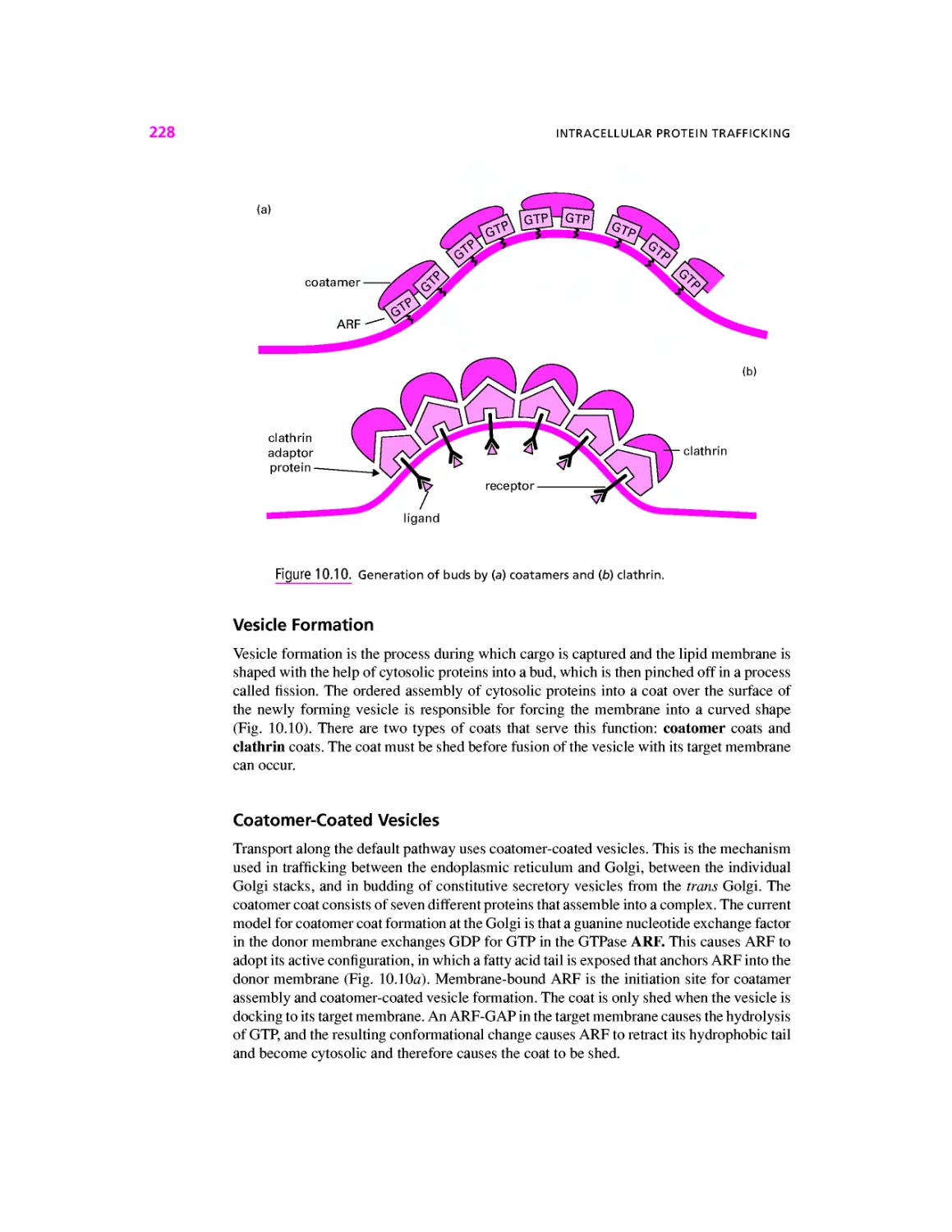
The Principle of Fission and Fusion, 226
Vesicle Formation, 228
Coatomer-Coated Vesicles, 228
Clathrin-Coated Vesicles, 229
The Trans-Golgi Network and Protein
Secretion, 229
Targeting Proteins to the Lysosome, 230
Fusion, 231
Summary, 232
Further Reading, 233
Review Questions, 233
Answers to Review Questions, 234
11 HOW PROTEINS WORK, 237
How Proteins Bind Other Molecules, 237
Dynamic Protein Structures, 238
Allosteric Effects, 238
Chemical Changes That Shift the
Preferred Shape of a Protein, 240
Enzymes Are Protein Catalysts, 241
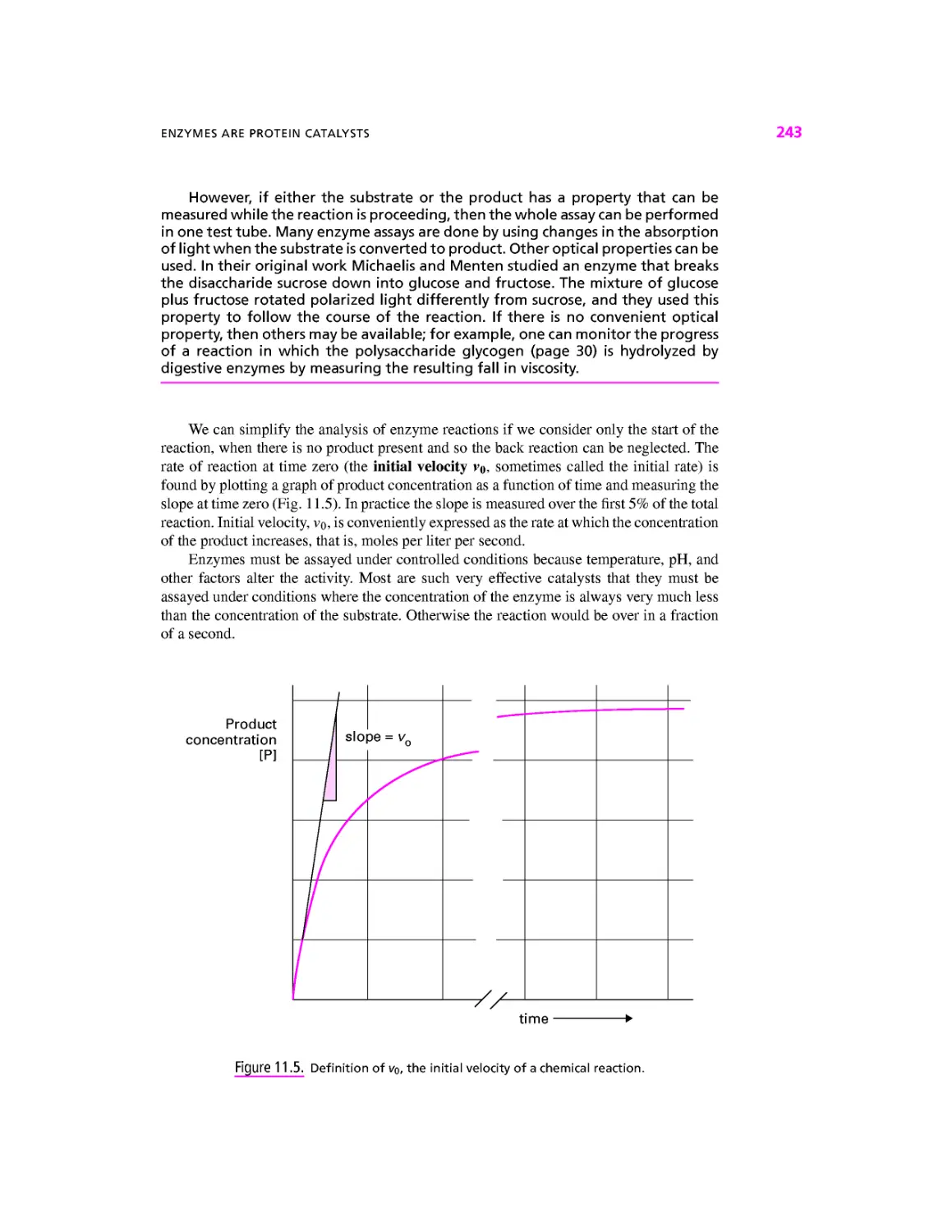
The Initial Velocity of an Enzyme
Reaction, 242
Effect of Substrate Concentration on
Initial Velocity, 244
The Effect of Enzyme Concentration, 245
The Specificity Constant, 247
Enzyme Catalysis, 247
Cofactors and Prosthetic Groups, 249
Enzymes Can Be Regulated, 251
Summary, 254
Further Reading, 254
Review Questions, 255
Answers to Review Questions, 256
12 ENERGY TRADING WITHIN THE
CELL, 257
Cellular Energy Currencies, 258
Reduced Nicotinamide Adenine
Dinucleotide (NADH), 259
Nucleoside Triphosphates (ATP plus
GTP, CTP, TTP, and UTP), 259
The Hydrogen Ion Gradient Across the
Mitochondrial Membrane, 261
The Sodium Gradient Across the Plasma
Membrane, 262
Energy Currencies Are Interconvertible, 263
Exchange Mechanisms Convert Between
the Four Energy Currencies, 263
Electron Transport Chain, 265
ATP Synthase, 269
Sodium/Potassium ATPase, 270
ADP/ATP Exchanger, 271
Photosynthesis, 271
All Carriers Can Change Direction, 275
Summary, 278
Further Reading, 278
Review Questions, 278
Answers to Review Questions, 279
13 METABOLISM, 281
The Krebs Cycle: The Central Switching
Yard of Metabolism, 283
CONTENTS
XI
From Glucose to Pyruvate: Glycolysis, 284
Glycolysis Without Oxygen, 286
Glycogen Can Provide Glucose for
Glycolysis, 288
Glucose May Be Oxidized to Produce
Pentose Sugars, 289
From Fats to Acetyl-CoA: p Oxidation, 290
Amino Acids as Another Source of
Metabolic Energy, 292
Making Glucose: Gluconeogenesis, 295
Making Glycogen: Glycogenesis, 298
Making Fatty Acids and Glycerides, 300
Synthesis of Amino Acids, 300
Carbon Fixation in Plants, 302
Control of Energy Production, 303
Feedback and Feedforward, 303
Negative Feedback Control of
Glycolysis, 304
Feedforward Control in Muscle
Cells, 304
Summary, 306
Further Reading, 306
Review Questions, 307
Answers to Review Questions, 308
14 IONS AND VOLTAGES, 309
The Potassium Gradient and the Resting
Voltage, 309
Potassium Channels Make the Plasma
Membrane Permeable to Potassium
Ions, 310
Concentration Gradients and Electrical
Voltage Can Balance, 311
The Chloride Gradient, 314
General Properties of Channels, 314
General Properties of Carriers, 316
The Glucose Carrier, 316
The Sodium-Calcium Exchanger, 317
Carriers with an Enzymatic Action:
The Calcium ATPase, 318
Summary, 322
Further Reading, 322
Review Questions, 322
Answers to Review Questions, 324
15 THE ACTION POTENTIAL, 325
The Calcium Action Potential in Sea Urchin
Eggs, 325
Effect of Egg Transmembrane Voltage on
Sperm Fusion, 325
The Voltage-Gated Calcium
Channel, 327
The Calcium Action Potential, 328
The Voltage-Gated Sodium Channel in
Nerve Cells, 330
The Voltage-Gated Sodium Channel, 330
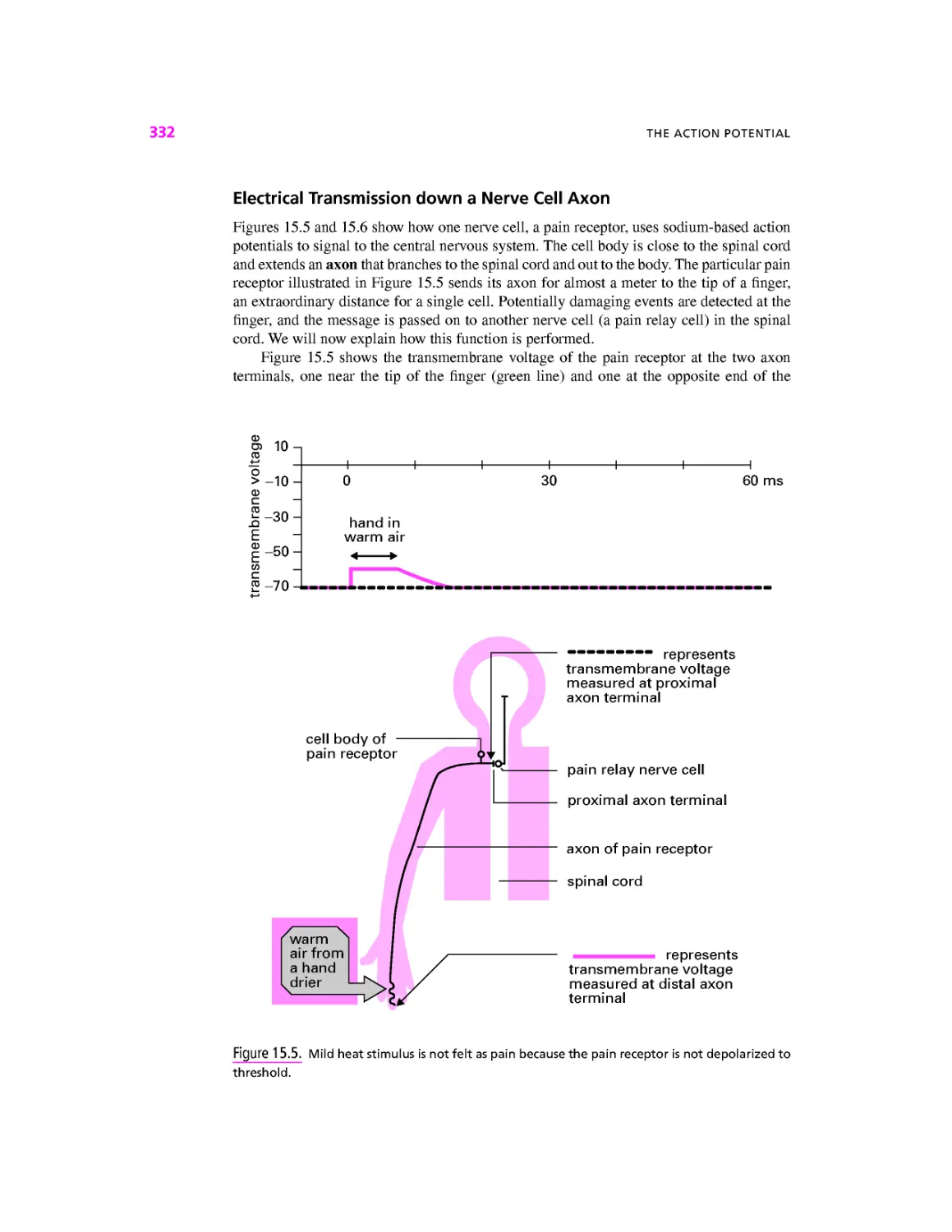
Electrical Transmission down a Nerve
CellAxon, 332
Myelination and Rapid Action Potential
Transmission, 334
Summary, 337
Further Reading, 338
Review Questions, 338
Answers to Review Questions, 339
16 INTRACELLULAR SIGNALING, 341
Calcium, 341
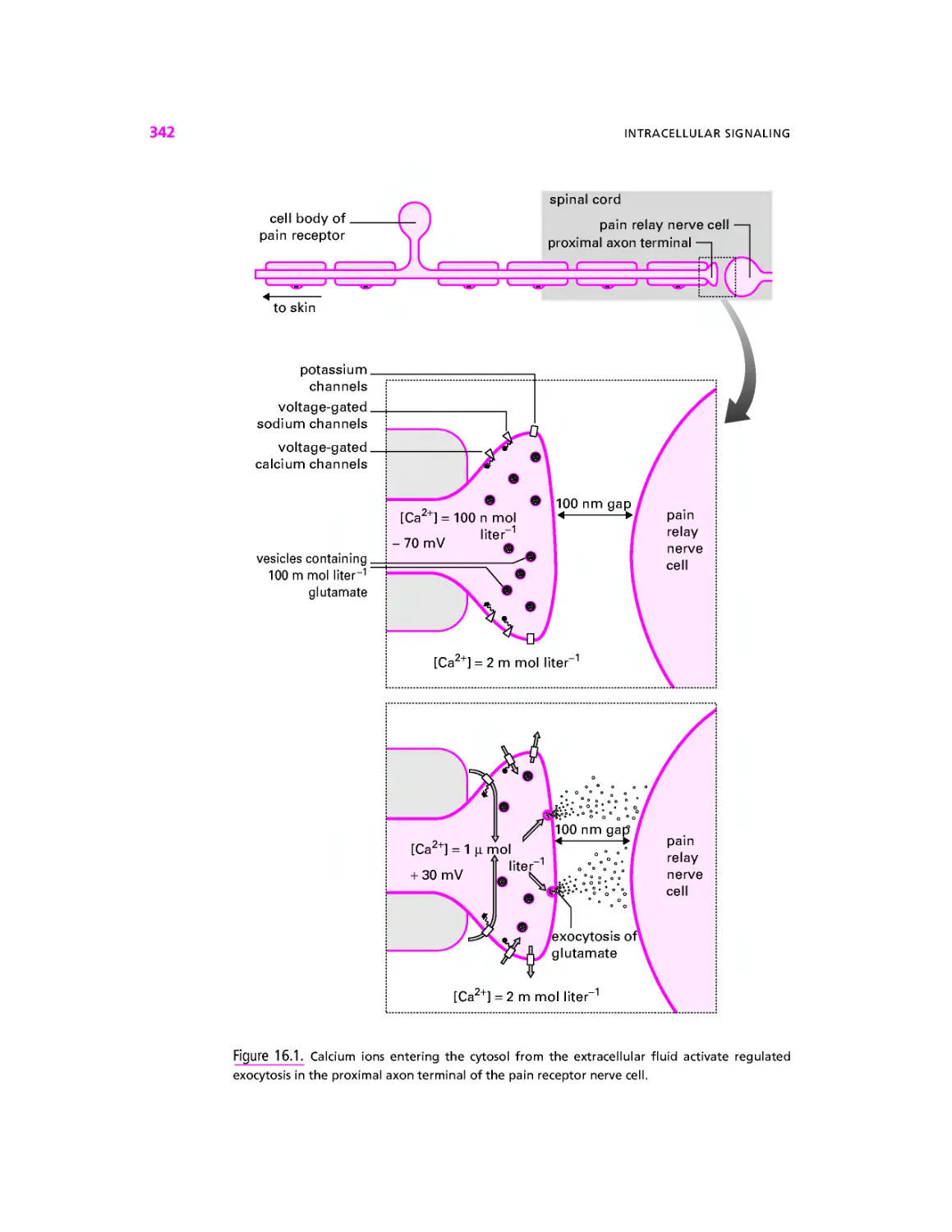
Calcium Can Enter from the Extracellular
Medium, 341
Calcium Can Be Released from the
Endoplasmic Reticulum, 344
Processes Activated by Cytosolic
Calcium Are Extremely Diverse, 348
Return of Calcium to Resting
Levels, 350
Cyclic Adenosine Monophosphate, 350
Cyclic Guanosine Monophosphate, 353
Multiple Messengers, 353
Biochemical Signaling, 353
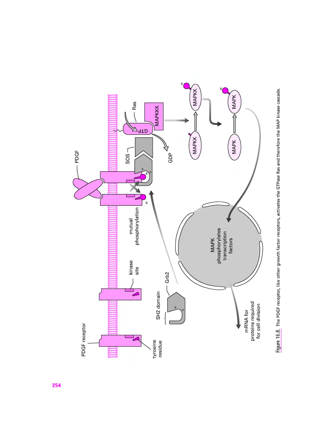
Receptor Tyrosine Kinases and the MAP
Kinase Cascade, 353
Growth Factors Can Trigger a Calcium
Signal, 356
Protein Kinase B and the Glucose
Transporter: How Insulin Works, 356
XII
CONTENTS
Crosstalk—Signaling Pathways or
Signaling Webs?, 357
Summary, 359
Further Reading, 360
Review Questions, 360
Answers to Review Questions, 361
17 INTERCELLULAR
COMMUNICATION, 363
Classifying Transmitters and Receptors, 363
Ionotropic Cell Surface Receptors, 364
Metabotropic Cell Surface
Receptors, 365
Intracellular Receptors, 365
Intercellular Communication in Action:
The Gastrocnemius Muscle, 365
Telling the Muscle to Contract:
The Action of Motoneurones, 367
Controlling the Blood Supply: Paracrine
Transmitters, 368
New Blood Vessels in Growing
Muscle, 371
Synapses Between Neurons, 372
Summary, 376
Further Reading, 377
Review Questions, 377
Answers to Review Questions, 378
18 MECHANICAL MOLECULES, 381
The Cytoskeleton is Both Strong and
Motile, 381
Microtubules, 381
Microtubule-Based Motility, 386
Cilia and Flagella, 386
Intracellular Transport, 389
Microfilaments, 390
Muscle Contraction, 393
Cell Locomotion, 395
Cytoplasmic Streaming, 395
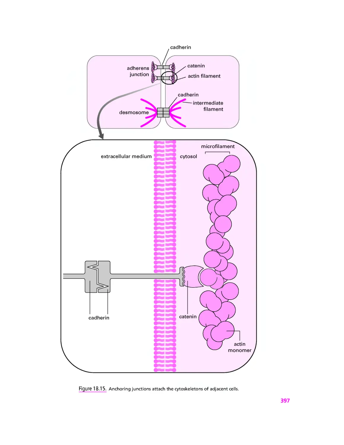
Intermediate Filaments, 396
Anchoring Cell Junctions, 396
Summary, 398
Further Reading, 398
Review Questions, 398
Answers to Review Questions, 400
19 CELL CYCLE AND CONTROL OF
CELL NUMBER, 401
Stages of Mitosis, 402
Meiosis and Fertilization, 404
Meiosis, 405
Fertilization and Inheritance, 406
Dominant Genetic Disease, 408
Crossing Over and Linkage, 408
Control of the Cell Division Cycle, 408
Molecular Regulation of the G2/M
(Interphase/Mitosis) Cell Cycle
Control Point, 410
What About the Gl/S Control Point?, 412
Apoptosis, 415
Instructed Death: Death Domain
Receptors, 416
Default Death: Absence of Growth
Factors, 416
The Sick Are Left to Die:
Stress-Activated Apoptosis, 417
Summary, 419
Further Reading, 420
Review Questions, 420
Answers to Review Questions, 421
20 CASE STUDY: CYSTIC
FIBROSIS, 423
Introduction, 423
Cystic Fibrosis is a Severe Genetic
Disease, 423
The Fundamental Lesion in Cystic Fibrosis
Lies in Chloride Transport, 424
Homing in on the CF Gene, 425
Cloning the Gene for CF, 426
The CFTR Gene Codes for a Chloride Ion
Channel, 426
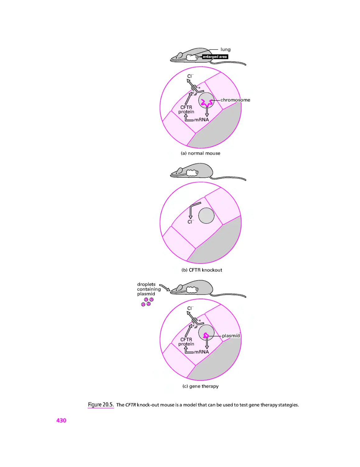
Gene Therapy for CF, 427
Diagnostic Tests for CF, 431
CONTENTS
XIII
The Future, 432
Summary, 433
Further Reading, 433
Review Questions, 434
Answers to Review Questions, 435
APPENDIX: CHANNELS AND
CARRIERS, 437
GLOSSARY, 441
INDEX, 501
PREFACE
Cell Biology, A Short Course aims to cover a wide area of cell biology in a form especially
suitable for first year undergraduates. We have deliberately kept the book to a manageable
size so that neither the cost, the content, nor the weight is too daunting for the student.
The overall theme for the book is the cell as the unit of life. We begin (Chapters 1-3)
by describing the components of the cell as seen under the microscope. We then (Chapters
4-8) turn to the central dogma of molecular biology and describe how DNA is used to make
RNA which in turn is used to make protein. The next section (Chapters 9-11) describes
how proteins are delivered to the appropriate location inside or outside the cell, and how
proteins perform their many functions. We then (Chapters 12-14) turn to cell energetics and
metabolism. Signaling within and between cells is covered in Chapters 15 through 17. To
conclude the book, Chapter 18 describes the composition and function of the cytoskeleton,
Chapter 19 covers cell birth and cell death, while Chapter 20 uses the example of the common
and severe genetic disease cystic fibrosis to illustrate many of the themes discussed earlier
in the book.
Boxed material throughout the book is divided into examples to illustrate the topics
covered in the main text, explanations of the medical relevance of the material, and in
depth sections that extend the coverage beyond the content of the main text. Questions are
provided at the end of each chapter to help the reader assess how well they have assimilated
and understood the material.
As well as giving references to printed material, we reference material available on
the internet in many places in the book. Rather than give detailed addresses, we provide
links to all these sites and many others from the book's homepage at
http://www.physiol.ucl.ac.uk/sbolsover/teaching/cbasc/cbasc.html.
XV
ACKNOWLEDGMENTS
We thank all the students, colleagues and family members who read the initial versions of
the book and whose suggestions and constructive criticism helped enormously.
XVII
INSTRUCTOR NOTES
Molecular cell biology courses now form a foundation for many subsequent specializations
in areas outside cell biology. We therefore cover molecular genetics, metabolic pathways
and electrophysiology in sufficient detail to make Cell Biology a suitable course book for
first year students who will later specialize in genetics, biochemistry, pharmacology or
physiology.
Each chapter comprises:
• The main text, with figures and tables.
• A numbered summary.
• Review questions with answers for student self-assessment. These questions concern
the main text only; no knowledge of the boxed material is required.
• Example boxes that illustrate the points made in the main text.
• Medical relevance boxes to show how basic cell biological knowledge illuminates
medical problems or has provided solutions.
• In Depth boxes that extend the content.
Self-assessment questions can form the basics for tutorials, with students asked to defend
the correct answer. They are also easily modified to generate new questions for student
assessment. Instructors are encouraged to submit new questions for inclusion on the CBASC
website.
Instructors may wish to specify parts of Cell Biology as core material for courses
targeted to particular specialties. The parts chosen can be customized to the particular
specialty in two ways:
1. By selecting from the complete set of twenty chapters. The following sections could
be used to support particular teaching modules:
Chapters 4 through 7 DNA, RNA and genetic engineering.
Chapters 8 through 10 Protein synthesis, structure and trafficking.
Chapters 11 through 13 Metabolism and cellular energetics.
Chapters 14 through 17 Electrophysiology and cell signaling.
Chapter 18 The cytoskeleton and cell motility.
Chapter 19 Cell division and apoptosis.
Chapters 16, 17 and 19 might in contrast be selected in a module concentrating
on the control of development, since these describe how growth factors and other
extracellular chemicals regulate cell division and cell death.
XIX
XX INSTRUCTOR NOTES
2. By including In Depth boxes. The following boxes are especially to be noted:
In Depth 1.2: Fluorescence Microscopy
In Depth 8.1: How We Study Proteins in One Dimension
describes SDS-PAGE
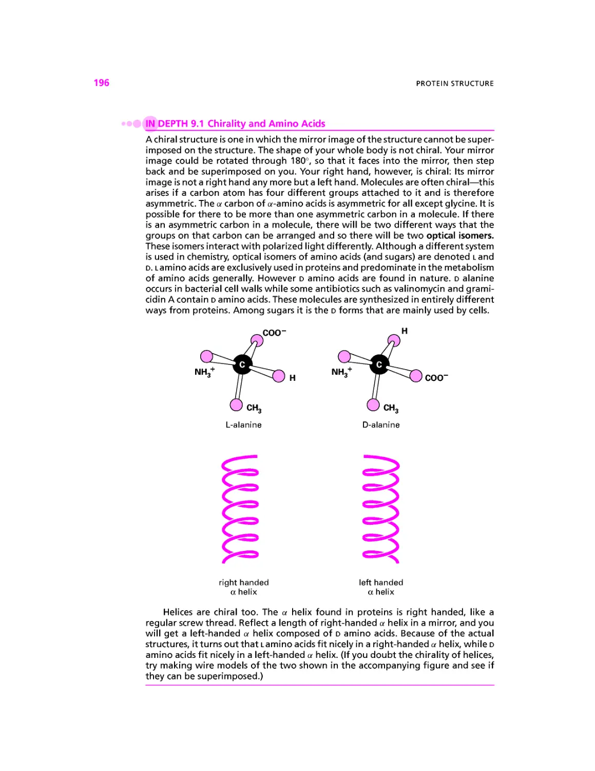
In Depth 9.1: Chirality and Amino Acids
In Depth 9.2: Hydropathy Plotting—The PDGF Receptor
In Depth 9.3: Curing Mad Mice with Smelly Fish
introduces the concept of osmolarity and osmosis and extends
the coverage of chaotropic and structure stabilizing agents
In Depth 11.1: What to Measure in an Enzyme Assay
In Depth 11.2: Determination of Vm and KM
the Lineweaver-Burk plot
In Depth 12.2: ATP Synthase, Rotary Motor, and Synthetic Machine
In Depth 12.3: Can It Happen? The Concept of Free Energy
In Depth 13.1: The Urea Cycle—The First Metabolic Cycle Discovered
In Depth 13.2: The Glyoxylate Shunt
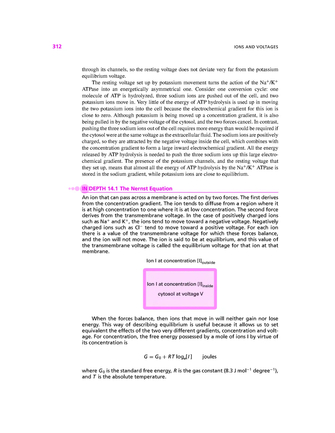
In Depth 14.1: The Nernst Equation
In Depth 14.2: Measuring the Transmembrane Voltage
In Depth 15.1: Frequency Coding in the Nervous System
In Depth 16.1: Ryanodine Receptors
In Depth 19.1: A Worm's Eye View of Cell Death
In Depth 20.1: Lipid Bilayer Voltage Clamp
For example, a course emphasizing protein structure would include In Depth 8.1,
9.1,9.2 and 9.3, while a course concentrating on metabolic pathways would include
In Depth 13.1 and 13.2.
The CBASC website is maintained by the authors. As well as providing over one hundred
links to sites with information that extends or illustrates the material in the book, we will use
the site to post typological or other errors, comments and test questions sent to us by read-
readers. The full address is http://www.physiol.ucl.ac.uk/sbolsover/teaching/cbasc/cbasc.html
or simply type 'CBASC into a search engine.
1
CELLS AND TISSUES
The cell is the basic unit of life. Microorganisms such as bacteria, yeast, and amoebae
exist as single cells. By contrast, the adult human is made up of about 30 trillion cells
A trillion = 1012) which are mostly organized into collectives called tissues. Cells are,
with a few notable exceptions, small (Fig. 1.1) with lengths measured in micrometers (/xm,
where 1000 /xm = 1 mm) and their discovery stemmed from the conviction of a small group
of seventeenth-century microscope makers that a new and undiscovered world lay beyond
the limits of the human eye. These pioneers set in motion a science and an industry that
continues to the present day.
The first person to observe and record cells was Robert Hooke A635-1703) who
described the cella (open spaces) of plant tissues. But the colossus of this era of discovery
was a Dutchman, Anton van Leeuwenhoek A632-1723), a man with no university education
but with unrivaled talents as both a microscope maker and as an observer and recorder of
the microscopic living world, van Leeuwenhoek was a contemporary and friend of the Delft
artist Johannes Vermeer A632-1675) who pioneered the use of light and shade in art at the
same time that van Leeuwenhoek was exploring the use of light to discover the microscopic
world. Sadly, none of van Leeuwenhoek's microscopes have survived to the present day.
Despite van Leeuwenhoek's Herculean efforts, it was to be another 150 years before, in 1838,
the botanist Matthias Schleiden and the zoologist Theodor Schwann formally proposed that
all living organisms are composed of cells. Their "cell theory," which nowadays seems
so obvious, was a milestone in the development of modern biology. Nevertheless general
acceptance took many years, in large part because the plasma membrane, the membrane
Cell Biology: A Short Course, Second Edition, by Stephen R. Bolsover, Jeremy S. Hyams,
Elizabeth A. Shephard, Hugh A. White, Claudia G. Wiedemann
ISBN 0-471-26393-1 Copyright © 2004 by John Wiley & Sons, Inc.
CELLS AND TISSUES
Figure 1.1. Dimensions of some example cells. 1 mm = 10~3 m; 1 /im = 10~6 m; 1 nm = 10~9 m.
surrounding the cell that divides the living inside from the nonliving extracellular medium
(Fig. 1.2) is too thin to be seen using a light microscope.
%$ PRINCIPLES OF MICROSCOPY
Microscopes make small objects appear bigger. A light microscope will magnify an image
up to 1500 times its original size. Electron microscopes can achieve magnifications up to
1 million times. However, bigger is only better when more details are revealed. The fineness
of detail that a microscope can reveal is its resolving power. This is defined as the smallest
distance that two objects can approach one another yet still be recognized as being separate.
The resolution that a microscope achieves is mainly a function of the wavelength of the illu-
illumination source it employs. The smaller the wavelength, the smaller the obj ect that will cause
diffraction, and the better the resolving power. The light microscope, because it uses visible
light of wavelength around 500 nanometers (nm, where 1000 nm = 1 /xm), can distinguish
objects as small as about half this: 250 nm. It can therefore be used to visualize the small-
smallest cells and the major intracellular structures or organelles. The microscopic study of cell
structure organization is known as cytology. An electron microscope is required to reveal the
ultrastructure (the fine detail) of the organelles and other cytoplasmic structures (Fig. 1.2).
The wavelength of an electron beam is about 100,000 times less than that of white
light. In theory, this should lead to a corresponding increase in resolution. In practice, the
PRINCIPLES OF MICROSCOPY
light microscope image
heterochromatin
electron microscope image
internal i
membranes
mitochondrial
ribosome
mitochondrion
peroxisome'
I
cytoplasmic ribosome
rough endoplasmic reticulum
lysosome
smooth
endoplasmic
reticulum
nuclear pore
nuclear envelope
10 u
Figure 1.2. Cell structure as seen through the light and transmission electron microscopes.
electron microscope can distinguish structures about 1000 times smaller than is possible in
the light microscope, that is, down to about 0.2 nm in size.
The Light Microscope
A light microscope (Figs. 1. 3a and 1.4) consists of a light source, which may be the sun or
an artificial light, plus three glass lenses: a condenser lens to focus light on the specimen,
an objective lens to form the magnified image, and a projector lens, usually called the
eyepiece, to convey the magnified image to the eye. Depending on the focal length of the
various lenses and their arrangement, a given magnification is achieved. In bright-field
CELLS AND TISSUES
light
source
|condenser lens
I objecti
ve lens
mirror -
introduced
for direct
viewing
by eye
light
projector
lens
image
viewed
directly
image
processing
computer
system
light
microscope
| display monitor]
(a)
transmission
electron
microscope
| display monitoi
(b)
| display monitor |
(c)
Figure 1.3. Basic design of light and electron microscopes.
Focussing system
Projector lenses
(eyepieces)
Objective lens
Specimen holder
Condenser lens
Light source
Figure 1.4. Simple upright light microscope.
PRINCIPLES OF MICROSCOPY
Bright field microscopy
-N
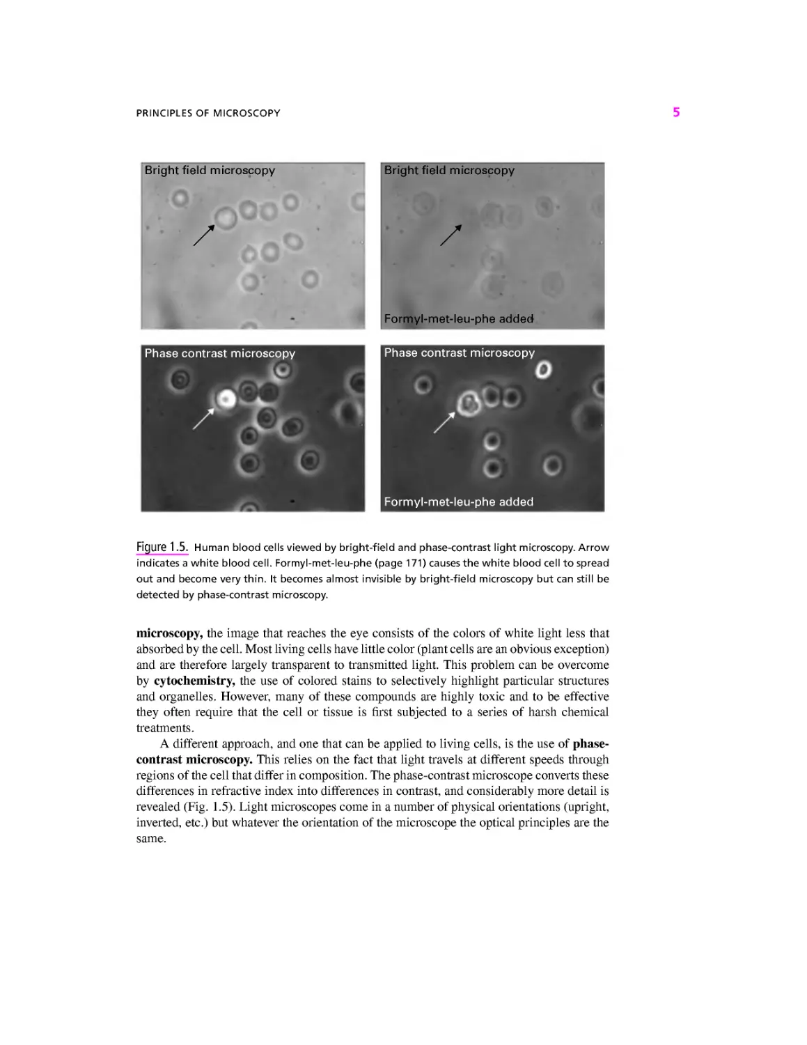
Figure 1.5. Human blood cells viewed by bright-field and phase-contrast light microscopy. Arrow
indicates a white blood cell. Formyl-met-leu-phe (page 171) causes the white blood cell to spread
out and become very thin. It becomes almost invisible by bright-field microscopy but can still be
detected by phase-contrast microscopy.
microscopy, the image that reaches the eye consists of the colors of white light less that
absorbed by the cell. Most living cells have little color (plant cells are an obvious exception)
and are therefore largely transparent to transmitted light. This problem can be overcome
by cytochemistry, the use of colored stains to selectively highlight particular structures
and organelles. However, many of these compounds are highly toxic and to be effective
they often require that the cell or tissue is first subjected to a series of harsh chemical
treatments.
A different approach, and one that can be applied to living cells, is the use of phase-
contrast microscopy. This relies on the fact that light travels at different speeds through
regions of the cell that differ in composition. The phase-contrast microscope converts these
differences in refractive index into differences in contrast, and considerably more detail is
revealed (Fig. 1.5). Light microscopes come in a number of physical orientations (upright,
inverted, etc.) but whatever the orientation of the microscope the optical principles are the
same.
CELLS AND TISSUES
IN DEPTH 1.1 Fluorescence Microscopy
Fluorescent molecules emit light when they are illuminated with light of a shorter
wavelength. Familiar examples are the hidden signature in bank passbooks, which
is written in fluorescent ink that glows blue (wavelength about 450 nm) when illu-
illuminated with ultraviolet light (UV) (wavelength about 360 nm), and the whitener
in fabric detergents that causes your white shirt to glow blue when illuminated
by the ultraviolet light in a club. The fluorescent dye Hoechst 33342 has a similar
wavelength dependence: It is excited by UV light and emits blue light. However, it
differs from the dyes used in ink or detergent in that it binds tightly to the DNA in
the nucleus and only fluoresces when so bound. Diagram a shows the optical path
through a microscope set up so as to look at a preparation stained with Hoechst.
White light from an arc lamp passes through an excitation filter that allows only
UV light to pass. This light then strikes the heart of the fluorescent microscope:
a special mirror called a dichroic mirror that reflects light of wavelengths shorter
than a designed cutoff but transmits light of longer wavelength. To view Hoechst,
we use a dichroic mirror of cutoff wavelength 400 nm, which therefore reflects
the UV excitation light up through the objective lens and onto the specimen. Any
Hoechst bound to DNA in the preparation will emit blue light. Some of this will be
(a) I
dichroic.
mirror
emission.
filter
mirror-
introduced
for direct
viewing
by eye
excitation
filter
projector
lens
image
viewed
directly
camera
z
image
processing
computer
system
| display monitor |
PRINCIPLES OF MICROSCOPY
captured by the objective lens and, because its wavelength is greater than 400 nm,
will not be reflected by the dichroic mirror but will instead pass through. An emis-
emission filter, set to pass only blue light, cuts out any scattered UV light. The blue light
now passes to the eye or camera in the usual way. Image b shows a field of cells
cultured from rat brain (gift of Dr. Charles Krieger, Simon Fraser University) after
staining with Hoechst. Only the nuclei are seen, as bright ovals.
Although some of the structures and chemicals found in cells can be selectively
stained by specific fluorescent dyes, others are most conveniently revealed by using
antibodies. In this technique an animal (usually a mouse, rabbit, or goat) is injected
with a protein or other chemical of interest. The animal's immune system recog-
recognizes the chemical as foreign and generates antibodies that bind to (and therefore
help neutralize) the chemical. Some blood is then taken from the animal and the
antibodies purified. The antibodies can then be labeled by attaching a fluorescent
dye. Images c and d show the same field of brain cells but with the excitation filter,
dichroic mirror, and emission filter changed so as to reveal in c a protein called
ELAV that is found only in nerve cells; then in d an intermediate filament protein
(page 000) found only in glial cells. The antibody that binds to ELAV is labeled with
a fluorescent dye that is excited by blue light and emits green light. The antibody
that binds to the glial filaments is labeled with a dye that is excited by green light
and emits red light. Because these wavelength characteristics are different, the
location of the three chemicals—DNA, ELAV, and intermediate filament—can be
revealed independently in the same specimen. See the CBASC website for an image
of all three signals in color and superimposed.
(d)
The technique just described is called primary immunofluorescence and re-
requires that the antibody to the chemical of interest be labeled with a dye. Only
antibodies to chemicals that many laboratories study are so labeled. In order to
reveal other chemicals, scientists use secondary immunofluorescence. In this ap-
approach, a commercial company injects an animal (e.g., a goat) with an antibody
from another animal (e.g., a rabbit). The goat then makes "goat anti rabbit" anti-
antibody. This, called the secondary antibody, is purified and labeled with a dye. All the
scientist has to do is make or buy a rabbit antibody that binds to the chemical of
interest. No further modification of this specialized, primary antibody is necessary.
Once the primary antibody has bound to the specimen and excess antibody rinsed
off, the specimen is then exposed to the secondary antibody that binds selectively
to the primary antibody. Viewing the stained preparation in a fluorescence micro-
microscope then reveals the location of the chemical of interest. The same dye-labeled
secondary antibody can be used in other laboratories or at other times to reveal
the location of many different chemicals because the specificity is determined by
the unlabeled primary antibody.
CELLS AND TISSUES
The Electron Microscope
The most commonly used type of electron microscope in biology is called the transmis-
transmission electron microscope because electrons are transmitted through the specimen to the
observer. The transmission electron microscope has essentially the same design as a light
microscope, but the lenses, rather than being glass, are electromagnets that bend beams
of electrons (Fig. 13b). An electron gun generates a beam of electrons by heating a thin,
V-shaped piece of tungsten wire to 3000°C. A large voltage accelerates the beam down
the microscope column, which is under vacuum because the electrons would be slowed
and scattered if they collided with air molecules. The magnified image can be viewed on a
fluorescent screen that emits light when struck by electrons. While the electron microscope
offers great improvements in resolution, electron beams are potentially highly destructive,
and biological material must be subjected to a complex processing schedule before it can
be examined. The preparation of cells for electron microscopy is summarized in Figure 1.6.
A small piece of tissue (~1 mm3) is immersed in glutaraldehyde
and osmium tetroxide. These chemicals bind all the component
parts of the cells together; the tissue is said to be fixed. It is
then washed thoroughly.
The tissue is dehydrated by soaking in acetone or ethanol.
The tissue is embedded in resin which is then baked hard.
Sections (thin slices less than 100 nm thick) are
cut with a machine called an ultramicrotome.
The sections are placed on a small copper grid and
stained with uranyl acetate and lead citrate. When
viewed in the electron microscope, regions that have
bound lots of uranium and lead will appear dark
because they are a barrier to the electron beam.
Figure 1.6. Preparation of tissue for electron microscopy.
ONLY TWO TYPES OF CELL
The transmission electron microscope produces a detailed image but one that is static,
two-dimensional, and highly processed. Often, only a small region of what was once a
dynamic, living, three-dimensional cell is revealed. Moreover, the picture revealed is essen-
essentially a snapshot taken at the particular instant that the cell was killed. Clearly, such images
must be interpreted with great care. Electron microscopes are large and require a skilled
operator. Nevertheless, they are the main source of information on the structure of the cell
at the nanometer scale, called the infrastructure.
The Scanning Electron Microscope
Whereas the image in a transmission electron microscope is formed by electrons transmitted
through the specimen, in the scanning electron microscope it is formed from electrons that
are reflected back from the surface of a specimen as the electron beam scans rapidly back
and forth over it (Fig. 1.3c). These reflected electrons are processed to generate a picture on
a display monitor. The scanning electron microscope operates over a wide magnification
range, from 10 x to 100,000x. Its greatest advantage, however, is a large depth of focus
that gives a three-dimensional image. The scanning electron microscope is particularly
useful for providing topographical information on the surfaces of cells or tissues. Modern
instruments have a resolution of about 1 nm.
IN DEPTH 1.2 Microscopy Rewarded
Such has been the importance of microscopy to developments in biology that two
scientists have been awarded the Nobel prize for their contributions to microscopy.
Frits Zernike was awarded the Nobel prize for physics in 1953 for the development
of phase-contrast microscopy and Ernst Ruska the same award in 1986 for the
invention of the transmission electron microscope. Ruska's prize marks one of the
longest gaps between a discovery (in the 1930s in the research labs of the Siemens
Corporation in Berlin) and the award of a Nobel prize. Anton van Leeuwenhoek
died almost two centuries before the Nobel prizes were introduced in 1901 and
the prize is not awarded posthumously.
•$ ONLY TWO TYPES OF CELL
Superficially at least, cells exhibit a staggering diversity. Some lead a solitary existence;
others live in communities; some have defined, geometric shapes; others have flexible
boundaries; some swim, some crawl, and some are sedentary; many are green (some are
even red, blue, or purple); others have no obvious coloration. Given these differences, it is
perhaps surprising that there are only two types of cell (Fig. 1.7). Bacterial cells are said to
be prokaryotic (Greek for "before nucleus") because they have very little visible internal
organization so that, for instance, the genetic material is free within the cell. They are also
small, the vast majority being 1-2 /xm in length.
The cells of all other organisms, from protists to mammals to fungi to plants, are eu-
karyotic (Greek for "with a nucleus"). These are generally larger E-100 /xm, although some
eukaryotic cells are large enough to be seen with the naked eye; Fig. 1.1) and structurally
more complex. Eukaryotic cells contain a variety of specialized structures known collec-
collectively as organelles, surrounded by a viscous substance called cytosol. The largest organelle,
the nucleus, contains the genetic information stored in the molecule deoxyribonucleic
10
CELLS AND TISSUES
prokaryotic
bacterial cell
ribosome
DNA
plasma membrane
1 nm
eukaryotic
Golgi apparatus
mitochondrion
rough
endoplasmic
reticulum
plasma
membrane
I 10 urn
plasmodesma-
cell wall-
chloroplast-
| plant cell |
Figure 1.7. Organization of prokaryotic and eukaryotic cells.
acid (DNA). The structure and function of organelles will be described in detail in subse-
subsequent chapters. Table 1.1 provides a brief glossary of the major organelles and summarizes
the differences between prokaryotic and eukaryotic cells.
Example Box 1.1
Sterilization by Filtration
Because even the smallest cells are larger than 1 /xm, harmful bacteria and protists can be removed
from drinking water by passing through a filter with 200-nm-diameter holes. Filters can vary in
size from huge, such as those used in various commercial processes, to small enough to be easily
transportable by backpackers. Filtering drinking water greatly reduces the chances of bringing back
an unwanted souvenir from your camping trip!
VIRUSES
11
Table 1.1. Differences Between Prokaryotic and Eukaryotic Cells
Size
Nucleus
DNA
Cell division
Internal membranes
Ribosomes
Cytoskeleton
Motility
First appeared
Prokaryotes
Usually 1-2 /xm
Absent
Usually a single circular
molecule (^chromosome)
Simple fission
Rare
70S*
Absent
Rotary motor (drives bacterial
flagellum)
3.5 x 10⁹ years ago
Eukaryotes
Usually 5-100/xm
Present, bounded by nuclear envelope
Multiple molecules (^chromosomes),
linear, associated with protein."
Mitosis or meiosis
Complex (nuclear envelope, Golgi
apparatus, endoplasmic reticulum,
etc.—Fig. 1.2)
80S G0S in mitochondria
and chloroplasts)
Microtubules, microfilaments,
intermediate filaments
Dynein (drives cilia and eukaryote
flagellum); kinesin, myosin
1.5 x 10⁹ years ago
" The tiny chromosomes of mitochondria and chloroplasts are exceptions; like prokaryotic chromosomes they are
often circular.
*The S value, or Svedberg unit, is a sedimentation rate. It is a measure of how fast a molecule moves in a
gravitational field, and therefore in an ultracentrifuge.
Special Properties of Plant Cells
Among eukaryotic cells the most striking difference is between those of animals and plants
(Fig. 1.7). Plants have evolved a sedentary lifestyle and a mode of nutrition that means
they must support a leaf canopy. Their cells are enclosed within a rigid cell wall that gives
shape to the cell and structural rigidity to the organism (page 53). This is in contrast to
the flexible boundaries of animal cells. Plant cells frequently contain one or more vacuoles
that can occupy up to 75% of the cell volume. Vacuoles accumulate a high concentration
of sugars and other soluble compounds. Water enters the vacuole to dilute these sugars,
generating hydrostatic pressure that is counterbalanced by the rigid wall. In this way the
cells of the plant become stiff or turgid, in the same way that when an inner tube is inflated
inside a bicycle tire the combination becomes stiff. Vacuoles are often pigmented, and
the spectacular colors of petals and fruit reflect the presence of compounds such as the
purple anthocyanins in the vacuole. Cells of photosynthetic plant tissues contain a special
organelle, the chloroplast, that houses the light-harvesting and carbohydrate-generating
systems of photosynthesis (page 271). Plant cells lack centrosomes (page 382) (Fig. 1.2)
although these are found in many algae.
# VIRUSES
Viruses occupy a unique space between the living and nonliving worlds. On one hand they
are made of the same molecules as living cells. On the other hand they are incapable of
independent existence, being completely dependent on a host cell to reproduce. Almost
all living organisms have viruses that infect them. Human viruses include polio, influenza,
herpes, rabies, ebola, smallpox, chickenpox, and the AIDS (acquired immunodeficiency
12 CELLS AND TISSUES
syndrome) virus HIV (human immunodeficiency virus). Viruses are submicroscopic parti-
particles consisting of a core of genetic material enclosed within a protein coat called the capsid.
Some viruses have an extra membrane layer called the envelope. Viruses are metabolically
inert until they enter a host cell, whereupon the viral genetic material directs the host cell
machinery to produce viral protein and viral genetic material. Viruses often insert then-
genome into that of the host, an ability that is widely made use of in molecular genet-
genetics (Chapter 7). Bacterial viruses, called bacteriophages (page 74), are used by scientists to
transfer genes between bacterial strains. Human viruses are used as vehicles for gene therapy.
By exploiting the natural infection cycle of a virus such as adenovirus, it is possible to in-
introduce a functional copy of a human gene into a patient suffering from a genetic disease
such as cystic fibrosis (Chapter 20).
4p> ORIGIN OF EUKARYOTIC CELLS
Prokaryotic cells are simpler and more primitive in their organization than eukaryotic cells.
According to the fossil record, prokaryotic organisms antedate, by at least 2 billion years,
the first eukaryotes that appeared some 1.5 billion years ago. It seems highly likely that
eukaryotes evolved from prokaryotes, and the most likely explanation of this process is the
endosymbiotic theory. The basis of this hypothesis is that some eukaryotic organelles orig-
originated as free-living prokaryotes that were engulfed by larger cells in which they established
a mutually beneficial relationship. For example, mitochondria would have originated as
free-living aerobic bacteria and chloroplasts as cyanobacteria, photosynthetic prokaryotes
formerly known as blue-green algae.
The endosymbiotic theory provides an attractive explanation for the fact that both mito-
mitochondria and chloroplasts contain DNA and ribosomes of the prokaryotic type (Table 1.1).
The case for the origin of other eukaryotic organelles is less persuasive. While it is clearly
not perfect, most biologists are now prepared to accept that the endosymbiotic theory pro-
provides at least a partial explanation for the evolution of the eukaryotic cell from a prokaryotic
ancestor. Unfortunately, living forms having a cellular organization intermediate between
prokaryotes and eukaryotes are rare. Some primitive protists possess a nucleus but lack
mitochondria and other typical eukaryotic organelles. They also have the prokaryotic type
of ribosomes. These organisms are all intracellular parasites and they include Microspora,
an organism that infects AIDS patients.
•?P> CELL SPECIALIZATION
All the body cells that comprise a single organism share the same set of genetic instructions
in their nuclei. Nevertheless, the cells are not all identical. Rather, plants and animals are
composed of different tissues, groups of cells that are specialized to carry out a common
function. This specialization occurs because different cell types read out different parts of the
DNA blueprint and therefore make different proteins, as we will see in Chapter 6. In animals
there are four major tissue types: epithelium, connective tissue, nervous tissue, and muscle.
Epithelia
Epithelia are sheets of cells that cover the surface of the body and line its internal cavities
such as the lungs and intestine. The cells may be columnar, taller than they are broad, or
CELL SPECIALIZATION 13
squamous, meaning flat. In the intestine, the single layer of columnar cells lining the inside,
or lumen, has an absorptive function that is increased by the folding of the surface into villi
(Fig. 1.8). The luminal surfaces of these cells have microvilli that increase the surface area
even further. The basal surface sits on a supporting layer of extracellular fibers called the
basement membrane. Many of the epithelial cells of the airways, for instance, those lining
the trachea and bronchioles, have cilia on their surfaces. These are hairlike appendages that
actively beat back and forth, moving a layer of mucus away from the lungs (Chapter 18).
Particles and bacteria are trapped in the mucus layer, preventing them from reaching the
delicate air exchange membranes in the lung. In the case of the skin, the epithelium is said
to be stratified because it is composed of several layers.
Medical Smoking Is Bad for Your Cilia
Relevance j^g c^a on me epithelial cells that line our airways move a belt of mucus that
p y
'¦' carries trapped particles and microorganisms away from the lungs. These cilia are
paralyzed by cigarette smoke. As a consequence, smokers have severely reduced mucociliary
clearance with the result that they have to cough to expel the mucus that continuously accumulates
in their lungs. Because of this impaired ciliary function, smokers are much more susceptible to
respiratory diseases such as emphysema and asthma. The cilia of passive smokers, people who
breathe smoky air, are also affected.
Connective Tissue
Connective tissues provide essential support for the other tissues of the body. They include
bone, cartilage, and adipose (fat) tissue. Unlike other tissues, connective tissue contains
relatively few cells within a large volume of extracellular matrix that consists of different
types of fiber embedded in amorphous ground substance (Fig. 1.8). The most abundant of
the fibers is collagen, a protein with the tensile properties of steel that accounts for about a
third of the protein of the human body. Other fibers have elastic properties that permit the
supported tissues to be displaced and then to return to their original position. The amorphous
ground substance absorbs large quantities of water, facilitating the diffusion of metabolites,
oxygen, and carbon dioxide to and from the cells in other tissues and organs. Of the many
cell types found in connective tissue, two of the most important are fibroblasts, which
secrete the ground substance and fibers, and macrophages, which remove foreign, dead,
and defective material from it. A number of inherited diseases are associated with defects
in connective tissue. Marfan's syndrome, for example, is characterized by long arms, legs,
and torso and by a weakness of the cardiovascular system and eyes. These characteristics
result from a defect in the organization of the collagen fibers.
Nervous Tissue
Nervous tissue is a highly modified epithelium that is composed of several cell types.
Principal among these are the nerve cells, also called neurons, along with a variety of
supporting cells that help maintain them. Neurons extend processes called axons, which
can be over a meter in length. Neurons constantly monitor what is occurring inside and
outside the body. They integrate and summarize this information and mount appropriate
responses to it (Chapters 15-17). Another type of cell called glia has other roles in nervous
tissue including forming the electrical insulation around axons.
14
CELLS AND TISSUES
one
microvillus
brush border
brush
border
epithelial
cell
tight junction
nucleus
one villus
connective
tissue
fibroblast
collagen
fibers
extracellular
matrix
capillary
red blood cell
smooth muscle cells
orientated circularly
around intestine
smooth muscle cells
orientated longitudinally
along intestine (cut in
cross section)
Figure 1.8. Tissues and structures of the intestine wall.
Muscle
Muscle tissue can be of two types, smooth or striated. Smooth muscle cells are long
and slender and are usually found in the walls of tubular organs such as the intestine and
many blood vessels. In general, smooth muscle cells contract slowly and can maintain
the contracted state for a long period of time. There are two classes of striated muscle:
cardiac and skeletal. Cardiac muscle cells make up the walls of the heart chambers. These
CELL SPECIALIZATION
15
D
cortex
parenchyma
cortex cell
epidermal cell
upper surface
of leaf
— cuticle
epidermis
|— parenchyma
cells
under surface
of leaf
Figure 1.9. Plants are composed of several tissues.
are branched cells that are connected electrically by gap junctions (page 55), and then-
automatic rhythmical contraction powers the beating of the heart. Each skeletal muscle is
a bundle of hundreds to thousands of fibers, each fiber being a giant single cell with many
nuclei. This rather unusual situation is the result of an event that occurs in the embryo when
the cells that give rise to the fibers fuse together, pooling their nuclei in a common cytoplasm.
The mechanism of contraction of skeletal muscle will be described in Chapter 18.
Plants
Plant cells are also organized into tissues (Fig. 1.9). The basic organization of a shoot or root
is into an outer protective layer, or epidermis, a vascular tissue that provides support and
16
CELLS AND TISSUES
transport, and a cortex that fills the space between the two. The epidermis consists of one or
more layers of closely packed cells. Above the ground these cells secrete a waxy layer, the
cuticle, which helps the plant retain water. The cuticle is perforated by pores called stomata
that allow gas exchange between the air and the photosynthetic cells and also constitute the
major route for water loss by a process called transpiration. Below ground, the epidermal
cells give rise to root hairs that are important in the absorption of water and minerals. The
vascular tissue is composed of xylem, which transports water and its dissolved solutes
from the roots, and phloem, which conveys the products of photosynthesis, predominantly
sugars, to their site of use or storage. The cortex consists primarily of parenchyma cells,
unspecialized cells whose cell walls are usually thin and bendable. They are the major site
of metabolic activity and photosynthesis in leaves and green shoots.
SUMMARY
1. All living organisms are made of cells.
2. Our understanding of cell structure and function has gone hand in hand with devel-
developments in microscopy and its associated techniques.
3. Light microscopy revealed the diversity of cell types and the existence of the major
organelles: nucleus, mitochondrion and, in plants, the vacuole and chloroplast.
4. The electron microscope revealed the detailed structure of the larger organelles and
resolved the cell ultrastructure, the fine detail at the nanometer scale.
5. There are two types of cells, prokaryotes and eukaryotes.
6. Prokaryotic cells have very little visible internal organization. They usually measure
1-2 /xm across.
7. Eukaryotic cells usually measure 5-100 /xm across. They contain a variety of spe-
specialized internal organelles, the largest of which, the nucleus, contains the genetic
material.
8. The endosymbiotic theory proposes that some eukaryotic organelles, such as mito-
mitochondria and chloroplasts, originated as free-living prokaryotes.
9. The cells of plants and animals are organized into tissues. In animals there are four
tissue types: epithelium, connective tissue, nervous tissue, and muscle. Plants are
formed of epidermis, cortex, and vascular tissues.
REVIEW QUESTIONS
For each question, choose the ONE BEST answer or completion.
1. Eukaryotic cells usually contain
A. a nucleus.
B. mitochondria.
C. ribosomes.
ANSWERS TO REVIEW QUESTIONS 17
D. microtubules.
E. all of the above.
2. The transmission electron microscope
A. is ideal for looking at living cells.
B. has a resolution of 0.2 nm.
C. uses lenses made of glass.
D. was invented by Anton van Leeuwenhoek.
E. is easily transportable.
3. Which of the following statements about tissues is incorrect?
A. Microvilli are typically associated with nervous tissue.
B. Columnar and squamous are types of epithelial tissue.
C. Collagen is a component of the extracellular matrix.
D. Glial cells form the insulation around nerve cells.
E. Epidermis, vascular tissue, and cortex are types of plant tissue.
4. 1,000,000 nm is equal to
A. 1 /xm.
B. 10 Aim.
C. 1 mm.
D. 10 mm.
E. 1 m.
5. Unlike eukaryotes, prokaryotes lack
A. a plasma membrane.
B. DNA.
C. ribosomes.
D. nuclei.
E. molecular motors.
6. Light passes through a light microscope in the following order:
A. condenser, specimen, objective, projector.
B. condenser, objective, specimen, projector.
C. condenser, projector, specimen, objective.
D. objective, specimen, projector, condenser.
E. objective, projector, specimen, condenser.
7. Which of the following is not a type of muscle cell?
A. Smooth.
B. Ciliated.
C. Striated.
D. Cardiac
E. Skeletal.
ANSWERS TO REVIEW QUESTIONS
1. E. These are all typical features of eukaryotic cells.
2. B. None of the others are true. In particular the fact that an electron beam is used means that the
lenses are electromagnets, not glass, and means that the interior of an electron microscope is held
at a vacuum, which means in turn that it is not a suitable environment for living tissue and tends
to mean that electron microscopes are bulky, heavy pieces of equipment.
18 CELLS AND TISSUES
3. A. Microvilli are found on epithelia, for example, the epithelium lining the intestine.
4. C. The international system prefixes used in this book are m for milli, meaning 10~3, /x for micro,
meaning 10, and n for nano meaning 10"9. Thus 1,000,000 times 10"9 is 10 so 1,000,000 nm
is equal to 1 mm.
5. D. Eukaryotes have all of these, but prokaryotes do not have nuclei. Rather, the DNA is free in the
cytoplasm.
6. A
7. B. No muscle cells are ciliated; as we will see in Chapter 18, the molecular motor systems used
in cilia and in muscles are different. Muscle cells are divided into smooth and striated; the striated
type is then further subdivided into cardiac and skeletal.
2
FROM WATER TO DNA: THE
CHEMISTRY OF LIFE
Organisms are made up of a lot of different chemicals. These vary in size, from small
molecules like water to large molecules like DNA, and interact and associate in many
different ways to generate the processes of life. In this chapter we will introduce some basic
concepts of how these chemicals are made and interact. We will then describe the most
important of these chemicals: water, carbohydrates, nucleotides, amino acids, and lipids.
•$ THE CHEMICAL BOND: SHARING ELECTRONS
Water is the most abundant substance in organisms. Cells are rich in water. Cytoplasm
consists of organelles floating in a watery medium called cytosol that also contains proteins.
The situation is not so different outside our cells. Although we are land animals living in
air, most of our cells are bathed in a watery fluid called extracellular medium. We will
therefore start by considering water itself.
Figure 2.1a shows a molecule of water, consisting of one atom of oxygen and two
hydrogen atoms, joined to form an open V shape. The lines represent covalent bonds
formed when atoms share electrons, each seeking the most stable structure. Oxygen has a
greater affinity for electrons than does hydrogen so the electrons are not distributed equally.
The oxygen grabs a greater share of the available negative charge than do the hydrogen
atoms. The molecule of water is polarized, with partial negative charge on the oxygen and
partial positive charges on the two hydrogens. We write the charge on each hydrogen as 8+
Cell Biology: A Short Course, Second Edition, by Stephen R. Bolsover, Jeremy S. Hyams,
Elizabeth A. Shephard, Hugh A. White, Claudia G. Wiedemann
ISBN 0-471-26393-1 Copyright © 2004 by John Wiley & Sons, Inc.
19
20
FROM WATER TO DNA: THE CHEMISTRY OF LIFE
5+
(b)
Figure 2.1. Water is a polar molecule while the chlorine molecule is nonpolar.
to indicate that it is smaller than the charge on a single hydrogen nucleus. The oxygen atom
has the small net negative charge 28—. Molecules that, like water, have positive regions
sticking out one side and negative regions sticking out the other are called polar.
Figure 2.1b shows a molecule of chlorine gas. It consists of two chlorine atoms, each of
which consists of a positively charged nucleus surrounded by negatively charged electrons.
Like oxygen, chlorine atoms tend to accept electrons when they become available, but the
battle is equal in the chlorine molecule: The two atoms share their electrons equally and the
molecule is nonpolar.
Na
Cl
Na+
=> cr
Cl
Na
=> cr
=> Na+
(a)
©000©
(b)
Figure 2.2. Formation of sodium chloride, an ionic compound.
INTERACTIONS WITH WATER: SOLUTIONS 21
Figure 2.2a shows what happens when a chlorine molecule is allowed to react with
the metal sodium. Each atom of chlorine takes over one electron from a sodium atom. This
leaves the sodium atoms with a single positive charge because there is now one more positive
charge on the sodium nucleus than negatively charged surrounding electrons. Similarly,
each chlorine atom now has a single negative charge because it now has one more electron
than there are positive charges in its nucleus. Chemical species that have either gained or
lost electrons, and that therefore bear an overall charge, are called ions. The reaction of
chlorine and sodium has produced sodium ions and chloride ions. Positively charged ions
like sodium are called cations while negatively charged ones like chloride are called anions.
The positively charged sodium ions and the negatively charged chloride ions now attract
each other strongly. If there are no other chemicals around, the ions will arrange themselves
to minimize the distance between sodium and chloride, and the resulting well-packed array
of ions is a crystal of sodium chloride, shown in Figure 2.2b.
3$ INTERACTIONS WITH WATER: SOLUTIONS
Ionic Compounds Will Dissolve Only in Polar Solvents
Figure 2.3a shows one molecule of octane, the main constituent of gasoline. Octane is an
example of a nonpolar solvent. Electrons are shared equally between carbon and hydrogen,
and the component atoms do not bear a net charge.
Figure 2.3b shows a small crystal of sodium chloride immersed in octane. At the edge
of the crystal, positively charged sodium ions are being pulled in toward the center of
the crystal by the negative charge on chloride ions, and negatively charged chloride ions
are being pulled in toward the center of the crystal by the positive charge on sodium ions.
The sodium and chloride ions will not leave the crystal. Sodium chloride is insoluble in
octane. However, sodium chloride will dissolve in water, and Figure 2.4a shows why. The
chloride ion at the top left is being pulled into the crystal by the positive charge on its sodium
ion neighbors, but at the same time it is being pulled out of the crystal by the positive charge
on the hydrogen atoms of nearby water molecules. Similarly, the sodium ion at the bottom
left is being pulled into the crystal by the negative charge on its chloride ion neighbors, but
at the same time it is being pulled out of the crystal by the negative charge on the oxygen
atoms of nearby water molecules. The ions are not held in the crystal so tightly and can
leave. Once the ions have left the crystal, they become surrounded by a hydration shell of
water molecules, all oriented in the appropriate direction (Fig. 2Ab)—oxygen inward for
a positive ion like sodium, hydrogen inward for a negative ion like chloride. A chemical
species in solution, whether in water or in any other solvent, is called a solute. Liquids
whose main constituent is water are called aqueous.
Acids Are Molecules That Give H+ to Water
When we exercise, our muscle cells can become acid, and this is what creates the pain
of cramping muscles and the heart pain of angina. Acidity is important in all areas of
biology, from the acidity gradient that drives our mitochondria (page 261) to the ecological
consequences of acid rain.
Acid solutions contain a high concentration of hydrogen ions. The hydrogen atom is
unusual in that it only has one electron while, in its most common isotope, its nucleus
22
FROM WATER TO DNA: THE CHEMISTRY OF LIFE
O0O0G
(b)
Figure 2.3. (a) Structure of the nonpolar compound octane. (?>) Ionic compounds are insoluble in
nonpolar solvents.
comprises a single proton. In gasses at very low pressure it is possible for bare protons
to exist alone and be manipulated, for example, in linear accelerators. However, in water
protons never exist alone but always associate with another molecule, for example, with
water to create the H3O+ ion. Acid solutions are those with an H3O+ concentration higher
than 100 nmol liter.
Sour cream contains lactic acid. Pure lactic acid has the structure shown at the left of
Figure 2.5a. The —COOH part in the box is called a carboxyl group. Both oxygens have a
tendency to pull electrons away from the hydrogen and, in aqueous solution, the hydrogen
is donated with a full positive charge to a molecule of water. The electron is left behind on
the now negatively charged lactate ion.
CH3CH(OH)COOH + H2O -+ CH3CH(OH)COO" + H3O+
INTERACTIONS WITH WATER: SOLUTIONS
23
a)
® ®
(b)
Figure 2.4. Ionic compounds dissolve readily in water.
24
FROM WATER TO DNA: THE CHEMISTRY OF LIFE
H OH OH
I I I
H—C — C—C=O
I I
H H
lactic acid
CH,
CH3—N
CH3
trimethylamine
H2O H3O+
(a)
H OH O~
I I I
=> H—C—C—C=O
I I
H H
lactate ion
CH3
=> CH,—N+—H
CH3
protonated
trimethylamine ion
(b)
Figure 2.5. Acids and bases, respectively, give up and accept H+ when dissolved in water.
For convenience we often write this as
CH3CH(OH)COOH -+ CH3CH(OH)COCT + H+
Here we are using H+ as a convenient symbol to denote H3O+. We do not mean that there
are real H+ ions, that is, bare hydrogen nuclei, in aqueous solutions.
The equilibrium constant, Ka for the dissociation of lactic acid, is defined as
[CH3CH(OH)COO"]e[H+]e
[CH3CH(OH)COOH]e
where the square brackets refer, by convention, to concentrations and the subscripts, e,
denote that these are the concentrations of each species at equilibrium.
Dissolving lots of lactic acid in water produces an acid solution, that is, one with a high
concentration of H+ (really H3O+) ions. For historical reasons, the acidity of a solution is
given as the pH, defined thus:
pH = — log10 ([H+]) where [H+] is measured in moles per liter
Pure water has a pH of 7, corresponding to [H+] = 100 nmol liter—this is said to be
neutral as regards pH. If the pH is lower than this, then there is more H+ about and the
solution is acid. Cytosol has a pH that lies very slightly on the alkaline side of neutrality, at
about 7.2.
The pH of a solution determines the ratio of lactate to undissociated lactic acid. As
the H+ concentration rises and pH falls, the equilibrium is pushed over from lactate toward
lactic acid. AtpH 3.9, the concentrations of lactate and lactic acid become equal. Looking at
INTERACTIONS WITH WATER: SOLUTIONS 25
the equation above, we can therefore see that when this happens Ka = [H+]. Just as acidity
is given on the logarithmic pH scale so is the scale of strengths of different acids, p Ka being
defined as — log10 Ka. The pKa is the pH at which the concentration of the dissociated acid
is equal to the concentration of the undissociated acid. For an acid that is weaker than lactic
acid, the pKa is higher than 3.9, meaning that the acid is less readily dissociated and needs
a lower concentration of protons before it will give up its H+. For an acid that is stronger
than lactic acid, the pKa is less than 3.9: Such an acid is more readily dissociated and needs
a higher H+ concentration before it will accept H+ and form undissociated acid.
Bases Are Molecules That Take H+ from Water
TMmethylamine is the compound that gives rotting fish its unpleasant smell. Pure trimethy-
lamine has the structure shown at the left of Figure 2.5b. When trimethylamine is dissolved
in water, it accepts an H+ to become the positively charged trimethylamine ion shown on the
right of the figure. We refer to molecules that have accepted H+ ions as protonated, using
"proton" as a short way of saying "hydrogen nucleus." Dissolving lots of trimethylamine
in water produces an alkaline solution, that is, one with a low concentration of H+ ions and
hence a pH greater than 7. The solution never runs out of H+ completely because new H+
are formed from water:
H2O -+ OH" + H+
Thus if we keep adding trimethylamine to water and using up H+, we end up with a low
concentration of H+, but lots of OH~.
The pH of a solution determines the position of equilibrium between protonated and
deprotonated trimethylamine, and as before we define the pKa as the pH at which the
concentration of protonated and deprotonated base are the same. The pKa of trimethylamine
is 9.74, meaning that the concentration of H+ must fall to the low level of 10~974 mol liter,
that is, 0.2 nmol liter, before half of the trimethylamines will give up their H+s.
Isoelectric Point
The large molecules called proteins (page 183) have many acidic and basic sites that will
give up or accept an H+ as the pH changes. In alkaline solutions proteins will tend to have
an overall negative charge because the acidic sites have lost an H+ and bear a negative
charge. As the pH falls, the acidic sites accept an H+ and become uncharged, and basic
sites also accept an H+ to gain a positive charge. Thus as the pH falls from an initial high
value, the overall charge on the protein becomes less and less negative and then more and
more positive. The pH at which the protein has no overall charge is called the isoelectric
point. The isoelectric points of different proteins are different, and this property is useful
in separating them during analysis (page 178). The majority of intracellular proteins have
an isoelectric point that is less than 7.2, so that at normal intracellular pH they bear a net
negative charge.
A Hydrogen Bond Forms When a Hydrogen Atom Is Shared
We have seen how oxygen tends to grab electrons from hydrogen, forming a polar bond.
Nitrogen and sulfur are similarly electron-grabbing. If a hydrogen attached to an oxygen,
26
FROM WATER TO DNA: THE CHEMISTRY OF LIFE
hydrogen bond
(a)
H
a hydrogen bond
cannot form here
because the N H N
atoms are not in a
straight line
O H
H C
\
C N
w
N H
deoxyribose
/
deoxyribose
thymine (T)
adenine (A)
(b)
Figure 2.6. The hydrogen bond.
nitrogen or sulfur by a covalent bond gets close to a second electron-grabbing atom; then that
second atom also grabs a small share of the electrons to form what is known as a hydrogen
bond. The atom to which the hydrogen is covalently bonded is called the donor because it
is losing some of its share of electrons; the other electron-grabbing atom is the acceptor.
For a hydrogen bond to form, the donor and acceptor must be within a fixed distance of one
another (typically 0.3 nm) with the hydrogen on a straight line between them.
CARBOHYDRATES: CANDY AND CANES 27
Liquid water is so stable because the individual molecules can hydrogen bond, as
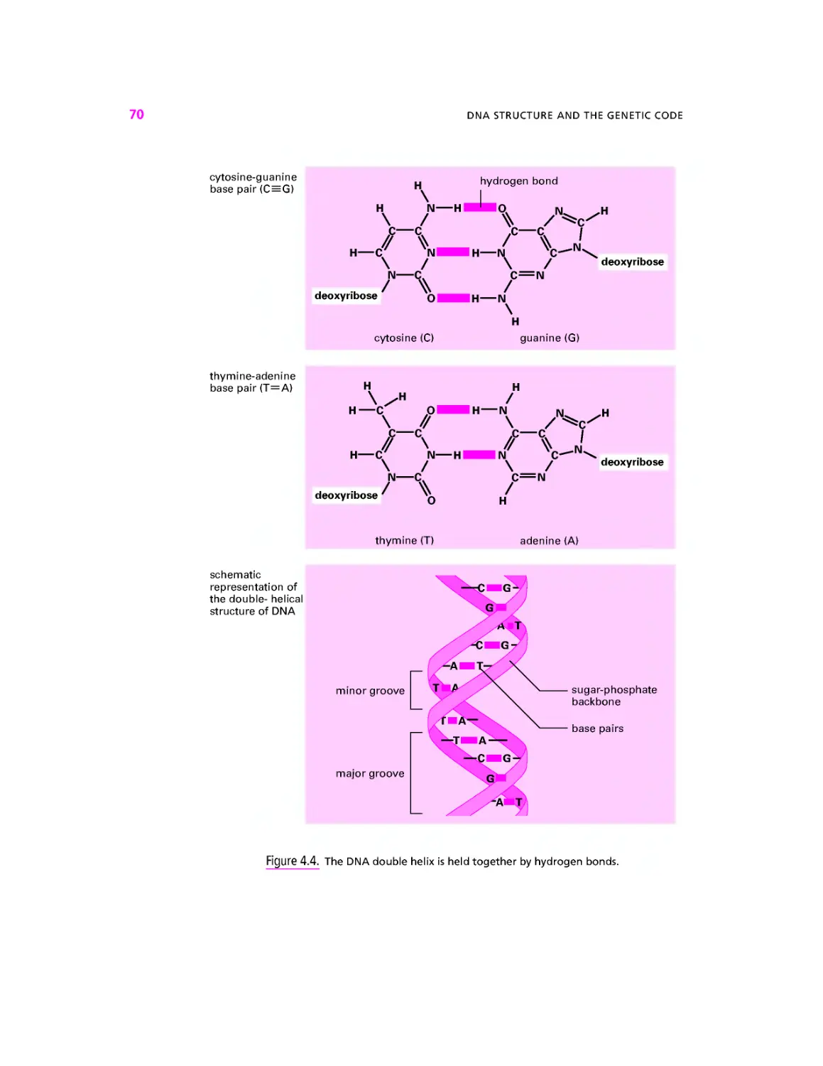
illustrated in Figure 2.6a. Hydrogen bonding also plays a critical role in allowing DNA to
store and replicate genetic information. Figure 2.6b shows how the base pairs (page 69)
of DNA form hydrogen bonds in which hydrogen atoms are shared between nitrogen and
oxygen and between nitrogen and nitrogen.
•$ BIOLOGICAL MACROMOLECULES
Very large molecules, or macromolecules, are central to the working of cells. Large bi-
biological molecules are polymers: they are assembled by joining together small, simpler
molecules, which are therefore called monomers. Chemical technology has mimicked
nature by producing many important polymers—polyethylene is a polymer of ethylene
monomers. Cells make a number of macromolecules that we will introduce, together with
their monomer building blocks, in this chapter.
<$> CARBOHYDRATES: CANDY AND CANES
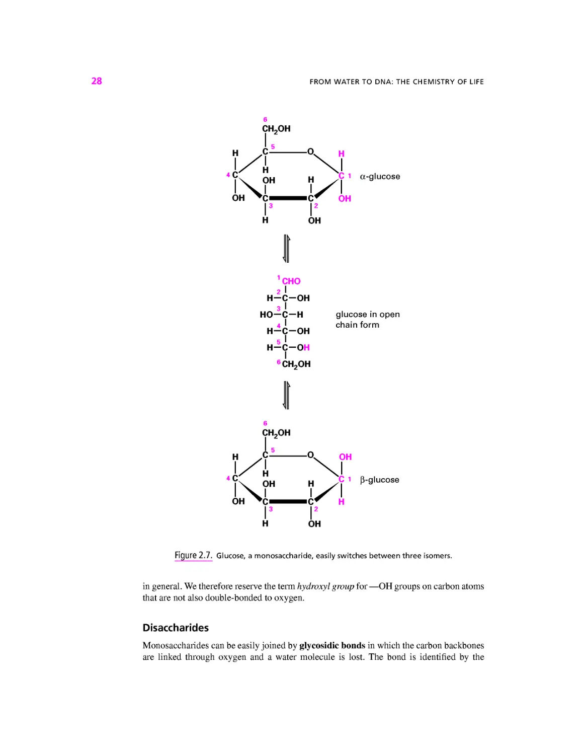
Carbohydrates—sugars and the macromolecules built from them—have many different
roles in cells and organisms.
An Assortment of Sweets
All carbohydrates are formed from the simple sugars called monosaccharides. Figure 2.7
shows the monosaccharide called glucose. The form shown at the top has five carbons
joined each to the other with an oxygen atom completing the ring. As well as the oxygen
in the ring, glucose has five other oxygens, each in an —OH (hydroxyl) group. Glucose
easily switches between the three forms, or isomers, illustrated in Figure 2.7. The two
ring structures are stereo isomers: although they comprise the same atoms connected by
the same bonds, they represent two different ways of arranging the atoms in space. The
two stereo isomers, named a and fi, continually interconvert in solution via the open-chain
form.
Figure 2.8 shows five other monosaccharides that we will meet again in this book.
Like glucose, each of these monosaccharides can adopt an open-chain form and a number
of ring structures. In Figure 2.8 we show each sugar in a form that it adopts quite often.
These sugars share with glucose the two characteristics of monosaccharides: they can adopt
a form in which an oxygen atom completes a ring of carbons, and they have many hydroxyl
groups. The generic names for monosaccharides are derived from the Greek for the number
of their carbon atoms, so glucose, galactose, mannose, and fructose are hexoses F carbons)
while ribose and ribulose are pentoses E carbons). Classically, a monosaccharide has the
general formula Cn(H2O)n, hence the name carbohydrate. All the monosaccharides shown
in Figures 2.7 and 2.8 fit this rule—the four hexoses can be written as Ce(H2ON and the
two pentoses as C5(H2OM.
It is worth noting that although both sugars and organic acids contain an —OH group,
the behavior of an —OH group that forms part of a carboxyl group is very different from
one that is not next to a double-bonded oxygen. In general the —OH group in a carboxyl
group will readily give up an H+ to water or other acceptors; this is not true of—OH groups
28 FROM WATER TO DNA: THE CHEMISTRY OF LIFE
CH2OH
H
2
H-C-OH
3>
HO—C—H glucose in open
L
H(oH
H-C-OH
I
6CH2OH
CH,OH
OH
Figure 2.7. Glucose, a monosaccharide, easily switches between three isomers.
in general. We therefore reserve the term hydroxyl group for —OH groups on carbon atoms
that are not also double-bonded to oxygen.
Disaccharides
Monosacchandes can be easily joined by glycosidic bonds in which the carbon backbones
are linked through oxygen and a water molecule is lost. The bond is identified by the
CARBOHYDRATES: CANDY AND CANES
29
6CH2OH
OH ,C O OH
OH H Jr i P-9alactose
H
H J?
H
6CH2OH
X 5
¦c
OH
OH
OH
Xi
a-mannose
3I
H H
5CH2OH
CH2OH
\H OH/1
I I / OH
1 I3
OH H
a-fructose
CH2OH
l\ H H /|
h\I I/h
OH
OH
I
Pi
C-ribose
I2
OH
CH2OH
>K A2
\\ H H /
H\l I /OH
a-ribulose
OH
I3
OH
Figure 2.8. Some monosaccharides, each shown as a common isomer.
30 FROM WATER TO DNA: THE CHEMISTRY OF LIFE
CH2OH
CH2OH
H
glucose residue
I2
H OH
galactose residue
Figure 2.9. The disaccharide lactose.
carbons linked. For example, Figure 2.9 shows lactose, a sugar found in milk that is formed
when galactose and glucose are linked by a (l->-4) glycosidic bond. Sugars formed from
two monosaccharide monomers are called disaccharides. Whenever a more complicated
molecule is made from simpler building blocks, we call the remnant of the individual
building blocks residues. Thus lactose is made of one galactose residue and one glucose
residue.
A complication arises here. Although free monosaccharides can easily switch between
the a and p forms, formation of the glycosidic bond locks the shape. Thus, although
galactose in solution spends equal time in the a and p forms, the galactose residue in
lactose is locked into the fi form, so the full specification of the bond in lactose is /31-»-4.
Out of the Sweet Comes Forth Strength
Formation of glycosidic bonds can continue almost indefinitely. A chain of up to 100 or so
monosaccharides is called an oligosaccharide, from the Greek word oligo meaning few.
Longer polymers are called polysaccharides and have characteristics very different from
those of their monosaccharide building blocks. Figure 2.10a shows part of a molecule of
glycogen, a polymer made exclusively of glucose monomers organized in long chains with
a 1 —*4 links. The glycogen chain branches at intervals, each branch being an a(l —»4) linked
chain of glucose linked to the main chain with an aA^6) bond. Solid lumps of glycogen
are found in the cytoplasm of muscle, liver, and some other cells. These glycogen granules
are 10-40 nm in diameter with up to 120,000 glucose residues. Glycogen is broken down
to release glucose when the cell needs energy (Chapter 13).
Cellulose makes up the cell wall of plants and is the world's most abundant macro-
molecule. Like glycogen, cellulose is a polymer of glucose, but this time the links are
/31—*4. It is shown in Figure 2.10&. Drawn on the flat page, it looks very like glycogen.
However, the difference in bond type is critical. Glucoses linked by a(l^-4) links arrange
themselves in a floppy helix, while glucoses linked by /3A—*4) links form extended chains,
CARBOHYDRATES: CANDY AND CANES 31
(a) glycogen
H OH
(b) cellulose
Figure 2.10. The polysaccharides glycogen and cellulose.
ideal for building the rigid plant cell wall. Animals have enzymes (protein catalysts) that
can break down the a(\ -»4) bond in glycogen, but only certain bacteria and fungi can break
the /3A—»4) link in cellulose. All animals that eat plants rely on bacteria in their intestines
to provide the enzymes to digest cellulose.
Modified Sugars
A number of chemically modified sugars are important in biology. Figure 2.11 shows
three. Deoxyribose is a ribose that is missing the —OH group on carbon number two. Its
32 FROM WATER TO DNA: THE CHEMISTRY OF LIFE
5CH2OH ^O^ OH
"C
h\| I/A
3 1 I 2
OH H
deoxyribose
6CH2OH
«|V OH H Jt
OH >C^^^C^ H
Is I2
H N H
C=O
CH3
N-acetyl glucosamine
O~
O^P—0"
o
H C—H
H *C O H
H C O H
?V OH OH Jt
H ^C^^^^CT O
H H
mannose-6-phosphate
Figure 2.11. Deoxyribose, W-acetyl glucosamine and mannose-6-phosphate are modified sugars.
CARBOHYDRATES: CANDY AND CANES
33
main use is in making deoxyribonucleic acid—DNA. Af-acetyl glucosamine is a glucose in
which an —NHCOCH3 group replaces the —OH on carbon two. It is used in a number of
oligosaccharides and polysaccharides, for example, in chitin, which forms the hard parts of
insects. Mannose-6-phosphate is a mannose that has been phosphorylated, that is, had
a phosphate group attached, on carbon number six. We will meet more phosphorylated
sugars in the next section.
adenine
H—N
HO CH2
ribose
(a) adenosine, a nucleoside
triphosphate
O~ 0" O"
—P—O—P—O—P—O—CH
Y I a
phosphate p phosphate
phosphate
(b) adenosine triphosphate, a nucleotide
Figure 2.12. Adenosine, a nucleoside, and adenosine triphosphate, a nucleotide.
34
FROM WATER TO DNA: THE CHEMISTRY OF LIFE
punnes
pyrimidines
/
N
\
N
\
C
\
C=N
C
H
adenine (A)
H
H
H
\
H-
)
N H
C—C
N C
H O
cytosine (C)
H
n
\
C
\
C=N
N
H—N
\
H
guanine (G)
•C—C
CN
H
hypoxanthine
H
.H
H—C
\
c—c
H—C
\
H-
)-cx
H O
thymine (T)
H O
H
N—H
)-c\
H O
uracil (U)
N—H
H
nicotinamide
Figure 2.13. Seven bases found in nucleotides.
NUCLEOSIDES, PHOSPHATE, AND NUCLEOTIDES 35
•$> NUCLEOSIDES, PHOSPHATE, AND NUCLEOTIDES
Figure 2.12a shows a nucleoside called adenosine. It is composed of ribose coupled to a
nitrogen-rich compound called adenine. The numbers on the sugar—1', 2'; and so forth—are
the same numbering system we have seen before; the 'symbol is pronounced "prime" and is
there to indicate that we are identifying the atoms of the sugar, not the atoms of the adenine.
The name nucleoside reflects the fact that phosphorylated nucleosides (see below) are the
building blocks of the nucleic acids that form the genetic material in the nucleus. However,
nucleosides also play important roles in other places inside and outside the cell. Seven
different compounds can be used to generate nucleosides (Fig. 2.13). All seven contain
many nitrogen atoms and one or more ring structures. They are the three purines called
adenine, guanine and hypoxanthine, the three pyrimidines called cytosine, thymine, and
uradl, and an odd man out called nicotinamide. These ring compounds are called bases.
Historically, the name arose because the compounds are indeed bases in the sense used
earlier in this chapter—they will exchange an H+ with water. The roots of the name are
now forgotten by most molecular biologists, who now use the word base to mean a purine,
O
II
H—O—P—O~
O_
(a)
O O
I II I II
-C—O—H + H—O—P—O~ „ H,0 + —C—O—P—O'
I I II
0_ 0_
(b)
—C—O—P—O~ + H+ + H—O—P—O"
—C—O—P—O—P—O" + H,0
(c)
Figure 2.14. Phosphate groups can attach to COH groups or to other phosphate groups.
36
FROM WATER TO DNA: THE CHEMISTRY OF LIFE
adenine
H—N
"O—P—O—CH
—P—O—CH
M
c—n:
H nicotinamide
rlbose
Figure 2.15. Nicotine adenine dinucleotide is formed of two nucleotides joined via their phos-
phosphate groups.
a pyrimidine, or nicotinamide. In general, a nucleoside is formed by attaching one of these
bases to the l'-carbon atom of ribose.
Phosphorous, although by weight a relatively minor fraction of the whole cell, plays a
number of critical roles. In solution phosphorous is mainly found as a phosphate ion with a
single hydrogen atom still attached, HPO42~ (Fig. 2.14a). We often indicate phosphate ions
with the symbol Pi, meaning inorganic phosphate. Where phosphate becomes important,
however, is when it is attached to an organic, that is, carbon-containing, molecule. Phosphate
can substitute into any C—OH group with the loss of a water molecule (Fig. 2. \Ab). The
equilibrium in the reaction shown lies far to the left, but cells have other strategies for
attaching phosphate groups to organic molecules. Once one phosphate group has been
added, more can be added to form a chain (Fig. 2.14c). Once again, the equilibrium in the
reaction shown lies far to the left, but cells can achieve this result using other strategies.
AMINO ACIDS, POLYPEPTIDES, AND PROTEINS
37
H
Figure 2.16. Inositol trisphosphate, a multiply phosphorylated polyalcohol.
Figure 2.12b shows adenosine with a chain of three phosphate groups attached to the
5' carbon of the ribose. This important molecule is adenosine triphosphate (ATP), and we
will meet it many times in the course of this book. The three phosphate groups are denoted
by the Greek letters a,p, and y. Phosphorylated nucleosides are called nucleotides.
Figure 2.15 shows another important molecule formed from adenosine. Here adenosine
and a nicotinamide nucleotide are joined through their phosphate groups. The resulting
molecule, called nicotinamide adenine dinucleotide (NAD+), plays a critical role in the
cellular energy budget.
Molecules with several OH groups can become multiply phosphorylated. Figure 2.16
shows inositol trisphosphate (IP3), an important messenger molecule we will meet again
in Chapter 16. Both ATP and IP3 have three phosphate groups, but to indicate the fact that
in ATP these are arranged in a chain, while in IP3 they are attached to different carbons,
we use the prefix tri in adenosine fnphosphate and the prefix tris in inositol frwphosphate.
Similarly, a compound with two phosphates in a chain is called a ^phosphate, while one
with one phosphate on each of two different carbons is called a bwphosphate.
•$ AMINO ACIDS, POLYPEPTIDES, AND PROTEINS
Amino acids contain both a COOH group, which readily gives an H+ to water and is therefore
acidic, and a basic NH2 group, which readily accepts H+ to become NH3+. Figure 2.17a
shows two amino acids, leucine and 7-amino butyric acid (GABA), in the form in which
they are found at normal pH: the COOH groups have each lost an H+ and the NH2 groups
have each gained one, so that the molecules bear both a negative and a positive charge.
We name organic acids by labeling the carbon that bears the carboxyl group a, the next
one p, and so on. When we add an amino group, making an amino acid, we state the letter
of the carbon to which the amino group is attached. Hence leucine is an a-amino acid
38 FROM WATER TO DNA: THE CHEMISTRY OF LIFE
tn On,
4h
OOC aCH NH+
leucine
(a)
"OOC —
y-amino
aCH2
butyric
-NH+
acid
"OOC CH NH+
a-amino acid general form
(b)
R. H R, H R, H R
11111 I I"
NH+ CH C N CH C N CH C N CH COO"
peptide peptide
bond bond
generalized peptide
(c)
Figure 2.17. Amino acids and the peptide bond.
while GABA stands for gamma-ammo butyric acid. a-Amino acids are the building blocks
of proteins. They have the general structure shown in Figure 2.17b where R is called the
side chain. Leucine has a simple side chain of carbon and hydrogen. Other amino acids
have different side chains and so have different properties. It is the diversity of amino side
chains that give proteins their characteristic properties (page 185).
a-Amino acids can link together to form long chains through the formation of a peptide
bond between the —COO~ group of one amino acid and the —NH3+ group of the next.
Figure 2.17c shows the generalized stucture of such a chain of a-amino acids. If there are
fewer than about 50 amino acids in a polymer we tend to call it a peptide. More and it
is a polypeptide. Polypeptides that fold into a specific shape are called proteins. Most of
the components of the machinery of the cell—the chemicals that carry out the processes
of life—are proteins. Peptides and polypeptides are formed inside cells by adding specific
amino acids to the end of a growing polymer. As we will see in Chapter 4, the particular
amino acid to be added at each step is defined by the instructions on a molecule called
LIPIDS 39
messenger ribonucleic acid (RNA), which in turn contains a copy of the information
recorded on the cell's DNA that is stored in the nucleus. This is the central dogma of
molecular biology: DNA makes RNA makes protein.
# LIPIDS
Figure 2.18a shows the fatty acid oleic acid. It comprises a carboxyl group plus a long
tail of carbons and hydrogens. At neutral pH oleic acid gives up its H+ to become the
oleate ion (Fig. 2.18&). The two ends of the oleate ion are very different. The carboxyl
group is negatively charged and hence will associate readily with water molecules. The
hydrocarbon tail is nonpolar and does not readily associate with water. The molecule is
said to be amphipathic, from the Greek for "hating both," meaning that one half does not
like to be in water while the other half does not like to be in a nonpolar environment like
octane.
Figure 2.18c shows the small molecule glycerol. Like sugars, glycerol has many hy-
droxyl groups and, like sugars, tastes sweet, as its name suggests. However, it is not a sugar
because it cannot adopt an oxygen-containing ring structure. Rather, because compounds
containing hydroxyl groups are called alcohols, glycerol is a polyalcohol.
As we shall see in Chapter 13, cells can join fatty acids and glycerol to make glycerides.
The bond is formed by removing the elements of water between the carboxyl group of the
fatty acid and a hydroxyl group of glycerol. Any bond of this type, between a carboxyl
group and a hydroxyl group, is called an ester bond. Figure 2.19 shows two glycerides. In
Figure 2.19a is trioleoylglycerol, the main component of olive oil. It is a triacylglycerol
(or triglyceride) formed from three molecules of oleic acid and one molecule of glycerol.
The fatty acid residues are called acyl groups. Formation of the ester bonds has removed
the charged carboxyl groups that rendered the oleate amphipathic. Almost all of the tria-
triacylglycerol molecule is simple hydrocarbon chain that cannot hydrogen bond with water
molecules. For this reason, olive oil and other triacylglycerols do not mix with water. They
are therefore said to be hydrophobic. Triacylglycerols that are liquid at room tempera-
temperature are called oils, those that are solid are called fats, but they are all the same sort of
molecule.
Example 2.1
Salad Dressing Is a Mixture of Solvents
Vinegar is a dilute solution of acetic acid in water. Acetic acid gives up an H+ to water to leave the
negatively charged acetate ion. The simplest salad dressing is a shaken-up mixture of olive oil and
vinegar. Olive oil is hydrophobic and so does not dissolve in the water, the two liquids remain as
separate droplets and soon separate again after shaking. However much one shakes, all the acetate
will remain in the water because it is an ion. If salt (Na+Cl~) is added to the dressing, it also dissolves
in the water, with none dissolving in the oil. In contrast if you add chilies to the salad dressing, the
active chemical component, capsacin, will dissolve in the oil because it is nonpolar. Only by shaking
up the mixture can you get all the tastes together.
Cells contain droplets of triacylglycerol within their cytoplasm. These are usually small,
but in fat cells, which are specialized as fat stores, the droplets coalesce into a single
large globule so that the cell's cytoplasm is squeezed into a thin layer surrounding the fat
40 FROM WATER TO DNA: THE CHEMISTRY OF LIFE
H
I
0 O"
1 I
c=o c=o
I I
H — C—H H — C—H
I I
H — C—H H C H
I I
H — C—H H C H
I I
H — C—H H C H
I I
H — C—H H C H
I I
H — C—H H C H
I I
H — C—H H C H
I I
H — C H — C
II II
H — C H — C
I I
H — C—H H C H
I I
H — C—H H C H
I I
H — C—H H C H
I I
H — C—H H C H
I I
H — C—H H C H
I I
H — C—H H C H
I I
H — C—H H C H
I I
H — C—H H C H
I I
H H
(a) oleic acid (b) oleate
H H H
I I I
H — C — C — C—H
I I I
0 0 0
I I I
H H H
(c) glycerol
Figure 2.18. Oleic acid and glycerol.
globule. During fasting triacylglycerols are broken down into free fatty acids and glycerol, a
process called lipolysis. The fatty acids and the glycerol then enter the circulation for use by
tissues.
Figure 2.19b shows phosphatidylcholine, an example of the phospholipids that make
the plasma membrane and other cell membranes. Like triacylglycerols, phospholipids have
I
c=o
H — C—H
H — C—H
H — C—H
H — C—H
H — C—H
H — C—H
H — C—H
4
H — C—H
I
H — C—H
I
H
I
I
O
c=o
H — C—H
H — C—H
H — C—H
H — C—H
H — C—H
H — C—H
H — C—H
H — C
H — C
H — C—H
H — C—H
H — C—H
I
H — C—H
H — C—H
H — C—H
H — C—H
H — C—H
I
H
I
C=O
H — C—H
H — C—H
H — C—H
H — C—H
H — C—H
H — C—H
H — C—H
4
H — C—H
I
H — C—H
I
H
glycerol h—c-
o
I
acyl
group
trioleoylglycerol, a triacylglycerol
(a)
c=o
H — C—H
H — C—H
H — C—H
H — C—H
H — C—H
H — C—H
H — C—H
H — C—H
I
H — C—H
H — C—H
H — C—H
H — C—H
I
H — C—H
H — C—H
H — C—H
H — C—H
H — C—H
I
H
I
CH3 N-CH3
O
O = P O"
o
I
H
I
o
c=o
H — C—H
H — C—H
H — C—H
C—H
C—H
H — C—H
C—H
C—H
I
H — C—H
C—H
II
C—H
H — C—H
I
C—H
C—H
H — C—H
H — C—H
H — C—H
H — C—H
H — C—H
charged
headgroup
phospho-
diester
link
glycerol
ester
bonds
acyl
group
phosphatidylcholine, a phospholipid
(b)
aqueous medium
lipid bilayer
~| hydrophobic
J lipid tails
hydrophilic
lipid
headgroups
(c)
aqueous medium
Figure 2.19. Lipids.
41
42
FROM WATER TO DNA: THE CHEMISTRY OF LIFE
fatty acids attached to glycerol, but only two of them. In place of the third fatty acid is
a polar, often electrically charged head group. The head group is joined to the glycerol
through phosphate in a structure called a phosphodiester link. The combination of head
group and negatively charged phosphate is able to associate strongly with water—it is said
to be hydrophilic. The two fatty acids, on the other hand, form a tail that, like olive oil,
is hydrophobic. Phospholipids can therefore neither dissolve in water (because of their
hydrophobic tails) nor remain completely separate, like olive oil (because then the head
group could not associate with water). Phospholipid molecules therefore spontaneously
form lipid bilayers between 5 and 10 nm thick (Fig. 2.19c) in which each part of the
molecule is in its preferred environment. Cell membranes are lipid bilayers plus some
added protein.
Medical
Relevance
2.1
The Kinks Have It: Double Bonds, Membrane Fluidity,
and Evening Primroses
One of the fatty acids commonly found as a component of animal fat is stearic acid,
illustrated as (a) opposite. Its 18 carbon atoms are joined by single bonds, making a long straight
molecule. In contrast, oleic acid (b) has a double bond between the ninth and tenth carbons. This
introduces a kink in the chain. A fatty acid with kinks is less able to solidify because it is less
able to pack in a regular fashion. The more double bonds in a fatty acid, the lower its melting
point. Thus stearic acid melts at 69.6°C, while oleic acid melts at 13.4°C and is therefore liquid at
room temperature. Fatty acids containing double bonds between the carbon atoms are said to be
unsaturated. Polyunsaturated fatty acids have more than one double bond, more than one bend,
and therefore even lower melting points. Linoleic acid (c) has two, and melts at —9°C; linolenic
acid (d) has three and a melting point of — 17°C.
For the triacylglycerols inside our cells, and the phospholipids in our membranes, the same
rule applies—the more double bonds in the acyl groups, the lower the melting point. The large
masses of triacylglycerols containing stearic acid in animal bodies are liquid at body temperature
but form solid lumps of fat at room temperature, while trioleoylglycerol is liquid at room temper-
temperature (but should not be kept in the fridge!). Membranes must not solidify—if they did, then they
would crack and the cell contents would leak out each time the cell was flexed. Unsaturated fatty
acids play an essential role in maintaining membrane liquidity.
Mammals are unable to introduce double bonds beyond carbon 9 in the fatty acid chain. This
means that linoleic and linolenic acids must be present in the diet. They are known as essential
fatty acids. Fortunately, the biochemical abilities of plants are not so restricted, and plant oils
form a valuable source of unsaturated fatty acids.
The normal form of linolenic acid, shown in (d), is a-linolenic acid, which has its double
bonds between carbons 9 and 10, 12 and 13, and 15 and 16. Some plant seed oils contain an
isomer of linolenic acid with double bonds between carbons 6 and 7, 9 and 10, and 12 and
13. This is called y-linolenic acid (e). The attractive, yellow-flowered garden plant called the
evening primrose (Oenethera perennis) has seeds that contain an oil with y-linolenic in its
triacyglycerols. The 6 to 7 double bond introduces a kink closer to the glycerol. This is thought
to increase fluidity when incorporated into membrane lipids. No one really knows if it really has
the marvelous health effects that some claim for it, or if it does, how it works. This ignorance
has not stopped people from making lots of money from evening primrose oil. It is included in
cosmetics and in alternative medicines for internal and external application. y-Linolenic acid
occurs in other plants—the seeds of borage (Borago officinalis) are one of the richest sources. It
is even found in some fungi. However, these lack the romance of the evening primrose!
LIPIDS
43
/
OH
Stearic acid, C18 no double bonds, melts at 69.6°
(a)
Oleic acid, C18 one double bond, melts at 13.4°
(b)
Linoleic acid, C18 two double bonds, melts at -9°
(c)
Linolenic acid, C18 three double bonds, melts at -17°
(d)
y-Linolenic acid, C18 three double bonds
(e)
44
FROM WATER TO DNA: THE CHEMISTRY OF LIFE
•SP> HYDROLYSIS
Many of the macromolecules of which cells are made are generated from their individual
building blocks by the removal of the elements of water. Equally, macromolecules can be
broken into their individual building blocks by hydrolysis—breakage by the addition of
water. Figure 2.20 shows four examples. Lactose is hydrolyzed to the monosaccharides
galactose and glucose by the addition of one water molecule. Next, we show a dipeptide be-
being broken into individual amino acids by hydrolysis. These two hydrolysis reactions occur
all the time in our intestines, though some people lose the ability to hydrolyze lactose as they
get older: they are lactose intolerant. Third, we show a dimeric (formed of two parts) inor-
inorganic phosphate ion called pyrophosphate being hydrolyzed to regular phosphate ions. The
hydrolysis of pyrophosphate is catalyzed by enzymes found throughout the body, both inside
cells and out. Later in the book we will meet a number of instances where a reaction creates
lactose
OH
1 HO
OH
r
CH—CH3
CH2
OH
H CH2
I I
TH3N CH C N CH COO"
1 HO
pyrophosphate
O O
II II
"O— P—O— P—O"
I I
OH O"
CH2OH
i H O
OH
r
"OOC CH NH3+
serine
r
CH CH3
r
"OOC CH NH,+
(b)
O
II
"O—P—OH
HO—P—O
H+
ar
(c)
Figure 2.20. Hydrolysis is the breakage of covalent bonds by the addition of the elements
of water.
!"¦
4H2O
CH3 N+ CH3
r
3H+
"O
I
C = 0
I
H C H
I
H C H
I
H C H
H C H
H C H
H C H
H C H
H C H
H C H
H C H
H C H
H C H
H C H
H C H
H C H
H C H
H C H
CH2 OH
CH OH
I
CH2 OH
glycerol
H C H
H C H
H C H
H C H
H C H
H C H
H C H
H C H
H C H
H C H
H C H
H C H
H C H
H C H
H C H
H C H
I
I
O
c
I
H C
H C
H C
C
II
C
H C
C
C
H C
C
H C
C
II
C
H C
H C
H C
H C
H C
I
V
o
II
"O—P—OH
O-
(d)
p
o
I
O = P — O"
I
o
I
C — H
H
"O
C=O
I
H C H
H C H
H C H
I
C H
II
C H
H C H
C H
II
C H
H C H
C H
II
C H
H C H
C H
C H
H C H
H C H
H C H
H C H
I
H C H
I
arachidonate
Figure 2.20. Continued
45
46
FROM WATER TO DNA: THE CHEMISTRY OF LIFE
pyrophosphate, but as soon as the pyrophosphate is produced it will be hydrolyzed to regu-
regular phosphate. Lastly, we show the complete hydrolysis of the phospholipid phosphatidyl-
choline. Five molecules result: one phosphate ion together with choline, glycerol, and two
fatty acids. This hydrolysis also occurs in our intestines, where it is a multistage process.
Hydrolysis reactions usually proceed rapidly in living tissue when the appropriate
catalyst is present, because water is present at high concentration. In contrast the generation
of macromolecules from their constituent building blocks, often by the elimination of the
elements of water between constituent building blocks, usually requires the expenditure of
energy by the cell. We will see how these reactions are performed in later chapters.
SUMMARY
1. When two atoms interact, electrons may be shared between the two to form a
covalent bond, or may pass completely from one atom to another, forming ions.
2. In water the electrons are not shared equally but are displaced toward the oxygen
atom, so that the molecule is polar.
3. Ionic compounds will only dissolve in polar solvents.
4. Acids are molecules that give an H+ to water, forming H3O+. Dissolving an acid
in water produces a solution of pH less than 7.
5. Bases are molecules that accept an H+ from water, leaving OH~. Dissolving a base
in water produces a solution of pH greater than 7.
6. Pure water has the neutral pH of 7.0.
7. A hydrogen bond can form when a hydrogen atom takes up a position between two
electron-grabbing atoms (oxygen, nitrogen, or sulfur), the three forming a straight
line.
8. Monosaccharides are compounds with a central skeleton of carbon to which are
attached many OH groups. They can switch between two oxygen-containing ring
structures via an open-chain configuration.
9. Monosaccharides can join together to form disaccharides such as lactose and to
form long chains such as glycogen and cellulose.
10. Adenine, guanine, hypoxanthine, cytosine, thymine, uracil, and nicotinamide are
nitrogen-rich ring-shaped molecules called bases. Bases combine with ribose or
deoxyribose to form nucleosides. Phosphorylation of a nucleoside on the 5' carbon
produces a nucleotide.
11. Amino acids are compounds with a —COOH acidic group and an —NH2 basic
group. At neutral pH these groups will, respectively, lose and gain an H+ to become
—COO" and—NH3+.
12. Polypeptides are polymers of a-amino acids linked by peptide bonds. The sequence
of amino acids is determined by instructions on the cell's DNA. Proteins are polypep-
polypeptides that fold into a specific shape.
REVIEW QUESTIONS 47
13. Lipids are formed by the attachment of hydrophobic long-chain fatty acids to a glyc-
erol backbone. Phospholipids also have a hydrophilic head group and spontaneously
form lipid bilayers.
14. Hydrolysis is the breakage of covalent bonds by the addition of the elements of
water.
FURTHER READING
Voet, D., and Voet, J. 2003. Biochemistry. New York: Wiley.
REVIEW QUESTIONS
For each question, choose the ONE BEST answer or completion.
1. Water
A. can give up an H+, becoming OH".
B. can accept an H+, becoming H3O+.
C. can form hydrogen bonds.
D. is a good solute for ionic substances.
E. all of the above.
2. The amino acid leucine has an acidic COOH group and a basic NH2 group. The reaction
—COOH ^ —COO" + H+ has &pKa of 2.3, while the reaction —NH3+ ^ —NH2 +
H+ has a pKa of 9.7. At pH 1.0
A. there will be more "OOC—CH(CH2.CH(CH3J)—NH2 than either
-OOC—CH(CH2.CH(CH3J)—NH3+ or HOOC—CH(CH2.CH(CH3J)—NH3+.
B. there will be equal amounts of "OOC—CH(CH2.CH(CH3J)—NH2 and
"OOC—CH(CH2.CH(CH3J)—NH3+, but not much HOOC—CH(CH2.CH(CH3J)—NH3+.
C. there will be more "OOC—CH(CH2.CH(CH3J)—NH3+ than either
HOOC—CH(CH2.CH(CH3J)—NH3+ or "OOC—CH(CH2.CH(CH3J)—NH2.
D. there will be equal amounts of HOOC—CH(CH2.CH(CH3J)—NH3+ and
"OOC—CH(CH2.CH(CH3J)—NH3+, but not much "OOC—CH(CH2.CH(CH3J)—NH2.
E. there will be more HOOC—CH(CH2.CH(CH3J)—NH3+ than either
"OOC—CH(CH2.CH(CH3J)—NH3+ or "OOC—CH(CH2.CH(CH3J)—NH2.
3. Which of the following statements is incorrect?
A. Glycogen is formed from units that include glucose.
B. Cellulose is formed from units that include glucose.
C. Lactose is formed from units that include ribose.
D. Adenosine is formed from units that include ribose.
E. Nicotinamide adenine dinucleotide is formed from units that include ribose.
4. Phospholipids
A. have hydrophilic fatty acid tails.
B. have charged, hydrophobic head groups.
48 FROM WATER TO DNA: THE CHEMISTRY OF LIFE
C. spontaneously form lipid bilayers in an aqueous environment.
D. form droplets within the cytoplasm.
E. all of the above.
5. When an acid is added to pure water
A. it will dissolve easily.
B. it will give up an H+ to form a negatively charged ion.
C. it will form a solution with a high concentration of H3O+ ions.
D. it will form a solution with a low pH.
E. all of the above.
6. Hydrogen bonds cannot form between
A. water and glucose.
B. water and water.
C. water and phosphate.
D. phosphate and octane.
E. phosphate and glucose.
7. Which of these statements about the polypeptides in cells is wrong?
A. They are a polymer of y-amino acids linked by peptide bonds.
B. The sequence of amino acids is determined by instructions on the cell's DNA.
C. They have an —NH3+ group at one end of the polymer.
D. They have a —COO" group at one end of the polymer.
E. They are called proteins if they fold into a specific shape.
ANSWERS TO REVIEW QUESTIONS
1. E. All these statements about water are true.
2. E. A pH of 1.0 is very acid (an H+ concentration of 100 mmol liter), more acid even than 2.3, the
pH at which the COOH group is half-protonated. Both the —COOH and NH2 groups on leucine
are therefore protonated at pH 1.0.
If you are still confused, consider the situation at pH 11. At this pH, the concentration of H+
is extremely low A0~u mol liter or 10 pmol liter). Leucine therefore loses all its hydrogen
ions and has the form "OOC—CH(CH2.CH(CH3J)—NH2. Now consider adding H+ so that the
pH falls. The NH2 group is basic and accepts H+ readily, even when H+ is present at quite a low
concentration. At pH 9.7 (an H+ concentration of 200 pmol liter) half the NH2 groups have
accepted an H+ to become NH3+, and at pH values less than 9.7 a majority of the NH2 groups are
protonated. This is the situation at neutral pH: the NH2 groups are protonated while the COOH
groups are deprotonated, so leucine has the form "OOC—CH(CH2.CH(CH3J)—NH3+. If we
keep adding H+, we eventually approach a pH of 2.3, where the H+ concentration has the high
value of 5 mmol liter. Even though the COOH groups like to give up H+, they are forced to
accept H+ when the concentration of H+ in solution gets this high. At pH 2.3 half the COOH
groups are protonated; half are still COO". At a pH less than 2.3, that is, at H+ concentrations
higher than 5 mmol liter, the majority of the COOH groups are protonated and leucine has the
form HOOC—CH(CH2.CH(CH3J)—NH3+.
3. C. Lactose is a disaccharide formed from glucose and galactose.
4. C. All the other statements are false. In particular: (A) The fatty acid tails are hydrophobic. (B) The
head groups arehydrophilic because they are polar and often are charged. (D) Phospholipids will not
form droplets because this would place the hydrophilic head group in a hydrophobic environment.
ANSWERS TO REVIEW QUESTIONS 49
5. E. All these statements are true. In particular, the definition of pH means that a solution with a
high concentration of H3O+ ions is one with a low pH.
6. D. Octane is formed of carbon and hydrogen only. It does not contain any of the electron-grabbing
atoms that, together with hydrogen, form a hydrogen bond. Octane therefore cannot form hydrogen
bonds with other chemicals.
7. A. Cellular polypeptides are polymers of a-amino acids, not y-amino acids. Changing y to a in
this question generates an accurate summary of the biology of polypeptides.
3
MEMBRANES AND ORGANELLES
The vast majority of the reactions that cells carry out take place in water. Eukaryotic cells
are, at any one time, carrying out an enormous range of such chemical manipulations:
collectively, these reactions are called metabolism (Chapter 13). In much the same way that
our homes are divided into rooms that are adapted for particular activities, so eukaryotic cells
contain distinct compartments or organelles to house specific functions. The term organelle
is used rather loosely. At one extreme some scientists use it to mean any distinct cellular
structure that has a more or less well-defined job to do; at the other are scientists who would
reserve the name organelle for those cellular compartments that contain their own DNA
and have some limited genetic autonomy. In this book, we will define organelles as those
cellular components whose limits, like those of the cell itself, are defined by membranes.
It is first necessary to consider some of the fundamental properties of cell membranes.
•$> BASIC PROPERTIES OF CELL MEMBRANES
It is difficult to overstate the importance of membranes to living cells; without them life
could not exist. The plasma membrane, also known as the cell surface membrane or
plasmalemma, defines the boundary of the cell. It regulates the movement of materials
into and out of the cell and facilitates electrical signaling between cells. Other membranes
define the boundaries of organelles and provide a matrix upon which complex chemical
reactions can occur. Some of these themes will be developed in subsequent chapters. In the
following section the basic structure of the cell membrane will be outlined.
Cell Biology: A Short Course, Second Edition, by Stephen R. Bolsover, Jeremy S. Hyams,
Elizabeth A. Shephard, Hugh A. White, Claudia G. Wiedemann
ISBN 0-471-26393-1 Copyright © 2004 by John Wiley & Sons, Inc.
51
52
MEMBRANES AND ORGANELLES
extracellular
medium
lipid bilayer
sugar residues
~| hydrophobic
-I lipid tails
hydrophilic
lipid
headgroups
cytosol
integral membrane protein
peripheral membrane protein
Figure 3.1. Membranes comprise a lipid bilayer plus integral and peripheral proteins.
The basic structure of a biological membrane is shown in Figure 3.1. Approximately half
the mass is phospholipid, which spontaneously organizes to form a lipid bilayer (page 42).
All the membranes of the cell, including the plasma membrane, also contain proteins. These
may be tightly associated with the membrane and extracted from it only with great difficulty,
in which case they are called integral proteins (e.g., the gap junction channel, page 55);
or they may be separated with relative ease, in which case they are termed peripheral
CO,
NO
H2O
Steroid hormones
Figure 3.2. Small uncharged molecules can pass through membranes by simple diffusion, but ions cannot.
BASIC PROPERTIES OF CELL MEMBRANES 53
proteins (e.g., clathrin adaptor protein, page 229). Membrane proteins are free to move later-
laterally, within the plane of the membrane. Integral membrane proteins are often glycosylated—
that is, have sugar residues attached—on the side facing the extracellular medium.
Straight Through the Membrane: Diffusion Through the Bilayer
Molecules of oxygen are uncharged. Although they dissolve readily enough in water, they
are also able to dissolve in the hydrophobic interior of lipid bilayers. Oxygen molecules can
therefore pass from the extracellular fluid into the interior of the plasma membrane, and
from there pass on into the cytoplasm, in a simple diffusion process (Fig. 3.2). Three other
small molecules with important roles in biology—carbon dioxide, nitric oxide, and water
itself—also pass across the plasma membrane by simple diffusion, as do the uncharged
hormones of the steroid family. In contrast, charged ions cannot dissolve in hydrophobic
regions (page 21) and therefore cannot cross membranes by simple diffusion.
Example 3.1
Rapid Diffusion in the Lungs
Blood is composed of cells, red and white, and plasma, a solution of sodium chloride, other ions,
various organic molecules and proteins. Red blood cells are very simple. They have no nucleus and
their plasma membrane encloses a cytosol packed with the oxygen-carrying protein hemoglobin,
bathed in a salt solution. The sodium concentration in the red blood cytosol is much lower than the
sodium concentration in plasma. It is important that the sodium concentration in the cytosol remain
low: if it increases, the red blood cells will swell and then burst as water rushes in. As red blood
cells pass through the lungs, they quickly gain oxygen because oxygen molecules can pass rapidly
across the plasma membrane by simple diffusion. The cells do not, however, gain sodium ions from
the plasma as they pass around the body because the lipid bilayer is impermeable to ions. Red blood
cells therefore remain the right size to pass though even the tiniest blood vessels in our bodies.
Beyond the Cell Membrane: The Extracellular Matrix
In connective tissue (page 13), the spaces between cells are filled by an extracellular matrix
consisting of polysaccharides and proteins such as collagen together with combinations of
the two called proteoglycans. The proteins provide tensile strength and elasticity while the
polysaccharides form a hydrated gel that expands to fill the extracellular space.
The sugar-based extracellular matrix reaches its highest form of expression in the cell
walls of plants (Fig. 3.3). The plant cell wall consists primarily of cellulose microfibrils
linked together by other polysaccharide molecules such as hemicellulose and pectin. The
thickness of the wall is determined largely by its pectin content. In healthy plant cells the
plasma membrane is constantly pressing against the cell wall.
In plants the extracellular medium is a much more dilute solution than the intracel-
lular solution. If no other forces operated, water would diffuse into plant cells down its
concentration gradient, a process called osmosis. However, plant cells are prevented from
expanding by the inextensible cellulose microfibrils in their cell walls. As water diffuses
in, the hydrostatic pressure in the cells rises, making the combination of cell plus cell wall
stiff. The same effect can be seen when a bicycle inner tube is blown up inside the tire, and
is called turgor. If plants are starved of water, water will leave the cell interior, hydrostatic
pressure drops, and the cells lose their turgor pressure, like a tire when the tube inside is
deflated. As a result the nonwoody parts of the plant wilt.
54
MEMBRANES AND ORGANELLES
direction of
expansion
Primary
cell
wall
cellulose
micro fibril
\
vacuole
cytoplasm
nucleus
primary
cell wall
secondary
cell wall
(a)
(b)
Figure 3.3. The cell wall directs plant cell growth.
Newly generated plant cells first lay down a primary cell wall of cellulose microfibrils
that are orientated in one direction around the plant cell (Fig. 3.3a). Hydrostatic pressure
therefore tends to cause a growing plant cell to elongate in one direction, perpendicular to
the axis of the microfibrils. Once the growth of the cell is complete, more layers of cellulose
and/or other compounds are added, most notably the polyphenolic compound lignin, to
form the secondary cell wall (Fig. 3.3&).
Cell Junctions
In multicellular organisms, and particularly in epithelia, it is often necessary for neighboring
cells within a tissue to be connected together. This function is provided by cell junctions. In
animal cells there are three types of junctions. Those that form a tight seal between adjacent
cells are known as tight junctions; those that allow communication between cells are
known as gap junctions. A third class of cell junction that anchors cells together, allowing
the tissue to be stretched without tearing, are called anchoring junctions. Plant cells do
BASIC PROPERTIES OF CELL MEMBRANES
55
not have tight junctions, gap junctions, or anchoring junctions but do contain a unique class
of communicating junction known as plasmodesmata.
Tight junctions are found wherever flow of extracellular medium is to be restricted
and are particularly common in epithelial cells such as those lining the small intestine. The
plasma membranes of adjacent cells are pressed together so tightly that no intercellular space
exists between them (Fig. 1.8 on page 14). Tight junctions between the epithelial cells of the
intestine ensure that the only way that molecules can get from the lumen of the intestine to
the blood supply that lies beneath is by passing through the cells, a route that can be selective.
Gap junctions are specialized structures that allow cell-to-cell communication in an-
animals (Fig. 3.4). When two cells form a gap junction, ions and small molecules can pass
directly from the cytosol of one cell to the cytosol of the other cell without going into the
extracellular fluid. Since ions can move through the junction, changes in electrical voltage
cytosol of right
hand cell
hydrophilic
solute
cytosol
of left
hand
cell
gap
junction
channel
plasma
membrane
plasma
membrane
Figure 3.4. Gap junctions allow solute and electrical current to pass from the cytosol of one cell
to the cytosol of its neighbor.
56 MEMBRANES AND ORGANELLES
are also rapidly transmitted from cell to cell by this route. The structure that makes this
possible is the gap junction channel. Channels, as we will see in Chapter 14, are water-
filled holes through membranes. When two gap junction channels or connexons meet, they
form a water-filled tube that runs all the way through the plasma membrane of the first cell,
across the small gap between the cells, and through the plasma membrane of the second cell.
In the middle of the channel is a continuous hole about 1.5 nm in diameter. This hole is large
enough to allow small ions through (and therefore to pass electrical current) together with
amino acids and nucleotides, but it is too small for proteins or nucleic acids. Gap junctions
are especially important in the heart, where they allow an electrical signal to pass rapidly
between all the cardiac muscle cells, ensuring that they all contract at the proper time. Each
gap junction channel is composed of six protein subunits that can twist against each other to
open and close the central channel in a process called gating (page 314) that allows the cell
to control the degree to which it shares solute with its neighbor. The plasmodesmata that
perforate the cell walls of many plant tissues (Fig. 1.7 on page 10) serve much the same
purpose as the gap junctions of animal cells but are much bigger and cannot shut quickly.
Some plant viruses use plasmodesmata to spread from cell to cell.
Example 3.2
Gap Junctions Keep Eggs Ready But Waiting
In the days leading up to ovulation, oocytes (cells that undergo meiosis to give rise to eggs) that
are to be released from the follicle are kept in a state of suspended development by a chemical in
their cytosol called cyclic AMP (page 350). The oocytes themselves do not make cAMP. Rather, the
follicle cells that surround them in the ovary make cAMP, which then passes through gap junctions
into the oocyte.
Only when the oocyte is released and begins its passage down the Fallopian tube does it prepare
for fertilization. In particular, the process of meiosis (page 404) is completed so as to remove half
the chromosomes, so that when the sperm adds its complement of chromosomes the fertilized egg
will have the correct number.
Anchoring junctions bind cells tightly together and are found in tissues such as the
skin and heart that are subjected to mechanical stress. These junctions are described later
(page 396).
•?$' ORGANELLES BOUNDED BY
DOUBLE-MEMBRANE ENVELOPES
Three of the major cell organelles, the nucleus, mitochondrion, and, in plant cells, the chloro-
plast, share two distinctive features. They are all enclosed within an envelope consisting of
two parallel membranes and they all contain the genetic material DNA.
The Nucleus
The nucleus is often the most prominent cell organelle. It contains the genome, the cell's
database, which is encoded in molecules of the nucleic acid, DNA. The nucleus is bounded
by a nuclear envelope composed of two membranes separated by an intermembrane space
(Fig. 3.5). The inner membrane of the nuclear envelope is lined by a meshwork of proteins
ORGANELLES BOUNDED BY DOUBLE-MEMBRANE ENVELOPES
57
ribosome
nucleolus
euchromatin
heterochromatin
nuclear pore
inner nuclear membrane
intermembrane space
outer nuclear membrane
nuclear lamina
cytosol
rough endoplasmic
reticulum
ribosome
Figure 3.5. The nucleus and the relationship of its membranes to those of the endoplasmic
reticulum.
called the nuclear lamina which provides rigidity to the nucleus. A two-way traffic of proteins
and nucleic acids between the nucleus and the cytoplasm passes through holes in the nuclear
envelope called nuclear pores. The nucleus of a cell that is synthesizing proteins at a low
level will have few nuclear pores. In cells that are undergoing active protein synthesis,
however, virtually the whole nuclear surface is perforated.
Within the nucleus it is usually possible to recognize discrete areas. Most of it is
occupied by chromatin, a complex of DNA and certain DNA-binding proteins such as
histones (page 73). In most cells it is possible to recognize two types of chromatin; lightly
staining euchromatin is that portion of the cell's DNA that is being actively transcribed into
RNA; heterochromatin, on the other hand, represents the inactive portion of the genome
where no RNA synthesis is occurring. The DNA in heterochromatin is tightly coiled then
supercoiled (see Fig. 4.5 on page 72), leading to its dense appearance in both the light and
electron microscopes.
Unlike DNA, RNA is not confined within the nucleus and other organelles but is also
found within the cytoplasm associated with particles called ribosomes whose function is to
make proteins. Ribosomes are made in the nucleus, in specialized regions called nucleoli
that form at specific sites on the DNA called nucleolar organizer regions. These contain
blocks of genes that code for the ribosomal RNA.
It should be stressed that the appearance of the nucleus we have described thus far
relates to the cell in interphase, the period between successive rounds of cell division. As
the cell enters mitosis (Chapter 19) the organization of the nucleus changes dramatically. The
58 MEMBRANES AND ORGANELLES
DNA becomes more and more tightly packed and is revealed as a number of separate rods
called chromosomes—46 in human cells. The nucleolus disperses, and the nuclear envelope
fragments. Upon completion of mitosis, these structural rearrangements are reversed and
the nucleus resumes its typical interphase organization.
Mitochondria and Chloroplasts
Although most of the genetic information of a eukaryotic cell resides in nuclear DNA, some
of the information necessary to make both chloroplasts and mitochondria is stored within
these organelles themselves. This information resides on small circular DNA molecules that
are similar to the chromosomes of bacteria (Table 1.1 on page 11) but very different from the
long linear DNA molecules in the nucleus. This is strong evidence for the endosymbiotic
theory of the origin of mitochondria and chloroplasts (page 12), which proposes that the
small circular DNA molecules found in these organelles is all that is left of the chromosomes
of the original symbiotic bacteria. Chloroplasts and mitochondria both contain ribosomes
(again, more like those of bacteria than the ribosomes in the cytoplasm of their own cell,
Table 1.1 on page 11), and synthesize a small subset of their own proteins, although the great
majority of proteins that form chloroplasts and mitochondria are encoded by nuclear genes
and synthesized in the cytoplasm. Perhaps the most distinctive feature of these organelles,
however, is that the inner of their two membranes is markedly elaborated and folded to
increase its surface area.
Mitochondria are among the most easily recognizable organelles due to the extensive
folding of their inner membrane to form shelflike projections named cristae (Fig. 3.6). The
number of cristae, like the number of mitochondria themselves, depends upon the energy
budget of the cell in which they are found. In striated muscle cells, which must contract
and relax repeatedly over long periods of time, there are many mitochondria that contain
numerous cristae; in fat cells, which generate little energy, there are few mitochondria and
their cristae are les s well developed. This gives a clue as to the function of mitochondria: they
are the cell's power stations. Mitochondria produce the molecule adenosine triphosphate
(ATP), one of the cell's energy currencies that provide the energy to drive a host of cellular
reactions and mechanisms (Chapter 12). The double-membrane structure provides four
distinct domains: the outer membrane, the inner membrane, the intermembrane space, and
the matrix. Each domain houses a distinct set of functions, details of which are given in
Chapters 12 and 13.
Chloroplasts are found only in photosynthetic protists and plant cells. In addition to
the two bounding membranes, they contain internal membranes called thylakoids which,
in plants, form stacks called grana (Fig. 3.6). The thylakoids contain the proteins and other
molecules responsible for light capture. The dark reactions of photosynthesis (page 302),
on the other hand, takes place in the matrix, called the stroma, which also contains the DNA
and ribosomes.
0 ORGANELLES BOUNDED BY
SINGLE-MEMBRANE ENVELOPES
Eukaryotic cells contain many sacs and tubes bounded by a single membrane. Although
these are often rather similar in appearance, they can be subdivided into different types
specialized to carry out distinct functions.
ORGANELLES BOUNDED BY SINGLE-MEMBRANE ENVELOPES
59
ribosome
cristae
matrix
mitochondrion
intermembrane space
inner membrane
outer membrane
ribosome
thylakoid
space
granum
thylakoid
membrane
inner membrane
intermembrane space
outer membrane
chloroplast
Figure 3.6. Mitochondrion and chloroplast.
Peroxisomes
Mitochondria and chloroplasts are frequently found close to another membrane-bound
organelle, the peroxisome. In human cells peroxisomes have a diameter of about 500 nm,
and their dense matrix contains a heterogeneous collection of proteins concerned with a
variety of metabolic functions, some of which are only now beginning to be understood.
Peroxisomes are so named because they are frequently responsible for the conversion of
the highly reactive molecule hydrogen peroxide (H2O2), which is formed as a by-product
60 MEMBRANES AND ORGANELLES
of the reactions in the mitochondrion, into water:
2H2O2 -> 2H2O + O2
This reaction is carried out by a protein called catalase, which sometimes forms an obvious
crystal within the peroxisome. Catalase is an enzyme—a protein catalyst that increases
the rate of a chemical reaction (page 241). In fact, it was one of the first enzymes to
be discovered. In humans, peroxisomes are primarily associated with lipid metabolism.
Understanding peroxisome function is important for a number of inherited human diseases
such as X-linked adrenoleukodystrophy where peroxisome malfunction and the consequent
inability to metabolise lipid properly typically leads to death in childhood or early adulthood
unless dietary lipid is extremely restricted.
IN DEPTH 3.1 Nobel Prizes for Organelle Researchers
In addition to Camillo Golgi's Nobel prize in 1906, modern studies of cell organelles
have also been recognized by the Nobel committee. George Palade, Albert Claude,
and Christian de Duve shared the 1974 Nobel prize in physiology or medicine for
their work on the identification and isolation of cell organelles. Gunter Blobel, a
former student of Palade's, was awarded the same prize in 1999 for the discovery
that proteins carry so-called targeting sequences that determine where in the cell
they should reside (page 215).
Endoplasmic Reticulum
The endoplasmic reticulum is a network of membrane enclosed channels that run throughout
the cell, forming a continuous network whose lumen (inside) is at all points separated
from the cytosol by a single membrane. The membrane of the endoplasmic reticulum is
continuous with the outer nuclear membrane (Fig. 3.5). Two regions can be recognized
in most cells, known as smooth endoplasmic reticulum and rough endoplasmic reticulum
(Fig. 1.2 on page 3). The basic difference is that the rough endoplasmic reticulum is covered
in ribosomes, which give it its rough appearance in the electron microscope.
The function of the smooth endoplasmic reticulum varies from tissue to tissue. In the
ovaries, testes, and the adrenal gland it is where steroid hormones are made; in the liver it is
the site of detoxication of foreign chemicals including drugs. Probably the most universal
role of the smooth endoplasmic reticulum is the storage and sudden release of calcium
ions. Calcium ions are pumped from the cytosol into the lumen of the smooth endoplasmic
reticulum to more than 100 times the concentration found in the cytosol. Many stimuli
can cause this calcium to be released back into the cytosol, where it activates many cell
processes (Chapter 16).
The rough endoplasmic reticulum is where the cells make the proteins that will end up
as integral membrane proteins in the plasma membrane, and proteins that the cell will export
to the extracellular medium (such as the proteins of the extracellular matrix, page 13).
Golgi Apparatus
The Golgi apparatus, named after its discoverer, 1906 Nobel prize winner Camillo Golgi, is
a distinctive stack of flattened sacks called cisternae. The Golgi apparatus is the distribution
SUMMARY
61
point of the cell where proteins made within the rough endoplasmic reticulum are further
processed and then directed to their final destination.
Appropriately, given this central role, the Golgi apparatus is situated at the so-called cell
center, a point immediately adjacent to the nucleus that is also occupied by a structure called
the centrosome (Fig. 1.2 on page 3). The centrosome helps to organize the cytoskeleton
(Chapter 18).
Lysosomes
Lysosomes are sometimes called "cell stomachs" because they contain enzymes that digest
cellular components. They are particularly plentiful in cells that digest and destroy other
cells, such as macrophages. Lysosomes are roughly spherical and usually 250-500 nm in
diameter.
SUMMARY
1. Membranes are made of phospholipids and protein.
2. Cells are bounded by membranes while cell functions are compartmentalized into
membrane-bound organelles.
3. Solutes with a significant solubility in hydrophobic solvents can pass across bio-
biological membranes by simple diffusion.
4. The extracellular matrix is found on the outside of animal cells. A cellulose-based
cell wall is found outside plant cells.
5. Turgor pressure gives nonlignified plant tissue its stiffness.
6. Tight junctions prevent the passage of extracellular water or solute between the cells
of an epithelium.
7. Gap junctions allow solute and electrical current to pass from the cytosol of one cell
to the cytosol of its neighbor.
8. The nucleus, mitochondrion, and chloroplast are bounded by double-membrane
envelopes. In the case of the nucleus, this is perforated by nuclear pores. All three
organelles contain DNA.
9. Mitochondria produce the energy currency adenosine triphosphate (ATP).
10. Chloroplasts capture light energy for use by plant cells.
11. Peroxisomes carry out a number of reactions including the destruction of hydrogen
peroxide.
12. The endoplasmic reticulum is the site of much protein synthesis. Cell stimulation
can often cause calcium ions stored in the endoplasmic reticulum to be released into
the cytosol.
13. The Golgi apparatus is concerned with the modification of proteins after they have
been synthesized.
14. Lysosomes contain powerful degradative enzymes.
62 MEMBRANES AND ORGANELLES
REVIEW QUESTIONS
For each question, choose the ONE BEST answer or completion.
1. Biological membranes
A. contain phospholipid.
B. contain protein.
C. are relatively impermeable to ionic solutes.
D. are relatively permeable to small uncharged solutes.
E. all of the above.
2. The extracellular matrix
A. can contain collagen.
B. can contain phospholipid.
C. can contain RNA.
D. is relatively impermeable to ionic solutes.
E. all of the above.
3. Which of the following statements about gap junctions is incorrect?
A. They are formed from six subunits.
B. They are found in both plants and animals.
C. They allow small solute molecules to pass between the cytosol of one cell and the cytosol of
its neighbor.
D. They allow electrical current to pass between the cytosol of one cell and the cytosol of its
neighbor.
E. They can switch (gate) to a closed state.
4. A cell with a high density of nuclear pores is likely to be
A. digesting and destroying other cells.
B. using lots of the energy currency ATP.
C. synthesizing lots of protein.
D. communicating electrically with its neighbors.
E. undergoing or soon to enter mitosis.
5. In diffusing from the mitochondrial matrix of one animal cell to the interior of the Golgi
apparatus of another cell 50 /xm away in the same animal a molecule of CO2 would cross
a minimum of
A. two membranes.
B. three membranes.
C. four membranes.
D. five membranes.
E. six membranes.
6. One organelle often found at the cell center is the
A. nucleus.
B. peroxisome.
C. endoplasmic reticulum.
D. Golgi apparatus.
E. lysosome.
7. A stimulated cell will often release calcium ions from its
A. nucleus.
B. endoplasmic reticulum.
ANSWERS TO REVIEW QUESTIONS 63
C. mitochondria.
D. lysosomes.
E. peroxisomes.
8. In addition to being found free in the cytoplasm, ribosomes are found on the outer surface
of the
A. nucleus.
B. smooth endoplasmic reticulum.
C. mitochondrion.
D. chloroplast.
E. plasma membrane.
ANSWERS TO REVIEW QUESTIONS
1. E. All these statements about biological membranes are true.
2. A. Collagen is a major constituent of the extracellular matrix. Considering the other answers: B
and C are wrong because phospholipid is found in membranes while RNA is intracellular. Answer
D is wrong because the extracellular matrix is an open meshwork through which the aqueous
extracellular medium and its dissolved solutes can easily pass.
3. B. Plants do not have gap junctions, indeed the presence of the cellulose cell wall means that
even if the cells produced the protein subunits neighboring cells never approach each other closely
enough to form gap junctions.
4. C. Proteins are synthesized using the information on RNA that must be exported from the nucleus
to the cytoplasm through nuclear pores. None of the other choices is correct. In particular, the
nuclear envelope with all its nuclear pores breaks down in cells undergoing mitosis.
5. D. The CO2 must diffuse across the inner mitochondrial membrane, the outer mitochondrial
membrane, the plasma membrane of the first cell, the plasma membrane of the second cell, and
the membrane of the Golgi apparatus.
6. D. The cell center is occupied by the centrioles (which are not organelles because they are not
membrane-bound) and by the Golgi apparatus.
7. B. The vast majority of cells are able to release calcium ions from the endoplasmic reticulum when
stimulated. The nuclear envelope, mitochondria, and lysosomes may be able to release calcium
ions in certain more limited circumstances.
8. A. Most ribosomes that are not free in the cytoplasm are found on the rough endoplasmic reticulum,
but some are found on the outer nuclear membrane, which is continuous with the membrane of the
endoplasmic reticulum (see Fig. 3.5). Mitochondria and chloroplasts have their own ribosomes,
but these are found inside these organelles, not on the outer surface.
4
DNA STRUCTURE AND
THE GENETIC CODE
•$ INTRODUCTION
Our genes are made of deoxynbonucleic acid (DNA). This remarkable molecule contains
all the information necessary to make a cell, and DNA is able to pass on this information
when a cell divides. This chapter describes the structure and properties of DNA molecules,
the way in which our DNA is packaged into chromosomes, and how the information stored
within DNA is retrieved via the genetic code.
$> THE STRUCTURE OF DNA
Deoxyribonucleic acid is an extremely long polymer made from units called deoxyribonu-
cleotides, which are often simply called nucleotides. These nucleotides differ from those
described in Chapter 2 in one respect: the sugar is deoxyribose, not ribose. Figure 4.1
shows one deoxyribonucleotide, deoxyadenosine triphosphate. Note that deoxyribose, un-
unlike ribose (page 29), has no OH group on its 2' carbon. Four bases are found in DNA;
they are the two purines adenine (A) and guanine (G) and the two pyrimidines cytosine
(C) and thymine (T) (Fig. 4.2). The combined base and sugar is known as a nucleoside
to distinguish it from the phosphorylated form, which is called a nucleotide. Four differ-
different nucleotides join to make DNA. They are 2'-deoxyadenosine-5'-triphosphate (dATP),
Cell Biology: A Short Course, Second Edition, by Stephen R. Bolsover, Jeremy S. Hyams,
Elizabeth A. Shephard, Hugh A. White, Claudia G. Wiedemann
ISBN 0-471-26393-1 Copyright © 2004 by John Wiley & Sons, Inc.
65
66
DNA STRUCTURE AND THE GENETIC CODE
Example 4.1
Gift from the Dead
Deoxyribonucleic acid was known to contain genetic information several years before its structure
was finally determined. As early as 1928 Fred Griffith carried out the now famous pneumococ-
cus transformation experiment. There are several strains of the bacterium Diplococcus pneumoniae.
Some strains cause pneumonia and are said to be virulent; others do not and are nonvirulent. The vir-
virulent bacteria possess a polysaccharide coat and, when grown on an agar plate, create colonies with
a smooth appearance—these are S bacteria. Nonvirulent bacteria do not have this polysaccharide
coat, and their colonies are rough in appearance—these are R bacteria. Mice injected with S bacteria
developed pneumonia and died, whereas those injected with R bacteria were unaffected. When S bac-
bacteria were killed by heat treatment before injection, the mice remained healthy. However, those mice
injected with a mixture of live R bacteria and heat-killed S bacteria died of pneumonia. This observa-
observation meant that something in the heat-killed S bacteria carried the information that enabled bacteria
to make the polysaccharide coat and therefore to change—transform—the R bacteria into a virulent
strain. Later experiments in the mid-1940s by Oswald Avery, Maclyn McCarty, and Colin MacLeod
clearly demonstrated that the transforming factor was DNA. They made extracts of S bacteria and
treated them with enzymes that destroy either DNA, RNA, or protein. These extracts were then mixed
with R bacteria. Only the extract rich in DNA was able to transform R bacteria into S bacteria.
mouse injected with
S bacteria dies
mouse injected with
R bacteria stays
healthy
mouse injected with
heat-killed S bacteria
stays healthy
mouse injected with live
R bacteria + heat-killed S
bacteria dies
a deoxynucleoside
triphosphate (dATP)
r r r
"O—P—O—P—O—P—O-
Y || Pll all
H N
o o
triphosphate
adenine (base)
deoxyribose
Figure 4.1. 2'-deoxyadenosine-5'-triphosphate.
THE STRUCTURE OF DNA 67
purines pyrimidines
H H
N NC—-N\ H (/ N
\ / H \ /
C=N N C
/ / \s
H HO
adenine (A) cytosine (C)
\ /%c^H "\ /«
/c~\ ¦' H~\ /
H N XC—-N\ C C
C=N H c' N H
/ \ /
H N N C
\ / ^
H HO
guanine (G) thymine (T)
Figure 4.2. The Four bases found in DNA.
2'-deoxyguanosine-5'-triphosphate (dGTP), 2'-deoxycytidine-5'-triphosphate (dCTP), and
2'-deoxythymidine-5'-triphosphate (dTTP).
DNA molecules are very large. The single chromosome of the bacterium Escherichia
coli is made up of two strands of DNA that are hydrogen-bonded together to form a single
circular molecule comprising 9 million nucleotides. Humans have 46 DNA molecules in
each cell, each forming one chromosome. We inherit 23 chromosomes from each parent.
Each set of 23 chromosomes encodes a complete copy of our genome and is made up of
6 x 10⁹ nucleotides (or 3 x 10⁹ base pairs—see below). We do not yet know the exact
number of genes that encode messenger RNA and therefore proteins in the human genome.
The current estimate is in the range of 30,000. Table 4.1 compares the number of predicted
messenger RNA genes in the genomes of different organisms. In each organism, there are
also approximately 100 genes that code for ribosomal RNAs and transfer RNAs. The role
these three types of RNA play in protein synthesis is described in Chapter 8.
Figure 4.3 illustrates the structure of the DNA chain. As nucleotides are added to the
chain by the enzyme DNA polymerase (Chapter 5), they lose two phosphate groups. The last
(the a phosphate) remains and forms a phosphodiester link between successive deoxyribose
residues. The bond forms between the hydroxyl group on the 3' carbon of the deoxyribose of
one nucleotide and the a-phosphate group attached to the 5' carbon of the next nucleotide.
Adj acent nucleotides are hence joined by a 3'-5' phosphodiester link. The linkage gives rise
68 DNA STRUCTURE AND THE GENETIC CODE
Table 4.1. Numbers of Predicted Genes in Various Organisms
Organism Number of Predicted Genes
Bacterium—Haemophilus influenzae 1,709
Yeast—Saccharomyces cerevisiae 6,241
Fruit fly—Dmsophila melannogaster 13,601
Worm—Caenorhabdites elegans 18,424
Plant—Arabidopsis thaliana 25,498
Human—Homo sapiens ~30,000
to the sugar-phosphate backbone of a DNA molecule. A DNA chain has polarity because its
two ends are different. In the first nucleotide in the chain, the 5' carbon of the deoxyribose is
phosphorylated but otherwise free. This is called the 5' end of the DNA chain. At the other
end is a deoxyribose with a free hydroxyl group on its 3' carbon. This is called the 3' end.
The DNA Molecule Is a Double Helix
In 1953 Rosalind Franklin used X-ray diffraction to show that DNA was a helical (i.e.,
twisted) polymer. James Watson and Francis Crick demonstrated, by building three-
dimensional models, that the molecule is a double helix (Fig. 4.4). Two hydrophilic
IN DEPTH 4.1 Gene Number and Complexity
As the genomes of more and more organisms were sequenced, the most sur-
surprising feature to emerge was just how few genes supposedly "complex" organ-
organisms possess. The first eukaryotic genome to be sequenced was that of the lowly
budding yeast, Saccharomyces cerevisiae, the simple unicellular fungus that we
use to make bread and beer. S. cerevisiae has about 6000 genes. The fruit fly,
Dmsophila melanogaster, a much more complex organism with a brain, nervous
and digestive systems, and the ability to fly and navigate, on the other hand, has
13,600 genes, or roughly twice the numberin ayeast. Even more surprising was the
case of the human genome. Prior to the completion of the Human Genome Project
predictions of the number of human genes were in the order of 100,000. Surely
this complex vertebrate that could send a spaceship to Mars and write War and
Peace would need vastly more genes than the fruit fly. In the event, the number
of human genes turned out to be much lower than expected, about 30,000, only
twice as many genes as in Drosophila.
If complex biological and social achievements are not the result of having more
genes, where do they come from? Two factors help the human genome generate
a more complex organism. First, having more DNA per gene means that more
DNA can be used in enhancer sequences (page 120), allowing more subtle control
of where, when, and to what extent a gene is expressed. Second, there is not a
straightforward one-to-one relationship between genes and proteins. Alternative
splicing (page 118) allows the cell to "cut and paste" a messenger RN A molecule in
different ways to produce many different proteins from the same gene. Estimates
are that something like 50% of human genes show alternative splicing with the
pattern of splicing (the range of proteins produced) varying from tissue to tissue.
Drosophila genes also show alternative splicing but those of yeast, which contain
few introns (page 98), do not.
THE STRUCTURE OF DNA
69
o- o- o
I I I
"O—P—O—P—O—P—O—-
T|| P|| -
0 0 0
5'
base
phosphodiester link
base
many repeating units
"O — P—O CH
base
3' end
sugar-phosphate backbone of DNA
Figure 4.3. The Phosphodiester link and the sugar-phosphate backbone of DNA.
sugar-phosphate backbones lie on the outside of the molecule, and the purines and pyrim-
idines lie on the inside of the molecule. There is just enough space for one purine and
one pyrimidine in the center of the double helix. The Watson-Crick model showed that
the purine guanine (G) would fit nicely with the pyrimidine cytosine (C), forming three
hydrogen bonds. The purine adenine (A) would fit nicely with the pyrimidine thymine (T),
forming two hydrogen bonds. Thus A always pairs with T, and G always pairs with C.
The three hydrogen bonds formed between G and C produce a relatively strong base pair.
Because only two hydrogen bonds are formed between A and T, this weaker base pair is
more easily broken. The difference in strengths between a G-C and an A-T base pair is
important in the initiation and termination of RNA synthesis (page 108). The two chains
of DNA are said to be antiparallel because they lie in the opposite orientation with respect
to one another, with the 3'-hydroxyl terminus of one strand opposite the 5'-phosphate ter-
terminus of the second strand. The sugar-phosphate backbones do not completely conceal
the bases inside. There are two grooves along the surface of the DNA molecule. One is
wide and deep—the major groove—and the other is narrow and shallow—the minor groove
(Fig. 4.4). Proteins can use the grooves to gain access to the bases (page 123).
The Two DNA Chains Are Complementary
A consequence of the base pairs formed between the two strands of DNA is that if the base
sequence of one strand is known, then that of its partner can be inferred. A G in one strand
70
DNA STRUCTURE AND THE GENETIC CODE
cytosine-guanine
base pair (C = G)
H—C
i
W
\
N-
deoxyribose '
\
<
cytosine (C)
hydrogen bond
-I\U
IH—N
N
H
guanine (G)
' deoxyribose
thymine-adenine
base pair (T=A)
IH — N
\
I\L
H—C N—HI
N C
deoxyribose ^j
\ r
deoxyribose
thymine (T)
adenine (A)
schematic
representation of
the double- helical
structure of DNA
minor groove
major groove
sugar-phosphate
backbone
base pairs
Figure 4.4. The DNA double helix is held together by hydrogen bonds.
PACKAGING OF DNA MOLECULES INTO CHROMOSOMES 71
will always be paired with a C in the other. Similarly an A will always pair with a T. The
two strands are therefore said to be complementary.
Example 4.2
Erwin Chargaff's Puzzling Data
In a key discovery of the 1950s, Erwin Chargaff analyzed the purine and pyrimidine content of DNA
isolated from many different organisms and found that the amounts of A and T were always the
same, as were the amounts of G and C. Such an identity was inexplicable at the time, but helped
James Watson and Francis Crick build their double-helix model in which every A on one strand of
the DNA helix has a matching T on the other strand, and every G on one strand has a matching C
on the other.
Different Forms of DNA
The original Watson-Crick model of DNA is now called the B-form. In this form, the two
strands of DNA form a right-handed helix. If viewed from either end, it turns in a clockwise
direction. B-DNA is the predominant form in which DNA is found. Our genome, however,
also contains several variations of the B-form double helix. One of these, Z-DNA, so-called
because its backbone has a zig-zag shape, forms a left-handed helix and occurs when the
DNA sequence is made of alternating purines and pyrimidines. Thus the structure adopted
by DNA is a function of its base sequence.
•$ DNA AS THE GENETIC MATERIAL
Deoxyribonucleic acid carries the genetic information encoded in the sequence of the four
bases—adenine, guanine, cytosine, and thymine. The information in DNA is transferred
to its daughter molecules through replication (the duplication of DNA molecules) and
subsequent cell division. DNA directs the synthesis of proteins through the intermediary
molecule RNA. The DNA code is transferred to RNA by a process known as transcription
(Chapter 6). The RNA code is then translated into a sequence of amino acids during protein
synthesis (Chapter 8). This is the central dogma of molecular biology: DNA makes RNA
makes protein.
Retroviruses such as human immunodeficiency virus, the cause of AIDS, are an ex-
exception to this rule. As their name suggests, they reverse the normal order of data transfer.
Inside the virus coat is a molecule of RNA plus an enzyme that can make DNA from an
RNA template by the process known as reverse transcription.
<$ PACKAGING OF DNA MOLECULES INTO CHROMOSOMES
Eukaryotic Chromosomes and Chromatin Structure
A human cell contains 46 chromosomes B3 pairs), each of which is a single DNA molecule
bundled up with various proteins. On average, each human chromosome contains about
1.3 x 10⁸ base pairs (bp) of DNA. If the DNA in a human chromosome were stretched as
72
DNA STRUCTURE AND THE GENETIC CODE
DNA has to be highly
compacted in order to
fit into the cell
HlVll
1400 nm
30nm histone
solenoid octamer
nucleosomes
("beads on a string")
Figure 4.5. How DNA is packaged into chromosomes.
PACKAGING OF DNA MOLECULES INTO CHROMOSOMES 73
far as it would go without breaking it would be about 5 cm long, so the 46 chromosomes in
all represent about 2 m of DNA. The nucleus in which this DNA must be contained has a
diameter of only about 10/xm, so large amounts of DNA must be packaged into a small space.
This represents a formidable problem that is dealt with by binding the DNA to proteins to
form chromatin. As shown in Figure 4.5, the DNA double helix is packaged at both small
and larger scales. In the first stage, shown on the right of the figure, the DNA double helix
with a diameter of 2 nm is bound to proteins known as histones. Histones are positively
charged because they contain high amounts of the amino acids arginine and lysine (page
185) and bind tightly to the negatively charged phosphates on DNA. A 146 bp length of
DNA is wound around a protein complex composed of two molecules each of four different
histones—H2A, H2B, H3, and H4—to form a nucleosome. Because each nucleosome
is separated from its neighbor by about 50 bp of linker DNA, this unfolded chromatin
state looks like beads on a string when viewed in an electron microscope. Nucleosomes
undergo further packaging. A fifth type of histone, HI, binds to the linker DNA and pulls
the nucleosomes together helping to further coil the DNA into chromatin fibers 30 nm in
diameter, which are referred to as 30-nm solenoids. The fibers then form loops with the
help of a class of proteins known as nonhistones, and this further condenses the DNA
(panels on left-hand side of Fig. 4.5) into a higher order set of coils in a process called
supercoiling.
In a normal interphase cell about 10% of the chromatin is highly compacted and visible
under the light microscope (page 57). This form of chromatin is called heterochromatin and
is the portion of the genome where no RNA synthesis is occurring. The remaining interphase
chromatin is less compacted and is known as euchromatin.
Chromatin is in its most compacted form when the cell is preparing for mitosis, as shown
at the top left of Figure 4.5. The chromatin folds and condenses further to form the 1400-
nm-wide chromosomes we see under the light microscope. Because the cell is to divide,
the DNA has been replicated, so that each chromosome is now formed by two chromatids,
each one a DNA double helix. This means the progeny cell, produced by division of the
progenitor cell, will receive a full set of 46 chromosomes. Figure 4.6 is a photograph of
human chromosomes as they appear at cell division.
Prokaryotic Chromosomes
The chromosome of the bacterium E. coli is a single circular DNA molecule of about
4.5 x 10⁶ base pairs. It has a circumference of 1 mm, yet must fit into the l-/xm cell, so
like eukaryotic chromosomes it is coiled, supercoiled, and packaged with basic proteins
that are similar to eukaryotic histones. However, an ordered nucleosome structure similar to
the "beads on a string" seen in eukaryotic cells is not observed in prokaryotes. Prokaryotes
do not have nuclear envelopes so the condensed chromosome together with its associated
proteins lies free in the cytoplasm, forming a mass that is called the nucleoid to emphasize
its functional equivalence to the eukaryotic nucleus.
74 DNA STRUCTURE AND THE GENETIC CODE
Plasmids
Plasmids are small circular minichromosomes found in bacteria and some eukaryotes. They
are several thousand base pairs long and are probably tightly coiled and supercoiled inside
the cell. Plasmids often code for proteins that confer resistance to a particular antibiotic.
In Chapter 7 we describe how plasmids are used by scientists and genetic engineers to
artificially introduce foreign DNA molecules into bacterial cells.
Viruses
Viruses (page 11) rely on the host cell to make more virus. Once viruses have entered cells,
the cells' machinery is used to copy the viral genome. Depending on the virus type, the
genome may be single- or double-stranded DNA, or even RNA. A viral genome is packaged
within a protective protein coat. Viruses that infect bacteria are called bacteriophages. One
of these, called lambda, has a fixed-size DNA molecule of 4.5 x 10⁴ base pairs. In contrast,
the bacteriophage Ml3 can change its chromosome size, its protein coat expanding in
parallel to accommodate the chromosome. This makes Ml3 useful in genetic engineering
(Chapter 7).
IN DEPTH 4.2 DNA—A Gordian Knot
At the start of his career Alexander the Great was shown the Gordian Knot, a tan-
tangled ball of knotted rope, and told that whoever untied the knot would conquer
Asia. Alexander cut through the knot with his sword. A similar problem occurs in
the nucleus, where the 46 chromosomes form 2 m of tangled, knotted DNA. How
does the DNA ever untangle at mitosis? The cell adopts Alexander's solution—it
cuts the rope. At any place where the DNA helix is under strain, for instance, where
two chromosomes press against each other, an enzyme called topoisomerase II cuts
one chromosome double helix so that the other can pass through the gap. Then,
surpassing Alexander, the enzyme rejoins the cut ends. Topoisomerases are active
all the time in the nucleus, relieving any strain that develops in the tangled mass
of DNA.
chromosome
. ••... under tension
*/ -^ topoisomerase II moves to
,.-. the region of most strain
THE GENETIC CODE
75
topoisomerase
ATP
1st chromosome
is bound to the
enzyme
topoisomerase II
2nd chromosome
is bound to the
enzyme
topoisomerase II
1st chromosome is
cut and the cut
ends are separated
2nd chromosome
passes through
the gap in the 1st
chromosome
ADP + Pi
V
DNA of 1st chromosome
is resealed
¦ 2nd chromosome is
released by the enzyme
THE GENETIC CODE
Proteins are linear polymers of individual building blocks called amino acids (page 37).
The sequence of bases along the DNA strand determines the sequence of the amino acids in
proteins. There are 20 different amino acids in proteins but only 4 different bases in DNA
(A, T, C, and G). Each amino acid is specified by a codon, a group of three bases. Because
76
DNA STRUCTURE AND THE GENETIC CODE
there are 4 bases in DNA, a three-letter code gives 64 D x 4 x 4) possible codons. These
64 codons form the genetic code—the set of instructions that tells a cell the order in which
amino acids are to be joined to form a protein. Despite the fact that the sequence of codons
on DNA determines the sequence of amino acids in proteins, the DNA helix does not itself
play a role in protein synthesis. The translation of the sequence from codons into amino
acids occurs through the intervention of members of a third class of molecule—messenger
RNAs (mRNA) (Fig. 4.7). Messenger RNA acts as a template, guiding the assembly of
amino acids into a polypeptide chain. Messenger RNA uses the same code as the one used
in DNA with one difference: In mRNA the base uracil (U) (Fig. 2.13 on page 34) is used
in place of thymine (T). When we write the genetic code, we usually use the RNA format,
that is, we use U instead of T.
The code is read in sequential groups of three, codon by codon. Adjacent codons do
not overlap, and each triplet of bases specifies one particular amino acid. This discovery
Figure 4.6. A spread of human chromosomes (at metaphase—see page 403). The green signal re-
reveals the gene called FM03, which when mutated causes trimethylaminuria (fish odor syndrome)
(Medical Relevance 4.1). There are two copies of the gene, one inherited from each parent, in-
indicated by the arrows. The FM03 gene is located on the long arm of chromosome 1, the longest
human chromosome.
THE GENETIC CODE
DNA
mRNA
protein
IT'
o
o
IT'
o
N-terminus
ATP
A 1 \3
ta r*
1AL.
1 A 1 1/^
1 AU vj
met
M
GGC
CCG
Iggc
giy
G
TAC
ATG
CCC
GGG
TGC
ACG
II transcription
lUAC
Icccl
UGC
translation
tyr
Y
pro
P
cys
C
CTG
GAC
IcugI
leu
L
O
o
O
C-terminus
77
The 3' to 5' DNA strand is transcribed into an mRNA molecule that is translated,
according to the genetic code, into protein. Note that mRNA is made in the 5'
to 3' direction and proteins are synthesized from the N terminus. The 3-and
1- letter amino acid codes are shown.
Figure 4.7. The central dogma of molecular biology.
was made by Sydney Brenner, Francis Crick, and their colleagues by studying the effect of
various mutations (changes in the DNA sequence) on the bacteriophage T4, which infects
the common bacterium E. coli. If a mutation caused either one or two nucleotides to be
added or deleted, from one end of the T4 DNA, then a defective polypeptide was produced,
with a completely different sequence of amino acids. However, if three bases were added
or deleted, then the protein made often retained its normal function. These proteins were
found to be identical to the original protein, except for the addition or loss of one amino
acid.
The identification of the triplets encoding each amino acid began in 1961. This was made
possible by using a cell-free protein synthesis system prepared by breaking open E. coli
cells. Synthetic RNA polymers, of known sequence, were added to the cell-free system
together with the 20 amino acids. When the RNA template contained only uridine residues
(poly-U) the polypeptide produced contained only phenylalanine. The codon specifying
this amino acid must therefore be UUU. A poly-A template produced a polypeptide of
lysine, and poly-C one of proline: AAA and CCC must therefore specify lysine and proline,
respectively. Synthetic RNA polymers containing all possible combinations of the bases A,
C, G, and U, were added to the cell-free system to determine the codons for the other amino
acids. A template made of the repeating unit CU gave a polypeptide with the alternating
sequence leucine-serine. Because the first amino acid in the chain was found to be leucine,
its codon must be CUC and that for serine UCU. Although much of the genetic code
was read in this way, the amino acids defined by some codons were particularly hard
to determine. Only when specific transfer RNA molecules (page 163) were used was it
possible to demonstrate that GUU codes for valine. The genetic code was finally solved
by the combined efforts of several research teams. The leaders of two of these, Marshall
Nirenberg and Gobind Khorana, received the Nobel prize in 1968 for their part in cracking
the code.
78
DNA STRUCTURE AND THE GENETIC CODE
The genetic code and the corresponding amino acid side chains.
alanine (ala) *
CH3
cysteine (cys) 1
SH
i
*H
2
histidine (his) H
v
H+ |
CH,
methionine (met
CH3
<
(CH2J
GCU
GCC
GCA
GCG
UGU
UGC
CAU
CAC
) M
AUG
asparagine (asn) N
AAU
0. xNH2 AAG
c
1
CH2
1
glutamine (gin) Q
CAA
<k , NH2 CAG
c
1
(CH2J
isoleucine (ile) 1
AUU
CH3 AUC
H.C—CH
3 1
phenylalanine (phe) F
uuu
r^N UUC
1 I
1
1 2
aspartate (asp)
c
CH2
1
glutamate (glu) 1
0 0
Q
1
(CH2J
leucine (leu) L
"V
C,H2
1
proline (pro) P
CH, CH,
\ /
—C N —
H
D argmme (arg) R
GAU
GAC
GAA
GAG
UUA
UUG
CUU
cue
CUA
CUG
ecu
CCC
CCA
CCG
NH2
NH
CH2»3
glycine (gly)
H
lysine (lys) K
(CH2L
serine (ser) S
OH
CH.
CGU
CGC
CGA
CGG
AGA
AGG
G
KcTtTH
GGC
GGA
GGG
EEZ3
AAG
AGU
AGC
UCU
UCC
UCA
UCG
threonine (thr) T
tryptophan (trp) W
tyrosine (tyr) Y
valine (val) V
CH,
HO—CH
ACU
ACC
ACG
ACA
UGG
UAU
UAC
CH3
CH
GUU
GUC
GUG
GUA
STOP
UGA
STOP
UAA
UAG
Figure 4.8. The genetic code and the corresponding amino acid side chains. The three- and one-
letter amino acid abbreviations are shown. Hydrophilic side chains are shown in green, hydropho-
bic side chains in black. The significance of this distinction is discussed in Chapter 9.
THE GENETIC CODE 79
Amino Acid Names Are Abbreviated
To save time we usually write an amino acid as either a three-letter abbreviation, for example,
glycine is written as gly and leucine as leu, or as a one-letter code, for example, glycine is G
and leucine is L. Figure 4.8 shows the full name, and the three- and one-letter abbreviations,
used for each of the 20 amino acids found in proteins.
The Code Is Degenerate But Unambiguous
To introduce the terms degenerate and ambiguous, consider the English language. English
shows considerable degeneracy, meaning that the same concept can be indicated using
a number of different words—think, for example, of lockup, cell, pen, pound, brig, and
dungeon. English also shows ambiguity, so that it is only by context that one can tell
whether cell means a lockup or the basic unit of life. Like the English language the genetic
code shows degeneracy, but unlike language the code is unambiguous.
The 64 codons of the genetic code are shown in Figure 4.8 together with the side
chains of the amino acids for which each codes. Amino acids with hydrophilic side chains
are shown in green while those with hydrophobic side chains are in black. The importance
of this distinction will be discussed in Chapter 9. Sixty-one codons specify an amino acid,
and the remaining three act as stop signals for protein synthesis. Methionine and tryptophan
are the only amino acids coded for by single codons. The other 18 amino acids are encoded
by either 2, 3, 4, or 6 codons and so the code is degenerate. No triplet codes for more than
one amino acid and so the code is unambiguous. Notice that when two or more codons
specify the same amino acid, they usually only differ in the third base of the triplet. Thus
mutations can arise in this position of the codon without altering amino acid sequences.
Perhaps degeneracy evolved in the triplet system to avoid a situation in which 20 codons
each meant one amino acid, and 44 specified none. If this were the case, then most mutations
would stop protein synthesis dead.
Start and Stop Codons and the Reading Frame
The order of the codons in DNA and the amino acid sequence of a protein are colinear.
The start signal for protein synthesis is the codon AUG specifying the incorporation of
methionine. Because the genetic code is read in blocks of three, there are three potential
reading frames in any mRNA. Figure 4.9 shows that only one of these results in the
synthesis of the correct protein. When we look at a sequence of bases, it is not obvious
which of the reading frames should be used to code for protein. As we shall see later
met his glu tyr
AU GlCU A|G AAlU AC... reading frame 1
cys stop asn
AlUGClUAGlAAUlAC... reading frame 2
ala arg ile
AUIGCUIaGAIaUAIc... reading frame 3
Figure 4.9. Reading frames—the genetic code is read in blocks of three.
80
DNA STRUCTURE AND THE GENETIC CODE
frameshift
mutation
nonsense
mutation
missense
mutation
met gin
AUG CAA
met gin
AUG CAA
met
AUG
met
AUG
gin
CAA
met gin
AUG CAA
gin
ca(a;
met his
AUG CAU
trp
(u)gg
val glu
GUC GAG A.
U deleted
normal
gly ser arg
GGG UCG AGA mutant
val glu
GUC GAG normal
changed to A
STO val glu
UGA GUC GAG mutant
trp val glu
UGG GUC GAG normal
changed to U
trp val glu
UGG GUC GAG mutant
Figure 4.10. Mutations that alter the sequence of bases.
(page 168), the ribosome scans along the mRNA until it encounters an AUG. This both
defines the first amino acid of the protein and the reading frame used from that point on.
A mutation that inserts or deletes a nucleotide will change the normal reading frame and is
called a frameshift mutation (Fig. 4.10).
The codons UAA, UAG, and UGA are stop signals for protein synthesis. A base
change that causes an amino acid codon to become a stop codon is known as a nonsense
mutation (Fig. 4.10). If, for example, the codon for tryptophan UGG changes to UGA,
then a premature stop signal will have been introduced into the messenger RNA template.
A shortened protein, usually without function, is produced.
The Code Is Nearly Universal
The code shown in Figure 4.8 is the one used by organisms as diverse as E. coli and humans
for their nuclear-encoded proteins.lt was originally thought that the code would be universal.
However, several mitochondrial genes use UGA to mean tryptophan rather than stop. The
nuclear code for some protists uses UAA and UAG to code for glutamine rather than stop.
Missense Mutations
A mutation that changes the codon from one amino acid to that for another is a missense
mutation (Fig. 4.10). As shown in Figure 4.8, the secondbase of each codon shows the most
consistency with the chemical nature of the amino acid it encodes. Amino acids with charged,
SUMMARY
81
hydrophilic side chains usually have A or G—a purine—in the second position. Those with
hydrophobic side chains usually have C or U—a pyrimidine—in that position. This has
implications for mutations of the second base. Substitution of a purine for a pyrimidine
is very likely to change the chemical nature of the amino acid side chain significantly
and can therefore seriously affect the protein. Sickle cell anemia is an example of such a
mutation. At position 6 in the /?-globin chain of hemoglobin the mutation in DNA changes
a glutamate residue encoded by GAG to a valine residue encoded by GTG (GUG in RNA).
The shorthand notation for this mutation is E6V, meaning that the glutamate (E) at position
6 of the protein becomes a valine (V). This change in amino acid alters the overall charge of
the chain, and the hemoglobin tends to precipitate in the red blood cells of those affected.
The cells adopt a sickle shape and therefore tend to block blood vessels, causing painful
cramplike symptoms and progressive damage to vital organs.
SUMMARY
1. DNA, the cell's database, contains the genetic information necessary to encode RNA
and protein.
2. The information is stored in the sequence of four bases. These are the purines, adenine
and guanine, and the pyrimidines, cytosine and thymine. Each base is attached to
the l'-carbon atom of the sugar deoxyribose. A phosphate group is attached to the
5'-carbon atom of the sugar. The base + sugar + phosphate is called a nucleo-
tide.
3. The enzyme DNA polymerase j oins nucleotides together by forming a phosphodiester
link between the hydroxyl group on the 3' carbon of deoxyribose of one nucleotide and
the 5'-phosphate group of another. This gives rise to the sugar-phosphate backbone
structure of DNA.
4. The two strands of DNA are held together in a double-helical structure because
guanine hydrogen bonds with cytosine and adenine hydrogen bonds with thymine.
This means that if the sequence of one strand is known, that of the other can be
inferred. The two strands are complementary in sequence.
5. DNA binds to histone and nonhistone proteins to form chromatin. DNA is wrapped
around histones to form a nucleosome structure. This is then folded again and
again. This packaging compresses the DNA molecule to a size that fits into the
cell.
6. The genetic code specifies the sequence of amino acids in a polypeptide. The code
is transferred from DNA to mRNA and is read in groups of three bases (a codon)
during protein synthesis. There are 64 codons; 61 specify an amino acid and 3 are
the stop signals for protein synthesis.
82
DNA STRUCTURE AND THE GENETIC CODE
Medical Trimethylaminuria
Relevance
Trimethylaminuria is an inherited disorder characterized by an unpleasant body odor
^¦' similar to rotting fish. Mutations in the gene FM03, located on human chromosome
1 (Fig. 4.6) produce a defective version of a protein called flavin-containing monooxygenase 3
(FM03). One of the functions of this protein, which is found in the liver, is to convert a chemical
called trimethylamine (very smelly) to trimethylamine iV-oxide (nonsmelly). FM03 carries out this
reaction by adding an oxygen onto the nitrogen of trimethylamine. Defective FM03 protein cannot
carry out this reaction. Affected individuals therefore excrete large amounts of trimethylamine in
their breath, sweat, and urine. Trimethylamine is the chemical that gives rotting fish its distinctive
and unpleasant smell, and this gives rise to the alternative name for trimethylaminuria—fish odor
syndrome.
Trimethylamine is derived from our diet in two ways. Fish contain lots of trimethylamine
Af-oxide (page 208). The bacteria that live in our intestines remove the oxygen from the nitrogen
of the Af-oxide to produce trimethylamine. However, one cannot avoid trimethylamine simply by
avoiding fish. The phospholipids in the cell membranes of food include phosphatidylcholine, and
this is hydrolyzed by our digestive enzymes to release free choline. Gut bacteria then break down
the choline further, producing trimethylamine together with acetaldehyde (also called ethanal).
Trimethylaminuria is a difficult disorder to live with. Because of their body odor, sufferers
often experience rejection and social isolation. A diet very low in choline, and other chemicals
that contain a trimethylamine group, can help to reduce the problem.
Analysis of the faulty genes in different families has revealed different mutations in the FM03
gene. Some families carry the missense mutation P153L in which a CCC that codes for a proline
as the 153rd amino acid in the normal protein is mutated to CTC (leu), a change that turns out to be
critical for the operation of the protein. Other families carry a nonsense mutation in which the GGA
(gly) at amino acid 148 in the normal protein, is mutated to TGA, a stop codon. Another family
carries a frameshift mutation in which the sequence... GGA AAG CGT GTC CTG G..., which
specifies amino acids 188gly lys arg val Ieul92 in the normal protein, is mutated, by the deletion
of the highlighted (bold) G, to... GGA AAG CGT TCC TGG..., thus generating the amino acid
sequence 188gly lys arg ser trpl92. The frameshift mutation changes not only the amino acid
sequence, but also introduces a stop codon further down the mRNA. Because different mutations
can give rise to trimethylaminuria, the development of a general diagnostic test for carriers of the
faulty gene (who also have one good copy and are therefore carriers who do not themselves have
the disorder; page 407) is not possible.
In contrast, a disorder such as sickle cell anemia is caused, in all individuals, by the same
mutation (page 81). One test will diagnose the defective gene and prospective parents can be
advised and counseled.
TRIMETHYLAMINURIA
83
H C
O
C=O
H—C—H
H—C—H
H—C—H
I
H—C—H
H—C—H
H—C—H
H—C—H
I
H—C—H
H—C—H
H—C—H
H—C—H
H—C—H
H—C—H
H—C—H
H—C—H
H—C—H
I
H—C—H
I
H
H—
H—
H—
H—
H—
H—
H—
H—
C=O
C—H
C—H
C—H
I
C—H
C—H
-C—H
C—H
II
C—H
-C—H
C—H
II
C—H
-C—H
C—H
C—H
C—H
C—H
I
C—H
C—H
C—H
A
-N+-CH3
r
r
o
I
= P O"
I
3H+
CH-N-CH,
phosphatidyl choline
-C H
H
human
digestive
enzymes
OH
I
= P O"
I
O-
I I I
-c—c—c—
I I I
OH OH OH
glycenol
o-
I
c=o
H — C—H
H — C—H
H — C—H
H — C—H
H — C—H
H — C—H
I
H — C—H
H — C—H
H — C—H
H — C—H
I
H — C—H
H — C—H
H — C—H
H — C—H
H — C—H
H — C—H
H — C—H
H
stearate
bacterial
enzyme
-V4O2
bacterial
enzyme
o-
I
c=o
H — C—H
H — C—H
H — C—H
C—H
C—H
H — C—H
h
h
C—H
H — C—H
C—H
I-H
C—H
H — C—H
H — C—H
H — C—H
CH,
-N-C
trimethylamine
¦ —-i
Ismellvl
human
FM03
CH3 N-CH3
O
trimethyjamine
N-oxide
+ CH
O
acetaldehyde
(ethanal)
H+
arachidonate
84 DNA STRUCTURE AND THE GENETIC CODE
FURTHER READING
DiGuilo, M. 1997. The origin of the genetic code. Trends Biochem. Sci. 22: 49-50.
Dolphin, C. T, Janmohamed, A., Smith, R. L., Shephard, E. A., and Phillips, I. R. 1997. Missense
mutation in flavin-containing monooxygenase 3 gene, FM03, underlies fish-odour syndrome.
Nature Genetics 17: 491-494.
Maddox, B. 2002. Rosalind Franklin: The Dark Lady of DNA. New York: Harper Collins.
Roca, J. 1995. The mechanisms of DNA topoisomerases. Trends Biochem. Sci. 20: 156-160
Watson, J. D., and Crick, F. H. C. 1953. A structure for deoxyribose nucleic acid. Nature 171:
737.
Woodcock, C. L., and Horowitz, R. A. 1995. Chromatin organization re-viewed. Trends Cell Biol.
5: 272-277.
REVIEW QUESTIONS
For each question, choose the ONE BEST answer or completion.
1. Which base is not found in the DNA of most organisms?
A. Adenine
B. Guanine
C. Cytosine
D. Uracil
E. Thymine
2. The bonds that join the nucleotides to form a single strand of DNA are
A. peptide bonds.
B. hydrogen bonds.
C. phosphodiester links.
D. van der Waals bonds.
E. disulfide bonds.
3. DNA is packaged into chromatin with the help of the protein called
A. DNA polymerase I.
B. histone.
C. DNA ligase.
D. topoisomerase I.
E. RNA.
4. Histone proteins are rich in the amino acids
A. arginine and asparagine.
B. leucine and isoleucine.
C. aspartate and glutamate.
D. arginine and lysine.
E. histidine and glutamine.
5. The 20 amino acids found in proteins are coded for by
A. 59 codons.
B. 60 codons.
C. 61 codons.
ANSWERS TO REVIEW QUESTIONS 85
D. 63 codons.
E. 64 codons.
6. Methionine is coded for by
A. one codon only.
B. two different codons.
C. three different codons.
D. four different codons.
E. six different codons.
7. A missense mutation is one that
A. creates a stop codon in the mRNA.
B. changes the codon of one amino acid to that of another.
C. changes the reading frame of the mRNA.
D. creates a break in the DNA strand.
E. has no effect on the amino acid sequence.
ANSWERS TO REVIEW QUESTIONS
1. D. Uracil, which hydrogen bonds with adenine, is present in RNA but is absent from the DNA of
most organisms.
2. C. The deoxyribose residues of adjacent nucleotides are joined by phosphodiester links.
3. B. Histones help to package DNA into chromatin.
4. D. Histones bind to negatively charged DNA because they are rich in the positively charged amino
acids arginine and lysine.
5. C. Of the 64 possible permutations of a triplet of four types of base, 61 code for the 20 amino
acids, leaving 3 (UGA, UAG, and UAA) to code for stop.
6. A. Only AUG codes for methionine, which is the first amino acid found in all newly synthesized
proteins of most species.
7. B. A missense mutation changes the codon of one amino acid to that of another. A nonsense
mutation is one that creates a stop codon and a frameshift mutation is one that changes the reading
frame of the mRNA.
5
DNA AS A DATA STORAGE MEDIUM
<$> INTRODUCTION
The genetic material DNA must be faithfully replicated every time a cell divides to ensure
that the information encoded in it is passed unaltered to the progeny cells. DNA molecules
have to last a long time compared to RNA and protein. The sugar-phosphate backbone of
DNA is a very stable structure because there are no free hydroxyl groups on the sugar—
they are all used up in bonds, either to the base or to phosphate. The bases themselves
are protected from chemical attack because they are hidden within the DNA double helix.
Nevertheless, chemical changes—mutations—do occur in the DNA molecule, and cells
have had to evolve mechanisms to ensure that mutation is kept to a minimum. Repair
systems are essential for both cell survival and to ensure that the correct DNA sequence
is passed on to daughter cells. This chapter describes how new DNA molecules are made
during chromosome duplication and how the cell acts to correct base changes in DNA.
3$ DNA REPLICATION
During replication the two strands of the double helix unwind. Each then acts as a template
for the synthesis of a new strand. This process generates two double-stranded daughter DNA
molecules, each of which is identical to the parent molecule. The base sequences of the
new strands are complementary in sequence to the template strands upon which they were
built. This means that G, A, C, and T in the old strand cause C, T, G, and A, respectively,
to be placed in the new strand.
Cell Biology: A Short Course, Second Edition, by Stephen R. Bolsover, Jeremy S. Hyams,
Elizabeth A. Shephard, Hugh A. White, Claudia G. Wiedemann
ISBN 0-471-26393-1 Copyright © 2004 by John Wiley & Sons, Inc.
87
88
DNA AS A DATA STORAGE MEDIUM
5'-
single-strand
binding protein
lagging strand
DNA
( Y \ (polymerase
5'
\J
Okazaki
RNA primer fragment
DNA polymerase
5'
3'
leading strand
Y
replication fork
5'
3'
5'
J
Figure 5.1. DNA replication. The helicases, and the replication fork, are moving to the left.
The DNA Replication Fork
Replication of a new DNA strand starts at specific sequences known as origins of repli-
replication. The small circular chromosome of Escherichia coli has only one of these, whereas
eukaryotic chromosomes, which are much larger, have many. At each origin of replication,
the parental strands of DNA untwist to give rise to a structure known as the replication fork
(Fig. 5.1). This unwinding permits each parental strand to act as a template for the synthesis
of a new strand. The structure of the double helix and the nature of DNA replication pose
a mechanical problem. How do the two strands unwind and how do they stay unwound so
that each can act as a template for a new strand?
H PROTEINS OPEN UP THE DNA DOUBLE HELIX
DURING REPLICATION
The DNA molecule must be opened up before replication can proceed. The helix is a very
stable structure, and in a test tube the two strands separate only when the temperature
reaches about 90°C. In the cell the combined actions of several proteins help to separate the
two strands. Much of our knowledge of replication comes from studying E. coli, but similar
systems operate in all organisms, prokaryote and eukaryote. The proteins this bacterium
uses to open up the double helix during replication include DnaA, DnaB, DnaC, and single-
strand binding proteins.
DnaA Protein
Several copies of the protein DnaA bind to four sequences of nine base pairs within the
E. coli origin of replication (ori C). This causes the two strands to begin to separate (or
"melt") because the hydrogen bonds in DNA are broken near to where the DnaA protein
PROTEINS OPEN UP THE DNA DOUBLE HELIX DURING REPLICATION
89
binds. The DNA is now in the open complex formation and has been prepared for the next
stage in replication, which is to open up the helix even further.
Medical Anthrax, Cipro, and the Need to Unwind
Relevance j^g DMA. helix must be able to rotate if it is to relieve the rotational stress caused
5-' by the formation of the replication fork. This is achieved by an enzyme called
topoisomerase I, which breaks a phosphodiester link in one strand of the helix. The DNA helix
can now rotate around the remaining unbroken strand to relieve the stress. The phosphodiester
link is then reformed. The repeated operation of topoisomerase 1 allows progressive unwinding
of the two template strands as replication proceeds.
Topoisomerase I should not be confused with topoisomerase II (page 74), which cuts both
strands of a DNA double helix to allow a second chromosome to pass through the gap. Topoiso-
merases II play an essential role during DNA replication in prokaryotes. When the chromosome
\ \ \ \
Topoisomerase I cleaves a
phosphodiester bond in
one of the DNA strands
\ \ \
^
X
\ \
DNA helix now rotates around the
remaining unbroken strand
The intact strand passes
through the nicked strand
and the phosphodiester
bond is reformed
unwinds to form the replication fork, the region of DNA in front of the fork overwinds; the twists
cannot dissipate because the prokaryotic chromosome is circular. Topoisomerase II relieves this
stress and allows DNA replication to proceed by performing the same action as described on
page 000, except that both the DNA double helix that is cut and then resealed, and the double
helix that is passed through the gap, belong to the same circular chromosome.
Concerns that a terrorist organization might release large amounts of anthrax spores have
caused several governments to stockpile large amounts of the antibiotic Cipro. This works by in-
inhibiting the prokaryotic form of topoisomerase II (sometimes called gyrase) and hence preventing
cell replication.
90 DNA AS A DATA STORAGE MEDIUM
DnaB and DnaC Proteins
DnaB is a helicase. It moves along a DNA strand, breaking hydrogen bonds, and in the
process unwinds the helix (Fig. 5.1). Two molecules of DnaB are needed, one for each
strand of DNA. One DnaB attaches to one of the template strands and moves in the 5'—»3'
direction; the second DnaB attaches to the other strand and moves in the 3' —> 5' direction.
The unwinding of the DNA double helix by DnaB is an ATP-dependent process. DnaB is
escorted to the DNA strands by another protein DnaC. However, having delivered DnaB to
its destination, DnaC plays no further role in replication.
Single-Strand Binding Proteins
As soon as DnaB unwinds the two parental strands, they are engulfed by single-strand
binding proteins. These proteins bind to adjacent groups of 32 nucleotides. DNA covered
by single-strand binding proteins is rigid, without bends or kinks. It is therefore an excellent
template for DNA synthesis (Fig. 5.1). Single-strand binding proteins are sometimes called
helix-destabilizing proteins.
$> BIOCHEMISTRY OF DNA REPLICATION
The synthesis of a new DNA molecule is catalyzed by DNA polymerase III. Its substrates
are the four deoxyribonucleoside triphosphates, dATP, dCTP, dGTP, and dTTP. DNA poly-
polymerase III catalyzes the formation of a phosphodiester link (Fig. 4.3 on page 69) between
the 3'-hydroxyl group of one sugar residue and the 5'-phosphate group of a second sugar
residue (Fig. 5.2a). The base sequence of a newly synthesized DNA strand is dictated by the
base sequence of its parental strand. If the sequence of the template strand is 3' CATCGA
5', then that of the daughter strand is 5' GTAGCT 3'.
DNA polymerase III can only add a nucleotide to a free 3'-hydroxyl group and therefore
synthesizes DNA in the 5' to 3' direction. The template strand is read in the 3' to 5' direction.
However, the two strands of the double helix are antiparallel. They cannot be synthesized
in the same direction because only one has a free 3'-hydroxyl group, the other has a free 5'-
phosphate group. No DNA polymerase has been found that can synthesize DNA in the 3' to
5' direction, that is, by attaching a nucleotide to a 5' phosphate, so the synthesis of the two
daughter strands must differ. One strand, the leading strand, is synthesized continuously
while the other, the lagging strand, is synthesized discontinuously. DNA polymerase III can
synthesize both daughter strands, but must make the lagging strand as a series of short 5' to
3' sections. The fragments of DNA, called Okasaki fragments after Reiji Okasaki who
discovered them in 1968, are then joined together by DNA polymerase and ligase (Fig. 5.1).
DNA Synthesis Requires an RNA Primer
DNA polymerase III cannot itself initiate the synthesis of DNA. An enzyme called primase
is needed to catalyze the formation of a short stretch of RNA complementary in sequence
to the DNA template strand (Fig. 5.1). This RNA chain, the primer, is needed to prime (or
start) the synthesis of the new DNA strand. DNA polymerase III catalyzes the formation of a
phosphodiester link between the 3'-hydroxyl group of the RNA primer and the 5'-phosphate
group of the appropriate deoxyribonucleotide. Several RNA primers are made along the
length of the lagging strand template. Each is extended in the 5' to 3' direction by DNA
BIOCHEMISTRY OF DNA REPLICATION
91
5 G—T—A—G^ 3
I I I I
3 C—A—T—C—G—A 5
5 G—T—A—G^ 3
I I I I
3 C—A—T—C—G—A 5
I
DNA polymerase
adds a C
to the growing
chain
I
DNA polymerase
makes a mistake
and adds an A to the
growing chain
5 G—T—A—G—C^ 3
I I I I I
3 C—A—T—C—G—A 5
5 G—T—A—G^ 3
I I I I
3 C—A—T—C—G—A 5
I
Replication continues
(a)
DNA polymerase III
uses its 3'—>5' exonuclease
activity to remove the
incorrect deoxynucleotide.
This is known as
'proof-reading'.
5 G—T—A—G^ 3
I I I I
3 C—A—T—C—G—A 5
I
DNA polymerase III
uses its 5'—»-3' polymerase
activity to insert the
correct deoxynucleotide
5 G—T—A—G—C^ 3
I I I I I
3 C—A—T—C—G—A 5
I
Replication continues
(b)
Figure 5.2. DNA polymerase III can correct its own mistakes.
92 DNA AS A DATA STORAGE MEDIUM
polymerase III until it reaches the 5' end of the next RNA primer (Fig. 5.1). In prokaryotes
the lagging strand is primed about every 1000 nucleotides whereas in eukaryotes this takes
place each 200 nucleotides.
RNA Primers Are Removed
Once the synthesis of the DNA fragment is complete, the RNA primers must be replaced
by deoxyribonucleotides. In prokaryotes the enzyme DNA polymerase I removes ribonu-
cleotides using its 5' ->¦ 3' exonuclease activity and then uses its 5' ->¦ 3' polymerizing
activity to incorporate deoxyribonucleotides. In this way, the entire RNA primer gets re-
replaced by DNA. Synthesis of the lagging strand is completed by the enzyme DNA ligase,
which joins the DNA fragments together by catalyzing formation of phosphodiester links
between adjacent fragments.
Eukaryotic organisms probably use an enzyme called ribonuclease H to remove their
RNA primers. This enzyme breaks phosphodiester links in an RNA strand that is hydrogen-
bonded to a DNA strand.
The Self-Correcting DNA Polymerase
The human genome consists of about 3 x 10⁹ base pairs of DNA. DNA polymerase III
makes a mistake about every 1 in 104 bases and joins an incorrect deoxyribonucleotide
to the growing chain. If unchecked, these mistakes would lead to a catastrophic mutation
rate. Fortunately, DNA polymerase III has an built-in proofreading mechanism that corrects
its own errors. If an incorrect base is inserted into the newly synthesized daughter strand,
the enzyme recognizes the change in shape of the double-stranded molecule, which arises
through incorrect base pairing, and DNA synthesis stops (Fig. 5.2b). DNA polymerase III
then uses its 3' to 5' exonuclease activity to remove the incorrect deoxyribonucleotide and
replace it with the correct one. DNA synthesis then proceeds. DNA polymerase III hence
functions as a self-correcting enzyme.
Example 5.1
The Meselson-Stahl Experiment
In 1958 Matthew Meselson and Franklin Stahl designed an ingenious experiment to test whether
each strand of the double helix does indeed act as a template for the synthesis of a new strand. They
grew the bacterium Escherichia coli in a medium containing the heavy isotope 15N that could be
incorporated into new DNA molecules. After several cell divisions they transferred the bacteria, now
containing "heavy" DNA, to a medium containing only the lighter, normal, isotope 14N. Any newly
synthesized DNA molecules would therefore be lighter than the original parent DNA molecules
containing 15N. The difference in density between the heavy and light DNAs allows their separation
using very high speed centrifugation. The results of this experiment are illustrated in the figure. DNA
isolated from cells grown in the 15N medium had the highest density and migrated the furthest during
centrifugation. The lightest DNA was found in cells grown in the 14N medium for two generations,
whereas DNA from bacteria grown for only one generation in the lighter 14N medium had a density
half way between these two. This is exactly the pattern expected if each strand of the double helix
acts as a template for the synthesis of a new strand. The two heavy parental strands separated during
replication, with each acting as a template for a newly synthesized light strand, which remained
bound to the heavy strand in a double helix. The resulting DNA was therefore of intermediate
density. Only in the second round of DNA replication, when the light strands created during the first
round of replication were allowed to act as templates for the construction of complementary light
strands, did DNA double helices composed entirely of 14N-containing building blocks appear.
The two scientists were awarded the Nobel prize for this discovery that DNA replication is
"semiconservative," meaning that the results are not completely new but are half new and half old.
BIOCHEMISTRY OF DNA REPLICATION
93
bacteria grown
in medium
enriched with
the heavy
isotope 15N
bacteria
transferred
to medium
containing
UN
bacteria
continue to
grow
in medium
containing
UN
2nd cell
division
CD
CD
the density of the DNA isolated from the bacteria was
analyzed by high speed centrifugation
least ¦
dense
most
dense
This experiment demonstrated that the two strands of the
parental DNA unwind and each acts as template for a new
daughter strand
new ¦
old-
94 DNA AS A DATA STORAGE MEDIUM
j$ DNA REPAIR
Deoxyribonucleic Acid can be damaged by a number of agents, which include oxygen,
water, naturally occurring chemicals in our diet, and radiation. Because damage to DNA
can change the sequence of bases, a cell must be able to repair alterations in the DNA code
if it is to survive and pass on the DNA database unaltered to its progeny cells.
Spontaneous and Chemically Induced Base Changes
The most common damage suffered by a DNA molecule is depurination—the loss of an
adenine or guanine because the bond between the purine base and the deoxyribose sugar
to which it is attached is hydrolyzed by water (Fig. 5.3). Within each human cell about
5000-10,000 depurinations occur every day.
Deamination is a less frequent event; it happens about 100 times a day in every
human cell. Collision of water molecules with the bond linking the amine group to cy-
tosine sets off a spontaneous deamination that produces uracil (Fig. 5.3). Cytosine base
pairs with guanine, whereas uracil pairs with adenine. If this change were not corrected,
then a CG base pair would mutate to a UA base pair the next time the DNA strand was
replicated.
Ultraviolet light or chemical carcinogens such as benzopyrene, found in cigarette
smoke, can also disrupt the structure of DNA. The absorption of ultraviolet light can cause
two adjacent thymine residues to link and form a thymine dimer (Fig. 5.4). If uncorrected,
thymine dimers create a distortion in the DNA helix known as a bulky lesion. This inhibits
normal base pairing between the two strands of the double helix and blocks the replication
process. Ultraviolet light has a powerful germicidal action and is widely used to sterilize
equipment. One of the reasons why bacteria are killed by this treatment is because the
formation of large numbers of thymine dimers prevents replication.
Repair Processes
If there were no way to correct altered DNA, the rate of mutation would be intolerable.
DNA excision and DNA repair enzymes have evolved to detect and to repair altered
DNA. The role of the repair enzymes is to cut out (excise) the damaged portion of DNA
and then to repair the base sequence. Much of our knowledge on DNA repair has been
derived from studies on E. coli, but the general principles apply to other organisms such as
ourselves. Repair is possible because DNA comprises two complementary strands. If the
repair mechanisms can identify which of the two strands is the damaged one, it can then be
repaired as good as new by rebuilding it to be complementary to the undamaged one.
Two types of excision repair are described in this section: base excision repair and
nucleotide excision repair. The common themes of each of these repair mechanisms are:
A) An enzyme recognizes the damaged DNA, B) the damaged portion is removed, C) DNA
polymerase inserts the correct nucleotide(s) into position (according to the base sequence
of the second DNA strand), and D) DNA ligase joins the newly repaired section to the
remainder of the DNA strand.
Base excision-repair is needed to repair DNAs that have lost a purine (depurination), or
where a cytosine has been deaminated to uracil (U). Although uracil is a normal constituent
of RNA, it does not form part of undamaged DNA and is recognized and removed by the
DNA REPAIR
95
depurination
N H
H20
guanine
-0 CH2
?
i\T i/i
depurinated sugar residue
I
deamination
—o—"ch2
i\v v/i °
' \ I I / J. cytosine
H,O+
nh;
—o—ch2
removal of uracil by the
enzyme uracil-DNA-
glycosidase
i\T T/i
depyrimidated sugar residue
Figure 5.3. Spontaneous reactions corrupt the DNA database.
repair enzyme uracil-DNA glycosidase (Fig. 5.3). This leaves a gap in the DNA where the
base had been attached to deoxyribose. There is no enzyme that can simply reattach a C into
the vacant space on the sugar. Instead, an enzyme called AP endonuclease recognizes the
gap and removes the sugar by breaking the phosphodiester links on either side (Fig. 5.5).
When DNA has been damaged by the loss of a purine (Fig. 5.3), AP endonuclease also
removes the sugar that has lost its base. The AP in the enzyme's name means apyrimidinic
(without a pyrimidine) or a/mrinic (without a purine).
The repair process for reinserting a purine, or a pyrimidine, into DNA is now the
same (Fig. 5.5). DNA polymerase I replaces the appropriate deoxyribonucleotide into
96
DNA AS A DATA STORAGE MEDIUM
<
,C CH3
H
O—P—O—CH,
thymine dimer
H
\
0
//
N C
/
o=c
-—
c/
Or
H
\
N-
/
\
C CH3
//
-c
\
H
— ultraviolet light
C—CH,
Figure 5.4. Formation of a thymine dimer in DNA.
DNA REPAIR
97
Stage 1
Hydrolysis of the phosphodiester
links surrounding the sugar
residue from which the base has
been removed
base
Stage 2
DNA pol I inserts the correct
deoxyribonucleotide and DNA ligase
catalyzes the formation of the
phosphodiester links
OH
---0—CH2
base
base
N—H
phosphodiester links are
cleaved by a specific AP
endonuclease
base
Figure 5.5. dna repair.
position. DNA ligase then seals the strand by catalyzing the reformation of a phosphodiester
link.
Example 5.2
A Curious Code
A bacteriophage called PBS2, which infects the bacterium Bacillus subtilis, is unusual in that it uses
uracil in place of thymine in its DNA. This would be expected to have the unfortunate result—for
the virus—that as soon as the bacteriophage DNA entered the bacterial cytoplasm, bacterial repair
enzymes would begin cutting the uracils out. PBS2 gets around this problem by quickly making a
protein called uracil glycosylase inhibitor, which binds irreversibly to the bacterium's uracil DNA
glycosylase and inactivates it. Once this DNA repair enzyme is inactivated, the next problem is
to persuade the bacterium to make lots of uracil nucleotides rather than thymine ones. Thymine
is normally made from uracil, so all PBS2 does is to produce a second protein that inhibits this
conversion, ensuring a good supply of dUTP for replication of its DNA.
Nucleotide excision-repair is required to correct a thymine dimer. The thymine dimer,
together with some 30 surrounding nucleotides, is excised from the DNA. Repairing damage
of this bulky type requires several proteins because the exposed, undamaged, DNA strand
must be protected from nuclease attack while the damaged strand is repaired by the actions
of DNA polymerase I and DNA ligase.
98
DNA AS A DATA STORAGE MEDIUM
Medical Bloom's Syndrome and Xeroderma Pigmentosum
Relevance
DNA helicases are essential proteins required to open up the DNA helix during repli-
5-2 cation. In Bloom's syndrome, mutations give rise to a defective helicase. The result is
excessive chromosome breakage, and affected people are predisposed to many different types of
cancers when they are young.
People who suffer from the genetic disorder known as xeroderma pigmentosum are deficient
in one of the enzymes for excision repair. As a result, they are very sensitive to ultraviolet light.
They contract skin cancer even when they have been exposed to sunlight for very short periods
because thymine dimers produced by ultraviolet light are not excised from their genomes.
<$' GENE STRUCTURE AND ORGANIZATION IN EUKARYOTES
Introns and Exons—Additional Complexity in Eukaryotic Genes
Genes that code for proteins should be simple things: DNA makes RNA makes protein, and
a gene codes for the amino acids of a protein by the three-base genetic code. In prokaryotes,
indeed, a gene is a continuous series of bases that, read in threes, code for the protein.
This simple and apparently sensible system does not apply in eukaryotes. Instead, the
protein-coding regions of almost all eukaryotic genes are organized as a series of separate
bits interspersed with noncoding regions. The protein-coding regions of the split genes are
called exons. The regions between are called introns, short for intervening sequences. In
Figure 5.6 we show the structure of the /3-globin gene, which contains three exons and
two introns. Introns are often very long compared to exons. As happens in prokaryotes,
messenger RNA complementary to the DNA is synthesized, but then the introns are spliced
out before the mRNA leaves the nucleus (page 118). This means that a gene is much longer
a-globin gene cluster - chromosome 16
embryo > fetus & adult
(J-globin gene cluster- chromosome 11
embryo > fetus >adult
exon 1 intron 1 exon 2
intron 2
exon 3
Promoter
and
enhancers
Figure 5.6. The human a- and ,6-globin gene family clusters. Adults only express a, f), and 8, and
of these the expression of 8 is very low. The exon/intron boundaries of the ,6-globin gene are
indicated at the bottom.
GENE STRUCTURE AND ORGANIZATION IN EUKARYOTES 99
than the mRNA that ultimately codes for the protein. The name exon derives from the fact
that these are the regions of the gene that, when transcribed into mRNA, exit from the
nucleus.
In fact there is an evolutionary rationale to this apparently perverse arrangement. As
we will see (page 200), a single protein is often composed of a series of domains, with
each domain performing a different role. The breaks between exons usually correspond to
domain boundaries. During evolution, reordering of exons has created new genes that have
some of the exons of one gene, and some of the exons of another, and hence generates novel
proteins composed of new arrangements of domains, each of which still does its job.
The Major Classes of Eukaryotic DNA
We do not yet fully understand the construction of our nuclear genome. Only about 1.5%
of the human genome codes for exonic sequences (i.e., makes protein) with about 23.5%
coding for introns, tRNA genes, and ribosomal RNA (rRNA) genes. Most protein-coding
genes occur only once in the genome and are called single-copy genes.
IN DEPTH 5.1 Genome Projects
A major goal of biology in the latter decades of the twentieth century was to de-
determine the base sequence of the human genome. This was published in 2001. The
goal of biology in the twenty-first century is to determine what all the genes do.
This is a difficult task, which is made easier by a comparative genomics approach.
Humans share many genes with other organisms. Genes such as those encoding
proteins important in metabolism, development, and the control of cell devision
are especially conserved through evolution.
The genome of the pufferfish, Fugu rubripes rupribes, has about the same
number of genes as the human genome, but is only one seventh of the size because
it has less extragenic DNA. The pufferfish genome has been sequenced and now
can be compared with the human genome to help identify human protein-coding
genes.
The genomes of other organisms are also being sequenced. The mouse is a very
good model to compare to the human genome. The mouse is a mammal but has a
rapid generation time. Mutant mouse models are proving very useful in defining
the function of human genes. Caenorhabditis elegans is a tiny nematode worm
with just 959 body cells. It is also transparent. Many mutant strains of C. elegans
now exist, and it is a good model for analyzing genes important in development
and aging. The zebraf ish (Danio rerio) is a vertebrate whose embryo is transparent.
It is thus easy to spot developmental mutants. A comparison of the mutant gene
with that of the human genome will help to identify genes important in human
development.
Sophisticated databases have been created to store and analyze base sequence
information from the genome projects. Computer programs analyze the data for
exon sequences and compare the sequence of one genome to that of another.
In this way sequences encoding related proteins (proteins that share stretches
of similar amino acids) can be identified. Some important programs, which can
be easily accessed through the Internet (see CBASC website) are BLASTN for
the comparison of a nucleotide sequence to other sequences stored in a nucleotide
database and BLASTP, which compares an amino acid sequence to protein sequence
databases.
100 DNA AS A DATA STORAGE MEDIUM
Many genes have been duplicated at some time during their evolution. Mutation over
the succeeding generations causes the initially identical copies to diverge in sequence and
produce what is known as a gene family. Members of a gene family usually have a re-
related function—the immunoglobulin gene family, for example, makes antibodies. Different
members of a family sometimes encode proteins that carry out the same specialized function
but at different times during development. These genes then generate related proteins or
isoforms, which are often distinguished by placing a Greek letter after the protein name,
for example, hemoglobin a and hemoglobin p. Figure 5.6 shows the a- and /?-globin gene
families. The /?-globin gene cluster is on human chromosome 11, and the a-globin gene
cluster is on human chromosome 16. Hemoglobin is composed of two a globins and two
P globins (page 204). The gene clusters encode proteins produced at specific times during
development: from embryo to fetus to adult. The different globin proteins are produced at
different stages of gestation to cope with the different oxygen transport requirements during
development. The duplication of genes and their subsequent divergence allows the expan-
expansion of the gene repertoire, the production of new protein molecules, and the elaboration of
ever more specialized gene functions during evolution.
Some sections of DNA are very similar in sequence to other members of their gene
family but do not produce mRNA. These are known as pseudogenes. There are two in
the a-globin gene cluster (\jr in Fig. 5.6). Pseudogenes may be former genes that have
mutated to such an extent that they can no longer be transcribed into RNA. Some pseu-
pseudogenes have arisen because an mRNA molecule has inadvertently been integrated into
the genome, copied back into mRNA by an enzyme called reverse transcriptase found in
some viruses (page 71). Such pseudogenes are immediately recognizable because some or
all of their introns were spliced out before the integration occured. Some have the poly-A
tail characteristic of intact mRNA (page 118). These are called processed pseudogenes.
Sometimes DNA that encodes RNA is repeated as a series of copies that follow one
after the other along the chromosome. Such genes are said to be tandemly repeated and
include the genes that code for ribosomal RNAs (about 250 copies/cell), transfer RNAs
E0 copies/cell), and histone proteins B0-50 copies/cell). The products of these genes are
required in large amounts.
This still leaves about 75% of our genome with no very clearly understood function.
A large proportion of this so-called extragenic DNA is made up of repetitious DNA
sequences that are repeated many times in the genome. Some sequences are repeated more
than a million times and are called satellite DNA. The repeating unit is usually several
hundred base pairs long, and many copies are often lined up next to each other in tandem
repeats. Most of the satellite DNA is found in a region called the centromere, which plays
a role in the physical movement of the chromosomes that occurs at cell division (page 403),
and one theory is that it has a structural function.
Our genome also contains minisatellite DNA where the tandem repeat is about 25 bp
long. Minisatellite DNA stretches can be up to 20,000 bp in length and are often found near
the ends of chromosomes, a region called the telomere. Microsatellite DNA has an even
smaller repeat unit of about 4 bp or less. Again the function of these repeated sequences
is unknown, but microsatellites, because their number varies between different individuals,
have proved very useful in DNA testing (page 146).
Other extragenic sequences, known as LINEs (long interspersed nuclear elements) and
SINEs (short interspersed nuclear elements) occur in our genome. There are about 50,000
copies of LINEs in a mammalian genome. SINEs probably arose by the insertion of small
SUMMARY
101
RNAs into the genome. The most abundant SINE in the human genome is known as the
Alu repeat. It occurs about one million times.
<P GENE NOMENCLATURE
One of the great difficulties that has arisen out of genome-sequencing projects is how to
name the genes and the proteins they encode. This has not been easy and a number of
committees have been set up to deal with this problem. In general, each gene is designated
by an abbreviation, written in capitalized italics. For example, the human gene for flavin-
containing monooxygenase is designated FMO. Because there is more than one FMO gene,
we assign a number to identify the specific gene to which we are referring. The gene
which when mutated gives rise to trimethylaminuria (page 82) is called FM03. The protein
encoded by the FM03 gene is written in normal capitals, as FMO3. Similarly, cytochrome
P-450 genes (page 249) are abbreviated to CYP. CYP3A4 is a gene that belongs to the CYP3
family. This family has several members so we must include additional information in the
gene name to specify precisely the member of the CYP3 gene family we are referring to.
The protein name is written as CYP3A4.
SUMMARY
1. During replication each parent DNA strand acts as the template for the synthesis
of a new daughter strand. The base sequence of the newly synthesized strand is
complementary to that of the template strand.
2. Replication starts at specific sequences called origins of replication. The two strands
untwist and form the replication fork. Helicase enzymes unwind the double helix, and
single-strand binding proteins keep it unwound during replication. DNA polymerase
III synthesizes the leading strand continuously in the 5' to 3' direction. The lagging
strand is made discontinuously in short pieces in the 5' to 3' direction. These are
joined together by DNA ligase.
3. DNA polymerase is a self-correcting enzyme. It can remove an incorrect base using
its 3'- to 5'-exonuclease activity and then replace it.
4. DNA repair enzymes can correct mutations. Uracil in DNA, resulting from the sponta-
spontaneous deamination of cytosine, is removed by uracil-DNA glycosidase. The depyrim-
idinated sugar is cleaved from the sugar-phosphate backbone by AP endonuclease,
and DNA polymerase then inserts the correct nucleotide. The phosphodiester link is
reformed by DNA ligase.
5. Protein-coding genes are split into exons and introns. Only exons code for protein.
The human genome has a large amount of DNA whose function is not obvious. This
includes much repetitious DNA, whose sequence is multiplied many times.
6. Protein-coding genes may be found in repeated groups of slightly diverging structure
called gene families, either close together or scattered over the genome. Some of the
family members have lost the ability to operate—they are pseudogenes.
102 DNA AS A DATA STORAGE MEDIUM
FURTHER READING
Brenner, S., Elgar, G., Sandford, R., Macrae, A., Venkatesh, B., and Aparicio, S. 1993. Characterization
of the pufferfish (Fugu) genome as a compact model vertebrate genome. Nature 366: 265-268.
Friedberg, E. C. 2001. How nucleotide excision repair protects against cancer. Nature Rev. Cancer 1:
22-33.
Radman, M., and Wagner, R. 1988. The high fidelity of DNA duplication. Sci. Am. 259: 40-46.2.
Scharer, O. D., and Jiricny, J. 2001. Recent progress in the biology, chemistry and structural biology
of DNA glycosylases. Bioessays 23: 270-281.
REVIEW QUESTIONS
For each question, choose the ONE BEST answer or completion.
1. When a DNA strand with the sequence 3' GACTAACGATGC 5' is used as a template
on which a daughter DNA sequence is formed, the daughter strand has the sequence
A. 3' CTGATTGCTACG 5'
B. 5' GCATCGTTAGTC 3'
C. 5' CTGATTGCTACG 3'
D. 3' CGTAGCAATCAG 5'
E. 3' CGUAGCAAUCAG 5'
2. Okazaki fragments are
A. short peptide sequences.
B. RNA primers.
C. newly synthesized stretches of DNA produced during replication of the lagging template strand.
D. degraded fragments of DNA.
E. the lengths of DNA between origins of replication.
3. Following a spontaneous DNA deamination of cytosine that generates uracil, repair
proceeds in the following order:
A. Removal of the uracil from its ribose, removal of the sugar to leave a gap, addition of an
appropriate new deoxyribonucleotide, reformation of the phosphodiester links.
B. Removal of the sugar from the sugar-phosphate backbone, removal of the hydrogen-bonded
uracil, addition of an appropriate new deoxyribonucleotide, reformation of the phosphodiester
links.
C. Removal of the uracil from its ribose, insertion of cytosine which hydrogen bonds to guanosine,
reformation of the bond between cytosine and ribose.
D. Addition of an appropriate new deoxyribonucleotide to the opposite side of the DNA helix,
rotation of the helix by topisomerase 1, reformation of the phosphodiester links.
E. Transamination of uracil to regenerate cytosine, checking of the conversion by self-correcting
DNA polymerase.
4. Which of the following do not play a role in replication?
A. RNA primers
B. Deoxynucleotides
C. Helicase
D. Stop codons
E. DNA polymerase
ANSWERS TO REVIEW QUESTIONS 103
5. During DNA replication, the new DNA is formed on the lagging strand in a series of
independent short sections because
A. DNA polymerase III can synthesize DNA in both the 3' to 5' and 5' to 3' directions but makes
more mistakes when working in the 5' to 3' direction.
B. DNA polymerase III can only operate in the 5' to 3' direction.
C. Thymine dimers are excised from the growing strand, leaving it cut into short fragments.
D. The hydrogen bonding between adenine and thymine is relatively weak.
E. DnaA causes the DNA strands to melt.
6. The human genome comprises
A. protein-coding genes.
B. genes encoding mRNAs.
C. sequences that do not encode genes.
D. genes for ribosomal RNAs and transfer RNAs.
E. all of the above.
ANSWERS TO REVIEW QUESTIONS
1. C. 5' CTGATTGCTACG 3'—DNA is always synthesized in the 5' to 3' direction, and the sequence
of the newly synthesized strand is complementary to that of the template strand.
2. C. Okazaki fragments are newly synthesized stretches of DNA produced during replication of the
lagging template strand.
3. A. Answers B, C, and E are more or less topologically and chemically plausible but are not the
way repair proceeds. Answer D is nonsense.
4. D. Stop codons are used to halt protein synthesis during translation of mRNA. None of the DNA
transcription machinery takes any special notice of bases that will generate a stop codon in mRNA;
they are copied in exactly the same way as the rest of the genome.
5. B. Because DNA polymerase can only proceed in the 5' to 3' direction, the lagging strand must be
replicated as a series of short sections called Okazaki fragments.
6. E. All are correct statements; the genome represents all the information on the DNA.
6
TRANSCRIPTION AND THE CONTROL
OF GENE EXPRESSION
Transcription (or RNA synthesis) is the process whereby the information held in the nu-
nucleotide sequence of DNA is transferred to RNA. The three major classes of RNA are
ribosomal RNA (rRNA), transfer RNA (tRNA), and messenger RNA (mRNA). All play
key roles in protein synthesis. Genes encoding mRNAs are known as protein-coding genes.
A gene is said to be expressed when its genetic information is transferred to mRNA and then
to protein. Two important questions are addressed in this chapter: how is RNA synthesized,
and what factors control how much is made?
<$ STRUCTURE OF RNA
Ribonucleic acid is a polymer made up of monomeric nucleotide units. RNA has a chemical
structure similar to that of DNA, but there are two major differences. First, the sugar in RNA
is ribose instead of deoxyribose (Fig. 6.1). Second, although RNA contains the two purine
bases adenine and guanine and the pyrimidine cytosine, the fourth base is different. The
pyrimidine uracil (U) replaces thymine (Fig. 6.1). The building blocks of RNA are therefore
the four ribonucleoside triphosphates adenosine 5'-triphosphate, guanosine 5'-triphosphate,
cytidine 5'-triphosphate, and uridine 5'-triphosphate. These four nucleotides are joined
together by phosphodiester links (Fig. 6.2). Like DNA the RNA chain has direction. In the
first nucleotide in the chain, the 5' carbon of the ribose is phosphorylated and is available
for formation of phosphodiester links. This is called the 5' end of the RNA chain. At the
Cell Biology: A Short Course, Second Edition, by Stephen R. Bolsover, Jeremy S. Hyams,
Elizabeth A. Shephard, Hugh A. White, Claudia G. Wiedemann
ISBN 0-471-26393-1 Copyright © 2004 by John Wiley & Sons, Inc.
105
106
TRANSCRIPTION AND THE CONTROL OF GENE EXPRESSION
HOCH
c
c.
ribose
H
uracil (U)
Figure 6.1. RNA contains the sugar ribose and the base uracil in place of deoxyribose andthymine.
other end is a ribose with a free hydroxyl group on its 3' carbon. This is called the 3' end.
RNA molecules are single-stranded along much of their length, although they often contain
regions that are double stranded due to intramolecular base pairing.
$> RNA POLYMERASE
In any gene only one DNA strand acts as the template for transcription. The sequence of
nucleotides in RNA depends on their sequence in the DNA template. The bases T, A, G, and
C in the DNA template will specify the bases A, U, C, and G, respectively, in RNA. DNA
is transcribed into RNA by the enzyme RNA polymerase. Transcription requires that this
enzyme recognize the beginning of the gene to be transcribed and catalyze the formation of
phosphodiester links between nucleotides that have been selected according to the sequence
within the DNA template (Fig. 6.2).
0 GENE NOTATION
Figure 6.3 shows the notation used in describing the positions of nucleotides within and
adjacent to a gene. The nucleotide in the template strand at which transcription begins is
designated with the number +1. Transcription proceeds in the downstream direction, and
nucleotides in the transcribed DNA are given successive positive numbers. Downstream
sequences are drawn, by convention, to the right of the transcription start site. Nucleotides
that lie to the left of this site are called the upstream sequences and are identified by negative
numbers.
$? BACTERIAL RNA SYNTHESIS
Escherichia coli genes are all transcribed by the same RNA polymerase. This enzyme is
made up of five subunits (polypeptide chains). The subunits are named a (there are two of
these), p, fi', and a. Each of the subunits has its own job to do in transcription. The role of
the sigma (a) factor is to recognize a specific DNA sequence called the promoter, which
lies just upstream of the gene to be transcribed (Fig. 6.3). E. coli promoters contain two
important regions. One centered around nucleotide —10 usually has the sequence TATATT.
O" O" O"
I I I
—P—O—P—O—P—O—CH,
H V-
r
OH
base U
-C
I
OH
OH O"
I I
O—P—O—P—O"
O O
pyrophosphate
"O—P—O—sP—O—P—O—CH,
T| P|| «| ,2
O O O C:
RNA
polymerase
NT
I3
OH
base C
H
hr h
I
OH
phosphodiester link
DNA
template
Figure 6.2. Synthesis of an RNA strand.
-35 box
TTGACA
DNA-
-10 box
TATATT
upstream | downstream
-1 +1
n
promoter
direction of
RNA synthesis
Figure 6.3. Numbering of a DNA sequence.
107
108
TRANSCRIPTION AND THE CONTROL OF GENE EXPRESSION
DNA
template
o factor
RNA polymerase
Unwinding of
DNA molecule
DNA f
template
5'
3'
Rewinding ^
of DNA *¦
molecule
RNA polymerase
Figure 6.4. (a) RNA polymerase binds to the promoter, (?>) DNA helix unwinds and RNA polymerase
synthesizes an RNA molecule.
This sequence is called the —10 box (or the Pribnow box). The second, centered near
nucleotide —35 often has the sequence TTGACA. This is the —35 box.
On binding to the promoter sequence (Fig. 6.4a), the a factor brings the other subunits
(two of a plus one each of ft and p') of RNA polymerase into contact with the DNA to be
transcribed. This forms the closed promoter complex. For transcription to begin, the two
strands of DNA must separate, enabling one strand to act as the template for the synthesis
of an RNA molecule. This formation is called the open promoter complex. The separation
of the two DNA strands is helped by the AT-rich sequence of the —10 box. There are
only two hydrogen bonds between the bases adenine and thymine; thus it is relatively easy
to separate the two strands at this point. DNA unwinds and rewinds as RNA polymerase
advances along the double helix, synthesizing an RNA chain as it goes. This produces a
transcription bubble (Fig. 6Ab). The RNA chain grows in the 5' to 3' direction, and the
template strand is read in the 3' to 5' direction (Fig. 6.4).
When the RNA chain is about 10 bases long, the a factor is released from RNA
polymerase and plays no further role in transcription. The p subunit of RNA polymerase
CONTROL OF BACTERIAL GENE EXPRESSION 109
binds ribonucleotides andjoins them together by catalyzing the formation of phosphodiester
links as it moves along the DNA template. The p' subunit helps to keep the RNA polymerase
attached to DNA. The two a subunits are important as they help RNA polymerase to
assemble on the promoter (see discussion of the lac operon below).
RNA polymerase has to know when it has reached the end of a gene. Escherichia coli has
specific sequences, called terminators, at the ends of its genes that cause RNA polymerase
to stop transcribing DNA. A terminator sequence consists of two regions rich in the bases
G and C that are separated by about 10 bp. This sequence is followed by a stretch of A
bases. Figure 6.5 shows how the terminator halts transcription. When the GC-rich regions
are transcribed, a hairpin loop forms in the RNA with the first and second GC-rich regions
aligning and pairing up. Formation of this structure within the RNA molecule causes the
transcription bubble to shrink because where the template DNA strand can no longer bind
to the RNA molecule it reconnects to its sister DNA strand. The remaining interactions
between the adenines in the DNA template and the uracils in the RNA chain have only
two hydrogen bonds per base pair and are therefore too weak to maintain the transcription
bubble. The RNA molecule is then released, transcription terminates, and the double helix
reforms. This type of transcription termination is known as rho-independent termination.
Some E. coli genes contain different terminator sites. These are recognized by a protein,
known as rho, which frees the RNA from the DNA. In this case transcription is terminated
by a process known as rho-dependent termination.
•$ CONTROL OF BACTERIAL GENE EXPRESSION
Many bacterial proteins are always present in the cell in a constant amount. However, the
amount of other proteins is regulated by the presence or absence of a particular nutrient. To
grow and divide and make the most efficient use of the available nutrients, bacteria have to
adjust quickly to changes in their environment. They do this by regulating the production of
proteins required for either breakdown or synthesis of a particular compound. Gene expres-
expression in bacteria is controlled mainly at the level of transcription. This is because bacterial
cells have no nuclear envelope, and RNA synthesis and protein synthesis are not separate but
occur simultaneously. This is one reason why bacteria lack the more sophisticated control
mechanisms that regulate gene expression in eukaryotes.
Each bacterial promoter usually controls the transcription of a cluster of genes coding
for proteins that work together on a particular task. This collection of related genes is called
an operon and is transcribed as a single mRNA molecule called a polycistronic mRNA.
As shown in Figure 6.6, translation of this mRNA produces the required proteins because
there are several start and stop codons for protein synthesis along its length. Each start and
stop codon (page 79) specifies a region of RNA that will be translated into one particular
protein. The organization of genes into operons ensures that all the proteins necessary to
metabolize a particular compound are made at the same time and hence helps bacteria to
respond quickly to environmental changes.
The three maj or factors involved in regulating how much RNA is made are A) nucleotide
sequences within or flanking a gene, B) proteins that bind to these sequences, and C)
the environment. The human intestine contains many millions of E. coli cells that must
respond very quickly to the sudden appearance of a particular nutrient. For instance, most
foods do not contain the disaccharide lactose (Fig. 6.7), but milk contains large amounts.
Within minutes of our drinking a glass of milk, E. coli in our intestines start to produce the
enzyme /^-galactosidase that cleaves lactose to glucose and galactose (Fig. 6.7). In general,
110 TRANSCRIPTION AND THE CONTROL OF GENE EXPRESSION
transcription bubble
, A
r
4*,. CCGGAAAAA
3' 5,
5' 3'
\
(a)
RNA hairpin loop
c AAAA
v — ^UUUU3' v
5' 3'
. J
V
transcription bubble shortens
\
(b)
U A
A U
G C
C G RNA chain
C G released
G C
AAUGAG>GCOUUUU DNAdouble
/ helix reforms
3' 5'
5' 3'
(c)
Figure 6.5. Transcription termination in Escherichia coli.
CONTROL OF BACTERIAL GENE EXPRESSION 111
promoter gene A gene B gene C
I
transcription
AUG AUG
stop start stop start stop
polycistronic mRNA
I
translation
produces
three proteins
protein A protein B protein C
Figure 6.6. A bacterial operon is transcribed into a polycistronic mRNA.
the substrates of /^-galactosidase are compounds like lactose that contain a /^-galactoside
linkage and are therefore called /^-galactosides.
lac, an Inducible Operon
j6-Galactosidase is encoded by one of the genes that make up the lactose (lac) operon, which
is shown in Figure 6.8. The operon contains three protein-coding genes called lac z, lac
y, and lac a. /^-Galactosidase is encoded by the lac z gene. As noted before, gene names
are always italicized, while the protein product is always in standard type. Lac y encodes
j6-galactoside permease, a carrier (page 316) that helps lactose get into the cell. The lac
a gene codes for transacetylase. This protein is thought to remove compounds that have a
structure similar to lactose but that are not useful to the cell.
In the absence of /^-galactoside compounds like lactose, there is no need for E. coli
to produce /^-galactosidase or /^-galactoside permease, and the cell contains only a few
molecules of these proteins. The lac operon is said to be inducible because the rate of
transcription into RNA increases greatly when a /^-galactoside is present. How is the tran-
transcription of the lac z, lac y, and lac a genes switched on and off? A repressor protein (the
product of the lac i gene) binds to a sequence in the lac operon known as the operator. The
operator lies next to the promoter so that, when the repressor is bound, RNA polymerase is
unable to bind to the promoter. In the absence of a /^-galactoside, the lac operon spends most
of its time in the state shown in Figure 6.8a. The repressor is bound to the operator, RNA
polymerase cannot bind, and no transcription occurs. Only for the small fraction of time
112
TRANSCRIPTION AND THE CONTROL OF GENE EXPRESSION
allolactose
i O
6CH2OH
6ch,—q—i o-o— i
OH
I o O
B 1—> 6 galactoside linkage
I* >
H OH
\
lactose
6CH2OH
6CH2OH
C-
IA
C* OH
I2
OH
B 1—> 4 galactoside linkage
I3
H
I2
OH
6CH2OH
6 CH2OH
OH C-
OH
iNT_JM
\A
|
H
?2 "
OH
OH
galactose
OH I
I3 I2
H OH
glucose
OH
Figure 6.7. Reactions catalyzed by ,8-galactosidase.
CONTROL OF BACTERIAL GENE EXPRESSION
113
lac I promoter operator lac z lacy lac a
active
repressor
protein
lac i
no transcription
(a) no C-galactoside sugars present—operon repressed
promoter) operator lac z lacy lac a
transcription
inactive [
repressor >
protein
(e.g
V
•
inducer
. allolactose)
RNA
polymerase
ribosome
translation
C-galactosidase
permease
transacetylase
(b) C-galactoside sugars present—operon derepressed
Figure 6.8. Transcription of the lac operon requires the presence of an inducer.
that the operator is unoccupied by the repressor can RNA polymerase bind and generate
mRNA. Thus in the absence of a /^-galactoside, only very small amounts of /^-galactosidase,
j6-galactoside permease, and transacetylase are synthesized.
If lactose appears, it is converted to an isomer called allolactose. This conversion is car-
carried out by j6-galactosidase (Fig. 6.7); as we have seen, a small amount of /^-galactosidase
is made even when /^-galactoside is absent. The repressor protein has a binding site for allo-
allolactose and undergoes a conformational change when bound to this compound (Fig. 6.8b).
This means that the repressor is no longer able to bind to the operator. The way is then
clear for RNA polymerase to bind to the promoter and to transcribe the operon. Thus in
114
TRANSCRIPTION AND THE CONTROL OF GENE EXPRESSION
a short time the bacteria produce the proteins necessary for utilizing the new food source.
The concentration of the substrate (lactose in this case) determines whether or not mRNA is
synthesized. The lac operon is said to be under negative regulation by the repressor protein.
The transcription of the lac operon is controlled not only by the repressor protein but
also by another protein, the catabolite activator protein (CAP). If both glucose and lactose
are present, it is more efficient for the cell to use glucose as the carbon source because the
utilization of glucose requires no new RNA and protein synthesis, all the proteins necessary
being already present in the cell. Only in the absence of glucose, therefore, does E. coli
transcribe the lac operon at a high rate. This control operates through an intracellular
messenger molecule called cyclic adenosine monophosphate (cyclic AMP) (Fig. 6.9).
When glucose concentrations are low, the concentration of cyclic AMP increases. Cyclic
AMP binds to CAP, and the complex then binds to a sequence upstream of the lac operon
promoter (Fig. 6.10) where it has a remarkable effect. The DNA surrounding CAP bends
by about 90°. Both a subunits of RNA polymerase are now able to make contact with CAP
at the same time so that the affinity of RNA polymerase for the lac promoter is increased.
The result is that now the lac z, lac y, and lac a genes can be transcribed very efficiently.
The lac operon is said to be under positive regulation by the CAP-cAMP complex.
To recap, the control of the lac operon is not simple. Several requirements need to be
met before it can be transcribed. The repressor must not be bound to the operator, and the
CAP-cyclic AMP complex and RNA polymerase must be bound to their respective DNA
binding sites. These requirements are only met when glucose is absent and a /^-galactoside,
such as the sugar lactose, is present.
Other compounds such as isopropylthio-/?-D-galactoside (IPTG) (Fig. 6.11) can bind
to the repressor but are not metabolized. These gratuitous inducers are very useful in DNA
research and in biotechnology. Chapter 7 deals with this and with some of the industrial
applications of the lac operon.
H—N
Figure 6.9. Cyclic adenosine monophosphate, also called cyclic AMP or just cAMP.
low glucose
ATP
<=
V
cAMP<
lac i CAP /promoter \ operator lacz lacy lac a
inactive [
repressor >
protein
(e.g
RNA
% polymerase
^ ^^
•
inducer
. allolactose)
*~"
S^\ S^~\ S^~\
o oo
translation
_ I
C-galactosidase
permease
transacetylase
Figure 6.10. For efficient transcription of the lac operon, both cAMP and a ,8-galactoside sugar
must be present.
CH2OH
iV"\!
C^ OH
H 4
V N
CH,
-C CH,
H OH
IPTG (isopropyl thio-p-D-galactoside)
Figure 6.11. Isopropylthio-je-D-galactoside (IPTG), which can bind to the lac repressor protein but
which is not metabolized.
115
116 TRANSCRIPTION AND THE CONTROL OF GENE EXPRESSION
Quorum Sensing: Squids That Glow in the Dark
The bacterium Vibrio fischeri lives free in seawater but is also found at high densities in the light-
emitting organs of the nocturnal squid Euprymna scolopes, where it synthesizes an enzyme called
luciferase that generates light. This phenomenon is called bioluminescence. When living free in
seawater V. fischeri synthesizes almost no luciferase; indeed, there would be little point in doing so
because the light emitted by a single bacterium would be too dim for anything to see. V. fischeri
only begins making lots of luciferase when the density of bacteria is high—just as it is in the squid
light organs. The word quorum is defined in Webster's Dictionary as "the number of... members of
a body that when duly assembled is legally competent to transact business." Thus quorum sensing
is a good description of V. fischeri's behavior. How does it work?
Luciferase is a product of the lux A and lux B genes in a bacterial operon called the lux operon. A
region in the lux operon promoter binds a transcription factor, LuxR, which is only active when it has
bound a small, uncharged molecule called iV-acyl-HSL (also called VAI for V. fischeri autoinducer).
iV-acyl-HSL in turn is made by an enzyme called iV-acyl-HSL synthase (or VAI synthase) that is
encoded by the gene lux I, which is part of the lux operon. In free-living V. fischeri the lux operon is
transcribed at a low level. Small amounts of iV-acyl-HSL are made, which immediately leak out of
the cell into the open sea without binding to luxR. When the bacterium is concentrated in the squid's
light organs, then some iV-acyl-HSL binds to LuxR, increasing transcription of the lux operon. This
makes more luciferase—but it also makes more iV-acyl-HSL synthase. The concentration of iV-acyl-
HSL therefore rises—so transcription of the lux operon increases further. This means that the genes
for Af-acyl-HSL synthase, luciferase, and the enzymes that produce the substrate for luciferase are
now transcribed at a high rate. This autoinduction of the lux operon by iV-acyl-HSL is a form of
positive feedback (page 303). When luciferase carries out its reaction, the squid will luminesce at
the intensity of moonlight so that as it glides at night over the coral reefs of Hawaii it does not cast
a dark shadow; thus its prey are less likely to notice its presence.
lux R operator promoter lux I lux A lux B
RNA transcription
polymerase
inactive
transcription ^^
factor N-acyl-HSL ^™
N-acyl HSL
synthase Luciferase \
light
trp, a Repressible Operon
Operons that code for proteins that synthesize ainino acids are regulated in a different way
from the lac operon. These operons are only transcribed if the ainino acid is not present,
and transcription is switched off if there is already enough of the amino acid around. In this
CONTROL OF BACTERIAL GENE EXPRESSION 117
trp R [promoter^ trp E trp D trp C trp B trp A
[Operator,
RNA m transcription
| RNA -
I polymerase I
aporepressor
(a) no tryptophan; operon transcribed
trp Ft promoter trp E trp D trp C trp B trp A
[Operator i
repressor
no transcription
aporepressor
+
A
tryptophan
corepressor
(b) tryptophan present; operon repressed
Figure 6.12. Transcription of the trp operon is controlled by the concentration of the amino acid
tryptophan. (a) No tryptophan, operon transcribed. (?>) Tryptophan present, operon repressed.
way the cell carefully controls the concentration of free amino acids. The tryptophan (trp)
operon is made up of five structural genes encoding enzymes that synthesize the amino
acid tryptophan (Fig. 6.12). This is a repressible operon. The cell regulates the amount
of tryptophan produced by preventing transcription of the trp operon mRNA when there is
sufficient tryptophan about. As with the lac operon, the transcription of the trp operon is
controlled by a regulatory protein. The gene trp R encodes an inactive repressor protein that
is called an aporepressor. Tryptophan binds to this to produce an active repressor complex
and is therefore called the corepressor. The active repressor complex binds to the operator
sequence of the trp operon and prevents the attachment of RNA polymerase to the trp
promoter sequence. Therefore, when the concentration of tryptophan in the cell is high,
the active repressor complex will form, and transcription of the trp operon is prevented.
However, when the amount of tryptophan in the cell decreases, the active repressor complex
cannot be formed. RNA polymerase binds to the promoter, transcription of the trp operon
118 TRANSCRIPTION AND THE CONTROL OF GENE EXPRESSION
proceeds, and the enzymes needed to synthesize tryptophan are produced. This is an example
of negative feedback (page 303).
Many other operons are regulated by similar mechanisms in which specific regulatory
proteins interact with specific small molecules.
EUKARYOTIC RNA SYNTHESIS
Eukaryotes have three types of RNA polymerase. RNA polymerase I transcribes the
genes that code for most of the ribosomal RNAs. All messenger RNAs are synthesized
using RNA polymerase II. Transfer RNA genes are transcribed by RNA polymerase
III. This last enzyme also catalyzes the synthesis of several small RNAs including the
5S ribosomal RNA. The chemical reaction catalyzed by these three RNA polymerases,
the formation of phosphodiester links between nucleotides, is the same in eukaryotes and
bacteria.
Messenger RNA Processing
A newly synthesized eukaryotic mRNA undergoes several modifications before it leaves the
nucleus (Fig. 6.13). The first is known as capping. Very early in transcription the 5'-terminal
triphosphate group is modified by the addition of a guanosine via a 5'-5'-phosphodiester
link. The guanosine is subsequently methylated to form the 7-methyl guanosine cap. The
3' ends of nearly all eukaryotic mRNAs are modified by the addition of a long stretch of
adenosine residues, the poly-A tail (Fig. 6.13). A sequence AAUAAA is found in most
eukaryotic mRNAs about 20 bases from where the poly-A tail is added and is probably a
signal for the enzyme poly-A polymerase to bind and to begin the polyadenylation process.
The length of the poly-A tail varies, it can be as long as 250 nucleotides. Unlike DNA,
RNA is an unstable molecule, and the capping of eukaryotic mRNAs at their 5' ends and
the addition of a poly-A tail to their 3' end increases the lifetime of mRNA molecules by
protecting them from digestion by nucleases.
Many eukaryotic protein-coding genes are split into exon and intron sequences. Both
the exons and introns are transcribed into mRNA. The introns have to be removed and the
exons joined together by a process known as RNA splicing before the mRNA can be used
to make protein. Removal of introns takes place within the nucleus. Splicing is complex
and not yet fully understood. It has, however, certain rules. Within an mRNA the first
two bases following an exon are always GU and the last two bases of the intron are AG.
Several small nuclear RNAs (snRNAs) are involved in splicing. These are complexed with
a number of proteins to form a structure known as the spliceosome. One of the snRNAs is
complementary in sequence to either end of the intron sequence. It is thought that binding
of this snRNA to the intron, by complementary base pairing, brings the two exon sequences
together, which causes the intron to loop out (Fig. 6.13). The proteins in the spliceosome
remove the intron and join the exons together. Splicing is the final modification made to
the mRNA in the nucleus. The mRNA is now transported to the cytoplasm for protein
synthesis.
As well as removing introns, splicing can sometimes remove exons in a process called
alternative splicing. This allows the same gene to give rise to different proteins at different
times or in different cells. For example, alternative splicing of the gene for the molecular
motor dynein produces motors that transport different types of cargo (page 390).
CONTROL OF EUKARYOTIC GENE EXPRESSION
119
exon 1
5' m7Gppp
5' m7Gppp
precursor —.
mRNA |
snRNAs
7-methyl guanosine cap
5' m7Gppp
intron 1
- transcription
, polyadenylation
exon 2
I
spliceosome
formation
o
|— intron loops out
AAUAAA AAAA 3'
poly A
tail
polyadenylation
"signal"
G AG
spliceosome
AAAA 31
exon 1 \ exon 2
5' m7Gppp AAAA 31
processed mRNA
nucleus
\
cytoplasm
Figure 6.13. mRNA processing in eukaryotes.
•§P> CONTROL OF EUKARYOTIC GENE EXPRESSION
Since most eukaryotes are multicellular organisms with many cell types, gene expression
must be controlled so that different cell lineages develop differently and remain different.
A brain cell is quite different from a liver cell because it contains different proteins even
though the DNA in the two cell types is identical. During development and differentiation,
different sets of genes are switched on and off. Hemoglobin, for example, is only expressed
in developing red blood cells even though the globin genes are present in all types of cell.
Genetic engineering technology (Chapter 7) has made the isolation and manipulation of
eukaryotic genes possible. This has given us some insight into the extraordinarily complex
120 TRANSCRIPTION AND THE CONTROL OF GENE EXPRESSION
processes that regulate transcription of eukaryotic genes and allow a fertilized egg to develop
into a multicellular, multitissue adult.
Unlike the situation in bacteria, the eukaryotic cell is divided by the nuclear envelope
into nucleus and cytoplasm. Transcription and translation are therefore separated in space
and in time. This means that the expression of eukaryotic genes can be regulated at more
than one place in the cell. Although gene expression in eukaryotes is controlled primarily
by regulating transcription in the nucleus, there are many instances in which expression is
controlled at the level of translation in the cytoplasm or by altering the way in which the
primary mRNA transcript is processed.
The interaction of RNA polymerase with its promoter is far more complex in eukaryotes
than it is in bacteria. This section describes how the transcription of a gene, encoding
mRNA, is transcribed by RNA polymerase II. In contrast to bacterial RNA polymerase,
RNA polymerase II cannot recognize a promoter sequence. Instead, other proteins known
as transcription factors bind to the promoter and guide RNA polymerase II to the beginning
of the gene to be transcribed.
The promoter sequence of most eukaryotic genes encoding mRNAs contains an AT-rich
region about 25 bp upstream of the transcription start site. This sequence, called the TATA
box, binds a protein called the transcription factor IID (TFIID), one of whose subunits is
called the TATA-binding protein, or TBP (Fig. 6.14a). Several other transcription factors
(TFIIA, TFIIB, TFHE, TFIIF, and TFHH) then bind to TFIID and to the promoter region
(Fig. 6.14b). TFIIF is the protein that guides RNA polymerase II to the beginning of the gene
to be transcribed. The complex formed between the TATA box, TFIID, the other transcription
factors, and RNA polymerase is known as the transcription preinitiation complex. Note that
the proteins with the prefix TFII are so named because they are /ranscription /actors that
help RNA polymerase II to bind to promoter sequences.
Although many gene promoters contain a TATA box, some do not. These TATA-less
genes usually encode proteins that are needed in every cell and are hence called housekeeping
genes. The promoters of these genes contain the sequence GGGGCGGGGC, called the GC
box. A protein called Spl binds to the GC box and is then able to recruit TATA-binding
protein to the DNA even though there is no TATA box for the latter to bind to. TATA-binding
protein then recruits the rest of the transcription preinitiation complex so that transcription
can proceeed.
In either case, transcription begins when the carboxy-terminal domain of RNA poly-
polymerase II is phosphorylated. This region is rich in the amino acids serine and threonine each
of which contains an OH group in their side chain. When these OH groups are phospho-
phosphorylated (pages 190, 252), RNA polymerase II breaks away from the preinitiation complex
and proceeds to transcribe DNA into mRNA (Fig. 6.14c).
Although the formation of the initiation protein complex is sometimes enough to pro-
produce a few molecules of RNA, the binding of other proteins to sequences next to the
gene greatly increases the rate of transcription producing much more mRNA. These pro-
proteins are also called transcription factors, and the DNA sequences to which they bind are
called enhancers, so named because their presence enhances transcription. Enhancer se-
sequences often lie upstream of a promoter, but they have also been found downstream.
Enhancer sequences and the proteins that bind to them play an important role in deter-
determining whether a particular gene is to be transcribed. Some transcription factors bind to
a gene to ensure that it is transcribed at the right stage of development or in the right
tissue. Figure 6.15 shows how one gene can be transcribed in skeletal muscle but not
CONTROL OF EUKARYOTIC GENE EXPRESSION
121
carboxy terminal domain
(b)
transcription
(c)
Figure 6.14. RNA polymerase is guided to the promoter by TFII accessory proteins.
in the liver simply because of the presence or absence of proteins that bind to enhancer
sequences.
Glucocorticoids Cross the Cell Membrane to Activate Transcription
Glucocorticoids are steroid hormones produced by the adrenal cortex that increase the tran-
transcription of several genes important in carbohydrate and protein metabolism. Because they
are uncharged and relatively nonpolar, steroid hormones can pass through the plasma mem-
membrane by simple diffusion to enter the cytosol. Here they encounter a class of transcription
factors that have a binding site for steroid hormones and are therefore called steroid hor-
hormone receptors (Fig. 6.16). In the absence of glucocorticoid hormone, its receptor remains
122
TRANSCRIPTION AND THE CONTROL OF GENE EXPRESSION
skeletal
muscle
,NFAT, a
transcription
factor
enhancer
sequence
myosin lla gene
transcription
promoter
liver
no transcription
Figure 6.15. Tissue-specific transcription.
in the cytosol and is inactive because it is complexed to two molecules of an inhibitor
protein known as Hsp90. However, when the glucocorticoid hormone enters the cell and
binds to its receptor, the Hsp90 protein is displaced. The targeting sequence (page 215) that
targets the receptor to the nucleus is uncovered, and the glucocorticoid receptor-hormone
complex can now move into the nucleus. Here, two molecules of the complex bind to a
plasma membrane
cytoplasm
receptor hormone
complex - inhibitory
protein released
- targeting sequence
exposed
inhibitory
" protein
nucleus
glucocorticoid 1
hormone receptor |
itnrv —J^^
glucocorticoid
hormone
inhibitory
protein (hsp 90)
receptor protein is now
¦f able to bind to DNA
transcription follows
mRNA
Figure 6.16. The steroid hormone receptor acts to increase gene transcription in the presence
of hormone.
CONTROL OF EUKARYOTIC GENE EXPRESSION
123
to rest of glucocorticoid
hormone receptor protein
to rest of glucocorticoid
hormone receptor protein
Figure 6.17. The palindromic HRE binds to dimerized steroid hormone receptor.
15-bp sequence known as the hormone response element (HRE) that lies upstream of the
TATA box. The HRE is an enhancer sequence. The glucocorticoid receptor-hormone com-
complex interacts with the preinitiation complex bound to the TATA box, and the rate at which
RNA polymerase transcribes genes containing the HRE is increased.
Figure 6.17 shows why the glucorticoid hormone receptor binds to DNA as a dimer. The
HRE is a palindrome—the sequence on both the top and bottom strands is the same when
read in the 5' to 3' direction. Each strand of the HRE has the 6-bp sequence 5' AGAACA 3'
that is known as the core recognition motif. This is the sequence to which a single receptor
molecule binds. Because the HRE contains two recognition motifs, it binds two molecules
of glucocorticoid receptor. The two 6-bp sequences are separated by 3 base pairs, which
presumably are there to provide sufficient space for the receptor homodimer to fit snugly
on the double helix. The glucocorticoid receptor is unaffected by the identity of these three
base pairs, so the nucleotides are simply indicated as N in Fig. 6.17.
Chemicals that are released from one cell and that alter the behavior of other cells
are called transmitters. Glucocorticoids are an example of transmitters that alter gene
transcription. The cells of multicellular organisms turn the transcription of genes on and
off in response to many extracellular chemicals. Unlike steroid hormones, most of these
transmitters cannot enter the cell and must activate intracellular messenger systems inside
the cell that in turn carry the signal onward from the plasma membrane to the nucleus
(Chapter 16).
124
TRANSCRIPTION AND THE CONTROL OF GENE EXPRESSION
Example 6.2
Glucocorticoid Hormones Can Repress Transcription:
Rheumatoid Arthritis
Glucocorticoid treatment has been shown to give some relief to individuals who suffer from the
debilitating disorder rheumatoid arthritis. CoUagenase, an enzyme that digests collagen, is generated
in the joints of these patients, causing destruction of the extracellular matrix and therefore chronic
inflammation. Transcription of the collagenase gene is controlled by an enhancer sequence called the
API site. For transcription to occur, the enhancer must be occupied by a transcription factor called
API, which, like the active glucocorticoid receptor, is a dimer. Glucocorticoid hormones inhibit
transcription of the collagenase gene by an ingenious mechanism. The glucocorticoid hormone, on
entering the cell interacts with the glucocorticoid receptor as shown in Figure 6.16. The receptor-
hormone complex moves to the nucleus and binds to the proteins that would otherwise dimerize to
form API. The heterodimer is unable to activate transcription of the collagenase gene. By depleting
the pool of API subunits, the glucocorticoid receptor-hormone complex prevents transcription of
the collagenase gene.
Medical St. John's Wort and the Pill
Relevance When we ingest plant products and other chemicals foreign to us, the body re-
6.1 sponds by increasing the amounts of a group of proteins known as the cytochromes
P450 (CYPs). This remarkable response, found largely in the liver, is our built-in detoxication
system. The foreign chemical signals the appropriate CYP gene to activate, transcription takes
place, and more CYP protein is produced. The CYP protein then metabolizes the foreign chemical
so that it will be cleared from the body quickly and efficiently through the urine or feces. To the
liver, a medicinal drug is just another foreign chemical, and the cytochrome P450 system is used
to destroy it.
St. John's wort is a herbal remedy, taken by many as a natural antidepressant. One of the active
components is the chemical hyperforin, which has been shown to decrease the effectiveness of
several therapeutic drugs including the oral contraceptive pill, the HIV protease inhibitor indinavir,
and immunosuppressants such as cyclosporin. This is because each of these drugs is metabolized
by a cytochrome P450 called CYP3A4, and the gene for CYP3A4 is activated by hyperforin.
Because the activation of the CYP3A4 gene results in more CYP3A4 protein, the prescribed drug,
in a person also taking St. John's wort, is metabolized more rapidly, cleared from the body quickly,
and is therefore less effective.
The transcription of the CYP3A4 gene is increased because hyperforin passes to the nucleus
and binds to a receptor called the pregnane X receptor (PXR). The receptor-hyperforin complex is
then able to dimerize with another receptor called RXR. The dimer binds to an enhancer sequence
of the CYP3A4 gene, interacts with the transcription preinitiation complex, and triggers RNA
polymerase II to begin transcription of CYP3A4 mRNA.
This activation of the CYP3A4 gene by St. John's wort means that this herbal remedy should
be taken with caution if a patient has been prescribed a therapeutic drug. CYP3A4 is responsible
for the metabolism of about 80% of currently prescribed drugs.
SUMMARY
125
SUMMARY
1. DNA is transcribed into RNA by the enzyme RNA polymerase. The three types
of RNA are ribosomal RNA (rRNA), transfer RNA (tRNA), and messenger RNA
(mRNA). Uracil, adenine, cytosine, and guanine are the four bases in RNA.
2. In bacteria, RNA polymerase binds to the promoter sequence just upstream of the start
site of transcription. The enzyme moves down the DNA template and synthesizes an
RNA molecule. RNA synthesis stops once the enzyme has transcribed a terminator
sequence.
3. Bacterial genes encoding proteins for the same metabolic pathway are often clustered
into operons. Some operons are induced in the presence of the substrate of their
pathway, for example, the lac operon. Others are repressed in the presence of the
product of the pathway, for example, the trp operon.
4. Eukaryotic mRNAs are modified by the addition of a 7-methyl-guanosine cap at their
5' end. A poly-A tail is added to their 3' end. Intron sequences are removed, and the
exon sequences joined together by the process known as splicing. The fully processed
mRNA is then ready for transport to the cytoplasm and protein synthesis.
5. In eukaryotes, there are three RNA polymerases—RNA polymerases I, II, and III.
RNA polymerase II needs the help of the TATA-binding protein and other transcrip-
transcription factors to become bound to a promoter. This group of proteins is called the
transcription preinitiation complex, and this is sufficient to make a small number of
RNA molecules. However, to make a lot of RNA in response to a signal, such as a
hormone, other proteins bind to sequences called enhancers. These proteins interact
with the initiation complex and increase the rate of RNA synthesis.
FURTHER READING
Latchman, D. 2002. Gene Regulation, A Eukaryotic Perspective. 4th ed. Cheltenham: Nelson
Thornes Ltd.
Lloyd, G., Landini, P., and Busby, S. 2001. Activation and repression of transcription initiation in
bacteria. Essays Biochem. 37: 17-31.
Roberts, G. C, and Smith, C. W. 2000. Alternative splicing: Combinatorial output from the genome.
Curr. Opin. Chem. Biol. 6: 375-383.
Tjian, R. 1995. Molecular machines that control genes. Sci. Am. 272: 38-45.
Quorum sensing: Hastings, J. W., and Greenberg, E. P. 1999. Quorum sensing: The explanation of
a curious phenomenon reveals a common characteristic of bacteria. 7. Bacteriol. 188: 2667-2668.
St. John's wort: Moore, L. B., Goodwin, B., Jones, S. A., Wisely, G. B., Serabjit-Singh, C. J., Willson,
T. M., Collins, and Kliewer, J. L., 2000. S. A. St. John's wort induces hepatic drug metabolism
through activation of the pregnane X receptor. Proc NatlAcad Sci USA 13: 7500-7502.
126 TRANSCRIPTION AND THE CONTROL OF GENE EXPRESSION
REVIEW QUESTIONS
For each question, choose the ONE BEST answer or completion.
1. A DNA strand of sequence 3' GTCAAGGATATTCGAT 5' would be transcribed in the
nucleus to form a polymer of sequence
A. 3' GTCAAGGATATTCGAT 5'.
B. 5' GUCAAGGAUAUUCGAU 3'.
C. 5' CAGUUCCUAUAAGCUA 3'
D. 3' AUCGAAUAUCCUUGAC 5'
2. RNA polymerase from E. coli is a multisubunit protein composed of a, fi, fi', and a.
The promoter sequence is recognized and bound by
A. a.
B.?.
C./S'.
D. a/S:/S' dimer.
E. a.
3. A rho-independent transcription terminator sequence in prokaryotic DNA has
A. a GC-rich region followed by a string of uracils.
B. a GC-rich region followed by a string of adenines.
C. several stop codons.
D. the sequences TTGACA and TATATT separated by about 17 bases.
E. the sequence AAUAAA and a poly-A tail up to 250 nucleotides long, separated by about 20
bases.
4. The lac operon is transcribed when
A. lactose is present and glucose is absent.
B. cAMP concentrations in the cell are high.
C. The cAMP-CAP protein is bound to the lac promoter region.
D. the lac repressor is bound to allolactose or a similar shaped molecule.
E. all of the above.
5. The trp operon is transcribed when
A. tryptophan concentrations in the cell are high.
B. the trp repressor is bound to tryptophan or a similar shaped molecule.
C. tryptophan is bound to its aporepressor.
D. the appropriate corepressor is absent.
E. all of the above.
6. Before a newly synthesized eukaryotic mRNA attaches to the ribosome, it undergoes
A. capping.
B. polyadenylation.
C. splicing.
D. transport to the cytosol.
E. all of the above.
7. The preinitiation complex of proteins forms at a eukaryotic protein-coding gene promoter
in order to
A. align RNA polymerase I.
B. align RNA polymerase II.
ANSWERS TO REVIEW QUESTIONS 127
C. align RNA polymerase III.
D. align the helicase DnaB.
E. inhibit transcription.
ANSWERS TO REVIEW QUESTIONS
1. C. 5' CAGUUCCUAUAAGCUA 3'. Transcription generates RNA (not protein, which is made
at the next step, translation, outside the nucleus in eukaryotes). RNA is synthesized in the 5' to
3' direction, and the sequence of the newly synthesized strand is complementary to that of the
template DNA strand.
2. E. It is the a factor that binds the promoter sequence and then brings the other subunits onto the
DNA.
3. B. The terminator sequence in DNA comprises a GC-rich region followed by a string of adenines.
Concerning other answers: (C) stop codons exert their effect during translation, not during
transcription. (D) The sequences TTGACA and TATATT, the —35 and —10 boxes, are found
in the promoter region of prokaryotic genes, not at the end. (E) Processed eukaryotic RNA ter-
terminates with the sequence AAUAAA and a poly-A tail up to 250 nucleotides long, separated by
about 20 bases. The presence of uracil should have been enough to tell you that this was not DNA!
4. E. The lac operon is transcribed when lactose is present, generating allolactose, which binds to the
repressor, preventing its binding to the promoter, AND if glucose is absent, causing an increase in
the c AMP concentration and therefore allowing the c AMP-CAP protein to bind to the lac promoter
region.
5. D. The corepressor is tryptophan itself, and the trp operon is only transcribed when tryptophan is
absent and therefore needs to be made by the cell. When tryptophan is present at a sufficiently high
concentration (viz. A), then it binds to its aporepressor (B, C), which then binds to the promoter,
preventing transcription.
6. E. All these processes occur before eukaryotic mRNA is translated at the ribosome.
7. B. RNA polymerase II transcribes protein-coding genes in eukaryotes. DnaB is part of the machin-
machinery of DNA replication that assembles, at the appropriate time, at the origin of replication—not at
a promoter.
7
RECOMBINANTDNAAND
GENETIC ENGINEERING
Deoxyribonucleic acid is the cell's database. Within the base sequence is all the information
necessary to encode RNA and protein. A number of biological and chemical methods now
give us the ability to isolate DNA molecules and to determine their base sequence. Once
we have the DNA and know the sequence, many possibilities open up. We can identify
mutations that cause disease, make a human vaccine in a bacterial cell, or alter a sequence
and hence the protein it encodes. The knowledge of the entire base sequence of the human
genome, and of the genomes of many other organisms, such as bacteria that cause disease, is
set to revolutionize medicine and biology. In future years the power of genetic engineering
is likely to impact ever more strongly on industry and on the way we live. This chapter
describes some of the important methods involved in recombinant DNA technology at the
heart of which is DNA cloning.
# DNA CLONING
Since DNA molecules are composed of only four nucleotides, their physical and chemical
properties are very similar. Hence it is extremely difficult to purify individual species of DNA
by classical biochemical techniques similar to those used successfully for the purification
of proteins. However, we can use DNA cloning to help us to separate DNA molecules from
each other. A clone is a population of cells that arose from one original cell (the progenitor)
and, in the absence of mutation, all members of a clone will be genetically identical. If a
foreign gene or gene fragment is introduced into a cell and the cell then grows and divides
Cell Biology: A Short Course, Second Edition, by Stephen R. Bolsover, Jeremy S. Hyams,
Elizabeth A. Shephard, Hugh A. White, Claudia G. Wiedemann
ISBN 0-471-26393-1 Copyright © 2004 by John Wiley & Sons, Inc.
129
130 RECOMBINANT DNA AND GENETIC ENGINEERING
repeatedly, many copies of the foreign gene can be produced, and the gene is then said to
have been cloned. A DNA fragment can be cloned from any organism. The basic approach
to cloning a gene is to take the genetic material from the cell of interest, which in the
examples we will describe is a human cell, and to introduce this DNA into bacterial cells.
Clones of bacteria are then generated, each of which contains and replicates one fragment
of the human genetic material. The clones that contain the gene we are interested in are then
identified and grown separately. We therefore use a biological approach to isolate DNA
molecules rather than physical or chemical techniques.
•$> CREATING THE CLONE
How do we clone a human DNA sequence? The human genome has 3 x 10⁹ base pairs of
DNA, and the DNA content of each cell (other than eggs and sperm) is identical. However,
each cell expresses only a fraction of its genes. Different types of cells express different
sets of genes and thus their mRNA content is not the same. In addition, processed mRNA
is shorter than its parent DNA sequence and contains no introns (page 98). Consequently,
it is much easier to isolate a DNA sequence by starting with its mRNA. We therefore start
the cloning process by isolating mRNA from the cells of interest. The mRNA is then copied
into DNA by an enzyme called reverse transcriptase that is found in some viruses. As the
newly synthesized DNA is complementary in sequence to the mRNA template, it is known
as complementary DNA, or cDNA. The sample of cDNAs, produced from the mRNA,
will represent the product of many different genes.
The way in which a cDNA molecule is synthesized from mRNA is shown in Figure 7.1.
Most eukaryotic mRNA molecules have a string of As at their 3' end, the poly-A tail
(page 118). A short run of T residues can therefore be used to prime the synthesis of
DNA from an mRNA template using reverse transcriptase. The resulting double-stranded
molecule is a hybrid containing one strand of DNA and one of RNA. The RNA strand is
removed by digestion with the enzyme ribonuclease H. This enzyme cleaves phosphodiester
links in the RNA strand of the paired RNA-DNA complex, making a series of nicks down
the length of the RNA. DNA polymerase (page 90) is then added. This homes in on the
nicks and then moves along replacing ribonucleotides with deoxyribonucleotides. Lastly,
DNA ligase (page 92) is used to reform any missing phosphodiester links. In this way a
double-stranded DNA molecule is generated by the replacement of the RNA strand with a
DNA strand. If the starting point had been mRNA isolated from liver cells, then a collection
of cDNA molecules representative of all the mRNA molecules within the liver will have
been produced. These DNA molecules have now to be introduced into bacteria.
Introduction of Foreign DNA Molecules into Bacteria
Cloning Vectors. To ensure the survival and propagation of foreign DNAs, they
must be inserted into a vector that can replicate inside bacterial cells and be passed on to
subsequent generations of the bacteria. The vectors used for cloning are derived from natu-
naturally occurring bacterial plasmids or bacteriophages. Plasmids (page 74) are small circular
DNA molecules found within bacteria. Each contains an origin of replication (page 88) and
thus can replicate independently of the bacterial chromosome and produce many copies of
itself. Plasmids often carry genes that confer antibiotic resistance on the host bacterium. The
advantage of this to the scientist is that bacteria containing the plasmid can be selected for
in a population of other bacteria simply by applying the antibiotic. Those bacteria with the
antibiotic resistance gene will survive, whereas those without it will die. Figure 7.2 shows
CREATING THE CLONE
131
mRNA 5'
AAAA 3'
Reverse transcriptase using
dATP, dCTP, dGTP,
dTTP, (T)n primer
mRNA 5'
DNA 3'
AAAA 3'
TTTT 5'
Ribonuclease H cleaves
phosphodiester links in
the mRNA molecule
chopped mRNA 5'
DNA 3'
AAAA 3'
TTTT 5'
DNA polymerase
replaces RNA strand
with a DNA strand
DNA with breaks 5'
DNA 3'
AAAA 3'
TTTT 5'
DNA ligase catalyzes
_ formation of
I phosphodiester links
DNA 5' AAAA 3'
DNA 3' TTTT 5'
Double-stranded DNA molecule
Figure 7.1. Synthesis of a double-stranded DNA molecule.
restriction
endonuclease site
antibiotic
resistance
gene
origin of replication
Figure 7.2. A plasmid cloning vector.
132
RECOMBINANT DNA AND GENETIC ENGINEERING
the basic components of a typical plasmid cloning vector: an antibiotic resistance gene, a
restriction endonuclease site (see next section) at which foreign DNA can be inserted, and
an origin of replication so the plasmid can copy itself many times inside the bacterial cell.
Example 7.1
The Cloning Vector pBluescript
The plasmid pBluescript is based on a naturally occurring plasmid that has been engineered to include
several valuable features. pBluescript has an origin of replication, and the ampicillin resistance gene
for selecting bacterial cells that have taken up the plasmid.The multiple cloning site (MCS) allows
the scientist to cut the plasmid with the most appropriate restriction endonuclease for the task at hand.
The MCS lies within the lac Z gene, which codes for the enzyme /S-galactosidase (page 112).
/S-Galactosidase converts a substrate known as X-gal to a bright blue product. Cells containing
pBluescript without a foreign DNA in the MCS will produce blue colonies when grown on agar
plates. However, when the lac Zgene is disrupted by insertion of a foreign DNA in the MCS, the cells
containing the recombinant plasmids will grow to produce a colony with the normal color of white.
This is because the function of the lac Z gene is destroyed and /S-galactosidase is not produced.
This is the basis of a test, called the blue/white assay, to identify colonies containing recombinant
plasmids.
Another important feature of pBluescript is the presence of the bacteriophage T7 and T3
promoter sequences, which flank the MCS. These promoter sequences are used to transcribe mRNA
from a cDNA cloned into one of the sites within the MCS. By selecting the promoter sequence,
and adding the appropriate RNA polymerase (T3 or T7 RNA polymerase), either the sense or the
antisense mRNA can be synthesized. Antisense RNAs are used in a number of techniques, for
example, in situ hybridization (page 147) to detect cells producing a specific mRNA.
¦lacZ
MCS
amp1
pBluescript
\T7
/T3
Transcription from
the T3 promoter
will transcribe 5'
the bottom 3'
strand into mRNA
T3
origin of
replication
transcription
from T7 promoter
3'-«— 5'
_I
5' ^+-3'
transcription
from T3 promoter
T7
3'
¦5'
MCS
restriction enzyme
recognition sites
transcription from
the T7 promoter
will transcribe the
top strand into mRNA
CREATING THE CLONE
133
Recognition sites of some common restriction endonucleases
Recognition sequences
Bacterial species/strain
Enzyme name
and cleavage sites
Bacillus amyloliquefaciens H Bam H1
Escherichia coll Ry13
Providencia stuartii 164
Serratia marcescens SB
Eco R1
Psf]
Sma H1
Rhodopseudomonas sphaeroides Rsa 1
cccIggg
ggg|ccc
|
ft
gtIac
ca|tg
Figure 7.3. Recognition sites of some common restriction endonucleases.
Bacteriophages (page 74) are viruses that infect bacteria and utilize the host cell's
components for their own replication. The bacteriophage genome is, like aplasmid, circular,
although many viruses use RNA rather than DNA as their genetic material. If human DNA
is inserted into a bacteriophage, the bacteriophage will do the job of introducing it into a
bacterium.
Joining Foreign DNAs to a Cloning Vector. Enzymes known as restriction en-
endonucleases are used to insert foreign DNA into a cloning vector. Each restriction endonu-
clease recognizes a particular DNA sequence of (usually) 4 or 6 bp. The enzyme binds to
this sequence and then cuts both strands of the double helix. Many restriction endonucle-
endonucleases have been isolated from bacteria. The names and recognition sequences of a few of the
common ones are shown in Figure 7.3. Restriction endonuclease names are conventionally
written in italics because they are derived from the Latin name for the bacterium in which
the protein occurs.
Some enzymes such as Bam HI, Eco RI, and Pst I make staggered cuts on each strand.
The resultant DNA molecules are said to have sticky ends (Fig. 7.4) because such fragments
can associate by complementary base pairing to any other fragment of DNA generated by
the same enzyme. Other enzymes such as Sma HI cleave the DNA smoothly to produce
134 RECOMBINANT DNA AND GENETIC ENGINEERING
\ i i i i i i i i i i i r
_____ i i i i i i i i i
a sticky end
i i i i i i i i r
_____ i i i i i i i i i
a blunt end
Figure 7.4. Restriction endonulceases generate two types of cut ends in double-stranded DNA.
blunt ends (Fig. 7.4). DNA fragments produced in this way can be joined to any other
blunt-ended fragment.
Figure 7.5 illustrates how human DNA is inserted into a plasmid that contains a Bam
HI restriction endonuclease site. A short length of synthetic DNA (an oligonucleotide) that
includes a Bam HI recognition site is added to each end of the human DNA fragment. Both
the human DNA and the cloning vector are cut with Bam HI. The cut ends are now com-
complementary and will anneal together. DNA ligase then catalyzes the formation of phospho-
diester links between the vector and the human DNA. The resultant molecule is known as a
recombinant plasmid. If our starting material was mRNA from a sample of liver, we would
now have a collection of plasmids each carrying a cDNA from one of the genes that was
being transcribed in this organ.
Introduction of Recombinant Plasmids into Bacteria. Figure 7.6 summarizes
how recombinant plasmids are introduced into bacteria such as Escherichia coli. Bacteria
are first treated with concentrated calcium chloride to make the cell wall more permeable
to DNA. DNA can now enter these cells, which are said to have been made competent.
Cells that take up DNA in this way are said to be transformed. The transformation process
is very inefficient, and only a small percentage of cells actually take up the recombinant
molecules. This means that it is extremely unlikely that any one bacterium has taken up
two plasmids. The presence of an antibiotic resistance gene in the cloning vector makes
it possible to select those bacteria that have taken up a molecule of foreign DNA, since
only the transformed cells can survive in the presence of the antibiotic. The collection of
bacterial colonies produced after this selection process is a clone library. All the cells of a
single colony harbor identical recombinant molecules that began as one mRNA molecule in
the original cell sample. Other colonies in the same clone library contain plasmids carrying
different DNA inserts. Isolating individual bacterial colonies will produce different clones
of foreign DNA. In the example we have described, where the starting DNA material used
to produce these clones was a population of cDNA molecules, the collection of clones is
called a cDNA library.
Selection of cDNA Clones
Having constructed a cDNA library—which may contain many thousands of different
clones—the next step is to identify the clones that contain the cDNA of interest. There
CREATING THE CLONE
135
human DNA
fragment
oligonucleotide i
with restriction;
endonuclease:
site!
*
oligonucleotide
with restriction
endonuclease
site
vector
DNA
GIGATCC:
CCTAGIGi
JGIGATCC
iCCTAGIG
Bam H1
BamW
GATCCi
Gi
G
iCCTAG
DNA strands anneal
via complementary
\base pairing
06
recombinant
plasmid
DNA ligase
repairs the
phosphodiester
bonds
Figure 7.5. Generation of a recombinant plasmid.
136 RECOMBINANT DNA AND GENETIC ENGINEERING
recombinant plasmids + competent E. colicells
I
chromosome
some bacterial
cells take up the
recombinant
plasmids
selection of
cells
containing ¦
E. coli with no plasmid I E. co//containing
plasmid will die ^ the plasmid is
and not produce ^__^^ able to grow and
any colonies ,^~ — ?± "'-^¦i.— divide
medium containing the
antibiotic ampicillin
Figure 7.6. Introduction of recombinant plasmids into bacteria.
are many ingenious ways of doing this. We will describe two ways of selecting cDNA
clones from a library. One method simply detects the presence of the foreign DNA attached
to the plasmid vector, and the second detects the protein encoded for by the foreign DNA.
We call the process of selecting specific clones "screening the library".
Preparation of the cDNA Library for Screening. Bacterial colonies are plated
onto agar plates, and the colonies are then replica-plated onto a nylon membrane, which is
then treated with detergent to burst (or lyse) the bound cells. If the clone is to be selected
by virtue of its DNA sequence (Fig. 7.7), the nylon membrane is processed with sodium
hydroxide. This is necessary to break all hydrogen bonds between the DNA strands bound
to the nylon membrane and ensures that the DNA is single-stranded. The processed nylon
membrane is an exact replica of the DNA contained within each bacterial colony on the agar
plate. If the clone is to be selected from the library by detecting the protein encoded by the
foreign DNA (Fig. 7.8), then colonies are again replica-plated on to a nylon membrane. This
time, however, the nylon membrane is processed to produce an exact copy of the proteins
synthesized by each bacterial colony.
Oligonucleotide Probes for cDNA Clones. If some of the amino acid sequence is
known for the protein whose cDNA is to be cloned, an oligonucleotide can be synthesized
in vitro (i.e., in a machine rather than in a cell) that has a sequence complementary to
CREATING THE CLONE
137
met gin lys phe asn known protein sequence
AUG CAq AAq UUq AAq all possible mRNA sequences
synthesize 16
primers
ATG
TTj AAj
DNA oligonucleotides
1
label each oligonucleotide
with polynucleotide kinase
+ Y32pATp
k*
*- '
membrane lift incubated with
radio-labelled oligonucleotides
clone hybridized
to oligonucleotide
Figure 7.7. Selection of a cDNA clone with a radioactive oligonucleotide probe.
138
RECOMBINANT DNA AND GENETIC ENGINEERING
transform
bacterial cells
Human cDNA
sequence
membrane
placed on
surface of
agar plate
human
proteins
expressed in
bacterial cells
remove
membrane
>rane^*
m
proteins bind
to membrane
\
1. incubate filter with primary antibody
2. incubate filter with secondary antibody
linked to alkaline phosphatase
••Y
add substrate for alkaline
phosphatase—a colored
product is produced
specific bacterial
colony identified
Figure 7.8. Selection of a cDNA clone with an antibody probe.
CREATING THE CLONE 139
one of the strands of the cDNA. The first step is to use the genetic code to predict all
possible DNA sequences that could code for a short stretch of amino acids within the
protein. This strategy is shown in Figure 7.7. The sequence—met gin lys phe asn—can be
coded for by 16 possible sequences, because of the redundancy of the genetic code. All 16
oligonucleotide sequences are synthesized. One of the 16 sequences will be complementary
to the cDNA we want to select from the library. The oligonucleotides are tagged with a
radioactive phosphate group C2P) at their 5' ends using the enzyme polynucleotide kinase
(PNK) and the substrate [y-32P]ATP, that is, ATP whose y phosphate is the radioactive
isotope 32P. PNK removes the 5'-phosphate group from each oligonucleotide, leaving a 5'-
hydroxyl group. The enzyme then transfers the y C2P) phosphate group of [y -32P] ATP to the
5'-hydroxyl.
The nylon membrane to which the library DNA is attached is incubated together with
the mixture of radiolabeled oligonucleotides. This process is called hybridization, a word
used whenever two nucleic acid strands associate together by hydrogen bonding. In this
case the oligonucleotide complementary in sequence to the clone we want to select will
hydrogen bond to the single-stranded DNA on the nylon membrane. Once hybridization
is complete, excess oligonucleotide is washed from the nylon membrane, which is now
covered with a sheet of X-ray film and placed in a light-tight cassette. The radioactivity in
the oligonucleotide will darken the silver grains on the X-ray film—a process known as
autoradiography. A positive clone will show up as a black spot on the film. Superimposing
the X-ray film back onto the original bacterial plate will identify the living bacterial colony
that contains the desired foreign DNA clone.
Antibody Probes for cDNA Clones. This method makes use of specific antibodies
to detect bacteria expressing the protein product of the DNA to be cloned. For this to work,
the foreign DNA must be expressed in the bacterial cells; that is to say, its information
must be copied first into mRNA and then into protein. To ensure efficient expression, the
plasmid vector contains a bacterial promoter sequence that is used to control transcription
of foreign DNA. Such cloning vectors are known as expression vectors. The promoter of
the lac operon is commonly used in this way. The clone library is plated onto agar plates
containing an inducer of the lac operon such as IPTG (page 114) to ensure that lots of
mRNA and in turn lots of protein is synthesized. Figure 7.8 shows how an antibody, linked
to an enzyme (usually alkaline phosphatase), can detect a positive clone by generating a
colored product. The pattern of colored spots on the nylon membrane is used to identify the
bacterial clones of interest on the original agar plate.
Genomic DNA Clones
The approach described in the previous section permits the isolation of cDNA clones.
Complementary DNA clones have many important uses, some of which are described below.
However, as a cDNA is a copy of mRNA only, when we want to isolate a gene to investigate
its structure and function, we need to create genomic DNA clones. Genes contain exons and
introns and have regulatory sequences at their 5' and 3' ends and are therefore much larger
than cDNAs. The vectors used to clone genes must therefore be able to hold long stretches
of DNA. Plasmids used for cDNA cloning cannot do this. A selection of vectors used to
clone genes is shown in Table 7.1. Vectors such as the PI artificial chromosomes (PACs)—
based on the bacteriophage PI—can hold about 150,000 bp of DNA. PACs have been used
very successfully in the Human Genome Project (page 144). Yeast artificial chromosome
140 RECOMBINANT DNA AND GENETIC ENGINEERING
Table 7.1. Vectors Used for Cloning Genomic DNA
Genomic DNA Cloning Vector
Bacteriophage A.
Cosmid
PAC (PI artificial chromosome)
BAC (bacterial artificial chromosome)
YAC (yeast artificial chromosome)
Size of Insert (kb)
9-23
30-44
130-150
Up to 300
200-600
(YAC) vectors can hold between 200,000 and 600,000 million base pairs of foreign DNA.
The choice of genomic vector is governed by the size of DNA the scientist needs to clone.
PAC, Bacterial artificial chromosome (BAC), and YAC vectors are needed if an entire gene
sequence is to be represented in a single clone. PAC, BAC, and YAC vectors contain all
the sequences needed to produce a mmichromosome in the appropriate cell type: PACs and
BACs in bacterial cells and YACs, as their name implies, to replicate foreign sequences in
a yeast cell. The YAC vector therefore contains sequences that will allow the DNA to be
processed by the host yeast cell as if it were a normal chromosome and to allow replication
alongside the other chromosomes in the cell. Thus a YAC vector has sequences that specify
the yeast centromere (page 403), telomeres (the ends of the new chromosome), and the
yeast origin of replication. A YAC vector also contains selectable marker genes so that only
those cells that have been transformed by a correctly constructed YAC chromosome will
survive.
To generate the large DNA fragments needed for genomic cloning, the chromosomal
DNA is incubated with a restriction endonuclease for a very short time. Not all the recog-
recognition sites for that enzyme are cleaved, and large fragments of DNA are hence produced
by partial digestion. Genomic DNA fragments are joined to genomic cloning vectors in the
same way we join cDNAs to cDNA cloning vectors. In the example shown in Figure 7.9,
human DNA has been introduced into the genome of a bacteriophage known as lambda (A).
This particular vector can accommodate up to 23,000 bp of foreign DNA in its genome.
Bacteria are then infected, generating a collection of bacteria called a genomic DNA library.
To select the genomic DNA sequence of interest, the library is plated onto a layer (or
lawn) of cultured bacteria so that many copies of the recombinant bacteriophage can be
produced. A single X bacteriophage infects a single E. coli. The recombinant bacteriophages
then multiply inside the host cells. The cells die and lyse, and the bacteriophages spread to
the surrounding layer of bacteria and infect them. These cells lyse, in turn, and the process
Example 7.2
Cloning a Receptor Protein cDNA
Glutamate is one of the most important transmitters in the brain. The gene coding for the glutamate
receptor, the protein on the surface of nerve cells that, upon binding glutamate, allows an influx of
sodium ions (page 372), remained uncloned for a number of years. Success came with the use of a
very clever cloning strategy, based on the function of the receptor. mRNA was isolated from brain
cells and used as the template for the production of cDNA molecules. These were inserted into a
plasmid expression vector. Following the introduction of these cDNAs into bacteria, a cDNA library
representative of all the mRNAs in the brain was produced. The many thousands of cDNA clones
in the library were then divided into pools. Each of the many pools was then injected into a frog egg
(Xenopus oocyte), which transcribed the cDNAs into RNA and translated the RNA into protein. To
see which of the oocytes had been injected with the cDNA for the glutamate receptor, these cells
were whole cell patch clamped (page 321). Glutamate was applied to the oocytes, and the oocyte
CLONING A RECEPTOR PROTEIN 141
whose injected pool had included the cDNA for the glutamate receptor responded with an inward
current of sodium ions indicating the presence of glutamate receptors in the plasma membrane.
The pool of cDNAs giving this response was further divided into smaller pools. Each of these
was rescreened for the presence of glutamate receptor activity. This was followed by several rounds
of rescreening. For each round a further subdivision was made of the cDNAs into pools containing
fewer and fewer cDNA molecules. Eventually each pool contained only a single cDNA so that the
cDNA for the glutamate receptor could be identified. A number of other receptors have now been
isolated using the same strategy in which a functional assay is used to identify the cDNA encoding
the receptor.
1. mRNA from brain used to make cDNA
I
2. i cDNA clones
subdivide into pools
r
3. pool 1 pool 2 pool 3 pool 4
4. extract cDNAs and inject into oocytes
ooo,
glutamate n0 receptor activity
oocytes producing an inward sodium current
on glutamate application identify the pool containing
the cDNA coding for the glutamate receptor
repeat steps 4 and 5 on pool 1 cDNAs, each
time using smaller cDNA subpools until a
single cDNA giving a positive response in
the receptor assay has been identified
142
RECOMBINANT DNA AND GENETIC ENGINEERING
E E
Human genomic
DNA
Partially digest with
Eco Rl (E) to generate
large fragments
bacteriophage X vector
1
the nonessential region
of the X genome can be
replaced by a piece
of foreign DNA
digest with Eco Rl
left X. arm
right X arm
X genome containing a fragment of human DNA
packaged to produce bacteriophage particles
infect a lawn of E. co//growing on an agar medium
infected cells replicate the
recombinant bacteriophage and
plaques develop
nylon membrane placed
I on surface of agar plate
remove nylon
membrane and
treat with alkali
incubate nylon
membrane with radiolabeled
¦ wv cDNA probe
specific
plaque
identified
X-ray film
autoradiogram
wash nylon
membrane and
expose to X-ray film
probe will hybridize to
complementary
gene sequences
Figure 7.9. Generation and selection of genomic DNA clones.
USES OF DNA CLONES
143
is repeated. The dead cells give rise to a clear area on the bacterial lawn called a plaque.
Each plaque contains many copies of a recombinant bacteriophage that can be transferred
to a nylon membrane (Fig. 7.9). Specific DNA clones are selected by incubating the nylon
membrane with a radiolabeled cDNA probe complementary to the genomic sequence being
searched for. This produces a radioactive area on the nylon membrane that is identified by
autoradiography. The use of a cDNA sequence as a gene probe makes the task of isolating
the corresponding genomic sequence much easier.
A radioactive cDNA probe can be synthesized using the method called random priming.
The cDNA clone is heated so that the two strands will separate. Each strand will act as
the template for the synthesis of a new DNA strand. A mixture of random hexamers, six
nucleotide sequences, containing all possible combinations of the four bases (A, T, C, G) is
added to the denatured cDNA along with DNA polymerase and the four deoxynucleotides
dATP, dTTP, dCTP, and dGTP. The hexamers will hydrogen-bond to their corresponding
sequences on the cDNA templates and prime the synthesis of new DNA strands. If, for
example, radiolabeled [a-32P]dATP is included in the reaction, the newly synthesized DNA
strands will be radioactive.
$?> USES OF DNA CLONES
DNA Sequencing
The ability to determine the order of the bases within a DNA molecule has been one of the
greatest technical contributions to molecular biology. DNA is made by the polymerization
of the four deoxynucleotides dATP, dGTP, dCTP, and dTTP. These are joined together
when DNA polymerase catalyzes the formation of a phosphodiester link between a free 3'-
hydroxyl on the deoxyribose sugar moiety of one nucleotide and a free 5'-phosphate group
on the sugar residue of a second nucleotide. However, the artificial dideoxynucleotides
ddATP, ddGTP, ddCTP, and ddTTP have no 3'-hydroxyl on their sugar residue (Fig. 7.10),
and so if they are incorporated into a growing DNA chain, synthesis will stop. This is the
basis of the dideoxy chain termination DNA sequencing technique devised by Frederick
Sanger and for which he was awarded a Nobel prize in 1980.
general
structure of a
dideoxynucleotide
0 0 0
triphosphate
H
cannot form a phosphodiester bond with
an incoming deoxynucleotide
base
Figure 7.10. A dideoxynucleotide.
144 RECOMBINANT DNA AND GENETIC ENGINEERING
This technique is illustrated in Figure 7.11. A cloned piece of DNA of unknown se-
sequence is first joined to a short oligonucleotide whose sequence is known. The DNA is
then made single-stranded so that it can act as the template for the synthesis of a new
DNA strand. All DNA synthesis requires a primer; in this case a primer is provided that
is complementary in sequence to the oligonucleotide attached to the template DNA. Four
separate mixtures are prepared. Each contains the DNA template, the primer (which has
been radiolabeled), DNA polymerase, and the four deoxynucleotides. The mixtures differ
in that each also contains a low concentration of one of the four dideoxynucleotides ddATP,
ddGTP, ddCTP, or ddTTP. When a molecule of dideoxynucleotide is joined to the newly
synthesized chain, DNA synthesis will stop.
Let us follow what happens in the tube containing ddTTP in Figure 7.11. The first base
that DNA polymerase encounters in the DNA template to be sequenced is an A. Since the
tube contains much more dTTP than ddTTP, DNA polymerase will add a dTTP to most
of the primer molecules. However, a small fraction of the primers will have a ddTTP added
to them instead of dTTP since DNA polymerase can use either nucleotide as a substrate. The
next base encountered is a G. DNA polymerase is unable to attach dCTP to the ddTTP since
there is no OH group on the 3' carbon of the sugar, and so DNA synthesis is terminated.
The majority of strands, however, had a dTTP added, and for these DNA polymerase can
proceed, building the growing strand. No problems are encountered with the next six bases.
However, the eighth base in the template strand is another A, and once again a small
fraction of the growing strands will have ddTTP added instead of dTTP. In the same way
as before, these strands can grow no further. This process will be repeated each time an A
occurs on the template strand. When the reaction is over, the tube will contain a mixture
of DNA fragments of different length, each of which ends in a ddTTP. Similarly, each of
the other three tubes will contain a mixture of DNA chains of different length, each of
which ends in either ddCTP, ddATP, or ddGTP. To determine the sequence of the newly
synthesized chains, each of the four samples is loaded onto a polyacrylamide gel. Because
the primer used was radiolabeled, all the new DNA chains will carry a radioactive tag so
that, after electrophoresis, the pattern of DNA fragments on the gel can be detected by
autoradiography. Each terminated reaction will show up as a black band on the X-ray film.
The smallest DNA molecules are that fraction in the tube containing ddTTP whose growth
was blocked after the first base, T. These move farthest and produce the band at the bottom
of the T lane. DNA molecules one nucleotide larger were produced in the tube containing
ddCTP—their growth was blocked after the second base, C. These move almost as far, but
not quite, producing the band at the bottom of the C lane. Reading bands up from the bottom
of the gel therefore tells us the sequence in which bases were added to the unknown strand:
T, C, and so on. Because the new chain is complementary in sequence to its template strand,
the sequence of the template strand can be inferred.
The Human Genome Project required the process of DNA sequencing to be automated.
Instead of using radioactivity and one reaction tube for each dideoxynucleotide, we now
use dideoxynucleotides tagged with fluorescent dyes. Each of the four dideoxynucleotides
ddATP, ddGTP, ddCTP, and ddTTP is tagged with a different fluorescent dye. This means
that all four of the termination reactions can be carried out in a single reaction tube and loaded
into the same well on the polyacrylamide gel. As the reaction product drips from the bottom
of the gel, the fluorescence intensity of each of the four colors corresponding to the four
dideoxynucleotides is monitored, and this information is transferred straight to a computer
where the data are analyzed. An example of a DNA trace produced using fluorescently tagged
dideoxynucleotides is shown on the CBASC website. Each peak represents a terminated
USES OF DNA CLONES
145
DNA
sequencing
by the
Sanger
method
single-stranded DNA
to be sequenced
known sequence
I II I
5' CTAGGCTTGACAGTAGCAG 3'
GTC ATCGTC +.
plus radioactively labeled
primer that pairs with
known sequence
plus dATP, dCTP,
dTTP, dGTP
plus DNA polymerase
incubated with chain
terminating
dideoxynucleotides
ddTTP ddCTP ddATP ddGTP
gel electrophoresis
ddTTP ddCTP ddATP ddGTP
autoradiogram
3'
sequence
read from
gel
5'
Figure 7.11. DNA sequencing and the dideoxy chain termination method.
146 RECOMBINANT DNA AND GENETIC ENGINEERING
DNA product; so by reading the order of the peaks, the DNA sequence is determined. In
this example G is yellow, A is green, T is red, and C is blue.
To determine the base sequence of the entire human genome, the DNA was cut by partial
restriction endonuclease digestion (page 140) to give large fragments of about 150,000 bp.
These were cloned into a vector such as PAC (Table 7.1). The aim was to create a library
of clones that overlapped one another. Each clone was sequenced, as described above,
and their base sequences compared. Because the clones overlapped, it was possible, by
comparing their sequences, to line up the position of each clone with respect to its neighbors.
This required the development of sophisticated computer programs, and the creation of a
large database of information to order the 3 x 10⁹ base pairs that comprise the human
genome.
Southern Blotting
In 1975 Ed Southern developed an ingenious technique, now known as Southern blotting,
which can be used to detect specific genes (Fig. 7.12). Genomic DNA is isolated and digested
with one or more restriction endonucleases. The resultant fragments are separated according
to size by agarose gel electrophoresis. The gel is soaked in alkali to break hydrogen bonds
between the two DNA strands and then transferred to a nylon membrane. This produces
an exact replica of the pattern of DNA fragments in the agarose gel. The nylon membrane
is incubated with a cloned DNA fragment tagged with a radioactive label (page 143). The
gene probe is heated before adding to the nylon membrane to make it single-stranded so it
will base pair, or hybridize, to its complementary sequences on the nylon membrane. As
the gene probe is radiolabeled, the sequences to which it has hybridized can be detected by
autoradiography.
Mutations that change the pattern of DNA fragments—for instance, by altering a re-
restriction endonuclease recognition site or deleting a large section of the gene—can easily
be detected by Southern blotting. This technique is therefore useful in determining whether
an individual carries a certain genetic defect or if a fetus is homozygous for a particular
disorder. All that is needed is a small DNA sample from white blood cells or, in the case
of a fetus, from the amniotic fluid in which it is bathed, or by removing a small amount of
tissue from the chorion villus that surrounds the fetus in the early stages of pregnancy.
Forensic laboratories use Southern blotting to generate DNA fingerprints from samples
of blood or semen left at the scene of a crime. A DNA fingerprint is a person-specific
Southern blot. The gene probe used in the test is a sequence that is repeated very many
times within the human genome—a microsatellite sequence (page 100). Everyone carries
a different number of these repeated sequences, and because they lie adjacent to each
other on the chromosome they are called VNTRs (variable number tandem repeats). When
genomic DNA is digested with a restriction endonuclease and then analyzed by Southern
blotting, a DNA pattern of its VNTRs is produced. Unless they are identical twins, it is
extremely unlikely that two individuals will have the same DNA fingerprint profile. It has
been estimated that if eight restriction endonucleases are used, the probability of two people
who are not identical twins generating the same pattern is one in 1030.
A special type of Southern blot, called a zoo blot, can be used to reveal genes that are
similar in different species. Such genes, which have been conserved through evolution, are
likely to code for crucial proteins. A probe generated from a genomic DNA library is used
to probe genomic DNA from many different species. A probe that hybridizes with DNA
from a number of species is likely to represent all or part of such a conserved gene.
USES OF DNA CLONES
147
DNA fragments separate
according to size in an
electric field
salt solution
agarose gel
nylon membrane '
stack of paper towels
preparation of
Southern blot
filter paper acts
as a wick
DNA transfers from the gel to
the nylon membrane as the salt
solution passes through the gel
and membrane and into the
paper towels
nylon membrane
L- agarose gel
nylon membrane
incubated with
radioactive cDNA
probe (*~)
nylon membrane
exposed to
X-ray film
autoradiogram
wash nylon
membrane
gene probe
hybridizes to
complementary
sequence
Figure 7.12. The technique of Southern blotting.
In situ Hybridization
It is possible, using the technique of in situ hybridization, to identify individual cells ex-
expressing a particular mRNA. To do this we need to synthesize an antisense RNA molecule—
an RNA that is complementary in sequence to the mRNA of interest. In a test tube, the
appropriate strand of the cloned cDNA is copied into the antisense RNA using RNA poly-
merase. The RNA is then labeled with a modified nucleotide that can subsequently be
detected using an antibody and a color reaction. The cDNA must first be cloned into an
148
RECOMBINANT DNA AND GENETIC ENGINEERING
1. Denature RNA
2. Separate RNAs on denaturing
agarose gel
size
markers
3. Blot RNA onto membrane filter
4. incubate membrane with cDNA probe
A/VN
5. Autoradiography
specific mRNA
detected by
cDNA probe
CO
nob
phe
E
o
CO
E
ani
ated
cu
T3
CU
CO
cu
c
E
2
u.
mal
ani
CYP2B1
mRNA
Figure 7.13. (a). The technique of northern blotting, (b). A northern blot reveals that the CYP2B1
gene is upregulated in animals given phenobarbital.
expression vector that contains a promoter sequence to which RNA polymerase can bind.
A thin tissue section, attached to a glass microscope slide, is incubated with the antisense
RNA. The antisense RNA will hybridize, in the cell, to its complementary mRNA partner.
Excess antisense RNA is washed off the slide, leaving only the hybridized probe. The color
reaction is now carried out so that the cells expressing the mRNA of interest can be identified
by bright-field microscopy (page 3).
Northern Blotting
Figure 7.13a shows a blotting technique that can determine the size of an mRNA and tell
us about its expression patterns. RNA is denatured by heating to remove any intramolec-
intramolecular double-stranded regions and then electrophoresed on a denaturing agarose gel. The
RNA is transferred to a nylon membrane (as described on page 136 for the transfer of
DNA). The nylon membrane is incubated with a radiolabeled, single-stranded cDNA probe
USES OF DNA CLONES 149
Table 7.2. Blotting Techniques
What Is Probed Nature of the Probe Book Page for Description
Southern blot DNA DNA 146
Northern blot RNA cDNAorRNA 148
Western blot Protein Antibody 166
(page 143), or an antisense RNA probe (page 147). Following hybridization, excess probe
is washed off and the nylon membrane exposed to X-ray film. The mRNA is visualized on
the autoradiogram (page 139) because it hybridized to the radioactive probe. By analogy
with Southern blotting, this technique is called northern blotting (Table 7.2). Figure 7.13b
shows a northern blot for a cytochrome P450 (page 249) mRNA known as CYP2B1. The
gene for this protein is activated in the liver by the barbiturate phenobarbital and hence a
lot of C YP2B1 mRNA is made.
Production of Mammalian Proteins in Bacteria
The large-scale production of proteins using cDNA-based expression systems has wide ap-
applications for medicine and industry. It is increasingly being used to produce polypeptide-
based drugs, vaccines, and antibodies. Such protein products are called recombinant be-
because they are produced from a recombinant plasmid. For a mammalian protein to be
synthesized in bacteria its cDNA must be cloned into an expression vector (as described on
page 139). Insulin was the first human protein to be expressed from a plasmid introduced
into bacterial cells and has now largely replaced insulin from pigs and cattle for the treat-
treatment of diabetes. Other products of recombinant DNA technology include growth hormone
and factor VDI, a protein used in the treatment of the blood clotting disorder hemophilia.
Factor VIII was previously isolated from donor human blood. However, because of the
danger of infection from viruses such as HIV, it is much safer to treat hemophiliacs with
recombinant factor VIII. It should, in theory, be possible to express any human protein via
its cDNA.
Protein Engineering
The ability to change the amino acid sequence of a protein by altering the sequence of its
cDNA is known as protein engineering. This is achieved through the use of a technique
known as site-directed mutagenesis. A new cDNA is created that is identical to the natural
one except for changes designed into it by the scientist. This DNA can then be used to
generate protein in bacteria, yeast, or other eukaryotic cell lines.
The first use of protein engineering is to study the protein itself. A comparison of the cat-
catalytic properties of the normal and mutated form of an enzyme helps to identify amino acid
residues important for substrate and cofactor binding sites (Chapter 11). This technique was
also used to identify the particular charged amino acid residues responsible for the selectivity
of ion channels (page 437). Now scientists are using protein engineering to generate new pro-
proteins as tools, not only for scientific research but for wider medical and industrial purposes.
Subtilisin is a protease and is one of the enzymes used in biological washing powder.
The natural source of this enzyme is Bacillus subtilis, an organism that grows on pig feces.
To produce, from this source, the 6000 tons of subtilisin used per year by the soap powder
150 RECOMBINANT DNA AND GENETIC ENGINEERING
industry is a difficult and presumably unpleasant task. The cDNA for subtilisin has been
isolated and is now used by industry to synthesize the protein on a large-scale in E. coll The
wild-type (natural) form of subtilisin is, however, prone to oxidation because of a methionine
present at position 222 in the protein. Its susceptibility to oxidation makes it an unsuitable
enzyme for a washing powder that must have a long shelf life and be robust enough to with-
withstand the rigours of a washing machine with all its temperature cycles. Scientists therefore
changed the codon for methionine (AUG) to the codon for alanine (GCG). When the mod-
modified cDNA was expressed in E. coli, the resulting enzyme was found to be active and not
susceptible to oxidation. This was excellent news for the makers of soap powder. However,
it is always necessary to check the kinetic parameters of a "new" protein produced from
a modified cDNA. For subtilisin (met222) the KM (page 244) is 1.4 x 10~4 M while for
subtilisin (ala222) the KM is 7.3 x 10~4 M. This means that at micromolar concentrations
of dirt the modified enzyme will bind less dirt than the wild-type one, but the dirt concentra-
concentrations caked onto our clothes are well above micromolar. The product turnover number, fccat
(page 242), is 50 s for subtilisin (met222) and 40 s for subtilisin (ala222): The mutant
enzyme is slightly slower, but not by much. By changing a met to an ala, a new enzyme has
been produced that can do a reasonable job and is stable during storage and in our washing
machines.
Green fluorescent protein is found naturally in certain jellyfish. Protein engineering
has now created a palette of proteins with different colors (blue, cyan, green, and yellow).
However, the great advantage of these proteins to biologists is that chimeric proteins
(proteins composed of two parts, each derived from a different protein) incorporating a
fluorescent protein are intrinsically fluorescent. This means that our protein of interest can
be imaged inside a living cell using a fluorescence microscope (page 6). The fluorescent
part of the chimeric protein tells us exactly where our protein is targeted in the cell and if
this location changes in response to signals.
Figure 7.14 illustrates how this approach can be used to determine what concentration of
glucocorticoid drug is required to cause the glucocorticoid receptor to move to the nucleus.
The plasmid, like many plasmids designed for convenience of use, contains a multiple
cloning site (MCS), sometimes called a polylinker, which is a stretch of DNA that contains
several restriction endonuclease recognition sites. A convenient restriction endonuclease
is used to cut the plasmid (in this case, for green fluorescent protein) and the cDNA for
the glucocorticoid receptor is inserted. The plasmid also contains a promoter sequence,
derived from a virus, that will drive the expression of the DNA into mRNA in mammalian
cells that have been infected with the plasmid (or transfected). The chimeric protein,
synthesized from the mRNA, will have one part coded for by the cDNA of interest—the
other part being the fluorescent protein. The plasmid is grown up in bacteria and then used
to transfect mammalian cells. In the absence of glucocorticoid the protein, and therefore
the green fluorescence, is in the cytosol. When enough glucocorticoid is added, it binds to
the chimeric protein, which then moves rapidly to the nucleus.
Polymerase Chain Reaction
A technique called the polymerase chain reaction (PCR) has revolutionized recombinant
DNA technology. It can amplify DNA from as little material as a single cell and from
very old tissue such as that isolated from Egyptian mummies, a frozen mammoth, and
insects trapped in ancient amber. A simple mouth swab can yield enough cheek cell DNA
to determine carriers of a particular recessive genetic disorder. PCR is used to amplify DNA
Cytomegalovirus promoter
for mammalian expression
CMV promoter
Origin of
replication
(ori)
on
Green
fluorescent
protein
Green
fluorescent
protein
Multiple
cloning
site
Glucocorticoid
receptor
Antibiotic
resistance
gene
Antibiotic
resistance
gene
Bacterial
promoter
Bacterial
promoter
transfect cells
add
glucocorticoid
Figure 7.14. A chimera of green fluorescent protein and the glucocorticoid receptor reveals its location in living cells.
152 RECOMBINANT DNA AND GENETIC ENGINEERING
from fetal cells or from small amounts of tissue found at the scene of a crime. The tool
that makes PCR possible is thermostable DNA polymerase, an enzyme that survives and
can even function at extremely high temperatures that would denature (page 206) most
enzymes. Thermostable DNA polymerases are isolated from bacteria that live in extremely
hot deep-sea volcanic environments.
Figure 7.15 shows how PCR uses a thermostable DNA polymerase and two short
oligonucleotide DNA sequences called primers. Each primer is complementary in sequence
to a short length of one of the two strands of DNA to be amplified. The primers direct DNA
polymerase to copy each of the template strands. The DNA duplex is heated to 90°C to
separate the two strands (step 1). The mixture is cooled to 60°C to allow the primers to anneal
to their complementary sequences (step 2). At 72°C the primers direct the thermostable
DNA polymerase to copy each of the template strands (step 3). These three steps, which
together constitute one cycle of the PCR, produce twice the number of original templates.
The process of template denaturation, primer annealing, and DNA synthesis is repeated
many times to yield many thousands of copies of the original target sequence.
Identifying the Gene Responsible for a Disease
Until recently, the starting point for an identification of the gene responsible for a particular
inherited disease was a pattern of inheritance in particular families plus a knowledge of the
tissues affected. It is very difficult to find the gene responsible for a disease when the identity
of the normal protein is unknown. Very often, the first clue is the identification of other
genes that are inherited along with the malfunctioning gene and that are therefore likely to
lie close on the same chromosome (this is linkage, page 408). In the past, chromosome
walking was then used to identify the disease gene. This is a slow and tedious process that
involves isolating a series of overlapping clones from a genomic clone library. One starts
by isolating a single clone and then uses this as a probe to find the next clone along the
chromosome. The second clone is used to find the third, and this is used to find a fourth
clone, and so on. Each successive clone is tested to find out if it might include all or part of
the gene of interest (e.g., by using northern blotting, page 148, to see if the gene is expressed
in a tissue known to be affected in the disease). Once a candidate gene is identified, one
can find out if it was the gene of interest by sequencing it in unaffected individuals and
in disease sufferers; if it is the disease gene, its sequence will be different in the affected
people. With the publication of the entire human genome, chromosome walking to generate
a series of overlapping clones for testing is unnecessary, but identifying the gene that is
responsible for a particular inherited condition is even now a time-consuming task.
Reverse Genetics
Because beginning with an inherited defect in function and working toward identification
of a gene is even today a time-consuming task, more and more scientists are now working
the other way: they take a gene with a known sequence but unknown function and deduce its
role. Since we know the complete genome of a number of species, we can sit at a computer
and identify genes that look interesting—for example, because their sequence is similar to
a gene of known function. The gene of interest can be mutated and reinserted into cells
or organisms, and the cells or organisms tested for any altered function. This approach is
called reverse genetics.
target sequence -
5' I I
I I 3'
stepi
heat to 90°C to
separate strand:
3' I I
add I
oligonucleotide
primers I
to their
complementary
sequences
¦V 1
:r 1
i
5' H
I
3'
3'
1
¦ 5'
1
1
1
] 3'
] 5'
DNA polymerase
extends primers
ISHECT
step 1
heat to 90°C to
separate strands
step 2
cool to 60°C
primers anneal
to their
complementary
sequences
I I
DNA polymerase
extends primers
I
step 1
heat to 90°C to
separate strands
step 2
cool to 60°C
primers anneal
to their
complementary
sequences
1 1
1 1
i—i
¦
i
—
i i
i i
—™
i
—™
i i
i i
i i
step 3
warm to 72°C
thermostable
DNA polymerase
extends primers
Figure 7.15. Amplification of a DNA sequence using the polymerase chain reaction.
w
Medical Microarrays and Cancer Classification
Relevance
Microarrays or gene chips are tiny glass wafers to which cloned DNAs are attached.
'¦' The principle of microarray technology is to isolate mRNA from a particular cell
and to hybridize this to the DNAs on the chip. Because the mRNAs are tagged with a fluorescent
dye, the DNAs to which they hybridize on the chip can be detected. Excess mRNA is removed, and
fluorescent areas are viewed using a special scanner and microscope. Computer algorithms have
been written to analyze the hybridization patterns seen for a particular microarray. The number
and type of DNAs used to make a microarray is dependent on the question to be answered.
One of the spin-offs of the Human Genome Project is to identify sets of genes involved in
disease. Microarrays are being used to type the mRNAs produced by different cancers in the hope
that this will lead to better diagnosis and therefore better treatment. Leukemia, a cancer affecting
the blood, is not a single type of disease. Microarray analysis is helping to classify different types
of leukemia more precisely by cataloging the mRNAs expressed in different patients. Preliminary
studies are encouraging. A microarray of 6817 cDNAs was used to compare the blood mRNAs
from patients with acute lymphoblastic leukemias or acute myeloid leukemias. The mRNAs of
each patient was passed over the microarray and complementary sequences hybridized. Fifty
cDNA/mRNA hybrids were found that could be used to refine the classification of lymphoblas-
lymphoblastic and myeloid leukemias. In addition, the patterns allowed the classification of lymphoblastic
leukemias into T-cell or B-cell classes. This separation of lymphoblastic leukemias into two classes
is an important distinction when it comes to deciding on the best treatment for a patient. As more
patients with leukemia are investigated using microarrays, it should be possible to design smaller
chips, with fewer cDNAs, but with greater prognosis value.
A recent study has shown that the fate of women with breast cancer can be predicted by
microarray analysis of their cancer tissue. Several genes were shown to be important for this
prediction. If the women had a certain gene expression pattern, then the disease was likely to recur
within a 5-year period. However, if the gene pattern was of a second type, the cancer almost never
returned. Women that fall into this second class also show no additional benefit from chemotherapy
or radiotherapy. Microarray analysis not only offers a survival prognosis for women with breast
cancer but has also shown that unpleasant treatment for the disease is unnecessary for those
possessing the second type of gene pattern.
12 3 4
micro array
chip of
12DNA
samples
'A
B
C
o
O
-—%
"—
o
o
hybridize to
mRNAs
•
•
( )
( )
( )
•
•
o
•
{ }
mRNAs hybridize to DNAs in
A1, A4, B1, B3, C2
154
gene
construct
isolate fertilized
eggs
inject gene
construct"
nucleus derived
from the sperm
nucleus derived
from the egg
implant eggs
into foster mother
pups
Bgll
mouse
metalothionein
5'-flanking
sequence
test for
transgene
rat growth
hormone
structural gene
Bam HI
plasmid
digest with Bgl I and Bam HI
Bgll
heavy metal
ion enhancer
sequence
Bam HI
inject eggs
implantation
L>- implan
pups
fed Zn++
(b)
Figure 7.16. (a). Transgenic mouse carrying a foreign gene. (?>). the metalothionein gene contains
a heavy-metal ion enhancer sequence. The + mice carry the transgene while littermates without
the transgene are indicated by -.
156
RECOMBINANT DNA AND GENETIC ENGINEERING
exon
1
I I
cross-over region^. )
1
I I I
exon
2
I
<
Ineol
|neo|
I
-—•
exon
3
I |
r- cross-over region
homologous recombination
targets transgene
to exon 2 on chromosome
3
I
i i
gene in
chromosome
genomic clone
(a)
strain 1 (white)
remove blastocysts
from pregnant mouse.
Isolate embryonic stem cells
grow embryonic
stem cells
transfect with
transgene and select
for neo-resistant cells
transgenic
chimeric
mouse
©
inject embryonic stem
cells into blastocyst of
strain 2 (black)
implant into
foster mother
pups born
(b)
Figure 7.17. Knock-out transgenic mice. (a). Transgene is incorporated into genome of embry-
embryonic stem cells by homologous recombination, (?>). Genetically modified embryonic stem cells are
injected into a blastocyst, which is implanted into a foster mother.
ETHICS OF DNA TESTING FOR INHERITED DISEASE 157
Transgenic Animals
A transgenic animal is produced by introducing a foreign gene into the nucleus of a fertilized
egg (Fig. 7.16a). The egg is then implanted into a foster mother and the offspring are tested
to determine whether they carry the foreign gene. If they do, a transgenic animal has been
produced. The first transgenic mice ever made were used to identify an enhancer sequence
that activates the metallothionein gene when an animal is exposed to metal ions in its diet.
The 5' flanking sequence of the metallothionein gene was fused to the rat growth hormone
gene (Fig. 7.16b). This DNA construct, the transgene, was injected into fertilized eggs.
When the mice were a few weeks old, they were given drinking water containing zinc. Mice
carrying the transgene grew to twice the size of their litter mates because the metallothionein
enhancer sequence, stimulated by zinc, had increased growth hormone production.
Transgenic farm animals—such as sheep synthesizing human factor VIII in their milk—
have been created. This is an alternative to producing human proteins in bacteria.
Transgenic mice are increasingly being used to prove a protein's function. To do this the
gene for the protein is knocked out. This is done by either inserting a piece of foreign DNA
into the gene, or by deleting the gene from the mouse genome. The consequences of the
protein's absence are then established. Figure 7.17 describes the method called insertional
mutagenesis for knocking out a gene's function. The first step is to isolate a genomic clone
containing the gene to be knocked-out. A marker gene, such as the drug resistance gene
neo, is then inserted into the genomic clone, usually in exon 2 of the gene. This means
that the normal, functional product of the gene cannot be synthesized. This construct is the
transgene, which is then introduced into embryonic stem (ES) cells. These are cells derived
from the inner mass of a blastocyst—that is, a very early embryo—of a white mouse.
Homologous recombination inside the embryonic stem cells will replace the normal gene
with the transgene. Cells in which this rare event happens will survive when grown on
neomycin while embryonic stem cells not containing the transgene will die. The genetically
modified embryonic stem cells are inserted into the blastocyst cavity of a black mouse and
the blastocyst implanted into a foster mother. Transgenic mice will be chimeric and have a
mixed color coat because the cells derived from the genetically modified embryonic stem
cells will give a white-color coat while the cells from the blastocyst will give a black-color
coat. Subsequent breeding will produce a pure white mouse that is homozygous for the
knocked-out gene. The effect of knocking-out the gene can then be analyzed.
# ETHICS OF DNA TESTING FOR INHERITED DISEASE
The applications of recombinant DNA technology are exciting and far-reaching. However,
the ability to examine the base sequence of an individual raises important ethical questions.
Would you want to know that you had inherited a gene that will cause you to die prematurely?
Some of you might feel fine about this and decide to live life to the full. We suspect most
people would not want to know their fate. But what if you have no choice and DNA testing
becomes obligatory should you wish to take out life insurance? In the United Kingdom
insurance companies are now able to ask for the results of the test for Huntington's disease.
This is a fatal degenerative brain disorder that strikes people in their forties. From the insur-
insurance company's point of view DNA testing could mean higher premiums according to life
expectancy or at worst refusal of insurance cover. There is much ongoing debate on this issue.
158 RECOMBINANT DNA AND GENETIC ENGINEERING
On the positive side, we can now test for changes in certain genes that are involved in
drug metabolism. Adverse drug reactions are the fourth highest cause of death in the United
States. By knowing, for example, which individuals carry mutations in genes coding for
cytochromes P450 (page 249), medicines that need to be metabolized by these proteins can
be avoided.
Analysis of fetal DNA can inform parents if their child is affected by an inherited
disorder. It is now possible, using the PCR, to examine the genotype of a single cell from
an eight-cell embryo. If the DNA test is negative, the embryo can be inserted into the
mother, and the parents know they have not passed on a defective gene to their child. This
combination of DNA testing and in vitro fertilization has given rise to the unfortunate phrase
"designer babies".
In the future it may be possible to correct genetic defects before or after birth by
replacing mutated genes by a normal copy. The technology to carry out such experiments is
very demanding and important ethical questions are raised. In Chapter 20 we will discuss
the potential use of gene therapy for the correction of the genetic disorder cystic fibrosis.
IN DEPTH 7.1 Genetically Modified (GM) Plants—Can They Help to Feed the
World?
The arguments about the value of GM crops and the damage they cause the en-
environment will continue for a long time. These have largely concerned plants that
produce insecticide and plants that are resistant to herbicide. Almost unnoticed in
the maelstrom of claim and counterclaim has been the use of genetic engineering
to produce nutritionally enhanced crops. Rice is a staple food in many countries
but lacks many vital nutrients. About 800 million children suffer from vitamin A
deficiency, which can result in blindness and a weakened immune system. In an
attempt to overcome this severe nutritional deficiency, a group of Swiss scientists
have engineered the rice endosperm to produce provitamin A (beta-carotene). This
is converted in the body to vitamin A. Four genes were introduced into the rice en-
endosperm, three genes from the daffodil and one from the bacterium Erwinia. These
genes code for all the proteins needed to make beta-carotene. The transgenic rice
is golden in color because of the presence of large amounts of beta-carotene.
Almost 24% of the world's population, mainly women, are deficient in iron.
Rice has been engineered with the gene for ferritin, an iron-binding protein. This
rice has higher amounts of iron and has been shown to correct iron deficiency in
laboratory animals. The plan is to cross the provitamin-A-enriched, iron-enriched
strains with agricultural rice strains to produce a hardy and nutritional crop. Golden
rice, enriched in iron, provides a great opportunity to improve the health of billions
of people.
SUMMARY
1. DNA sequences can be cloned using reverse transcriptase, which copies mRNA into
DNA to make a hybrid mRNA:DNA double-stranded molecule. The mRNA strand
is then converted into DNA by the enzymes ribonuclease H and DNA polymerase.
The new double-stranded DNA molecule is called complementary DNA (cDNA).
2. Restriction endonucleases cut DNA at specific sequences. DNA molecules cut with
the same enzyme can be joined together. To clone a cDNA, it is joined to a cloning
REVIEW QUESTIONS
159
vector—a plasmid or a bacteriophage. Genomic DNA clones are made by joining
fragments of chromosomal DNA to a cloning vector. When the foreign DNA fragment
has been inserted into the cloning vector, a recombinant molecule is formed.
3. Recombinant DNA molecules are introduced into bacterial cells by the process of
transformation. This produces a collection of bacteria (a library) each of which con-
contains a different DNA molecule. The DNA molecule of interest is then selected from
the library using either an antibody or a nucleic acid probe.
4. There are many important medical, forensic and industrial uses for DNA clones.
These include:
• Determination of the base sequence of the cloned DNA fragment.
• In situ hybridization to detect specific cells making RNA complementary to the
clone.
• Southern blotting and genetic fingerprinting to analyze an individual's DNA pat-
pattern.
• Synthesis of mammalian proteins in bacteria or eukaryotic cells.
• Changing the DNA sequence to produce a new protein.
• Generation of fluorescent protein chimeras for subsequent microscopy on live cells.
• The polymerase chain reaction, which lets us produce many copies of a DNA
molecule in a test tube.
• Production of transgenic animals to study gene function.
FURTHER READING
McPherson J. D., et al. 2001. A physical map of the human genome. Nature 409: 934—941.
Mullis, K. B. 1990. The unusual origin of the polymerase chain reaction. Sci. Am. 262: 56-65.
Venter, J. C, et al. 2001. The sequence of the human genome. Science 291: 1304-51.
Golden rice: Beyer, P., Al-Babili, S., Ye, X., Lucca, P., Schaub, P., Welsch, R., and Potrykus, I. 2002.
Golden Rice: Introducing the beta-carotene biosynthesis pathway into rice endosperm by genetic
engineering to defeat vitamin A deficiency J. Nutrition 132: 506S-510S.
Microarrays: Friend, S. H., and Stoughton, R. B. 2002. The magic of microarrays. Sci. Am. 286:
44-49.
REVIEW QUESTIONS
For each question, choose the ONE BEST answer or completion.
1. Plasmids used as cloning vectors
A. are circular molecules.
B. have an origin of replication.
C. carry antibiotic resistance genes.
D. have unique restriction endonuclease cutting sites.
E. all of the above.
160 RECOMBINANT DNA AND GENETIC ENGINEERING
2. A cDNA clone library is a collection of clones representing
A. the entire genome of the sample tissue.
B. all protein-coding gene sequences in the genome of the sample tissue.
C. all intronic sequences of genes expressed in the sample tissue.
D. the mRNAs expressed in the sample tissue.
E. all tRNAs being used in the sample tissue.
3. A cDNA clone library can be screened with an antibody if
A. the plasmid vector contains a convenient promoter such as that of the lac operon to increase
expression.
B. the plasmid vector contains a multiple cloning site.
C. the nylon "colony lift" membrane is first soaked in alkali to denature DNA.
D. the nylon "colony lift" membrane is first treated with a strong protease to break peptide bonds.
E. an antibody that recognizes IPTG, and that is linked to an enzyme such as alkaline phosphatase,
is available.
4. A radioactive gene probe can be synthesized by the method of random priming if the
following factors are present in the incubation mixture:
A. The DNA template from which the probe will be made, present as single-stranded DNA
B. The four deoxynucleotides, dATP, dCTP, dGTP, and dTTP, one of which should carry a ra-
radioactive tag
C. Primers for DNA synthesis
D. DNA polymerase
E. All of the above
5. To sequence DNA using the automated dideoxy chain termination method, the reaction
mixture should contain the DNA to be sequenced plus
A. the four deoxynucleotides, dATP, dCTP, dGTP, and dTTP.
B. the four dideoxynucleotides, ddATP, ddCTP, ddGTP, and ddTTP, each carrying a different
fluorescent tag.
C. a DNA primer.
D. DNA polymerase.
E. all of the above.
6. The technique called
A. Southern blotting is used to analyze RNA.
B. western blotting is used to analyze DNA.
C. eastern blotting is used to analyze RNA.
D. Southern blotting is used to analyze DNA.
E. northern blotting is used to analyze protein.
7. Each cycle in the polymerase chain reaction involves the following steps:
A. Polymerization then annealing then denaturation
B. Denaturation then polymerization then annealing
C. Denaturation then annealing then polymerization
D. Denaturation then chimera formation then polymerization
E. Denaturation then polymerization then sequencing
ANSWERS TO REVIEW QUESTIONS
1. E. All these statements are true. Plasmids used as cloning vectors are circular, have an origin of
replication so that they can replicate in the host bacterium, carry antibiotic genes so that transformed
ANSWERS TO REVIEW QUESTIONS 161
cells can be selected for, and have unique restriction enzyme cutting sites to allow genes of interest
to be inserted into the plasmid.
2. D. cDNA means DNA that is complementary to the mRNA found in a tissue sample. cDNAs are
generated using a poly-T primer that binds to processed mRNA, but which will not bind to tRNA
or rRNA, which have no poly-A sequence.
3. A. Addition of the gratuitous inducer lac operon inducer IPTG should ensure good production
of the protein, which can then be recognized by the antibody. Considering the other answers:
(B) The multiple cloning site makes insertion of the gene of interest easier, but has no implications
regarding the method to be used to then screen the library. (C) If one is using an antibody to
recognize protein, the presence of DNA is largely irrelevant. (D) The antibody is used to recognize
the particular protein coded by the gene of interest. It would therefore be highly counterproductive
to destroy the proteins with a protease. (E) There is no reason why one should want to use an
antibody that recognizes IPTG.
4. E. The primers bind to the template, allowing DNA polymerase to generate the probe using the
deoxynucleotides provided. Because one of the deoxynucleotides carries a radioactive tag, the
resulting probe is radioactive and can therefore be used, for example, to reveal the position of
complementary DNA on a Southern blot.
5. E. The primer binds to the DNA to be sequenced. DNA polymerase then begins work using the
deoxynucleotides, but each time it instead incorporates a dideoxynucleotide; synthesis of that
strand stops so that the DNA of that specific length is flagged with a fluorophore of the color
corresponding to the base encountered.
6. D. Southern blotting is used to analyze DNA, northern blotting is used to analyze RNA, and western
blotting is used to analyze protein (Table 7.2). There is no eastern blotting (although there is a
technique called Southwestern blotting!).
7. C. PCR proceeds by cycles of denaturation (separation of the DNA strands), annealing (of primers
onto the strands), and polymerization to form double helices.
8
MANUFACTURING PROTEIN
The genetic code (page 75) dictates the sequence of amino acids in a protein molecule.
The synthesis of proteins is quite complex, requiring three types of RNA. Messenger RNA
(mRNA) contains the code and is the template for protein synthesis. Transfer RNAs (tRNAs)
are adapter molecules that carry amino acids to the mRNA. Ribosomal RNAs (rRNAs) form
part of the ribosome that brings together all the components necessary for protein synthesis.
Several enzymes also help in the construction of new protein molecules. This chapter
describes how the nucleotide sequence of an mRNA molecule is translated into the amino
acid sequence of a protein.
Figure 8.1 shows the basic mechanism of protein synthesis, also called translation.
In the first step, free amino acids are attached to tRNA molecules. In the second step, a
ribosome assembles on the mRNA strand to initiate synthesis. In the third step, the ribosome
travels along the mRNA. At each codon on the RNA a tRNA binds, bringing the amino acid
defined by that codon to be added to the growing polypeptide chain. In the last, fourth, step
the ribosome encounters a stop codon and protein synthesis is terminated.
•$ ATTACHMENT OF AN AMINO ACID TO ITS tRNA
Amino acids are not directly incorporated into protein on a messenger RNA template. An
amino acid is carried to the mRNA chain by a tRNA molecule. tRNAs are small, about 70-
80 nucleotides in length, and are folded into precise three-dimensional structures because
of hydrogen bonding between bases in particular stretches of the molecule. This gives rise
Cell Biology: A Short Course, Second Edition, by Stephen R. Bolsover, Jeremy S. Hyams,
Elizabeth A. Shephard, Hugh A. White, Claudia G. Wiedemann
ISBN 0-471-26393-1 Copyright © 2004 by John Wiley & Sons, Inc.
163
164
MANUFACTURING PROTEIN
• = aminoacid (aa)
-anticodon
¦ aa
¦tRNA
ribosome
protein
Figure 8.1. Overview of protein synthesis
to four double-stranded regions, and it is these that give tRNA its characteristic cloverleaf
structure when drawn in two dimensions as in Figure 8.2.
Each tRNA molecule has an amino acid attachment site at its 3' end and an anticodon,
three bases that are complementary in sequence to a codon on the mRNA. The tRNA binds
to the mRNA molecule because hydrogen bonds form between the anticodon and codon.
For example, the codon for methionine is 5' AUG 3' which will base pair with the anticodon
3' UAC 5'.
Transfer RNA, the Anticodon, and the Wobble
Although 61 codons specify the 20 different amino acids, there are not 61 tRNAs; instead
the cell economizes. The codons for some amino acids differ only in the third position of the
codon. Figure 4.8 on page 78 shows that when an amino acid is encoded by only two differ-
different triplets the third bases will be either C and U, or A and G. For example aspartate is coded
by GAC and GAU and glutamine by CAA and CAG. The wobble hypothesis suggests that
the pairing of the first two bases in the codon and anticodon follows the standard rules—G
bonds with C and A bonds with U—but the base pairing in the third position is not as
restricted and can wobble. If there is a pyrimidine, U or C, in the third position of the codon,
it can fit with any purine, G or A, in the anticodon, and vice versa. Thus, only one tRNA
molecule is required for two codon sequences. The anticodon of some tRNAs contains
the unusual nucleoside inosine (I), whose base is the purine hypoxanthine (Fig. 2.13, page
34). Inosine can base pair with any of U, C, or A in the third position of the codon. Some
THE RIBOSOME 165
3' end - amino acid
attachment site
5' end
>—' -
Hydrogen bonds between
complementary bases
anticodon
Figure 8.2. Transfer RNA (tRNA).
tRNA molecules can therefore base pair with as many as three different codons provided
the first two bases of the codon are the same. For example, the tRNA for isoleucine has the
anticodon UAI and can therefore base pair with any of AUU, AUC, or AUA.
The attachment of an amino acid to its correct tRNA molecule is illustrated in Figure 8.3.
This process occurs in two stages, both catalyzed by the enzyme aminoacyl tRNA synthase.
During the first reaction, the amino acid is joined, via its carboxyl group, to an adenosine
monophosphate (AMP) and remains bound to the enzyme. All tRNA molecules have at
their 3' end the nucleotide sequence CCA. In the second reaction aminoacyl tRNA synthase
transfers the amino acid from AMP to the tRNA, forming an ester bond between its carboxyl
group and either the 2'- or 3'-hydroxyl group of the ribose of the terminal adenosine (A)
on the tRNA to form an aminoacyl tRNA. This step is often referred to as amino acid
activation because the energy of the ester bond can be used in the formation of a lower
energy peptide bond between two amino acids. A tRNA that is attached to an amino acid is
known as a charged tRNA. There are at least 20 aminoacyl tRNA synthases, one for each
amino acid and its specific tRNA.
<P THE RIBOSOME
The ribosome is the cell's factory for protein synthesis. Each ribosome consists of two
subunits, one large and one small, each of which is made up of RNA plus a large number of
proteins. The ribosomal subunits and their RNAs are named using a parameter, called the
S value. The S value, or Svedberg unit, is a sedimentation rate. It is a measure of how fast
a molecule moves in a gravitational field. For example, the bigger a ribosomal subunit, the
quicker it will sediment and the larger the S value. Prokaryotic ribosomes, and those found
166 MANUFACTURING PROTEIN
IN DEPTH 8.1 How We Study Proteins in One Dimension
The technique known as SDS-PAGE is widely used to analyze the spectrum of
proteins made by a particular tissue, cell type, or organelle. It is also invaluable
for assessing the purity of isolated proteins. SDS stands for sodium dodecyl sulfate
and PAGE for polyacrylamide gel electrophoresis.
The aim of the technique is to denature the proteins to be analyzed and then
to separate them according to their size in an electrical field. To do this we first
add a chemical called 2-mercaptoethanol to the protein sample. This will break
any disulfide bonds (page 191) within a protein or between protein subunits. Next
SDS, which is an anionic detergent, is added and the protein sample boiled. SDS
coats each protein chain with negative charge. Each individual polypeptide in the
sample becomes covered with an overall net negative charge. This means that
when placed in an electrical field the SDS-coated proteins will separate according
to their size because the smaller proteins move most quickly toward the positive
electrode or anode. Polyacrylamide providesthematrixthrough which the proteins
move during electrophoresis. The monomeric form, acrylamide, is poured into a
mold. A solid but porous gel forms as the acrylamide polymerizes. The shape of
the mold is such that wells are formed in the gel into which the protein sample
can be loaded for electrophoresis.
When electrophoresis is complete, the proteins are stained by incubating the
gel in a solution of Coomassie brilliant blue. Each protein band stains blue and is
detectable by eye. However, if the amount of protein is very low, a more sensitive
detection system is needed such as a silver stain. Proteins of known molecular mass
are also electrophoresed on the gel. By comparison with the standard proteins, the
mass of an unknown protein can be determined.
If we want to follow the fate of a single protein in a complex mixture of pro-
proteins, we combine SDS-PAGE with a technique called western blotting. The name
western blotting is, like northern blotting, a play on the name of Dr. Ed Southern
who devised the technique of Southern blotting to analyze DNA (Table 7.2).
The protein mixture is separated by SDS-PAGE. A nylon membrane is then
placed up against the polyacrylamide gel and picks up the proteins, so that the
pattern of protein spots on the original polyacrylamide gel is preserved on the
nylon membrane. The nylon membrane is then incubated with an antibody specific
for the protein of interest. This antibody, the primary antibody, will seek out and
bind to its partner protein on the nylon membrane. A second antibody is added
that will bind to the primary antibody. To be able to detect the specific protein
of interest on the membrane, the secondary antibody is attached to an enzyme.
In the figure shown, the enzyme used was horseradish peroxidase. A substrate
is added and is converted by the enzyme into a colored product. The protein of
interest is seen as a colored band on the nylon membrane. The same enzyme-linked
secondary antibody can be used in other laboratories or at other times for western
blotting of many different proteins because the specificity is determined by the
unlabeled primary antibody.
Part A of the figure shows the Coomassie brilliant-blue-stained pattern of pro-
proteins isolated from the endoplasmic reticulum of liver. The leftmost lane is from a
phenobarbital-treated animal while the middle lane is from an untreated control
animal. The dark bands indicate the presence of protein. The spectrum of proteins
is very similar in the two samples, except that a band with a relative molecular mass
(Mr) of about 52,000 is much darker in the sample from the treated animal. This
tells us that drug treatment has caused an increase in the production of a protein
with this relative molecular mass. Western blotting (part B) using an antiCYP2B1
SDS-PAGE 167
-ve
c
II
94,000
67,000
52,000
43,000
30,000
\
+ve
SDS - PAGE Western blot
(a) (b)
antibody confirms that the induced protein is the cytochrome P450 protein known
as CYP2B1. The CYP2B1 gene is activated by phenobarbital to produce more
CYP2B1 protein to metabolize and clear the drug from the body (page 249).
We already showed how northern blotting revealed that transcription of the
CYP2B1 gene is increased after phenobarbital treatment (Fig. 7.13 on Page 148).
The western blot shown here demonstrates that, as expected, the amount of
CYP2B1 protein is increased as well.
Sodium dodecyl sulfate (SDS) is a major constituent of hair shampoo, where it
is usually called by its alternative name of sodium lauryl sulfate.
168 MANUFACTURING PROTEIN
q- amino acid + ATP
I
O = P—OH
q aminoacyl tRNA
| — | synthase *~
O = P—O~
O-
pyrophosphate
. amino acid .
I
AMP
tRNA
amino acid
A
¦ amino acid .
I
AMP aminoacyl tRNA
AMP (charged tRNA)
Figure 8.3. Attachment of an amino acid to its tRNA.
inside mitochondria and chloroplasts, are 70S when fully assembled and comprise a larger,
50S subunit and a smaller 30S one. Eukaryotic ribosomes are 80S when fully assembled
and comprise a larger, 60S subunit and a smaller 40S one.
The formation of a peptide bond (page 38) between two amino acids takes place on
the ribosome. The ribosome has binding sites for the mRNA template and for two charged
tRNAs (Fig. 8.4). An incoming tRNA with its linked amino acid occupies the aminoacyl
site (A site), and the tRNA attached to the growing polypeptide chain occupies the peptidyl
site (P site).
$§> BACTERIAL PROTEIN SYNTHESIS
Ribosome-Binding Site
For protein synthesis to take place, a ribosome must first attach to the mRNA template.
AUG is not only the start codon for protein synthesis; it is used to code for all the other
methionines in the protein. How does the ribosome recognize the correct AUG at which to
BACTERIAL PROTEIN SYNTHESIS 169
5' (^_ UCG UUC JCUG
Figure 8.4. The P and A sites on the ribosome.
begin protein synthesis? All bacterial mRNAs have at their 5' end a stretch of nucleotides
called the untranslated (or leader) sequence, which do not code for the protein. These
nucleotides are nevertheless essential for the correct placing of the ribosome on the mRNA.
Bacterial mRNA molecules usually have a nucleotide sequence similar to 5' GGAGG 3'
whose center is about 8 to 13 nucleotides upstream of E' to) the AUG start codon. This
sequence is complementary to a short stretch of sequence, 3' CCUCC 5', found at the 3'
end of the rRNA molecule within the 30S ribosomal subunit (Fig. 8.5). The mRNA and the
rRNA interact by complementary base pairing to place the 30S ribosomal subunit in the
correct position to start protein synthesis. The sequence on the mRNA molecule is called
the ribosome-binding site. This is sometimes referred to as the Shine-Dalgarno sequence
after the two scientists who found it.
Because the genetic code is read in triplets of three bases, there are three possible
reading frames (page 79). The reading frame that is actually used by the cell is defined by
the first AUG that the ribosome encounters downstream of the ribosome-binding site.
Chain Initiation
The first amino acid incorporated into a new bacterial polypeptide is always a modified
methionine, formyl methionine (fmet) (Fig. 8.6). Methionine first attaches to a specific
tRNA molecule, tRNAfmet, and is then modified by the addition of a formyl group that
attaches to its amino group. tRNAfmet has the anticodon sequence 5' CAU 3' that binds to
its complementary codon, the universal start codon AUG.
ribosome
binding site
mRNA
5' GGAGG
5'
5'
5'
5'
untranslated
sequence
Cccucc
Lggagg
IF1
^_^
jF2
GDP + Pi
start
codon
I
AUG
\
30S ribosomal
subunit
3'
+ IF3
IF1 AUG
\
c
AUG
r
fmet
3'
IF2 + GTP
fmet
3'
30S initiation complex
70S initiation complex
Figure 8.5. Formation of the 70S initiation complex.
170
BACTERIAL PROTEIN SYNTHESIS 171
I
s
CH,
CH2 H
"OOC—CH—N
C^O formyl
H group
Figure 8.6. Formyl methionine.
Example 8.1
The Irritating Formyl Methionine
White blood cells are strongly attracted by any peptide that begins with formyl methionine: they
assume an amoeboid shape (Fig. 1.5 on page 5) and begin to crawl toward the source of the peptide.
To a white blood cell, the presence of a peptide beginning with formyl methionine means that
there is an infection nearby that needs to be fought. This is because the body's own proteins do
not contain formyl methionine: only prokaryotes begin protein synthesis with this modified amino
acid.
The 70S Initiation Complex
The initiation phase of protein synthesis involves the formation of a complex between the
ribosomal subunits, an mRNA template and tRNAfmet (Fig. 8.5). A 30S subunit attaches
to the ribosome-binding site as described above. tRNAfmet then interacts with the AUG
initiation codon, and finally the 50S ribosomal subunit attaches. The ribosome is now
complete, and the first tRNA and its amino acid are in place in the P site of the ribosome.
A 70S initiation complex has been formed, and protein synthesis can begin. The ribosome
is orientated so that it will move along the mRNA in the 5' to 3' direction, the direction in
which the information encoded in the mRNA molecule is read.
Three proteins called initiation factors 1, 2, and 3, together with the nucleotide guano-
sine triphosphate, are needed to help the 70S initiation complex form. Initiation factors 1
and 3 are attached to the 30S subunit. Initiation factor 3 helps in the recognition of the
ribosome-binding site on the mRNA. Initiation factor 2 specifically recognizes tRNAfmet
and binds it to the ribosome. When the 50S subunit attaches, the three initiation factors are
released and the guanosine triphosphate is hydrolyzed, losing its y phosphate to become
guanosine diphosphate.
Elongation of the Protein Chain
The synthesis of a protein begins when an aminoacyl tRNA enters the A site of the ribosome
(Fig. 8.7). The identity of the incoming aminoacyl tRNA is determined by the codon on
5'
5'
UAC
AUG UUC JCUG
+ fEF-tiO + GTP
5'
tRNA
peptidyl transferase
CAAG A
UUC CUG J
Figure 8.7. Elongation of the protein chain. Pi represents the inorganic phosphate ion
172
BACTERIAL PROTEIN SYNTHESIS 173
the mRNA. If, for example, the second codon is 5' UUC 3', then phenylalanyl tRNAphe
(whose anticodon is 5' GAA 3') will occupy the A site. The P site has, of course, already
been occupied by tRNAfmet during the formation of the initiation complex. Now that both
the A and P sites are occupied, the enzyme peptidyl transferase catalyzes the formation of
a peptide bond between the two amino acids (fmet and phe in this example). The dipeptide
is attached to the tRNA occupying the A site. Because the tRNA in the P site is no longer
attached to an amino acid, it is released from the ribosome and it can be reused. The
ribosome now moves along the mRNA to the right so that the tRNA dipeptide now occupies
the P site (leaving the A site free for another incoming aminoacyl tRNA). The movement
of the ribosome, three nucleotides at a time, relative to the mRNA, is called translocation,
a term often used in biology to describe movement that is actively driven and precisely
controlled. The process of peptide bond formation is followed by translocation, and the
whole process is repeated until the ribosome reaches a stop signal and protein synthesis
terminates.
Proteins are synthesized beginning at their amino or N terminus (page 184). The
first amino acid hence has a free (although formylated) amino group. The last amino
acid in the chain has a free carboxyl group and is known as the carboxyl or C terminus
(page 185).
Elongation of a polypeptide chain needs the help of three proteins called elongation
factors. These proteins are needed to speed up the process of protein synthesis. To bind
to the A site, the aminoacyl tRNA must form a complex with elongation factor tu and a
molecule of guanosine triphosphate (GTP). On hydrolysis of GTP to guanosine diphosphate
(GDP) and inorganic phosphate ion (Pi), the aminoacyl tRNA is able to enter the A site.
Elongation factor tu is released from the aminoacyl tRNA by the action of a second protein,
elongation factor ts. This removes the GDP bound to elongation factor tu, and the protein is
recycled. The movement of the ribosome needs the help of a third protein elongation factor
G and the energy supplied by the hydrolysis of a second GTP molecule.
Example 8.2
The Diptheria Bacterium Inhibits Protein Synthesis
Some bacteria cause disease because they inhibit eukaryotic protein synthesis. Diphtheria was once
a widespread and often fatal disease caused by infection with the bacterium Corynebacterium
diptheriae. This organism produces an enzyme (diphtheria toxin) that inactivates eukaryotic elon-
elongation factor 2 (the equivalent of the bacterial elongation factor G). Diphtheria toxin splits the
bond between ribose and nicotinamide in NAD+ (page 37), releasing free ribose and attaching the
remainder, called ADP-ribose, to elongation factor 2, a process known as ADP ribosylation. The
protein is now inactive and is unable to assist in the movement of the ribosome along the mRNA
template. Protein synthesis therefore stops in the affected human cells. The advantage of this to the
bacterium is that all the amino acids that the host was using to make protein are now available for its
The Polyribosome
More than one polypeptide chain is synthesized from an mRNA molecule at any given time.
Once a ribosome has begun translocating along the mRNA, the start AUG codon is free,
and another ribosome can bind. A second 70S initiation complex forms. Once this ribosome
174
MANUFACTURING PROTEIN
ribosome
mRNA
5'
3'
polypeptide chain
Figure 8.8. Many ribosomes reading one mRNA form a polyribosome.
has moved away, a third ribosome can attach to the start codon. This process is repeated
until the mRNA is covered with ribosomes. Each of these spans about 80 nucleotides. The
resultant structure, the polyribosome or polysome (Fig. 8.8), is visible under the electron
microscope. This mechanism allows many protein molecules to be made at the same time
on one mRNA.
Termination of Protein Synthesis
There are three codons, UAG, UAA, and UGA, that have no corresponding tRNA molecule.
These codons are called stop codons. Instead of interacting with tRNAs, the A site occupied
by one of these codons is filled by proteins known as chain release factors. In the presence
of these factors the newly synthesized polypeptide chain is freed from the ribosome, and the
mRNA, tRNA, and the 30S and 50S ribosomal subunits dissociate (Fig. 8.9). Release factor
1 causes polypeptide chain release from UAA and UAG, and release factor 2 terminates
chains with UAA and UGA. A third protein, release factor 3, cooperates with the other
two to stop protein synthesis. When the A site is occupied by a release factor, the enzyme
peptidyl transferase is unable to add an amino acid to the growing polypeptide chain and
instead catalyzes the hydrolysis of the bond joining the polypeptide chain to the tRNA. The
carboxyl (COOH) end of the protein is therefore freed from the tRNA, and the protein is
released.
EUKARYOTIC PROTEIN SYNTHESIS IS A LITTLE MORE COMPLEX
175
polypeptide chain
5'
release
factor 1
3'
release
factor 3
peptidyl
transferase
5'
3'
ribosomal subunits dissociate
release
factor 1
free
polypeptide
chain
Figure 8.9. Termination of protein synthesis.
The Ribosome Is Recycled
It would be wasteful if a complex structure such as the ribosome were used only once.
When protein synthesis is over, the two ribosomal subunits dissociate and can be reused
when needed.
EUKARYOTIC PROTEIN SYNTHESIS IS A LITTLE
MORE COMPLEX
Elongation of the polypeptide chain and the termination of protein synthesis in eukaryotes
does not differ very much from that described for bacteria. However, the initiation of
protein synthesis is more complex in eukaryotes. Their proteins always start with methionine
176 MANUFACTURING PROTEIN
instead of the formyl methionine used in bacterial protein synthesis. A special transfer RNA,
tRNA™et, is used to initiate protein synthesis from the AUG start codon. The methionine
is often removed from the protein after synthesis. Eukaryotic mRNAs do not contain the
bacterial Shine-Dalgarno sequence for ribosome binding. Eukaryotic mRNAs have at their
5' end a 7-methyl guanosine cap (page 118). A number of proteins are needed to attach the
small, 40S, ribosomal subunit to the cap. Once attached, the 40S subunit slides along the
leader sequence of the mRNA looking for the start codon. All eukaryotic mRNAs have a
sequence very similar to 5' CCACC 3' adjacent to the initiating AUG codon. This sequence
(known as the Kozak sequence after the scientist who noted it) tells the ribosome that it
has reached the start AUG codon. The recognition of the AUG codon that specifies the start
site for translation also requires the help of at least nine proteins. The tRNA™et binds to the
40S subunit and then the large 60S ribosomal subunit attaches to form the 80S initiation
complex. Protein synthesis can now begin.
Example 8.3
How Hepatitis C Takes Control
The hepatitis C virus shuts down the liver protein synthesis machinery and forces the resources
of the infected cell to be diverted into production of viral proteins that will be used to make new
virus. It does this by commandeering the cell's ribosomes to its own mRNAs. Hepatitis C mRNAs
do not have a 7-methyl guanosine cap at their 5' ends. Instead they have internal ribosome entry
sites (IRES), which are specific sequences to which the ribosomes bind tightly. The clamping of the
ribosome to the IRES forces initiation of viral protein synthesis at the expense of host cell protein
synthesis.
Hepatitis C is a particularly nasty virus because infected people can go on to develop liver
cancer, cirrhosis, and other chronic liver disease. In the United States alone, 10,000 people die per
year from hepatitis C infection. It is hoped that drugs can be developed that will prevent the host
ribosome from binding to the viral IRES and so halt the production of new virus in an infected
liver.
Antibiotics and Protein Synthesis
Many antibiotics work by blocking protein synthesis, a property that is extensively ex-
exploited in research and medicine. Many antibiotics only inhibit protein synthesis in bacteria
and not in eukaryotes. They are therefore extremely useful in the treatment of infections
because the invading bacteria will die but protein synthesis in the host organism remains
unaffected. Examples are chloramphenicol, which blocks the peptidyl transferase reaction,
and tetracycline, which inhibits the binding of an aminoacyl tRNA to the A site of the
ribosome. Both of these antibiotics therefore block chain elongation. Streptomycin, on the
other hand inhibits the formation of the 70S initiation complex because it prevents tRNAfmet
from binding to the P site of the ribosome.
Puromycin causes the premature release of polypeptide chains from the ribosome and
acts on both bacterial and eukaryotic cells. This antibiotic has been widely used in the study
of protein synthesis. Puromycin can occupy the A site of the ribosome because its structure
resembles the 3' end of an aminoacyl-tRNA (Fig. 8.10). However, puromycin does not bind
to the mRNA. Puromycin blocks protein synthesis because peptidyl transferase uses it as
a substrate and forms a peptide bond between the growing polypeptide and the antibiotic.
Once translocation has occurred, the growing polypeptide has no strong attachment to the
mRNA and is therefore released from the ribosome.
EUKARYOTIC PROTEIN SYNTHESIS IS A LITTLE MORE COMPLEX
177
growing polyi
Figure 8.10. The antibiotic puromycin inhibits protein synthesis.
IN DEPTH 8.2 Proteomics
Proteomics is the study of the proteome—the complete protein content of a cell.
It is the proteins a cell makes that allow it to carry out its specialized functions.
Although, for example, a liver and a kidney cell have many proteins in common,
they also each possess a unique subset of proteins, which gives to them their own
characteristics. Similarly, a cell will need to make different proteins, and proteins
in different amounts, according to its metabolic state. The goal of proteomics is to
identify all of the proteins produced by different cells and how a particular disease
changes a cell's protein profile.
The separation of a cell's protein mixture into individual components is tackled
using a technique known as two-dimensional polyacrylamide gel electrophoresis.
This produces a pattern of protein spots. These patterns are recorded and serve
as templates for the comparison of the proteomes of different cells. Spots that
change during a cell's development, or changes that occur in disease, can be easily
identified. A protein spot of interest is excised from the polyacrylamide gel and the
protein broken into small, overlapping peptide fragments by proteolytic enzymes.
178
MANUFACTURING PROTEIN
Separation in
first dimension
protein
mixture
separates
according
to charge
pH4.0
protein stops migrating
when it reaches the pH
of its isoelectric point
pHIO.O
Separation
in second
dimension
pH4.0
pHIO.O
proteins separate
according to size
on an SDS polyacrylaminde
gel
The fragments are fed into a mass spectrometer, and their peptide mass fingerprint
determined. The fingerprint identifies the protein.
The dream of many scientists, now that we know the base sequence of the
human genome, is to define the human proteome and to determine all of its vari-
variations. However, the genome projects use a single automated method to sequence
the order of the four bases in DNA, and data are obtained at a relatively rapid pace.
In contrast it is very labor intensive to identify proteins and much effort is being
made to devise ways of speeding things up. The aim of proteomic research centers
is to increase their throughput from about 40 to 100 peptide samples an hour, to
the daunting number of 1 million peptides a day.
SUMMARY
1. During protein synthesis, the genetic code in an mRNA is translated into a sequence
of amino acids. An amino acid attaches to the 3' end of a tRNA to form an aminoacyl
tRNA. Protein synthesis takes place on the ribosome, that binds both to mRNA and
tRNA. It has two tRNA binding sites, the P site and the A site.
2. Initiation of protein synthesis in bacteria involves the binding of the 30S ribosomal
subunit to the mRNA. tRNAfmet binds to the initiation codon, and then the 50S
ribosomal subunit attaches and the 70S initiation complex is formed.
3. Protein synthesis begins when a second aminoacyl tRNA occupies the A site. Each
incoming amino acid is specified by the codon on the mRNA. The anticodon on the
tRNA hydrogen bonds to the codon, thus positioning the amino acid on the ribosome.
REVIEW QUESTIONS 179
A peptide bond is formed, by peptidyl transferase, between the amino acids in the P
and A sites. The newly synthesized peptide occupies the P site, and another amino
acid is brought into the A site. This process of elongation requires a number of
proteins (elongation factors); as it continues, the peptide chain grows. When a stop
codon is reached, the polypeptide chain is released with the help of proteins known
as release factors.
4. More than one ribosome can attach to an mRNA. This forms a polyribosome, and
many protein molecules can be made simultaneously from the same mRNA.
5. Many antibiotics fight disease because they inhibit particular steps in protein
synthesis.
FURTHER READING
Arnez, J. G., and Moras, D. 1997. Structural and functional considerations of the aminoacylation
reaction. Trends Biochem. Sci. 22: 211-216.
Lytle, J. R., Wu, L., and Robertson, H. D. 2001. The ribosome binding site of hepatitis C virus mRNA.
J. Virol. 75: 7629-7636.
Moore, P. B., and Steitz, T. A. 2002. The involvement of RNA in ribosome function. Nature 418:
229-235.
Ribas de Pouplana, L., and Schimmel, P. 2001. Aminoacyl-tRNA synthetases: Potential markers of
genetic code development. Trends Biochem. Sci. 26: 591-596.
REVIEW QUESTIONS
For each question, choose the ONE BEST answer or completion.
1. Which of the following steps is NOT part of translation?
A. Synthesis of mRNA
B. Charging of tRNA
C. Initiation of protein synthesis
D. Elongation of the polypeptide chain
E. Termination of protein synthesis
2. In bacterial and eukaryotic mRNAs, the normal start codon for protein synthesis is
A. GAU.
B. UGA.
C. AUG.
D. AGU.
E. UAG.
3. The enzyme that attaches the amino acid to its tRNA is
A. aminoacyl mRNA synthase.
B. aminoacyl tRNA synthase.
C. amino acid attachment enzyme.
180 MANUFACTURING PROTEIN
D. elongation factor tu.
E. tRNA polymerase.
4. Bacterial mRNAs usually have the nucleotide sequence 5' GGAGG 3', which bonds to
A. tRNA.
B. methionine.
C. the 30S ribosomal subunit.
D. the 40S ribosomal subunit.
E. protein initiation factors.
5. The enzyme that catalyzes the formation of a peptide bond between two amino acids is
A. amino acid transferase.
B. peptide joinase.
C. peptidyl transferase.
D. peptide elongation factor.
E. puromycin.
6. Which of the following do NOT play a part in the termination of protein synthesis?
A. The codons AUG, AGG, and GUA
B. The codons UAA, UAG and UGA
C. Release factor 1
D. Release factor 3
E. Peptidyl transferase
7. The base hypoxanthine in the nucleotide inosine can hydrogen bond with
A. G, U, and C.
B. G, U, and A.
C. G, A, and C.
D. U, C, and A.
E. A only.
ANSWERS TO REVIEW QUESTIONS
1. A. Synthesis of mRNA is not part of translation. All the others are, in particular, "charging" means
attaching an amino acid to a tRNA ready for use in protein synthesis.
2. C. AUG, which codes for the amino acid methionine.
3. B. The enzyme is called aminoacyl tRNA synthase. Concerning the other answers: (A) Because
amino acids are not attached to mRNA, there is no such process in cells. (C) Amino acid attachment
enzyme is a plausible name, albeit less specific since it does not say to what the amino acid is
attached. However, the name is not used. (D) Elongation factor tu plays a role later in the process
of protein synthesis, not during tRNA charging. (E) tRNA polymerase is not a name that is ever
used. If it did mean anything, it would mean the enzyme that makes tRNA (there is such an enzyme
in eukaryotes: it is called RNA polymerase III), not the one that attaches an amino acid.
4. C. The rRNA molecule within the 30S ribosomal subunit has a short stretch of sequence, 3' CCUCC
5', at its 3' end that is complementary in sequence to the 5' GGAGG 3' of the mRNA. Hydro-
Hydrogen bonding between these two sequences aligns the ribosome for protein synthesis. Concerning
answer D: the 40S ribosomal subunit plays a similar role in eukaryotes as the 30S unit does in
prokaryotes. However, eukaryotic mRNAs do not have the prokaryotic GGAGG Shine-Dalgarno
sequence.
ANSWERS TO REVIEW QUESTIONS 181
5. C. The enzyme is called peptidyl transferase. Concerning the other answers: A, B, and D are
plausible answers, though D is the least plausible since most enzymes have names ending in ase;
however, they are not the names used. Puromycin is an antibiotic, not a protein (and therefore
certainly not an enzyme).
6. A. These are codons directing the incorporation of methionine, arginine, and valine, respectively.
All the others do play a part in the termination of protein synthesis, in particular, UAA, UAG,
and UGA are stop codons, while if the A site is occupied by a release factor, peptidyl transferase
catalyzes the hydrolysis of the bond joining the polypeptide chain to the tRNA. The carboxyl
(COOH) end of the protein is therefore freed from the tRNA and the completed protein is released.
7. D. Hypoxanthine can bond with any of U, C, or A.
9
PROTEIN STRUCTURE
Virtually everything cells do depends upon proteins. We are all, regardless of build, made up
of water plus more or less equal amounts of fat and protein. Although the DNA in our cells
contains the information necessary to make our bodies, DNA itself is not a significant part
of our body mass. Nor is it a chemically interesting molecule, in the sense that one length of
DNA is much the same as another in terms of shape and chemical reactivity. The simplicity
of DNA arises because it is a polymer made up of only four fairly similar monomers, and
this is appropriate because the function of DNA is simply to remain as a record and to be
read during transcription. In contrast, proteins made using the instructions in DNA vary
enormously in physical characteristics and function and can be considered as constituting
the much more complex proteome (page 178). Silk, hair, the lens of an eye, an immunoassay
(such as found in a pregnancy test kit), and cottage cheese are all just protein plus more or
less water, but they are different because the proteins they contain are different. Proteins
carry out almost all of the functions of the living cell including, of course, the synthesis of
new DNA. Neither growth nor development would be possible without proteins.
Most proteins have functions that depend on their ability to recognize other molecules
by binding. This recognition depends on specific three-dimensional binding sites that make
multiple interactions with the ligand, the molecule being bound. To do this a protein must
itself have a specific three-dimensional structure. Each of the huge number of protein
functions demands its own protein structure. Evolution has produced this diversity by using a
palette of 20 amino acid monomers, each with its own unique shape and chemical properties,
as the building blocks of proteins. Huge numbers of very different structures are therefore
possible.
Cell Biology: A Short Course, Second Edition, by Stephen R. Bolsover, Jeremy S. Hyams,
Elizabeth A. Shephard, Hugh A. White, Claudia G. Wiedemann
ISBN 0-471-26393-1 Copyright © 2004 by John Wiley & Sons, Inc.
183
184 PROTEIN STRUCTURE
%$ NAMING PROTEINS
To be able to discuss proteins, we need to give them names. Naming conventions vary
between different areas of biology. We have already seen how enzymes such as DNA
polymerase are named for the reaction they catalyze. Many proteins have names that describe
their structure or their role in cells, such as hemoglobin and connexin. However, the pace
with which new proteins are being discovered at the moment means that many are not given
proper names but are simply referred to by their size: p38 (page 418) and p53 (page 415) have
relative molecular masses of about 38,000 and about 53,000, respectively. Clearly this could
cause confusion, so we sometimes add the name of the gene as a superscript: <p\6INK4a is a
protein of relative molecular mass (Mr) of about 16,000 that is the product of the INK4a gene.
$> POLYMERS OF AMINO ACIDS
Translation produces linear polymers of a-amino acids. If there are fewer than around 50
amino acids in a polymer, we tend to call it & peptide. More and it is a. polypeptide. Proteins
are polypeptides, and most have dimensions of a few nanometers (nm), although structural
proteins like keratin in hair are much bigger. The relative molecular masses of proteins can
range from 5000 to hundreds of thousands.
The Amino Acid Building Blocks
The general structure of a-amino acids, the building blocks of polypeptides and proteins,
is shown in Figure 9.1. R is the side chain. It is the side chain that gives each amino acid its
unique properties.
During the process of translation (page 173), peptidyl transferase joins the amino group
of one amino acid to the carboxyl of the next to generate a peptide bond. A generalized
polypeptide is shown in Figure 9.1. The backbone, a series of peptide bonds separated by
the a carbons, is shown in green. At the left-hand end is a free amino group; this is known
as the N terminal or amino terminal. At the right-hand end is a free carboxyl group; this is
"OOC—CH —NH3+
a- amino acid
R- rl Rn rl Ro H R
NH3+—CH—C—N—CH —C—N—CH—C N-CH-COO"
OOO
polypeptide
Figure 9.1. a-Amino acids and the peptide bond.
POLYMERS OF AMINO ACIDS 185
known as the C terminal or carboxy terminal. Peptides are normally written this way, with
the C terminal to the right.
The properties of individual polypeptides are conferred by the side chains of then-
constituent amino acids. Many different properties are important—size, electrical charge,
the ability to participate in particular reactions—but the most important is the affinity of the
side chain for water. Side chains that interact strongly with water are hydrophilic. Those
that do not are hydrophobic. We have already encountered the 20 amino acids coded for by
the genetic code (page 75), but we will now describe each in turn, beginning with the most
hydrophilic and ending with the most hydrophobic. Each amino acid has both a three-letter
abbreviation and a one-letter code (Fig. 9.2). In the following section we refer to each amino
acid by its full name and give the three- and one-letter abbreviations. This will help you to
familiarize yourself with the amino acid abbreviations, which are used in other sections of
the book.
Figure 9.3 shows four amino acids with hydrophilic side chains. Aspartate (Asp, D) and
glutamate (Glu, E) have acidic side chains. At the pH of the cytoplasm, these side chains
bear negative charges, which interact strongly with water molecules. As they are usually
charged, we generally name them as their ionized forms: aspartate and glutamate rather
than aspartic acid and glutamic acid. A polypeptide made entirely of these amino acids is
very soluble in water. Lysine (Lys, K) and arginine (Arg, R) have basic side chains. At the
pH of the cytoplasm, these side chains bear positive charges, which again interact strongly
with water molecules.
Figure 9.4 shows histidine (His, H), whose side chain is weakly basic with a pKa of
7.0. At neutral pH about half the histidine side chains, therefore, bear a positive charge. The
fact that histidine is equally balanced between protonated and unprotonated forms gives it
important roles in enzyme catalysis. At neutral pH a polypeptide made entirely of histidine
will be very soluble in water.
Figure 9.4 also shows cysteine (Cys, C) whose thiol (—SH) group is weakly acidic
with a pKa (page 25) of about 8. At neutral pH most (about 90%) of cysteine side chains
will have their hydrogen attached. Even so the charge on the remaining 10% means that
a polypeptide made entirely of cysteine will be soluble in water. Cysteine's thiol group is
chemically reactive and has important roles in some enzyme-active sites. Two cysteine thiol
groups can form a disulflde bond (page 191).
Figure 9.5 shows the three amino acids serine (Ser, S), threonine (Thr, T), and tyrosine
(Tyr, Y). These amino acids have hydroxyl (—OH) groups in their side chains that can
hydrogen bond with water molecules. A polypeptide composed of these amino acids is
soluble in water.
Asparagine (Asn, N) and glutamine (Gin, Q) (Fig. 9.6) are the amides of aspartate and
glutamate. They are hydrophilic.
Glycine (Gly, G; Fig. 9.6) has nothing but a hydrogen atom for its side chain. It is
relatively indifferent to its surroundings.
The five amino acids shown in Figure 9.7 have side chains of carbon and hydrogen
only. These are alanine (Ala, A), valine, (Val, V), leucine (Leu, L), isoleucine (He, I), and
phenylalanine (Phe, F). The side chains cannot interact with water so they are hydrophobic.
A polypeptide composed entirely of these amino acids does not dissolve in water but will
dissolve in olive oil.
Tryptophan (Trp, W; Fig. 9.8) is the largest of the amino acids. Its double ring side
chain is mainly hydrophobic. Methionine (Met, M; Fig. 9.8) is also hydrophobic: its sulfur
atom is in the middle of the chain so cannot interact with water. Last comes proline (Pro, P;
Fig. 9.9). Proline is not really an amino acid at all—it is an imino acid, but biologists give it
186
PROTEIN STRUCTURE
The genetic code and the corresponding amino acid side chains.
alanine (ala) A
asparagine (asn) N
cysteine (cys) C
aspartate (asp) D
charged.
Can be
phosphorylated
glutamine (gin)
glutamate (glu) E
SH
UGU
UGC
About 10% are
deprotonated and hence
negatively charged.
Forms disulfide bonds
histidine (his) H
CAA
CAG
HN—I
CAU
CAC
About 50%
. - are protonated
I pK is 7.0
Can be phosphorylated
AUG
C
(CH2J
isoleucine (ile)
9«2
H3C-CH
GAA
GAG
V"
(CH2J
Negatively charged
Can be phosphorylated
leucine (leu) L
AUU
AUC
AUA
H C
3
CH,
CH
CH2
UUA
UUG
CUU
cue
CUA
CUG
methionine (met) M phenylalanine (phe) F proline (pro) P
-3
s
(CH2J
threonine (thr) T
UUU
UUC
Introduces a kink
in the polypeptide
chain
tryptophan (trp) W tyrosine (tyr) Y
CH,
—C N —
ecu
CCC
CCA
CCG
arginine (arg)
H2'3 Positively
CGC
CGA
CGG
AGA
AGGl
glycine (gly)
GG
GGC
GGA
GGG
The smallest
side chain
lysine (lys) K
(CH2L
AAA
AAG
Positively charged
serine (ser) S
OH
Can be
phosphorylated
valine (val) V
AGU
AGC
UCU
UCC
UCA
UCG
CH3
HO—CH
1
ACU
ACC
ACG
ACA
Can be phosphorylated.
Site for attachment
of sugars
<
/
H2
STOP
UGG
The largest
side chain
UGA
UAU
OH UAC
\\
J^ Can be
CH phosphorylated
STOP UAA
UAG
H3CV /CH;
CH
GUU
GUC
GUG
GUA
Figure 9.2. The genetic code and the corresponding amino acid side chains, with special properties
noted. Hydrophilic side chains are shown in green, hydrophobic side chains in black.
honorary amino acid status. Because the side chain (which is hydrophobic) is connected to
the nitrogen atom and therefore to the peptide bond, including proline in the polypeptide
chain introduces a kink.
How is it that amino acids with hydrophobic side chains are present in solution in the
cytosol and can be picked up by their respective tRNAs? The answer is simple: as free
amino acids, they all bear two charges, a positive charge on the amino group and a negative
cocr
CH2
Asp D Aspartate I
"OOC—CH — NH3+
COO"
CH
Glu E Glutamate p
CH2
"OOC—CH — NH3+
NH3+
CH2
Lys K Lysine CH2
CH2
CH2
"OOC—CH — NH3+
NH2
IMH
Arg R Arginine .
= NH2+
CH2
CH2
CH2
"OOC—CH — NH,H
Figure 9.3. Amino acids with hydrophilic side chains.
His H Histidine
HN
HC || HC ||
\\ [I pK = 7.0 N\ I"
CH2 H CH2
-OOC CH NH3+ -OOC CH NH3+
Cys C Cysteine
SH S~
| pK = 8.0 |
CH2 ^ 1 CH2 + HH
"OOC CH NH3+ "OOC CH NH3+
Figure 9.4. Histidine and cysteine.
187
188 PROTEIN STRUCTURE
OH
Ser S Serine CH2
"OOC—CH — NH3+
CH3
Thr T Threonine CH—OH
"OOC—CH — NH3+
Tyr Y Tyrosine
"OOC—CH — NH3+
Figure 9.5. Amino acids with hydroxl groups in their side chains.
charge on the carboxyl group. Like all small ions they are soluble in water. Formation of
the peptide bond removes these charges. The term residue is used to refer to the amino acid
when it is part of a peptide chain.
The Unique Properties of Each Amino Acid
Although we have classified side chains on the basis of their affinity for water, their other
properties are important (Fig. 9.2).
S
C—NH2
Asn N Asparagine \
CH2
"OOC—CH — NH,
y '"
Gin Q Glutamine ?H2
CH2
"OOC—CH — NH3+
H
Gly G Glycine |
"OOC—CH — NH3+
Figure 9.6. Asparagine, glutamine, and glycine.
POLYMERS OF AMINO ACIDS
189
Ala A Alanine
CH3
-00C—CH — NH,+
CH,
Val V Valine
"OOC—CH — NH,+
CH,
Leu L Leucine
CH,
"OOC—CH — NH,+
CH,
lie I Isoleucine
CH,
OOC—CH — NH,+
Phe F Phenylalanine
CH,
"OOC—CH — NH,
Figure 9.7. Amino acids with simple hydrophobic side chains.
Trp W Tryptophan
"OOC—CH—NH,+
Met M Methionine
r
s
CH2
CH2
"OOC—CH — NH,
Figure 9.8. Amino acids with more complicated hydrophobic side chains.
190 PROTEIN STRUCTURE
CH2 CH2
-OOC—C NH2+
H
Pro P Proline
Figure 9.9. Proline, an imino acid.
CHARGE. If a side chain is charged it will be hydrophilic. But charge has other
effects too. A positively charged residue such as a lysine will attract a negatively charged
residue such as glutamate. If the two residues are buried deep within a folded polypeptide,
where neither can interact with water, then it will be very difficult to pull them apart. We
call such an electrostatic bond inside a protein a salt bridge. Negatively charged residues
will attract positively charged ions out of solution, so a pocket on the surface of a protein
lined with negatively charged residues will, if it is the right size, form a binding site for a
particular positively charged ion like sodium or calcium.
POSTTRANSLATIONAL MODIFICATIONS. After a polypeptide has been synthe-
synthesized some of the amino acid side chains may be modified. A good example of this is
glycosylation, in which chains of sugars may be added to asparagine or threonine side
chains (page 225). A very specific posttranslational modification occurs in the connective
tissue protein collagen (page 13). After the polypeptide chain has been synthesized, specific
prolines and lysines are hydroxylated to hydroxyproline and hydroxylysine. Because hy-
droxyl groups can hydrogen bond with water, this helps the extracellular matrix to form a
hydrated gel (page 13).
Example 9.1
Hydroxyproline, Hydroxylysine, and Scurvy
Scurvy, a very unpleasant disease in which blood vessels become leaky, is caused by alack of vitamin
C in the diet. As far as we know, the only use of vitamin C in the body is to participate in the post-
posttranslational modification of proline and lysine to generate the hydroxyproline and hydroxylysine in
collagen molecules. Without vitamin C collagen, necessary for the physical strength of blood vessels,
fails to form properly. In 1747 James Lind demonstrated that citrus fruits (which we now know to
be rich in vitamin C) prevented scurvy, a finding that allowed explorers to make long-distance sea
voyages that had previously been impossible.
Not all posttransational modifications are so permanent. One very important modi-
modification is phosphorylation, the attachment of a phosphate group. We have already seen
how sugars can be phosphorylated (page 33), but so can the side chains of the six amino
acids serine, threonine, tyrosine, aspartate, glutamate, and histidine (Fig. 9.10). Usually the
phosphate group comes from ATP, in which case the enzyme that does the job is called a
kinase. Phosphate groups carry two negative charges and can therefore markedly alter the
balance of electrical forces within a protein. Another group of enzymes called phosphatases
remove the phosphate groups. Since phosphorylation is readily reversible, it is often used
to turn proteins off and on. For example, the transcription factor NFAT (page 222) is only
OTHER AMINO ACIDS ARE FOUND IN NATURE
191
phosphoesters
O
II
-o—p—cr
I
o
CH2
C CH NH-
II
O
phosphoserine
CH, 0-
CH—O — P=O
O"
¦C CH —NH
II
O
phosphothreonine
phosphoanhyd rides
f
C—O—P—O"
CH,
¦C CH —NH-
I
CH,
CH,
C CH—NH-
phosphoimides
O
C—O—P—O- -n p
P N
I \
cr
:CH
C'
CH2
C CH NH-
phosphoaspartate
phosphoglutamate
phosphohistidine
Figure 9.10. Six side chains can be phosphorylated.
active in the dephosphorylated state, while insulin receptor substrate number 1 (page 356)
is activated by phosphorylation.
DISULFIDE BONDING. Under oxidizing conditions the thiol (—SH) groups of two
cysteine residues can link together to form a disulfide bond (or disulfide bridge) (Fig. 9.11).
Proteins made for use outside the cell often have disulfide bonds that confer additional
rigidity on the protein molecule. If all the peptide bonds in a polypeptide are hydrolyzed,
any cysteines that were linked by disulfide bonds remain connected in dimers called—very
confusingly—cystine molecules.
% OTHER AMINO ACIDS ARE FOUND IN NATURE
Ornithine and citrulline (page 293) are a-amino acids that play a vital role in the body—they
are used as part of the urea cycle to get rid of ammonium ions, which would otherwise poison
us (page 293). However, they are not used as building blocks in the synthesis of polypeptides.
192
PROTEIN STRUCTURE
H—i
0 =
NH
-CH2-S-S-CH2— i
NH
—H
C=0
disulfide bond
between 2 cysteines
NH3
NH3+
I I
H—C-CH,-S-S-CH,—C— H
I 2 2 I
COO" COO"
the double amino acid cystine
Figure 9.11. Oxidation of adjoining cysteine residues produces a disulfide bond. Proteolysis of the
polypeptide releases cystine.
There are many other nonprotein amino acids. Very early in the evolution of life, the palette
of amino acids used to make polypeptides became fixed at the 20 that we have described.
Almost the entire substance of all living things on earth is either polypeptide, composed
of these 20 monomers, or other molecules synthesized by enzymes that are themselves
proteins made of these 20 monomers. Natural selection has directed evolution within the
constraints imposed by the palette.
THE THREE-DIMENSIONAL STRUCTURES OF PROTEINS
Proteins are polypeptides folded into specific, complex three-dimensional shapes. Gener-
Generally, hydrophobic amino acids pack into the interior of the protein while hydrophilic amino
acid side chains end up on the surface where they can interact with water. The amino acid side
chains pack together tightly in the interior: there are no spaces. It is the three-dimensional
structure of a particular protein that allows it to carry out its role in the cell. The shape can be
fully defined by stating the position and orientation of each amino acid, and such knowledge
lets us produce representations of the protein (Fig. 9.12). However, when discussing protein
structure, it is helpful to think in terms of a series of different levels of complexity.
The primary structure of a protein is the sequence of amino acids. Lysozyme is an
enzyme that attacks bacterial cell walls. It is found in secretions such as tears and in the
white of eggs. Lysozyme has the following primary structure:
(NH2)KVFGRCELAAAMKRHGLDNYRGYSLGNWVCAAKFESNFNTQATNRNTD
GSTDYGILQINSRWWCDNGRTPGSRNLCNIPCSALLSSDITASVNCAKKIVSDGDG
MNAWVAWRNRCKGTDVQAWIRGCRL(COOH).
Numbering is always from the amino terminal end where synthesis of the protein begins
on the ribosome. Lysozyme has four disulfide bonds between four pairs of cysteines. The
129 amino acids of hen egg white lysozyme are shown in linear order in Figure 9.12a
THE THREE-DIMENSIONAL STRUCTURES OF PROTEINS
193
30
(a)
(b)
(d)
Figure 9.12. Lysozyme: (a) linear map, (?>) space filling model, (c) backbone representation, and
(of) cartoon.
H H H H H
I I I I I
—N—C—C—N—C—C—N—C—C—N—C—C—N—C—C—
I I II I I II I I II I I II I I II
H R, O H R2 O H R3 O H R4 O H R5 O
(b)
a-helix showing the position
of a single hydrogen bond
hydrogen bond
(a)
a-helix position of a
single hydrogen bond
<&'¦
(c)
a-helix with all
hydrogen bonds shown
(d)
a-helix top view
Figure 9.13. The a helix: (a) the nitrogen atom in each peptide bond forms a hydrogen bond with
the oxygen of the peptide bond four ahead, (?>) An a helix, with one hydrogen bond drawn, (c) An
a helix, with all the hydrogen bonds drawn, (of) An end-on view of an a helix.
194
THE THREE-DIMENSIONAL STRUCTURES OF PROTEINS 195
with the disulfide bonds indicated. Lysozyme was the first enzyme to have its three-
dimensional structure fully determined (in 1965). Figure 9.12b shows that structure, with
all of the atoms that form the molecule displayed. We see little except an irregular surface.
However, if the amino acid side chains are stripped away and the path of the peptide-bonded
backbone drawn (Fig. 9.12c), we see that some regions of the protein backbone are ordered
in a repeating pattern. Two types of protein backbone organization are common to many pro-
proteins. These are named the a helix and the f3 sheet. Figure 9.12J redraws the peptide back-
backbone of lysozyme to emphasize these patterns, with the lengths of peptide participating in fi
sheets represented as arrows. Collectively these repeating patterns are known as secondary
structures. There are other regions of the protein that do not have any such ordered pattern.
In an a helix the polypeptide chain twists around in a spiral, each turn of the helix
taking 3.6 amino acid residues. This allows the nitrogen atom in each peptide bond to
form a hydrogen bond with the oxygen four residues ahead of it in the polypeptide chain
(Fig. 9.13). All the peptide bonds in the helix are able to form such hydrogen bonds,
producing a rod in which the amino acid side chains point outward. Because it introduces
a kink into the polypeptide chain, proline cannot participate in an a helix.
In a p sheet lengths of polypeptide run alongside each other, and hydrogen bonds
form between the peptide bonds of the strands. This generates a sheet that has the side
chains protruding above and below it (Fig. 9.14). Along a single strand the side chains
alternate up then down, up then down. Because the actual geometry prevents them from being
completely flat, they are sometimes called fi pleated sheets. A polypeptide chain can form
two types of p sheet: Either all of the strands in the p sheet are running in the same direction
(Fig. 9.14a) forming a parallel C sheet or they can alternate in direction (Fig. 9.14b)
making an antiparallel C sheet. The polypeptide chains in p sheets are fully extended unlike
the chain in an a helix.
In structural proteins like the keratin in hair or the fibroin in silk, the whole polypeptide
chain is ordered into one of these secondary structures. Such fibrous proteins have relatively
simple repeating shapes and do not have binding sites for other molecules. Most proteins
have regions without secondary structures, the precise folding and packing of the amino
acids being unique to the protein, side by side with regions of secondary structure. We
have seen that secondary structures such as the a helix and ft sheet are formed because
of hydrogen bonds involving the peptide bonds of the backbone. The three-dimensional
structure is held together by various interactions most of which are individually weak but
collectively produce stable molecules. These interactions involve the amino acid side chains
that interact with one another, with the backbone, with water molecules, and (if there is
one) with a prosthetic group or cofactor (page 205). These interactions are hydrogen bonds,
electrostatic interactions, van der Waals forces, hydrophobic interactions, and (in some
proteins) disulfide bonds.
Hydrogen Bonds
If a hydrogen attached to an oxygen, nitrogen, or sulfur by a covalent bond gets close to
a second electron-grabbing atom, then that second atom also grabs a small share of the
electrons to form a hydrogen bond. The atom to which the hydrogen is covalently bonded
is called the donor because it is losing some of its share of electrons; the other electron-
grabbing atom is the acceptor. In order for a hydrogen bond to form, the donor and acceptor
must be within a fixed distance of one another (typically 0.3 nm) with the hydrogen on a
straight line between them. In an a helix the nitrogen atom within a peptide bond shares its
196 PROTEIN STRUCTURE
IN DEPTH 9.1 Chirality and Amino Acids
Achiral structure is one in which the mirror image of the structure cannot be super-
superimposed on the structure. The shape of your whole body is not chiral. Your mirror
image could be rotated through 180°, so that it faces into the mirror, then step
back and be superimposed on you. Your right hand, however, is chiral: Its mirror
image is not a right hand any more but a left hand. Molecules are often chiral—this
arises if a carbon atom has four different groups attached to it and is therefore
asymmetric. The a carbon of a-amino acids is asymmetric for all except glycine. It is
possible for there to be more than one asymmetric carbon in a molecule. If there
is an asymmetric carbon in a molecule, there will be two different ways that the
groups on that carbon can be arranged and so there will be two optical isomers.
These isomers interact with polarized light differently. Although a different system
is used in chemistry, optical isomers of amino acids (and sugars) are denoted Land
d. Lamino acids are exclusively used in proteins and predominate in the metabolism
of amino acids generally. However d amino acids are found in nature, d alanine
occurs in bacterial cell walls while some antibiotics such as valinomycin and grami-
gramicidin A contain d amino acids. These molecules are synthesized in entirely different
ways from proteins. Among sugars it is the d forms that are mainly used by cells.
COO-
COO"
D-alanine
right handed left handed
a helix a helix
Helices are chiral too. The a helix found in proteins is right handed, like a
regular screw thread. Reflect a length of right-handed a helix in a mirror, and you
will get a left-handed a helix composed of d amino acids. Because of the actual
structures, it turns out that Lamino acids fit nicely in a right-handed a helix, while d
amino acids fit nicely in a left-handed a helix. (If you doubt the chirality of helices,
try making wire models of the two shown in the accompanying figure and see if
they can be superimposed.)
THE THREE-DIMENSIONAL STRUCTURES OF PROTEINS 197
(a)
N
N
N
N
C
N
H
1
R
H
1
R
H
1
R
i
i
i
parallel
H
1
T
R
R
1
T
H
1
T
R
1
0
J
S||
H
1
0
J
antiparallel
o
f
f
H
1
f
/
H
1
f
H
R
1
'll
R
R
>c
'll
p-sheets
N\
H
1
0
II
T
H
1
R
1
'l
H
H
|
R
R
1
H
p-sheets
Figure 9.14.
H
\
0
\
\
0
\
H
c
C
c
s
0
1
H
1
f
0
1
c
N
C
0
1
H
1
0
1
H
1
(a) Parallel
H
1
R
H
R
H
1
R
H
1
R
R
1
H
H
1
T
R
and
O
/
H
1
0
f
H
1
0
/
H
1
0
II
1
H
1
0
II
T
H
1
0
B
X
H
1
R
1
H
R
R
H
R
1
H
H
|
Y
R
R
1
-T
H
(?>) antiparallel /
S^
f
0
1
H
1
X
0
1
H
\
0
%
\
i
0
%
¦\
0
1
H
1
s
0
1
H
1
3 sheets.
H
1
R
H
1
R
H
1
R
H
1
R
R
1
'\
H
H
1
R
198
PROTEIN STRUCTURE
in this region
amino acid
side chains are
hydrophobic
O
extracellular
medium
cytosol
Figure 9.15. Cartoon representation of the platelet derived growth factor receptor.
THE THREE-DIMENSIONAL STRUCTURES OF PROTEINS 199
hydrogen atom with the carbonyl group oxygen of the peptide bond four residues ahead of
it in the chain.
Electrostatic Interactions
If positive and negatively charged amino acid residues are buried in the hydrophobic interior
of a protein, where neither can interact with water, then they will attract each other and the
force between them will be stronger than it would be in water. Such an electrostatic bond
inside a protein is called a salt bridge.
Polar groups such as hydroxyl and amide groups are dipoles. They have an excess of
electrons at one atom and a corresponding deficiency at another, as we have already seen
for the water molecule (page 27). The partial charges of dipoles will be attracted to other
dipoles and to fully charged groups.
van der Waals Forces
These are relatively weak close-range interactions between atoms. Imagine two atoms sitting
close together. At a given instant more of the electrons on one atom may be on one side, and
this exposes the positive charge on the nucleus. This positive charge attracts the electrons
of the adjacent atom thus exposing its nuclear charge, which would attract the electrons
of another atom and so on. The next instant the electrons will have moved so we have a
situation of fluctuating attractions between atoms. These forces are important in the close-
packed interiors of proteins and membranes and in the specific binding of a ligand to its
binding site.
Hydrophobic Interactions
Molecules that do not interact with water (and that are therefore classed as hydrophobic)
force the water around them to become more organized: hydrogen-bonded "cages" form.
This organization is thermodynamically undesirable and is minimized by the clustering
together of such molecules. This is called the hydrophobic effect A polypeptide with
hydrophilic and hydrophobic residues will spontaneously adopt a configuration in which
the hydrophobic residues are not exposed to water. They can achieve this either by sitting in
a lipid bilayer (Fig. 9.15) or by adopting a globular shape in which the hydrophobic residues
are clustered in the center of the protein.
Disulfide Bonds
Extracellular proteins often have disulfide bonds between specific cysteine residues. These
are strong covalent bonds, and they tend to lock the molecule into its conformation. Although
relatively few proteins contain disulfide bonds, those that do are more stable and are therefore
easy to purify and study. For this reason many of the first proteins studied in detail, such as
the digestive enzymes chymotrypsin and ribonuclease and the bacterial cell wall degrading
enzyme lysozyme, have disulfide bonds.
200 PROTEIN STRUCTURE
•SP> TERTIARY STRUCTURE: DOMAINS AND MOTIFS
The three-dimensional protein structure often has protrusions, clefts, or grooves on the
surface where particular amino acids are positioned to form sites that bind ligands and,
in the case of enzymes, catalyze reactions within or between ligands. The whole three-
dimensional arrangement of the amino acids in the protein is called the tertiary structure.
A tertiary structure is unique to a particular protein. However, common patterns or
motifs occur in tertiary structures. For example, many proteins with different functions
show a /^-barrel structure where fi sheet is rolled up to form a tube. Green fluorescent
protein (page 150) is one example (Fig. 9.16). Often the tertiary structure is seen to
divide into discrete regions. The calcium-binding protein calmodulin shows this clearly
(Fig. 9.17). Its single chain is organized into two domains, one shown in green, the other in
black, joined by only one strand of the polypeptide chain. In calmodulin the two domains
are very similar, and the modern gene probably arose through duplication of an ancestral
gene that was half as big. Domains are easier to see than to define. "A separately folded
region of a single polypeptide chain" is as good a definition as any.
Domains may be similar or different in both structure and function. The catabolite-
activating protein (CAP) of Escherichia coli (page 114) binds to a specific sequence of
bases on DNA, assisting RNA polymerase to bind to its promoter and initiate transcription
of the lac operon. It does this only when it has bound cAMP (which in turn only happens
when the intracellular glucose concentration is low). One of the domains of CAP has the
job of binding cAMP. Another recognizes DNA sequences using a helix-turn-helix motif
(Fig. 9.18). One of these helices fits into the major groove of the DNA where it can make
specific interactions with the exposed edges of the bases.
Proteins that interact with DNA often do so via zinc fingers. The DNA binding domain
of the glucocorticoid receptor (page 121) contains two zinc finger motifs. Each finger is
formed by the coordination of four cysteine residues with a Zn2+ ion (Fig. 9.19a). The
domain contains two a helices (Fig. 9.19&), one in each finger. The first helix, also known
as the recognition helix, fits into the major groove of the DNA and makes contacts with
specific bases. The second helix is bent at right angles to the first and helps to stabilize
the receptor homodimer on DNA by promoting dimerization between the two receptor
monomers. This finger can alternatively interact with other proteins that help to activate or
repress transcription, such as API subunits (page 124).
Domains usually correspond to exons (page 98) in the gene. It is therefore relatively
easy for evolution to create new proteins by mixing and matching domains from exist-
existing proteins. The cAMP gated channel (page 350) arose in the ancestor of the verte-
vertebrates when a mutation occurred that spliced the cAMP-binding domain from cAMP-
dependent protein kinase (page 353) onto the end of the voltage-gated sodium channel
(page 330).
Integral membrane proteins have special tertiary structures. The polypeptide chain
may cross the membrane once or many times, but each time it does so it has a region of
hydrophobic side chains that can be in a hydrophobic environment such as the interior of
a lipid bilayer. The commonest membrane-spanning structure is a 22-amino-acid a helix.
Figure 9.15 shows an integral membrane protein called the platelet-derived growth factor
receptor (page 353). Hydrophobic side chains on the transmembrane a helix interact with
the hydrophobic interior of the membrane. The protein is held tightly in the membrane
because for it to leave would expose these hydrophobic side chains to water.
Figure 9.16. Green fluorescent protein molecule comprises a 13 barrel and a central a helix.
COOH
Figure 9.17. Calmodulin is composed of two very similar domains.
202
PROTEIN STRUCTURE
cyclic
AMP
binding
domain
DNA
binding
domain
Active CAP
is a dimer of
two identical
a helix-
turn-helix
DNA-binding
proteins,
one shown
in green,
one in gray.
Figure 9.18. Active catabolite activator protein is a dimer.
Example 9.2
Recognition by Binding
Many of the jobs proteins do depend on recognition and binding. Proteins vary greatly in the
specificity with which they bind other substances. Histones are at one extreme. About 25% of their
amino acids are lysine or arginine, whose side chains are positively charged. Histones therefore bind
to DNA—any DNA—through an electrostatic attraction to the negative charges on the phosphates
of the DNA backbone.
The glucocorticoid receptor also binds DNA, but it is at the very other extreme of specificity.
After binding glucocorticoid hormone, the hormone-receptor complex enters the nucleus and binds
to the base sequence 5' AGAACA 3'. The protein does this using a zinc finger domain (page 200),
which inserts an a helix into the major groove of DNA. The receptor works as a dimer so the
sequence 5' AGAACA 3' must appear twice in the DNA, separated by three nucleotides (page 203).
Once bound the receptor binds other proteins that in turn stimulate transcription.
TERTIARY STRUCTURE: DOMAINS AND MOTIFS
203
to rest of glucocorticoid
hormone receptor protein
to rest of glucocorticoid
hormone receptor protein
Figure 9.19. (a) Two zinc finger motifs in the glucocorticoid receptor. Each circle is one amino acid
residue. Amino acid residues that are important in binding Zn2+ or DNAare specified, (?>) Cartoon
representation of the zinc finger domains of a dimerized pairof glucocorticoid hormone receptors
interacting with DNA.
204
PROTEIN STRUCTURE
IN DEPTH 9.2 Hydropathy Plotting—The PDGF Receptor
One of the central problems in structural biology is the prediction of the struc-
structure of a protein from the primary structure or indeed from the DNA sequence
once the gene has been found. Although we have come some way in under-
understanding protein folding, the problem remains unsolved. It is possible, however,
to identify proteins that will have similar structures. One of the simpler things
that can be looked for is regions of hydrophobic amino acids: if these are 21-22
amino acids long, they are likely to be membrane spanning a helices. The figure
shows a hydropathy plot for the platelet-derived growth factor (PDGF) receptor.
Each amino acid in the protein has a hydrophobicity allotted to it, so that ion-
ionized groups like those on aspartate and arginine get a big negative score while
groups like those on phenylalanine and leucine get a big positive score. The plot is
a running average of the hydrophobicity along the polypeptide chain. The pro-
protein begins with a somewhat hydrophobic region: this is the signal sequence
(page 215) that directs the growing protein to the endoplasmic reticulum. The rest
1096
Amino acid number
of the protein is neutral or somewhat hydrophilic except for a prominent short
hydrophobic region in the center. We can therefore predict that this protein will
cross the membrane once at this location. Figure 9.15 shows the full predicted
structure of this protein.
QUATERNARY STRUCTURE: ASSEMBLIES
OF PROTEIN SUBUNITS
Many proteins associate to form multiple molecular structures that are held together tightly
but not covalently by the same interactions that stabilize tertiary structures. For example,
gap junction channels (page 55) are formed from six identical monomers. CAP is only
active when it dimerizes (Fig. 9.18). Hemoglobin, the protein that carries oxygen in our
red blood cells, is formed from four individual polypeptide chains, two a chains and two
PROSTHETIC GROUPS
205
Figure 9.20. Hemoglobin, a tetrameric protein with heme prosthetic groups. (Illustration: Irving
Geis. Rights owned by Howard Hughes Medical Institute. Reproduction by permission only.)
P chains (Fig. 9.20). In all these cases, we call the three-dimensional arrangement of the
protein subunits as they fit together to form the multimolecular structure the quaternary
structure of the protein.
PROSTHETIC GROUPS
Even with the enormous variety of structures available, there are some functions that proteins
need help with because the 20 protein amino acid side chains do not cover the properties
required. Moving electrons in oxidation and reduction reactions and binding oxygen are
good examples. Proteins therefore associate with other chemical species that have the re-
required chemical properties. Hemoglobin uses iron-containing heme groups to carry oxygen
molecules (Fig. 9.21). The general name for a nonprotein species that is tightly bound to a
protein and helps it perform its function is prosthetic group.
Some proteins have tightly bound metal ions that are essential to their function. We
have already met the zinc fingers of the glucocorticoid receptor and the iron in hemoglobin.
Other proteins use magnesium, manganese, or copper.
206
PROTEIN STRUCTURE
H2C=CH
"OOC—CH2—CH2
CH2—CH2—COO-
Figure 9.21. Iron-containing prosthetic group heme in the form in which it is found in oxygenated
hemoglobin. The oxygen molecule is at the top. (From D. Voet and J. D. Voet, Biochemistry, 2nd ed.,
New York: Wiley, 1995, p. 216. ©1995 John Wiley & Sons, Inc. This material is used by permission
of John Wiley & Sons, Inc.)
$> THE PRIMARY STRUCTURE CONTAINS ALL
THE INFORMATION NECESSARY TO SPECIFY
HIGHER-LEVEL STRUCTURES
Protein structures are stable and functional over a small range of environmental condi-
conditions. Outside this range the pattern of interactions that stabilizes the tertiary structure
is disrupted and the molecule denatures—activity disappears as the molecule loses its
structure. Denaturation may be caused by many factors, which include excessive tem-
temperature, change of pH, and detergents. Concentrated solutions of urea (8 mol per liter)
have long been used by biochemists to denature proteins. Unlike heat and pH, urea does
not cause the protein to precipitate. Physicochemical techniques have shown that in urea
solution all of the higher levels of structure are lost and that polypeptide chains adopt ran-
random, changing conformations. Reagents such as urea that do this are called chaotropic
reagents. If the urea is removed (by dialysis or simply by dilution), the protein refolds,
regaining its structure and biological activity. This shows that the sequence of amino acids
contains all of the information necessary to specify the final structure. The refolding of
a urea-denatured protein cannot be random. Even a small protein with 100 amino acids
would take some 1050 years to try out all of the structural conformations available. The
fact that refolding does happen and happens on a time scale of seconds tells us that there
THE PRIMARY STRUCTURE
207
must be a folding pathway, and the process is not random. Secondary structures may form
first and act as folding units. In the cell folding is assisted by proteins called chaperones
(page 221).
Medical Protein Folding Gone Awry—Mad Cow Disease
Relevance some years ago one of the dogmas of biology was overthrown—it was long be-
"¦' lieved that diseases could only be transmitted by structures that contained nucleic
acids, that is, by viruses or by microorganisms. It is now clear that a group of brain diseases can
be transmitted by a protein. These are the diseases called spongiform encephalopathies: bovine
spongiform encephalopathy in cows (mad cow disease), scrapie in sheep, and Creutzfeldt-Jacob
disease and kuru in humans. These diseases are rare in humans but have recently increased in farm
animals.
The infectious agent is called a prion and is a protein. It is coded for by a gene the animals
have as part of their genome. In healthy individuals the gene is expressed in the cells of the nervous
system and generates an innocuous protein called PrPc (prion-related protein cellular). PrPc has a
globular region at the C-terminal end but the N-terminal region seems to be unstructured.
Sometimes the same polypeptide chain folds up differently with the disordered N-terminal
section instead folding into a structure rich in fi sheet. This is called PrPsc (prion-related protein
scrapie). The disease arises because PrPsc can cause normal PrPc protein molecules to fold up into
the abnormal form. This keeps happening and lumps of PrPsc form that damage nerve cells. The
evidence is that it spread in cattle herds through food that contained recycled meat from infected
animals. It is not yet known how one PrP150 molecule triggers the aberrant folding of a normal PrP
molecule.
Before the problem was fully comprehended, infected animals had been used for human
food. There have been a number of cases in people that are thought to have come from eating
contaminated beef—these are new variant Creutzfeldt-Jacob disease. It remains to be seen how
many people will develop this fatal disease.
208 PROTEIN STRUCTURE
IN DEPTH 9.3 Curing Mad Mice with Smelly Fish
If a cell is immersed in a solution more dilute than its own cytosol, it tends to
swell, as we have seen in the case of plant cells (page 53). On the other hand, cells
immersed in concentrated solutions shrink. The overall strength of a solution is its
osmolarity, and the movement of water into or out of cells because of solution
strength is called osmosis. Many fish are exposed to changes in the salt concentra-
concentration of the water around them, either because they swim from salt water to fresh
and back (salmon, eels) or because they live in estuaries. The cells of these fish ad-
adjust the osmolarity of their cytosol to match the surrounding water by increasing
the concentration of various small, easily synthesized molecules, one of which is
urea. However, this introduces a further problem because high concentrations of
urea are chaotropic—they cause cellular proteins to unfold.
CH3—N—CH3
1
O
Trimethylamine A/-oxide
The small molecule trimethylamine A/-oxide, in contrast, helps proteins to
fold into their correct shape. One of the most dramatic examples of this is
that trimethylamine A/-oxide, added to mouse nerve cells in culture along with
the scrapie agent PrPsc, protects the endogenous PrPc prion protein from be-
being incorrectly refolded to generate PrPsc (page 207). Fish producing urea,
therefore, also upregulate a gene encoding a flavin-containing monooxygenase
(page 82) and produce trimethylamine A/-oxide. A battle of opposing forces
then ensues, with urea tending to denature the protein and trimethylamine
A/-oxide counteracting this to maintain the protein in its correctly folded state.
When fish die and rot, bacterial action converts trimethylamine A/-oxide to
trimethylamine, which gives rotting fish its distinctive smell (page 82).
SUMMARY
209
SUMMARY
1. Polypeptides are linear polymers of a-amino acids linked by peptide bonds. There
are 20 amino acids coded for by the genetic code. They differ in the properties of
their side chains, which range from hydrophobic groups to charged and uncharged
hydrophilic groups.
2. In addition to their hydrophilicity, side chains of individual amino acids have specific
properties of which the most important are charge, the ability to form disulfide bonds,
and the ability to undergo posttranslational modification.
3. Phosphorylation (on S, T, Y, D, E, or H) is a posttranslational modification that
allows the balance of electrical charges on a protein to be dramatically and reversibly
altered.
4. Proteins are polypeptides that have a complex three-dimensional shape.
5. It is convenient to consider protein structure as having three levels. The primary
structure is the linear sequence of the amino acid monomers.
6. In some parts of a protein regular, repeated foldings of the polypeptide chain can
be seen: these are secondary structures. Secondary structures are held together by
hydrogen bonds between the carboxyl oxygens and the hydrogens on the nitrogens
of the peptide bonds.
7. There are two common types of secondary structure. In the a helix the chain coils
upon itself making a spiral with hydrogen bonds running parallel to the length of the
spiral. In fi sheets the hydrogen bonds are between extended strands of polypeptide
that run alongside one another. The hydrogen bonds are at right angles to the strands
and in the plane of the sheet. There are two types—parallel and antiparallel.
8. The final, complex folding of a protein is its tertiary structure. Interactions between
side chains stabilize the tertiary structure.
9. Some proteins have a quaternary structure. This is an association of subunits, each
of which has a tertiary structure.
FURTHER READING
Branden, C, and Tooze. J. 1999. Introduction to Protein Structure, 2nd ed. New York: Garland.
Creighton, T. 1993. Proteins, Structures and Molecular Properties, 2nd ed. New York: W. H. Freeman.
Fersht, A. 1999. Structure and Mechanism in Protein Science. New York: W. H. Freeman.
McGee, H. 1987. On Food and Cooking, London: Unwin.
Perutz, M. 1992. Protein Structure: New Approaches to Disease and Therapy. New York: W. H.
Freeman.
Tanford, C, and Reynolds, J. 2001. Nature's Robots—A History of Proteins. Oxford: Oxford
University Press.
Voet D., and Voet J. D. 2003. Biochemistry, 3rd ed. New York: Wiley.
210 PROTEIN STRUCTURE
REVIEW QUESTIONS
For each question, choose the ONE BEST answer or completion.
1. Peptide bonds result from
A. the reaction of amino groups with water.
B. the reaction of two carboxyl groups and removal of water.
C. the reaction of an amino group with a carboxyl group and loss of water.
D. reactions between amino acid side chains.
E. transfer of a phosphate group from ATP to the amino acid.
2. The amino acids found in proteins are called
A. a amino acids.
B. 6 amino acids.
C. y amino acids.
D. S amino acids.
E. jr amino acids.
3. The side chains of the amino acids serine, asparagine, and glutamine are all
A. normally negatively charged at pH 7.
B. normally positively charged at pH 7.
C. able to interact with water.
D. hydrophobia
E. indifferent to their environment.
4. The amino acids making up an a helix
A. have side chains that point outward—away from the center of the helix.
B. have side chains that point inward—making up a hydrophobic core for the helix.
C. have side chains that do not interact with amino acids elsewhere in the protein.
D. have side chains that are always hydrophobic.
E. include proline.
5. Disulfide bonds
A. form between two methionine residues.
B. form between two cystine residues.
C. form between two cysteine residues.
D. are found in cytoplasmic proteins.
E. attach microtubules to organelles.
6. Integral membrane proteins are helped to locate across the lipid bilayer by
A. glycosylation.
B. formation of disulfide bonds.
C. phosphorylation.
D. using an a helix made up of amino acids with hydrophilic side chains.
E. using an a helix made up of amino acids with hydrophobic side chains.
7. Proteins cannot usually be denatured by
A. concentrated urea solutions.
B. oxygen.
C. strong acid.
D. concentrated ethanol solutions.
E. heat.
ANSWERS TO REVIEW QUESTIONS 211
ANSWERS TO REVIEW QUESTIONS
1. C. The amino group of one amino acid reacts with the carboxyl group of another with loss of water.
2. A. They are a-amino acids meaning that the amino group is on the same carbon as the carboxyl
group.
3. C. Serine, asparagine, and glutamine all have polar side chains that have no overall charge (so A
and B are wrong). These are able to hydrogen bond with water and so are hydrophilic.
4. A. The side chains point outward away from the center of the helix. Note that because proline
introduces a kink into the polypeptide chain, it cannot participate in an a helix, so E is wrong.
5. C. Disulfide bonds are formed between two cysteine residues. Concerning answer B: cystine is
the name given to the double amino acid found when proteins containing disulfide bonds are
hydrolyzed. If we spoke of a cystine residue in a protein we would therefore mean two cysteines
that were already joined by a disulfide bond. Two such residues would not be able to form a further
disulfide bond.
6. E. Most proteins cross membranes by forming an a helix made up of hydrophobic amino acids. The
side chains of amino acid residues in an a helix point out, so the residues need to be hydrophobic
to interact with the hydrophobic interior of the bilayer. Such a helix is usually 22 amino acids long,
which matches the thickness of a membrane bilayer. Formation of disulfide bonds is unlikely to
help proteins adopt a transmembrane configuration, while glycosylation and phosphorylation both
make proteins more hydrophilic, making a transmembrane configuration less likely.
7. B. Concentrated urea solutions, strong acid, concentrated ethanol solutions, and heat can all dena-
denature proteins. In contrast, although oxygen may sometimes oxidize amino acids, such an oxidation
does not normally result in loss of structure.
10
INTRACELLULAR PROTEIN
TRAFFICKING
An essential feature of all eukaryotic cells is their highly compartmentalized organization,
which allows the spatial and temporal separation of the synthesis, modification, degradation,
secretion, or take up of molecules. Each of the various organelles within cells is specialized
for one or more tasks and therefore needs a specialized set of proteins to carry out its
function. In this chapter we will examine how a protein, once synthesized on a ribosome,
is precisely and actively moved (translocated) to the correct cellular compartment.
•§§?> THREE MODES OF INTRACELLULAR PROTEIN TRANSPORT
Newly synthesized proteins must be delivered to their appropriate site of function within
the cell. The mechanisms and machinery that eukaryotic cells use to accomplish this are
highly conserved from yeast to humans. There are three possible ways by which the cell
accomplishes the task (Fig. 10.1). First, the protein may fold into its final form as it is
synthesized and then move through an aqueous medium to its final destination, remaining
folded all the way. Delivery of proteins to the nucleus follows this scheme; the proteins are
synthesized on cytosolic ribosomes and pass through nuclear pores into the nucleoplasm by
a process called gated transport. In the second form of transport, called transmembrane
translocation, unfolded polypeptide chains are threaded across one or more membranes to
reach their final destination. Proteins destined for the interior of peroxisomes, mitochondria,
and chloroplasts are synthesized on cytosolic ribosomes and may fold up completely or
Cell Biology: A Short Course, Second Edition, by Stephen R. Bolsover, Jeremy S. Hyams,
Elizabeth A. Shephard, Hugh A. White, Claudia G. Wiedemann
ISBN 0-471-26393-1 Copyright © 2004 by John Wiley & Sons, Inc.
213
214
INTRACELLULAR PROTEIN TRAFFICKING
mitochondria
(and chloroplasts
in plants)
transmembrane
translocation
peroxisomes
rough endoplasmic reticulum
cis face
1 exocytosis
endocytosis
Figure 10.1. The three modes of protein transport.
TRANSPORT TO AND FROM THE NUCLEUS 215
partially into their final form but are then unfolded in order to be transported across the
appropriate membrane. Proteins that are synthesized on the rough endoplasmic reticulum
are threaded across the membrane and into the interior of that organelle while they are being
synthesized. In the third form of transport, small closed bags made of membrane, called
vesicles, carry newly synthesized protein from the endoplasmic reticulum to the Golgi
apparatus and between other compartments in a process called vesicular trafficking. The
fate of a protein—whether to remain in the cytosol or be sent along one of these alternative
transport paths—is determined by sections of the protein itself that act as sorting signals.
When the protein is first made on the ribosome, it is simply a stretch of polypeptide.
The initial sorting decisions must therefore be made on the basis of particular amino acid
sequences called targeting sequences. For proteins synthesized on the rough endoplasmic
reticulum, additional sorting signals such as sugars and phosphate groups can be added by
enzymes that modify the chemical structure of the protein. In general, sorting operates by
proteins containing a specific sorting signal binding to a receptor protein, which in turn
binds to translocation machinery situated in the membrane of the appropriate compartment.
Targeting Sequences
Targeting sequences (also known as localization sequences) usually comprise a length of
3-80 amino acids that are recognized in the cytosol by specific receptors that then guide the
protein to the correct site and make contact with the appropriate translocation machinery.
Once the protein has been imported into the new location the targeting sequence is often
removed by enzymes that break the peptide bond between the targeting sequence and the
rest of the protein. Some targeting sequences have been characterized better than others. The
targeting sequence encoding import into the endoplasmic reticulum consists of about 5-15
mostly hydrophobic amino acids at the N terminus of the protein called the signal sequence.
The import signal for mitochondria is a stretch of 20-80 amino acids in which positively
charged side chains stick out on one side of the helix and hydrophobic side chains stick out
on the other, a so-called amphipathic helix. A cluster of about five positively charged amino
acids located within the protein sequence targets a protein to the nucleus, while the best
known peroxisomal targeting sequence is the C-terminal tripeptide Ser-Lys-Leu-COOH.
Retention
Another class of sorting signal does not activate the transport of a protein out of its present
location but is rather a signal to the cell that the protein has reached its final destination
and should not be moved. For example, proteins with the motif Lys-Asp-Glu-Leu-COOH
(KDEL) at their C terminus are retained within the endoplasmic reticulum.
•$ TRANSPORT TO AND FROM THE NUCLEUS
In contrast to the situation in prokaryotes, RNA transcription and protein synthesis are
separated in space and time in eukaryotes. Exchange of material between the nucleus
and the cytoplasm is essential for the basic functioning of these cells and must be tightly
controlled. RNA and ribosomal subunits that are assembled in the nucleus have to enter the
cytoplasm where they are required for protein synthesis. On the other hand, proteins such
216 INTRACELLULAR PROTEIN TRAFFICKING
as histones and transcription factors must enter the nucleus to carry out their functions.
The nuclear pore complex mediates trafficking between the nucleus and the cytoplasm. The
pore is made from a large number of proteins, and we refer to this type of structure as a
multiprotein complex. Transport through the nuclear pore complex is mediated by signals
and requires both energy and transporter proteins.
Example 10.1
Holding Calcium Ions in the Endoplasmic Reticulum
One of the functions of the smooth endoplasmic reticulum is to hold calcium ions ready for release
into the cytosol when the cell is stimulated. A protein called calreticulin (short for calcium-binding
protein of the endoplasmic reticulum) helps hold the calcium ions. Its primary structure is
(NH2)MLLSVPLLLGLLGLAVAEPAVYFKEQFLDGDGWTSRWIESKHKSDFGKFVLSSGKF
YGDEEKDKGLQTSQDARFYALSASFEPFSNKGQTLVVQFTVKHEQNIDCGGGYVKLFP
NSLDQTDMHGDSEYNIMFGPDICGPGTKKVHVIFNYKGKNVLINKDIRCKDDEFTHLYT
LIVRPDNTYEVKIDNSQVESGSLEDDWDFLPPKKIKDPDASKPEDWDERAKIDDPTDSKP
EDWDKPEHIPDPDAKKPEDWDEEMDGEWEPPVIQNPEYKGEWKPRQIDNPDYKGTWIH
PEIDNPEYSPDPSIYAYDNFGVLGLDLWQVKSGTIFDNFLITNDEAYAEEFGNETWGVTK
AAEKQMKDKQDEEQRLKEEEEDKKRKEEEEAEDKEDDEDKDEDEEDEEDKEEDEEED
VPGQAKDEL(COOH)
The first 17 amino acids include 14 hydrophobic ones, shown in black: this is the signal sequence
that triggers translocation of the protein into the endoplasmic reticulum. The last 4 amino acids,
KDEL, ensure that it remains there. In between these two sorting signals is the functional core of
the protein.
The Nuclear Pore Complex
The nuclear pore complex is embedded in the double membrane of the nuclear envelope
(Fig. 10.2). The basic structure of the nuclear pore complex, which has been mainly eluci-
elucidated by electron microscopy, is very similar in all eukaryotic cells. It consists of over 50
different components, called nucleoporins, which form eight identical subunits arranged in
a circle. Seen from the side the nuclear pore complex comprises three rings. The inner ring
sends out radial spokes into the center of the pore and includes transmembrane proteins
that anchor the complex to the nuclear envelope. A central transporter, also called the plug,
is situated in the heart of each nuclear pore complex, forming the gate for macromolecular
traffic.
Gated Transport Through the Nuclear Pore
In general, a protein has to display a distinct signal for it to be transported through the nuclear
pore. Proteins with a nuclear localization signal are transported in, while proteins with a
nuclear export signal are transported out. Mobile transporter proteins, usually mediating
either export or import, recognize the appropriate targeting sequence and then interact
with structural elements of the nuclear pore. This causes the pore to expand, allowing the
active transport of the transporter-protein complex. The precise mechanism by which large
molecules move through the nuclear pore is not yet fully understood. However, the discovery
of the role played by the GTPase Ran in nuclear transport has given valuable insight into
TRANSPORT TO AND FROM THE NUCLEUS
217
Spokes
Plug
nuclear
envelope
ribosome
nucleolus
euchromatin
heterochromatin
nuclear pore
inner nuclear membrane
intermembrane space
outer nuclear membrane
nuclear lamina
rough endoplasmic
reticulum
cytosol
Figure 10.2. The nuclear pore.
218 INTRACELLULAR PROTEIN TRAFFICKING
GTP GDP
guanine nucleotide
exchange factor
(GEF)
| active
GDP bound state \ / GTP bound
GTPase activating protein ^s ' state
(GAP)
Figure 10.3. The GDP/GTP cycle of a GTPase. Pi represents an inorganic phosphate ion.
how directionality of transport into and out of the nucleus is achieved. GTP hydrolysis by
Ran provides the energy for transport. We will now describe this process.
GTPases and the GDP/GTP Cycle
GTPases form a family of proteins that are often involved when cells need to control complex
processes. They all share the ability to hydrolyze the nucleotide GTP but otherwise differ
markedly in the processes they control and their mode of operation. We have already met
one GTPase: the protein EF-tu, a component of the protein synthesis mechanism (page 172).
Once a GTPase has hydrolyzed GTP to GDP it adopts an inactive shape and is unable to
activate its target process (Fig. 10.3). In contrast, if the protein expels the GDP and binds a
molecule of GTP, it then adopts its active form. The cycle between the GDP-bound and GTP-
bound state is regulated by effector proteins. GTPase activating proteins or GAPs speed up
the rate at which GTPases hydrolyze GTP, and hence the rate at which they inactivate, while
guanine nucleotide exchange factors, or GEFs, assist in the exchange of GDP for GTP and
therefore help GTPases adopt their active configuration.
GTPases in Nuclear Transport
In the case of nuclear pore transport, the GEFs that operate on Ran are found in the nucleus,
while Ran GAPs are cytosolic (Fig. 10.4). Thus nucleoplasmic Ran is predominantly in the
GTP bound state (Ran:GTP), while most cytosolic Ran has GDP bound (Ran:GDP).
Figure 10.5 shows how Ran regulates import of proteins into the nucleus. An import
transporter binds the nuclear localization sequence on the protein. As long as the transporter
remains on the cytosolic side of the nuclear envelope, its cargo will remain bound. However,
TRANSPORT TO AND FROM THE NUCLEUS
219
nucleoplasm
GTP
GDP
Pi
cytosol
Figure 10.4. Ran GEF and GAP are localized to the nucleoplasm and cytosol, respectively. Pi rep-
represents an inorganic phosphate ion.
once the transporter finds itself on the nucleoplasmic side, Ran in its active, GTP-bound
state binds and causes the cargo to be released. Now, as long as the transporter remains
on the nucleoplasmic side, it will have Ran:GTP bound and will be unable to bind cargo.
However, once the transporter finds itself on the cytoplasmic side Ran-GAPs attached to
the cytosolic face of the nuclear pore complex will cause Ran to hydrolyze its bound GTP
to GDP and Ran will dissociate from the transporter, which is then able to bind more
cargo.
The same principle is used when a protein is to be exported from the nucleus (Fig. 10.6).
In this case the export receptor can only bind proteins with an export sequence if they also
bind Ran:GTP. As long as the transporter remains on the nucleoplasmic side of the nuclear
envelope, its cargo will remain bound. However, once the transporter finds itself on the
cytoplasmic side Ran GAPs will cause Ran to hydrolyze its bound GTP to GDP and both
Ran and the cargo will dissociate from the transporter, which is then unable to bind more
cargo until it moves back into the nucleoplasm where Ran:GTP is available.
Although Ran can drive both nuclear import and nuclear export, in each case it is the
active GTP-bound form of the protein that binds to the transporter. The GDP-bound form
is inactive and cannot bind the transporter.
Some proteins move back and forth between the cytosol and the nucleus by succes-
successively revealing and masking nuclear localization sequences. For example, the glucocorti-
coid receptor (page 121) only reveals its nuclear localization sequence when it has bound
glucocorticoid.
220
INTRACELLULAR PROTEIN TRAFFICKING
nuclear
localization
sequence
cargo
protein
nuclear
import
transporter
Pi
cytosol
nucleoplasm
imported
cargo
Figure 10.5. Nuclear import. Pi represents an inorganic phosphate ion.
nucleoplasm
cytosol
exported Pi
cargo
nuclear
export
transporter
nuclear
export
sequence
J
cargo
protein
Figure 10.6. Nuclear export. Pi represents an inorganic phosphate ion.
TRANSPORT ACROSS MEMBRANES 221
•$ TRANSPORT ACROSS MEMBRANES
Transport to Mitochondria
Mitochondria have their own DNA and manufacture a small number of their own proteins.
However, the majority of mitochondrial proteins are coded for by nuclear genes. These
are synthesized on free ribosomes and only imported into the mitochondrion posttrans-
lationally. For example, proteins destined for the mitochondrial matrix carry a targeting
sequence at their N terminus and are recognized by a receptor protein in the outer mem-
membrane. The mitochondrial receptor makes contact with a translocation complex, which
unfolds the protein and moves it across both outer and inner membrane simultaneously.
Once the protein is translocated, the targeting sequence is cleaved off and the protein
refolded.
Chaperones and Protein Folding
A correctly addressed protein may fail to be targeted to an organelle if it folds too soon into its
final three-dimensional shape. For example, movement of proteins into the mitochondrial
matrix requires that a protein must move through a channel through the outer and inner
membranes. This channel is just wide enough to allow an unfolded polypeptide to pass
through. Our cells have proteins called chaperones, which as the name indicates, "look
after" proteins. Chaperones use energy derived from the hydrolysis of ATP to keep newly
synthesized proteins destined for the mitochondrial matrix in an unfolded state. As soon as
the protein moves through the channels and into the matrix, the matrix targeting sequence is
cleaved. The protein now folds into its correct shape. Some small proteins can fold without
help. Larger proteins are helped to fold in the mitochondrial matrix by a chaperone protein
called chaperonin, which provides a surface on which another protein can fold. Chaperonin
itself does not change shape when helping another protein to fold.
Certain stresses that cells can experience, such as excessive heat, can cause proteins to
denature (page 206). The cell responds by making proteins called heat-shock proteins in
large amounts. The heat-shock proteins bind to misfolded proteins, usually to a hydrophobic
region exposed by denaturation, and help the protein to refold. Like chaperone proteins, the
heat-shock proteins are not themselves changed, but instead form a platform on which the
denatured protein can refold itself. Heat-shock proteins are found in all cell compartments
and also in bacteria.
Transport to Peroxisomes
Most organelles that are bound by a single membrane have their proteins made at the
rough endoplasmic reticulum and transported to them in vesicles (Fig. 10.1). Peroxi-
Peroxisomes are an exception: their proteins are synthesized on free-floating polyribosomes and
then transported to their final destination. Peroxisomal targeting sequences on the protein
bind to peroxisome import receptors in the cytosol. The complex of cargo and receptor
docks onto the peroxisomal membrane and then crosses the membrane to enter the peroxi-
peroxisome. Here, the protein cargo is released and the import receptor is shuttled back into the
cytosol.
222
INTRACELLULAR PROTEIN TRAFFICKING
Medical Blocking Calcineurin—How Immunosuppressants Work
Relevance jjje jj^g cyclosporin A is invaluable in modern medicine because it suppresses the
'Q-' immune response that would otherwise cause the rejection of transplanted organs.
It does this by blocking a critical stage in the activation of T lymphocytes, one of the cell types
in the immune system. T lymphocytes signal to other cells of the immune system by synthesizing
and releasing the protein interleukin 2. Transcription of the interleukin 2 gene is activated by a
transcription factor called NFAT.
foreign protein
plasmalemma
cytoplasm
Receptor
inactive
calcineurin
nuclear localization
sequence
t
acts via Gq and IP3 (see Chapter 16)
active
, . phosphatase
Ca+
calmodulin
X
cyclosporin
dephosphorylated NFAT
phosphorylated NFAT
nucleus
NFAT binding
site in enhancer
region of
interleukin 2 gene
J
RNA polymerase
transcription
mRNA coding for
interleukin 2
TRANSPORT ACROSS MEMBRANES
223
NFAT has a sorting signal that would normally direct it to the nucleus, but in unstimulated
cells this is masked by a phosphate group, so NFAT remains in the cytoplasm and interleukin 2 is
not made. However, when foreign proteins, for example, any beginning with f-met (page 171),
activate the T lymphocyte, they cause the concentration of calcium ions in the cytosol to increase
(an increase of calcium concentration is a common feature of cell stimulation to be described in
Chapter 16). Calcium activates a phosphatase called calcineurin, which removes the phosphate
group from many substrates including NFAT. NFAT then moves to the nucleus and activates
interleukin 2 transcription. The released interleukin 2 activates other immune system cells that
attack the foreign body. Cyclosporin blocks this process by inhibiting calcineurin, so that even
though calcium rises in the cytoplasm of the T lymphocyte, NFAT remains phosphorylated and
does not move to the nucleus.
Medical How Protein Mistargeting Can Give You Kidney Stones
Relevance primary hyperoxaluria type 1 is a rare genetic disease in which calcium oxalate
10.3 "stones" accumulate in the kidney. Healthy people convert dietary glyoxylate (from
plants, see page 297) to the useful amino acid glycine by the enzyme alanine glyoxylate amino-
transferase (AGT) (page 250). AGT is located in peroxisomes in liver cells. If glyoxylate cannot
be converted to glycine, it is instead oxidized to oxalate and excreted by the kidney, where it tends
to precipitate as hard lumps of calcium oxalate. Two thirds of patients with primary hyperoxaluria
type 1 have a mutant form of AGT that simply fails to work. However, the other third have an
AGT with a single amino acid change (G170R) that works reasonably well, at least in the test tube.
However, this amino acid change is enough to make the mitochondrial import system believe that
AGT is a mitochondrial protein and import it inappropriately, so that no AGT is available to be
transported to the peroxisomes. For the clinician, the mistargeting of AGT in primary hyperox-
hyperoxaluria type 1 poses an unusual problem, namely, how to explain to a patient that the way to cure
their kidney stones is to have a liver transplant!
Synthesis on the Rough Endoplasmic Reticulum
Synthesis of proteins destined for import into the endoplasmic reticulum starts on free
polyribosomes. When the growing polypeptide chain is about 20 amino acids long, the
endoplasmic reticulum signal sequence is recognized by a signal recognition particle that
is made up of a small RNA molecule and several proteins (Fig. 10.7). The signal recognition
particle brings the ribosome to the endoplasmic reticulum membrane where it interacts with
a specific receptor—the signal recognition particle receptor (or the docking protein). This
interaction directs the polypeptide chain to a protein translocator. Once this has occurred
the signal recognition particle and its receptor are no longer required and are released.
Protein synthesis now continues; and, as the polypeptide continues to grow, it threads its
way through the membrane via the protein translocator, which acts as a channel allowing
hydrophilic stretches of polypeptide chain to cross. Once the polypeptide chain has entered
the lumen of the endoplasmic reticulum, the signal sequences may be cleaved off by an
enzyme called signal peptidase. Some proteins do not undergo this step but instead retain
their signal sequences.
The platelet-derived growth factor receptor is an example of an integral membrane
protein. It contains a stretch of 22 hydrophobic amino acids that spans the plasma membrane
(Fig. 9.15 on page 198). The first part of the polypeptide to be synthesized is an endoplasmic
reticulum signal sequence, so the polypeptide begins to be threaded into the lumen of the
endoplasmic reticulum. This section will become the extracellular domain of the receptor.
224
INTRACELLULAR PROTEIN TRAFFICKING
mRNA
ribosome
o
ER signal —..**
sequence
signal recognition
particle
signal recognition
particle receptor
lumen ) ] cytosol
\
signal recognition particle
and its receptor dissociate
growing
polypeptide
chain
free
polypeptide
ER signal
sequence
oligosaccharide
Figure 10.7. Transport of a growing protein across the membrane of the endoplasmic reticulum.
TRANSPORT ACROSS MEMBRANES
225
When the stretch of hydrophobic residues is synthesized, it is threaded into the translocator
in the normal way but cannot leave at the other end because the amino acid residues do not
associate with water. As synthesis continues, therefore, the newest length of polypeptide
bulges into the cytosol. Once synthesis stops, this section is left as the cytosolic domain.
If a protein contains more than one hydrophobic stretch, then synthesis of the second
stretch reinitiates translocation across the membrane, so that the protein ends up crossing
the membrane more than once.
We can identify key amino acid sequences that play a role in protein targeting by making
use of protein engineering. If we join the stretch of nucleotides that codes for the endoplasmic
reticulum signal sequence to the cDNA that codes for a cytosolic protein, we produce a
chimeric DNA molecule (page 150). When this cDNA is transfected (page 150) into cells,
it will be transcribed into mRNA and then translated into a protein that is unchanged except
that it now has, at its N terminus, the endoplasmic reticulum import sequence. The protein
is targeted to the endoplasmic reticulum. However, because the protein has no other sorting
signal, it is not recognized by any other receptor protein. It gets secreted from the cell by
the constitutive route. This result shows that constitutive exocytosis is the default route for
proteins synthesized on the rough endoplasmic reticulum.
Glycosylation: The Endoplasmic Reticulum and Golgi System
Most polypeptides synthesized on the rough endoplasmic reticulum are glycosylated, that
is, they have sugar residues added to them, as soon as the growing polypeptide chain enters
the lumen of the endoplasmic reticulum. In a process called N-glycosylation a premade
oligosaccharide composed of two N-acetyl glucosamines, then nine mannoses, and then
three glucoses is added to an asparagine residue by the enzyme oligosaccharide transferase.
The three glucose residues are subsequently removed, marking the protein as ready for
export from the endoplasmic reticulum to the Golgi apparatus.
The stacks of the Golgi apparatus (Fig. 10.8) have distinct polarity that reflects the
passage of proteins through the organelle. The asymmetry of the stack is reflected in the
cis face
V
cis-
o
cis cisterna
trans cisterna
trans-
Golgi
network
transfer
vesicle
G
O
secetory
vesicle
Figure 10.8. The Golgi apparatus.
226 INTRACELLULAR PROTEIN TRAFFICKING
morphology of the membranes from which it is formed. The cis cisternae are made of
membranes 5.5 nm thick, like those of the rough endoplasmic reticulum, while the trans
cisternae are 10 nm thick, like the plasma membrane. Each cisterna is characterized by
a central flattened region where the luminal space, as well as the gap between adjacent
cisternae, is uniform. The margin of each cisterna is often dilated and is often fenestrated
(i.e., has holes through it) as well.
Small, spherical vesicles are always found in association with the Golgi apparatus,
especially with the edges of the cis face. These are referred to as transfer vesicles; some
of them carry proteins from the rough endoplasmic reticulum to the Golgi stacks, others
transfer proteins between the stacks, that is, from cis to middle and from middle to trans. As
proteins move through the Golgi apparatus, the oligosaccharides already attached to them
are modified, and additional oligosaccharides can be added. As well as having important
functions once the protein has reached its final destination, glycosylations play an important
role in sorting decisions at the trans-Golgi network.
$? VESICULAR TRAFFICKING BETWEEN
INTRACELLULAR COMPARTMENTS
Most of the single-membrane organelles of the eukaryotic cell pass material between them-
themselves by vesicular traffic, in vesicles that bud off from one compartment to fuse with
another (Fig. 10.9). In this way the cargo proteins are never in contact with the cytosol.
Two main directions of traffic can be identified (Fig. 10.1). The exocytotic pathway runs
from the endoplasmic reticulum through the Golgi apparatus to the plasma membrane. The
endocytotic pathway runs from the plasma membrane to the lysosome. This is the route by
which extracellular macromolecules can be taken up and processed. If vesicles are to be
moved over long distances, they are transported along cytoskeletal highways (page 389).
The Principle of Fission and Fusion
Figure 10.9 illustrates how budding of a vesicle from one organelle, followed by fusion with
a second membrane, can transport both soluble proteins and integral membrane proteins
to the new compartment. The process retains the "sidedness" of the membrane and the
compartment it encloses: the side of an integral membrane protein that faced the lumen of
the first compartment ends up facing the lumen of the second compartment, while soluble
proteins in the lumen of the first compartment do not enter the cytosol but end up in the
lumen of the second compartment, or in the extracellular medium if the target membrane
is the plasma membrane.
IN DEPTH 10.1 Trafficking Movies
Chimeric proteins that contain green fluorescent protein (page 150) and appropri-
appropriate sorting signals will be processed by the cells machinery just as proteins coded
for on its own genes. Thus protein trafficking can be viewed by fluorescence mi-
microscopy in live cells that have been transfected with plasmids coding for such
chimeras. Movies illustrating many aspects of membrane transport are available
on the World Wide Web (see the CBASC website).
hydrophobic
hydrophilic
LUMEN
> <\
¦ hydrophilic
protein
donor
membrane
<7
membrane undergoes
shape change
orientation of
protein is
conserved
Fission
V
Transport
Vesicle
Transport
Vesicle
target
membrane
</ vl
Docking
'"»••:: />
Fusion
Figure 10.9. Fission and fusion.
228
INTRACELLULAR PROTEIN TRAFFICKING
(a)
coatamer
ligand
Figure 10.10. Generation of buds by (a) coatamers and (?>) clathrin.
Vesicle Formation
Vesicle formation is the process during which cargo is captured and the lipid membrane is
shaped with the help of cytosolic proteins into a bud, which is then pinched off in a process
called fission. The ordered assembly of cytosolic proteins into a coat over the surface of
the newly forming vesicle is responsible for forcing the membrane into a curved shape
(Fig. 10.10). There are two types of coats that serve this function: coatomer coats and
clathrin coats. The coat must be shed before fusion of the vesicle with its target membrane
can occur.
Coatomer-Coated Vesicles
Transport along the default pathway uses coatomer-coated vesicles. This is the mechanism
used in trafficking between the endoplasmic reticulum and Golgi, between the individual
Golgi stacks, and in budding of constitutive secretory vesicles from the trans Golgi. The
coatomer coat consists of seven different proteins that assemble into a complex. The current
model for coatomer coat formation at the Golgi is that a guanine nucleotide exchange factor
in the donor membrane exchanges GDP for GTP in the GTPase ARF. This causes ARF to
adopt its active configuration, in which a fatty acid tail is exposed that anchors ARF into the
donor membrane (Fig. 10.10a). Membrane-bound ARF is the initiation site for coatamer
assembly and coatomer-coated vesicle formation. The coat is only shed when the vesicle is
docking to its target membrane. An ARF-GAP in the target membrane causes the hydrolysis
of GTP, and the resulting conformational change causes ARF to retract its hydrophobic tail
and become cytosolic and therefore causes the coat to be shed.
VESICULAR TRAFFICKING BETWEEN INTRACELLULAR COMPARTMENTS 229
Clathrin-Coated Vesicles
Clathrin-coated vesicles mediate selective transport. They are, for example, the means by
which protein in the trans Golgi that bears mannose-6-phosphate is collected into a vesicle
bound for the lysosome. Clathrin-coated vesicles carry proteins and lipids from the plasma
membrane to the endosome, and operate in other places where selective transport is required.
Figure 10.10& illustrates how clathrin generates a vesicle. The process starts when the
cargo of interest binds to integral proteins of the donor membrane that are selective receptors
for that cargo. Clathrin adaptor proteins then bind to cargo-loaded receptors and begin
to associate, forming a complex. Lastly, clathrin molecules bind to this complex, forming
the coat and bending the membrane into the bud shape.
Even though clathrin can force the membrane into a bud shape, it cannot force the
bud to leave as an independent vesicle. One of the best-studied membrane fission events
is endocytosis. Here a GTPase called dynamin forms a ring around the neck of a budding
vesicle. GTP hydrolysis then causes a change in dynamin's shape that mechanically pinches
the vesicle off from its membrane of origin. Unlike coatamer, clathrin coats dissociate as
soon as the vesicle is formed, leaving the vesicle ready to fuse with the target membrane.
Example 10.2
Buds in a Test Tube
If phospholipids are shaken up with aqueous medium, they spontaneously form artificial vesicles
called liposomes. If these are then incubated with purified coatomer proteins, ARF, and GTP, bud
formation and coated vesicles can be observed by electron microscopy. In contrast neither buds
nor vesicles form if liposomes are incubated with clathrin and clathrin adaptor proteins. Clathrin-
mediated budding requires ligand to be present in the vesicles, and receptors for that ligand to
be present in the membrane of the vesicle. Furthermore even if buds did form, clathrin on its own,
unlike coatamer, cannot cause fission. Clathrin-coated buds require another protein such as dynamin
to trigger fission.
The Trans-Golgi Network and Protein Secretion
At its trans face the Golgi apparatus breaks up into a complex system of tubes and sheets
called the trans-Golgi network (Fig. 10.8). Although there is some final processing of
proteins in the trans-Golgi network, most of the proteins reaching this point have received
all the modifications necessary to make them fully functional and to specify their final
destination. Rather, the trans-Golgi network is the place where proteins are sorted into the
appropriate vesicles and sent down one of three major pathways: constitutive secretion,
regulated secretion, or transport to lysosomes.
Vesicles for constitutive or regulated secretion, though functionally different, look very
much alike and are directed to the cell surface (Fig. 10.1). When the vesicle membrane comes
into contact with the plasma membrane, it fuses with it. At the point of fusion the membrane
is broken through, the contents of the vesicle are expelled to the extracellular space, and
the vesicle membrane becomes a part of the plasma membrane. This process, by which the
contents of a vesicle are delivered to the plasma membrane and following membrane fusion
and breakthrough are released to the outside, is called exocytosis.
The difference between the constitutive and regulated pathways of secretion by exocy-
exocytosis is that the former is always "on" (vesicles containing secretory proteins are presented
230
INTRACELLULAR PROTEIN TRAFFICKING
for exocytosis continuously) while the regulated pathway is an intermittent one in which
the vesicles containing the substance to be secreted accumulate in the cytoplasm until they
receive a specific signal, usually an increase in the concentration of calcium ions in the
surrounding cytosol, whereupon exocytosis proceeds rapidly (page 343). After secretion,
vesicular membrane proteins are retrieved from the plasma membrane by endocytosis and
transported to the endosomes (Fig. 10.1). From the endosomes vesicles are targeted to the
lysosomes, back to the Golgi, or into the pool of regulated secretory vesicles. In a cell that
is not growing in size, the amount of membrane area added to the plasma membrane by
exocytosis is balanced over a period of minutes by endocytosis of the same area of plasma
membrane.
Notice that in exocytosis the membrane of the vesicle becomes incorporated into the
plasma membrane; consequently the integral proteins and lipids of the vesicle membrane
become the integral proteins and lipids of the plasma membrane. This is the principal, if not
the only way, that integral proteins made on the rough endoplasmic reticulum are added to
the plasma membrane.
Targeting Proteins to the Lysosome
One of the best-understood examples of sorting in the trans-Golgi network is lysosomal tar-
targeting (Fig. 10.11). Proteins that are destined for the lysosome are synthesized on the rough
endoplasmic reticulum and therefore, like all proteins synthesized here, have a mannose-
containing oligosaccharide added. Because they do not have an endoplasmic reticulum
retention signal such as KDEL, they are transported to the Golgi apparatus. There proteins
Golgi apparatus
(pre) lysosome
r
1
membrane and
receptor decycling
addition of a phosphate
group to a mannose group
on the polypeptide
mannose-
6-phosphate
receptor
transport
vesicle
lysosomal protein
is released
Figure 10.11. Targeting of protein to the lysosome.
VESICULAR TRAFFICKING BETWEEN INTRACELLULAR COMPARTMENTS 231
destined for the lysosome are modified by phosphorylation of some of their mannose
residues to form mannose-6-phosphate (Fig. 2.11 on page 32). Once the proteins reach
the trans-Golgi network, specific receptors for mannose-6-phosphate recognize this sort-
sorting signal and cause the proteins to be packaged into vesicles that are transported to the
lysosome, where they fuse. In the low pH E.0) environment of the lysosome, the lyso-
somal protein can no longer bind to its receptor. The phosphate group is removed by a
phosphatase. Vesicles containing the receptor bud off from the lysosome and deliver the
mannose-6-phosphate receptors back to the trans-Golgi network.
The function of the lysosome is to degrade unwanted materials. To carry out this
function, an inactive or primary lysosome fuses with a vesicle containing the material to be
digested. This makes a secondary lysosome. The vesicles with which primary lysosomes
fuse may be bringing materials in from outside the cell or they may be vesicles made by
condensing a membrane around worn out or unneeded organelles in the cells own cytoplasm.
The latter are sometimes called autophagic vacuoles. Some materials may not be digestible
and remain in the lysosome for the lifetime of the cell. These small dense remnant lysosomes
are called residual bodies.
Fusion
Membrane fusion is the process by which a vesicle membrane incorporates its components
into the target membrane and releases its cargo into the lumen of the organelle or, in the
case of secretion, into the extracellular medium. Different steps in membrane fusion are
distinguished. First, the vesicle and the target membrane mutually identify each other. Then,
proteins from both membranes interact with one another to form stable complexes and bring
the two membranes into close apposition, resulting in the docking of the vesicle to the target
membrane. Finally, considerable energy needs to be supplied to force the membranes to fuse,
since the low-energy organization—in which the hydrophobic tails of the phospholipids are
kept away from water while the hydrophilic head groups are in an aqueous medium—must
be disrupted, even if only briefly, as the vesicle and target membranes distort and then
fuse.
Each type of vesicle must only dock with and fuse with the correct target membrane,
otherwise the protein constituents of all the different organelles would become mixed with
each other and with the plasma membrane. Our understanding of the molecular processes
leading to membrane fusion is only just beginning to take shape, but our current under-
understanding is that two types of proteins, called SNARES and Rab family GTPases work
together to achieve this. SNARES located on the vesicles (v-SNARES) and on the target
membranes (t-SNARES) interact to form a stable complex that holds the vesicle very close
to the target membrane (Fig. 10.12). Not all vSNARES can interact with all tSNARES, so
SNARES provide a first level of specificity. So far, over 50 members of the Rab family
have been identified in mammalian cells, and each seems to be found at one particular site
where it regulates one specific transport event, thus controlling which vesicle fuses with
which target. For example, the recycling of the mannose-6-phosphate receptor back from
the lysosome to the trans-Golgi network (Fig. 10.11) requires Rab9, and yellow fluorescent
protein-Rab9 chimeras (page 150) locate to the returning vesicles. GTP hydrolysis by Rabs
is thought to provide energy for membrane fusion.
Another important factor in membrane fusion is the lipid composition at the fusion site.
In particular phosphoinositides, a group of lipids that we will encounter again in Chapter 16
(page 346), seem to play a crucial role.
232
INTRACELLULAR PROTEIN TRAFFICKING
v-SNARE
t-SNARES
complex formation
very stable
fusion and dissociation
Figure 10.12. SNAREs and vesicle fusion.
Example 10.3
SNARES, Food Poisoning, and Face-Lifts
Botulism, food poisoning caused by a toxin released from the anaerobic bacterium Clostridium
botulinum, is fortunately rare. Botulinum toxin comprises a number of enzymes that specifically
destroy those SNARE proteins required for regulated exocytosis in nerve cells. Without these proteins
regulated exocytosis cannot occur, so the nerve cells cannot tell muscle cells to contract. This causes
paralysis: most critically, paralysis of the muscles that drive breathing. Death in victims of botulism
results from respiratory failure.
Low concentrations of botulinum toxin (or "BoTox") can be injected close to muscles to
paralyze them. For example, in a "chemical facelift," botulinum toxin is used to paralyze facial
muscles, producing an effect variously described as "youthful" and "zombie-like."
SUMMARY
1. The basic mechanisms of intracellular protein trafficking are similar in all eukaryotic
cells, from yeast cells to human nerve cells.
2. The final destination of a protein is defined by sorting signals that are recognized by
specific receptors. The polypeptide chain itself contains targeting sequences while
glycosylation and phosphorylation can add additional sorting signals.
3. Some sorting signals activate translocation of a protein to a new location, while
others such as the endoplasmic reticulum retention signal KDEL cause the protein
to be retained at its present location.
4. Nuclear proteins are synthesized on free ribosomes and carried through the nuclear
pore by Ran-mediated gated transport. Other proteins with a nuclear export signal
are carried the other way, again by a Ran-mediated process.
5. Peroxisomal proteins, together with the majority of mitochondrial and chloroplast
proteins that are not coded for by mitochondrial or chloroplast genes, are synthe-
synthesized on free ribosomes and then transported across the membrane(s) of the target
organelle.
REVIEW QUESTIONS
233
6. Proteins with an endoplasmic reticulum signal sequence are synthesized on the rough
endoplasmic reticulum. The growing polypeptide chain is fed across the membrane
as it is synthesized. The signal sequence may then be cleaved off.
7. The default pathway for proteins synthesized on the rough endoplasmic reticulum is
to pass through the Golgi apparatus and be secreted from the cell via the constitutive
pathway.
8. Glycosylation in the endoplasmic reticulum and then in the Golgi apparatus has
two functions: to produce the final, functional form of the protein and to add further
sorting signals such as the lysosomal sorting signal mannose-6-phosphate.
9. Vesicles shuttle between most of the single-membrane cellular organelles, the ex-
exception being peroxisomes. Budding and then fission of vesicles from the donor
membrane can be driven by coatamer family proteins or by clathrin plus dynamin.
10. A tight association of vesicular and target SNARE proteins forces vesicles into close
association with the target membrane. Fusion requires the action of a Rab family
GTPase.
FURTHER READING
Hammer III, J. A., and Wu, X.S. 2002. Rabs grab motors: Defining the connections between Rab
GTPases and motor proteins. Curr. Opin. Cell Biol. 14: 69-75
Jahn, R., and Siidhof, T. C. 1999. Membrane fusion and exocytosis. Annu. Rev. Biochem. 68: 863-893.
Lin, R. C, and Scheller, R. H. 2000. Mechanisms of synaptic vesicle exocytosis. Annu. Rev. CellDev.
Biol. 16: 19^9.
Segev, N. 2001. Ypt and Rab GTPases: Insight into functions through novel interactions. Curr. Opin.
Cell Biol. 13:500-511.
Zerial, M., and McBride, H. 2001. Rab proteins as membrane organizers. Nat. Rev. Mol. Cell Biol. 2:
107-117.
REVIEW QUESTIONS
For each question, choose the ONE BEST answer or completion.
1. Cytosolic ribosomes synthesize proteins that will remain free in the cytosol plus those
destined for the interior of
A. peroxisomes, endoplasmic reticulum, and the Golgi apparatus.
B. peroxisomes, mitochondria, chloroplasts, and lysosomes.
C. peroxisomes, mitochondria, chloroplasts, and the nucleus.
D. mitochondria, chloroplasts, lysosomes, and the nucleus
E. the Golgi apparatus, lysosomes, and the nucleus.
2. The motif KDEL at the C terminus of a protein marks it for
A. import into the endoplasmic reticulum and subsequent translocation to the Golgi apparatus.
B. import into the endoplasmic reticulum and subsequent translocation to the lysosome.
234 INTRACELLULAR PROTEIN TRAFFICKING
C. import into mitochondria and subsequent translocation to the mitochondrial intermembrane
space.
D. retention within the endoplasmic reticulum.
E. retention within the trans-Golgi network.
3. Addition of the appropriate GAP to a solution containing a GTPase, GTP, and GDP only
will
A. increase the fraction of the GTPase that is in the GDP-loaded state.
B. increase the fraction of the GTPase that is in the GTP-loaded state.
C. cause an active cycling process in which the GTPase binds GTP, hydrolyses it to GDP, releases
the GDP, and binds another GTP.
D. cause an active cycling process in which the GTPase binds GDP, hydrolyses it to GTP, releases
the GTP, and binds another GDP.
E. have no detectable effect.
4. Glycosylation of proteins occurs in the
A. peroxisome.
B. mitochondrion.
C. lysosome.
D. endoplasmic reticulum.
E. all these organelles.
5. The lysosomal sorting signal is
A. Af-acetyl-glucosamine.
B. asparagine.
C. Ran:GTP.
D. ribose-6-phosphate.
E. mannose-6-phosphate.
6. Endocytotic vesicles can only form in the presence of
A. receptors specific for a ligand that is present in the extracellular medium.
B. adaptor proteins.
C. clathrin.
D. dynamin.
E. all of the above.
7. Ribosomes that are located on the rough endoplasmic reticulum are there by virtue of
A. an interaction between a signal recognition particle, a hydrophobic sequence at the N terminus
of the protein being synthesized by the ribosome, and a receptor in the membrane of the
endoplasmic reticulum.
B. a KDEL sequence at the C terminus of the 60S subunit.
C. an interaction between the sequence CCUCC on the 16S subunit and a receptor in the membrane
of the endoplasmic reticulum.
D. mannose-6-phosphate at the 5' terminus of the 40S subunit.
E. a hydrophobic stretch of RNA at the 5' terminus of the 40S subunit.
ANSWERS TO REVIEW QUESTIONS
1. C. See Figure 10.1.
2. D. The KDEL motif does not activate a translocation process; rather it holds proteins bearing the
sequence in the endoplasmic reticulum if they have already been translocated there.
ANSWERS TO REVIEW QUESTIONS 235
3. A. GAP means GTPase activator protein, so the GTPase will hydrolyze GTP to GDP more rapidly
once the GAP has been added. Concerning other answers: (B) A GEF, a guanine nucleotide
exchange factor, would do this. (C) This won't happen because there is no GEF present to cause
the GTPase to release its GDP and then bind a GTP. (D) This is nonsense—hydrolysis of GTP
yields GDP, not the other way around.
4. D. Glycosylation occurs in the endoplasmic reticulum and in the Golgi apparatus.
5. E. The lysosomal sorting signal is mannose-6-phosphate. Concerning other answers: (B) It is true
that the oligosaccharide that contains the mannose-6-phosphate signal is attached to an asparagine
residue, but asparagine by itself is not a lysosomal targeting signal. (C) Ran is concerned with
nuclear import/export. (D) Ribose is a pentose sugar so it does not have a number 6 carbon!
6. E. All of these are required for endocytotic vesicles to form. Receptors specific for a ligand that is
present in the extracellular medium, adaptor proteins, and clathrin are required for the generation
of a clathrin-coated invagination of the plasma membrane; dynamin is then required to pinch the
invagination off to make an endocytotic vesicle.
7. A. All the other answers are more or less nonsensical. In particular, it is important to note that
ribosomes are found at the endoplasmic reticulum by virtue of a signal sequence on the protein
that they are synthesizing, not because the ribosome itself is any different from those that are free
in the cytosol, as was implied by answers B through E.
11
HOW PROTEINS WORK
The three-dimensional structures of proteins generate binding sites for other molecules.
This reversible binding is central to most of the biological roles of proteins, whether the
protein is a gap junction channel that binds a similar channel on another cell (page 55) or
a transcription factor that binds to DNA. One special class of proteins, enzymes, have sites
that not only bind another molecule but then catalyze a chemical reaction involving that
molecule.
How Proteins Bind Other Molecules
Proteins can bind other protein molecules, DNA or RNA, polysaccharides, lipids, and a
very large number of other small molecules and inorganic ions and can even bind dissolved
gases such as oxygen, nitrogen, and nitric oxide. Binding sites are usually very specific for
a particular ligand, although the degree of specificity can vary widely. Usually the binding
is reversible so that there is an equilibrium between the free and bound ligand.
A binding site is usually a cleft or pocket in the surface of the protein molecule, which
is made up of amino acid side chains appropriately positioned to make specific interactions
with the ligand. All of the forces that stabilize tertiary structures of proteins are also used
in ligand-protein interaction: Hydrogen bonds, electrostatic interactions, the hydrophobic
effect, and van der Waals forces all have their roles. Even covalent bonds may be formed
in a few cases—some enzymes form a transient covalent bond with the substrate as part of
the mechanism used to effect the reaction.
Cell Biology: A Short Course, Second Edition, by Stephen R. Bolsover, Jeremy S. Hyams,
Elizabeth A. Shephard, Hugh A. White, Claudia G. Wiedemann
ISBN 0-471-26393-1 Copyright © 2004 by John Wiley & Sons, Inc.
237
238 HOW PROTEINS WORK
(glucose
glucose
Figure 11.1. The glucose carrier switches easily between two shapes.
Dynamic Protein Structures
It is easy to get the impression that protein structures are fixed and immobile. In fact proteins
are always flexing and changing their structure slightly around their lowest energy state.
A good term for this is "breathing." Many proteins have two low-energy states in which
they spend most of their time, like a sleeper who, though twisting and turning throughout
the night, nevertheless spends most time lying on their back or side. An example is the
glucose carrier (Fig. 11.1). This is a transmembrane protein that forms a tube through the
membrane. It is stable in one of two configurations. In one the tube is open to the cytosol;
in the other the tube is open to the extracellular medium. By switching between the two
states, the glucose carrier carries glucose into and out of the cell.
Allosteric Effects
The glucose carrier is able to bind a ligand—the glucose molecule—in either of its low-
energy conformations. In contrast, the lac repressor (page 113) can only bind its ligand, the
operator region of the lac operon, in one conformation. On its own the protein predominantly
adopts this conformation so transcription is prevented as it binds to the DNA. When the
lac repressor binds allolactose (a signal that lactose is abundant), it is locked into a second,
inactive form that cannot bind to the DNA (Fig. 6.8 on page 113). Transcription is no longer
repressed, although the cAMP-CAP complex is additionally required if transcription is to
proceed at a high rate. This type of interaction, in which the binding of a ligand at one
place affects the ability of the protein to bind another ligand at another location, is called
allosteric and is usually a property of proteins with a quaternary structure (i.e., with multiple
subunits).
Hemoglobin (Fig. 9.20 on page 205) is an example of a protein where allosteric effects
play an important role. Each heme prosthetic group, one on each of the four subunits, can
bind an oxygen molecule. We can get an idea of what one subunit on its own can do by looking
at myoglobin (Fig. 11.2a), a related molecule that moves oxygen within the cytoplasm.
Myoglobin has just one polypeptide chain and one heme. The green line in Figure 11.2b
shows the oxygen-binding curve for myoglobin. Starting from zero oxygen, the first small
increase in oxygen concentration produces a large amount of binding to myoglobin; the
% of binding
sites occupied
100
80
60
40
20
'0
Ge-'S
S^fniyogjob
^^^^Rf hemoalobin ^^1
^|
|
|
|
|
|
2000
4000
6000
O, pressure in pascal
(b)
Figure 11.2. (a) The monomeric oxygen-carrying protein myoglobin. (Illustration: Irving Geis.
Rights owned by Howard Hughes Medical Institute. Reproduction by permission only.) (?>) Oxy-
Oxygen binding of myoglobin (in green) and hemoglobin (in black) as oxygen pressure increases.
240 HOW PROTEINS WORK
next increase in oxygen produces a slightly smaller amount of binding, and so on, until
myoglobin is fully loaded with oxygen. A curve of this shape is known as hyperbolic. The
black line in Figure 112b shows the oxygen-binding curve for hemoglobin. Starting from
zero oxygen, the first small increase in oxygen concentration produces hardly any binding
to hemoglobin. The next increase in oxygen produces much more binding so the curve gets
steeper before leveling off again as the hemoglobin becomes fully loaded. This behavior is
called cooperative. The explanation for this behavior is that the hemoglobin subunits can
exist in one of two states, only one of which has a high affinity for oxygen. The way that
the four subunits fit together means that they all must be in one form or the other. When
oxygen concentration is low, most of the hemoglobin molecules have their subunits in the
low-affinity form. As oxygen increases, it begins to bind to the hemoglobin—little at first
as most of the hemoglobin is in the low-affinity form and only a little in the high-affinity
form. As oxygen binds, more molecules switch to the high-affinity form as the low- and
high-affinity forms are in equilibrium. Eventually virtually all of the molecules have made
the switch to the high-affinity form. This produces the curve shown in Figure 112b. This
cooperative oxygen binding makes hemoglobin an effective transporter as it will load up
with oxygen in the lungs but will release it readily in the capillaries of the tissues where
the oxygen concentration is low. Myoglobin would release little of its bound oxygen at the
oxygen concentrations typical of respiring tissues.
Some enzymes show cooperative behavior caused by an allosteric effect that causes
binding of one substrate molecule to make it easier for the other substrate to bind. The
degree of cooperativity can be altered by the binding of other molecules (called effectors)
that act to switch the enzyme on or off.
Chemical Changes That Shift the Preferred Shape of a Protein
Proteins can change conformation as a result of environmental changes, by binding a ligand
or by having a particular group attached covalently to them. Anything that changes the
pattern of electrostatic interactions within a protein will alter the relative energy of its
states. If, for example, a protein contains histidine residues, merely changing the pH will do
this. In solutions with pH greater than 7 most of the histidine residues in a protein will be
histidine
residues
pH8
pH6
Figure 11.3. A pH change that alters the charge on histidine will alter the balance of forces within
a protein. At pH 6, the structure on the right will predominate.
ENZYMES ARE PROTEIN CATALYSTS 241
Resting state phosphorylated phosphory|ated
held -SS?
non-covalently
in crevice ADP
aspartate y phosphate of ATP
residue / transferred to aspartate
residue
Figure 11.4. Phosphorylation changes the charge pattern, and hence the balance of forces within
the calcium ATPase, forcing a change of shape.
uncharged (page 187). In solutions with a pH less than 7, most of the histidine residues will
bear a positive charge. A protein conformation that had two histidine residues close together
(Fig. 11.3) would therefore be stable in alkaline conditions, but in acid conditions the two
residues would each bear a positive charge and repel, destabilizing the conformation.
Another mechanism that is used to change the conformation of proteins is the addition of
a negatively charged phosphate group by a class of enzymes called protein kinases. Proteins
can be phosphorylated on serine, threonine, tyrosine, aspartate, glutamate, or histidine. The
calcium ATPase (Fig. 11.4) is a transmembrane protein that forms a tube that can be open
to the cytosol or to the extracellular medium. In its resting state the tube is open to the
cytosol, while ATP is held by noncovalent interactions in a crevice in a cytosolic domain.
Phosphorylation transfers the y phosphate of ATP to a nearby aspartate residue. Repulsion
between the phosphoaspartate and the remaining ADP means that the lowest energy state
of the protein is now one in which the tube is open to the extracellular medium. This
mechanism is used to force calcium ions out of the cytoplasm into the extracellular fluid.
The calcium ATPase is described in more detail on page 318.
The movements produced in a protein by phosphorylation are small, on the order of
nanometers or less. When these are repeated many times and amplified by lever systems,
they can produce movements of micrometers or even meters. The beating of a flagellum
(page 386) or the kicking of a leg are both produced by phosphorylation-induced protein
shape changes.
•§$> ENZYMES ARE PROTEIN CATALYSTS
Life depends on complex networks of chemical reactions. These are mediated by enzymes.
Enzymes are catalysts of enormous power and high specificity. Consider a lump of sugar.
It is combustible but quite difficult to set alight. A chemical catalyst would speed up its
combustion, and we would end up with heat, a little light, carbon dioxide, and water.
Swallowed and digested, the sucrose is broken down in many steps to carbon dioxide and
242 HOW PROTEINS WORK
water by the action of at least 22 different enzymes, and the energy released is used to drive
other reactions in the body.
At a basic level, a reaction carried out by an enzyme can be expressed as
where E is the enzyme, which binds the substrate S to form the complex ES. The term
substrate is used for a ligand that binds to an enzyme and that is then transformed to the
product, P. The region of the protein where the substrate binds and the reaction occurs is
called the active site. This binding is specific, often highly so. The enzyme /^-galactosidase
(page 109) is moderately specific and will split not only lactose but also any other dis-
accharide that has a glycosidic bond to /^-galactose. By contrast glycogen phosphorylase
kinase (page 305) acts with absolute specificity on a single substrate, another enzyme
called glycogen phosphorylase—none of the thousands of other proteins in the cell can
substitute. In general, the specificity of an enzyme is conferred by the shape of the active
site and by particular amino acid side chains that interact with the substrate. Binding of the
substrate produces the enzyme-substrate complex ES; the catalytic function of the protein
then converts the substrate to product, still bound to the enzyme in the complex EP. Finally
the product dissociates from the enzyme. Chemical engineers use catalysts made of many
materials, and within cells there are catalysts called ribozymes that are made of RNA.
However, only proteins, with their enormous repertoire of different shapes can produce
catalysts of high selectivity.
The catalytic rate constant kcat (also known as the turnover number) of an enzyme
gives us an idea of the enormous catalytic power of most enzymes. It is defined as the
number of molecules of substrate converted to product per molecule of enzyme per unit
time (equally it is moles of substrate converted per mole of enzyme per unit time). Many
enzymes have fccat values around 1000 to 10,000 per second. The reciprocal of fccat is the time
taken for a single event. Thus if fccat is 10,000 s, one substrate molecule will be converted
every tenth of a millisecond. Some enzymes achieve very much higher rates. Catalase, an
enzyme found in peroxisomes, has a fccat of 4 x 10⁷s~', and so it takes only 25 ns to split a
molecule of hydrogen peroxide into oxygen and water.
The Initial Velocity of an Enzyme Reaction
The basic experiment in the study of enzyme behavior is measurement of the appearance of
product as a function of time (Fig. 11.5). This is often called an enzyme assay. The product
appears most rapidly at the very beginning of the reaction. As the reaction progresses, the
rate at which the product appears slows down and eventually becomes zero when the system
has reached equilibrium. All reactions are in principle reversible; thus the observed overall
rate of the reaction is actually the difference between the rate at which the product is being
formed and the rate at which the product is being broken down again in the reverse reaction.
In many enzyme reactions the equilibrium lies strongly toward the product.
IN DEPTH 11.1 What to Measure in an Enzyme Assay
In an enzyme assay we measure the appearance of product as a function of time.
In principle we could do this by starting identical reactions in a series of test tubes
and stopping the reaction in each test tube at a different time after the start and
then measuring the amount of product in each tube.
ENZYMES ARE PROTEIN CATALYSTS
243
However, if either the substrate or the product has a property that can be
measured while the reaction is proceeding, then the whole assay can be performed
in one test tube. Many enzyme assays are done by using changes in the absorption
of light when the substrate is converted to product. Other optical properties can be
used. In their original work Michaelis and Menten studied an enzyme that breaks
the disaccharide sucrose down into glucose and fructose. The mixture of glucose
plus fructose rotated polarized light differently from sucrose, and they used this
property to follow the course of the reaction. If there is no convenient optical
property, then others may be available; for example, one can monitor the progress
of a reaction in which the polysaccharide glycogen (page 30) is hydrolyzed by
digestive enzymes by measuring the resulting fall in viscosity.
We can simplify the analysis of enzyme reactions if we consider only the start of the
reaction, when there is no product present and so the back reaction can be neglected. The
rate of reaction at time zero (the initial velocity v0, sometimes called the initial rate) is
found by plotting a graph of product concentration as a function of time and measuring the
slope at time zero (Fig. 11.5). In practice the slope is measured over the first 5% of the total
reaction. Initial velocity, vo, is conveniently expressed as the rate at which the concentration
of the product increases, that is, moles per liter per second.
Enzymes must be assayed under controlled conditions because temperature, pH, and
other factors alter the activity. Most are such very effective catalysts that they must be
assayed under conditions where the concentration of the enzyme is always very much less
than the concentration of the substrate. Otherwise the reaction would be over in a fraction
of a second.
Product
concentration
[P]
slope = v
time
Figure 11.5. Definition of i/q, the initial velocity of a chemical reaction.
244
HOW PROTEINS WORK
Enzymes are studied for many reasons. An understanding of how they achieve then-
catalytic excellence and specificity is of fundamental interest and has many practical ap-
applications as we increasingly use them in industrial processes and we even seek to design
enzymes for particular tasks. Measurements of enzyme concentrations and properties al-
allow us to study processes within cells and organisms. The starting point of any study of
an enzyme is to determine its activity, measured as fccat, and how tightly it can bind its
substrate—its substrate affinity. Substrate affinity is measured by another constant called
the Michaelis constant, KM. We will now explain how these constants are measured.
Effect of Substrate Concentration on Initial Velocity
Let us consider a series of experiments designed to see how the initial velocity of an
enzyme reaction varies with the concentration of substrate (which is always much greater
than that of the enzyme). Each of the smaller graphs in Figure 11.6 shows the result of one
of these experiments. As we increase the substrate concentration, we find that at first the
velocity increases with each increase in substrate concentration but that, as the substrate
time-
Figure 11.6. vo measured in a number of reaction tubes (with [E] constant and always less than
[S]) forms a hyperbolic curve when plotted as a function of substrate concentration.
ENZYMES ARE PROTEIN CATALYSTS 245
concentration becomes larger, the increases in rate produced get smaller and smaller. We
have already met the name of a curve of this type: it is a hyperbola. The initial velocity
approaches a maximum value that is never exceeded. Reactions that show this sort of
dependence on substrate concentration are said to show saturation kinetics.
How can we explain this? The reaction sequence can be simplified to
(we are using initial velocities so we can ignore any back reaction). The enzyme and substrate
must collide in solution, and the substrate must bind at the enzyme's active site to form the
ES complex. The chemical reaction then takes place within the ES complex, and finally the
product is released. In experiments with higher and higher substrate concentrations, there
is ever more ES present, and this increasing ES gives an increasing rate of product release.
At very high substrate concentrations virtually all of the enzyme is present as ES, and
the observed rate is limited by the ES ->¦ E + P step. Thus, as the substrate concentration
increases, the reaction rate levels off as it approaches a maximal velocity called Vm or Vmax.
We can define Vm as the limiting initial velocity obtained as the substrate concentration
approaches infinity. It is the product of the catalytic rate constant kat and the amount of
enzyme present, that is, Vm = kat [Etotai]- Having defined Vm, we now define the Michaelis
constant, KM, as that substrate concentration that gives an initial velocity equal to half Vm.
The plot of vo against [S] gives a hyperbolic curve that is described by the equation
Vm[S]
vo =
KM + [S]
where [S] is the substrate concentration. This is called the Michaelis-Menten equation
after Maud Menten and Leonor Michaelis who propounded a general theory of enzyme
action in 1913. The Michaelis constant KM is the substrate concentration that gives an
initial velocity numerically equal to half of Vm. Another way of looking at this is to say that
if the enzyme is saturated with substrate, then the rate of reaction will be Vm, while at a
substrate concentration giving an initial velocity of l/2Vm the enzyme is half saturated with
substrate. A small value of KM means that the enzyme has a high affinity for the substrate.
The Effect of Enzyme Concentration
As the concentration of an enzyme increases, so does the initial velocity, and this relationship
is linear: if you double the enzyme concentration, you double the initial velocity. This fact
means that we can measure how much of an enzyme is present by measuring the rate of the
reaction it catalyzes.
246
HOW PROTEINS WORK
IN DEPTH 11.2 Determination of Vm and KM
The initial rate of a reaction depends on Vm and KM according to the Michaelis-
Menten equation:
vo =
VmlS]
Thus the information necessary to estimate Vm and Km is contained within data
such as that shown in Figure 11.6. However, it is not immediately obvious how
one would go about this: The curve flattens off well before Vm is reached so that
guessing Vm by eye is near to impossible. Nowadays many computer programs
are available that will fit the hyperbolic Michaelis-Menten equation to the raw
data. Before this, biologists used mathematical transformations of the Michaelis-
Menten equation that yielded straight-line relationships. The best known is called
the Lineweaver-Burk plot after its inventors. It simply inverts the Michaelis-Menten
equation to get
1
+ [5] Km
= _[S]_
Vm[S] Vm[S] + Vm[S]
hence
Vm
J_
[S]
Vm
This function can be recognized as having the form of the equation of a straight
line y = mx + c, where y is 1 /v0 and x is 1/[S]. Thus if one plots 1 /v0 on the vertical
axis and 1/[S] on the horizontal axis, one gets a straight line where the y intercept
is 1/Vm, thex intercept is -1//CM, and the slope is KM/Vm.
M
-1
Km
[S]
ENZYME CATALYSIS 247
Table 11.1. Values of kcatIKM for a Number of Enzymes
Enzyme ^catC?) KM (mol liter) kC!il/KM (liter mol s)
Trimeric G protein (page 344)
Lysozyme (page 192)
Aminoacyl tRNA synthase (page 165)
Intestinal ribonuclease (page 199)
Acetylcholinesterase (page 368)
Catalase (page 60)
0.02
0.5
7.6
7.9 x 10²
1.4 x 10⁴
4x 10⁷
6
6
9
7.9
9
X
X
X
X
X
1
io-7
10
10
lO
io-5
.1
3.3
8.3
8.4
1
1.6
4
X
X
X
X
X
X
IO4
IO3
IO3
IO5
IO8
IO7
The Specificity Constant
The Michaelis constant KM reflects the affinity of an enzyme for its substrate; fccat reflects
the catalytic ability of an enzyme. The ratio of these, k^/KM, is the specificity constant,
which is a measure of how good the enzyme is at its job. A high specificity constant means
that a reaction goes fast (fccat is big) and the enzyme does not need a high concentration of
substrate (KM is small). When an enzyme has relatively low specificity, that is, it can work
on a number of different substrates, the substrate that has the largest specificity constant is
the preferred substrate for the enzyme.
A reaction cannot go faster than the rate at which enzyme and substrate actually collide.
In some enzymes this rate of collision is the factor limiting the overall rate, and such
enzymes are said to be "diffusion limited" and are considered to have reached a perfection
of biological design. Diffusion-limited values of kCit/KM can be calculated to be in the
range 108—1010 liter per mole per second. Table 11.1 shows fccat, KM, and kclA/KM for a
number of enzymes.
Example 11.1
Speed Isn't Everything
In this chapter we emphasize enzymes as tools for performing a reaction that is required in the
cell. For this type of enzyme, the faster the reaction occurs, the better, although in many cases
an "optimized" enzyme must be controlled so that it only operates at maximum rate when lots of
product is needed.
For other enzymes, slowness is a virtue. We will meet trimeric G proteins in Chapter 16. Like
the GTPases that we have already met (EF-tu, Ran, and Rab), these are active when they have GTP
bound, and switch to an inactive state when they hydrolyze the GTP to GDP. While they are active,
they turn on their target processes. In these enzymes, therefore, a slow rate of reaction allows them
to act as timer switches. They are activated when GDP is ejected and GTP bound. They then remain
active until they hydrolyze the GTP. For the typical trimeric G protein listed in Table 11.1, kcsl is
0.02 per second, so the "timer" is set to 50 s (the reciprocal of 0.02 s).
•$ ENZYME CATALYSIS
A catalyst speeds the rate of reaction but does not alter the position of the equilibrium E +
S ^ E + P. Reaction speed is increased by lowering the activation energy barrier between
the reactants and products. Figure 11.7 illustrates this effect. The top graph shows the total
energy of the system as the reaction proceeds, the horizontal axis representing the progress
248
HOW PROTEINS WORK
energy
transition
state
without
enzyme
progress of reaction
(a)
energy
with
enzyme
transition
state
progress of reaction
(b)
Figure 11.7. Catalysts act by reducing the activation energy (A) of a reaction.
Medical Measurement of Enzyme Concentrations in Blood
Relevance Because the initial velocity of a reaction is a linear function of the enzyme concen-
' ' ¦' tration, we can measure the concentration of an enzyme by measuring the rate of the
reaction it catalyzes. Clinicians make use of this to measure the amount of an enzyme present
in the blood. If enzymes that are normally found within cells appear in the blood in more than
trace amounts, this indicates that cells are being damaged and their contents are leaking out. The
presence of enzymes characteristic of a particular organ tells the clinician which tissue is affected.
The digestive enzyme trypsin is stored in an inactive form called trypsinogen in the cells of the
pancreas. Trypsinogen is activated once it is secreted into the lumen of the gut. Trypsinogen can
be detected in the blood by converting it to the active enzyme and measuring its activity. The
presence of trypsinogen in the blood of a fetus is a sign that the cells of the pancreas are dying,
and this is in turn an early indicator of cystic fibrosis (page 426).
COFACTORS AND PROSTHETIC GROUPS 249
of the reaction so that at the left we have the reactants, on the right we have the products,
and in the middle we have the transition state that the system has to pass through for the
reaction to occur. Because the transition state is of higher energy than the reactants, the
reaction will only occur if the reactants have additional energy (e.g., as thermal motion)
equal to the activation energy. The bottom graph shows the same reaction in the presence of
a catalyst. Although the energy of the reactants and products has not changed, the presence
of the catalyst has reduced the energy of the transition state and has therefore reduced the
activation energy. A greater proportion of all the reactant molecules in solution will possess
this smaller amount of extra energy, so more can undergo the reaction: the catalyst has
speeded up the rate at which the reaction occurs. Each enzyme has evolved to reduce the
activation energy of a particular reaction. Clearly the binding of the substrate is as central
to catalysis as it is to specificity. The active site contains precisely positioned amino acid
side chains that promote the reaction. Daniel Koshland proposed that the enzyme's active
site was shaped not so much to fit the substrate but to fit a molecule that was halfway
between substrate and product, and therefore to reduce the activation energy of the reaction
by stabilizing, and hence reducing the energy of, the transition state. He proposed that the
enzyme might actually change shape when it bound the substrate to promote catalysis. This
is called induced fit and many enzymes have been shown to do this.
$> COFACTORS AND PROSTHETIC GROUPS
Enzymes that need to perform reactions that are outside the repertoire of the 20 amino acid
side chains recruit other chemical species to help them do the job. The ammotransferases
provide a good example. These enzymes are central to amino acid metabolism (page 292)
and catalyze the interconversion of amino acids and oxo-acids by moving an amino group
(Fig. 11.8). Pyridoxal phosphate (derived from vitamin B6) is bound by the protein and
accepts the amino group from the amino acid donor, which then leaves the enzyme con-
converted to a new oxo-acid. The oxo-acid substrate then binds, accepts the amino group from
the pyridoxal phosphate, and leaves having been converted to an amino acid. Pyridoxal
phosphate ends up exactly as it started and remains bound to the enzyme ready for another
cycle. These helper chemicals are called cofactors when, like pyridoxal phosphate, they
are not tightly bound to the enzyme. We already know the term for a molecule that is very
tightly bound to a protein and helps it perform its job—it is a prosthetic group (page 205).
These terms overlap and usage varies.
The prosthetic group of hemoglobin and myoglobin, heme, (Fig. 9.21 on page 206)
binds oxygen. It is a versatile molecule that is also found in electron transfer proteins called
cytochromes. Cytochromes found in the inner mitochondrial membrane play important roles
in the electron transport chain (page 265). Other cytochromes, the cytochromes P450 that
we have met earlier (pages 124, 158, 166), are found in the membranes of the endoplasmic
reticulum and mitochondria. There are at least 50 different cytochrome P450s in humans.
Some of these proteins carry out very specific metabolic reactions in the process of convert-
converting cholesterol into the different steroid hormones our body utilizes. Other cytochromes
P450 are less specific in their reactions. Their role is to detoxicate the millions of foreign
chemicals to which we are exposed on a daily basis. However, all cytochromes P450 share
one property in common: they all insert a single atom of oxygen, derived from molecular
oxygen, into a substrate. Subsequent rearrangements often result in the oxygen being in the
form of a hydroxyl group in the final reaction product. This transformation converts a
250
HOW PROTEINS WORK
"O—
pyridoxal phosphate is
the cofactorfor
aminotransfe rases
during the reaction it picks up the
amino group from the amino acid
substrate
V
"O P O CH2
O"
pyridoxamine phosphate
Alanine glyoxylate aminotransferase catalyzes the reaction:
CH3
OOC—CH—NH,
alanine
H—C
O"
glyoxylate
CH3
"OOC—C=O
pyruvate
H
I
H—C—NH,"
O-
glycine
the reaction is in two stages:
E-pyridoxal phosphate + alanine
E-pyridoxamine phosphate + glyoxylate
E-pyridoxamine phosphate + pyruvate
E-pyridoxal phosphate + glycine
Figure 11.8. Aminotransferases use a cofactor that participates in the reaction but ends up un-
unchanged. E represents the enzyme molecule.
hydrophobic molecule into one that is more hydrophilic. After hydroxylation other
molecules are coupled to the hydroxyl group to make the foreign chemical even more
hydrophilic and hence allow it to be rapidly excreted in the urine and feces. Unfortunately,
in very rare cases, the addition of an oxygen atom into a molecule can increase its reactivity
and, instead of being rendered harmless by the action of a cytochrome P450, it is transformed
into a very dangerous chemical that can damage DNA. If the damage causes mutation or
strand breakage, then cancer can result. Polycyclic aromatic hydrocarbons, found in tobacco
ENZYMES CAN BE REGULATED 251
smoke, and the chemical aflatoxin, found in the mold that grows on peanuts, are converted
into chemical carcinogens by the action of certain cytochromes P450.
$$ ENZYMES CAN BE REGULATED
In this chapter we have discussed the catalytic role of enzymes in isolation. In fact enzymes
are like other proteins in showing the complex behavior described in Chapter 9 such as mul-
multiple states, multiple binding sites, quaternary structure, and phosphorylation. For instance,
some important enzymes have quaternary structures and show cooperativity between the
active sites. Such enzymes will not follow the Michaelis-Menten equation, but instead the
curve of initial velocity against substrate concentration will have the same S-shaped or
sigmoid curve seen in the binding of oxygen to hemoglobin (Fig. 11.2).
A good example of an allosteric enzyme is to be found in the processes that allow us to
use sugars as metabolic fuels. The pathway called glycolysis (page 284) converts glucose
and fructose to pyruvate with a net production of two ATP molecules. The pyruvate is then
passed to the mitochondria, which use it to produce more ATP.
The first committed step in this pathway is the addition of a second phosphate group
to monophosphorylated fructose. The y phosphate of ATP is transferred to the number 1
carbon of fructose-6-phosphate. Before this step, sugars can be converted to glycogen or
metabolized in other ways, but after this step the sugar is committed to be broken down to
pyruvate. The step is catalyzed by the enzyme phosphofructokinase (Fig. 11.9). ATP serves
as an allosteric inhibitor of this enzyme so that it operates slowly when ATP concentrations
are high and there is no need for more ATP to be produced. When there is a drain on ATP,
levels of AMP increase (page 259). AMP competes with ATP for the regulatory site and
causes the enzyme to switch to the active, high-affinity conformation. Thus as the ATP
concentration is reduced and AMP levels increase, the pathway is more and more activated
until it is turned on to full speed. We very often see this type of control system, where the
product of a pathway or process feeds back to allosterically inhibit an enzyme at the start
of that pathway. They are an example of negative feedback (page 303).
It is not only enzymes involved in metabolism that are regulated by ligand binding.
Cdkl, the enzyme that triggers mitosis (page 411), is an allosterically regulated enzyme. On
its own, in solution, it spends the vast majority of time in an inactive conformation. In order to
assume the active conformation, Cdkl must bind aprotein ligand called cyclin B (Fig. 11.10).
In addition to this allosteric regulation, Cdkl is also regulated by phosphorylation: for most
of the time the catalytic site is blocked by two phosphate groups within the catalytic domain,
activated by AMP
¦ +ve
fructose-6-phosphate + ATP ¦ > fructose-1,6-bisphosphate
T-ve
inhibited by ATP
Figure 11.9. Phosphofructokinase is regulated by the binding of ATP or AMP at a regulatory site
that is separate from the active site. Binding of ATP inhibits while binding of AMP activates.
252
HOW PROTEINS WORK
inactive
inactive
active kinase
inactive
active
catalytic
site
cyclin B
cyclin B
Figure 11.10. Control of Cdk1 by cyclin B and by phosphorylation.
at threonine 14 and tyrosine 15. In order to become an active enzyme, Cdkl must lose these
two phosphates while cyclin B is bound. This complex control means that Cdkl can act as
a checkpoint. If the phosphate groups have been removed from T14 and Y15, AND cyclin
B is present at a high enough concentration, THEN it is safe to proceed into mitosis.
Cdkl is an example of an enzyme that is turned off by phosphorylation. Other enzymes
are turned on by phosphorylation. Examples include RNA polymerase II (page 120) and
glycogen phosphorylase (page 305).
IN DEPTH 11.3 Rapid Reaction Techniques
Enzyme measurements are usually carried out with low concentrations of the
enzyme because higher concentrations would give initial velocities too fast to
measure. Initial velocity measurements are always made long after the enzyme-
substrate complex has formed. It would be very interesting to be able to observe
the actual formation of enzyme-substrate complexes and indeed, more generally,
to observe the formation of protein-ligand complexes. These reactions are rapid,
occurring on a millisecond time scale or less.
Hartridge and Roughton devised the method illustrated in the accompanying
diagram for measuring the rate at which hemoglobin combined with oxygen. A
ram pushes two syringes, one containing hemoglobin and one containing oxy-
oxygenated solution. The solutions mix and pass down a tube, so that the further one
looks to the right in the tube, the more time the oxygen has had to bind to the
hemoglobin. The association of oxygen with hemoglobin can be monitored by an
optical change, the same color change that causes arterial blood to be bright red
while venous blood is a dark bluish red. Simply moving an optical detector from left
to right along the tube showed the concentration of oxygen-hemoglobin complex
after different reaction times. However, all the time the measurements are being
made, the hemoglobin solution is running out of the pipe at the right-hand end,
so that one can only use this approach when large quantities of the protein are
available.
The second figure shows a less wasteful method called stopped flow. Here two
solutions are mixed and passed into an observation chamber and from it into a sy-
syringe. The plunger of this "stopping syringe" moves back until it hits a stopping
RAPID REACTION TECHNIQUES
253
plate and the flow stops. Observations are made on the solution as it ages in the
observation chamber, using a high-speed recording device that is triggered by the
stopping syringe as it hits the plate. Stopped flow allows observations of reactions
down to about 0.1 ms after mixing. For example, the rate at which calmodulin
(page 200) binds calcium can be measured by filling one syringe with calmodulin,
the other with a calcium solution. In this case the reaction can be followed be-
because one tyrosine residue on calmodulin (Tyr138) increases its fluorescence when
calmodulin binds calcium.
monochromator
move along pipe
to sample
reaction at
different times
after mixing
meter
| | monochromator
light beam
microswitch
-|stop
filling valves
detector
stopping syringe
trigger
oscilloscope
254
HOW PROTEINS WORK
SUMMARY
1. Most protein functions arise from their ability to specifically bind other molecules
in a reversible yet specific fashion. Shape changes, triggered by the binding of other
molecules, mediate protein movements and function. Allostery is a special case where
a shape change induced by a ligand at one site changes the affinity at another site.
2. Enzymes are highly specific biological catalysts. The turnover number or catalytic rate
constant, fccat, is the maximum number of substrate molecules that can be converted
to product per molecule of enzyme per unit time.
3. The initial velocity (i.e., the rate at the start when product is absent) of many en-
enzymes shows a hyperbolic dependence on substrate concentration. At high substrate
concentrations the initial velocity approaches a limiting value Vm, as the enzyme
is saturated with substrate. The substrate concentration that gives an initial velocity
equal to half Vm, is the Michaelis constant KM- This indicates the enzyme's affinity
for the substrate.
4. The initial velocity is related to the substrate concentration by the Michaelis-Menten
equation:
vo =
Vm[S]
KM + [S]
5. The initial velocity is directly proportional to the enzyme concentration.
6. Some enzymes use cofactors to carry out reactions that require different properties
from those of the side chains of the 20 amino acids found in proteins.
7. Enzyme activity within cells is modulated by a variety of methods that include phos-
phorylation and allosteric effects.
FURTHER READING
Cornish-Bowden, A. 1995. Fundamentals of Enzyme Kinetics, rev. ed. London: Portland Press.
Fersht, A. 1999. Structure and Mechanism in Protein Science. New York: W. H. Freeman.
Voet, D., Voet, J. D. 2003. Biochemistry, 3rd ed. New York: Wiley.
REVIEW QUESTIONS 255
REVIEW QUESTIONS
For each question, choose the ONE BEST answer or completion.
1. Conformational or shape changes in proteins allow
A. conversion of chemical energy into kinetic (movement) energy.
B. cooperativity between subunits in a quaternary structure.
C. allosteric control of enzymes.
D. proteins to carry other molecules across membranes.
E. all of the above.
2. The turnover or catalytic constant (fccat) of an enzyme is
A. the number of molecules of substrate converted to product per mole of enzyme per unit time.
B. the time taken for a single catalytic event.
C. the number of substrate molecules in a mole.
D. the number of moles of substrate converted to product per mole of enzyme per unit time.
E. the initial velocity of the reaction when half the enzyme molecules have bound substrate.
3. The oxygen-binding curve for hemoglobin is S-shaped: starting from zero oxygen, the
first small increase in oxygen concentration produces hardly any binding to hemoglobin.
The next increase in oxygen produces much more binding so that the curve gets steeper
before leveling off again as the hemoglobin becomes fully loaded. Hemoglobin can show
this behavior because
A. it contains heme prosthetic groups.
B. it contains magnesium, in contrast to the iron in myoglobin.
C. it comprises two a chains of high oxygen affinity and two fi chains of low oxygen affinity.
D. its four subunits can exist in one of two states, only one of which has a high affinity for oxygen.
The way that the four subunits fit together means that they all must be in one form or the other.
When oxygen concentration is low, most of the hemoglobin molecules have their subunits in
the low-affinity form. As oxygen binds, more molecules switch to the high-affinity form.
E. its four subunits can exist in one of two states, only one of which has a high affinity for oxygen.
The way that the four subunits fit together means that they all must be in one form or the other.
When oxygen concentration is low, most of the hemoglobin molecules have their subunits in
the high-affinity form. As oxygen binds, more molecules switch to the low-affinity form.
4. Which of the following statements about enzymes is false?
A. They bind their substrate.
B. They lower the activation energy for the reaction they catalyze.
C. They alter the equilibrium constant for the reaction they catalyze.
D. They stabilize intermediates along the pathway of the reaction they catalyze.
E. They can change shape to promote the reaction.
5. Protein kinases alter the conformation of their target protein by adding
A. H+ ions.
B. phosphate groups.
C. histidine residues.
D. oxygen.
E. heme groups.
256 HOW PROTEINS WORK
6. A prosthetic group is
A. a highly charged domain or motif that alters the shape of a protein.
B. a nonprotein molecule that is loosely attached to a protein and helps it to perform its function.
C. a nonprotein molecule that is tightly bound to a protein and helps it to perform its function.
D. a steroid molecule attached to a histidine residue.
E. a region of a protein that can accept a phosphate group.
7. Phosphofructokinase
A. produces a product that is inevitably broken down to pyruvate.
B. uses ATP as a substrate in its catalytic site.
C. binds ATP at a regulatory site.
D. works more slowly if ATP concentrations increase.
E. all of the above.
ANSWERS TO REVIEW QUESTIONS
1. E. All these statements about shape changes in proteins are true.
2. D. Note that this statement is equivalent to "?cat is the number of molecules of substrate converted
to product per molecule of enzyme per unit time" but is not equivalent to the statement in answer
A, which is false.
3. D. Because the subunits begin at low affinity but switch to the high-affinity form as oxygen
binds, the curve for oxygen binding starts out shallow and then gets steeper. Answer E would
produce exactly the opposite effect. Of the other answers: (A) is true, but irrelevant—myoglobin
also contains heme prosthetic groups, but does not show cooperative behavior. (B) is false: both
myoglobin and hemoglobin contain iron. It is chlorophyll, in plants, that uses magnesium. (C) is
false: the oxygen affinities of all four subunits are similar and, in any case, a difference in affinity
would not cause the binding curve to be S-shaped.
4. C. No catalyst, whether inorganic or biological, can alter the equilibrium constant of a reaction.
(If one did, it would allow the generation of a perpetual motion machine.)
5. B. Concerning the other answers: (A) there are no enzymes that add H+, those groups that can
be protonated (e.g., —NH2, histidine residues) accept H+ rapidly without the requirement for
any catalyst. (C) Amino acids are not added to proteins once they have been synthesized on the
ribosome, although processing of synthesized proteins can often involve the removal of one or
more amino acids, as, for example, when the endoplasmic reticulum signal sequence is removed
(page 223). (D) Those proteins that do bind oxygen, such as hemoglobin, do so spontaneously,
without the need for a catalyst.
6. C. Both cofactors and prosthetic groups are nonprotein molecules that help a protein to perform
its function, but cofactors are only loosely attached while prosthetic groups are attached tightly.
7. E. ATP is a substrate for phosphofructokinase, but it also acts at an independent regulatory site,
reducing the activity of the enzyme.
12
ENERGY TRADING WITHIN THE CELL
In the nonliving world complex things degrade naturally to simpler things: Gradients of
temperature or concentration disappear, chemical reactions approach equilibrium, and uni-
uniformity triumphs. Living things do not appear to follow these trends. Cells are complex
and divide to make other complex cells: A fertilized egg differentiates to make a whole
complex organism. Living things must obey the laws of thermodynamics. The escape from
the behavior of nonliving systems is allowed because living systems take matter and energy
from the environment and use it to grow, to reproduce, and to repair themselves. Living
systems are open systems while nonliving systems are closed.
A chemical reaction that has reached equilibrium can do no work. A good definition
of death is the state at which all of the chemical reactions in a cell/organism have reached
equilibrium. In a living organism the concentrations of metabolites are often very far from
the equilibrium concentrations and yet are more or less constant: This is said to be a steady
state. Cells can do this because they are open systems taking energy and matter from their
environment.
We can use an analogy with the world of economics. It is unlikely that people would
spontaneously repair our houses, or feed us, or give us this book, but we can drive these
otherwise unlikely processes by spending money. In a similar way, cells can drive otherwise
unlikely processes by using up one of four energy currencies that are then replaced using
energy taken from the outside world.
Cell Biology: A Short Course, Second Edition, by Stephen R. Bolsover, Jeremy S. Hyams,
Elizabeth A. Shephard, Hugh A. White, Claudia G. Wiedemann
ISBN 0-471-26393-1 Copyright © 2004 by John Wiley & Sons, Inc.
257
258 ENERGY TRADING WITHIN THE CELL
%$ CELLULAR ENERGY CURRENCIES
The scientific way of saying that aprocess will proceed (although a catalyst may be necessary
to achieve a reasonable reaction speed) is to say that the change in Gibbs free energy,
expressed as AG, is negative. One reaction that we will meet again is the hydrolysis of
glucose-6-phosphate to yield glucose and a phosphate ion:
Glucose-6-phosphate + H2O -> Glucose + HPO42" AG = -19 kJmol
Since this reaction has a negative A G, it can proceed, releasing 19 kJ of energy for every mole
of glucose-6-phosphate hydrolyzed. Another reaction we will meet again is the hydrolysis
of nucleotides, the building blocks of DNA and RNA (Chapter 4). Simply losing the terminal
phosphate from the nucleotide ATP releases 30 kJmol:
Adenosine triphosphate + H2O ->¦ Adenosine diphosphate + HPO42~ + H+
= -30kJmor1
The reverse of these reactions will of course not proceed. For instance, cells need to phos-
phorylate glucose to make glucose-6-phosphate but cannot use the reaction
Glucose + HPO42" -* Glucose-6-phosphate + H2O AG = +19 kJmol
The reaction will not proceed because it has a positive A G. Crucially, though, an unfavorable
(positive AG) reaction can occur if it is tightly coupled to a second reaction that has
a negative free-energy change (negative AG) so the overall change for the reactions put
together is negative. Thus cells phosphorylate glucose by carrying out the following reaction:
Glucose + Adenosine triphosphate ->¦ Glucose-6-phosphate
+ Adenosine diphosphate + H+ AG = —11 kJinoF1
Adenosine triphosphate, or ATP, has given up the energy of its hydrolysis to drive an
otherwise energetically unfavorable reaction forward. We call ATP a cellular currency to
draw an analogy with money in human society. Just as we can spend money to cause
someone to do something they would not otherwise do, such as give us food or build us a
house, the cell can spend its energy currency to cause processes that would otherwise not
occur. However, the analogy is not exact because energy currencies are not hoarded. There
is a continuous turnover of ATP to ADP and back again. ATP is therefore not an energy store
but simply a way of linking reactions. It can be thought of as a truck that carries metabolic
energy to where it is needed and that returns empty to be refilled. The number of trucks is
small but the amount moved can be large. An average person hydrolyzes about 50 kg of
ATP per day but makes exactly the same amount from ADP and inorganic phosphate. We
will see how this happens in this chapter. The cell has a number of energy currencies of
which four—NADH, ATP, the hydrogen ion gradient across the mitochondrial membrane,
and the sodium gradient across the plasma membrane—are the most important. We will
now discuss each of these in turn.
CELLULAR ENERGY CURRENCIES 259
Reduced Nicotinamide Adenine Dinucleotide (NADH)
This, the most energy rich of the four currencies, is shown in Figure 12.1a. NADH is a
strong reducing agent. It will readily react to allow two hydrogen atoms to be added to
molecules, in the general reaction NADH + H+ + X ->• NAD+ + H2X. NAD+ is shown
in Figure 12.1b. Addition of hydrogen atoms to molecules, or the removal of oxygen
atoms, is called reduction. The opposite of reduction is oxidation, the addition of oxygen.
Because oxygen atoms tend to take more than their fair share of electrons in any bonds they
make, addition of oxygen means removal of electrons, and vice versa, so the most general
definition of reduction is the addition of electrons, with oxidation being defined as the
removal of electrons. We will later see NADH acting to reduce complex cell chemicals like
pyruvate and acetoacetate. However, when it is acting as an energy currency, NADH simply
passes its two hydrogen atoms to oxygen, making water. This releases a lot of energy: Every
mole of NADH that is used in this way releases 206 kJ of energy.
Medical NAD+, Pellagra, and Chronic Fatigue Syndrome
Relevance ^ small fraction of our total body NAD+ is lost from the body each day. New NAD+
is synthesized from the vitamin niacin, which in turn can be synthesized from the
essential amino acid tryptophan. If we don't get enough niacin or tryptophan, we develop the
disease called pellagra. In pellagra parts of our bodies that use a lot of energy, such as the brain,
begin to fail. Pellagra killed about 100,000 Americans in the first half of the twentieth century
before Joseph Goldberger of the U.S. Public Heath Service showed that it could be prevented and
cured by a varied diet. Niacin is now added to all flour, and pellagra is almost unknown in Western
countries.
Since chronic fatigue syndrome is manifested as a lack of energy, some people have suggested
thatNAD" itself, given as a dietary supplement, might reenergize the patients. There is no scientific
evidence for this idea, but nevertheless many chronic fatigue syndrome sufferers buy and take
NAD+ in the hope that it may help.
Nucleoside Triphosphates (ATP plus GTP, CTP, TTP, and UTP)
Adenosine triphosphate, the second most energy rich of the four currencies, is shown in
Figure 12.2. In earlier chapters we met many chemical processes in the cell that are driven
by ATP hydrolysis. When one mole of ATP is hydrolyzed, 30 kJ of energy are released.
The y phosphate is easily transferred between nucleotides in reactions such as this one:
ATP + GDP ^ ADP + GTP
So as far as energy is concerned, we can regard GTP, CTP, TTP, and UTP as equivalent to the
most commonly used nucleotide energy currency, ATP. Another easy, reversible reaction is
this one:
2 ADP ^ AMP + ATP
in which one ADP transfers its fi phosphate to another, so that it itself is left as AMP while
converting the other ADP to ATP. This conversion is at equilibrium in cells and is important
in maintaining the supply of ATP when ATP is used in reactions.
NADH and ATP take part in so many reactions within the cell that they are often called
coenzymes, meaning molecules that act as second substrates for many enzymes as they do
o
II
"O—P—
H—N N.
O-'
/C=N
H
CH2
adenine
H
ribose
C — N
V
H nicotinamide
H-JA
¦Cf
I I
OH OH
ribose
adenine
N\
/
C=N
"O —P—O CH
r
O—P—C
II
o
H nicotinamide
ribose
(a) NADH
(b) NAD+
Figure 12.1. Nicotinamide adenine dinucleotide, NADH, shown in (a), is a strong reducing agent and energy currency. The oxidized form,
NAD+, is shown in (b).
CELLULAR ENERGY CURRENCIES
261
adenine
O"
triphosphate
O"
O"
"O—P—O—P—O—P—O CH
W W W
! t !
Y I a
phosphate p phosphate |
phosphate OH
Figure 12.2. Adenosine triphosphate, an energy currency.
their particular jobs. The term is easy to confuse with cofactor (page 249). A cofactor is a
chemical species that is loosely associated with an enzyme, helps it carry out its function,
and, although it may undergo reactions, ends up in the same state that it began. The concept
of coenzymes is very different. Coenzymes are bonafide substrates that are converted to
products (e.g., NAD+ and ADP) by the enzyme.
The Hydrogen Ion Gradient Across the Mitochondrial Membrane
The endosymbiotic theory states that mitochondria are derived from bacteria that evolved
to live in eukaryotic cells (page 12). The bacterial cytosol is usually about 0.6 pH unit more
alkaline than the world outside; that is, H+ ions are four times more concentrated outside
than inside. If they could move freely across the bacterial cell membrane, H+ ions would
rush in down this gradient. Furthermore, there is a voltage difference across the membrane:
The inside is about 140 mV more negative than the extracellular medium. Transmembrane
voltages are always referred to in terms of the internal voltage relative to that outside: in this
case, —140 mV. The transmembrane voltage attracts the positively charged H+ ions into the
bacterium. Any combination of a concentration gradient and a voltage gradient is called an
electrochemical gradient. For hydrogen ions at the bacterial membrane the electrochemical
gradient is large and inward. Should H+ ions be allowed to rush into the bacterium, they
would release energy: about 17 kJ for every mole that enters.
Figure 12.3 is a representation of a mitochondrion inside a eukaryotic cell. This is
not an accurate picture of what a mitochondrion looks like (see Fig. 3.6 on page 59) but
rather emphasizes the topology and the function. In the center is shown the mitochon-
mitochondrion with its two membranes. In the very middle is a volume equivalent to bacterial
cytosol that is called the mitochondrial matrix. Next comes the inner mitochondrial mem-
membrane, then the outer mitochondrial membrane. The intermembrane space is the small space
between the two mitochondrial membranes. The green region is the cytosol, bound by
262
ENERGY TRADING WITHIN THE CELL
electron
transport
chain
ATP
synthase
ADP/ATP
exchanger
Na+/K+
ATPase
porin plasma
membrane
outer mitochondrial
membrane
ATP
LOW[Na+] ADp+p|
inner mitochondrial
membrane
3Na+
HIGH
[Na+]
Figure 12.3. Sites in the cell where the energy currencies are interconverted.
the plasma membrane. An integral membrane protein of the outer mitochondrial mem-
membrane, called porin, forms a hole or channel that lets through all ions and molecules of
Mr < 10, 000, so the ionic composition of the intermembrane space is the same as that of
cytosol. However, there is a large electrochemical gradient across the inner mitochondrial
membrane. When H+ ions move in from the intermembrane space to the mitochondrial ma-
matrix, they release 17 kJ mol~' of energy, exactly as in the mitochondrion's proposed bacterial
ancestors.
The Sodium Gradient Across the Plasma Membrane
Unlike bacteria, most eukaryotic cells do not have an H+ electrochemical gradient across
their plasma membranes. Rather, it is sodium ions that are more concentrated outside the
cell than inside (Fig. 12.3). Typically, the sodium concentration in the cytosol is about 10
mmol liter while the concentration in the extracellular medium is about 150 mmol liter.
This chemical gradient is supplemented by a voltage gradient. The cytosol is between 70
and 90 mV more negative than the extracellular medium, that is, the transmembrane voltage
of the plasma membrane is between —70 and —90 mV. There is therefore a large inward
electrochemical gradient for sodium ions. If sodium ions are allowed to rush down this
gradient, they release energy—approximately 15 kJ for every mole of Na+ entering the
cytosol.
ENERGY CURRENCIES ARE INTERCONVERTIBLE
263
•$ ENERGY CURRENCIES ARE INTERCONVERTIBLE
A company that buys raw materials in the United States and Mexico, spending dollars
and pesos, and then sells products in Europe and Japan, receiving euros and yen, simply
converts from euros and yen to dollars and pesos to pay its bills. In the same way, cells
convert from the energy currency in which they are in credit to the energy currency they are
using up.
Exchange Mechanisms Convert Between the Four Energy Currencies
The cell has mechanisms that transfer energy between the four currencies. The conversions
are summarized in Figure 12.4. In a typical animal cell oxidation of fuel molecules in the
mitochondria (by the Krebs cycle, page 283) tops up the supply of NADH. The cell then
converts this energy currency into the other three. All the interconversions are reversible.
Figure 12.3 shows where each conversion mechanism is located.
Consider what happens if a few sodium ions move out of the extracellular fluid and
into the cytosol of a eukaryotic cell—for example, when a nerve cell transmits the electri-
electrical signal called an action potential (Chapter 15). The sodium gradient has been slightly
depleted: the cell holds less of this energy currency than it did before. However, the cell
still has plenty of energy in the form of ATP that it can convert into energy as a sodium
gradient. It does this using the sodium/potassium ATPase. This protein is located in the
plasma membrane. Its function is to move 3Na+ ions out of the cell and to move 2K+
ions into the cell. For this to happen ATP is hydrolyzed to ADP thus giving up energy
that is used to push Na+ out of the cytosol to its higher energy state in the extracellular
medium.
The cell has now used some ATP, but it still holds plenty of energy in another currency—
the H+ gradient across the inner mitochondria! membrane. The enzyme ATP synthase is
located in the inner mitochondrial membrane. The protein interconverts the two energy
currencies: as H+ ions move into the mitochondrion they give up energy that is then used
by ATP synthase to make ATP from ADP and inorganic phosphate.
The energy of the H+ gradient is now depleted. However, the cell still has plenty
of energy in its NADH account. The electron transport chain allows interconversion of
energy as NADH to energy as H+ gradient. NADH is used to reduce molecular oxygen to
high energy
low energy
Electron transport chain
H+ outside
mitochondrion
H+ inside
mitochondrion
ATP synthase
Na+/K+
ATPase
Na+ in extracellular
medium
Na+ in cytosol
Figure 12.4. Energy flow between the currencies in a normal animal cell.
264
ENERGY TRADING WITHIN THE CELL
water, releasing energy that is used to push H+ ions out of the mitochondrion to their higher
energy state in the cytosol.
Each of the three energy conversion systems is a protein structure called a carrier
because it carries solute across the membrane. The sodium/potassium ATPase carries sodium
and potassium ions, while both ATP synthase and the electron transport chain carry H+ ions.
There are many carriers in the cell with a wide variety of functions, some of which we will
meet later in this book. The three that convert between the energy currencies are vital and
are evolutionarily ancient.
Example 12.1
The Fuel That Makes Bacteria Swim
Although by no means all bacteria are motile, some, such as Escherichia coli and Salmonella
typhimurium, can swim. Their small size makes the process very different from swimming as we
experience it. If you, the reader, dive into a swimming pool, your momentum will carry you some
distance because your mass is great relative to the density of the water. For a bacterium whose mass
is extremely small, water is a highly viscous medium. A bacterium swimming in water experiences
roughly what a human swimmer in treacle might feel! Despite this, motile bacteria can achieve
speeds of up 100 /xm s by the use of a structure called the bacterial flagellum. This consists of a
rigid helical filament, 20-40 nm in diameter and up to 10 /xm long, composed of a single protein,
flagellin. The bacterial flagellum operates like a boat's propeller, pushing the bacterium along as it
is turned by the motor at its base.
The flagellar motor consists of a series of rings that allow the motor to rotate within the complex
layers of membranes and cell wall that make up the bacterial cell surface. Like ATP synthase, the
flagellar motor allows H+ ions to flow in across the plasma membrane. The energy released as the
H+ ions flow down their electrochemical gradient is used to turn the rotor of the motor and hence
the flagellum at up to 100 Hz (=100 times per second).
cell wall
plasma membrane
ENERGY CURRENCIES ARE INTERCONVERTIBLE 265
high I NAnu I H+ outside
energy I INML'r' I Lmitochondrion
low
energy
NAD+
H+ inside
mitochondrion
Figure 12.5. Currency conversion: the electron transport chain converts between NADH and the
H+ gradient.
Electron Transport Chain
The reason we breathe is to supply oxygen to the electron transport chain, which uses the
oxygen to oxidize NADH in the overall chemical reaction:
NADH + H+ + 1/2O2 ^ NAD+ + H2O
This reaction releases energy, 206 kJ for every mole of NADH used. The energy is used
to carry about 12H+ ions from their low-energy state in the mitochondria! matrix to their
high-energy state in the cytosol. Figure 12.5 summarizes the reaction in terms of energy
currencies. The circle symbolizes the linkage between the energy released in the conversion
of NADH to NAD+ and the energy used to drive H+ out of the mitochondria! matrix. If
the electron transport chain simply allowed NADH to reduce oxygen to water, then the
reaction's energy would be released as heat. Instead, the enzymatic function of the electron
transport chain is tightly coupled to its function as a carrier that moves H+ ions. The energy
of NADH is thus converted to the energy of the H+ gradient.
The electron transport chain has six components: four large protein complexes, each
containing many polypeptide chains, embedded in the inner membrane, plus two mobile
electron carriers. The electron transport process has been the subject of intensive investi-
investigation for decades and this has led to some rather complex nomenclature. Some aspects of
the process remain unclear.
Electrons are mostly carried by prosthetic groups built into the protein complexes. We
have already seen the heme prosthetic group in hemoglobin and myoglobin. The heme-
containing proteins of the electron transport chain are, like hemoglobin, colored red or
brown and are called cytochromes from the Latin for cell and color. The iron in a reduced
cytochrome is Fe2+ while in an oxidized cytochrome it is Fe3+. The exact colors of the
cytochromes vary because the proteins in which they are buried provide different environ-
environments. These spectral differences are shown by different letters: cytochromes a, b, c with
subscript numbers for more subtle differences: cytochrome c\, a3. Other prosthetic groups
are iron-sulfur clusters that vary in detail but are essentially iron ions coordinated to sul-
furs in the thiol groups of cysteine side chains or free sulfides. Flavins are also present as
electron-carrying prosthetic groups and the final complex also contains copper.
The first of the mobile electron carriers is called ubiquinone or coenzyme Q. This is
chemically a quinone (hence "Q"), which is fairly hydrophobic, and its hydrophobicity is
increased by a long hydrocarbon tail (Fig. 12.6). It carries both hydrogen ions and electrons.
266
ENERGY TRADING WITHIN THE CELL
Coenzyme Q
CH3O
CH,
oxidized form
(quinone)
Q
reduced by one electron
(semiquinone)
QH«
reduced form
QH,
OH
Figure 12.6. Coenzyme Q carries two hydrogen atoms.
The second electron carrier is a small protein found in the mitochondrial intermem-
brane space called cytochrome c. It has a relative molecular mass of 12,270, which is just
large enough to be retained by the mitochondrion (the channel porin in the outer membrane
passes solute of Mr = 10, 000 or less). Cytochrome c is soluble but associates with the
inner mitochondrial membrane.
We have said that oxidation is loss of electrons, and reduction gain of electrons. Electron
carriers that cycle between the reduced state and the oxidized state normally move either one
ENERGY CURRENCIES ARE INTERCONVERTIBLE
267
Overview of the mitochondrial electron transport chain
4H+
A
Q
intermembrane
space
4H+
cytoN
NADH NAD+
succinate fumarate
matrix
Figure 12.7. The electron transport chain comprises four large multimolecular complexes in the
inner mitochondrial membrane, three of which are hydrogen ion carriers.
or two electrons. NADH and a cofactor called FADH2 each carry two electrons. Reduced
cytochromes carry single electrons as do the iron-sulfur clusters. Flavins and coenzyme Q
can carry either one or two electrons.
Figure 12.7 shows an overview of the electron transport chain. Complex I (sometimes
called NADH dehydrogenase or NADH-Q oxidoreductase) accepts electrons from NADH,
thus oxidizing it, and uses these to reduce coenzyme Q (Fig. 12.8): In doing so it moves
four hydrogen ions outward from the matrix to the mitochondrial intermembrane space.
The reaction can be summarized as
NADH + Q + 5H+atnx -> NAD+ + QH2 + 4H+emiem
The transport of the hydrogen ions is accomplished using coenzyme Q as it picks up a
hydogen ion when it picks up an electron. Complex I has a bound coenzyme Q that collects
two hydrogen ions from the matrix when it accepts two electrons from iron-sulfur clusters
(which received them from a bound flavin which accepted them from NADH). The bound
coenzyme QH2 formed then passes its electrons to iron-sulfur clusters, which finally reduce
a mobile coenzyme Q, which again collects two hydrogen ions from the matrix. These are
released to the intermembrane space when this mobile carrier is reoxidized. The machinery
that produces this directionality is not yet clear.
Complex II (also known as succinate-Q reductase complex or succinate dehydroge-
dehydrogenase) is the only one of the four complexes that is not a hydrogen ion pump. As we will
see in Chapter 13 (page 284) complex II is part of the Krebs cycle—it is the enzyme that
oxidizes succinate to fumarate using FAD, which becomes FADH2. It passes its electrons
to mobile coenzyme Q. Other mitochondrial enzymes that generate FADH2 share this prop-
property of being able to reduce coenzyme Q. Because hydrogen ions are not moved across the
membrane in this process one gets less ATP formed from the electrons on FADH2 than from
those on NADH.
Complex III (Q-cytochrome c oxidoreductase or cytochrome reductase) accepts elec-
electrons from reduced coenzyme Q (QH2) and uses them to reduce the other mobile electron
268
ENERGY TRADING WITHIN THE CELL
matrix
NADH
NAD
Q QH,
fumarate
succinate
QH2 Q
cyto C cyto C
(oxid) (reduced)
Figure 12.8. Overview of the operation of the mitochondrial electron transport chain.
carrier cytochrome c. This process results in the movement of four hydrogen ions from the
matrix to the intermembrane space. Complex II marks a change from two electrons being
carried to one. It contains hemes, bound coenzyme Q, and iron-sulfur clusters.
Cytochrome c moves the electrons to complex IV (cytochrome c oxidase). This protein
complex reduces oxygen to water and moves four hydrogen ions from the matrix to the
intermembrane space. It contains two types of heme and copper ions. Oxygen binds to
Fe2+ in a heme just as it binds to the heme in myoglobin or hemoglobin. The overall
reaction is
4Cyto creduc
8H+
O2 -)• 4Cyto coxidized + 2H2O + 4H+term
The electron transport chain is very similar in all organisms. In eukaryotes the com-
complexes are found in the inner mitochondrial membrane, while in prokaryotes the complexes
are found in the plasma membrane.
ENERGY CURRENCIES ARE INTERCONVERTIBLE
269
high energy
low energy
H+ outside
mitochondrion
H+ inside
mitochondrion
Figure 12.9. Currency conversion: ATP synthase interconverts the H+ gradient and ATP.
ATP Synthase
The energy stored in the hydrogen ion gradient can be used to drive the synthesis of ATP from
ADP and inorganic phosphate. The conversion is accomplished by a marvelous "nanoma-
chine" called ATP synthase. At its most basic, the operation is a simple one and is summa-
summarized in Figure 12.9. Once again, the circle symbolizes the linkage between the enzyme and
carrier functions. ATP synthase is a protein complex that is also known as mitochondria!
ATPase (because it can carry out the reverse reaction, hydrolyzing ATP and driving H+ out
of the mitochondria) or FiF0 ATPase (because it can be prepared in two fragments Fi and
Fq) while others call it complex V.
In most mitochondria ATP synthase is the only route by which the large electrochemical
gradient for H+ generated by the electron transport chain can be relieved. If the supply of
ADP runs out, then ATP synthase will stop moving H+ in. If this happens, the electron
transport chain will stop too because it cannot keep pushing H+ out against a greater and
greater electrochemical gradient. Another way of stopping ATP synthase is to block its
operation with the antibiotic oligomycin; again, if this is done, the electron transport chain
will stop too.
The coupling between ATP synthesis and electron transport can be broken if hydrogen
ions can be carried back across the membrane by an alternative route. Weak organic acids
such as 2,4-dinitrophenol can do this. The dinitrophenol is uncharged in the intermembrane
space and in this form is sufficiently hydrophobic to diffuse into the membrane. If it enters
the matrix, it will tend to lose its hydrogen ion as the hydrogen ion concentration is much
lower. The result is movement of hydrogen ions down their concentration gradient. Such
molecules are called "uncouplers." In the presence of an uncoupler electron transport (and
so oxidation of fuels) will carry on regardless of the availability of ADP or a working ATP
synthase.
270 ENERGY TRADING WITHIN THE CELL
IN DEPTH 12.1 Brown Fat
Triacylglycerols are stored within specialized fat cells in the body. Most fat cells
are composed of a droplet of lipid surrounded by a thin layer of cytoplasm with a
nucleus and a few mitochondria. The resulting tissue is white in color and simply
releases or stores fatty acids in response to the needs of the organism. This is the
kind of fat that is typically found around our kidneys and under the skin.
A second kind of fat is found in babies. Brown fat cells not only have stored tria-
triacylglycerols but are also rich in mitochondria, the cytochromes of the mitochondria
giving the brown color. Brown fat is a heat-generating tissue. A channel selective
for H+ called thermogenin is found in the inner mitochondrial membrane. As fast
as the electron transport chain pushes H+ ions out of the mitochondrial matrix,
they flow through thermogenin back down their electrochemical gradient into
the matrix. In other cells this flux would only occur through ATP synthase and
would be tightly coupled to the production of ATP from ADP. The presence of
thermogenin uncouples the phosphorylation of ADP from the flow of electrons to
oxygen so that the electron transport chain can work flat out even though there is
no ADP available, as long as the cell contains triacylglycerols, which can be oxidized
to regenerate NADH (see p oxidation, page 290). This generates a lot of heat and
helps the infant maintain body temperature. Large blocks of brown fat are found
in animals that hibernate.
A similar uncoupling mechanism is used by some plants to generate heat. Some
arum lilies rely on carrion-eating flies for pollination. Uncoupled mitochondria at
the base of the flower generate sufficient heat to evaporate the evil-smelling
odorants used to attract the flies.
Sodium/Potassium ATPase
This is a single protein in the plasma membrane, with a carrier action that is tightly linked to
an enzymatic one. Under normal conditions, it hydrolyzes ATP. The energy released drives
sodium ions up their electrochemical gradient out of the cell. The Na+/K+ ATPase also
moves potassium ions the other way, into the cytosol. For every ATP hydrolyzed, three Na+
ions are moved out and two K+ ions are moved in. Figure 12.10 summarizes the reaction
in terms of energy currencies.
high
energy
low
energy
ATP
ADP
Na+ in extracell
v ij medium
u
Na+ in cytosol
ular
Figure 12.10. Currency conversion: the sodium-potassium ATPase interconverts ATP and the Na+
gradient.
ENERGY CURRENCIES ARE INTERCONVERTIBLE
271
ADP/ATP Exchanger
ATP synthase makes ATP inside the mitochondrion. For the ATP to be available to the rest of
the cell, there needs to be a mechanism to enable it to leave the mitochondrion for use in the
cytosol. This job is performed by another carrier, the ADP/ATP exchanger. This protein has
no enzymatic action; it simply moves ADP in one direction across the mitochondria! inner
membrane and ATP in the opposite direction. In most eukaryotic cells the carrier operates
in the direction shown in Figure 12.3. Carriers such as the Na+/K+ ATPase together with
many synthetic processes use up ATP in the cytosol, producing ADP. Then ADP enters the
mitochondria by the ADP/ATP exchanger and is reconverted to ATP by ATP synthase. ATP
then leaves the mitochondrion with the help of the ADP/ATP exchanger.
Example 12.2
Chemicals That Interfere with Energy Conversion Are Highly
Toxic
The electron transport chain, ATP synthase, and the sodium/potassium ATPase together run the
energy currency market and are vital to the cell. Chemicals that interfere with them are very toxic.
Rotenone, the most widely used rat poison, blocks complex I of the electron transport chain while
cyanide blocks complex IV. Digitalis, from foxgloves, blocks the Na+/K+ ATPase.
blocks Na+/K+
ATPase
block electron
transport chain
high energy
low energy
Electron transport chain
Na+in extracellular
medium
ATP synthase
Na+/K+ -
ATPase
Na+ in cytosol
Photosynthesis
The fuels that our cells oxidize to give NADH and FADH2 must of course come from some-
somewhere! Ultimately that somewhere is the fixation of carbon dioxide into more complex
molecules, which is in turn driven by light energy captured by photosynthesis. Photosyn-
Photosynthesis also generates the oxygen in the atmosphere. Some 1010 tons of carbon are fixed
every year.
The dark reactions that fix carbon will be described in Chapter 13 (page 302). However,
the overall effect can be summarized as:
6CO2 + 18ATP+ 12NADPH+
C6H12O6 + 18ADP+ 18Pi + 12NADP+ + 6H4
272 ENERGY TRADING WITHIN THE CELL
IN DEPTH 12.2 ATP Synthase, Rotary Motor, and Synthetic Machine
ATP synthase can be seen in the electron microscope as tiny beads attached to the
the matrix face of the inner mitochondrial membrane by a thinner region. We now
know that this enzyme is a molecular motor. The beadlike head or Fi fragment is
attached to proteins that span the membrane—the Fo part.
Most of the bulk of Fi is composed of three a and three p subunits arranged al-
alternately. The p subunits bind ATP or ADP and Pi. They are joined to the membrane-
bound Fo part by a central stalk composed of y and s subunits. The Fo part has 10-14
subunits (called c subunits) forming a ring embedded in the membrane and an a
subunit, which binds to the outside of the ring of c subunits. The head of the Fi
part is connected to the base by the b subunit, which connects to the a subunit in
the membrane.
We have described how the splitting of ATP can drive a conformational change
in the protein that has bound it and that this conformational change can produce
movement (page 241). The converse of this is true—a conformational change can
be produced by movement and this conformational change can drive a reaction.
The stalk of the Fi part (the y subunit) is not symmetrical. It fits into the center
of the circle of alternating a and p subunits and makes different interactions with
each p subunit. The p subunits have three conformations possible—a T conforma-
conformation, which binds ATP very strongly, an L conformation, which binds ADP and Pi but
cannot release them, and an O or open conformation, which can release bound
nucleotides. The T conformation has been shown experimentally to catalyze the
conversion of ADP and Pi to bound ATP with an equilibium constant near 1 (at
equilibrium the concentrations of substrates and products are 1:1). Rotation of
the stalk {y subunit) alters the conformation of the p subunits (which do not rotate
because the b subunit holds the head stationary).
Imagine that all three p subunits have bound nucleotide. One is in the T con-
conformation, one has the L conformation, and one is O. The T conformation subunit
has ADP+Pi interconvertingto ATP, and the O and L conformations will have bound
ADP and Pi. As the stalk rotates 120°, it converts the T conformation subunit into
O so that the ATP can escape and more ADP + Pi can bind. The same movement
converts the previous O state subunit into L, trapping the ADP and Pi, and the
subunit in the L state converts into T. One third of a rotation generates one ATP,
so a full rotation of the stalk generates three as each of the three p subunits cycles
from T to O to L and back to T.
This sounds too good to be true! Experiments using the reverse reaction indi-
indicate it is true. Isolated Fi parts were attached to a glass surface so the stalk was
sticking upward. The y subunit (the stalk) was linked to a fluorescently labeled
actin filament. This could be seen using a fluorescence microscope, and the movies
acquired are published on the Web (see CBASC website). Addition of ATP caused
the filament to rotate, and it could be demonstrated that the hydrolysis of a single
ATP caused a 120° rotation!
This has not told us how hydrogen ion flow causes the rotation. This remains
speculation, but Howard Berg and George Oster have proposed an appealing hy-
hypothesis. In the membrane Fo has a ring of c subunits with the single a subunit
attached. The a subunit has two half length hydrogen ion channels. Each c subunit
has an aspartate residue in the center that is exposed to the hydrophobic interior
of the membrane. If this residue has lost its hydrogen ion, the negative charge is
very unfavorable in the hydrophobic environment of the membrane interior. Nor-
Normally this residue will have its hydrogen attached as the uncharged form can be
tolerated in the hydrophobic environment of the membrane interior.
ATP SYNTHASE
273
matrix
intermembrane space
The a subunit makes close contact with two c subunits so their aspartates are
exposed to the two half channels. One channel opens toward the matrix and one
toward the intermembrane space. The channels provide a hydrophilic environment
in which the aspartic acids can lose their hydrogen ions. Once its aspartate has ion-
ionized, a c subunit is stuck—it cannot rotate away from the channel on the a subunit.
It is much more likely that an aspartate will pick up a hydrogen ion again if it is
in contact with the half channel leading to the intermembrane space as the hy-
hydrogen ion concentration is around four times higher than it is on the matrix side,
and the large transmembrane voltage acts to pull H+ toward the matrix. Similarly
a c subunit contacting the half channel leading to the matrix is much more likely
to be ionized. Molecular movements ensure that as soon as a c subunit becomes
uncharged, it will be able to move into the hydrophobic membrane. As all of the
c subunits are linked in a ring, this movement is a rotation of the ring. The adja-
adjacent c subunit, which was in contact with the half channel to the matrix, is very
likely to have a charged aspartate. Movement of this into the membrane is unfa-
unfavorable so the only allowed rotation is movement toward the other channel on
the a subunit—the one contacting the hydrogen-ion-rich intermembrane space. It
is likely to become uncharged and can rotate into the membrane repeating the
process. The ring of csubunits acts to carry hydrogen ions from the intermembrane
space back to the matrix. The rotation has to be clockwise because of the design
of the a subunit, and the hydrogen ion concentration difference across the mem-
membrane. The size of the ring seems to vary with the species, but 10 c subunits are
found in ATP synthase from yeast. Ten subunits would use 10 hydrogen ions for
one rotation, and one rotation generates 3 ATP so 3.33 hydrogen ions are used per
ATP generated.
The experiment described above tends to confirm this theory as the rotation
of the stalk of the immobilized Fi part is anticlockwise, and this is driven by the
hydrolysis of ATP so the rotation should be clockwise for ATP synthesis.
The understanding of how cells generate and interconvert their energy cur-
currencies is one of the triumphs of twentieth-century biological science. In 1997 the
Nobel prize for chemistry was awarded to Jens Skou, Paul Boyer, and John Walker
for working out the structure and mechanism of ATP synthase.
The Fo component of ATP synthase is evolutionary related to the motor that
drives the bacterial flagellum (Example 12.1 on page 264): both generate rotational
force from the H+ gradient. Which came first is a matter for speculation.
274 ENERGY TRADING WITHIN THE CELL
The process requires 3ATP and 2NADPH for every carbon dioxide fixed. These are regener-
regenerated by the light reactions of photosynthesis. In plants both light and dark reactions occur
in the chloroplast, which, like the mitochondrion, is thought to have originated through
endosymbiosis (page 12). Figure 3.6 (on page 59) shows the structure of a chloroplast. It
has an outer membrane that has porins, and so is permeable to many molecules, and an
inner membrane that is impermeable. The infolding of the inner membrane has been carried
further in chloroplasts than in mitochondria and has produced discrete membrane structures
called thylakoids. Within the thylakoids is the thylakoid space or lumen, and outside the
thylakoids is the stroma. The dark reactions take place in the stroma and the light reactions
in the thylakoid membranes.
When a molecule absorbs a photon of light of an appropriate wavelength (determined
by the electronic structure of the molecule), an electron jumps to a higher energy level.
Normally it drops back to its normal level, and the energy of the photon is lost as heat.
In photosynthesis, however, this high-energy electron is captured by another molecule
and transferred through a chain of electron carriers rather as we have seen in mitochondrial
electron transport. This allows hydrogen ions to be pumped across the thylakoid membrane,
setting up a hydrogen ion electrochemical gradient with the inside of the thylakoid space
having a positive voltage, and a lower pH, compared to the stroma. An ATP synthase, very
similar to that found in mitochondria, then allows hydrogen ions to flow back from the
thylakoid space into the stroma, driving the synthesis of ATP.
Like electron transport in mitochondria, photosynthetic electron transport takes place
in large multiprotein complexes that span the thylakoid membrane and uses mobile carriers
to shuttle electrons between the complexes. There are actually two different photosystems
called photosystem I and photosystem II, which work together. The fundamental pigment
involved is chlorophyll. Chlorophyll is rather like heme (Fig. 9.21 on page 206) but differs
in detail and importantly has a magnesium ion rather than iron at the center and has a long
hydrocarbon tail called phytol. Two types of chlorophyll are present: chlorophyll a and
chlorophyll b. Structural differences give them different colors, which means they absorb
light of different wavelengths.
Most of the chlorophyll molecules are antenna chlorophylls, which act to harvest
light and pass the energy to a pair of reaction center chlorophylls. Antenna chlorophylls are
supplemented by other pigments such as carotenoids, which absorb different wavelengths
of light to enable efficient use of most of the light energy reaching the plant.
Let us start with the reaction center chlorophylls in PSII. These chlorophylls absorb
light at 680 nm and so the reaction center is called P680. As soon as an electron in one
of these is raised to a higher energy (either by energy transfer from antenna chlorophylls
or by direct light absorbtion), it is immediately captured by an adjacent molecule (called
pheophytin). This generates a charge pair: the chlorophyll is +ve and the pheophytin —ve.
The complex structure prevents the charges neutralizing by rapidly transferring the electron
from pheophytin to a quinone called plastoquinone, which is very similar in structure
to ubiquinone (coenzyme Q) in mitochondria (Fig 12.6). This leaves a positively charged
reaction center chlorophyll P680+. Also at the reaction center is a cluster of four manganese
ions together with bound water. An electron is abstracted from the bound water—this is
done four times resulting in oxidation of two molecules of water to give a molecule of O2.
The net result is a movement of electrons from water to plastoquinone, which is reduced
to plastoquinol (QH2). During this process hydrogen ions are pumped from the stroma into
the thylakoid space.
ENERGY CURRENCIES ARE INTERCONVERTIBLE 275
Plastoquinol passes its electrons to a copper protein called plastocyanin. This process is
carried out by a large membrane protein complex called cytochrome bf, which is analogous
to complex III in the mitochondrial electron transport chain. The other photosystem now
comes into play. Here again we have a pair of reaction center chlorophyll a molecules
called P700 (as they absorb light of wavelength 700 nm) at the center of a complex of other
chlorophyll molecules and accessory pigments. The electron lifted to a higher energy level
is captured by another chlorophyll and immediately passed to plastoquinone. This leaves a
+ve charge at the reaction centre (P700+). This charge is neutralized by an electron from
plastocyanin, which came from photosystem II.
The captured electron passes from plastoquinone to an iron-sulfur cluster, which in turn
passes electrons to a protein called ferredoxin. Ferredoxin can pass electrons to an enzyme
called ferredoxin-NADP+ reductase, which reduces NADP+ to NADPH. The process has
used light energy to oxidize water to oxygen and to drive electrons through a series of
carriers to produce NADPH and the movement of hydrogen ions from the stroma to the
thylakoid space. This hydrogen ion gradient is used by ATP synthase to make ATP from
ADP and inorganic phosphate. The ATP and NADPH are used in the dark reactions to fix
carbon dioxide into complex molecules.
If there is no NADP+ available to accept the electrons from ferredoxin, photosynthesis
would have to stop were it not for an alternate route. Ferredoxin can also pass its electrons
back to the cytochrome bf complex, resulting in a cyclic flow of electrons. As the electrons
pass along the chain of carriers, hydrogen ions are pumped into the thylakoid space. This
process, which allows ATP synthesis without production of either NADPH or oxygen, is
called cyclic photophosphorylation.
All Carriers Can Change Direction
In a normal animal cell, the primary source of energy is the Krebs cycle (page 283). This
regenerates NADH from NAD+, making at the same time a small amount of ATP. Because
the NADH currency is always being topped up, while the others are being used, the direction
of operation of the energy conversion systems is usually that shown in Figure 12.3. However,
all the carriers are reversible.
Yeast cells in a wine barrel, or muscle cells in the leg of a sprinter, are anaerobic; there
is no oxygen available. In this situation cells can make NADH, but the electron transport
chain cannot drive H+ out of the mitochondria because there is no molecular oxygen waiting
to be reduced by NADH. Instead, the cell's energy needs are met by anaerobic glycolysis
(page 286), which makes ATP. Figure 12.11 shows how the cell maintains the amounts
of energy currencies. Any drain on the mitochondrial H+ gradient is counteracted by ATP
synthase running in the opposite direction from that shown in Figure 12.3. ATP is hydrolyzed
and H+ ions are pushed out of the mitochondrion. The ADP/ATP exchanger also reverses
its direction. ATP is regenerated by anaerobic respiration in the cytosol and is used up by
ATP synthase in the mitochondrial matrix.
Chloroplasts do not contain the enzymes of the Krebs cycle, so at night they have no
internal source of energy and are dependent on their host cell. ATP from the cytosol (most
of it created by mitochondria) enters the chloroplast by the ADP/ATP exchanger, which
is carrying ADP and ATP in the opposite direction from that shown in Figure 12.3. ATP
synthase hydrolyzes ATP and maintains the H+ gradient.
276
ENERGY TRADING WITHIN THE CELL
Anaerobic glycolysis
high energy
low energy
O
H+ outside
mitochondrion,
electron transport chain
t
H+ inside
mitochondrion
Na+ in extracellular
medium
ATP synthase
Na+/K+ -
ATPase
Na+ in cytosol
does not function in absence
of molecular oxygen
Figure 12.11. Energy flow between the currencies in an anaerobic cell.
Healthy cells maintain themselves in a steady state in which none of the energy cur-
currencies are allowed to run down. The direction in which energy moves between the four
currencies depends on the primary source of energy for that cell.
IN DEPTH 12.3 Can It Happen? The Concept of Free Energy
We know from experience that some chemical reactions give out heat (the burning
of organic matter for instance) while others absorb heat (so heat is used to make
the reaction happen). The study of how energy affects matter and particularly how
it affects chemical reactions is called thermodynamics. A vast amount of research
in the late nineteenth and early twentieth centuries led to the development of
this complex field. To make a very long story short, a process results in a change in
two parameters: heat and entropy. Heat we are familiar with and understand as
the random motion of molecules, but entropy may be unfamiliar. Entropy is the
degree of disorder in a system: Entropy always tends to increase. The melting of ice
is favorable because it results in an increase in entropy as the ordered, crystalline
ice becomes more random water.
A physical process can absorb or emit heat and/or result in an increase or
decrease in entropy. If we wish to know whether a process can occur or not, we
need to consider both. The concept of free energy was formulated by the American
physical chemist J. Willard Gibbs in 1878. It is now called the Gibbsfree energy and
given the symbol G in his honour. The change in free energy in a process, AG (A is
the Greek upper-case delta used to denote a change) is
AG = AH - T AS
Where AH is the heat change (called enthalpy) in the process, T is the absolute
temperature in degrees Kelvin, and AS is the change in entropy. If AG for a process
is negative, it will proceed, although the rate may be very slow in the absence of
a catalyst. If it is not negative, the process cannot occur.
Unfortunately, we can only measure changes and not absolute values for G,
H, and S in chemical processes. This difficulty is avoided by defining standard
ENERGY CURRENCIES ARE INTERCONVERTIBLE 277
states so we can make comparisons. A free-energy change in a process under
standard conditions is denoted AG°. Biochemists use a slightly different standard
state, which is in water at pH 7, and this is shown as AG°.
The free-energy change is related to the equilibrium constant for a reaction.
Consider the reaction
A+ B^C + D
And
AG = AG°' + /?
where R is the gas constant and T the absolute temperature.
At equilibrium AG = 0 so
AG°' = -RT\n K'eq
where K'eq is the equilibrium constant under standard conditions.
Conditions in the cell are not usually the standard ones, but if we know AG° for
a reaction and the relevant concentrations, we can calculate AG for the reaction.
For this book we have calculated AG values under human cellular conditions by
using the following reasonable concentration values:
[ATP] = 6 mmol liter
[ADP] = 0.6 mmol liter
[AMP] = 0.2 mmol liter
[Pi] =4.5 mmol liter
Glucose = 250 /xmol liter
Glucose-6-phosphate = 10 mmol liter
[NAD+] / [NADH] = 4 in the mitochondrial matrix
[O2] = 21 n mol liter
Temperature 37°C
For example, we have used the hexokinase reaction (page 258) to illustrate cou-
coupling a reaction with a negative free-energy change with one which has a positive
free energy change.
Glucose + ATP -> Glucose-6-phosphate + ADP + H+
Under biochemical standard conditions this reaction has a AG° of -36 kJmol.
However, inside cells the concentration of glucose-6-phosphate is much higher than
the concentration of unphosphorylated glucose; when this and other concentra-
concentration effects are taken into account, the calculated overall free-energy change is
-11 kJmol: The reaction will certainly still proceed but is not as strongly favored
as the AG° would suggest.
The AG values we have given for the four energy currencies are those that
apply in an aerobic animal cell, as shown in Figure 12.3, where each of the con-
conversion reactions has a negative AG, as it must if it is to proceed as shown. Under
conditions where one or more of the conversion reactions is operating in the op-
opposite directions, the concentrations, and therefore AG values, will of course be
different.
278
ENERGY TRADING WITHIN THE CELL
SUMMARY
1. Reactions with a positive Gibbs free-energy change (A G) can be caused to happen in
a cell by linking them with a second reaction with a larger, negative AC The second
reaction drives the first.
2. The maj ority of such reactions in the cell are driven by one of four energy currencies—
NADH, ATP, the hydrogen ion gradient across the inner mitochondria! membrane,
and the sodium gradient across the plasma membrane.
3. The electron transport chain in the mitochondrial inner membrane converts between
energy as NADH and energy in the hydrogen ion gradient.
4. ATP synthase in the mitochondrial inner membrane converts between energy in the
hydrogen ion gradient and energy as ATP.
5. The sodium/potassium ATPase in the plasma membrane converts between energy in
ATP and energy in the sodium gradient.
6. In a healthy cell, none of the energy currencies are allowed to run down. The direction
of energy exchange between the four currencies depends on the primary source of
energy available to the cell.
FURTHER READING
Voet, D., and Voet, J. D. 2003. Biochemistry, 3rd ed. New York: Wiley
* REVIEW QUESTIONS
For each question, choose the ONE BEST answer or completion.
1. A reaction that has a positive A G will
A. proceed, although the rate may be very slow in the absence of an enzyme.
B. only proceed in the presence of an enzyme.
C. generate water.
D. not proceed, even if an enzyme is present.
E. be coupled to a carrier.
2. Which of the following is not a cellular energy currency?
A. The sodium gradient across the plasma membrane
B. Glucose
C. ATP
D. NADH
E. The H+gradient across the mitochondrial inner membrane
3. Which of these statements about the proteins responsible for converting between the four
energy currencies is not correct?
A. They are all reversible.
B. They are all located on the mitochondrial inner membrane.
ANSWERS TO REVIEW QUESTIONS 279
C. Drugs that block them are highly toxic.
D. They are all carriers.
E. None of the above are true.
4. Hydrogen ions that enter the mitochondrial matrix through ATP synthase release a lot
of energy because
A. the mitochondrial matrix is electrically negative with respect to the cytosol.
B. H+ is at a higher concentration in the mitochondrial matrix than in the cytosol.
C. the mitochondrial inner membrane contains thermogenin.
D. H+ is at a higher concentration in the cytosol than in the mitochondrial matrix.
E. There is a large electrochemical gradient favoring H+ entry.
5. The overall reaction catalyzed by the electron transport chain is
A. Glucose + ATP -* Glucose-6-phosphate + ADP
B. ATP + GDP ^ ADP + GTP
C. NADH + H+ + 1/2O2 -* NAD+ + H2O
D. ATP + H2O -* ADP + H2PO4-
E. NADH + OH"+ ATP -* NAD + H++ ADP + HPO42-
6. Which of the following statements is incorrect?
A. Mitochondria may have been derived in evolution from a prokaryote.
B. Mitochondria have their own DNA.
C. Mitochondria have a large negative voltage across their inner membrane.
D. Mitochondria increase their generation of ATP in cells starved of oxygen.
E. Mitochondria generate NADH.
7. The electron transport chain in mitochondria
A. accepts electrons from NADH, thus oxidizing it to NAD+.
B. passes electrons to oxygen, thus reducing it to water.
C. uses the hydrophobic molecule ubiquinone to carry electrons between integral membrane protein
complexes.
D. uses the hydrophilic molecule cytochrome c to carry electrons between integral membrane
protein complexes.
E. all of the above.
ANSWERS TO REVIEW QUESTIONS
1. D. Reactions with negative AG can proceed; those with positive AG cannot proceed. Enzymes
are catalysts and can therefore speed up the rate of a reaction, but they cannot cause a reaction to
proceed if the AG is positive.
2. B. Although glucose is the major extracellular fuel supplied by the blood to our cells, it is not
used as an energy currency inside cells. Rather, on entering cells energy is supplied to glucose to
convert it to glucose-6-phosphate.
3. B. The sodium/potassium ATPase is located on the plasma membrane.
4. E. Answers B and C are false: H+ is at a lower concentration in the mitochondrial matrix than
in the cytosol, and thermogenin (an alternative route for H+ entry expressed in a few cell types,
especially brown fat) will not act to increase the energy released by H+ as it enters. Of answers
A, D, and E the last is the best answer because it states the complete source of the energy. It is
true that hydrogen ions that enter the mitochondrial matrix through ATP synthase release energy
because the mitochondrial matrix is electrically negative with respect to the cytosol, but this is not
280 ENERGY TRADING WITHIN THE CELL
the only source of energy—the concentration gradient also favors H+ entry. Similarly it is true that
hydrogen ions that enter the mitochondrial matrix through ATP synthase release energy because
H+ is at a higher concentration in the cytosol than in the mitochondrial matrix, but this is not the
only source of energy—the electrical gradient also favors H+ entry.
5. C. Answers A, B, and D are reactions that occur somewhere in the cell, but they are not the reaction
catalyzed by the electron transport chain. Answer E is a nonsense equation; in particular, there is
no compound NAD as written, and nicotine adenine dinucleotide is always found as NADH or as
NAD+. Note that answers A, B, D, and E involve ATP: ATP is not a substrate or product of the
electron transport chain.
6. D. Under anaerobic conditions mitochondria cannot generate ATP and are dependent on the rest of
the cell for an ATP supply. In an anaerobic animal or yeast cell, the ATP is generated by glycolysis.
7. E. All of answers A through D are true. In particular, while ubiquinone and cytochrome c both
function as electron carriers, ubiquinone is hydrophobic and remains within the lipid bilayer, while
cytochrome c is a hydrophilic molecule found in the aqueous intermembrane space.
13
METABOLISM
In Chapter 12 we described the energy currencies NADH and ATP. In this chapter we
will describe the chemical pathways that regenerate these currencies when their levels
are depleted. We will then consider some other important chemical pathways, some that
operate in all cells, others that are found only in certain types of organism or in specialized
biochemical centers like the liver. Figure 13.1 is an overview of the main metabolic pathways
within a cell.
All the processes that occur within a living cell are ultimately driven by energy taken
from the outside world. Green plants and some bacteria take energy directly from sun-
sunlight. Other organisms take compounds made using sunlight and break them down to
release energy, a process called catabolism. The most common way of breaking down
these food compounds is to oxidize them, that is, to burn them but in a controlled
way. The energy trapped in energy currencies can then be used for the building, repair,
and homeostatic processes termed anabolism. The collective term for all of the reac-
reactions going on inside a cell is metabolism. All metabolic reactions share some general
features:
• They are catalyzed by enzymes.
• They are universal in that all organisms show remarkable similarity in the main
pathways.
• They involve relatively few types of chemical reaction.
Cell Biology: A Short Course, Second Edition, by Stephen R. Bolsover, Jeremy S. Hyams,
Elizabeth A. Shephard, Hugh A. White, Claudia G. Wiedemann
ISBN 0-471-26393-1 Copyright © 2004 by John Wiley & Sons, Inc.
281
282
METABOLISM
glucose j
sglycogen
glycolysis
amino acids
A
alanine^
only
V
gluconeogenesis
. pyruvate
V
=>acetyl CoA
fatty acid
synthesis
fats
fatty acids
NAD+
(FAD)
Figure 13.1. Overview of metabolism.
They are controlled, often by modulation of key regulatory enzymes.
They are compartmentalized within cells. In eukaryotes different sets of metabolic
reactions are carried out in different organelles. In animals and plants this com-
partmentalization is carried further, so that in some cases different reactions take
place in different body organs. Prokaryotes too show compartmentalization of a
simpler sort—some processes are associated with regions on the inner face of the cell
membrane.
They usually involve coenzymes, molecules that are second substrates in a number
of different reactions.
The pathways that break particular molecules down are different from those used to
synthesize them. This allows them to be controlled separately.
Molecules that are common second substrates for a number of reactions are called coen-
coenzymes. These are either energy currencies themselves (such as NADH and ATP) or carriers
of chemical groups such as coenzyme A. ATP is used to drive other reactions by transfer of
a phosphate group. The resultant ADP can be rephosphorylated back to ATP. The energy
to do this can be derived from NADH passing its electrons to the electron transport chain,
which converts the NADH back to its oxidized form NAD+. The total amount of ATP +
ADP in a cell is relatively constant, but the proportion that is ATP can vary. Similarly
the amount of NADH + NAD+ is constant, but the proportion present as NADH can
vary.
THE KREBS CYCLE: THE CENTRAL SWITCHING YARD OF METABOLISM
283
•$ THE KREBS CYCLE: THE CENTRAL SWITCHING
YARD OF METABOLISM
The fuels we take in in the diet are mainly fats, proteins, and carbohydrates. At the very
center of metabolism is a cycle of reactions that takes place in the mitochondria! matrix. The
cycle is named after its discoverer, Hans Krebs, and is also known as the tricarboxylic acid
(TCA) cycle or the citric acid cycle. The foods we eat are converted to the two-carbon unit
acetate, CH3COO~. The acetate is not free but is carried by a coenzyme called coenzyme
A. Acetate bound to coenzyme A—acetyl-CoA for short—is then fed into the Krebs cycle
and may be completely oxidized to carbon dioxide and water. In the process the energy
currency NADH is produced. The Krebs cycle is central to carbohydrate, fat, and amino
acid metabolism.
We shall first describe the Krebs cycle and then look at the other pathways that interact
with it. The reactions are shown in Figure 13.2.
CH3-C-C0A
CoA
acetyl CoA
NADH
NAD+
CoA
succinate
succinyl CoA
Figure 13.2. The Krebs cycle.
284 METABOLISM
1. A molecule of acetyl-CoA enters the cycle, and the acetate (two carbons) is combined
with the four-carbon molecule oxaloacetate, making citrate (as carboxyl groups are
ionized at the pH in the cell, we normally speak of them as ions; so we say "citrate"
rather than citric acid).
2. Citrate is rearranged to isocitrate.
3. In the first oxidation step isocitrate is oxidized to 2-oxoglutarate (sometimes called
a-ketoglutarate). A carbon is lost as CO2 and NAD+ is reduced to NADH.
4. A second oxidation converts 2-oxoglutarate to succinyl-CoA. A second carbon
leaves as CO2 and again NAD+ is reduced to NADH. Note that the product is
attached to coenzyme A. This reaction is catalyzed by oxoglutarate dehydrogenase.
5. The bond between succinate and CoA is now broken and the energy released
used to drive the phosphorylation of a GDP to GTP. y-phosphate groups can be
swapped between nucleotides, so this GTP can be used to regenerate ATP from
ADP.
6. Succinate is oxidized to fumarate. The oxidant in this case is flavin adenine dinu-
cleotide (FAD) and not NAD+ so an FADH2 is produced. FADH2, the reduced form
of FAD, does not carry as much energy as NADH but like NADH is used to drive
H+ up its electrochemical gradient out of the mitochondria! matrix (page 265). The
enzyme involved is succinate dehydrogenase, which is actually part of the electron
transport chain (page 267).
7. Water is added to the double bond in fumarate making malate.
8. Malate is oxidized to oxaloacetate in a reaction catalyzed by malate dehydrogenase.
One NAD+ is reduced to NADH. The starting compound oxaloacetate has been
regenerated and is ready to accept another acetyl group to start another turn of the
cycle.
The reactions of the Krebs cycle can be summarized as
CH3C0-C0A + C NAD+ + FAD) + GDP + Pi + 3H2O -+
CoA-H + C NADH + FADH2) + GTP + 2CO2 + 3H+
where Pi represents an inorganic phosphate ion.
•$- FROM GLUCOSE TO PYRUVATE: GLYCOLYSIS
Glucose is an important fuel for most organisms, and glycolysis is the main pathway that
enables it to be used. The word glycolysis simply means the breakdown of glucose. It is an
ancient pathway that can function without oxygen and indeed is thought to have evolved
before there was much oxygen in the atmosphere. The pathway is present in almost all cells
and takes place in the cytosol. It is shown in Figure 13.3.
1. Free glucose is phosphorylated on carbon 6 to produce glucose-6-phosphate. As we
discussed earlier (page 258), this reaction, catalyzed by hexokinase, is driven by the
free energy available from ATP.
2. Glucose-6-phosphate is isomerized to fructose 6-phosphate.
FROM GLUCOSE TO PYRUVATE: GLYCOLYSIS
285
CH2OH
OH
HO^^'OH
ATP ADP OH
glucose-6-phosphate
)OCH2 CH2OH
fructose-6-phosphate
ADP-
- catalyzed by
phosphofructokinas'
C=O
dihydroxyacetone I
phosphate (DHAP) CH2OH
catalyzed -. -.
by aldolase (?)OCH2 0»20\^)
V° xo ,
+ HC-OH h
CH2O0 OH
glyceraldehyde-3-phosphate fructose-1,6-biphosphate
OH
catalyzed by ~~
glyceraldehyde
3-phosphate
dehydrogenase
© - -j-o-
ADP ATP
I
HC-OH
CH2O0
HC-OH
CH2O0
1,3-bisphosphoglycerate
3-phosphoglycerate
H2O
C-0 <=
CH3
pyruvate
C-O@
CH2
phosphoenolpyruvate
HJ-O©
CH2OH
2-phosphoglycerate
Figure 13.3. Glycolysis breaks glucose down into pyruvate.
3. Fructose-6-phosphate is phosphorylated to produce fructose- 1,6-bisphosphate.
Again ATP is used and the reaction is carried out by phosphofructokmase. This
reaction commits the sugar to being broken down and used to provide energy rather
than used for other purposes. Phosphofructokmase is regulated allosterically, being
inhibited by ATP.
4. The fructose-1,6-bisphosphate is now split into two halves. Each of the halves
has a phosphate attached. The two products are dihydroxyacetone phosphate and
glyceraldehyde-3-phosphate. The enzyme is aldolase.
286 METABOLISM
5. Triose phosphate isomerase interconverts dihydroxyacetone phosphate and
glyceraldehyde-3-phosphate. This allows both halves of the original glucose to be
used.
The glucose-6-phosphate has now been converted into two molecules of glyceraldehyde-3-
phosphate. Each of the following reactions occurs twice for each molecule of glucose fed
into the pathway.
6. Glyceraldehyde-3-phosphate is oxidized in a reaction that also attaches one inor-
inorganic phosphate ion to give 1,3-bisphosphoglycerate. NAD+ is the oxidant and a
NADH is produced. The enzyme is called glyceraldehyde-3-phosphate dehydroge-
nase.
7. One of the phosphate groups from bisphosphoglycerate is transferred to ADP. This
is a process called substrate-level phosphorylation. ATP has been made from ADP
by a single enzyme, without any involvement of ATP synthase. We are left with
3-phosphoglycerate.
8. After a rearrangment to 2-phosphoglycerate, water is removed leaving phospho-
enolpyruvate.
9. In another substrate-level phosphorylation phosphoenolpyruvate transfers its phos-
phosphate group to ADP leaving pyruvate.
Figure 13.4 illustrates the various ways that the cell can use pyruvate. If it is to be used
to make fatty acids or to be oxidized in the Krebs cycle, it is carried into the mitochondria!
matrix. Here it is oxidized and decarboxylated by the complex enzyme pyruvate dehydro-
genase (Fig. 13.4a). NAD+ is used as the oxidant, producing NADH. Coenzyme A is also
added. The product is therefore acetyl-CoA, which may enter the Krebs cycle or can be
converted to fatty acids or some other molecules. Overall glycolysis has used up two ATP
molecules but has produced four: a net gain of 2 ATP per glucose.
The Krebs cycle is limited by the availability of oxaloacetate. This may run low as some
biosynthetic pathways use one or the other component of the Krebs cycle as their starting
material. If this happens new oxaloacetate is made from pyruvate (Fig. 13.4&). Pyruvate
may also be converted to the amino acid alanine in a transamination reaction (Fig. 13.4c;
Fig. 11.8 on page 250).
Glycolysis Without Oxygen
The leg muscles of a sprinter cannot be supplied with oxygen rapidly enough to supply the
mitochondria. Muscle cells need to make ATP by a method that does not require oxygen.
As we have seen glycolysis itself produces two ATPs per glucose. Can the cell let the
pyruvate pile up and use it when oxygen becomes available again? No, because glycolysis
as far as pyruvate also converts one NAD+ to NADH. If this was all that happened, the cell
would quickly convert all its NAD+ to NADH, and glycolysis would stop as the cell does
not have oxygen available for the mitochondria to oxidize the NADH back to NAD+. To
solve this problem cells reduce pyruvate to lactate (Fig. 13 Ad) and in doing so regenerate
the NAD+ needed to allow glycolysis to carry on. The buildup of lactic acid in poorly
FROM GLUCOSE TO PYRUVATE: GLYCOLYSIS
287
in eukaryotes
NAD+ NADH
pyruvate
T
CoA
(a)
=> acetyl CoA
CO,
fatty acids
other lipids
oxidation in
Krebs cycle
pyruvate
oxaloacetate
pyruvate
alanine
(c)
NADH NAD+
pyruvate
lactate
NADH NAD+
(d)
in microorganisms
pyruvate
NADH NAD+
acetaldehyde i > ethanol
Figure 13.4. Pyruvate can be used in a number of ways.
oxygenated muscles is thought to cause the pain of cramp. The blood carries the lactate
to the liver where it is reoxidized to pyruvate. When we stop using the muscle, the blood
can supply more oxygen to the muscle and the need for lactate production abates. Red
blood cells lack mitochondria and are entirely dependent on glycolysis for their energy
needs.
288
METABOLISM
Medical sleeping Sickness
Relevance Qn page 259 we discussed chronic fatigue syndrome, a problem of un-
"-1 known cause characteristic of Western countries. In contrast sleeping sickness,
caused by a parasite called Trypanosoma brucei, is a serious disease in Africa. Trypanosomes are
single-celled eukaryotes with a complex life cycle, part of which is spent in the human bloodstream.
Here they reveal a prodigious appetite for glucose. Each tiny parasite can consume its own body
weight in glucose every hour. The reason for the parasite's insatiable demand for glucose is
twofold. First, trypanosomes dispense with mitochondrial ATP production and rely on anaerobic
respiration to generate their ATP, so they only make 2 ATPs per glucose rather than the 30 or so
that they could if their mitochondria were still working. Second, trypanosomes do not have any
glycogen or fat stores and are therefore entirely dependent on a continuous supply of glucose in
the host blood. The enormous consumption of glucose by the parasites leaves little for the host,
who is overcome by extreme langor such that even keeping the eyes open is an unsurmountable
effort.
Some microorganisms—particularly yeasts—regenerate their NAD+ in a different way.
Pyruvate is first decarboxylated to acetaldehyde (also called ethanal) and then reduced to
ethanol by alcohol dehydrogenase, which uses NADH and regenerates NAD+. One molecule
of CO2 is also produced (Fig. 13.4e).
Example 13.1
Anaerobes Good and Bad
The fact that yeasts regenerate their NAD+ by making the gas CO2 together with ethanol has been
utilized in breadmaking, brewing, and winemaking since prehistoric times. Other microorganisms
are like our muscles and generate lactate under anaerobic conditions. These too have their uses
in the food industry: yogurts, many cheeses, sauerkraut, and dill pickles all rely on lactic acid
released in anaerobic respiration. On the minus side, some food-spoiling bacteria can only function
when there is no oxygen. These obligate anaerobes include the deadly Clostridium botulinum
(page 232).
Glycogen Can Provide Glucose for Glycolysis
The polysaccharide glycogen (page 30) is used as a store of glucose particularly in liver
and muscle cells. We saw in Chapter 2 how the glycosidic bond can be hydrolyzed with the
broken ends of the bond being sealed with groups from a water molecule, so that a hydrogen
atom is added to one side of the broken bond and a hydroxyl group is added to the other
(page 44). The enzyme glycogen phosphorylase specifically breaks the aA ->-4) glycosidic
bond in glycogen but seals the broken ends with groups from inorganic phosphate, so that
a hydrogen atom is added to one side of the broken bond and a phosphate group is added
to the freed glucose monomer (Fig. 13.5). The resulting glucose-1-phosphate is readily
converted to glucose-6-phosphate for glycolysis. Breaking up glycogen this way is more
energy efficient than simply hydrolyzing it (as happens in the intestine) since the ATP that
would otherwise be required to make glucose-6-phosphate from free glucose is saved. The
occasional a A^-6) links that attach side arms to the glycogen chain are broken by other
enzymes.
One of the many important roles of the liver is to maintain the level of glucose in
the blood—it is the most important of the circulating fuels and is the primary fuel for red
FROM GLUCOSE TO PYRUVATE: GLYCOLYSIS
289
HO—P—O"
O"
OH >rl
i
-0. H H ?
H ^CV /CV f
glycogen
chain
continues
V
O—P—0"
OH O-
glycogen
chain
continues
glucose-1-phosphate
Figure 13.5. Glycogen phosphorylase cleaves a glucose monomer off glycogen and phosphory-
lates it.
blood cells and the brain. Glycogen stores in the liver can provide glucose when none is
available from the gut. The glucose carrier (page 238) allows glucose to enter and leave
the liver cells but cannot transport phosphorylated sugars, so glucose-6-phosphate must be
converted to free glucose for transport out into the extracellular medium and from there
to the blood. To do this, liver has the enzyme glucose-6-phosphate phosphatase, which
removes the phosphate group. Muscle also has stores of glycogen, but these are for its own
use: it does not have glucose-6-phosphate phosphatase so cannot release glucose into the
extracellular medium.
Glucose May Be Oxidized to Produce Pentose Sugars
Cells need the five-carbon sugar ribose to manufacture nucleotides. This is made from
glucose in an oxidative pathway called the pentose phosphate pathway (Fig. 13.6). This
pathway also provides reducing power for biosynthetic reactions in the form of NADPH.
Glucose-6-phosphate undergoes two oxidations generating a molecule of a pentose phos-
phosphate, CO2, and two molecules of NADPH. Like glycolysis, these reactions occur in the
cytoplasm. The pentose phosphate pathway interacts with glycolysis, and this allows it to
perform a different function. In a cell that does not need lots of ribose, but does need lots
of NADPH for biosynthesis, the pentose is recycled by being combined with glycolytic
intermediates. Going around the loop six times converts a glucose entirely to 6CO2, giving
12 NADPH for biosynthesis. Although the pentose phosphate pathway breaks up sugar to
give CO2 and the strong reducing agent NADPH, it is not used for energy production: only
in the mitochondria, where NADH can be used to power the electron transport chain, can
reducing agents be converted to other energy currencies.
290 METABOLISM
glucose 6-phosphate ====> ==^=> ribulose 5-phosphate + CO2
% \
fructose-6-phosphate
fructose-1,6-bis phosphate
ribose-5-phosphate |
dihydroxy- ^ glyceraldehyde-3-phosphate
acetone phosphate
I
I
I
pyruvate
Figure 13.6. The reactions of the pentose phosphate pathway are shown in black with the reac-
reactions of glycolysis in green.
$> FROM FATS TO ACETYL-CoA: /3 OXIDATION
Cells must be able to break down fatty acids (page 39) and use the energy released, whether
the fatty acids come from the lipolysis of triacylglycerols in fat droplets or triacylglycerols
and phospholipids in food. Before they can be broken down, the fatty acids are coupled to
coenzyme A to give an acyl-CoA in a reaction driven by the conversion of ATP to AMP. A
spiral of reactions in the mitochondria! matrix called p oxidation (Fig. 13.7) then oxidizes
the fatty acyl-CoA. Each turn of the spiral shortens the fatty acyl chain by two carbons,
releasing an acetyl-CoA and generating an NADH and an FADH2 for each acetyl-CoA. The
acetyl-CoA may be oxidized in the Krebs cycle or converted to other molecules.
During fasting, fat cells supply fuels for other parts of the body. Because fats are
insoluble and fatty acids have only a limited solubility, fat cells convert fatty acids into
soluble, circulating fuels called ketone bodies (Fig. 13.8) (the word ketone means any
chemical containing a carbon atom with single bonds to two other carbons and a double bond
to an oxygen). The fundamental ketone body is acetoacetate, which the liver synthesizes
from acetyl-CoA. Acetoacetate is then reduced to 3-hydroxybutyrate. These two molecules
are important circulating fuels in mammals. Heart muscle, for instance, prefers ketone
bodies to glucose as a fuel source.
FROM FATS TO ACETYL-CoA: B OXIDATION
291
>R-CH2-CH2-CH2-C-CoA fatty acid
R-CH2-CH=CH-C-CoA
R-CH2-CH-CH2-C-CoA
If
C-
NAD+
NADH
R-CH2-C-CH2-C-CoA
V
'R-CH2-C-CoA + CH3-C-C0A
fatty acid chain
shortened by
2 carbons
Figure 13.7. I3 oxidation of fatty acids produces acetyl-CoA.
2 Acetyl - CoA
CH3
C=O
H-C-H
COO"
acetoacetate
NADH
NAD"*
CH3
H-C-OH
I
H-C-H
I
COO"
3-hydroxy
butyrate
Figure 13.8. Formation of ketone bodies from acetyl-CoA.
292
METABOLISM
•SP> AMINO ACIDS AS ANOTHER SOURCE
OF METABOLIC ENERGY
Protein forms a considerable part of the animal diet, even in vegetarians. It is broken down
to free amino acids during digestion. These amino acids can be used for the biosynthesis
of new proteins in the cell, but those in excess of this need can serve as metabolic fuels.
To begin this process, the amino groups are removed in a process called transammation by
aminotransferases such as alanine glyoxylate aminotransferase (page 249). The resulting
carbon skeletons are then converted to intermediates in the Krebs cycle or to acetyl-CoA.
Since there are 20 amino acids, there are many different steps, but the overall effect is
that the amino groups are passed to oxaloacetate or 2-oxoglutarate to form aspartate and
glutamate, respectively. The amino groups on glutamate and aspartate are then converted
to urea (see below) for excretion.
IN DEPTH 13.1 The Urea Cycle—The First Metabolic Cycle Discovered
Dietary amino acids that are not required for protein synthesis have their amino
groups passed to oxaloacetate or 2-oxoglutarate to form aspartate and glutamate,
respectively. The amino groups on each of these amino acids are then used in the
liver to make urea, which is excreted in the urine. The first step is the release of
the amino group as a free ammonium ion.
NADH+H +
A
glutamate
COO"
r
r =
-OOC —CH —NH3+
H2O
J_
2-oxoglutarate
COO"
r
IT
NAD+
-ooc—c=o
+
H
H—N+—H
I
H
ammonium
(a)
This ammonium ion, plus the amino groups on aspartate, is then used to make
urea in the urea cycle, which operates only in the liver. First, the ammonium ion is
converted to carbamoyl phosphate. This is then combined with the a-amino acid
ornithine to produce citrulline. In turn, citrulline is joined with aspartate, which
carries a second nitogen into the cycle, producing argininosuccinate. Argininosuc-
cinate is now cleaved to release fumarate and arginine. Urea is removed from
arginine to regenerate ornithine, which is now ready for another cycle. Urea can
then be excreted in the urine, eliminating two nitrogen atoms per urea molecule.
AMINO ACIDS AS ANOTHER SOURCE OF METABOLIC ENERGY
293
The overall cycle converts two ATP to ADP and one ATP to AMP. This is energetically
equivalent to a total of four ATP to ADP conversions, since a fourth ATP is required
to turn the AMP into ADP in the reaction:
The fumarate can enter the Krebs cycle and be converted to malate, which is
oxidized to oxaloacetate, which can (among other things) be transaminated back
to aspartate to carry in another nitrogen.
H —
ammonium
2ATP
carbon
dioxide
X—NH2
(b)
Ornithine plays the same role in the urea cycle as oxaloacetate in the Krebs
cycle: it accepts the incoming molecule, undergoes a series of interconversions,
and is regenerated allowing the cycle to begin again. The availability of ornithine
determines the rate at which the cycle can operate. Arginine is a protein amino
acid and is normally present in the diet as a source of ornithine. Vice versa the urea
cycle can make arginine so this is not normally considered to be an essential amino
acid for adults. Arginine is, however, essential in the diet of growing children as
the net protein synthesis of growth needs more of it than can be supplied from
the urea cycle without draining too much and so impeding the cycle.
Since the urea cycle only occurs in the liver, liver failure results in a buildup
of ammonium ions in the blood. This is toxic to nerve cells, producing first mental
confusion and finally coma and death.
294
METABOLISM
It is interesting to note that of the three major energy sources in the diet—carbohydrate,
fat, and protein—only protein is not used as an energy store in our bodies. We make specific
proteins when we need them but never simply as a way of storing amino acids. Of course,
if one eats a lot of protein, one puts on weight: once the amino acids have been converted
to carbon skeletons or to acetyl-CoA, these can then be used to make glucose and hence
glycogen (page 298) or to make fat (page 300).
Medical phenylketonuria
Relevance
All the phenylalanine in the diet that is not required for protein synthesis is con-
13.2 verted to tyrosine by the enzyme phenylalanine hydroxylase. If this enzyme is
missing or defective through mutation, then there is a serious problem. The standard way
the body deals with amino acids is for aminotransferases (page 249) to transfer the NH3+
group to aspartate or glutamate, which are then processed by the urea cycle (In Depth 13.1 on
page 292). However, the product formed when phenylalanine has its NH3+ group transferred
away, a phenylketone called phenylpyruvate, cannot be further metabolized. Both phenylalanine
and phenylpyruvate therefore accumulate in the body. About 1 in 20,000 new-born babies
have this defect, which is called phenylketonuria because the phenylpyruvate appears in the
PHENYLALANINE
HYDROXYLASE
"OOC CH
Phenylalanine
NH3+
"OOC CH NH3+
Tyrosine
TRANSAMINATION
cocr
Phenylpyruvate
MAKING GLUCOSE: GLUCONEOGENESIS 295
The disease is a devastating one, and, if left untreated, babies of only a few weeks old begin
to suffer severe neurological damage. All newborn babies in the United States and in most other
developed countries are therefore tested for phenylketonuria within the first week of life. This
represents the largest genetic screening program carried out by the medical profession. The test,
often called the heel-prick test or more correctly the Guthrie test after the scientist who developed
it, is relatively simple. A drop of blood taken from a baby's heel is dried onto a small filter paper
disk. Disks from hundreds of infants can be tested at the same time by placing their disks onto
an agar plate containing bacteria that require phenylalanine for growth. If the bacteria grow, then
the baby is at risk from phenylketonuria and blood from the baby will be retested a few days
later to ensure the infant does indeed suffer from phenylketonuria. The Guthrie test is simple and
cost-effective and can test for phenylketonuria irrespective of the mutation in DNA that has caused
the problem.
If detected in the first weeks of life the prognosis for the patient is good. Affected infants are
fed a strict diet that provides just enough phenylalanine for protein synthesis but no more. This
treatment, carried on to maturity, is very effective and individuals develop normally.
•$ MAKING GLUCOSE: GLUCONEOGENESIS
Such is the importance of glucose that there is a pathway for its synthesis from other
molecules. Gluconeogenesis enables animals to maintain their glucose levels even during
starvation. It makes use of some of the enzymes of glycolysis but uses different enzymes
to bypass the steps that are not freely reversible. Figure 13.9 shows the glycolytic pathway
in green. The new reactions that allow the entire pathway to run in reverse are shown in
black.
New step 1: Pyruvate is carboxylated to make oxaloacetate (a reaction that also serves
to "top-up" levels of oxaloacetate for the Krebs cycle). The oxaloacetate is moved from the
mitochondria into the cytosol where it is converted to phosphoenolpyruvate and CO2. The
source of the phosphate group is GTP, which is converted to GDP.
From phosphoenolpyruvate all of the reactions are reversible until fructose bisphosphate
is reached. However, the reaction catalyzed by phosphofructokinase is not reversible and
this step is avoided by:
New step 2: One of the phosphoester bonds on fructose bisphosphate is hydrolyzed by
a phosphatase.
The interconversion of fructose-6-phosphate and glucose-6-phosphate is easily re-
reversible. The final production of free glucose is accomplished by:
New step 3: The other phosphoester bond is hydrolyzed by glucose-6-phosphate phos-
phosphatase.
Gluconeogenesis is an expensive process: The conversion of 2 pyruvates to a glucose
molecule uses 4 ATP, 2 GTP, and 2 NADH.
Different compounds can be fed into the gluconeogenesis pathway as appropriate.
Glutamate is converted to 2-oxoglutarate (page 292), fed into the Krebs cycle, and tapped
off as oxaloacetate to feed gluconeogenesis. Lactate and alanine are converted to pyruvate
and hence oxaloacetate. Glycerol, released from lipids by hydrolysis, is phosphorylated
and then oxidized to generate dihydroxyacetone phosphate. However, animals cannot make
glucose from fats. Figures 13.1 and 13.2 (pages 282 and 283) might suggest that they could:
use p oxidation to make acetyl CoA, feed the acetyl-CoA into the Krebs cycle, and tap off
296
METABOLISM
CH2OH
G
HOW'OH
OH
glucose
ft
CH-o-0
ATP ADP " OH
glucose-6-phosphate
+h,o n
© ¦ -r-o-
CH2OH
o
H,O +
OH
fructose-6-phosphate
3 ATP
©OCH
OH
fructose-1,6-biphosphate
I
CH2O®
C =0
I
CH2OH
dihydroxyacetone c
phosphate (DHAP) <
I
HC-OH
CH2O©
glyceraldehyde
-3-phosphate
'NAD+
ATP ADP
Y
HC-O©
CH2OH
2-phosphoglycerate
HC-OH -
|
CH2O@
3-phosphoglycerate
ATP ADP
I
HC-OH
ch.oCp)
1,3-bisphosphogly cerate
H,0-
ADP ATP
ATP ADP
c-o® =
CH2
phosphoenolpyruvate
> C-0
CH3
pyruvate
COO"
I
c=o
O +co2
COO"
oxaloacetate
CO,
GDP GTP
Figure 13.9. Gluconeogenesis allows glucose to be made from pyruvate.
MAKING GLUCOSE: GLUCONEOGENESIS
297
oxaloacetate to feed gluconeogenesis. However, in passing around the Krebs cycle from
citrate to oxaloacetate, the two carbons delivered by the acetyl-CoA have already been lost
as CO2.
IN DEPTH 13.2 The Glyoxylate Shunt
Many plants store triacylglycerol oils in their seeds. We have emphasized that mam-
mammals cannot make glucose from acetyl-CoA as two carbon dioxides are lost for every
two carbons on acetyl-CoAthat enter the Krebs cycle. Plants, however, can convert
acetyl-CoA into glucose. Special organelles called glyoxysomes carry out a series
of reactions called the glyoxylate pathway or glyoxylate shunt, which effectively
short-circuits the Krebs cycle.
As in the Krebs cycle acetyl-CoA combines with oxaloacetate to give citrate,
which is converted to isocitrate. However, rather than undergoing oxidation and
loss of CO2, the isocitrate is split to give succinate and the two carbon glyoxylate.
Glyoxylate is then combined with another acetyl-CoA to yield malate, which is
oxidized to oxaloacetate. In parallel the succinate is also converted to oxaloac-
oxaloacetate using the standard Krebs cycle reactions. The overall effect of one complete
cycle is therefore to generate one oxaloacetate that can enter gluconeogenesis
(page 295).
O
CH,—C—CoA
n CoA
COO"
isocitrate
COO
fu ma rate 9,H
HC
I
COO
succinate
298
METABOLISM
Many bacteria can also carry out these reactions. Indeed, some bacteria can
grow on acetate as the sole carbon source because they are able to combine free
acetate with coenzyme A to form acetyl-CoA, which is then fed into the glyoxylate
pathway as required.
$$ MAKING GLYCOGEN: GLYCOGENESIS
Glucose is stored as the polymer glycogen. Glucose polymerization, as a stand-alone reac-
reaction, has a positive A G and will not occur. The synthesis of glycogen is therefore driven by
the hydrolysis of nucleoside triphosphates—not only ATP but also UTP. If phosphorylated
glucose is not available, then it is made by hexokinase using ATP (page 284). Glucose-
1-phosphate then reacts with UTP to make UDP-glucose (Fig. 13.10). Glycogen synthase
then transfers glucose from the UDP to the growing glycogen chain (Fig. 13.11). Other
enzymes insert the aA^-6) branches at intervals.
CH,OH
i O
K" \ O
N°H \A II
hoN—Ko—p—o" + o"—
OH O~
P O—P-
I I
Or O"
glucose-1-
phosphate
v
HINT
O
~O—p^o ci^^\
O CH
UDP-glucose
O
II
¦P
5 CH2
uridine
triphosphate
OH OH
(pyrophosphate)
H20
=> 2Pi
Figure 13.10. Uridine diphosphate glucose is synthesized from UTP and glucose-1-phosphate.
MAKING GLYCOGEN: GLYCOGENESIS
299
CH2OH
P O uridine
CH2OH
CH2OH
i
OH
i
OH
UDP-glucose
glycogen (n residues)
CH2OH
C O.
V
CH2OH
C O.
II
H OH
UDP
II
H OH
glycogen (n +1 residues)
CH2OH
H OH
Glycogen synthesized from UDP-glucose monomers.
Figure 13.11. Glycogen is synthesized from UDP-glucose monomers.
Diabetes, Starvation, Ketone Bodies, and the Odor
of Sanctity
During starvation the only source of glucose is gluconeogenesis, which must use body protein amino
acids as a source of precursors. It is clearly far better to use fat reserves than to degrade body protein,
so those tissues that can shift to using ketone bodies as their main fuel source do so. Even the brain
changes over to get three quarters of its energy needs from ketone bodies. Fat stores can keep a
starving human being going for weeks.
Diabetes mellitus is characterized by a similar shift to using ketone bodies. Diabetes arises when
either no insulin is produced (Type 1) or cells are unable to respond to this hormone (Type 2). In both
cases the body switches to a starvation type metabolism and the ketone bodies 3-hydroxybutyrate
and acetoacetate are made to excess. Acetoacetate is chemically unstable and slowly loses carbon
dioxide to form acetone. When ketone bodies are at a high concentration, sufficient acetone is present
to give the breath the fruity smell of acetone. This pathological condition is described as ketosis and
is characteristic of untreated diabetes.
Medieval saints were given to mortification of the body by voluntary starvation. Perhaps
the so-called odor of sanctity was simply acetone on the breath of starving and thus ketotic
saints?
I 3
c=o
H-C-H
I
cocr
acetoacetate
I 3
C=O acetone
CH3
CO,
300 METABOLISM
•SP> MAKING FATTY ACIDS AND GLYCERIDES
All cells need fatty acids for membrane lipids. Fat cells make large amounts of fat (triacyl-
glycerols) in times of plenty. The basic machinery is a multienzyme complex (in bacteria)
or a multidomain protein (in eukaryotes) that uses the substrate acetyl-CoA. In both cases
the growing fatty acid chain is not released: it swivels from enzyme to enzyme or domain to
domain in the array, adding two carbons for each complete cycle until the limiting length of
16 carbons is reached: The product, palmitic acid, is then released. Although the reactions
look similar (Fig. 13.12), the process is not a reversal of fi oxidation (page 290). It uses
entirely different enzymes, takes place in the cytosol rather than in the mitochondria, and
is separately regulated. Like much of biosynthesis it is reductive, and the reducing power
comes not from NADH but from the closely related dinucleotide NADPH.
Initially acetyl-CoA is carboxylated to malonyl-CoA. From here on, however, fatty acid
synthesis does not use free coenzyme A to carry the growing chain but instead uses a protein
called acyl carrier protein (ACP). The malonyl residue is transferred to ACP from malonyl-
CoA. This condenses with a molecule of acetyl-ACP (made from an acetyl-CoA) to give a
four-carbon molecule with the release of ACP and CO2. The four-carbon acetoacetyl-ACP
is next reduced to hydroxybutyryl-ACP. The next enzyme (molecule or domain) removes
water, leaving a double bond, which is again reduced to give butyryl-CoA. Another malonyl-
CoA is condensed with this and the cycle continues. Finally a chain 16 carbons long has
been made (palmitic acid). At this point it is hydrolyzed from the ACP. Overall 14 NADPH
molecules, 1 acetyl-CoA, and 7 malonyl-CoA molecules have been used to make palmitic
acid. Palmitic acid is then used by enzymes on the endoplasmic reticulum that extend the
chains and that can introduce double bonds. Mammals cannot, however, synthesize all the
different kinds of fatty acids that they need for their membranes and must obtain essential
fatty acids in food (page 42).
The main use of fatty acids is to make glycerides, both triacylglycerols for storage in
fat globules and phospholipids for membranes. The process uses glycerol phosphate, which
is usually generated by reduction of dihydroxyacetone phosphate. The fatty acid is then
swopped in, replacing the phosphate group, which leaves as inorganic phosphate.
Example 13.3
Eating Well, Getting Fat
Glycerol phosphate, the building block from which lipids and phospholipids are made, is obtained
in most cells by reducing dihydroxyacetone phosphate. This in turn is generated in the glycolytic
pathway. This means that adipose cells can only make fats when glucose is abundant—when we
are eating lots of sugar or carbohydrate. In contrast, liver cells have an enzyme called glycerol
kinase that phosphorylates glycerol to make glycerol phosphate directly, so that some lipids and
phospholipids can be made even during times of fasting.
0 SYNTHESIS OF AMINO ACIDS
Nitrogen is an important constituent of proteins and nucleic acids and many other molecules
important in cells. Although nitrogen gas is plentiful, making up 80% of the atmosphere,
it is inert. It is chemically a very difficult task to break the triple bond and reduce nitrogen
gas to ammonia that can be used for incorporation into biomolecules. Chemical fertilizers
are made using the Haber process, which fixes nitrogen by the use of pressures of around
SYNTHESIS OF AMINO ACIDS
301
o
CH3-C-C0A + HCO3"
acetyl CoA bicarbonate
ATP ADP
C-CH^-C-CoA
q malonyl CoA
CH3-C-C0A + ACP
acetyl CoA acyl carrier
0 o protein
C-CH^-C-CoA + ACP
-q malonyl CoA acyl carrier
protein
CH3-C-ACP + CoA
acetyl ACP
\ °
C-CHz-C-ACP + CoA
-q malonyl ACP
CH3-C-ACP
acetyl ACP
CO
0L
,<JL>
C-CH2-C-ACP
malonyl ACP
CH3-C-CH2-C-ACP
L=NADPH
r1 ?
H,0
CH;j-CH=CH-C-ACP
L=NADPH
[WNADP+
CH3-CHrCH2-C-ACP
C-CH2-C-ACP
O malonyl ACP
=> process repeats
to produce a chain
16 carbons long
Figure 13.12. Synthesis of fatty acids.
302 METABOLISM
300 atmospheres and temperatures of 500°C. Some nitrogen is fixed naturally by lightning,
but most is fixed by various types of prokaryotes, which possess the complex enzyme
called nitrogenase. Some of these nitrogen-fixing organisms are free-living while others
form symbiotic relationships with plants: the root nodules of legumes are a good example
of plants generating a special environment for their nitrogen-fixing symbionts.
Nitrogenase consists of two protein complexes: a reductase and an iron-molybdenum
protein. Both proteins have iron-sulfur clusters. The reductase accepts electrons from donors
such as ferredoxin (page 275), which can get its electrons through photosynthetic electron
transport or from other reactions. The reductase passes electrons to the iron-molybdenum
protein (also called dinitrogenase). Nitrogen gas is bound to the iron-molybdenum cofactor
where it is reduced to ammonia. Ammonia in water forms ammonium ions NH4+, which
can be incorporated into the amino acid glutamine and from there into other amino acids
and other molecules.
The overall reaction is
N2 + 8e~ + 8 H+ + 16 ATP -> 2NH3 + H2 + 16 ADP + 16 Pi
where e~ represents an electron.
Nitrogenase is inhibited by oxygen. Plants with nitrogen-fixing symbiotic bacteria have
evolved methods of restricting the oxygen concentration in the vicinity of the bacteria. For
example, legumes (the pea family) surround the bacteria with cells that produce a molecule
called leghaemoglobin, which is very similar to myoglobin (page 239). The leghaemoglobin
binds oxygen, preventing it from reaching the nitrogenase.
Given ammonium ions, plants and bacteria can synthesise all 20 amino acids. Animals
are more limited and must obtain some amino acids from their diet. Amino transferases
(page 249) allow animals to move amino groups from an amino acid to an oxo-acid to gener-
generate anew amino acid, but there are some carbon skeletons that cannot be synthesized. Adult
humans require histidine, isoleucine, leucine, lysine, methionine, phenylalanine, threonine,
tryptophan, and valine in their diet—these are the essential amino acids. Adult humans
are in nitrogen balance—we excrete the same amount of nitrogen as we take in. Growing
infants, however, have a net uptake of nitrogen: they take in more nitrogen than they excrete.
Growth clearly demands more amino acids, and in this case the limited ability we have to
synthesize arginine is insufficient and so it must be present in the diet as well.
Other molecules, such as the nucleic acid bases, are synthesized from amino acid
starting materials.
•$> CARBON FIXATION IN PLANTS
We have already described how chloroplasts use the energy of light to oxidize H2O
to give O2 and to generate ATP and NADPH (page 271). A series of so-called dark reac-
reactions in the chloroplast stroma uses ATP and NADPH in a process that grabs carbon dioxide
from the air and builds it into larger molecules, a process called carbon fixation. These re-
reactions are often called the Calvin cycle in honor of their discoverer (Fig. 13.13). The
initial step is carried out by the enzyme ribulose bisphosphate carboxylase, said to be the
most abundant protein on earth. CO2 is combined with the five-carbon sugar ribulose bis-
phosphate to give a six-carbon intermediate that immediately splits into two molecules of
3-phosphoglycerate. Further reactions use NADPH and ATP to convert these three-carbon
CONTROL OF ENERGY PRODUCTION 303
ATP,
2 x 3-phosphoglycerate
2 x 1,3 bisphosphoglycerate
ribulose-1,5-bisphosphate
ribulose-5-phosphate 2 x glyceraldehyde-3-phosphate
fructose-6-phosphate
complex sugar
intraconversions
Figure 13.13. The dark reactions of photosynthesis capture carbon dioxide from the air.
units to fructose-6-phosphate. It is interesting that many of these reactions are similar to
those of the pentose phosphate pathway (page 289).
•§P> CONTROL OF ENERGY PRODUCTION
Feedback and Feedforward
We have seen how an energy currency that runs low is topped up by conversion from another
currency. This is not enough to ensure a constant energy supply, however. Therefore the cell
has more mechanisms that ensure that the supply of cellular energy is accelerated or slowed
as appropriate. These mechanisms are of two types called feedforward and feedback. We
will introduce the terms by analogy with real money. Consider a bank teller. During the day
people deposit checks but draw out cash to spend. As time passes the stock of banknotes and
change in the till gets low. The teller signals to the supervisor, who opens the bank vault,
takes out more cash, and refills the teller's till. This is an example of negative feedback.
In general negative feedback is said to occur when a change in some parameter activates a
mechanism that reverses the change in that parameter. We have already met an analogous
negative feedback system in the control of tryptophan biosynthesis (page 116). A downward
change in the concentration of tryptophan in the bacterial cell activates the mechanism that
causes the cell to make more tryptophan. Positive feedback is less common in both biology
304 METABOLISM
activated by AMP
l+ve
fructose-6-phosphate + ATP <= => fructose-1,6-bisphosphate
T-ve
inhibited by ATP
Figure 13.14. Phosphofructokinase is regulated by the binding of ATP or AMP at a regulatory site
that is separate from the active site. Binding of ATP inhibits while binding of AMP activates.
and banking. It is said to occur when a change in some parameter activates a mechanism
that accelerates the change. In banking this occurs when a rumor starts that a bank is about
to fail. The lower a bank's reserves of money get, the more its depositors rush to take
their money out before it is too late. Biological examples of positive feedback are unusual.
Quorum sensing in luminescent bacteria (page 116) and the action potential (page 325) are
two examples of positive feedback that we describe in this book.
What about feedforward? The bank is especially busy at lunchtime, with lots of cash
withdrawn between 12:30 and 2:00. During this time, everyone is rushed off their feet.
The supervisors do not wait until tellers signal that they are short of cash but instead open
the vaults at 12 noon and bring out enough cash to see the tellers through the lunchtime
rush. They are preparing for a future drain on cash by stocking up the tills before the drain
occurs—this is feedforward. Feedforward happens in biological systems too, as we will see
in this section.
Negative Feedback Control of Glycolysis
Phosphofructokinase catalyzes the first irreversible step in glycolysis after the paths from
glucose and glycogen converge (Fig. 13.3). The enzyme is allosterically regulated by ATP
(Fig. 13.14). When ATP concentrations are high, ATP binds to regulatory sites on phospho-
phosphofructokinase and locks it into an inactive (low affinity for fructose phosphate) conformation.
When the concentration of ATP is reduced, levels of AMP increase (because ADP is con-
converted to ATP and AMP, page 259). AMP will compete with ATP so an increasing number
of phosphofructokinase molecules come to have AMP in the regulatory sites, which causes
the enzyme to switch to the active, high-affinity conformation. Fructose-1,6-bisphosphate
is produced, feeding the glycolytic pathway that in turn feeds the mitochondria with pyru-
vate for the production of ATP. This process is negative feedback because changes in the
concentration of ATP act, through its allosteric action on phosphofructokinase, to reverse
the change in ATP concentration.
Feedforward Control in Muscle Cells
When a signal goes out from our brains to the muscles in our legs to tell them to start
working, it causes the endoplasmic reticulum to release calcium ions into the cytosol. Cal-
Calcium is acting as an intracellular messenger, a topic we will cover in more detail in Chapter
16. The increase of calcium activates several processes. It causes the muscle cell to con-
contract, using the energy released by ATP hydrolysis to do mechanical work (page 393). At
CONTROL OF ENERGY PRODUCTION
305
inactive protein c
kinase A
nactive glycogen
phosphorylase
kinase
active glycogen
phosphorylase
kinase
active
protein kinase A
phosphorylation
of glycogen
phosphorylase
kinase
phosphorylation of glycogen
phosphorylase
inactive glycogen
phosphorylase
active glycogen
phosphorylase
0
II
lucose — P —0"
to glycolysis and
therefore ATP
production
glycogen
Figure 13.15. Calcium and cyclic-AMP both activate glycogen breakdown in muscle and liver.
the same time, other calcium ions pass through a channel into the mitochondrial matrix,
attracted by the large negative voltage of the mitochondrion interior. Once there, calcium
activates three key enzymes: pyruvate dehydrogenase, oxoglutarate dehydrogenase, and
malate dehydrogenase (pages 286, 284). The cell has not waited until the ATP concen-
concentration starts to decrease before activating the Krebs cycle in the mitochondria, so this is
feedforward control. Meanwhile, in the cytosol, calcium ions bind to the protein calmodulin
(page 201), which in turn binds to and activates an enzyme called glycogen phosphory-
phosphorylase kinase (Fig. 13.15). Glycogen phosphorylase kinase is only able to phosphorylate one
target, glycogen phosphorylase. Glycogen phosphorylase is activated by phosphorylation
and proceeds to break down glycogen to release glucose-1-phosphate, which is fed into
the glycolytic pathway. The cell has not waited until glucose concentration falls before
activating glycogen breakdown, so this is feedforward control.
In fact, muscles can begin to break down glycogen even before the message goes out
from the brain to tell them to contract. When the brain realizes that we are in a dangerous
situation and might be going to have to run, it causes the release of the hormone adrenaline
from the adrenal glands above the kidneys. Adrenaline binds to an integral membrane protein
of the skeletal muscle cells called the /3-adrenergic receptor. This causes the production of
the intracellular messenger cyclic AMP (cAMP) within the cytosol of the muscle cell. This
topic is dealt with in more detail in Chapter 16. cAMP then activates cAMP-dependent
protein kinase, which is given the short name of protein kinase A (Fig. 13.15). Glycogen
phosphorylase kinase, the enzyme that phosphorylates glycogen phosphorylase, is itself
phosphorylated by protein kinase A, and is then active even when cytosolic calcium is low.
306
METABOLISM
Thus even before we know for sure that we need to run, the muscles are breaking down
glycogen and making the glucose they will use if running becomes necessary.
SUMMARY
1. Metabolism is the collective term for all of the reactions going on inside a cell. These
reactions are divided into catabolic—those that break down chemical compounds to
provide energy—and anabolic—those that buildup complex molecules from simpler
ones. Catabolic reactions are oxidative and anabolic reactions are reductive.
2. The Krebs cycle is at the center of the cell's metabolism. It can act to oxidize two
carbon units derived from carbohydrates, fats, or amino acids and acts as a central
switching yard for the molecules in metabolism.
3. Glycolysis converts glucose to pyruvate. If the pyruvate is reduced to lactate, glycol-
ysis can continue in the absence of oxygen as this reduction regenerates the NAD+
needed for glycolysis.
4. Glycogen can act as a reserve of glucose for glycolysis.
5. Glucose-6-phosphate can be converted to a pentose sugar for nucleotide manufacture
with production of NADPH for biosynthesis.
6. Fats (triacylglycerols) are concentrated fuel stores. Their fatty acid components are
oxidized to two-carbon units by fi oxidation.
7. Amino groups must be removed before excess dietary amino acids can be used as
fuels. This is done by transferring them to make aspartate or glutamate, and thence
to urea for excretion.
8. Gluconeogenesis allows the synthesis of glucose from noncarbohydrate precursors
(but mammals cannot make glucose from fatty acids).
9. Biosynthetic pathways for molecules follow different routes from the catabolic
pathways. Good examples are fatty acid synthesis and breakdown (fi oxidation) and
glycogen synthesis and breakdown.
10. Plants use light energy to generate ATP and reducing power as NADPH and hence
to fix carbon dioxide into sugars.
11. Metabolic reactions are controlled by feedforward and feedback mechanisms, which
make use of allosteric control and covalent modification of key enzymes.
FURTHER READING
Bender, D. A. 2002. An Introduction to Nutrition and Metabolism, 3rd ed. London: Taylor and Francis.
Buchanan, B. B., Gruissem, W., and Jones, R. L. 2000. Biochemistry and Physiology of Plants.
Rockville, Maryland: American Society of Plant Physiologists.
Devlin, T. M. 2002. Textbook of Biochemistry with Clinical Correlations, 5th ed. New York: Wiley.
Voet, D., and Voet, J. D. 2003. Biochemistry, 3rd ed. New York: Wiley.
REVIEW QUESTIONS 307
•$ REVIEW QUESTIONS
For each question, choose the ONE BEST answer or completion.
1. Metabolism is
A. the means by which DNA is stored.
B. the sum of all the anabolic and catabolic reactions in a cell.
C. the reactions that break fuel molecules down to gain energy.
D. the reactions that synthesise molecules in the cell.
E. the sum of all the reactions taking place in the mitochondrial matrix.
2. The Krebs cycle
A. breaks down pyruvate.
B. oxidizes two-carbon units to produce carbon dioxide and reduced coenzymes.
C. oxidizes glycerol.
D. drives H+ out of the mitochondrial matrix.
E. uses GTP rather than ATP to supply phosphate groups.
3. Which of the following statements about the glycolysis pathway in the cytosol is incor-
incorrect?
A. It makes ATP.
B. It makes acetyl-CoA.
C. It interacts with the pentose phosphate pathway.
D. All but three steps are reversible and therefore also operate in gluconeogenesis.
E. It is oxidative under aerobic conditions.
4. p oxidation
A. breaks the /S-glycosidic bonds in cellulose.
B. uses acetyl-CoA, NADH, and FADH2 to produce long-chain fatty acids.
C. generates pentose sugars.
D. oxidizes fatty acids to give acetyl-CoA, NADH, and FADH2.
E. Is part of triacylglycerol biosynthesis.
5. Gluconeogenesis is not capable of making glucose from
A. alanine.
B. lactate.
C. glycerol.
D. palmitate.
E. 2-oxoglutarate.
6. Which of the following statements is incorrect concerning photosynthesis in green
plants?
A. It generates ATP, NADPH, and oxygen.
B. It adds carbon dioxide to a five-carbon sugar that is then cleaved to form two molecules of
phosphoglycerate.
C. Its enzymes are found in the mitochondria.
D. It uses light energy to drive the splitting of water.
E. Its enzymes are found in the chloroplasts.
7. Positive feedback is operating
A. when adenosine monophosphate activates phosphofructokinase.
B. when cAMP activates transcription of the lac operon.
308 METABOLISM
C. when tryptophan inhibits transcription of the trp operon.
D. when iV-acyl-HSL promotes transcription of the lux operon.
E. in all the examples above.
8. ATP
A. is converted to ADP as H+ enters the mitochondrial matrix.
B. is a coenzyme.
C. passes through the outer mitochondrial membrane by simple diffusion.
D. cannot be made in the absence of oxygen.
E. concentrations fall to a low level in resting cells.
ANSWERS TO REVIEW QUESTIONS
1. B. It is the sum of all of the catabolic and anabolic reactions in a cell.
2. B. The Krebs cycle oxidizes acetyl-CoA and two carbon dioxides are lost for each turn of the cycle.
The cycle does not use pyruvate, rather, pyruvate is first converted to acetyl-CoA by pyruvate
dehydrogenase, and it is acetyl-CoA that is fed into the cycle. Concerning answer D: the electron
transport chain drives H+ out of the mitochondrial matrix. ATP synthase can also (by running in
reverse) perform this function, but the enzymes of the Krebs cycle certainly do not. Concerning
answer E: the Krebs cycle does not use either GTP or ATP to supply phosphate groups, though it
does generate one GTP from GDP (step 5 in Fig. 13.2).
3. B. Glycolysis generates pyruvate. Pyruvate can then be converted to acetyl-CoA, but this takes
place in mitochondria, not in the cytosol, and is not regarded as being part of the glycolysis pathway.
4. D. fi oxidation produces acetyl-CoA from fatty acids and also produces NADH and FADH2.
5. D. Glucose cannot be produced from any fatty acid, including palmitate.
6. C. Photosynthesis takes place in chloroplasts.
7. D. One of the genes within the lux operon is luxl, which codes for the enzyme that makes iV-acyl-
HSL. This is therefore an example of positive feedback. Concerning the other examples (A and C):
these are examples of negative feedback. Activation of phosphofructokinase will cause more fuel
to be sent to the mitochondria and will therefore help the mitochondria convert AMP and ADP to
ATP. Inhibiting transcription of the trp operon will reduce the rate at which the cell synthesizes
tryptophan. (B) This is a sensible control mechanism, causing the cell to use glucose when it is
available in preference to lactose. However, there is no feedback involved: the products of the lac
operon are concerned with lactose metabolism, while cAMP levels in bacteria are affected by the
glucose concentration.
8. B. A coenzyme is a molecule that acts as a second substrate for many enzymes; ATP is a substrate
for a wide range of enzymes; some we have already met are polynucleotide kinase (page 139), hex-
okinase (page 284), phosphofructokinase (page 285), glycogen phosphorylase kinase (page 305),
and c AMP-dependent protein kinase (page 305). Answer A is false: As H+ enters the mitochondria
ADP is converted to ATP, not the other way around. Answer C is false: ATP is a highly charged ion
and will certainly not pass through membranes by simple diffusion; it crosses the outer mitochon-
mitochondrial membrane by passing through porin. Answer D is false: in the absence of oxygen ATP can be
made by anaerobic glycolysis. Answer E is false: in a healthy cell none of the energy currencies
are allowed to run down.
14
IONS AND VOLTAGES
We described in Chapter 2 how membranes are composed of phospholipids arranged so that
their hydrophobic tails are directed toward the center of the membrane, while the polar hy-
drophilic head groups face out. Membranes are a barrier to the movement of many solutes. In
particular, small hydrophilic solutes such as ions and sugars cannot pass through membranes
easily because, to do so, they would have to lose the cloud of water molecules that forms
their hydration shell (page 21). Two consequences follow from the fact that membranes are
barriers. First, the composition of the liquid on one side of a membrane can be different from
the composition of the liquid on the other side. Indeed, by allowing cells to retain proteins,
sugars, ATP, and many other solutes, the barrier property of the cell membrane makes life
possible. Table 14.1 shows how five important ions have different concentrations in cytosol
and extracellular medium. Second, the cell must make proteins called channels and carriers
whose job it is to help hydrophilic solutes across the membrane. This chapter describes the
properties of membranes, with particular emphasis on their role in energy storage.
3$ THE POTASSIUM GRADIENT AND THE RESTING VOLTAGE
Ions are electrically charged. This fact has two consequences for membranes. First, the
movement of ions across a membrane will tend to change the voltage across that membrane.
If positive ions leave the cytosol, they will leave the cytosol with a negative voltage, and
vice versa. Second, a voltage across a membrane will exert a force on all the ions present.
If the cytosol has a negative voltage, then positive ions such as sodium and potassium will
Cell Biology: A Short Course, Second Edition, by Stephen R. Bolsover, Jeremy S. Hyams,
Elizabeth A. Shephard, Hugh A. White, Claudia G. Wiedemann
ISBN 0-471-26393-1 Copyright © 2004 by John Wiley & Sons, Inc.
309
310
IONS AND VOLTAGES
Table 14.1. Typical Concentrations for Five Important Ions in Mammalian Cytosol and
Extracellular Medium"
Ion
Cytosol
Extracellular Medium
Sodium Na+
Potassium K+
Calcium Ca2+
Chloride Cl"
Hydrogen ion H+ (really H3O+)
10 mmol liter '
140 mmol liter
100 nmol liter
5 mmol liter
63 nmol liter or pH 7.2
150 mmol liter
5 mmol liter
1 mmol liter
100 mmol liter
40 nmol liter or pH 7.4
"Note the units n for nano is one million times smaller than m for milli.
be attracted in from the extracellular medium. In this chapter, we will begin to address the
question of how ions and voltages interact by considering the effect of potassium movements
on the voltage across the plasma membrane.
Potassium Channels Make the Plasma Membrane Permeable
to Potassium Ions
The potassium channel (Fig. 14.1) is a protein found in the plasma membrane of almost all
cells. It is a tube that links the cytosol with the extracellular fluid. Potassium ions, which
cannot pass through the lipid bilayer of the plasma membrane, pass through the potassium
extracellular
medium
cytosol
D
extracellular medium
Figure 14.1. The positively charged potassium ion cannot cross the lipid bilayer but passes easily
through a water-filled tube in the potassium channel.
THE POTASSIUM GRADIENT AND THE RESTING VOLTAGE 311
channel easily. Other ions cannot go through. The precise shape of the tube, and the position
of charged amino acid side chains within the tube, blocks their movement. The channel is
selective for potassium.
We saw earlier that the Na+/K+ ATPase (page 270) uses the energy of ATP hydrolysis
to drive sodium ions out of the cell and, at the same time, brings potassium ions into the
cell. This ensures that potassium is much more concentrated in the cytosol than outside—
typically 140 mmol liter"' in the cytosol but only 5 mmol liter"' in the extracellular medium.
There is an apparent paradox here. If potassium can pass through the potassium channel,
why is this ion much more concentrated inside the cell than outside? Why doesn't all the
potassium rush out? To explain why, we must think about the effects of ion movement on
transmembrane voltage. First a word on nomenclature. All cells have a voltage across then-
membrane when they are not being stimulated. This resting voltage is about —80 mV in
a relaxed skeletal muscle cell. As soon as the muscle is stimulated to contract, there is a
sudden sharp change in the transmembrane voltage called the action potential (a process
described in more detail in Chapter 15). Many cells never change their transmembrane
resting voltage.
Concentration Gradients and Electrical Voltage Can Balance
A few potassium ions do escape from the cell through the potassium channel. As they do
so, they carry out their positive charge and leave the cytosol with a negative voltage that
attracts positively charged ions like potassium. There is still a tendency for potassium ions
to leave the cell down the concentration gradient, but there is now an electrical force pulling
the positively charged potassium ions back inside. We have met a similar situation before,
where we described how a concentration gradient and an electrical force combine to form
an electrochemical gradient down which H+ ions will rush into bacteria or mitochondria
(page 261). However, in the case of potassium ions at the plasma membrane, the concentra-
concentration gradient and the electrical force act in opposite directions. As potassium ions continue
to leave the cell, carrying out their positive charge and leaving the cytosol at a more and
more negative voltage, the electrical force pulling them back in gets increasingly strong.
Soon, the opposing electrical and concentration gradients are equal, and the overall elec-
electrochemical gradient for potassium is zero. Potassium ions then stop leaving the cell, even
though they are much more concentrated inside than outside.
For every ion present on both sides of a membrane, it is possible to calculate the
transmembrane voltage that will exactly balance the concentration gradient. This voltage is
called the equilibrium voltage for that ion at that membrane. For potassium at the plasma
membrane of a normal animal cell, the equilibrium voltage is about —90 mV.
The departure of potassium ions through the potassium channels, leaving negative
charge behind, produces the resting voltage across the plasma membrane. Because the
potassium channels are the major pathway by which ions can cross the plasma membrane
of an unstimulated cell, the resting voltage has a value close to the potassium equilibrium
voltage. In some cells, such as white blood cells, potassium channels are the only channels in
the plasma membrane, and potas sium ions move out until the transmembrane voltage tending
to pull them back in exactly balances their tendency to move out down their concentration
gradient. The resting voltage of white blood cells therefore has a value equal to the potassium
equilibrium voltage, about —90 mV. In other cells the situation is more complicated. In
muscle cells, for instance, the resting voltage is —80 mV; in nerve cells it is —70 mV. Even
in these cells, though, the major influence on the resting voltage is potassium movement
312 IONS AND VOLTAGES
through its channels, so the resting voltage does not deviate very far from the potassium
equilibrium voltage.
The resting voltage set up by potassium movement turns the action of the Na+/K+
ATPase into an energetically asymmetrical one. Consider one conversion cycle: one
molecule of ATP is hydrolyzed, three sodium ions are pushed out of the cell, and two
potassium ions move in. Very little of the energy of ATP hydrolysis is used up in moving
the two potassium ions into the cell because the electrochemical gradient for this ion is
close to zero. Although potassium is being moved up a concentration gradient, it is also
being pulled in by the negative voltage of the cytosol, and the two forces cancel. In contrast,
pushing the three sodium ions out of the cell requires more energy than would be required if
the cytosol were at the same voltage as the extracellular fluid. The sodium ions are positively
charged, so they are attracted by the negative voltage inside the cell, which combines with
the concentration gradient to form a large inward electrochemical gradient. All the energy
released by ATP hydrolysis is needed to push the three sodium ions up this large electro-
electrochemical gradient. The presence of the potassium channels, and the resting voltage that
they set up, means that almost all the energy of ATP hydrolysis by the Na+/K+ ATPase is
stored in the sodium gradient, while potassium ions are close to equilibrium.
IN DEPTH 14.1 The Nernst Equation
An ion that can pass across a membrane is acted on by two forces. The first derives
from the concentration gradient. The ion tends to diffuse from a region where it
is at high concentration to one where it is at low concentration. The second force
derives from the transmembrane voltage. In the case of positively charged ions
such as Na+ and K+, the ions tend to move toward a negative voltage. Negatively
charged ions such as Cl" tend to move toward a positive voltage. For each ion
there is a value of the transmembrane voltage for which these forces balance,
and the ion will not move. The ion is said to be at equilibrium, and this value of
the transmembrane voltage is called the equilibrium voltage for that ion at that
membrane.
Ion I at concentration [l]outside
Ion I at concentration
cytosol at voltage V
When the forces balance, then ions that move in will neither gain nor lose
energy. This way of describing equilibrium is useful because it allows us to set
equivalent the effects of the two very different gradients, concentration and volt-
voltage. For concentration, the free energy possessed by a mole of ions I by virtue of
its concentration is
G = Go + RT\oge[l] joules
where Go is the standard free energy, R is the gas constant (8.3 J mol degree),
and T is the absolute temperature.
THE NERNST EQUATION 313
A mole of I passing in therefore moves from a region where it had a free
energy of
Goutside = Go + /?7"loge[lOutside] joules
to one where its free energy is
= Go + RT logjlinside] joules
One mole of ions I moving inward therefore gains by virtue of the concentration
gradient free energy equal to
RT logjlinside] - RT logjloutsidel joules
Now consider the electrical force. The definition of a volt means that one coulomb
of charge moving across a membrane with a transmembrane voltage of V volts
gains V joules of free energy. However, we are working in moles, not coulombs.
One mole of ions has a charge of zF coulombs, where z is the charge on the ion
in elementary units. For Na+ and K+zis 1; for Ca2+ zis 2; and for Cl" z is —1. The
term F is a number that relates the coulomb to the mole. It has the value 96,500.
One mole of ions I moving inward gains by virtue of the transmembrane voltage
free energy equal to
zFV joules
This does not mean that an ion always gains energy from the transmembrane
voltage when it moves inward: the term zF V can just as easily be negative as posi-
positive.
When the effects of concentration and voltage just balance, a mole of ions
moving inward neither gains nor loses free energy. Hence, at equilibrium
RT logjlinsidel " RT logjloutsidel + zF Veq = 0
This can be simplified to
This is the Nernst equation. At a typical mammalian body temperature of 37°C, the
Nernst equation can be written in the more convenient form:
i/ 62 i (['outside] \ ..
Veq = — log10 mvolts
Z \ ['inside] /
Note the change in the type of logarithm and the units.
"In" and "out" can refer to any two solutions separated by a membrane. At
the plasma membrane in is the cytosol and out is the extracellular medium, but
when considering equilibria across the inner mitochondrial membrane in is the
mitochondrial matrix and out is the intermembrane space.
314 IONS AND VOLTAGES
•SP> THE CHLORIDE GRADIENT
Chloride ions (Fig. 14.2) are at a lower concentration in the cytosol than in the extracellular
medium. Typically, their concentration in the cytosol is 5 mmol liter compared with 100
mmol liter in the extracellular fluid. This is because of the resting voltage set up by the
potassium channels. Chloride ions are repelled by the negative voltage of the cytosol. They
leave until their tendency to enter the cell down their concentration gradient exactly matches
their tendency to be repelled by the negative voltage of the cytosol.
[Cl"]= 100 mmol liter1
chloride would
move down its
concentration
but
A
the negatively charged
cytosol pushes the
negative chloride ions
outward
V
[Cl~] = 5 mmol liter1
cytosol -80 mV
OmV
Figure 14.2. Chloride is close to equilibrium across most plasma membranes.
•?$' GENERAL PROPERTIES OF CHANNELS
Channels are integral membrane proteins that form water-filled tubes through the membrane.
We have already introduced three: the gap junction channel (page 55), porin (page 262), and
the potassium channel. Channels that, like the potassium channel, are selective for particular
ions can set up transmembrane voltages. The gap junction channel (page 55) is much less
selective than the potassium channel. It forms a tube, 1.5 nm in diameter, through which any
solute of Mr < 1000 can pass. The gap junction channel is not always open. It opens only
when it connects with a second gap junction channel on another cell, forming a tube through
which solutes can pass from the cytosol of one cell to the cytosol of the other. Channels
that are sometimes open and sometimes shut are said to be gated. When a gap junction
channel contacts another on another cell, its gate opens and solute can pass through; at other
times the gate is shut. The usefulness of gating is obvious: if the gap junction channels not
contacting others were open, many solutes, including ATP and sodium, would leak out into
the extracellular fluid exhausting the cell's energy currencies.
Porin in the outer mitochondria! membrane plays an important role in energy conver-
conversion. It forms a very large diameter tube that allows all solutes of Mr < 10,000 to pass and
seems to spend a large fraction of time open under most circumstances. This is why the
outer mitochondrial membrane is permeable to most solutes and ions. Appendix 1, at the
end of this book, lists all the different types of channels described in this book. It represents
only a small fraction of the total number known.
GENERAL PROPERTIES OF CHANNELS
315
Example 14.1
Cytochrome c—Vital But Deadly
We have described how the electron carrier cytochrome c resides in the intermembrane space be-
between the outer and inner mitochondrial membranes and helps the electron transport chain to convert
energy as NADH to energy as the hydrogen ion electrochemical gradient across the mitochondrial
inner membrane (page 266). Although cytochrome c is a soluble protein of relative molecular mass
12,270, it cannot escape from the intermembrane space into the cytosol because porin, the channel
of the outer mitochondrial membrane, only allows solutes of Mr < 10,000 to pass. Although
cytochrome c is essential for mitochondrial function, it has another, deadly role. If cytochrome c
comes into contact with a class of cytosolic enzymes called caspases, it activates them, turning on the
process of cell suicide called apoptosis (page 417). Under certain conditions, porin can associate with
other proteins to form a channel of larger diameter; when this happens, cytochrome c can leak out
and the cell dies by apoptosis. This process seems to occur in hearts during heart attacks, and in the
brain during a stroke: there is therefore a considerable research effort aimed at preventing this from
occurring.
Normal
cytochrome c Mr= 12,270
porin : cutoff Mr = 10,000
outer mitochondrial
membrane
inner mitochondrial
membrane
After stress
Apoptosis
*
inactive caspase 9
=> active caspase 9
cytochrome c
porin plus other proteins
316
IONS AND VOLTAGES
%$ GENERAL PROPERTIES OF CARRIERS
We have already met the three carriers that interconvert the four energy currencies of the cell
(page 263). Carriers are like channels in that they are integral membrane proteins that allow
solute to cross the membrane and, like channels, they form a tube across the membrane.
However, there is a critical difference. In carriers the tube is never open all the way through;
it is always closed at one or other end. Solutes can move into the tube through the open
end. When the carrier changes shape, so that the end that was closed is open, the solute can
move into the solution on the other side of the membrane.
The Glucose Carrier
One of the simplest carriers is the glucose carrier (Fig. 14.3). It switches freely between a
form that is open to the cytosol and a form that is open to the extracellular medium. Inside the
tube is a site to which a glucose molecule can bind. On the left, a glucose molecule is entering
extracellular
medium
V V
glucose
carrier
glucose
cytosol
A
glucose
Na+/Ca2+
exchanger
3Na+
1Ca
2+
fir
W -A -A q
v
V
3Na+
1Ca
,2+
Figure 14.3. The glucose carrier and the sodium/calcium exchanger are sometimes open to the
cytosol and sometimes open to the extracellular medium.
GENERAL PROPERTIES OF CARRIERS 317
the tube and binding to the site. Sometimes glucose leaves the binding site before the carrier
switches shape. In Figure 14.3 we see the other possibility: the carrier switches shape before
the glucose has left. The binding site is now open to the cytosol, and the glucose can escape
into the cytosol. It has been carried across the plasma membrane. Unlike channels, carriers
never form open tubes all the way across the membrane. Instead, they bind one or more
molecules or ions, then change shape to carry the molecules or ions across the membrane.
Example 14.2
The Glucose Carrier Is Essential
The cells of our bodies are bathed in a glucose-rich solution. However, glucose cannot cross the
plasma membrane by simple diffusion because it is strongly hydrophilic: It can only get in via the
glucose carrier. Some cells have glucose carriers in their membranes all the time, but others such
as muscle and fat cells translocate the glucose carrier to the plasma membrane only in the presence
of insulin. Insulin-dependent diabetics cannot produce their own insulin, and unless they inject
synthetic insulin, their muscles and fat cells cannot take up glucose and therefore run out of energy,
even though the concentration of glucose in the blood becomes very high. This is why muscular
weakness is one symptom of diabetes.
The glucose carrier is very simple, whereas other carriers are more complex. We will
next consider two carriers, the sodium/calcium exchanger and the calcium ATPase, which
do much the same job—they push calcium ions up their concentration gradient out of the
cell—but which take their energy from different currencies. The sodium/calcium exchanger
uses the sodium concentration gradient while the calcium ATPase uses ATP.
The Sodium-Calcium Exchanger
Figure 14.3 shows that like the glucose carrier, the sodium/calcium exchanger can exist in
two shapes, one open to the extracellular medium and one open to the cytosol. Inside the
tube are three sites that can bind sodium ions and one site that can bind a calcium ion. The
sodium/calcium exchanger is not free to switch between its two shapes at any time. Instead,
it switches only if either all the sodium sites are filled and the calcium site is empty or if
the calcium site is filled and all the sodium sites are empty.
On the left, the carrier is open to the extracellular medium. It can switch its shape so that
it is open to the cytosol if one of two things happen. It could, as shown, bind three sodium
ions (keeping the calcium site empty) or it could bind one calcium and keep the sodium sites
vacant. This second option does not often happen, since sodium is at high concentration in the
extracellular fluid, and one or more of the sodium sites is usually occupied. Therefore nearly
all the switches from open-to-outside to open-to-inside are of the type shown. Once the tube
has opened to the low sodium environment of the cytosol, the sodium ions tend to leave.
Once the carrier is open to the cytosol, it can switch back to the open-to-outside form
by binding either one calcium or three sodium ions. Since sodium is scarce in the cytosol,
the latter event is unlikely. More frequently a calcium ion will bind and will be carried out.
The carrier is now ready to bind sodium again and perform another cycle.
The overall effect of one cycle is to carry three sodium ions into the cell down their
electrochemical gradient, and one calcium ion out of the cell up its electrochemical gradient.
A simple rule about when the carrier can switch shape has produced a machine that uses the
energy currency of the sodium gradient to do work in pushing calcium ions out of the cell.
Figure 14.4 shows a simple way of representing what is happening. The circle represents
one cycle of operation, from open to extracellular medium back to open to extracellular
medium again.
318 IONS AND VOLTAGES
1Ca+
3Na+
cytosol extracellular medium
Figure 14.4. Action of the sodium/calcium exchanger.
Carriers with an Enzymatic Action: The Calcium ATPase
The electron transport chain, ATP synthase, and the Na+/K+ ATPase are carriers with an
additional level of complexity in that they carry out an enzymatic action as well as their
carrier function. Figure 14.5 illustrates another carrier with a linked enzymatic function: the
Ca2+ ATPase. Like the sodium/calcium exchanger this is found in the plasma membrane.
The transmembrane part of this carrier forms a tube that can be open to the cytosol or to
the extracellular medium. Two —COO~ groups in the tube can form a binding site for a
calcum ion. Other domains of the protein lie in the cytosol and can hydrolyze ATP. Changes
in the shape of the cytosolic region are transmitted to the transmembrane region and force
it between the open-to-cytosol and open-to-outside shapes. Figure 14.5 shows our present
understanding of how this might happen. A) is the relaxed shape of the protein. ATP is held
by noncovalent interactions in a crevice in one of the cytosolic domains. One of the COO~
groups in the tube is protonated at this time. B) A calcium ion moving in from the cytosol
pushes the tube open and binds to both binding sites inside the tube. The positively charged
calcium ion displaces an H+ ion and is then able to interact with both COO~ groups. The
distortion in protein shape caused by the calcium ion pushing the tube open is transmitted
to the cytosolic region and brings the y phosphate of ATP close to an aspartate residue
on a neighboring domain. C) An intrinsic kinase activity of the two domains transfers the
y-phosphate group from ATP to the aspartate residue. As the insert shows, this means that a
phosphate group, with two negative charges, is sitting very close to ADP, with three negative
charges. D) The repulsion between the phosphorylated aspartate and the ADP forces the
two domains apart. The shape change is transmitted to the transmembrane region, which is
forced into a wide open-to-outside shape. The calcium ion, which can no longer bind to the
COO~ groups on both sides of the tube, is held only weakly and tends to escape into the
extracellular medium. An H+ ion moves in to protonate one of the COO~ groups. E) A third
cytosolic domain, which has phosphatase catalytic ability, swings in and dephosphorylates
the aspartate residue. Meanwhile, ATP moves in and displaces ADP at the binding crevice.
Once the aspartate has been dephosphorylated, the protein is no longer held in the open-to-
outside shape by electrical repulsion and relaxes back to the resting shape shown in A). It
is now ready to accept another calcium ion from the cytosol and repeat the process.
Figure 14.6 summarizes what is happening. The circle represents one cycle of operation.
One molecule of ATP is hydrolyzed to ADP and inorganic phosphate, one calcium ion moves
out of the cell, and one H+ moves in. The energy released by ATP hydrolysis has been used
to drive one calcium ion up its electrochemical gradient out of the cell.
cytosol
aspartate
residue
phosphatase
catalytic
site
Figure 14.5. Calcium ATPase undergoes a cycle of phosphorylation and dephosphorylation. These drive shape changes that in turn push calcium
ions out of the cell.
320
IONS AND VOLTAGES
Medical Poisoned Hearts Are Stronger
Relevance Digitalis is used to treat heart failure. Digitalis inhibits the sodium/potassium
14.1 ATPase and is extremely toxic. Nevertheless, a small dose, which inhibits the
sodium/potassium pump just a little, causes the heart muscle to beat more strongly. The reason
is that inhibiting the sodium/potassium pump just a little causes a small increase of cytosolic
sodium concentration. Because the sodium/calcium exchanger has three binding sites for sodium,
its activity is extremely sensitive to sodium concentration, and even a small increase of cytosolic
sodium reduces its activity significantly. The calcium concentration in the cytosol therefore rises.
The mechanical motor that drives heart contraction (Chapter 18) is controlled by calcium, so that
a small increase of cytosolic calcium makes the heart beat more strongly.
Digitalis
extracellular
medium
Cytosol
Na+/K+
ATPase
Na+/Ca++.
Exchanger
Force
of
contraction
All the cells of our bodies have one of these two calcium pumps—the sodium/calcium
exchanger or the calcium ATPase—and many have both. Because of the action of these
carriers, the calcium concentration in the cytosol is much less than the concentration in
the extracellular medium: usually about 100 nmol liter compared with 1 mmol liter.
Because the resting voltage is attracting the positively charged calcium ions inward, the
overall result is a large electrochemical gradient favoring calcium entry into cells.
IN DEPTH 14.2 Measuring the Transmembrane Voltage
In 1949 Gilbert Ling and Ralph Gerard discovered that when a fine glass mi-
micropipette filled with an electrically conducting solution impaled a cell, the plasma
membrane sealed to the glass, so that the transmembrane voltage was not dis-
discharged. The voltage difference between a wire inserted into the micropipette
and an electrode in the extracellular fluid could then be measured. By passing
current through the micropipette, the transmembrane voltage could be altered.
Twenty-five years later Erwin Neher and Bert Sakmann showed that the mi-
micropipette did not have to impale the cell. If it just touched the cell, a slight
suction caused the plasma membrane to seal to the glass. The technique, called
MEASURING THE TRANSMEMBRANE VOLTAGE
321
glass
micropipette
tip ,
diameter
less than
1|xm
voltmeter
electrode in
extracellular medium
(a)
channel
ammeter, to
measure the
current flowing
(b)
" voltmeter
(c)
cell-attached patch clamping, can measure currents through the few channels
present in the tiny patch of membrane within the pipette.
Stronger suction bursts the membrane within the pipette. The transmembrane
voltage can now be measured.
Alternatively, current can be passed through the micropipette to change the
transmembrane voltage—this is the whole cell patch clamp technique. In 1991,
Neher and Sakmann received the Nobel prize for medicine.
322
IONS AND VOLTAGES
1Ca+
cytosol
extracellular medium
Figure 14.6. Action of the calcium ATPase.
SUMMARY
1. Channels are membrane proteins with a central water-filled hole through which hy-
drophilic solutes, including ions, can pass from one side of the membrane to the
other. Changes in protein structure may act to gate the channel but are not required
for movement of the solute from one side to the other.
2. The presence of potassium channels in the plasma membrane, and the resting voltage
that they set up, means that almost all the energy of ATP hydrolysis by the Na+/K+
ATPase is stored in the sodium gradient, while potassium ions are close to equilibrium.
3. The resting voltage repels chloride ions from the cell interior.
4. Carriers, like channels, form a tube across the membrane, but the tube is always
closed at one end. Solutes can move into the tube through the open end. When the
carrier changes shape, so that the end that was closed is open, the solute can leave to
the solution on the other side of the membrane.
5. The glucose carrier is present in the plasma membrane of all human cells.
6. The sodium/calcium exchanger is a carrier that uses the energy of the sodium gradient
to push calcium ions out of the cell.
7. The electron transport chain, ATP synthase, the sodium/potassium ATPase, and cal-
calcium ATPase are both carriers and enzymes, the two actions being tightly linked.
FURTHER READING
Ashcroft, F. M. 2000. Ion Channels and Disease. San Diego: Academic Press.
Levitan, I. B., and Kaczmarek, L. K. 2002. The Neuron. New York: Oxford University Press.
# REVIEW QUESTIONS
For each question, choose the ONE BEST answer or completion.
1. Which of the following statements about potassium ions is false?
A. Potassium ions are more concentrated in the extracellular medium than in cytosol.
REVIEW QUESTIONS 323
B. Potassium ions are moved into cells by the Na+/K+ ATPase.
C. Most plasma membranes contain potassium-selective channels.
D. Potassium would flow out of most cells rapidly but for the effect of the negative resting voltage
E. Answers A though D are false.
2. Which of the following statements is false?
A. The potassium channel allows potassium ions to pass from one side of the plasma membrane
to the other.
B. Porin allows chloride ions to pass from one side of the inner mitochondrial membrane to the other.
C. The glucose carrier allows glucose to pass from one side of the plasma membrane to the
other.
D. The gap junction channel allows potassium ions to pass from the cytosol of one cell to the
cytosol of its neighbor.
E. Thermogenin allows H+ to pass from one side of the inner mitochondrial membrane to the
other.
3. The resting voltage
A. is between —70 and —90 mV in most cells.
B. attracts chloride ions in from the extracellular medium.
C. is mainly determined by the chloride concentration gradient across the plasma membrane.
D. helps retain ATP and glucose within cells.
E. helps keep the sodium concentration low in the cytosol.
4. Solute movement through which channels is mainly responsible for the resting voltage?
A. Porin
B. Gap junction channels
C. Glucose
D. Potassium
E. All of the above contribute equally to the resting voltage.
5. Which of the following statements is false?
A. Channels form water-filled tubes through the membrane.
B. Channels are often gated.
C. Channels are only found in the plasma membrane.
D. Channels cannot transport solute up an electrochemical gradient.
E. Channels are often selective for a particular solute or type of solute.
6. Carrier proteins
A. never form a tube open all the way through a membrane.
B. can transport solute up an electrochemical gradient.
C. can often carry out an enzymatic function in addition to their carrier function.
D. are found in the plasma membrane and in intracellular membranes.
E. all of the above.
7. An ion X+ is at equilibrium across the plasma membrane of a cell. The cell is voltage
clamped, that is, the value of the transmembrane voltage is set by the experimenter, not
by the properties of the membrane. Which of the following would cause X+ to no longer
be at equilibrium?
A. A change of transmembrane voltage
B. A change in the concentration of X+ in the extracellular medium
C. Either answer A or B
D. Closure of the channels that allow X+ to cross the membrane
E. A change in the selectivity of the channel that allows X+ to cross the membrane so that Y+ can
cross as well
F. Either answer D or E
324 IONS AND VOLTAGES
8. Which of the following is not a carrier?
A. The electron transport chain
B. Hexokinase
C. ATP synthase
D. The sodium/potassium ATPase
E. Calcium ATPase
F. The sodium/calcium exchanger
G. The ADP/ATP exchanger
ANSWERS TO REVIEW QUESTIONS
1. A. Potassium ions are more concentrated in the cytosol due to the action of the Na+/K+ ATPase.
2. B. Although porin will certainly allow chloride ions to pass, it is present in the outer mitochondrial
membrane, not the inner. All the other statements are true, although, regarding answer D, note that
the gap junction channel will allow many different solutes to pass; it is not in any way selective
for potassium.
3. A. All the others are false. In particular, (B) chloride ions are repelled from the negative cytosol; (C)
the resting voltage is mainly determined by the potassium concentration gradient across the plasma
membrane; (D) ATP is negatively charged and is therefore not attracted by a negatively charged
cytosol, it is retained by the cytosol because it is not able to cross the plasma membrane. Glucose
is uncharged and is therefore unaffected by the transmembrane voltage; it is retained within cells
because it is rapidly phosphorylated to glucose-6-phosphate, which is not able to cross the plasma
membrane. (E) Sodium is attracted into cells by the negatively charged cytosol; the reason it has
a low concentration in the cytosol is because it is pushed out of cells by the Na+/K+ ATPase.
4. D. Because most cells express potassium channels in their plasma membranes, potassium is the ion
that can most easily cross the plasma membrane and therefore is mainly responsible for the resting
voltage. Concerning answer C, note that there is no "glucose channel," although large channels,
including porin and the gap junction channel, can carry glucose along with many other solutes.
5. C. Channels are found in all intracellular membranes. We have already discussed porin, a channel
of the outer mitochondrial membrane. All the other statements are true. In particular, channels
cannot transport solute up an electrochemical gradient; they simply make it easier for solute to
pass down its electrochemical gradient.
6. E. Answers A through D are true. In particular, carrier proteins can transport solute up an
electrochemical gradient by linking the movement with another process with negative AG. In
most cases this is either the hydrolysis of ATP to ADP and inorganic phosphate or the movement
of another solute down its electrochemical gradient. The electron transport chain links the
movement of H+ up its electrochemical gradient with the reaction
NADH + H+ + JO2 = NAD+ + H2O AG = -206 kj mol
7. C. If X+ is at equilibrium across the plasma membrane, its tendency to move one way down
its concentration gradient is matched by its tendency to move the other way down the voltage
gradient. Changing the amplitude of either gradient will cause these two effects to no longer
balance. Neither answer D nor E is true. If X+ is at equilibrium, then it will remain at equilibrium
irrespective of how easily it or other ions can cross the membrane.
8. B. Hexokinase is a soluble protein of the cytosol; it catalyzes the vital metabolic step of phospho-
rylating glucose to glucose-6-phosphate but has no carrier action. All the others are carriers.
15
THE ACTION POTENTIAL
An action potential is an explosive change in the voltage across the plasma membrane. The
most sophisticated electrically excitable cells—cells that can produce action potentials—
are the nerve cells that allow our brains to carry out the complex electrical data processing
called thought. However, we will begin with a much simpler electrically excitable cell: the
sea urchin egg.
•§P> THE CALCIUM ACTION POTENTIAL IN SEA URCHIN EGGS
In most animals, eggs that are fertilized by more than one sperm fail to develop. A number of
mechanisms exist to prevent such "polyspermy," one of which is based on action potentials.
Effect of Egg Transmembrane Voltage on Sperm Fusion
An experiment first carried out in 1976 by Laurinda Jaffe is illustrated in Figure 15.1a.
She impaled a sea urchin egg with a micropipette (page 320). Instead of just measuring
the natural resting transmembrane voltage (around —70 mV), she depolarized the plasma
membrane by passing positive current through the pipette. We use the word depolarization
to mean any positive shift in the transmembrane voltage, whatever its size or cause. In this
case an electronic feedback circuit was used to alter the value of the current passed though
the electrode so that the transmembrane voltage was depolarized to +10 mV and held at
Cell Biology: A Short Course, Second Edition, by Stephen R. Bolsover, Jeremy S. Hyams,
Elizabeth A. Shephard, Hugh A. White, Claudia G. Wiedemann
ISBN 0-471-26393-1 Copyright © 2004 by John Wiley & Sons, Inc.
325
326 THE ACTION POTENTIAL
(a) pass whatever (b) pass whatever
electrical current is electrical current is
necessary to keep the necessary to keep
transmembrane the transmembrane
voltage at +10 mV voltage at -70 mV
many sperm fuse-cell
no sperm fuse division is abnormal
and zygote cannot
develop
record
(c) | | transmembrane i i i i
voltage but do .
not alter it subsequent
II II sperm cannot
II II fuse-cell
first sperm division is
sucessfully normal
fuses ar|d
^ zygote will
develop
¦+¦
E is
c O
>
Ie °t 100 200 ms
Figure 15.1. In sea urchin eggs transmembrane voltage controls sperm fusion.
that value. This technique, in which the experimenter, not the cell, determines the value of
the transmembrane voltage, is called voltage clamp.
Jaffe observed that sperm would not fuse with eggs in which the transmembrane voltage
was clamped at +10 mV, and the eggs remained unfertilized (Fig. 15.1a). However, an egg
clamped at —70 mV (Fig. 15.1b) was fertilized by multiple sperm, leading to a defective
embryo. The transmembrane voltage is able to control the fusion of sperm and egg.
When no current is passed through the micropipette, the transmembrane voltage is
set by the channels in the egg plasma membrane (Fig. 15.1c). The resting voltage of the
unfertilized egg is set by potassium movement through potassium channels. The first sperm
that arrives can fuse because the transmembrane voltage is —70 mV. But within 100 ms,
the transmembrane voltage depolarizes to +10 mV and further sperm fusion is blocked.
Thus transmembrane voltage changes to prevent multiple sperm fusion. The sea urchin
THE CALCIUM ACTION POTENTIAL IN SEA URCHIN EGGS
327
egg, which when unfertilized has a transmembrane voltage of —70 mV and is receptive
to fertilization, depolarizes its membrane following successful fertilization to protect itself
from subsequent fertilization. The result is shown on the right of Figure 15.1c. Only one
sperm has fused, so the fertilized egg has the correct number of chromosomes and can go
on to develop into an embryo and then into a mature sea urchin.
The Voltage-Gated Calcium Channel
The rapid depolarization that occurs upon fertilization of the sea urchin egg is an example of
an action potential. Like many cells, urchin eggs can generate action potentials because their
plasma membranes contain, together with potassium channels, an ion-conducting channel
called the voltage-gated calcium channel.
Figure 15.2 shows how the voltage-gated calcium channel works. On the left is the shape
of the protein when the transmembrane voltage is —70 mV, as in an unfertilized egg. The
protein forms a tube, but it is not open all the way through the membrane. If the membrane
depolarizes so that the cytosol is less negative or even becomes positive, positively charged
amino acid residues on the protein are repelled, popping the tube into an open state. Calcium
ions can now pass through the channel. If the membrane is then repolarized, the channel
quickly recloses because the positive charges are pulled back toward the inside of the cell.
A protein domain in the cytosol called the inactivation plug is constantly jiggling about
at the end of a flexible link and can bind to the inside of the open channel. The resulting
blockage is called inactivation, and it occurs after about 100 ms. As long as the plasma
membrane remains depolarized, the voltage-gated calcium channel will remain inactivated.
When the plasma membrane is repolarized, the positive charges are attracted back toward
the inside of the cell, squeezing the plug out. In summary:
1. When the transmembrane voltage is —70 mV, the voltage-gated calcium channel is
gated shut.
2. When the plasma membrane is depolarized, the channel opens rapidly and after
about 100 ms inactivates.
depolarization
repolarization
H
extracellular
medium
Ca+
inactivation plug
Cytosol
repolarization
Figure 15.2. The voltage-gated calcium channel.
328 THE ACTION POTENTIAL
3. After the channel has gone through this cycle, it must spend at least 100 ms with the
transmembrane voltage at the resting voltage before it can be opened by a second
depolarization.
The voltage-gated calcium channel goes through this process whether or not calcium ions
are present. To understand the action potential, we must now think about the effect that
movements of calcium ions through this channel have on the transmembrane voltage.
The Calcium Action Potential
At point A in Figure 15.3 the egg is unfertilized. The potassium channels are open and
set the transmembrane voltage to —70 mV. This negative voltage closes the voltage-gated
calcium channels.
At point B we show what happens if an experimenter passes enough current down a
micropipette to depolarize the plasma membrane by 10 mV, to —60 mV, and then stops pass-
passing current and once again allows the ion channels in the plasma membrane to determine
the transmembrane voltage. This small depolarization opens only a very few voltage-gated
calcium channels, so what happens next depends almost entirely on the movement of ions
through the potassium channels. The slight decrease in the negative voltage of the cytosol
reduces its attraction to potassium ions, and they move out of the cell down their concen-
concentration gradient. In so doing, they carry positive charge away from the cytosol, and the
transmembrane voltage returns to its resting value of —70 mV.
At point C, the experiment is repeated, but this time the membrane is fleetingly depo-
depolarized by 20 to —50 mV. Again, most of the voltage-gated calcium channels stay shut, but
some open, and calcium ions rush into the cell down their electrochemical gradient. The
flow of positive charge inward on the calcium ions outweighs the outward flow of positive
charge on potassium ions, so the cytosol is gaining positive charge and its voltage is moving
in the positive direction. As a result more voltage-gated calcium channels open, and this
allows more calcium ions to flood in. This is an example of positive feedback (page 303)
because every calcium ion that moves in makes the cytosol more positive, causing more
calcium channels to open, and therefore making it easier for the next calcium ion to move
in. At point D, all the calcium channels are open, the inward calcium current is much greater
than the outward potassium current, and the plasma membrane is rapidly depolarizing.
However, after 100 ms, the voltage-gated calcium channels begin to inactivate and the
inward flow of calcium ions stops. The plasma membrane stops depolarizing and returns to
—70 mV as potassium leaves through its channels.
This experiment shows the critical feature of an action potential: it is all or nothing.
The depolarization at point B was too small to generate an action potential, and the trans-
transmembrane voltage simply fell back to its resting state. At point C the depolarization was big
enough to start the process, and the action potential took off in an explosive, self-amplifying
way until all the voltage-gated calcium channels were open and the plasma membrane had
greatly depolarized. In all excitable cells there is a threshold for initiating an action poten-
potential; in the sea urchin egg it lies between —60 and —50 mV. Depolarizations to below the
threshold elicit nothing; depolarizations to voltages more positive than the threshold elicit
the complete action potential. At the heart of the action potential is a positive feedback loop.
Depolarization causes voltage-gated calcium channels to open, and open calcium channels
cause depolarization.
THE CALCIUM ACTION POTENTIAL IN SEA URCHIN EGGS
329
-70
voltage-gated
calcium channels
Ca
,2+
tj>
V*^?
-70 mV
(a)
t
-60 mV \
(b)
-50 mV
(c)
potassium
channels
o
I
K+
K+ K+
K+
,2+
4* _ .membrane
-50 mV % A/ of sperm
K+
Figure 15.3. The calcium action potential in a sea urchin egg.
In nature the initial depolarization that sets off an action potential is produced by the
sperm. This is illustrated at point E. The membrane of the sperm contains an unusual type
of calcium channel that is open even at the resting voltage. When the membrane of the
sperm is added to that of the egg, the inward current through the sperm's calcium channels
depolarizes the plasma membrane of the egg by 20 mV to —50 mV, enough to cause the
330 THE ACTION POTENTIAL
critical number of the egg's own voltage-gated calcium channels to open. The mechanism
of the action potential takes over and the plasma membrane depolarizes rapidly to +10 mV.
The graph has been cut off at the right-hand edge (where the voltage-gated calcium channels
inactivate) where other mechanisms, not described here, take over and maintain a positive
transmembrane voltage. The action potential has served its purpose in preventing second
and subsequent sperm fusions in the 100 ms following the first fertilization.
$> THE VOLTAGE-GATED SODIUM CHANNEL IN NERVE CELLS
The voltage-gated calcium channel appeared early in evolution and is found in all eukaryotes
and in a wide variety of cell types. Later on in evolution, when multicellular organisms arose,
there was a need for a system that could send electrical signals rapidly over long distances
within the body. This system uses sodium rather than calcium ions.
The Voltage-Gated Sodium Channel
The cells in multicellular animals that are specialized for rapid conduction have a second,
voltage-gated channel that is selective for sodium instead of calcium ions. It operates like
the voltage-gated calcium channel, but both opening and inactivation are faster (Fig. 15.4).
In summary:
1. When the transmembrane voltage is —70 mV, the voltage-gated sodium channel is
gated shut.
2. When the plasma membrane is depolarized, the channel opens rapidly and then,
after about 1 ms, inactivates.
3. After the channel has gone through this cycle, it must spend at least 1 ms with the
transmembrane voltage at the resting voltage before it can be opened by a second
depolarization.
extracellular
medium
spontaneous
after 1 ms
Cytosol
repolarization
Figure 15.4. The voltage-gated sodium channel.
THE VOLTAGE-GATED SODIUM CHANNEL IN NERVE CELLS
331
Example 15.1
Chewing off the Inactivation Plug
Proteases are enzymes that hydrolyze the peptide bonds within proteins. In 1976 Emilio Rojas and
Bernardo Rudy investigated the effect of introducing protease into squid axons. The membrane of
squid axons contains voltage-gated sodium channels that, like ours, normally inactivate about 1 ms
after they are opened by a depolarization. However, after introduction of the protease, depolariza-
depolarization caused the voltage-gated sodium channels to open and remain open indefinitely, although the
channels would close if the membrane was repolarized. Back in 1976 Rojas and Rudy had no idea
why this should be so. Now we can understand the result—the protease cuts the linker between the
main part of the voltage-gated sodium channel and the inactivation plug. The plug then floats off
and is not available to block the open channel.
depolarization
repolarization
©I I©
Na+
Like calcium, sodium is at a much higher concentration outside the cell than inside. In a
mammal, the sodium concentration in the blood is 150 mmol liter, whereas in the cytosol
it is 10 mmol liter. When voltage-gated sodium channels open, sodium ions rush into the
cell carrying positive charge and depolarizing the plasma membrane. This then favors the
opening of more voltage-gated sodium channels, and so on. This positive feedback produces
a depolarization to +30 mV called the sodium action potential. Because the voltage-gated
sodium channels inactivate so quickly, the sodium-based action potential lasts only for 1 ms.
Medical A Sodium "Channelopathy"
Relevance Epilepsy, in which nerve cells in the brain fire action potentials in an uncontrolled
' '¦' fashion, is not uncommon in young children but often clears up as the child gets older.
However, one form of childhood-onset epilepsy, termed GEFS+ for "generalized epilepsy with
febrile seizures that extend beyond six years of age," does not improve with age. Robyn Wallace
and co-workers in Australia showed that this condition could be caused by a mutation that slows
the rate at which the inactivation plug moves to block the voltage-gated sodium channel. The slow
rate of inactivation makes it easier for stimuli to trigger action potentials in the nerve cells in the
brain.
Although enough sodium ions move into the cell to dramatically change the trans-
membrane voltage, the concentration of sodium ions inside the cell is increased only very
slightly. The amount depends on the electrical capacitance of the cell membrane and the cell
volume, but, for example, it can be calculated that a single sodium-based action potential
increases the sodium concentration in the nerve cell axon of Figure 15.7 by only 250 nmol
liter.
332
THE ACTION POTENTIAL
Electrical Transmission down a Nerve Cell Axon
Figures 15.5 and 15.6 show how one nerve cell, a pain receptor, uses sodium-based action
potentials to signal to the central nervous system. The cell body is close to the spinal cord
and extends an axon that branches to the spinal cord and out to the body. The particular pain
receptor illustrated in Figure 15.5 sends its axon for almost a meter to the tip of a finger,
an extraordinary distance for a single cell. Potentially damaging events are detected at the
finger, and the message is passed on to another nerve cell (a pain relay cell) in the spinal
cord. We will now explain how this function is performed.
Figure 15.5 shows the transmembrane voltage of the pain receptor at the two axon
terminals, one near the tip of the finger (green line) and one at the opposite end of the
o> 10
CO
1
E
-30-
|-50-
@
§-70-
hand in
warm air
30
—I
60 ms
cell body of
pain receptor
warm
air from
a hand
drier
——— represents
transmembrane voltage
measured at proximal
axon terminal
pain relay nerve cell
proximal axon terminal
axon of pain receptor
spinal cord
represents
transmembrane voltage
measured at distal axon
terminal
Figure 15.5. Mild heat stimulus is not felt as pain because the pain receptor is not depolarized to
threshold.
THE VOLTAGE-GATED SODIUM CHANNEL IN NERVE CELLS
333
30-,
Ita
o
>
CD
C
(C
.a
E
CD
E
@
(C
10
-10
-30
-50
-70 -I
hand in
hot air
H
30
—I
60 ms
I
f
I
I
%
cell body of
pain receptor
——— represents
transmembrane voltage
measured at proximal axon
terminal
pain relay nerve cell
proximal axon terminal
axon of pain
receptor
spinal cord
represents
transmembrane voltage
measured at distal axon
terminal
Figure 15.6. Stronger heat stimulus evokes a propagating action potential.
cell, in the spinal cord (dashed black line). The axon terminal in the skin is the distal,
or far away, terminal and that in the spinal cord is the proximal one. When the finger is
passed through the warm air from a hand dryer, the heat causes the plasma membrane of the
distal terminal to depolarize by 10 mV (continuous green line). This depolarization is not
enough to open more than a very few voltage-gated sodium channels. Even in the warm air,
the transmembrane voltage of the distal axon terminal is below threshold, and as soon as the
finger leaves the warm air jet it sags back to —70 mV under the influence of the potassium
channels. The axon is so long that the small voltage change at the distal terminal does not
affect the voltage at the proximal terminal at all (dashed black line). No signal passes to the
pain relay cell, and the subject feels no sensation of pain.
334 THE ACTION POTENTIAL
Figure 15.6 shows what happens if we pass our finger through hotter air. The depolar-
depolarization caused by the heat is greater and this causes more voltage-gated sodium channels
to open (continuous green line). Just as in the sea urchin egg at fertilization, the plasma
membrane then depolarizes rapidly (in this case as more and more sodium enters) un-
until the transmembrane voltage is +30 mV. After only 1 ms, the voltage-gated sodium
channels inactivate, and the transmembrane voltage returns to —70 mV, due to the action
of the potassium channels. At the proximal terminal (dashed black line), a meter away,
there is also an action potential after a short delay. The signal then passes on to the brain
and the person notices the hot air in time to pull the hand away before too much dam-
damage is caused. The pain receptor is one example of how we are quickly made aware of
changes in our environment by sodium action potentials traveling rapidly along nerve cell
axons.
Myelination and Rapid Action Potential Transmission
The axon transmits the action potential so rapidly because for most of its length it is
insulated by a fatty sheath called myelin made by glial cells (the glial cells outside the brain
and spinal cord are often called Schwann cells). Figure 15.7 shows part of the axon of the
pain receptor and its associated glial cells, each of which wraps around the axon to form an
electrically insulating sheath. Only at short gaps called nodes is the membrane of the nerve
cell exposed to extracellular fluid. Note that the vertical and horizontal scales of this diagram
are completely different. The axon together with its myelin sheath is only 3 /xm across, but
the section of axon between nodes is 1 mm long, a large distance by normal cell standards.
Between nodes the axon is an insulated electrical cable: just as voltage changes at one end
of an insulated metal wire have an almost instantaneous effect on the voltage at the other
end, so a change in transmembrane voltage at one node has an almost instantaneous effect
at the next node along. The plasma membrane at the nodes has potassium and voltage-gated
sodium channels and can therefore generate action potentials.
Example 15.2
Local Anaesthetic, Overall Well-being
Nerve conduction is exploited when a dentist uses local anaesthetic. A patient feels pain from the
drill because pain receptors in the tooth are depolarized and transmit action potentials toward the
brain. The site at which they are being depolarized is inside the tooth and therefore inaccessible to
drugs until the drill has made a hole. However, the axons of the pain receptors run through the
gum. Local anaesthetics injected into the gum close to the axon bind to the nerve cell membrane
at the node and prevent the opening of voltage-gated sodium channels. Drilling into the tooth still
depolarizes the pain receptor membrane, and action potentials begin their journey toward the brain,
but they cannot pass the injection site because the nodes there cannot generate an action potential.
The patient therefore feels no pain.
Figure 15.7 represents a snapshot of one brief instant in the operation of the nerve cell.
The node on the left is generating an action potential. Voltage-gated sodium channels are
open, sodium ions are flooding in, and the transmembrane voltage is +30 mV. Some of
the charge flowing in is carried out again by potassium ions flowing through the potassium
channels. However, some positive charge flows axially up and down the cytosol of the nerve
Na+ K+
potassium channel-
sodium channel"
r
1 mm
-> ^
r
+30 mV axial current flow ^
r
Na+ Na+
'—cytoplasm of nerve cell
— fatty sheath of myelin
created by glial cell
nucleus of glial cell
node
3 |im
J ,.
Figure 1 5.7. The myelin sheath and the node.
336 THE ACTION POTENTIAL
cell. Because myelin is an electrical insulator, this charge cannot leave the cytosol until the
next node. At the node on the right the incoming charge depolarizes the plasma membrane
to threshold, and voltage-gated sodium channels at this node open as well. Sodium rushes
in, and this node generates an action potential. The action potential has jumped from node
to node in the extremely short time of 50 /xs. Now sodium ions will rush into the node on
the right, current will pass axially up the interior of the axon to the next node up, and in
a further 50 /xs the action potential will have jumped to the next node, and so on, all the
way up to the proximal terminal at an overall speed of 20 m s. This process, in which the
action potential jumps from node to node, is named after the Latin for "to jump," saltere,
so it is saltatory conduction.
The combination of axon plus myelin sheath is called a nerve fiber. The nerve fiber
shown in Figure 15.7 has an overall diameter of 3 /xm. The human body contains myelinated
nerve fibers with diameters ranging from 1 /xm, conducting at 6 m s~', to 10 /xm, conducting
at 60 m s. A number of axons are not myelinated and generally conduct more slowly
than myelinated fibers. The axons of scent-sensitive nerve cells (page 350) are an example.
Getting scent information from the nose to the brain as fast as possible is apparently not a
significant advantage, given that a scent can take many seconds to waft from its source to
our nose.
IN DEPTH 15.1 Frequency Coding in the Nervous System
At the distal axon terminal of the pain receptor, the intensity of the stimulus is
represented by the amount of depolarization produced. The hotterthe air blowing
on the skin, the more positive the transmembrane voltage of the terminal becomes.
Other sensory cells behave in the same way. For instance, in Chapter 16 we will meet
the scent-sensitive nerve cells in the nose that detect smell chemicals. The higher
the concentration of the smell chemical, the more these cells depolarize.
However, the signal that passes toward the brain is an all-or-none action po-
potential. Either the stimulus produces a depolarization that is below threshold, in
which case no signal passes to the brain, or the membrane depolarizes beyond
threshold, in which case an action potential is generated. How then is the brain
told about the intensity of the stimulus?
Action potentials continue to be produced and travel up toward the brain as
long as the stimulus is maintained. The diagrams represent what happens when the
hand is being held in a constant airstream. If the stimulus is strong, the membrane
of the pain receptor depolarizes rapidly, so that after each action potential the
threshold voltage is soon reached once more. Thus time between successive action
potentials is short. The stronger the stimulus, the higher the frequency of action
potentials generated.
Amplitude modulation (AM) and frequency modulation (FM) are familiarterms
from radio. Here we are seeing exactly the same two coding strategies in the
nervous system. AM is used in the distal terminals of the pain receptor. The strength
of stimulus is coded for by the amplitude of the depolarization. FM is used in
almost all axons, including those of the pain receptor. The strength of stimulus
is coded for by the frequency of action potentials, each of which has the same
amplitude.
Man-made systems use exactly the same strategy. On the control unit of a
games console, for instance, the force with which the player presses on a button
is coded in terms of the amplitude of a voltage change. This is then recoded in
all-or-none digital pulses for transmission to the main console.
SUMMARY
337
>
volt
0>
C
CO
ll
.Q
E
CD
01
C
CO
H
weak stimulus
0 —
mV
-70 -
1 second
stimulus just over threshold
E 0 ¦
o
E
F -70 -
strong stimulus
1 second
i i I i
I -70
1 se
:ond
>
E
S 0-
.a
E
mV
-70-
1 second
i I i
I -70 H
nd
SUMMARY
1. The voltage-gated calcium channel is shut at the resting voltage but opens upon
depolarization. After about 100 ms the channel inactivates.
2. While the calcium channel is open, calcium ions pour into the cell down their elec-
electrochemical gradient. The mutual effect of current through the channel on trans-
membrane voltage, and of transmembrane voltage on current through the channel,
constitutes a positive feedback system. If the membrane is initially depolarized to
threshold, the positive feedback of the calcium current system ensures that depo-
depolarization continues in an all-or-nothing fashion. Repolarization occurs when the
338 THE ACTION POTENTIAL
calcium channel inactivates. The entire cycle of depolarization and repolarization is
called an action potential.
3. The same process occurs in cells expressing voltage-gated sodium channels. But,
since these channels inactivate within about 1 ms, the action potential lasts only this
long. Long nerve cell processes called axons transmit action potentials at speeds up
to 60 m per second. They can transmit the signal at such high rates because myelin,
a fatty sheath, insulates the 1 mm distances between nodes.
4. A number of axons are not myelinated; these generally conduct action potentials
more slowly than do myelinated axons and are found where high conducation speed
is not required.
FURTHER READING
Ashcroft, F. M. 2000. Ion Channels and Disease. San Diego: Academic Press.
Levitan, I. B., and Kaczmarek, L. K. 2002. The Neuron. New York: Oxford University Press.
REVIEW QUESTIONS
For each question, choose the ONE BEST answer or completion.
1. A sea urchin egg is used in a voltage clamp experiment; that is, the value of the trans-
membrane voltage is set by the experimenter, not by the properties of the membrane.
The transmembrane voltage is held at +10 mV for one minute. At the end of that time
A. the voltage-gated calcium channels are all open.
B. the voltage-gated calcium channels are all inactivated.
C. the voltage-gated calcium channels have recovered from inactivation.
D. the voltage-gated calcium channels are going through cycles of opening and inactivation as the
cell generates repetitive action potentials.
E. any of answer A though D above could occur, depending on the density of potassium channels.
2. In a calcium action potential
A. the influx of calcium ions depolarizes the plasma membrane.
B. depolarization of the plasma membrane opens voltage-gated calcium channels.
C. open voltage-gated calcium channels carry an inward current of calcium ions.
D. an inward current of calcium ions adds positive charge to the cytosol.
E. all of the above occur.
3. Action potentials in nerve cell axons last a much shorter time than action potentials in
sea urchin eggs because
A. sodium has only a single positive charge while calcium has two.
B. voltage-gated sodium channels are found only at the nodes, while voltage-gated calcium chan-
channels are found over the entire surface of the egg cell.
C. sodium ions are removed rapidly from the cytosol by the Na+/K+ ATPase.
D. voltage-gated sodium channels inactivate more rapidly than do voltage-gated calcium channels.
E. calcium has a higher atomic weight than does sodium and therefore diffuses more slowly.
ANSWERS TO REVIEW QUESTIONS 339
4. An action potential is being generated at the node of a myelinated axon. Which of the
following statements is false?
A. Sodium is entering the axon interior through voltage-gated sodium channels.
B. Potassium is leaving the axon interior through potassium channels.
C. Calcium is entering the axon interior through voltage-gated calcium channels.
D. A net inward current is flowing across the plasma membrane of the node.
E. Current is leaving the region of the node axially, through the cytosol of the nerve cell.
5. The nodes of a myelinated axon are spaced a distance apart of approximately
A. 1 nm.
B. 1 /xm.
C. 1 mm.
D. 1 cm.
E. 1 m.
6. A sperm can trigger an action potential in a sea urchin egg because
A. its cytosol contains a lot of calcium ions that, upon fertilization, rush into the egg.
B. its membrane contains ion channels that, upon fertilization, pass an inward current that depo-
depolarizes the egg plasma membrane.
C. its membrane contains ion channels that, upon fertilization, pass an outward current that repo-
larizes the egg plasma membrane.
D. its cytosol contains a chemical that tends to pop the voltage-gated calcium channels into an
open state.
E. its cytosol contains a protease that modifies the behavior of voltage-gated calcium channels in
the plasma membrane of the egg.
7. When a nerve cell axon is depolarized to threshold, the flow of potassium through
potassium channels in the plasma membrane is
A. outward, and of a greater absolute amplitude than at the resting voltage.
B. inward, and of a greater absolute amplitude than at the resting voltage.
C. outward, but of a smaller absolute amplitude than at the resting voltage.
D. inward, but of a smaller absolute amplitude than at the resting voltage.
E. zero.
ANSWERS TO REVIEW QUESTIONS
1. B. They are all inactivated. Upon depolarization they opened and inactivated, and since the trans-
membrane voltage was never returned to the resting value, the channels have not recovered from
inactivation. Regarding answer D, note that the cell cannot generate repetitive action potentials
because it is voltage clamped: The value of the transmembrane voltage is set by the experimenter,
not by the properties of the membrane. For this reason the density of potassium channels (answer
E) is irrelevant—the density of potassium channels will affect the current through those channels,
but since the cell is voltage clamped, the current through potassium channels will have no effect
on transmembrane voltage and therefore no effect on the behavior of the voltage-gated calcium
channels.
2. E. Answers A through D are true. The influx of calcium acts, through the voltage change it causes,
to increase the influx of calcium—this is why the generation of an action potential is an example
of positive feedback.
3. D. Statements A, B, C, and E are all true in themselves but are not reasons why action potentials in
nerve cell axons are shorter than those in sea urchin eggs. Concerning answer C, the sodium ions
340 THE ACTION POTENTIAL
do not have to be removed from the cytosol in order for the action potential to terminate. The very
small amounts of sodium that do enter (see page 331) are removed over a timescale of seconds by
the Na+/K+ ATPase. If the Na+/K+ ATPase is blocked by the toxin digitalis (page 271), a nerve
cell can continue to generate action potentials as normal. Only after many thousands of action
potentials have occurred in a digitalis-poisoned nerve cell will the cytosolic sodium concentration
rise to a concentration sufficient to significantly affect the threshold for action potential generation.
4. C. There are no voltage-gated calcium channels at the node. All the other statements are true:
although some charge is being carried out of the axon at the node by potassium, the current carried
by the inward movement of sodium is larger, so there is a net inward current. The excess charge
is carried out of the node axially.
5. C.
6. B. Statements A, D, and E are untrue. Statement C is true in itself in that, like most cells, the sperm
plasma membrane will contain some potassium channels that will pass an outward current, but as
the answer states, the action of these will be to repolarize the egg membrane, not depolarize it.
The depolarizing effect of the calcium influx through the sperm calcium channels, which are open
even at the resting voltage, is greater.
7. A. Potassium ions are usually attracted to the negative interior of the cell, and this force opposes
the tendency of potassium ions to leave down their concentration gradient. Depolarization reduces
the inward electrical force, so potassium ions leave at a greater rate than at rest. At the same time,
voltage-gated sodium channels are carrying an inward current, but that movement is not relevant
to the question.
16
INTRACELLULAR SIGNALING
The behavior of cells is not constant. Cells need to be able to alter their behavior in response
to internal changes or to external events, and the internal signaling mechanisms that allow
them to do so are varied and complex. We will begin by continuing the story begun in
Chapter 15 and explain how calcium ions carry a signal from the plasma membrane of
nerve cells to vesicles deep within the cytosol. The rest of the chapter will describe some
other very different methods of intracellular signaling.
3$ CALCIUM
Calcium ions are present at a very low concentration (about 100 nmol liter) in the cytosol
of a resting cell. An enormous number of processes in many types of cells are activated when
the concentration of calcium rises. Calcium can move into the cytosol from two sources:
the extracellular medium or the endoplasmic reticulum.
Calcium Can Enter from the Extracellular Medium
In Chapter 15, we saw how heating the hand causes action potentials to travel from the hand
to the spinal cord along the axons of pain receptors. When the action potential reaches the
proximal axon terminal, the amino acid glutamate is released onto the surface of another
nerve cell, the pain relay cell. Glutamate is a transmitter that stimulates the pain relay nerve
cell, so that the message that the finger is being damaged is passed toward the brain.
Cell Biology: A Short Course, Second Edition, by Stephen R. Bolsover, Jeremy S. Hyams,
Elizabeth A. Shephard, Hugh A. White, Claudia G. Wiedemann
ISBN 0-471-26393-1 Copyright © 2004 by John Wiley & Sons, Inc.
341
342
INTRACELLULAR SIGNALING
cell body of
pain receptor
J_
spinal cord
pain relay nerve cell
proximal axon terminal
~ ~
to skin
potassium,
channels
voltage-gated,
sodium channels
voltage-gated,
calcium channels
vesicles containing.
100 m mol liter
glutamate
I
xocytosis o
lutamate
Figure 16.1. Calcium ions entering the cytosol from the extracellular fluid activate regulated
exocytosis in the proximal axon terminal of the pain receptor nerve cell.
CALCIUM
343
The proximal axon terminal of the nerve cell is unmyelinated, so that the plasma
membrane is exposed to the extracellular medium (Fig. 16.1). The membrane contains not
only potassium channels and voltage-gated sodium channels, but also voltage-gated calcium
channels. These are closed in a resting cell, but when an action potential travels in from
the skin and depolarizes the plasma membrane of the proximal axon terminal, the voltage-
gated calcium channels open. Calcium ions pour in, increasing their concentration in the
cytosol by 10 times, from the normal concentration of 100 nmol liter to 1 /xmol liter.
At the proximal axon terminal the cytosol of the nerve cell contains regulated exocytotic
vesicles (page 229). In the case of the pain receptor, these vesicles are filled with sodium
glutamate. In response to the increased concentration of cytosolic calcium the regulated
exocytotic vesicles move to the plasma membrane and fuse with it, releasing their contents
into the extracellular medium. The glutamate then diffuses across the gap to the pain relay
cell, stimulating it (we will describe how on page 372). As the action potential in the axon
terminal of the pain receptor cell is over in 1 ms, the voltage-gated calcium channels do not
have time to inactivate. They simply return to the ready-to-open state and can be reopened
immediately by the next action potential that arrives.
Example 16.1
Visualizing Calcium Signals
Calcium signals in cells can be visualized by using dyes that fluoresce brightly when they bind
calcium. The images in the figure show a cell body of a pain receptor cell that has been filled with a
calcium indicator dye. In the first image, at rest, the cell is dim. The second image was acquired after
100 ms of depolarization. In this the edges of the cell are bright because calcium ions have entered
through voltage-gated calcium channels. The bright object in the lower part of the cell is the nucleus.
In the process that we have described, calcium ions act as a link between the depolar-
depolarization of the plasma membrane and the regulated exocytotic vesicles within the cytosol.
344 INTRACELLULAR SIGNALING
Calcium ions are intracellular messengers. Regulated exocytotic vesicles that are triggered
by an increase of cytosolic calcium concentration were first discovered in nerve cells but
are now known to be a feature of almost all cells.
Before we continue with the topic of intracellular messengers, we will review two
general points raised by our description of the pain receptor proximal axon terminal. The first
point is that different nerve cells release different transmitters. Glutamate is in the exocytotic
vesicles of many nerve cells, but othernerve cells release other transmitters. The second point
concerns nomenclature. Many nerve cells, like the pain receptor, have their axon terminals
close to a second cell and release their transmitter onto it. In such cases the complete
unit of axon terminal, gap, and the part of the second cell that receives the transmitter
is called a synapse. The part of the axon terminal that releases transmitter is called a
presynaptic terminal, and the cell upon which the transmitter is released is called the
postsynaptic cell. Many nerve cells do not come close enough to a specific second cell to
form a synapse but simply release transmitter from their axon terminals into the extracellular
medium.
Calcium Can Be Released from the Endoplasmic Reticulum
Cells may show an increase of cytosolic calcium not because of an action potential but
because of the appearance of a transmitter or other chemical in the extracellular medium.
The presence of the chemical is detected by integral membrane proteins, each one a receptor
that recognizes a particular chemical with high affinity. These receptors then participate in
a more general mechanism, the end result of which is the release of calcium ions from the
smooth endoplasmic reticulum into the cytosol. This mechanism will be illustrated with the
particular example of blood platelets. We will then discuss how much of the mechanism is
general to a wider range of cells.
Platelets are common in the blood. They are small fragments of cells and contain no
nucleus, but they do have a plasma membrane and some endoplasmic reticulum. Blood
platelets use the release of calcium from the endoplasmic reticulum as one step in the
mechanism of blood clotting (Figs. 16.2 and 16.3). Two new mechanisms are involved in
calcium release from the endoplasmic reticulum. The inositol trisphosphate-gated calcium
channel (Fig. 16.3), like the voltage-gated calcium channel, allows only calcium ions to
pass. Most of the time its gate is shut, and no ions flow. It is not opened by a change in the
voltage across the endoplasmic reticulum membrane. Instead, when the intracellular solute
inositol trisphosphate (IP3 for short) binds to the cytosolic face of the channel, the channel
changes to an open shape.
The second new mechanism that we must describe is the one that makes IP3 (Fig. 16.2).
If a blood vessel is cut, cytosol from damaged cells at the edge of the cut can leak
into the blood. The appearance of solutes that normally are found only inside cells is a
sure sign that damage has occurred. Adenosine diphosphate (ADP) is one such solute,
and it acts to stimulate platelets, causing them to begin a blood clot to help plug the
damaged vessel. The plasma membrane of the platelet contains a protein receptor that
binds ADP, that is, ADP is its ligand. When the ADP has bound, the receptor, which is
free to move in the plasma membrane, becomes a guanine nucleotide exchange factor
(page 218) for a trimeric G protein called Gq. Like the GTPases Ran, ARF, and Rab
that we have met earlier, trimeric G proteins are GTPases that activate target proteins
when they have GTP bound, but turn themselves off by hydrolyzing the GTP to GDP.
Trimeric G proteins have a slight additional complication in that, as the name indicates,
ADP released from damaged
blood platelets
ADP
DAG
Figure 16.2. ADP from damaged cells activates Gq and hence phospholipase C/i in platelets.
346
INTRACELLULAR SIGNALING
ADP
blood platelet
endoplasmic
reticulum
t
high
Ca
interior of
endoplasmic
reticulum
cytosol
Figure 16.3. The inositol trisphosphate-gated calcium channel receptor is a calcium-selective chan-
channel in the membrane of the endoplasmic reticulum. An increase of cytosol ic calcium concentration
in platelets makes them sticky, initiating blood clotting.
they are composed of three subunits. The a subunit is homologous to the GTPases we
have met before, while the p and y subunits dissociate from the a subunit when it has
GTP bound and only reassociate when the GTP has been hydrolyzed. GTP-loaded Gq
activates the p isoform of an enzyme called phosphoinositide phospholipase C, which we
will call phospholipase C or just PLC. (The capital C refers to the bond hydrolyzed by
the enzyme; A, B, and D phospholipases also exist, but we will not meet them in this
book). PLC specifically hydrolyzes phosphatidylinositol bisphosphate (PIP2 for short),
a phospholipid in the plasma membrane that has phosphorylated inositol as its polar head
group. Hydrolysis releases the IP3 to diffuse freely in the cytosol, leaving behind the glycerol
backbone with its attached fatty acids: this residual lipid is called diacylglycerol, or DAG.
As it diffuses through the cytosol, IP3 reaches the endoplasmic reticulum and binds to the
inositol trisphosphate-gated calcium channels. The inositol trisphosphate-gated calcium
stimulus
from
nerve
adrenaline
f- |3-adrenergic receptor
endoplasmic
reticulum
adenylate
cyclase
cAMP
activate force¦^
generation by
cytoskeleton
ponn
mitochondrial
calcium channel
activate enzymes
of Krebs cycle in
mitochondria
glycogen i
activate production of
glucose ready for use
by glycolysis and
hence mitochondria
=> glucose
¦ muscle cell
Figure 16.4. Calcium and cyclic-AMP activate distinct but overlapping sets of target processes in skeletal muscle cells.
s
l
348 INTRACELLULAR SIGNALING
channels open, and calcium ions pour out of the endoplasmic reticulum into the cytosol.
The increase of calcium concentration causes the platelet to change shape and to become
very sticky, so platelets begin to clump together in a clot.
Phosphatidylinositol bisphosphate, Gq, PLQ6, and the inositol trisphosphate-gated cal-
calcium channel are found in almost all eukaryotic cells, but the distribution of the ADP
receptor is more restricted. Only cells that express the ADP receptor will show an increase
of cytosolic calcium concentration in response to ADP. Other cells respond to other chem-
chemicals. Each produces a receptor specific for that chemical, which then activates Gq. Over
100 such receptors are known. We will meet two more in the next chapter.
Processes Activated by Cytosolic Calcium Are Extremely Diverse
The targets activated by an increase of cytosolic calcium concentration differ between
different cells. An increase of calcium is a crude signal that says "do it" but contains no
information about what the cell should do. This depends on what the cell was designed to
do. Cells and cell regions designed for regulated exocytosis (e.g., salivary gland cells and
axon terminals) exocytose when cytosolic calcium increases. Cells designed to contract
(e.g., muscle cells) contract when calcium increases, and so on. In each case, the calcium
ions bind to a calcium-binding protein; the calcium ion-calcium-binding protein complex
activates the target process.
In skeletal muscle cells (page 14) a transmitter released from nerve axon terminals
leads to the escape of calcium from the endoplasmic reticulum (Fig. 16.4). This activates
several processes. First, calcium ions bind to a protein called troponin that is attached to the
cytoskeleton. This causes the cytoskeleton to contract, using the energy released by ATP
hydrolysis to do mechanical work (page 393). Second, the calcium binds to the protein
calmodulin, which in turn activates glycogen phosphorylase kinase and hence glycogen
breakdown as part of the feedforward control of energy metabolism (page 305).
Mitochondria are also affected by calcium, which passes down its electrochemical
gradient from the cytosol into the mitochondrial matrix through a calcium channel. Once
there, calcium stimulates the mitochondria to increase the production of NADH and ATP
(page 305).
IN DEPTH 16.1 Ryanodine Receptors
Many cells contain a second type of calcium channel in the membrane of the endo-
endoplasmic reticulum. This channel was initially distinguished from the IP3 receptor by
the fact that it bound the plant alkaloid ryanodine, so it was called the ryanodine
receptor. Ryanodine receptors were first identified in skeletal muscle. In these cells
they are physically linked to voltage-gated calcium channels in the plasma mem-
membrane (left-hand side of diagram). When the voltage-gated calcium channels switch
to their open configuration, they induce the ryanodine receptors below them to
switch into an open configuration, allowing calcium to flow out of the smooth
endoplasmic reticulum into the cytosol.
No such direct linkage between ryanodine receptors and plasma membrane
channels is found in other cell types. Instead, ryanodine receptors are opened
when the concentration of calcium ions in the cytosol rises above a critical level.
For example, in heart muscle (right-hand side of diagram) depolarization causes
voltage-gated calcium channels in the plasma membrane to open. The calcium ions
that enter through this channel bind to the cytosolic aspect of the ryanodine recep-
receptor, causing it to open too, allowing calcium to flow out of the smooth endoplasmic
reticulum into the cytosol.
skeletal muscle cell
extracellular medium
cardiac muscle
voltage gated calcium channel
depolarization
repolarization
plasmalemma
ryanodine
receptor
lumen of
endoplasmic
reticulum
calcium
binding
site
membrane of
endoplasmic
reticulum
voltage gated calcium channel
depolarization
repolarization
ryanodine
receptor
lumen of
endoplasmic
reticulum
s
350 INTRACELLULAR SIGNALING
The same simple intracellular messenger, calcium, has many different actions inside
the skeletal muscle cell. Under its influence the cytoskeleton begins to use ATP, glycogen
phosphorylase releases more glucose, and the mitochondria produce more ATP. The skeletal
muscle cell is an example of how diverse mechanisms inside a cell can be integrated by the
action of intracellular messengers.
Example 16.2
Training for Anaerobic Respiration
Muscle cells that are contracted for long periods, cutting off the blood supply—such as those in
the arms of rock climbers—have a much enhanced capacity for anaerobic respiration compared to
those that are used briefly and then relaxed (such as those in the arms of baseball pitchers). This
occurs because the calcium signals that activate muscle contraction (page 393) also act to increase
the transcription of the genes coding for glycolysis (via calcineurin and NFAT, page 222).
Return of Calcium to Resting Levels
As soon as a stimulus, be it depolarization or extracellular chemical, disappears, cytosolic
calcium concentration falls again. Calcium ions are pumped up their electrochemical
gradients from the cytosol into the extracellular medium or into the endoplasmic reticulum
by two carriers. The Ca2+ ATPase (page 318) uses the energy released by ATP hydrolysis
to move calcium ions up their electrochemical gradient out of the cytosol, while the
Na+/Ca2+ exchanger (page 317) uses the energy released by sodium ion movement into
the cytosol to do the same job.
$> CYCLIC ADENOSINE MONOPHOSPHATE
We have already met the nucleotide cyclic adenosine monophosphate, or cAMP (Fig. 16.5),
in the context of the regulation of the lac operon (page 114). (It is worth mentioning that
cAMP is always pronounced "cy-clic A-M-P," with five syllables.) In eukaryotes, cAMP
is also important and acts as an intracellular messenger in a great many cells, including
the scent-sensitive nerve cells in our nose (Fig. 16.6). These cells have their cell bodies in
the skin of the air passages in the nose. Each cell sends an axon into the brain, and shorter
processes, called dendrites, into the mucus lining the air passages. Scent-sensitive nerve
cells are stimulated by scents in the air. Particular chemicals in the air stimulate these cells
because the cells have protein receptors that specifically bind the scent (Fig. 16.7). When
the scent binds, the receptor becomes a guanine nucleotide exchange factor for a trimeric G
protein called Gs. GTP-loaded Gs activates an enzyme called adenylate cyclase that converts
ATP to cAMP. The next stage in the detection of scents is a channel in the plasma membrane
called the cAMP-gated channel. Like the inositol trisphosphate-gated calcium channel, the
cAMP-gated channel is opened by a cytosolic solute, in this case cAMP. When the channel
is open, it allows sodium and potassium ions to pass through. The electrochemical gradient
pushing sodium ions into the cell is much greater than the electrochemical gradient pushing
potassium ions out of the cell. Thus, when the cAMP-gated channel opens, sodium ions
pour in, carrying their positive charge and depolarizing the plasma membrane. The plasma
membrane also contains voltage-gated sodium channels. Thus, when enough cAMP-gated
channels open, the transmembrane voltage reaches threshold for generation of sodium action
potentials. These then propagate along the axon to the brain, and the person becomes aware
of the particular scent for which that scent-sensitive nerve cell was specific.
N C
C=N
M
I3'
-o
iSL—L/l
OH
Figure 16.5. Cyclic adenosine monophosphate, also called cyclic-AMP or just cAMP.
axon terminal
axon of scent-
sensitive nerve
cell
Figure 16.6. Scent-sensitive nerve cells send axons to the brain.
351
352
INTRACELLULAR SIGNALING
cAMP binding site
dendrite of
scent-sensitive
nerve cell
extracellular
fluid
Na+
Cytosol
trimeric
G protein
Gs
cell body
of scent-
sensitive
nerve cell
receptor for scent chemical
cAMP
ATP
Figure 16.7. Scent chemicals activate Gs and hence adenylate cyclase in scent-sensitive nerve cells.
cAMP then opens a nonselective cation channel in the plasma membrane.
Gs and adenylate cyclase are found in many cells, but only scent-sensitive nerve cells
are sensitive to scents, because only they have specific scent receptors in their plasma
membranes. Other cells that use cAMP as an intracellular messenger are sensitive to other
specific chemicals because each makes a receptor that binds the chemical, whatever it may
be, and then activates Gs. Once the stimulating chemical has gone, the cAMP concentration
BIOCHEMICAL SIGNALING 353
returns to resting levels. The enzyme cAMP phosphodiesterase hydrolyzes cAMP to AMP,
which is inactive at cAMP-gated channels and other cAMP-binding proteins.
Most of the symptoms of the deadly disease cholera are caused by a toxin released by
the gut bacterium Vibrio cholera. The toxin is an enzyme that enters the cytosol of the cells
lining the gut and attaches an ADP ribosyl group to the catalytic domain of Gs, preventing
it from hydrolyzing GTP. Gs is therefore locked in the active state, and activates adenylate
cyclase nonstop. The cAMP concentration in the cytosol then shoots up. Ion channels in
the plasma membrane are opened by the increase of cAMP, allowing ions to leak from the
cells into the gut contents. If untreated this loss of ions, and of the water that accompanies
them, leads to death from dehydration.
# CYCLIC GUANOSINE MONOPHOSPHATE
Another nucleotide that acts as a intracellular messenger is cGMP. In light-sensitive nerve
cells called photoreceptors, cGMP plays a role like that of cAMP in scent-sensitive nerve
cells. In the dark, the enzyme guanylate cyclase makes cGMP from GTP. cGMP binds to
and opens a channel that allows sodium and potassium ions to pass, so that in the dark the
photoreceptor is depolarized to about —40 mV because of the constant influx of sodium
ions through the cGMP-gated channel. In the light the concentration of cGMP in the cytosol
falls, and the transmembrane voltage changes to the more typical resting voltage of —70 mV.
The changing transmembrane voltage is transmitted to other nerve cells, making us aware
of the pattern of light and dark.
$?> MULTIPLE MESSENGERS
Many cells use more than one intracellular messenger at once. Skeletal muscle cells are
a good example (Fig. 16.4). In the excitement before a race, the runner's adrenal glands
release adrenaline into the blood. This binds to a receptor on skeletal muscle cells called
the j6-adrenergic receptor. The complex of adrenaline plus /^-adrenergic receptor can now
activate Gs and hence adenylate cyclase. This in turn generates cAMP, which activates
cAMP-dependent protein kinase. This enzyme, which is given the short name of protein
kinase A, phosphorylates glycogen phosphorylase kinase and turns on the latter enzyme
even when cytosolic calcium is low (see Fig. 13.15 on page 305). The end result is that
even before the runner begins to run, the muscles break down glycogen and make the
glucose-6-phosphate (page 288) they will need once the race begins.
•$ BIOCHEMICAL SIGNALING
Receptor Tyrosine Kinases and the MAP Kinase Cascade
When platelets are stimulated, they not only help the blood to clot, but also trigger the
damaged blood vessel to repair itself. They do this by regulated exocytosis of a protein
called platelet-derived growth factor (PDGF) (Fig. 16.8). The plasma membranes of the
cells of the blood vessel contain receptors for this protein. PDGF receptors (Fig. 9.15 on
page 198) comprise an extracellular domain, which binds PDGF, a single polypeptide chain
that crosses the plasma membrane, and a cytosolic domain with tyrosine kinase activity.
The PDGF receptor is therefore able to phosphorylate tyrosine residues on other proteins.
PDGF
PDGF receptor
kinase
site
tyrosine
residue
SH2 domain
Grb2
mutual
phosphorylation
MAPK
phosphorylates
transcription
factors
mRNAfor
proteins required
for cell division
GDP
Ras
Figure 16.8. The PDGF receptor, like other growth factor receptors, activates the GTPase Ras and therefore the MAP kinase cascade.
BIOCHEMICAL SIGNALING 355
In the absence of PDGF, the PDGF receptor does not come in contact with proteins to
phosphorylate. PDGF can bind to two receptor molecules, drawing them close to each other
and allowing them to phosphorylate each other on tyrosine residues.
A number of cytoplasmic proteins have a domain called SH2 that is just the right shape
to stick to phosphorylated tyrosine. At the bottom of a deep pocket in the protein surface
is a positively charged arginine. Although proteins can be phosphorylated on amino acids
other than tyrosine, only tyrosine is long enough to insert down the pocket so that the
negative phosphate can stick to the positive arginine. Proteins with SH2 domains therefore
stick to dimerized PDGF receptors. In contrast, SH2 domains do not stick to solitary PDGF
receptors whose tyrosines do not carry the negatively charged phosphate group.
One protein that has an SH2 domain is called growth factor receptor binding protein
number 2 (Grb2). Grb2 has no catalytic function but recruits a second protein called SOS,
and SOS is a guanine nucleotide exchange factor for a GTPase called Ras, allowing it
to discard GDP and bind GTP. Active Ras turns on a chain of protein kinases, each of
which phosphorylates and hence activates the next, culminating in a kinase called mitogen-
associated protein kinase (MAP kinase or MAPK). The kinase that phosphorylates MAP
kinase is called MAP kinase kinase (MAPKK) while the one that phosphorylates MAPKK
is called MAP kinase kinase kinase (MAPKKK). MAPKKK is activated by Ras.
The word mitogen means a chemical that tends to cause mitosis, reflecting the fact that
MAP kinase is activated by transmitters such as PDGF that turn on cell division. When
MAP kinase is phosphorylated, it moves to the nucleus and phosophorylates transcription
factors that in turn stimulate the transcription of the genes for cyclin D (page 413) and other
proteins that are required for DNA synthesis and cell division.
Platelet-derived growth factor is one of many growth factors. All work in much the same
way: Their receptors are tyrosine kinases that are triggered to dimerize and phosphorylate
their partners upon tyrosine when the growth factor binds. The general term for this type of
receptor is receptor tyrosine kinase. Phosphorylated tyrosine then recruits proteins with
SH2 domains including Grb2, which in turn allows activation of Ras and the MAP kinase
pathway, leading to DNA synthesis and cell division.
Medical Blocking Growth Factor Receptors
Relevance Active growth factor receptors cause cell division, by activating the MAP kinase
>v-i pathway, and keep cells alive, by activating PKB (page 417). Turning off growth factor
receptors therefore tends both to stop cells dividing and to kill them. The drug trastuzumab is
effective in slowing down the progression of breast cancer because it prevents a growth factor,
called epidermal growth factor (EGF), from binding to its receptor tyrosine kinase. This slows
cell division and promotes cell death in the cancer cells.
Medical R1 -| 5777 B|ocks Ras
Relevance Like other GTPases, Ras turns itself off by hydrolyzing its bound GTP. Mutant
16.2 forms of Ras without GTPase activity are therefore constitutively active: they
are always in the on state and will therefore be activating the pathway that terminates in MAP
kinase and cell division at all times, even in the absence of growth factor. Such mutant forms of
Ras are found in about 20% of all human cancers. The drug Rl 15777 prevents Ras from activating
MAPKKK and should therefore help stop the uncontrolled cell division that is characteristic of
cancer cells.
356
INTRACELLULAR SIGNALING
phosphorylation
of PLCy
Active PLCy
hydrolyses PIP2
Figure 16.9. The PDGF receptor, like other growth factor receptors, phosphorylates and hence
activates phospholipase Cy.
Growth Factors Can Trigger a Calcium Signal
A second protein that contains an SH2 domain and that is therefore recruited to phospho-
phosphorylated receptor tyrosine kinases is another isoform of phosphoinositide phospholipase C,
PLCy. Binding to the phosphorylated tyrosine holds PLCy at the growth factor receptor
long enough for it itself to be phosphorylated (Fig. 16.9), and this activates its enzymatic
action. Active PLCy hydrolyzes PIP2 into diacylglycerol and inositol trisphosphate, and
in turn inositol trisphosphate triggers the release of calcium from the smooth endoplasmic
reticulum.
Protein Kinase B and the Glucose Transporter: How Insulin Works
Figure 16.10 shows the insulin receptor. Like other receptor tyrosine kinases, the insulin
receptor has an extracellular domain that can bind the transmitter, in this case the pro-
protein insulin, a single polypeptide chain that crosses the plasma membrane, and a cytosolic
domain with tyrosine kinase activity. Unlike growth factor receptors, the insulin receptor
exists as a dimer even in the absence of its ligand, insulin. When insulin binds, the shape
and orientation of the individual insulin receptors change a little, and this allows each re-
receptor to phosphorylate its partner upon tyrosine. An associated protein called the insulin
receptor substrate number 1 (IRS-1) is also phosphorylated on tyrosine. Proteins with
SH2 domains are therefore recruited, either to the phosphorylated tyrosines on the insulin
receptors themselves or to the phosphorylated tyrosines on IRS-1. The most important
protein to be recruited is phosphoinositide 3-kinase (PI 3-kinase). Binding to the phos-
phosphorylated tyrosine holds PI 3-kinase close to the insulin receptor long enough for it to
BIOCHEMICAL SIGNALING
357
insulin
insulin
receptor
kinase site
IRS-1
PI-3 kinase
phosphorylation of
PI-3 kinase
O-t
=>
\f]\
Active PI-3 kinase
phosphorylates PIP2
¦PIP,
Figure 16.10. The insulin receptor phosphorylates and hence activates PI 3-kinase.
become phosphorylated, and this activates its enzymatic action. PI 3-kinase operates on
the same substrate, PIP2, that PLC does, but instead of hydrolyzing it, PI 3-kinase adds
another phosphate group to the inositol head group, generating the intensely charged lipid
phosphatidylinositol trisphosphate (PIP3).
Just as phosphotyrosine is bound by a wide variety of proteins that contain SH2 domains,
so highly phosphorylated inositols, such as those in PIP3, are bound by a domain called the
PH domain found on many proteins. Most important among these is a protein kinase called
protein kinase B (PKB). PKB is itself activated by phosphorylation, but the kinase that does
this is located at the plasma membrane and therefore only gets a chance to phosphorylate
PKB when PKB is held at the plasma membrane through its binding to PIP3 (Fig. 16.11).
In many cell types, particularly fat cells and muscle cells, the final stage in trafficking
(page 226) of the glucose carrier (page 316) from the Golgi apparatus to the plasma mem-
membrane requires active PKB. At the same time, glucose carriers are being endocytosed and
are only returned to the plasma membrane if PKB is active. When we eat a large meal,
insulin concentrations in the blood increase. Activation of the insulin receptor therefore
causes an increase in the activation of PKB and hence a translocation of glucose carriers to
the plasma membrane. This allows muscle and fat cells to take up large amounts of glucose
from the extracellular medium. Muscle cells convert the glucose to glycogen (page 298);
fat cells convert the glucose to fat (page 300). The action of PKB on a protein called BAD
that is shown in Figure 16.11 will be described in Chapter 19.
Crosstalk—Signaling Pathways or Signaling Webs?
Cell biologists often talk of signaling pathways in which a transmitter activates a chain of
events culminating in an effect. The vertical arrows in Figure 16.12 show four signaling
pathways that we have discussed in this chapter. We have already seen one example of
358
INTRACELLULAR SIGNALING
serine and threonine
residues
/
PH
domain
Protein kinase B
kinase specific
for PKB
Active
PKB
serine
residue
Many other
cellular events
glucose
carrier
cytochrome C
Figure 16.11. PIP3 recruits protein kinase B (PKB) to the plasma membrane, where it is activated.
Active PKB has many effects including exocytosis of vesicles containing glucose carriers and inac-
tivation of BAD.
how these pathways can interact: growth factor receptors can trigger a calcium signal by
activating phospholipase Cy. In fact, the pathways can interact in many ways and at many
levels. The green arrows in Figure 16.12 show some of the most important. PI 3-kinase can
bind to phosphotyrosme on growth factor receptors via its SH2 domain and be activated
by phosphorylation, thus growth factors, as well as insulin, will activate protein kinase B
(arrow X in Fig. 16.12). In Chapter 19 we will see that this process is literally vital for our
cells—if it stops, they die (page 417).
Many of the actions of cAMP occur through the phosphorylation of other proteins by
protein kinase A. In particular, cAMP can activate transcription of many eukaryotic genes
(arrow Y in Fig. 16.12), but it does so by causing the phosphorylation of transcription
factors, and hence activating them. This is in sharp contrast to the cAMP-CAP system in
Escherichia coli (page 114).
Calcium ions can activate a number of protein kinases, notably protein kinase C
and caldum-calmodulin-activated protein kinase. These kinases phosphorylate many of
the same targets as protein kinase A and hence can activate the same downstream events
(arrows Z in Fig. 16.12). Paradoxically, calcium-calmodulin also activates the phosphatase
calcineurin, which dephosphorylates and hence activates the transcription factor NFAT
(page 222). The more we know about intracellular signaling systems, the more they come
to resemble a web of interactions rather than a set of discrete pathways.
SUMMARY
359
insulin
growth factor
agonist
(eg. ADP, adrenaline)
agonist
(eg. noradrenaline)
I
insulin
receptor
I
I
growth factor
receptor
receptor
receptor
1/ 1 V
1
PI3 kinase
1
3rotein kinase
B
I
targets 1
(e.g. glucose
carrier)
Ras
I
MAP
kinase
Y
targets 2
(e.g. gene
transcription)
Adenylate Phospholipase Phospholipase
cyclase Cy Cp
t
cAMP
Protein kinase
A
V-
targets 3
(e.g. ion
channels)
Ca++
Ca++ ^
activated
protein
kinases and
phosphatases
targets 4
(e.g. regulated
exocytosis)
Figure 16.12. Interactions of signaling pathways.
SUMMARY
1. An mtracellular messenger is an mtracellular solute whose concentration changes in
response to cell stimulation; it activates or modulates a variety of cellular processes.
The most important mtracellular messengers are calcium ions, cyclic adenosine
monophosphate (cAMP), and cyclic guanosine monophosphate (cGMP).
2. The increased cytosolic calcium may be derived from two sources: the extracellular
fluid and the smooth endoplasmic reticulum.
3. A complex cascade of reactions causes release of calcium from the smooth endo-
endoplasmic reticulum in response to stimulation of the cell. Binding of extracellular
chemical to a cell surface receptor activates the trimeric G protein Gq, which in
turn activates the enzyme phospholipase Cfi, which cleaves the hydrophilic head
group inositol trisphosphate (IP3) from the lipid phosphatidylmositol bisphosphate.
Inositol trisphosphate then binds to and opens a calcium channel in the membrane
of the endoplasmic reticulum.
360
INTRACELLULAR SIGNALING
4. A different set of receptors activates Gs and hence adenylate cyclase, which makes
cyclic AMP from ATP. Many of the actions of cAMP are mediated by cAMP-
dependent protein kinase (protein kinase A).
5. The majority of transmitters are released from cells by exocytosis induced by an
increase of cytosolic calcium concentration.
6. Many nerve cells have their axon terminals close to a second cell, and they release
their transmitter onto it. The complete unit of axon terminal, gap, and the part of
the cell that receives the transmitter is called a synapse.
7. Receptor tyrosine kinases are caused to dimerize when their ligand binds. This
allows each partner to phosphorylate the other upon tyrosine; this in turn recruits
proteins containing SH2 domains.
8. Grb2 is an SH2-domain-containing protein that serves to bring together SOS
and Ras, allowing activation of Ras and therefore activation of the MAP kinase
pathway. This culminates in the transcription of genes necessary forDNA synthesis
and cell division.
9. Phospholipase Cy and phosphoinositide 3-kinase, two enzymes that act on the
membrane lipid phosphatidylinositol bisphosphate (PIP2), both have SH2 domains
and are both recruited to, and phosphorylated by, receptor tyrosine kinases: this
activates them.
10. Protein kinase B is recruited to the plasma membrane by PIP3; this allows it to be
phosphorylated, which activates it.
11. In many cells including fat and skeletal muscle, glucose transporters only appear
at the plasma membrane when PKB is kept active through the activation of the
insulin receptor.
FURTHER READING
Downward, J. 1998. Mechanisms and consequences of activation of protein kinase B/Akt. Curr. Opin.
Cell. Biol. 10: 262-267.
Marshall, C. J. 1996. Ras effectors. Curr. Opin. Cell Biol. 8: 197-204.
Toescu E. C, and Verkhratsky, A. 1998. Integrative Aspects of Calcium Signalling. New York: Plenum
Press.
REVIEW QUESTIONS
For each question, choose the ONE BEST answer or completion.
1. Which of the following does not exist?
A. Protein kinase A
B. Protein kinase B
C. Phospholipase Ca
ANSWERS TO REVIEW QUESTIONS 361
D. Phospholipase C/S
E. Phospholipase Cy
2. Which of the following is an enzyme, one of whose substrates is a nucleotide triphos-
phate?
A. The PDGF receptor
B. The insulin receptor
C. Ras
D. Protein kinase A
E. All of the above
3. Arrival of a scent chemical at a scent-sensitive nerve cell causes that cell to
A. activate adenylate cyclase.
B. open cAMP-gated channels in the plasma membrane.
C. open voltage-gated sodium channels in the plasma membrane.
D. generate action potentials.
E. all of the above.
4. Which of the following statements is false?
A. An increase of cytosolic c AMP concentration can activate a protein kinase.
B. An increase of cytosolic calcium concentration can activate a protein kinase.
C. Dimerization of growth factor receptors can activate a protein kinase.
D. Appearance of PIP3 at the plasma membrane can activate a protein kinase.
E. Hydrolysis of GTP by Ras can activate a protein kinase.
5. Depolarization of axon terminals causes calcium ions to flow into the cytosol through
A. voltage-gated calcium channels.
B. inositol trisphosphate-gated calcium channels.
C. ryanodine receptors.
D. calcium ATPases.
E. cAMP-gated calcium channels.
6. Inositol trisphosphate-gated calcium channels are located in
A. the plasma membrane.
B. the smooth endoplasmic reticulum.
C. the inner mitochondrial membrane.
D. regulated exocytotic vesicles.
E. all of the above.
7. Phosphatidylinositol bisphosphate is used in two different signaling pathways. These
convert it, respectively, into
A. IP3 and PIP3.
B. IP3 and cAMP.
C. PIP2andPIP3.
D. PIP2 and cAMP.
E. cAMP and cGMP.
ANSWERS TO REVIEW QUESTIONS
1. C. While protein kinase A, protein kinase B, phospholipase C/3, and phospholipase Cy areenzymes
with important roles in signaling, there is no enzyme with the name phospholipase Ca.
2. E. Protein kinases transfer the y -phosphate group of ATP to a protein. Thus all protein kinases
(including the PDGF receptor and the insulin receptor, both of which are protein kinases) are
362 INTRACELLULAR SIGNALING
enzymes that operate on ATP, a nucleotide triphosphate. Ras, in contrast, hydrolyzes the nucleotide
triphosphate GTP.
3. E. These statements constitute a summary of signaling in scent-sensitive nerve cells.
4. E. Hydrolysis of GTP by Ras turns Ras off, so that it is no longer able to activate MAPKK kinase.
The other statements are true. In particular (A) cAMP activates protein kinase A, (B) calcium
activates a number of protein kinases, (C) dimerization activates growth factor receptors, and (D)
PIP3 recruits PKB to the plasma membrane, where it is phosphorylated and hence activated.
5. A. Statements B, D, and E are completely false, in particular (B) inositol trisphosphate-gated
calcium channels are opened by inositol trisphosphate, which is not generated by depolarization.
(D) Calcium ATPases push calcium ions out of the cytosol—they do not allow it in. (E) c AMP-gated
calcium channels are opened by cAMP, which is not generated by depolarization. Some scientists
think that (C) may operate in a restricted set of axon terminals, that is, depolarization of the axon
terminal allows calcium to flow in through voltage-gated calcium channels, and this calcium then
opens ryanodine receptors (page 348). However, this is certainly not a general phenomenon.
6. B. Answers C through E are completely false. Some rare cells (the best studied is a sensory cell
in lobsters) do express inositol trisphosphate-gated calcium channels in their plasma membranes
(answer A), but this is certainly not typical.
7. A. Phospholipase C hydrolyzes phosphatidylinositol bisphosphate (PIP2), releasing the head group
as IP3, while phosphatidylinositol 3-kinase phosphorylates the inositol head group of PIP2, gener-
generating PIP3. Each of these products has an important role in cell signaling. IP3 acts to cause release
of calcium from the endoplasmic reticulum, while PIP3 causes activation of protein kinase B.
17
INTERCELLULAR COMMUNICATION
The millions of cells that make up a multicellular organism can work together only because
they continually exchange the chemical messages called transmitters. Here we describe
how systems of cells use transmitters to cooperate for the good of the organism. Most of
the many transmitters known are found in all animals and probably evolved with ancestral
multicellular organisms more than a billion years ago. We will introduce the general princi-
principles of intercellular communication and then illustrate how transmitters operate in a single
tissue, the gastrocnemius muscle.
<$> CLASSIFYING TRANSMITTERS AND RECEPTORS
Transmitter mechanisms can be classified in two ways. The first depends on their lifetime
in the extracellular fluid. A transmitter that is rapidly broken down or taken up into cells
acts only near its release site. One that is broken down slowly can diffuse a long way
and may act on cells a long way away. The shortest lived transmitters of all are those
released at synapses (page 344), where the distance from release site to receptor is only
100 nm.
At the other extreme are transmitters that last many minutes or, sometimes, even longer.
Hormones are long-lived transmitters that are released into the blood and travel around the
body before being broken down. Most are released by specialized groups of secretory cells
that form a structure called an endocrine gland. Paracrine transmitters also last many
Cell Biology: A Short Course, Second Edition, by Stephen R. Bolsover, Jeremy S. Hyams,
Elizabeth A. Shephard, Hugh A. White, Claudia G. Wiedemann
ISBN 0-471-26393-1 Copyright © 2004 by John Wiley & Sons, Inc.
363
364
INTERCELLULAR COMMUNICATION
acetylcholine
binding site
Na+
extracellular medium
cytosol
acetylcholine
binding opens
the channel
Na+
Figure 17.1. The nicotinic acetylcholine receptor is an ionotropic cell surface receptor that opens
when acetylcholine in the extracellular medium binds.
minutes before being broken down, but they are released into specific tissues rather than
into the blood, and only diffuse within the tissue before they are destroyed.
The second way of classifying transmitter mechanisms depends on the location and
action of the receptor on the target cell. There are three types of receptors: ionotropic cell
surface receptors, metabotropic cell surface receptors, and intracellular receptors.
Ionotropic Cell Surface Receptors
Ionotropic cell surface receptors are channels that open when a specific chemical binds to the
extracellular face of the channel protein. The nicotinic acetylcholine receptor (Fig. 17.1),
so named because the drug nicotine binds to it, is one example. In the absence of a transmitter
called acetylcholine, the channel is closed. When acetylcholine binds, the channel opens and
allows sodium and potassium ions to pass through. The electrochemical gradient pushing
sodium ions into the cell is much greater than that pushing potassium ions out of the cell, so
when the channel opens, sodium ions pour in carrying positive charge and depolarizing the
INTERCELLULAR COMMUNICATION IN ACTION: THE GASTROCNEMIUS MUSCLE 365
plasma membrane. This mechanism is similar to that of two channels we met in Chapter 16,
the inositol trisphosphate-gated calcium channel and the cAMP-gated channel. There is,
however, a major difference: Those two channels were opened by cytosolic solutes, while
ionotropic cell surface receptors are opened by extracellular solutes.
Metabotropic Cell Surface Receptors
Metabotropic cell surface receptors are linked to enzymes. We have already met a number
of metabotropic cell surface receptors in Chapter 16. When the ADP receptor (page 344)
binds extracellular ADP, it activates Gq and hence phospholipase Cfi. The receptor for smell
chemicals (page 350) and the /^-adrenergic receptor (page 353) are linked to Gs and hence
adenylate cyclase so ligand binding increases cytosolic cAMP. Receptor tyrosine kinases
are themselves protein kinases activated when their ligand binds.
The a-adrenergic receptor (Fig. 17.2) is another receptor that causes cytosolic calcium
concentration to increase. The a- and /^-adrenergic receptors are distinct proteins that bind
the same transmitters, adrenaline and noradrenaline. To simplify the issue somewhat, we
can say that noradrenaline acts mainly on a receptors and adrenaline acts mainly on p
receptors. Because the a and p receptors are distinct proteins, it is possible to design drugs
(a and p blockers) that interfere with one or the other.
Intracellular Receptors
Intracellular receptors lie within the cell (in the cytosol or in the nucleus) and bind transmit-
transmitters that diffuse through the plasma membrane. They always exert their effects by activating
enzymes. The receptors for nitric oxide and steroid hormones are two examples.
Nitric oxide, or NO, is a transmitter in many tissues. It is not stored ready to be released
but is made from arginine at the time it is needed. NO diffuses easily through the plasma
membrane and binds to various cytosolic proteins that are NO receptors. One particularly
important NO receptor is the enzyme guanylate cyclase, which in the presence of NO con-
converts the nucleotide GTP to the intracellular messenger cyclic guanosine monophosphate,
or cGMP.
Steroid hormones have intracellular receptors such as the glucocorticoid receptor (page
121). In the absence of hormone this receptor remains in the cytosol and is inactive because
it is bound to an inhibitor protein. However, when the glucocorticoid hormone binds to its
receptor, the inhibitor protein is displaced. The complex of the glucocorticoid receptor with
its attached hormone now moves into the nucleus. Here two molecules of the complex bind
to a 15-bp sequence known as the hormone response element (HRE), which lies upstream
of the TATA box (page 122). The HRE is a transcriptional enhancer sequence.The binding
of the glucocorticoid hormone receptor to the HRE stimulates transcription.
•§P> INTERCELLULAR COMMUNICATION IN ACTION:
THE GASTROCNEMIUS MUSCLE
The gastrocnemius muscle illustrates how transmitters and receptors operate in a single
tissue. This is the calf muscle at the back of the lower leg. When it contracts, it pulls on the
Achilles tendon so that the toes push down on the ground. Most of the bulk of the muscle
is made up of one type of cell, skeletal muscle cells.
noradrenaline
GDP
O=P—O" O=P—O"
inositol
trisphosphate
Figure 17.2. Noradrenaline activates Gq and hence phospholipase Q8 in many cells including smooth muscle.
INTERCELLULAR COMMUNICATION IN ACTION: THE GASTROCNEMIUS MUSCLE
367
motoneurone
gastrocnemius
muscle
axon of motoneurone
synaptic vesicle
containing
acetylcholine
voltage-gated
calcium channels
voltage-gated
sodium channels
potassium
channels
action potential
produced by
voltage-gated
sodium channels
transmembrane voltage of
skeletal muscle cell (mV)
k k
0
1
5 ms time
threshold
depolarization due to
sodium influx
through acetylcholine
receptor channel
-axon presynaptic terminal
- axon of motoneurone
plasmalemma
of muscle cell
extracellular medium
»Na+
cytosol
Figure 17.3. Motoneurones release the transmitter acetylcholine that binds to nicotinic receptors
on the muscle cells. The plasma membrane of the muscle cell is depolarized to threshold and fires
an action potential.
Telling the Muscle to Contract: The Action of Motoneurones
All skeletal muscle cells are relaxed until they receive a command to contract from nerve
cells called motoneurones. Motoneurone cell bodies are in the spinal cord while the myeli-
nated axons run to the muscle. The axon terminal of the motoneurone that controls the
gastrocnemius muscle contains vesicles of the transmitter acetylcholine (Fig. 17.3). To
368 INTERCELLULAR COMMUNICATION
press the foot down, action potentials travel from the spinal cord down the motoneurone
axon to its terminal, opening voltage-gated calcium channels in the plasma membrane.
Calcium ions pour in, raising their cytosolic concentration from 100 nmol liter to
1 /xmol liter. The calcium torrent causes the synaptic vesicles containing acetylcholine
to fuse with the plasma membrane, releasing the transmitter into the extracellular fluid.
Acetylcholine survives only 0.2 ms in the extracellular fluid since it is quickly hy-
drolyzed into choline and acetate by the enzyme acetylcholinesterase. However, its target—
the skeletal muscle cell—is only 100 nm away. It hits its mark before it is broken down
and binds to the nicotinic acetylcholine receptors present in the plasma membrane of the
skeletal muscle cell. These open, allowing both potassium and sodium ions to pass. There is
no large electrochemical gradient for potassium, but sodium is at a high concentration in the
extracellular fluid and rushes in, depolarizing the plasma membrane of the skeletal muscle
cell. The plasma membrane of the skeletal muscle cell also contains voltage-gated sodium
channels. When the flow of sodium ions through the nicotinic acetylcholine receptors depo-
depolarizes the plasma membrane to threshold, the skeletal muscle cell fires an action potential,
which in turn causes release of calcium from the endoplasmic reticulum (page 348). The
resulting increase of calcium concentration in the cytosol of the muscle cell causes it to
contract (page 393).
Controlling the Blood Supply: Paracrine Transmitters
Figure 17.4 is a cut-away drawing of a blood vessel in the muscle. Lining the tube is a
thin layer of endothelial cells. Wrapped around these are muscle cells of a different type,
smooth muscle cells. Both endothelial cells and smooth muscle cells are much smaller
than skeletal muscle cells. A small blood vessel may be only as large as a single skeletal
muscle cell. Smooth muscle cells have no input from motoneurones. Instead, like many
other internal organs, they are supplied by a distinct class of nerve cells that are called
autonomic. Autonomic nerve cells usually use one of two transmitters: acetylcholine or
noradrenaline. Two other transmitters, adrenaline and nitric oxide, also help to adjust the
blood flow on a timescale of seconds to minutes. The nerve cells that release noradrenaline
onto the smooth muscle of blood vessels are called vasoconstrictors because they cause
blood vessels to contract. The smooth muscle cells have a-adrenergic receptors in their
plasma membranes. Binding of noradrenaline to a-adrenergic receptors activates PLC/?,
which generates IP3, which in turn releases calcium from the endoplasmic reticulum into
the cytosol. The increase of cytosolic calcium concentration causes the smooth muscle
cells to contract, constricting the blood vessel and reducing the flow. Vasoconstrictor nerves
are used to restrict the flow of blood to muscles and other organs that are not in heavy
use.
The hormone adrenaline is chemically related to noradrenaline but is more stable,
lasting a minute or so in the extracellular fluid before being broken down. It is released
from an endocrine gland (the adrenal gland) during times of stress and spreads around the
body in the blood. Adrenaline that diffuses to the skeletal muscle cells stimulates them to
begin breaking down glycogen to make glucose-6-phosphate (page 305). The smooth muscle
cells of blood vessels within skeletal muscles also have /3-adrenergic receptors connected
to adenylate cyclase. However, they do not contain glycogen, and cAMP has another effect
in these cells. When cAMP rises, it activates cAMP-dependent protein kinase. This in
turn phosphorylates proteins that relax the smooth muscle cell. The action of adrenaline is
INTERCELLULAR COMMUNICATION IN ACTION: THE GASTROCNEMIUS MUSCLE
369
axon of autonomic nerve -
cell
endothelial
cells
blood
vessel
skeletal muscle
cell
smooth
muscle
cells
FGF released from
active skeletal
muscle cells
stimulates cell
division in
endothelial cells
adrenaline
diffusing from the
blood relaxes
smooth muscle
and triggers
glycogen
breakdown in the
skeletal muscle
NO diffuses easily
through many
plasma membranes
and relaxes smooth
muscle cells
(
skeletal muscle
cell
blood
endothelial
cell
smooth muscle
cell
acetylcholine
released from
vasodilator nerve
cells causes
endothelial cells
to make NO
noradrenaline
released from
vasoconstrictor
nerve cells
causes smooth
muscle to
contract
-o
axon of
autonomic
nerve cell
Figure 17.4. Transmitters regulate the blood supply to muscles.
370 INTERCELLULAR COMMUNICATION
therefore to increase the blood supply to all the muscles of the body in preparation for flight
or fight. If we are very frightened and too much adrenaline is released, so much blood is
diverted from the brain to the muscles that we faint.
Just as autonomic vasoconstrictor nerve cells are used to shut down blood flow to par-
particular regions, autonomic vasodilator nerves are used to dilate blood vessels in muscles
that are about to be used. They release acetylcholine, but neither smooth muscle cells nor
endothelial cells have nicotinic acetylcholine receptors. Endothelial cells have, instead, a
different receptor called the muscarinic acetylcholine receptor. This is named for a chem-
chemical that binds it: muscarine, from the poisonous fly agaric mushroom Amanita muscaria
(page 376). Like the a-adrenergic receptor, the muscarinic acetylcholine receptor is linked
to Gq and therefore to phospholipase Q6, so that binding of acetylcholine causes an in-
increase of cytosolic calcium concentration. Endothelial cells do not contract in response to a
calcium increase. However, they have an enzyme that makes nitric oxide, and this enzyme
is activated by calcium. When endothelial cells are stimulated with acetylcholine, they
make nitric oxide. This intercellular transmitter easily passes through the plasma mem-
membranes of both endothelial and smooth muscle cells and reaches its receptor within the
smooth muscle cells. Here it activates guanylate cyclase, causing an increase of cGMP
concentration.
Just as cAMP exerts many of its effects through cAMP-dependent protein kinase, called
protein kinase A for short, so cGMP exerts many of its effects through cGMP-dependent
protein kinase, also called protein kinase G. One of the targets of protein kinase G is
calcium ATPase (page 318). When this is phosphorylated by protein kinase G it works
harder, reducing the concentration of calcium ions in the cytosol. This has the effect of
relaxing the smooth muscle cells. Nitric oxide lasts for only about 4 s before being broken
down. It is therefore a paracrine transmitter, able to diffuse through and relax all the smooth
muscle cells surrounding the endothelial cells, but without lasting long enough to pass into
more remote tissues.
Example 17.1
Nitroglycerine Relieves Angina
The discovery in 1987 that nitric oxide was a transmitter explained why nitroglycerine (more familiar
as an explosive than as a medicine) relieved angina pectoris. Angina is a pain felt in an overworked
heart. Nitroglycerine spreads throughout the body via the bloodstream and slowly breaks down,
releasing nitric oxide that then dilates blood vessels. The heart no longer has to work so hard to
drive the blood around the body.
Example 17.2
Viagra
Just as cAMP phosphodiesterase hydrolyzes cAMP to AMP and hence terminates its action as an
intracellular messenger, so cGMP is inactivated by cGMP phosphodiesterase. There are a number
of isoforms of this enzyme in different human tissues. The drug sildenafil, sold as Viagra, inhibits
the form of the enzyme found in the penis. If cGMP is not being made, this has little effect on
blood flow to the region. However, when cGMP is made in response to a local production of NO,
its concentration in blood vessel smooth muscle increases much more than would otherwise occur
because its hydrolysis to GMP is blocked. This in turn causes a greater activation of protein kinase
G, a greater activation of the calcium ATPase, a lower cytosolic calcium concentration, a greater
relaxation of blood vessel smooth muscle, and therefore greater blood flow.
INTERCELLULAR COMMUNICATION IN ACTION: THE GASTROCNEMIUS MUSCLE
371
New Blood Vessels in Growing Muscle
All the phenomena we have discussed so far occur on a timescale of seconds. However,
when a muscle is repeatedly exercised over many days, it becomes stronger: the individ-
individual skeletal muscle cells enlarge. This is because high cytosolic calcium acts via NFAT
(page 222) to stimulate the transcription of genes coding for structural proteins. Further-
Furthermore, new blood vessels sprout and grow into the enlarging muscle. A growth factor called
FGF is released by stimulated muscle. The receptor tyrosine kinase for FGF, like that for
PDGF (page 353) is found on endothelial and smooth muscle cells and like the PDGF
receptor signals via Ras and MAP kinase (page 355) to trigger cell division and hence the
growth of new blood vessels. (FGF stands for fibroblast growth factor, but FGF is effective
on a vast range of cell types including both endothelial and smooth muscle cells).
In our study of the gastrocnemius muscle and its blood supply, we have seen examples of
all types of transmitter mechanism. Acetylcholine acts as a synaptic transmitter at the axon
terminal of the motoneurone. Adrenaline is a hormone. The other transmitters are paracrine.
The nicotinic acetylcholine receptor is an ionotropic cell surface receptor. The nitric oxide
receptor is intracellular. The other receptors are metabotropic cell surface receptors. There
is a wide variety of timescales of action. The acetylcholine released from the axon terminal
of the motoneurone causes a contraction of the skeletal muscle cell within 5 ms, by which
time the acetylcholine has already been destroyed. Adrenaline lasts 1 min and dilates blood
vessels for all this time. FGF lasts 10 min, but its effects are much longer lasting. FGF
triggers the synthesis of proteins, which then act to cause cell proliferation that lasts for
presynaptic cell = pain receptor nerve cell
one action
potential
postsynaptic cell = pain relay nerve cell
glutamate
no signal
o
-50-
-70-
time
o
-50-
-70-
time
t
depolarization due to
sodium ions entering
pain relay cell through
glutamate receptor
channels
Figure 17.5. At most synapses, one presynaptic action potential is insufficient to depolarize the
postsynaptic nerve cell to threshold.
372 INTERCELLULAR COMMUNICATION
days. A similar pattern of intercellular communication, using some of the same transmitters
and receptors but also many others, is found in every tissue of the body.
$> SYNAPSES BETWEEN NEURONS
The synapse between a motoneurone and a skeletal muscle cell is unusual in that a sin-
single action potential in the presynaptic cell releases enough transmitter to depolarize the
postsynaptic cell to threshold and hence produce an action potential in the postsynap-
postsynaptic cell. The synapse between a pain receptor and a pain relay nerve cell (page 341)
is more typical (Fig. 17.5). An action potential in the axon terminal of the pain recep-
receptor raises cytosolic calcium and releases the transmitter glutamate. This diffuses to the
pain relay cell where it binds to glutamate receptors, which are ionotropic and allow both
potassium and sodium ions to pass. The postsynaptic nerve cell depolarizes as sodium
ions move in through the glutamate receptor channels. However, the depolarization is not
enough to take the transmembrane voltage to threshold. As soon as glutamate is removed
from the extracellular medium, the transmembrane voltage returns to the resting level.
In the axon of the relay cell, some distance from the synapse, the transmembrane volt-
voltage does not change, and no message passes on to the brain. The subject does not feel
pain.
Example 17.3
A Toxic Glutamate Analogue
The grasspea is a protein-rich crop that has been cultivated since ancient times and is still an important
source of calories and protein in India, Africa, and China. The peas contain the amino acid /?-N-
oxalyl-L-a-/S-diaminopropionic acid. If untreated peas are eaten, the toxin binds to and opens the
glutamate receptor on nerve cells. The resulting long-lasting depolarization damages and finally
kills the nerve cells. Boiling the peas during cooking destroys the toxin; but in times of famine,
when fuel is scarce, many people are poisoned, and the resulting brain damage is irreversible.
COO"
I
CH2
I
CH,
I 2
OOC—CH—NH3+
glutamate
COO"
I
I
c=o
I
I
NH
I
I
CH,
I 2
"OOC—CH—NH3+
C-N-oxalyl-L-a-p-
diaminopropionic acid
A subject does feel pain when they move their finger into the hot air from a hair dryer
(Fig. 17.6). This is because the hot air heats a large area and causes many pain receptors
to fire action potentials. Many of the pain receptors synapse onto one relay cell, which
SYNAPSES BETWEEN NEURONS
373
action potential
in each of many
pain receptor
nerve cells
postsynaptic cell = pain relay
nerve cell
sodium glutamate
action potential
action potential
\
depolarization due to
sodium ions entering
pain relay cell
through glutamate
receptor channels
Figure 17.6. Spatial summation at a synapse.
therefore receives many doses of glutamate. Because of this spatial summation, enough
glutamate receptor channels open to depolarize the relay cell to threshold, and an action
potential travels along the axon of the relay cell to the brain. The subject feels pain.
An intense stimulus to a small area is also painful. When the subject is jabbed with
a needle, only one pain receptor is activated, but that receptor is intensely stimulated and
fires a rapid barrage of action potentials, each of which causes the release of glutamate and
an extra depolarization of the pain relay cell (Fig. 17.7). Soon the transmembrane voltage
of the relay cell reaches threshold, and an action potential travels along its axon toward the
brain and the subject feels pain. Such temporal summation only occurs if the presynaptic
action potentials are frequent enough to ensure that the depolarizations produced in the
postsynaptic cell add.
Another amino acid, y-amino butyric acid (GABA) (page 37), is also a synaptic
transmitter. Its receptor is an ionotropic cell surface receptor that forms a channel selective
374
INTERCELLULAR COMMUNICATION
presynaptic cell = pain receptor
nerve cell
9
->
many action potentials
transmembrane voltage
of presynapticcell
*
v V
time
postsynaptic cell = pain relay
nerve cell
-sodium glutamate
depolarizations in postsynaptic
cell add in time
Figure 17.7. Temporal summation at a synapse.
for chloride ions. Figure 17.8 illustrates a single nerve cell bearing both glutamate and
y-amino butyric acid receptors.
At (A) an action potential in the GABA-secreting axon releases GABA onto the surface
of the postsynaptic cell, causing the GABA receptor channels to open. Although chloride
ions could now move into or out of the cell, they do not, because their tendency to travel
into the cell down their concentration gradient is balanced by the repulsive effect of the
negative voltage of the cytosol: chloride ions are at equilibrium (page 314). The opening
of GABA channels therefore causes no ion movements and therefore does not alter the
transmembrane voltage.
At (S), action potentials occur simultaneously in six glutamate-secreting axons. In
this example, this activity provides enough glutamate to depolarize the postsynaptic cell to
threshold, and the postsynaptic nerve cell fires an action potential.
At ©, action potentials occur simultaneously in six glutamate-secreting axons and
also in the GABA-secreting axon. The same number of glutamate receptor channels open as
before, and the same number of sodium ions flow into the postsynaptic nerve cell, tending
to depolarize it. However, as soon as the transmembrane voltage of the postsynaptic cell
deviates from the resting value, chloride ions start to enter through the GABA receptor
channels because the cytosol is no longer negative enough to prevent them from entering
the cell down their concentration gradient. The inward movement of negatively charged
chloride ions neutralizes some of the positive charge carried in by sodium ions moving in
through the glutamate receptor channels. The postsynaptic nerve cell therefore does not
SYNAPSES BETWEEN NEURONS
375
six axons with terminals that
release glutamate
postsynaptic nerve cell
one axon with a
terminal that
releases GABA
© ©
Figure 17.8. Inhibition by a GABAergic synapse.
depolarize as much and does not reach threshold. No action potential is generated in the
postsynaptic nerve cell.
Antianxiety drugs such as Valium act on the GABA receptor and increase the chance of
its channel opening. Nerve cells exposed to the drug are less likely to depolarize to threshold.
Valium therefore reduces action potential activity in the brain, calming the patient.
376
INTERCELLULAR COMMUNICATION
Medical cigarettes. Mushrooms, and Insecticides
Relevance Nicotine is one of the most addictive substances known. The reason for this is
''¦' still unclear. The muscle weakness that new smokers experience is due to nicotine
binding to and blocking receptors on the muscle cells, but we are still not sure how nicotine acts
in the brain to cause a pleasurable experience.
The fly agaric mushroom Amanita muscaria is seldom taken recreationally. Its popular use
is more prosaic, in killing flies, hence its English and Latin names. However, Robert Graves
has argued that it was used by the classical Greeks and by the Vikings to produce a state of
berserk strength with hallucinations. Any such use would be highly dangerous: muscarine acts by
stimulating the muscarinic acetylcholine receptor, and the difference between a dose of muscarine
large enough to cause pharmacological effects and a lethal one is very small.
Amanita muscaria has an even more dangerous relative, A. phalloides, the death cap mush-
mushroom. The toxin it produces, phalloidin, does not affect acetylcholine signaling. Instead, it inter-
interferes with the actin cytoskeleton (page 390).
Farmers and others who suffer insecticide toxicity show similar symptoms to those of
muscarine poisoning. This is because these insecticides work by blocking the enzyme acetyl-
cholinesterase, which breaks down acetylcholine. With the enzyme blocked, acetylcholine hangs
around in the extracellular medium, stimulating the receptor for longer than it should. Nerve gases
developed to kill people (like Sarin, released in the Tokyo subway by members of the Aum cult
in 1995) work in the same way. Paradoxically, sufferers are treated with another toxin, atropine,
from the deadly nightshade plant. Atropine turns off the muscarinic acetylcholine receptor.
SUMMARY
1. Transmitter molecules can be classified in two ways. One depends on their lifetime
and the extracellular medium they are found in, the other on the location and action
of the receptor on the target cell.
2. Synaptic transmitters are extremely short lived. Paracrine transmitters and hormones
have a longer lifetime and are found in specific tissues and in the blood, respectively.
3. Receptors can be divided into ionotropic cell surface receptors, metabotropic cell
surface receptors, and intracellular receptors. Ionotropic cell surface receptors are
ion channels that open in response to ligand binding. The effect upon the target cell is
electrical. Metabotropic cell surface receptors are linked to enzymes. The effect upon
the target cell is mediated through a biochemical process. Intracellular receptors lie
within the target cell and bind transmitters that are able to cross the cell membrane
by simple diffusion.
4. The gastrocnemius muscle provides examples of all types of intercellular signaling
operating in concert to fit the operation of the tissue to the requirements of the
organism.
5. Synapses between nerve cells do not generally mediate a 1-to-l transmission of the
action potential. The presynaptic signal must show summation in time or space to
elicit a postsynaptic action potential.
6. Stimulation of ionotropic cell surface receptors that pass chloride ions makes it more
difficult for a nerve cell to be depolarized to threshold.
REVIEW QUESTIONS 377
FURTHER READING
Seeley, R. R., Stephens, T. D., and Tate, P. 1995. Anatomy and Physiology. St. Louis: Mosby.
Siegel, G. J., Agranoff, B. W., Albers, R. W., Fisher, S. K., and Uhler, M. D. 1999. Basic Neuwchem-
istry. Philadelphia: Lippincott-Raven.
REVIEW QUESTIONS
For each question, choose the ONE BEST answer or completion.
1. Ionotropic cell surface receptors
A. are ion channels permeable to both sodium and potassium.
B. allow the transmembrane voltage of the cell possessing them to change in response to
transmitter.
C. are coupled to, or are themselves, enzymes.
D. are only found on nerve cells.
E. open when the concentration of acetylcholine in the extracellular medium increases.
2. Metabotropic cell surface receptors
A. are ion channels.
B. exert their effects by activating enzymes.
C. are tyrosine kinases.
D. cause the concentration of cAMP in the cytosol to increase.
E. make it more difficult for a postsynaptic cell to be depolarized to threshold.
3. Application of adrenaline to a tissue tends to increase blood flow through it, while
noradrenaline has the opposite effect. This is because
A. noradrenaline, but not adrenaline, is able to diffuse through plasma membranes.
B. the receptor at which noradrenaline is most effective is a c AMP-gated channel while the receptor
at which adrenaline is most effective is a calcium channel.
C. adrenaline is a hormone, while noradrenaline is released close to the target tissue.
D. the electrochemical gradient for noradrenaline is inward, so it tends to make the cytosol more
positive, while the electrochemical gradient for adrenaline is outward.
E. the receptor at which noradrenaline is most effective is coupled to phospholipase C while the
receptor at which adrenaline is most effective is coupled to adenylate cyclase.
4. Nitric oxide
A. is generated in endothelial cells when the cytosolic calcium concentration increases.
B. easily diffuses across plasma membranes.
C. acts at intracellular receptors.
D. causes an increase of cGMP in the target cell.
E. All of the above.
5. When seven action potentials arrive in close succession at a particular axon terminal in
the brain, the postsynaptic cell is depolarized to just negative of threshold. If, one minute
later, the same train of seven action potentials arrives again, the postsynaptic cell is more
likely to fire an action potential if
A. simultaneously, another nerve cell fires an action potential and releases glutamate onto the same
postsynaptic cell.
378 INTERCELLULAR COMMUNICATION
B. simultaneously, another nerve cell fires an action potential and releases GAB A onto the same
postsynaptic cell.
C. simultaneously, another nerve cell fires an action potential and releases noradrenaline onto the
same postsynaptic cell.
D. the same seven action potentials arrive but spread over a longer period of time, that is, their
frequency is less.
E. PDGF is present in the extracellular medium.
6. Which of the following statements about the gastrocnemius muscle and its blood supply
is untrue?
A. Acetylcholine released from motoneurons causes skeletal muscle cells to contract.
B. Acetylcholine released from sympathetic neurons causes smooth muscle cells to contract.
C. NO released from endothelial cells causes smooth muscle cells to relax.
D. PDGF released from platelets causes endothelial cells to enter the cell cycle.
E. FGF released from skeletal muscle cells causes endothelial cells to enter the cell cycle.
7. The cells in the adrenal gland that secrete adrenaline have, in their plasma membranes,
nicotinic acetylcholine receptors, muscarinic acetylcholine receptors, and voltage-gated
calcium channels. Application of acetylcholine will therefore cause
A. an increase of cytosolic calcium concentration, the vast majority of which is due to influx of
calcium from the extracellular medium.
B. an increase of cytosolic calcium concentration, the vast majority of which is due to release of
calcium from the smooth endoplasmic reticulum.
C. an increase of cytosolic calcium concentration, due to a combination of influx of calcium from
the extracellular medium and release of calcium from the smooth endoplasmic reticulum.
D. a simultaneous increase of cytosolic calcium concentration and a shift in the transmembrane
voltage to a more negative value.
E. a reduction in the cytosolic calcium concentration.
ANSWERS TO REVIEW QUESTIONS
1. B. Ionotropic cell surface receptors pass current when they open and therefore change the trans-
membrane voltage. Considering the other answers: (A) Some ionotropic cell surface receptors are
ion channels permeable to both sodium and potassium, but others (such as the GABA receptor)
are not. (C) Metabotropic, not ionotropic, receptors are coupled to, or are themselves, enzymes.
(D) Many other cells have ionotropic cell surface receptors. Skeletal muscle cells are one example.
(E) One class of ionotropic cell surface receptors (nicotinic acetylcholine receptors) respond to
acetylcholine, but others respond to other transmitters.
2. B. This is the definition of a metabotropic cell surface receptor. Of the other answers: (A)
Metabotropic cell surface receptors are not ion channels. (C) Some metabotropic cell surface
receptors are tyrosine kinases, but many are not, incuding those that signal to Gq or Gs. (D) The
subset of metabotropic cell surface receptors that signal to Gs will increase the concentration of
cAMP, but many others will not. (E) Metabotropic receptors affect many aspects of cell function,
so some will indeed make it more difficult for a postsynaptic cell to be depolarized to threshold,
but this is not a general rule.
3. E. Both receptors are on smooth muscle cells. The receptor at which noradrenaline is most effective
is coupled to phospholipase C and therefore causes an increase of cytosolic calcium concentration,
leading to contraction and hence a closing of the blood vessel. The receptor at which adrenaline
is most effective is coupled to adenylate cyclase and therefore causes an increase of cAMP con-
concentration, leading to phosphorylation and therefore relaxation of the contractile machinery and
ANSWERS TO REVIEW QUESTIONS 379
hence an opening of the blood vessel. Of the other answers A, B, and D are nonsense. Statement C
is true but is not the underlying reason why adrenaline relaxes, and noradrenaline contracts, the
smooth muscle.
4. E. These statements constitute a summary of the role of nitric oxide in blood vessels.
5. A. If more glutamate is released onto the postsynaptic cell, more glutamate receptors will open,
more sodium will flow in, and so the cell will depolarize further. Of the other answers: (B) Opening
of GAB A receptors, allowing chloride to enter the postsynaptic cell, will make it harder for the cell
to depolarize to threshold. (C) Noradrenaline acts at metabotropic receptors, so if it does have an
effect, it will be slow and therefore will not affect the ability of the seven other action potentials to
trigger an action potential. (D) If the action potentials are spaced further apart, the transmembrane
voltage has more time to relax back to the resting voltage between each one, so the maximum
voltage during each action potential will be less, that is, further from threshold. (E) Nerve cells
do not usually have PDGF receptors. If they did, the effect of this metabotropic receptor would
be slow and therefore would not affect the ability of the seven other action potentials to trigger an
action potential.
6. B. The smooth muscle cells in blood vessels do not have receptors for acetylcholine. Rather,
acetylcholine acts indirectly, via endothelial cells and NO, to relax the smooth muscle. All the
other statements are true.
7. C. Binding of acetylcholine to nicotinic acetylcholine receptors will depolarize the cells, opening
voltage-gated calcium channels and allowing calcium ions to flow in from the extracellular medium.
Simultaneously, binding of acetylcholine to muscarinic acetylcholine receptors will activate phos-
pholipase C/S and therefore cause release of calcium from the smooth endoplasmic reticulum.
Answers A and B are therefore wrong since neither influx nor release from endoplasmic reticulum
can be said to constitute the vast majority of the calcium entering the cytosol. Answers D and E are
completely wrong: the transmembrane voltage becomes more positive and the cytosolic calcium
concentration increases.
18
MECHANICAL MOLECULES
<$> THE CYTOSKELETON IS BOTH STRONG AND MOTILE
Eukaryotic cells are supported by a network of struts and cables called the cytoskeleton. In
animal cells, which lack a rigid cell wall, it is the cytoskeleton that determines cell shape.
In addition the cytoskeleton is responsible for cell locomotion, for moving components
from place to place within the cell, and for cell division. The cytoskeleton is composed
of three cytoplasmic filament networks: microtubules, microfilaments, and intermediate
filaments (Fig. 18.1). Although all three are physically strong, microtubules and microfila-
microfilaments have the additional role of organizing movement, both of entire cells and of structures
within them. The term cytoskeleton implies a rigid set of "bones" within the cell. Nothing
could be further from the truth, and all three filament systems are highly dynamic, altering
their organization in response to the needs of the cell. The molecules that make up the
three filament systems have been highly conserved throughout evolution; the cytoskeletal
proteins present in the cells of complex organisms such as humans are much the same as
those in a simple organism such as a yeast. Although the individual molecules making up the
cytoskeleton are below the limit of resolution of the light microscope, the cytoskeleton itself
can be readily observed within the cell by using fluorescence microscopy (e.g., page 6).
$| MICROTUBULES
Microtubules possess a combination of physical properties that allows them to participate
in multiple cellular functions. They can form bundles of rigid fibers that make excellent
Cell Biology: A Short Course, Second Edition, by Stephen R. Bolsover, Jeremy S. Hyams,
Elizabeth A. Shephard, Hugh A. White, Claudia G. Wiedemann
ISBN 0-471-26393-1 Copyright © 2004 by John Wiley & Sons, Inc.
381
382
MECHANICAL MOLECULES
centrosome —,
nucleus
microtubules
microfilaments
(stress fibers)
intermediate filaments
Figure 18.1. Typical spatial organization of microtubules, stress fibers (one form of microfila-
microfilaments), and intermediate filaments.
structural scaffolds and hence serve an important role in the determination of cell shape.
They have an inherent structural polarity that defines a polarity (front vs. back) to the cell.
They provide a system of intracellular highways that supports a two-way traffic of organelles
and small vesicles powered by enzymes that interact with the microtubule surface. They
can be rapidly formed and broken down, a property that allows the cell to respond to subtle
environmental changes. Finally, they play a role in one of the most exquisite and precise
of all movements within the cell, the segregation of chromosomes at mitosis and meiosis
(Chapter 19).
Animal cells contain a network of several thousand microtubules, each 25 nm in diame-
diameter. All the cell's microtubules can be traced back to a single structure called the centrosome,
which is tightly attached to the surface of the nucleus at the cell centre (Fig. 18.1). The
centrosome is the microtubule organizing center of the cell and consists of amorphous ma-
material enclosing a pair of centrioles (Fig. 18.2). Centrioles have a characteristic nine-way
pattern that we will meet again in cilia and flagella (page 386). Microtubules in plant cells
have a quite different organization, lying immediately beneath the cell membrane, oriented
at right angles to the direction of cell expansion. The role of microtubules in plants is to
direct the deposition of cellulose fibers on the outside of the cell membrane. Cellulose
synthase, a multiprotein enzyme complex that spans the cell membrane, is thought to move
along microtubules forming new parallel tracks of cellulose microfibrils on the cell surface
(Fig. 18.3). As a consequence of the position of the microtubules beneath the membrane,
cellulose is laid down in hoops that encase the plant cell in a rigid corset and allow it to
expand only in one direction. Plant cells do not contain centrioles, and it remains unclear
whether their microtubules arise from a defined organizing center.
Microtubules are composed of a protein called tubulin that consists of two dissimilar
subunits designated a and p. In the human genome there are about five a-tubulin genes and
roughly the same number for p tubulin. There is a third member of the tubulin superfamily,
y tubulin, which does not itself contribute to microtubule structure but which is found at
the centrosome and plays a role in initiating microtubule assembly, a -Tubulin//? -tubulin
dimers assemble into chains called protofilaments, 13 of which make up the microtubule
wall (Fig. 18.4). Within each protofilament the tubulin dimers are arranged in a "head-to-
tail" manner, a fi a fi and so on. This gives the microtubule an built-in molecular polarity
that is reflected in the way it grows. Tubulin subunits are added to, and lost from, one end
much more rapidly than the other. By convention, the fast growing end is referred to as the
(+) end and the slow growing end as the (—) end.
cross
section
Figure 18.2. The microtubule organizing center or centrosome consists of amorphous material enclosing a pair of centrioles.
384 MECHANICAL MOLECULES
(a) ^ Direction of cell
expansion
cellulose
microfibril
. cellulose microfibril
plasma
membrane
microtubule
cellulose
synthase
Figure 18.3. Cellulose synthase runs on microtubule tracks.
In cells, the minus end of each microtubule is embedded in the centrosome so that only
the plus ends are free to grow or shrink. This process is surprisingly complex. Individual
microtubules undergo periods of slow growth followed by rapid shrinkage, sometimes
disappearing completely. This phenomenon is referred to as dynamic instability. By chance,
the growing ends of certain microtubules may be captured by sites at the cell membrane and
stabilized, so that they are protected from shrinkage. Their further growth influences the
shape of the cell (Fig. 18.5). Groups of microtubules having a common orientation make
an excellent structural framework. Because microtubules are dynamic, the framework can
be continually remodeled as the needs of the cell change.
One of the most important tools in establishing microtubule function in cells has been
the plant alkaloid colchicine. Extracted from the corms of the autumn crocus, Colchicum
MICROTUBULES
385
a-tubulin/p-tubulin dimer
microtubule
protofilament
Figure 18.4. Microtubule structure.
autumnale, colchicine has been used since Roman times as a treatment for gout. Cells
exposed to colchicine lose their shape, and the movement of organelles within the cytoplasm
ceases. When the drug is washed away, microtubules reassemble from the centrosome and
normal functions are resumed (Fig. 18.6). Another drug, taxol, obtained from the bark of
the Pacific yew, Taxus brevifolia, has the opposite effect, causing large numbers of very
stable microtubules to form in the cell, an effect that is difficult to reverse.
Unlike animal cells, plant cells do not use their cytoskeletons to define their cell shape,
but instead use their extracellular cell walls. The direction in which a growing plant cell
extends is determined in the first instance by the direction in which the cellulose micro-
filaments run. However, these are laid down by cellulose synthase following microtubule
tracks so in fact the cytoskeleton has defined the direction of cell growth, but indirectly
rather than by itself acting as a load-bearing component.
centrosome
nucleus
the ends of these two
microtubules have
been "captured" and
stabilized. Now, they
do not shrink but only
grow, altering the cell
shape
dynamic instability,
microtubules grow
and shrink, "sensing"
their environment
Figure 18.5. Microtubules show dynamic instability.
386 MECHANICAL MOLECULES
colchicine
Figure 18.6. Effects of taxol and colchicine on microtubules.
$$ MICROTUBULE-BASED MOTILITY
Cells move for a variety of reasons. Human spermatozoa in their millions swim frantically
toward an ovum; the soil amoeba, Acanthamoeba (said to be the most abundant organism
on Earth), crawls over and between soil particles, engulfing bacteria and small organic
particles as it does so. Cells in the early human embryo show similar crawling movement
as they reorganize to form tissues and organs; the invasive properties of some cancer cells
is due to their reverting to this highly motile embryonic state. Of course, not all cells
show these obvious forms of motility, but careful observation of even the most sedentary
eukaryotic cells often reveals a remarkable repertoire of intracellular movements. Both
microtubules and actin filaments play important roles in cell motility. We will describe
microtubule-based motility first; actin-based motility will be covered following the section
on microfilaments.
Cilia and Flagella
Cilia and flagella appeared very early in the evolution of eukaryotic cells and have remained
essentially unchanged to the present day. The terms cilium (meaning an eyelash) and
flagellum (meaning a whip) are often used arbitrarily. Generally, cilia are shorter than
flagella (<10 /xm compared to >40/xm) and are present on the surface of the cell in much
greater numbers (ciliated cells often have hundreds of cilia but flagellated cells usually
have a single flagellum). The real difference, however, lies in the nature of their movement
(Fig. 18.7). Cilia row like oars. The movement is biphasic, consisting of an effective stroke
in which the cilium is held rigid and bends only at its base and a recovery stroke in which
the bend formed at the base passes out to the tip. Flagella wriggle like eels. They generate
waves that pass along their length, usually from base to tip at constant amplitude. Thus
the movement of water by a flagellum is parallel to its axis while a cilium moves water
perpendicular to its axis and, hence, perpendicular to the surface of the cell.
Cilia are such a conspicuous feature of some protists that they are called ciliates. The
swimming of a paramecium (Fig. 18.8), for example, is generated by the coordinated motion
of several thousand cilia on the cell surface. Cilia also play a number of important roles in
the human body. The respiratory tract, for example, is lined with about 0.5 m2 of ciliated
epithelium, bearing something like 1012 cilia. The beating of these cilia moves a belt of
mucus containing inhaled particles and microorganisms away from the lungs. This activity
is paralyzed by cigarette smoke, causing mucus to accumulate in the smoker's lung and
causing the typical cough.
MICROTUBULE-BASED MOTILITY
387
cilium
medium pushed this way
cell pushed this way
flagellum
basal body
basal body
cell pushed
this way
Figure 18.7. How cilia and flagella bend.
388
MECHANICAL MOLECULES
Paramecium (cilia)
cross section ¦
sea urchin spermatozoon
(flagellum)
B microtubule
A microtubule
interdoublet link
outer dynein arm
inner dynein arm
membrane
central pair projections
radial spoke
central pair microtubules
tubulin
subunit
Figure 18.8. Cilia and flagella have identical structure.
Despite their different pattern of beating, cilia and flagella are indistinguishable struc-
structurally (Fig. 18.8). Both consist of a regular arrangement of microtubules and associated
proteins with the nine-way pattern we have also seen in centrioles. Unlike centrioles, cilia
and flagella have a central pair of microtubules, so that the overall structure is called the
9 + 2 axoneme. The axoneme is enclosed by an extension of the cell membrane. Attached
to the nine outer doublet microtubules are projections, or arms, composed of a motor protein
called dynein. Dynein is an ATPase that converts the energy released by ATP hydrolysis into
the mechanical work of ciliary and flagellar beating. Using ATP produced by mitochondria
near the base of the cilium or flagellum as fuel, the dynein arms push on the adjacent outer
doublets, forcing a sliding movement to occur between adjacent outer doublets. Because
the arms are activated in a strict sequence both around and along the axoneme and because
the amount of sliding is restricted by the radial spokes and interdoublet links, sliding is
converted into bending.
Bacterial flagella (page 264) use a fundamentally different mechanism. Like the pro-
pellor of a boat, the motion of the bacterial flagellum is entirely driven by the rotary motor
at its base. The bacterial flagellum itself is a specialized piece of extracellular cell wall,
MICROTUBULE-BASED MOTILITY 389
made of one protein (flagellin) that has no similarity to tubulin or dynein. Cilia and flagella
are full of cytosol all the way to their tips, and use the ATP in that cytosol to generate force
all the way along their length.
IN DEPTH 18.1 Calcium and Paramecium Swimming
Paramecium, although it is a single-celled organism, is a fast-swimming predator.
Its surface is covered with hundreds of cilia that are arranged in rows that lie
obliquely to the cell axis. Cilia beat with their effective stroke oriented toward the
rear, propelling the cell forward in a spiral motion. The beating of the individual
cilia is coordinated to produce "metachronal waves" that pass from the front of
the animal to the rear, rather like the wind passing across a corn field. Paramecium
needs millisecond-to-millisecond control of its cilia as it swims through pondweed
in search of prey. One reflex, the "avoiding reaction," was first described almost a
century ago. When a Paramecium is swimming forward, the internal calcium con-
concentration is kept low, at about 100 nmol liter. When the front of the animal
bumps into an object, the plasma membrane depolarizes, opening voltage-gated
calcium channels in the cilia all the way down the animal as a calcium action
potential is generated. The influx of calcium ions raises the intracellular calcium
concentration, and this in turn causes the cilia to swing through 180° so that the
effective stroke is now directed toward the anterior. This causes the Paramecium
to reverse a few body lengths, taking it out of contact from the object with which
it collided. Calcium is now pumped out of the cell and the internal calcium con-
concentration falls back to 100 nmol liter. The cilia now swing back to their original
position and forward swimming is resumed.
Medical A sinister Complaint
Relevance a small number of men seeking the help of fertility clinics turn out to have not
'<*¦' only nonswimming sperm but a failure of all the cilia and flagella in their bodies.
On questioning, they are often found to suffer from chronic respiratory disease because the cilia
lining their airways are also immobile. Most surprising is the fact that half of these individuals
also exhibit Kartegener's syndrome, a condition in which the internal organs have the reversed
orientation from normal, that is, the heart is on the right, the appendix on the left, and so on. It
turns out that cilia set up a flow from right to left across the early embryo. Transmitters carried in
this flow tell the left-hand side to develop its characteristic organs, and vice versa. If a mutation
renders the cilia immobile, the left-right gradient of transmitter does not get set up, and the body
organs are positioned by chance and not design.
Intracellular Transport
The beating of cilia and flagella is an obvious manifestation of movement generated by
a microtubule-based structure having a defined and geometric shape. However, motility
is a general property of microtubules within cells. It is seen particularly clearly in some
specialized cell types such as the pigment cells called chromatophores in the skin of
fish and amphibia. The inward and outward movement of pigment granules along radial
arrays of microtubules underlies the remarkable color changes such animals are able to
display (Fig. 18.9). But this is just an exaggerated example of a process that occurs less
390
MECHANICAL MOLECULES
<=
=>
Figure 18.9. Pigment migration in fish chromatophores.
spectacularly in all cells. For instance, nerve cell axons extend up to 1 m from the cell
body. Organelles, small vesicles, and even mRNA are transported in both directions in a
phenomenon referred to as axonal transport (Fig. 18.10). This is subdivided into outward
or anterograde transport and inward or retrograde transport. Both are dependent upon
microtubules that are abundant in nerve cells.
The two forms of axonal transport are dependent upon different molecular motors.
Dynein, sometimes referred to as cytoplasmic dynein to distinguish it from its relative in
cilia and flagella, moves vesicles and organelles in the retrograde direction while another
protein, kinesin, is the motor for movement along microtubules in the anterograde direction.
Their directionality is specified by the polarity of the microtubules. Thus, dynein moves
along a microtubule in aplus to minus direction while kinesin works in the opposite direction.
Both proteins consist of a tail that binds to the cargo to be transported and two (kinesin) or
three (dynein) globular heads that interact with the surface of the microtubule, generating
movement. Specificity is imparted to this process by having multiple dyneins and kinesins
in a single cell, each responsible for transporting a specific type of cargo. This multiplicity
of dyneins and kinesins is created by alternative splicing of a smaller number of genes
(page 118).
4p> MICROFILAMENTS
Microfilaments are fine fibers, about 7 nm in diameter, that are made up of subunits of
the protein actin. Because it is a globular protein, the actin monomer is designated G-
actin while the filament that forms from it is referred to as F-actin. Each actin filament is
composed of two chains of actin monomers twisted around one another like two strands of
beads (Fig. 18.11). In animal cells, actin is particularly associated with the cell periphery.
When they are grown in plastic cell culture dishes, nonmotile cells have two main types of
microfilaments: bundles of actin filaments called stress fibers run across the cell and help to
anchor it to the dish (Fig. 18.1) while under the cell membrane can be seen loose meshwork
of filaments that give the edges of the cell structural strength. In actively moving cells,
MICROFILAMENTS
391
vesicle
kinesin
molecules
vesicle
plus
dynein
molecules
Figure 18.10. Axonal transport.
the stress fibers disappear and actin becomes concentrated at the leading edge. Projections
from the cell surface such as microvilli (page 13) are maintained by rigid bundles of actin
filaments.
The equilibrium between G- and F-actin is affected by many factors including actin-
binding proteins (Fig. 18.11). Filament growth is prevented by profilin, which binds
G-actin monomers and prevents their polymerization, and by capping proteins that bind
to F-actin filament ends. In contrast, filament nucleation proteins such as actin-related
protein (Arp) act as a base on which new filaments can form. Crosslinking proteins stick
to two existing filaments, forming a mechanically strong lattice. Of these villin, found in
392
MECHANICAL MOLECULES
Growing filaments push membrane forward
elongation
capping protein
capped filaments
cannot elongate
<P
Co
actin flament
/ 09
profilin : actin dimer
G-actin
profilin
Figure 18.11. Actin polymerization is regulated by actin-binding proteins.
microvilli, generates parallel bundles, while related proteins bind criss-crossing filaments
together to form a viscous, three-dimensional cytoplasmic gel.
Example 18.1
Spearing Eggs
Upon contacting an egg the sperm of the marine organism Thyone suddenly extends a needlelike
process called an acrosome from its front end. This 90-/xm-long needle helps the sperm to penetrate
the egg. Prior to activation, a vesicle at the sperm front end contains G-actin at concentrations way
above that which would normally result in spontaneous polymerization into F-actin filaments. The
reason it does not do so is that all the actin is bound to profilin. Upon contact with the egg, salts
from seawater flow into the vacuole and cause dissociation of the profilin-actin dimers. The actin
then immediately polymerizes to form the long microfilaments of the acrosome.
Cells are anchored to the extracellular matrix through transmembrane proteins such
as integrins. These are dimeric proteins that have an extracellular domain that binds to
collagen and other extracellular matrix proteins and an intracellular domain that attaches to
actin microfilaments on the inside (Fig. 18.12).
MICROFILAMENTS
393
extracellular matrix
integrin
adaptor protein
actin filament
cytosol
Figure 18.12. Integrins anchor the actin cytoskeleton to the extracellular matrix.
Medical Some Bacteria Highjack the Cytoskeleton for Their Own Purposes
Relevance We ^g ar{ familiar vvith the ways that bacteria and viruses spread from person to
i°-2 person by various forms of social or antisocial contact, but how does a bacterium
spread through the cells of a human body? A number of bacteria including the important pathogens
Listeria (responsible for sepsis and meningitis in immunocompromised patients and for infections
of the fetus during pregnancy) and Shigella (which causes dysentery), avoid contact with the human
body's antibodies and white blood cells by remaining hidden within the cytoplasm of our cells as
they spread through our tissues. The bacteria use actin to power their journey from cell to cell.
Listeria has a protein called ActA at one end of its rod-shaped body. Like Arp, ActA promotes
the polymerization of actin filaments. This generates enough force to drive the bacterium through
the cytoplasm in the opposite direction. Movement is random, but occasionally the bacterium will
bump into the cell membrane causing the formation of a fingerlike projection from the cell surface.
The membrane of the projection fuses with the membrane of the neighboring cell, transferring the
bacterium in a membrane sac from which it quickly escapes to repeat the procedure. The really
clever thing (from the bacterium's point of view) about such a transfer mechanism is that the
bacterium never leaves the safety of the cytoplasm.
Muscle Contraction
Striated muscle, the kind of muscle found attached to bones or in our hearts, gains that
appearance because passing along the cell one encounters in turn regions of parallel mi-
microfilaments (which are relatively transparent) then regions of thick filaments of a second
protein called myosin (Fig. 18.13). The complete repeating unit is called a sarcomere and is
delineated by the Z disc, which holds the microfilaments in a regular pattern, so that striated
muscle has an almost crystalline appearance in transverse section. The myosin molecule
has a distinctive structure, consisting of a tail and two globular heads. The thick filament is
formed from a large number of myosin molecules arranged in a tail-to-tail manner. This de-
design means that the thick filament is a bipolar structure with myosin heads at both ends. In a
resting striated muscle, the myosin heads are unable to operate on the actin microfilaments.
394
MECHANICAL MOLECULES
sarcomere ~2.5 jo,m
I
Z disc
sliding
sliding
I
Z disc
-cm
¦KttH-
contraction
m»n»m
. ) e
nnmtim
enlarged
contraction
thick filament (myosin) —
thin filament (actin) —
c
o
enlarged area
thin filament
thick filament
myosin tail
Figure 18.13. Muscle contraction.
When the cytosolic calcium concentration increases, calcium binds to a protein called tro-
ponin that changes shape to allow myosin access to the actin. The myosin heads now crawl
along the actin (thin) filaments and generate sliding that is powered by the hydrolysis of
ATP. Because of the geometry of the system, the two Z discs are pulled toward each other
and the cell shortens (Fig. 18.13).
Several types of myosin are found in nonmuscle cells. One of them, called myosin II,
is very similar to muscle myosin but does not assemble into filaments to the same extent,
MICROFILAMENTS
395
High calcium,
actin depolymerization
(gel-sol)
myosin contraction
endoplasm
(sol)
Low calcium,
actin polymerization
(sol-gel)
Tail
V
focal contacts
Direction of movement
ectoplasm (gel)
Pseudopodium
plasma
membrane
i Surface
Figure 18.14. Amoeboid movement resembles the progression of a military tank.
probably because the levels offeree required within nonmuscle cells is relatively small. The
primary role of myosin II is in cell division (page 404). Myosin V is also two-headed and
is responsible for carrying cargo (vesicles or organelles) along actin filaments. Unlike the
microtubule-associated motors dynein and kinesin, which can make long journeys, myosin
V can only make short excursions along an actin filament before falling off.
Cell Locomotion
For a cell such as an amoeba to crawl across a surface it must generate traction. Less than 1 %
of the ventral surface of a crawling cell is in contact with the surface over which it is moving,
so that the cell does not slither along on its belly but rather "tiptoes" delicately across the
surface. The transient points of attachment with the surface are called focal contacts. At
the leading edge actin polymerization (Fig. 18.11) causes the cell to extend projections or
pseudopodia in the direction of progress. The overall increase in actin polymerization and
crosslinking in this region causes the cytoplasm to form a viscous ectoplasm (Fig. 18.14). In
contrast, raised cytosolic calcium concentrations in the tail activate actin-severing proteins
such as gelsolin. Calcium also activates myosin II, the resulting contraction driving the now
fluid endoplasm forward and displacing the existing actin cytoskeleton backward. Since
actin is connected via the focal contacts to the surface, the backward movement of the
ectoplasm drives the cell forward, operating rather like the tracks of a military tank.
Cytoplasmic Streaming
Even though the cells themselves do not move, the cytoplasm of many plant cells also shows
active cytoplasmic streaming. In the epidermal cells of the leaves of the water plant, Elodea,
a belt of moving cytosol carries the chloroplasts and other organelles in a continuous,
unidirectional stream around the central vacuole. In the giant cells of the alga Nitella,
the chloroplasts are embedded in a layer of viscous ectoplasm while other organelles are
carried along in the flowing endoplasm. Cytoplasmic streaming is driven by the interaction
of myosin molecules attached to the moving organelles with cables of actin filaments in the
ectoplasm.
396 MECHANICAL MOLECULES
%$ INTERMEDIATE FILAMENTS
Intermediate filaments were so named originally because their diameter, 10 nm, lay between
that of the thin and thick muscle filaments. They are the most stable of the cytoplasmic fil-
filament systems. Although intermediate filaments from different mammalian tissues look
much the same in the electron microscope, they are in fact composed of different protein
monomers (and can therefore be distinguished by immunofluorescence, page 6). Nerve
cells contain neurofilaments, muscle cells contain desmin filaments, fibroblasts contain
vimentin filaments, and epithelial cells contain filaments composed of keratin, the protein
that gives our skin its protective coating, forms our hair and finger nails, and makes the
horns and hooves of our domestic animals and pets. These different proteins can generate
a common structure because all share the same basic design. This consists of a central,
a-helical rod and a nonhelical head and tail. Most of the variation between intermediate
filament proteins is in the head and tail, and these regions probably confer subtly different
properties on different intermediate filament classes. The basic building block of interme-
intermediate filaments is a tetramer of pairs of subunit proteins joined by their central region to
form coiled coils.
In the cell, intermediate filaments tend to form wavy bundles that extend from the
nucleus to the cell surface (Fig. 18.1). It appears that the nucleus is suspended by intermediate
filaments stretching to the plasma membrane, rather like a sailor in a hammock. Indeed
intermediate filaments share a high degree of amino acid sequence homology with the
nuclear laniins that reinforce the nuclear envelope on the inside (page 57).
Anchoring Cell Junctions
The cells that form tissues in multicellular organisms are attached together with anchoring
junctions (Fig. 18.15). Integral membrane proteins called cell adhesion molecules, of which
cadherin is an example, extend out from each cell and bind tightly together, while their cy-
tosolic domains attach to the cytoskeleton. There are two basic types of anchoring junction.
In adherens junctions the cell adhesion molecules are linked to actin microfilaments by
linking proteins such as catenin. In desmosomes the cell adhesion molecules are linked to
intermediate filaments. Tissues that need to be mechanically strong, such as the epithelial
cells of the gut (page 14) and cardiac muscle, have many anchoring junctions linking the
cytoskeletons of the individual cells. Anchoring junctions are one of the three types of cell
junctions, the others being tight junctions (page 55) and gap junctions (page 55).
Example 18.2
Protected by the Dead
The epidermis of the skin is made up of a layer of living cells called keratinocytes covered by
a protective layer of their dead bodies. Dead keratinocytes form a good protective layer because
while alive they generate a dense internal cytoskeleton of the intermediate filament keratin, with
adjacent cells being linked by desmosomes. When the cells die, the keratin fibers remain because
intermediate filaments are stable. Since the intermediate filaments were joined by desmosomes, the
resulting protective fibers do not stop at the edge of the now dead cell, but are strongly connected
with the fibers in the next cell, and the next, and so on, forming an extremely strong network of
fibers.
cadherin
adherens
junction
desmosome
catenin
actin filament
cadherin
-intermediate
filament
microfilament
extracellular medium
cadherin
cytosol
catenin
Figure 18.15. Anchoring junctions attach the cytoskeletons of adjacent cells.
397
398
MECHANICAL MOLECULES
SUMMARY
1. The cytoskeleton is made up of microtubules, microfilaments, and intermediate
filaments.
2. Microtubules are composed of equal amounts of a and fi tubulin. In animal cells
their minus (—) end is stabilized at the centrosome while their plus (+) end shows
dynamic instability.
3. Cilia and flagella contain a 9+2 axoneme of microtubules plus the motor protein
dynein.
4. Dynein and kinesin transport cargo along microtubules.
5. Microfilaments are composed of actin.
6. Actin-binding proteins control actin polymerization and determine the organization
of the actin lattice.
7. The motor protein myosin operates in all cell types but is particularly prominent in
muscle cells. It operates on actin microfilaments when cytosolic calcium concen-
concentration rises.
8. Cell locomotion is driven by spatially distinct zones of actin polymerization and
depolymerization, aided by myosin. A similar cycle underlies cytoplasmic streaming
in plants.
9. The proteins that form intermediate filaments differ in different cells. Intermedi-
Intermediate filaments are more stable than microtubules and microfilaments and serve a
structural role only.
10. Anchoring cell junctions attach the cytoskeletons of adjacent cells together. They
are divided into adherens junctions, which link to actin, and desmosomes, which
link to intermediate filaments.
FURTHER READING
HirokawaN. 1998. Kinesin and dynein superfamily proteins and the mechanism of organelle transport.
Science 219: 519-526.
Preston, T. M., King, C. A., and Hyams, J. S. 1990. The Cytoskeleton and Cell Motility. Glasgow:
Blackie.
REVIEW QUESTIONS
For each question, choose the ONE BEST answer or completion.
1. Which of the following are not components of the cytoskeleton?
A. Actin filaments
REVIEW QUESTIONS 399
B. Collagen fibers
C. Microtubules
D. Microfilaments
E. Intermediate filaments
2. Which of the following statements are incorrect?
A. Dynein mediates retrograde axonal transport.
B. Myosin molecules move along actin filaments.
C. Muscle thin filaments are composed of actin.
D. Bacterial flagella have a 9+2 structure.
E. Cilia are abundant in the human respiratory tract.
3. Arp, profilin, and villin are all
A. cell adhesion molecules.
B. molecular motors.
C. actin-binding proteins.
D. intermediate filament proteins.
E. nonmuscle myosins.
4. Plant cells lack
A. microtubules.
B. centrioles.
C. actin.
D. gelsolin.
E. cellulose synthase.
5. In cross section cilia and flagella have nine equally spaced sets of micro-
tubule structures. This organization—nine equally spaced units—is also shown
by
A. nuclear lamins.
B. the nuclear pore.
C. bacterial flagellin.
D. centrosomes.
E. connexins.
6. Motor proteins acting on actin microfilaments include
A. dynamin.
B. myosin.
C. kinesin.
D. dynein.
E. all of the above.
7. Desmosomes differ from adherens junctions in that
A. desmosomes are structurally strong, while adherens junctions block the movement of extracel-
extracellular medium.
B. desmosomes block the movement of extracellular medium, while adherens junctions are struc-
structurally strong.
C. desmosomes connect to intermediate filaments while adherens junctions connect to micro-
microtubules.
D. desmosomes connect to intermediate filaments while adherens junctions connect to microfila-
microfilaments.
E. desmosomes anchor the nucleus in position within the cell while adherens junctions connect
cells together.
400 MECHANICAL MOLECULES
ANSWERS TO REVIEW QUESTIONS
1. B. Collagen is a component of the extracellular matrix.
2. D. The bacterial flagellum is made of the protein called flagellin; it does not have a 9 + 2 structure.
Tubulin and dynein, the components of the 9 + 2 axoneme, are only found in eukaryotes.
3. C.
4. B. Plant cells do not contain centrioles, and it remains unclear whether their microtubules arise
from a defined organizing center.
5. D. Centrosomes are made of tubulin and in cross section have an organization similar to that of
cilia and flagella. Bacterial flagellin has no obvious subdivision; nuclear lamins, the nuclear pore,
and connexins are formed of subunits arranged in patterns of 4, 8, and 6, respectively.
6. B. Myosin acts on actin. In contrast kinesin and dynein act on microtubules, and dynamin causes
the pinching off of clathrin-coated vesicles.
7. D. Both desmosomes and adherens junctions are types of anchoring cell junction, desmosomes
connecting to intermediate filaments while adherens junctions connect to microfilaments.
19
CELL CYCLE AND CONTROL
OF CELL NUMBER
All cells arise by the division of an existing cell. The life of a cell from the time it is generated
by the division of a progenitor cell to the time it in turn divides is called the cell division
cycle or just the cell cycle for short. The duration of the cell cycle varies from 2 to 3 h in a
single-celled organism like the budding yeast Saccharomyces cerevisiae to around 24 h in a
human cell grown in a culture dish. During this period the cell doubles in mass, duplicates
its genome and organelles, and partitions these between two new progeny cells. These
events have to be carried out with great precision and in the correct order, and cells have
established sophisticated control processes to ensure that the cell cycle proceeds with the
required accuracy. In humans the cells of some tissues, such as the skin, the lining of the gut,
and the bone marrow continue to divide throughout life. Others, such as the light-sensitive
cells of the eye and skeletal muscle cells, show almost no replacement. The latter, laid down
in infancy, must last a lifetime. Not only must cell division be precisely controlled in terms
of which cells divide when, the whole process has to know when to stop. A human being is
bigger than a mouse and smaller than an elephant. Allowing for some minor differences in
basic design, this is because humans are made up of more cells than a mouse (and, hence,
are the result of more cell cycles) and less than an elephant. How are these differences
achieved? Why don't humans grow to be the size of elephants or whales? What has become
increasingly clear over recent years is that cells contain what have become known as "cell
cycle control genes." The proper functioning of such genes not only determines how big
we are, it also prevents cell division becoming "out of control" and leading to cancer.
Cell Biology: A Short Course, Second Edition, by Stephen R. Bolsover, Jeremy S. Hyams,
Elizabeth A. Shephard, Hugh A. White, Claudia G. Wiedemann
ISBN 0-471-26393-1 Copyright © 2004 by John Wiley & Sons, Inc.
401
402
CELL CYCLE AND CONTROL OF CELL NUMBER
Under the microscope it is possible to distinguish two elements of the cell cycle.
Interphase occupies about 90% of the cell cycle and is a period of synthesis and growth,
during which the cell roughly doubles in mas s but without displaying obvious morphological
changes. Once interphase is complete, the cell enters mitosis, which is a brief period of
profound structural changes. The focal point of mitosis is the behavior of the chromosomes,
and it is this that we will deal with first.
•$> STAGES OF MITOSIS
Mitosis is designed to produce two progeny cells each containing an identical set of the
progenitor cell's chromosomes. To achieve this, the chromosomes execute a precisely chore-
choreographed sequence of movements that were first described well over a century ago. Clas-
Classically, mitosis is divided into five stages, each of which is characterized by changes in the
appearance of the chromosomes and their organization with respect to a cellular structure,
called the mitotic spindle, that is responsible for their segregation. The stages of mitosis
are shown diagramatically in Figure 19.1.
One copy of our genome is encoded in 23 chromosomes and is made up of 3 x 10⁹
base pairs. Human cells contain 46 chromosomes, 23 inherited from each parent. This state,
in which each cell has two complete sets of chromosomes, is called diploid. For simplicity
microtubule-
centrosome-
nuclear-
envelope
chromatids
kinetochore
Interphase
Prophase
Cytokinesis
Telophase
Prometaphase
Metaphase
Anaphase
spindle
Figure 19.1. Stages of mitosis. One pair of chromosomes is shown, one chromosome originating
from the father (light green kinetochores), and the matching one from the mother (dark green
kinetochores).
STAGES OF MITOSIS 403
Figure 19.1 shows just one pair of chromosomes, one that came from the father (shown with
light green kinetochores), and the matching one from the mother (dark green kinetochores).
Prophase. The first evidence of mitosis in most cells is the compaction of the threads
of chromatin that existed through interphase into compact chromosomes that are visible in
the light microscope. As the chromosomes compact, each can be seen to be paired structures
composed of two chromatids. This is the visible effect of the DNA molecules having been
replicated (page 87) in interphase. Chromosome condensation reduces the chance of long
DNA molecules becoming tangled and broken. Each chromosome has a constriction called
the kinetochore, a structure that forms around a region rich in satellite DNA (page 100)
called the centromere. The kinetochore is the point of attachment of the chromosome to
the spindle. At the same time as the chromosomes are condensing within the nucleus, the
centrosomes, which lie on the cytoplasmic side of the nuclear envelope, begin to separate
to establish the mitotic spindle.
Prometaphase. At the breakdown of the nuclear envelope, the chromosomes be-
become free to interact with the forming spindle. Microtubule assembly from the centrosomes
is random and dynamic. The growing ends of individual microtubules make chance contact
with and are captured by the kinetochores. Because of the random nature of these events,
the kinetochores of chromatid pairs are initially associated with different numbers of micro-
microtubules, and the forces acting upon each chromosome are unbalanced. Initially, therefore,
the spindle is highly unstable and chromosomes make frequent excursions toward and away
from the poles. Gradually, a balance of forces is established and the chromosomes become
aligned at the equator, with the kinetochores of each member of a chromatid pair oriented
toward opposite poles.
Example 19.1
Counting Chromosomes
The DNA molecules of an interphase cell can only be observed using an electron microscope
(page 8). However, at mitosis, when they condense, individual chromosomes can be made out easily
using a standard light microscope (page 76). Mitotic cells can then be screened for chromosome
abnormalities such as the presence of three copies of chromosome 21, a condition that causes Down
syndrome, and the "Philadelphia chromosome," a rearrangement between chromosomes 9 and 21
(which results in a form of leukemia).
Metaphase. Metaphase is the most stable period of mitosis. The system can be
regarded as being at steady state with the chromosomes lined up rather like athletes at the
start of a race (albeit half facing in one direction and half the other). The metaphase spindle
consists of two major groups of microtubules: those connecting the chromatids to the poles
and a second group extending from each pole toward the other. The second group overlap
at the spindle equator. If a single chromatid becomes detached from the spindle (Fig. 19.2),
it sends a signal that tells the other chromosomes not to start anaphase without it. The other
chromosomes dutifully wait until the missing chromatid reattaches, whereupon all of the
chromosomes enter anaphase together.
Anaphase. The trigger for the separation of the paired chromatids and the start of
their journey to the spindle poles is the degradation of the protein cohesin, which acts as the
glue holding the pairs of chromatids together. In anaphase A the microtubules holding the
chromosomes shorten, pulling the chromosomes to the spindle poles. The chromosomes
move as a "V" with the kinetochores, at which the force for chromosome movement is
404
CELL CYCLE AND CONTROL OF CELL NUMBER
forces on
chromatids unbalanced
forces on
chromatids balanced
stay in
metaphase
start
anaphase
Figure 19.2. Anaphase only begins when all chromatids are correctly attached to spindle micro-
tubules.
applied, leading the way. In contrast, in anaphase B the microtubules that overlap at the
spindle equator lengthen, extending the distance between the poles. Compared to other
forms of cell motility, the movement of chromosomes at anaphase is extremely slow, less
than 1 /xm per minute.
Example 19.2
Taxol Stops Mitosis
During cell division, cytoskeletal microtubules break down and the tubulin monomers reform as the
mitotic spindle. Taxol (page 385) stabilizes existing microtubules, making it impossible for the cell
to form spindles and therefore preventing cell division. For this reason taxol, usually referred to by
its generic name palitaxel, is a valuable anti-cancer drug.
Telophase. This stage sees the reversal of many of the events of prophase; the
chromosomes decondense, the spindle disassembles, the nuclear envelope reforms, Golgi
apparatus and endoplasmic reticulum reform, and the nucleolus reappears. Each progeny
nucleus now contains one complete copy of the genome from the father and one copy from
the mother. Since in Figure 19.1 we show only one pair of chromosomes, our diagram
shows each nucleus containing two chromosomes, one that originated from the father plus
one that originated from the mother.
Cytokinesis. During the last stages of telophase, the cell itself divides. In animal
cells, a cleavage furrow made of actin and its motor protein myosin II constricts the middle
of the cell; in plants, a structure called the phragmoplast forms at the equator of the spindles
where it directs the formation of a new cell wall.
MEIOSIS AND FERTILIZATION
In sexually reproducing organisms, the germ cells that give rise to the eggs and sperm arise
by a different type of cell division from the somatic cells that make up most of the body.
In regular cell division DNA synthesis is followed by mitosis so that the two progeny end
MEIOSIS AND FERTILIZATION
405
up with an identical set of the original cell's chromosomes. To produce germ cells, a single
round of DNA synthesis is followed by two nuclear divisions. The result is that an individual
sperm or egg cell is haploid; that is, it contains only one copy of each chromosome. Fusion
of an egg and sperm at fertilization creates a diploid cell.
Meiosis
Each meiotic division (known as meiosis I and II), involves the formation of a meiotic spin-
spindle and the same sequence of prophase, prometaphase, metaphase, anaphase, and telophase.
The steps are illustrated in Figure 19.3. Once again one pair of chromosomes is shown with
green kinetochores, with the chromosome that came from the father shown with a light green
kinetochore and the matching one from the mother with a dark green kinetochore. We also
show a second chromosome, light gray and dark gray kinetochores again indicating paternal
and maternal origin. Mitosis is usually a rather brief process, but meiosis is often extended
Metaphase I
AnaphaseI
cu
CO
D
_
Q.
D
5
>
V
/ 1 \ \
/i / y* y
\ \ / /
A
V
/lx\
/ 1 \ \
/ ¦ ( jj \,
< t .> i AI =
\ A / /
A
Telophase I/Cytokinesis I
sell
CD
Q.
CD
c
V
X
y
X
Telophase Il/Cytokinesis II
Figure 19.3. Stages of meiosis. Two pairs of chromosomes are shown; two paternal chromosomes
(light green and light gray kinetochores) and two matching maternal ones (dark green and dark
gray kinetochores).
406 CELL CYCLE AND CONTROL OF CELL NUMBER
and, in different organisms can last for months or even years. Most of this is occupied by
a lengthy prophase of meiosis I, prophase I. Prophase I differs from mitotic prophase in
that the paternal and maternal chromosomes (which, because DNA synthesis has occurred
in the preceding interphase, are each composed of two chromatids) line up side by side. As
the cell progresses to metaphase I, the maternal/paternal chromosome pairs line up along
the metaphase plate. At anaphase I, the homologous pairs separate but, unlike the situation
in mitosis, the paired chromatids remain attached and journey together to the pole. The two
progeny nuclei formed during telophase I almost immediately enter meiosis II. Prophase II
is often so brief as to be undetectable. Metaphase II and anaphase II resemble their mitotic
counterparts, the chromatids finally separating to give haploid gametes (sperm or eggs).
For each part of the genome, each gamete contains only one copy, derived from either the
father or the mother. Since in Figure 19.3 we show only two pairs of chromosomes, one
with green and one with gray centromeres, the final panel in the figure shows each gamete
containing two chromosomes.
Although male and female meiosis in animals follow roughly the same lines, there are
some important distinctions. In males meiosis produces four equal sized haploid cells called
spermatids, each subsequently developing into a spermatozoon. In females, both meiotic
divisions are asymmetric, resulting in one large cell that survives, the oocyte, and three
small cells, called polar bodies, that are discarded.
Fertilization and Inheritance
At fertilization the sperm cell fuses with the much larger egg cell. The nuclei that originated
in sperm and egg, now called pronuclei, move together and eventually fuse to restore the
normal somatic cell chromosome number. This diploid cell then divides many times by
mitosis to generate the multicellular organism.
Many of the attributes that make each human being distinct are determined by the
pattern of genes we inherit. The simplest pattern of inheritance is seen when one form of
a gene fails to make a functional protein. Brown eye color is an example. If you inherit a
functional BEY (brown eye) gene, then you can make brown pigment and your eyes will be
brown. The gene is found on chromosome 15. If you inherit a chromosome from one parent
with a defective BEY gene, and a chromosome from the other parent with a functional BEY
gene, then you can make enough of the BEY protein to make brown pigment and your eyes
will be brown. Brown eye color is said to be dominant.
If both of your BEY genes are defective, a different gene, on chromosome 19, becomes
important. The GEY (green eye) gene codes for a protein that makes green pigment. If you
have a functional BEY gene, then the GEY gene is largely irrelevant, because the brown
pigment drowns out the more subtle green pigment. However, if you have two nonfunctional
BEY genes and at least one functional GEY gene, you have green eyes. If both BEY genes and
both GEY genes are nonfunctional, you don't make brown pigment or green pigment and
your eyes are blue. Green eye color is said to be recessive to brown because an individual
can only have green eyes if they lack any brown eye genes. Similarly, blue eye color is
recessive to brown and green because an individual can only have blue eyes if he or she
lacks any functional brown and green eye genes.
Clearly most of us are not unhappy with green or blue eyes. However, when a more
vital protein doesn't work, the results are much less benign. Phenylketonuria is a recessive
genetic disease that leads to severe mental retardation (page 294). If an individual inherits
a chromosome from one parent with a defective phenylalanine hydroxylase gene, and a
MEIOSIS AND FERTILIZATION
407
chromosome from the other parent with a functional phenylalanine hydroxylase gene, then
they can make enough phenylalanine hydroxylase to convert phenylalanine to tyrosine
and are perfectly normal. However, when a baby inherits a nonfunctional phenylalanine
hydroxylase gene from each parent, then they have the potentially disastrous condition
called phenylketonuria (page 294). People with one functional gene and one nonfunctional
one are called carriers. If two carriers of the same defective gene have a child, that child
has a one in four chance of getting a defective gene from both parents and exhibiting the
recessive condition, be it nonbrown eyes or phenylketonuria.
Medical why Marrying Your Cousin Is Dangerous
Relevance Marriage between first cousins is illegal in 30 states of the United States. The scientific
'*¦' rationale behind these laws is that cousin marriage provides an easy mechanism
for a single carrier of a recessive genetic disease to generate greatgrandchildren with the disease.
The accompanying figure shows how this could happen in an imaginary family tree. Harry and
Ingrid are first cousins: they share a grandmother, Alice, who was a carrier for phenylketonuria.
We indicate this in the letters below Alice's name: P indicates the gene coding for a functional
phenylalanine hydroxylase while p indicates the gene coding for a defective protein. Both Alice's
children, Constance and David, inherited her defective gene and were therefore carriers. Of their
four children, Harry, Ingrid, and Jill inherited the defective gene and are carriers. The children
of Harry and Ingrid therefore each had a one-in-four chance of inheriting one defective gene
from Harry plus one defective gene from Ingrid. One of their children, Maria, does so and has
phenylketonuria.
In the case we have drawn we know that Constance, David, Harry, and Ingrid are carriers.
If we did not know this, what is the probability that a child of first-cousin parents will have
phenylketonuria, given that one shared grandparent is a carrier? This is easy to calculate: each child
of a carrier has a l-in-2 chance of inheriting the defective gene, while each child of two carriers has
a l-in-4 chance of having the disease. The overall probability is therefore V2 x '^x '^x V2 x 'A or
under 1.6% per child. The extent to which this risk is significant enough to require legislation, given
the low frequency of cousin marriage in those countries and states where it is allowed, is debatable.
Alice ¦
Pp
Edward
PP
I
¦Constance
Pp
Brad
PP
\
David
Pp
-Frances
PP
Geoff
PP
Harry
Pp
I
Kyle
PP
. Ingrid
Pp
I
Jill
Pp
Lucia
Pp
I
Maria
PP
408 CELL CYCLE AND CONTROL OF CELL NUMBER
Dominant Genetic Disease
More unusually, a defective gene may cause a problem even if the individual has one good
copy, that is, the defective gene is dominant over the normal one. One example is familial
Creutzfeldt-Jacob disease. As we described earlier (page 207), the brain damage that occurs
in Creutzfeldt-Jacob disease results when an alternatively folded form of PrP protein called
PrPsc causes all the PrP protein around it to fold up in the alternative way. Sufferers from
familial Creutzfeldt-Jacob disease have a mutant form of the PrP gene that generates protein
that spontaneously folds into the PrPsc form. The PrPsc then triggers the normal protein,
generated by the second, normal copy of the gene, to fold into the PrPsc shape. Thus one
copy of the defective gene is sufficient to cause the disease.
Crossing Over and Linkage
Prophase I lasts such a long time because the chromosomes deliberately tie themselves
in knots and then untangle (using topoisomerase II, page 74). Figure 19.4 shows what
happens. At the top we see the paternal and maternal chromosomes, each composed of
two chromatids, lined up side by side. As before we show the chromosome that originated
from the father in light green and the maternal one in dark green. During prophase 1 the
chromosomes are cut and resealed at points called chiasmata (singular chiasma) so that
lengths of paternal chromosome are transferred to a maternal one and vice versa. The rest
of meiosis I proceeds, followed by meiosis II, and the end result is that some gametes
contain chromosomes that are neither completely paternal nor completely maternal but are
a recombination of the two.
The biological advantage of sexual reproduction is that it allows organisms to possess a
random selection of the genes from their ancestors. Those individuals with a complement of
genes that makes them better suited to their environment tend to do better, allowing evolution
by natural selection of the individuals posessing the better genes. Without crossing over
this could not happen: Those genes that are located on the same chromosome would remain
linked down the generations, greatly reducing the number of gene permutations possible
at each generation. Crossing over allows a child to inherit, for example, his grandmother's
green eyes without also inheriting her defective sodium channel gene (page 331), although
both genes are on chromosome 19. Even with crossing over, genes on the same chromosome
are inherited together more than they would be if they were on different chromosoomes.
The closer the genes, the less likely it is that a chisma will form between them, and therefore
the greater the probability that they will be inherited together. This phenomenon is used to
help identify the genes responsible for specific diseases such as cystic fibrosis (page 426).
$> CONTROL OF THE CELL DIVISION CYCLE
Although mitosis is the most dramatic period of the cell cycle, it is the culmination of
a sequence of biochemical and structural events that occur during interphase. The most
important of these is the duplication of the genome in S phase (the "S" stands for DNA
synthesis). In this cell cycle shorthand, mitosis is referred to as M phase. S phase and M
phase must occur (a) in the correct order and (b) only once per cell cycle. To make sure
this is the case, S phase and M phase do not follow immediately after one another but are
separated by "gaps" that allow the cell to check that everything is in order before going
CONTROL OF THE CELL DIVISION CYCLE
409
chiasma
during prophase I
\
t -4.
( ^
(
()
^- n ~\
\^ n ^
o
completion of meiosis I
meiosis II follows
Figure 19.4. Chiasmata allow crossing over of genetic material during prophase I of meiosis.
on to the next stage. The gap between M phase and S phase is called Gl (gap 1), and the
gap between S phase and M phase is called G2 (gap 2). These four phases, Gl, S, G2, and
M make up the classic cell cycle "clock" (Fig. 19.5). Nondividing or quiescent cells are
said to be in the GO (gap 0) phase of the cell cycle. Cells can only enter GO from Gl. GO
cells can remain viable for months or even years, and most of the cells in the human body
are in fact in this nondividing state. Cells are said to be terminally differentiated if they are
unable to return to the cell division cycle. Nerve cells are one such example. In contrast
410
CELL CYCLE AND CONTROL OF CELL NUMBER
non dividing cell
DNA replication
-^
S Phase
amount of DNA in
nucleus is doubled
cell mass is
doubled
nucleus
cell
Cell division
Figure 19.5. Cell division cycle.
other differentiated cells, such as glial cells (page 13), can return to the cycle if they receive
the correct signals from their neighbors.
For dividing cells, two points in the cell cycle are particularly critical. The first is at
the Gl/S boundary at which point the cell is committed to DNA replication. The second is
at the G2/M transition when it is committed to mitosis. These are the major control points
of the cell cycle, and before crossing them the cell must be sure that conditions are such
that S phase and M phase can be executed successfully. At Gl/S, the cell must decide
whether it is big enough and whether nutritional conditions are appropriate to begin the
crucial process of replicating its genome. In G2 the primary concern is that its DNA is in
perfect condition before entering mitosis. There are sensitive mechanisms for detecting the
presence of unreplicated or damaged DNA, and cells will not commit themselves to mitosis
until any defects have been attended to.
Molecular Regulation of the G2/M (Interphase/Mitosis)
Cell Cycle Control Point
As cells enter mitosis, they undergo a remarkable sequence of structural changes. Not
only do the chromosomes condense as we have seen above but, in addition, the nuclear
envelope breaks down, the nucleolus disperses, the membranes of the Golgi apparatus
and endoplasmic reticulum fragment, and the cytoskeleton undergoes remodeling to form
a completely new cellular structure, the mitotic spindle. How are so many and diverse
CONTROL OF THE CELL DIVISION CYCLE
411
Figure 19.6. Cell fusion experiments reveal how constituents of the cytoplasm control the behavior
of the nucleus. Mitotic cells are identified by the presence of the mitotic spindle. Nuclei in S, G1,
and G2 phases are represented by different color shades.
structural changes coordinated in a narrow window of the cell cycle, often lasting only a
few minutes? During the 1960s some remarkable experiments started the hunt that led to
the identification of a cytoplasmic master switch that activated all these changes. These
involved fusing together cultured mammalian cells at different stages of the cell cycle
(Fig. 19.6). These experiments demonstrated that cell cycle progress was under the control
of cytoplasmic factors and that cells had to complete certain cell cycle steps before entering
subsequent ones. In particular, row (d) shows how fusion of an M-phase cell with a G2
cell causes the G2 nucleus to enter mitosis. Similar results were obtained when cytoplasm
from unfertilized Xenopus oocytes (which are naturally arrested in meiotic M phase) was
injected into interphase oocytes (Fig. 19.7). The implication of these experiments was that
the cytoplasm of dividing cells contained a factor capable of controlling entry into mitosis.
This activity was given the name M-phase promoting factor, or MPF. Defining MPF in
biochemical terms took almost 20 years and culminated in the award of the 2001 Nobel
prize in physiology or medicine to Paul Nurse, Tim Hunt, and Leland Hartwell for their
pioneering work on the identification of the molecules that drive the cell division cycle.
Remarkably, given the variety of the changes it induces, MPF is a complex of only two
proteins. These are an enzyme called cyclin-dependent kinase 1 (Cdkl) and its regulatory
subunit called cyclin B. Cdkl can act as a cell cycle switch because it is a protein kinase, an
412
CELL CYCLE AND CONTROL OF CELL NUMBER
transfer some cytoplasm
to a cell in interphase
cell in meiosis
interphase cell enters meiosis
Figure 19.7. Injection of cytosol from a mitotic cell into an interphase one triggers mitosis in the
interphase nucleus.
enzyme that can modify the function of other proteins by phosphorylation. The substrates
of Cdkl are proteins associated with the chromosomes, the nuclear envelope, nucleolus,
centrosomes, and so on and phosphorylation of such proteins sets in train the structural
changes that culminate in mitosis. However, this activity must be activated only at the
G2/M boundary and then must be quickly inactivated if the cell is not to undergo multiple
rounds of mitosis. Figure 19.8 shows how Cdkl activity is regulated through the cell cycle.
Without cyclin B Cdk 1 is very slow at phosphorylating its target but, to ensure that it remains
completely inactive through interphase, it is itself phosphorylated by another protein kinase
called Weel (because mutants, with unphosphorylated Cdkl, divide too early, while they
are still "wee"—Scottish for small!). Weel adds two phosphates to Cdkl, one on athreonine
residue at amino acid number 14, the other on a tyrosine residue at amino acid number 15.
These two amino acids lie within the ATP-binding site of Cdkl, and the presence of two
phosphates prevents ATP binding, the first step of the phosphorylation reaction. Immediately
following mitosis, the concentration of cyclin B in the cell is vanishingly small. Cyclin B is
made steadily so its concentration gradually increases, and so more and more Cdkl comes
to have cyclin B attached (Fig. 19.8&). However, this heterodimer is inactive because it is
phosphorylated on T14 and Y15. At the G2/M boundary the phosphates are removed by a
second enzyme called Cdc25. MPF is now active and can phosphorylate its target proteins.
Once mitosis is initiated, it is essential that Cdkl be deactivated for the reasons outlined
above. This is achieved by the destruction of cyclin B and the rephosphorylation of Cdkl
by Weel.
What About the G1/S Control Point?
The decision to begin DNA synthesis is arguably an even more critical decision than entry
into M phase because once a cell begins, it is committed to eventually dividing. (Cells that
replicate their DNA but then fail to undergo mitosis in a reasonable time commit suicide by
apoptosis (page 415) rather than hang around in a polyploid state). Cell fusion experiments
again indicated that factors in the cytoplasm controlled advance to the next stage in the cell
CONTROL OF THE CELL DIVISION CYCLE
413
c
o
ntrati
o
o
c
<D
n
Q-
y
G1
s
Active
Cdk1
/
G2
cyclin B
Inactive
y
M | G1
s
G2
Active
Ina
M
Cell cycle stage
(a)
inactive
inactive
active kinase
inactive
active
catalytic
site
synthesis
of cyclin B
cyclin B
cyclin B
destroyed
(b)
Figure 19.8. Control of Cdk1 by cyclin B and by phosphorylation.
cycle. In particular, fusion of an S-phase cell with a Gl cell caused the Gl nucleus to begin
DNA synthesis (Fig. 19.6a). We now know that entry into S phase is regulated not by one
but by three cyclin-dependent kinases called Cdk2, Cdk4, and Cdk6, which, like Cdkl,
are active at high efficiency only when coupled to a cyclin partner, in this case cyclin D
for Cdks 4 and 6 and cyclin E for Cdk2. The function of Cdk4 is most clearly understood
and is illustrated in Figure 19.9. DNA replication requires a specific set of proteins not
used in quiescent cells (page 87), and transcription of the genes coding for these proteins
is activated by a transcription factor called E2F-1. In quiescent cells E2F-1 is prevented
from activating transcription by being bound to RB, the product of a gene that is mutated
in the cancer retinoblastoma. The main function of Cdk4 seems to be to phosphorylate
RB, preventing its association with E2F-1, which is therefore freed to bind to enhancer
sequences, activate transcription, and hence trigger DNA synthesis. Growth factors trigger
cell division in part through activating transcription of the cyclin-D gene and hence allowing
activation of
DNA repair
\
UV
4
DNA
damage
synthesis of BAX
cell-cell
contact
p53 concentration rises
synthesis of
CKIs
RB
apoptosis
Growth
factor
receptors
PKB I
Ras
r
MAP Kinase
cyclin D synthesis
Cdk 4 : cyclin D
I
DNA replication
Figure 19.9. Retinoblastoma protein RB sequesters E2F-1, the critical transcription factor controlling entry into S phase.
APOPTOSIS 415
Cdk4 to operate. Once S phase has begun, RB is dephosphorylated to prevent further rounds
of DNA replication.
The Gl Cdks have an extra level of regulation above that described for Cdkl-cyclin
B because they are inhibited by an additional class of proteins called CKIs for cyclin-
dependent kinase inhibitor proteins. There are many CKIs mediating a number of path-
pathways all with the same outcome: stopping cell division. One of these pathways mediates
contact inhibition. Cells in a culture dish, or at the edge of a wound, divide until they
touch each other. When they contact neighboring cells, they stop dividing because contact
causes production of two CKIs called pi6INK4a and p27s/>i. These inhibit the Gl Cdks and
therefore prevent DNA synthesis. Loss of contact inhibition is one of the first changes seen
in the transformation of normal cells into cancer cells.
Another CKI called p21c/pi is produced in response to the transcription factor p53.
p53 is produced all the time in cells but is broken down rapidly, so its concentration is
usually low. However, if the DNA is damaged, for example, by ultraviolet light (page 94),
the destruction of p53 is halted. As p53 concentrations increase, a number of processes
are activated. The systems that repair DNA are activated (page 94). p2lCIP1 is produced,
inhibiting the Gl CDKs and therefore preventing replication of the defective DNA. Lastly,
if the DNA cannot be repaired within a set time (usually a few hours), the cell is triggered
to self-destruct in the process called apoptosis.
•§P> APOPTOSIS
An adult human is made up of about 30 trillion C x 1013) cells, all of which originate from
a single fertilized egg. If this first cell divides into 2, the 2 progeny cells into 4, and so
on, it would take only about 45 rounds of division to produce the number of cells required
to make an adult human. In fact, cell division occurs constantly through our lifetimes,
such that we generate a new complete set of 3 x 10¹³ cells every 2 weeks. The reason that
multicellular organisms do not become infinitely large is because the proliferation of cells
is balanced by cell death. Cells die for two quite different reasons. One is accidental, the
result of mechanical trauma or exposure to some kind of toxic agent, and often referred to
as necrosis. This is the only type of death seen in unicellular organisms. The other type
of death is deliberate, the result of an built-in suicide mechanism known as apoptosis or
programmed cell death. The two types of cell death are quite distinct. In cells that are
injured, ATP concentrations fall so low that the Na+/K+ ATPase can no longer operate,
and therefore ion concentrations are no longer controled. This causes the cells to swell
and then burst. The cell contents then leak out, causing the surrounding tissues to become
inflamed. Cells that die by suicide on the other hand shrink, and their cell contents are
packaged into small membrane-bound packets called blebs. The nuclear DNA becomes
chopped up into small fragments, each of which becomes enclosed in a portion of the
nuclear envelope. The dying cell modifies its plasma membrane, signaling to macrophages
(page 13), which respond by engulfing the blebs and the remaining cell fragments and by
secreting cytokines that inhibit inflammation. The changes that occur during apoptosis are
the result of hydrolysis of cellular proteins by a family of proteases called caspases (short for
cysteine-containing, cleaving at aspartate). All the cells of our body contain caspases, but
they are normally locked in an inactive form by an integral inhibitory domain of the protein.
Proteolysis cleaves the inhibitory domain off, releasing the active caspases. The advantage
416
CELL CYCLE AND CONTROL OF CELL NUMBER
Receptor.
" tyrosine
kinases
Death
domain '
receptors
Ras
Cell
I
. // ,' DNA inactive
/' " DAMAGE caspase 8 "=
active
caspase 8
Protein
Kinase
B
I
p53 concentration
rises
M
synthesis
ofCKIs
inactive
effector1-
caspases '
inactive ^=^^ act|ve
caspase 9 * caspase 9
active
> effector
caspases
inactive
BAD:
14-3-3
complex
mitochondria
synthesis of
/ BAX
" cytochrome c
/I \
DNA cytoskeleton "eat"
destruction destruction me
signals
Apoptosis
Figure 19.10. Pathways controlling apoptosis.
to the cell of this strategy is that no protein sythesis is required to activate the apoptotic
pathway—all the components are already present. Thus, for example, if a virus infects a
cell and takes over all protein synthesis, the cell can still commit suicide and hence prevent
viral replication. Figure 19.10 summarizes the complex control systems that regulate the
decision to die or survive. Cells activate apoptosis in response to three types of event.
Instructed Death: Death Domain Receptors
Cells are instructed to die when a ligand binds to one of a family of death domain receptors.
This occurs, for example, in the case described above: If a cell is infected by a virus, white
blood cells recognize viral proteins on the cell surface and activate Fas, a death domain
receptor on the surface of the unlucky cell. On binding its ligand, a death domain receptor
causes caspase 8 to activate. In turn, caspase 8 can hydrolyze and hence activate the effector
caspases that begin the processes of cell destruction.
Default Death: Absence of Growth Factors
Cells that are not required by the organism die. To make sure that this occurs, death is the
default option for the cells of a multicellular organism: Only if a cell receives growth factors
from other cells will it survive. We have already described the first part of this pathway
APOPTOSIS 417
(page 356). Active receptor tyrosine kinases recruit and in turn activate phosphatidyli-
nositol 3-kinase, which generates the highly charged membrane lipid phosphoinositide
trisphosphate. This causes protein kinase B to visit the membrane, where it is itself phos-
phorylated and hence activated. Protein kinase B phosphorylates one of a family of bcl-2
family proteins called bcl-2 associated death promoter, or BAD. Phosphorylated BAD
is inactive and is sequestered by binding to a protein called 14-3-3. However, if PKB is
ever allowed to stop working, BAD loses its phosphates and is then active. Active BAD
makes holes in the outer mitochondrial membrane (how it does this is unclear) allowing cy-
cytochrome c to escape into the cytosol. Although cytochrome c plays a vital role in allowing
mitochondria to generate the H+ gradient energy currency (page 266), once in the cytosol it
is deadly. It activates another caspase, number 9, which then hydrolyzes and hence activates
the effector caspases.
Example 19.3
Neurotrophin Trafficking
During fetal development motoneurons die unless they succeed in growing their axons all the way
to an appropriate muscle. Those that do find the target are bathed in neurotrophin 3, a growth factor
released by the muscle cells. Neurotrophin 3 binds to its receptor tyrosine kinase, called Trk C, on
the nerve cell surface. However, the activated kinase then has to activate protein kinase B in the cell
body in order to prevent apoptosis, and the cell body may be many millimeters away. To achieve
this, the neurotrophin-receptor complex is endocytosed using the clathrin mechanism (page 229).
The endocytotic vesicles are then transported back to the cell body by dynein (page 390). Once in
the cell body, protein kinase B is activated and apoptosis prevented.
The nerve that contains motoneurons also contains pain receptor axons. Instead of Trk C
these express a related receptor, Trk A, which requires a different neurotrophin (number 1): Trk A
cannot be activated by neurotrophin 3. However, both motoneurons and pain receptors express a
second, unrelated receptor called the p75 neurotrophin receptor, which binds both neurotrophin 1
and neurotrophin 3. p75 is a death domain receptor. The overall result is that when an axon arrives at
a target, it will automatically receive a signal to die. However, if it has arrived at the correct location,
it will receive a countermanding signal to survive.
The Sick Are Left to Die: Stress-Activated Apoptosis
If a unicellular organism is damaged, it will try to repair itself since the alternative is the death
of the organism. However, if a cell of a multicellular organism is stressed or damaged, it
may be more efficient to allow the cell to undergo a quick and polite suicide and replace it by
cell division of a healthy neighbor. There are therefore a number of mechanisms that trigger
apoptosis in response to cell stress. One mechanism operates directly on mitochondria. When
mitochondria are stressed, they can spontaneously release cytochrome c: This appears to
happen when porin, a channel of the outer mitochondrial membrane, associates with other
proteins to form a channel of large enough diameter to allow cytochrome c to leak out. This
is a major medical problem because it can occur in tissues that are unable to rebuild by cell
division: in hearts during heart attacks and in brain nerve cells during a stroke.
A second mechanism operates through the transcription factor p53, which we have
already met in the context of cell cycle control (page 415). p53 concentrations increase
when DNA is damaged. DNA repair mechanisms are activated, but so is transcription of
the gene for another bcl-2 family protein called BAX. Like BAD, BAX allows cytochrome
418 CELL CYCLE AND CONTROL OF CELL NUMBER
c to escape from mitochondria, so if the DNA is not repaired in time, BAX concentrations
increase, cytochrome c escapes, and apoptosis results. Without this mechanism, cancers,
which result from changes in the DNA of our body cells, would be far more abundant.
Example 19.4
Sunburn, Skin Cancer, and Cell Death
Without knowing it, we have all observed apoptosis in action. All of us at some time or other have
been out in the sun without proper protection. The ultraviolet (UV) light causes damage to the DNA
of the skin cells and activates p53. If the DNA damage is minor, p53 simply arrests the cell cycle
until the DNA repair machinery has had time to repair the lesions. However, if the damage was more
severe, the skin cells activate the apoptoptic pathway—and the sunburned skin dies and sloughs off.
A third mechanism (not shown in Fig. 19.10) operates through a protein kinase called
p38. Although p38 is related to MAP kinase, it is activated not by growth factors but rather
by cell stresses such as swelling, shrinkage, or radiation. The effects of p38 and another
stress-activated protein kinase, called JNK, are complex, and we will consider only two.
p38 phosphorylates p53, which protects it from the rapid breakdown that normally occurs.
An increase of p53 concentration then triggers the usual consequences, including synthesis
of BAX. p38 also has a direct effect on the founder member of the bcl-2 family, bcl-2
itself. Unlike BAD and BAX, bcl-2 cannot trigger cytochrome c release from mitochondria.
Indeed, bcl-2 is antiapoptotic because it can bind to BAD and BAX and prevent them from
releasing cytochrome c. p38 phosphorylates bcl-2, preventing it from associating with BAD
and BAX. When BAD and BAX are freed, they can trigger cytochrome c release.
Since caspases can self-activate by mutual proteolysis, there is a steady slow rate of
activation even in a healthy cell. To prevent this triggering apoptosis, cells produce apoptosis
inhibitor proteins, which block caspase action. If the extent of caspase activation exceeds
the capacity of the inhibitory proteins, death results.
IN DEPTH 19.1 A Worm's Eye View of Cell Death
Our understanding of the mechanisms and importance of cell death has been
greatly influenced by work on the nematode worm, Caenorhabditis elegans. This
tiny creature (about 1 mm long) is a true animal with nervous, digestive, and
muscular systems but is small enough to be grown in Petri dishes (where it is fed
a diet of Escherichia coli). Crucially, every C. elegans worm is made up of a precise
number of cells. In the newly hatched worm this number is 558, rising to 959
somatic cells and a variable number of germ cells in the adult. Because the worm
is transparent, it has been possible to trace the lineage of every somatic cell back
to the original fertilized egg. This pattern is identical in every worm examined.
Strikingly, exactly 131 cells die during the developmental process as the result of
programmed cell death.
Workers trying to understand this process isolated ced (= cell death defective)
mutants in which various organs contained abnormally high numbers or abnor-
abnormally low numbers of cells because the cell death program was abnormal. When
these genes were cloned, it became clear that similar genes were present in other
animals including humans. For example, ced-3 was found to code for a proteolytic
enzyme. Screening the human genome for similar enzymes revealed the family of
apoptotic proteases we now know as caspases.
SUMMARY
419
The contribution of the simple nematode to biology was recognized by the
award of the 2002 Nobel prize in physiology or medicine to the pioneers of nema-
nematode research, Sydney Brenner, Robert Horvitz, and John Sulston.
SUMMARY
1. Haploid cells contain only one copy of each chromosome. Diploid cells contain two
copies, one from the organism's father, one from the mother.
2. The cell cycle comprises S, Gl, M, and G2 phases. G2, S, and Gl phases together
constitute interphase. DNA is replicated in S phase, so that each chromosome be-
becomes a pair of identical chromatids. The cell physically divides in mitosis, or
M phase.
3. Mitosis comprises prophase, prometaphase, metaphase, anaphase, telophase, and
cytokinesis. The end result of mitosis is two diploid cells whose chromosome com-
complement is the same as that of the original cell before it underwent S phase.
4. Meiosis generates haploid germ cells: in vertebrates, eggs and sperm. Like mitosis,
it follows an S phase but comprises two cycles of cell division, so that the end result
is four cells whose chromosome complement is only half that of the original cell
before it underwent S phase.
5. During meiosis I homologous chromosomes undergo recombination, a physical
resplicing of homologous chromosomes that allows information on chromosomes
originating from father and mother to be mixed.
6. Recessive genes are usually those that fail to make functional protein. Examples
are the gene for blue eyes and the gene for phenylketonuria. The functional gene
is called dominant because an individual need inherit only one copy to be able to
make functional protein.
7. In a few cases a defective gene, for example, the gene for familial Creutzfeldt-Jacob
disease, is dominant over a normal one.
8. Cells enter mitosis when cyclin-dependent kinase 1 is active. This in turn requires
that cyclin B be present in high enough concentration and that cyclin-dependent
kinase 1 be dephosphorylated by Cdc25. Once mitosis is initiated, cyclin-dependent
kinase 1 is rapidly turned off through phosphorylation by WEE1 in parallel with
the proteolytic destruction of cyclin B.
9. The entry of cells into S phase is a more complex decision involving cyclin-
dependent kinases 2,4, and 6. The main effect of active cyclin-dependent kinase 4 is
to phosphorylate RB, causing it to release the transcription factor E2F-1 and hence
allowing the synthesis of proteins required for DNA synthesis. Important compo-
components of the decision are a raised concentration of cyclin D as a result of MAP kinase
activity and a low concentration of CKIs. Cell-cell contact upregulates CKIs so that
when an organ has filled the space available it stops growing.
420 CELL CYCLE AND CONTROL OF CELL NUMBER
10. p53 is continually produced in cells but is as quickly destroyed. An increase in the
concentration of p53 follows DNA damage or other cell stress and has three main
effects: (a) activation of DNA repair mechanisms, (b) synthesis of CKIs, preventing
cell division, and (c) activation of apoptosis.
11. In contrast to necrosis, which causes inflammation, apoptosis is a regulated
mechanism of cell suicide that has little effect on the surrounding tissue. The final
effectors of apoptosis are a family of proteases called caspases.
12. Apoptosis can be triggered in three ways: (a) binding of ligand to death domain
receptors, (b) denial of growth factors, and (c) cell stress.
FURTHER READING
Hyams, J. S. 2002. The cell cycle and mitosis. Biol. Sci. Rev. 14: 37-41.
Jacobson, M. D., Weil, M., and Raff, M. C. 1997. Programmed cell death in animal development.
Cell 88: 347-354.
Michael D., Jacobson, M. D., and McCarthy, N. (eds.) 2002. Apoptosis: The Molecular Biology of
Programmed Cell Death. Oxford: Oxford University Press.
Mitchison T J., and Salmon E. D. 2001. Mitosis: A history of division. Nature Cell Biol. 3:
E17-E21.
Nicklas R. B. 1997. How cells get the right chromosomes. Science 275: 632-637.
Sharp, D. J., Rogers, G. C, and Scholey, J. M. 2000. Microtubule motors in mitosis. Nature 407:
41-47.
Vaux D. L., and Korsmeyer, S. J. 1999. Cell death in development. Cell Jan 96: 245-254.
REVIEW QUESTIONS
For each question, choose the ONE BEST answer or completion.
1. Mitosis proceeds in the order
A. cytokinesis, prophase, prometaphase, telophase, metaphase, and anaphase.
B. telophase, anaphase, prophase, prometaphase, metaphase, and cytokinesis.
C. prophase, prometaphase, metaphase, anaphase, telophase, and cytokinesis.
D. prophase, prometaphase, metaphase, cytokinesis, anaphase, and telophase.
E. prophase, prometaphase, cytokinesis, metaphase, anaphase, and telophase.
2. Paired chromatids separate and begin to move toward the spindle poles in mitotic
A. prophase.
B. prometaphase.
C. metaphase.
D. anaphase.
E. telophase.
3. Meiosis differs from mitosis in that
A. the centrioles do not separate during the first meiotic division.
B. meiotic divisions are always asymmetric.
ANSWERS TO REVIEW QUESTIONS 421
C. meiosis generates cells with half as much DNA as the progeny of mitotic division have.
D. movement of chromosomes toward the spindles is slower.
E. All of the above.
4. Cdkl can only be fully active when
A. it is phosphorylated on threonine 14.
B. it is phosphorylated on tyrosine 15.
C. it is bound to cyclin A.
D. it is dephosphorylated by Cdc25.
E. All of the above.
5. The main target of cdk4 is
A. RB.
B. E2F-1.
C. P53.
D. nuclear lamins.
E. Weel.
6. Caspases are activated by
A. phosphorylation.
B. dimerization.
C. trimerization.
D. proteolysis.
E. binding to bcl-2 family members.
7. Phenylketonuria is said to be a recessive inherited disease because
A. the fraction of babies with the disease falls in successive generations.
B. the disease is not observed in individuals with one mutant gene and one normal one.
C. phenylketonuria accumulates at the back of the foot.
D. myelin recedes from axons, causing failure of action potential transmission.
E. the condition only appears following meiotic recombination.
ANSWERS TO REVIEW QUESTIONS
1. C. Mitosis proceeds in the order prophase, prometaphase, metaphase, anaphase, telophase, and
cytokinesis.
2. D. Anaphase begins when the protein cohesin, the glue holding the pairs of chromatids together,
is degraded, allowing the chromosomes to begin traveling toward the spindle poles.
3. C. Like mitosis, meiosis is preceded by one round of DNA synthesis but generates four progeny
cells rather than two. Concerning the other answers: (A) The centrioles always separate in cell
division, becoming the spindle poles on either side of the nucleus. If you answered answer A you
may have been thinking of the fact that in the first meiotic division the centromeres of paired
chromatids do not separate, rather, homologous pairs of chromosomes separate. (B) Although
meiosis in females is asymmetric, producing one large egg cell and three small polar bodies, in
males meiosis is symmetrical, producing four equally sized spermatids. (D) Meiosis often takes
much longer than mitosis, but this is because of a lengthy prophase I rather than any change in the
duration of anaphase.
4. D. For Cdkl to be fully active, it must be dephosphorylated (not phosphorylated) on threonine 14
and tyrosine 15 by Cdc25 and must be bound to cyclin B (not cyclin A, which does play a role in
the cell division cycle but which is not described in this book).
422 CELL CYCLE AND CONTROL OF CELL NUMBER
5. A. Cdk4 phosphorylates RB, which is then unable to sequester E2F-1. None of the others are
targets of Cdk4, although nuclear lamin is certainly a target of Cdkl.
6. D. As generated at the ribosome, caspases are locked into an inactive form by an integral inhibitory
domain. Proteolysis removes this inhibitory domain.
7. B. Recessive diseases are so called because the defective gene is overruled by the dominant normal
gene if the latter is present.
20
CASE STUDY: CYSTIC FIBROSIS
•$ INTRODUCTION
In the final Chapter of the book we describe the biochemical basis of the disease cystic
fibrosis (CF), the search for the CF gene, the prospects for CF gene therapy, and prenatal
diagnosis. We have selected this disease as a case study to show how the combined efforts of
biochemistry, genetics, molecular and cell biology, and physiology were needed to find the
cause of CF and the function of the protein encoded by the CF gene. The basic principles of
many of the techniques used in achieving this knowledge are described in Chapters 1-19.
•$> CYSTIC FIBROSIS IS A SEVERE GENETIC DISEASE
Among Caucasians (white, non-Jewish) about 1 child in 2500 is born with CF. Inheritance
is simple: when both parents are carriers, there is a 1 in 4 chance that a pregnancy will
result in a child with the disease. The disease is a distressing one. Most of its symptoms
arise from faults in the way the body moves liquids, leading in particular to a build up of
inadequately hydrated, sticky mucus in various parts of the body. In the lungs this leads
to difficulty in breathing, a persistent cough, and a greatly increased risk of infection. A
bacterium known as Pseudomonas aerigunosa thrives in the lung mucus and is particularly
recalcitrant to antibiotic treatment. The pancreas—which provides a digestive secretion
that flows to the intestine—is also affected and may be badly damaged (which explains
Cell Biology: A Short Course, Second Edition, by Stephen R. Bolsover, Jeremy S. Hyams,
Elizabeth A. Shephard, Hugh A. White, Claudia G. Wiedemann
ISBN 0-471-26393-1 Copyright © 2004 by John Wiley & Sons, Inc.
423
424
CASE STUDY: CYSTIC FIBROSIS
the disease's full name, cystic fibrosis of the pancreas). Often, there are digestive problems
because the damaged pancreas is not producing enzymes. The reproductive system is also
harmed and most males who survive to adolescence are infertile. Even though there are
so many varied symptoms, the simple Mendelian pattern of inheritance led researchers to
believe that the cause was an abnormality or absence of a single protein.
Until recently babies with CF did not survive to their first birthday. Today, the life ex-
expectancy is 22 years. This remarkable improvement is due to intensive therapy designed to re-
reverse individual symptoms. Digestive enzymes are taken by mouth to replace those proteins
the pancreas fails to produce. Physiotherapy—helping patients to cough up the mucus in then-
lungs by slapping their backs—reduces the severity of the lung disease. Yet, however remark-
remarkable the increase in survival, each life that extends only to early adulthood is still a tragedy.
$> THE FUNDAMENTAL LESION IN CYSTIC FIBROSIS LIES
IN CHLORIDE TRANSPORT
According to the Almanac of Children's Songs and Games from Switzerland, "the child will
soon die whose brow tastes salty when kissed." Cystic fibrosis has long been recognized as a
fatal disease of children. The link between a salty brow and a disease known for its effects on
the pancreas and lungs was not, however, made until 1951. During a heat wave in New York
that summer, Paul Di Sant' Agnese and co-workers noticed that babies with cystic fibrosis
were more likely than others to suffer from heat prostration. This prompted them to test their
sweat, and they found that it contained much more salt than the sweat of unaffected babies.
Sweat glands have two regions that perform different jobs (Fig. 20.1). The secretory
region deep in the skin produces a fluid that has an ionic composition similar to that of
extracellular medium; that is, it is rich in sodium and chloride. If the sweat glands were
simply to pour this liquid onto the surface of the skin, they would do a good job of cooling,
but the body would lose large amounts of sodium and chloride. A reabsorptive region closer
to the surface removes ions from the sweat, leaving the water (plus a small amount of
sodium chloride) to flow out of the end of the gland. CF patients produce normal volumes
-watery
sweat
salty
sweat
H2O
normal sweat gland
sweat gland of CF patient
Figure 20.1. Sodium, chloride, and water transport in the sweat gland.
HOMING IN ON THE CFGENE
425
voltmeter
|-30mV|
Input Common
voltmeter
|-70mV|
Input Common
normal sweat gland
sweat gland of CF patient
Figure 20.2. Measuring the transepithelial voltage of human sweat glands in situ. The voltmeter
measures the voltage of the input terminal with respect to the common terminal. In this case
a reading of -30 mV indicates that the droplet of sweat has a voltage 30 mV negative to the
extracellular medium.
of sweat, but this contains lots of sodium and chloride, implying that the secretory region
is working fine but that the reabsorptive region has failed. The pathways by which sodium
and chloride ions are removed are distinct. Which one has failed in CF? A simple electrical
test gave the answer. A normal sweat gland has a small voltage difference across the gland
epithelium (Fig. 20.2). In CF patients the inside of the gland is much more negative than
usual. This result tells us at once that it is the chloride transport (and not sodium transport)
that has failed. In CF sweat glands the reabsorptive region has the transport systems to move
sodium, but the chloride ions remain behind in the sweat, giving it a negative voltage. The
sodium transport system cannot continue to move sodium ions out of the sweat in the face
of this larger electrical force pulling them in, so sodium movement stops too, and sodium
chloride is lost in the sweat, which then tastes salty. Every one of the symptoms suffered
by CF patients is caused by a failure of chloride transport.
HOMING IN ON THE CFGENE
The first family studies were carried out more than 50 years ago, and family pedigrees
showing CF as a classic case of recessive inheritance were published in 1946. However,
there was no obvious way to identify the millions of people who carry a single copy of the
defective CF gene. There were many attempts to find a test for CF. For a time it seemed that
a simple stain might do thejob; but this was abandoned. Other tests were more eccentric. For
example, there was a claim that an extract from the blood of CF patients, and perhaps even
from that of their parents, who must be carriers, slowed the beating of the cilia of oysters.
426 CASE STUDY: CYSTIC FIBROSIS
Most other claims that particular gene products were peculiar to CF carriers also
failed to stand up or proved to be symptoms rather than cause. For instance, trypsinogen,
a precursor of the digestive enzyme trypsin, is found in fluid bathing fetuses homozygous
for CF, and not in others—but this proves to be because the pancreas is already dying at
this time. In the 1980s there was hope that this result could be used to diagnose affected
pregnancies, but the approach was quickly overtaken by developments in studying the
gene itself. As for most inborn diseases, the big problem was that nobody had any real idea
what the faulty gene did; looking for a defect in an unknown protein is a task that requires
hard work and good ideas. After much time-consuming work, in 1985 the research group
led by Lap-Chee Tsui published data indicating that the gene for CF lay on chromosome 7.
Researchers then used linkage measurements (page 408) to try to pinpoint the region of
chromosome 7 containing the CF gene. An international scientific collaboration analyzed
DNA from 200 families. The results indicated that the CF gene must lie between two
marker regions on chromosome 7, named met and D7S8. The distance between the markers
was 2 million base pairs. It was now possible to think about isolating the CF gene.
% CLONING THE GENE FOR CF
Sheer hard work won the day when it came to cloning the CF gene. The scientists knew
that the gene was on chromosome 7, somewhere between the met and D7S8 markers. Then-
strategy was to "walk" along the chromosome (page 152), away from each marker, and
hopefully approach the CF gene from either side. In the 1980s, the only genomic vectors
available were cosmids (insert size 40,000 bp) and those based on the bacteriophage lambda
(insert size 20,000 bp) (page 140). So each genomic clone covered only a tiny part of
chromosome 7. Clever tricks had to be devised to speed up the process, and finally a series
of cosmid clones, thought to possibly contain regions of the CF gene, were isolated. But
how could they tell which clones were the right ones?
Since CF affects so many organs, the gene is likely to be a critical one conserved
through mammalian evolution. A zoo blot (page 146) was therefore used in the hope of
identifying the gene. In turn, each of the human cosmid clones was radiolabeled and used to
probe a zoo blot. Researchers found three clones that hybridized to the DNA of all the test
mammals. The next step was to check whether these clones contained sequences that code
for mRNAs expressed in normal tissues that are affected by CF. Each clone was used to
probe a northern blot (page 148) of sweat gland mRNA. One sequence passed the test—with
luck, it would contain part of the CF gene. With hindsight we now know that the scientists'
luck had almost run out: The one positive sequence did contain part of the gene, but only a
tiny part—113 bp, less than 1% of the total gene.
Things moved swiftly from then on. The 113-bp section was used to screen cDNA
libraries (page 134) made from sweat gland mRNA. This gave scientists cDNA for the entire
CF mRNA, which could then be sequenced. Meanwhile, the cDNA was used to screen a
genomic library to find the rest of the CFgene. The gene is 220,000 bp long and has 24 exons.
<$> THE CFTR GENE CODES FOR A CHLORIDE ION CHANNEL
Once CF cDNA had been sequenced, it could be read, revealing that CF codes for a
protein of 1480 amino acids. Although we knew that CF was caused by a defect in chloride
GENE THERAPY FOR CF 427
regulatory
domain
Phenylalanine
at position 508
Figure 20.3. The CFTR protein forms a chloride ion channel in the plasma membrane of epithelial
ceiiT
ion transport, it was not immediately clear that the product of the CF gene was itself
a chloride channel. Although hydropathy plots (page 204) indicated that the CF protein
was a transmembrane protein, the investigators had to consider the possibility that it was,
for example, a cell surface receptor whose activation triggered expression of the proteins
mediating chloride transport. The protein was therefore given the catch-all name "cystic
fibrosis transmembrane regulator" (CFTR) and its gene was renamed as the CFTR gene.
However, for once things were simple: CFTR is indeed itself the chloride ion channel in
the plasma membrane (Fig. 20.3). We know this because if purified CFTR is introduced
into lipid bilayers, chloride currents can be measured (see In Depth 20.1). In resting cells
the channel mouth is blocked by a plug called the regulatory domain. When the regulatory
domain is phosphorylated by cAMP-dependent protein kinase (page 353), the plug leaves
the channel, allowing chloride ions to flow through.
Comparing the sequence of the normal CFTR gene to the one in people with CF
revealed the mutations that caused the disorder. In 70% of CF patients, three nucleotides
are missing, generating a protein that is missing the amino acid phenylalanine at position 508
(Fig. 20.4). The protein is a functional chloride channel, but the mutation affects its ability
to fold properly (page 192), and so the mutant protein fails to be inserted into the plasma
membrane. In the remaining 30% of patients more than 300 CFTR gene mutations have
been documented. These include missense, nonsense, and frameshift mutations (page 80).
In some people mutations that cause incorrect exon-intron splicing (page 118) occur. The
severity of CF symptoms is dependent on the particular mutation. Some cause a complete
failure to express the protein and produce severe symptoms. Other mutations produce a
protein that is correctly trafficked to the plasma membrane and that works, but not as well
as the normal protein.
•$ GENE THERAPY FOR CF
It is an exciting prospect to think that we might one day treat disease by introducing a
normal gene into the cells of patients and correct a defect. However, gene therapy is a
428
CASE STUDY: CYSTIC FIBROSIS
difficult procedure, still in its infancy, and one that is not without risk to the patient. At
present, permission has only been granted to carry out gene therapy on somatic cells. In
this method the defect is corrected in specific cells or tissues rather than in the germ line
cells that will give rise to eggs and sperm. Before trying out gene therapy on CF patients,
an animal study was conducted. A knock-out mouse (page 157), lacking the CFTR gene,
was created (Fig. 20.5). The mouse showed the symptoms of CF. A plasmid containing the
CFTR cDNA was used to prepare an aerosol. When the mice inhaled the aerosol droplets,
the results were spectacular. The lung symptoms disappeared for many days. The CFTR
cDNA had been expressed into protein and the protein targeted to the plasma membrane.
Could this approach work in humans?
IN DEPTH 20.1 Lipid Bilayer Voltage Clamp
In 1992 Christine Bear and co-workers published a definitive demonstration that
the CFTR is a chloride ion channel. They used a technique called the lipid bilayer
voltage clamp, which is illustrated in the accompanying figure. A lipid bilayer is
constructed from phospholipid and used to plug a hole between two chambers
filled with salt solution. Since a lipid bilayer is a barrier to ion movement, no
current is recorded when a voltage is imposed across the bilayer. However, when
artificial vesicles containing integral membrane proteins are added to one bath,
the vesicles fuse with the artificial membrane so that the proteins now span the
barrier bilayer. When Bear and her co-workers did this with purified CFTR, they
recorded currents with the direction and amplitude expected of a chloride-selective
channel. In particular, no current flowed when the voltage difference across the
membrane was equal to the calculated chloride equilibrium voltage (page 312).
No other integral membrane proteins were present in this experiment, so the CFTR
must itself be a chloride channel.
ammeter
1 time
¦ salt solution containing cAMP dependent
protein kinase
-50mV '
artificial
vesicles
containing*^
purified
CFTR
¦CFTR
-1pA-
time
GENE THERAPY FOR CF
429
sequence of
bases in the
normal CFTR gene
3 base deletion causes CF
i
ATC ATC TTT GGT GTT
amino acid
sequence of CFTR
protein
ile ile phe gly val
506 507 508 509 510
sequence of bases
in mutated CFTR
gene
ATC ATT GGT GTT
amino acid
sequence of
mutant CFTR
protein
ile
506
ile
507
gly
508
val
509
Figure 20.4. The mutation seen in 70% of CF patients: a deletion of three nucleotides from the
CFTR gene has removed a phenlyalanine at position 508.
In 1992 the National Institutes of Health in the United States granted permission
for a CFTR gene therapy trial. Because viruses are so good at infecting our cells,
they are useful tools for transferring DNA into somatic cells. Viral vectors for gene
therapy have been engineered to remove their harmful genes. The gene to be trans-
transferred into the patient is inserted into the modified viral genome. A modified aden-
ovirus vector containing the CFTR cDNA was introduced into the lung cells of four
patients. Each of the patients made CFTR protein, but only for a short time. Some
patients showed adverse reactions to the therapy, probably due to the adenovirus it-
itself. Subsequent trials have delivered an adenovirus, containing the CFTR cDNA, to
the nasal epithelium of CF patients. However, again adverse reactions to the treatment
developed.
On a more positive note, a method called liposomal-mediated gene transfer shows some
promise. This technique has the advantage that it avoids the use of viruses. The plasmid
containing the CFTR cDNA is coated with a synthetic lipid bilayer to form a liposome
(page 229). The liposome can fuse with the epithelial cell membrane and deliver its DNA
contents into the cell. In the case of CF this approach seems to work when the liposomes are
delivered to the lungs in the form of an aerosol spray. The CFTR cDNA is expressed and
lung
chromosome
(a) normal mouse
(b) CFTR knockout
(c) gene therapy
Figure 20.5. The CFTR knock-out mouse is a model that can be used to test gene therapy stategies.
430
DIAGNOSTIC TESTS FOR CF
431
some chloride ion channels form in the plasma membrane. The defect is not permanently
corrected and continual use of the aerosol will be necessary to alleviate the lung symptoms
of CF. Other organs, such as the pancreas, are affected in CF. Gene therapy on pancreatic
cells will prove much more difficult. These cells are not as easily accessible as the epithelial
cells of the lung.
•$> DIAGNOSTIC TESTS FOR CF
Because 70% of CF patients carry the same mutation, the deletion of phe at postion 508,
a diagnostic test was developed. The test, based on PCR (page 150), requires a few cells
from the inside of the mouth and two DNA primers that will amplify a stretch of DNA that
will include the region encoding amino acid 508 (Fig. 20.6). The PCR products are dotted
onto two membrane filters. One membrane is incubated with an oligonucleotide containing
the sequence encoding phe508 and the other is incubated with an oligonucleotide lacking
the codon for phe508. By comparing the patterns of hybridization, carriers of CF can be
detected. Other mutations that cause CF can also be tested for. However, because there are
so many CFTR mutations, many families may require a new predictive test to be designed
to match their version of the defective gene.
PRENATAL IMPLANTATION DIAGNOSIS FOR CF. In 1992 the first preimplan-
tation diagnosis for CF was carried out. Three sets of parents took part in the trial. Each
parent was a carrier for the phe508 mutation. Oocytes were removed from the women
1. PCR amplify section of CFTR gene
spanning codon for phe 508
2. Dot DNA onto membrane filters
add 'normal'
oligonucleotide
O O O
I
add 'mutant'
oligonucleotide
'„ )
Individual 1: CF carrier
Individual 2: both copies of gene are normal
Individual 3: both copies of gene are mutant: CF patient
Individual 4: CF carrier
Figure 20.6. A diagnostic test for the most common CFTR gene mutation.
432
CASE STUDY: CYSTIC FIBROSIS
and fertilized with sperm from their partners. Success was reported for only one couple.
Six of their oocytes developed into fertilized embryos. A single cell was tested from each
embryo, using PCR, for the presence of the defective CFTR gene. Five embryos were
characterized. Two embryos each had two copies of the normal CFTR gene, one on each
of their chromosome 7's. Two embryos each had two copies of the phe508 deletion. One
embryo tested positive for both the normal gene and the phe508 deletion and was there-
therefore a carrier. One normal/normal embryo and the carrier embryo were implanted into the
mother. A baby girl was born. Her DNA showed that both copies of her CFTR gene were
normal.
$> THE FUTURE
Gene therapy strategies for CF have progressed, but much needs to be improved before this
type of treatment can offer real hope for patients. The best gene therapy can offer is an alle-
alleviation of some of the symptoms. A cure for CF does not seem a likely prospect. Detection
of CF carriers and prenatal diagnosis together with genetic counseling informs parents of
the risks and gives them the choice of terminating an affected pregnancy. Preimplantation
diagnosis raises ethical questions about selecting a specific embryo. In the future it may
be possible to carry out germ line therapy in the egg or sperm of carriers. Most countries
regard such interference as unethical.
Medical Success and a Setback with SCID
Relevance Children with severe combined immunodeficiency disease (SCID) show an almost
*Q-' complete failure to fight infection and die soon after birth if not kept in sterile
conditions. SCID is more widely known as "bubble boy disease" because confinement to a germ-
free plastic bubble was the way in which patients were kept alive for a number of years. One rare
form of SCID, caused by a failure to make an enzyme called adenosine deaminase, can be treated
by regular injections of recombinant protein (page 149). In 1990 the first trial of a genetic therapy
began at the National Institutes of Health in Washington. White blood cells from patients were
transfected with a plasmid encoding adenosine deaminase, which inserted into random positions
in the genome and began to be transcribed. Although the therapy does seem to have helped the
patients, they still need to be periodically injected with recombinant adenosine deaminase.
The commonest form of the disease, X-SCID, affects only boys and occurs in about 1 in every
150,000 births. X-SCID is caused by a defective receptor for interleukin 2 (see page 222 for more on
interleukin 2). In March 1999 a team at INSERM in Paris initiated a trial in which white blood cells
from X-SCID patients were transfected with a viral vector containing the normal, interleukin-2
receptor gene, which inserted randomly into the genome. So far 10 patients have been treated in
Paris and one at University College London. Of these, 9 are now essentially cured; they are at
home living a normal life and are unlikely to need any further treatment. Tragically, however, one
of the children has developed leukemia—one of the transfected cells began dividing out of control.
Apparently, the plasmid inserted into the gene for a transcription factor called LMO-2 and the
viral promoter caused the transfected cells to make lots of LMO-2 as well as lots of the desired
interleukin-2 receptor. Expression of LMO-2 causes white blood cells to divide uncontrollably.
At the time of writing, it is uncertain whether this method of treatment for X-SCID will
continue. In the long term, this tragic accident will increase the drive toward treating genetic
disorders by homologous recombination (page 157) so that only the defective gene is changed.
SUMMARY
433
SUMMARY
1. Cystic fibrosis is the most common serious single-gene inherited disease in the
Western world. Many organs are affected. Sticky mucus builds up in the reproductive
tract and in the lungs. The pancreas is always affected and usually fails completely.
2. Electrical measurements show that the basic problem is in chloride transport.
3. CF is a classic recessive disorder. The gene is on chromosome 7.
4. A combination of hard work and novel techniques helped isolate a small part of the
CFTR gene. This sequence helped to isolate the normal CFTR cDNA from a sweat
gland clone library.
5. From the cDNA it was possible to identify the gene and infer its amino acid se-
sequence in both normal and mutated forms. The gene codes for a chloride chan-
channel protein called CFTR. Hundreds of different CFTR mutations have now been
found.
6. For the time being gene therapy only holds out hope of alleviating the lung problems,
leaving other less accessible organs untreated. Tests have been devised for prenatal
diagnosis and to detect carriers of CF.
FURTHER READING
Bragonzi, A., and Conese, M. 2002. Non-viral approach toward gene therapy of cystic fibrosis lung
disease. Curr. Gene Therapy 2: 295-305.
Handyside, A. H., Lesko, J. G., Tarin, J. J., Winston, R. M., and Hughes, M. R. 1992. Birth of a normal
girl after in vitro fertilization and preimplantation diagnostic testing for cystic fibrosis. N. Engl.
J Med. 327: 905-909.
Kerem, B., Rommens, J. M., Buchanan, J. A., Markiewicz, D., Cox, T. K., Chakravarti, A., Buchwald,
M., and Tsui, L. C. 1989. Identification of the cystic fibrosis gene: genetic analysis. Science 245:
1073-1080.
Rich, D. P., Anderson, M. R, Gregory, R. J., Cheng, S. H., Paul, S., Jefferson, D. M., McCann,
J. D., Klinger, K. W., Smith, A. E., and Welsh, M. J. 1990. Expression of cystic fibrosis trans-
membrane conductance regulator corrects defective chloride channel regulation in cystic fibrosis
airway epithelial cells. Nature 347: 358-363.
Riordan, J. R., Rommens, J. M., Kerem, B., Alon, N., Rozmahel, R., Grzelczak, Z., Zielenski, J.,
Lok, S., Plavsic, N., Chou, J. L., Drumm, M. L., Iannuzzi, M. C, Collins, F. S., and Tsui, L-C.
1989. Identification of the cystic fibrosis gene: Cloning and characterization of complementary
DNA. Science 245: 1066-1073.
Welsh, M. J., and Smith, A. E. 1995. Cystic fibrosis. Sci. Am. 273: 52-59.
Bear, C. E., Li, C. H., Kartner, N., Bridges, R. J., Jensen, T. J., Ramjeesingh, M. and Riordan, J. R.
1992. Purification and functional reconstitution of the cystic fibrosis transmembrane conductance
regulator (CFTR). Cell 68: 809-818.
Benjamini, E., Coico, R. and Sunshine, G. 2000. Immunology, a Short Course, 4th ed. New York:
Wiley-Liss, pp. 348-353.
Check, E. 2002. A tragic setback. Nature 420: 116-118.
434 CASE STUDY: CYSTIC FIBROSIS
REVIEW QUESTIONS
For each question, choose the ONE BEST answer or completion.
1. A mother and a father, both CF carriers, have two children that do not suffer from CF.
The chance of a third pregnancy producing a child with the disease is
A. zero.
B. 1 in 4.
C. 1 in 3.
D. 1 in 2.
E. very high—the child will almost certainly have CF.
2. Cystic fibrosis results from a defect in a protein that forms
A. a plasma membrane sodium ion channel.
B. a plasma membrane potassium ion channel.
C. a plasma membrane calcium ion channel.
D. a plasma membrane chloride ion channel.
E. a plasma membrane phenylalanine carrier.
3. A zoo blot helps to detect DNA sequences that
A. are mutating at a fast rate.
B. are conserved between species.
C. show no similarity between species.
D. are lost due to species extinction.
E. are processed pseudogenes.
4. Somatic gene therapy attempts to correct a gene defect by introducing the normal gene
into
A. the egg.
B. the sperm.
C. the fertilized egg.
D. cultured embryonic stem (ES) cells.
E. cells in the patient's body other than eggs or sperm.
5. The most common mutation that causes CF
A. is a nonsense mutation at codon 508 that causes premature chain termination of the CFTR
protein.
B. is a frameshift mutation such that all amino acids following phe at 508 are different from the
normal protein.
C. is a deletion of three nucleotides that causes phe508 to be absent in the mutant protein.
D. is a nonsense mutation causing failure to express a receptor tyrosine kinase whose activation
triggers expression of the proteins mediating chloride transport.
E. only causes disease in boys.
6. Cystic fibrosis is said to be "linked" to another gene if
A. CFTR is part of an operon that includes the other gene.
B. CFTR and the other gene are located near each other on the same chromosome.
C. CFTR is translated as a chimera with the other protein.
D. both CFTR and the protein product of the other gene are activated when phosphorylated by
cAMP-dependent protein kinase.
E. the function of the other gene requires chloride transport.
ANSWERS TO REVIEW QUESTIONS 435
7. CFTR and the voltage-gated calcium channel are similar in that
A. both pass calcium ions.
B. both are opened by depolarization.
C. both are caused to open by an increase in the concentration of an intracellular messenger.
D. both are closed when a cytosolic plug enters the channel lumen and blocks it.
E. both play a critical role in brain cells.
ANSWERS TO REVIEW QUESTIONS
1. B. Each pregnancy has a l-in-4 chance of producing a child with CF. The presence or absence of
the disease in earlier children is irrelevant.
2. D. CF is characterized by a defect in chloride ion transport.
3. B. Zoo blots detect protein-coding sequences that have been conserved through evolution and are
therefore present in many species.
4. E. In somatic gene therapy the normal gene is introduced into a somatic cell where expression of
the new gene will alleviate or cure the disease. This is any cell other than the gametes, the egg,
and sperm.
5. C. Seventy percent of individuals with CF lack three nucleotides in their CFTR gene. This causes
the phe at 508 in the normal protein to be absent from the mutant protein.
6. B. Concerning the other answers: (A) This would certainly produce genetic linkage, but operons
are not found in eukaryotes. (C) Chimeras, which are characterized by the physical linkage on
the same polypeptide chain of two proteins that are normally independent, can be produced by
naturally ocurring mutations, but this is not what we mean by linkage. Answers D and E are not
relevant to the concept of linkage.
7. D. The voltage-gated calcium channel is inactivated when a cytosolic plug enters the channel
lumen and blocks it, while the CFTR is opened when phosphorylation of its cytosolic plug causes
the plug to vacate the channel lumen. Of the other answers: (A) CFTR does not pass calcium ions.
(B) CFTR is not opened by depolarization. (C) This answer is not completely wrong because while
the CFTR opens when phosphorylated by cAMP-dependent protein kinase, phosphorylation by
cAMP-dependent protein kinase does also make it easier for depolarization to open the voltage-
gated calcium channel. However, phosphorylation alone cannot open the voltage-gated calcium
channel, so this answer is not as good as answer D. (E) As far as we know, CFTR plays no role in
the brain. CF patients are cognitively normal.
APPENDIX:
CHANNELS AND CARRIERS
All the channels and carriers described in the book are listed here for reference.
Name
Page for
Details
Location
Selective for
Opened by
Comments
CHANNELS
Large Channels
Gap junction
channel
Porin
Potassium
channels
Potassium
channel
55
262
310
Plasma
membrane of
many cells
Outer mito-
chondrial
membrane
Plasma
membrane of
all cells
Any solute of
Mr < 1000
Any solute of
Mr < 10,000
Potassium
ions
Contact with
second gap
junction channel
Probably spends a
large fraction of
time open under
most
circumstances
Some isoforms are
opened by
depolarization;
some are open
all the time
Allows solutes to
pass from
cytosol to cytosol
Stress can cause
switch to a larger
form that allows
cytochrome c to
pass
Responsible for the
resting voltage
(Continued)
Cell Biology: A Short Course, Second Edition, by Stephen R. Bolsover, Jeremy S. Hyams,
Elizabeth A. Shephard, Hugh A. White, Claudia G. Wiedemann
ISBN 0-471-26393-1 Copyright © 2004 by John Wiley & Sons, Inc.
437
438
APPENDIX: CHANNELS AND CARRIERS
Name
Calcium
channels
Voltage-gated
calcium
channel
Inositol
trisphosphate-
gated calcium
channel
Ryanodine
receptor;
skeletal
muscle
isoform.
Ryanodine
receptor;
standard
isoforms
Mitochondrial
calcium
channel
Page for
Details
327
344
348
348
305
Location
Plasma
membrane of
sea urchin
egg, axon
terminal,
muscle, and
other cells
Membrane of
endoplasmic
reticulum
Membrane of
endoplasmic
reticulum
Membrane of
endoplasmic
reticulum
Inner mito-
mitochondrial
membrane
Selective for
Calcium ions
Calcium ions
Calcium ions
Calcium ions
Calcium ions
Opened by
Depolarization of
plasma
membrane
Binding of
cytosolic
inositol
trisphosphate
Direct physical
link to
voltage-gated
calcium
channels in the
plasma
membrane
Binding of
cytosolic
calcium
Not easily studied,
probably always
open
Comments
The inward
movement of
calcium ions
changes the
transmembrane
voltage and
raises the
calcium
concentration in
the cytosol
Part of the system
that allows an
extracellular
solute to raise
cytosolic calcium
Allows rapid
increase of
cytosolic calcium
concentration in
response to
depolarization of
plasma
membrane
Allows a small
increase of
cytosolic calcium
concentration to
trigger a larger
increase
Allows an increase
of cytosolic
calcium
concentration to
activate
mitochondria.
Sodium and
unselective
cation
channels
Voltage-gated
sodium
channel
330
Plasma
membrane of
nerve and
muscle cells
Sodium ions
Depolarization of
plasma
membrane
Produces brief
action potentials
(Continued)
APPENDIX: CHANNELS AND CARRIERS
439
Name
Nicotinic
acetylcholine
receptor
channel
Glutamate
receptor
channel
cAMP-gated
channel
cGMP-gated
channel
Chloride
channels
GABA receptor
CFTR
Page for
Details
364
372
350
353
373
426
Location
Plasma
membrane of
skeletal
muscle cells
and some
nerve cells
Plasma
membrane of
some nerve
cells
Plasma
membranes
of scent-
sensitive
nerve cells
Plasma
membranes
of photore-
ceptor
cells
Plasma
membrane of
some nerve
cells
Plasma
membrane of
sweat gland
cells, airway
epithelial
cells, and of
many other
cells
Selective for
Sodium and
potassium
Sodium and
potassium,
some
isoforms
also pass
calcium
Sodium and
potassium
Sodium and
potassium
Chloride ions
Chloride ions
Opened by
Extracellular
acetylcholine
Extracellular
glutamate
cAMP in the
cytosol
cGMP in the
cytosol
Extracellular
GABA
Phosphorylation
by protein
kinase A
Comments
An ionotropic cell
surface receptor
that causes the
plasma
membrane to
depolarize
An ionotropic cell
surface receptor
that causes the
plasma
membrane to
depolarize
Depolarizes the
plasma
membrane so
that the cell
produces action
potentials
Depolarizes the
plasma
membrane
When this channel
is open, it is
more difficult to
depolarize the
cell to threshold
Required for
several transport
processes in
glands
440
APPENDIX: CHANNELS AND CARRIERS
Name
Page for
Details
Location
Mode of Action
Comments
CARRIERS
Carriers with no
enzymatic action
Glucose carrier
316 Plasma membrane
of all cells
(J
-+¦
\ Glucose
Required by all human cells
ADP/ATP exchanger
Na+/Ca2+ exchanger
271 Inner mitochondrial
membrane
317 Plasma membrane
of many cells
I
ATP
ADP
)
I
Ca2+
3Na+
I
^TP and ADP across
inner mitochondrial
membrane
Pushes calcium ions out of
the cytosol
/S-Galactoside permease
111
Bacterial plasma
membrane
"^/^ ST
\^ J
T. ,
Brings lactose into the cell.
One of the products of
the lac operon
Carriers with an
enzymatic action as
well as a carrier
action
ATP synthase
Ca2+ ATPase
272 Inner mitochondrial
membrane
318 Plasma membrane
of many cells
10H+
ine V3ATP
Matrix
Converts between energy in
the H+ gradient and
energy as ATP
Pushes calcium ions out of
the cytosol. All cells
have either this or the
Na+/Ca2+ exchanger;
many have both
Na+/K+ ATPase 270
Electron transport chain 265
Plasma membrane of
all cells
Inner mitochondrial '"'"".
membra
membrane
NADH
+ H*
.+ VjO,
iNAD*
H,O
Converts between energy
as ATP and energy in the
Na+ gradient
Converts between energy
as NADH and energy in
the H+ gradient
GLOSSARY
14-3-3: protein that binds phosphorylated BAD, hence preventing it from triggering
apoptosis.
7-methyl guanosine cap: modified guanosine found at the 5' terminus of eukaryotic mRNA.
A guanosine is attached to the mRNA by a 5'- 5'-phosphodiester link and is subsequently
methylated on atom number 7 of the guanine.
9 + 2 axoneme: structure of cilium or flagellum; describes the arrangement of nine pe-
peripheral microtubules surrounding two central microtubules.
A (adenine): one of the bases present in DNA and RNA; adenine is a purine.
a-amino acid: amino acid in which the carboxyl and amino groups are attached to the
same carbon.
a adrenergic receptor: a receptor for the related chemicals adrenaline and noradrenaline.
The a adrenergic receptor activates Gq and therefore generates a calcium signal. To a first
approximation, a receptors respond to noradrenaline, while adrenaline acts mainly on fi
adrenergic receptors.
a helix: a common secondary structure in proteins, in which the polypeptide chain is coiled,
each turn of the helix taking 3.6 amino acid residues. The nitrogen atom in each peptide
bond forms a hydrogen bond with the oxygen four residues ahead of it in the polypeptide
chain.
Cell Biology: A Short Course, Second Edition, by Stephen R. Bolsover, Jeremy S. Hyams,
Elizabeth A. Shephard, Hugh A. White, Claudia G. Wiedemann
ISBN 0-471-26393-1 Copyright © 2004 by John Wiley & Sons, Inc.
441
442 GLOSSARY
A site: (aminoacyl site) site on a ribosome occupied by an incoming tRNA and its linked
amino acid.
acceptor: in a hydrogen bond, the atom (oxygen, nitrogen, or sulfur) that is not covalently
bonded to the hydrogen but that nevertheless accepts a small share of the electrons.
acetone: CH3—CO—CH3. A chemical with a fruity smell produced from acetoacetate
during the ketosis that can occur in diabetes.
acetylcholine:
ch
ch3-c-o-ch2-ch2-n+-ch3
O CH3
A transmitter released by various nerve cells including motoneurones and autonomic va-
vasodilator neurones.
acetylcholine receptor: integral membrane protein that binds acetylcholine. There are two
types: the nicotinic acetylcholine receptor is ionotropic while the muscarinic acetylcholine
receptor is metabotropic, linked via Gq to phospholipase Cf5.
acetylcholinesterase: enzyme that hydrolyzes acetylcholine, terminating its action at
synapses.
acid: molecule that readily gives H+ to water. Most organic acids are compounds containing
the group —COOH, although —SH is also weakly acidic.
acrosome: actin-rich structure that helps the sperm to penetrate the egg at fertilization.
ActA: protein on the surface of the bacterium Listeria from which actin filaments
polymerize.
actin-binding proteins: proteins that bind to and modulate the function of G-actin or
F-actin.
actin-related protein (Arp): an actin nucleation protein. New actin filaments can grow out
from an Arp base.
actin-severing protein: one of a group of enzymes that cuts actin microfilaments. Gelsolin
is an example.
action potential: explosive depolarization of the plasma membrane.
activation energy: height of the energy barrier between the reactants and the products of
a chemical reaction.
acyl group: group having the general formula
CnHm—C —
O
Acyl groups are formed when fatty acids are attached to other compounds by ester bonds.
adenine (A): one of the bases present in DNA and RNA; adenine is a purine.
GLOSSARY 443
adenosine: adenine linked to the sugar ribose. Adenosine is a nucleoside.
adenosine diphosphate (ADP): adenosine with two phosphates attached to the 5' carbon
of ribose.
adenosine monophosphate (AMP): adenosine with one phosphate attached to the 5' carbon
of ribose.
adenosine triphosphate (ATP): adenosine with three phosphates attached to the 5' carbon
of ribose. ATP is a coenzyme and one of the cell's energy currencies.
adenylate cyclase: enzyme that converts ATP to the intracellular messenger cyclic AMP
(cAMP). Also called adenyl cyclase.
adenyl cyclase: enzyme that converts ATP to the intracellular messenger cyclic AMP
(cAMP). Also called adenylate cyclase.
adherens junctions: type of anchoring junction in which the cell adhesion molecules are
linked to actin microfilaments.
adipocytes: cells that store fats (triacylglycerols).
adipose tissue: type of fatty connective tissue.
ADP: adenosine diphosphate; adenosine with two phosphates attached to the 5' carbon of
ribose.
ADP/ATP exchanger: carrier in the inner mitochondrial membrane. ADP is moved in one
direction and ATP in the other.
adrenaline: hormone released into the blood when an individual is under stress. Adrenaline
acts at /3-adrenergic receptors to activate Gs and hence adenylate cyclase.
adrenergic receptor: receptor for the related chemicals adrenaline and noradrenaline. There
are two isoforms, a and fi. To a first approximation, noradrenaline acts mainly on a receptors
linked to Gq and therefore generates a calcium signal, while adrenaline acts mainly on fi
receptors linked to Gs and therefore generates a cAMP signal.
a helix: common secondary structure in proteins in which the polypeptide chain is coiled,
each turn of the helix taking 3.6 amino acid residues. The nitrogen atom in each peptide
bond forms a hydrogen bond with the oxygen four residues ahead of it in the polypeptide
chain.
Akt: earlier name for protein kinase B, a protein kinase that is activated when it is itself
phosphorylated; this in turn only occurs when Akt is recruited to the plasma membrane
by phosphatidylinositol trisphosphate (PIP3). Akt phosphorylates proteins on serine and
threonine residues (e.g., the bcl-2 family protein BAD).
alkali: strong base (that will take H+ from water) such as sodium hydroxide or potassium
hydroxide.
alkaline: of a solution: one with a low concentration of H+ (really H3O") so that the pH is
greater than 7.0.
allolactose: disaccharide sugar. Lactose is converted to allolactose by the enzyme fi-
galactosidase. Allolactose is an inducer of lac operon transcription.
444 GLOSSARY
all or nothing: phenomenon that, once initiated, proceeds to completion even if the stimulus
is removed. A process that involves positive feedback, such as an action potential, will tend
to be all or nothing.
allosteric; allostery: when the binding of a ligand to one site on a protein affects the
binding at another site. These interactions can be between sites for the same ligand or
different ligands. Molecules that are allosteric almost always have a quaternary structure.
alternative splicing: phenomenon in which a single eukaryotic primary mRNA transcript
can be processed to yield a number of different processed mRNAs and can therefore generate
a number of different proteins.
a-amino acid: amino acid in which the carboxyl and amino groups are attached to the
same carbon.
amino acid: chemical that has both a —COOH group and an —NH2 group. In an a-
amino acid, both the —COOH and —NH2 groups are attached to the same carbon atom.
All proteins are generated using the genetically encoded palette of 19 a-amino acids plus
proline.
aminoacyl site (A site): site on a ribosome occupied by an incoming tRNA and its linked
amino acid.
aminoacyl tRNA: tRNA attached to an amino acid via an ester bond.
aminoacyl tRNA synthases: family of enzymes, each of which attaches an amino acid to
the appropriate tRNA.
7-amino butyric acid: a y -amino acid that acts to open chloride channels in the plasma
membrane of sensitive nerve cells. Usually called y-amino butyric acid rather than y-amino
butyrate, even though the latter ("OOC—CH2—CH2—CH2—NH3+) is the form in which
it is found at neutral pH.
amino terminal: end of a peptide or polypeptide that has a free a-amino group.
amorphous: without form. Not a specifically scientific word.
AMP: adenosine monophosphate; adenosine with one phosphate attached to the 5' carbon
of ribose.
amphipathic: "hating both"—a molecule with a hydrophobic region and a hydrophilic
region is said to be amphipathic.
anabolism: those metabolic reactions that build up molecules: biosynthesis.
anaerobic: without air. Obligate anaerobes are poisoned by oxygen and therefore can only
function anaerobically. Other cells, such as yeast and skeletal muscle, can switch to using
anaerobic respiration when they are denied oxygen.
anaerobic respiration: partial breakdown of sugars and other cellular fuels that can be
accomplished in the absence of oxygen.
anaphase: period of mitosis or meiosis during which sister chromatids or homologous
chromosome pairs separate; consists of anaphase A and anaphase B.
anaphase A: part of anaphase in which the chromosomes move to the spindle poles.
GLOSSARY 445
anaphase B: part of anaphase in which the spindle poles are separated.
anaphase I: anaphase of the first meiotic division (meiosis I).
anaphase II: anaphase of the second meiotic division (meiosis II).
anchoring junction: class of cell junction that attaches the cytoskeleton of one cell to
the cytoskeleton of its neighbor, forming a physically strong connection. There are two
types: desmosomes, which connect to intermediate filaments, and adherens junctions, which
connect to actin microfilaments.
anion: negatively charged ion, e.g., chloride, Cl~, or phosphate, HPO42~.
anode: positively charged electrode, e.g., in a gel electrophoresis apparatus used for SDS-
PAGE.
antenna chlorophyll: chlorophyl molecules that capture light and pass the energy to reac-
reaction center chlorophylls.
anterograde: forward movement; when applied to axonal transport it means away from the
cell body.
anthrax: lethal bacterial disease. When food supplies are exhausted, anthrax bacteria pro-
produce spores that can survive for long periods in the soil or in (illegal) stocks of biological
weapons.
antibiotic: chemical that is produced by one type of organism and kills others—often
by inhibiting protein synthesis. The most useful antibiotics to humans are those that are
selective for prokaryotes.
antibody: protein formed by the immune system that binds to and helps eliminate another
chemical. Antibodies can be extremely selective for their ligand and are useful in many
aspects of cell biology such as immunofluorescence microscopy and western blotting.
anticodon: three bases on a tRNA molecule that hydrogen-bond to the codon on an mRNA
molecule.
antiparallel C sheet: p sheet in which alternate parallel polypeptide chains run in opposite
directions.
AP endonuclease: DNA repair enzyme that cleaves the phosphodiester links on either side
of a depurinated or depyrimidinated sugar residue. AP stands for apurinic/apyrimidinic.
apoptosis: process in which a cell actively promotes its own destruction, as distinct from
necrosis. Apoptosis is important in vertebrate development, where tissues and organs are
shaped by the death of certain cell lineages.
apoptosis inhibitor protein: proteins that block the action of caspases and hence help
prevent apoptosis.
aporepressor: protein that binds to an operator region and represses transcription only
when it is complexed with another molecule.
aqueous: watery.
ARF: GTPase that plays a critical role in the formation of coated vesicles.
446 GLOSSARY
Arp (actin-related protein): actin nucleation protein. New actin filaments can grow out
from an Arp base.
A site (aminoacyl site): site on a ribosome occupied by an incoming tRNA and its linked
amino acid.
assay: term for a chemical measurement, e.g., one in which the activity of an enzyme
reaction is measured.
ATP: adenosine triphosphate; adenosine with three phosphates attached to the 5' carbon of
ribose. ATP is a coenzyme and one of the cell's energy currencies.
ATP/ADP exchanger: carrier in the inner mitochondria! membrane. ADP is moved in one
direction and ATP in the other.
ATP synthase: carrier of the inner mitochondria! membrane that is built around a rotary
motor. Ten H+ enter the mitochondria! matrix for every three ATP made.
ATP synthetase: another name for ATP synthase; a carrier of the inner mitochondrial
membrane that is built around a rotary motor. Ten H+ enter the mitochondrial matrix for
every three ATP made.
autoinduction: process of self-induction that occurs when a product of a reaction stimulates
the production of more of itself. An example is the activation of the lux operon by the small
molecule N-acyl-HSL; transcription of the lux operon then causes the production of more
Af-acyl-HSL.
autonomic nerves: nerves that control processes of which we are usually not consciously
aware, such as blood vessel size.
autoradiography: process that detects a radioactive molecule. For example, in a Southern
blot experiment, the membrane that has been hybridized to a radioactive gene probe is
placed in direct contact with a sheet of X-ray film. Radioactive decay activates the silver
grains on the emulsion of the X-ray film. When the film is developed, areas that have been
in contact with radioactivity will show as black.
axon: long process of a nerve cell, specialized for the rapid conduction of action potentials.
axonal transport: movement of material along microtubules within a nerve cell process;
can be outward (anterograde) or inward (retrograde).
C adrenergic receptor: a receptor for the related chemicals adrenaline and noradrenaline.
The p adrenergic receptor activates Gs and therefore generates a cAMP signal. To a first
approximation, p receptors respond to adrenaline, while noradrenaline acts mainly on a
adrenergic receptors.
C oxidation: process by which fatty acids are broken down into individual two-carbon units
coupled to CoA to form acetyl-CoA. The process, which takes place in the mitochondrial
matrix, generates both NADH and FADH2.
C sheet: common secondary structure in proteins in which lengths of fully extended
polypeptide run alongside each other, hydrogen bonds forming between the peptide bonds
of the adjoining strands.
B-DNA: right-handed DNA double helix.
GLOSSARY 447
BAC (bacterial artificial chromosome): cloning vector used to propagate DNAs of about
300,000 bp in bacterial cells.
bacteriophage: (sometimes shortened to phage) a virus that infects bacterial cells.
BAD: a bcl-2 family protein that causes the release of cytochrome c from mitochondria,
triggering apoptosis. BAD is phosphorylated and thereby inactivated by protein kinase B.
basal body: structure from which cilia and flagella arise; has the same structure as the
centriole.
base: there are two meanings in cell biology: A) A chemical that will accept an H+. Many
organic bases contain the group —NH2, which accepts an H+ to become —NH3+. B) One
of a group of ring-containing nitrogenous compounds that combine with a sugar to create a
nucleoside; the members include adenine, guanine, hypoxanthine, cytosine, thymine, uracil,
and nicotinamide.
base excision repair: process that repairs DNA double helices that have lost a purine
(depurination) or in which a cytosine has been deaminated to uracil (U). The entire damaged
monomer is removed and a correct deoxyribonucleotide inserted in its place.
basement membrane: thin layer of extracellular fibers that supports epithelial cells.
base pair: The Watson-Crick model of DNA showed that guanine in one DNA strand would
fit nicely with cytosine in another strand while adenine would fit nicely with thymine. The
two hydrogen-bonded bases are called a base pair. RNA can also participate in base pairing:
instead of thymine it is uracil that now pairs with adenine. The rare base inosine, found in
some tRNAs, can base pair with any of uracil, cytosine, or adenine.
BAX: bcl-2 family protein that causes the release of cytochrome c from mitochondria,
triggering apoptosis.
bcl-2: antiapoptotic protein. By binding to BAD and BAX, it prevents them causing release
of cytochrome c from mitochondria.
bcl-2 family: family of related proteins that regulate cytochrome c release from mitochon-
mitochondria.
B-DNA: right-handed DNA double helix.
/3-galactosidase: enzyme that cleaves the disaccharide lactose to produce glucose and galac-
tose and that also catalyzes the interconversion of lactose and allolactose. fi Galactosidase
is a product of the lac operon.
binding site: region of a protein that specifically binds a ligand. A property of the protein's
tertiary structure.
bioluminesence: production of light by a living organism.
bisphosphate: of a compound, bearing two independent phosphate groups (as opposed to
a diphosphate, which bears a chain of two phosphates in a line). Fructose-1,6-bisphosphate
is an example.
blastocyst: early embryo.
448 GLOSSARY
Bloom's syndrome: disease resulting from a deficiency in helicase. Affected individuals
cannot repair their DNA and are susceptible to developing skin cancer and other cancers.
blue-green algae: old name for the photosynthetic prokaryotes now known as cyanobac-
teria.
blunt ends: ends of a DNA molecule produced by an enzyme that cuts the two DNA strands
at sites directly opposite one another.
C oxidation: process by which fatty acids are broken down into individual two-carbon units
coupled to CoA to form acetyl-CoA. The process, which takes place in the mitochondrial
matrix, generates both NADH and FADH2.
bright-field microscopy: most basic form of light microscopy. The specimen appears
against a bright background and appears darker than the background because of the light it
has absorbed or scattered.
brown fat: tissue specialized for generating heat. Thermogenin in the inner mitochondrial
membrane allows H+ to enter down its electrochemical gradient so that the rate at which
the electron transport chain works is not limited by the availability of ADP.
C sheet: common secondary structure in proteins in which lengths of fully extended
polypeptide run alongside each other, hydrogen bonds forming between the peptide bonds
of the adjoining strands.
bulky lesion: distortion of the DNA helix caused by a thymine dimer.
C (cytosine): one of the bases present in DNA and RNA; cytosine is a pyrimidine.
Ca2+ ATPase: carrier that uses the energy released by ATP hydrolysis to move calcium ions
up their concentration gradient out of the cytosol. Different isoforms of calcium ATPase
are located at the plasma membrane and in the membrane of the endoplasmic reticulum.
cadherin: cell adhesion molecule that helps form adherens junctions.
caldneurin: calcium-calmodulin-activated phosphatase, that is, an enzyme that removes
phosphate groups from proteins, opposing the effects of kinases. Calcineurin is inhibited
by the immunosuppressant drug cyclosporin.
calcium action potential: action potential driven by the opening of voltage-gated calcium
channels and the resulting calcium influx.
calcium ATPase: carrier that uses the energy released by ATP hydrolysis to move calcium
ions up their concentration gradient out of the cytosol. Different isoforms of calcium ATPase
are located at the plasma membrane and in the membrane of the endoplasmic reticulum.
calcium-binding protein: any protein that binds calcium. Calmodulin, troponin and cal-
reticulin are examples found in the cytosol, attached to actin filaments in striated muscle,
and in the endoplasmic reticulum respectively.
calcium-calmodulin-activated protein kinase: important regulatory enzyme, activated
when calcium-loaded calmodulin binds, which phosphorylates target proteins on serine
and threonine residues.
GLOSSARY 449
calcium-induced calcium release: process in which a rise of calcium concentration in the
cytoplasm triggers the release of more calcium from the endoplasmic reticulum. The best
understood mechanism of calcium-induced calcium release is via ryanodine receptors.
calcium pump: carrier that moves calcium ions up their electrochemical gradient out of
the cytosol into the extracellular medium or into the endoplasmic reticulum. There are two
important calcium pumps: the sodium/calcium exchanger is found on the plasma membrane,
while different isoforms of the calcium ATPase are found on the plasma membrane and on
the membrane of the endoplasmic reticulum.
calmodulin: calcium-binding protein found in many cells. When calmodulin binds cal-
calcium, it can then activate other proteins such as the enzymes calcineurin and glycogen
phosphorylase kinase.
Calvin cycle: series of reactions in the chloroplast that act to fix atmospheric carbon dioxide
and build it into larger molecules.
CaM-kinase: another name for calcium-calmodulin-activated protein kinase; an important
regulatory enzyme, activated when calcium-loaded calmodulin binds, that phosphorylates
target proteins on serine and threonine residues.
cAMP (cyclic adenosine monophosphate): nucleotide produced from ATP by the action
of the enzyme adenylate cyclase. cAMP is an intracellular messenger in many cells.
cAMP-dependent protein kinase (protein kinase A; PKA): protein kinase that is activated
by the intracellular messenger cyclic-AMP. PKA phosphorylates proteins (e.g., glycogen
phosphorylase kinase) on serine and threonine residues.
cAMP-gated channel: channel found in the plasma membrane of scent-sensitive nerve
cells. The channel opens when cAMP binds to its cytoplasmic face and allows sodium and
potassium ions to pass.
cAMP phosphodiesterase: enzyme that hydrolyzes cyclic AMP, producing AMP and hence
turning off signaling through the cAMP system.
cap: methylated guanine added to the 5' end of a eukaryotic mRNA.
CAP (catabolite activator protein): protein found in prokaryotes that binds to cAMP. The
CAP-cAMP complex then binds within the promoter region of some bacterial operons and
helps RNA polymerase to bind to the promoter.
carbohydrates: monosaccharides and all compounds made from monosaccharide
monomers.
carbon fixation: process of reducing atmospheric carbon dioxide and building it into larger
molecules.
carboxylation: introduction of a carboxyl group (—COOH).
carboxyl group: —COOH group. Carboxyl groups give up hydrogen ions to form the
deprotonated group —COO~, so molecules that bear carboxyl groups are usually acids.
carboxyl terminal: end of a peptide or polypeptide that has a free a-carboxyl group. This
end is made last at the ribosome.
cardiac muscle: form of striated muscle that is found in the heart.
450 GLOSSARY
carrier: there are two meanings used in this book: A) An integral membrane protein that
forms a tube through the membrane that is never open all the way through. Solutes can
move into the tube through the open end. When the channel changes shape, so that the end
that was closed is open, the solute can leave on the other side of the membrane. B) A person
who has one nonfunctional or mutant copy of a gene, but who shows no effects because the
other copy produces sufficient functional protein.
caspase: cysteine-containing protease that cleaves at aspartate residues. Caspases are re-
responsible for the degradative processes that occur during apoptosis.
catabolism: metabolic reactions that break down molecules to derive chemical energy.
catabolite activator protein (CAP): protein that binds to cAMP. The CAP-cAMP complex
then binds within the promoter region of some bacterial operons and helps RNA polymerase
to bind to the promoter.
catalyst: chemical or substance that reduces the activation energy of a reaction, allowing it
to proceed more quickly. Many biological reactions would proceed at an infinitesimal rate
without the aid of enzymes, which are protein catalysts.
catalytic rate constant (k,.at): proportionality constant that relates the maximal initial
velocity (Vm)of an enzyme-catalyzed reaction to theenzyme concentration. fccat = Vm /[E].
The units are reciprocal time. See also Turnover number and Maximal velocity.
cation: positively charged ion, for example, Na+, K+, and Ca2+.
cdc: cell division cycle. The acronym is usually used to denote genes, especially of the
yeasts Saccharomyces cerevisiae and Schizosaccharomyces pombe, which, when mutated,
cause the cell division cycle to be abnormal.
Cdc25: protein phosphatase involved in the regulation of CDK1.
Cdkl (cyclin-dependent kinase 1): protein kinase involved in the regulation of the G2/M
transition of the cell cycle. Associates with cyclin B to form MPF.
Cdk2 (cyclin-dependent kinase 2): protein kinase involved in the regulation of the Gl
phase of the cell cycle. Associates with cyclin E.
Cdk4 (cyclin-dependent kinase 2): protein kinase involved in the regulation of the Gl
phase of the cell cycle. Associates with cyclin D.
Cdk6 (cyclin-dependent kinase 2): protein kinase involved in the regulation of the Gl
phase of the cell cycle. Associates with cyclin D.
cDNA (or complementary DNA): DNA copy of an mRNA molecule.
cDNA library: collection of bacterial cells each of which contains a different foreign cDNA
molecule.
cell: fundamental unit of life. A membrane-bound collection of protein, nucleic acid, and
other components that is capable of self-replication using simpler building blocks.
cell adhesion molecule: integral membrane protein responsible for sticking cells together.
The extracellular domain binds a cell adhesion molecule on another cell while the intracel-
lular domain binds to the cytoskeleton, either directly or via a linker protein.
GLOSSARY 451
cell center: point immediately adjacent to the nucleus of eukaryotes where the centrosome
and Golgi apparatus are located.
cell cycle (cell division cycle): ordered sequence of events that must occur for successful
cell division; consists of Gl, S, G2, and M phases.
cell division cycle: another name for the cell cycle: the ordered sequence of events that
must occur for successful cell division; consists of Gl, S, G2, and M phases.
cell division cycle (CDC) mutants: mutants, especially of the yeasts Saccharomyces cere-
visiae and Schizosaccharomyces pombe, that are temperature-sensitive for cell division.
cell junctions: points of cell-cell interaction in tissues; includes tight junctions; anchoring
junctions and gap junctions.
cell membrane: membrane that surrounds the cell; also known as the plasmalemma or
plasma membrane.
cell surface membrane: another name for the plasma membrane.
cellulose: major structural polysaccharide of the plant cell wall; a fi 1 ->¦ 4 polymer of
glucose.
cellulose synthase: multiprotein complex of the plant plasma membrane that makes cellu-
cellulose.
cell wall: rigid case that encloses plant and fungal cells and many prokaryotes. The cell wall
lies outside the plasma membrane. Plant cell walls are composed of cellulose plus other
polysaccharide molecules such as hemicellulose and pectin.
central dogma (of molecular biology): "DNA makes RNA makes protein"—the concept
that the sequence of bases on DNA defines the sequence of bases on RNA, and the sequence
of bases on RNA then defines the sequence of amino acids on protein.
centriole: structure found at the centrosome (= microtubule organizing center) of animal
cells; composed of microtubules.
centromere: region of the chromosome at which the kinetochore (where the microtubules
of the mitotic or meiotic spindle attach) is formed.
centrosome (microtubule organizing center): structure from which cytoplasmic micro-
microtubules arise.
CF (cystic fibrosis): inherited disease characterized by failure of the pancreas and by thick
sticky mucus in the lungs leading to fatal lung infection unless treated. Cystic fibrosis is
caused by failure to make, or properly target, plasma membrane chloride channels.
cGMP-dependent protein kinase (PKG; protein kinase G): protein kinase that is acti-
activated by the intracellular messenger cyclic GMP. PKG phosphorylates proteins (e.g., the
calcium ATPase) on serine and threonine residues.
chain release factors: proteins that occupy the A site in the ribosome when a stop codon
is encountered and that act to trigger termination of polypeptide synthesis.
channel: integral membrane protein that forms a continuous water-filled hole through the
membrane.
452 GLOSSARY
chaotropic reagents: reagents such as urea, which cause proteins to lose all their higher
levels of structure and adopt random, changing conformations.
chaperone: protein that helps other proteins to remain unfolded for correct protein targeting,
or to fold into their correct three-dimensional structure.
charge: A) excess or deficit of electrons giving a negative or positive charge, respectively;
B) transfer RNA is said to be charged when it has an amino acid attached.
charged tRNA: tRNA attached to an amino acid.
chiasmata (singular chiasma): structures formed during crossing over between the chro-
matids of homologous chromosomes during meiosis; the physical manifestation of genetic
recombination.
chimera: structure formed from two different parts. Chimeric proteins are generated by
joining together all or part of the protein coding sections of two distinct genes. Chimeric
organisms are formed by mixing two or more distinct clones of cells.
chimeric protein: proteins generated by joining together all or part of the protein coding
sections of two distinct genes, for example, GFP and a protein of interest.
chiral: structure whose mirror image cannot be superimposed on it. Organic molecules
will be chiral if a carbon atom has four different groups attached to it and is therefore
asymmetric.
chlorophyll: major photosynthetic pigment of plants and algae.
chloroplast: photosynthetic organelle of plant cells.
chromatid: complete DNA double helix plus accessory proteins subsequent to DNA repli-
replication in eukaryotes. At mitosis, the chromosome is seen to be composed of two chromatids;
these then separate to form the chromosomes of the two progeny cells.
chromatin: complex of DNA and certain DNA-binding proteins such as histones.
chromatophore: pigment cell in the skin of fish and amphibia.
chromosome: single, enormously long molecule of DNA, together with its accessory pro-
proteins. Chromosomes are the units of organization of the nuclear chromatin and carry many
genes. In eukaryotes chromosomes are linear; in prokaryotes they are circular.
chromosome walking: investigating a chromosome bit by overlapping bit, each bit being
used to clone the next.
cilium (plural cilia): locomotory appendage of some epithelial cells and protozoa.
CIP1: gene encoding a cyclin-dependent kinase inhibitor protein that prevents cells entering
the S phase of the cell cycle. p21CIP1 is upregulated by cell-cell contact.
ds: side to which material is added. Of the Golgi complex, the surface that receives vesicles
from the endoplasmic reticulum.
dsternae: flattened membrane-bound sacs, e.g., those that make up the Golgi apparatus.
CKI (cyclin-dependent kinase inhibitor): type of cell cycle regulatory protein. Binds to
and inactivates CDKs.
GLOSSARY 453
clathrin: protein that functions to cause vesicle budding in response to binding of specific
ligand.
clathrin adaptor protein: protein that binds to specific transmembrane receptors and which
in turn recruits clathrin to form a coated vesicle. The vesicle therefore contains the molecule
for which the receptor is specific.
cleavage furrow: in animal cells, the structure that constricts the middle of the cell during
cytokinesis.
clone: a number of genetically identical individuals.
clone library: collection of bacterial clones where each clone contains a different foreign
DNA molecule.
cloning: strictly, the creation of a number of genetically identical organisms. In molecular
genetics, the term is used to mean the multiplication of particular sequences of DNA by an
asexual process such as bacterial cell division.
cloning vector: DNA molecule that carries genes of interest, can be inserted into cells, and
which will then be replicated inside the cells. Cloning vectors range in size from plasmids
to entire artificial chromosomes.
closed promoter complex: structure formed when RNA polymerase binds to a promoter
sequence prior to the start of transcription.
coatamer: protein complex that encapsulates one class of coated vesicle. Formation of the
coatamer coat on a previously flat membrane forces the membrane into a curved shape and
therefore drives vesicle formation.
coated vesicle: cytoplasmic vesicle encapsulated by a protein coat. There are two types of
coated vesicles, coated by coatamer and clathrin, respectively.
codon: sequence of three bases in an mRNA molecule that specifies a particular amino
acid.
coenzyme: molecule that acts as a second substrate for a group of enzymes. ATP/ADP,
NADH/NAD+, and Acyl-coenzyme A/coenzyme A are all coenzymes.
cofactor: nonprotein molecule or an ion necessary for the activity of a protein. Cofactors are
associated tightly with the protein but can be removed. Examples are pyridoxal phosphate
in aminotransferases and zinc in zinc finger proteins.
cohesin: protein that holds two chromatids together. Degradation of cohesin allows the two
chromatids to separate at the start of anaphase in mitosis and in meiosis II.
colchicine: plant alkaloid from the autumn crocus, Colchicum autumnale; binds to tubulin.
collagen: major structural protein of the extracellular matrix.
columnar: taller than it is broad. Used as a description of some types of epithelial cells.
competent: in molecular genetics this refers to a bacterial culture treated with a solution
such as calcium chloride so that uptake of foreign DNA is enhanced.
454 GLOSSARY
complementary: two structures are said to be complementary when they fit into or associate
with each other. The two strands of the DNA double helix are complementary, as are the
anticodon and codon of transfer and messenger RNA.
complementary DNA (cDNA): DNA copy of an mRNA molecule.
complex (as a noun): association of molecules that is held together by noncovalent inter-
interactions and often can be readily dissociated.
complex I, complex II, complex III, complex IV: large multimolecular complexes that
make up the electron transport chain in the inner mitochondrial membrane.
condenser lens: lens of light and electron microscopes that focuses light (or electrons) onto
the specimen.
connective tissue: tissue that contains relatively few cells within a large volume of extra-
extracellular matrix.
connexon (gap junction channel): channel in the plasma membrane with a central hole
about 1.5 nm in diameter. Gap junction channels only open when they contact a second
channel on another cell, in this case they open and form a water-filled tube that runs all the
way through the plasma membrane of the first cell, across the small gap between the cells,
and through the plasma membrane of the second cell, so allowing passage of solute from
the cytosol of one cell to the cytosol of the other.
constitutive: operating all the time without obvious regulation. Housekeeping genes, ex-
expressed all the time, are sometimes called constitutive genes. Proteins that are secreted all
the time are said to use the constitutive route.
constitutively active: mutant or modified protein that is always in the "on" state. A con-
stitutively active enzyme is active in the absence of its normal regulators. A constitutively
active GTPase activates its downstream targets in the absence of GTP exchange factors.
Paradoxically, the easiest way to generate a consitutively active GTPase is to eliminate its
enzymatic activity, so that it does not hydrolyze GTP and therefore remains in the active
GTP-bound state.
constitutive secretion: secretion that continues all the time, without the need for a signal
such as an increase of cytosolic calcium concentration.
contact inhibition: inhibition of cell division by cell-cell contact. Contact inhibition allows
cells to proliferate to fill a gap and then stop dividing.
contigs: overlapping lengths of DNA that can together be used to build up a map of the
genome.
—COOH: carboxyl group. Carboxyl groups give up hydrogen ions to form the deprotonated
group —COO~, so molecules that bear carboxyl groups are usually acids.
corepressor: chemical species that binds to a repressor protein, the complex then binding
to an operator region on DNA to prevent transcription. Tryptophan is a corepressor at the
trp operon.
cortex: outer part of any organ or structure. For instance, the tissue outside the vascular
tissue of plants, and the tissue that forms the outer region of the brain, are called cortex.
GLOSSARY 455
covalent bond: strong bond between two atoms in which electrons are shared.
cristae: name given to the folds of the inner membrane of mitochondria.
crossing-over: physical exchange of material that takes place between homologous chro-
chromosomes during recombination and is manifest in the formation of chiasmata.
crosstalk: two messenger systems show crosstalk when one messenger can produce some
or all of the effects of the other.
C-terminal (carboxyl terminal): end of a peptide or polypeptide that has a free a-carboxyl
group. This end is made last on the ribosome.
cyanobacteria: photosynthetic prokaryotes that were formerly known as blue-green algae.
cyclic adenosine monophosphate (cAMP): nucleotide produced from ATP by the action
of the enzyme adenylate cyclase. cAMP is an intracellular messenger in many cells and
exerts many of its actions by activating protein kinase A.
cyclic guanosine monophosphate (cGMP): nucleotide produced from GTP by the action
of the enzyme guanylate cyclase. cGMP is an intracellular messenger in many cells and
exerts many of its actions by activating protein kinase G.
cyclin: one of a family of proteins whose level oscillates (cycles) through the cell divi-
division cycle. Cyclins associate with and activate cyclin-dependent kinases and hence allow
progression through cell cycle control points.
cyclin B: one of the two proteins that make up maturation promoting factor (MPF), the
other being CDK1; one of a family of proteins whose level oscillates (cycles) through the
cell division cycle.
cyclin D: protein that binds to CDK4 and 6 to form a complex that is active at the Gl control
point of the cell division cycle; one of a family of proteins whose level oscillates (cycles)
through the cell division cycle.
cyclin-dependent kinase (CDKs): one of the family of protein kinases that regulate the cell
cycle. Cyclin-dependent kinases are only active when bound to one of the family of cyclin
proteins. For example, CDK1 associates with cyclin B and regulates the G2/M transition
while CDK2 associates with cyclin E and regulates the Gl/S transition.
cyclin E: protein that binds to CDK2 to form a complex that is active at the Gl control
point of the cell division cycle; one of a family of proteins whose level oscillates (cycles)
through the cell division cycle.
cystic fibrosis (CF): inherited disease characterized by failure of the pancreas and by thick
sticky mucus in the lungs leading to fatal lung infection unless treated. Cystic fibrosis is
caused by failure to make, or properly target, plasma membrane chloride channels.
cystine: double amino acid formed by two cysteine molecules joined by a disulfide bond.
cytochemistry: use of chemical compounds to stain specific cell structures and organelles.
cytochrome: proteins with a heme prosthetic group that are able to transfer electrons.
Cytochromes form a critical part of the electron transport chain of mitochondria and also
form part of the cytochrome P450 detoxification system in the liver.
456 GLOSSARY
cytochrome c: soluble protein of the mitochondria! intermembrane space, often found
loosely associated with the inner mitochondria! membrane. Cytochrome c transports elec-
electrons between components of the electron transport chain. If it is allowed to escape from
mitochondria cytochrome c activates caspase 9 and hence triggers apoptosis.
cytokinesis: process by which a cell divides in two; part of the M phase of the cell division
cycle.
cytology: study of cell structure by light microscopy.
cytoplasm: semiviscous ground substance of the cell. All the volume outside the nucleus
and inside the plasma membrane is cytoplasm.
cytoplasmic dynein: motor protein thatmoves organelles along microtubules in aretrograde
direction.
cytoplasmic streaming: movement of cytoplasm that is commonly seen in plant cells and
in amoebae; generated by actin and myosin.
cytosine: one of the bases present in DNA and RNA; cytosine is a pyrimidine.
cytoskeleton: cytoplasmic filament system consisting of microtubules, microfilaments, and
intermediate filaments.
cytosol: viscous, aqueous medium in which the organelles and the cytoskeleton are bathed.
DAG (diacylglycerol): two acyl groups (fatty acid chains) joined by ester bonds to aglycerol
backbone. Diacylglycerol is produced by the action of phospholipase C on phospholipid
and helps to activate protein kinase C.
dark reactions: reactions occurring in plants that use ATP and reducing power provided
by NADPH to fix atmospheric carbon dioxide and use it to make sugar.
deamination: removal of an amino group. Deamination of cytosine to form uracil is a form
of DNA damage.
death domain: domain found on proteins concerned with regulating apoptosis, such as Fas
and the p75 neurotrophin receptor. When activated, death domain proteins turn on caspase
8 and hence initiate apoptosis.
denature: to cause to lose three-dimensional structure by breaking noncovalent intramolec-
intramolecular bonds.
dendrite: branching cell process. The term is commonly used to name those processes of
nerve cells that are too short to be called axons.
deoxyribonucleic acid (DNA): polymer of deoxyribonucleotides. DNA specifies the in-
inherited instructions of a cell.
deoxyribonucleotide: building block of DNA made up of a nitrogenous base and the sugar
deoxyribose to which a phosphate group is attached.
deoxyribose: ribose that instead of —OH has only —H on carbon 2. Deoxyribose is the
sugar used in the nucleotides that make up DNA.
depolarization: any positive shift in the transmembrane voltage, whatever its size or cause.
GLOSSARY 457
depth of focus: distance toward and away from an object over which components of the
object remain in clear focus.
depurination: removal of either of the purine bases, adenine and guanine, from a DNA
molecule. Depurination is a form of DNA damage.
desmin: protein that makes up the intermediate filaments in muscle cells.
desmosome: type of anchoring junction that joins the intermediate filaments of neighboring
cells. Desmosomes are common in tissues such as skin.
diabetes: The word diabetes simply refers to conditions in which a patient produces lots
of urine. The only form of diabetes we discuss in this book is diabetes mellitus in which
the patient produces large volumes of urine containing sugar. Diabetes mellitus is caused
by either a failure in the endocrine gland that produces insulin or a failure of the tissues to
respond adequately to insulin.
diacylglycerol (DAG): two acyl groups (fatty acid chains) joined by ester bonds to a glycerol
backbone. Diacylglycerol is produced by the action of phospholipase C on phospholipid
and helps to activate protein kinase C.
dideoxyribonucleotide: man-made molecule similar to a deoxyribonucleotide but lacking
a 3'-hydroxyl group on its sugar residue. Used in DNA sequencing.
differentiation: process whereby a cell becomes specialized for a particular function.
diffusion: movement of a substance that results from the individual small random thermal
movements of its molecules.
dimeric: formed of two parts. Of a molecule, formed of two parts that are not covalently
linked.
diphosphate: of a compound, bearing a chain of two phosphates in a line (as opposed to a
bisphosphate, which bears two independent phosphate groups). Adenosine diphosphate is
an example.
diploid: containing two sets of chromosomes, in humans, this means two sets of 23, one
from the father, one from the mother. Most of the cells of the body (the somatic cells) are
diploid.
dipolar bond: unusual form of covalent bond found in trimethylamine N-oxide.
disaccharide: dimer of two monosaccharides. Examples are lactose (galactose >S1 ^ 4
glucose) and sucrose (glucose a 1 ->• fi2 fructose).
distal: far from the center.
disulfide bond (bridge): covalent bond between two sulfur atoms. In proteins disulfide
bonds form by oxidation of two thiol (—SH) groups of cysteine residues. Found chiefly in
extracellular proteins.
DNA (deoxyribonucleic acid): polymer of deoxyribonucleotides; DNA specifies the in-
inherited instructions of a cell.
DNAa: DNA binding protein that causes the two strands of the double helix to separate in
the first stages of DNA replication.
458 GLOSSARY
DNAb: helicase that moves along a DNA strand, breaking hydrogen bonds, and in the
process unwinding the helix.
DNAc: DNA-binding protein that serves to bring DNAb to the DNA strands.
DNA chip: tiny glass wafer to which cloned DNAs are attached. Also known as gene chips
or microarrays.
DNA excision: process of cutting out damaged DNA prior to repair.
DNA fingerprint: individual pattern of DNA fragments determined by the number and
position of specific repeated sequences.
DNA ligase: enzyme that joins two DNA molecules by catalyzing the formation of a
phosphodiester bond.
DNA polymerase: enzyme that synthesizes DNA by catalyzing the formation of a phos-
phosphodiester bond. DNA is always synthesized in the 5' to 3' direction.
DNA repair enzymes: enzymes that detect and repair altered DNA.
DNA replication: process in which the two strands of the double helix unwind and each
acts as a template for the synthesis of a new strand of DNA.
DNA sequencing: determining the order of bases on the DNA strand.
docking protein (signal recognition particle receptor): receptor on the endoplasmic retic-
ulum to which the signal recognition particle binds during the process of polypeptide chain
synthesis and import into the endoplasmic reticulum.
dogma: belief. The "central dogma" of molecular biology is that "DNA makes RNA makes
protein"—the concept that the sequence of bases on DNA defines the sequence of bases
on RNA, and the sequence of bases on RNA then defines the sequence of amino acids on
protein.
domain: separately folded segment of the polypeptide chain of a protein.
dominant: gene that exerts its effect even when only one copy is present. Most dominant
genes are dominant because they produce functional protein while the recessive gene does
not. However, some mutant genes are dominant because even having 50% of one's protein
as the mutant form is enough to cause an effect; the gene causing familial Creutzfeldt-Jacob
disease is an example.
donor: that which gives. In a hydrogen bond, the donor is the atom (oxygen, nitrogen, or
sulfur) to which the hydrogen is covalently bonded and that gives up some of its share of
electrons to a second electron-grabbing atom.
double helix: structure formed when two filaments wind about each other, most commonly
applied to DNA, but also applicable to, e.g., F-actin.
downstream: general term meaning the direction in which things move. When applied
to the DNA within and adjacent to a gene, it means lying on the side of the transcription
start site that is transcribed into RNA. When applied to signaling pathways it means in the
direction in which the signal travels, thus MAP kinase is downstream of Ras.
duplication: doubling-up of a particular sequence of the genetic material.
GLOSSARY 459
dynamic instability: term that describes the behavior of microtubules in which they switch
from phases of growth to phases of shortening.
dynamin: GTPase that plays a critical role in the formation of clathrin-coated vesicles.
dynein: motor protein. Cytoplasmic dynein moves vesicles along microtubules while dynein
arms power ciliary and flagellar beating by generating sliding between adj acent outer doublet
microtubules.
E2F-1: transcription factor required for DNA synthesis. In quiescent cells E2F-1 is pre-
prevented from activating transcription by being bound to RB, the product of the retinoblastoma
gene. E2F-1 is released when RB is phosphorylated by CDK4.
ectoplasm: viscous, gel-like outer layer of cytoplasm.
effective stroke: part of the beat cycle of a cilium that pushes on the extracellular medium.
effector caspase: caspase that digests cellular components in the process of apoptosis.
Effector caspases are differentiated from caspases 8 and 9, which do not themselves digest
cellular components but which activate effector caspases by hydrolyzing particular peptide
bonds in them.
electrically excitable: able to produce action potentials.
electrochemical gradient: free energy gradient for an ion in solution. The arithmetical sum
of gradients due to concentration and voltage.
electron gun: source of electrons in electron microscopes.
electron microscope: microscope in which the image is formed by passage of electrons
through, or scattering of electrons by, the object.
electron transport chain: series of electron acceptor/donator molecules found in the inner
mitochondria! membrane which transport electrons from the reduced carriers NADH and
FADH2 (thus reoxidizing them) to oxygen. The entire complex forms a carrier that uses the
energy of NADH and FADH2 oxidation to transport hydrogen ions up their electrochemical
gradient out of the mitochondrion.
electrophoresis: method of separating charged molecules by drawing them through a fil-
filtering gel material using an electrical field.
electrostatic bond: a strong attraction between ions or charged groups of opposite charge.
electrostatic interaction: attraction or repulsion between ions or charged groups.
elongation factors: proteins that speed up the process of protein synthesis at the ribosome.
Elongation factor tu (EF-tu) is a GTPase.
embryonic stem cell: cells derived from an early embryo. Embryonic stem cells have the
capability to divide indefinitely and to become any cell type in the body.
endocytosis: inward budding of plasma membrane to form vesicles. Endocytosis is the pro-
process by which cells retrieve plasma membrane and take up material from their surroundings.
endonuclease: enzyme that digests nucleic acids by cleaving internal phosphodiester bonds.
endoplasm: fluid, inner layer of cytoplasm that streams during cytoplasmic streaming.
460 GLOSSARY
endoplasmic reticulum (ER): network (reticulum) of membrane-delimited tubes and sacs
that extends from the outer membrane of the nuclear envelope almost to the plasma mem-
membrane. There are two types of ER, rough endoplasmic reticulum (RER) with a surface
coating of ribosomes and smooth endoplasmic reticulum (SER).
endosome: organelle to which newly formed endocytotic vesicles are translocated and with
which they fuse.
endosymbiotic theory: proposal that some of the organelles of eukaryotic cells originated
as free-living prokaryotes.
endothelial cells: cells that line blood vessels and other body cavities that do not open to
the outside.
endothelium: layer of cells that lines blood vessels and other body cavities that do not open
to the outside.
energy currency: source of energy that the cell can use to drive processes that would
otherwise not occur because their A G is positive. Energy currencies can be coenzymes such
as ATP and NADH, which give up energy on conversion to, respectively, ADP and NAD+,
or electrochemical ion gradients such as the hydrogen ion gradient across the mitochondria!
membrane and the sodium gradient across the plasma membrane.
enhancer sequence: specific DNA sequence to which a protein binds to increase the rate
of transcription of a gene.
enthalpy: thermodynamic description of heat.
entropy: degree of disorder in a system.
envelope: closed sheet enclosing a volume. The term is used to describe, among other
things, the double membrane layer enclosing certain organelles, and the outer membrane
of certain viruses.
enzyme: catalyst made mainly of protein. Like all catalysts, enzymes work by reducing the
activation energy of the reaction.
epidermis: protective outer cell layer of an organism.
epithelial cells: cells that make up an epithelium.
epithelium: sheet of cells covering the surface of the human body and those internal cavities
such as the lungs and gut that open to the outside. In contrast endothelium lines internal
spaces such as blood vessels.
equilibrium: total balance of opposing forces. A process or object is in equilibium if the
tendency to go in one direction is exactly equal to the tendency to go in the other direction.
For an ion this condition is equivalent to saying that the electrochemical gradient for that
ion across the membrane is zero. For a chemical reaction, equilibrium occurs when the rate
of the forward reaction is equal to the rate of the reverse reaction, e.g., 2H2O **¦ OH~ +
H3O+.
equilibrium voltage: transmembrane voltage that will exactly balance the concentration
gradient of a particular ion.
GLOSSARY 461
ER (endoplasmic reticulum): network (reticulum) of membrane channels that extends
from the outer membrane of the nuclear envelope almost to the plasma membrane; there are
two types of ER, rough endoplasmic reticulum (RER) with a surface coating of ribosomes
and smooth endoplasmic reticulum (SER).
ES (embryonic stem) cell: cell derived from a very early embryo. ES cells have not
yet determined their developmental fate and can therefore, depending on the conditions,
generate the entire range of tissue types.
essential amino acid: amino acid that the organism needs but cannot synthesize. Histidine,
isoleucine, leucine, lysine, methionine, phenylalanine, threonine, tryptophan, and valine are
essential for humans.
essential fatty acid: fatty acid that the organism needs but cannot synthesize. Linoleic and
linolenic acids are essential for humans.
ester bond: bond formed between the hydrogen of an alcohol group and the hydroxyl of a
carboxyl group by the elimination of water.
eucliromatin: that portion of the nuclear chromatin that is not tightly packed. Euchromatin
contains genes that code for proteins that are being actively transcribed.
eukaryotic: organism whose cells contain distinct nuclei and other organelles; includes all
known organisms except prokaryotes (bacteria and cyanobacteria).
exocytosis: fusion of a vesicle with the plasma membrane. Exocytosis causes the soluble
contents of the vesicle to be released to the extracellular medium, while integral membrane
proteins of the vesicle become integral membrane proteins of the plama membrane.
exon: in a eukaryotic gene, exons are those parts that after RNA processing leave the
nucleus. In contrast introns are spliced out before the RNA leaves the nucleus.
exonuclease: enzyme that digests nucleic acids by cleaving phosphodiester links succes-
successively from one end of the molecule.
expression (of a gene): appearance of the protein which the gene encodes.
expression vector: cloning vector containing a promoter sequence recognized by the host
cell, thus enabling a foreign DNA insert to be transcribed into mRNA.
extracellular matrix: meshwork of filaments and fibers that surrounds and supports mam-
mammalian cells. The major protein of the extracellular matrix is collagen.
extracellular medium: aqueous medium outside cells. For a unicellular organism the ex-
extracellular medium is the outside world. For a multicellular organism such as a human being
the extracellular medium is the fluid between the cells.
extragenic DNA: DNA that can neither be identified as coding for protein or RNA nor as
being promoters or enhancers regulating transcription.
Fas: metabotropic cell surface receptor that activates caspase 8 and hence triggers apoptosis.
fat: triacylglycerol (triglyceride) that is solid at room temperature. In contrast, oils are liquid
at room temperature.
fat cells (adipocytes): cells that store fats (triacylglycerols).
462 GLOSSARY
fatty acid: carboxyl group attached to a long chain of carbon atoms with attached hydrogens,
that is, a chemical of the general form
CnHm-C-OH
O
feedback: process in which the result of a process modifies the mechanisms carrying out
that process to increase or decrease their rate. In negative feedback a change in some
parameter activates a mechanism that reverses the change in that parameter; an example is
the effect of tryptophan on expression of the trp operon. In positive feedback a change in
some parameter activates a mechanism that accelerates the change; an example is the effect
of depolarization on the opening of voltage-gated sodium channels.
feedforward: control process in which a change in aparameter '^predicted, and mechanisms
initiated that will act to reduce the change in that parameter.
ferredoxin: protein component of the electron transport system of chloroplasts.
fibroblast: cell found in connective tissue. Fibroblasts synthesize collagen and other com-
components of the extracellular matrix.
fibroblast growth factor: paracrine transmitter that opposes apoptosis and promotes
cell division in target cells. Although named for its effect on fibroblasts, FGF triggers
proliferation of many tissues and plays critical roles in determining cell fate during
development.
fission: breakage into two parts. The word is used for prokaryote replication. In addition
it describes the division of a single membrane-bound organelle into two and the process
whereby a vesicle breaks away from a membrane.
flagellin: protein that is the bacterial flagellum.
flagellum: swimming appendage. In eukaryotes flagella are extensions of the cell that use a
dynein/microtubule motor system. In prokaryotes flagella are extracellular proteins rotated
by a motor at their base.
fluid mosaic model: generally accepted hypothesis of how cell membranes are formed
from a lipid bilayer plus protein.
fmet (formyl methionine): methionine modified by the attachment of a formyl group; fmet
is the first amino acid in all newly made bacterial polypeptides.
focal contact: points at which a locomoting cell makes contact with its substrate.
formyl methionine (fmet): methionine modified by the attachment of a formyl group; fmet
is the first amino acid in all newly made bacterial polypeptides.
fourteen-three-three A4-3-3): protein that binds phosphorylated BAD, hence preventing
it from triggering apoptosis.
frameshift mutation: mutation that changes the mRNA reading frame, caused by the
insertion or deletion of a nucleotide.
G (guanosine): guanine linked to the sugar ribose. Guanosine is a nucleoside.
GLOSSARY 463
GO: describes the quiescent state of cells that have left the cell division cycle.
Gl (gap 1): period of the cell division cycle that separates mitosis from the following S
phase.
G2 (gap 2): period of the cell division cycle between the completion of S phase and the
start of cell division or M phase.
GABA G-amino butyric acid): y-amino acid that acts to open chloride channels in the
plasma membrane of sensitive nerve cells. Usually called y-amino butyric acid rather than
y-amino butyrate, even though the latter ("OOC—CH2—CH2—CH2—NH3+) is the form
in which it is found at neutral pH.
G-actin: globular, subunit form of actin.
gamete: sperm or egg. Gametes are haploid; that is, they contain just one set of chromosomes
B3 in humans).
gamma-amino butyric acid G-amino butyric acid, GABA): y-amino acid that acts to
open chloride channels in the plasma membrane of sensitive nerve cells. Usually called
y-amino butyric acid rather than y-amino butyrate, even though the latter (~OOC—CH2—
CH2—CH2—NH3+) is the form in which it is found at neutral pH.
GAP (GTPase activating protein): protein that speeds up the rate at which GTPases
hydrolyze GTP and therefore switch from the active to the inactive state.
gap 0 (GO): describes the quiescent state of cells that have left the cell division cycle.
gap 1 (Gl): period of the cell division cycle that separates mitosis from the following S
phase.
gap 2 (G2): period of the cell division cycle between the completion of S phase and the
start of cell division or M phase.
gap junction: type of cell junction that allows solute to pass from the cytosol of one cell to
the cytosol of its neighbor without passing through the extracellular medium. Gap junctions
consist of many paired gap junction channels or connexons.
gap junction channel (connexon): channel in the plasma membrane with a central hole
about 1.5 nm in diameter. Gap junction channels only open when they contact a second
channel on another cell, in this case they open and form a water-filled tube that runs all the
way through the plasma membrane of the first cell, across the small gap between the cells,
and through the plasma membrane of the second cell, so allowing passage of solute from
the cytosol of one cell to the cytosol of the other.
gastrocnemius muscle: muscle at the back of the shin. When it contracts the toes move
down.
gated, gating: a channel is gated if it can switch to a shape in which the tube through the
membrane is closed.
gated transport: transport of fully folded proteins through intracellular pores that open to
allow their passage.
GDP: guanosine diphosphate; guanosine with two phosphates attached to the 5' carbon of
ribose.
464 GLOSSARY
GEF (guanine nucleotide exchange factor): protein that accelerates the rate at which GDP
leaves a GTPase to be replaced by GTP, thus switching the GTPase from its inactive state
to its active state.
GEFS (generalized epilepsy with febrile seizures): relatively common form of childhood
epilepsy, usually clearing up spontaneously. GEFS+ is the less common condition in which
the seizures still occur past the age of 6 years.
gelsolin: type of actin-binding protein that binds to and fragments actin filaments.
gene: fundamental unit of heredity. In many cases a gene contains the information needed
to code for a single polypeptide.
gene chip: tiny glass wafer to which cloned DNAs are attached. Also known as microarrays
or DNA chips.
gene family: group of genes that share sequence similarity and usually code for proteins
with a similar function.
gene probe: cDNA or genomic DNA fragment used to detect a specific DNA sequence to
which it is complementary in sequence. The probe is tagged in some way to make it easy
to detect. The tag could be, for example, a radioactive isotope or a fluorescent dye.
gene therapy: correction or alleviation of a genetic disorder by the introduction of a normal
gene copy into an affected individual.
genetically modified (GM): organism with a genome that has been modified by modern
molecular techniques, usually by the addition of novel gene(s) or by swopping in new DNA
to replace existing gene(s).
genetic code: relationship between the sequence of the four bases in DNA and the amino
acid sequence of proteins.
genome: complete set of genes in an organism.
genomic DNA library: collection of bacterial clones each of which contains a different
fragment of foreign genomic DNA.
germ cells: cells that give rise to the eggs and sperm.
Gibbs free energy: if the change of Gibbs free energy during a reaction is negative, the
reaction is favored and will proceed if the activation energy is sufficiently low.
glial cells: electrically inexcitable cells found in the nervous system.
glucocorticoid: steroid hormone produced by the adrenal cortex that forms part of the
system controlling blood sugar levels.
glucocorticoid receptor: intracellular receptor to which glucocorticoid hormone binds.
gluconeogenesis: synthesis of glucose from noncarbohydrate precursors such as amino
acids and lactate.
glucose: hexose monosaccharide. Glucose is the commonest sugar in the blood and is the
dominant cellular fuel in animals, being used in glycolysis to generate ATP and pyruvate,
the latter fueling the Krebs cycle.
GLOSSARY 465
glucose carrier: plasma membrane protein that carries glucose into or out of cells. Some
cells, such as skeletal muscle cells, will only translocate glucose carriers to their membranes
when protein kinase B is active.
glyceride: compound formed by attaching units to a glycerol backbone. Triacylglycerols
(previously called triglycerides) and phospholipids are glycerides.
glycerol: CH2OH—CHOH—CH2OH. The backbone to which acyl groups (fatty acid
chains) are attached to make triacylglycerols and phospholipids.
glycogen: glucose polymer that can be quickly hydrolyzed to yield glucose; an a 1 ->• 4
polymer of glucose with a 1 —> 6 branches.
glycogen phosphorylase: enzyme that releases glucose-1-phosphate monomers from
glycogen. The glucose-1-phosphate is then converted to glucose-6-phosphate, which can
be used in respiration or dephosphorylated to glucose for release into the blood.
glycogen phosphorylase kinase (phosphorylase kinase): kinase that is activated by the
calcium-calmodulin complex and phosphorylates glycogen phosphorylase, activating the
latter enzyme.
glycolysis: breakdown of glucose to pyruvate.
glycosidic bond: bond linking monosaccharide residues in which the carbon backbones
are linked through oxygen and a water molecule is lost.
glycosylation: addition of sugar residues to a molecule. Both proteins and lipids can be
glycosylated.
glyoxisome: type of peroxisome found in plant cells that is concerned with the conversion
of fatty acids to carbohydrate.
glyoxylate: CHO—COO~; an intermediate in various metabolic pathways including the
glyoxylate shunt.
glyoxylate shunt: pathway found in plants and bacteria that allows acetyl-CoA to be con-
converted into glucose.
GM (genetically modified): organism with a genome that has been modified by modern
molecular techniques, usually by the addition of novel gene(s) or by swopping in new DNA
to replace existing gene(s).
Golgi apparatus: system of flattened cisternae concerned with glycosylation and other
modifications of proteins.
G protein (trimeric G protein): protein that links a class of metabotropic cell surface
receptors with downstream targets. Trimeric G proteins comprise an a subunit that binds
and hydrolyzes GTP and a fly subunit that dissociates from the a subunit while the latter
is in the GTP-bound state. Important G proteins are Gq, which activates phospholipase Q6,
and Gs, which activates adenylate cyclase.
Gq: isoform of trimeric G protein that activates phospholipase Cfi and therefore generates
a calcium signal.
grana: distinctive structures within the chloroplast formed by the stacking of the thylakoid
membranes.
466 GLOSSARY
gratuitous inducer: inducer of transcription that is not itself metabolized by the resulting
enzymes.
Grb2 (growth factor receptor binding protein number 2): linker protein that has an SH2
domain and is therefore recruited to phosphotyrosine, e.g., on receptor tyrosine kinases.
Grb2 in turn recruits SOS, bringing SOS to the plasma membrane where it can act as a
guanine nucleotide exchange protein (GEF) for Ras.
green fluorescent protein: fluorescent protein made by the jellyfish Victoria victoria.
Unlike other colored or fluorescent proteins, it contains no prosthetic groups and therefore
will fluoresce when expressed by any cell in which the gene is successfully inserted and
expressed.
growth factor: paracrine transmitter that modifies the developmental pathway of the target
cell, often by causing cell division.
growth factor receptor binding protein number 2 (Grb2): linker protein that has an SH2
domain and is therefore recruited to phosphotyrosine, e.g., on receptor tyrosine kinases.
Grb2 in turn recruits SOS, bringing SOS to the plasma membrane where it can act as a
guanine nucleotide exchange protein (GEF) for Ras.
Gs: isoform of trimeric G protein that activates adenylate cyclase and therefore causes an
increase of cAMP concentration.
GTP: guanosine triphosphate; guanosine with three phosphates attached to the 5' carbon
of ribose.
GTPase: enzyme that hydrolyzes GTR The name is usually restricted to that family of
proteins that bind GTP and adopt a new shape that can then activate target proteins. Once
they hydrolyze the GTP they have bound, they switch back to the original form. Examples
are EF-tu, Arf, Ran, dynamin, Gq, and Gs.
GTPase activating protein (GAP): protein that speeds up the rate at which GTPases
hydrolyze GTP and therefore switch from the active to the inactive state.
guanine: one of the bases found in DNA and RNA; guanine is a purine.
guanine nucleotide exchange factor (GEF): protein that accelerates the rate at which GDP
leaves a GTPase to be replaced by GTP, thus switching the GTPase from its inactive state
to its active state.
guanosine: guanine linked to the sugar ribose; guanosine is a nucleoside.
guanosine diphosphate (GDP): guanosine with two phosphates attached to the 5' carbon
of ribose.
guanosine triphosphate (GTP): guanosine with three phosphates attached to the 5' carbon
of ribose.
guanylate cyclase: An enzyme that converts GTP to the intracellular messenger cyclic
GMP (cGMP). One isoform of guanylate cyclase is activated by NO.
Guthrie test: test for phenylketonuria. Newborn babies' blood is tested for the presence of
phenylalanine at unusually high concentration.
GLOSSARY 467
hairpin loop: loop in which a linear object folds back on itself. Used to describe the loop
formed in a RNA molecule due to complementary base pairing.
haploid: containing a single copy of each chromosome, in humans, this means 23 chro-
chromosomes. Sperm and eggs are haploid while somatic cells contain one set of 23 from each
parent and are referred to as being diploid.
head group: hydrophilic group found in phospholipids. The head group is attached to the
glycerol backbone by a phosphodiester link. Examples are choline and inositol.
heel prick test (Guthrie test): test for phenylketonuria. Newborn babies' blood is tested
for the presence of phenylalanine at unusually high concentration.
helicase: enzyme that helps unwind the DNA double helix during replication.
a helix: a common secondary structure in proteins, in which the polypeptide chain is coiled,
each turn of the helix taking 3.6 amino acid residues. The nitrogen atom in each peptide
bond forms a hydrogen bond with the oxygen four residues ahead of it in the polypeptide
chain.
hemicellulose: polysaccharide component of the plant cell wall that links cellulose fibrils
together.
hemoglobin: oxygen-carrying, iron-containing protein of the blood.
heterochromatin: that portion of the nuclear chromatin that is tightly packed. Much of the
heterochromatin is repetitive DNA with no coding function.
heterozygote: individual whose two copies of a gene differ, e.g., one may be mutant. We
have not used the term in this book but have used the simpler word carrier for the more
restricted case where an individual is a heterozygote for a recessive gene.
hexokinase: enzyme that phosphorylates glucose on the number 6 carbon.
histone: positively charged protein that binds to negatively charged DNA and helps to fold
DNA into chromatin.
histone octamer: two molecules each of histones H2A, H2B, H3, and H4, the whole
forming a nucleosome.
homologous recombination: process in which a length of DNA with ends that are ho-
homologous to a section of chromosome swops in, replacing the existing length of DNA in
the chromosome. Homologous recombination occurs naturally at chiasmata during cross-
crossing over during meiosis. It can also occur in some cells when they transfected with the
appropriate exogenous DNA.
homozygote: individual whose two copies of a gene are identical. Most of us are homozy-
gous for most of our genes.
hormone: long-lived transmitter that is released into the blood and travels around the body
before being broken down.
hormone response element (HRE): specific DNA sequence to which a steroid hormone
receptor binds.
468 GLOSSARY
housekeeping gene: gene that is transcribed into mRNA in nearly all the cells of a eukaryotic
organism; in bacterial cells a housekeeping gene is one that is always being transcribed.
HRE (hormone response element): specific DNA sequence to which a steroid hormone
receptor binds.
hybridization: association of unlike things. In molecular genetics, the association of two
nucleic acid strands (either RNA or DNA) by complementary base pairing.
hydration shell: cloud of water molecules that surrounds an ion in solution.
hydrocarbon tail: long chain of carbon atoms with attached hydrogens found in phospho-
lipids and triacylglycerols. The tail represents all of a molecule of fatty acid except the
carboxyl group.
hydrogen bond: relatively weak bond formed between a hydrogen atom and two elec-
electronegative atoms (such as nitrogen or oxygen) where the hydrogen is shared between the
other atoms.
hydrogen ion gradient: energy currency. Hydrogen ions are more concentrated outside
bacteria and mitochondria than inside, and this chemical gradient is supplemented by a
voltage gradient pulling hydrogen ions in. If hydrogen ions are allowed to rush in down
their electrochemical gradient, they release 17,000 J/mol.
hydrolysis: breakage of a covalent bond by the addition of water. —H is added to one side,
—OH to the other.
hydropathy plot: running average of side chain hydrophobicity along a polypeptide chain.
From the hydropathy plot one can predict, e.g., membrane-spanning domains in integral
membrane proteins.
hydrophilic: molecule or part of a molecule that can interact with water.
hydrophobic: a molecule or part of a molecule that will associate with other hydrophobic
molecules in preference to water.
hydrophobic effect: tendency of hydrophobic molecules or parts of molecules to cluster
together away from water, such as hydrophobic amino acid residues in the center of a
protein, or the fatty acid chains in lipid bilayers.
hydroxyl group: —OH group. The term is specifically not used for an —OH that forms
part of a carboxyl group —COOH.
hypoxanthine: purine that is used to make the nucleotide inosine. Inosine can pair with
any of uracil, cytosine, or adenine.
imino acid: organic molecule containing both carboxyl (—COOH) and imino (—NH—)
groups. Proline is an imino acid although it is usually called an amino acid.
immunofluorescence: use of fluorescently labeled antibodies to reveal the location of
specific chemicals, e.g., in fluorescence microscopy or in western blotting.
inactivation (of voltage gated channels): long term closure, often because of blockage of
the open channel with a plug that is attached to the cytosolic face of the protein.
GLOSSARY 469
indirect immunofluorescence microscopy: form of light microscopy in which a fluorescent
secondary antibody is used to label a preapplied primary antibody specific for a particular
protein or subcellular structure. Also called secondary immunofluorescence microscopy.
inducible operon: operon that is transcribed only when a specific substance is present.
initial velocity (of a reaction): rate at which an enzyme converts substrate to product in
the absence of product, that is, at the onset of a reaction.
inositol: cyclic polyalcohol, (CHOHN, that forms the headgroup of the phospholipid phos-
phatidylinositol. Phosphorylation of inositol yields inositol trisphosphate.
inositol trisphosphate (IP3, InsP3): small phosphorylated cyclic polyalcohol that is re-
released into the cytosol by the action of phospholipase C on the membrane lipid phos-
phatidylinositol bisphosphate and that acts to cause release of calcium ions from the endo-
plasmic reticulum.
inositol trisphosphate-gated calcium channel: channel found in the endoplasmic reticu-
reticulum of many cells. The channel opens when inositol trisphosphate binds to its cytoplasmic
aspect. It allows only calcium ions to pass.
insertional mutagenesis: insertion of additional nucleotides into a stretch of DNA.
in situ hybridization: binding of a particular labeled sequence of RNA (or DNA) to its
matching sequence in the genome as a way of searching for the location of that sequence
in a tissue sample.
InsP3 (inositol trisphosphate, IP3): small phosphorylated cyclic polyalcohol that is re-
released into the cytosol by the action of phospholipase C on the membrane lipid phos-
phatidylinositol bisphosphate and that acts to cause release of calcium ions from the endo-
endoplasmic reticulum.
insulin: hormone produced by endocrine cells in the pancreas. It activates its own receptor
tyrosine kinase, which in turn acts mainly through activation of PI-3-kinase and hence
protein kinaseB.
insulin receptor: receptor tyrosine kinase specific for insulin, which acts mainly through
activation of PI-3-kinase and hence protein kinase B.
insulin receptor substrate number 1 (IRS-1): protein phosphorylated on tyrosine by the
insulin receptor. Once phosphorylated, it recruits PI-3-kinase, which can then be phospho-
phosphorylated and hence activated.
integral protein (of a membrane): class of protein that is tightly associated with a mem-
membrane, usually because its polypeptide chain crosses the membrane at least once. Integral
membrane proteins can only be isolated by destroying the membrane, e.g., with detergent.
In contrast, peripheral membrane proteins are more loosely associated.
integrin: dimeric proteins with an extracellular domain that binds to extracellular matrix
proteins and an intracellular domain that attaches to actin microfilaments.
intermediate filament: one of the filaments that makes up the cytoskeleton; composed of
various subunit proteins.
470 GLOSSARY
intermembrane space: in organelles such as mitochondria, chloroplasts, and nuclei that
are bound by two membranes, the intermembrane space is the aqueous space between the
inner and outer membranes. The intermembrane space of nuclei is contiguous with the
lumen of the ER. The intermembrane space of mitochondria has the ionic composition of
cytosol because porin in the outer mitochondria! membrane allows solutes of Mr < 10,000
to pass.
interphase: period of synthesis and growth that separates one cell division from the next;
consists of the Gl, S, and G2 phases of the cell division cycle.
intracellular messenger: cytosolic solute that changes in concentration in response to
external stimuli or internal events, and which acts on intracellular targets to change then-
behavior. Calcium ions, cyclic AMP and cyclic GMP are the three common intracellular
messengers.
intracellular receptors: receptors that are not on the plasma membrane but that lie within
the cell and bind transmitters that diffuse through the plasma membrane.
intron: in a eukaryotic gene, introns are those parts that are spliced out before the RNA
leaves the nucleus. In contrast exons are those parts that after RNA processing leave the
nucleus.
ion: charged chemical species. A single atom that has more or less electrons than are required
to exactly neutralize the charge on the nucleus is an ion (e.g., Na+, Cl~). A molecule with
one or more charged regions is also an ion (e.g., phosphate, HPO42~; leucine, ~OOC—
CH(CH2 .CH(CH3 J)—NH3+.
ionotropic cell surface receptors: channels that open when a specific chemical binds to
the extracellular face of the channel protein.
IP3 (inositol trisphosphate, InsP3): small phosphorylated cyclic polyalcohol that is re-
released into the cytosol by the action of phospholipase C on the membrane lipid phos-
phatidylinositol bisphosphate and that acts to cause release of calcium ions from the endo-
plasmic reticulum.
IP3 receptor: calcium channel in the membrane of the endoplasmic reticulum that opens
when inositol trisphosphate binds to its cytosolic aspect.
IRS-1: protein phosphorylated on tyrosine by the insulin receptor. Once phosphorylated, it
recruits PI-3-kinase, which can then be phosphorylated and hence activated.
isoelectric point: pH at which a protein or other molecule has no net charge.
isoforms: related proteins that are the products of different genes or of differential splicing
of mRNAs from one gene.
isomers: different compounds with the same molecular formula. For example, glucose and
mannose are both C6Hi2O6 and are therefore isomers.
JNK: stress-activated protein kinase that stimulates cell repair but can also trigger apoptosis.
K+/Na+ ATPase (sodium/potassium ATPase): plasma membrane carrier. For every ATP
hydrolyzed, three Na+ ions are moved out of the cytosol and two K+ ions are moved in.
GLOSSARY 471
Kartegener's syndrome: human condition in which the heart is on the wrong side. Male
subjects may also be sterile. Caused by mutations in components of cilia and flagella.
keratin: protein that makes up the intermediate filaments in epithelial cells.
ketone: any chemical containing a carbon atom with single bonds to two other carbons and
a double bond to an oxygen. Acetone (CH3—CO—CH3) and acetoacetate (CH3—CO—
CH2—COO") are ketones.
ketone bodies: acetoacetate (CH3—CO—CH2—COO") and 3-hydroxybutyrate (CH3—
CHOH—CH2—COO~); circulating fuels formed from fats and used to fuel body tissues
in times of starvation. Acetoacetate slowly loses carbon dioxide to form acetone.
ketosis: overproduction of ketone bodies that is seen in extreme starvation and in diabetes.
kinase: enzyme that phosphorylates a molecule by transferring a phosphate group from
ATP to the molecule.
kinesin: molecular motor protein responsible for movement along microtubules in the
anterograde direction.
kinetochore: point of attachment of the chromosome to the spindle. The kinetochore forms
around the centromere.
Km (Michaelis constant): substrate concentration at which one measures an initial velocity
that is half as fast as the maximal velocity (Vm) of an enzyme reaction.
Krebs cycle (tricarboxylic acid cycle): series of reactions in the mitochondrial matrix
in which acetate is completely oxidized to CO2 with the attendant reduction of NAD+ to
NADH and FAD to FADH2.
lac (lactose) operon: cluster of three bacterial genes that encode enzymes involved in
metabolism of lactose.
lactose: disaccharide comprising galactose linked to glucose by a p 1 ->¦ 4 glycosidic bond.
lactose intolerant: unable to hydrolyze dietary lactose.
lactose (lac) operon: cluster of three bacterial genes that encode enzymes involved in
metabolism of lactose.
lagging strand: strand of DNA that grows discontinuously during replication; in contrast
to the leading strand, which is synthesized continuously.
lamins: proteins that make up the nuclear lamina. Lamins are chemically related to inter-
intermediate filaments.
leading strand: strand of DNA that grows continuously in the 5' to 3' direction by the
addition of deoxyribonucleotides.
ligand: when two molecules bind together, one (often the smaller one) is called the ligand,
and the other (often the bigger one) is called the receptor.
light reactions: reactions in photosynthesis which use the energy of light to split water
to release oxygen, to produce NADPH, and to move hydrogen ions across the thylakoid
membrane setting up a gradient which is then used to drive the formation of ATP from ADP.
The NADPH and ATP are used in the dark reactions.
472 GLOSSARY
linkage, linked (of genes): physical association of genes on the same chromosome. Linked
genes tend to be inherited together.
linker DNA: stretch of DNA that separates two nucleosomes.
lipid bilayer: two layers of lipid molecules that form a membrane.
localization sequence (targeting sequence): stretch of polypeptide that determines the
cellular compartment to which a synthesized protein is sent.
lumen: inside of a closed structure or tube.
lysosome: membrane-bound organelle containing digestive enzymes.
M-phase: the period of the cell division cycle during which the cell divides; M phase
consists of mitosis and cytokinesis.
M-phase promoting factor (MPF): the complex of CDK1 and cyclin B that regulates the
G2/M phase transition of the cell division cycle.
macromolecule: large molecule.
macrophage: phagocytic housekeeping cell that engulfs and digests bacteria and dead cells.
major groove (of DNA): larger of the two grooves along the surface of the DNA double
helix.
mannose: hexose monosaccharide.
mannose-6-phosphate: mannose that is phosphorylated on carbon number 6. Mannose-6-
phosphate is the sorting signal that identifies lysosomal proteins.
MAPK, MAP kinase (mitogen-assodated protein kinase): enzyme that phosphorylates
numerous targets, including transcription factors that trigger transcription of the cyclin-D
gene.
MAPKK (MAP kinase kinase): enzyme that phosphorylates and activates MAP kinase,
it itself being phosphorylated and hence activated by MAPKK kinase.
MAPKKK (MAP kinase kinase kinase): enzyme that phosphorylates and activates MAPK
kinase. The most important isoform of MAPKKK is also called Raf. Raf is activated by the
G protein Ras.
matrix: very vague term meaning a more or less closed location, often but not exclusively
one that is a solid basis on which things can grow or attach. The term is used of the
extracellular matrix in animals, formed of collagen and other fibers, and of the aqueous
volume inside various organelles.
maximal velocity (of an enzyme-catalyzed reaction): limiting value of the initial velocity
of a reaction as the substrate concentration is increased at constant enzyme concentration.
Occurs when the enzyme is saturated with substrate.
meiosis: form of cell division that produces gametes, each with half the genetic material of
the cells that produce them.
meiosis I and II: first and second meiotic divisions.
GLOSSARY 473
meiotic spindle: bipolar, microtubule-based structure on which chromosome segregation
occurs during meiosis I and II.
membrane: bilayer formed from lipid plus protein.
messenger RNA (mRNA): RNA molecule that carries the genetic code. The order of bases
on mRNA specifies the amino acid sequence of a polypeptide chain. In eukaryotes, the
mRNA leaves the nucleus and is translated into protein in the cytoplasm.
metabolism: all of the reactions going on inside a cell.
metabotropic cell surface receptors: receptors that are linked to, and activate, enzymes.
metaphase: period of mitosis or meiosis at which the chromosomes align prior to separation
at anaphase.
metaphase plate: equator of the mitotic or meiotic spindle; the point at which the chromo-
chromosomes congregate at metaphase of mitosis or meiosis.
7-methyl guanosine cap: modified guanosine found at the 5' terminus of eukaryotic mRNA.
A guanosine is attached to the mRNA by a 5'- 5'-phosphodiester link and is subsequently
methylated on atom number 7 of the guanine.
Michaelis constant (KM): substrate concentration at which one measures an initial velocity
that is half as fast as the maximal velocity (Vm) of an enzyme reaction.
Michaelis-Menten equation: equation that defines the effect of substrate concentration on
the initial velocity of an enzyme-catalyzed reaction:
Vm[S]
vo =
KM + [S]
where Vm is the limiting initial velocity obtained as the substrate concentration approaches
infinity, [S] is the substrate concentration, and KM is a constant called the Michaelis constant,
equal to the substrate concentration at which one measures an initial velocity that is half as
fast as the maximal velocity.
microarray: tiny glass wafer to which cloned DNAs are attached. Also known as gene
chips or DNA chips.
microfilament: one of the major filaments of the cytoskeleton; also known as actin filament
or F-actin; synonymous with the thin filament of striated muscle.
microsatellite DNA: repetitious DNA of unknown function comprising many repeats of a
unit of four or fewer base pairs.
microtubular molecular motor: protein that moves vesicles along microtubules; examples
are cytoplasmic dynein and kinesin.
microtubule: tubular cytoplasmic filament composed of tubulin. Tubulin is the major com-
component of 9 + 2 cilia and flagella and of the mitotic and meiotic spindle.
microtubule organizing center (MTOC): structure from which cytoplasmic microtubules
arise; synonymous with the centrosome.
474 GLOSSARY
microvilli (sing, microvillus): projections from the surface of epithelial cells that increase
the absorptive surface; contain actin filaments.
minisatellite DNA: repetitious DNA of unknown function comprising up to 20,000 repeats
of a unit of about 25 base pairs.
minor groove (of DNA): smaller of the two grooves along the surface of the DNA double
helix.
missense mutation: base change in a DNA molecule that changes a codon so that it now
specifies a different amino acid.
mitochondrial inner membrane: inner membrane of mitochondria that is elaborated into
cristae. The electron transport chain, and ATP synthase, are integral membrane proteins of
the mitochondrial inner membrane.
mitochondrial matrix: aqueous space inside the mitochondrial inner membrane, where
the enzymes of the Krebs cycle are located.
mitochondrial outer membrane: outer membrane of mitochondria, permeable to solutes
of Mr < 10,000 because of the presence of the channel porin.
mitochondrion: cell organelle concerned with aerobic respiration.
mitogen: anything that promotes cell division. FGF and PDGF (fibroblast growth factor
and platelet-derived growth factor) are potent mitogens for endothelial and smooth muscle
cells.
mitogen-associated protein kinase (MAP kinase, MAPK): enzyme that phosphorylates
numerous targets, including transcription factors that trigger transcription of the cyclin-D
gene.
mitosis: type of cell division found in somatic cells, in which each daughter cell receives
the full complement of genetic material present in the original cell.
mitotic spindle: microtubule-based structure upon which chromosomes are arranged and
translocated during mitosis.
mole: amount of substance comprising 6.023 x 10²³ (Avogadro's number) molecules. One
mole has a mass equal to the value of the relative molecular mass expressed in grams.
monomer: single unit, usually used to refer to a single building block of a larger molecule.
Thus DNA is formed of nucleotide monomers. By analogy, the word is sometimes used to
describe proteins that act as a single unit to distinguish them from related proteins that act as
larger units, so myoglobin is said to be monomeric by comparison with hemoglobin, which
has four subunits. GTPases such as Ran, Arf, and Ras are sometimes called "monomeric G
proteins" to distinguish them from trimeric G proteins such as Gq and Gs.
monosaccharide: sweet-tasting chemical with many hydroxyl groups that can adopt a form
in which an oxygen atom completes a ring of carbons. All the monosaccharides in this book
have the general formula Cn(H2O)n where n = 5 (pentoses) or 6 (hexoses).
motif: recognizable conserved sequence of bases (in DNA) or amino acids (in a polypep-
tide). A motif in DNA may bind one transcription factor, e.g., 5'AGAACA3' binds the
GLOSSARY 475
glucocorticoid receptor. A motif in a polypeptide may, e.g., fold in a particular way (e.g., a
helix-turn-helix motif), or bind to a target (e.g., KDEL).
motoneuron (motoneurone): nerve cell that carries action potentials from the spine to the
muscles. It releases the transmitter acetylcholine onto the muscle cells, causing them to
depolarize and hence contract.
M phase: period of the cell division cycle during which the cell divides; consists of mitosis
and cytokinesis.
MPF (M-phase promoting factor): complex of CDK1 and cyclin B that regulates the
G2/M phase transition of the cell division cycle.
M-phase promoting factor (MPF): complex of CDK1 and cyclin B that regulates the
G2/M phase transition of the cell division cycle.
Mr: Relative Molecular Mass (sometimes called "molecular weight" and abbreviated Mr
or RMM) is the mass of one mole of a substance compared to the mass of one mole of
hydrogen. It is dimensionless but is sometimes expressed in Daltons.
mRNA (messenger RNA): RNA molecule that carries the genetic code. The order of bases
on mRNA specifies the amino acid sequence of a polypeptide chain.
MTOC (microtubule organizing center): structure from which cytoplasmic microtubules
arise; synonymous with the centrosome.
muscarinic acetylcholine receptor: integral membrane protein that binds acetylcholine
and then becomes a GEF for the trimeric G protein Gq, hence leading to the activation
of phospholipase Cfi which in turn leads to release of calcium ions from the endoplasmic
reticulum.
muscle: tissue specialized for generating contractile force.
mutation: inherited change in the structure of a gene or chromosome.
myelin: fatty substance that is wrapped around nerve cell axons by glial cells.
myosin: type of motor protein that moves along, or pulls on, actin filaments. The thick
filaments in skeletal muscle are formed of myosin II molecules.
myosin II: class of myosin that forms the large filaments seen in striated muscle but that is
also found in other cell types.
myosin V: class of myosin that carries cargo along actin filaments.
N-terminal (amino terminal): the end of a peptide or polypeptide that has a free a-amino
group. This end is made first on the ribosome.
NADH (nicotinamide adenine dinucleotide, reduced form): combination of two nu-
cleotides that is a strong reducing agent and one of the cell's energy currencies.
NADPH (nicotinamide adenine dinucleotide phosphate, reduced form): phosphorylated
NADH; like NADH it is a strong reducing agent, but NADPH is not an energy currency,
rather it is used in synthetic reactions in the cytoplasm.
Na+/K+ATPase: plasma membrane carrier. For every ATP hydrolyzed, three Na+ ions are
moved out of the cytosol and two K+ ions are moved in.
476 GLOSSARY
necrosis: cell death that is due to damage so severe that the cell cannot maintain the level
of its energy currencies and therefore falls apart—distinct from apoptosis.
negative feedback: control system in which a change in some parameter activates a mech-
mechanism that reverses the change in that parameter; an example is the effect of tryptophan on
expression of the trp operon.
negative regulation (of transcription): inhibition of transcription due to the presence of
a particular substance, which is often the end product of a metabolic pathway.
Nernst equation: equation that allows the equilibrium voltage of an ion across a membrane
to be calculated. The general form is
RT . /PoutsideA
Veq = — log, VOltS
Zt \ UinsideJ /
where R is the gas constant (8.3 joules per degree per mole), T is the absolute temperature,
z is the charge on the ion in elementary units, and F is 96,500 coulombs in a mole of
monovalent ions. At 37°C the equation approximates to
62 / [Ioutsidel
— loSio 7; t
Z \ UinsideJ
T, "•" i / l>outsideJ \ .„. ,
Veq = — log10 ( — - ) millivolts
nerve cell: electrically excitable cell with a long axon specialized for transmission of
(usually sodium) action potentials.
nerve growth factor (NGF): older name for neurotrophin 1.
nervous tissue: tissue formed of nerve and glial cells that carries out electrical data pro-
processing.
neurofilament: type of intermediate filament found in nerve cells.
neuron: another name for a nerve cell.
neurone: yet another name for a nerve cell.
neurotrophin: one of a family of paracrine transmitters that act upon nerve cells to prevent
apoptosis and trigger differentiation. Neurotrophin 1 used to be called nerve growth factor
or NGF.
NFAT: transcription factor that only translocates to the nucleus when dephosphorylated by
the calcium-calmodulin activated phosphatase calcineurin.
NGF (nerve growth factor): older name for neurotrophin 1.
nicotinamide: base used in the double nucleotides NADH and NADPH.
nicotinamide adenine dinucleotide (reduced form) (NADH): combination of two nu-
nucleotides that is a strong reducing agent and one of the cell's energy currencies.
nicotinamide adenine dinucleotide phosphate (reduced form) (NADPH): phosphory-
lated NADH; like NADH it is a strong reducing agent, but NADPH is not an energy currency,
rather it is used in synthetic reactions in the cytoplasm.
GLOSSARY 477
nicotinic acetylcholine receptor: integral membrane protein of the plasma membrane that
forms an ion channel that allows both sodium and potassium ions to pass. When it binds
acetylcholine it opens, which in turn leads to electrical depolarization.
nine plus two (9 + 2) axoneme: structure of cilium or flagellum; describes the arrangement
of nine peripheral microtubules surrounding two central microtubules.
nitric oxide (NO): paracrine transmitter that acts on intracellular receptors, the most im-
important of which is guanylate cyclase.
node (of a myelinated axon): gap between adjacent lengths of myelin sheath, where the
nerve cell plasma membrane directly contacts the extracellular medium. Also called node
of Ranvier, after its discoverer, Louis Ranvier.
nonpolar: covalent bonds in nonpolar molecules have electrons shared equally so that the
constituent atoms do not carry a charge.
nonsense mutation: base change that causes a codon that coded for an amino acid to
become a stop codon.
northern blotting: blotting technique in which RNAs, separated by size, are probed using
a single-stranded cDNA probe or an antisense RNA probe.
N-terminal (amino terminal): end of a peptide or polypeptide that has a free a-amino
group. This end is made first on the ribosome.
NTR: neurotrophin receptor. Although the Trk family are receptor tyrosine kinases for
neurotrophins, the name NTR is usually reserved for the death domain receptor called p75
NTR.
nuclear envelope: double-membrane system enclosing the nucleus; it contains nuclear
pores and is continuous with the endoplasmic reticulum.
nuclear lamina: meshwork of proteins lining the inner face of the nuclear envelope.
nuclear pores: holes running through the nuclear envelope that regulate traffic of proteins
and nucleic acids between the nucleus and the cytoplasm.
nuclear pore complex: multiprotein complex that lies within and around the nuclear pore
and that regulates import into and export from the nucleus in a proces s called gated transport.
nuclease: enzyme that degrades nucleic acids.
nucleic acid: polymer of nucleotides joined together by phosphodiester bonds; DNA and
RNA are nucleic acids.
nucleoid: region of a bacterial cell that contains the chromosome.
nucleolar organizer regions: region of one or more chromosomes at which the nucleolus
is formed.
nucleolus: region(s) of the nucleus concerned with the production of ribosomes.
nucleoside: purine, pyrimidine, or nicotinamide attached to either ribose or deoxyribose.
nucleosome: beadlike structure formed by a stretch of DNA wrapped around a histone
oc tamer.
478 GLOSSARY
nucleotide: building block of nucleic acids; a nucleoside that is phosphorylated on its 5'
carbon atom.
nucleotide excision repair: process that removes a thymine dimer, together with some
30 surrounding nucleotides, from DNA. The gap is then repaired by the actions of DNA
polymerase I and DNA ligase.
nucleus: cell organelle housing the chromosomes; enclosed within a nuclear envelope.
objective lens: lens of a light or electron microscope that forms a magnified image of a
specimen.
obligate anaerobe: organism that is poisoned by oxygen so that it can only function anaer-
obically.
oil: triacylglycerol (triglyceride) that is liquid at room temperature. In contrast, fats are
solid at room temperature.
Okasaki fragment: series of short fragments that are joined together to form the lagging
strand during DNA replication.
oleic acid: Ci7H33—COOH; a fatty acid with a single double bond. The dominant acyl
group in olive oil is derived from oleic acid.
olfactory neuron: nerve cell found in the nose, whose dendrites are sensitive to smell
chemicals.
oligonucleotide: short fragment of DNA or RNA.
oligosaccharide: chain of up to 100 or so monosaccharides linked by glycosidic bonds.
oligosaccharide transferase: enzyme that adds an oligosaccharide group onto a protein.
oocyte: cell that undergoes meiosis to give rise to an egg.
open complex: complex between DNA and protein that causes the two strands of DNA to
separate, e.g., during DNA replication and transcription.
open promoter complex: structure formed when the two strands of the double helix separate
so that transcription can commence.
operator: DNA sequence to which a repressor protein binds to prevent transcription from
an adjacent promoter.
operon: cluster of genes that encode proteins involved in the same metabolic pathway and
that are transcribed as one length of mRNA under the control of one promoter. As far as we
know, operons are only found in prokaryotes.
optical isomers: two molecules that differ only in that one is the mirror image of the other.
organelle: membrane-bound, intracellular structure such as a mitochondrion, chloroplast,
lysosome, etc.
organic: in chemistry, an organic compound is one that contains carbon atoms. When
applied to farming and food, organic means the avoidance of high-intensity agricultural
techniques including genetic engineering.
GLOSSARY 479
organism: cell or clone of cells that functions as a discrete and integrated whole to maintain
and replicate itself.
origin of replication: site on a chromosome at which DNA replication can commence.
osmolarity: overall strength of a solution; the more solute that is dissolved in a solution,
the higher the osmolarity.
osmosis: movement of water down its concentration gradient.
outer doublet microtubules: paired microtubules that make the 9 of the 9 + 2 axoneme
in cilia and flagella.
oxidation: removal of electrons from a molecule, e.g., by adding oxygen atoms, which tend
to take more than their fair share of electrons in any bonds they make. Removal of hydrogen
atoms from a molecule oxidizes it. (Don't confuse with deprotonation, which is the loss
ofH+).
C oxidation: process by which fatty acids are broken down into individual two-carbon units
coupled to CoA to form acetyl-CoA. The process, which takes place in the mitochondrial
matrix, generates both NADH and FADH2.
oxidizing agent: agent that will act to lessen the share of electrons that the atoms of a
molecule have. Oxidizing agents often work by adding oxygen atoms or by removing
hydrogen atoms (Don't confuse with a base, which will accept H+, not H). Examples are
oxygen itself, NAD+, and FAD.
P site (peptidyl site): the site on a ribosome that is occupied by the growing polypeptide
chain.
p21c/w: inhibitor of cyclin-dependent kinases. p21c/pi, which is upregulated by cell-cell
contact, prevents cells entering the S phase of the cell cycle.
p38: a stress-activated protein kinase related to mitogen associated protein kinase (MAP
kinase) but with a very different role: p38 is activated by cell stress and stimulates both cell
repair and apoptosis.
p53: transcription factor that stimulates cell repair but also apoptosis. Cancer cells are
frequently found to have mutated, nonfunctional p53 genes.
p75 neurotrophin receptor: death domain receptor for neurotrophins. Upon binding neu-
rotrophins p75 activates caspase 8 and hence initiates apoptosis unless a countermanding
signal to survive is present.
PAC (PI artificial chromosome): cloning vector, derived from the bacteriophage PI, that
is used to propagate, in E. coli, DNAs of about 150,000 bp.
pain receptor: nerve cell whose distal axon terminal is depolarized by potentially damaging
events such as heat or stretching.
pain relay cell: this nerve cell, upon which a pain receptor synapses, carries the message
on toward the brain.
paracrine transmitters: agonists released by cells into the extracellular fluid that can last
many minutes and can therefore diffuse widely within the tissue before they are destroyed.
480 GLOSSARY
parallel C sheet: p sheet in which all the parallel polypeptide chains run in the same
direction.
parenchyma cells: unspecialized plant cells whose cell walls are usually thin and bendable;
the major site of metabolic activity and photosynthesis in leaves and green shoots.
patch clamp: technique in which a glass micropipette is sealed to the surface of a cell to
allow electrical recording of cell properties.
PCR (polymerase chain reaction): method for making many copies of a DNA sequence
where the sequence, at least at each end, is known.
PDGF (platelet-derived growth factor): paracrine transmitter that opposes apoptosis and
promotes cell division in target cells such as endothelial cells and smooth muscle.
pectin: polysaccharide component of the plant cell wall; the amount of pectin in the cell
wall determines its thickness.
pentose: monosaccharide with five carbon atoms.
pentose phosphate pathway: pathway that makes pentoses and generates reducing power
in the form of NADPH by oxidizing glucose-6-phosphate.
peptide: short linear polymer of amino acids.
peptide bond: bond between amino acids. The bond is formed between the carboxyl group
of one amino acid and the amino group of the next.
peptidyl site (P site): site on a ribosome that is occupied by the growing polypeptide chain.
peptidyl transferase: enzyme that catalyzes the formation of a peptide bond between two
amino acids.
peripheral membrane protein: class of protein that is easily detached from a cell mem-
membrane, unlike integral membrane proteins, which can only be isolated by destroying the
membrane, e.g., with detergent.
peroxisome: class of cell organelles of diverse function. Peroxisomes frequently contain
the enzyme catalase, which breaks down hydrogen peroxide into oxygen and water.
pH: measure of the acidity of a solution, equal to minus the logarithm to base 10 of the
hydrogen ion concentration in moles liter. The smaller the pH value, the more acid the
solution. A neutral solution has a pH of 7, that is, the H+ concentration is 10~7 mol liter
or 100 nmol liter.
PH domain: protein domain that binds phosphorylated inositols. The PH group on protein
kinase B is recruited to the membrane by the intensely charged lipid phosphatidylinositol
trisphosphate.
phage: short for bacteriophage, i.e., a virus that infects bacterial cells.
phase-contrast microscopy: type of light microscopy in which differences in the refractive
index of a specimen are converted into differences in contrast.
PH domain: protein domain that binds phosphorylated inositols. The PH group on protein
kinase B is recruited to the membrane by the intensely charged lipid phosphatidylinositol
trisphosphate.
GLOSSARY 481
phenylketonuria: inherited disease in which the enzyme phenylalanine hydroxylase is
missing or defective. Unless intake of phenylalanine is drastically curtailed, phenylala-
phenylalanine and its transamination product phenylpyruvate build up in the body and cause brain
damage. The heel prick or Guthrie test, performed on all newborn babies, tests for this
condition.
phloem: part of the plant vascular system; conveys the products of photosynthesis to then-
sites of use or storage.
phosphatase: enzyme that removes phosphate groups from substrates. Glucoses-
phosphate phosphatase and calcineurin are phosphatases operating on a sugar and on pro-
proteins, respectively.
phosphate: properly, a name for the ions H2PO4~, HPO42~, and PO43~. The word is also
very commonly used to mean the group
O"
I
-O-P=O
I
O"
and we have used this convention in this book. If one wants to specificically describe the
group, not the ions, one can refer to a phosphoryl group (a term that does not include the
leftmost oxygen in the diagram above).
phosphatidylinositol bisphosphate (PIP2): membrane lipid. PIP2 releases inositol trispho-
sphate into the cytosol upon hydrolysis by phospholipase C. Alternatively, PIP2 can be
further phosphorylated to yield phosphatidylinositol trisphosphate.
phosphatidylinositol trisphosphate (PIP3): a membrane lipid. Its intensely charged head-
group can recruit proteins that contain PH domains to the plasma membrane.
phosphodiester link: link between two parts of a molecule in which a phosphate is attached
through an oxygen atom to each of the two parts. In phospholipids, the headgroup is attached
to glycerol by a phosphodiester link. In DNA and RNA, successive nucleotides are joined
together by phosphodiester links.
phosphofructokinase: enzyme that phosphorylates fructose-6-phosphate to generate
fructose-1,6-bisphosphate.
phosphoinositide 3-kinase (PI-3-kinase): enzyme that phosphorylates phosphatidylinos-
phosphatidylinositol bisphosphate on the 3 position of the inositol ring to generate the intensely charged
lipid phosphatidylinositol trisphosphate (PIP3). PIP3 can then recruit proteins containing
PH domains to the plasma membrane.
phospholipase C: enzyme that hydrolyzes the membrane lipid phosphatidylinositol bis-
bisphosphate (PIP2) to release inositol trisphosphate into the cytosol. The p isoform is
activated by the trimeric G protein Gq, while the y isoform is activated by tyrosine
phosphorylation.
482 GLOSSARY
phosphoryl group:
O"
I
-P = O
I
O"
phosphorylase: enzyme that cleaves a glycosidic bond by adding phosphate.
phosphorylase kinase (glycogen phosphorylase kinase): kinase that is activated by the
calcium-calmodulin complex and which phosphorylates glycogen phosphorylase, activat-
activating the latter enzyme.
phosphorylated: having had a phosphate group added. The phosphate groups are usually
substituted into a hydroxyl group to form a phosphoester bond but are sometimes substi-
substituted into an acid to form a phosphoanhydride or attached to a nitrogen atom to form a
phosphoimide.
phosphorylation: addition of a phosphate group to a molecule. Usually substituted into a
hydroxyl group to form a phosphoester bond. Sometimes substituted into an acid to form a
phosphoanhydride, or attached to a nitrogen atom to form a phosphoimide.
photosynthesis: synthesis of complex organic molecules, and oxidation of water to release
oxygen, that is driven by the energy of light.
phragmoplast: structure associated with the formation of a new cell wall during cytokinesis
in plant cells.
PI 3-kinase (phosphoinositide 3-kinase): enzyme that phosphorylates phosphatidylinos-
itol bisphosphate on the 3 position of the inositol ring to generate the intensely charged
lipid phosphatidylinositol trisphosphate (PIP3). PIP3 can then recruit proteins containing
PH domains to the plasma membrane.
PIP2 (phosphatidylinositol bisphosphate): membrane lipid that releases inositol trispho-
trisphosphate into the cytosol upon hydrolysis by phospholipase C.
pKa: parameter equal to — logwKa and representing the pH at which the concentration of
the dissociated acid is equal to the concentration of the undissociated acid.
PKA (protein kinase A; cAMP-dependent protein kinase): protein kinase that is activated
by the intracellular messenger cyclic AMP. PKA phosphorylates proteins (e.g., glycogen
phosphorylase kinase) on serine and threonine residues.
PKB (protein kinase B): protein kinase that is activated when it is itself phospho-
phosphorylated; this in turn only occurs when PKB is recruited to the plasma membrane
by phosphatidylinositol trisphosphate (PIP3). PKB phosphorylates proteins on serine
and threonine residues (e.g., the bcl-2 family protein BAD). An older name for PKB
is Akt.
PKC (protein kinase C): protein kinase that is activated by a rise of cytosolic calcium
concentration; it is also activated by diacylglycerol. PKC phosphorylates proteins on serine
and threonine residues.
GLOSSARY 483
PKG (protein kinase G; cGMP-dependent protein kinase): protein kinase that is ac-
activated by the intracellular messenger cyclic GMP. PKG phosphorylates proteins (e.g.,
calcium ATPase) on serine and threonine residues.
plaque: area of dead bacteria in a lawn of live bacteria that is caused by infection of bacterial
cells by bacteriophages.
plasmalemma: membrane that surrounds the cell. Also called the plasma membrane or the
cell membrane.
plasma membrane: membrane that surrounds the cell. Also called the plasmalemma or
the cell membrane.
plasm id: circular DNA molecule that is replicated independently of the host chromosome
in bacterial cells.
plasmodesmata (singular, plasmodesma): type of cell junction unique to plant cells, which
provides a much bigger hole for passage of substances between the cytoplasm of the two
cells than gap junctions do.
plastoquinone: quinone electron carrier found in chloroplasts.
platelet: small fragment of cell that contains no nucleus but that has a plasma membrane
and some endoplasmic reticulum. Platelets are critical in the process of blood clotting, and
they also release platelet-derived growth factor.
platelet-derived growth factor (PDGF): paracrine transmitter that opposes apoptosis and
promotes cell division in target cells such as endothelial cells and smooth muscle.
PLC (phospholipase C): enzyme that hydrolyzes the membrane lipid phosphatidylinos-
itol bisphosphate (PIP2) to release inositol trisphosphate into the cytosol. The p isoform
is activated by the trimeric G protein Gq, while the y isoform is activated by tyrosine
phosphorylation.
polar: having covalent bonds in which the electrons are unequally shared, so that atoms
have partial charges. Polar molecules can interact with water by electrostatic interactions
and by hydrogen bonding.
poly adenosine tail: string of adenine residues added to the 3' end of a eukaryotic mRNA.
polyadenylation: process whereby a poly-A tail is added to the 3' end of a eukaryotic
mRNA.
poly-A tail: string of adenine residues added to the 3' end of a eukaryotic mRNA.
polycistronic mRNA: mRNA that, when translated, yields more than one polypeptide.
polymer: chemical composed of a long chain of identical or similar subunits.
polymerase: enzyme that makes polymers, i.e., long chains of identical or very similar
subunits. DNA and RNA polymerase, respectively, are involved in making DNA and RNA.
polymerase chain reaction (PCR): method for making many copies of a DNA sequence
when the sequence, at least at each end, is known.
polynucleotide kinase: enzyme that adds a phosphate group to the 5' end of DNA or RNA.
484 GLOSSARY
polypeptide: polymer of more than 50 ammo acids joined by peptide bonds.
polyploid: having three or more sets of chromosomes.
polyribosome (or polysome): chain of ribosomes attached to an mRNA molecule.
polysaccharide: chain of more than 100 or so monosaccharides linked by glycosidic bonds.
polysome (or polyribosome): chain of ribosomes attached to an mRNA molecule.
polyunsaturated: having several double bonds. Usually applied to fatty acids; triacylglyc-
erols with polyunsaturated acyl (fatty acid) groups are liquid even at low temperatures.
porin: channel found in the mitochondrial outer membrane. It spends most of its time open
and allows all solutes of Mr < 10,000 to pass.
positive feedback: process in which the consequences of a change act to increase the
magnitude of that change, so that a small initial change tends to get bigger and bigger.
positive regulation (of transcription): process whereby transcription is activated in the
presence of a particular substance.
postsynaptic cell: cell upon which a nerve cell releases its transmitter at a synapse.
potassium channel: channel in the plasma membrane of many cells that allows potassium
ions to pass.
potassium/sodium ATPase (Na+/K+ ATPase): plasma membrane carrier. For every ATP
hydrolyzed, three Na+ ions are moved out of the cytosol and two K+ ions are moved in.
preinitiation complex: complex formed between transcription factors and RNA polymerase
at the promoter of a eukaryotic gene.
presynaptic terminal: region of an axon terminal that is specialized for exocytosis of
transmitter.
primary antibody: first antibody applied to the preparation. In many cases the primary
antibody is not itself labeled but must be revealed by applying a labeled secondary antibody.
primary cell wall: first layer of the plant cell wall.
primary endosome: acidic cell compartment with which coated vesicles fuse.
primary immunofluorescence: technique in which the location or presence of a chemical
is revealed by treating the sample with a dye-labeled antibody.
primary structure (of a protein): sequence of amino acids held together by peptide bonds
making up a polypeptide.
primase: enzyme that synthesizes the RNA primers needed for the initiation of synthesis
of the leading and lagging DNA strands.
primer: short sequence of nucleic acid (RNA or DNA) that acts as the start point at which
a polymerase can initiate synthesis of a longer nucleic acid chain.
primosome: protein complex, including the enzyme primase, that is involved in the syn-
synthesis of RNA primers for DNA replication.
prion: protein that is itself an infective agent for a disease.
GLOSSARY 485
profilin: type of actin-binding protein that regulates the assembly of actin filaments.
progenitor: ancestor; an individual from which others are descended. In this book we use
the term progenitor cell to mean the cell that divides to give rise to two other cells (in
mitosis) or four others (in meiosis).
progeny: children, offspring. In this book we use the termprogeny cells to mean the products
of cell division.
programmed cell death (apoptosis): process in which a cell actively promotes its own
destruction, as distinct from necrosis. Apoptosis is important in vertebrate development,
where tissues and organs are shaped by the death of certain cell lineages.
projector lens: lens of light or electron microscope that carries the image to the eye; more
commonly known as the "eyepiece."
prokaryotic: type of cellular organization found in bacteria in which the cells lack a distinct
nucleus and other organelles.
prometaphase: period of mitosis or meiosis that sees the breakdown of the nuclear envelope
and the attachment of the chromosomes to the mitotic spindle.
promoter: region of DNA to which RNA polymerase binds to initiate transcription.
pronuclei: nuclei of the egg and sperm prior to fusion.
prophase: period of mitosis or meiosis in which the chromosomes condense.
prophase I: first prophase of meiosis.
prophase II: second prophase of meiosis.
prosthetic group: nonprotein molecule necessary for the activity of a protein. The concept
overlaps with the concept of cofactor. The difference is just how tightly they are bound:
a prosthetic group is very tightly bound and cannot be removed without at least partial
unfolding of the protein. Examples are the heme groups of myoglobin, hemoglobin, and
the cytochromes.
protein: polypeptide (a polymer of a-amino acids) that has a preferred way of folding.
protein engineering: designing a novel protein and then causing it to be built by altering
the sequence of DNA.
protein kinase: enzyme that phosphorylates a protein by transferring a phosphate group
from ATP to the molecule.
protein kinase A: protein kinase that is activated by the intracellular messenger cAMP.
Protein kinase A phosphorylates proteins (e.g., glycogen phosphorylase kinase) on serine
and threonine residues.
protein kinase B (PKB): protein kinase that is activated when it is itself phosphorylated; this
in turn only occurs when PKB is recruited to the plasma membrane by phosphatidylinositol
trisphosphate (PIP3). PKB phosphorylates proteins on serine and threonine residues (e.g.,
the bcl-2 family protein BAD). An older name for PKB is Akt.
486 GLOSSARY
protein kinase C (PKC): protein kinase that is activated by a rise of cytosolic calcium
concentration; it is also activated by diacylglycerol. PKC phosphorylates proteins on serine
and threonine residues.
protein kinase G (PKG; cGMP-dependent protein kinase): protein kinase that is ac-
activated by the intracellular messenger cyclic GMP. PKG phosphorylates proteins (e.g.,
calcium ATPase) on serine and threonine residues.
protein phosphatase: enzyme that removes phosphate groups from proteins.
protein phosphorylation: addition of a phosphate group to a protein. The addition of
the charge on the phosphoryl group can markedly alter the tertiary structure and therefore
function of a protein.
protein targeting: delivery of proteins to their correct cellular location.
protein translocator: channel-like protein of the endoplasmic reticulum that allows
polypeptide chains to cross the membrane as they are synthesized.
proteoglycans: heavily glycosylated proteins that contribute to the extracellular matrix.
proteome: complete protein content of the cell.
proteomics: study of the proteome. For example, one might compare the protein profiles
of cells in two tissues.
protofilaments: chains of subunits that make up the wall of a microtubule.
protonated: having accepted an H+. For example, the lactate ion, CH3CH(OH)COO~, will
become protonated in acid solutions to become lactic acid, CH3CH(OH)COOH. The word
proton is being used as shorthand for hydrogen atom nucleus since, for the commonest
isotope of hydrogen, the nucleus is a single proton.
proximal: close to the center.
pseudogene: gene that has mutated such that it no longer codes for a protein.
pseudopodium: projection extended by an amoeba or other crawling cell in the direction
of movement.
P site (peptidyl site): site on the ribosome occupied by the growing polypeptide chain.
purine: nitrogenous base found in nucleotides and nucleosides; adenine, guanine and hy-
poxanthine are purines.
pyrimidine: nitrogenous base found in nucleotides and nucleosides; cytosine, thymine, and
uracil are pyrimidines.
pyrophosphate:
O O
II II
-o-p-o-p-cr
I i
OH O"
GLOSSARY 487
the pyrophosphate ion is stable if kept away from biological tissue but is rapidly hydrolyzed
to two phosphate ions by intracellular and extracellular enzymes.
quaternary structure: structure in which subunits of a protein, each of which has a tertiary
structure, associate to form a more complex molecule. The subunits associate tightly, but
not covalently and may be the same or different.
quiescent: at rest. Nondividing cells are often said to be quiescent.
Rab family: family of GTPases that mediate fusion of vesicles with other membranes.
Raf: isoform of MAPKKK, the enzyme at the top of the mitogen-associated protein kinase
cascade.
Ras: GTPase that activates the MAP kinase pathway and hence promotes cell division. Ras
is activated by SOS, its GTP exchange factor (GEF), which is in turn recruited to the plasma
membrane via receptor tyrosine kinases and the adaptor protein Grb2.
RB: protein that binds to the transcription factor E2F-1 and therefore prevents transcription
of proteins required for DNA synthesis. Phosphorylation of RB by CDK4 causes it to release
E2F-1, allowing entry to S phase. Mutations in RB lead to the formation of the eye cancer
called retinoblastoma.
reading frame: reading of the genetic code in blocks of three bases—there are three possible
reading frames for each mRNA only one of which will produce the correct protein.
receptor: protein that specifically binds a particular solute. Receptors can be transmem-
brane, cytosolic, or nuclear proteins. Particular receptor proteins perform additional func-
functions (ion channel, enzyme, activator of endocytosis, transcription factor...) that are acti-
activated by the binding of the solute.
receptor-mediated endocytosis: process in which ligands bind to specific receptors in the
plasma membrane and trigger clathrin-mediated vesicle budding.
receptor tyrosine kinase: integral membrane protein with a binding site for transmitter on
the extracellular aspect and tyrosine kinase catalytic ability on the cytosolic aspect. Binding
of transmitter causes self-phosphorylation on tyrosine and hence recruitment of proteins
with SH2 domains such as Grb2, phospholipase Cy, and phosphoinositide 3-kinase, which
may then themselves be phosphorylated. The PDGF receptor, the FGF receptor, the Trk
family of receptors, and the insulin receptor are all receptor tyrosine kinases.
recessive: gene is recessive if its effects are hidden when the organism posesses a second,
dominant, version. Recessive genes usually code for nonfunctional proteins, so that if the
individual can make the protein using the other, working gene, no effects are seen.
recombinant DNA: artificial DNA molecule formed of DNA from two or more different
sources.
recombinant plasmid: plasmid into which a foreign DNA sequence has been inserted.
recombinant protein: protein expressed from the foreign DNA inserted into a recombinant
plasmid or other cloning vector. Recombinant proteins are often expressed in bacteria, yeast,
insect, or mammalian cells.
488 GLOSSARY
recombination: "cut-and-paste" of DNA. Recombination occurs at chiasmata during meio-
sis and also in embryonic stem cells, allowing insertional mutagenesis.
recovery stroke: part of the beat cycle of a cilium in which the cilium is moved back into a
position where it can push again; used in contrast to the effective stroke that generates the
force.
reduction: addition of electrons to a compound, e.g., by the addition of hydrogen atoms or
the removal of oxygen atoms.
refractive index: measure of the capacity of any material to slow down the passage of light.
regulated secretion: secretion that only occurs in response to a signal, such as a rise in the
cytosolic concentration of calcium ions.
Relative Molecular Mass: mass of one mole of a substance compared to the mass of
one mole of hydrogen. It is abbreviated Mr or RMM and is sometimes called "molecular
weight." It is dimensionless but is sometimes expressed in Daltons.
repetitious DNA: DNA sequence that is repeated many times within the genome.
replication (of DNA): The process whereby two DNA molecules are made from one.
replication fork: Y-shaped structure formed when the two strands of the double helix
separate during replication.
repressible operon: operon whose transcription is repressed in the presence of a particular
substance; often the final product of the metabolic pathway.
RER (rough endoplasmic reticulum): portion of the endoplasmic reticulum associated
with ribosomes and concerned with the synthesis of secreted proteins. Proteins destined to
remain within the majority of single-membrane-bound organelles (Golgi, lysosomes,...)
are also made on the rough ER, as are integral proteins of these organelles and of the plasma
membrane.
residue: that which is left over. In chemistry, when a molecule is built into a larger molecule
with the loss of some part, the part that remains and forms part of the larger molecule is
called a residue. For example, an amino acid that has been built into a polypeptide, with the
loss of the elements of water, is said to be an amino acid residue.
resolving power: measure that defines the smallest object that can be distinguished using
a microscope.
resting voltage: voltage across the plasma membrane of an unstimulated cell, typically
-70 to -90 mV.
restriction enzyme endonuclease: enzyme that cleaves phosphodiester bonds within a
specific sequence in a DNA molecule.
retinoblastoma: cancer of the eye, usually caused by a mutation in the RB gene.
retrograde: backward movement; when applied to axonal transport it means toward the
cell body.
retrovirus: virus whose genetic information is stored in RNA.
GLOSSARY 489
reverse genetics: research that begins with a gene of known sequence but unknown effect
and works toward deducing its effects and therefore function.
reverse transcriptase: enzyme of some viruses that copies RNA into DNA.
reverse transcription: process whereby RNA is copied into DNA.
ribonuclease: enzyme that cleaves phosphodiester links in RNA.
ribonuclease H: enzyme that cleaves phosphodiester links in an RNA molecule that is
joined to a DNA molecule by complementary base pairing.
ribonucleic acid (RNA): polymer of ribonucleoside monophosphates; see mRNA, tRNA,
and rRNA.
ribonucleic acid polymerase: enzyme that synthesizes RNA.
ribonucleic acid primer: short length of RNA, complementary in sequence to a DNA
strand, that allows DNA polymerase III to attach and begin DNA synthesis.
ribonucleoside monophosphate: nitrogenous base attached to the sugar ribose that has
one phosphate group on its 5' carbon atom; also known as a ribonucleotide.
ribose: pentose sugar used to make the nucleotides that form RNA.
ribosomal RNA (rRNA): RNA component of ribosomes. rRNA forms a major part of the
ribosome and participates fully in the translation process.
ribozyme: RNA molecule with enzyme-like catalytic activity.
RNA (ribonucleic acid): polymer of ribonucleoside monophosphates. Cells contain three
types of RNA: messenger RNA, ribosomal RNA, and transfer RNA.
RNA polymerase: enzyme that synthesizes RNA.
RNA primer: short length of RNA, complementary in sequence to a DNA strand that allows
DNA polymerase III to attach and begin DNA synthesis.
RNA splicing: removal of introns from an RNA molecule and the joining together of exons
to form the final RNA product.
rough endoplasmic reticulum (RER): portion of the endoplasmic reticulum associated
with ribosomes and concerned with the synthesis of secreted proteins. Proteins destined to
remain within the majority of single-membrane-bound organelles (Golgi, lysosomes,...)
are also made on the rough ER, as are integral proteins of these organelles and of the plasma
membrane.
rRNA (ribosomal RNA): RNA component of ribosomes. rRNA forms a major part of the
ribosome and participates fully in the translation process.
ryanodine: plant toxin that binds to the ryanodine receptor, with complex effects on gating
of the channel.
ryanodine receptor: calcium channel found in the membrane of the endoplasmic reticulum.
In most cells it opens in response to a rise of calcium concentration in the cytosol. In skeletal
muscle ryanodine receptors are directly linked to voltage-gated calcium channels in the
plasma membrane and open when the latter open.
490 GLOSSARY
a factor (sigma factor): subunit of bacterial RNA polymerase that recognises the promoter
sequence.
S phase: period of the cell division cycle during which DNA replication occurs.
S value (sedimentation coefficient): a value that describes how fast macromolecules and
organelles sediment in a centrifuge.
saltatory conduction: jumping of an action potential from node to node down a myelinated
axon.
salt bridge (in protein structure): interaction between a positively charged amino acid
residue (such as arginine) and a negatively charged residue (such as aspartate).
sarcomere: contractile unit of striated muscle.
sarcoplasmic reticulum: type of smooth endoplasmic reticulum found in striated muscle;
concerned with the regulation of the concentration of calcium ions.
satellite DNA: DNA sequence that is tandemly repeated many times.
saturated (of fatty acids): containing no carbon-carbon double bonds.
saturation kinetics: are said to occur when the rate of a reaction approaches a maximum,
limiting value as the concentration of reactant increases. The rate becomes limited by the
availabilty of binding sites on the catalyst for the reactant.
scanning electron microscope (SEM): type of electron microscope in which the image is
formed from electrons that are reflected back from the surface of a specimen as the electron
beam scans rapidly back and forth over it. The scanning electron microscope is particularly
useful for providing topographical information about the surfaces of cells or tissues.
Schwann cell: glial cell of the peripheral (outside the brain and spinal cord) nervous system.
SDS-PAGE (sodium dodecyl sulfate-polyacrylamide gel electrophoresis): technique
for separating proteins by their relative molecular mass.
secondary antibody: in immunofluorescence, a labeled antibody that is used to reveal
the location of an unlabeled primary antibody. Secondary antibodies are produced by one
species of animal in response to the injection of another animal's antibodies, thus a "goat
antirabbit" secondary will bind to any primary antibody that has been generated by a rabbit.
Labeled secondary antibodies can be used to study many different questions because the
specificity in the final image or western blot is provided by the primary antibody, not by the
secondary.
secondary cell wall: layers of a plant cell wall formed external to the primary cell wall.
secondary immunofluorescence microscopy: form of light microscopy in which a flu-
fluorescent secondary antibody is used to label a preapplied primary antibody specific for
a particular protein or subcellular structure. Also called indirect immunofluorescence mi-
microscopy.
secondary structure: regular, repeated folding of the backbone of a polypeptide. The side
chains of the amino acids have an influence but are not directly involved. There are two
common types of secondary structue: the a helix and the p sheet.
GLOSSARY 491
second messenger: another name for intracellular messenger (the "first messenger" being
the extracellular transmitter chemical). A cytosolic solute that changes in concentration
in response to external stimuli or internal events and that acts on intracellular targets to
change their behavior. Calcium ions, cyclic AMP, and cyclic GMP are the three common
intracellular messengers.
secretion: synthesis and release from the cell of a chemical.
secretory vesicles: vesicles derived from the Golgi apparatus that transport secreted proteins
to the cell membrane with which they fuse.
sedimentation coefficient (S value): value that describes how fast macromolecules and
organelles sediment in a centrifuge.
semiconservative replication: mode of replication used for DNA; both strands of the
double helix serve as templates for the synthesis of new daughter strands.
serine-threonine kinase: enzyme that phosphorylates proteins on serine or threonine
residues, by transferring the terminal phosphate of ATP to the amino acid side chain. With a
few exceptions, protein kinases are either serine-threonine kinases (which can phosphory-
late on serine or on threonine) or tyrosine kinases (which only phosphorylate on tyrosine).
seven-methyl guanosine cap: modified guanosine found at the 5' terminus of eukary-
otic mRNA. A guanosine is attached to the mRNA by a 5'- 5'-phosphodiester link and is
subsequently methylated on atom number 7 of the guanine.
SH2 domain: domain found in a number of proteins that binds to phosphorylated tyrosine.
Many proteins with SH2 domains are recruited to receptor tyrosine kinases when the lat-
latter phosphorylate themselves in response to ligand binding. Important proteins with SH2
domains are Grb2, PI-3-kinase, and phospholipase Cy.
f3 sheet: common secondary structure in proteins, in which lengths of fully extended
polypeptide run alongside each other, hydrogen bonds forming between the peptide bonds
of the adjoining strands.
Shine-Dalgarno sequence: sequence on a bacterial mRNA molecule to which theribosome
binds.
side chain (of amino acids): group attached to the a carbon of an amino acid.
sigma factor (a factor): subunit of bacterial RNA polymerase that recognizes the promoter
sequence.
signal peptidase: enzyme that cleaves the signal sequence from a polypeptide as it enters
the lumen of the endoplasmic reticulum.
signal recognition particle: ribonucleoprotein particle that recognizes and binds to the
signal sequence at the N terminus of a polypeptide.
signal recognition particle receptor: receptor on the endoplasmic reticulum to which the
signal recognition particle binds during the process of polypeptide chain synthesis and
import into the endoplasmic reticulum. Also called the "docking protein."
signal sequence: short stretch of amino acids found at the N terminus of polypeptides that
targets them to the endoplasmic reticulum.
492 GLOSSARY
simple diffusion: not a specific term: we often say "simple diffusion" to emphasize that a
solute movement is passive, down a concentration gradient, and does not require a carrier
or channel.
single-strand binding protein: protein that binds to the separatedDNA strands to keep them
in an extended form during replication, thus preventing the double helix from reforming.
site-directed mutagenesis: technique used to change the base sequence of a DNA molecule
at a specific site.
skeletal muscle cells: large multinucleate muscle cells that are attached to bone. Most cuts
of meat are mainly skeletal muscle.
small nuclear RNAs (snRNAs): small RNA molecules found in the nucleus that play a
role in RNA splicing.
smooth endoplasmic reticulum (SER): portion of the endoplasmic reticulum without
attached ribosomes, among its functions are the synthesis of lipids and the storage and
stimulated release of calcium ions.
smooth muscle: nonstriated muscle, found in many places in the body including blood
vessels and the intestine.
smooth muscle cells: small muscle cells that lack the characteristic striations seen in skeletal
and cardiac muscle.
SNARES: proteins that mediate fusion of vesicles with other membranes.
snRNAs (small nuclear RNAs): small RNA molecules found in the nucleus that play a
role in RNA splicing.
sodium action potential: action potential driven by the opening of sodium channels and
the resulting sodium influx.
sodium/calcium exchanger: carrier in the plasma membrane. Three sodium ions move
into the cell down their electrochemical gradient and one calcium is moved out up its
concentration gradient.
sodium gradient: energy currency. Sodium ions are more concentrated outside the cell than
inside, and this chemical gradient is usually supplemented by a voltage gradient pulling
sodium ions in. If sodium ions are allowed to enter down their electrochemical gradient,
they release 15,000 J/mol.
sodium/potassium ATPase: plasma membrane carrier. For every ATP hydrolyzed, three
Na+ ions are moved out of the cytosol and two K+ ions are moved in.
sodium pump (sodium/potassium ATPase): plasma membrane carrier. For every ATP
hydrolyzed, three Na+ ions are moved out of the cytosol and two K+ ions are moved in.
solute: substance that is dissolved in a liquid.
somatic cells: cells that make up all the normal tissues of the human body; distinct from
the germ cells that form the gametes (eggs and sperm).
sorting signal: section of a protein that causes the cell to direct the protein to a specific cell
compartment such as the nucleus or mitochondrion. Sorting signals can be lengths of peptide
GLOSSARY 493
(targeting sequences), such as the signal sequence that targets a protein to the endoplasmic
reticulum, or can be the result of posttranslational modification, e.g., mannose-6-phosphate,
which targets a protein to the lysosome.
sorting vesicle: vesicle that carries proteins from one membrane compartment to another.
SOS: guanine nucleotide exchange factor for a GTPase called Ras. SOS is recruited to the
plasma membrane by binding to growth factor receptor binding protein number 2, which
in turn binds activated receptor tyrosine kinases.
Southern blotting: blotting technique in which DNAs, separated by size, are probed using
a single-stranded cDNA probe.
spatial summation: the process in which simultaneous release of synaptic transmitter by
a number of presynaptic nerve cells depolarizes the postsynaptic neuron to threshold.
specific activity: ratio between activity and amount of material. For an enzyme it is the
ratio between measured activity and mass of protein. It can be used as an indication of the
purity of an enzyme preparation.
specificity constant: ratio between the catalytic rate constant (fccat) and the Michaelis con-
constant (KM) for an enzyme. It is a rate constant with dimensions of liters mol s and is
used to compare different substrates for the same enzyme or to compare the effectiveness
of one enzyme with another.
spermatid: cell formed by meiosis that differentiates to form a spermatozoon.
spermatozoon: motile male gamete.
S phase: period of the cell division cycle during which DNA replication occurs.
splicesome: complex of proteins and small RNA molecules involved in RNA splicing.
squamous: flat. A term used of epithelial cells.
standard state: defined set of conditions under which to state the thermodynamics of a
reaction. For example, AG°' is determined for aqueous solutions at pH 7.0
start signal: start signal for protein synthesis is the codon AUG, specifying the incorporation
of methionine.
stearic acid: Ci7H35—COOH; a fatty acid with no double bonds; i.e., it is fully saturated.
The acyl group derived from stearic acid is common in animal fats and phospholipids.
stereo isomers: isomers (i.e., different compounds with the same molecular formula) in
which the atoms have the same connectivity but that differ in the way the atoms are arranged
in space.
steroid hormones: these transmitters act on intracellular receptors to activate transcription
of particular genes. Glucocorticoids are one type of steroid hormone; the sex hormones
(oestradiol, testosterone, progesterone) are another.
steroid hormone receptor: transcription factors that, upon binding their appropriate steroid
hormone, activate transcription of particular genes.
sticky ends (of DNA): short single-stranded ends produced by cleavage of the two strands
of a DNA molecule at sites that are not opposite to one another.
494 GLOSSARY
stop codon: codons UAA, UAG, and UGA are codons that signal protein synthesis to stop.
Also known as termination codons.
stop signal: signal, to stop protein synthesis, given to the ribosome by a stop codon on
mRNA.
stratified epithelium: type of epithelium consisting of several layers, such as the skin.
stress-activated protein kinase: kinase that stimulates cell repair but can also trigger
apoptosis. p38 and JNK are stress-activated protein kinases.
stress fiber: bundle of actin filaments commonly seen in cultured, nonmotile animal cells.
striated muscle: striped muscle; includes skeletal and cardiac muscle.
stroma: volume within the chloroplast inner membrane but outside the thylakoids.
substrate: in normal English, a substrate is a solid base. In biology, the term is used to
mean A) a reactant in a reaction catalyzed by an enzyme, e.g., lactose is a substrate for
j6-galactosidase; B) a base that cells grow and move on, e.g., collagen is a good substrate
for cell attachment.
sucrose: disaccharide comprising glucose linked to fructose by an al^> fi2 glycosidic
bond.
summation (at synapses): additive effects of more than one presynaptic action potential
upon the postsynaptic voltage.
supercoiling: organization of a linear structure into coils at more than one spatial scale.
S value (sedimentation coefficient): value that describes how fast macromolecules and
organelles sediment in a centrifuge.
Svedberg unit (S value): value that describes how fast macromolecules and organelles
sediment in a centrifuge.
synapse: structure formed from the axon terminal of a nerve cell and the adjacent region of
the postsynaptic cell. Transmitter released by the axon terminal diffuses across the synapse
gap and acts upon the postsynaptic cell.
T (thymine): one of the four bases found in DNA; thymine is a pyrimidine.
tandem repeats: many copies of the same DNA sequence that lie adjacent to each other
on the chromosome.
targeting sequence: stretch of polypeptide that determines the cellular compartment to
which a synthesized protein is sent.
TATA box: sequence found about 20 bases upstream of the beginning of many eukary-
otic genes that forms part of the promoter sequence and is involved in positioning RNA
polymerase for correct initiation of transcription.
taxol: compound obtained from the bark of the Pacific yew, Taxus brevifolia; binds to
tubulin. Taxol is a powerful anticancer drug.
telomeres: specialized regions at the ends of eukaryotic chromosomes. Telomeres are rich
in minisatellite DNA.
GLOSSARY 495
telophase: final period of mitosis or meiosis in which the chromosomes decondense and
the nuclear envelope reforms.
telophase I: telophase of the first meiotic division (meiosis I).
telophase II: telophase of the second meiotic division (meiosis II).
temporal summation: the process in which action potentials in a presynaptic nerve cell
occur at a high enough frequency that the depolarizations that they evoke in the postsynaptic
cell do not have time to decay back to the resting voltage but rather ride on each other and
depolarize the postsynaptic cell to threshold.
terminally differentiated: term that describes a cell that cannot return to the cell division
cycle. Nerve cells are terminally differentiated; glial cells are not.
termination codon: codons UAA, UAG, and UGA are codons that signal protein synthesis
to stop. Also known as stop codons.
terminator (of transcription): DNA sequence that, when transcribed into mRNA, causes
transcription to terminate.
tertiary structure: three-dimensional folding of a polypeptide chain into a biologically
active protein molecule. It usually includes regions of secondary structure. Interactions of
the amino acid side chains are central in its formation.
thermodynamics: study of how energy affects matter and chemical reactions.
thermogenin: H+ channel found in the inner mitochondrial membrane of brown fat cells.
Its presence uncouples NADH oxidation from ATP synthesis, that is, the electron transport
chain can operate, pumping H+ out of the mitochondrial matrix, even when ATP synthase
is not allowing H+ back in.
thick filament: one of the two filaments that form the cytoskeleton of striated muscle;
composed of the motor protein myosin II.
thin filament: one of the two filaments that form the cytoskeleton of striated muscle;
composed of actin.
thiol group: —SH group.
threshold (voltage): plasma membrane transmembrane voltage at which enough calcium
or sodium channels open to initiate an action potential.
thylakoid: folded inner membrane of the chloroplast; site of the light reaction of photosyn-
photosynthesis. Thylakoids are arranged in stacks called grana.
thymine: one of the four bases found in DNA; thymine is a pyrimidine.
tight junctions: type of cell junction in which a tight seal is formed between adjacent cells
occluding the extracellular space.
tissue: group of cells having a common function.
topoisomerases: enzymes that cut and rejoin DNA strands. Topoisomerase I relieves simple
torsional stress by cutting one strand, allowing rotation about the phosphodiester link of the
other strand, then rejoining the cut strand. Topoisomerase II cuts both strands of a double
helix and passes another complete double helix through the gap, keeping hold of the ends
496 GLOSSARY
and rejoining them when the other strand has passed through. Topoisomerases are essential
during DNA replication.
toxin: poison.
transcription: synthesis of an RNA molecule from a DNA template.
transcription bubble: structure formed when two strands of DNA separate and one acts
as the template for synthesis of an RNA molecule.
transcription complex: A complex of RNA polymerase and various transcription factors.
transcription factor: protein (other than RNA polymerase) that is required for gene tran-
transcription.
trans: side from which material is removed. Of the Golgi apparatus, the surface from which
vesicles bud to pass to the plasma membrane and to lysosomes.
transfected, transfection: cell, prokaryotic or eukaryotic, that has been infected by a
foreign DNA molecule(s) is said to be transfected. The process of DNA infection is called
transfection.
transfer RNA (tRNA): RNA molecule that carries an amino acid to an mRNA template.
transform, transformation: in addition to its common English meaning, this is used in
molecular genetics to mean introduction of foreign DNA into a cell.
transgenic animal: animal carrying a gene from another organism; the foreign gene is
usually injected into the nucleus of a fertilized egg.
trans-Golgi network: complex network of tubes and sheets that comprise the trans face
of the Golgi apparatus. It is in the trans-Golgi network that proteins made on the rough
endoplasmic reticulum are sorted as to their final destination.
translation: synthesis of a protein molecule from an mRNA template.
translocation: movement. When used of the ribosome, it means the movement, three nu-
cleotides at a time, of the ribosome on the mRNA molecule.
transmembrane proteins: class of proteins that span the plasma membrane.
transmembrane translocation: form of protein transport in which unfolded polypeptide
chains are threaded across one or more membranes as a simple polypeptide chain and then
(re)folded at their final destination.
transmembrane voltage: voltage difference between one side of a membrane and the other.
It is usually stated as the voltage inside with respect to outside.
transmission electron microscope: type of microscope in which the image is formed by
electrons that are transmitted through the specimen.
transmitter: chemical that is released by one cell and that changes the behavior of another
cell.
transpiration: loss of water from plant leaves.
transport vesicle: membrane vesicle that transports proteins from one membrane compart-
compartment to another.
GLOSSARY 497
triacylglycerol (triglyceride): three acyl (i.e., fatty acid) groups attached to a glycerol
backbone by ester bonds. If the compound is liquid at room temperature, it is called an oil;
if it is solid, it is called a fat.
tricarboxylic acid cycle (Krebs cycle): series of reactions in the mitochondria! matrix
in which acetate is completely oxidized to CO2 with the attendant reduction of NAD+ to
NADH and FAD to FADH2.
triglyceride (triacylglycerol): three acyl (i.e., fatty acid) groups attached to a glycerol
backbone by ester bonds. If the compound is liquid at room temperature, it is called an oil;
if it is solid, it is called a fat.
trimeric: formed of three subunits.
trimeric G protein: protein with three subunits, a, fi and y, where the a subunit is a GTPase
that dissociates from the fiy units when it is in its GTP bound state. Both the a subunit,
in its GTP bound state, and the now independent fiy subunit, can activate target proteins.
Examples are Gs, which activates adenylate cyclase, and Gq, which activates phospholipase
cp.
trimethylamine: substance that gives rotting fish its characteristic smell. A base; when
dissolved in water, trimethylamine will accept an H+. Produced by the action of intesti-
intestinal bacteria on trimethylamine JV-oxide (from dietary fish), choline (from dietary phos-
pholipid), or other chemicals containing a trimethylamine group. Trimethylamine is ox-
oxidized in mammalian liver by the action of flavin-containing monooxygenase. A muta-
mutation in the FM03 gene causes the disorder trimethylaminuria. Affected individuals ex-
excrete large amounts of trimethylamine in their urine, sweat, and breath and consequently
have a fishy body odor. The structures of trimethylamine and protonated trimethylamine
are
CH3 CH3
CH3-N-CH3 CH3-N+-CH3
H
trimethylamine N-oxide: The compound
CH3-N-CH3
o
The arrow indicates a so-called dipolar bond between nitrogen and oxygen. This bond can
also be represented as N+—O~. Trimethylamine N-oxide is generated from trimethy-
trimethylamine by the action of flavin-containing monooxygenase. A mutation in the FM03
gene causes the disorder trimethylaminuria. Affected individuals excrete large amounts
of trimethylamine in their urine, sweat, and breath and consequently have a fishy body
odor.
trisphosphate: having three phosphate groups attached at three different points on the
molecule. Inositol trisphosphate and phosphatidylinositol trisphosphate are examples.
Trk: a family of receptor tyrosine kinases that bind growth factors of the neurotrophin clas s.
498 GLOSSARY
tRNA (transfer RNA): RNA molecule that carries an ammo acid to an mRNA template.
troponin: calcium-binding protein found in muscle cells.
tryptophan (trp) operon: cluster of five bacterial genes involved in the synthesis of the
amino acid tryptophan.
tubulin: protein that forms microtubules; exists as a, fi, and y subunits.
tumour: proliferative cell mass associated with many cancers.
turnover number: number of moles of substrate converted to product per mole of enzyme
per unit time. Another term for catalytic rate constant, fccat.
tyrosine kinase: enzyme that phosphorylates on tyrosine residues in proteins by transferring
a phosphate group from ATP. In this book all the tyrosine kinases described are receptor
tyrosine kinases.
U (uracil): one of the four bases found in RNA; uracil is a pyrimidine.
ultramicrotome: machine for cutting thin sections (<100 nm) for electron microscopy.
ultrastructure: fine structure of the cell and its organelles revealed by electron microscopy.
uncouples, uncoupler, uncoupled (of mitochondria): mitochondria are uncoupled when
the tight link between the respiratory chain and ATP synthesis is broken. Since the link is
the gradient of H+ across the inner mitochondria! membrane, any chemical that allows H+
ions to cross the inner mitochondria! membrane is an uncoupler. If the chemical is present
in more than a small fraction of the cells of the body, it will kill, but expression of the protein
uncoupler thermogenin in brown fat allows that tissue to generate heat.
unsaturated (of fatty acids): containing carbon-carbon double bonds.
untranslated sequence: sequence of bases in an mRNA molecule that does not code for
protein. Untranslated regions are found at the 5' and 3' ends of an mRNA.
upstream: general term meaning the direction from which things have come. When applied
to the DNA within and adjacent to a gene, it means lying on the side of the transcription
start site that is not transcribed into RNA. When applied to signaling pathways, it means
opposite to the direction in which the signal travels; thus the insulin receptor is upstream
of protein kinase B.
uracil: one of the four bases found in RNA; uracil is a pyrimidine.
uracil-DNA glycosidase: DNA repair enzyme that recognizes and removes uracil from
DNA molecules.
urea: H2N—(CO)—NH2, compound made in the liver that contains two nitrogen atoms but
is less toxic than ammonia. Urea is chaotropic: at high concentration it reversibly denatures
proteins.
urea cycle: metabolic cycle in the liver that makes urea that is then excreted in the urine.
vacuole: large membrane-bound compartment. Plant cells often contain a large vacuole
filled with sugars and pigments.
GLOSSARY 499
Valium: antianxiety drug that increases the chance that the GABA receptor channel will
open, allowing chloride ions to pass.
van der Waals force: weak close-range attraction between atoms.
vascular tissue: blood vessels. The term is also used to describe the water transporting and
support tissue of plants that is composed of xylem and phloem.
vasoconstrictor: substance that constricts blood vessels.
vasodilator: substance that dilates (widens) blood vessels.
vector: Something that carries something else. The term is often used to describe a plas-
mid or bacteriophage that carries a foreign DNA molecule and is capable of independent
replication within a bacterial cell.
vesicle: small closed bags made of membrane.
vesicular trafficking: precisely controlled movement of vesicles between different or-
ganelles and/or the plasma membrane.
villin: type of actin-binding protein that crosslinks actin filaments.
villus (plural villi): fmgerlike extension of an epithelial surface that increases the surface
area.
vimentin: protein that makes up the intermediate filaments in cells of mesenchymal origin
such as fibroblasts.
virus: packaged fragment of DNA or RNA that uses the synthetic machinery of a host cell
to replicate its component parts.
Vm: maximal velocity of an enzyme-catalyzed reaction: the limiting initial velocity ob-
obtained as the substrate concentration approaches infinity. Vm is also often used to mean
transmembrane voltage.
VNTRs (variable number tandem repeats): DNA sequences that occur many times within
the human genome. Each person carries a different number of these repeats.
voltage clamp: technique in which the experimenter passes current to one side of a mem-
membrane to artificially set the value of the transmembrane voltage to a desired level.
voltage-gated calcium channel: channel that is selective for calcium ions and that opens
upon depolarization. Found in the plasma membrane of many cells.
voltage-gated sodium channel: channel that is selective for sodium ions and that opens
upon depolarization. Found in the plasma membrane of nerve and muscle cells.
Weel: protein kinase that phosphorylates and hence inactivates CDK1.
wobble (in tRNA binding): flexibility in the base pairing between the 5' position of the
anticodon and the 3' position of the codon.
xeroderma pigmentosum: inherited human disease caused by defective DNA repair en-
enzymes; affected individuals are sensitive to ultraviolet light and contract skin cancer when
exposed to sunlight.
xylem: part of the plant vascular system; transports water from the roots to the leaves.
500 GLOSSARY
YAC (yeast artificial chromosome): cloning vector used to propagate DNAs, of about
500,000 bp, in yeast cells.
Z disc: disc that is set within and at right angles to the actin microfilaments in striated
muscle, holding them in a regularly spaced array.
Z-DNA: left-handed helical form of DNA.
Zellweger's syndrome: inherited human disease resulting from aberrant targeting of pro-
proteins to the peroxisome.
zinc fingers: structural motif in some families of DNA binding proteins in which a zinc
ion coordinated by cysteines and/or histidines stabilizes protruding regions that touch the
edges of the base pairs exposed in the major groove of DNA.
INDEX
Underlined page numbers indicate a glossary entry, often a good place to begin reading about a topic.
-10 box, 108
-35 box, 108
1,3-bisphosphoglycerate, 286
2-oxoglutarate
in Krebs cycle, 284
in nitrogen excretion, 292
2-phosphoglycerate, 286
3-dimensional structure of proteins, 192-208
specification by primary structure, 206
3-hydroxybutyrate, 290
3-phosphoglycerate, 286, 302
7-methyl guanosine cap, 118, 176, 441
9 + 2 axoneme, 388, 44!
14-3-3 protein, 358, 417, 444
30S subunit, of prokaryote ribosome, 168-175
40S subunit, of eukaryote ribosome, 168, 176
50S subunit, of prokaryote ribosome, 168-175
60S subunit, of eukaryote ribosome, 168, 176
70S initiation complex, 171, 176
80S initiation complex, 176
a-adrenergic receptor, 365, 368, 441
a-amino acid, 37-39, 184, 44J_
a-blocker, 365
a form, of monosaccharide, 27-28
a-globin gene family cluster, 100
a helix, 194-196, 441
A site, 168, 444
Acanthamoeba, 386
Acceptor atom in hydrogen bond, 26, 442
Acetate, 39
breakdown product of acetylcholine, 368
Acetic acid, 39
Acetoacetate, 290
Acetoacetyl-ACP, 300
Acetone, on breath, 299, 442
Acetylcholine, 364, 442
at motoneurone:muscle synapse, 367-368
in control of blood supply, 370
receptor, muscarinic, 370, 376, 442
receptor, nicotinic, 364, 439, 442
Acetylcholinesterase, 442
at synapse, 368
inhibition, 376
specificity constant, 247
Acetyl-CoA, 290, 291, 295, 300
glyoxylate shunt and, 297-298
Af-Acetyl glucosamine, 32-33
Achilles tendon, 365
Acid, 21-25, 442
as amino acid side group, 185
organic, identifying carbon atoms, 37
ACP (acyl carrier protein), 300
Acquired immunodeficiency syndrome (AIDS),
11-12
Acrosome, 392, 442
Acrylamide, 166
ActA, 393, 442
Actin, 390-396
at cell junctions, 396
-binding proteins, 391, 442
cell locomotion, 395
cytoplasmic streaming, 395
F-actin, 390-391
G-actin, 390-391
muscle, 393-394
phalloidin, 376
profilin, 391
-related protein (Arp), 391, 442
severing proteins, 395, 442
villin, 391-392
Action potential, 325-337, 373-375, 442
Activation energy, 249, 442
Acyl
carrier protein (ACP), 300
group, 39, 442
Af-Acyl-HSL, 116
synthase, 116
Adenine (A), 34, 35, 46, 69, 441
bonding with thymine, 26, 69
Adenosine, 35, 442
Cell Biology: A Short Course, Second Edition, by Stephen R. Bolsover, Jeremy S. Hyams,
Elizabeth A. Shephard, Hugh A. White, Claudia G. Wiedemann
ISBN 0-471-26393-1 Copyright © 2004 by John Wiley & Sons, Inc.
501
502
INDEX
Adenosine diphosphate (ADP), 258-261, 263,
269, 272-273, 443
ADP/ATP exchanger, 271
ADP-ribosylation, 173
receptor, 344
sign of cell damage, 344
Adenosine monophosphate (AMP), 259,
277, 443
cAMP, 56, 114, 200, 305, 350-353, 358
regulation of phosphofructokinase, 251, 304
Adenosine triphosphate (ATP), 37, 259, 277,
443
anaerobic generation by glycolysis, 286-288
ATP/ADP exchanger, 271, 440
ATPase, calcium, 318, 350, 370, 440
ATPase, sodium/potassium, 270, 271, 311,
312,318,320,440
coenzyme/energy source, 90, 221, 270, 282,
290, 293, 295, 298, 302, 318, 388, 394
DNA helix unwinding, 90
feedforward control of ATP production,
304-306
necrosis, 415
phosphate source, forkinase, 190, 251, 258,
282,284,285,293,318,412
radioactive, 139
regulation of phosphofructokinase, 251, 304
RNA synthesis, 105
substrate for adenylate cyclase, 350
synthase, 269, 272-273, 274, 275
Adenylate cyclase, 350, 352, 353, 443
Adherens junction, 396, 443
Adipose tissue and cells, 13, 300, 443
ADP, 258-261, 263, 269, 272-273, 443
ADP/ATP exchanger, 271, 443
ADP-ribosylation, 173
receptor, 344
sign of cell damage, 344
Adrenal gland, 60, 121, 305, 368
Adrenaline, 353, 368-371, 443
action on blood vessels, 368
acts mainly at fi receptor, 365
promotion of glycogen breakdown, 305
Adrenergic receptor, 365, 443
a adrenergic receptor, 365, 368
fi adrenergic receptor, 305, 368
Adrenoleukodystrophy, 60
Adverse drug reactions, 158
Aerobic, 277
Aflatoxin, 251
Agarosegel, 146, 148
AGT (alanine glyoxylate aminotransferase),
223, 250, 292
Akt (protein kinase B), 356-358, 417, 443
Alanine, 185
aminotransferase (AGT), 223, 250, 292
conversion to pyruvate, 250
genetic code, 78
glyoxylate aminotransferase (AGT), 223,
250, 292
synthesis, 302
Alexander the Great, 74
Algae, 11,395
blue-green algae, 12
Alkaline, 25, 443
alkaline phosphatase, in DNA cloning,
139
Allolactose, 113,443
All or nothing feature, of action potential, 328,
444
Allosteric, 238-240, 444
enzymes, 251
Alpha-adrenergic receptor, 365, 368
Alpha-amino acid, 37-39, 184
Alpha-blocker, 365
Alpha form, of monosaccharide, 27-28
Alpha-globin gene family cluster, 100
Alpha helix, 194-196
Alternative splicing, 68, 118, 444
Alu repeat, 101
Amanita muscaria, 370, 376
Amanita phalloides, 376
Ambiguity, in codes, 79
Amino acids, 37-39, 46, 75-76, 444
1-letter abbreviation, 78, 79
3-letter abbreviation, 78, 79
a-amino acids, 37-38, 444
y-amino acids, 37-38
activation, 165
attachment to tRNA, 163-165
chirality of, 196
d Amino acids, 196
essential, 302
genetic code, 77-79
l Amino acids, 196
nonprotein, 192
optical isomers, 196
polymers of, 184
residue, 188
side chains, 184-188
source of metabolic energy, 292-294
synthesis, 300-302
unique properties of, 188-191
Aminoacyl site (A site), 168, 444
Aminoacyl tRNA, 165, 171, 173, 178, 444
synthase, 165, 444
Amino terminal, 184, 444
Aminotransferases, 249
in amino acid synthesis, 302
in amino acid breakdown, 292
in primary hyperoxaluria type 1, 223
mechanism, 249
Amoeba, 1,386,395
Amorphous ground substance, 13
INDEX
503
AMP, 259, 277, 444
and regulation of phosphofructokinase, 251,
304
cAMP, 56, 114, 200, 305, 350-353, 358
Amphipathic, 39, 444
Amplitude modulation (AM), 336
Anabolism, 281, 444
Anaerobic, 275, 350, 444
direction of energy flow, 275
glycolysis, 286-288
mode of life (anaerobes), 288
Anaesthetic, local, 334
Anaphase, 403-404, 444
Anchoring cell junction, 54, 56, 396, 445
Anemia, sickle cell, 81, 82
Angina, 370
Animal cell, summary, 10
Anion, 21, 445
Anode, 166, 445
Antenna chlorophylls, 274, 445
Anterograde transport, 390
Anthocyanin, 11
Anthrax, 89, 445
Antianxiety drugs, 375
Antibiotics, 445
blocking ATP synthase, 269
blocking protein synthesis, 176
blocking topoisomerase I, 89
Cipro, 89
contain D amino acids, 196
in cystic fibrosis, 423
in DNA cloning, 130, 134
oligomycin, 269
puromycin, 176
resistance, 74, 130, 131, 157
Antibodies, 7, 139, 445
manufacture in bacteria, 149
Anticodon, 164-165, 178, 445
Antiparallel
P sheet, 195, 445
DNA structure, 69
Antisense RNA, 147-148
probe, 149
Anxiety, 375
API transcription factor, 124, 200
AP endonuclease, 95, 101, 445
Apoptosis, 315, 412, 415-418, 420, 445
inhibitor proteins, 418, 445
p53 and DNA damage, 417
pathways controlling, 416
stress-activated, 417-418
Aporepressor, 117, 445
Aqueous liquid, 21
ARF GTPase, 228, 445
Arginine, 185
essential in infants, 302
genetic code, 78
in histones, 73
NO synthesis, 365
SH2 domain, 355
synthesis, 293, 302
Arp (actin-related protein), 391, 446
Arthritis, 124
Arum lily, 270
A site, 168, 446
Asparagine, 185
genetic code, 78
glycosylation, 190, 225
synthesis, 302
Aspartate, 185
ATP synthase, role in, 273
calcium ATPase, role in, 241, 318
caspase, 415
genetic code and wobble, 78, 164
nitrogen excretion, role in, 292-293
phosphorylation, 190, 241, 318
synthesis, 302
urea cycle, 292-293
Assay, 132, 183,242,446
ATP, 37, 259, 277, 446
allosteric regulation of phosphofructokinase,
251,304
anaerobic generation by glycolysis, 286-288
as coenzyme/energy source, 90, 221, 270,
282, 290, 293, 295, 298, 302, 318, 388, 394
as phosphate source, for kinase, 190, 251,
258, 282, 284, 285, 293, 318, 412
ATP/ADP exchanger, 271, 440, 446
ATPase, calcium, 318, 350, 370, 440
ATPase, sodium/potassium, 270, 271, 311,
312,318,320,440
DNA helix unwinding, 90
feedforward control of ATP production,
304-306
necrosis, 415
radioactive, 139
RNA synthesis, 105
substrate for adenylate cyclase, 350
synthase, 269, 272-273, 274, 275, 446
Atropine, 376
AUG codon, 79, 80, 150, 168, 171, 176
Autoinduction, 116, 446
Autonomic nerve cells, 368, 446
Autophagic vacuole, 231
Autoradiography, 139, 446
Autumn crocus, 384
Avery, Oswald, 66
Axon, 13, 446
axonal transport, 390, 446
electrical transmission down, 332-336
Axoneme, 388
fi adrenergic receptor, 305, 368, 446
P barrel structure, of proteins, 200
504
INDEX
p blocker, 365
P form, of monosaccharide, 27-28
/S-galactosides and galactosidase, 109-111
in pBluescript vector, 132
/S-galactoside permease, 111, 440
P oxidation of fatty acid, 290, 446
P pleated sheet, 195
P sheet, 195, 446
B-form, of DNA, 71,446
Bacillus subtilis, 97
Back reaction, 243
Bacteria, bacterial, 9
artificial chromosome (BAC), 140
cytoskeleton hijacking by, 393
flagellum, 388-389
introducing foreign DNA molecules into,
130-133
introducing recombinant plasmids into,
134
motility of, 264
producing mammalian proteins in, 149
protein synthesis in, 168-175, 178
RNA synthesis, 106-118
Bacteriophage, 12, 74, 97, 133, 140, 447
BAD protein, 358, 417, 447
Basal body, 387, 447
SAMH1,133
Base, 25, 35, 447
amino acid side chain, 185
as opposed to acid, 25
excision repair, 94-95, 447
in nucleic acids, 35, 94
mutation, 94
pair, 27, 67, 69, 447
Basement membrane, 13, 447
BAX protein, 417-418, 447
Bcl-2, 418,447
associated death promoter (BAD), 417
family proteins, 417-418, 447
B-DNA, 71,447
Bear, Christine, 428
Berg, Howard, 272
Berserk, 376
Beta-adrenergic receptor, 305, 368
Beta barrel structure, of proteins, 200
Beta blocker, 365
Beta form, of monosaccharide, 27-28
Beta-galactosides and galactosidase, 109-111,
447
in pBluescript vector, 132
Beta-galactoside permease, 111, 440
Beta oxidation of fatty acid, 290
Beta pleated sheet, 195
Beta sheet, 195
Bicycle tire, 53
Bilayer, 42, 52
bilayer voltage clamp, 428
Binding, binding site, 447
in carriers, 316-320
of ions on proteins, 190
of molecules to proteins, 183, 199, 202, 237,
412
of mRNA on ribosome, 168-169
single strand binding proteins, 90
Biochemical signaling, 353-359
Biochemistry, 19-46, 237-306
Bioluminescence, 116,447
Bisphosphoglycerate, 286
Blobel, Giinter, 60
Blood, 53
ammonium, 293
cells, 5, 81, 171,287,311,416,432
clotting, 149, 344-348
enzyme concentrations in, 248
flow regulation, 368-370
fuels for cells, 288, 290, 299, 317
hemoglobin, 204-205, 238-240, 252
hormones, 363
leukemia, 154, 432
parasites, 288
platelets, 149, 344-348
scurvy, 190
sickle cell anaemia, 81
test for cystic fibrosis, 425
test for phenylketonuria, 295
vessel structure and growth, 190, 368-372
Bloom's syndrome, 98, 448
Blotting techniques, 149
Blue eyes, 406
Blue-green algae, 12, 448
Blunt ends, 134, 448
Bond
covalent, 19
disulfide, 191, 192, 199
double bonds in acyl groups, 42, 300
electrostatic, 190, 199
ester, 39
glycosidic, 28-31
hydrogen, 25-27, 69, 88, 90, 108, 109, 163,
164, 195
hydrolysed by phospholipases, 346
peptide, 38, 168, 173, 184
triple, in nitrogen, 300
Borage, 42
BoTox, 232
Botulism, botulinum toxin, 232, 288
-10 Box, 108
-35 Box, 108
P oxidation, 290-291, 448
Boyer, Paul, 273
Breast cancer, 154, 355
Breathing, of proteins, 238
Brenner, Sydney, 76
Bright-field microscopy, 3-5, 448
INDEX
505
Brown
eyes, 406
fat, 270, 448
0 sheet, 195, 448
Bubble
boy disease, 432
transcription, 108
Bud formation, 229
Budding yeast, 68, 401
Bulky lesion, 94, 448
C terminal, 173, 185
Cadherin, 396, 448
Caenorhabditis elegans, 68, 99, 418-419
Calcineurin, 222-223, 350, 358, 448
Calcium
action potential, 325-330, 448
activation of NO synthesis, 370
ATPase, 318, 370, 448
binding protein, 200, 216, 448
binding sites, 190,318
calcineurin, 222-223, 358
cell locomotion and mobility, 389, 394, 395
channel, IP3 gated, 344, 438
channel, mitochondrial, 305, 348, 438
channel, ryanodine receptor, 348, 438
channel, voltage gated, 327, 389, 438
chloride, in DNA cloning, 134
concentration, 310
control of glycogen breakdown, 305
control of mitochondria, 305, 348
endoplasmic reticulum, 60, 216, 344,
348-349
gelsolin, 395
indicator dye, 343
influx from extracellular medium, 341-343
intracellular messenger, 341-350, 356,
368-370
kinases, calcium activated, 305, 358
muscle cells, 304-306, 348-350, 394
nerve cells, 341-344
Paramecium, swimming control, 389
phosphatase, calcium-activated, 222-223, 358
release from endoplasmic reticulum, 344—348
removal from cytosol, 350
sea urchin eggs, 327-330
stimulation of gene transcription, 222, 350,
358
trigger for exocytosis, 230, 343, 348
Calf muscle, 365
Calming, by Valium, 375
Calmodulin, 200, 449
calcineurin activation, 222-223, 358
regulator of glycogen phosphorylase kinase,
305
structure, 200
study by stopped flow, 253
Calreticulin, 216
Calvin cycle, 302-303, 449
cAMP, 350-353, 449
control of glycogen breakdown, 305
dependent protein kinase, 200, 305, 353, 358,
427,449
gated channel, 200, 350, 449
gene expression control, 114, 200, 358
intracellular messenger, 350-353
oocyte, 56
phosphodiesterase, 353, 449
muscle cells, 305
Cancer, 401
activation of mutagens, by cytochrome P450,
250
apoptosis, protective, 418
classification, 154
DNA repair enzyme deficiency, 98
drugs to fight cancer, 355, 404
motility in cancer cells, 386
retinoblastoma, 413-415
CAP (catabolite activator protein), 114, 200,
204, 449
Cap, 7-methyl guanosine, on RNA, 118, 176,
449
Capsaicin, 39
Capsid, 12
Carbohydrate, 27-34, 449
metabolism, 284-289
Carbon dioxide
diffusion across membrane, 53
generation during anaerobic glycolysis, 288
generation in Krebs cycle, 283-284
generation in pentose phosphate pathway, 289
photosynthesis, 302-303
Carbon fixation, 302-303, 449
Carboxyl group, 22, 27, 449
Carboxyl terminal, 173, 185,449
Carcinogens, 250-251
Cardiac muscle, 14-15, 56, 396, 449
Carriers, 264, 275, 322, 407, 450
glucose, 238, 316-317, 357
list of, 437-440
properties, 316-320
with enzymatic action, 318-320
Caspases, 415, 418, 450
Catabolism, 281,450
Catabolite activator protein (CAP), 114, 200,
204, 450
Catalase, 60
function, 60
kcat, 242
location, 60
specificity constant, 247
Catalysis, 247-249, 450
Catalytic rate constant (Jfccat), 242, 244, 245, 247,
450
506
INDEX
Catenin, 396
Cation, 21, 450
Cdc25 enzyme, 412, 419, 450
Cdkl enzyme, 251-252, 411-412, 450
Cdk2 enzyme, 413, 450
Cdk4 enzyme, 413, 450
Cdk6 enzyme, 413, 450
cDNA, 130, 450
cloning, 130-139
library and screening, 134-139, 426,
450
microarray, 154
probe, 143, 148
synthesis from mRNA, 130
Cell, 1, 450
adhesion molecules, 396, 450
-attached patch clamping, 320-321
center, 61, 451
cycle, 401-406, 408-415, 451
death, 415-418
division cycle, 401-406, 408-415, 45J_
junction, 54-56, 396, 45J_
membranes, 51-56, 451
theory, 1
types, 9-11
wall, 53, 54, 45_L
Cell surface membrane (plasma membrane),
51
action potential, 325-337
apoptosis, modification in, 415
crossing by polypeptide, 200, 204
diffusion across, 53, 121, 365
endocytosis, 229, 230
patch clamping, 320-321
permeability to potassium ions, 310-311
resting voltage, 311-312
sodium gradient, as energy currency, 262
source of its protein and lipid, 60, 230
Cella, 1
Cellulose, 30-31, 53, 382, 45_L
synthase, 382, 384, 45J_
Central dogma, 39, 451
Central transporter, 216
Centriole, 382, 45J_
Centromere, 100, 403, 45J_
Centrosome, 11, 382, 383, 45J_
absence in plants, 11
CF (cystic fibrosis), 423-432, 45J_
CF gene, CFTR, 425-427
cGMP (cyclic guanosine monophosphate), 353,
451
dependent protein kinase, 370
gated channel, 353
phosphodiesterase, 370
photoreceptors, role in, 353
smooth muscle cells, role in, 370
synthesis in response to NO, 365, 370
Chain initiation in protein synthesis
in eukaryotes, 175-176
in prokaryotes, 169
Chain release factors, 174, 451
Channel, 314, 437-439, 45_L
calcium, IP3 gated, 344, 438
calcium, mitochondrial, 305, 348, 438
calcium, ryanodine receptor, 348, 438
calcium, voltage gated, 327, 389, 438
cAMP gated, 200, 350, 439
CFTR (cystic fibrosis...), 426-428, 439
cGMP gated, 353, 439
chloride, 373-375, 426-428, 439
currents through, measuring, 320
gap junction, 55-56, 314, 437
glutamate receptors, 372-374, 439
hydrogen ion channels, in ATP synthase, 272
mitochondrial calcium, 305, 348, 438
mitochondrial protein import, 221
nicotinic acetylcholine receptor, 364, 439
porin, 262, 266, 314, 315, 417-418, 437
potassium, 310-312, 437
protein translocator, 223
ryanodine receptor, 348, 438
sodium, 200, 330-336, 408, 438
solute-gated, 344, 350, 364, 438-439
thermogenin, 270
Chaotropic reagents, 206, 452
Chaperones, 207, 221, 452
Chaperonin, 221
Chargaff, Erwin, 71
Charge, 21,452
on amino acid side chain, 190
Charged tRNA, 165, 452
Chemical bond, 19-21
Chemistry, 19-46, 237-306
Chiasma, chiasmata, 408, 409, 452
Chilies, 39
Chimeric proteins, 150, 452
Chiral structure, of amino acids, 196, 452
Chitin, 33
Chloramphenicol, 176
Chloride, 21
calcium, in DNA cloning, 134
channel, 373-375, 426-428
concentration, 310
gradient, 314
transport, in cystic fibrosis, 424-425
Chlorine, 20
Chlorophyll, 274, 452
Chloroplast, 58, 452
appearance, 58
carbon fixation, 302-303
cytoplasmic streaming, 395
DNA, 12
energy flux, 271-275
origin as cyanobacteria, 12
INDEX
507
Cholera, 353
Choline, 45
breakdown product of acetylcholine, 368
in phospholipid structure, 40, 82
fish odor syndrome, 82-83
trimethylaminuria, 82-83
Chromatid, 73, 403, 406, 408, 452
Chromatin, 57, 73, 403, 452
euchromatin, 57, 73
heterochromatin, 57, 73
Chromatophore, 389, 452
Chromosome, 58, 71, 452
changes at mitosis, 403
counting, 403
eukaryotic, 71-73
human, 76, 402
packaging of DNA molecules into, 71-75
prokaryotic, 73
walking, 152, 452
Chronic fatigue syndrome, 259
Cigarettes, 13, 94, 250, 376, 386
Ciliates (cilated protists), 2, 386
Cilium, cilia, 13, 386-389, 398, 452
CIP1,415
Cipro, 89
cis cisternae, 226
cis face of Golgi, 226
Cisternae, 60, 452
Citrate, 284, 297
Citric acid cycle, 283
Citrulline, 191
Citrus fruit, 190
CKIs (cyclin dependent kinase inhibitors), 415,
452
Classification of transmitters, 363
Clathrin, 228-229, 453
Claude, Albert, 60
Cleavage furrow, 404, 453
Cleavage sites, of restriction endonucleases, 133
Clone, 129, 453
library, 134, 453
Cloning, DNA, 129-130, 453
Cloning vectors, 130-132, 453
joining foreign DNAs to, 133-134
Closed promoter complex, 108, 453
Clostridium botulinum, 232
Clotting of blood, 149, 344-348
Coatamer, 228, 453
Coated vesicle, 228-229, 453
Code, genetic, 75-80
Coding, by action potential frequency, 336-337
Codons, 75-76, 453
start and stop, 79-80
Coenzyme, 259-261, 282, 453
coenzyme A, 283-284, 286, 290-291, 297
coenzyme Q, 265-268
Cofactors, 249, 254, 453
Cohesin, 403, 453
CO2
diffusion across membrane, 53
generation during anaerobic glycolysis, 288
generation in Krebs cycle, 283-284
generation in pentose phosphate pathway, 289
photosynthesis, 302-303
Colchicine, 384-385, 453
Collagen, 13,453
collagenase and arthritis, 124
posttranslational modification in, 190
Columnar cells, 12-13, 453
Communication, intercellular, 363-379
via gap junctions, 15, 55-56
Competent cells, 134, 453
Complementarity of DNA chains, 69-71
Complementary DNA (cDNA), 130, 454
cloning, 130-139
library and screening, 134—139, 426
microarray, 154
probe, 143, 148
synthesis from mRNA, 130
Complex I, 267, 454
Complex II, 267, 454
Complex III, 267-268, 454
Concentration gradients, 311-312
Condenser lens, 3, 454
Connective tissue, 13, 454
Connexon, 56, 454
Constitutive secretion, 229-230, 454
Contact inhibition, 415, 454
Contraception, St. John's wort and, 124
Contraction of muscle, 393-394
activation by calcium, 394
Coomassie brilliant blue, 166
Cooperative oxygen binding, 240
Copper, 205, 265, 268, 275
Copying DNA by PCR, 150
Core recognition motif, 123
Corepressor, 117, 454
Cortex, plant, 16, 454
Corynebacterium diptheriae, 173
Cousins, marriage between, 407
Covalent bonds, 46, 455
Creutzfeldt-Jacob disease, 207, 408
Crick, Francis, 68, 76
Cristae, 58, 455
Crossing over, of genetic material, 408, 409,
455
Crosstalk, 357-359, 455
CTP (cytidine triphosphate) as energy currency,
259
dCTP, building block of DNA, 65-67
C terminal/terminus, 173, 185, 455
Currency, energy, 257-264
Cuticle, 16
Cyanide, 271
508
INDEX
Cyanobacteria, 12, 455
Cyclic adenosine monophosphate (cAMP),
350-353, 455
control of glycogen breakdown, 305
dependent protein kinase, 200, 305, 353, 358,
427
gated channel, 200, 350
gene expression control, 114, 200, 358
intracellular messenger, 350-353
muscle cells, 305
oocyte, 56
phosphodiesterase, 353
Cyclic guanosine monophosphate (cGMP), 353,
455
dependent protein kinase, 370
gated channel, 353
phosphodiesterase, 370
photoreceptors, role in, 353
smooth muscle cells, role in, 370
synthesis in response to NO, 365, 370
Cyclic photophosphorylation, 275
Cyclin B, 251-252, 411-412, 419, 455
CyclinD, 355, 413,455
Cyclin-dependent kinase 1 (Cdkl), 411-412,
419, 455
Cyclin-dependent kinase inhibitor proteins
(CKIs), 415, 419
Cyclin E, 413,455
Cyclosporin A, 222-223
CYP (cytochrome P450), 124, 149, 158,
166-167,249-251
Cysteine, 185
caspase, 415
disulfide bond, 191, 199
genetic code, 78
iron binding, 265
synthesis, 302
zinc binding, 200
Cystic fibrosis, 423-432, 455
Cystine, 191,455
Cytidine triphosphate (CTP) as energy currency,
259
dCTP, building block of DNA, 65-67
Cytochemistry, 5, 455
Cytochrome, 249, 265, 455
c, 266, 268, 315, 417-418, 456
c oxidase, 268
electron transport chain, 265-268
P450 (CYP), 124, 149, 158, 166-167,
249-251
Cytokinesis, 404, 456
Cytology, 2, 456
Cytoplasm, 19, 456
cytoplasmic dynein, 390, 456
cytoplasmic streaming, 395, 456
Cytosine (C), 34, 35, 46, 69, 456
base pair with guanine, 69
Cytoskeleton, 381-397, 456
changes at mitosis, 403
Cytosol, 19, 261,317,456
Cytosolic calcium, 359, 371, 372
processes activated by, 348-350, 395
d Amino acids, 196
DAG (diacylglycerol), 346, 456
Dark reactions, 271, 302-303, 456
dATP, building block of DNA, 65-67
dCTP, building block of DNA, 65-67
Deadly nightshade, 376
Deamination, 94, 456
Death cap mushroom, 376
Death domain receptors, 416, 456
de Duve, Christian, 60
Degeneracy, of genetic code, 79
Dehydrated tissue, 8
Deletion, of bases in DNA, 82, 431
Dalgarno, 169
Denatured proteins, 206-207, 221
Dendrite, 350, 456
Deoxyadenosine-5'-triphosphate (dATP), 65-67
Deoxycytidine-5'-triphosphate (dCTP), 65-67
Deoxyguanosine-5'-triphosphate (dGTP), 65-67
Deoxyribonucleic acid (DNA), 65, 456
amount, in cells, 67, 68, 71-73
bases found in, 65-67
blotting, 146
classes, 99-101
cloning, 129-143
damage, 94, 415, 417-418
data storage medium, 87-103
double helix, 68-69
excision enzymes, 94
fingerprint, 146
forms of, 71
helicase, 90, 98
introduction into bacteria, 130-133
ligase, 90, 92, 97, 130, 134
minisatellite, 100
notation, 106
packaging, 71-73
polymerase I, 92, 95, 97
polymerase III, 90-92
polymerase, thermostable, 152
proof that it's the genetic material, 66
purification, 129
purine and pyrimidine content of, 71
recombinant, 129-158
repair, 94-98
repetitious, 100
replication, 87-93
satellite, 100, 403
sequencing, 143-146
staining for microscopy, 6-7
structure, 65-71
INDEX
509
sugar-phosphate backbone, 69
synthesis, 87-93, 130
testing, 157-158
Deoxyribonucleoside triphosphates, 90
Deoxyribonucleotides, 92, 456
Deoxyribose, 31-33, 65, 456
Deoxythymidine-5'-triphosphate (dTTP), 65-67
Dephosphorylation, 190-191
of proteins, 190-191, 252, 318, 412
of sugars, 295
Depolarization, 325-327, 328-330, 456
Deprotonation, 22-25
Depurination, 94, 457
Designer babies, 158
Desmin, 396, 457
Desmosomes, 396, 457
Detoxication, by cytochromes P450, 249
dGTP (deoxyguanosine-5'-triphosphate), 65-67
Diabetes, 299, 317,457
Diacylglycerol (DAG), 346, 457
Dideoxyadenosine triphosphate (ddATP),
143-145
Dideoxy chain termination method, 143-146
Dideoxycytidine triphosphate (ddCTP),
143-145
Dideoxyguanosine triphosphate (ddGTP),
143-145
Dideoxynucleotides, 143-145
Dideoxythymidine triphosphate (ddTTP),
143-145
Diet
essential amino acids, 302
essential fatty acids, 42
fish odor syndrome, 82-83
glyoxylate and kidney stones, 223
metabolism of food chemicals, 44—46, 109,
283-295
phenylalanine and phenylketonuria, 294—295
niacin and pellagra, 259
starvation and ketone bodies, 290, 299
trimethylaminuria, 82-83
vitamin C and scurvy, 190
Differentiation, 119,457
Diffusion, 457
across membranes, 53
limit to enzyme specificity constant, 247
through gap junctions, 55-56
through connective tissue, 13
Digitalis, 271,320
Dihydroxyacetone phosphate, 285-286, 295,
300
Diphtheria bacterium, protein synthesis and, 173
Diplococcus pnewnoniae, transformation, 66
Diploid, 402, 405, 457
Dipole, 199
Disaccharide, 28, 457
Di Sant' Agnese, Paul, 424
Dissolve, 21, 42
Distal axon terminal, 333, 336
Disulfide bond (bridge), 185, 191, 199, 457
SDS-PAGE and, 166
DNA (deoxyribonucleic acid), 65, 457
amount, in cells, 67, 68, 71-73
bases found in, 65-67
blotting, 146
classes, 99-101
cloning, 129-143
damage, 94, 415, 417-418
data storage medium, 87-103
double helix, 68-69
excision enzymes, 94
fingerprint, 146, 458
forms of, 71
helicase, 90, 98
introduction into bacteria, 130-133
ligase, 90, 92, 97, 130, 134, 458
microsatellite, 100, 146
minisatellite, 100
notation, 106
packaging, 71-73
polymerase I, 92, 95, 97
polymerase III, 90-92
polymerase, thermostable, 152
proof that it's the genetic material, 66
purification, 129
purine and pyrimidine content of, 71
recombinant, 129-158
repair, 94-98
repetitious, 100
replication, 87-93, 458
satellite, 100, 403
sequencing, 143-146, 458
staining for microscopy, 6-7
structure, 65-71
sugar-phosphate backbone, 69
synthesis, 87-93, 130
testing, 157-158
DnaA protein, 88-89, 457
DnaB protein, 90, 458
DnaC protein, 90, 458
Docking protein, 223, 458
Domain, 99, 200, 458
Dominant, 458
genes, 406
genetic diseases, 408
Donor atom, 26, 458
Double bond in acyl group (fatty acids), 42-43,
300
Double helix structure, 68-69, 458
opening during replication, 88-90
Double-membrane envelopes, 56-58, 61
Down syndrome, 403
Downstream sequences, 106, 458
Drinking water, filtering, 10
510
INDEX
Driving energetically unfavourable reactions,
258
Drosophila, 68
dTTP (deoxythymidine-5'-triphosphate), 65-67
Duplication, of genes, 100, 458
Dynamic instability, 384, 459
Dynamic protein structures, 238
Dynamin, 229, 459
Dynein, 118, 388, 398, 390, 459
E2F-1 transcription factor, 413, 414, 459
EcoRl, 133
Ectoplasm, 395, 459
Endoplasm, 395, 459
Effective stroke, 386, 459
Effector caspases, 416, 459
Egg
dimensions, 2
oocytes and gap junctions, 56
transmembrane voltage and sperm fusion,
325-327
white lysozyme, 192
Electrical
electrically excitable cells, 325-338, 459
force, on ions at a membrane, 311-314
signalling, via gap junctions, 15, 55-56
summation, 372-375
transmission, down a nerve cell axon,
332-334
voltage, 311-312
Electrochemical gradient, 261, 459
Electron gun, 8, 459
Electron microscope, 2-3, 4, 8-9, 16, 459
Electrons, sharing, 19-21
Electron transport chain, 263, 265-268, 278,
459
photosynthetic, 274
Electrophoresis, 166, 177, 459
Electrostatic interactions, 199, 459
Elodea, 395
Elongation
factors, 173, 459
of protein chain atribosome, 171-174
Embedding for microscopy, 8
Embryonic stem (ES) cells, 157, 459
Endocrine gland, 363
Endocytosis, 229, 230, 459
endocytotic pathway, 226
endocytotic vesicle, 229, 417
Endonuclease, 459
AP endonuclease, 95
restriction endonuclease, 133
Endoplasmic reticulum, 57, 60, 61, 460
calcium storage and release, 216, 344—348
changes at mitosis, 410
fat synthesis, 300
glycosylation and, 225-226
protein synthesis on, 223-225
rough, 60, 223-225
smooth, 60
Endosome, 230, 460
Endosymbiotic theory, 12, 16, 261, 460
Endothelium, endothelial cells, 368-371, 460
Energy currencies, 257, 258-262, 278, 460
interconvertibility of, 263-278
Energy production, control of, 303-306
Energy trading, within cells, 257-280
Engineering
genetic, 129-158
protein, 149-150
Enhancer sequences, 68, 120, 123, 460
Enthalpy, 276, 460
Entropy, 276, 460
Envelope, double membrane, 56-58
Enzyme, 241,460
assay, 242-243
biological catalyst, 254
carrier action, 265-274, 318, 440
catalysis, 241-249
cleavage, 133-134
complex with substrate, 249
concentration, effect of, 245
cooperative behavior of, 240
DNA excision and repair, 94
reaction, initial velocity of, 242-244
regulation of, 251-252
study of, 244
timer function, 247
Epidermal growth factor (EGF), 355
Epidermis, 15, 396, 460
Epilepsy, 331
Epithelium, 12-13, 55, 386, 396, 425, 429, 460
Equilibrium, 257, 276-277, 460
chemical, 242
constant, 277
voltage, 311-314,460
ER (endoplasmic reticulum), 57, 60, 61, 461
calcium storage and release, 216, 344-348
changes at mitosis, 410
fat synthesis, 300
glycosylation and, 225-226
protein synthesis on, 223-225
rough, 60, 223-225
smooth, 60
ES cell, 157, 461
ES complex, 242, 245
Escherichia coli
chromosome size, 67
Meselson-Stahl experiment and, 92-93
replication in, 88-89
RNA synthesis in, 106-109
transcription termination in, 110
Essential amino acids, 259, 302, 46J_
Essential fatty acids, 42, 461
INDEX
511
Ester bond, 39, 461
Esterase, specific for acetylcholine, 368
Ethics
of DNA testing for inherited disease, 157-158
of gene therapy, 432
Euchromatin, 57, 461
Eukaryotes, eukaryotic, 9, 10, 16, 461
chromosomes, 71-73
differences from prokaryotic cells, 11
DNA, major classes of, 99-101
gene, complexity in, 98-99
gene expression, control of, 119-124
gene structure and organization in, 98-101
mRNAs, 125, 176
organization of, 213
origin of, 12
protein-coding genes, 118
protein synthesis, 175-178
ribosomes, S value of, 168
RNA synthesis, 118-119
Euprymna scolopes, 116
Evening primrose oil, 42
Evolution
crossing over (chiasmata), 408
of cAMP gated channel, 200
of eukaryotic cell, 12
Exchange mechanisms for energy currencies,
263-264
Excision repair, 94—98
Exocytosis, 229, 230, 343, 461
Exocytotic pathway, 226
Exon, 98, 101, 118,461
Exonuclease, 92, 461
Explosive, nitroglycerine, 370
Export, from nucleus, 219
Expression, gene, 105, 461
Expression vector, 139, 149, 461
Extracellular matrix, 13, 53-54, 61, 46J_
Extracellular medium, 2, 19, 461
calcium entry from, 341-344
Extragenic DNA, 100, 461
Eye color, 406
Eyepiece, 3
F-actin, 390-391
a factor, 106, 108
Factor VIII, 149, 157
FAD (flavin adenine dinucleotide), 267, 271,
284, 290
Fainting, 370
Fallopian tube, 56
Fas receptor, 416, 461
Fat, 39-43, 46J_
adipose tissue, 13
brown fat, 270
cell, 39, 46J.
energy store, 294
hydrolysis, 45-46
synthesis, 300
Fatty acid, 39-43, 462
fi oxidation, 290
synthesis, 300
Feedback, 116, 118, 251, 303-304, 328, 331,
462
Feedforward, 304-306, 462
Ferredoxin, 275, 462
Fertilization, 406-407
Fetal DNA, analysis of, 158
FGF (fibroblast growth factor), 371
Fibroblast, 13, 396, 462
growth factor (FGF), 371,462
Fibroin, 195
Filter
optical, 6-7
water sterilization, 10
Filtration, sterilization by, 10
Fish odor syndrome, 82
Fission, 462
cell division, 11
vesicular, 226-229
Fixation
carbon, 302-303
nitrogen, 300-302
tissue for microscopy, 8
Flagellin, 264, 389, 462
Flagellum, flagella, 462
eukaryotic, 386-389
prokaryotic, 264, 388
Flavin adenine dinucleotide (FAD), 267, 271,
284, 290
Flavin-containing monooxygenase, 82, 101
Fluidity of membranes, 42-43
Fluorescence microscopy, 6-7
Fluorescent protein, 150, 200
Fly agaric mushroom, 376
fmet (formyl methionine), 5, 169-171, 176,
462
FM03 gene, 76, 82, 101
Focal contacts, 395, 462
Follicle, 56
Foreign DNAs, joining to a cloning vector,
133-134
Forms, a and /S, of monosaccharide, 27-30
Formyl group, 169, 171
Formyl methionine (fmet), 5, 169-171, 176,
462
Fourteen-three-three protein, 358, 417, 462
Foxglove, 271
Frameshift mutation, 80, 82, 462
Franklin, Rosalind, 68
Free energy, 258, 276-277
of solute at concentration [I], 312
Frequency coding, 336
Frequency modulation (FM), 336
512
INDEX
Fructose, 27, 29, 243, 303
6-phosphate and 1,6-bisphosphate, 284-285,
295, 303
in gluconeogenesis, 295
in glycolytic pathway, 284-285
Fruit fly, 68
Fumarate, 267, 283-284, 292-293
Fusion, membrane, 231
y-amino butyrate (GABA), 37-38, 373-375
y-linolenic acid, 42-43
G (Gibbs free energy), 258, 276-277
of solute at concentration [I], 312
GO (gap 0) phase, 409, 463
Gl (gap 1) phase, 409, 463
Gl/S cell cycle control point, 412-415
G2 (gap 2) phase, 409, 463
G2/M cell cycle control point, molecular
regulation of, 410-412
Gq, 344-346
Gs, 350, 353
GABA (y-amino butyrate), 37-38, 373-375,
463
Galactose, 27, 29
lac operon, 109-115
G-actin, 390-391, 463
/S-Galactosidase, 109-113, 132
in pBluescript vector, 132
/S-Galactoside, 111-112,115
Gamete, 406, 408, 463
GAP (GTPase activating protein), 218,463
Gap junction, 55-56, 463
channel, 56, 204, 314,463
GapO (GO) phase, 409, 463
Gapl (Gl) phase, 409, 463
Gap2 (G2) phase, 409, 463
Gastrocnemius muscle, 363, 365-372, 376, 463
Gated, gating, of channels, 314, 463
Gated transport, 213, 214, 463
through the nuclear pore, 216-218
GC box, 120
GDP (guanosine diphosphate), 173, 259, 463
GEF (guanine nucleotide exchange factor),
218-219, 228, 344, 350, 355, 464
GEFS (generalized epilepsy with febrile
seizures), 331, 464
Gelsolin, 395, 464
Gene, 464
chip, 154,464
detection, 146-147
expression, 105, 109-124
family, 100, 464
knockout, 157
nomenclature, 101
notation, 106
number, 67-68
probe, 143, 464
structure/organization, in eukaryotes, 98-101
therapy, 158, 428-431, 432, 464
transcription, 105-124
Generalized epilepsy with febrile seizures, 331
Genetic
code, 75-80, 464
counseling, 432
disease, 82-83, 294-295, 406-408, 423-424,
432
engineering, 129-158
fingerprinting, 146
material, proof that it is DNA, 66
modification of plants (GM), 158, 464
Genome, 56, 67, 464
project, 68, 99, 144, 154
Genomic DNA clones, 139-143, 159
generation and selection of, 142
Genomic DNA library, 140, 464
Gerard, Ralph, 320
Germ cells, 404-405, 464
Gibbs, J. Willard, 276
free energy, 258, 276-277, 312, 464
Gland
adrenal, 305, 353, 368
endocrine, 363
salivary, 348
sweat, 424-425
Glia, glial, 7, 13, 334, 410, 464
Glucocorticoid, 121-124, 464
Gluconeogenesis, 295-297, 306, 464
Glucosamine, iV-acetyl, 32-33
Glucose, 27-31, 243, 464
6-phosphate, 258, 277
breakdown, 284-288
carrier, 238, 316-317, 357, 465
concentration in cells, 277
diabetes, 317
energetics of phosphorylation, 258, 277
fats as source, 295-297
gluconeogenesis, 295
glycogen as source, 288-289
glyoxylate shunt, 297
oils as source, 295-297
oxidation in pentose phosphate pathway, 289
phosphorylation, 258, 277
regulation by feedback and feedforward,
304-306
repression of lac operon, 114
sleeping sickness, 288
storage as glycogen, 298
synthesis, 295
Glutamate, 185
charge on side chain, 185, 190
conversion to 2-oxoglutarate, 292
genetic code, 78
phosphorylation of side chain, 190-191
mutation, in sickle cell anaemia, 81
INDEX
513
nitrogen metabolism and urea cycle, 292-293
receptor, 140-141, 372, 439
synthesis, 302
toxic analogue, 372
transmitter action, 341-343, 372
Glutamine, 185
genetic code and wobble, 78, 80, 164
nitrogen fixation, 302
synthesis, 302
Glutaraldehyde, 8
Glyceraldehyde 3-phosphate, 285-286
Glycerides, 39, 465
synthesis, 300
Glycerol, 39, 40, 300, 465
Glycine, 185, 186, 188
Glycogen, 30, 243, 465
breakdown, 288-289, 305
glucose source, 288-289
phosphorylase, 288, 305, 465
phosphorylase kinase, 242, 305, 465
synthesis, 298-299
Glycolysis, 251, 284-290, 306, 465
glycogen and, 288
negative feedback control of, 304
without oxygen, 286-288
Glycosidic bond, 28, 465
Glycosylation, 53, 190, 225-226, 465
Glyoxylate, 465
shunt, 297-298, 465
GM (genetically modified), 158, 465
Goldberger, Joseph, 259
Golgi apparatus, 60-61, 215, 225-231, 357, 465
cell center, 61
changes at mitosis, 410
glycosylation, 226
Golgi, Camillo, 60
Gordian knot, 74
Gout, 385
G protein (trimeric), 247, 344-346, 350, 359,
465
Gq, 344-346, 465
Gramicidin A, 196
Granum, grana, 58, 465
Gratuitous inducer, 114, 466
Graves, Robert, 376
Grb2, 355, 466
Green eyes, 406
Green fluorescent protein, 150, 466
fi barrel structure, 200
chimeric proteins, 150
protein trafficking, 226
Griffith, Fred, 66
Ground substance, 13
Growth factor(s), 466
apoptosis inhibition, 416-417
cell division promotion, 371, 413
mechanism and pathways, 353-356, 358-359
receptor binding protein number 2 (Grb2),
355, 466
receptor structure and synthesis, 198, 204,
223
Growth hormone, 157
Gs, 350, 353, 466
GTP, 466
as energy currency, 259
building block of RNA,
dGTR building block of DNA, 65-67, 90
generation in Krebs cycle, 284
guanylate cyclase, 353, 365
in gluconeogenesis, 295
in protein synthesis, 173
GTPase, 218, 247, 344, 466
activating protein (GAP), 218, 466
ARF and coatamer coat formation, 228, 229
dynamin and vesicle budding, 229
Gq, 344-346
Gs, 350, 353
Rab and vesicle fusion, 231
Ran and nuclear transport, 218-220
Ras and MAP kinase, 355
timer function, 247
Guanine (G), 34, 35, 46, 69, 466
Guanine nucleotide exchange factor (GEF),
218-219, 228, 344, 350, 355, 466
Guanosine diphosphate (GDP), 173, 259, 466
Guanosine triphosphate (GTP), 466
as energy currency, 259
building block of RNA,
dGTR building block of DNA, 65-67, 90
generation in Krebs cycle, 284
guanylate cyclase, 353, 365
in gluconeogenesis, 295
in protein synthesis, 173
Guanylate cyclase, 353, 365, 466
Guthrie test, 295, 466
Gyrase, 89
H+, 21-25
concentrations, 261, 310
gradient, driving bacterial flagellum, 264
gradient, energy currency, 261-262
protonation, 25, 185
transport by ATP synthase, 269, 272-273
transport by electron transport chain, 265-268
transport by thermogenin, 270
H2O2, 59-60
H3O+, 22
Hair, 183, 195
dryer and pain receptors, 372
keratin, 184, 195,396
root hair, 16
shampoo, 167
Hairpin loop, in transcription termination, 109,
467
514
INDEX
Haploid cells, 405, 419, 467
Hartridge, Hamilton, 252
Hartwell, Leland, 411
Head group, 42, 467
Heart, 14-15, 56, 290, 315, 320, 348, 370, 393,
396, 417
Heat
as denaturing agent, 206
in thermodynamics, 276
generation in brown fat, 270
-killed bacteria, 66
prostration, in CF, 424
-shock proteins, 221
Heavy DNA, Meselson-Stahl experiment,
92-93
Heel prick test, 295, 467
Helicase, 90, 467
Helix
actin, 390
a-helix, 195, 467
amphipathic helix, 215
chirality, 196
destabilizing proteins, 90
DNA helix, 68-71
glycogen, 30
helix-turn-helix motif, 200
recognition helix, 200
transmembrane, 200
Heme, 205, 249, 265, 268, 274
Hemicellulose, 53, 467
Hemoglobin, 119, 204-205, 467
allosteric effects and, 238-240
globin gene family, 100
oxygen-binding curve for, 239, 240
sickle cell anaemia, 81, 82
Hemophilia, 149
Hepatitis C virus, 176
Herbicide resistance (GM), 158
Heterochromatin, 57, 467
Heterozygote, 467
Hexokinase, 277, 284, 467
Hexose, 27
Higher-level protein structures,
206-207
Histidine, 185
essential amino acid, 302
genetic code, 78
pH sensitivity, 185, 240-241
phosphorylation, 190, 241
protonation, 185, 240-241
Histones, 57, 73, 100, 202, 467
H2O2, 59-60
H3O+, 22
Hoechst dye, 6-7
Homologous recombination, 157, 467
Hooke, Robert, 1
Hooves, 396
Hormone, 363, 467
response element (HRE), 122-123, 365, 467
steroid, 53, 60, 121-124, 249, 365
Horns, 396
Horseradish peroxidase, 166
Housekeeping gene, 120, 468
HRE (hormone response element), 122-123,
365, 468
Hsp90, 122
Human Genome Project, 68
disease genes and, 154
DNA sequencing and, 144—146
Human immunodeficiency virus (HIV), 12
Human proteome, 178
Hunt, Tim, 411
Huntington's disease, 157
Hybridization, 139, 468
Hydrated gel, 190
Hydration shell, 21, 468
Hydrocarbon tail, 39, 468
Hydrogen bond, 25-26, 195, 468
between bases in DNA, 26, 67-71
in a helix, 195
in/S sheet, 195
in protein structure, 195
in water, 27
Hydrogen ion, 21-25
concentrations, 261, 310
gradient, driving bacterial flagellum, 264
gradient, energy currency, 261-262, 468
protonation, 25, 185
transport by ATP synthase, 269, 272-273
transport by electron transport chain, 265-268
transport by thermogenin, 270
See also pH
Hydrogen peroxide, 59-60
Hydrolysis, 44-46, 47, 468
Hydropathy plot, 204, 468
Hydrophilic, 42, 468
amino acid side chains, 80-81, 185, 186, 192,
204
action of cytochromes P450, 249-251
head group, of phospholipids, 42
solute, transport across membranes,
309-320
Hydrophobic, 39, 468
acyl groups, 39
amino acid side chains, 80-81, 185-186, 192,
199, 204
effect, 199
interaction, 199
lipid bilayer interior, 53, 309
signal sequence, 215, 216, 223-225
solvent, 39
tail, of ARF, 228
tail, of chlorophyll, 274
tail, of ubiquinone/coenzyme Q, 265
INDEX
515
tail, of fatty acid, 39
transmembrane helix, 200, 204, 223-225
Hydrostatic pressure, 11, 54
3-hydroxybutyrate, 290
Hydroxyl groups, 27, 28, 468
in side chains, 188
Hydroxylysine, 190
Hydroxyproline, 190
Hyperforin, 124
Hypoxanthine, 34, 35, 46, 164, 468
Imino acids, 185-186, 468
Immune system, 7, 222, 432
Immunofluorescence, 7, 468
Immunoglobulin gene family, 100
Immunosuppressants, 222-223
Import
ER, 223-225
nuclear, 218-219
mitochondrial, 215
Inactivation, 327, 330, 331,468
Induced fit, 249
Inducers, gratuitous, 114
Inducible operon, 111-115, 469
Inheritance, 406-407
Inhibition
acetylcholine receptor by atropine, 376
acetylcholinesterase by Sarin etc., 376
contact, 415
GABA at synapse, 374
phosphofructokinase by ATP, 251, 304
Initial rate, 243
Initial velocity (vo), 242-244, 254, 469
effect of substrate concentration on, 244-245
measurements of, 252
Initiation (of)
coatamer assembly, by ARF, 228
complex, onDNA, transcriptional, 120, 122
complex, 70S, protein synthesis, 171, 176
complex, 80S, protein synthesis, 176
factors, 171
protein synthesis, 168-171, 175-176
transcription, 106-109, 120
INK4a, 184,415
Inosine, 164
Inositol, 469
Inositol trisphosphate (IP3), 37, 344-348, 359,
469
gated calcium channel, 344, 346-348, 469
Input-output relations at a synapse, 372-375
Insecticide toxicity, 376
Insertional mutagenesis, 157, 469
In situ hybridization, 147-148, 469
Insulation, of axons, 334
Insulin, 469
diabetes, 299, 317
mechanism and pathways, 317, 356-357
receptor, 356-357, 469
receptor substrate number 1 (IRS-1), 356,
469
recombinant, 149
Insurance, DNA testing and, 157-158
Integral membrane protein, 52-53, 200, 469
synthesis, 223-225
trafficking, 226, 230
Integration
of cell processes by intracellular messengers,
350
of mRNA back into genome, 100
summation at synapse, 372-375
Integrin, 392-393, 469
Intercellular communication, 55-56, 363-375
Interleukin 2, 222, 432
Intermediate filament, 396, 469
Intermembrane space, 470
mitochondrial, 58, 261-262, 266
nuclear, 56
Internal ribosome entry site (IRES), 176
Interphase, 57, 401, 408, 470
Interphase/mitosis cell cycle control, 410-412
Intestine, 12-13, 14, 44-46, 55, 82, 109, 423
Intracellular
compartments, vesicular trafficking between,
226-232
messengers, 341-353, 368-370, 470
protein transport, modes of, 213-215
receptors, 365, 470
signaling, 341-362
transport, 389-390
Intron, 68, 98, 101, 118,470
Ion, 21,470
anion, 21
binding site on protein, 190
cation, 21
concentrations, 310
equilibrium voltage, 311,312-313
force due to voltage, 311 -314
hydration in solution, 21
movement across membranes, 53, 55,
309-320
prosthetic group, 205
Ionic compounds, 21, 23
Ionotropic cell surface receptor, 364-365, 470
IP3 (Inositol trisphosphate), 37, 344-348, 359,
470
gated calcium channel, 344, 346-348
IPTG(isopropylthio-/S-D-galactoside), 114, 139
IRES (internal ribosome entry site), 176
Iron, 158, 205, 265-268, 275, 302
IRS-1 (insulin receptor substrate number 1),
356, 470
Isocitrate, 284, 297
Isoelectric point, 25, 178, 470
Isoforms, 100, 470
516
INDEX
Isoleucine, 185
essential amino acid, 302
genetic code, 78
inosine in tRNA, 165
Isomers, 27, 196, 470
Isopropylthio-/S-D-galactoside (IPTG), 114, 139
Jaffe, Laurinda, 325-327
Jellyfish, 150
JNK protein kinase, 418, 470
Junctions, 54-56, 396
adherens, 396
anchoring, 396
desmosome, 396
gap, 15, 55-56, 314, 437
tight, 55
Ka, 24-25
kcal,242, 244, 245, 247
Keq, 277
Kartegener's syndrome, 389, 471
KDEL, 215, 216
Keratin, 184, 195, 396, 471
Keratinocytes, 396
Ketone bodies, 290, 299, 47!
Khorana, Gobind, 77
Kidney stones, 223
Kinase, 190, 471
A, 200, 305, 353, 368, 427
B, 357, 358, 417
C, 358
G, 370
calcium-calmodulin-activated, 358
cAMP-dependent, 200, 305, 353, 368,
427
cGMP-dependent, 370
cyclin dependent, 411-415
glycerol, 300
glycogen phosphorylase kinase, 242, 305,
348, 353
hexokinase, 277, 284
intrinsic to Ca++ ATPase, 318
MAP, 353-355, 371
phosphofructokinase, 251, 285, 304
phosphoinositide 3-kinase, 356, 358, 417
polynucleotide, 139
protein, 190, 241
protein kinase B, 357, 358, 417
protein kinase C, 358
receptor tyrosine, 353-357, 365, 371, 417
stress activated, 418
Kinesin, 390, 398, 47J_
Kinetics, of chemical reaction, 242-248
saturation kinetics, 245
Kinetochore, 403, 47J_
Kinks, in fatty acid chains, 42-43
KIP1,415
KM, 244-247, 471
of recombinant enzyme, 150
Knock-out transgenic mice, 156, 157
CFTR, 428, 430
Koshland, Daniel, 249
Kozak sequence, 176
Krebs cycle, 283-284, 306, 47_L
Krebs, Hans, 283
lac operon and repressor, 109-114, 238, 471
in DNA cloning, 132, 139
Lactic acid, lactate, 22, 24, 287
Lactose, 30, 47J_
intolerance, 44, 471
lac operon, 111-115, 47!
lac Z gene, 132
Lagging strand, 90, 471
Lambda (bacteriophage), 140, 426
l Amino acids, 196
Lamins, 396, 47J_
Lead citrate, 8
Leader sequence, 169, 176
Leading strand, 90, 47!
Lens, 3, 8, 183
Leucine, 185
essential amino acid, 302
genetic code, 77, 78
Leukemia, 154
Library
cDNA, 134
genomic DNA, 140
in cloning CF gene, 426
screening, 136-139
Ligand, 183, 471
allosteric effects, 238, 251
binding site on protein, 237
clathrin-mediated vesicle formation, 229
rapid reaction techniques, 252-253
regulation of protein activity, 238, 251
Light
absorption, in assays, 243, 252-253
DNA, Meselson-Stahl experiment, 92-93
emission, squid, 116
fluorescence, 6-7
polarized, 196, 243
microscope, 3-7
reactions of photosynthesis, 274-275, 471
sensitive-nerve cells, 353, 401
ultraviolet, DNA damage, 94, 98, 415, 418
Lignin, 54
Limit
of resolution in microscopy, 2-3
to enzyme-catalyzed reactions, 245, 247
Lind, James, 190
LINEs (long interspersed nuclear elements), 100
Lineweaver-Burk plot, 246
Ling, Gilbert, 320
INDEX
517
Link, linker
DNA, 73
phosphodiester, 42, 67, 89, 90, 92, 95, 97,
105, 109, 118
polylinker, 150
inactivation plug, 327, 330, 331
Linkage, 408, 472
Linoleic acid, 42-43
Linolenic acid, 42, 43
Lipid, 39-43
P oxidation, 290
bilayer, 42, 52-53, 309, 472
bilayer voltage clamp, 428
diacylglycerol (DAG), 346
fish odor syndrome, 82-83
hydrolysis, 45
liposomes, 229
liposomal-mediated gene transfer, 429
peroxisomes, 60
phosphatidylinositides (PIP2, PIP3 etc), 231,
346, 357, 417
phospholipid, 40-45
synthesis, 300
translocation to plasma membrane, 230
trimethylaminuria, 82-83
Lipolysis, 40
Listeria, 393
Liver (cell)
alanine glyoxylate aminotransferase and
kidney stones, 223
detoxication (cytochromes P450), 60, 124,
149, 166-167
failure, 293
fish odor syndrome, 82-83
glucose export, 289
glycogen, 30, 288-289
hepatitis C, 176
identity and gene expression, 119
ketone bodies, 290
lactate oxidation, 287
lipid synthesis, 300
specialized biochemical center, 281
trimethylaminuria, 82-83
urea cycle, 292-293
Local anaesthetic, 334
Localization
nuclear localization signal, 216, 218-219
sequence, 215, 472
Long interspersed nuclear elements (LINEs),
100
Luciferase, 116
Lung
diffusion of gases, 53
disease in cystic fibrosis, 423, 428-431
epithelium, 12-13
hemoglobin oxygen loading, 238-240
mucus clearance and cilia, 13, 386
Lttxoperon, 116
LuxR, 116
Lymphocyte and immunosuppressants, 222
Lysine, 185
charge on side chain, 190
essential amino acid, 302
genetic code, 77, 78
hydroxylation, in collagen, 190
in histones, 73, 202
Lysosomes, 61, 472
function of, 231
targeting proteins to, 230-231
Lysozyme, 192-195
specificity constant, 247
structure and shape, 192-195, 199
M phase, 408-410, 472
M phase promoting factor (MPF), 411, 472
Ml3 bacteriophage, 74
MacLeod, Colin, 66
Macromolecule, 27-39, 472
Macrophage, 13, 61, 415, 472
Mad cow disease, 207, 208, 408
Magnesium, 205, 274
Major groove, 69, 200, 202, 472
Malate, 284, 293, 297
dehydrogenase, regulation by calcium, 305
malonyl-CoA, 300
Mammalian proteins, produced in bacteria, 149
Manganese, 205, 274
Mannose, 27, 29, 472
6-phosphate, lysosomal targeting, 32, 33, 231
glycosylation in endoplasmic reticulum, 225
MAP kinase, 355, 472
cascade, 353-355
MAP kinase kinase (MAPKK), 355, 472
MAP kinase kinase kinase (MAPKKK), 355,
472
Marian's syndrome, 13
Matrix, 472
chloroplast, 58
extracellular, 13, 53-54, 124, 190, 392
mitochondrial, 58, 261-262, 283, 290, 305
Maximal velocity (Vm), 245, 246, 472
McCarty, Maclyn, 66
MCS (multiple cloning site), 132, 150
Mechanical molecules, 381-400
Medieval saints, 299
Meiosis, 404-406, 419, 472
Meiosis I, 405, 472
Meiosis II, 405, 472
Melting point, of fats, 39, 42-43
Membrane, 42, 51-53, 473
barrier function, 51, 309, 315, 317
basement, 13
-bound organelles, 56-61
cell surface, 51
518
INDEX
Membrane (Continued)
cracking, 42
diffusion across, 53, 121, 365
double-membrane envelopes, 56-58
fish odor syndrome, 82-83
fission and fusion, 226-231
fluidity, 42
integral membrane proteins, 52-53, 202,
223-225, 226, 230
intermembrane space, 56-58, 261-262, 266
nylon, in genetic engineering, 136, 139, 143,
146, 148-149
nylon, in western blotting, 166-167
plasma, 1, 51
proteins, tertiary structures in, 200
single-membrane envelopes, 58-61
solidifying, 42
structure, 39-43, 52
transmembrane translocation, 213, 221-225
trimethylaminuria, 82-83
voltage, 261, 311-314, 320-321, 325-336,
372-375
Menten, Maud, 245
Meselson, Matthew, 92
Meselson-Stahl experiment, 92-93
Messenger RNA, 75-76, 105-120, 473
cDNA library, 130
central dogma, 39
northern blotting, 148-149
polycistronic, 109
processing after transcription, 68, 98-99,
118-119
splicing, 68, 98-99, 118-119
translation, 163-176
Messenger, intracellular, 341-353, 368-370
Metabolism, 281-308, 473
Metabotropic cell surface receptors, 365, 473
Metalothionein gene, 155
Metaphase, 403, 473
Methionine, 185
essential amino acid, 302
formyl, 5, 169, 171
genetic code, 78, 79
start signal, 79, 168, 175-176
subtilisin, engineering methionine out, 150
7-Methyl guanosine cap, 118, 176, 473
Michaelis, Leonor, 245
constant (KM), 244-247, 473
Michaelis-Menten equation, 245, 246, 473
Microarray analysis, 154, 473
Microfilament, 390-392, 398, 473
Micropipette, 320-321, 325-326
Microsatellite DNA, 100, 146, 473
Microscope, microscopy, 2-9
bright-field, 3-5
electron, 2-3, 4, 8-9
fluorescence, 6-7
light, 3-7
phase-contrast, 5
principles of, 2-9
scanning electron, 9
Microspora, 12
Microtubule, 381-390, 473
dynamic instability, 384
mitotic spindle, 403, 404
molecular motors, 388-390, 473
organizing center, 382, 473
Microvillus, microvilli, 13, 474
Minisatellite DNA, 100, 474
Minor groove, 69, 474
Mirror
dichroic, 6
image structures, 196
Missense mutation, 80-81, 82, 474
Mitochondria, mitochondrial, 58, 474
apoptotic role, 315, 417
ATPase, 269
P oxidation, 290
calcium channel, 305, 348, 438
chromosomes, 11
electron transport chain, 265-268
heat generation, 270
hydrogen ion gradient, 261-262
inner membrane, 261-269, 272-273, 474
Krebs cycle, 283-284
matrix, 58, 261-262, 283, 290, 305, 474
[NAD+]/[NADH] ratio, 277
origin as bacteria, 12
outer membrane, 221, 261-262, 315, 417, 474
protein targeting and import, 215, 221, 223
ribosomes, 11, 168
transmembrane voltage, 261-262
unusual genetic code, 80
Mitogen-associated protein kinase (MAP
kinase), 355, 474
Mitosis, mitotic, 402-404, 474
control by cdkl, 410-412
spindle, 402, 474
Molecular biology, central dogma of, 39
Molecular motor, 264, 388, 390, 393-395
Molybdenum, 302
Monomer, 27, 474
Monosaccharide, 27-29, 474
modified, 31
Motifs, in tertiary structures, 200, 474
Motility, 386-390, 393-395
Motoneurone, 367-368, 475
Motor proteins, 264, 388, 390, 393-395
Mouse genome, 99
M phase, 408-410, 475
promoting factor (MPF), 411, 475
mRNA, 75-76, 105-120, 475
cDNA library, 130
central dogma, 39
INDEX
519
northern blotting, 148-149
polycistronic, 109
processing after transcription, 68, 98-99,
118-119
splicing, 68, 98-99, 118-119
translation, 163-176
MTOC (microtubule organizing center), 382,
475
Multiple cloning site (MCS), 132, 150
Muscarine, 376
Muscarinic acetylcholine receptor, 370, 475
inhibition by atropine, 376
Muscle, 14, 475
activation, 348-349, 367-368
anaerobic, 275, 286-287, 350
anchoring junctions, 396
blood supply, 368-372
calcium effects, 348-350, 394
calf, 365-372
cAMP effects, 353
cGMP effects, 370
cardiac, 14, 55-56
cramp, 21, 286-287
desmin, 396
digitalis, 320
feedforward control, 304-306
fiber, 15
gap junctions, 56
gastrocnemius, 365-372
glucose carrier, translocation to plasma
membrane, 317, 357
glycogen storage and breakdown, 30, 288,
304-306
intermediate filament, 396
ketone bodies as fuel, 290
mechanism of contraction, 393-394
mitochondria, 58
neurotrophin synthesis, 417
paralysis (BoTox), 232
resting voltage, 311
ryanodine receptors, 348-349
skeletal, 14, 365
smooth, 14, 368
striated, 14
Viagra, 370
Mushroom toxicity, 376
Mutagenesis
insertional, 157
site-directed, 149
Mutation, 87, 475
detection by Southern blotting, 146
FM03 gene, 82
frameshift, 80, 82
gene families, generation of, 100
missense, 80-81, 82
nonsense, 80, 82
pseudogenes, generation of, 100
Myelin, myelin sheath, 334-336, 475
Myoglobin, 238-240
Myosin, 393-395, 398, 475
Myosin II, 394, 475
Myosin V, 395, 475
Af-Acetyl glucosamine, 32-33, 225
Af-Acyl-HSL, 116
Af-Acyl-HSL synthase, 116
N terminal, 184,475
Na+, 21
action potential, 332-337
binding site, 190
/calcium exchanger, 317, 320
channel, voltage gated, 200, 330-336, 350,
368, 438
chloride, 21,424
concentration, 310
crossing lipid bilayer, 53
dodecyl sulfate (SDS), 166, 167
glutamate, 343
gradient, energy currency, 262
hydroxide, 136
lauryl sulfate, 167
/potassium ATPase, 270, 271, 312
NAD+, NADH, 37, 259, 263-277, 281-295,
348, 475
ADP ribosylation, 173
dehydrogenase, 267
in the electron transport chain, 265-267
oxidation, 259
NADH-Q oxidoreductase, 267
NADP+, NADPH, 271-275, 289, 300, 302, 475
Na+/K+ ATPase, 270, 271, 475
Names, of genes, 101
Necrosis, 415, 476
Negative
feedback, 118, 251, 303-304, 476
regulation, 114, 476
Neher, Erwin, 320
Nematode (c. elegans), 68, 418
Nernst equation, 312-313, 476
Nerve, nerve cell, 13, 476
action potential, 325, 330-337
autonomic, 368-370
axon, 13
axonal transmission, 332-336
axonal transport, 390
botulinum toxin, 232
ELAV, 7
electrical signalling, 330-337
fiber, 336
intermediate filaments, 396
light sensitive, 353
motoneurone, 367
nerve gas, 376
neurotrophin, 417, 476
520
INDEX
Nerve, nerve cell (Continued)
resting voltage, 311
saltatory conduction, 336
scent-sensitive, 350-353
stroke, 417
synapse, 341-344, 367-368, 372-375
terminal, 332-334, 341-344, 367, 371,
372-375
terminally differentiated, 409
urea toxicity, 293
voltage-gated sodium channel, 330-336, 350,
368, 438
Nervous system, frequency coding in, 336
Nervous tissue, 13
Neurofilament, 396, 476
Neuron (nerve cell), 13, 476
action potential, 325, 330-337
autonomic, 368-370
axon, 13
axonal transmission, 332-336
axonal transport, 390
botulinum toxin, 232
ELAV, 7
electrical signalling, 330-337
fiber, 336
intermediate filaments, 396
light sensitive, 353
motoneurone, 367
neurotrophin, 417
resting voltage, 311
saltatory conduction, 336
scent-sensitive, 350-353
stroke, 417
synapse, 341-344, 367-368, 372-375
terminal, 332-334, 341-344, 367, 371,
372-375
terminally differentiated, 409
urea toxicity, 293
voltage-gated sodium channel, 330-336, 350,
368, 438
Neurotrophin, 417
p75 neurotrophin receptor, 417
Neutral pH, 24
NFAT, 190-191, 222-223, 350, 358, 371, 476
N-glycosylation, 225
Niacin, 259
Nicotinamide, 34, 35, 46, 476
Nicotinamide adenine dinucleotide (NAD+,
NADH), 37, 259, 263-277, 281-295, 348,
476
ADP ribosylation, 173
dehydrogenase, 267
in the electron transport chain, 265-267
oxidation, 259
Nicotinamide adenine dinucleotide phosphate
(NADP+, NADPH), 271-275, 289, 300,
302, 476
Nicotine, 376
Nicotinic acetylcholine receptor, 364, 368, 376,
439, 477
Nightshade, 376
Nirenberg, Marshall, 77
Nitella, 395
Nitric oxide, 53, 365, 370, 477
receptor, 365
Nitrogen, 25-26, 27, 35, 82, 195, 237, 292-293
excretion, 292-293
fixation, 300-302
Nitrogenase, 302
Nitroglycerine, 370
NO (nitric oxide), 53, 365, 370
receptor, 365
Node, 334, 335, 477
Nomenclature, gene, 101
Nondividing cells, 409
Nonmuscle myosin, 394-395
Nonpolar, 20, 21,477
Nonsense mutation, 80, 82, 477
Nonvirulent bacteria, 66
Noradrenaline, 365, 368
Northern blot, 148-149, 477
Nose and scent receptors, 350-353
N terminal/terminus, 173, 184, 477
Nucleoid, 73, 477
Nucleolus, 57, 477
changes at mitosis, 58, 410, 412
nucleolar organizer region, 57, 477
Nucleoporins, 216
Nucleoside, 35-37, 477
triphosphates, 259-261
Nucleosome, 73, 477
Nucleotide, 37, 478
bases found in, 34
as energy currencies, 258, 259-261
excision-repair, 97, 478
in DNA, 65-67
inRNA, 105-106
Nucleus, nuclear, 56-58, 478
changes at mitosis, 57-58, 402-404, 410, 412
envelope, 56, 477
import/export, 216-220
lamina, lamins, 57, 396, 477
pore, 216-220, 477
Numbering
amino acids in polypeptides, 192
bases in DNA, 106
carbons in sugars, 35
Nurse, Paul, 411
Objective lens, 3, 478
Obligate anaerobes, 288, 478
Octane, 21
Odor of sanctity, 299
-OH (hydroxyl group), 27-28
INDEX
521
Oil, 39, 478
Okasaki, Reiji, 90
fragments, 90, 478
Oleate, oleic acid, 39, 42-43, 478
Olfactory neurone (scent-sensitive nerve cell),
350, 478
Oligomycin, 269
Oligonucleotide, 134, 478
primer, for PCR, 152,431
probe for library screening, 136
Oligosaccharide, 30, 478
glycosylation in ER and Golgi, 225-226, 230
transferase, 225, 478
Olive oil, 39
Oocyte, 406, 478
cAMP, gap junctions, 56
cell cycle control, 411
gene cloning, 140-141
generation by meiosis, 406
MPF, 411
pre-implantation diagnosis for CF, 431
Open complex, 89, 108, 478
DNA replication, 89
promoter, 108, 478
Operator, in transcriptional control, 111-117,
478
Operon, 109, 111, 125, 478
inducible, 111-115
repressible, 116-118
Optical isomers, 196, 478
Organelle, 9-10, 51, 56-61, 478
cytoplasmic streaming, 395
definition, 51
double-membrane bounded, 56-58
endosymbiotic theory, 12
single-membrane bounded, 58-61
trafficking, to specific organelles, 213-225,
230-231
transport on actin microfilaments, 395
transport on microtubules, 389-390
Organic molecules, 36, 478
Organisms, gene number and complexity in, 68
Orientation
antiparallel in DNA, 69
cellulose, 54, 382
chromatids, at mitosis, 403
ciliary beat, Paramecium, 389
microscope, 5
organs, Kartegener's syndrome, 389
plant microtubules, 382
ribosome on mRNA, 171
water in hydration shell, 21
Ori, origin of replication, 88, 479
in cloning vectors, 130, 132, 140
Ornithine, 191,293
Osmium tetroxide, 8
Osmolarity, 208, 479
Osmosis, 53, 208, 479
Oster, George, 272
Outer doublet microtubules, 388, 479
Ovulation, 56
Oxaloacetate
de novo generation from pyruvate, 286
in gluconeogenesis, 295
in glyoxylate shunt, 297
in Krebs cycle, 284
in nitrogen excretion/urea cycle, 292-293
Oxidation, 259, 479
P oxidation, 290-291, 306, 479
2-oxoglutarate
in Krebs cycle, 284
in nitrogen excretion, 292
Oxoglutarate dehydrogenase, 284
activation by calcium, 305
Oxygen
addition, by cytochromes P450, 249
anaerobic, 275, 286-288
cooperative binding, 240
diffusion across membranes, 53
electron-grabbing, 19
generation by photosynthesis, 274
heme group, 206
hydrogen bond, 25-27, 195
loading, myo- and hemoglobin, 238-240,
252-253
monosaccharide ring, 27
removal, by leghemoglobin, 302
oxidation, 259
why we breathe, 265
P site, 168, 479
PI artificial chromosomes (PACs), 139, 479
v\6mK4a, 184,415
p21c/w,415,479
p27HW,415
p38 protein kinase, 418, 479
p53 transcription factor, 415, 417, 418, 420, 479
p75 neurotrophin receptor, 417, 479
PAC, 140
Pain receptors, 334, 336, 344, 372-375, 479
Pain relay cell, 332, 341, 372-375, 479
Palade, George, 60
Palmitic acid, 300
Paracrine transmitters, 363-364, 368-370, 479
Parallel p sheets, 195, 197, 480
Paramecium, 386, 389
Parenchyma cells, 16, 480
Patch clamp, 320-321, 480
glutamate receptor cloning, 140-141
pBluescript cloning vector, 132
PBS2 bacteriophage, 97
PCR (polymerase chain reaction), 150-152,
158,431,432,480
PDGF (platelet derived growth factor), 353, 480
522
INDEX
PDGF receptor, 200, 204, 223-225, 353-355
Pectin, 53, 480
Pellagra, 259
Pentose, 27, 480
phosphate pathway, 289, 480
Peptide(s), 38, 184,480
bond, 38, 173,331,480
generation on ribosome, 163-176
hydrolysis, 44, 331
mass spectroscopy, 178
polypeptide, 38, 163, 184, 213
Peptidyl site (P site), 168, 480
Peptidyl transferase, 173, 184,480
Pericentriolar material, 383
Peripheral (membrane) protein, 52-53, 480
Peroxisome, 59-60, 480
alanine glyoxylate aminotransferase (AGT)
and kidney stones, 223, 250, 292
transport to, 215, 221, 223
pH, 24, 480
cytoplasmic, 310
denaturing effect, 206
extracellular, 310
AG° standard state, 277
gradient, as energy currency, 261-262
isoelectric point, 25, 178
neutral, 24
protein shape changes, 240
receptor/ligand dissociation, 231
See also Hydrogen ion
Phage (bacteriophage), 12, 74, 97, 133, 140, 480
Phalloidin, 376
Phase-contrast microscopy, 5, 480
PH domain, 357, 480
Phenobarbital, 149, 167
Phenylalanine, 185
deletion in CF, 427
essential amino acid, 302
genetic code, 77, 78
hydroxylase, 294
toxicity in phenylketonuria, 294-295,
406-407
Phenylketonuria, 294-295, 406-407, 481
Phenylpyruvate, 294
Philadelphia chromosome, 403
Phloem, 16, 48J_
Phosphatase, 190, 223, 48J_
Phosphate, 33, 36, 481
a, p and y phosphates, 37
group, 36
radioactive, 139
sugar-phosphate backbone, 68
See also Phosphorylation
Phosphatidylcholine, 40, 41
Phosphatidylinositides (PIP2, PIP3 etc), 231,
346, 356-357
Phosphoanhydrides, 191
Phosphoaspartate, 191
Phosphodiester link, 42, 67, 105, 481
5'-5'-phosphodiester link, 118
DNA, 67, 89, 90, 92, 95, 97
phospholipid, 42
RNA, 105,109, 118
Phosphoenolpyruvate, 286, 295
Phosphoesters, 191
Phosphofructokinase, 285, 48J_
allosteric regulation by ATP, AMP, 251, 304
2-Phosphoglycerate, 286
3-Phosphoglycerate, 286, 302
1,3-bisphosphoglycerate, 286
Phosphoglutamate, 191
Phosphohistidine, 191
Phosphoinositide 3-kinase (PI 3-kinase), 356,
481
Phosphoinositide phospholipase C (PLC), 346
P isoform, 346
y isoform, 356
Phospholipase C (PLC), 346, 481
P isoform, 346
y isoform, 356
Phospholipid, 40-43, 52
artificial bilayer, 428
fish odor syndrome, 82-83
hydrolysis, 45-46
synthesis, 300
trimethylaminuria, 82-83
Phosphoryl group, 482
Phosphorylase kinase (glycogen phosphorylase
kinase), 242, 305, 348, 353, 482
Phosphorylation, 190-191, 482
amino acid side group, 190-191
CFTR, 427
control of gene expression, 358
MPF and cell cycle, 411-412
effect on protein shape, 241
enzyme regulation, 251-252, 305, 356, 357
energy cost, 258
generator of motile force, 241
mannose and protein targeting, 230-231
substrate level, 286
Phosphoserine, 191
Phosphothreonine, 191
Phosphotyrosine, 191
Photoreceptor, 353
Photosynthesis, 11, 58, 271-275, 302-303, 482
Phragmoplast, 404, 482
Pi (inorganic phosphate), 36
Pigment cell (chromatophore), 389
PIP2 (phosphatidylinositol bisphosphate), 346,
357, 482
PIP3 (phosphatidylinositol trisphosphate), 357
Pipette, for electrical measurements, 320-321
pKa, 25, 482
of histidine and cysteine, 185
INDEX
523
PKA (protein kinase A), 305, 353, 358, 427, 482
PKB (protein kinase B), 356-357, 417, 482
PKC (protein kinase C), 358, 482
PKG (protein kinase G), 370, 483
Plant(s), 11
carbon fixation in, 302-303
cells, 11
genetically modified, 15
glyoxylate shunt in, 297-298
tissues, 15-16
Plaque, 143, 483
Plasmalemma (plasma membrane), 51, 483
action potential, 325-337
apoptosis, modification in, 415
crossing by polypeptide, 200, 204
diffusion across, 53, 121, 365
endocytosis, 229, 230
patch clamping, 320-321
permeability to potassium ions, 310-311
resting voltage, 311 -312
sodium gradient, as energy currency, 262
source of its protein and lipid, 60, 230
Plasma membrane, 1,51, 483
action potential, 325-337
apoptosis, modification in, 415
crossing by polypeptide, 200, 204
diffusion across, 53, 121, 365
endocytosis, 229, 230
patch clamping, 320-321
permeability to potassium ions, 310-311
resting voltage, 311 -312
sodium gradient, as energy currency, 262
source of its protein and lipid, 60, 230
Plasmid, 74, 483
cloning vectors, 130, 132, 150
recombinant, 134
in gene therapy, 429, 432
Plasmodesma, plasmodesmata, 55, 483
Plastoquinone, 274-275, 483
Platelet, 344, 483
calcium signalling, 344-348
derived growth factor (PDGF), 353
derived growth factor receptor, 200, 204,
223-225, 353-355, 483
PLC (phospholipase C), 346, 483
P isoform, 346
y isoform, 356
Plug
nuclear pore, 216
inactivation, 327, 330, 331
CFTR regulatory domain, 427
Pneumococcus transformation experiment, 66
Pneumonia, 46
Polar, 483
body, 406
chemical groups, 199
head group, 42, 346
molecules, 20
solvents, 21
Polarity
DNA chain, 68
Golgi apparatus, 225
microtubules, 382, 390
Poly-A tail, 100,118,483
in cDNA cloning, 130
Polyadenylation, 118,483
Polycistronic mRNA, 109, 483
Polylinker, 150
Polymerase, 483
chain reaction (PCR), 150-152, 158, 431,
432, 483
DNA polymerase I, 92, 95, 97
DNA polymerase III, 90, 92
DNA polymerase, thermostable, 152
poly-A polymerase, 118
RNA polymerase, 106-117, 118
RNA polymerase I, 118
RNA polymerase II, 118, 120, 122, 252
RNA polymerase III, 118
Polymer, 27, 483
of amino acids, 184-191
Polynucleotide kinase (PNK), 139, 483
Polypeptide, 38, 163, 184,213,484
synthesis, 163-176
Polyploid, 412, 484
Polyribosome, 173-174, 484
Polysaccharide, 30, 484
Polysome, 173-174, 484
Polyunsaturated fatty acids, 42, 484
Pore
nuclear, 216-220
stoma, 16
Porin, 314, 315,484
Positive feedback, 116, 303-304, 328, 331,484
Postsynaptic cell, 344, 372-375, 484
Potassium
channel, 310, 437,484
concentration, 310
gradient, 309-312
resting voltage, 311-312, 326, 328
Na+/K+ ATPase, 270, 271
Preferred state, of proteins, 238
Pregnancy test kit, 183
Preinitiation complex, 120, 122, 484
Prenatal implantation CF diagnosis, 431-432
Presynaptic terminal, 341-344, 372-375, 484
Pribnow box, 108
Primary
antibody, 7, 166, 484
cell wall, 54, 484
hyperoxaluria, 223
immunofluorescence, 7, 484
lysosome, 231
structure, 192, 204, 206, 484
524
INDEX
Primase, 90, 484
Primer, 152, 484
Prion, 207, 484
Processed pseudogene, 100
Processing, of mRNA, 118
Product, of reaction, 242
Profilin, 391,485
Progenitor, 73, 129, 401, 485
Programmed cell death, 415-418, 485
Projector lens, 3, 485
Prokaryote, prokaryotic, 9, 10, 16, 485
differences from eukaryotes, 11
chromosomes, 73
ribosomes, 165, 168
Proline, 185
genetic code, 77, 78
hydroxylation in collagen, 190
steric (shape) effects, 185, 195
Prometaphase, 403, 485
Promoter, 106, 108, 111, 117, 120, 485
bcl-2 associated death promoter, 417
Pronucleus, 406, 485
Proof-reading, 91, 92
Prophase, 403, 485
I, 406, 408, 485
II, 406, 485
Prosthetic group, 205-206, 249-251, 265, 485
Proteases, 331
Protein, 183,485
backbone, 195
binding ligand, 183, 190, 199, 200, 202, 237
catalysts, 241-249
conformational changes, 240-241
denaturation, 206, 208, 221
domains, 99, 200-202
energy source, 292
engineering, 149-150, 485
folding, 206-208, 221
function, 183
infective agent, 207
integral membrane, 149-150
kinase, 190,241,485
kinase A, 305, 353, 358, 427, 485
kinase B, 355, 356-357, 417, 485
kinase C, 358, 486
kinase G, 370, 486
naming, 184
periperal membrane, 52-53
phosphatase, 190, 222-223, 318, 358, 486
phosphorylation, 190-191, 241, 251-252,
305, 356, 357, 411-412, 427, 486
primary structure, 192
prion, 207
quaternary structure, 204-205
recognition by binding, 202
secondary structure, 195-199
secretion, 229-230, 231
separation, 166-167, 178
size and molecular weight, 166, 184
sorting, 229
structure, 183-207, 238
synthesis, 58, 163-176, 223-225
targeting, 213-232, 486
tertiary structure, 200-202
three-dimensional structures of, 192-199
translocation, 213-231
trafficking, 213-231
translocator, 223, 486
western blot, 166-167
Proteoglycans, 53, 486
Proteome, 177, 486
Proteomics, 177-178, 486
Protist, 9, 10, 12, 58, 80, 386
Protofilament, 382, 486
Proton, 22, 25
Protonation, 25, 185, 486
Proximal axon terminal, 333, 343
PrP, 207, 208, 408
Pseudogene, 100, 101,486
Pseudopodium, pseudopodia, 395, 486
P site, 168, 486
Pstl, 133
Pufferfish genome, 99
Purines, 35, 65, 71,486
bonding with pyrimidine, 69
depurination, 94
Puromycin, 176
Pyridoxamine, pyridoxal phosphate, 249
Pyrimidines, 35, 65, 71, 486
bonding with purine, 69
Pyrophosphate, 44-46, 486
Pyruvate, 285
conversion to acetaldehyde and ethanol, 288
conversion to alanine, 286
conversion to lactate, 286
conversion to oxaloacetate, 286
dehydrogenase, 286, 305
generation from glucose, 284
in gluconeogenesis, 295
uses in cell, 286, 287
Quaternary structure, 204-205, 487
Quiescent cells, 409, 487
Quorum sensing, 116
R bacteria, 66
Rl 15777, 355
Rab family GTPases, 231, 233, 487
Radioactive, radiation
DNA damage, 94
probes in genetic engineering, 139-149
Raf, 487
Ran GTPase, 216-219, 220
Random priming method, 143
INDEX
525
Ran-GAP, 218, 219, 220
Rapid reaction techniques, 252-253
RasGTPase, 355, 371,487
RB (retinoblastoma), 413, 414, 487
Reading frame, 79-80, 169, 487
Receptor, 487
a-adrenergic, 365, 368
ADP, 344, 348
/S-adrenergic, 305, 353, 365, 368
classification, 363-365
cloning, 140-141
death domain, 416, 417
endocytosis, 229, 417, 487
fibroblast growth factor, 371
fluorescent chimera, 150
glucocorticoid, 121-124, 150, 200, 202, 205,
365
glutamate, 372-375
growth factor, 353-356, 358
insulin, 356-357
interleukin-2, 432
intracellular, 365
ionotropic cell surface, 364—365,
mannose-6-phosphate, 231
metabotropic cell surface, 365
muscarinic acetylcholine, 370, 376
neurotrophin, 417
nicotinic acetylcholine, 364, 368, 376, 439
nitric oxide, 365
p75, 417
pain, 332, 334, 336, 341-344, 372-375
photoreceptor, 353
platelet derived growth factor, 200, 204,
223
pregnane X, 124
ryanodine, 348-349
scent, 350, 352
signal recognition particle, 223
sorting signal, 215, 219, 221
steroid, 121-124, 365
Trk, 417
tyrosine kinase, 353-358, 417, 487
vesicle budding, 229
Recessive gene, 406-407, 425, 487
Recognition
helix, 200
of AUGcodon, 176
ofligand, 183,202
of ribosome-binding site, 171
motif, 123
signal recognition particle, 223
site, of restriction enzyme, 133, 146, 150
Recombinant, recombination, 488
bacteriphage, 140
DNA, 129-158, 487
genetic, 408
homologous, 157
plasmid, 134, 487
protein, 149, 487
Recovery stroke, 386, 488
Red blood cell, 53, 81, 119, 204, 287
Reduced nicotinamide adenine dinucleotide
(NADH), 259, 263-277, 281-295, 348
dehydrogenase, 267
in the electron transport chain, 265-267
oxidation, 259
Reduced nicotinamide adenine dinucleotide
phosphate (NADPH), 271-275, 289, 300,
302
Reduction, reducing agent, 259, 488
Refractive index, 5, 488
Regulated secretion, 229-230
Regulation
of blood flow, 368-370
of enzyme activity, 251-252
of gene expression, 109-124
Release factors, 174
Repair of DNA, 94-98, 415, 417
Repeats, tandem, 100, 146
Repetitious DNA, 100, 488
Replication of DNA, 87-93, 488
fork, 88, 488
Gl/S control point, 412
origin of replication, 88
Repressible operon, 116-118, 488
Repressor (protein), 111-118, 238
RER (rough endoplasmic reticulum), 57, 60,
223-225, 488
changes at mitosis, 410
glycosylation and, 225-226
protein synthesis on, 223-225
Residual bodies, 231
Residue, 30, 188,488
charged, 190
Resistance to antibiotics, 74, 130, 132, 134,
157
Resolving power, 2, 488
Resting voltage, 311-312, 314, 326, 488
Restriction endonucleases, 133, 134, 158, 488
Retention signal, ER, 215
Retinoblastoma, 413, 414, 488
Retrograde transport, 390, 488
Retrovirus, 71,488
Reverse
genetics, 152, 489
transcriptase, 100, 130, 489
transcription, 71, 100, 130, 489
Rheumatoid arthritis, 124
Rho, 109
-dependent termination, 109
-independent termination, 109
Ribonuclease, 489
H, 92, 130, 489
intestinal, 199, 247
526
INDEX
Ribonucleic acid (RNA), 105, 118,489
antisense, 132, 147
catalytic molecule, 242
instability, 118
messenger (mRNA), 105-124
northern blot, 148
polymerase, 106-109, 118, 120, 489
primer, in DNA synthesis, 90, 92, 489
ribosomal (rRNA), 57, 99, 105, 163, 165
ribozymes, 242
splicing, 68, 98, 118
synthesis, 73, 106-123
transfer (tRNA), 99, 163-165, 169-176
types, 105, 163
viral, 71,74
Ribose, 27, 29, 31, 65, 105, 489
Ribosome, ribosomal, 57, 60, 165-176,
223-225
binding site, on mRNA, 168, 176
recycling, 175
Ribozyme, 242, 489
Ribulose, 27, 29
bisphosphate, 302
RNA (ribonucleic acid), 105, 118, 489
antisense, 132, 147
catalytic molecule, 242
instability, 118
messenger (mRNA), 105-124
northern blot, 148
polymerase, 106-109, 118, 120, 489
primer, in DNA synthesis, 90, 92
ribosomal (rRNA), 57, 99, 105, 163, 165
ribozymes, 242
splicing, 68, 98, 118,489
synthesis, 73, 106-123
transfer (tRNA), 99, 163-165, 169-176
types, 105, 163
viral, 71,74
Rock climbers, 350
Root, 15-16
Rotary motion, 264, 272-273, 388
Rotenone, 271
Rough endoplasmic reticulum (RER), 57, 60,
223-225, 489
changes at mitosis, 410
glycosylation and, 225-226
protein synthesis on, 223-225
Roughton, Francis, 252
rRNA, 57, 99, 105, 163, 165, 489
Ruska, Ernst, 9
Ryanodine receptor, 348, 489
a factor, 106, 108, 490
S bacteria, 66
S phase, 408-410, 412-415, 490
S value, 165-168, 490
Saccharomyces, 68, 401
Saints, odor of sanctity, 299
Saint John's wort, 124
Sakmann, Bert, 320
Salad dressing, 39
Salmonella, 264
Saltatory conduction, 336, 490
Salt, 21,39
bridge, 190, 199, 490
sweat, in CF, 424-425
Sanctity, odor of, 299
S anger, Frederick, 143
DNA sequencing, 143-146
Sarcomere, 393, 490
Satellite DNA, 100, 403, 490
Saturation, saturated
enzyme, 245
fatty acids, 42, 490
kinetics, 245, 490
Scanning electron microscope, 9, 490
Scent-sensitive nerve cells, 336, 350, 351, 352
Schleiden, Matthias, 1
Schwann, Theodor, 1
Schwann cells, 334, 490
SCID (severe combined immunodeficiency
disease), 432
Scrapie, 207, 208
Screening a library, 136
Scurvy, 190
SDS-PAGE, 166-167, 490
Sea urchin egg, 325-330
Secondary
antibody, 7, 490
cell wall, 54, 490
lysosome, 231
immunofluorescence, 7, 490
structure, 195, 490
Secretory vesicle, 228, 230, 49J_
Sedimentation coefficient (S value), 165-168,
491
Selection, DNA clones, 134-139
Self-correcting enzyme, 92
Semi-conservative replication, 92-93, 491
Sequencing of DNA, 143-146
Serine, 185
genetic code, 77, 78
phosphorylation of side chain, 120, 190, 241
70S initiation complex, formation of, 170, 171
Severe combined immunodeficiency disease
(SCID), 432
SH2 domain, 355, 358, 491
P sheet, 195, 491
Shigella, 393
Shine-Dalgarno sequence, 169, 491
Shoot, of plant, 15-16
Short interspersed nuclear elements (SINEs),
100-101
Sickle cell anemia, 81, 82
INDEX
527
Side chains, 38, 78, 49J_
hydrophilic and hydrophobic, 185, 186, 187,
189
hydroxyl groups in, 188
posttranslational modifications in, 190
Sigma (a) factor, 106, 108, 491
Signal
death, 417
ER retention, 215,216
mitochondrial import, 215
nuclear localization, 215-219, 222
peptidase, 223, 49J_
recognition particle, 223, 491
sequence, 204, 215, 216, 223-225, 49J_
sorting, 215, 230-231
start, 79
stop, 78-80
Signalling
biochemical, 353-358
electrical, 325-337
intracellular, 341-359
pathways, 357-359
webs, 357-359
Sildenafil, 370
Silk, 183, 195
Silver stain, 166
SINEs (short interspersed nuclear elements),
100-101
Single
celled organisms, 1
copy genes, 99
-membrane envelopes, 58-61
-strand binding proteins, 90, 492
Site-directed mutagenesis, 149, 492
Skeletal muscle, 14, 492
activation, 348, 367-368
calcium signals, 348
control of energy production, 305, 348, 350,
353
enlargement, 371
mechanism of contraction, 393-394
resting voltage, 311
ryanodine receptor, 348-349
Skin cancer, 418
Skou, Jens, 273
Sleeping sickness, 288
Sma HI, 133
Small nuclear RNA (snRNA), 118,492
Smell detection, 350-353
Smoking, 13, 94, 376, 386
Smooth
endoplasmic reticulum, 60, 216, 300, 492
muscle, 14, 368-371, 492
SNARE proteins, 231, 232, 492
snRNA (small nuclear RNA), 118,492
Sodium, 21
action potential, 332-337, 492
binding site, 190
/calcium exchanger, 317, 320, 492
channel, voltage gated, 200, 330-331
chloride, 21,424
concentration, 310
crossing lipid bilayer, 53
dodecyl sulfate (SDS), 166, 167
glutamate, 343
gradient, energy currency, 262, 492
hydroxide, 136
lauryl sulfate, 167
/potassium ATPase, 270, 271,312,492
Solubility
peptides, 185-186
polar and non-polar solute, 21, 39
Solute-gated channel, 344, 350, 365
Solvent, 21, 39
Somatic cells, 404, 406, 428, 429, 492
Sorting signal, 215, 230-231, 492
SOS, 355, 493
Southern, Ed, 146
Southern blotting, 146-147, 493
Spatial summation, 373, 493
Specific activity, 493
Specificity constant, 247, 493
Sperm, spermatozoon, 493
actin in acrosome, 392
calcium channel, 329
formation, 404-406
fusion, effect of membrane voltage, 325-327
Kartegener's syndrome, 389
motility, 386, 388
Spermatid, 406, 493
S phase, 408-410, 412-415, 493
Spl, 120
Spindle, 402, 405, 410
Spliceosome, splicing, 68, 118, 390, 493
Spongiform encephalopathies, 207
Squamous cells, 13, 493
Squid
axon, 331
light emission, 116
SSB (single-strand binding protein), 90
Stahl, Franklin, 92
Stain, for microscopy, 8
Standard states, 276-277, 493
Start codon, 79
Start signal, 79, 493
Starvation metabolism, 299
Steady state, 257, 276
Stearic acid, 42-43, 493
Stereo isomers, 27, 493
Sterilization, by filtration, 10
Steroid hormone, 493
receptor, 121-124, 365, 493
diffusion across membrane, 53, 365
synthesis, 60, 249
528
INDEX
Sticky ends, 133, 493
St. John's wort, 124
Stoma, stomata, 16
Stomach, cell, 61
Stop codons, 79-80, 174, 494
Stop signals, 79-80, 109, 173-174, 494
Stopped flow, 252-253
Stratified epithelium, 13, 494
Streptomycin, 176
Stress
-activated apoptosis, 417-418
-activated protein kinase, 418, 494
adrenaline in, 368
fiber, 390-391,494
heat shock proteins, 221
rotational, in DNA, 89
Striated muscle, 14, 393, 494
activation, 348-349, 367-368
anaerobic, 275, 286-287, 350
blood supply, 368-372
calcium effects, 348-350, 394
calf, 365-372
cAMP effects, 353
desmin, 396
digitalis, 320
feedforward control, 304—306
fiber, 15
gastrocnemius, 365-372
glucose carrier translocation, 317, 357
glycogen storage and breakdown, 30, 288,
304-306
intermediate filament, 396
ketone bodies as fuel, 290
mechanism of contraction, 393-394
mitochondria, 58
neurotrophin synthesis, 417
paralysis (BoTox), 232
resting voltage, 311
ryanodine receptors, 348-349
Stroma, 274, 494
Substrate, 241-242, 494
Subtilisin, 149-150
Succinate, 267, 284, 297
succinate-Q reductase, 267
succinate dehydrogenase, 267, 284
Sucrose, 243, 494
Sugar, 27-33
-phosphate backbone, 68
Suicide, cell, 417
Sulfur, 25-26, 185, 191, 195, 265-256, 275
Summation at synapse, 372-375, 494
Sunburn, 418
Supercoiling, 73, 494
S value, 165-168, 494
Svedberg unit (S value), 165-168, 494
Sweat gland, 424-425
Synapse, 344, 372-375, 376, 494
Synaptic transmitter, 344, 371
Synthesis, 295-303
amino acids, 300-302
DNA, 87-92
fatty acids, 300
glucose, 295-297
glycogen, 298-299
lipid, 300
protein,
RNA, 105-109, 118
T lymphocyte, 222
T3 promoter sequence, 132
T7 promoter sequence, 132
Tail
hydrocarbon, 39, 42, 228
poly A, 118, 130
Tandemly repeated genes, 100, 494
Tangling, of chromosomes, 74
Targeting
sequence, 60,215,221,494
to lysosome, 230-231
to mitochondria, 221
to nucleus, 215-220, 222
to peroxisome, 221, 223
TATA box, 120,494
TATA-binding protein, 120, 125
Taxol, 385, 404, 494
TBP (TATA-binding protein), 120, 125
Telomere, 100, 494
Telophase, 404, 495
Temporal summation, 373, 374, 495
Tendon, Achilles, 365
Terminally differentiated cells, 409-410, 495
Terminal, axon, 332-334, 341-344, 348,
367-368, 372-375
Termination
dideoxy chain, 143-146
of protein synthesis, 174
of RNA synthesis, 109
Terminator, 109, 495
Tertiary structure, 200-202, 495
Tetracycline, 176
TFII accessory proteins, 120, 121
TGN (trans Golgi network), 226, 228,
229-231
Thermodynamics, 257, 276-277, 312-313, 495
Thermogenin, 270, 495
Thermostable DNA polymerase, 152
Thick filament, 393, 495
Thin filament, 394, 495
Three dimensional structure of proteins,
192-208
specification by primary structure, 206
Threonine, 185
essential amino acid, 302
genetic code, 78
INDEX
529
glycosylation, 190
phosphorylation of side group, 190
Threshold, of action potential, 328, 372-375,
495
Thylakoid, 58, 274, 495
Thymidine triphosphate (TTP)
dTTP, building block of DNA, 65-67
energy currency, 259
Thymine (T), 34, 35, 495
bonding with adenine, 70
dimer, 94, 97
Thyone, 392
Tight junctions, 54, 55, 61, 495
Tire, 11,53
Tissue, 12, 495
epithelium, 12
connective, 13
muscle, 14-15
nervous, 13
plant, 15-16
T lymphocyte, 222
Tobacco, 94, 250, 376
Topoisomerase I, 89, 495
Topoisomerase II, 74-75, 89, 495
Trafficking, 213-232, 357
trans cisternae, 226
trans Golgi network, 226, 228, 229-231, 496
Transamination, 292, 294
Transcription, 105-124, 496
bubble, 108-109, 496
control, 109-118, 119-124
factor, 120-124, 190, 216, 222-223, 355,
358, 413, 415, 417, 432, 496
preinitiation complex, 120
tissue-specific, 122
Transepithelial voltage, 425
Transfection, 150, 496
Transfer
RNA (tRNA), 99, 163-165, 169-176, 496
vesicle, 226
Transformation, 66, 134, 415, 496
Transgenic
animal, 157, 496
plant, 158
Transition state, 249
Translation, 76, 163-176, 496
Translocation, 173, 496
protein, 213-232, 317, 357
ribosome on mRNA, 173
Transmembrane
helix, 200, 204
translocation, 213, 221, 223-225, 496
voltage, 261-262, 311-314, 320-321,
325-337, 372-375, 496
Transmission electron microscope, 8, 496
Transmitter, 123, 341, 344, 357, 363-375, 496
Transpiration, 16, 496
Transport
across membranes, 221-225, 264, 310-320
intracellular, 389-390
to and from the nucleus, 215-220
to mitochondria, 221
to peroxisomes, 221
vesicular, 214, 215, 226-232
Trastuzumab, 355
Triacylglycerol, 39, 270, 290, 300, 497
Tricarboxylic acid (TCA) cycle, 283, 497
Triglyceride (triacylglycerol), 39, 270, 290, 300,
497
Trimeric G proteins, 247, 344-346, 350, 359,
497
Trimethylamine, 25, 82-83, 208, 497
N-oxide, 82-83, 208, 497
Trimethylaminuria, 82-83, 101
Trioleoylglycerol, 41
Trk, 417, 497
tRNA, 99, 163-165, 169-176, 498
troponin, 394, 498
rrpoperon, 116-118,498
Trypanosomes, 288
Trypsin, trypsinogen, 248
Tryptophan, 185
essential amino acid, 259, 302
genetic code, 78, 79, 80
operon, 116-118,498
synthesis, 302
tSNARE proteins, 231, 232
Tsui, Lap-Chee, 426
TTP (thymidine triphosphate)
dTTP, building block of DNA, 65-67
energy currency, 259
Tubulin, 382, 498
Turgid, turgor, 11, 53
Turnover number (kcal\ 242, 244, 245, 247, 498
Two-dimensional electrophoresis, 177
Tyrosine, 185
fluorescence, 253
genetic code, 78
kinase, 353-359, 365, 371, 417, 498
phenylketonuria, 294, 406-407
phosphorylation of side chain, 190, 241, 412
synthesis, 294
Ubiquinone, 265
UDP-glucose, 298
Ultramicrotome, 8, 498
Ultrastructure, 2, 9, 498
Ultraviolet (UV) light, 6
DNA damage, 94, 98, 415, 418
fluorescence, 6
thymine dimers, 94
Unambiguity of genetic code, 79
Uncoupler, 269, 270, 498
Unsaturated fatty acids, 42, 498
530
INDEX
Untangling chromosomes, 74
Untranslated sequence, 169, 498
Unwinding of DNA, 87-90, 108
Upstream, 106, 498
Uracil (U), 34, 35, 46, 94, 498
DNA glycosidase, 95, 498
Uranyl acetate, 8
Urea, 498
cycle, 292-293, 498
denaturing agent, 206, 208
Uridine
diphosphate-glucose, 298
dUTP, in DNA, 97
triphosphate, 105, 259
UV (ultraviolet) light, 6
DNA damage, 94, 98, 415, 418
fluorescence, 6
thymine dimers, 94
Vm (maximal velocity), 245, 246
vo (initial velocity), 243-246
Vaccines, manufacture in bacteria, 149
Vacuole, 11,498
acrosomal, 392
autophagic, 231
Valine, 185
essential amino acid, 302
genetic code, 77, 78
synthesis, 302
Valinomycin, 196
Valium, 375, 499
van der Waals forces, 199, 237, 499
van Leeuwenhoek, Anton, 1
Variable number tandem repeats (VNTRs),
146
Vascular tissue, plant, 15-16
Vasoconstrictor, 368, 499
Vasodilator, 370, 499
Vector, 499
cloning, 130-132
expression, 139, 149
genomic DNA, 139-140
Vermeer, Johannes, 1
Vesicle, 215, 233,499
clathrin-coated, 229
coatomer-coated, 228
formation of, 228
Golgi apparatus and, 226
transfer, 226
Vesicular trafficking, 214, 215, 499
between intracellular compartments,
226-232
Viagra, 370
Vibrio
cholera, 353
fischeri,116
Vikings, 376
Villin, 391-392, 499
Villus, villi, 13, 499
Vimentin, 396, 499
Vinegar, 39
Viral vectors, for gene therapy, 429
Virulent bacteria, 66
Virus, 11-12, 71, 74, 97, 176, 416, 429, 499
Vitamin
A, 158
B6, 249
C, 190
niacin (B3), 259
Vm (maximal velocity), 245, 246, 499
VNTRs (variable number tandem repeats), 146,
499
vo (initial velocity), 243-246
Voltage
clamp, 326, 428, 499
equilibrium, 311-313
-gated calcium channel, 327-328, 343,
348-349, 389, 499
-gated sodium channel, 200, 330-331, 499
measurement, 320-321
mitochondrial, 261
resting, 311-312
sperm fusion, 325-327
summation, 372-375
sweat gland, 425
thylakoid, 274
transmembrane, 261-262, 311-314,
320-321, 325-337, 372-375
transmission down axons, 332-336
transmission through gap junctions,
55-56
vSNARES, 231,232
Walker, John, 273
Washing powder, 150
Water, 19
abundance, 19, 183
diffusion across membranes, 53, 208
filtration, 10
H+ donor and acceptor, 22-25
hydrogen bonding, 25-27
hydrolysis, 44-46
oxidation in chloroplast, 274
solvent, 21,39
viscosity, 264
Watson, James, 68
Watson-Crick model, 69
Weel, 412,499
Western blotting, 166-167
White blood cell
calcineurin and immunosuppressants,
222
Fas activation, 416
fmet-leu-phe activation, 5, 171
INDEX
531
resting voltage, 311
severe combined immunodeficiency disease
(SCID), 432
Wilting, 53
Wobble hypothesis, 164-165, 499
Xenopus oocyte, 140-141, 411
Xeroderma pigmentosum, 98, 499
X-linked adrenoleukodystrophy, 60
X-ray, 68
film, 139, 144, 149
X-SCID, 432
Xylem, 16, 499
Yeast, 1,68,275,288,401
artificial chromosome (YAC) vectors,
139-140
Yew, 385
Z disc, 393-394, 500
Zebrafish genome, 99
Zellweger's syndrome, 500
Zernike, Frits, 9
Z-DNA, 71,500
Zinc, 157, 200, 202, 205
finger, 200, 500
Zoo blot, 146,426